
Crystal structure of human purine nucleoside phosphorylase complexed with acyclovir
Upload
independentCategory
view
5download
0
Quantity of partial agonist activity for antiglucocorticoidscomplexed with mutant glucocorticoid receptors is constant intwo di�erent transactivation assays but not predictable from
steroid structure
Nicholas J. Sarlis, Suzanne F. Bayly 1, Daniele Szapary, S. Stoney Simons Jr.*
Steroid Hormones Section, Laboratory of Molecular and Cellular Biology, National Institute of Diabetes, Digestive, and Kidney Diseases, National
Institutes of Health, Bethesda, MD 20892, USA
Received 16 April 1998; accepted 20 November 1998
Abstract
An unsolved question in steroid hormone action is why the amount of agonist activity displayed by antisteroids is notconstant but varies with the assay conditions. Receptor mutations have provided insight into hormone action, presumably due
to changes in the tertiary structure of the receptor that alter its interaction surfaces with the transcriptional machinery or/andco-factors. We have now employed two mechanistically di�erent induction assays to determine whether disparate transactivationprocesses are similarly altered by receptor mutations. The two activation assays studied were (i) the standard induction of
GREtkLUC in transiently transfected CV-1 cells and (ii) a novel modulation of endogenous receptor activity by transientlytransfected receptors in HeLa cells. Five di�erent mutations in the ligand binding and DNA binding domains of the ratglucocorticoid receptor (CS1, CS1/CD, 451/9, C656G, and R732Q) and seven steroids of varied structures (®ve antagonists and
two agonists) were selected for use. The results in both induction assays were the same. However, no generalizations regardingsteroid structure and activity emerged. Neither of two potent glucocorticoids were active with GR-CS1, or GR-CS1/CD, whileRU 486 was the only antisteroid with appreciable agonist activity. With the GR-451/9 mutant, three antagonists a�orded partialagonist activity. We con®rmed that the C656G mutant is both ``super-sensitive'' and ``super-selective'' for transactivation. In
contrast, the R732Q mutation caused signi®cant decreases in activity with both antagonists and subsaturating concentrations ofagonists. This inability to generalize about the behavior of any class of steroids with mutant receptors may re¯ect an induced ®tfor each receptor±steroid complex. Nevertheless, the activity of a given steroid appeared to be constant in two di�erent
transactivation assays for a given mutant receptor. Thus, disparate transactivation processes may utilize identical receptorsurfaces, even in the expression of partial agonist activity for speci®c antiglucocorticoids. Published by Elsevier Science Ltd.
Keywords: Glucocorticoid receptors; Point mutations; Antiglucocorticoids; Activation assays
1. Introduction
The general model of steroid hormone action
involves steroid binding to the cognate receptor fol-lowed by increased receptor binding to the hormoneresponse element (HRE) of responsive genes. This ster-oid±receptor±DNA complex then regulates the tran-scription of the selected gene, presumably viainteractions with the transcriptional machinery and as-sociated co-factors. This model predicts both that ster-oid binding to receptors is the rate limiting step andthat the activity for a given steroid is independent ofthe gene be regulated. However, this model has been
Journal of Steroid Biochemistry and Molecular Biology 68 (1999) 89±102
0960-0760/99/$ - see front matter Published by Elsevier Science Ltd.
PII: S0960-0760(99 )00021 -7
1 Current address: Section on Biomedical Chemistry, Laboratory
of Medicinal Chemistry, NIDDK, NIH, Bethesda, MD 20892-0805,
USA.
* Corresponding author. Tel.: +1-301-496-6796; fax: +1-301-402-
3572.
E-mail address: [email protected] (S.S. Simons Jr.)

unable to explain two common observations for anti-steroids. The ®rst is that virtually every steroid hor-mone antagonist can display some agonist activityunder selected conditions. The second is that theamount of agonist activity displayed by various anti-steroids is not constant but rather can vary with thepromoter [1±4], the composition [4,5] or spacing [6] ofthe response element, the gene [4,7±9], the cell [1,3,4],cell density [10], cis-acting elements [11], and agentssuch as Br-cAMP [12±14] and dopamine [15]. Forexample, the endogenous glucocorticoid receptors(GRs) in Fu5-5 cells bound by the irreversible antiglu-cocorticoid dexamethasone 21-mesylate (Dex±Mes)[16] displayed, in the same cells, signi®cantly moreagonist activity for the induction of the tyrosine ami-notransferase (TAT) gene than (i) for another en-dogenous gene (glutamine synthetase) [17], (ii) forstably transfected MMTV [8], or (iii) for transientlytransfected glucocorticoid-responsive TAT±CATreporters lacking a 21-bp element of the TAT gene[11,18±20]. Similarly, the activity in CV-1 cells of theantiglucocorticoid RU 486 with transiently transfectedchimeric human GRs containing the VP16 activationdomain at their N-terminus depended on the nature ofthe co-transfected receptor gene [4]. Finally, theamount of agonist activity seen with several antigluco-corticoids, including RU 486, was found to increase inproportion to the total amount of receptor present inthe cell [21]. Thus, parameters other than those in thegeneral model are clearly involved in the expression ofreceptor±steroid complex activity.
E�orts to unravel the underlying molecular mechan-ism for the expression of agonist vs. antagonist activityfor a given receptor±steroid complex have been greatlyadvanced by the use of mutant receptors. While theresults from receptor mutations are sometimes inextric-ably entwined with e�ects on protein folding of themature protein [22], they are often capable of yieldingvaluable information concerning the role of speci®cresidues or regions of the receptor. An early discoverywas that the DNA binding activity of receptors couldreadily be separated from the transactivation activitiesin the amino and carboxyl portions of receptors andthat the steroid binding activity of receptors is locatedin the carboxyl terminal region of receptors, which isusually called the ligand binding domain (LBD).Subsequently, it was shown that the LBD is very com-plex and participates in many diverse functions(reviewed in Ref. [23]). Extensive studies using partialproteolysis have revealed that ligand binding inducesunique conformations at the C-terminus of receptors[23±25]. These conformational changes presumablylead to altered receptor interactions with the transcrip-tional machinery and/or co-factors, although it doesnot appear that agonist- vs. antagonist-speci®c changesare generally observed [26,27]. Additional studies have
succeeded in dissociating glucocorticoid regulated acti-vation from repression [28±31], thereby showing thatdistinct regions of the receptor protein are recruitedfor disparate functions.
A relatively untested hypothesis in steroid hormoneaction is that all instances of gene induction by a givenreceptor occur by the same mechanism. This is to becontrasted with numerous studies of the mechanismsfor silencing and repression, which can be quite dissim-ilar from activation. Silencing by thyroid receptorsappears to require sequences that di�er from those foractivation [32]. Likewise, repression by GRs can occurby several di�erent mechanisms, most of which can befunctionally separated by receptor mutations from theprocess of induction [29±31,33±37]. However, ligand-dependent and -independent transactivation by thyroidreceptors has been reported to occur via di�erentmechanisms, and to be selectively in¯uenced by recep-tor mutations [38]. Similarly, point mutations of GRswere reported to unequally a�ect gene activation inmammalian vs. yeast cells [31,39]. Thus, it is notunreasonable to suspect that all inductions by gluco-corticoid receptors may not utilize identical processes.In fact, we have recently described a modulation ofthe kinetic properties of the GRs of HeLa cells in thepresence of transfected receptors that appears toinvolve a di�erent induction mechanism [21]. Underthese experimental conditions, the maximal activitythat was induced by saturating concentrations (1 mM)of the agonist dexamethasone (Dex) did not increasewith transfected receptors [21,40]. This is contrary tothe usual increased transactivation seen with higherconcentrations of GR. Nevertheless, the e�cacy,expressed as percent of maximal induction by 1 mMDex, both for a subsaturating concentration of Dexand for a saturating concentration of antiglucocorti-coid did increase in HeLa cells with transfected recep-tors. More importantly, while truncated GRs havesome activity in the classical induction assay of a glu-cocorticoid responsive gene, such as GREtkLUC, inCV-1 cells, they have little or no activity in the HeLacell modulation assay. Also, the ability of GRs to bindto a glucocorticoid response element (GRE) is absol-utely required in the classical CV-1 assay but not inthe HeLa cell modulation assay [21].
The di�erences in the above two transactivationassays with glucocorticoid receptors thereby enabledus to examine whether changes in receptor structurecould di�erentially a�ect the induction activity of avariety of steroids. Because the underlying principlesgoverning antagonist activity are the least well under-stood, most of the steroids selected were antiglucocor-ticoids with assorted structures. The receptormutations were concentrated in the LBD, as this is theregion is known to undergo ligand-induced confor-mational changes [23,27] that are proposed to reposi-
N.J. Sarlis et al. / Journal of Steroid Biochemistry and Molecular Biology 68 (1999) 89±10290
tion receptor sequences for interaction with the tran-scriptional machinery and various co-factors [26,41].However, one DNA binding domain mutant was alsoselected to probe the di�erent requirements for GRbinding to DNA in the two assays. Our studies indi-cated that the consequences of altering steroid andreceptor structure were the same in the two dissimilartransactivation assays. Nevertheless, the activity ofvarious glucocorticoid steroids was not interpreted uni-formly by the mutant receptors. Thus, the relative ac-tivity of two antisteroids was often quite di�erent withthe assorted mutant GRs. These results argue for theinvolvement of the same receptor sequences of aspeci®c receptor-steroid complex in the two transacti-vation assays but against a common tertiary structureof the GR protein in the expression of antiglucocorti-coid steroid activity.
2. Materials and methods
Unless otherwise indicated, all operations were per-formed at 08C.
2.1. Chemicals
Deacylcortivasol (DAC), RU 486, and ZK 98,299were gifts from Roussel-UCLAF (Romainville,France), Dr. Etienne Baulieu (Paris, France), and Dr.David Henderson (Schering, Berlin, Germany), re-spectively. Dexamethasone 21-mesylate (Dex±Mes)[42,43] was prepared as previously described. CPRG(chlorophenol red b-D-galactopyranoside) was fromBoehringer±Mannheim (Indianapolis, IN). The repor-ter construct pCMBb-galactose was obtained fromClonetech (Palo Alto, CA). PBS (Mg2+ and Ca2+-free) was purchased from Quality Biological(Rockville, MD), while all restriction enzymes wereobtained through New England Biolabs (Beverly,MA), Boehringer Mannheim (Indianapolis, IN), orStratagene (La Jolla, CA). All other chemicals, includ-ing progesterone (PG), deoxycorticosterone (DOC),and dexamethasone (Dex) were obtained from Sigma(St. Louis, MO).
2.2. Preparation of reporter plasmid
GREtkLUC was constructed as follows. TheHindIII/BamHI fragment from the original thymidinekinase-luciferase plasmid (designated tkLUC, a giftfrom Keiko Ozato, NIH, Bethesda, MD), as well asthe SmaI/XhoI fragment from the originalGREtkCAT plasmid (originally named PRE-PBL7 [44]and a gift from Jon Ashwell, NIH, Bethesda, MD)were treated with Klenow polymerase to produce bluntends, and then ligated. The resulting GREtkLUC con-
struct, which was con®rmed by digestion with EcoRI,contains two inverted repeats of the 23-base pair glu-cocorticoid response element (GRE) II in front of thethymidine kinase promoter driving the LUC gene.
2.3. Preparation of mutant receptors
pSVLGR [45] was a gift from Keith Yamamoto(UCSF, San Francisco, CA). The single mutants of therat GR, C656G and R732Q, were constructed in apSVL vector backbone as previously described[21,22,46]. The double GR mutant H451N/S459G(451/9) [47], the double mutant M770A/L771A (CS1),as well as a derivative mutant containing a further del-etion of two residues (P780 and K781) (CS1/CD) [48],were kindly provided by Sandro Rusconi (Universityof Fribourg, Fribourg, Switzerland). All enzymaticmanipulations for plasmid construction were per-formed according to the supplier's recommendations.The constructions were transformed in DH5a compe-tent cells (Life Technologies, Gaithersburg, MD), andplasmid DNAs were extracted and puri®ed by aQiagen (Chatsworth, CA) procedure, using theirMaxiKit. All plasmids were veri®ed by restrictionenzyme digestion, and the point mutation in R732Qwas con®rmed by sequencing using the Sequenase T7DNA polymerase kit, version 2.0 (USB, Cleveland,OH).
2.4. Cell cultures and transient transfections
Monolayer cultures of either CV-1 cells (Africangreen monkey kidney from ATCC, Rockville, MD) orHeLa cells (epithelial adenocarcinoma from humancervix; gift of Gordon Hager, NCI, NIH) were platedat a density of 2.5� 105 cells or 5� 105 cells/60 mmdish, respectively, in Dulbecco's modi®ed Eagle's med-ium (DMEM) (Life Technologies) supplemented with10% heat-inactivated fetal bovine serum (Bio¯uids,Rockville, MD) and maintained in 5% CO2 atmos-phere at 378C. Triplicate dishes (60 mm) were trans-fected by the calcium phosphate method [49] witheither (i) 2 mg/dish of GREtkLUC reporter 20.1 mg/dish of receptor plasmid for CV-1 cells or (ii) 0.125mg/dish of GREtkLUC reporter 20.2 mg/dish of recep-tor plasmid for HeLa cells. In both instances, the totalamount of DNA was adjusted to 3 mg/dish with anunrelated DNA, pBluescript K(+) (pBSK(+))(Stratagene, La Jolla, CA). Thirty six hours aftertransfection, when the cells were approximately 75%con¯uent, the cells were treated for 18±22 h with eithervehicle (ethanol) or steroids (®nal ethanol concen-tration=1%).
N.J. Sarlis et al. / Journal of Steroid Biochemistry and Molecular Biology 68 (1999) 89±102 91
2.5. b-Galactosidase and secreted alkaline phosphatase(SeAP) assays for standardization of transfections
The assay for b-galactosidase was performed on aBeckman DU-8 Spectrophotometer according toEustice et al. [50,51]. For the SeAP assays, T150 tissueculture ¯asks of CV-1 cells were transfected withGREtkLUC reporter (33 mg)26.7 mg pSVL plasmidcontaining glucocorticoid receptor cDNA and/or 8.3mg CMV-SeAP1 (from Tropix, Bedford, MA), with thebalance of the DNA being Salmon testes DNA up to a®nal amount of 50 mg. The cells were split after 18 hinto triplicate 60 mm dishes and incubated for 24 h, atwhich time SeAP activity in the cell media wasdetected by chemiluminescence as recommended byTropix.
2.6. Luciferase assay
Following steroid treatment, the cells were washedtwice with 1� phosphate bu�ered saline (PBS),detached from culture plates using sterile cell scrapers,transferred to microfuge tubes and centrifuged at15,000 rpm for 2 min. The cell-containing pellets wereresuspended in 0.25 M Tris±HCl bu�er, pH 7.4 andlysed by three freeze (ÿ808C)±thaw cycles. After cen-trifugation (15,000 rpm for 15 min), the supernatant(cell lysate) was transferred to new tubes. Twenty ml ofcell lysate from each tube was transferred to white
opaque ¯at bottom 96-well plates (Corning Costar,Cambridge, MA). The LUC enzymatic activity wasdetermined by a chemo-luminescence assay, using aMicroLumat LB96P luminometer (EG&G Berthold,Wellesley, MA) and the procedure described in theLuciferase Assay System kit (Promega, Madison, WI),which contained ®re¯y luciferin. The luciferase reac-tion light signal was measured by using a 20 s delayand a 20 s measurement time. As a positive control, asolution of 5 ng (in a volume of 20 ml) of luciferase(Sigma) in 0.25 M Tris±HCl bu�er, pH 7.4, with 1.0%BSA was used for the high end detection limit; 20 mlof 0.25 M Tris±HCl bu�er was used as a negative con-trol. All values were normalized for protein content inthe cell lysate and expressed as ``absolute LUC ac-tivity'' in (RLU) (sÿ1 mgÿ1). All reactions included celllysates of simultaneously mock-transfected cells.Protein measurements for the cell lysates were per-formed using the BioRad Protein Assay (BioRadLabs, Hercules, CA) with various dilutions of 0.1%BSA as standards.
2.7. Analysis of data and statistics
The ``fold induction'' by various steroids was calcu-lated as the LUC activity observed with 1 mM Dexdivided by the basal activity obtained with ethanol.Thus, fold induction by ethanol was always 1. The``relative LUC activity'' with various steroids was
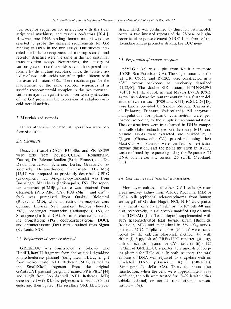
Fig. 1. E�ect of composition of transfected DNA on kinetics of appearance of SeAP activity. CV-1 cells were transiently transfected with
GREtkLUC2glucocorticoid receptor cDNA (pSVLGR or pSVLGR/C661S) and/or CMV-SeAP1 with the total DNA being brought to 50 mg/T150 ¯ask with Salmon testes DNA and then split into three dishes as described in Section 2. The average value of triplicate samples for the
detection of SeAP by chemiluminescence over time was plotted for the indicated samples. Similar results were obtained in ®ve other experiments.
N.J. Sarlis et al. / Journal of Steroid Biochemistry and Molecular Biology 68 (1999) 89±10292
expressed as a percentage of maximal activity with 1mM Dex, except when expressing the data from cellscontaining receptors that do not bind Dex (and hencedo not show any appreciable activity with 1 mM Dex),such as the mutants CS1 and CS1/CD. Thus, relativeLUC activity for cells treated with ethanol was 0%and for cells treated with 1 mM Dex was 100%. Unlessotherwise noted, all statistical analyses were performedwith the unpaired two-tailed Student t-test using theprogram InStat 2.03 for Macintosh (GraphPadSoftware, San Diego, CA). When the standard devi-ations of the two populations were signi®cantly di�er-ent, the Mann±Whitney test was used. All values areexpressed as mean2standard error of the mean(SEM).
3. Results
3.1. Normalization of transient transfection assays
In order to compare the fold induction of gene ex-pression in cells that are transiently transfected withdi�erent receptors, it is helpful to have a internal con-trol for transfection e�ciency. Unfortunately, cotrans-fection with the often used b-galactosidase gene wasnot suitable as the reporter activity was found to beweakly induced by 1 mM concentrations of either Dexor Dex±Mes (induction=1.2820.05 (SEM, n=28)and 1.3620.08 (SEM, n=24) for Dex and Dex±Mes
respectively). The fact that the fold induction by an
agonist (Dex) and an antagonist (Dex±Mes) was indis-
tinguishable (P=0.53 by two tailed Mann±Whitney
test) argues that the e�ect was not mediated by gluco-
corticoid receptors. However, it did mean that we
could not use the co-transfected b-galactosidase gene
as an internal control. Others have also reported pro-
blems with using the b-galactosidase gene as an in-
ternal control [52].
In order to avoid possible steroid-mediated changes
in an internal control, we turned to the secreted alka-
line phosphatase (SeAP) assay, in which the alkaline
phosphatase in the tissue culture medium is assayed
after the usual 36 h of co-transfection but before the
addition of steroid. This time, the total activity of the
control was in¯uenced by the precise composition of
the transfected DNA, even though the total amount of
DNA was constant (Fig. 1). Because we have been
unable to ®nd a control that was not in¯uenced either
by steroid or by DNA composition, we have normal-
ized all of our transfection assays by the total protein
of the assayed cells. It should be noted, however, that
internal controls are less crucial in determinations of
the relative activity of a given steroid. This is because
the steroid's activity is expressed as percent of maximal
induction by saturating concentrations of a full agonist
by the same receptor in the same cells, thus eliminating
the problems of inter-receptor or inter-cell compari-
sons.
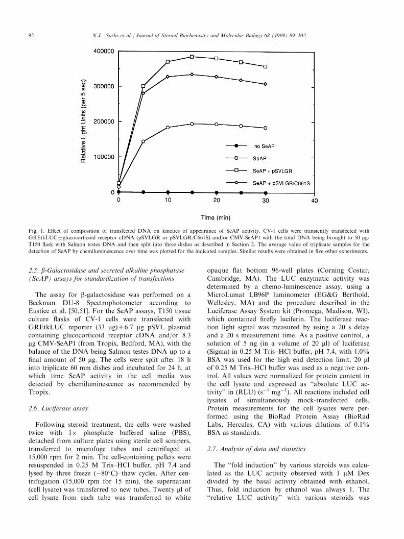
Fig. 2. Structure of wild type glucocorticoid receptor (wtGR) and mutant receptors used in this study. The numbers below the boxed regions of
DNA and steroid correspond to the end points of the DNA binding [53] and steroid binding [22] domains. The position and nature of the var-
ious mutations are indicated above and/or below each mutant receptor.
N.J. Sarlis et al. / Journal of Steroid Biochemistry and Molecular Biology 68 (1999) 89±102 93
3.2. Glucocorticoid induction of GREtkLUC reporter inCV-1 cells
The standard assay for GR-dependent transactiva-tion is based on the quanti®cation of induction bytransiently transfected GR of a simple reporter con-struct, such as GREtkLUC. For this assay, we trans-
fected either wild type (wt) GR or one of ®ve GRmutants (CS1, CS1/CD, 451/9, C656G, and R732Q ofFig. 2) with assorted properties. The CS1 and CS1/CDmutants are inducible by RU 486 but inactive withDex [48] and Dex±Mes [21]. 451/9 contains a doublemutation in the GR DNA binding domain, which con-ferred increased transactivation (and DNA binding
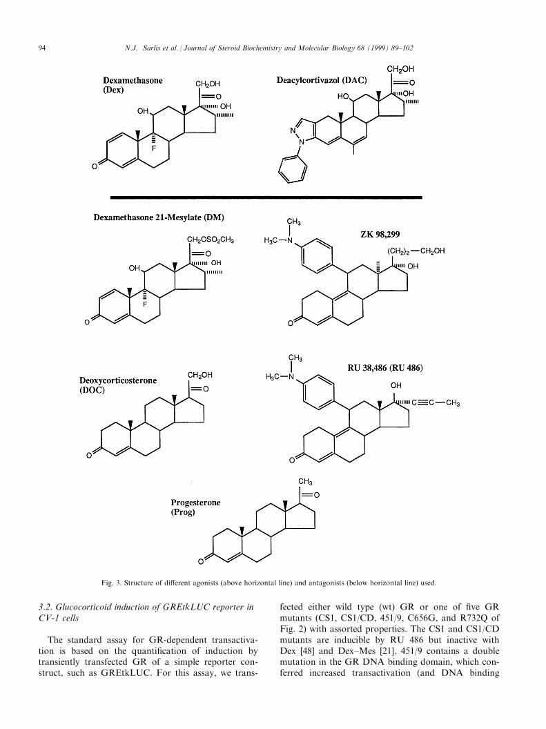
Fig. 3. Structure of di�erent agonists (above horizontal line) and antagonists (below horizontal line) used.
N.J. Sarlis et al. / Journal of Steroid Biochemistry and Molecular Biology 68 (1999) 89±10294

Fig. 4. Transcriptional activity of transfected receptors with various steroids in CV-1 cells. (A) Fold induction in cells2transfected wild type,
CS1, or CS1/CD GR after treatment with di�erent steroids. Triplicate dishes were assayed as described in Section 2 and the average fold
induction2SEM for n=3±9 separate experiments was plotted for the indicated steroids. (B) Relative transcriptional activity in cells2transfected
wild type, C656G, 451/9, or R732Q GR after treatment with di�erent steroids. Triplicate dishes were assayed as in (A) and the relative activity,
expressed as percent of full induction by 1 mM Dex, was determined. The average values2SEM for n=3±9 separate experiments except for PG
and DOC with 451/9 and C656G, where n=2, were then plotted for the indicated steroids (ND=not determined). In all cases, a lack of error
bar indicates an SEM<0.1.
N.J. Sarlis et al. / Journal of Steroid Biochemistry and Molecular Biology 68 (1999) 89±102 95

Fig. 5. Modulation of endogenous HeLa cell GR activity by transfected receptors in the presence of various steroids. (A) Fold induction of en-
dogenous GR2transfected wild type, CS1, or CS1/CD GR after treatment with di�erent steroids. Triplicate dishes were assayed as described in
Section 2 and the average fold induction2SEM for n=3±19 separate experiments was plotted for the indicated steroids. (B) Relative transcrip-
tional activity of endogenous GR2transfected wild type, C656G, 451/9, or R732Q GR after treatment with di�erent steroids. Triplicate dishes
were assayed as in (A) and the relative activity, expressed as percent of full induction by 1 mM Dex, was determined. The average values2SEM
for n=3±19 separate experiments were then plotted for the indicated steroids.
N.J. Sarlis et al. / Journal of Steroid Biochemistry and Molecular Biology 68 (1999) 89±10296

a�nity) in the context of the DNA binding domainfragment of 407±556 [47]. The C656G is a ``super-GR''with increased sensitivity to Dex [54] while the R732Qmutant possessed decreased sensitivity to Dex [21]. Thesteroids used were both saturating (1 mM) and subsa-turating (1 nM) concentrations of one of two agonists(Dex and DAC), as well as saturating (1 mM) concen-trations of one of ®ve antagonists (DOC, Dex±Mes,progesterone, RU 486, and ZK 98,299) with a varietyof structures (Fig. 3). The ®rst four antagonists aretype I antiglucocorticoids, displaying varying amountsof partial agonist activity in classical transactivationsystems. ZK 98,299 has been proposed as a ``pure anti-glucocorticoid'', or type II antagonist, that preventsthe DNA binding of receptor±steroid complexes [55],although modi®ed gel shift conditions disclosed goodDNA binding of ZK 98,299 bound progesterone recep-tors [56].
The endogenous GRs of CV-1 cells are insu�cientfor signi®cant levels of GR-mediated induction fromthe transiently transfected GREtkLUC reporter gene.Co-transfection of GR led to a dramatic increase infold induction values with saturating concentrations (1mM) of Dex or DAC (Fig. 4A).
The activities of the two agonists, Dex and DAC,were next examined with various mutant GRs. Bothsteroids failed to induce transactivation in cells thatwere co-transfected with either the CS1 or CS1/CDmutants (Fig. 4A). This is consistent with the inabilityof these receptors to bind Dex, and consequently toinduce transactivation [48], and suggests that DAC,like Dex, also does not bind to these receptors. Thisinactivity was not due to a lack of expression of thereceptors as RU 486 was able to induce reporter ac-tivity (Fig. 4A and Ref. [48]). Furthermore, the CS1and CS1/CD receptors can be covalently labeled byDex±Mes in transiently transfected cells [48]. However,no induction of either mutant was observed for Dex±Mes (Fig. 4A). A consistently weak, but not statisti-cally signi®cant (n=3), induction was seen for ZK98,299 with CS1 and CS1/CD. It should be noted thatthese data are presented in terms of fold induction foreach steroid as opposed to the more usual manner ofpercent of maximal induction by saturating concen-trations of Dex. This was necessitated by the inactivityof Dex with the CS1 and CS1/CD mutants.
When the 451/9 mutation was tested in the contextof the full length receptor, there was no augmentationin the activation by subsaturating concentrations ofDex (1 nM) compared to the wild type receptor (Fig.4B). This is to be compared to the increased activity ofthe receptor fragment 407±556 containing the 451/9mutation [48]. In contrast, the percentage of maximalactivity of C656G following treatment with subsaturat-ing concentrations of agonists was considerably greaterthan that for wild type receptors. These results con-
®rmed and extended our earlier reports that C656Gwas a ``super-sensitive'' GR mutant [46,54]. As waspreviously observed for the R732Q mutant, low con-centrations of Dex were essentially inactive (Fig. 4B),due to a ``right shift'' in the dose±response curve [21].
Treatment of the 451/9 mutant with Dex±Mes, pro-gesterone, and DOC, but not RU 486 or ZK 98,299,resulted in partial agonist activity. Interestingly, Dex±Mes was the only antagonist to give a response withthe 451/9 mutant that was signi®cantly di�erent fromthat of the wild type receptor (Fig. 4B). Conversely,the only antagonist that had the same activity withwild type and C656G receptors was Dex±Mes. RU486, progesterone, and DOC displayed signi®cantamounts of agonist activity with the wild type receptorbut none with the C656G (Fig. 4B). Dex±Mes alsoretained signi®cant levels of agonist activity whenbound to R732Q (Fig. 4B).
These results show that, with the exception of ZK98,299, there is no consistent behavior of all of the an-tagonists between individual mutant GRs. Thus, thevarious mutant receptor±steroid complexes present avariety of transcriptionally active complexes withwhich to probe the process of gene activation.
3.3. Modulation of endogenous GR transactivationactivity by transiently transfected GR
Our second transactivation assay, with transientlytransfected GRs in HeLa cells, is signi®cantly di�erentfrom the above described transactivation of a reportergene in transiently transfected CV-1 cells. For example,the binding of transfected GR to the GRE was notnecessary to produce a response in the HeLa cell assaybut is absolutely required in the assay of Fig. 4 withCV-1 cells [21]. This, along with the other propertiesdiscussed in Section 1, di�erentiate our HeLa cell in-duction assay from the conventional induction assays,such as the above assay for GREtkLUC activation inCV-1 cells.
As we reported earlier [21], transfection of concen-trations of GR expression plasmid su�cient to causean approximately 7-fold increase in the induction ofco-transfected GREtkLUC reporter in CV-1 cells (Fig.4A) generated less than a 50% increase in fold induc-tion of the same reporter in HeLa cells (Fig. 5A; foldinduction with 1 mM Dex after transfection of GR inHeLa cells=25.221.9 (SEM, n=36) vs. 20.621.4(SEM, n=36) in untransfected HeLa cells, valuesnon-signi®cantly di�erent (P=0.054)). Nonetheless,co-transfected GR produced a signi®cant increase inthe percent of maximal activity seen with subsaturatingconcentrations of Dex or DAC. At the same time,transfected GR caused a dramatic increase in the per-cent agonist activity displayed by antiglucocorticoids.Even the response to ZK 98,299 was increased,
N.J. Sarlis et al. / Journal of Steroid Biochemistry and Molecular Biology 68 (1999) 89±102 97

although the absolute increase was small. This re-sponse to transfected GR was relatively speci®c, inthat transfected progesterone receptors were withoute�ect [21].
Co-transfection of HeLa cells with the CS1 or CS1/CD mutants did not a�ord any signi®cant increase inthe fold induction by 1 mM Dex or DAC, in contrastto the increases seen with wild type GR (Fig. 5A).Transfected 451/9 and C656G mutant receptors eachled to increased activity with subsaturating concen-trations of both agonists (Fig. 5B). Conversely, theR732Q mutant bound by subsaturating concentrationsof Dex or DAC displayed no e�ect.
All antagonists tested with transfected CS1 or CS1/CD, with the exception of RU 486, failed to producean increase in the percent agonist activity in HeLacells. Treatment of the 451/9 mutant only with pro-gesterone, or DOC, led to any appreciable increase inthe percent agonist activity. The augmentation seenwith Dex±Mes in the presence of 451/9 was reproduci-ble but not statistically signi®cant (P=0.15, n=3). Incontrast, Dex±Mes was the only antagonist able toa�ord increased agonist activity with C656G (Fig. 5B).Treatment of R732Q transfected cells with Dex±Mesand RU 486 led to a weak, but not statistically signi®-cant, increase in the percent agonist activity (P=0.1or 0.07 respectively, n=4).
3.4. Comparison of activities of mutant receptors in thetwo transactivation assays
The activity of each steroid with the various GRswas virtually identical in the two di�erent transactiva-tion assays. For the CS1 and CS1/CD receptors, theonly steroid to show any signi®cant activity was RU486. Consistently higher values than background wereseen with ZK 98,299 but the level was much less thanthat with RU 486. To the extent that one can general-ize from a limited number of steroids, it appears thatCS1 and CS1/CD are inactive with agonists and dis-play activity with only one antagonist, RU 486. Whatis unusual about RU 486 is presently unknown.Interestingly, the absolute level of RU 486 activitywith CS1 or CS1/CD was equivalent to that seen withwild type GR in both assays (Fig. 4A vs. Fig. 5A).Thus, we conclude that the CS1 and CS1/CD mu-tations do not convert the wild type receptor to onethat is now activated by RU 486 but rather that thesemutations eliminate the ability of GR to respond toany other steroid that we have tested.
The activity of the 451/9 mutant with agonists wasidentical to that of the wild type receptor (Fig. 4B vs.Fig. 5B). The activity of the ®ve antagonists with the451/9 mutant was generally less than that with thewild type receptor except for progesterone and DOC,which displayed about the same activity with both
receptors. Likewise the C656G receptor, which hasmany properties that diverge from those of the wildtype receptor, responded similarly in the two assays.C656G was more active than wild type receptor withboth agonists and with Dex±Mes but had little or noactivity with the four other antiglucocorticoids (Fig.4B vs. Fig. 5B). This inactivity with at least progester-one was not due to an inability to bind progesterone,although the selectivity was certainly higher with theC656G [54]. The R732Q receptor had reduced activitywith subsaturating concentrations of agonists, appar-ently due to a decreased binding a�nity [21], and withsaturating concentrations of Dex±Mes. We have nottested the other antisteroids with R732Q in CV-1 cellsbut predict that they will be inactive, just as they werein the transactivation assay with HeLa cells.
In both activation assays, no generalizations couldbe made with regard to a relationship between steroidstructure and biological activity for either the wildtype or mutant GRs. However, the activity of eachsteroid with the various receptors, relative to full in-duction by 1 mM Dex, was the same in the two acti-vation assays. Thus, while there may be multiplemechanisms for repression [29±31,33,34,36], we couldnot detect any di�erences in our two transactivationassays. Therefore, it may be that the possibly diversemechanisms of activation all require similar confor-mations/structures of the GR receptor±steroid com-plex.
4. Discussion
This study shows that the expression of agonist ac-tivity either by two glucocorticoids or by ®ve antiglu-cocorticoids, all of dissimilar structure, can varybetween wild type and mutant receptors but remainsthe same among two dissimilar transactivation assaysin mammalian cells. These results argue that those fea-tures of receptor±steroid complex tertiary structurethat are required for the induction of biological ac-tivity are the same in our two assays. Whether thisconstancy will be maintained among other, yet to bediscovered, transactivation assays is an intriguing pro-spect that must await the characterization of suchfuture activation systems. To the best of our knowl-edge, our study is the ®rst to examine the behavior ofdi�erent steroids with glucocorticoid receptors in twodi�erent transactivation assays in mammalian cells.
The identical activity of each receptor in HeLa cells,with endogenous glucocorticoid receptors, and CV-1cells, with almost no receptors, argues against anye�ects being due to heterodimerization of receptors.Furthermore, our conclusions should not be a�ectedby possible di�erences in receptor concentration inHeLa vs. CV-1 cells. We have previously found that
N.J. Sarlis et al. / Journal of Steroid Biochemistry and Molecular Biology 68 (1999) 89±10298

the absolute activity of a given steroid, as a percent ofmaximal agonist activity, does depend upon the absol-ute level of receptors. However, the relative activity ofany two steroids moved up, or down, together withvarying amounts of receptor and thus was independentof receptor concentration [21].
Our observations of equal activity in two mamma-lian cell transactivation assays are not to be confusedwith the unequal activity of some steroid±glucocorti-coid receptor complexes in yeast vs. mammalian cells.In some cases, the di�erences could re¯ect modi®-cations of the receptor protein. Thus, the activity ofDex in CV-1 cells (Fig. 4B) but not in yeast [57]appears to be due to the 11000 fold lower a�nity ofDex for cell free glucocorticoid receptors from yeastvs. rat HTC cells [57]. Furthermore, transactivationdi�erences between yeast and mammalian cells havealso been seen in the absence of steroid binding. Forexample, the constitutive activities of three individualmutants (R488Q, R489K, and N491S) in the contextof the truncated rat GR (amino acids 1±556) were verydissimilar in yeast vs. mammalian cells [28]. Similarly,other C-terminal truncated receptors (amino acids 1±525) containing multiple mutations displayed di�erentproperties in yeast vs. mammalian cells [31]. Mostlikely, these inequalities stem from di�erences in thetranscriptional machinery and/or abundance of co-acti-vators, as suggested by the ability of GRIP1 to aug-ment GR action in yeast but to have no e�ect [58] orsquelch transactivation [59] in mammalian cells.Therefore, it is di�cult to separate the discrepancies intransactivation in yeast vs. mammalian cells frominteresting variations in the basic transcriptional ma-chinery. In contrast, our two assays, both in mamma-lian cell lines, o�er a way to probe for possibledi�erent interactions of a given receptor steroid com-plex with the same transcriptional components in dis-parate activation systems.
Our data also demonstrate that one is unable to pre-dict a relationship between steroid structure and ac-tivity among wild type or mutant GRs in either of ourmammalian transactivation assays. Conversely, thee�ect of a given receptor mutation was not interpreteduniformly, but rather varied among the various anti-glucocorticoids. Several reports have appeared wherelimited modi®cations in receptor sequence werecapable of converting an antagonist to an agonist[48,60,61]. However, as shown in the present study, itmay not be possible to generalize from the behavior ofone antisteroid to that of all antisteroids. The C656Gmutant displays good activity with Dex±Mes but notRU 486, progesterone, or DOC (Fig. 4B and Fig. 5B).Even more dramatic is that only RU 486, and maybeZK 98,299, displayed any agonist activity with the CS1and CS1/CD mutant receptors (Fig. 4A and Fig. 5A).This suggests that subtle modi®cations in receptor
structure are involved in the expression of agonist vs.antagonist activity, as might be expected for aninduced ®t following the binding of structurally diversesteroids to receptors [62,63].
It should also be noted that the amount of tran-scriptional activity induced by RU 486 (and ZK98,299) with either CS1 or CS1/CD was very similar tothat observed in RU 486-treated cells transfected withwtGR. Thus, it seems more reasonable to proposethat, in this situation, the CS1 and CS1/CD mutationsdid not ``convert'' an antisteroid to an agonists butrather eliminated the ability of other steroids to causeany activation. The low level of activity of ZK 98,299,which had been previously noted [64], also suggeststhat ZK 98,299 should not be considered as a type IIantagonist giving complexes incapable of binding toGRE sequences. This conclusion is consistent with therecent observation that ZK 98,299 bound progesteronereceptors can bind to DNA [56].
It is interesting that the previously describedincreased biological activity of the 451/9 doublemutant in the context of the truncated GR (aminoacids 407±556) [47] was not maintained in the fulllength receptor in either transactivation assay (Fig. 4Band Fig. 5B) or in its ability to increase the fold induc-tion (fold-induction with 1 mM Dex after transfectionof 0.1 mg of pSVLGR into CV-1 cells=12.2723.01-fold vs. 12.5925.69-fold with GR 451/9 (2 SD,n=3); P=0.94). This was particularly relevant in theCV-1 cell assay of Fig. 4, where DNA binding isrequired for biological activity. On the other hand, onemight not have expected mutations at positions 451and 459, which are involved in DNA backbone con-tacts and the speci®city of DNA-binding [65], to e�ectbiological activity in view of the generally poor corre-lation between the a�nity of DNA binding and themagnitude of the transcriptional activation response[66]. For this reason, it was somewhat surprising thatthis mutation did modify the amount of activity dis-played by both 1 nM Dex and 1 mM Dex±Mes in eachassay (Fig. 4B and Fig. 5B). Thus, there may be somecommunication between the GR DNA binding domainand the transactivation domains at either ends of thereceptor. A similar ability of the DNA binding domainto alter the activity of the full length receptor has beendescribed by Yamamoto et al. [39].
The C656G ``super-GR'' displayed increased induc-tion with Dex and DAC, but was inactive with mostantagonists. These results support our previous ®nd-ings of a decreased relative a�nity of the C656G GRfor progesterone and aldosterone [54]. The Cys-656residue lies within the steroid-binding cavity of GRbut appears to be closest to the C-17 side chain of thesteroidal D-ring [46,67]. Therefore, a molecular expla-nation for the increased speci®city of the C656Gmutant for modi®cations in other regions of the ster-
N.J. Sarlis et al. / Journal of Steroid Biochemistry and Molecular Biology 68 (1999) 89±102 99
oid structure is not yet clear but may involve ligandbinding induced, but currently unpredictable, reorgani-zations of the ligand binding domain [62,63].
The R732Q mutant possessed decreased inductionwith subsaturating concentrations of both agonists(Dex and DAC) and was inactive with most antagon-ists. The Arg-732 (R732) residue is highly conserved inthe nuclear receptor family and represents the ®rstamino acid of helix 10 of the proposed structure of theGR ligand binding domain [41]. Although helix 10 isthought not to participate directly in ligand binding ortransactivation, secondary e�ects on protein structureare always possible, as have been observed for a pointmutation within the glucocorticoid receptor DNAbinding domain [68,69]. Indeed, it has been previouslydemonstrated that the point mutant R732Q shows ap-proximately the same amount of transcriptional ac-tivity with wtGR in the presence of Dex±Mes, but aright-shifted dose±response curve for Dex induction inCV-1 cells [21]. Hence, R732Q has now been furthercharacterized, and indeed meets the criteria of a ``rightshifted'' receptor, in direct antithesis with the ``leftshifted'' C656G mutant.
In summary, the present studies show that no simplegeneralizations are possible about receptor±steroidcomplex activity. While the properties of a given com-plex were the same in two transactivation assays, thevarious transcriptional responses with di�erent steroidssuggest that a continuum of tertiary structures, per-haps due to induced ®ts of the receptor ligand bindingdomain, are obtained in response to the binding of theassorted steroid structures. Further variety of re-sponses is possible if the ability of receptor to interactwith the ever increasing number of co-activators andco-adaptors [70] is also sensitive to the continuum ofsteroid-induced tertiary structures. It will be interestingto see whether these predictions can be experimentallycon®rmed.
Acknowledgements
We thank Jon Ashwell, Etienne Baulieu, GordonHager, David Henderson, Keiko Ozato, SandroRusconi, and Keith Yamamoto for the generous do-nation of reagents, Keith Yamamoto for the sharingof unpublished data, and Paul M. Yen (NIH) for criti-cal review of this manuscript.
References
[1] M. Berry, D. Metzger, P. Chambon, Role of the two activating
domains of the oestrogen receptor in the cell-type and promo-
ter-context dependent agonistic activity of the anti-oestrogen 4-
hydroxytamoxifen, EMBO J. 9 (1990) 2811±2818.
[2] S. Nagpal, M. Saunders, P. Kastner, B. Durand, H. Nakshatri,
P. Chambon, Promoter context- and response element-depen-
dent speci®city of the transcriptional activation and modulating
functions of retinoic acid receptors, Cell 70 (1992) 1007±1019.
[3] M.T. Tzukerman, A. Esty, D. Santiso-Mere, P. Danielian,
M.G. Parker, R.B. Stein, J.W. Pike, D.P. McDonnell, Human
estrogen receptor transactivational capacity is determined by
both cellular and promoter context and mediated by two func-
tionally distinct intramolecular regions, Mol. Endocrinol. 8
(1994) 21±30.
[4] E.C. Guido, E.O. Delorme, D.L. Clemm, R.B. Stein, J. Rosen,
J.N. Miner, Determinants of promoter-speci®c activity by glu-
cocorticoid receptor, Mol. Endocrinol. 10 (1996) 1178±1190.
[5] B. Durand, M. Saunders, C. Gaudon, B. Roy, R. Losson, P.
Chambon, Activation function 2 (AF-2) of retinoic acid recep-
tor and 9-cis retinoic acid receptor: presence of a conserved
autonomous constitutive activating domain and in¯uence of
the nature of the response element on AF-2 activity, EMBO J.
13 (1994) 5370±5382.
[6] W.L. Kraus, M.M. Montano, B.S. Katzenellenbogen,
Identi®cation of multiple, widely spaced estrogen-responsive
regions in the rat progesterone receptor gene, Mol. Endocrinol.
8 (1994) 952±969.
[7] L. Mercier, P.A. Miller, S.S. Simons Jr., Antiglucocorticoid
steroids have increased agonist activity in those hepatoma cell
lines that are more sensitive to glucocorticoids, J. Steroid
Biochem. 25 (1986) 11±20.
[8] G. Wasner, H. Oshima, E.B. Thompson, S.S. Simons Jr.,
Unlinked regulation of the sensitivity of primary glucocorti-
coid-inducible responses in mouse mammary tumor virus
infected Fu5-5 rat hepatoma cells, Mol. Endocrinol. 2 (1988)
1009±1017.
[9] B. Turcotte, M.-E. Meyer, M.-T. Bocquel, L. Belanger, P.
Chambon, Repression of the a-fetoprotein gene promoter by
progesterone and chimeric receptors in the presence of hor-
mones and antihormones, Mol. Cell. Biol. 10 (1990) 5002±
5006.
[10] H. Oshima, S.S. Simons Jr., Modulation of glucocorticoid in-
duction of tyrosine aminotransferase gene expression by vari-
ations in cell density, Endocrinology 130 (1992) 2106±2112.
[11] H. Oshima, S.S. Simons Jr., Modulation of transcription factor
activity by a distant steroid modulatory element, Mol.
Endocrinol. 6 (1992) 416±428.
[12] C.A. Beck, N.L. Weigel, M.L. Moyer, S.K. Nordeen, D.P.
Edwards, The progesterone antagonist RU486 acquires agonist
activity upon stimulation of cAMP signaling pathways, Proc.
Natl. Acad. Sci. USA 90 (1993) 4441±4445.
[13] N. Fujimoto, B.S. Katzenellenbogen, Alteration in the agonist/
antagonist balance of antiestrogens by activation of protein
kinase A signaling pathways in breast cancer cells: antiestrogen
selectivity and promoter dependence, Mol. Endocrinol. 8
(1994) 296±304.
[14] A. Chauchereau, K. Cohen-Solal, A. Jolivet, A. Bailly, E.
Milgrom, Phosphorylation sites in ligand-induced and ligand-
independent activation of the progesterone receptor,
Biochemistry 33 (1994) 13295±13303.
[15] R.F. Power, S.K. Mani, J. Codina, O.M. Conneely, B.W.
O'Malley, Dopaminergic and ligand-independent activation of
steroid hormone receptors, Science 254 (1991) 1636±1639.
[16] S.S. Simons Jr., E.B. Thompson, Dexamethasone 21-mesylate:
an a�nity label of glucocorticoid receptors from rat hepatoma
tissue culture cells, Proc. Natl. Acad. Sci. USA 78 (1981) 3541±
3545.
[17] L. Mercier, E.B. Thompson, S.S. Simons Jr., Dissociation of
steroid binding to receptors and steroid induction of biological
activity in a glucocorticoid responsive cell, Endocrinology 112
(1983) 601±609.
N.J. Sarlis et al. / Journal of Steroid Biochemistry and Molecular Biology 68 (1999) 89±102100

[18] D. Szapary, H. Oshima, S.S. Simons Jr., Modulation of gluco-
corticoid induction of stably transfected tyrosine aminotrans-
ferase gene constructs involves elements up-stream of the
glucocorticoid responsive element, Endocrinology 130 (1992)
3492±3502.
[19] S.S. Simons Jr., H. Oshima, D. Szapary, Higher levels of con-
trol: Modulation of steroid hormone-regulated gene transcrip-
tion, Mol. Endocrinol. 6 (1992) 995±1002.
[20] C.D. Collier, H. Oshima, S.S. Simons Jr., A negative tyrosine
aminotransferase gene element that blocks glucocorticoid mod-
ulatory element-regulated modulation of glucocorticoid-
induced gene expression, Mol. Endocrinol. 10 (1996) 463±476.
[21] D. Szapary, M. Xu, S.S. Simons Jr., Induction properties of a
transiently transfected glucocorticoid-responsive gene vary with
glucocorticoid receptor concentration, J. Biol. Chem. 271
(1996) 30576±30582.
[22] M. Xu, P.K. Chakraborti, M.J. Garabedian, K.R. Yamamoto,
S.S. Simons Jr., Modular structure of glucocorticoid receptor
domains is not equivalent to functional independence. Stability
and activity of the steroid binding domain are controlled by
sequences in separate domains, J. Biol. Chem. 271 (1996)
21430±21438.
[23] S.S. Simons Jr., F.D. Sistare, P.K. Chakraborti, Steroid bind-
ing activity is retained in a 16-kDa fragment of the steroid
binding domain of rat glucocorticoid receptors, J. Biol. Chem.
264 (1989) 14493±14497.
[24] G.F. Allan, X. Leng, S.Y. Tsai, N.L. Weigel, D.P. Edwards,
M.-J. Tsai, B.W. O'Malley, Hormone and antihormone induce
distinct conformational changes which are central to steroid
receptor activation, J. Biol. Chem. 267 (1992) 19513±19520.
[25] E. Vegeto, G.F. Allan, W.T. Schrader, M.-J. Tsai, D.P.
McDonnell, B.W. O'Malley, The mechanism of RU486 antag-
onism is dependent on the conformation of the carboxy-term-
inal tail of the human progesterone receptor, Cell 69 (1992)
703±713.
[26] S. S. Simons Jr., Structure and function of the steroid and
nuclear receptor ligand-binding domain, in: L.P. Freedman
(Ed.), The Molecular Biology of Steroid and Nuclear Hormone
Receptors, BirkaÈ user, Boston, 1998, pp. 35±104.
[27] K.J. Modarress, J. Opoku, M. Xu, N.J. Sarlis, S.S. Simons Jr.,
Steroid-induced conformational changes at ends of the hor-
mone binding domain in the rat glucocorticoid receptor are
independent of agonist vs. antagonist activity, J. Biol. Chem.
272 (1997) 23986±23994.
[28] M. Schena, L.P. Freedman, K.R. Yamamoto, Mutations in the
glucocorticoid receptor zinc ®nger region that distinguish inter-
digitated DNA binding and transcriptional enhancement activi-
ties, Genes Develop. 3 (1989) 1590±1601.
[29] S. Heck, M. Kullmann, A. Gast, H. Ponta, H.J. Rahmsdorf,
P. Herrlich, A.C.B. Cato, A distinct modulating domain in glu-
cocorticoid receptor monomers in the repression of activity of
the transcription factor AP-1, EMBO J. 13 (1994) 4087±4095.
[30] D.B. Starr, W. Matsui, J.A. Thomas, K.R. Yamamoto,
Intracellular receptors use a common mechanism to interpret
signaling information at response elements, Genes Develop. 10
(1996) 1271±1283.
[31] J.A. Iniguez-Lluhi, D.I. Lou, K.R. Yamamoto, Three amino
acid substitutions selectively disrupt the activation but not the
repression function of the glucocorticoid receptor N terminus,
J. Biol. Chem. 272 (1997) 4149±4156.
[32] Z. Nawaz, M.-J. Tsai, B. O'Malley, Speci®c mutations in the
ligand binding domain selectively abolish the silencing function
of human thyroid hormone receptor beta, Proc. Natl. Acad.
Sci. USA 92 (1995) 11691±11695.
[33] F.C. Lucibello, E.P. Slater, K.U. Jooss, M. Beato, R. Muller,
Mutual transrepression of fos and the glucocorticoid receptor:
involvement of a functional domain in fos which is absent in
fosB, EMBO 9 (1990) 2827±2834.
[34] H.-F. Yang-Yen, J.-C. Chambard, Y.-L. Sun, T. Smeal, T.J.
Schmidt, J. Drouin, M. Karin, Transcriptional interference
between c-Jun and the glucocorticoid receptor: mutual inhi-
bition of DNA binding due to direct protein±protein inter-
action, Cell 62 (1990) 1205±1215.
[35] J. Drouin, Y.L. Sun, M. Chamberland, Y. Gauthier, A. De
Lean, M. Nemer, T. Schmidt, Novel glucocorticoid receptor
complex with DNA element of the hormone-repressed POMC
gene, EMBO J. 12 (1993) 145±156.
[36] T. Meyer, D.B. Starr, J. Carlstedt-Duke, The rat glucocorticoid
receptor mutant K461A di�erentiates between two di�erent
mechanisms of transrepression, J. Biol. Chem. 272 (1997)
21090±21095.
[37] J. Liden, F. Delaunay, I. Rafter, J. Gustafsson, S. Okret, A
new function for the C-terminal zinc ®nger of the glucocorti-
coid receptor, J. Biol. Chem. 272 (1997) 21467±21472.
[38] E.B. Helmer, B.M. Raaka, H.H. Samuels, Hormone-dependent
and -independent transcriptional activation by thyroid hor-
mone receptors are mediated by di�erent mechanisms,
Endocrinology 137 (1996) 390±399.
[39] J.A. Lefstin, J.R. Thomas, K.R. Yamamoto, In¯uence of a
steroid receptor DNA-binding domain on transcriptional regu-
latory functions, Genes Develop. 8 (1994) 2842±2856.
[40] J.N. Vanderbilt, R. Miesfeld, B.A. Maler, K.R. Yamamoto,
Intracellular receptor concentration limits glucocorticoid-
dependent enhancer activity, Mol. Endocrinol. 1 (1987) 68±74.
[41] J.-M. Wurtz, W. Bourguet, J.-P. Renaud, V. Vivat, P.
Chambon, D. Moras, H. Gronemeyer, A canonical structure
for the ligand-binding domain of nuclear receptors, Nature
Struct. Biol. 3 (1996) 87±94.
[42] S.S. Simons Jr., M. Pons, D.F. Johnson, a-Keto mesylate: a
reactive thiol-speci®c functional group, J. Org. Chem. 45
(1980) 3084±3088.
[43] S. S. Simons Jr., Glucocorticoid receptor, in: H. Gronemeyer
(Ed.), A�nity Labelling and Cloning of Steroid and Thyroid
Hormone Receptors, Ellis Horwood Ltd, Chichester, England,
1988, pp. 28±54.
[44] S.Y. Tsai, M.-J. Tsai, B.W. O'Malley, Cooperative binding of
steroid hormone receptors contributes to transcriptional syner-
gism at target enhancer elements, Cell 57 (1989) 443±448.
[45] R. Miesfeld, S. Rusconi, P.J. Godowski, B.A. Maler, S. Okret,
A.-C. Wikstrom, J.-A. Gustafsson, K.R. Yamamoto, Genetic
complementation of a glucocorticoid receptor de®ciency by ex-
pression of cloned receptor cDNA, Cell 46 (1986) 389±399.
[46] P.K. Chakraborti, M.J. Garabedian, K.R. Yamamoto, S.S.
Simons Jr., Role of cysteines 640, 656, and 661 in steroid bind-
ing to rat glucocorticoid receptors, J. Biol. Chem. 267 (1992)
11366±11373.
[47] E. Zandi, I. Galli, U. DoÈ bbeling, S. Rusconi, Zinc ®nger mu-
tations that alter domain interactions in the glucocorticoid
receptor, J. Mol. Biol. 230 (1993) 124±136.
[48] R.B. Lanz, S. Rusconi, A conserved carboxy-terminal subdo-
main is important for ligand interpretation and transactivation
by nuclear receptors, Endocrinology 135 (1994) 2183±2194.
[49] D. Szapary, H. Oshima, S.S. Simons Jr., A new cis-acting el-
ement involved in tissue-selective glucocorticoid inducibility of
tyrosine aminotransferase gene expression, Mol. Endocrinol. 7
(1993) 941±952.
[50] D.C. Eustice, P.A. Feldman, A.M. Colberg-Poley, R.M.
Buckery, R.H. Neubauer, A sensitive method for the detection
of beta-galactosidase in transfected mammalian cells,
Biotechniques 11 (1991) 739±740.
[51] D.C. Eustice, P.A. Feldman, A.M. Colberg-Poley, R.M.
Buckery, R.H. Neubauer, A sensitive method for the detection
N.J. Sarlis et al. / Journal of Steroid Biochemistry and Molecular Biology 68 (1999) 89±102 101
of beta-galactosidase in transfected mammalian cells,
Biotechniques 11 (1991) 742±743.
[52] D.B. Schowalter, W.P. Sullivan, N.J. Maihle, A.D.W. Dobson,
O.M. Conneely, B.W. O'Malley, D.O. Toft, Characterization
of progesterone receptor binding to the 90-and 70-kDa heat
shock proteins, J. Biol. Chem. 266 (1991) 21165±21173.
[53] S.S. Simons Jr., Function/activity of speci®c amino acids in
glucocorticoid receptors, Vitamins Hormones 48 (1994) 49±130.
[54] P.K. Chakraborti, M.J. Garabedian, K.R. Yamamoto, S.S.
Simons Jr., Creation of ``super'' glucocorticoid receptors by
point mutations in the steroid binding domain, J. Biol. Chem.
266 (1991) 22075±22078.
[55] M.T. Bocquel, J. Ji, T. Ylikomi, B. Benhamou, A. Vergezac, P.
Chambon, H. Gronemeyer, Type II antagonists impair the
DNA binding of steroid hormone receptors without a�ecting
dimerization, J. Steroid Biochem. Mol. Biol. 45 (1993) 205±
215.
[56] E.K. Gass, S.A. Leonhardt, S.K. Nordeen, D.P. Edwards, The
antagonists RU486 and ZK98299 stimulate progesterone recep-
tor binding to deoxyribonucleic acid in vitro and in vivo, but
have distinct e�ects on receptor conformation, Endocrinology
139 (1998) 1905±1919.
[57] M.J. Garabedian, K.R. Yamamoto, Genetic dissection of the
signaling domain of a mammalian steroid receptor in yeast,
Mol. Biol. Cell 3 (1992) 1245±1257.
[58] J.J. Voegel, M.J.S. Heine, C. Zechel, P. Chambon, H.
Gronemeyer, T1F2, a 160 kDa transcriptional mediatior for
the ligand-dependent activation function AF-2 of nuclear
receptors, EMBO J. 15 (1996) 3667±3675.
[59] H. Hong, K. Kohli, A. Trivedi, D.L. Johnson, M.R. Stallcup,
GRIP1, a novel mouse protein that serves as a transcriptional
coactivator in yeast for the hormone binding domains of ster-
oid receptors, Proc. Natl. Acad. Sci. USA 93 (1996) 4948±
4952.
[60] S.-Y. Jiang, S.M. Langan-Fahey, A.L. Stella, R. McCague,
V.C. Jordan, Point mutation of estrogen receptor (ER) in the
ligand-binding domain changes the pharmacology of antiestro-
gens in ER-negative breast cancer cells stably expressing comp-
lementary DNAs for ER, Mol. Endocrinol. 6 (1992) 2167±
2174.
[61] A. Mahfoudi, E. Roulet, S. Dauvois, M.G. Parker, W. Wahli,
Speci®c mutations in the estrogen receptor change the proper-
ties of antiestrogens to full agonoists, Proc. Natl. Acad. Sci.
USA 92 (1995) 4206±4210.
[62] A.M. Brzozowski, A.C.W. Pike, Z. Dauter, R.E. Hubbard, T.
Bonn, O. Engstrom, L. Ohman, G.L. Greene, J.-A.
Gustafsson, M. Carlquist, Molecular basis of agonism and an-
tagonism in the oestrogen receptor, Nature 389 (1997) 753±
758.
[63] J. Ostrowski, T. Roalsvig, L. Hammer, A. Marinier, J.E.
Starrett, K.-L. Yu, P.R. Reczek, Serine 232 and methionine
272 de®ne the ligand binding pocket in retinoic acid receptor
subtypes, J. Biol. Chem. 273 (1998) 3490±3495.
[64] S.S. Simons Jr., P.M. Yen, Variations in agonist activity
among antiglucocorticoid steroids and its relation to glucocor-
ticoid regulated genes, in: T.C. Spelsberg, R. Kumar (Eds.),
Steroid and Sterol Hormone Action, Martinus Nijho�, Boston,
1987, pp. 251±268.
[65] B.F. Luisi, W.X. Xu, Z. Otwinowski, L.P. Freedman, K.R.
Yamamoto, P.B. Sigler, Crystallographic analysis of the inter-
action of the glucocorticoid receptor with DNA, Nature 352
(1991) 497±505.
[66] A.J. Adler, A. Scheller, D.M. Robins, The stringency and mag-
nitude of androgen-speci®c gene activation are combinatorial
functions of receptor and nonreceptor binding site sequences,
Mol. Cell. Biol. 13 (1993) 6326±6335.
[67] S.S. Simons Jr., J.G. Pumphrey, S. Rudiko�, H.J. Eisen,
Identi®cation of cysteine-656 as the amino acid of HTC cell
glucocorticoid receptors that is covalently labeled by dexa-
methasone 21-mesylate, J. Biol. Chem. 262 (1987) 9676±9680.
[68] H. Berglund, M. Wolf-Watz, T. Lundback, S. van den Berg, T.
Hard, Structure and dynamics of the glucocorticoid receptor
DNA-binding domain: comparison of wild type and a mutant
with altered speci®city, Biochemistry 36 (1997) 11188±11197.
[69] M.A.A. van Tilborg, J.A. Lefstin, R. Boelens, K.R.,
Yamamoto, R. Kaptein (in preparation).
[70] K.B. Horwitz, T.A. Jackson, D.L. Bain, J.K. Richer, G.S.
Takimoto, L. Tung, Nuclear receptor coactivators and core-
pressors, Mol. Endocrinol. 10 (1996) 1167±1177.
N.J. Sarlis et al. / Journal of Steroid Biochemistry and Molecular Biology 68 (1999) 89±102102
Copyright © 2022 FDOKUMEN


![A Flexible Copper(I)-Complexed [4]Rotaxane Containing Two Face-to-Face Porphyrinic Plates that Behaves as a Distensible Receptor](https://static.fdokumen.com/doc/165x107/632a7148d2eed9476207508b/a-flexible-copperi-complexed-4rotaxane-containing-two-face-to-face-porphyrinic.jpg)