Preventing the Predictable. Type 1 diabetes in children - Lund ...
140
Preventing the Predictable. Type 1 diabetes in children: Risk factors and impact of participation in prospective follow-up Lundgren, Markus 2017 Document Version: Förlagets slutgiltiga version Link to publication Citation for published version (APA): Lundgren, M. (2017). Preventing the Predictable. Type 1 diabetes in children: Risk factors and impact of participation in prospective follow-up. Lund University: Faculty of Medicine. Total number of authors: 1 Creative Commons License: Ospecificerad General rights Unless other specific re-use rights are stated the following general rights apply: Copyright and moral rights for the publications made accessible in the public portal are retained by the authors and/or other copyright owners and it is a condition of accessing publications that users recognise and abide by the legal requirements associated with these rights. • Users may download and print one copy of any publication from the public portal for the purpose of private study or research. • You may not further distribute the material or use it for any profit-making activity or commercial gain • You may freely distribute the URL identifying the publication in the public portal Read more about Creative commons licenses: https://creativecommons.org/licenses/ Take down policy If you believe that this document breaches copyright please contact us providing details, and we will remove access to the work immediately and investigate your claim.
-
Upload
khangminh22 -
Category
Documents
-
view
2 -
download
0
Transcript of Preventing the Predictable. Type 1 diabetes in children - Lund ...
LUND UNIVERSITY
PO Box 117221 00 Lund+46 46-222 00 00
Preventing the Predictable. Type 1 diabetes in children: Risk factors and impact ofparticipation in prospective follow-up
Lundgren, Markus
2017
Document Version:Förlagets slutgiltiga version
Link to publication
Citation for published version (APA):Lundgren, M. (2017). Preventing the Predictable. Type 1 diabetes in children: Risk factors and impact ofparticipation in prospective follow-up. Lund University: Faculty of Medicine.
Total number of authors:1
Creative Commons License:Ospecificerad
General rightsUnless other specific re-use rights are stated the following general rights apply:Copyright and moral rights for the publications made accessible in the public portal are retained by the authorsand/or other copyright owners and it is a condition of accessing publications that users recognise and abide by thelegal requirements associated with these rights. • Users may download and print one copy of any publication from the public portal for the purpose of private studyor research. • You may not further distribute the material or use it for any profit-making activity or commercial gain • You may freely distribute the URL identifying the publication in the public portal
Read more about Creative commons licenses: https://creativecommons.org/licenses/Take down policyIf you believe that this document breaches copyright please contact us providing details, and we will removeaccess to the work immediately and investigate your claim.

MA
RK
US LU
ND
GR
EN
Preventing the Predictable: Type 1 diabetes in children: Risk factors and im
pact of participation in prospective follow-up 2017:165 9
7891
7619
5475
Lund UniversityDepartment of Clinical Sciences, Malmö
Lund University, Faculty of Medicine Doctoral Dissertation Series 2017:165
ISBN 978-91-7619-547-5ISSN 1652-8220
Preventing the PredictableType 1 diabetes in children: Risk factors and impact of participation in prospective follow-upMARKUS LUNDGREN FACULTY OF MEDICINE | LUND UNIVERSITY
This thesis focuses on several steps in the type 1 diabetes research process. We start by pre-senting data on early-life risk factors from the DiPiS and TEDDY studies relating to umbilical cord blood autoantibodies, early life stress and the use of analgesic antipyretics. We then present data on the impact of prospec-tive follow-up and study participation regar-ding status at diabetes diagnosis and during regular clinical follow-up. Finally, results from a clinical trial, DiAPREV-IT, on immune tole-rance with GAD-Alum in high-risk children are presented.
Preventing the Predictable
Markus Lundgren is a pediatric diabetologist at Kristianstad central hospital. His research focuses on the DiPiS, TEDDY and DiAPREV-IT studies at the unit for Diabetes and Celiac disease, Department of clinical sciences Malmö.

1
Preventing the Predictable Type 1 diabetes in children: Risk factors and impact of participation in prospective
follow-up
Markus Lundgren
DOCTORAL DISSERTATION by due permission of the Faculty of Medicine, Lund University, Sweden.
To be defended at the lecture hall at Kvinnokliniken, Jan Waldenströms gata 47, Skåne University Hospital, Malmö on Friday November 24th, 2017 at 13.00.
Faculty opponent Lars Christian Stene (MD, PhD)
Oslo Diabetes Research Centre, Oslo, Norway

Organization LUND UNIVERSITY
Document name Doctoral Dissertation
Department of Clinical Sciences, Malmö Date of issue November 24th, 2017
Author(s): Markus Lundgren Sponsoring organization Title and subtitle: Preventing the Predictable: Type 1 diabetes in children: Risk factors and impact of participation in prospective follow-up
Abstract
Aim: The aim of this thesis was to identify risk factors for type 1 diabetes in children, evaluate the effects of
participation in prospective follow-up and the safety of immune tolerance treatment with GAD-Alum in children with
islet autoimmunity.
Methods: Subjects from the Swedish DiPiS cohort were studied regarding risk factors, cord blood islet
autoantibodies and the effects of early severe life events. We further evaluated morbidity at diagnosis and glycemic
control during the first two years of diabetes, comparing the DiPiS follow-up cohort to non-enrolled children. The
TEDDY cohort was used to analyze prevalence and effect of analgesic antipyretics before 2.5 years of age on islet
autoimmunity at 6 years of age and patterns of analgesic antipyretic use at the study sites. The safety and efficacy of
immune tolerance treatment with GAD-Alum were analyzed in a five-year follow-up of at-risk children.
Results: IA-2A autoantibodies in cord blood predicted an increased hazard for type 1 diabetes (HR 6.88; 95% CI
1.46, 32.4; p=0.003.). Parental severe life events after pregnancy predicted type 1 diabetes risk for both the total
cohort (HR 1.66; 955 CI 1.02, 2.70; p=0.043) and the DQ2/8 cohort (HR 2.21; 95%CI 1.08, 4.51; p=0.03). The use of
analgesic antipyretics did not predict islet autoimmunity at age six years (HR 1.02; 95% CI 0.99, 1.09; p=0.27) but
weakly predicted islet autoimmunity at age 3 years. The use of analgesic antipyretics differed between study sites,
with a higher prevalence in the US (95.7%) and Sweden (94.8%) than in Finland (78.1%) and Germany (80.2%). Use
in the absence of fever was common, especially in the US in comparison to the other sites. Subjects enrolled in DiPiS
follow-up were diagnosed with diabetes with less morbidity (p=0.014) and lower prevalence of ketoacidosis
(p=0.005). Among subjects enrolled in DiPiS follow-up, HbA1c at diagnosis was lower (9 mmol/mol; p=0.006) and
glycemic control was better after diagnosis (12 months, 4 mmol/mol; p=0.009, 24 months, 9 mmol/mol; p <0.001).
Immune tolerance treatment with GAD-Alum is safe but does not affect the time to diabetes diagnosis in this cohort.
Conclusion: Early severe life events and IA-2A cord blood antibodies but not early use of analgesic antipyretics may
increase type 1 diabetes risk. Severe life events and cord blood autoantibodies are both rare events, and care must
be taken in interpreting these results. Participants in prospective follow-up are diagnosed at an early stage, with low
morbidity and improved glycemic control, which may be an important factor for recruitment and ethical approval.
Immune tolerance with GAD-Alum is safe, but larger, stratified studies are needed to ascertain the possible effects. Key words: Type 1 diabetes; Risk factors; Prospective follow-up; Immune tolerance; Severe life events
Classification system and/or index terms (if any)
Supplementary bibliographical information Language:English
ISSN and key title: 1652-8220, Lund University, Faculty of Medicine Doctoral Dissertation Series 2017:165
ISBN 978-91-7619-547-5
Recipient’s notes Number of pages 110 Price
Security classification I, the undersigned, being the copyright owner of the abstract of the above-mentioned dissertation, hereby grant to all reference sources permission to publish and disseminate the abstract of the above-mentioned dissertation.
Signature Date October 19th, 2017
3
Preventing the Predictable Type 1 diabetes in children: Risk factors and
impact of participation in prospective follow-up
Markus Lundgren
4
Cover photo by Mikael Risedal, Lund university image bank.
© Markus Lundgren Faculty of Medicine Doctoral Dissertation Series 2017:165Department of Clinical Sciences, Malmö Lund University ISBN 978-91-7619-547-5 ISSN 1652-8220 Printed in Sweden by Media-Tryck, Lund University Lund 2017

5
“The prophylactic and etiological treatment of diabetes will surely play an important role in the future, and it is already
plain that progress will be along two lines: 1) toward the early detection of the disease, and 2) toward the prevention of the
development of the disease in those susceptible to it.” Elliott Joslin, 1916

6
Table of contents
Thesis at a glance ..................................................................................................... 8Abbreviations ........................................................................................................... 9Abstract .................................................................................................................. 11List of publications ................................................................................................ 13Introduction ............................................................................................................ 15
The history of T1D ...................................................................................... 15Diagnosis of Diabetes .................................................................................. 17
Definition of diabetes .......................................................................... 17Stages of T1D ..................................................................................... 20
Epidemiology of T1D .................................................................................. 21Pathogenesis of T1D .................................................................................... 25T1D risk assessment .................................................................................... 26
Autoantibodies .................................................................................... 26HLA .................................................................................................... 30Non-HLA Genetic risk factors ............................................................ 31Environmental risk factors .................................................................. 33
Prediction of T1D ........................................................................................ 36Early Prediction of T1D risk ............................................................... 36Prediction of T1D risk ........................................................................ 36Prediction of progression to diabetes .................................................. 37
Prevention of T1D ....................................................................................... 39β-cell support ...................................................................................... 40β-cell protection .................................................................................. 40Immune modulation ............................................................................ 41Immune Suppression ........................................................................... 43Antigen-based therapies ...................................................................... 43Combination therapies ........................................................................ 44
Aims of the thesis ................................................................................................... 47Study Populations .................................................................................................. 49
The DiPiS Study .......................................................................................... 49The TEDDY Study ...................................................................................... 51The DiAPREV-IT study .............................................................................. 52
Study drug ........................................................................................... 52Study inclusion ................................................................................... 52
Laboratory Methods ............................................................................................... 55

7
HLA Genotyping ......................................................................................... 55HLA genotyping in the DiPiS study ................................................... 55HLA genotyping in the TEDDY study ............................................... 55
Islet autoantibody analysis ........................................................................... 56Islet autoantibody analysis in the DiPiS and DiAPREV-IT studies ... 56Islet autoantibody analysis in the TEDDY Study ............................... 57
Statistical methods ................................................................................................. 59Paper I .......................................................................................................... 59Paper II ........................................................................................................ 60Paper III ....................................................................................................... 63Paper IV ....................................................................................................... 65Paper V ........................................................................................................ 67Ethical approval ........................................................................................... 68
Results .................................................................................................................... 69Do umbilical cord blood islet autoantibodies increase the risk of T1D (Paper I)? ..................................................................................................... 69Are stress and severe life events in early life a risk factor for T1D (Paper II)? .................................................................................................... 71Is the use of analgesic antipyretics in early life a risk factor for islet autoimmunity (Paper III)? ........................................................................... 73Does participation in longitudinal follow-up affect peri-diagnosis morbidity and short-term glycemic control (Paper IV)? .............................................. 77Can we safely use Alum-formulated GAD65 to induce immune tolerance in children at high risk for diabetes (Paper V)? ............................................... 79
Discussion .............................................................................................................. 83General discussion .............................................................................. 83Weaknesses ......................................................................................... 85Future issues ....................................................................................... 86
Conclusions ............................................................................................................ 89Summary in Swedish ............................................................................................. 91Acknowledgements ................................................................................................ 95References .............................................................................................................. 97
8
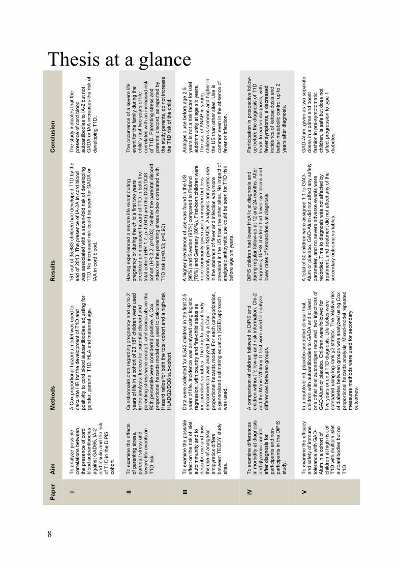
Thesis at a glance C
oncl
usio
n
The
stud
y in
dica
tes
that
the
pres
ence
of c
ord
bloo
d au
toan
tibod
ies
to IA
-2 b
ut n
ot
GA
DA
or I
AA
incr
ease
s th
e ris
k of
de
velo
ping
T1D
.
The
occu
rren
ce o
f a s
ever
e lif
e ev
ent f
or th
e fa
mily
dur
ing
the
child
´s fi
rst t
wo
year
s of
life
co
rrel
ates
with
an
incr
ease
d ris
k of
T1D
. Par
entin
g st
ress
and
pa
rent
al d
isco
rd, a
s re
porte
d by
th
e st
udy
pare
nts,
do
not i
ncre
ase
the
T1D
risk
of t
he c
hild
.
Ana
lges
ic u
se b
efor
e ag
e 2.
5 ye
ars
is n
ot a
risk
fact
or fo
r isl
et
auto
imm
unity
at a
ge s
ix y
ears
. Th
e us
e of
AN
AP
in y
oung
ch
ildre
n is
com
mon
and
hig
her i
n th
e U
S th
an o
ther
site
s. U
se is
co
mm
on e
ven
in th
e ab
senc
e of
fe
ver o
r inf
ectio
n.
Par
ticip
atio
n in
pro
spec
tive
follo
w-
up b
efor
e th
e di
agno
sis
of T
1D
lead
s to
ear
lier d
iagn
osis
, with
fe
wer
sym
ptom
s, a
dec
reas
ed
inci
denc
e of
ket
oaci
dosi
s an
d be
tter m
etab
olic
con
trol u
p to
2
year
s af
ter d
iagn
osis
.
GA
D-A
lum
, giv
en a
s tw
o se
para
te
dose
s in
a p
rime
and
boos
t fa
shio
n in
pre
-sym
ptom
atic
ch
ildre
n, is
saf
e bu
t doe
s no
t af
fect
pro
gres
sion
to ty
pe 1
di
abet
es.
Res
ults
151
out o
f 35,
683
child
ren
had
deve
lope
d T1
D b
y th
e en
d of
201
3. T
he p
rese
nce
of IA
-2A
in c
ord
bloo
d w
as a
ssoc
iate
d w
ith a
sev
en-fo
ld ri
sk o
f dev
elop
ing
T1D
. No
incr
ease
d ris
k co
uld
be s
een
for G
AD
A o
r IA
A in
cor
d bl
ood.
Hav
ing
expe
rienc
ed a
sev
ere
life
even
t dur
ing
preg
nanc
y or
dur
ing
the
child
’s fi
rst t
wo
year
s pr
edic
ted
an in
crea
sed
haza
rd o
f T1D
in b
oth
the
tota
l coh
ort (
HR
1.7
; p=0
.043
) and
the
DQ
2/D
Q8
coho
rt (H
R 2
.2; p
=0.0
3). N
eith
er th
e pa
rent
al d
isco
rd
inde
x no
r the
par
entin
g st
ress
inde
x co
rrel
ated
with
T1
D ri
sk (p
=0.5
3; p
=0.9
0)
A h
ighe
r pre
vale
nce
of u
se w
as fo
und
in th
e U
S
(96%
) and
Sw
eden
(95%
) com
pare
d to
Fin
land
(7
8%) a
nd G
erm
any
(80%
). Fi
rst-b
orn
child
ren
wer
e m
ore
com
mon
ly g
iven
ace
tam
inop
hen
but l
ess
com
mon
ly g
iven
NS
AID
s. A
nalg
esic
ant
ipyr
etic
use
in
the
abse
nce
of fe
ver a
nd in
fect
ion
was
mor
e pr
eval
ent i
n th
e U
S th
an th
e ot
her s
ites.
No
impa
ct o
f an
alge
sic
antip
yret
ic u
se c
ould
be
seen
for T
1D ri
sk
befo
re a
ge s
ix y
ears
.
DiP
iS c
hild
ren
had
low
er H
bA1c
at d
iagn
osis
and
du
ring
regu
lar f
ollo
w-u
p at
12
and
24 m
onth
s. A
fter
diag
nosi
s, D
iPiS
chi
ldre
n ha
d fe
wer
sym
ptom
s an
d lo
wer
rate
s of
ket
oaci
dosi
s at
dia
gnos
is.
A to
tal o
f 50
child
ren
wer
e as
sign
ed 1
:1 to
GA
D-
Alu
m o
r pla
cebo
. GA
D-A
lum
did
not
affe
ct a
ny s
afet
y pa
ram
eter
, and
no
seve
re a
dver
se e
vent
s w
ere
reco
rded
. Tim
e to
dia
gnos
is w
as n
ot a
ffect
ed b
y tre
atm
ent,
and
treat
men
t did
not
affe
ct a
ny o
f the
se
cond
ary
outc
ome
varia
bles
.
Met
hods
A C
ox p
ropo
rtion
al h
azar
ds m
odel
was
use
d to
ca
lcul
ate
HR
for t
he d
evel
opm
ent o
f T1D
and
po
sitiv
ity to
cor
d bl
ood
auto
antib
odie
s, a
djus
ting
for
gend
er, p
aren
tal T
1D, H
LA a
nd m
ater
nal a
ge.
Que
stio
nnai
re d
ata
rega
rdin
g pr
egna
ncy
and
up to
2
year
s of
life
in a
coh
ort o
f 23,
187
child
ren
wer
e us
ed
in th
e an
alys
is. I
ndic
es fo
r par
enta
l dis
cord
and
pa
rent
ing
stre
ss w
ere
crea
ted,
and
sco
res
abov
e th
e 90
th p
erce
ntile
wer
e co
nsid
ered
pos
itive
. A C
ox
prop
ortio
nal h
azar
ds m
odel
was
use
d to
cal
cula
te
Haz
ard
ratio
s fo
r bot
h th
e to
tal c
ohor
t and
a h
igh-
risk
HLA
DQ
2/D
Q8
sub-
coho
rt.
Dat
a w
ere
colle
cted
for 8
,542
chi
ldre
n in
the
first
2.5
ye
ars
of li
fe. I
ncid
ence
was
ana
lyze
d us
ing
logi
stic
re
gres
sion
, with
cou
ntry
and
firs
t-chi
ld s
tatu
s as
in
depe
nden
t var
iabl
es. T
he ti
me
to a
utoa
ntib
ody
sero
conv
ersi
on w
as a
naly
zed
usin
g a
Cox
pr
opor
tiona
l haz
ards
mod
el. F
or e
ach
cate
goriz
atio
n,
a ge
nera
lized
est
imat
ing
equa
tion
(GE
E) a
ppro
ach
was
use
d.
A c
ompa
rison
of c
hild
ren
follo
wed
in D
iPiS
and
ch
ildre
n w
ithou
t fol
low
-up
and
risk
info
rmat
ion.
Chi
-2
and
the
Man
n W
hitn
ey U
-test
wer
e us
ed to
ana
lyze
di
ffere
nces
bet
wee
n gr
oups
.
In a
dou
ble-
blin
d, p
lace
bo-c
ontro
lled
clin
ical
tria
l, ch
ildre
n w
ith a
utoa
ntib
odie
s to
GA
DA
and
at l
east
on
e ot
her i
slet
aut
oant
igen
rece
ived
tw
o in
ject
ions
of
GA
D-A
lum
or p
lace
bo. C
hild
ren
wer
e fo
llow
ed fo
r fiv
e ye
ars
or u
ntil
T1D
dia
gnos
is. L
ife ta
bles
wer
e co
mpa
red
usin
g lo
g-ra
nk χ
2 st
atis
tic. T
he re
lativ
e ris
k of
dia
bete
s be
twee
n gr
oups
was
ana
lyze
d us
ing
Cox
pr
opor
tiona
l haz
ards
ana
lysi
s. M
ixed
-mod
el re
peat
ed
mea
sure
s m
etho
ds w
ere
used
for s
econ
dary
ou
tcom
es.
Aim
To a
naly
ze p
ossi
ble
corr
elat
ions
bet
wee
n th
e pr
esen
ce o
f cor
d bl
ood
auto
antib
odie
s ag
ains
t GA
D65
, IA
-2
and
Insu
lin a
nd th
e ris
k of
T1D
in th
e D
iPiS
co
hort.
To e
xam
ine
the
effe
cts
of p
aren
ting
stre
ss,
pare
ntal
stre
ss a
nd
seve
re li
fe e
vent
s on
T1
D ri
sk.
To e
xam
ine
the
poss
ible
ef
fect
on
the
risk
of is
let
auto
imm
unity
and
to
desc
ribe
use
and
how
th
e us
e of
ana
lges
ic
antip
yret
ics
diffe
rs
betw
een
TED
DY
stu
dy
site
s.
To e
xam
ine
diffe
renc
es
in m
orbi
dity
at d
iagn
osis
an
d gl
ycem
ic c
ontro
l af
ter d
iagn
osis
for
parti
cipa
nts
and
non-
parti
cipa
nts
in th
e D
iPiS
st
udy.
To e
xam
ine
the
effic
acy
and
safe
ty o
f im
mun
e to
lera
nce
with
GA
D-
Alu
m in
a c
ohor
t of
child
ren
at h
igh
risk
of
T1D
with
mul
tiple
isle
t au
toan
tibod
ies
but n
o T1
D.
Pap
er
I II III
IV
V
9
Abbreviations
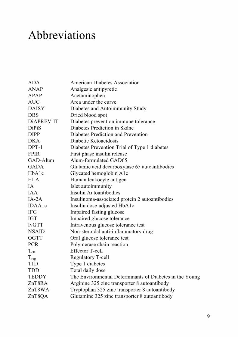
ADA American Diabetes Association ANAP Analgesic antipyretic APAP Acetaminophen AUC Area under the curve DAISY Diabetes and Autoimmunity Study DBS Dried blood spot DiAPREV-IT Diabetes prevention immune tolerance DiPiS Diabetes Prediction in Skåne DIPP Diabetes Prediction and Prevention DKA Diabetic Ketoacidosis DPT-1 Diabetes Prevention Trial of Type 1 diabetes FPIR First phase insulin release GAD-Alum Alum-formulated GAD65 GADA Glutamic acid decarboxylase 65 autoantibodies HbA1c Glycated hemoglobin A1c HLA Human leukocyte antigen IA Islet autoimmunity IAA Insulin Autoantibodies IA-2A Insulinoma-associated protein 2 autoantibodies IDAA1c Insulin dose-adjusted HbA1c IFG Impaired fasting glucose IGT Impaired glucose tolerance IvGTT Intravenous glucose tolerance test NSAID Non-steroidal anti-inflammatory drug OGTT Oral glucose tolerance test PCR Polymerase chain reaction Teff Effector T-cell Treg Regulatory T-cell T1D Type 1 diabetes TDD Total daily dose TEDDY The Environmental Determinants of Diabetes in the Young ZnT8RA Arginine 325 zinc transporter 8 autoantibody ZnT8WA Tryptophan 325 zinc transporter 8 autoantibody ZnT8QA Glutamine 325 zinc transporter 8 autoantibody

10
11
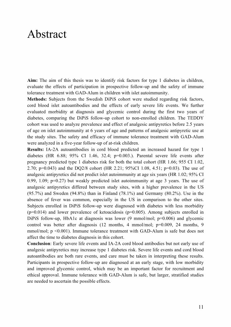
Abstract
Aim: The aim of this thesis was to identify risk factors for type 1 diabetes in children, evaluate the effects of participation in prospective follow-up and the safety of immune tolerance treatment with GAD-Alum in children with islet autoimmunity. Methods: Subjects from the Swedish DiPiS cohort were studied regarding risk factors, cord blood islet autoantibodies and the effects of early severe life events. We further evaluated morbidity at diagnosis and glycemic control during the first two years of diabetes, comparing the DiPiS follow-up cohort to non-enrolled children. The TEDDY cohort was used to analyze prevalence and effect of analgesic antipyretics before 2.5 years of age on islet autoimmunity at 6 years of age and patterns of analgesic antipyretic use at the study sites. The safety and efficacy of immune tolerance treatment with GAD-Alum were analyzed in a five-year follow-up of at-risk children. Results: IA-2A autoantibodies in cord blood predicted an increased hazard for type 1 diabetes (HR 6.88; 95% CI 1.46, 32.4; p=0.003.). Parental severe life events after pregnancy predicted type 1 diabetes risk for both the total cohort (HR 1.66; 955 CI 1.02, 2.70; p=0.043) and the DQ2/8 cohort (HR 2.21; 95%CI 1.08, 4.51; p=0.03). The use of analgesic antipyretics did not predict islet autoimmunity at age six years (HR 1.02; 95% CI 0.99, 1.09; p=0.27) but weakly predicted islet autoimmunity at age 3 years. The use of analgesic antipyretics differed between study sites, with a higher prevalence in the US (95.7%) and Sweden (94.8%) than in Finland (78.1%) and Germany (80.2%). Use in the absence of fever was common, especially in the US in comparison to the other sites. Subjects enrolled in DiPiS follow-up were diagnosed with diabetes with less morbidity (p=0.014) and lower prevalence of ketoacidosis (p=0.005). Among subjects enrolled in DiPiS follow-up, HbA1c at diagnosis was lower (9 mmol/mol; p=0.006) and glycemic control was better after diagnosis (12 months, 4 mmol/mol; p=0.009, 24 months, 9 mmol/mol; p <0.001). Immune tolerance treatment with GAD-Alum is safe but does not affect the time to diabetes diagnosis in this cohort. Conclusion: Early severe life events and IA-2A cord blood antibodies but not early use of analgesic antipyretics may increase type 1 diabetes risk. Severe life events and cord blood autoantibodies are both rare events, and care must be taken in interpreting these results. Participants in prospective follow-up are diagnosed at an early stage, with low morbidity and improved glycemic control, which may be an important factor for recruitment and ethical approval. Immune tolerance with GAD-Alum is safe, but larger, stratified studies are needed to ascertain the possible effects.

12
13
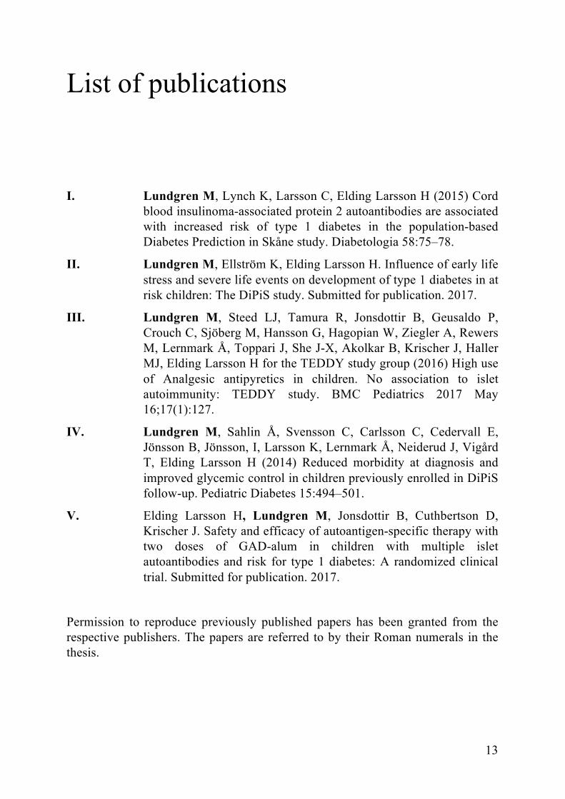
List of publications
I. Lundgren M, Lynch K, Larsson C, Elding Larsson H (2015) Cord blood insulinoma-associated protein 2 autoantibodies are associated with increased risk of type 1 diabetes in the population-based Diabetes Prediction in Skåne study. Diabetologia 58:75–78.
II. Lundgren M, Ellström K, Elding Larsson H. Influence of early life stress and severe life events on development of type 1 diabetes in at risk children: The DiPiS study. Submitted for publication. 2017.
III. Lundgren M, Steed LJ, Tamura R, Jonsdottir B, Geusaldo P, Crouch C, Sjöberg M, Hansson G, Hagopian W, Ziegler A, Rewers M, Lernmark Å, Toppari J, She J-X, Akolkar B, Krischer J, Haller MJ, Elding Larsson H for the TEDDY study group (2016) High use of Analgesic antipyretics in children. No association to islet autoimmunity: TEDDY study. BMC Pediatrics 2017 May 16;17(1):127.
IV. Lundgren M, Sahlin Å, Svensson C, Carlsson C, Cedervall E, Jönsson B, Jönsson, I, Larsson K, Lernmark Å, Neiderud J, Vigård T, Elding Larsson H (2014) Reduced morbidity at diagnosis and improved glycemic control in children previously enrolled in DiPiS follow-up. Pediatric Diabetes 15:494–501.
V. Elding Larsson H, Lundgren M, Jonsdottir B, Cuthbertson D, Krischer J. Safety and efficacy of autoantigen-specific therapy with two doses of GAD-alum in children with multiple islet autoantibodies and risk for type 1 diabetes: A randomized clinical trial. Submitted for publication. 2017.
Permission to reproduce previously published papers has been granted from the respective publishers. The papers are referred to by their Roman numerals in the thesis.

14

15
Introduction
“Diabetes is a dreadful affliction, not very frequent among men, being a melting down of the flesh and limbs into urine. the patients never stop making water and the flow is incessant, like the opening of aqueducts. Life is short, unpleasant and painful, thirst unquenchable, drinking excessive and disproportionate to the large quantity of urine, for yet more urine is passed. If for a while they abstain from drinking, their mouths become parched and their bodies dry; the viscera seem scorched up, the patients are affected by nausea, restlessness and a burning thirst, and within a short time, they expire.”
Aretaeus of Cappadocia (2nd century CE). Adapted from Papaspyros (1952) The history of
Diabetes Mellitus.
The history of T1D
Descriptions of diabetes mellitus, or polyuric disease, have existed for at least 3,500 years, as the disease was mentioned in ancient Egyptian papyrus rolls. The name diabetes is derived from the Greek word for syphon or “where the water flows out.” Early in the first millennium, it was recognized that polyuric urine tasted sweet, like honey. The “mellitus,” or honeyed, adjective was not added to the disease description until the late 1800s. The sweet taste of the urine was identified as glucose in the early 1800s, and shortly thereafter, glucose was shown to be normally present in the blood. In 1880, two distinctive forms of diabetes were proposed by Lancereaux: diabète maigre (lean patients) and diabète gras (obese patients). Early in the insulin age, it was observed that some patients were more or less responsive to insulin. The more insulin-sensitive patients tended to be the lean patients that needed insulin to avoid going into diabetic ketoacidosis (DKA), whereas older, obese patients were insulin-insensitive and ketosis-resistant1.
Before the 1920s, doctors could do little to counteract the symptoms of diabetes. The clinical course of diabetes patients was short, miserable and inevitably lethal. A multitude of therapies were tested, with exclusion or calorie-restricted diets being the only approaches that had any effect in terms of prolonging the time until the patients died from starvation or DKA. The cause of diabetes still eluded physicians in the early 19th century. Oskar Minkowski (1858-1913) and Josef von

16
Mering (1849-1908) made a significant breakthrough in the understanding of diabetes in 1889 when they reported that pancreatectomy resulted in severe hyperglycemia in dogs2.
The concept that internal secretions could direct bodily functions had been proposed at the time, and shortly thereafter, Murray reported that myxedema could be treated with a thyroid extract from sheep. This finding instilled hope that diabetes could also be treated. This goal proved much more elusive, however, as early attempts at insulin treatment all failed. Discouraged, the medical community turned again to diet as a treatment for the disease. The best-known regimen was the starvation regimen of Frederick Madison Allen. This regimen proposed intensive exercise and “as little to eat as possible” and was promoted by Elliott P. Joslin, who was one of the greatest diabetologists of the 20th century. The treatment could in some ways be described as pyrrhic, since the patients could survive for months at the cost of a very low quality of life and died of starvation rather than diabetes.
Insulin was finally discovered by Banting, Best, Macleod and Collip at the University of Toronto in 1921, earning Banting and Macleod the Nobel prize in medicine in 1922. Banting officially decided to share his prize with Best and MacLeod with Collip. Insulin derived from bovine pancreas was first used to treat diabetes patients in 1922, resulting in dramatic results for the first patients, with lowered blood sugar and eliminated glycosuria and ketosis3,4. By 1923, bovine insulin was widely available for treatment. The crystallized insulin was impure and had to be injected several times each at great pain. Early treatment advocated resting the pancreas by aggressively lowering blood sugar. This approach combined with available methods to test blood sugar was detrimental to many patients, who suffered greatly from hypoglycemia. Hence, during the early decades of insulin treatment, complications were common and came at an early age. Insulin pharmacology has developed since that time, including delayed action preparations, recombinant human insulin and modern insulin analogs developed via recombinant DNA molecular cloning technology.
The importance of glycemic control was poorly understood during the first decades of insulin treatment, and few physicians thought that glycemic control could prevent diabetic complications5. The first good measurement of glycemic control was discovered in 1968 when it was published that glycated hemoglobin (HbA1c) was present in the blood of people with diabetes, representing an objective measurement of glycemic control6. However, it was not until the late 1970s, with the introduction of test strips for measuring blood glucose, that regular blood glucose control became practically feasible7. The final component of modern diabetes treatment came with the replacement of glass and steel injection syringes with insulin injection pens in the early 1980s and later the portable insulin infusion pump8,9.
17
The uncertainty regarding the importance of glycemic control that had plagued parts of the 20th century was finally laid to rest when the Diabetes Control and Complications trial was published in 1993. The study firmly established the current dogma that good glycemic control prevents and delays the progression of microvascular complications in patients with type 1 diabetes (T1D)10. Numerous other studies have since confirmed this finding, in both type 1 and type 2 diabetes11-13.
The current state of diabetes treatment is characterized by slow, step-wise improvements in monitoring, insulin treatment and management.
With the use of insulin Degludec, a long duration insulin, more flexibility and a more consistent insulin profile can be achieved14. On the other end of the spectrum, the faster acting insulin Aspart (Fi-Asp) may improve post-prandial glucose control and variability15. The evolution of blood glucose monitoring, with continuous and flash glucose monitoring, gives diabetes patients more and faster information about their blood glucose than ever before16. Smarter, smaller, more advanced insulin pumps combined with carbohydrate counting would further improve the glycemic control of diabetes patients today.
However, none of these innovations will address the main problem of the diabetes patient: insulin dependency. Although better insulins and technical aids have been developed, a significant portion of patients fail to reach the treatment goals needed to minimize long-term complications17,18, are forced to work hard to accommodate school days19 and suffer from higher levels of depression and anxiety than their peers20. Unfortunately, even children who manage to reach their treatment goals have double the risk of death from cardiovascular complications21.
These outcomes highlight the need for continued efforts, not only toward C-peptide conservation for those already affected but also toward exposing the triggering events that start the diabetic immune reaction and preventing the disease process from starting at all.
Diagnosis of Diabetes
Definition of diabetes
The term diabetes mellitus describes a group of metabolic disorders of insulin secretion, insulin action or both. Chronic hyperglycemia is the common denominator among these disorders, although the underlying etiology is diverse. Criteria for the diagnosis of diabetes mellitus, regardless of etiology, have been adopted by the leading world diabetes organizations, as well as the World Health

18
Organization22-24. The diagnostic criteria for diabetes mellitus are summarized in Table 1. Table 1. Criteria for the diagnosis of diabetes22,25
HbA1c ≥6.5% (48 mmol/mol) † The test should be performed in a laboratory using a method that is NGSP-certified
and standardized to the DCCT assay*
OR
Fasting plasma glucose ≥126 mg/dL (7.0 mmol/L)* Fasting is defined as no caloric intake for at least 8 h
OR
Two-hour plasma glucose during oral glucose tolerance testing (OGTT) ≥200 mg/dL (11.1 mmol/L) . The test should be performed using a glucose load containing the equivalent of 75 g of
anhydrous glucose dissolved in water or 1.75g/kg of body weight to a maximum of 75 g*
OR
. Classic symptoms of diabetes or hyperglycemic crisis, with . plasma glucose concentration≥200 mg/dL (11.1 mmol/L)
* In the absence of unequivocal hyperglycemia, the diagnosis of diabetes based on these criteria should be confirmed by repeat testing. † A value of less than 6.5% does not exclude diabetes diagnosed using glucose tests. The role of HbA1c alone in diagnosing T1D in children is unclear and not part of the ISPAD criteria.
The broad classification of diabetes into T1D, with an absolute, eventual, deficiency of insulin secretion, and type 2 diabetes, with a combination of insulin resistance and inadequate insulin secretion, has existed since the mid-20th century. With improved genetic and metabolic testing, the number of different diabetes mellitus diagnoses is increasing. Increased knowledge about the genetic etiology of these disorders has improved our understanding of maturity-onset diabetes of the young (MODY), neonatal diabetes, and diabetes caused by defects in insulin action26-29 (Table 2.).
To improve the diagnosis of T1D, additional diagnostic tools besides plasma glucose can be used:
Islet autoantibodies: 96-98% of newly diagnosed children with T1D are positive for at least one islet autoantibody (IAA, GADA, IA-2A or ZnT8A), all but confirming the diagnosis30-34.
C-peptide: Measuring fasting C-peptide can indicate whether or not the patient is insulin-deficient. However, measurements during the initial period after the T1D diagnosis can be hard to interpret due to remission of insulin secretion. This test can still be an efficient tool to discriminate between T1D, type 2 diabetes and monogenic diabetes35,36.
HLA genotype: In those patients in which the HLA genotype is analyzed at the time of diagnosis, a very low- (HLA DQ6.2) or low-risk genotype warrants further testing if combined with negative islet autoantibodies37,38.

19
In children without islet autoantibodies, a young age (<6 months), autosomal dominant diabetes in the family, associated conditions (blindness, deafness, and syndromic features) or exposure to drugs related to insulin resistance, further testing should be considered39-41.
Table 2. Classification of diabetes (adapted from the American Diabetes Association and ISPAD22,25)
CEL, carboxyl ester lipase; HNF, hepatocyte nuclear factor; IPEX, immunodysregulation polyendocrinopathy enteropathy X-linked syndrome; IPF, insulin promoter factor; MODY, maturity-onset diabetes of the young; PAX4, Paired Domain gene 4.
Type 1 diabetes
Type 2 diabetes
Other specific types of diabetes
Genetic defects
MODY 1 (chromosome 20, HNF-4a), MODY 2 (chromosome 2, glucokinase), MODY 3 (chromosome 12, HNF-1a), MODY 4 (chromosome 13, IPF 1), MODY 6 (Chromosome 2, NeuroD1), MODY 7 (chromosome 9, CEL)
Transient neonatal diabetes (Imprinting defect on 6q24; mutations in ABCC8 or KIR6.2)
Permanent neonatal diabetes (WRS, mutation in EIF2AK3; IPEX syndrome; mutations in INS and FOXP3)
Mitochondrial DNA mutation
Others
Genetic defects in insulin action
Type A Insulin Resistance, Rabson Mendenhall Syndrome, Leprechaunism (mutations in INSR)
Diseases of the exocrine pancreas
Pancreatitis, Pancreatectomy, Cystic fibrosis, Hemochromatosis and others
Endocrinopathies Acromegaly, Cushing´s syndrome, Glucagonoma, Pheochromocytoma, Hyperthyroidism, Hyperthyroidism, and others
Drug or chemical induced
Glucocorticoids, Thyroid hormone, Diazoxide, B-adrenergic Agonists, Thiazides, Phenytoin, Γ Interferon, Nicotinic acid, Vacor, and others,
Infections Congenital Rubella, CMV, others
Uncommon forms of immune-mediated diabetes
Stiff-man syndrome, Anti-insulin receptor antibodies, Autoimmune Polyendocrine Syndrome (APS) Types I and II, and othersand others
Other genetic syndromes
Down syndrome, Klinefelter syndrome, Turner syndrome, Wolfram syndrome, Friedrich ataxia, Huntington syndrome, Prader Willi syndrome, and others
Gestational Diabetes

20
Stages of T1D
It has long been known that a long prodromal period precedes the clinical diagnosis of T1D. Initially, this period was thought to be a matter of months, during which incidental dysglycemia occurred due to decreasing β-cell reserves. The current understanding of islet autoimmunity (IA) changes the previous notion of T1D as a chronic disease with acute onset into a slowly progressing, immune mediated β-cell destruction. The etiology is not known but recent data suggest that different triggering events may induce either IAA or GADA as the first appearing autoantibody42-4445. IAA appear first in children with the DR4-DQ8 haplotype while GADA appear first in DR3-DQ2 children. A first autoantibody is often followed over months and years to a second, third or fourth autoantibody independent of the HLA type of the child. The progressive pathogenesis is associated with β-cell loss eventually leading to dysglycemia and hyperglycemia. This change has also lead to a new classification of the early stages of T1D, including the period of IA, into three stages, from IA to overt, clinical disease46(Figure 1.). However, taking the concept of pre-symptomatic T1D one step further and accepting IA as a disease entity in itself is currently under debate47,48.
Figure 1. Stages of Type 1 diabetes. According to the 2015 statement of ADA/JDRF/Endocrine Society. Adapted with permission from Insel RA and
colleagues46.
Stage 1: IA, with persistent seropositivity for at least two islet autoantibodies but with normal glucose and normal HbA1c. The β-cell mass is still large enough to maintain normoglycemia. In individuals with genetic HLA risk, the risk of developing symptomatic T1D in this group is approximately 44% in 5 years and 70% in 10 years. The lifetime risk approaches 100%49-51.
Variable genetic &
environmental risk for type 1
diabetes
β-Cell autoimmunityNormoglycemiaPresymptomatic
β-Cell autoimmunityDysglycemia
Presymptomatic
β-Cell autoimmunityDysglycemiaSymptomatic
Func
tiona
l β-c
ell M
ass
0%
100%
Presymptomatic Type 1 diabetes
Symptomatic Type 1 diabetes
Stage 1 Stage 2 Stage 3Proposed nomenclature
Phenotypic Characteristics
21
Stage 2: IA, with persistent seropositivity for at least two islet autoantibodies and glucose intolerance/dysglycemia. Dysglycemia in this stage is most often defined as either impaired glucose tolerance (IGT), with elevated 2-hour plasma glucose values ≥ 7.8 mmol/L, or elevated glucose levels at intermediate time points (plasma glucose ≥ 11.1mmol/L) on a standardized OGTT and/or HbA1c ≥ 39 mmol/mol. The two-year risk of symptomatic disease for this phase is estimated at 60%, and the 4- to 5-year risk is 75%52. Stage 3: Symptomatic disease. T1D with classical signs of the disease: polyuria, polydipsia, weight loss, fatigue and possible metabolic decompensation and DKA.
The staging of T1D, which further signifies this condition as a progressive disease for which symptomatic disease is the end point, allows for earlier attempts at prevention and intermediate, more efficient, endpoints in clinical trials53. Staging also allows for early diagnosis, significantly decreasing the rate of metabolic decompensation and DKA54-57. Whether early diagnosis benefits patients and their families psychologically remains unclear and must be further addressed58-60.
Epidemiology of T1D
The incidence rate suggests a diabetes epidemic Childhood diabetes has been recognized for hundreds of years. However, reliable global incidence data for T1D have not been available until the late 20th century. In the 1980s, the diabetes hotspots in Finland and Sardinia were not yet recognized, and incidence estimates were not available for 90% of the world’s population61. Diabetes incidence reports from the early 1900s are rare and of limited scope. However, using existing data, one could assume a rising incidence. For example, the incidence appears to have risen from 2 to 7 cases per 100,000/year between 1900-1920 in Norway and from 2 to 4 cases per 100,000/year in Denmark62. Hence, childhood diabetes appears to have been a rare condition at the start of the 20th century.
More reliable incidence data became available later in the 20th century. The US national health survey during the 1930s reported incidence numbers of 0.35-0.41/1,000 for children younger than 15 years63, and additional data from Norway, which were collected between 1925 and 1954, support an estimated incidence of 4.1/100,00064. In the Norwegian data, it is also interesting to note that no discernible increase in incidence was seen during the 30-year study period. Early Finnish data are hard to interpret and compare to the present-day situation due to the hardships of the 1930s and 1940s. More reliable data from 1953 estimate the Finnish incidence at 12.5/100,000/year, a meager number in comparison to the current incidence65.
22
When this increase in incidence levels occurred is not easy to pinpoint. It has been proposed that the incidence increased during the 1950s66. The previously mentioned Norwegian study was later extended for another 10 years. At the end of this period, the incidence numbers had climbed to 8.4/100000/year67. Data from Danish sources paint a similar picture, with an approximately doubling of the childhood diabetes incidence68.
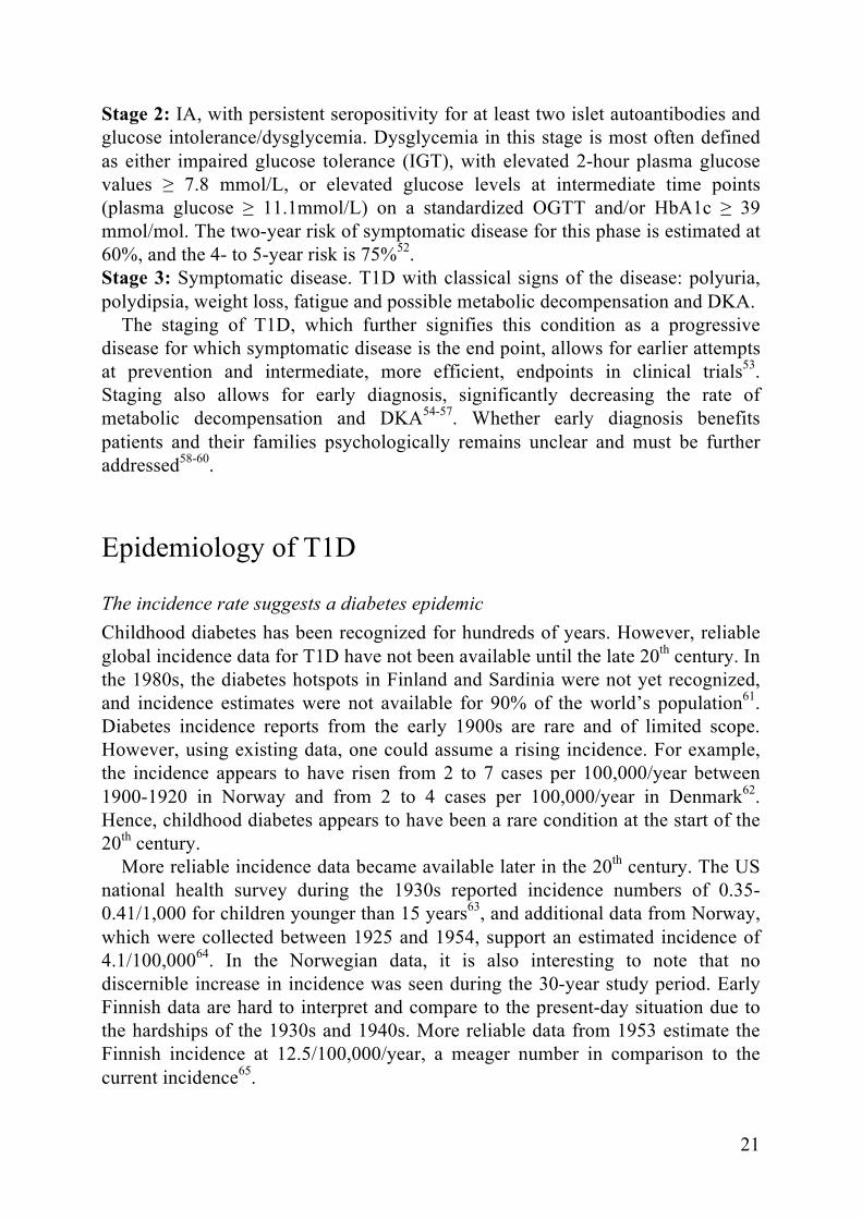
Better data are available from the later part of the 20th century. In a comprehensive review of 37 studies performed between 1960 to 1996, a rising trend was noted in 24 studies, with an average estimated annual increase of 3%62. This number is also supported by later data69. The extrapolation of these studies predicted a 40% increase in the childhood T1D incidence until 2010 and a possible rise of 70% between 2005 and 202070 (Figure 2.).
Figure 2. Incidence of T1D in children aged 0-14 years over time. Time-based trends in the incidence of T1D in children ages 0-14 years. Reproduced with permission from Atkinson and colleagues.71-74
In the global setting, the T1D incidence varies significantly, with a difference of over 400-fold between countries75. Generally, T1D is uncommon in Asia and South America, with incidence rates of approximately 0.1/100,000, and vastly more common in Finland and Sardinia, with an incidence rate of 50/100,000. However, the distribution is more complex, as countries in close proximity have very different incidence numbers.
In-country variations in incidence The incidence of T1D differs not only between countries but also within countries and tends to increase through childhood, with a peak between 10-15 years76.

23
However, at present, there are indications that a shift towards the younger age groups is occurring, with higher incidence rates in the 0- to 4-year group77 78. Generally, incidence rates peak at puberty and are lower after age 1579. Significant differences also exist within countries, with 1.5-fold differences described in Sweden and Norway, with clusters of higher incidence, as well as a north-south gradient in incidence rates80,81.
There are small differences in incidence rates between the sexes. Generally, the incidence peak in children occurs earlier in girls, but the general differences in the age group between 0-15 years are small. After puberty, a male predominance of 1.3-2 to 1 is present in many populations79,82.
Figure 3. Seasonality at first insulin injection. Stratified by age group. Reproduced with permission from Green A and colleagues83.
The incidence of T1D differs by seasonality as has been described by several studies84. More patients are diagnosed during the winter and autumn months, a phenomenon that is present with small variations between countries. This seasonal variation also seems to be more significant in the older age group, in which children are between 10 and 14 years at diagnosis (Figure 3).

24

25
Pathogenesis of T1D
Many journal articles on T1D start by stating that it is a chronic, autoimmune disease of β-cell destruction. This statement is based on the presence of insulitis, inflammation of the pancreatic islets, first reported over 50 years ago85. A model for the development and pathogenesis of T1D was proposed in the mid 1980´s86.According to this hypothesis, individuals have a fixed number of β-cells at birth. A trigger, possibly a virus, induces cellular damage and as a result β-cell autoimmunity. Activated, auto-reactive, T-cells destroy β-cells leading to a continuously decreasing ability of insulin secretion. When approximately 10% of the β-cells remain, hyperglycemia and T1D diagnosis is reached. That this model does not paint a complete picture of T1D pathogenesis has been proven since its publication, although parts of it is still valid.
The anatomical location of the pancreas has hampered the histological and functional evaluation of the pancreas itself during T1D progression. Until recently, studies of pancreatic pathology have had to rely on specimens from T1D patients obtained at autopsy. The nPOD cooperation, however, focus on collecting pancreatic tissue from deceased patients with T1d as well as subjects with IA87. At diagnosis approximately 70% of the pancreatic islets display an absence of insulin but some patients display islets without any signs of inflammation88. Islets deficient of insulin, are significantly less likely to display signs of insulitis89. The number of lost β-cells at diagnosis also seem to vary, where once thought to amount to about 90%, as little as two thirds of the β-cells lost have been found in newly diagnosed T1D patients. The destruction of β-cells is very selective, leaving the other hormone producing cells of the islets (α-cells, γ-cells and PP-cells) unaffected90. The insular infiltrates of the pancreas consist mainly of CD8+ T-cells as well as macrophages, CD4+ T-cells, B-lymphocytes, and plasma cells90. This, together with the dominant role of HLA in T1D risk as well as ample experimental and clinical results, is the basis of the concept of a T-cell driven disease91.
One model of cellular autoimmunity in T1D proposes that after the initial trigger damages β-cells they undergo apoptosis. This makes the intracellular components available to the immune system after being absorbed by antigen presenting cells, among them dendritic cells. These cellular components are then presented to T-cells using the HLA heterodimers on the cell surface of antigen presenting cells. CD4+ T-cells, T-Helper cells, activate both macrophages, CD8+ T-cells as well as B-cells. B-cells start producing antibodies to the presented cellular components leading to islet autoantibodies whereas CD8+ T-cells attack β-cells leading to further damage and insular infiltration92.
Several alterations to the previously established model of T1D pathogenesis has been proposed but as of yet no new unifying theory has been proposed. Several factors are still undetermined in the pathogenic process, which will need to be

26
further addressed. The curious specificity of β-cell death, with remaining endocrine cells being unaffected in the islets, is still not explained93. Additionally, the triggering event leading to autoimmunity still eludes us. The heterogeneity of β-cell death, and possibly resurrection, is also unknown, where some T1D patients have remaining insulin producing β-cells for many years after T1D diagnosis88.
Giving a full account of all aspects of T1D is well beyond this thesis. The focus of the latter parts of this chapter will focus on factors related to prediction and prevention, although at the same time tightly connected to our understanding of T1D pathogenesis.
T1D risk assessment
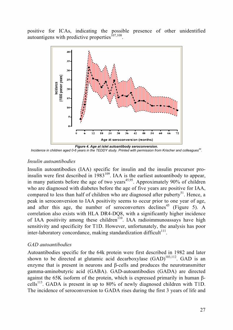
Autoantibodies
Seroconversion Our knowledge of islet autoantibody seroconversion in children has expanded significantly during recent years. We now know that seroconversion occurs early, often during the first year of life, but can occur significantly later45,94 (Figure 4.). The most common first islet autoantibody is IAA, and seroconversion to GADA tends to be a later phenomenon95 (Figure 5.). A single islet autoantibody correlates with a small increase in T1D risk, but we also know that a portion of children revert back to antibody negativity and return to their previous T1D risk level96. Further development of islet autoantibodies happens in a sequential manner; the risk of multiple autoantibody positivity is significantly lower if a second islet autoantibody does not appear during the first year after initial seroconversion97.
Islet cell autoantibodies The association between T1D and islet autoantibodies has been recognized since the 1970s98,99. At that time, islet cell antibodies (ICAs) were identified in newly diagnosed T1D patients. ICAs were later shown to be present earlier in the disease process, an indicator of the long period of IA leading up to clinical diagnosis100,101. ICAs were found in approximately 80% of newly diagnosed T1D patients, making them a useful instrument for prediction98. However, the method was troublesome in that it required human pancreatic tissue from blood group O donors for analysis102. ICA-staining was eventually found to be attributed partly to autoantibodies against IA-2, GAD65 or possibly insulin103-106. Given the methodological and sensitivity/specificity-related problems associated with ICA analysis, its use is now limited. However, studies show that up to 5% of newly diagnosed children who are negative for GADA, IAA, IA-2A and ZnT8A test
27
positive for ICAs, indicating the possible presence of other unidentified autoantigens with predictive properties107,108.
Figure 4. Age at islet autoantibody seroconversion.
Incidence in children aged 0-6 years in the TEDDY study. Printed with permission from Krischer and colleagues44
.
Insulin autoantibodies Insulin autoantibodies (IAA) specific for insulin and the insulin precursor pro-insulin were first described in 1983109. IAA is the earliest autoantibody to appear, in many patients before the age of two years45,95. Approximately 90% of children who are diagnosed with diabetes before the age of five years are positive for IAA, compared to less than half of children who are diagnosed after puberty51. Hence, a peak in seroconversion to IAA positivity seems to occur prior to one year of age, and after this age, the number of seroconverters declines45 (Figure 5). A correlation also exists with HLA DR4-DQ8, with a significantly higher incidence of IAA positivity among these children110. IAA radioimmunoassays have high sensitivity and specificity for T1D. However, unfortunately, the analysis has poor inter-laboratory concordance, making standardization difficult111.
GAD autoantibodies Autoantibodies specific for the 64k protein were first described in 1982 and later shown to be directed at glutamic acid decarboxylase (GAD)103,112. GAD is an enzyme that is present in neurons and β-cells and produces the neurotransmitter gamma-aminobutyric acid (GABA). GAD-autoantibodies (GADA) are directed against the 65K isoform of the protein, which is expressed primarily in human β-cells113. GADA is present in up to 80% of newly diagnosed children with T1D. The incidence of seroconversion to GADA rises during the first 3 years of life and

28
then appears to remain relatively constant during early childhood45 (Figure X). GADA also exhibits a more persistent positivity, even when β-cell function has been severely compromised.
Figure 5. Incidence of IAA only, GADA only, and IA-2A only as first-appearing IA and multiple IA at seroconversion. Results are shown for the total cohort (A) and for the general population (B) and first-degree relatives (C) separately. Reprinted with permission from Krischer and colleagues44.
This finding, combined with a strong correlation with C-peptide levels, makes GADA a good candidate for prediction efforts114,115. GADA is associated with the high-risk haplotype HLA DR3-DQ245,95. More recently, the possibility of further increasing the sensitivity and specificity of GADA has been investigated using N-terminally truncated GADA and anti-idiotypic GADA116-118. Standardized GADA assays using radio-binding assays or ELISA have been standardized and show good concordance between laboratories119.
IA-2 Autoantibodies The autoantigen for autoantibodies to insulinoma-associated protein 2 (IA-2), which was previously known as ICA512 and first described in 1994, is a tyrosine phosphatase-like enzyme120-122. The enzyme is present in two isoforms in pancreatic α- and β-cells, as well as in neuroendocrine tissue. Like the GAD and ZnT8 antibodies, IA-2 antibodies recognize a cytoplasmic, intracellular part of the protein: the C-terminal region105. IA-2 is typically not the first appearing autoantibody, and only 9% of children seroconvert to IA-2A as their first islet autoantibody. However, cases in which the initial seroconversion is to IA-2 are associated with a more rapid progression to T1D45,51,95. Higher titers of IA-2 have also been attributed to a more rapid progression to clinical diabetes123-125. IA-2A positivity has been reported to be inversely correlated with HLA DR4-DQ8 and correlated with the DRB1*0401 allele126. In contrast, the DR3-DQ2 haplotype was negatively associated with IA-2A 127. Standardized radio-binding assays for IA-2A
29
present good concordance between laboratories, demonstrating high sensitivity and specificity128,129.
ZnT8 Autoantibodies The cation efflux transporter zinc transporter 8 (ZnT8) has recently been described as a target autoantigen in patients with T1D130. This protein is involved in zinc-insulin crystallization, as well as insulin secretion, by accumulating zinc into the insulin granulae131. There are three isoform variants of the ZnT8 autoantibody, which are directed at different variants at position 325 in the protein: ZnT8RA (arginine 325 Zinc transporter 8 autoantibody), ZnT8WA (tryptophan 325 Zinc transporter 8 autoantibody) and ZnT8QA (glutamine 325 Zinc transporter 8 autoantibody). Autoantibodies to any of these isoforms represent additional markers of T1D risk132,133. Up to 80% of new-onset pediatric T1D patients are positive for ZnT8A, including 26% of patients who are negative for GADA, IA-2A and IAA34,107,134-137. Among newly diagnosed T1D patients from 0.6 to 58 years of age, ZnT8RA was present in 53%, ZnT8WA in 44% and ZnT8QA 34%, respectively138. ZnT8A titers tend to increase with age and have a higher prevalence, with a peak in late adolescence137. ZnT8A positivity has been reported to predict a more rapid progression to diabetes but can also persist for many years and often remain at diagnosis34,130,139. However, after diagnosis, titers of ZnT8A rapidly decline. Approximately 40% of ZnT8A-positive patients become negative 5 years after diagnosis140,141. At 25 years post-diagnosis, only 7% of T1D patients remained ZnT8A-positive, compared to 26% for GADA and 20% for IA-2A. Radio-binding assays for ZnT8A were standardized and validated in the international DASP workshop on islet autoantibody concordance142.
Novel minor autoantigens In addition to the previously mentioned T1D-related autoantigens, several other minor autoantigens have been described. These autoantigens include candidate autoantigens that may be associated with β-cell autoimmunity and T1D. Some of the present candidate autoantigens include Tetraspanin 7143,144, Neuropeptide Y145, HSP60146, ICA69147, INS-IGF2148, Carboxypeptidase H149, vesicle-associated membrane protein-2 (VAMP2)150, ICA/SOX13151, glima-38152, GLUT-2153 and imogen 38154.

30
HLA
More than 40% of the genetic susceptibility to T1D can be explained by the HLA genes155,156, representing three classes: Class I, Class II and Class III, all located on the short arm of chromosome six. The HLA genotype affects the configuration of the peptide binding groove of the HLA molecule on the surface of antigen presenting cells, determining which peptide will be bound in the groove and presented to T-cells (Figure 6.). Hence, allelic variations in the HLA coding region affect antigen recognition and presentation to the immune system through the interaction between the HLA molecules and the T-cell receptor of T-cells157,158. The HLA class II region has three regions: DR, DQ and DP. The HLA DQ molecule is a heterodimer, consisting of an α-chain encoded by the DQA1/A2 allele and a β-chain encoded by the DQB1/B2 allele159. For categorization, each amino acid variant of the α- and β-chains is given a four digit number, preceded by the gene designation (e.g., DQA105:01-DQB102:01)160. The alleles within this particular location are often inherited together as haplotypes, in linkage disequilibrium as the distance between the alleles are very short and that recombination events therefore are reduced. This is of particular importance as e.g. HLA-DQ A1 and B1 allele are especially polymorphic161.
Figure 6. Structure and binding of Class II MHC molecules. Schematic diagram and crystal structure of the HLA class II molecule with the peptide binding cleft (left). Peptide bound on the floor of the HLA class II peptide binding cleft (right). Reproduced with permission from Abbas and colleagues, Basic immunology 4th ed.162
The HLA-DR and HLA-DQ alleles have been shown to be the major determinants of HLA-derived T1D risk and can confer a protective, neutral or susceptible effect. However, the risk conferred by the HLA haplotype is not constant between populations or between ethnic groups163. The highest risk haplotypes are conferred by HLA DR3 (DRB1*03) together with DQ2 (DQA1*05:01-DQB1*02:01) and by DR4 (DRB1*04) together with DQ8 (DQA1*03:01-DQB1*03:02)164,165(Table 3.).
31
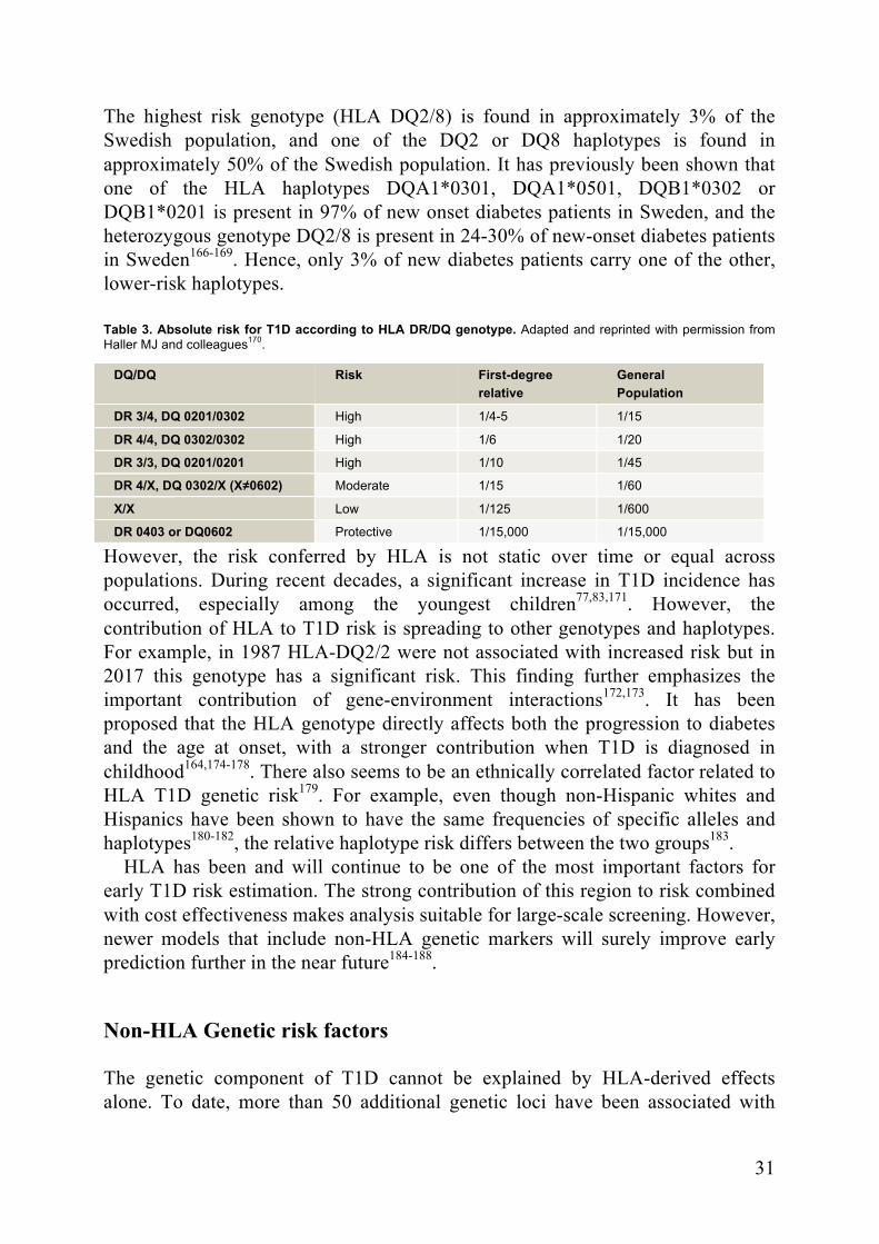
The highest risk genotype (HLA DQ2/8) is found in approximately 3% of the Swedish population, and one of the DQ2 or DQ8 haplotypes is found in approximately 50% of the Swedish population. It has previously been shown that one of the HLA haplotypes DQA1*0301, DQA1*0501, DQB1*0302 or DQB1*0201 is present in 97% of new onset diabetes patients in Sweden, and the heterozygous genotype DQ2/8 is present in 24-30% of new-onset diabetes patients in Sweden166-169. Hence, only 3% of new diabetes patients carry one of the other, lower-risk haplotypes. Table 3. Absolute risk for T1D according to HLA DR/DQ genotype. Adapted and reprinted with permission from Haller MJ and colleagues170.
DQ/DQ Risk First-degree relative
General Population
DR 3/4, DQ 0201/0302 High 1/4-5 1/15
DR 4/4, DQ 0302/0302 High 1/6 1/20
DR 3/3, DQ 0201/0201 High 1/10 1/45
DR 4/X, DQ 0302/X (X≠0602) Moderate 1/15 1/60
X/X Low 1/125 1/600
DR 0403 or DQ0602 Protective 1/15,000 1/15,000
However, the risk conferred by HLA is not static over time or equal across populations. During recent decades, a significant increase in T1D incidence has occurred, especially among the youngest children77,83,171. However, the contribution of HLA to T1D risk is spreading to other genotypes and haplotypes. For example, in 1987 HLA-DQ2/2 were not associated with increased risk but in 2017 this genotype has a significant risk. This finding further emphasizes the important contribution of gene-environment interactions172,173. It has been proposed that the HLA genotype directly affects both the progression to diabetes and the age at onset, with a stronger contribution when T1D is diagnosed in childhood164,174-178. There also seems to be an ethnically correlated factor related to HLA T1D genetic risk179. For example, even though non-Hispanic whites and Hispanics have been shown to have the same frequencies of specific alleles and haplotypes180-182, the relative haplotype risk differs between the two groups183.
HLA has been and will continue to be one of the most important factors for early T1D risk estimation. The strong contribution of this region to risk combined with cost effectiveness makes analysis suitable for large-scale screening. However, newer models that include non-HLA genetic markers will surely improve early prediction further in the near future184-188.
Non-HLA Genetic risk factors
The genetic component of T1D cannot be explained by HLA-derived effects alone. To date, more than 50 additional genetic loci have been associated with
32
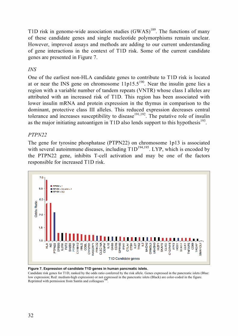
T1D risk in genome-wide association studies (GWAS)189. The functions of many of these candidate genes and single nucleotide polymorphisms remain unclear. However, improved assays and methods are adding to our current understanding of gene interactions in the context of T1D risk. Some of the current candidate genes are presented in Figure 7.
INS One of the earliest non-HLA candidate genes to contribute to T1D risk is located at or near the INS gene on chromosome 11p15.5190. Near the insulin gene lies a region with a variable number of tandem repeats (VNTR) whose class I alleles are attributed with an increased risk of T1D. This region has been associated with lower insulin mRNA and protein expression in the thymus in comparison to the dominant, protective class III alleles. This reduced expression decreases central tolerance and increases susceptibility to disease191,192. The putative role of insulin as the major initiating autoantigen in T1D also lends support to this hypothesis193.
PTPN22 The gene for tyrosine phosphatase (PTPN22) on chromosome 1p13 is associated with several autoimmune diseases, including T1D194,195. LYP, which is encoded by the PTPN22 gene, inhibits T-cell activation and may be one of the factors responsible for increased T1D risk.
Figure 7. Expression of candidate T1D genes in human pancreatic islets. Candidate risk genes for T1D, ranked by the odds ratio conferred by the risk allele. Genes expressed in the pancreatic islets (Blue: low expression; Red: medium-high expression) or not expressed in the pancreatic islets (Black) are color-coded in the figure. Reprinted with permission from Santin and colleagues196.

33
CTLA-4 Cytotoxic T-lymphocyte-associated protein 4 is a co-stimulatory receptor that inhibits T-cell activation and functions in CD4 T-regulatory cells. Variations in this gene have a strong effect, possibly via the regulation of peripheral tolerance, and this gene is also associated with several other autoimmune diseases197,198. HLA and CTLA-4 alleles revealed support for a bidirectional trigger for either IAA or GADA as a first appearing β-cell autoantibody in early life 42.
IL2RA IL2RA encodes CD25 on naive T-regulatory cells, memory T-cells and monocytes199. The regulation of the CD25 protein is important for the suppression of T-cell proliferation by an immunogenic stimulus200,201, and IL2RA has also been implicated in the pathogenesis of other autoimmune diseases202.
Environmental risk factors
The increase of T1D that has occurred during recent decades cannot be explained by genetic factors alone. The impact or presence of some environmental factor must have changed. It is also possible that the impact differs based on the HLA-DQ genotype, which may explain some of the dilution of the high-risk HLA genotypes44. That environmental factors play a role in the risk of T1D has been postulated for a long time, but the responsible risk factors have proved elusive. However, there is strong support for the impact of environmental factors. In Europe, populations that are genetically close but separated by socioeconomic borders have significantly different T1D incidence rates203, and we also know that immigrants tend to acquire genetic risk levels similar to those of the inhabitants of their new homeland, despite coming from low incidence areas204. Through migration and free movement, diabetes risk is becoming more homogenous, despite the stability of genetic factors205.
A plethora of environmental factors have been proposed to influence the risk of T1D and will be summarized in this chapter (Figure 8.). A more thorough review of all of these risk factors is beyond the scope of this thesis. For the sake of clarity, a separation between the factors thought to initiate IA and the factors that accelerate and support the progression to clinical diabetes will be made, to the extent that current evidence allows.
Prenatal risk factors One of the earliest described prenatal risk factors for type one diabetes is congenital rubella206, with approximately 20% of affected children developing a T1D-like disease. Other factors that have been reported to increase T1D risk include a higher relative birthweight207,208 and delivery by cesarean section 209,
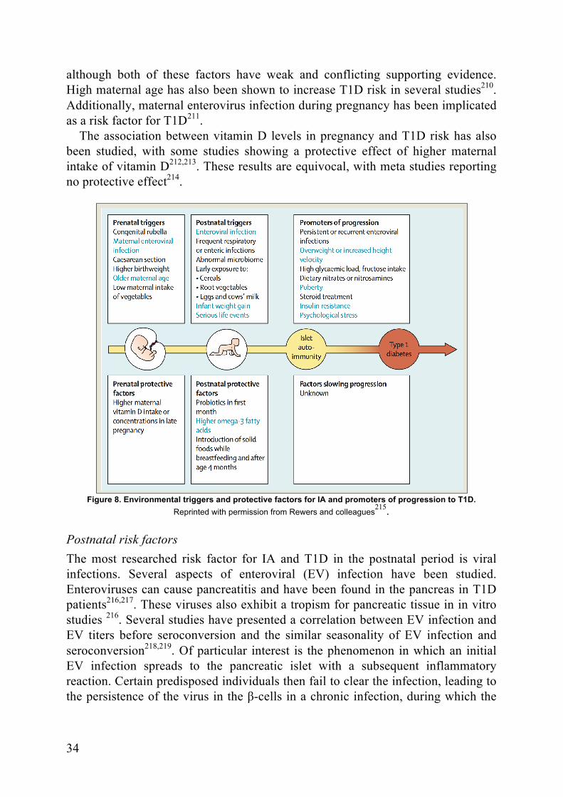
34
although both of these factors have weak and conflicting supporting evidence. High maternal age has also been shown to increase T1D risk in several studies210. Additionally, maternal enterovirus infection during pregnancy has been implicated as a risk factor for T1D211.
The association between vitamin D levels in pregnancy and T1D risk has also been studied, with some studies showing a protective effect of higher maternal intake of vitamin D212,213. These results are equivocal, with meta studies reporting no protective effect214.
Figure 8. Environmental triggers and protective factors for IA and promoters of progression to T1D.
Reprinted with permission from Rewers and colleagues215.
Postnatal risk factors The most researched risk factor for IA and T1D in the postnatal period is viral infections. Several aspects of enteroviral (EV) infection have been studied. Enteroviruses can cause pancreatitis and have been found in the pancreas in T1D patients216,217. These viruses also exhibit a tropism for pancreatic tissue in in vitro studies 216. Several studies have presented a correlation between EV infection and EV titers before seroconversion and the similar seasonality of EV infection and seroconversion218,219. Of particular interest is the phenomenon in which an initial EV infection spreads to the pancreatic islet with a subsequent inflammatory reaction. Certain predisposed individuals then fail to clear the infection, leading to the persistence of the virus in the β-cells in a chronic infection, during which the
35
virus slowly replicates and produces viral DNA. This persistence, in turn, stimulates innate immunity and drives inflammation and autoimmunity220.
In addition to EV infection, several other viral infections have been implicated in T1D risk. Among these are respiratory and intestinal infections, such as rotavirus and cytomegalovirus221,222. Data from microbiome studies suggest an increased T1D risk in individuals with an abnormal microbiome, although few large-scale and long-term studies have been reported223-225. Additionally, early exposure to cereals226,227, early introduction of cow’s milk228,229 and increased infant weight gain230 have been proposed as potential T1D risk factors.
The countries with the largest increases in T1D incidence have experienced large improvements in sanitation and living standards. The so-called "hygiene hypothesis" postulates that decreased microbial pressure and a cleaner environment, coupled with a human immune system that has not had time to adapt, increase the risk of both allergy and autoimmunity231. However, it is likely that this is a multifactorial process that relies on additional environmental and genetic factors232.
Higher omega 3 fatty acid intake has been reported to decrease diabetes risk in children, as has supplementation with probiotics during the child´s first month of life233. Breast feeding has shown conflicting results, with several studies seeing no correlation with T1D risk234 and some studies showing a protective effect in some subsets of subjects235.
Promoters of progression Several factors that accelerate the progression to T1D have been identified. Conflicting evidence exists regarding the effect of nitrite/nitrate intake and progression to T1D236,237. Increased growth, in terms of both length and weight, as well as puberty, increases the demands on β-cells and induces insulin resistance in some instances. Accelerated progression to T1D has been described for patients with increased weight gain, as well as increased weight and length238,239. Some data also suggest that increased intake of carbohydrates will accelerate progress from stage 2 to stage 3 of T1D240.
At the present time, no protective factors that slow the progression to T1D have been described.
36
Prediction of T1D
Early Prediction of T1D risk
Screening of newborns and very young children was originally accomplished using heredity data from first-degree relatives. We now know that 85-90% of newly diagnosed T1D patients lack a first-degree relative with the disease; thus, this approach misses far too many potential subjects241. The use of HLA genotype data has provided a cost-effective and rapid screening process. However, since only approximately 50% of the genetic risk is conferred by HLA, a fairly large proportion of risk information has been missed by this approach156. The utility of enhanced, high-resolution HLA genotyping with next-generation sequencing has recently been demonstrated, with a specificity and sensitivity of 90% and the ability to identify 80% of those with a lifetime risk of T1D242.
Several studies have now attempted to improve early T1D prediction by using previously identified T1D-related SNPs243. The TEDDY study demonstrated that PTPN22, ERBB3, SH2B3 and INS correlate with the risk of developing IA in TEDDY children. The presence of PTPN22 and ERBB3 was later shown to confer an increased risk for GADA only or IAA only as the first islet autoantibody, while the INS SNP was shown to protect against IAA only and a polymorphism in BACH2 was associated with an increased risk of GADA only44. Similarly, in the BABYDIAB cohort, SNPs in IFIH1, CTLA4, PTPN22, IL18RAP, SH2B3, KIAA0350, COBL and ERBB3 were identified as candidates to increase the precision of T1D risk assessment244. In the DAISY study, the use of additional SNPs improved prediction when used together with the HLA genotype. PTPN22 and UBASH3A SNPs were associated with IA development and INS, UBASH3A and IFIH1 were associated with progression to T1D188. These findings were later tested in a genetic risk model that included three or ten SNPs in an effort to further improve diabetes prediction, with possible improved prediction in the general population185.
A TrialNet study also reported results that support the improved prediction of IA when using non-HLA genetic markers, with GLIS3 and SH2B3 improving risk prediction in comparison to HLA alone, as well as CTLA4 for predicting progression from single to multiple autoantibodies187.
Prediction of T1D risk
The presence of islet autoantibodies has added significant accuracy to the prediction of T1D risk and also represents the earliest sign of β-cell autoimmunity. It has been reported that a single autoantibody increases the T1D risk from 0.4% to
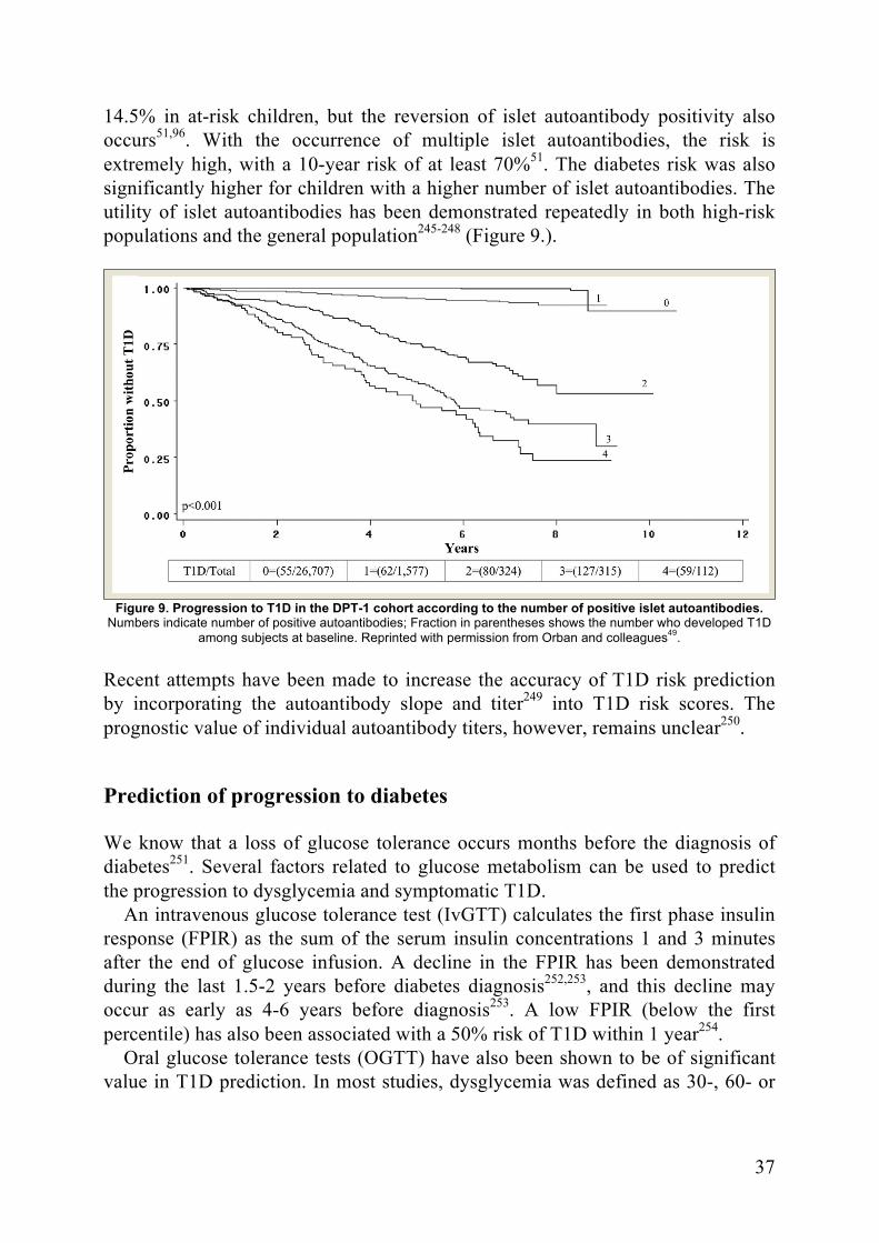
37
14.5% in at-risk children, but the reversion of islet autoantibody positivity also occurs51,96. With the occurrence of multiple islet autoantibodies, the risk is extremely high, with a 10-year risk of at least 70%51. The diabetes risk was also significantly higher for children with a higher number of islet autoantibodies. The utility of islet autoantibodies has been demonstrated repeatedly in both high-risk populations and the general population245-248 (Figure 9.).
Figure 9. Progression to T1D in the DPT-1 cohort according to the number of positive islet autoantibodies. Numbers indicate number of positive autoantibodies; Fraction in parentheses shows the number who developed T1D
among subjects at baseline. Reprinted with permission from Orban and colleagues49.
Recent attempts have been made to increase the accuracy of T1D risk prediction by incorporating the autoantibody slope and titer249 into T1D risk scores. The prognostic value of individual autoantibody titers, however, remains unclear250.
Prediction of progression to diabetes
We know that a loss of glucose tolerance occurs months before the diagnosis of diabetes251. Several factors related to glucose metabolism can be used to predict the progression to dysglycemia and symptomatic T1D.
An intravenous glucose tolerance test (IvGTT) calculates the first phase insulin response (FPIR) as the sum of the serum insulin concentrations 1 and 3 minutes after the end of glucose infusion. A decline in the FPIR has been demonstrated during the last 1.5-2 years before diabetes diagnosis252,253, and this decline may occur as early as 4-6 years before diagnosis253. A low FPIR (below the first percentile) has also been associated with a 50% risk of T1D within 1 year254.
Oral glucose tolerance tests (OGTT) have also been shown to be of significant value in T1D prediction. In most studies, dysglycemia was defined as 30-, 60- or

38
90-minute glucose values 11.1 mol/L or 120-minute glucose values of 7.8-11.1. The C-peptide response during OGTT starts to differ from controls at least two years before diabetes diagnosis, with a decrease in the early C-peptide response. Conversely, the late C-peptide response increases, leading to a normal C-peptide AUC255,256 (Figure 10). Significant declines in the peak C-peptide and the C-peptide AUC were only detected six months before diagnosis. Impaired glucose tolerance in the OGTT is associated with a HR above 8 for T1D diagnosis and a median time to diagnosis of 0.7 years257.
Figure 10. C-peptide levels from 0-30 minutes (left) and percentage of subjects with a peak C-peptide at 120 minutes (right) in the OGTT. X-axis shows years before diagnosis for progressors (grey line) or years before last visit (non-progressors). Progressors n=36; non-progressors n=80. Reprinted with permission from Sosenko and colleagues251.
As a fairly late predictor of β-cell failure and T1D diagnosis, HbA1c has also proved to be a useful, simple and inexpensive tool. A slow increase in HbA1c, within the normal limits, has been described in multiple islet autoantibody-positive subjects258. In multiple antibody-positive children, a 10% increase in HbA1c was associated with an 84% 3-year risk of T1D and a HR of 5.7 in two large prospective studies53,259. HbA1c levels tend to be consistently higher in T1D progressors two years before diagnosis, and when HbA1c levels exceed 41 mmol/mol, the median time to diagnosis is 0.9 years259.
Additionally, a simple random plasma glucose test can be of value for prediction. The DAISY study demonstrated the modest benefit of random plasma glucose, with a HR of 1.4 for T1D development258. In the DIPP study, a similar result was associated with a stronger risk for time to diabetes diagnosis259.
With the development of continuous and flash glucose monitors, new possibilities for assessing glucose control and variability have emerged. Several studies are ongoing, and this technology will no doubt prove useful in prediction efforts.
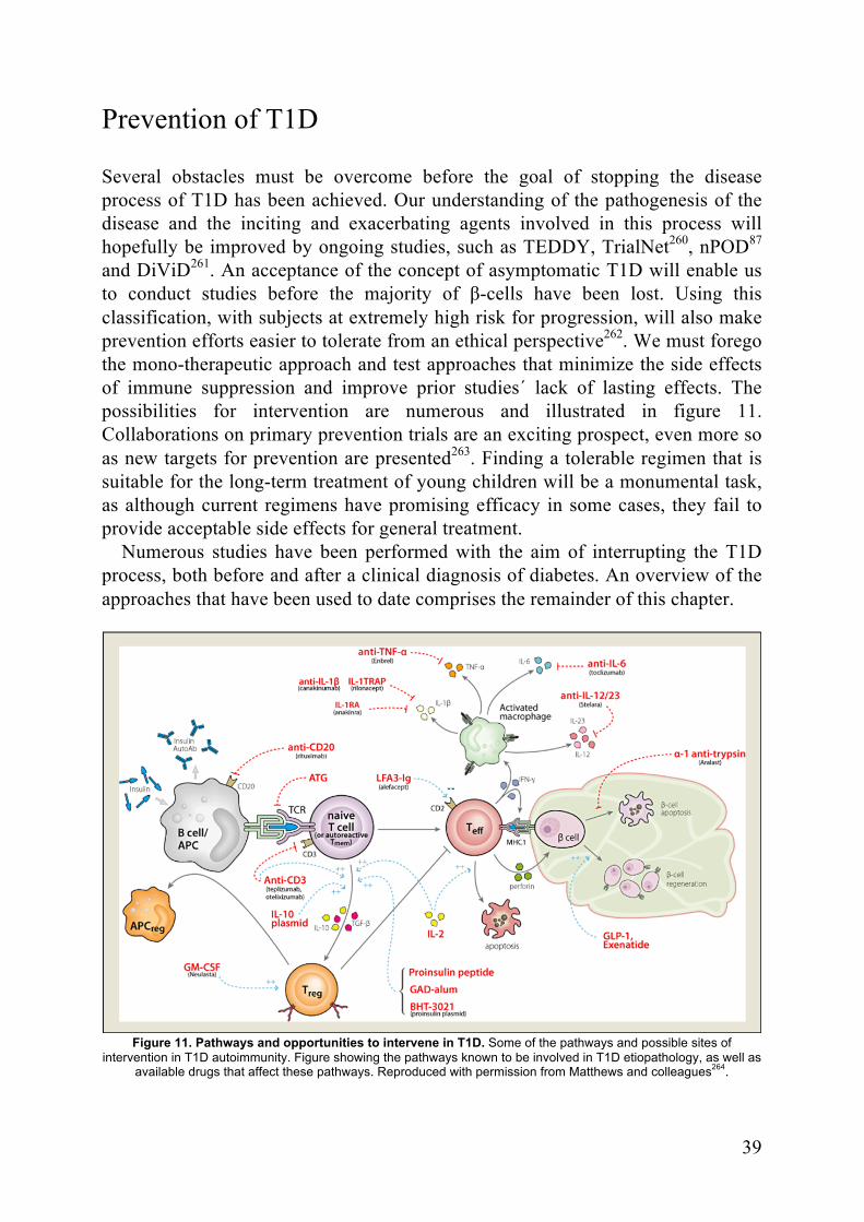
39
Prevention of T1D
Several obstacles must be overcome before the goal of stopping the disease process of T1D has been achieved. Our understanding of the pathogenesis of the disease and the inciting and exacerbating agents involved in this process will hopefully be improved by ongoing studies, such as TEDDY, TrialNet260, nPOD87 and DiViD261. An acceptance of the concept of asymptomatic T1D will enable us to conduct studies before the majority of β-cells have been lost. Using this classification, with subjects at extremely high risk for progression, will also make prevention efforts easier to tolerate from an ethical perspective262. We must forego the mono-therapeutic approach and test approaches that minimize the side effects of immune suppression and improve prior studies´ lack of lasting effects. The possibilities for intervention are numerous and illustrated in figure 11. Collaborations on primary prevention trials are an exciting prospect, even more so as new targets for prevention are presented263. Finding a tolerable regimen that is suitable for the long-term treatment of young children will be a monumental task, as although current regimens have promising efficacy in some cases, they fail to provide acceptable side effects for general treatment.
Numerous studies have been performed with the aim of interrupting the T1D process, both before and after a clinical diagnosis of diabetes. An overview of the approaches that have been used to date comprises the remainder of this chapter.
Figure 11. Pathways and opportunities to intervene in T1D. Some of the pathways and possible sites of intervention in T1D autoimmunity. Figure showing the pathways known to be involved in T1D etiopathology, as well as
available drugs that affect these pathways. Reproduced with permission from Matthews and colleagues264.

40
β-cell support
It has been hypothesized that β-cell stress leading to the increased release of antigens into the circulation could contribute to the autoimmune process of T1D and that over-worked β-cells lead to cellular dysfunction265. Attempts have been made to pharmacologically support the β-cells of at-risk or newly diagnosed T1D patients to delay the time to total insulin dependency.
Insulin The DPT-1 study attempted to delay or prevent T1D diagnosis in first-degree relatives who were deemed to have a five-year risk of above 50% via the administration of subcutaneous insulin. No effects could be seen regarding the time to diagnosis or the cumulative incidence of diabetes between the groups266.
Diazoxide Attempts to achieve a resting period for the β-cells by blocking insulin secretion with Diazoxide have also been made. A short-term increase in insulin secretion was observed after Diazoxide treatment ended. Unfortunately, no differences in C-peptide AUC were observed between the treatment and placebo groups during follow-up267.
β-cell protection
β-cells are known to have a lower capacity to cope with oxidative stress in comparison to the other cells of the endocrine pancreas. This limited anti-oxidative capacity and the ensuing vulnerability of β-cells have led to several attempts at protective treatments.
Vitamin D Vitamin D has been reported to have an immune modulatory effect, shifting the immunological balance from a Th1 to Th2 response, as well as improving the ability of β-cells to withstand stress268,269. Epidemiologically, vitamin D has been linked to protection against T1D if patients are supplemented at birth or during the first year of life212,270,271. Maternal vitamin D levels during pregnancy have been inversely correlated with T1D risk213. However, when used as single therapy in new-onset T1D patients, this treatment has not been successful in delaying or preventing diagnosis or preserving insulin secretion272,273.

41
Anti-oxidants As an anti-oxidative agent, nicotinamide has been thought to be able to protect β-cells from damage. However, clinical trials have demonstrated no efficacy in preventing T1D clinical onset274. Other attempts at mono-therapy with anti-oxidants have also failed to show effects in humans, despite promising animal studies275.
Immune modulation
BCG vaccination Vaccination with Bacillus Calmette-Guerin (BCG) has been used in early studies as an immune modulator. In theory, vaccination would increase the host’s TNF-α levels and reduce the levels of autoreactive T-cells. However, clinical trials have demonstrated no beneficial effect276. In addition, epidemiological studies have reported no correlation between T1D incidence and vaccination status277.
Anti-CD3 CD3, which is a co-receptor for the T-cell receptor (TCR), is involved in the activation and differentiation of naive T-cells into pathogenic effector T-Cells (Teff). CD3 prevents activation and promotes the depletion of T-cells, with Teffs being more sensitive than regulatory T-cells (Treg). This process leads to the depletion of Teff and the restoration of the Teff/Treg ratio.
Using anti-CD3 monoclonal antibodies (Oxelizumab, Teplizumab), several large studies have been performed, with conflicting but promising results. Oxelizumab has been shown to preserve insulin secretion in new-onset T1D patients. However, a lower dose failed to preserve insulin secretion in new-onset patients278-280.
Co-stimulation blockade Abatacept is a CTLA-4 receptor blocker fused to a modified fc-portion from human IgG1. This drug acts as decoy receptor for cd80/86 and blocks CD28-induced co-stimulation, interfering with T-cell activation, proliferation and differentiation. In a TrialNet study, new-onset T1D patients received 27 infusions of Abatacept or placebo over two years. A decreased rate of C-peptide decline and a higher C-peptide AUC were seen in the treatment group. Unfortunately, the autoimmune process only seems to be delayed by the treatment281.
B-lymphocyte-targeting approaches B-cells are involved in both antigen presentation and T-cell activation. On the B-cell membrane, CD20 is required for B-cell activation and
42
proliferation. Rituximab, which is a monoclonal antibody to CD20, was used in a TrialNet study in which a four-dose course was given to new-onset T1D patients (8-45 years old). The mean C-peptide AUC at year 1 was higher for the treatment group. The treatment group also had a lower HbA1c and required fewer insulin doses282. However, at 30 months post-randomization, there was no difference in the decline of the AUC C-peptide between the treatment groups283. The lack of long-term effect may be due to rapid natural B-cell replenishment; thus, additional therapeutic agents may be required to achieve a long-lasting immunological effect.
Cytokine-based targets A dysregulation in the balance between regulatory Treg cells and Teff cells has been suspected to be central to autoimmunity. Cytokines, such as IL-2, regulate both the function and size of the T-cell population. The IL-2 receptor on Treg cells has a higher affinity and is more numerous. Thus, Treg cells respond to a lower concentration of IL-2 than Teff cells. This concept has been used in several studies to treat the autoimmune process and has been proven to prevent T1D and reverse established disease in mice13. IL-2 has been used without significant adverse events and with a dose-dependent effect on Treg cells in a phase I study284. Phase II studies are ongoing in new-onset T1D patients but have yet to reach their endpoints. IL-23 and IL-12 are both involved in the amplification of pro-inflammatory pathways. Ustekinumab targets the shared p40 subunit of IL-12 and IL-23, blocking differentiation and central immunological signaling.
Two studies are currently active. One phase 1 study involves new-onset T1D patients (18-35 years) and ends in 2016. A second study uses a combination of Ustekinumab and islet neogenesis-associated protein (INGAP)-peptide in established T1D patients (19-40 years)14.
Tyrosine kinase inhibitors The tyrosine kinase inhibitors Imatinib and Sumatinib have previously been shown to preserve β-cell function in NOD mice, possibly via the platelet-derived growth factor receptor (pdgfr)15. Only case report evidence exists regarding the effects of these inhibitors in humans. A phase II study in patients with new-onset T1D is currently in progress.
T-regulatory cell-based therapy Treg-cells inhibit Teff-cells and regulate autoimmunity and immune tolerance. A low number of Treg-cells and impaired Treg cell function have been observed among T1D patients16. Treg-cells that are expanded ex vivo and then infused back into the patients have been investigated in studies that reported higher C-peptide levels in the treatment groups.
43
Ex vivo-expanded Treg-cells have shown good suppressive function in in vitro studies and a good safety profile in a phase 1 study18. In children with new-onset T1D, the autologous infusion of ex vivo-expanded Treg-cells prolonged remission, with higher C-peptide levels than the non-treated group17. A dosage study and a combination trial with polyclonal Treg-cells and IL-2 have been started19.
Immune Suppression
Corticosteroids Corticosteroids have proved to be extremely valuable in many autoimmune diseases. Unfortunately, because corticosteroids induce insulin resistance, they are not suitable for the long-term treatment of T1D autoimmunity. Few studies have been performed. The results showed possible immune effects and conflicting efficacy regarding the ability to preserve insulin secretion285,286.
Cyclosporin Cyclosporin treatment was one of the first attempts to stop the immune process of T1D. This was achieved, however, with a significant risk of side effects related to immune suppression and renal damage287,288. Studies using lower doses of Cyclosporin have shown greater safety but at the cost of a reduced effect289.
Azathioprine Azathioprine, as a single treatment, has been used in previous studies. The initial results regarding remission were promising but failed to achieve effects beyond the initial period after diagnosis290-292.
Anti-Thymocyte globulin Anti-thymocyte globulin antibodies against human T-cells have also shown promising effects on remission but failed to achieve long-term effects as a single therapy293.
Antigen-based therapies
Reverse vaccination, which is similar to the ASIT concept used in allergology, involves repeated doses of exogenous autoantigens that are given to achieve a long-term effect on regulatory, non-inflammatory T-cell responses. The goal is to induce immunological tolerance to the antigen via T-cell anergy, as well as the induction of Treg-cells294.

44
Insulin In the DPT-1 study, 372 subjects with a five-year T1D risk of 26-50% received oral insulin in a double-blind, placebo-controlled study. No effect could be seen on diabetes rates in the total cohort. However, a possible effect could be seen in subjects with insulin autoantibodies295. The results of a follow-up TrialNet study of oral insulin, which was closed in 2016, are still pending publication. In the Pre-Point study, the protective effects of high-dose oral insulin in children (2-7 years old) with a HLA risk genotype were studied, with potential effects on Treg-cells for children receiving 67.5 mg of oral insulin daily296. An active small, open-label TrialNet study is being conducted to assess the effects of dosing and schedules of oral insulin. Nasal insulin has also been used, but the treatment failed to delay or prevent T1D297.
Pro-insulin peptide As an immunologically active but non-functional agent, the pro-insulin peptide has been used to achieve immune tolerance. In a phase I study, the peptide was demonstrated to be safe and to have possible positive immunological effects298. A recent study also demonstrated the safety of the concept and reported promising results regarding the preservation of insulin secretion299.
GAD As one of the major autoantigens in T1D, GAD is an interesting target for immune modulation therapy. Early studies in mice yielded promising results regarding both safety and efficacy300-302. Safety was established in a phase I study, as well as later phase II/III studies. A potential effect on C-peptide preservation in adult LADA patients and safety information have also been published303,304. In children with new-onset disease, GAD was also demonstrated to be safe and to significantly preserve C-peptide in children with a shorter disease duration, but no effect was reported in other studies305-307.
Combination therapies
Given the disappointing results of many potent therapeutic agents in single therapy trials, especially with respect to long-lasting effects and the avoidance of adverse events, combination therapy may be the only way forward.
In a phase I/II study, high-dose immunosuppression combined with autologous stem cell transplantation is being used to induce tolerance in new-onset T1D patients (13-31 years). Cyclophosphamide and G-CSF are used to harvest stem cells and later re-infuse them after conditioning with Cyclophosphamide and anti-thymocyte globulin. During 30 months of follow-up, C-peptide was increased, and the majority of the patients were insulin-free or required only low-dose insulin308.

45
Other studies investigated similar regimens with beneficial results on insulin dependency six months after treatment and 4 years after treatment309. However, treatment is associated with significant side effects, with 52% of subjects experiencing adverse effects and one death caused by immune suppression and sepsis.
Protocols are being altered to lower the risks of immune suppression, including a lower dose of anti-thymocyte globulin and prolonged treatment with G-CSF. Using this regimen, a majority of treated subjects showed no decline in β-cell function 12 months after treatment and exhibited an improved Treg/Teff ratio. Side effects occurred at more acceptable levels and were reversible310.

46
47
Aims of the thesis
This thesis focuses parts of the process of T1D research in a pediatric setting, from risk factor identification through follow-up and intervention attempts. The specific aims of this thesis were to:
• Investigate the effect of islet autoantibodies in umbilical cord blood and the risk of T1D in the DiPiS cohort. (Paper I)
• Evaluate the possible effects of early life stress and severe life events on the risk of T1D in the DiPiS cohort. (Paper II)
• Examine the effects of exposure to analgesic antipyretics during the first two and a half years of life on the development of IA at age six years in the TEDDY cohort and describe the differences in use between the respective sites. (Paper III)
• Describe the outcomes of the children who had been enrolled in DiPiS at T1D diagnosis with respect to autoantibody status, metabolic derangement and peri-diagnostic morbidity. (Paper IV)
• Examine the outcomes of children who were previously enrolled in the DiPiS study follow-up after the diagnosis of T1D in comparison to those of children who were diagnosed from the general population. (Paper IV)
• Investigate whether GAD-Alum, given as two subcutaneous injections, is safe or causes severe adverse events in children with a high risk of T1D. (Paper V)
• Determine if GAD-Alum delays the time until T1D diagnosis. (Paper V)

48
49
Study Populations
The DiPiS Study
The DiPiS (Diabetes Prediction in Skåne) study is a prospective follow-up study in at-risk children. Children with a primarily increased genetic risk of T1D are followed until the age of 15 years. The aim of the study is to identify environmental risk factors for T1D and improve diabetes prediction.
Between September 2000 and August 2004, 48,058 children were born in the five participating hospitals in Skåne, which is in the southern part of Sweden. After obtaining oral consent from the mothers, 35,683 umbilical cord blood samples were collected for HLA-DQ and cord blood autoantibody analysis. When the child was two months old, the parents were invited to participate in the study and complete a questionnaire to collect demographic data, family medical history of interest and data concerning events and illnesses from pregnancy until two years of age. The parents of 25,378 children completed the questionnaire and gave written consent to participate in the study. A basic risk score was constructed based on the following parameters:
• HLA-DQ genotype (DQ2/8, DQ8/8 or DQ8/X, DQ2/2 or DQ2/X or DQX/X (X is neither 8 nor 2))
• Presence of maternal infections during pregnancy • High or low relative birth weight • First-degree relative with insulin-dependent diabetes • Positive for umbilical cord autoantibodies
Using this risk score, 7,826 children were offered yearly follow-up with a questionnaire and sampling for islet cell autoantibodies. In total, 3,889 parents consented to follow-up and entered the main DiPiS cohort (Figure 12.) At the time of the planning of the study, we presumed that very few children would develop diabetes during the first years of life, so follow-up of the children started at age two years.
The participants are sampled yearly for islet autoantibodies and asked to return a questionnaire covering the last year in the child´s life. Children who seroconverted to two or more islet autoantibodies were offered more intense follow-up every 3 months, which included islet autoantibodies, random plasma

50
glucose, HbA1c, growth parameters and a yearly oral glucose tolerance test (OGTT)
Figure 12. DiPiS study screening and enrollment.
As of August 31, 2017, a total of 222 children from the original birth cohort of 35,683 had developed T1D. Before follow-up began at two years of age, 14 children had been diagnosed. In the cohort that consented to prospective follow-up, 130 children had been diagnosed with T1D and 88 multiple autoantibody positive children were in active, intense follow-up (Figure 13.).
Figure 13. Flow chart of the children diagnosed with T1D in DiPiS.

51
The TEDDY Study
The environmental determinants of diabetes in the young study (TEDDY) is a prospective cohort study that aims to find environmental factors that influence the development of IA and T1D in children. The study includes six clinical research centers, including three in the US (Colorado, Georgia/Florida, Washington) and three in Europe (Finland, Germany, and Sweden)74 (Figure 14.)
Figure 14. Children enrolled in the TEDDY study, primary study endpoints and expected children at each endpoint by study completion. ZnT8A is only analyzed if any of the other autoantibodies are positive.
From September 2004 to February 2010, TEDDY screened 434,620 children, of whom 21,321 were eligible for follow-up. Screening was performed at TEDDY´s six clinical centers. Eligibility was determined using separate genetic HLA DQ criteria for children from the general population and children with a first-degree relative with T1D311.
Out of the eligible children, 8,676 children were enrolled in intensive follow-up. Follow-up started at age 3 months and included site visits every 3 months until the age of 4 years. After that, visits are performed every six months for autoantibody-negative children and every three months for children who are islet autoantibody-positive. A plethora of data are collected according to the study plan, including blood for islet autoantibodies, PBMC, DNA/mRNA, HbA1c and storage, urine, nasal swabs, nail clippings, tap water, salivary cortisol, accelerometer data, body composition data, etc. Interviews are performed at each visit to account for infection, medication and immunization data, food records, negative life events, parental anxiety and depression and physical activity312.
All samples are kept in a central repository, and all data are kept in the study data coordinating center in Tampa, Florida.
Clinical diabetes
ADA/WHO Criteria
Confirmed persistent islet autoimmunity
GADA, IA-2A or IAA (+ZnT8A)At least twice 3 months apart &
confirmed in second lab
Genetic susceptibility
Primary endpoint Primary endpointScreening criteria
Enrolledn=8676
Expected by study endn ~ 800
Expected by study endn ~ 400

52
The DiAPREV-IT study
Diabetes prevention immune tolerance, DiAPREV-IT, is an investigator-initiated, randomized, double-blind, placebo-controlled clinical trial (EuCT 2008-007484-16, NCT01122446). The primary aim of the study is to investigate the safety of GAD-Alum in children at risk for T1D. The second aim is to evaluate if immune tolerance treatment with GAD-Alum can delay and prevent the autoimmune process that leads to a diagnosis of T1D.
Study drug
The study drug for DiAPREV-IT, Diamyd® (GAD-Alum), was previously tested in several studies. However, none of those studies included children who were at high risk of T1D but not yet diagnosed. Previous studies of GAD-Alum have indicated effects on the progression of IA in spontaneously diabetic non-obese diabetic (NOD) mice302,313-316. A phase IIa study in patients with latent autoimmune diabetes in adults (LADA) demonstrated safety and potential efficacy in the group of patients receiving GAD-Alum303. Another phase IIb study was unfortunately invalidated due to identification issues. However, no concerns regarding safety were raised during the 18-month study period.
The potential efficacy of GAD-Alum in children has also been presented in a phase II study in recently (<18 months) diagnosed children with T1D. This study demonstrated both a favorable safety profile and an effect on the preservation of β-cell function after 30 months305. Further, a 3-armed phase III study in children with a recent onset of T1D (<3 months) has been published. In this study, one study arm received GAD-Alum as two single doses and a second study arm received two additional single doses on days 90 and 270 to potentially optimize the treatment. This study did not fulfill its efficacy goals after 15 months of follow-up and was closed early. However, no safety concerns were raised in the study307.Additionally, a three-armed TrialNET study on 145 new-onset patients (3-45 years) with either 3 doses of GAD-Alum, 2 doses of GAD-Alum or Placebo filed to demonstrate efficacy on preservation of insulin secretion at 1 year after treatment306.
Study inclusion
Children with positivity to GADA and at least one more islet autoantibody were recruited to the study. After consent and randomization, the participants received 20 µg of GAD-Alum in a prime-and-boost regimen on days 1 and 30.

53
Children with ongoing IA were recruited from the DiPiS study (described above), the TEDDY study (described above) and from the subjects followed in the Trial-Net natural history study (TN01). Trial-net participants were only invited when no TrialNet prevention study was available or suitable for the child. The inclusion/exclusion criteria can be seen below.
Inclusion criteria • Aged 4-17.99 years • Positive for GAD autoantibodies • Positive for at least one more islet autoantibody (IAA, IA-2A or
ZnT8R/W/Q/A) • Absence of the protective HLA DQ6 genotype
Exclusion criteria • Diabetes diagnosis • Ongoing treatment with immunosuppressant therapy • Significant abnormal hematology results at screening • History of acute reaction to vaccines • History of epilepsy, serious head trauma or cerebrovascular accident or
clinical features of continuous motor unit activity in proximal muscles • Participation in other clinical trials the last 3 months • Significant illness other than diabetes within 2 weeks prior to first dosing • HIV or hepatitis • Other serious disease or condition precluding subcutaneous injection
Study participants were recruited between April 2009 and January 2012. After baseline staging with both IvGTT and OGTT on two separate visits, the children were randomized to receive either GAD-Alum (Diamyd®, Diamyd Medical, Sweden) (n=25) or placebo (n=25). The study participants were then followed for five years, with visits every 3 months. Follow-up included physical examination, hematology, cellular immunological analyses, islet autoantibodies, glucose tolerance tests, HbA1c and thyroid and celiac autoantibodies.
For the diagnosis of diabetes, the criteria adopted by the WHO and the ADA were used24,25.

54

55
Laboratory Methods
HLA Genotyping
HLA genotyping in the DiPiS study
HLA genotyping was performed on dried blood spots (DBS) on filter paper, as originally described by Ilonen and Sjöros317-319. For the analyses in this thesis, HLA was classified as HLA-DQA1*0501-DQB1*0201 (DQ2) or HLA-DQA* 0301-DQB1*0302 (DQ8) and stratified into four risk groups: DQ 2/8, DQ8/8 or 8/X, DQ2/2, or 2/X or DQ X/X (X is neither DQ2 nor DQ8).
HLA genotyping in the TEDDY study
The TEDDY screening system that involves HLA-based mass screening for T1D risk has been thoroughly described previously320.
Genotype screening is performed using either a dried blood spot (DBS) punch or a small-volume whole blood lysate (WBL) specimen format321. The screening blood sample is typically obtained at birth as a cord blood sample, but potential participants, especially first-degree relatives of T1DM patients, can be screened using a heel stick capillary sample up to the age of 4 months. After PCR amplification of exon 2 of the HLA Class II gene (DRB1, DQA1 or DQB1), alleles are identified by direct sequencing, oligonucleotide probe hybridization, or other genotyping techniques. Additional typing to sufficiently identify certain DR-DQ haplotypes is specified for each clinical center.
Confirmation of the HLA genotypes is performed by the central HLA Reference Laboratory at Roche Molecular Systems in Oakland, CA (Dr. Henry Erlich) on 100% of the eligible subjects at 9 months of age. This process includes high-resolution genotyping of HLA-DRB1, DQA1, and DQB1. If a subject is not confirmed to be HLA eligible, a detailed explanation is provided to the parents, together with the option of continuing in TEDDY. These non-eligible subjects that stay in follow-up are noted in the databases maintained by the DCC. The HLA Central Laboratory genotypes the DNA sample obtained at 9 months of age and also evaluates three non-HLA markers of T1D genetic susceptibility: insulin 5’-
56
VNTR using the –23 Hph polymorphism, the CTLA-4 A49G polymorphism in exon 1 and the PTPN R620W polymorphism. Genotyping for HLA- DPB1, HLA-A, HLA-B, MIC-A and/or other MHC and non-MHC markers may occur in the future, as determined by the study group311.
Islet autoantibody analysis
Islet autoantibody analysis in the DiPiS and DiAPREV-IT studies
Autoantibodies to GAD65 and IA-2: Analysis in dried blood spots, serum or plasma GAD65 autoantibodies (GADA) and IA-2 autoantibodies (IA2A) were analyzed in dried blood spots (DBS) with a radioligand binding assay (RBA)322. GADA and IA2A levels were expressed as units per mL (U/mL) derived from the WHO standard 97/550. The samples were considered to be positive if the IA2A levels were above 5 U/mL or the GADA levels were above 34 U/mL.
Autoantibodies to IAA Noncompetitive method: Serum samples (7 mL) were added to duplicate wells of a 96-well microplate, and 36 mL of 125I insulin (30) with an activity of 60,000 cpm/well was added, then incubated on a shaker at 4°C for 48 h. PAS in a 40% slurry (50 mL) was added to a filter plate and washed three times with 200 mL of Tris buffer using a Micro-Plate Strip Washer (BioTek ELx50; BioTek Instruments, Bedfordshire, U.K.). Supermix scintillation solution (50 mL) was added to the wells after the plate had dried for 15 min. The radioactivity was measured in a ß-counter (Wallac Micro Beta Trilux; PerkinElmer).
Competitive method: Positive samples for IAA were further analyzed using a competitive method. Serum samples (7 mL) were added to four wells in a 96-well plate. To these wells, 36 mL of 125I insulin with an activity of 60,000 cpm/well were added, with 0.072 IU (or 2 IU/mL) of unlabeled insulin (Actrapid; Novo Nordisk) added to the last two wells. The plates were incubated and examined under conditions similar to those described for the noncompetitive method. IAA levels were calculated as relative units and were related to positive controls. Positivity for IAA was set to 1.9 relative units. The competitive method was used to verify false-positive binding in the noncompetitive assay. However, in subsequent analysis, the competitive assay was used.
57
Autoantibodies to ZnT8 ZnT8 autoantibodies (ZnT8A) were analyzed in 5 µL of serum with RBA, as described previously323. Duplicate samples were incubated with equal amounts of the three radio-labelled ZnT8 R/W/Q variants. Every putative positive sample (cut-off >59 U/mL) was analyzed for each ZnT8 variant separately.
Islet autoantibody analysis in the TEDDY Study
Islet autoantibodies to insulin, GAD65 and IA-2 were measured in two laboratories by RBA128,324. In the U.S., all sera were assayed at the Barbara Davis Center for Childhood Diabetes at the University of Colorado Denver; in Europe, all sera were assayed at the University of Bristol, the U.K. Both laboratories have previously shown high sensitivity and specificity, as well as concordance119. All samples positive for islet autoantibodies and 5% of negative samples were re-tested in the other reference laboratory, and the results were deemed to be confirmed if concordant312.
To optimize concordance, harmonized assays for GADA and IA-2A replaced previous assays, in January 2010128. Based on a receiver-operator curve analysis, prior samples that needed to be re-analyzed with the harmonized assays included: Denver GADA between -0.015 and 0.042; Bristol GADA between 10.69 and 36.72; Denver IA-2A between -0.004 and 0.016; and Bristol IA-2A between 6.69 and 10.58.
Definition of IA in the TEDDY study In the TEDDY study, persistent IA is defined as being positive to IAA, GADA or IA-2A, with the results confirmed at both laboratories and on at least two consecutive visits. The date of persistent autoimmunity was defined as the draw date of the first sample of the two consecutive samples which deemed the child as persistently positive for a specific autoantibody (or any autoantibody).
To exclude children who were born with maternal islet autoantibodies, the islet autoantibody status of the mother was assessed when the child was 9 months. If a child was islet autoantibody-positive under the age of 18 months, their autoantibody status was set as “pending.” Autoantibody sampling was performed every three months until 18 months of age. If maternal autoantibodies were present, the child was not deemed to be persistently islet autoantibody-positive unless they had a negative sample prior to their first positive sample.

58
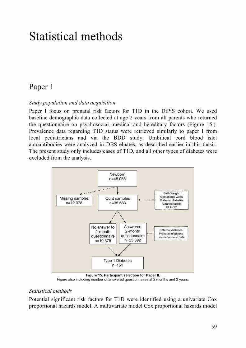
59
Statistical methods
Paper I
Study population and data acquisition Paper I focus on prenatal risk factors for T1D in the DiPiS cohort. We used baseline demographic data collected at age 2 years from all parents who returned the questionnaire on psychosocial, medical and hereditary factors (Figure 15.). Prevalence data regarding T1D status were retrieved similarly to paper I from local pediatricians and via the BDD study. Umbilical cord blood islet autoantibodies were analyzed in DBS eluates, as described earlier in this thesis. The present study only includes cases of T1D, and all other types of diabetes were excluded from the analysis.
Figure 15. Participant selection for Paper II.
Figure also including number of answered questionnaires at 2 months and 2 years.
Statistical methods Potential significant risk factors for T1D were identified using a univariate Cox proportional hazards model. A multivariate model Cox proportional hazards model

60
was used to test for independent associations for each autoantibody, birth weight, gestational length and infections. The model was stratified for HLA-DQ risk and also adjusted for gender, maternal age, and parental T1D. Hazard ratios for HLA-DQ risk groups were calculated in a model without stratification, and a separate baseline hazard for HLA genotype was fitted for each group. The variables available as possible independent predictors were autoantibody status in umbilical cord blood for IA-2, insulin and GAD (deemed positive if above the 95th percentile and treated as categorical variables), gestational week (<37, 37-39, 40-41, >41), the relative birth weight percentile, and infection during pregnancy (yes/no). As possible confounders in the analysis, HLA risk group (DQ X/X, DQ2/2 or 2/X, DQ 8/8 or 8/X, DQ 2/8, X is neither 2 nor 8), gender, parental T1D and mother’s age at delivery were available.
Statistical analyses were performed using SPSS version 21 (SPSS, Chicago, IL) and R version 3.03, using the survival package (R Core Team (2014). R Foundation for Statistical Computing, Vienna, Austria.).
Paper II
Study population and data acquisition In paper II, we focus on the possible effects of early life stress on T1D risk. When the children sampled with umbilical cord samples were two months old, a two-part questionnaire was sent out, with one part focusing on prenatal and antenatal events, as well as baseline demographic data on the parents, and the other part focusing on hereditary data on diseases in the family. A total of 23,187 (65%) questionnaires were returned to the study. The children who were invited to follow-up were then asked to complete a more thorough questionnaire regarding the child´s first two years of life. The questions covered a wide range of fields, including socioeconomic data, support system, depression and anxiety related to parenting, introduction of food, infections and medications. This “Two-year questionnaire” was answered by 3,861 families (49%). The analysis in this study uses data from both the two-month and the two-year questionnaires. The study selection can be seen in Figure 16. T1D incidence was reported by local pediatricians, as well as via the Better Diabetes Diagnosis study (BDD), with excellent coverage across Sweden134. All children with answers to the two-month and two-year questionnaires were included in the study.
Severe life events On the two-month questionnaire, the family answered questions regarding severe life events during or after pregnancy. The overall question of ”Did you experience anything that you would consider to be a severe life event during pregnancy or

61
after the child´s birth?” was followed by eight choices with tick-boxes for "Death of close relative"; "Severe disease in the family"; "Serious accident in family"; "Own divorce or separation"; "Been subjected to violence"; "Lost employment"; "Husband/Partner lost employment"; and "Serious conflict with the child´s father or another significant person," as well as tick-boxes to indicate if the event took place during or after pregnancy. These questions were analyzed regarding T1D risk, both as a joint variable indicating that an event of any kind had taken place during or after pregnancy and as individual events.
Figure 16. Flow chart of Study participation in Paper IV
Family stress indices Five questions regarding parental discord were answered on the two-year questionnaire, as well as six questions regarding parenting problems and child temperament. All of these questions were answered on a Likert scale. The exact translated wording can be seen in Figure 17. For analysis, we recoded all answers as a value between 0 and 1, giving all questions equal weight. The recoded values were then added up to an index value. Index values above the 90th percentile were considered to be positive in the analysis. Separate indices were calculated for
Newbornn=48 058
Missing Cord Samplesn=12 375
Cord blood samplesn=35 683
Returned 2-month questionnaire
n=23 187
Not offered Follow-up
n=15 361
Non-participantsn=12 496
Returned 2-year questionnaire
n=3861
Type 1 Diabetesn=152
Diabetes Risk - offered Follow-up
n=7826
Type 1 Diabetesn=73
Declined follow-upn=3965

62
answers given by the mother, answers given by the father and a joint variable with index values from both parents combined.
Figure 17. Questions and possible answers included in the parental discord and Child disharmony indexes
Tau-equivalent reliability was calculated using Cronbach’s α, showing acceptable internal consistency for maternal and paternal answers on parental discord when analyzed separately (Cronbach’s α coefficient of 0.65 for mothers and 0.59 for fathers) and good internal consistency for the joint parental index (Cronbach’s α coefficient of 0.76). For the Parenting Stress index, internal consistency was good regarding the separate maternal and paternal answers (Cronbach’s α coefficient of 0.78 for mothers and 0.76 for fathers), and consistency was excellent for the joint parental index (Cronbach’s α coefficient of 0.86). The distribution of index values for both indices can be seen in Figure 18.
Statistical analysis To assess potential confounders in the model, a univariate Cox proportional hazards model was used. Confounders with a significance value of p <0.2 were included in a multivariate Cox proportional hazards model. The included confounders included maternal insulin-dependent diabetes, paternal insulin-dependent diabetes, IA-2A cord blood positivity325, mother born in Sweden and father born in Sweden. The multivariate analysis was stratified for HLA risk group (DQ2/8, DQ8/8 or DQ8/X, DQ2/2 or DQ2/X and DQX/X, (X is neither 8 nor

63
2)). To further examine the possible effects of stress, a separate analysis was performed for the highest-risk group: HLA DQ2/DQ8. In this multivariate model, maternal insulin-dependent diabetes, paternal insulin-dependent diabetes, IA-2A cord blood positivity and paternal education level were used as covariates. In the sub group analysis, the SLE “Own divorce or separation” failed to meet the requirements of the statistical model and was not included in the results.
Figure 18. Tukey box-plot of answers to the parental discord and parenting stress indices.
Censoring for cases that had not been diagnosed with T1D was set at April 20th, 2016. Missing data in questionnaires were handled in a pairwise manner. Statistical analysis was performed using SPSS version 24 (IBM Corp, Armonk, NY, USA) and GraphPad Prism 7 (GraphPad Software, La Jolla, California, USA).
Paper III
Study population and data acquisition For the analysis in paper III, the data received at the TEDDY data coordinating center as of 31st of December 2014 were used. At this time, 8,876 children were enrolled in the study. For this analysis, only children with medication data regarding for the first two years of life, first child status available and islet autoantibody status were included. After exclusion due to missing data regarding medication use, first child status and participants lacking full SNP data, 6,894 participants remained for the analysis. Study inclusion can be seen in Figure 19. At study visits, every three months, interviewers recorded all medication, with the name of the drug, the start date and the duration since the last study visit. Data regarding infections were documented by the parents in the “TEDDY Book” and for every illness episode, parents answered “yes” or ”no” regarding the presence of

64
fever. All parents received written guidance that “yes” should only be selected if the body temperature was 38°C/101° F.
Figure 19. Excluded subjects in Paper III.
A total of 1648 subjects were excluded due to missing data leaving a total of 6894 subjects included in the study.
Approximately 18 months into the study, these choices were expanded to “Yes – measured,” “Yes – not measured,” and “No”. All illness was recorded as ICD-10 codes, start of episode and duration. Infection was defined as an ICD-10 code defined as infection by the study clinical implementation and infectious agents committee. The method for reporting and categorizing illness in TEDDY has been previously reported elsewhere326. Medication was recorded by brand name and categorized according to active ingredient. For analysis, all medication including that ingredient was included. For further analysis, each medication was also categorized as either acetaminophen(APAP) or non-steroidal anti-inflammatory (NSAID). Each use of an analgesic antipyretic (ANAP) was defined as an episode and was defined as associated with infection and/or fever.
Statistical analysis The impact of ANAP use in the first 90, 180, and 365 days of life, as well as the first 2.5 years of life, on IA at age three and six years was assessed using a Cox proportional hazards model in which country was used as a stratification variable. The number of infections early in life was added as a time-dependent covariate327. As additional covariates, first-degree relative328, HLA159, gender, ever breastfed329,330, probiotic use before 3 months of age233 and eight single nucleotide polymorphisms previously identified in the TEDDY study were included331. Cumulative use of ANAPs during the child´s first 2.5 years was used as the primary covariate of interest. In cases with missing data regarding the duration of
65
use, that subject was excluded from the analysis for that specific analgesic. To better fit the linear statistical model, log transformed data were used for the analysis of the total duration of ANAP use per year. A logistic regression model was used to analyze subject incidence, with country and first child status as independent variables. Episodes were categorized as either with concurrent fever (yes/no) or infection (yes/no), both fever and infection (yes/no) or neither fever nor infection (yes/no). Using an ignorable working index, a generalized estimating equation (GEE) was used for analysis, with country and first child status as independent variables. An ignorable working matrix was assumed for the GEE analysis, with the empirical sandwich estimate used for the standard errors. For pairwise comparisons, in both binary and continuous analyses, we corrected for multiplicity of comparisons using the Bonferroni-Holm procedure.
Statistical analyses were performed using the Statistical Analysis System software, version 9.3 (SAS Institute Inc., Cary, NC, USA). Figures were created using GraphPad Prism 7 for Mac (GraphPad Software, La Jolla California, USA).
Paper IV
Study population and data acquisition In paper IV, we aimed to analyze the status at diagnosis and for the two first years after diagnosis, focusing on whether the children followed in the DiPiS study differed from children in the general population. Before the age of two years, at which follow-up began, 14 children had been diagnosed with T1D. These children were excluded from the analysis. In the DiPiS cohort, 129 children had been diagnosed with T1D as of July 31st, 2013. Risk information was only offered to children whose parents consented to follow-up at age two. Out of these children, 4,340 children consented to follow-up, and 82 of those children had been in intense follow-up every three months. The remaining 4,258 children participated in yearly follow-up (Figure 20.). For this study, all children who had received information about T1D risk and participated in follow-up at any time formed the Follow-up group. The Non-follow-up group consisted of children who chose not to participate in the 2-month questionnaire, who were not offered follow-up or who chose not to participate in follow-up. These children had not received information regarding T1D risk. At the time of analysis, 32 participants had not yet reached 24 months post-diagnosis, including 14 in the follow-up group and 18 in the non-follow-up group. The number of participants available for analysis can be seen in Table 4. HLA DQ genotyping and autoantibody analysis were performed as previously described in the methods section of this thesis. Prevalence data regarding diabetes diagnosis were reported from pediatricians at the pediatric

66
clinics in Region Skåne at the time of diagnosis. Additional prevalence data were retrieved from the Better Diabetes Diagnosis study (BDD)134 and the Skåne study.
Figure 20. Flow diagram of study participants in Paper I
Diagnosis data and data regarding follow-up (HbA1c and insulin doses) were retrieved from electronic patient medical records (Melior, Siemens AG, Berlin, Germany). Insulin doses were analyzed as the total daily dose (TDD) of insulin per kg bodyweight. A +/- one-month window was allowed for follow-up data to maximize data retrieval. DKA was defined as blood pH <7.3, with severe DKA defined as pH <7.1.
Table 4. Follow-up data available for analysis after diagnosis Group Diagnosis 3 months 6 months 12 months 24 months
Follow-up 51 45 45 43 33
No Follow-up 77 74 74 70 56
Statistical methods Statistical analysis was performed via group-wise comparisons between the follow-up and non-follow-up groups. Categorical variables were tested using Pearson’s !2 or Fischer´s exact test, where applicable. The Mann-Whitney U test was used for continuous variables. Spearman´s rho was used for correlation analysis between Hba1c and insulin doses during follow-up. HbA1c was recorded and analyzed as IFCC (mmol/mol) and converted to NGSP (%) for presentation purposes332. Corrections for multiple comparisons were performed using the Bonferroni method, where applicable. SPSS 21 was used for statistical analysis (SPSS Inc., Chicago, IL, USA).

67
Paper V
Study population and data acquisition According to the original study plan, 50 participants were recruited to the study. The participants were randomized to either two doses of 20 µg of GAD-alum or placebo as a subcutaneous injection, with one month between injections. All participants and study personnel were blinded during both treatment and follow-up. After the administration of the study drug or placebo, the participants were followed for five years or until a diabetes diagnosis. Glucose tolerance tests were performed every six months, alternating between OGTT and IvGTT. At all visits, laboratory samples were collected, including islet autoantibodies (GADA, IAA, IA-2A, ZnT8 RA/WA/QA), hematology, blood chemistry, HbA1c, random plasma glucose and random or fasting C-peptide (depending on visit). Annual screening for celiac and thyroid autoimmunity was performed.
Information from the study visits was entered into a clinical research form (CRF) and archived both electronically and on paper. Adverse events were graded as mild, moderate or severe. All alterations in health status were recorded as adverse events.
Glucose tolerance tests OGTT was performed after overnight fasting. An oral dose of 1.75 g of glucose/kg body weight of Nutrical (N.V. Nutrical Zoetermeer, Holland) was given. At 0, 30, 60, 90 and 120 minutes after ingesting the venous plasma glucose, serum C-peptide and serum insulin were measured.
IvGTT was performed after overnight fasting. An intravenous dose of 500 mg/kg body weight of glucose was injected within 3 minutes as a 300 mg/ml solution. At -10, 0, 1, 3, 5, 7, 10, 30, 50, 70 and 90 minutes after injection, a venous sample was collected for the analysis of plasma glucose, serum C-peptide, and serum insulin. Glucose values were plotted semi-logarithmically against time. The disappearance rate of glucose was estimated on the near-straight line in order to calculate the K-value333.
Statistical analysis All 50 children included in the study were included in the final statistical analysis. Unblinding was performed after the database was locked and assigned to the intent to treat, full, per protocol and safety populations. All statistical analysis and grouping of participants were performed according to the statistical analytical plan outlined in the original protocol. One child was excluded from the per protocol analysis due to diabetes values at the baseline visit.
For analysis, non-normally distributed numeric variables were log transformed, then back-transformed for presentation. A K-test or Wilcoxon´s signed rank test

68
was used to compare baseline characteristics for continuous variables. Pearson’s !2 or Fischer´s exact test was used for categorical variables. Using log-rank !2 statistics, Kaplan-Meier life tables were compared. A univariate Cox proportional hazards model was used to compare risk between groups. A multivariate Cox model was used to account for possible covariates affecting the results. This model included sex (girl/boy), treatment with GAD-Alum (yes/no), age at randomization, first-degree relative (yes/no), impaired glucose tolerance at baseline (yes/no), baseline FPIR, baseline C-peptide area under the curve (AUC) and autoantibody stratum (2 autoantibodies/3-6 autoantibodies). The assumptions of the Cox proportional hazards model were confirmed for all variables.
A mixed-models repeated measures approach was used to analyze longitudinal data on secondary end-points during follow-up. Subject effects were treated as random effects, and time and the variable of interest were treated as fixed effects. The validity of mixed models was examined using goodness of fit statistics (log likelihood deviance, AIC, BIC). No adjustments were made for multiplicity of comparison, except when part of the multivariate analysis. All tests of significance were two-tailed, and the significance level was set at 0.05.
Statistical analysis was performed using SAS 9.3 (SAS Institute, Inc., Cary, NC, USA).
Ethical approval
Ethical approval for papers I, II and IV was granted by the ethics review board in Lund, Sweden via approvals for the DiPiS study.
Paper III received ethical approval from the Colorado Multiple Institutional Review Board, the Augusta University Institutional Review Board, the University of Florida Health Center Institutional Review Board, the Western Institutional Review Board, the Ethics Committee of the Hospital District of Southwest Finland, the Bayerischen Landesärztekammer (Bavarian Medical Association) Ethics Committee, and the Lund University Committee for Continuing Ethical Review. The study is also overseen by an external advisory board formed by the National Institutes of Health. The data that support the findings of paper III will be available from the NIDDK repository (http://niddkrepository.org) approximately 6 months after the manuscript has appeared in print.
Approval for paper V was granted by the ethics review board in Lund, Sweden, as well as by the medical product agency (MPA) in Sweden.

69
Results
Do umbilical cord blood islet autoantibodies increase the risk of T1D (Paper I)?
Baseline characteristics of the study Umbilical cord blood samples from 35,683 children were collected at birth as part of the DiPiS screening. Baseline data were collected from both data collected at birth and from the two-month questionnaire answered by the parents of 25,392 children. Of the parents who responded to the two-month questionnaire, 190 mothers reported insulin-treated diabetes and 790 mothers reported gestational diabetes. Paternal insulin-treated diabetes was reported for 285 children. At the time of analysis (December 31st, 2013), 151 children from the originally screened cohort had been diagnosed with T1D at a median age of 5.8 years (range: 0.8-12.2 years).
How many children are islet autoantibody-positive at birth? Islet autoantibodies against one or more islet autoantibodies were found in umbilical cord blood from 757 children (2.1%). GADA were found in 295 children (0.8%), IA-2A were found in 78 children (0.2%) and IAA were found in 509 children (1.4%). Of the IAA-positive children, 105 (21%) had mothers with insulin-treated diabetes, 214 (42%) had mothers who were not treated with insulin and 190 (37%) were missing data regarding maternal insulin use. The distribution of single-, double- and triple-positive samples can be seen in figure 21.
Is the T1D risk higher for children with cord blood islet autoantibodies? All probable covariates were initially analyzed in a univariate model. In this analysis, IA-2A (HR 13.5; 95%CI 5.02, 36.6; p <0.001) and GADA (HR 3.44; 95% CI 1.27, 9.29; p=0.015) but not IAA (HR 2.41; 95% CI 0.98, 5.88; p=0.053) were predictors of T1D risk.
When using a multivariate model, with stratification for HLA risk group and with maternal insulin-treated diabetes, paternal insulin-treated diabetes, IA-2A, GADA and IAA included as covariates, only IA-2A showed evidence of association with T1D risk (HR 7.73; 95% CI 1.94, 30.7; p <0.001). This result
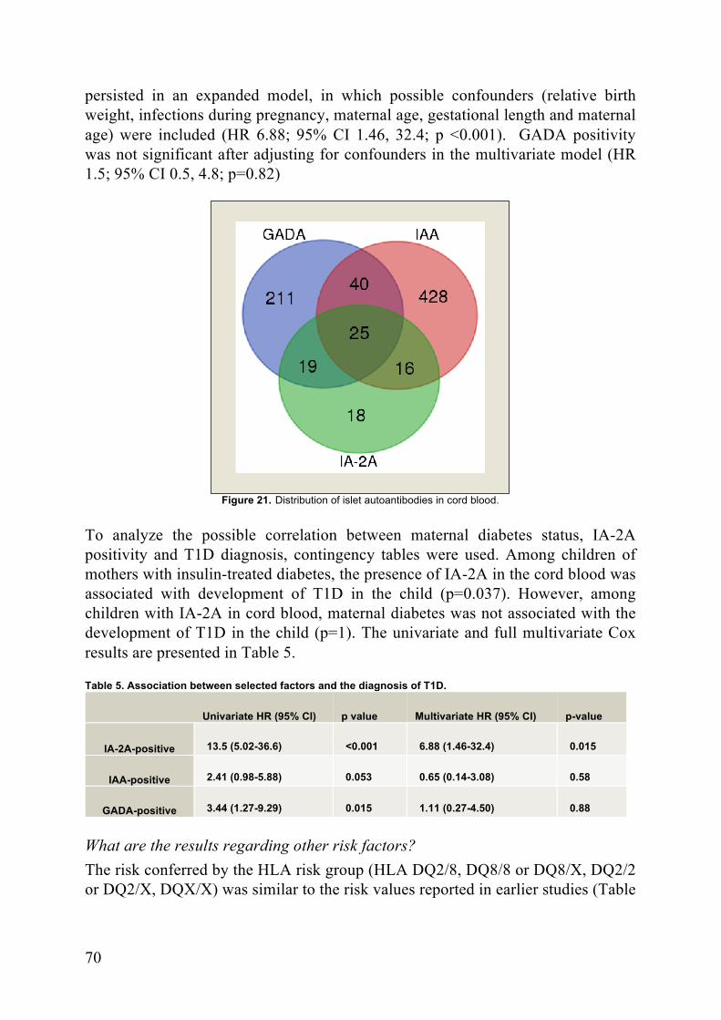
70
persisted in an expanded model, in which possible confounders (relative birth weight, infections during pregnancy, maternal age, gestational length and maternal age) were included (HR 6.88; 95% CI 1.46, 32.4; p <0.001). GADA positivity was not significant after adjusting for confounders in the multivariate model (HR 1.5; 95% CI 0.5, 4.8; p=0.82)
Figure 21. Distribution of islet autoantibodies in cord blood.
To analyze the possible correlation between maternal diabetes status, IA-2A positivity and T1D diagnosis, contingency tables were used. Among children of mothers with insulin-treated diabetes, the presence of IA-2A in the cord blood was associated with development of T1D in the child (p=0.037). However, among children with IA-2A in cord blood, maternal diabetes was not associated with the development of T1D in the child (p=1). The univariate and full multivariate Cox results are presented in Table 5.
Table 5. Association between selected factors and the diagnosis of T1D.
Univariate HR (95% CI) p value Multivariate HR (95% CI) p-value
IA-2A-positive 13.5 (5.02-36.6) <0.001 6.88 (1.46-32.4) 0.015
IAA-positive 2.41 (0.98-5.88) 0.053 0.65 (0.14-3.08) 0.58
GADA-positive 3.44 (1.27-9.29) 0.015 1.11 (0.27-4.50) 0.88
What are the results regarding other risk factors? The risk conferred by the HLA risk group (HLA DQ2/8, DQ8/8 or DQ8/X, DQ2/2 or DQ2/X, DQX/X) was similar to the risk values reported in earlier studies (Table

71
6.). Having a father with insulin-treated diabetes was associated with an increased risk of T1D (HR 3.4; 95% CI 1.6, 7.0; p <0.001). Having a mother with insulin-treated diabetes was significant in the univariate analysis (HR 3.5; 95% CI 1.3, 9.5; p=0.014) but failed to reach significance in the multivariate analysis (HR 1.38; 95% CI 0.24; 7.84; p=0.71).
Table 6. Association between HLA risk group and T1D risk.
Univariate Analysis HR (95% CI) p value Multivariate Analysis
HR (95% CI) p-value
HLA risk-group
-DQ 2/8 18.2 (13.1-25-3) <0.001 42.4 (23.3-77.2) <0.001
-DQ 8/8 or 8/X 3.48 (2.42-5.01) <0.001 10.9 (5.74-20.7) <0.001
-DQ 2/2 or 2/X 2.02 (1.30-3.16) 0.002 8.51 (4.20-17.2) <0.001
-DQ X/X (ref) ref ref ref ref
Are stress and severe life events in early life a risk factor for T1D (Paper II)?
Baseline characteristics of the study Responses to the two-month questionnaire were received from the parents of 23,187 children, and responses to the two-year questionnaire were received from the parents of 3,861 children. The mean age at T1D diagnosis in this cohort was 8.19 years (SD 3.8) (Table 7.).
Do early severe life events increase the risk of T1D? In the total cohort, using a univariate cox regression model, only having experienced violence during or after pregnancy was associated with an increased risk of T1D (HR 4.42; 95% CI 1.41, 13.88; p=0.011). However, in multivariate analysis stratified for the HLA risk group, having experienced any severe life event after birth was associated with an increased risk of T1D (HR 1.66; 95% CI 1.02, 2.70; p=0.043) as was conflict with spouse (HR 2.28; 95% CI 1.19, 4.36; p=0.013) and having been the victim of violence (HR 4.52; 95% CI 1.29, 15.8; p=0.018). For the high-risk HLA DQ2/8 sub cohort, univariate analyses show an increased risk of T1D among children of parents who experienced any of the severe life events after pregnancy (HR 2.15; 95% CI 1.09, 4.27; p=0.028) (Figure 22.), as well as parents who had been the victim of violence (HR 7.42; 95% CI 1.81, 30.5; p=0.005).

72
Table 7. Baseline characteristics of respondents to the 2-month and 2-year DiPiS questionnaires. 2-month
Questionnaire 2-year
Questionnaire
Number of questionnaires n 23187 3861
Maternal age at birth Mean (IQR) 30,8 (6,0) 31.4 (6.0)
Male child n (%) 11589 (51.6) 1899 (52.2)
Gestational age Median (IQR) 39,3 (2,0) 39.1 (2.0)
Maternal IDDM n (%) 163 (0.7) 117 (3.1)
Paternal IDDM n (%) 227 (1.0) 125 (3.3)
HLA risk group
DQ2/DQ8 n (%) 865 (3.7) 431 (11.4)
DQ8/8 or 8/X n (%) 2163 (9.3) 1140 (30.1)
DQ2/2 or 2/X n (%) 1937 (8.4) 1016 (26.8)
DQ X/X n (%) 18190 (78.6) 1197 (31.6)
Maternal education
Nine-year compulsory school n (%) 1412 (6.1) 196 (5.2)
Senior high school n (%) 11611 (50.4) 1812 (48.1)
College/University n (%) 10028 (43.5) 1761 (46.5)
Paternal education
Nine-year compulsory school n (%) 2079 (10.0) 336 (9.6)
Senior high school n (%) 11411 (49.2) 1885 (53.7)
College/University n (%) 7379 (35.4) 1287 (36.7)
Mother born in Sweden n (%) 20419 (89.2) 3472 (93.0)
Father born in Sweden n (%) 18553 (88.8) 3434 (92.1)
In the multivariate model that included HLA risk-group stratification, having experienced any severe life event after pregnancy was still significantly correlated with T1D risk (HR 2.21; 95% CI 1.08, 4.51; p=0.030), as was the father being unemployed (HR 3.50; 95% CI 1.09, 11.3; p=0.036) and conflict with spouse (HR 3.12; 95% CI 1.22, 7.99; p=0.018).
Are self-reported parenting stress and parental discord correlated with increased T1D risk? Using the parental discord index in the total cohort, answers above the 90th percentile given by the father were associated with an increased T1D hazard ratio in only the univariate analysis (HR 1.98; 95% CI 1.04, 3.78; p=0.038). No other significant hazards for the parental discord index were found in multivariate analysis, either when analyzing the parental answers separately or when analyzing the parental answers as a joint variable (mother p=0.41; father p=0.27; joint p=0.28). Index scores above the 90th percentile for the parenting stress index were not correlated with an increased T1D risk for the joint answers from both parents or for the answers from the mother or father separately (p=0.532; p=0.772; p=0.335). For the high-risk HLA DQ2/8 sub-cohort, parental discord, as reported

73
by the father and the joint parental answers, correlated with an increased T1D risk (HR 2.68; 95% CI 1.02, 7.08; p=0.047 and HR 2.87; 95% CI 1.17, 7.10; p=0.022, respectively) in the univariate analysis.
Figure 22. Kaplan Meier curve for severe life events after pregnancy. Separate curves are resented for the total study cohort and the HLA DQ2/DQ8 sub-cohort. Green line: Yes; Blue line: No.
Reported parental discord from the mother in the univariate analysis (p=0.054) and the indices from both parents in the multivariate analysis did not correlate with the T1D risk (mother p=0.17; father p=0.11; combined p=0.082). No correlation with T1D risk was found for the parental stress index in the univariate (mother p=0.68; father p=0.41; combined p=0.90) or multivariate analysis (mother p=0.80; father p=0.68; combined p=0.86).
Is the use of analgesic antipyretics in early life a risk factor for islet autoimmunity (Paper III)?
Baseline characteristics of the study The study cohort consisted of 8,542 children, 51% male and 11% first-degree relatives, who were enrolled in the TEDDY study. The country distribution can be seen in Table 9. In general, the use of APAP and NSAIDs before the age of 2.5 years was very common in the studied cohort. Parents reported that 87.8% of children had used APAP and 45.4% had used NSAIDs at any time. The mean number of treatment episodes was reported to be 3.6 (SD 2.1), with a mean duration of treatment per year of 8.5 days (SD 10.8) (Figure 23.).
Cum
ulat
ive
surv
ival
Cum
ulat
ive
surv
ival
Age at T1D diagnosis Age at T1D diagnosis
Total Cohort HLA DQ2/DQ8 cohort

74
FIGURE 23. Use of ANAP below 2.5 years of age. A: Prevalence of analgesic/antipyretic use. B: Prevalence of analgesic/antipyretic use in the absence of fever or infection. All significances were corrected for multiple comparisons using the Holm procedure. *: p<0.05, n.s.: non-significant
Analgesic antipyretics and the risk of islet autoimmunity Analysis was performed for the cumulative use of APAP or NSAIDs, with or without concurrent fever, as well as for a joint, cumulative variable of total ANAP use with or without fever. Only cumulative APAP use with concurrent fever or infection before age 2.5 years was associated with a significant hazard. However, the increased risk was only significant for IA at three years of age (HR 1.05; 95% CI 1.01, 1.09; p=0.024) but not at six years of age (HR 1.03; 95% CI 0.99, 1.08; p=0.189). Further analysis of the time of exposure, found a significant hazard correlated with T1D risk for APAP exposure with fever or infection before one year of age (HR 1.06; 95% CI 1.00, 1.12; p=0.011). No other exposures to APAP or NSAIDs, with or without fever/infection, before 90, 180 or 365 days of life were correlated with T1D risk (Table 8.).
Table 8. Hazard ratios for seroconversion to persistent IA at 3 and 6 years for analgesic variables of interest.
Analgesic Variable Exposed subjects
n (%)
3-Year Analysis 398 antibody+ subjects HR (95% CI) p-value
6-Year Analysis 511 antibody+ subjects HR (95% CI) p-value
Acetaminophen, any exposure 7496 (87.8%) 1.01 (0.97, 1.05) 0.603 1.01 (0.98, 1.05) 0.576
Acetaminophen with fever + infection 5179 (60.6%) 1.05 (1.01, 1.09) 0.022 1.03 (0.99, 1.08) 0.189
Acetaminophen without fever or infection
5941 (69.6%) 1.02 (0.98, 1.06) 0.346 1.01 (0.96, 1.05) 0.769
NSAID, any exposure 3874 (45.4%) 1.01 (0.97, 1.05) 0.753 1.01 (0.97, 1.04) 0.763
NSAID with fever + infection 2652 (31.0%) 1.01 (0.97, 1.05) 0.673 1.01 (0.98, 1.05) 0.459
NSAID without fever or infection 1955 (22.9%) 1.00 (0.96, 1.05) 0.856 1.01 (0.97, 1.05) 0.623
Any analgesic, any exposure 7744 (91%) 1.02 (0.99, 1.05) 0.130 1.02 (0.99, 1.04) 0.267
Any analgesic with fever + infection 5699 (67%) 1.06 (0.97, 1.15) 0.219 1.02 (0.94, 1.10) 0.667
APAP NSAID0
20
40
60
80
100
Substance
Med
icat
ion
use
<2.
5 yr
s (%
) *
**
* **
n.s.
**
* **
Prevalence of analgesic/antipyretic use
APAP NSAID0
10
20
30
40
50
Substance
Fra
ctio
n of
use
(%
)
Total
US
Finland
Germany
Sweden
*
**
* ** *
** *
n.s.
*
Treatment without fever or infectionBA
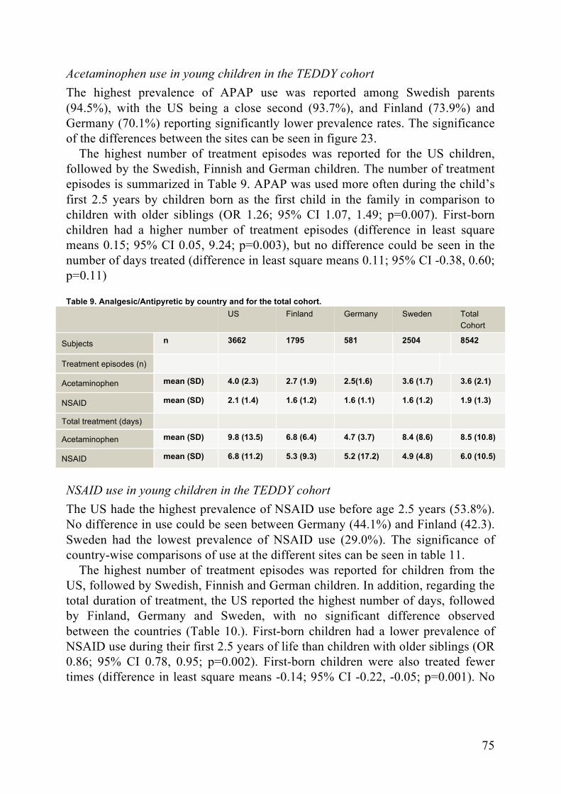
75
Acetaminophen use in young children in the TEDDY cohort The highest prevalence of APAP use was reported among Swedish parents (94.5%), with the US being a close second (93.7%), and Finland (73.9%) and Germany (70.1%) reporting significantly lower prevalence rates. The significance of the differences between the sites can be seen in figure 23.
The highest number of treatment episodes was reported for the US children, followed by the Swedish, Finnish and German children. The number of treatment episodes is summarized in Table 9. APAP was used more often during the child’s first 2.5 years by children born as the first child in the family in comparison to children with older siblings (OR 1.26; 95% CI 1.07, 1.49; p=0.007). First-born children had a higher number of treatment episodes (difference in least square means 0.15; 95% CI 0.05, 9.24; p=0.003), but no difference could be seen in the number of days treated (difference in least square means 0.11; 95% CI -0.38, 0.60; p=0.11)
Table 9. Analgesic/Antipyretic by country and for the total cohort. US Finland Germany Sweden Total
Cohort
Subjects n 3662 1795 581 2504 8542
Treatment episodes (n)
Acetaminophen mean (SD) 4.0 (2.3) 2.7 (1.9) 2.5(1.6) 3.6 (1.7) 3.6 (2.1)
NSAID mean (SD) 2.1 (1.4) 1.6 (1.2) 1.6 (1.1) 1.6 (1.2) 1.9 (1.3)
Total treatment (days)
Acetaminophen mean (SD) 9.8 (13.5) 6.8 (6.4) 4.7 (3.7) 8.4 (8.6) 8.5 (10.8)
NSAID mean (SD) 6.8 (11.2) 5.3 (9.3) 5.2 (17.2) 4.9 (4.8) 6.0 (10.5)
NSAID use in young children in the TEDDY cohort The US hade the highest prevalence of NSAID use before age 2.5 years (53.8%). No difference in use could be seen between Germany (44.1%) and Finland (42.3). Sweden had the lowest prevalence of NSAID use (29.0%). The significance of country-wise comparisons of use at the different sites can be seen in table 11.
The highest number of treatment episodes was reported for children from the US, followed by Swedish, Finnish and German children. In addition, regarding the total duration of treatment, the US reported the highest number of days, followed by Finland, Germany and Sweden, with no significant difference observed between the countries (Table 10.). First-born children had a lower prevalence of NSAID use during their first 2.5 years of life than children with older siblings (OR 0.86; 95% CI 0.78, 0.95; p=0.002). First-born children were also treated fewer times (difference in least square means -0.14; 95% CI -0.22, -0.05; p=0.001). No
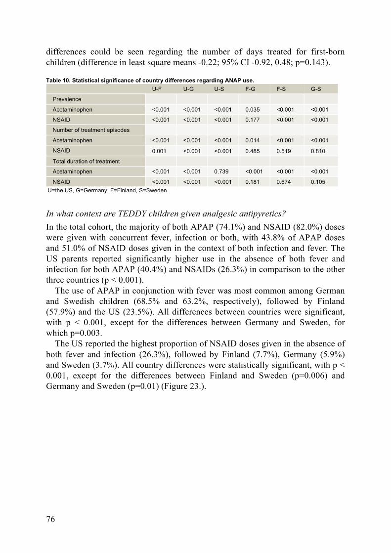
76
differences could be seen regarding the number of days treated for first-born children (difference in least square means -0.22; 95% CI -0.92, 0.48; p=0.143).
Table 10. Statistical significance of country differences regarding ANAP use. U-F U-G U-S F-G F-S G-S
Prevalence
Acetaminophen <0.001 <0.001 <0.001 0.035 <0.001 <0.001
NSAID <0.001 <0.001 <0.001 0.177 <0.001 <0.001
Number of treatment episodes
Acetaminophen <0.001 <0.001 <0.001 0.014 <0.001 <0.001
NSAID 0.001 <0.001 <0.001 0.485 0.519 0.810
Total duration of treatment
Acetaminophen <0.001 <0.001 0.739 <0.001 <0.001 <0.001
NSAID <0.001 <0.001 <0.001 0.181 0.674 0.105 U=the US, G=Germany, F=Finland, S=Sweden.
In what context are TEDDY children given analgesic antipyretics? In the total cohort, the majority of both APAP (74.1%) and NSAID (82.0%) doses were given with concurrent fever, infection or both, with 43.8% of APAP doses and 51.0% of NSAID doses given in the context of both infection and fever. The US parents reported significantly higher use in the absence of both fever and infection for both APAP (40.4%) and NSAIDs (26.3%) in comparison to the other three countries (p < 0.001).
The use of APAP in conjunction with fever was most common among German and Swedish children (68.5% and 63.2%, respectively), followed by Finland (57.9%) and the US (23.5%). All differences between countries were significant, with p < 0.001, except for the differences between Germany and Sweden, for which p=0.003.
The US reported the highest proportion of NSAID doses given in the absence of both fever and infection (26.3%), followed by Finland (7.7%), Germany (5.9%) and Sweden (3.7%). All country differences were statistically significant, with p < 0.001, except for the differences between Finland and Sweden (p=0.006) and Germany and Sweden (p=0.01) (Figure 23.).

77
Does participation in longitudinal follow-up affect peri-diagnosis morbidity and short-term glycemic control (Paper IV)?
Baseline characteristics of the study The cohort of paper IV consisted of a total of 129 children, including 67 girls (52%), with a mean age at diabetes diagnosis of 6.6 years (SD 2.7 years). The children in the follow-up and non-follow-up groups in paper IV did not differ with respect to gender (p=0.38) or age at diabetes diagnosis (p=0.45). Two children were islet autoantibody-negative at the time of diagnosis. However, both of those cases were diagnosed with T1D, with one case being antibody-positive in confirming samples and one case presenting with clinically typical T1D.
Does the status of the children at the time of T1D diagnosis differ for children enrolled in DiPiS follow-up compared to controls? At diagnosis, the children in the follow-up group had a lower frequency of diabetic symptoms, a higher percentage of children diagnosed without any symptoms and a lower frequency of DKA. No children that had been in intense follow-up, one child who had previously been in follow-up and three children who had not been in follow-up and had risk information presented with severe DKA. Complete results can be seen in table 11.
Table 11. Symptoms at diagnosis. Comparison between the Follow-up group and the Non-follow-up group.
n Follow-up
Prevalence (%) No Follow-up
Prevalence (%) OR 95% CI p pcorr*
Symptom, any 129 84 97 0.14 0.03; 0.70 0.014 0.028
Polydipsia 129 80 96 0.16 0.04; 0.63 0.006 0.018
Polyuria 129 76 96 0.13 0.04; 0.49 0.001 0.003
Weight loss 122 46 65 0.45 0.22; 0.95 0.034 0.10
DKA 129 2 18 0.09 0.01; 0.72 0.005 0.01
DKA, Severe 129 2 5 0.5 0.05; 4.94 1.0 1.0
DKA, Diabetic Ketoacidosis (pH <7.3); Severe DKA, Diabetic ketoacidosis (pH<7.1);
* Bonferroni-corrected p-value; Reprinted with permission from Wiley334
Does prospective follow-up influence glycemic control during the first two years after diagnosis? At diagnosis, the follow-up group had significantly lower median levels of Hba1c than the non-follow-up group (77 mmol/mol (IQR 64-91) and 87 mmol/mol (IQR 75-99), respectively; p=0.006; pcorr=0.03). During follow-up after diagnosis, significant differences could be seen at both 12 months post-diagnosis (53

78
mmol/mol (IQR 48-57) and 57 mmol/mol (IQR 51-65), respectively; p=0.009; pcorr=0.045) and 24 months post-diagnosis (53 mmol/mol (IQR 48-61) and 62 mmol/mol (IQR 54-69), respectively (p < 0.009; pcorr <0.001)). No significant differences in HbA1c could be seen at three and six months post-diagnosis (p=0.12; p=0.34) (Figure 24.).
Figure 24. HbA1c during the 2 years after follow-up. Reprinted with permission from Wiley334.
Can the differences in glycemic control be explained by insulin dosage or remission? Only at three months after diabetes diagnosis could a significant difference in insulin dose be seen when calculated as the total daily dose (TDD), with the follow-up group reporting a lower median dose of insulin (p=0.026). However, after adjusting for multiple comparisons, this difference was not significant.
Partial remission, which was defined as TDD <0.5 U/kg/day, was not significantly more common in the follow-up group than the non-follow-up group. In an unpublished analysis, partial remission was also analyzed using insulin dose-adjusted HbA1c (IDAA1C=NGSP % + 4 x TDD)335. No significant differences could be seen at any time point (Table 12.). Table 12. Insulin dose-adjusted HbA1c during the two years after diagnosis.
n
Follow-Up Median (IQR)
No Follow-Up Median (IQR) p
IDAA1C 3 months 122 8.5 (7.7-.9) 8.8 (8.1-9.9) 0.07
IDAA1C 6 months 121 9.0 (8.0-9.8) 9.4 (8.3-10.2) 0.17
IDAA1C 12 months 121 9.5 (8.6-10.5) 9.9 (9.0-10.9) 0.16
IDAA1C 24 months 120 10.0 (8.9-11.2) 10.2 (9.5-11.2) 0.34

79
Can we safely use Alum-formulated GAD65 to induce immune tolerance in children at high risk for diabetes (Paper V)?
Baseline characteristics of the study During screening, 54 children aged 4-17.8 years were screened for study participation. Two screened children were not positive for GADA and one additional autoantibody, one child was diagnosed with T1D and one parent withdrew consent prior to randomization. Hence, a total of 50 children were eligible for the study and consented to participate. The children were randomized 1:1 to GAD-alum or placebo treatment. The median age of the participants was 5.2 years, 27 participants (54%) were boys, and 16 participants (32%) had a first-degree relative with T1D. No difference in participant characteristics at baseline was seen between the treatment and placebo groups regarding sex, BMI, age at randomization, first-degree relative, HLA DQ risk group or autoantibody stratum (2 positive autoantibodies; 3 positive autoantibodies). Regarding glucose metabolism at baseline, no discernible difference could be seen between the randomization groups (Table 13.).
Table 13. Baseline characteristics of the DiAPREV-IT study GAD-ALUM
n=25 Placebo
n=25 p
Age at randomization (mean, range) 6.0 (4.1-15.1) 5.0 (4.0-17.9) 0.152
BMI Z-score (mean, SD) 0.22 (0.75) 0.36 (1.03) 0.570
Sex Male 14 (56%) 13 (52%) 0.777
Population source FDR 9 (36%) 7 (28%) 0.544
Study DiPiS TEDDY TrialNet
10 (40%) 11 (44%) 4 (16%)
6 (24%) 17 (68%)
2 (8%) 0.329
Stratum 2 positive AAb 3-6 positive AAb
7 (28%) 18 (72%)
7 (28%) 18 (72%)
>0.999
Glucose tolerance Impaired 13 (52%) 13 (52%) >0.999
The median follow-up time was 4.92 years (0.47-5.01 years). One child left the study after 4.25 years of follow-up. All other children were followed for five years or until diabetes diagnosis.
Is Alum-formulated GAD safe to use in children with a high risk of T1D who are positive for GAD autoantibodies? During study follow-up, a total of four serious adverse events were recorded. One of these events was hospital-treated pneumonia, three events were upper limb

80
fractures, and no events were deemed to be related to the study. An overwhelming majority of the total 878 reported adverse events in the study were classified as mild. No difference regarding the number of adverse events was seen between the treatment and placebo groups. Adverse events that were possibly related to the study drug administration were rare (12/878; 1.5%) and transient. The study drug-related adverse events included vomiting, headache, fever, nausea, sore throat, diarrhea, stomach ache and dizziness. Injection site reactions, all mild or moderate, were reported by 48/50 participants, with the same frequency being reported by the placebo and treatment groups (p=0.77).
No differences in hematology, electrolytes or liver enzymes were seen between the treatment and placebo groups during follow-up. No rapid decreases in C-peptide levels, as a sign of C-peptide collapse, were seen during follow-up. GADA titers increased in the treatment group, compared to the placebo group (p=0.001), and were significantly different up to 27 months after treatment (Figure 25.). The titers of the other autoantibodies (IA-2A, IAA, ZnT8R/W/WA, TPOA, ThglA, tTGA) analyzed during follow-up were similar between the treatment and placebo groups.
Figure 25. GADA titers in relation to treatment group during follow-up.
Control group (Red line); Treatment group (Blue line)
Does GAD-Alum affect the progression to a T1D diagnosis? A total of 18 children (36%) were diagnosed with T1D during the five-year follow-up (170-1,830 days after the first injection). Impaired glucose metabolism at the baseline visit in the study was associated with a higher rate of progression to diabetes in comparison to children with normal glucose metabolism at baseline (p=0.013). No difference in progression to T1D was seen for first-degree relatives
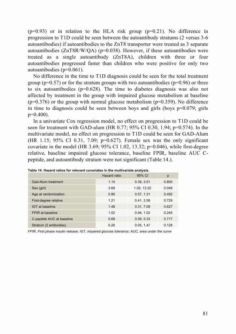
81
(p=0.93) or in relation to the HLA risk group (p=0.21). No difference in progression to T1D could be seen between the autoantibody stratums (2 versus 3-6 autoantibodies) if autoantibodies to the ZnT8 transporter were treated as 3 separate autoantibodies (ZnT8R/W/QA) (p=0.038). However, if these autoantibodies were treated as a single autoantibody (ZnT8A), children with three or four autoantibodies progressed faster than children who were positive for only two autoantibodies (p=0.061).
No difference in the time to T1D diagnosis could be seen for the total treatment group (p=0.57) or for the stratum groups with two autoantibodies (p=0.96) or three to six autoantibodies (p=0.628). The time to diabetes diagnosis was also not affected by treatment in the group with impaired glucose metabolism at baseline (p=0.376) or the group with normal glucose metabolism (p=0.359). No difference in time to diagnosis could be seen between boys and girls (boys p=0.079; girls p=0.400).
In a univariate Cox regression model, no effect on progression to T1D could be seen for treatment with GAD-alum (HR 0.77; 95% CI 0.30, 1.94; p=0.574). In the multivariate model, no effect on progression to T1D could be seen for GAD-Alum (HR 1.15; 95% CI 0.31, 7.09; p=0.627). Female sex was the only significant covariate in the model (HR 3.69; 95% CI 1.02, 13.32; p=0.046), while first-degree relative, baseline impaired glucose tolerance, baseline FPIR, baseline AUC C-peptide, and autoantibody stratum were not significant (Table 14.).
Table 14. Hazard ratios for relevant covariates in the multivariate analysis. Hazard ratio 95% CI p
Gad-Alum treatment 1.15 0.38, 3.51 0.800
Sex (girl) 3.69 1.02, 13.32 0.046
Age at randomization 0.86 0.57, 1.31 0.492
First-degree relative 1.21 0.41, 3.58 0.729
IGT at baseline 1.48 0.31, 7.09 0.627
FPIR at baseline 1.02 0.94, 1.02 0.245
C-peptide AUC at baseline 0.68 0.09, 5.33 0.717
Stratum (2 antibodies) 0.26 0.05, 1.47 0.128
FPIR, First phase insulin release; IGT, impaired glucose tolerance; AUC, area under the curve

82

83
Discussion
General discussion
The search for risk factors for T1D has been pursued for several decades, and we still only partially understand the multifactorial pathogenesis of the disease. In this thesis, we have examined the correlation between umbilical cord blood islet antibodies and T1D risk (Paper I). IA-2 autoantibodies increased the risk, even after adjustment for possible confounding factors. The maternal origin of cord blood autoantibodies has been established in previous studies336,337. However, the effect on T1D risk is equivocal. Previous studies have shown decreased337, neutral338 and increased risk339,340. The pathophysiological basis for this effect is also unclear. It is possible that in-utero exposure to maternal autoantibodies could affect the fetal immune system. This effect may also be a sign of exposure to maternal immune system activation.
We also investigated medication use of young children below 2.5 years of age (Paper III). The use of ANAPs is common in children, but few publications have investigated its prevalence. In particular, APAP has been investigated in conjunction with other immune mediated diseases, especially childhood asthma341, and also in relation to autism342. Our results show widespread use of ANAP in this young cohort and reveal significant differences between the study sites. With regard to IA risk at six years of age, the risk does not appear to be increased in children who are exposed to ANAP, including NSAIDs or APAP. The same results apply after stratifying for age of exposure and total exposure. A weak correlation exists between APAP and IA at age three years, but only with concomitant fever. This effect was not seen in any of the other analyses. Thus, this result must be interpreted with caution. It is possible but highly unlikely that this result represents a true risk increase.
That increased stress influences T1D risk has long been proposed. In this thesis, we present data supporting the idea that parental experiences before the child turns two years old predict increased risk of T1D (paper II). Similar results have been reported by the ABIS study, in which severe life events predicted an increased T1D risk343, although it was not possible to adjust for HLA-related risk. Other previous studies have provided equivocal results344,345. However, comparisons with these earlier studies are not easy because both the study endpoints and the background data differ significantly between studies.

84
The study of rare events, such as severe life events, is difficult, and the results must be interpreted with caution and repeated in other cohorts. That we are able reproduce similar results strengthens the concept that early life stress is an immune modulator with possible effects on immune tolerance. The analyzed parental discord and parenting stress indices failed to show any correlation with T1D risk. This finding may be due to the true absence of a correlation but may also be attributed to the use of questionnaire data without a validated instrument. Additional analysis, including laboratory markers of stress, could further strengthen the present analysis.
The backbone of current T1D pediatric research is prospective follow-up of at-risk subjects. Several large-scale studies have been and are being performed. Understanding the impact of follow-up on children and parents becomes crucial, both for study accrual and for ethical defense of the procedure. We examined the status of the children followed in DiPiS at the time of diabetes diagnosis, in comparison to children of the same age and geographic distribution (Paper IV). All children in the follow-up cohort had received risk information regarding HLA-related risk and had been prospectively followed either every year or every three months. The follow-up group was diagnosed with fewer diabetic symptoms and with a lower HbA1c, signifying a shorter and/or milder dysglycemic period before diagnosis. Those children also presented with a lower prevalence of DKA (2% compared to 18% in the control group), which is a potentially fatal complication of insulin deficiency. Studies in the DAISY54, TEDDY346, DPT-157 and BABYDIAB56 cohorts present the same kind of results, indicating that study children are diagnosed at an early stage of the disease. We also show that the follow-up group exhibited better glycemic control 12 and 24 months after diagnosis. This finding could be attributed to the early stage at which the children are diagnosed, but that possibility seems unlikely up to two years after diagnosis. This outcome could be the result of prolonged partial remission or improved β-cell function in children who are diagnosed early. However, recent data show no difference in the rate of C-peptide decline for children diagnosed in an asymptomatic stage347. No demographic or socioeconomic variables were included in this analysis, and one could argue that the families willing to participate in prospective follow-up differ from the general population.
Further study will have to be done to validate these findings and to elucidate the observed effect. Understanding our follow-up cohorts is important for both interpreting results and generalizability. The ability to demonstrate beneficial, positive, results of study participation will also ease future study recruitment and help motivate further studies of otherwise healthy children.
Screening and prospective follow-up is a prerequisite of current prevention studies. Using immune tolerance is an attractive concept for inducing self-tolerance and stopping the diabetic process. Using GAD-alum, we have demonstrated that a regimen of two subcutaneous doses in children with GADA
85
and at least one other islet autoantibody but not T1D can be administered safely without significant side effects (paper V). The safety of GAD-alum has been studied in newly diagnosed diabetes patients with similar results. We can now add to this knowledge regarding children before diabetes diagnosis. The efficacy results are consistent with previous studies305,306, even though some promise has been shown in a meta-analysis348. The study failed to demonstrate efficacy in terms of progression to T1D or any of the surrogate markers, but the study was not fully powered to do so. The study highlights the need for larger scale trials of GAD-Alum, in which dosing and timing can be examined more closely. The current protocol has been extremely time consuming, both for the investigators and the patient’s families. If safety has been established, a less demanding protocol could be approved for further studies. Using immune tolerance as part of a combination treatment is also a promising concept for future trials.
Finally, we present data on the use of ANAPs in young children in the TEDDY cohort. These results are the product of an analysis of the risk of IA and use of ANAPs. The amount of data recorded in the search for the cause of T1D is extensive and enables additional analyses to be performed, enhancing our understanding of factors related to general pediatrics. We report the widespread use of ANAPs in very young children in the TEDDY cohort, with significant differences in prevalence between the TEDDY sites. We also observed a preference for the use of APAP among first-born children in comparison to their younger siblings. The use of ANAPs was primarily related to fever or infection in the studied cohort, but a significant portion of participants also used these drugs without fever or infection, especially in the US cohort. Understanding basic patterns of use in pediatric patients is important for the pediatric community, and to or knowledge, no similar data have been published to date.
Weaknesses
There are weaknesses associated with the papers in this thesis. The statistical analysis of rare events is not easy and can be controversial. In both paper I and paper II, there are low numbers of exposures and a relatively low number of events. This factor leads to increased uncertainty in the model. Enhancing the statistical model further with a penalized model or choosing a different statistical test altogether could alleviate some of this uncertainty. For paper II, extending the data to a longer time frame would increase the background data but lead to a significant reduction in the number of participants. The analysis in paper I was performed as a group-wise comparison between the follow-up and non-follow-up groups. This approach does not take into account any differences in baseline variables that may influence the result. Ongoing extended analysis of these data up to five years after diagnosis should include baseline parameters, as well as a better

86
measurement of residual C-peptide and growth parameters to be included in a longitudinal model. In paper III, the statistical limitation is the opposite: a very widespread exposure in the cohort, with the exposed groups surpassing the non-exposed group, leading to uncertainty in the statistical model. All papers related to the DiPiS study rely partially on questionnaire data recorded by the parents at home, even though the data are collected prospectively. This approach is associated with some level of recall and measurement bias, as well as inconsistent results from year to year. Missing data in the questionnaires also limit the number of subjects available for multivariate analysis. In the TEDDY study, all data are collected at nurses’ visits, but the analysis still relies on parental recollection and interpretation. Although extensive data were collected, uncertainties remain regarding the exact reasons for the use of ANAP outside of fever and infections.
In paper II, questionnaire data are used to assess life events and to compute the two stress indices. Using a validated questionnaire to assess family stress would have improved the validity of the analysis. An additional weakness in papers II and IV is the lack of supporting laboratory data. The inclusion of C-peptide values and stress biomarkers, respectively, would have improved the interpretation of the results in these papers, but that approach was not part of the original study plan and was thus not available for the analysis. A general weakness of papers I – IV is the high-risk composition of the cohort. It is possible that the risk factors described in these HLA-derived high-risk cohorts are not valid outside of this group. This factor is a problem that both DiPiS and TEDDY share with several other cohorts. If T1D, and IA to some extent, are used as endpoints, this limitation is in many ways unavoidable due to both time and monetary constraints.
Future issues
Our pursuit of the causes and pathogenesis of T1D has been ongoing for a long time. Still, even though our knowledge is significantly better, large pieces of the puzzle are missing. Prior methods of research have been painstaking and slow, relying on long-term follow-up with T1D as the study endpoint. Ongoing studies, like the TEDDY study, that use IA as an earlier, alternate endpoint, together with a large, multinational study cohort and intensive and thorough follow-up hold great promise for achieving the goal of identifying the environmental determinants of diabetes and improving our understanding of the natural history of T1D. Using alternate endpoints besides diabetes diagnosis will be important in the future for both monetary and timing reasons. In particular, intervention trials will benefit from the use of alternate endpoints when new treatment regimens and combinations are used. For intervention trials, the use of combination treatments for induction and maintenance treatment could build on the success of pediatric

87
oncological treatment. Finding an acceptable level of side effects for long-term treatment, which is a better option than T1D, is crucial.
The amount of research surrounding risk factors, natural history, genetic and epigenetic factors is expanding to a level that is hard to grasp. T1D is truly a multifactorial disease. Personalizing this data for the individual will be harder as our knowledge increases. Machine learning and enhanced algorithms hold promise for improving our understanding of risk factors and extracting data from large datasets.
One of the central issues that must be addressed is the severity of T1D and establishing the extremely high risk that is associated with multiple islet autoantibody positivity. Today, T1D is perceived as a fairly benign disease, for which patients must take their insulin, do their glucose test, and otherwise live a normal life. Many, if not all, families living with T1D would disagree with that description. T1D is a disease that encompasses every aspect of life and places an overwhelming burden on parents and children. In addition, even if patients reach the difficult goal of glycemic control, they remain at an increased risk of vascular damage and a shortened life span. In ethical discussions, these aspects must also be considered. It is easy to say no to involving children in research, deeming it unethical. However, it may be the failure to learn more and research diseases that affect children that is unethical.

88

89
Conclusions
• IA-2A cord blood autoantibodies and early severe life events during the child’s first two years of life potentially increase the risk of T1D in children (Paper I, Paper II).
• ANAP use in children below 2.5 years of age does not predict the risk of IA at age six years, although a weak correlation with IA was observed at age three years (Paper III).
• Children enrolled in follow-up in the DiPiS cohort are less affected by diabetes symptoms and have a significantly lower prevalence of DKA at diagnosis (Paper IV).
• Children enrolled in follow-up in the DiPiS cohort have better glycemic control up to two years after diagnosis, compared to children who are not enrolled in follow-up (Paper IV).
• Treatment with GAD-Alum does not present any safety concerns when treating multiple islet autoantibody-positive children who have not yet been diagnosed with T1D (Paper V).
• GAD-Alum does not show efficacy in terms of delaying or preventing T1D diagnosis in multiple islet autoantibody-positive children in this small-scale trial (Paper V).
• The use of ANAP in children below 2.5 years is widespread, and significant between-country differences exist (Paper III).

90

91
Summary in Swedish
Vad är typ 1 diabetes Typ 1 diabetes är en av de vanligaste kroniska sjukdomarna som drabbar barn. Sjukdomen orsakas av att kroppen attackerar de så kallade Langerhanska cellöarna i bukspottskörteln via en autoimmun reaktion. De egna cellerna uppfattas alltså som främmande och förgörs. Detta leder till en oförmåga att producera insulin, det hormon som styr vår blodsockernivå i blodet. Denna autoimmuna process kan i nuläget varken förhindras eller stoppas. När sjukdomen är fullt utvecklad krävs insulininjektioner vid ett flertal tillfällen dagligen för att blodsockret skall hållas på rätt nivå samt även upprepade kontroller av blodsockernivån.
Vi vet nu att utvecklingen av typ 1 diabetes startar långt innan några symtom visar sig. I flertalet fall kan tecken i blodet ses åtskilliga år tidigare, inte sällan så tidigt som innan 1 års ålder. Sårbarheten för att vara mottaglig för typ 1 diabetes är genetisk och framför allt relaterad till ett område som kallas HLA, som är en viktig del av immunförsvaret. Den så kallade HLA-genotyp en individ har bestämmer ungefär hälften den medfödda genetiska risken för typ 1 diabetes. Den andra hälften av den medfödda risken verkar komma från ett flertal andra gener som vi i nuläget inte fullt ut har utforskat. Omgivningsfaktorer är också viktiga för risken att utveckla typ 1 diabetes. Vi vet idag att vissa omgivningsfaktorer verkar påverka diabetesrisken, däribland virusinfektioner. Vår förståelse är dock på inget sätt fullständig och vi lär oss ständigt mer om hur risken för typ 1 diabetes samverkar, både med vår genetik men också med vår omgivning.
Bedömning av risk för typ 1 diabetes Det första sättet att avgöra risk för typ 1 diabetes är genom tester för HLA-genotyp. De varianter som medför högst risk för typ 1 diabetes är dock vanliga i svensk befolkning och någon av dem finns hos en stor del av befolkningen. Senare i livet kan dock en av de sex diabetesrelaterade autoantikropparna utvecklas som tecken på en sjukdomsprocess. Dessa antikroppar är immunförsvarets målsökande missiler, i detta fallet riktade mot olika delar av de insulinproducerande cellerna. Autoantikropparna har namn efter vilken struktur de är riktade mot och kallas IAA, IA-2A, GADA, ZnT8 (WA/RA/QA). I de flesta fall uppkommer en variant av dessa för att senare följas av ytterligare. En enstaka autoantikroppsvariant medför en lätt ökad risk för typ 1 diabetes, men denna kan också försvinna igen och med den ökade diabetesrisken. De barn som utvecklar mer än en

92
autoantikropp har dock en mycket hög risk för typ 1 diabetes inom 5–10 år. Vissa hävdar att det till och med är att likna vid ett sjukdomstillstånd, även om inga symtom än har visat sig.
För att ytterligare klargöra risken för att insjukna kan vi bedöma kroppens förmåga att hantera socker genom både orala glukosbelastningar, där en bestämd mängd sockerlösning dricks, eller intravenösa glukosbelastningar, där socker ges direkt i blodet. I samband med dessa kan man månader, och ibland år, innan diabetesdiagnosen sätts, se en långsam successiv försämring i insulininsöndring och blodsockerkontroll.
Hur har data till studierna samlats in? I vårt arbete har vi framförallt använt två stora grupper av barn som följts sedan födseln. I Skåne screenades 35 683 barn mellan september 2000 och augusti 2004 vid födseln avseende diabetesrisk. De närmre 8000 barn som bedömdes ha ökad risk erbjöds att delta i uppföljning fram till barnen fyllde 15 år. Knappt 3900 barn har deltagit i DiPiS studiens (DiabetesPrediktion i Skåne) uppföljning årligen från två års ålder med blodprover och frågeformulär. Barn som utvecklar tecken på autoimmunitet har följts var tredje månad. TEDDY-studien (The Environmental Determinants of Diabetes in the Young) screenade närmre 415 000 barn vid sex olika centra i Sverige, Finland, Tyskland och USA. Cirka 21 500 barn hade ökad risk och erbjöds vara med i studien varav 8700 tackade ja. TEDDY studiens uppföljning började vid 3 månaders ålder och är väldigt omfattande. Prover från såväl blod, urin, avföring, hår, nässekret, hushållets vatten med mera samt redogörelser för faktorer i barnens liv så som sjukdomar, mathållning och medicinering och fysisk aktivitet samlas in. Barnen följs upp var tredje månad till fyra års ålder och för de som inte utvecklat tecken till autoimmunitet glesas då besöken ut till var sjätte månad.
Vilka är resultaten? Denna avhandling försöker förbättra vår kunskap om typ 1 diabetes på flera områden i sjukdomsprocessen. Vårt första mål var att undersöka riskfaktorer relaterade till navelsträngsblod. Vi tog prover från navelsträngen vid barnets födsel och undersökte vilka barn som hade diabetesrelaterade autoantikroppar från mamman. Risken för de barn som hade IA-2 autoantikroppar i navelsträngsblodet ter sig i denna studie vara cirka sju gånger högre än för barnen utan antikroppar. De övriga antikropparna som analyserades verkade inte påverka diabetesrisken (IAA och GADA). I nuläget har vi ingen förklaring på varför detta diabetesrisken påverkas men hoppas att detta kan bekräftas och klargöras i framtida studier.
Vi har därefter undersökt hur användningen av febernedsättande och smärtlindrande läkemedel innan 2,5 års ålder ser ut hos TEDDY-studiens barn. Detta rör sig framförallt om paracetamol (t. ex. Alvedon och Panodil) och NSAID-preparat/ibuprofen (t. ex. Ipren, Nurofen, Treo eller Albyl). Vi undersökte därefter

93
om användningen av dessa preparat medförde en ökad risk för utveckling av autoantikroppar vid sex års ålder. Våra resultat visar inte på någon riskökning för ö-cellsautoimmunitet vid sex års ålder även om en svag koppling finns vid tre års ålder. Vi kan även beskriva stora skillnader i hur mycket och ofta dessa preparat används i de olika länderna. Att en stor andel av förbrukningen hos dessa väldigt unga personer sker utan varken feber eller infektion var även en nyhet för oss.
Den sista faktorn avseende diabetesrisk vi har undersökt är hur föräldrars stress och negativa livshändelser för familjen innan 2 års ålder påverkar diabetesrisken. Vi använde oss av svar från frågeformulär vid 2 månader och 2 års ålder. För att lättare kunna bedöma föräldrarnas stressnivåer utformades två olika index, dels faktorer talande för föräldrarnas oro och dels för hur stressande relationen till barnet var. Inget av dessa index, och därmed svaren gällande föräldrars stress, ter sig påverka barnets risk för typ 1 diabetes. Vi har även undersökt om negativa livshändelser påverkar diabetesrisken. Detta kan röra sig om våld i familjen, arbetslöshet, separation, sjukdom mm. Våra data visar en ökad risk för typ 1 diabetes hos de barn vars familjer upplevt en negativ livshändelse innan barnet fyllt två år. I nuläget kan vi inte fullt förklara vad som ligger bakom denna effekt men en förklaring kan vara att stresshormoner påverkar immunförsvaret och därmed ökar risken för autoimmun sjukdom.
Att vara med i en studie under barndomen är en speciell situation för barn och familjen. Vi ville undersöka om de barn som deltagit i DiPiS-studien och som fått information om sin diabetesrisk skiljde sig från barn som ej deltagit, när de får typ 1 diabetes. Vi kan visa att barn som deltagit i DiPiS har väsentligt färre symtom vid diagnos och dessutom har haft lägre nivåer på sitt blodsocker innan diagnos. Det är även färre som har syraförgiftning, ketoacidos, när de får sin diagnos. Ketoacidos är ett mycket allvarligt tillstånd som i vissa fall leder till intensivvård och kan vara dödligt. När vi sedan följer dessa två grupper barn under två år ser vi att de barn som deltagit i studien har bättre blodsockerkontroll både ett och två år efter diagnos. Det är glädjande att vi kan diagnosticera studiebarnen tidigt utan att de hunnit bli allvarligt sjuka, vilket kan både minska chocken vid diagnos och även korta vårdtiden. Att blodsockerkontrollen är bättre upp till två år efter diagnos kan bero på att större andel insulinproducerande celler kvarstår vid en tidig diagnos, men kan också bero på att familjen är bättre förberedd på barnets sjukdom. Vi kommer undersöka detta närmare för att veta bättre.
Slutligen visar vi resultat från en studie som försöker stoppa eller förlångsamma sjukdomsprocessen. Vi har undersökt 50 barn med hög risk för diabetes och minst två autoantikroppar. Hälften av dessa barn har fått behandling vid två tillfällen med GAD-Alum och hälften fick overksam substans (placebo). Detta preparat skall försöka vänja kroppen vid GADA, en av de autoantikroppar som är involverade i utvecklingen av typ 1 diabetes. Studiens huvudmål var att avgöra om det var säkert att ge detta preparat till barn utan diabetes. Inga allvarliga biverkningar rapporterades och vi kan visa att preparatet kan ges säkert på detta
94
sätt. Vi undersökte även om GAD-Alum påverkade risken för att få diabetes eller hur lång tid det tog att få diabetes. Våra resultat visar att det inte verkar ha någon effekt på diabetesutveckling. Gruppen studiepersoner är dock väldigt liten och det blir därmed svårt att visa en eventuell effekt rent statistiskt jämfört med om studien varit större.
Sammanfattning Vi kan visa att autoantikroppar i navelsträngsblod mot IA-2 och allvarliga livshändelser under barnets två första levnadsår verkar öka risken för typ 1 diabetes. Däremot ser vi ingen påverkan på risken för förstadiet till typ 1 diabetes, autoimmunitet, relaterat till intag av smärtstillande/febernedsättande medel hos barn under 2,5 år. De barn vi följer i DiPiS-studien diagnosticeras med diabetes tidigt i sjukdomsförloppet och är också mindre påverkade vid sin diagnos. Deras blodsockerkontroll är bättre både vid 1 och 2 års uppföljning efter diabetesdiagnosen. Slutligen är behandling med GAD-Alum, till barn med autoantikroppar men inte diabetes, säkert men verkar inte påverka risken för diabetes.

95
Acknowledgements
Many people have contributed to this thesis in different ways: Friends, family colleagues and collaborators. Without the support you have provided it would not have been possible to be where I am today.
My greatest admiration and thanks go to the children and families who participate in our studies. Your time and effort helps us understand type 1 diabetes better and get closer to predicting and preventing this disease. Thank you so much for sticking with us for so many years.
My main supervisor, Helena Elding Larsson: You have always been there during these last few years with support and enthusiasm. Despite a heavy workload you can always be sure to get a supportive, clear cut answer or a file filled with revisions showing me what I wanted to say, but wasn´t able to. This was fun, you are not rid of me yet.
Åke Lernmark, my assistant supervisor and a source of constant ideas and encouragement. Your clarity when discussing research and enthusiasm for new projects are truly inspiring.
Ida Jönson and Anita Ramelius, without your help with data and endless questions, especially when starting up without really knowing what I was doing, my life would have been so much harder. Thomas Gard, thank you for being the constant fixer in everything economical and administrative. All the study nurses and laboratory staff in TEDDY, DiPiS, TrialNET and DiAPREV-IT, the work you do is fantastic. Thanks for all support, encouragement and for taking care of our study kids.
The diabetes team in Kristianstad, Malin Johnsson, Susanne Dahlberg, Anna-Karin Nilsson, Ingela Stålberg-Almroth, Marie Olsson, Annelie Paulin, Karin Larsson, Emma Hedlund, Sofie Svärd, thanks for covering for me so many times during the last few years. The diabetes patients in the northeast are lucky to have your support, enthusiasm and knowledge.
To all my colleagues at the pediatric department in Kristianstad. Thanks for the opportunity to spend time doing research as well as continuing to be a clinician. For fixing things when I wasn´t there and for not tiring of me talking about research related topics. Thanks to Hedvig von Schantz and Christine Karström,
96
for allowing me to take time off during these years as a PhD student. A special thanks to my former boss Lars Almroth. Without your inspiration and encouragement regarding research I don´t think I would have taken the chance. Who would have thought that an e-mail to Åke would lead to me being a PhD student one week later?
Mamma och Pappa, ni har alltid funnits där, stöttat, uppmuntrat och stått ut med mina infall och idéer. Allt jag är är jag tack vare er.
Julius, Hjördis och Torsten: ni påminner mig varje dag om vad som är verkligen viktigt i livet och står ut med att jag ibland har tankarna på annat håll. Tack för att ni finns och gör livet bättre och fyllt med glädje.
Min älskade fru och livskamrat, Lina, tack för att du alltid stöttat, funnits där och trott på mig. Framöver får det bli mer resor, mer drinkar och mer tid tillsammans.

97
References
1 Himsworth HP. Diabetes mellitus: its differentiation into insulin-sensitive and insulin-insensitive types. The Lancet 1936. DOI:10.1016/S0140-6736(01)36134-2.
2 Mering von J, Minkowski O. Diabetes Mellitus nach Pankreas-Extirpation, 1889. 3 Banting FG, Best CH, Collip JB, Campbell WR, Fletcher AA. Pancreatic Extracts in the Treatment of Diabetes
Mellitus. Canadian Medical Association Journal 1922; 12: 141–6. 4 Banting FG, Campbell WR, Fletcher AA. Further Clinical Experience with Insulin (Pancreatic Extracts) in the
Treatment of Diabetes Mellitus. Br Med J 1923; 1: 8–12. 5 WALKER GF. Reflections on diabetes mellitus; answers to a questionnaire. Lancet 1953; 265: 1329–32. 6 Rahbar S. An abnormal hemoglobin in red cells of diabetics. Clin Chim Acta 1968; 22: 296–8. 7 Walford S, Gale EA, Allison SP, Tattersall RB. Self-monitoring of blood-glucose. Improvement of diabetic
control. Lancet 1978; 1: 732–5. 8 Paton JS, Wilson M, Ireland JT, Reith SB. Convenient pocket insulin syringe. Lancet 1981; 1: 189–90. 9 Pickup JC, Keen H, Parsons JA, Alberti KG. Continuous subcutaneous insulin infusion: an approach to achieving
normoglycaemia. Br Med J 1978; 1: 204–7. 10 Diabetes Control and Complications Trial Research Group, Nathan DM, Genuth S, et al. The effect of intensive
treatment of diabetes on the development and progression of long-term complications in insulin-dependent diabetes mellitus. N Engl J Med 1993; 329: 977–86.
11 Intensive blood-glucose control with sulphonylureas or insulin compared with conventional treatment and risk of complications in patients with type 2 diabetes (UKPDS 33). UK Prospective Diabetes Study (UKPDS) Group. Lancet 1998; 352: 837–53.
12 The relationship of glycemic exposure (HbA1c) to the risk of development and progression of retinopathy in the diabetes control and complications trial. Diabetes 1995; 44: 968–83.
13 White NH, Sun W, Cleary PA, et al. Prolonged effect of intensive therapy on the risk of retinopathy complications in patients with type 1 diabetes mellitus: 10 years after the Diabetes Control and Complications Trial. Arch Ophthalmol 2008; 126: 1707–15.
14 Woo VC. A Review of the Clinical Efficacy and Safety of Insulin Degludec and Glargine 300 U/mL in the Treatment of Diabetes Mellitus. Clin Ther 2017; 39: S12–S33.
15 Heise T, Hövelmann U, Brøndsted L, Adrian CL, Nosek L, Haahr H. Faster-acting insulin aspart: earlier onset of appearance and greater early pharmacokinetic and pharmacodynamic effects than insulin aspart. Diabetes Obes Metab 2015; 17: 682–8.
16 Bruen D, Delaney C, Florea L, Diamond D. Glucose Sensing for Diabetes Monitoring: Recent Developments. Sensors (Basel) 2017; 17: 1866.
17 Samuelsson U, Åkesson K, Peterson A, Hanas R, Hanberger L. Continued improvement of metabolic control in Swedish pediatric diabetes care. 2016; published online Nov 3. DOI:10.1111/pedi.12467.
18 Anderzén J, Samuelsson U, Gudbjörnsdottir S, Hanberger L, Åkesson K. Teenagers with poor metabolic control already have a higher risk of microvascular complications as young adults. J Diabetes Complicat 2016; 30: 533–6.
19 Särnblad S, Åkesson K, Fernström L, Ilvered R, Forsander G. Improved diabetes management in Swedish schools: results from two national surveys. 2016; published online July 29. DOI:10.1111/pedi.12418.
20 Buchberger B, Huppertz H, Krabbe L, Lux B, Mattivi JT, Siafarikas A. Symptoms of depression and anxiety in youth with type 1 diabetes: A systematic review and meta-analysis. Psychoneuroendocrinology 2016; 70: 70–84.
21 Lind M, Svensson A-M, Kosiborod M, et al. Glycemic Control and Excess Mortality in Type 1 Diabetes. N Engl J Med 2014; 371: 1972–82.
22 Craig ME, Jefferies C, Dabelea D, Balde N, Seth A, Donaghue KC. Definition, epidemiology, and classification of diabetes in children and adolescents. 2014; 15 Suppl 20: 4–17.
23 American Diabetes Association. Diagnosis and Classification of Diabetes Mellitus. Diabetes Care 2009; 33: S62–9.
98
24 World Health Organization. Definition and diagnosis of diabetes mellitus and intermediate hyperglycemia: Report of a WHO/IDF consultation. Geneva, Switzerland. Report of a WHO/IDF consultation. Geneva, 2006.
25 American Diabetes Association. Diagnosis and classification of diabetes mellitus. Diabetes Care. 2014; 37 Suppl 1: S81–90.
26 Hattersley AT, Patel KA. Precision diabetes: learning from monogenic diabetes. 2017; 60: 769–77. 27 Rubio-Cabezas O, Hattersley AT, Njølstad PR, et al. The diagnosis and management of monogenic diabetes in
children and adolescents. 2014; 15 Suppl 20: 47–64. 28 Barrett TG. Differential diagnosis of type 1 diabetes: which genetic syndromes need to be considered? 2007; 8
Suppl 6: 15–23. 29 Semple RK, Savage DB, Cochran EK, Gorden P, O'Rahilly S. Genetic syndromes of severe insulin resistance.
Endocr Rev 2011; 32: 498–514. 30 Watkins RA, Evans-Molina C, Blum JS, DiMeglio LA. Established and emerging biomarkers for the prediction of
type 1 diabetes: a systematic review. Transl Res 2014; 164: 110–21. 31 Bingley PJ, Christie MR, Bonifacio EE, et al. Combined analysis of autoantibodies improves prediction of IDDM
in islet cell antibody-positive relatives. Diabetes 1994; 43: 1304–10. 32 Oettingen von JE, Wolfsdorf JI, Feldman HA, Rhodes ET. Utility of diabetes-associated autoantibodies for
classification of new onset diabetes in children and adolescents. Pediatr Diabetes 2016; 17: 417–25. 33 Bingley PJ, Bonifacio E, Williams AJ, Genovese S, Bottazzo GF, Gale EA. Prediction of IDDM in the general
population: strategies based on combinations of autoantibody markers. Diabetes 1997; 46: 1701–10. 34 Wenzlau JM, Juhl K, Yu L, et al. The cation efflux transporter ZnT8 (Slc30A8) is a major autoantigen in human
type 1 diabetes. Proc Natl Acad Sci USA 2007; 104: 17040–5. 35 Dabelea D, Pihoker C, Talton JW, et al. Etiological approach to characterization of diabetes type: the SEARCH
for Diabetes in Youth Study. Diabetes Care 2011; 34: 1628–33. 36 Redondo MJ, Rodriguez LM, Rodriguez LM, et al. Types of pediatric diabetes mellitus defined by anti-islet
autoimmunity and random C-peptide at diagnosis. 2013; 14: 333–40. 37 Pugliese A, Boulware D, Yu L, et al. HLA-DRB1*15:01-DQA1*01:02-DQB1*06:02 Haplotype Protects
Autoantibody-Positive Relatives From Type 1 Diabetes Throughout the Stages of Disease Progression. Diabetes 2016; 65: 1109–19.
38 Erlich H, Valdes AM, Noble J, et al. HLA DR-DQ haplotypes and genotypes and type 1 diabetes risk: analysis of the type 1 diabetes genetics consortium families. Diabetes 2008; 57: 1084–92.
39 Karges B, Meissner T, Icks A, Kapellen T, Holl R, Holl RW. Management of diabetes mellitus in infants. Nature Reviews Endocrinology 2012; 8: 201–11.
40 Antosik K, Borowiec M. Genetic Factors of Diabetes. Arch Immunol Ther Exp (Warsz) 2016; 64: 157–60. 41 Urano F. Wolfram Syndrome: Diagnosis, Management, and Treatment. Curr Diab Rep 2016; 16: 6. 42 Lynch KF, Lee H-S, Törn C, et al. Gestational respiratory infections interacting with offspring HLA and CTLA-4
modifies incident β-cell autoantibodies. Journal of Autoimmunity 2017; published online Sept 20. DOI:10.1016/j.jaut.2017.09.005.
43 Krischer JP, Liu X, Lernmark Å, et al. The Influence of Type 1 Diabetes Genetic Susceptibility Regions, Age, Sex, and Family History to the Progression from Multiple Autoantibodies to Type 1 Diabetes: A TEDDY Study Report. Diabetes 2017; : db170261.
44 Krischer JP, Lynch KF, Lernmark Å, et al. Genetic and Environmental Interactions Modify the Risk of Diabetes-Related Autoimmunity by 6 Years of Age: The TEDDY Study. Diabetes Care 2017; : dc170238.
45 Krischer JP, Lynch KF, Schatz DA, et al. The 6 year incidence of diabetes-associated autoantibodies in genetically at-risk children: the TEDDY study. Diabetologia 2015; 58: 980–7.
46 Insel RA, Dunne JL, Atkinson MA, et al. Staging Presymptomatic Type 1 Diabetes: A Scientific Statement of JDRF, the Endocrine Society, and the American Diabetes Association. Diabetes Care 2015; 38: 1964–74.
47 Bonifacio E, Mathieu C, Nepom GT, et al. Rebranding asymptomatic type 1 diabetes: the case for autoimmune beta cell disorder as a pathological and diagnostic entity. 2016; 60: 1–4.
48 Knip M, Selvenius J, Siljander H, Veijola R. Reclassification of asymptomatic beta cell autoimmunity: a critical perspective. 2016; 60: 1–4.
49 Orban T, Sosenko JM, Cuthbertson D, et al. Pancreatic islet autoantibodies as predictors of type 1 diabetes in the Diabetes Prevention Trial-Type 1. Diabetes Care 2009; 32: 2269–74.
50 Steck AK, Vehik K, Bonifacio E, et al. Predictors of Progression From the Appearance of Islet Autoantibodies to Early Childhood Diabetes: The Environmental Determinants of Diabetes in the Young (TEDDY). Diabetes Care 2015; : dc142426–6.
51 Ziegler AG, Rewers M, Simell O, et al. Seroconversion to multiple islet autoantibodies and risk of progression to diabetes in children. JAMA: The Journal of the American Medical Association 2013; 309: 2473–9.

99
52 Sosenko JM, Palmer JP, Rafkin-Mervis L, et al. Incident dysglycemia and progression to type 1 diabetes among participants in the Diabetes Prevention Trial-Type 1. Diabetes Care 2009; 32: 1603–7.
53 the Type 1 Diabetes TrialNet Study Group, Krischer JP. The use of intermediate endpoints in the design of type 1 diabetes prevention trials. 2013; 56: 1919–24.
54 Barker JM, Goehrig SH, Barriga K, et al. Clinical characteristics of children diagnosed with type 1 diabetes through intensive screening and follow-up. Diabetes Care 2004; 27: 1399–404.
55 Larsson HE, Elding Larsson H, Vehik K, et al. Reduced Prevalence of Diabetic Ketoacidosis at Diagnosis of Type 1 Diabetes in Young Children Participating in Longitudinal Follow-Up. Diabetes Care 2011; 34: 2347–52.
56 Winkler C, Schober E, Ziegler A-G, Holl RW. Markedly reduced rate of diabetic ketoacidosis at onset of type 1 diabetes in relatives screened for islet autoantibodies. 2012; 13: 308–13.
57 Triolo TM, Chase HP, Barker JM, DPT-1 Study Group. Diabetic subjects diagnosed through the Diabetes Prevention Trial-Type 1 (DPT-1) are often asymptomatic with normal A1C at diabetes onset. Diabetes Care 2009; 32: 769–73.
58 Johnson SB. Psychological impact of screening and prediction in type 1 diabetes. Curr Diab Rep 2011; 11: 454–9. 59 Bennett Johnson S, Lynch KF, Roth R, Schatz D, TEDDY Study Group. My Child Is Islet Autoantibody Positive:
Impact on Parental Anxiety. Diabetes Care 2017; : dc170166. 60 Hood KK, Bennett Johnson S, Carmichael SK, Laffel LMB, She J-X, Schatz DA. Depressive symptoms in
mothers of infants identified as genetically at risk for type 1 diabetes. Diabetes Care 2005; 28: 1898–903. 61 Geographic patterns of childhood insulin-dependent diabetes mellitus. Diabetes Epidemiology Research
International Group. Diabetes 1988; 37: 1113–9. 62 Gale EAM. The rise of childhood type 1 diabetes in the 20th century. Diabetes 2002; 51: 3353–61. 63 Spiegelman M, Marks HH. Age and Sex Variations in the Prevalence and Onset of Diabetes Mellitus. Am J Public
Health Nations Health 1946; 36: 26–33. 64 Westlund K. Incidence of Diabetes Mellitus in Oslo, Norway 1925 to 1954: Report No. 11 of the Life Insurance
Companies' Institute for Medical Statistics at the Oslo City Hospitals. British Journal of Preventive & Social Medicine 1966; 20: 105.
65 SOMERSALO O. Studies of childhood diabetes. I. Incidence in Finland. Ann Paediatr Fenn 1954; 1: 239–49. 66 Krolewski AS, Warram JH, Rand LI, Kahn CR. Epidemiologic approach to the etiology of type I diabetes mellitus
and its complications. N Engl J Med 1987; 317: 1390–8. 67 Ustvedt HJ, Olsen E. Incidence of diabetes mellitus in Oslo, Norway 1956-65. British Journal of Preventive &
Social Medicine 1977; 31: 251–7. 68 Green A, Andersen PK, Svendsen AJ, Mortensen K. Increasing incidence of early onset type 1 (insulin-dependent)
diabetes mellitus: a study of Danish male birth cohorts. Diabetologia 1992; 35: 178–82. 69 Green A, Patterson CC, EURODIAB TIGER Study Group. Europe and Diabetes. Trends in the incidence of
childhood-onset diabetes in Europe 1989-1998. Diabetologia 2001; 44 Suppl 3: B3–8. 70 Patterson CC, Dahlquist GG, Gyürüs E, Green A, Soltész G, EURODIAB Study Group. Incidence trends for
childhood type 1 diabetes in Europe during 1989-2003 and predicted new cases 2005-20: a multicentre prospective registration study. Lancet 2009; 373: 2027–33.
71 Berhan Y, Waernbaum I, Lind T, Möllsten A, Dahlquist G, Swedish Childhood Diabetes Study Group. Thirty years of prospective nationwide incidence of childhood type 1 diabetes: the accelerating increase by time tends to level off in Sweden. Diabetes 2011; 60: 577–81.
72 Ehehalt S, Dietz K, Willasch AM, Neu A, Baden-Württemberg Diabetes Incidence Registry (DIARY) Group. Epidemiological perspectives on type 1 diabetes in childhood and adolescence in germany: 20 years of the Baden-württemberg Diabetes Incidence Registry (DIARY). Diabetes Care 2010; 33: 338–40.
73 Knip M. Pathogenesis of type 1 diabetes: implications for incidence trends. Horm Res Paediatr 2011; 76 Suppl 1: 57–64.
74 TEDDY Study Group. The Environmental Determinants of Diabetes in the Young (TEDDY) Study. Annals of the New York Academy of Sciences 2008; 1150: 1–13.
75 Karvonen M, Tuomilehto J, Libman I, LaPorte R. A review of the recent epidemiological data on the worldwide incidence of Type 1 (insulin-dependent) diabetes mellitus. Diabetologia 1993; 36: 883–92.
76 DIAMOND Project Group. Incidence and trends of childhood Type 1 diabetes worldwide 1990-1999. Diabet Med 2006; 23: 857–66.
77 Karvonen M, Pitkäniemi J, Tuomilehto J. The onset age of type 1 diabetes in Finnish children has become younger. The Finnish Childhood Diabetes Registry Group. Diabetes Care 1999; 22: 1066–70.
78 Dahlquist G, Mustonen L. Analysis of 20 years of prospective registration of childhood onset diabetes time trends and birth cohort effects. Swedish Childhood Diabetes Study Group. Acta Paediatr 2000; 89: 1231–7.
100
79 Kyvik KO, Nyström L, Gorus F, et al. The epidemiology of Type 1 diabetes mellitus is not the same in young adults as in children. Diabetologia 2004; 47: 377–84.
80 Nyström L, Dahlquist G, Östman J, et al. Risk of developing insulin-dependent diabetes mellitus (IDDM) before 35 years of age: indications of climatological determinants for age at onset. Int J Epidemiol 1992; 21: 352–8.
81 Aamodt G, Stene LC, Njølstad PR, Søvik O, Joner G, Norwegian Childhood Diabetes Study Group. Spatiotemporal trends and age-period-cohort modeling of the incidence of type 1 diabetes among children aged <15 years in Norway 1973-1982 and 1989-2003. Diabetes Care 2007; 30: 884–9.
82 Gale EA, Gillespie KM. Diabetes and gender. Diabetologia 2001; 44: 3–15. 83 Green A, Patterson CC, EURODIAB TIGER Study Group. Europe and Diabetes. Trends in the incidence of
childhood-onset diabetes in Europe 1989-1998. Diabetologia 2001; 44 Suppl 3: B3–8. 84 Patterson CC, Gyürüs E, Rosenbauer J, et al. Trends in childhood type 1 diabetes incidence in Europe during
1989–2008: evidence of non-uniformity over time in rates of increase. 2012; 55: 2142–7. 85 Gepts W. Pathologic anatomy of the pancreas in juvenile diabetes mellitus. Diabetes 1965; 14: 619–33. 86 Eisenbarth GS. Type I diabetes mellitus. A chronic autoimmune disease. N Engl J Med 1986; 314: 1360–8. 87 Pugliese A, Yang M, Kusmarteva I, et al. The Juvenile Diabetes Research Foundation Network for Pancreatic
Organ Donors with Diabetes (nPOD) Program: goals, operational model and emerging findings. Pediatr Diabetes 2014; 15: 1–9.
88 Keenan HA, Sun JK, Levine J, et al. Residual insulin production and pancreatic ß-cell turnover after 50 years of diabetes: Joslin Medalist Study. Diabetes 2010; 59: 2846–53.
89 Foulis AK, Stewart JA. The pancreas in recent-onset type 1 (insulin-dependent) diabetes mellitus: insulin content of islets, insulitis and associated changes in the exocrine acinar tissue. Diabetologia 1984; 26: 456–61.
90 Willcox A, Richardson SJ, Bone AJ, Foulis AK, Morgan NG. Analysis of islet inflammation in human type 1 diabetes. Clin Exp Immunol 2009; 155: 173–81.
91 Pugliese A. Advances in the etiology and mechanisms of type 1 diabetes. Discov Med 2014; 18: 141–50. 92 Notkins AL. Immunologic and genetic factors in type 1 diabetes. J Biol Chem 2002; 277: 43545–8. 93 Atkinson MA, Bluestone JA, Eisenbarth GS, et al. How does type 1 diabetes develop?: the notion of homicide or
β-cell suicide revisited. Diabetes 2011; 60: 1370–9. 94 Frohnert BI, Ide L, Dong F, et al. Late-onset islet autoimmunity in childhood: the Diabetes Autoimmunity Study
in the Young (DAISY). 2017; 60: 998–1006. 95 Ilonen J, Hammais A, Laine A-P, et al. Patterns of β-cell autoantibody appearance and genetic associations during
the first years of life. Diabetes 2013; 62: 3636–40. 96 Vehik K, Lynch KF, Schatz DA, et al. Reversion of β-Cell Autoimmunity Changes Risk of Type 1 Diabetes:
TEDDY Study. Diabetes Care 2016; : dc160181. 97 Yu L, Rewers M, Gianani R, et al. Antiislet autoantibodies usually develop sequentially rather than
simultaneously. Journal of Clinical Endocrinology & Metabolism 1996; 81: 4264–7. 98 Lendrum R, Walker G, Gamble DR. Islet-cell antibodies in juvenile diabetes mellitus of recent onset. Lancet 1975;
1: 880–2. 99 Lernmark Å, Baekkeskov S. Islet cell antibodies-theoretical and practical implications. 1981; 21: 431–5. 100 Gorsuch AN, Spencer KM, Lister J, et al. Evidence for a long prediabetic period in type I (insulin-dependent)
diabetes mellitus. Lancet 1981; 2: 1363–5. 101 Tarn AC, Thomas JM, Dean BM, et al. Predicting insulin-dependent diabetes. Lancet 1988; 1: 845–50. 102 Bottazzo GF, Dean BM, Gorsuch AN, Cudworth AG, Doniach D. Complement-fixing islet-cell antibodies in type-
I diabetes: possible monitors of active beta-cell damage. Lancet 1980; 1: 668–72. 103 Baekkeskov S, Aanstoot HJ, Christgau S, et al. Identification of the 64K autoantigen in insulin-dependent diabetes
as the GABA-synthesizing enzyme glutamic acid decarboxylase. Nature 1990; 347: 151–6. 104 Bonifacio E, Lampasona V, Genovese S, Ferrari M, Bosi E. Identification of protein tyrosine phosphatase-like IA2
(islet cell antigen 512) as the insulin-dependent diabetes-related 37/40K autoantigen and a target of islet-cell antibodies. J Immunol 1995; 155: 5419–26.
105 Payton MA, Hawkes CJ, Christie MR. Relationship of the 37,000- and 40,000-M(r) tryptic fragments of islet antigens in insulin-dependent diabetes to the protein tyrosine phosphatase-like molecule IA-2 (ICA512). J Clin Invest 1995; 96: 1506–11.
106 Månsson L, Törn C, Landin-Olsson M. Islet cell antibodies represent autoimmune response against several antigens. Int J Exp Diabetes Res 2001; 2: 85–90.
107 Andersson C, Kolmodin M, Ivarsson SA, et al. Islet cell antibodies (ICA) identify autoimmunity in children with new onset diabetes mellitus negative for other islet cell antibodies. 2014; 15: 336–44.
108 Siljander HTA, Simell S, Hekkala A, et al. Predictive characteristics of diabetes-associated autoantibodies among children with HLA-conferred disease susceptibility in the general population. Diabetes 2009; 58: 2835–42.

101
109 Palmer JP, Asplin CM, Clemons P, et al. Insulin antibodies in insulin-dependent diabetics before insulin treatment. Science 1983; 222: 1337–9.
110 Castaño L, Ziegler AG, Ziegler R, Shoelson S, Eisenbarth GS. Characterization of insulin autoantibodies in relatives of patients with type I diabetes. Diabetes 1993; 42: 1202–9.
111 Schlosser M, Mueller PW, Törn C, Bonifacio E, Bingley PJ, Participating Laboratories. Diabetes Antibody Standardization Program: evaluation of assays for insulin autoantibodies. 2010; 53: 2611–20.
112 Baekkeskov S, Nielsen JH, Marner B, Bilde T, Ludvigsson J, Lernmark Å. Autoantibodies in newly diagnosed diabetic children immunoprecipitate human pancreatic islet cell proteins. Nature 1982; 298: 167–9.
113 Petersen JS, Russel S, Marshall MO, et al. Differential expression of glutamic acid decarboxylase in rat and human islets. Diabetes 1993; 42: 484–95.
114 Borg H, Marcus C, Sjöblad S, Fernlund P, Sundkvist G. Islet cell antibody frequency differs from that of glutamic acid decarboxylase antibodies/IA2 antibodies after diagnosis of diabetes. Acta Paediatr 2000; 89: 46–51.
115 Jensen RA, Gilliam LK, Törn C, et al. Multiple factors affect the loss of measurable C-peptide over 6 years in newly diagnosed 15- to 35-year-old diabetic subjects. J Diabetes Complicat 2007; 21: 205–13.
116 Wester A, Skärstrand H, Lind A, et al. An Increased Diagnostic Sensitivity of Truncated GAD65 Autoantibodies in Type 1 Diabetes May Be Related to HLA-DQ8. Diabetes 2017; 66: 735–40.
117 Larsson HE, Jönsson I, Lernmark Å, et al. Decline in Titers of Anti-Idiotypic Antibodies Specific to Autoantibodies to GAD65 (GAD65Ab) Precedes Development of GAD65Ab and Type 1 Diabetes. PLoS ONE 2013; 8: e65173.
118 Oak S, Gilliam LK, LANDIN-OLSSON M, et al. The lack of anti-idiotypic antibodies, not the presence of the corresponding autoantibodies to glutamate decarboxylase, defines type 1 diabetes. Proc Natl Acad Sci USA 2008; 105: 5471–6.
119 Törn C, Mueller PW, Schlosser M, et al. Diabetes Antibody Standardization Program: evaluation of assays for autoantibodies to glutamic acid decarboxylase and islet antigen-2. 2008; 51: 846–52.
120 Lu J, Li Q, Xie H, et al. Identification of a second transmembrane protein tyrosine phosphatase, IA-2beta, as an autoantigen in insulin-dependent diabetes mellitus: precursor of the 37-kDa tryptic fragment. Proc Natl Acad Sci USA 1996; 93: 2307–11.
121 Lan MS, Lu J, Goto Y, Notkins AL. Molecular cloning and identification of a receptor-type protein tyrosine phosphatase, IA-2, from human insulinoma. DNA Cell Biol 1994; 13: 505–14.
122 Rabin DU, Pleasic SM, Shapiro JA, et al. Islet cell antigen 512 is a diabetes-specific islet autoantigen related to protein tyrosine phosphatases. J Immunol 1994; 152: 3183–8.
123 Pöllänen PM, Lempainen J, Laine A-P, et al. Characterisation of rapid progressors to type 1 diabetes among children with HLA-conferred disease susceptibility. 2017; 60: 1284–93.
124 Parikka V, Näntö-Salonen K, Saarinen M, et al. Early seroconversion and rapidly increasing autoantibody concentrations predict prepubertal manifestation of type 1 diabetes in children at genetic risk. 2012; 55: 1926–36.
125 Achenbach P, Hummel M, Thümer L, Boerschmann H, Höfelmann D, Ziegler AG. Characteristics of rapid vs slow progression to type 1 diabetes in multiple islet autoantibody-positive children. Diabetologia 2013; 56: 1615–22.
126 Sanjeevi CB, Hagopian WA, Landin-Olsson M, et al. Association between autoantibody markers and subtypes of DR4 and DR4-DQ in Swedish children with insulin-dependent diabetes reveals closer association of tyrosine pyrophosphatase autoimmunity with DR4 than DQ8. Tissue Antigens 1998; 51: 281–6.
127 Graham J, Hagopian WA, Kockum I, et al. Genetic effects on age-dependent onset and islet cell autoantibody markers in type 1 diabetes. Diabetes 2002; 51: 1346–55.
128 Bonifacio E, Yu L, Williams AK, et al. Harmonization of glutamic acid decarboxylase and islet antigen-2 autoantibody assays for national institute of diabetes and digestive and kidney diseases consortia. J Clin Endocrinol Metab 2010; 95: 3360–7.
129 Bingley PJ, Williams AJK, Colman PG, et al. Measurement of islet cell antibodies in the Type 1 Diabetes Genetics Consortium: efforts to harmonize procedures among the laboratories. Clin Trials 2010; 7: S56–64.
130 Wenzlau JM, Moua O, Sarkar SA, et al. SlC30A8 is a major target of humoral autoimmunity in type 1 diabetes and a predictive marker in prediabetes. Annals of the New York Academy of Sciences 2008; 1150: 256–9.
131 Lemaire K, Chimienti F, Schuit F. Zinc transporters and their role in the pancreatic β-cell. J Diabetes Investig 2012; 3: 202–11.
132 Achenbach P, Lampasona V, Landherr U, et al. Autoantibodies to zinc transporter 8 and SLC30A8 genotype stratify type 1 diabetes risk. Diabetologia 2009; 52: 1881–8.
133 Andersson C, Larsson K, Vaziri-Sani F, et al. The three ZNT8 autoantibody variants together improve the diagnostic sensitivity of childhood and adolescent type 1 diabetes. Autoimmunity 2011; 44: 394–405.
102
134 Delli AJ, Vaziri-Sani F, Lindblad B, et al. Zinc transporter 8 autoantibodies and their association with SLC30A8 and HLA-DQ genes differ between immigrant and Swedish patients with newly diagnosed type 1 diabetes in the Better Diabetes Diagnosis study. Diabetes 2012; 61: 2556–64.
135 Brorsson C, Vaziri-Sani F, Bergholdt R, et al. Correlations between islet autoantibody specificity and the SLC30A8 genotype with HLA-DQB1 and metabolic control in new onset type 1 diabetes. Autoimmunity 2011; 44: 107–14.
136 Vermeulen I, Weets I, Asanghanwa M, et al. Contribution of antibodies against IA-2β and zinc transporter 8 to classification of diabetes diagnosed under 40 years of age. Diabetes Care 2011; 34: 1760–5.
137 Nielsen LB, Vaziri-Sani F, Pörksen S, et al. Relationship between ZnT8Ab, the SLC30A8 gene and disease progression in children with newly diagnosed type 1 diabetes. Autoimmunity 2011; 44: 616–23.
138 Wenzlau JM, Liu Y, Yu L, et al. A common nonsynonymous single nucleotide polymorphism in the SLC30A8 gene determines ZnT8 autoantibody specificity in type 1 diabetes. Diabetes 2008; 57: 2693–7.
139 Lampasona V, Petrone A, Tiberti C, et al. Zinc transporter 8 antibodies complement GAD and IA-2 antibodies in the identification and characterization of adult-onset autoimmune diabetes: Non Insulin Requiring Autoimmune Diabetes (NIRAD) 4. Diabetes Care 2010; 33: 104–8.
140 Vaziri-Sani F, Oak S, Radtke J, et al. ZnT8 autoantibody titers in type 1 diabetes patients decline rapidly after clinical onset. Autoimmunity 2010; 43: 598–606.
141 Wenzlau JM, Walter M, Gardner TJ, et al. Kinetics of the post-onset decline in zinc transporter 8 autoantibodies in type 1 diabetic human subjects. J Clin Endocrinol Metab 2010; 95: 4712–9.
142 Lampasona V, Schlosser M, Mueller PW, et al. Diabetes antibody standardization program: first proficiency evaluation of assays for autoantibodies to zinc transporter 8. American Association for Clinical Chemistry, 2011: 1693–702.
143 Walther D, Eugster A, Jergens S, et al. Tetraspanin 7 autoantibodies in type 1 diabetes. 2016; : 1–4. 144 McLaughlin KA, Richardson CC, Ravishankar A, et al. Identification of Tetraspanin-7 as a Target of
Autoantibodies in Type 1 Diabetes. Diabetes 2016; 65: 1690–8. 145 Skärstrand H, Vaziri-Sani F, Delli AJ, et al. Neuropeptide Y is a minor autoantigen in newly diagnosed type 1
diabetes patients. 2014; published online Sept 25. DOI:10.1111/pedi.12222. 146 Ozawa Y, Kasuga A, Nomaguchi H, et al. Detection of autoantibodies to the pancreatic islet heat shock protein 60
in insulin-dependent diabetes mellitus. Journal of Autoimmunity 1996; 9: 517–24. 147 Roep BO, Duinkerken G, Schreuder GM, Kolb H, de Vries RR, Martin S. HLA-associated inverse correlation
between T cell and antibody responsiveness to islet autoantigen in recent-onset insulin-dependent diabetes mellitus. Eur J Immunol 1996; 26: 1285–9.
148 Kanatsuna N, Taneera J, Vaziri-Sani F, et al. Autoimmunity against INS-IGF2 protein expressed in human pancreatic islets. J Biol Chem 2013; 288: 29013–23.
149 Castaño L, Russo E, Zhou L, Lipes MA, Eisenbarth GS. Identification and cloning of a granule autoantigen (carboxypeptidase-H) associated with type I diabetes. Journal of Clinical Endocrinology & Metabolism 1991; 73: 1197–201.
150 Hirai H, Miura J, Hu Y, et al. Selective screening of secretory vesicle-associated proteins for autoantigens in type 1 diabetes: VAMP2 and NPY are new minor autoantigens. Clin Immunol 2008; 127: 366–74.
151 Törn C, Shtauvere-Brameus A, Sanjeevi CB, Landin-Olsson M. Increased autoantibodies to SOX13 in Swedish patients with type 1 diabetes. Annals of the New York Academy of Sciences 2002; 958: 218–23.
152 Aanstoot HJ, Kang SM, Kim J, et al. Identification and characterization of glima 38, a glycosylated islet cell membrane antigen, which together with GAD65 and IA2 marks the early phases of autoimmune response in type 1 diabetes. J Clin Invest 1996; 97: 2772–83.
153 Pehuet-Figoni M, Alvarez F, Bach JF, Chatenoud L. Autoantibodies in recent onset type-1 diabetic patients to a Mr 60K microsomal hepatic protein: new evidence for autoantibodies to the type-2 glucose transporter. Clin Exp Immunol 2000; 122: 164–9.
154 Arden SD, Roep BO, Neophytou PI, et al. Imogen 38: a novel 38-kD islet mitochondrial autoantigen recognized by T cells from a newly diagnosed type 1 diabetic patient. J Clin Invest 1996; 97: 551–61.
155 Noble JA, Valdes AM. Genetics of the HLA region in the prediction of type 1 diabetes. Curr Diab Rep 2011; 11: 533–42.
156 Lambert AP, Gillespie KM, Thomson G, et al. Absolute risk of childhood-onset type 1 diabetes defined by human leukocyte antigen class II genotype: a population-based study in the United Kingdom. Journal of Clinical Endocrinology & Metabolism 2004; 89: 4037–43.
157 Eerligh P, van Lummel M, Zaldumbide A, et al. Functional consequences of HLA-DQ8 homozygosity versus heterozygosity for islet autoimmunity in type 1 diabetes. Genes Immun 2011; 12: 415–27.
158 Todd JA, Bell JI, McDevitt HO. HLA-DQ beta gene contributes to susceptibility and resistance to insulin-dependent diabetes mellitus. Nature 1987; 329: 599–604.

103
159 She J-X. Susceptibility to type I diabetes: HLA-DQ and DR revisited. Immunol Today 1996; 17: 323–9. 160 ZHANG L, Eisenbarth GS. Prediction and prevention of Type 1 diabetes mellitus. J Diabetes 2011; 3: 48–57. 161 Slatkin M. Linkage disequilibrium--understanding the evolutionary past and mapping the medical future. Nat Rev
Genet 2008; 9: 477–85. 162 Abbas AK, Lichtman AH, Pillai S. Basic Immunology, 4 edn. Elsevier Saunders, 2014. 163 Gomes KFB, Santos AS, Semzezem C, et al. The influence of population stratification on genetic markers
associated with type 1 diabetes. Sci Rep 2017; 7: 43513. 164 Graham J, Kockum I, Sanjeevi CB, et al. Negative association between type 1 diabetes and HLA DQB1*0602-
DQA1*0102 is attenuated with age at onset. Swedish Childhood Diabetes Study Group. Eur J Immunogenet 1999; 26: 117–27.
165 Kockum I, Sanjeevi CB, Eastman S, Landin-Olsson M, Dahlquist G, Lernmark Å. Complex interaction between HLA DR and DQ in conferring risk for childhood type 1 diabetes. Eur J Immunogenet 1999; 26: 361–72.
166 Kockum I, Lernmark Å, Dahlquist G, et al. Genetic and immunological findings in patients with newly diagnosed insulin-dependent diabetes mellitus. The Swedish Childhood Diabetes Study Group and The Diabetes Incidence in Sweden Study (DISS) Group. Horm Metab Res 1996; 28: 344–7.
167 Notkins AL, Lernmark Å. Autoimmune type 1 diabetes: resolved and unresolved issues. J Clin Invest 2001; 108: 1247–52.
168 Sabbah E, Savola K, Kulmala P, et al. Disease-associated autoantibodies and HLA-DQB1 genotypes in children with newly diagnosed insulin-dependent diabetes mellitus (IDDM). The Childhood Diabetes in Finland Study Group. Clin Exp Immunol 1999; 116: 78–83.
169 Nejentsev S, Sjöroos M, Soukka T, et al. Population-based genetic screening for the estimation of Type 1 diabetes mellitus risk in Finland: selective genotyping of markers in the HLA-DQB1, HLA-DQA1 and HLA-DRB1 loci. Diabet Med 1999; 16: 985–92.
170 Haller MJ, Atkinson MA, Schatz D. Type 1 diabetes mellitus: etiology, presentation, and management. Pediatric Clinics of North America 2005; 52: 1553–78.
171 Pundziute-Lyckå A, Dahlquist G, Nyström L, et al. The incidence of Type I diabetes has not increased but shifted to a younger age at diagnosis in the 0-34 years group in Sweden 1983-1998. 2002; 45: 783–91.
172 Gillespie KM, Bain SC, Barnett AH, et al. The rising incidence of childhood type 1 diabetes and reduced contribution of high-risk HLA haplotypes. Lancet 2004; 364: 1699–700.
173 Hermann R, Knip M, Veijola R, et al. Temporal changes in the frequencies of HLA genotypes in patients with Type 1 diabetes--indication of an increased environmental pressure? Diabetologia 2003; 46: 420–5.
174 Caillat-Zucman S, Garchon HJ, Timsit J, et al. Age-dependent HLA genetic heterogeneity of type 1 insulin-dependent diabetes mellitus. J Clin Invest 1992; 90: 2242–50.
175 Hathout EH, Hartwick N, Fagoaga OR, et al. Clinical, autoimmune, and HLA characteristics of children diagnosed with type 1 diabetes before 5 years of age. PEDIATRICS 2003; 111: 860–3.
176 Ludvigsson J, Samuelsson U, Beauforts C, et al. HLA-DR 3 is associated with a more slowly progressive form of type 1 (insulin-dependent) diabetes. Diabetologia 1986; 29: 207–10.
177 Shield JP, Wadsworth EJ, Baum JD. The genetic contribution to disease pathogenesis in childhood diabetes is greatest in the very young. Diabet Med 1995; 12: 377–9.
178 Gillespie KM, Gale EAM, Bingley PJ. High familial risk and genetic susceptibility in early onset childhood diabetes. Diabetes 2002; 51: 210–4.
179 Stoupa A, Dorchy H. HLA-DQ genotypes - but not immune markers - differ by ethnicity in patients with childhood onset type 1 diabetes residing in Belgium. Pediatr Diabetes 2016; 17: 342–50.
180 McCarthy BJ, Lipton R, Nichol L. HLA-DQA1 and -DQB1 alleles in Latino and African American children with diabetes mellitus. Journal of Pediatric Endocrinology and Metabolism 2004; 17: 297–306.
181 Rubinstein P, Walker M, Mollen N, et al. No excess of DR*3/4 in Ashkenazi Jewish or Hispanic IDDM patients. Diabetes 1990; 39: 1138–43.
182 Zeidler A, Raffel LJ, Costin G, et al. Autoantibodies and human leucocyte antigen class II in first-degree family members of Mexican-American type 1 diabetic patients. Journal of Clinical Endocrinology & Metabolism 2001; 86: 4957–62.
183 Emery LM, Babu S, Bugawan TL, et al. Newborn HLA-DR,DQ genotype screening: age- and ethnicity-specific type 1 diabetes risk estimates. Pediatr Diabetes 2005; 6: 136–44.
184 Winkler C, Krumsiek J, Buettner F, et al. Feature ranking of type 1 diabetes susceptibility genes improves prediction of type 1 diabetes. Diabetologia 2014; 57: 2521–9.
185 Frohnert BI, Laimighofer M, Krumsiek J, et al. Prediction of type 1 diabetes using a genetic risk model in the Diabetes Autoimmunity Study in the Young. Pediatr Diabetes 2017; 16: 465.
104
186 Maziarz M, Hagopian W, Palmer JP, et al. Non-HLA type 1 diabetes genes modulate disease risk together with HLA-DQ and islet autoantibodies. Genes Immun 2015; 16: 541–51.
187 Steck AK, Xu P, Geyer S, et al. Can Non-HLA Single Nucleotide Polymorphisms Help Stratify Risk in TrialNet Relatives at Risk for Type 1 Diabetes? J Clin Endocrinol Metab 2017; 102: 2873–80.
188 Steck AK, Dong F, Wong R, et al. Improving prediction of type 1 diabetes by testing non-HLA genetic variants in addition to HLA markers. 2014; 15: 355–62.
189 Barrett JC, Clayton DG, Concannon P, et al. Genome-wide association study and meta-analysis find that over 40 loci affect risk of type 1 diabetes. Nat Genet 2009; 41: 703–7.
190 Bell GI, Horita S, Karam JH. A polymorphic locus near the human insulin gene is associated with insulin-dependent diabetes mellitus. Diabetes 1984; 33: 176–83.
191 Pugliese A, Zeller M, Fernandez A, et al. The insulin gene is transcribed in the human thymus and transcription levels correlated with allelic variation at the INS VNTR-IDDM2 susceptibility locus for type 1 diabetes. Nat Genet 1997; 15: 293–7.
192 Vafiadis P, Bennett ST, Todd JA, et al. Insulin expression in human thymus is modulated by INS VNTR alleles at the IDDM2 locus. Nat Genet 1997; 15: 289–92.
193 Kent SC, Chen Y, Bregoli L, et al. Expanded T cells from pancreatic lymph nodes of type 1 diabetic subjects recognize an insulin epitope. Nature 2005; 435: 224–8.
194 Bottini N, Musumeci L, Alonso A, et al. A functional variant of lymphoid tyrosine phosphatase is associated with type I diabetes. Nat Genet 2004; 36: 337–8.
195 Begovich AB, Carlton VEH, Honigberg LA, et al. A missense single-nucleotide polymorphism in a gene encoding a protein tyrosine phosphatase (PTPN22) is associated with rheumatoid arthritis. Am J Hum Genet 2004; 75: 330–7.
196 Santin I, Eizirik DL. Candidate genes for type 1 diabetes modulate pancreatic islet inflammation and β-cell apoptosis. Diabetes Obes Metab 2013; 15 Suppl 3: 71–81.
197 Ueda H, Howson JMM, Esposito L, et al. Association of the T-cell regulatory gene CTLA4 with susceptibility to autoimmune disease. Nature 2003; 423: 506–11.
198 Wing K, Onishi Y, Prieto-Martin P, et al. CTLA-4 control over Foxp3+ regulatory T cell function. Science 2008; 322: 271–5.
199 Corthay A. How do regulatory T cells work? Scand J Immunol 2009; 70: 326–36. 200 Malek TR, Bayer AL. Tolerance, not immunity, crucially depends on IL-2. Nat Rev Immunol 2004; 4: 665–74. 201 Salomon B, Lenschow DJ, Rhee L, et al. B7/CD28 costimulation is essential for the homeostasis of the
CD4+CD25+ immunoregulatory T cells that control autoimmune diabetes. Immunity 2000; 12: 431–40. 202 Maier LM, Lowe CE, Cooper J, et al. IL2RA genetic heterogeneity in multiple sclerosis and type 1 diabetes
susceptibility and soluble interleukin-2 receptor production. PLoS Genet 2009; 5: e1000322. 203 Kondrashova A, Reunanen A, Romanov A, et al. A six-fold gradient in the incidence of type 1 diabetes at the
eastern border of Finland. Ann Med 2005; 37: 67–72. 204 Oilinki T, Otonkoski T, Ilonen J, Knip M, Miettinen PJ. Prevalence and characteristics of diabetes among Somali
children and adolescents living in Helsinki, Finland. Pediatr Diabetes 2012; 13: 176–80. 205 Jarosz-Chobot P, Polanska J, Szadkowska A, et al. Rapid increase in the incidence of type 1 diabetes in Polish
children from 1989 to 2004, and predictions for 2010 to 2025. Diabetologia 2011; 54: 508–15. 206 Burgess MA, Forrest JM. Congenital rubella and diabetes mellitus. Diabetologia 2009; 52: 369–70–
authorreply373. 207 Cardwell CR, Stene LC, Joner G, et al. Birthweight and the risk of childhood-onset type 1 diabetes: a meta-
analysis of observational studies using individual patient data. 2010; 53: 641–51. 208 Harder T, Roepke K, Diller N, Stechling Y, Dudenhausen JW, Plagemann A. Birth weight, early weight gain, and
subsequent risk of type 1 diabetes: systematic review and meta-analysis. Am J Epidemiol 2009; 169: 1428–36. 209 Cardwell CR, Stene LC, Joner G, et al. Caesarean section is associated with an increased risk of childhood-onset
type 1 diabetes mellitus: a meta-analysis of observational studies. Diabetologia 2008; 51: 726–35. 210 Cardwell CR, Carson DJ, Patterson CC. Parental age at delivery, birth order, birth weight and gestational age are
associated with the risk of childhood Type 1 diabetes: a UK regional retrospective cohort study. Diabet Med 2005; 22: 200–6.
211 Viskari H, Knip M, Tauriainen S, et al. Maternal enterovirus infection as a risk factor for type 1 diabetes in the exposed offspring. Diabetes Care 2012; 35: 1328–32.
212 Hypponen E, Läärä E, Reunanen A, Järvelin MR, Virtanen SM. Intake of vitamin D and risk of type 1 diabetes: a birth-cohort study. Lancet 2001; 358: 1500–3.
213 Fronczak CM, Barón AE, Chase HP, et al. In Utero Dietary Exposures and Risk of Islet Autoimmunity in Children. Diabetes Care 2003; 26: 3237–42.

105
214 Dong J-Y, Zhang W-G, Chen JJ, Zhang Z-L, Han S-F, Qin L-Q. Vitamin D intake and risk of type 1 diabetes: a meta-analysis of observational studies. Nutrients 2013; 5: 3551–62.
215 Rewers M, Ludvigsson J. Environmental risk factors for type 1 diabetes. Lancet 2016; 387: 2340–8. 216 Ylipaasto P, Klingel K, Lindberg AM, et al. Enterovirus infection in human pancreatic islet cells, islet tropism in
vivo and receptor involvement in cultured islet beta cells. Diabetologia 2004; 47: 225–39. 217 Krogvold L, Edwin B, Buanes T, et al. Detection of a low-grade enteroviral infection in the islets of langerhans of
living patients newly diagnosed with type 1 diabetes. Diabetes 2015; 64: 1682–7. 218 Yeung W-CG, Rawlinson WD, Craig ME. Enterovirus infection and type 1 diabetes mellitus: systematic review
and meta-analysis of observational molecular studies. BMJ 2011; 342: d35–5. 219 Lönnrot M, Salminen K, Knip M, et al. Enterovirus RNA in serum is a risk factor for beta-cell autoimmunity and
clinical type 1 diabetes: a prospective study. Childhood Diabetes in Finland (DiMe) Study Group. J Med Virol 2000; 61: 214–20.
220 Tracy S, Smithee S, Alhazmi A, Chapman N. Coxsackievirus can persist in murine pancreas by deletion of 5' terminal genomic sequences. J Med Virol 2015; 87: 240–7.
221 Honeyman MC, Coulson BS, Stone NL, et al. Association between rotavirus infection and pancreatic islet autoimmunity in children at risk of developing type 1 diabetes. Diabetes 2000; 49: 1319–24.
222 Pak CY, Eun HM, McArthur RG, Yoon J-W. Association of cytomegalovirus infection with autoimmune type 1 diabetes. Lancet 1988; 2: 1–4.
223 de Goffau MC, Luopajärvi K, Knip M, et al. Fecal microbiota composition differs between children with β-cell autoimmunity and those without. Diabetes 2013; 62: 1238–44.
224 Murri M, Leiva I, Gomez-Zumaquero JM, et al. Gut microbiota in children with type 1 diabetes differs from that in healthy children: a case-control study. BMC Med 2013; 11: 46.
225 Kostic AD, Gevers D, Siljander H, et al. The dynamics of the human infant gut microbiome in development and in progression toward type 1 diabetes. Cell Host Microbe 2015; 17: 260–73.
226 Norris JM, Barriga K, Klingensmith G, et al. Timing of initial cereal exposure in infancy and risk of islet autoimmunity. JAMA: The Journal of the American Medical Association 2003; 290: 1713–20.
227 Ziegler AG, Schmid S, Huber D, Hummel M, Bonifacio E. Early infant feeding and risk of developing type 1 diabetes-associated autoantibodies. JAMA: The Journal of the American Medical Association 2003; 290: 1721–8.
228 Virtanen SM, Läärä E, Hypponen E, et al. Cow's milk consumption, HLA-DQB1 genotype, and type 1 diabetes: a nested case-control study of siblings of children with diabetes. Childhood diabetes in Finland study group. Diabetes 2000; 49: 912–7.
229 Dahlquist GG, Blom LG, Persson LA, Sandström AI, Wall SG. Dietary factors and the risk of developing insulin dependent diabetes in childhood. BMJ 1990; 300: 1302–6.
230 Dahlquist G, Bennich SS, Källén B. Intrauterine growth pattern and risk of childhood onset insulin dependent (type I) diabetes: population based case-control study. BMJ 1996; 313: 1174–7.
231 Bach J-F, Chatenoud L. The hygiene hypothesis: an explanation for the increased frequency of insulin-dependent diabetes. Cold Spring Harb Perspect Med 2012; 2: a007799.
232 Kamradt T, Göggel R, Erb KJ. Induction, exacerbation and inhibition of allergic and autoimmune diseases by infection. Trends Immunol 2005; 26: 260–7.
233 Uusitalo U, Liu X, Yang J, et al. Association of Early Exposure of Probiotics and Islet Autoimmunity in the TEDDY Study. JAMA Pediatr 2016; 170: 20–8.
234 Couper JJ, Steele C, Beresford S, et al. Lack of association between duration of breast-feeding or introduction of cow's milk and development of islet autoimmunity. Diabetes 1999; 48: 2145–9.
235 Åkerblom HK, Vaarala O, Hyöty H, Ilonen J, Knip M. Environmental factors in the etiology of type 1 diabetes. Am J Med Genet 2002; 115: 18–29.
236 Muntoni S, Cocco P, Muntoni S, Aru G. Nitrate in community water supplies and risk of childhood type 1 diabetes in Sardinia, Italy. Eur J Epidemiol 2006; 21: 245–7.
237 Moltchanova E, Rytkönen M, Kousa A, et al. Zinc and nitrate in the ground water and the incidence of Type 1 diabetes in Finland. Diabet Med 2004; 21: 256–61.
238 Larsson HE, Hansson G, Carlsson A, et al. Children developing type 1 diabetes before 6 years of age have increased linear growth independent of HLA genotypes. 2008; 51: 1623–30.
239 Yassouridis C, Leisch F, Winkler C, Ziegler A-G, Beyerlein A. Associations of growth patterns and islet autoimmunity in children with increased risk for type 1 diabetes: a functional analysis approach. 2016; published online Feb 18. DOI:10.1111/pedi.12368.
240 Lamb MM, Frederiksen B, Seifert JA, Kroehl M, Rewers M, Norris JM. Sugar intake is associated with progression from islet autoimmunity to type 1 diabetes: the Diabetes Autoimmunity Study in the Young. 2015; published online June 7. DOI:10.1007/s00125-015-3657-x.

106
241 Eisenbarth GS. Banting Lecture 2009: An unfinished journey: molecular pathogenesis to prevention of type 1A diabetes. Diabetes. 2010; 59: 759–74.
242 Zhao LP, Carlsson A, Elding Larsson H, et al. Building and Validating a Prediction model for Pediatric Type 1 Diabetes Risk Using Next Generation Targeted Sequencing of Class II HLA Genes. Diabetes Metab Res Rev 2017; published online July 29. DOI:10.1002/dmrr.2921.
243 Rich SS, Concannon P. Role of Type 1 Diabetes-Associated SNPs on Autoantibody Positivity in the Type 1 Diabetes Genetics Consortium: Overview. Diabetes Care 2015; 38 Suppl 2: S1–3.
244 Winkler C, Krumsiek J, Lempainen J, et al. A strategy for combining minor genetic susceptibility genes to improve prediction of disease in type 1 diabetes. Genes Immun 2012; 13: 549–55.
245 Bonifacio E, Genovese S, Braghi S, et al. Islet autoantibody markers in IDDM: risk assessment strategies yielding high sensitivity. Diabetologia 1995; 38: 816–22.
246 Knip M, Korhonen S, Kulmala P, et al. Prediction of type 1 diabetes in the general population. Diabetes Care 2010; 33: 1206–12.
247 LaGasse JM, Brantley MS, Leech NJ, et al. Successful prospective prediction of type 1 diabetes in schoolchildren through multiple defined autoantibodies: An 8-year follow-up of the Washington State Diabetes Prediction Study. Diabetes Care 2002; 25: 505–11.
248 Kulmala P, Savola K, Petersen JS, et al. Prediction of insulin-dependent diabetes mellitus in siblings of children with diabetes. A population-based study. The Childhood Diabetes in Finland Study Group. J Clin Invest 1998; 101: 327–36.
249 Köhler M, Beyerlein A, Vehik K, et al. Joint modeling of longitudinal autoantibody patterns and progression to type 1 diabetes: results from the TEDDY study. Acta Diabetol 2017; 373: 2027.
250 Bonifacio E. Predicting Type 1 Diabetes Using Biomarkers. Diabetes Care 2015; 38: 989–96. 251 Sosenko JM, Skyler JS, Herold KC, Palmer JP. The metabolic progression to type 1 diabetes as indicated by serial
oral glucose tolerance testing in the Diabetes Prevention Trial–Type 1. Diabetes 2012. DOI:10.2337/db11-1660/-/DC1.
252 Sosenko JM, Skyler JS, Beam CA, et al. Acceleration of the loss of the first-phase insulin response during the progression to type 1 diabetes in diabetes prevention trial-type 1 participants. Diabetes 2013; 62: 4179–83.
253 Koskinen MK, Helminen O, Matomäki J, et al. Reduced β-cell function in early preclinical type 1 diabetes. European Journal of Endocrinology 2016; 174: 251–9.
254 Vardi P, Crisa L, Jackson RA. Predictive value of intravenous glucose tolerance test insulin secretion less than or greater than the first percentile in islet cell antibody positive relatives of type 1 (insulin-dependent) diabetic patients. Diabetologia 1991; 34: 93–102.
255 Sosenko JM, Palmer JP, Rafkin LE, et al. Trends of earlier and later responses of C-peptide to oral glucose challenges with progression to type 1 diabetes in diabetes prevention trial-type 1 participants. Diabetes Care 2010; 33: 620–5.
256 Sosenko JM, Palmer JP, Rafkin-Mervis L, et al. Glucose and C-peptide changes in the perionset period of type 1 diabetes in the Diabetes Prevention Trial-Type 1. Diabetes Care 2008; 31: 2188–92.
257 Helminen O, Aspholm S, Pokka T, et al. OGTT and random plasma glucose in the prediction of type 1 diabetes and time to diagnosis. 2015; 58: 1787–96.
258 Stene LC, Barriga K, Hoffman M, et al. Normal but increasing hemoglobin A1c levels predict progression from islet autoimmunity to overt type 1 diabetes: Diabetes Autoimmunity Study in the Young (DAISY). 2006; 7: 247–53.
259 Helminen O, Aspholm S, Pokka T, et al. HbA1c Predicts Time to Diagnosis of Type 1 Diabetes in Children at Risk. Diabetes 2015; 64: 1719–27.
260 Mahon JL, Sosenko JM, Rafkin-Mervis L, et al. The TrialNet Natural History Study of the Development of Type 1 Diabetes: objectives, design, and initial results. Pediatr Diabetes 2009; 10: 97–104.
261 Krogvold L, Edwin B, Buanes T, et al. Pancreatic biopsy by minimal tail resection in live adult patients at the onset of type 1 diabetes: experiences from the DiViD study. 2014; 57: 841–3.
262 Ziegler AG, Bonifacio E, Powers AC, Todd JA. Type 1 Diabetes Prevention: A Goal Dependent on Accepting a Diagnosis of an Asymptomatic Disease. Diabetes 2016. DOI:10.2337/db16-0687.
263 Ziegler AG, Danne T, Dunger DB, et al. Primary prevention of beta-cell autoimmunity and type 1 diabetes - The Global Platform for the Prevention of Autoimmune Diabetes (GPPAD) perspectives. Mol Metab 2016; 5: 255–62.
264 Matthews JB, Staeva TP, Bernstein PL, Peakman M, Herrath Von M, ITN-JDRF Type 1 Diabetes Combination Therapy Assessment Group. Developing combination immunotherapies for type 1 diabetes: recommendations from the ITN-JDRF Type 1 Diabetes Combination Therapy Assessment Group. Clin. Exp. Immunol. 2010; 160: 176–84.
265 Leahy JL, Bonner-Weir S, Weir GC. Beta-cell dysfunction induced by chronic hyperglycemia. Current ideas on mechanism of impaired glucose-induced insulin secretion. Diabetes Care 1992; 15: 442–55.

107
266 Diabetes Prevention Trial--Type 1 Diabetes Study Group. Effects of insulin in relatives of patients with type 1 diabetes mellitus. N Engl J Med 2002; 346: 1685–91.
267 Örtqvist E, Björk E, Wallensteen M, et al. Temporary preservation of beta-cell function by diazoxide treatment in childhood type 1 diabetes. Diabetes Care 2004; 27: 2191–7.
268 Mathieu C, Gysemans C, Giulietti A, Bouillon R. Vitamin D and diabetes. 2005; 48: 1247–57. 269 Boonstra A, Barrat FJ, Crain C, Heath VL, Savelkoul HF, O'Garra A. 1alpha,25-Dihydroxyvitamin d3 has a direct
effect on naive CD4(+) T cells to enhance the development of Th2 cells. J Immunol 2001; 167: 4974–80. 270 Stene LC, Joner G, Norwegian Childhood Diabetes Study Group. Use of cod liver oil during the first year of life is
associated with lower risk of childhood-onset type 1 diabetes: a large, population-based, case-control study. American Journal of Clinical Nutrition 2003; 78: 1128–34.
271 Vitamin D supplement in early childhood and risk for Type I (insulin-dependent) diabetes mellitus. The EURODIAB Substudy 2 Study Group. 1999; 42: 51–4.
272 Walter M, Kaupper T, Adler K, Foersch J, Bonifacio E, Ziegler AG. No effect of the 1alpha,25-dihydroxyvitamin D3 on beta-cell residual function and insulin requirement in adults with new-onset type 1 diabetes. Diabetes Care 2010; 33: 1443–8.
273 Bizzarri C, Pitocco D, Napoli N, et al. No protective effect of calcitriol on beta-cell function in recent-onset type 1 diabetes: the IMDIAB XIII trial. Diabetes Care 2010; 33: 1962–3.
274 Gale EAM, Bingley PJ, Emmett CL, Collier T, European Nicotinamide Diabetes Intervention Trial (ENDIT) Group. European Nicotinamide Diabetes Intervention Trial (ENDIT): a randomised controlled trial of intervention before the onset of type 1 diabetes. Lancet 2004; 363: 925–31.
275 Ludvigsson J, Samuelsson U, Johansson C, Stenhammar L. Treatment with antioxidants at onset of type 1 diabetes in children: a randomized, double-blind placebo-controlled study. Diabetes Metab Res Rev 2001; 17: 131–6.
276 Allen HF, Klingensmith GJ, Jensen P, Simoes E, Hayward A, Chase HP. Effect of Bacillus Calmette-Guerin vaccination on new-onset type 1 diabetes. A randomized clinical study. Diabetes Care 1999; 22: 1703–7.
277 Infections and vaccinations as risk factors for childhood type I (insulin-dependent) diabetes mellitus: a multicentre case-control investigation. EURODIAB Substudy 2 Study Group. 2000; 43: 47–53.
278 Aronson R, Gottlieb PA, Christiansen JS, et al. Low-dose otelixizumab anti-CD3 monoclonal antibody DEFEND-1 study: results of the randomized phase III study in recent-onset human type 1 diabetes. Diabetes Care 2014; 37: 2746–54.
279 Keymeulen B, Walter M, Mathieu C, et al. Four-year metabolic outcome of a randomised controlled CD3-antibody trial in recent-onset type 1 diabetic patients depends on their age and baseline residual beta cell mass. 2010; 53: 614–23.
280 Keymeulen B, Vandemeulebroucke E, Ziegler AG, et al. Insulin needs after CD3-antibody therapy in new-onset type 1 diabetes. N Engl J Med 2005; 352: 2598–608.
281 Orban T, Bundy B, Becker DJ, et al. Co-stimulation modulation with abatacept in patients with recent-onset type 1 diabetes: a randomised, double-blind, placebo-controlled trial. Lancet 2011; 378: 412–9.
282 Pescovitz MD, Greenbaum CJ, Krause-Steinrauf H, et al. Rituximab, B-lymphocyte depletion, and preservation of beta-cell function. N Engl J Med 2009; 361: 2143–52.
283 Pescovitz MD, Greenbaum CJ, Bundy B, et al. B-lymphocyte depletion with rituximab and β-cell function: two-year results. Diabetes Care 2014; 37: 453–9.
284 Hartemann A, Bensimon G, Payan CA, et al. Low-dose interleukin 2 in patients with type 1 diabetes: a phase 1/2 randomised, double-blind, placebo-controlled trial. Lancet Diabetes Endocrinol 2013; 1: 295–305.
285 Secchi A, Pastore MR, Sergi A, Pontiroli AE, Pozza G. Prednisone administration in recent onset type I diabetes. Journal of Autoimmunity 1990; 3: 593–600.
286 Goday A, Pujol-Borrell R, Fernandez J, et al. Effects of a short prednisone regime at clinical onset of type 1 diabetes. Diabetes Research and Clinical Practice 1993; 20: 39–46.
287 Stiller CR, Dupré J, Gent M, et al. Effects of cyclosporine immunosuppression in insulin-dependent diabetes mellitus of recent onset. Science 1984; 223: 1362–7.
288 Lipton R, LaPorte RE, Becker DJ, et al. Cyclosporin therapy for prevention and cure of IDDM. Epidemiological perspective of benefits and risks. Diabetes Care 1990; 13: 776–84.
289 Skyler JS, Rabinovitch A. Cyclosporine in recent onset type I diabetes mellitus. Effects on islet beta cell function. Miami Cyclosporine Diabetes Study Group. J Diabetes Complicat 1992; 6: 77–88.
290 Silverstein J, Maclaren N, Riley W, Spillar R, Radjenovic D, Johnson S. Immunosuppression with azathioprine and prednisone in recent-onset insulin-dependent diabetes mellitus. N Engl J Med 1988; 319: 599–604.
291 Harrison LC, Colman PG, Dean B, Baxter R, Martin FI. Increase in remission rate in newly diagnosed type I diabetic subjects treated with azathioprine. Diabetes 1985; 34: 1306–8.
108
292 Cook JJ, Hudson I, Harrison LC, et al. Double-blind controlled trial of azathioprine in children with newly diagnosed type I diabetes. Diabetes 1989; 38: 779–83.
293 Eisenbarth GS, Srikanta S, Jackson R, et al. Anti-thymocyte globulin and prednisone immunotherapy of recent onset type 1 diabetes mellitus. Diabetes Res 1985; 2: 271–6.
294 Pabst O, Mowat AM. Oral tolerance to food protein. Mucosal Immunol 2012; 5: 232–9. 295 Skyler JS, Krischer JP, Wolfsdorf J, et al. Effects of oral insulin in relatives of patients with type 1 diabetes: The
Diabetes Prevention Trial--Type 1. Diabetes Care 2005; 28: 1068–76. 296 Bonifacio E, Ziegler AG, Klingensmith G, et al. Effects of high-dose oral insulin on immune responses in children
at high risk for type 1 diabetes: the Pre-POINT randomized clinical trial. JAMA: The Journal of the American Medical Association 2015; 313: 1541–9.
297 Näntö-Salonen K, Kupila A, Simell S, Siljander H. Nasal insulin to prevent type 1 diabetes in children with HLA genotypes and autoantibodies conferring increased risk of disease: a double-blind, randomised …. The Lancet 2008. DOI:10.1016/S0140-6736(08)61309-4.
298 Thrower SL, James L, Hall W, et al. Proinsulin peptide immunotherapy in type 1 diabetes: report of a first-in-man Phase I safety study. Clin Exp Immunol 2009; 155: 156–65.
299 Alhadj Ali M, Liu Y-F, Arif S, et al. Metabolic and immune effects of immunotherapy with proinsulin peptide in human new-onset type 1 diabetes. Sci Transl Med 2017; 9: eaaf7779.
300 Tisch R, Yang XD, Singer SM, Liblau RS, Fugger L, McDevitt HO. Immune response to glutamic acid decarboxylase correlates with insulitis in non-obese diabetic mice. Nature 1993; 366: 72–5.
301 Kaufman DL, Clare-Salzler M, Tian J, et al. Spontaneous loss of T-cell tolerance to glutamic acid decarboxylase in murine insulin-dependent diabetes. Nature 1993; 366: 69–72.
302 Tian J, Clare-Salzler M, Herschenfeld A, et al. Modulating autoimmune responses to GAD inhibits disease progression and prolongs islet graft survival in diabetes-prone mice. Nature Medicine 1996; 2: 1348–53.
303 Agardh C-D, Cilio CM, Lethagen A, et al. Clinical evidence for the safety of GAD65 immunomodulation in adult-onset autoimmune diabetes. J Diabetes Complicat 2005; 19: 238–46.
304 Agardh CD, Lynch KF, Palmér M, Link K, Lernmark Å. GAD65 vaccination: 5 years of follow-up in a randomised dose-escalating study in adult-onset autoimmune diabetes. 2009; 52: 1363–8.
305 Ludvigsson J, Faresjo M, Hjorth M, et al. GAD treatment and insulin secretion in recent-onset type 1 diabetes. N Engl J Med 2008; 359: 1909–20.
306 Wherrett DK, Bundy B, Becker DJ, et al. Antigen-based therapy with glutamic acid decarboxylase (GAD) vaccine in patients with recent-onset type 1 diabetes: a randomised double-blind trial. Lancet 2011; 378: 319–27.
307 Ludvigsson J, Krisky D, Casas R, et al. GAD65 antigen therapy in recently diagnosed type 1 diabetes mellitus. N Engl J Med 2012; 366: 433–42.
308 Couri CEB, Oliveira MCB, Stracieri ABPL, et al. C-peptide levels and insulin independence following autologous nonmyeloablative hematopoietic stem cell transplantation in newly diagnosed type 1 diabetes mellitus. JAMA: The Journal of the American Medical Association 2009; 301: 1573–9.
309 D'Addio F, Valderrama Vasquez A, Ben Nasr M, et al. Autologous nonmyeloablative hematopoietic stem cell transplantation in new-onset type 1 diabetes: a multicenter analysis. Diabetes 2014; 63: 3041–6.
310 Haller MJ, Gitelman SE, Gottlieb PA, et al. Anti-thymocyte globulin/G-CSF treatment preserves β cell function in patients with established type 1 diabetes. J Clin Invest 2015; 125: 448–55.
311 Hagopian WA, Erlich H, Lernmark ÅA, et al. The Environmental Determinants of Diabetes in the Young (TEDDY): genetic criteria and international diabetes risk screening of 421 000 infants. 2011; 12: 733–43.
312 TEDDY Study Group. The Environmental Determinants of Diabetes in the Young (TEDDY) study: study design. Pediatr Diabetes 2007; 8: 286–98.
313 Jun H-S, Chung Y-H, Han J, et al. Prevention of autoimmune diabetes by immunogene therapy using recombinant vaccinia virus expressing glutamic acid decarboxylase. 2002; 45: 668–76.
314 Plesner A, Worsaae A, Dyrberg T, Gotfredsen C, Michelsen BK, Petersen JS. Immunization of diabetes-prone or non-diabetes-prone mice with GAD65 does not induce diabetes or islet cell pathology. Journal of Autoimmunity 1998; 11: 335–41.
315 Tisch R, Liblau RS, Yang XD, Liblau P, McDevitt HO. Induction of GAD65-specific regulatory T-cells inhibits ongoing autoimmune diabetes in nonobese diabetic mice. Diabetes 1998; 47: 894–9.
316 Petersen JS, Karlsen AE, Markholst H, Worsaae A, Dyrberg T, Michelsen B. Neonatal tolerization with glutamic acid decarboxylase but not with bovine serum albumin delays the onset of diabetes in NOD mice. Diabetes 1994; 43: 1478–84.
317 Ilonen J, Reijonen H, Herva E, et al. Rapid HLA-DQB1 genotyping for four alleles in the assessment of risk for IDDM in the Finnish population. The Childhood Diabetes in Finland (DiMe) Study Group. Diabetes Care 1996; 19: 795–800.
109
318 Sjöroos M, Iitiä A, Ilonen J, Reijonen H, Lövgren T. Triple-label hybridization assay for type-1 diabetes-related HLA alleles. BioTechniques 1995; 18: 870–7.
319 Larsson HE, Lynch K, Lernmark B, et al. Diabetes-associated HLA genotypes affect birthweight in the general population. 2005; 48: 1484–91.
320 Kiviniemi M, Hermann R, Nurmi J, et al. A high-throughput population screening system for the estimation of genetic risk for type 1 diabetes: an application for the TEDDY (the Environmental Determinants of Diabetes in the Young) study. Diabetes Technol Ther 2007; 9: 460–72.
321 Dantonio P, Meredith-Molloy N, Hagopian WA, et al. Proficiency testing of human leukocyte antigen-DR and human leukocyte antigen-DQ genetic risk assessment for type 1 diabetes using dried blood spots. Journal of Diabetes Science and Technology 2010; 4: 929–41.
322 Grubin CE, Daniels T, Toivola B, et al. A novel radioligand binding assay to determine diagnostic accuracy of isoform-specific glutamic acid decarboxylase antibodies in childhood IDDM. 1994; 37: 344–50.
323 Vaziri-Sani F, Delli AJ, Elding Larsson H, et al. A novel triple mix radiobinding assay for the three ZnT8 (ZnT8-RWQ) autoantibody variants in children with newly diagnosed diabetes. J Immunol Methods 2011; 371: 25–37.
324 Babaya N, Yu L, Miao D, et al. Comparison of insulin autoantibody: polyethylene glycol and micro-IAA 1-day and 7-day assays. Diabetes Metab Res Rev 2009; 25: 665–70.
325 Lundgren M, Lynch K, Larsson C, Elding Larsson H, Diabetes Prediction in Skåne study group. Cord blood insulinoma-associated protein 2 autoantibodies are associated with increased risk of type 1 diabetes in the population-based Diabetes Prediction in Skåne study. 2015; 58: 75–8.
326 Lönnrot M, Lynch K, Larsson HE, et al. A method for reporting and classifying acute infectious diseases in a prospective study of young children: TEDDY. 2015; 15: 24.
327 Roivainen M, Klingel K. Virus infections and type 1 diabetes risk. Curr Diab Rep 2010; 10: 350–6. 328 Ziegler AG, Hillebrand B, Rabl W, et al. On the appearance of islet associated autoimmunity in offspring of
diabetic mothers: a prospective study from birth. 1993; 36: 402–8. 329 Kimpimäki T, Erkkola M, Korhonen S, et al. Short-term exclusive breastfeeding predisposes young children with
increased genetic risk of Type I diabetes to progressive beta-cell autoimmunity. 2001; 44: 63–9. 330 Virtanen SM, Kenward MG, Erkkola M, et al. Age at introduction of new foods and advanced beta cell
autoimmunity in young children with HLA-conferred susceptibility to type 1 diabetes. 2006; 49: 1512–21. 331 Törn C, Hadley D, Lee H-S, et al. Role of Type 1 Diabetes-Associated SNPs on Risk of Autoantibody Positivity in
the TEDDY Study. Diabetes 2015; 64: 1818–29. 332 Hoelzel W, Weykamp C, JEPPSSON J-O, et al. IFCC reference system for measurement of hemoglobin A1c in
human blood and the national standardization schemes in the United States, Japan, and Sweden: a method-comparison study. Clin Chem 2004; 50: 166–74.
333 Henriksen JH, Tronier B, Bülow JB. Kinetics of circulating endogenous insulin, C-peptide, and proinsulin in fasting nondiabetic man. Metab Clin Exp 1987; 36: 463–8.
334 Lundgren M, Sahlin Å, Svensson C, et al. Reduced morbidity at diagnosis and improved glycemic control in children previously enrolled in DiPiS follow-up. Pediatr Diabetes 2014; 15: 494–501.
335 Max Andersen MLC, Hougaard P, Pörksen S, et al. Partial Remission Definition: Validation based on the insulin dose-adjusted HbA1c (IDAA1C) in 129 Danish Children with New-Onset Type 1 Diabetes. Pediatr Diabetes 2014; 15: 469–76.
336 Hämäläinen AM, Savola K, Kulmala PK, et al. Disease-associated autoantibodies during pregnancy and at birth in families affected by type 1 diabetes. Clin Exp Immunol 2001; 126: 230–5.
337 Koczwara K, Bonifacio EE, Ziegler AG. Transmission of maternal islet antibodies and risk of autoimmune diabetes in offspring of mothers with type 1 diabetes. Diabetes 2004; 53: 1–4.
338 Stanley HM, Stanley HM, Norris JM, et al. Is presence of islet autoantibodies at birth associated with development of persistent islet autoimmunity? The Diabetes Autoimmunity Study in the Young (DAISY). Diabetes Care 2004; 27: 497–502.
339 Eising S, Nilsson A, Carstensen B, et al. Danish children born with glutamic acid decarboxylase-65 and islet antigen-2 autoantibodies at birth had an increased risk to develop type 1 diabetes. Eur J Endocrinol 2011; 164: 247–52.
340 Lindberg B, Ivarsson S-A, Landin-Olsson M, Sundkvist G, Svanberg L, Lernmark Å. Islet autoantibodies in cord blood from children who developed type I (insulin-dependent) diabetes mellitus before 15 years of age. 1999; 42: 181–7.
341 Kreiner-Møller E, Sevelsted A, Vissing NH, Schoos A-MM, Bisgaard H. Infant acetaminophen use associates with early asthmatic symptoms independently of respiratory tract infections: the Copenhagen Prospective Study on Asthma in Childhood 2000 (COPSAC(2000)) cohort. J Allergy Clin Immunol 2012; 130: 1434–6.
342 Becker KG, Schultz ST. Similarities in features of autism and asthma and a possible link to acetaminophen use. Med Hypotheses 2010; 74: 7–11.

110
343 Nygren M, Carstensen J, Koch F, Ludvigsson J, Frostell A. Experience of a serious life event increases the risk for childhood type 1 diabetes: the ABIS population-based prospective cohort study. 2015; 58: 1–10.
344 Cosgrove M. Do stressful life events cause type 1 diabetes? Occup Med (Lond) 2004; 54: 250–4. 345 van Dijk AE, van Eijsden M, Stronks K, Gemke RJBJ, Vrijkotte TGM. No associations of prenatal maternal
psychosocial stress with fasting glucose metabolism in offspring at 5–6 years of age. J Dev Orig Health Dis 2014; 5: 361–9.
346 Larsson HE, Vehik KK, Gesualdo P, et al. Children followed in the TEDDY study are diagnosed with type 1 diabetes at an early stage of disease. 2013; 15: 118–26.
347 Steck AK, Larsson HE, Liu X, et al. Residual beta-cell function in diabetes children followed and diagnosed in the TEDDY study compared to community controls. 2017; published online Jan 27. DOI:10.1111/pedi.12485.
348 Beam CA, MacCallum C, Herold KC, et al. GAD vaccine reduces insulin loss in recently diagnosed type 1 diabetes: findings from a Bayesian meta-analysis. 2017; 60: 43–9.

Paper I

SHORT COMMUNICATION
Cord blood insulinoma-associated protein 2 autoantibodiesare associated with increased risk of type 1 diabetesin the population-based Diabetes Prediction in Skåne study
Markus Lundgren & Kristian Lynch & Christer Larsson &
Helena Elding Larsson & for the Diabetes Prediction inSkåne study group
Received: 29 August 2014 /Accepted: 3 September 2014 /Published online: 2 October 2014# Springer-Verlag Berlin Heidelberg 2014
AbstractAims/hypothesis The aim of this study was to examine theeffect of cord blood autoantibodies on the risk for type 1diabetes in children followed prospectively from birth.Methods The Diabetes Prediction in Skåne (DiPiS) studyconsists of 35,853 children from the general population bornduring 2000–2004. Samples were collected at birth andanalysed for HLA genotypes and autoantibodies to glutamatedecarboxylase 65 (GAD65), insulin and insulinoma-associated protein 2 (IA-2). After adjusting for HLA, sex,maternal age and parental type 1 diabetes, independent asso-ciations with risk of diabetes were assessed using multivariateCox proportional hazards models.Results In total, 151 children (0.4%) had developed type 1diabetes by the end of 2013 at a median age of 5.8 years(0.8–12.2 years). In the multivariate analysis, the presence
of IA-2 autoantibodies (IA-2A) in cord blood (HR 6.88, 95%CI 1.46,32.4; p=0.003), but not maternal diabetes (HR 1.38,95% CI 0.24,7.84; p=0.71), was associated with risk ofdeveloping type 1 diabetes. No increased risk could be seenfor the presence of autoantibodies to GAD65 or insulin.Conclusions/interpretation Our study indicates that the pres-ence of cord blood IA-2A superimposes maternal diabetes andother cord blood islet autoantibodies as a predictor of type 1diabetes development in the child. These findings may be ofsignificance for future screening and study protocols on type 1diabetes prediction.
Keywords GAD65 autoantibodies . IA-2 autoantibodies .
Insulin autoantibodies . Paediatric . Prediction . Type 1diabetes
AbbreviationsBDD Better Diabetes DiagnosisDBS Dried blood spotDiPiS Diabetes Prediction in SkåneGADA Glutamic acid decarboxylase 65 autoantibodiesIAA Insulin autoantibodiesIA-2A Insulinoma-associated protein 2 autoantibodies
Introduction
Early prediction of type 1 diabetes is crucial in the attempts atpreventing or stalling the disease. Identifying risk factors forislet autoimmunity and factors accelerating the autoimmuneprocess will enable more successful study enrolment andtreatment attempts.
Islet autoantibodies are found in 3–5% of cord bloodsamples from newborns in the general population [1], withan even higher proportion found, as expected, in offspring of
Electronic supplementary material The online version of this article(doi:10.1007/s00125-014-3394-6) contains peer-reviewed but uneditedsupplementary material, which is available to authorised users.
M. Lundgren (*)Department of Paediatrics, Kristianstad Central Hospital,JA Hedlunds väg 5, S-291 33 Kristianstad, Swedene-mail: [email protected]
M. Lundgren :H. Elding LarssonDepartment of Clinical Sciences Malmö, Lund University,Lund, Sweden
K. LynchPediatric Epidemiology Center, Department of Pediatrics,University of South Florida, Tampa, FL, USA
C. LarssonDepartment of Laboratory Medicine, Lund University,Lund, Sweden
H. Elding LarssonSkåne University Hospital, Malmö, Sweden
Diabetologia (2015) 58:75–78DOI 10.1007/s00125-014-3394-6

mothers with type 1 diabetes [2]. Previous studies, mainlyrecruiting children with first-degree relatives with type 1diabetes, have investigated the significance of cord bloodautoantibodies with contradictory results [3, 4].
As only 10–13% of newly diagnosed children and youngadults with type 1 diabetes have a first-degree relative with thedisease it is difficult to make predictions for the generalpopulation based on analyses of these subgroups. In theDiabetes Prediction in Skåne (DiPiS) study, children fromthe general population are followed from birth with the aimof identifying risk factors and predictive markers of type 1diabetes. In the present study we had the opportunity toexamine the impact of cord blood autoantibodies on risk oftype diabetes.
Methods
Study population and participants In the southernmost part ofSweden, cord blood of children born between September2000 and August 2004 was analysed for HLA-DQ genotypes,glutamate decarboxylase 65 autoantibodies (GADA), insulinautoantibodies (IAA) and insulinoma-associated protein 2autoantibodies (IA-2A) in the DiPiS study [5]. When the childreached 2 months of age the parents were asked to fill out awritten consent form and a questionnaire regarding familyhistory of diabetes, birthweight, gestational age and perinatalinfections [5]. The study protocol and collection of data areillustrated in ESM Figure 1.
By the end of 2013, the DiPiS cohort had reached9–13 years of age and 151 children had been diagnosed withtype 1 diabetes. New patients are registered via the BetterDiabetes Diagnosis (BDD) study, covering an overwhelmingmajority of new diabetes cases in Sweden. The regional ethicsreview board in Lund, Sweden approved the study.
HLA genotyping HLA was analysed on dried blood spot(DBS) filters as described in detail elsewhere [6]. In thecurrent analysis, HLA was classified as HLA-DQA1*0501-DQB1*0201 (DQ2) or HLA-DQA*0301-DQB1*0302(DQ8) and stratified into the following four risk groups: (1)DQ 2/8; (2) DQ8/8 or 8/X; (3) DQ2/2 or 2/X; (4) DQ X/X(X is neither DQ2 nor DQ8).
Cord blood autoantibodies DBS eluates were incubated withlabelled antigen to GAD65 and IA-2, and autoantibody-boundlabelled antigen was separated from free with ProteinA-Sepharose (Amersham Biosciences, Uppsala, Sweden).Positive samples (combined GADA and IA-2A analysis>99th percentile) were reanalysed in separate assays forGADA and IA-2A. IAA was screened in serum in amicroassay. All samples above the 99th percentile were
reanalysed to correct for nonspecific binding. The exactprocedure has been described elsewhere [7].
Statistical methods Statistical analyses were performedusing SPSS (version 21, SPSS, Chicago, IL, USA) andR version 3.03 using the survival package (R Core Team[2014] R Foundation for Statistical Computing, Vienna,Austria). HRs with corresponding 95% CIs, as estimatedby univariate Cox proportional hazards models, wereused to identify factors with significant influence on riskof type 1 diabetes in the child. A multivariate Coxproportional hazards model tested for an independentassociation of each autoantibody with the child’s diabetesrisk while also adjusting for HLA-DQ, sex, maternal ageand parental type 1 diabetes. A separate baseline hazardwas fitted for each HLA risk group. HRs for HLAgenotype were calculated in a multivariate model withoutstratification. Multivariate analysis included the follow-ing variables: cord blood IA-2A, IAA and GADA(categorised as positive if above the 99th percentile,treated as categorical variables), gestational week, rela-tive birthweight percentile, infection during pregnancy,HLA risk group, sex, maternal and paternal type 1 dia-betes, and mother’s age at delivery.
Results
Baseline characteristics A total 35,683 cord blood sampleswere collected in 48,058 children born during the studyperiod. Parents of 25,392 children returned the 2-monthquestionnaire. Of the responders to the questionnaire, 190children had a mother with type 1 diabetes and 985 hadgestational diabetes. Paternal type 1 diabetes was reportedin 285 of children (ESM Table 1). As of 31 December2013, 151 children had been diagnosed with type 1 diabe-tes with a median age at diagnosis of 5.8 years (0.8–12.2 years). In total, 12 children had a parent with type 1diabetes (8%); eight fathers and four mothers. Autoantibodystatus and demographics at diagnosis are shown in (ESMTable 1).
Autoantibodies in cord blood and their influence on T1Drisk Autoantibodies against GAD65, IA-2 and insulin werefound in 295 (0.8%), 78 (0.2%) and 509 (1.4%) cord bloodsamples, respectively. A total of 44 samples (0.1%) werepositive for both IA-2A and GADA, and 25 samples(0.07%) were triple positive. Contingency tables with autoan-tibody status and overlap with maternal type 1 diabetes areshown in ESM Tables 2–4. In children of diabetic mothers thepresence of IA-2A in cord blood was associated withincreased risk for the development of type 1 diabetes in the
76 Diabetologia (2015) 58:75–78
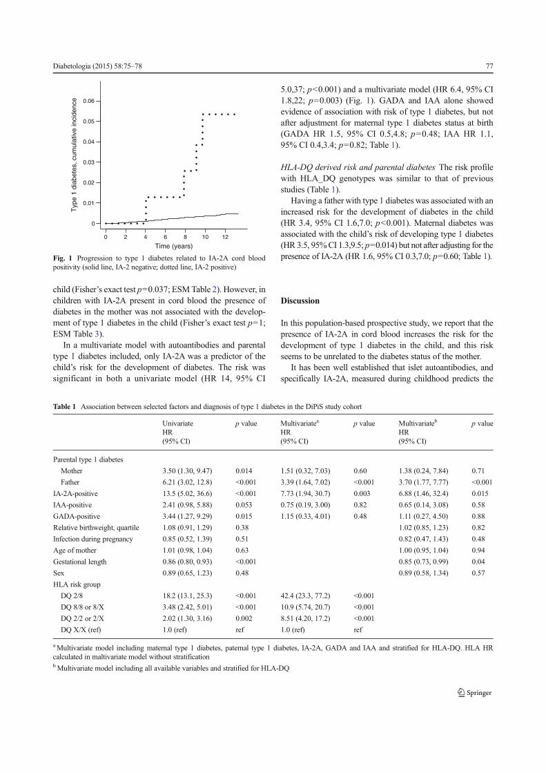
child (Fisher’s exact test p=0.037; ESMTable 2). However, inchildren with IA-2A present in cord blood the presence ofdiabetes in the mother was not associated with the develop-ment of type 1 diabetes in the child (Fisher’s exact test p=1;ESM Table 3).
In a multivariate model with autoantibodies and parentaltype 1 diabetes included, only IA-2A was a predictor of thechild’s risk for the development of diabetes. The risk wassignificant in both a univariate model (HR 14, 95% CI
5.0,37; p<0.001) and a multivariate model (HR 6.4, 95% CI1.8,22; p=0.003) (Fig. 1). GADA and IAA alone showedevidence of association with risk of type 1 diabetes, but notafter adjustment for maternal type 1 diabetes status at birth(GADA HR 1.5, 95% CI 0.5,4.8; p=0.48; IAA HR 1.1,95% CI 0.4,3.4; p=0.82; Table 1).
HLA-DQ derived risk and parental diabetes The risk profilewith HLA_DQ genotypes was similar to that of previousstudies (Table 1).
Having a father with type 1 diabetes was associated with anincreased risk for the development of diabetes in the child(HR 3.4, 95% CI 1.6,7.0; p<0.001). Maternal diabetes wasassociated with the child’s risk of developing type 1 diabetes(HR 3.5, 95%CI 1.3,9.5; p=0.014) but not after adjusting for thepresence of IA-2A (HR 1.6, 95% CI 0.3,7.0; p=0.60; Table 1).
Discussion
In this population-based prospective study, we report that thepresence of IA-2A in cord blood increases the risk for thedevelopment of type 1 diabetes in the child, and this riskseems to be unrelated to the diabetes status of the mother.
It has been well established that islet autoantibodies, andspecifically IA-2A, measured during childhood predicts the
0
0.01
0.02
0.03
0.04
0.05
0.06
Time (years)
Typ
e 1
diab
etes
, cum
ulat
ive
inci
denc
e
0 2 4 6 8 10 12
Fig. 1 Progression to type 1 diabetes related to IA-2A cord bloodpositivity (solid line, IA-2 negative; dotted line, IA-2 positive)
Table 1 Association between selected factors and diagnosis of type 1 diabetes in the DiPiS study cohort
UnivariateHR(95% CI)
p value Multivariatea
HR(95% CI)
p value Multivariateb
HR(95% CI)
p value
Parental type 1 diabetes
Mother 3.50 (1.30, 9.47) 0.014 1.51 (0.32, 7.03) 0.60 1.38 (0.24, 7.84) 0.71
Father 6.21 (3.02, 12.8) <0.001 3.39 (1.64, 7.02) <0.001 3.70 (1.77, 7.77) <0.001
IA-2A-positive 13.5 (5.02, 36.6) <0.001 7.73 (1.94, 30.7) 0.003 6.88 (1.46, 32.4) 0.015
IAA-positive 2.41 (0.98, 5.88) 0.053 0.75 (0.19, 3.00) 0.82 0.65 (0.14, 3.08) 0.58
GADA-positive 3.44 (1.27, 9.29) 0.015 1.15 (0.33, 4.01) 0.48 1.11 (0.27, 4.50) 0.88
Relative birthweight, quartile 1.08 (0.91, 1.29) 0.38 1.02 (0.85, 1.23) 0.82
Infection during pregnancy 0.85 (0.52, 1.39) 0.51 0.82 (0.47, 1.43) 0.48
Age of mother 1.01 (0.98, 1.04) 0.63 1.00 (0.95, 1.04) 0.94
Gestational length 0.86 (0.80, 0.93) <0.001 0.85 (0.73, 0.99) 0.04
Sex 0.89 (0.65, 1.23) 0.48 0.89 (0.58, 1.34) 0.57
HLA risk group
DQ 2/8 18.2 (13.1, 25.3) <0.001 42.4 (23.3, 77.2) <0.001
DQ 8/8 or 8/X 3.48 (2.42, 5.01) <0.001 10.9 (5.74, 20.7) <0.001
DQ 2/2 or 2/X 2.02 (1.30, 3.16) 0.002 8.51 (4.20, 17.2) <0.001
DQ X/X (ref) 1.0 (ref) ref 1.0 (ref) ref
aMultivariate model including maternal type 1 diabetes, paternal type 1 diabetes, IA-2A, GADA and IAA and stratified for HLA-DQ. HLA HRcalculated in maltivariate model without stratificationbMultivariate model including all available variables and stratified for HLA-DQ
Diabetologia (2015) 58:75–78 77
onset of type 1 diabetes. However, few studies have investi-gated the association between diabetes risk and the presenceof cord blood autoantibodies, and the existing data are some-what conflicting. Several studies of children born to motherswith type 1 diabetes found that cord blood autoantibodieswere of maternal origin [1, 8, 9]. Moreover, the maternalorigin of cord blood autoantibodies was confirmed in a studyof non-diabetic mothers [7], but further studies on this matterare needed. In the offspring of mothers with type 1 diabetes,cord blood autoantibodies have been reported to increase therisk for development of diabetes in the child [10], but also tohave no effect [4] or even be protective [3]. In a retrospectivecase-control study of cord blood sera from children diagnosedbefore the age of 15 years, the presence of cord blood auto-antibodies was reported to increase the risk of the child devel-oping type 1 diabetes, even after excluding mothers withdiabetes [10]. Unfortunately, no analysis of IA-2 was per-formed, with islet cell antibodies being used instead [10]. Itcannot be excluded that IA-2A may have contributed to therisk of cord blood autoantibodies for type 1 diabetes in thatstudy. A recent Danish study reported an increased risk of type1 diabetes in children bornwith cord blood autoantibodies, butno separate analysis was performed for IA-2A and GADA [9].
One of the major strength of our analysis is that the dataoriginate from a large-scale screening programme of the gen-eral population, with 70% of all children born in the Skåneregion sampled at birth. Our data regarding the prevalence oftype 1 diabetes in this cohort, now aged 9–13 years, areconsidered complete. The well-covered study area in Sweden,with five paediatric clinics and validation against the BDDstudy, minimises the risk of having missed new cases.
Our study is limited by reliance on questionnaire data regard-ing some pre- and perinatal factors. We can, therefore, assumethat the data suffer from recall bias.Missing data from the 2-yearquestionnaire affect the number of patients eligible for multivar-iate analysis. However, we were still able to use data from17,287 children and 89 type 1 diabetes patients. The numberof children who were autoantibody-positive at birth was small,as was the number of children of mothers with type 1 diabetes.This introduces some uncertainty into the statistical analysis.
In conclusion, our study indicates that the presence of cordblood autoantibodies to IA-2, but not GADA or IAA, in-creases the risk of developing type 1 diabetes compared withthe general population. The increased risk ofmaternal diabetesdisappears after adjusting for IA-2A in cord blood, suggestingthat IA-2A may be the primary risk factor. Further studies areneeded to confirm this finding.
Acknowledgements We thank all the participating parents and childrenin the DiPiS study. We thank Å. Lernmark for support and valuablecomments on the manuscript. The members of the DiPiS study groupare: C. Andersson, R. Bennet, I. Jönsson, M. Ask, J. Bremer, C. Brundin,C. Cilio, C. Hansson, G. Hansson, S. Ivarsson, B. Jonsdottir, Å Lernmark,B. Lindberg, B. Lernmark, Z. Mestan, A. Ramelius, I. Wigheden,
U.-M. Carlsson (Department of Clinical sciences Malmö, Lund Univer-sity, Sweden) A. Carlsson (Department of Clinical sciences Lund, LundUniversity, Sweden), E. Cedervall (Department of Paediatrics,Ängelholm hospital, Sweden), B. Jönsson (Department of Paediatrics,Ystad Hospital, Sweden), K. Larsson (Department of Paediatrics,Kristianstad Central Hospital, Sweden) and J. Neiderud (Department ofpaediatrics, Helsingborg Hospital, Sweden).
Funding Our research is supported in part by the Swedish ResearchCouncil (grant no. 14064 to Åke Lernmark), JDRF, Swedish ChildhoodDiabetes Foundation, Swedish Diabetes Association, Nordisk InsulinFund, SUS funds, Lion Club International, district 101-S, SUS founda-tions and the Skåne County Council Foundation for Research andDevelopment.
Duality of interest The authors declare that there is no duality ofinterest associated with this manuscript.
Contribution statement ML analysed the data and wrote the manu-script. KL was involved in the study design and critically revised themanuscript for important intellectual content. CL provided statisticalsupport, database management and contributed to and edited the manu-script. HEL designed the study, was involved in data collection,interpreted data and contributed to and edited the manuscript. All authorsgave final approval of the version to be published. ML is responsible forthe integrity of the work as a whole.
References
1. Hämäläinen A-M, Hämäläinen A-M, Ilonen J et al (2002) Prevalenceand fate of type 1 diabetes-associated autoantibodies in cordblood samples from newborn infants of non-diabetic mothers.Diabetes Metab Res Rev 18:57–63
2. Hämäläinen AM, Savola K, Kulmala PK et al (2013) Disease-associated autoantibodies during pregnancy and at birth in familiesaffected by type 1 diabetes. Clin Exp Immunol 126:230–235
3. Koczwara K, Bonifacio EE, Ziegler AG (2004) Transmission ofmaternal islet antibodies and risk of autoimmune diabetes in offspringof mothers with type 1 diabetes. Diabetes 53:1–4
4. Stanley HM, Stanley HM, Norris JM et al (2004) Is presence of isletautoantibodies at birth associated with development of persistent isletautoimmunity? The Diabetes Autoimmunity Study in the Young(DAISY). Diabetes Care 27:497–502
5. Lernmark B, Elding Larsson H, Hansson G et al (2004) Parentresponses to participation in genetic screening for diabetes risk.Pediatr Diabetes 5:174–181
6. Larsson HE, Lynch KF, Lynch KF et al (2005) Diabetes-associatedHLA genotypes affect birthweight in the general population.Diabetologia 48:1484–1491
7. Lynch KF, Lernmark B, Merlo J et al (2008) Cord blood isletautoantibodies and seasonal association with the type 1 diabeteshigh-risk genotype. J Perinatol 28:211–217
8. Ludvigsson J, Wahlberg J (2002) Diabetes-related autoantibodies incord blood from children of healthy mothers have disappeared by thetime the child is one year old. Ann N YAcad Sci 958:289–292
9. Eising S, Nilsson A, Carstensen B et al (2011) Danish children bornwith glutamic acid decarboxylase-65 and islet antigen-2 autoanti-bodies at birth had an increased risk to develop type 1 diabetes.Eur J Endocrinol 164:247–252
10. Lindberg B, Ivarsson S-A, Landin-Olsson M et al (1999) Islet auto-antibodies in cord blood from children who developed type I (insulin-dependent) diabetes mellitus before 15 years of age. Diabetologia 42:181–187
78 Diabetologia (2015) 58:75–78

Paper II

RESEARCH ARTICLE Open Access
Analgesic antipyretic use among youngchildren in the TEDDY study: no associationwith islet autoimmunityMarkus Lundgren1* , Leigh Johnson Steed2, Roy Tamura3, Berglind Jonsdottir1, Patricia Gesualdo4, Claire Crouch5,Maija Sjöberg6, Gertie Hansson1, William A. Hagopian5, Anette G. Ziegler7, Marian J. Rewers4, Åke Lernmark1,Jorma Toppari6, Jin-Xiong She2, Beena Akolkar8, Jeffrey P. Krischer3, Michael J. Haller9, Helena Elding Larsson1
and for the TEDDY Study Group
Abstract
Background: The use of analgesic antipyretics (ANAP) in children have long been a matter of controversy. Data ontheir practical use on an individual level has, however, been scarce. There are indications of possible effects onglucose homeostasis and immune function related to the use of ANAP. The aim of this study was to analyzepatterns of analgesic antipyretic use across the clinical centers of The Environmental Determinants of Diabetes inthe Young (TEDDY) prospective cohort study and test if ANAP use was a risk factor for islet autoimmunity.
Methods: Data were collected for 8542 children in the first 2.5 years of life. Incidence was analyzed using logisticregression with country and first child status as independent variables. Holm’s procedure was used to adjust formultiplicity of intercountry comparisons. Time to autoantibody seroconversion was analyzed using a Coxproportional hazards model with cumulative analgesic use as primary time dependent covariate of interest. Foreach categorization, a generalized estimating equation (GEE) approach was used.
Results: Higher prevalence of ANAP use was found in the U.S. (95.7%) and Sweden (94.8%) compared to Finland (78.1%)and Germany (80.2%). First-born children were more commonly given acetaminophen (OR 1.26; 95% CI 1.07,1.49; p = 0.007) but less commonly Non-Steroidal Anti-inflammatory Drugs (NSAID) (OR 0.86; 95% CI 0.78, 0.95;p = 0.002). Acetaminophen and NSAID use in the absence of fever and infection was more prevalent in theU.S. (40.4%; 26.3% of doses) compared to Sweden, Finland and Germany (p < 0.001).Acetaminophen or NSAID use before age 2.5 years did not predict development of islet autoimmunity by age6 years (HR 1.02, 95% CI 0.99-1.09; p = 0.27). In a sub-analysis, acetaminophen use in children with feverweakly predicted development of islet autoimmunity by age 3 years (HR 1.05; 95% CI 1.01-1.09; p = 0.024).
Conclusions: ANAP use in young children is not a risk factor for seroconversion by age 6 years. Use of ANAPis widespread in young children, and significantly higher in the U.S. compared to other study sites, where useis common also in absence of fever and infection.
Keywords: Type 1 diabetes, Analgesics, Islet autoimmunity, Prospective studies
* Correspondence: [email protected] of Clinical Sciences, Diabetes and Celiac disease unit, LundUniversity, Clinical Research Centre, Jan Waldenströms gata 35, 205 02Malmö, SwedenFull list of author information is available at the end of the article
© The Author(s). 2017 Open Access This article is distributed under the terms of the Creative Commons Attribution 4.0International License (http://creativecommons.org/licenses/by/4.0/), which permits unrestricted use, distribution, andreproduction in any medium, provided you give appropriate credit to the original author(s) and the source, provide a link tothe Creative Commons license, and indicate if changes were made. The Creative Commons Public Domain Dedication waiver(http://creativecommons.org/publicdomain/zero/1.0/) applies to the data made available in this article, unless otherwise stated.
Lundgren et al. BMC Pediatrics (2017) 17:127 DOI 10.1186/s12887-017-0884-y

BackgroundThe administration of analgesic-antipyretic (ANAP)medications to children has been discussed in the litera-ture for decades. Surveys of Canadian and American pe-diatricians reflect the routine use of acetaminophen andnon-steroidal anti-inflammatory drugs (NSAID) forchildhood fever and discomfort [1, 2]. In the 1980s, theterm “fever phobia” was used to describe the parentalpressure facing pediatric practitioners to manage fever[3]. Parental misconceptions often lead parents to the in-appropriate management of fever in their children [4]and parents report the use of antipyretics even whenthere was minimal or no fever [5] as parents were fre-quently concerned with the need to maintain a “normaltemperature” in their ill child [6]. Nevertheless, add-itional studies are needed to support this as evidence-based practice [7, 8]. Acetaminophen and NSAID areused widely in children, but limited data exist regardingpatterns of use in countries beyond the United States,United Kingdom, France, and Canada [9].Notably, acetaminophen has been shown to have ef-
fects on glucose homeostasis. High doses have beenshown to induce hyperglycemia [10], whereas low andchronic doses can lower blood glucose in animal models[11–13]. Possible effects on asthma risk have also beeninvestigated [14, 15]. NSAIDs have also been shown tolower blood glucose [16, 17], but have additional anti-inflammatory properties that could have an impact onthe process leading up to T1D [18].The Environmental Determinants of Diabetes in the
Young (TEDDY) Study is an international, multi-centerstudy designed to identify the environmental triggers ofT1D in genetically at-risk children [19]. The aim of thecurrent study was to describe the use of ANAP in theTEDDY study, as well as differences in relation to coun-try, birth order (first child versus a child with older sib-lings) and fever status. Specifically, we sought toexamine if the use of ANAP: (1) is associated with riskfor islet autoimmunity (IA), (2) differs between coun-tries, (3) is given preferentially to first-born children.
MethodsThe Environmental Determinants of Diabetes in theYoung (TEDDY) is a prospective cohort study funded bythe National Institutes of Health with the primary goalto identify environmental causes of type 1 diabetes(T1D). It includes six clinical research centers - three inthe US: Colorado, Georgia/Florida, Washington andthree in Europe: Finland, Germany, and Sweden. De-tailed study design and methods have been previouslypublished [19, 20]. Written informed consents were ob-tained for all study participants from a parent or primarycaretaker for genetic screening and participation in pro-spective follow-up. The study was approved by local
Institutional or Ethics Review Boards (Additional file 1),and is monitored by an External Advisory Board formedby the National Institutes of Health.
Data collectionThe dataset analyzed was the data received by theTEDDY Data Coordinating Center as of December 31,2014. The total number of subjects enrolled was 8676.Analysis was restricted to confirmed HLA eligible sub-jects and subjects with medication information in thefirst 2 years of age. Out of the enrolled subjects, 134were missing medication data and were excluded fromthe analysis. Information regarding first child status wasmissing for 919 subjects who were also excluded, leavinga total of 7623 subjects (Additional file 2).Study visits were conducted every 3 months with the
first visit occurring between 3 and 4.5 months of age. Ateach visit, interviewers recorded the name, reason, startdate and duration of reported medications for the mostrecent visit interval. Parents were asked to documentfever as either “Yes” or “No” for every illness entry in a“TEDDY Book.” The “TEDDY Book” provided writtenguidance that “Yes” should only be marked fortemperature equal to or greater than 38 °C or 101 °F.Approximately 18 months into the study, these choiceswere expanded to “Yes – measured,” “Yes – not mea-sured,” and “No.” The rationale for this change was tocapture all uses of ANAP, even with low-grade fevers.Each use of an ANAP was defined as an episode. Re-
corded medications were categorized based on active in-gredient. When analyzing specific substances, allmedications containing that particular substance wereincluded. Drugs were also defined and grouped as eitheranalgesic or non-steroidal anti-inflammatory (NSAID)(Additional file 3). Episodes were described as associatedwith infection and/or fever. Infection was defined as ei-ther an ICD-10 code indicating Infection (Additional file4) or an acute illness designated as infectious within a15 day time period of the medication date [21]. Feverwas defined as either an ICD-10 code of fever associatedwith the medication or an acute illness associated withfever within a 15-day time period of the medicationdate.
Islet autoimmunityBlood samples were drawn every 3 months between 3and 48 months of age, and every 6 months thereafter,except for autoantibody positive children, who continuedwith visits every 3 months. Persistent IA was defined aspositive antibodies to insulin (IAA), glutamic acid de-carboxylase (GAD65), or insulinoma-associated antigen2 (IA-2), each analyzed by radiobinding assay [22, 23],on at least 2 consecutive study visits. Two central auto-antibody laboratories were used; one in the U.S. (Barbara
Lundgren et al. BMC Pediatrics (2017) 17:127 Page 2 of 9

Davis Center for Childhood Diabetes at the University ofColorado) and one in Europe (University of Bristol). Allpositive islet autoantibodies and 5% of negative isletautoantibodies were confirmed in both central autoanti-body laboratories. Both laboratories have previouslydemonstrated high sensitivity, specificity [24] and con-cordance. Positive results in the child that were deemedto be due to maternal IgG transmission were excludedfrom the IA-positive group.
Statistical methodsA Cox proportional hazards model was used to assessthe impact of ANAP use in the first 90, 180, 365 days ofage and 2.5 years of age in the risk of positive autoanti-bodies through 6 years of age. The number of infectionsearly in life was included as a time dependent covariate[25]. Country was included as a stratification factor inthe proportional hazards analyses. Additional covariatesincluded in the model were first-degree relative [26],HLA [27], gender, ever breastfed [28, 29], probiotic useprior to 3 months of age [30], and eight different previ-ously identified single nucleotide polymorphisms [31].The primary variable of interest was cumulative ANAPuse through 2.5 years of life as a time dependent covari-ate. Included covariates can be seen in Table 1.
The statistical analysis for the number of episodes peryear and duration per year excluded subjects for whichthe first child status was missing. Subjects with a missingduration for a specific analgesic were excluded from theanalysis for that analgesic. The statistical analysis of totalduration per year was based on log-transformed data tobetter satisfy the assumptions of the linear models.Subject incidence was analyzed using logistic regression
with country and first child status as independent vari-ables in the model. In both the binary and continuousanalyses, pairwise comparisons between countries wereconducted using Holm’s procedure to adjust for the multi-plicity of comparisons. Each specific episode of ANAPusage was classified by concurrent fever (yes/no) or infec-tion (yes/no). Episodes were categorized as associated withFever, Infection, both Fever and Infection, or neither fevernor infection. For each categorization, a generalized esti-mating equation (GEE) was used for analysis with countryand first child as independent variables in the model. Anignorable working matrix was assumed for the GEE ana-lysis with the empirical sandwich estimate used for thestandard errors. Pair-wise comparisons across countrieswere conducted using Holm’s procedure from the GEEanalyses. Analyses on the episode level excluded subjectswho reported no episodes.
Table 1 Covariates included in the Cox proportionate hazards analysis of time to persistent confirmed autoantibody positivityFixed Covariates Hazard Ratio (95% CI) Wald test p-valuea
First-Degree Relative (Ref = No) 2.51 (2.06, 3.30) <0.001
HLA (Ref = DR3/DR4)
DR4/DR4 0.69 (0.54, 0.88) 0.003
DR4/DR8 0.70 (0.54, 0.91) 0.008
DR3/DR3 0.46 (0.35, 0.60) <0.001
All Others 0.46 (0.29, 0.72) <0.001
Gender (Ref = male) 0.77 (0.65, 0.92) 0.003
SNP
RS1004446_a 0.84 (0.74, 0.96) 0.010
RS10517086_a 1.14 (1.00, 1.31) 0.050
RS12708716_g 0.87 (0.76, 0.99) 0.034
RS2292239_a 1.24 (1.09, 1.41) <0.001
RS2476601_a 1.55 (1.31, 1.83) <0.001
RS2816316_c 1.07 (0.91, 1.25) 0.429
RS3184504_a 1.33 (1.17, 1.50) <0.001
RS4948088_a 0.74 (0.53, 1.04) 0.086
Ever Breastfed (Ref = No) 1.96 (1.01, 3.81) 0.042
Probiotics <3 Mo Age (Ref = No) 0.72 (0.55, 0.94) 0.015
Time Dependent Covariates
Cumulative Number of Infections 1.02 (0.99, 1.03) 0.407
Cumulative Weeks Analgesic Use 1.02 (0.99, 1.04) 0.269
Number of persistent confirmed cases = 511aHo: Hazard Ratio = 1
Lundgren et al. BMC Pediatrics (2017) 17:127 Page 3 of 9

Statistical analysis was performed using SAS version9.3 (SAS Institute Inc., Cary, NC, U.S.A).
ResultsUse of ANAP below the age of 2.5 yearsThe use of both acetaminophen and NSAIDs were verycommon in the study population. In the total cohort,87.8% of children reported the use of acetaminophenand 45.4% of NSAIDs before the age of 2.5 years. Themean number of treatment episodes per year was3.6 ± 2.1 and mean duration of treatment 8.5 ± 10.8 daysper year in the total cohort (Fig. 1a–c).
Acetaminophen useSwedish parents reported a significantly higher preva-lence of acetaminophen use (94.5%), followed by U.S.(93.7%), Finnish (73.9%), and German parents (70.1%).Prevalence differed between all countries (Finland vs.Germany: p = 0.035, all other p < 0.001). U.S. parents re-ported the highest number of treatment episodes peryear and highest total duration of treatment per year(mean 4.0 ± 2.3 episodes; mean 9.8 ± 13.5 days),followed by Swedish (mean 3.6 ± 2.1 episodes; mean8.4 ± 8.6 days), Finnish (mean 2.7 ± 1.9 episodes; mean6.8 ± 6.4 days), and German parents (mean 2.5 ± 1.6 epi-sodes; mean 4.7 ± 3.7 days). All country differences, ac-cording to number of treatment episodes, werestatistically significant (Finland vs. Germany: p = 0.014,all others p < 0.001) (Fig. 1). Children born as the firstchild in the family had more often been given acet-aminophen during their first 2.5 years of life comparedto children with older siblings (OR 1.26; 95% CI 1.07,1.49; p = 0.007). The number of episodes of treatmentwith acetaminophen was also higher (difference in leastsquare means 0.15; 95% CI 0.05, 9.24; p = 0.003). No dif-ference could be seen regarding the number of daystreated (difference in least square means 0.11; 95% CI-0.38, 0.60; p = 0.111).
NSAID useThe highest prevalence of NSAID use was reportedby U.S. parents (58.3%), followed by German (44.1%),Finnish (42.3%), and Swedish parents (29.0%). Allcountry differences, except between Finland andGermany, were statistically significant (Finland vs.Germany: p = 0.177, all others p < 0.001). U.S. par-ents reported the highest number of treatment epi-sodes with NSAID per year (mean 2.1 ± 1.4 episodes;mean 6.8 ± 11.2 days), followed by Swedish, Finnish,and German parents (mean 1.6 ± 1.2; mean 1.6 ± 1.2;mean 1.6 ± 1.1) Total duration of treatment washighest in the U.S. (mean 6.8 ± 11.2 days), followedby Finland (mean 5.3 ± 9.3 days), Germany (mean5.2 ± 17.2 days), and Sweden (mean 4.9 ± 4.8 days).Both the mean number of treatment episodes andmean total duration of treatment were significantlyhigher in the U.S. compared to the other countries(p < 0.001). No other significant country differencescould be seen (Fig. 1). The prevalence of NSAID useduring the first 2.5 years of life were lower in first-born children (OR 0.86; 95% CI 0.78, 0.95; p = 0.002)and they were also treated fewer times (difference inleast square means −0.14; 95% CI -0.22, −0.05;p = 0.001). No differences could be seen regardingthe number of days treated (difference in least squaremeans −0.22; 95% CI -0.92, 0.48; p = 0.143).
A
B
C
Fig. 1 Use of ANAP below 2,5 years of age. *:p < 0.05, n.s.: nonsignificant. All significances corrected for multiple comparisons usingHolm procedure. a Prevalence of analgesic/antipyretic use. bTreatment episodes per year. c Duration of medication use per year
Lundgren et al. BMC Pediatrics (2017) 17:127 Page 4 of 9
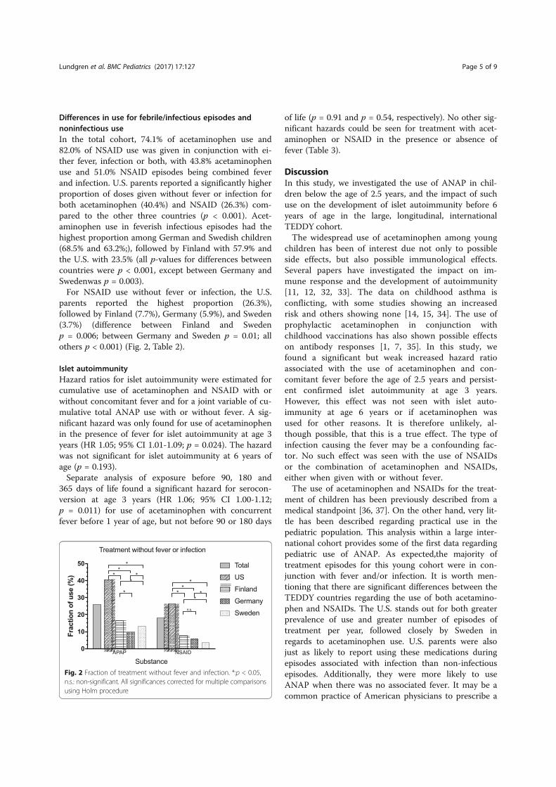
Differences in use for febrile/infectious episodes andnoninfectious useIn the total cohort, 74.1% of acetaminophen use and82.0% of NSAID use was given in conjunction with ei-ther fever, infection or both, with 43.8% acetaminophenuse and 51.0% NSAID episodes being combined feverand infection. U.S. parents reported a significantly higherproportion of doses given without fever or infection forboth acetaminophen (40.4%) and NSAID (26.3%) com-pared to the other three countries (p < 0.001). Acet-aminophen use in feverish infectious episodes had thehighest proportion among German and Swedish children(68.5% and 63.2%;), followed by Finland with 57.9% andthe U.S. with 23.5% (all p-values for differences betweencountries were p < 0.001, except between Germany andSwedenwas p = 0.003).For NSAID use without fever or infection, the U.S.
parents reported the highest proportion (26.3%),followed by Finland (7.7%), Germany (5.9%), and Sweden(3.7%) (difference between Finland and Swedenp = 0.006; between Germany and Sweden p = 0.01; allothers p < 0.001) (Fig. 2, Table 2).
Islet autoimmunityHazard ratios for islet autoimmunity were estimated forcumulative use of acetaminophen and NSAID with orwithout concomitant fever and for a joint variable of cu-mulative total ANAP use with or without fever. A sig-nificant hazard was only found for use of acetaminophenin the presence of fever for islet autoimmunity at age 3years (HR 1.05; 95% CI 1.01-1.09; p = 0.024). The hazardwas not significant for islet autoimmunity at 6 years ofage (p = 0.193).Separate analysis of exposure before 90, 180 and
365 days of life found a significant hazard for serocon-version at age 3 years (HR 1.06; 95% CI 1.00-1.12;p = 0.011) for use of acetaminophen with concurrentfever before 1 year of age, but not before 90 or 180 days
of life (p = 0.91 and p = 0.54, respectively). No other sig-nificant hazards could be seen for treatment with acet-aminophen or NSAID in the presence or absence offever (Table 3).
DiscussionIn this study, we investigated the use of ANAP in chil-dren below the age of 2.5 years, and the impact of suchuse on the development of islet autoimmunity before 6years of age in the large, longitudinal, internationalTEDDY cohort.The widespread use of acetaminophen among young
children has been of interest due not only to possibleside effects, but also possible immunological effects.Several papers have investigated the impact on im-mune response and the development of autoimmunity[11, 12, 32, 33]. The data on childhood asthma isconflicting, with some studies showing an increasedrisk and others showing none [14, 15, 34]. The use ofprophylactic acetaminophen in conjunction withchildhood vaccinations has also shown possible effectson antibody responses [1, 7, 35]. In this study, wefound a significant but weak increased hazard ratioassociated with the use of acetaminophen and con-comitant fever before the age of 2.5 years and persist-ent confirmed islet autoimmunity at age 3 years.However, this effect was not seen with islet auto-immunity at age 6 years or if acetaminophen wasused for other reasons. It is therefore unlikely, al-though possible, that this is a true effect. The type ofinfection causing the fever may be a confounding fac-tor. No such effect was seen with the use of NSAIDsor the combination of acetaminophen and NSAIDs,either when given with or without fever.The use of acetaminophen and NSAIDs for the treat-
ment of children has been previously described from amedical standpoint [36, 37]. On the other hand, very lit-tle has been described regarding practical use in thepediatric population. This analysis within a large inter-national cohort provides some of the first data regardingpediatric use of ANAP. As expected,the majority oftreatment episodes for this young cohort were in con-junction with fever and/or infection. It is worth men-tioning that there are significant differences between theTEDDY countries regarding the use of both acetamino-phen and NSAIDs. The U.S. stands out for both greaterprevalence of use and greater number of episodes oftreatment per year, followed closely by Sweden inregards to acetaminophen use. U.S. parents were alsojust as likely to report using these medications duringepisodes associated with infection than non-infectiousepisodes. Additionally, they were more likely to useANAP when there was no associated fever. It may be acommon practice of American physicians to prescribe a
Fig. 2 Fraction of treatment without fever and infection. *:p < 0.05,n.s.: non-significant. All significances corrected for multiple comparisonsusing Holm procedure
Lundgren et al. BMC Pediatrics (2017) 17:127 Page 5 of 9

combination therapy approach to the use of analgesicsand antipyretics forthe sustained management of fever.However, parents may then assume that even prophylac-tic use should be a combination therapy.We also found that first-born children were preferen-
tially given acetaminophen, both in prevalence of useand in the higher number of treatment episodes than fortheir younger siblings. The inverse relationship was ob-served for NSAID use, in which both the prevalence andnumber of treatment episodes were lower for first-bornchildren. We can only speculate on the possible
rationale behind this finding since, to our knowledge, noearlier study has presented similar data. It is possiblethat acetaminophen is perceived by first-time parents asa better tolerated treatmentthan NSAIDS, a perceptionthat fades by the time younger siblings requiretreatment.Country-specific differences in the use of analge-
sics may be culturally influenced. The lower inci-dence of use of all standard analgesics in Germanycould reflect the prevalence of Complimentary Alter-native Medicine (CAM) in this country. According
Table 2 Summary of treatment episodes associated with fever and/or infectionUS Finland Germany Sweden Total Cohort Country differences
U-F U-G U-S F-G F-S G-S
Total episodes n 25,340 7427 2091 17,285 52,643
Acetaminophen Fever and infection n 4272 2907 983 9224 17,386 <0.001 <0.001 <0.001 <0.001 <0.001 0.003
Fever or infection 6557 1277 311 3421 12,066
No fever and no infection n 7348 841 141 1953 10,283 <0.001 <0.001 <0.001 <0.001 0.006 0.01
NSAID Fever and infection n 2680 1273 477 1572 6002 <0.001 <0.001 <0.001 <0.001 <0.001 0.024
Fever or infection n 2472 717 125 330 3644
No fever and no infection n 1841 167 38 74 2120 <0.001 <0.001 <0.001 0.174 <0.001 0.041
Country differences: U = US, G = Germany, F = Finland, S = Sweden, Country differences described as p-value for difference between the respective countries
Table 3 Hazard ratios for seroconversion to persistent islet autoimmunity at 3 and 6 years of age for analgesic variables of interestAnalgesic Variable Exposed
subjects n(%)
3 Year Analysis 6 Year Analysis
398 antibody + subjects 511 antibody + subjects
HR (95% CI) p-value HR (95% CI) p-value
Acetaminophen, any exposure 7496 (87.8%) 1.01 (0.97, 1.05) 0.603 1.01 (0.98, 1.05) 0.576
Acetaminophen with fever + infection 5179 (60.6%) 1.05 (1.01, 1.09) 0.022 1.03 (0.99, 1.08) 0.189
Exposed <90 days of life 220 (2.6%) 0.97 (0.59, 1.61) 0.914 1.00 (0.68, 1.46) 0.986
Exposed <180 days of life 1519 (17.8%) 1.07 (0.87, 1.32) 0.542 1.06 (0.88, 1.27) 0.527
Exposed <365 days of life 3795 (44.4%) 1.06 (1.00, 1.12) 0.011 1.05 (0.99, 1.11) 0.101
Acetaminophen without fever or infection 5941 (69.6%) 1.02 (0.98, 1.06) 0.346 1.01 (0.96, 1.05) 0.769
Exposed <90 days of life 4016 (47.0%) 1.00 (0.90, 1.11) 0.994 0.99 (0.85, 1.14) 0.837
Exposed <180 days of life 4597 (53.8%) 0.98 (0.84, 1.14) 0.814 0.97 (0.84, 1.13) 0.729
Exposed <365 days of life 5310 (62.2%) 1.03 (0.99, 1.06) 0.114 1.02 (0.99, 1.06) 0.228
NSAID, any exposure 3874 (45.4%) 1.01 (0.97, 1.05) 0.753 1.01 (0.97, 1.04) 0.763
NSAID with fever + infection 2652 (31.0%) 1.01 (0.97, 1.05) 0.673 1.01 (0.98, 1.05) 0.459
Exposed <90 days of life 22 (0.3%) 1.01 (0.92, 1.10) 0.897 1.00 (0.92, 1.09) 0.960
Exposed <180 days of life 223 (2.6%) 0.99 (0.88, 1.11) 0.822 1.00 (0.94, 1.07) 0.927
Exposed <365 days of life 1318 (15.4%) 1.01 (0.97, 1.06) 0.530 1.02 (0.98, 1.05) 0.378
NSAID without fever or infection 1955 (22.9%) 1.00 (0.96, 1.05) 0.856 1.01 (0.97, 1.05) 0.623
Exposed <90 days of life 222 (2.6%) 1.00 (0.88, 1.12) 0.943 0.99 (0.88, 1.12) 0.874
Exposed <180 days of life 458 (5.4%) 0.99 (0.88, 1.11) 0.815 1.00 (0.94, 1.07) 0.956
Exposed <365 days of life 1120 (13.1%) 1.01 (0.96, 1.06) 0.824 1.01 (0.97, 1.05) 0.638
Any analgesic, any exposure 7744 (91%) 1.02 (0.99, 1.05) 0.130 1.02 (0.99, 1.04) 0.267
Any analgesic with fever + infection 5699 (67%) 1.06 (0.97, 1.15) 0.219 1.02 (0.94, 1.10) 0.667
Each hazard ratio is calculated from a Cox proportional hazards model with the analgesic variable and covariates indicated in text
Lundgren et al. BMC Pediatrics (2017) 17:127 Page 6 of 9
to a cross-sectional survey of German physicians in2007, more than two-thirds of patients in Germany useCAM provided either by physicians or non-medical practi-tioners (“Heilpraktiker”) [38]. In 2007, only 40% of adultsin the U.S. had used CAM therapy in the past 12 months.Children in the U.S. whose parents used CAM were almostfive times as likely (23.9%) to use CAM than childrenwhose parent did not use CAM (5.1%) [39]. The reasonsunderlying greater use of acetaminophen among Swedishparents is more unclear but may be the result of acet-aminophen being widely available and perceived as safeand effective.The TEDDY study is one of the largest longitudinal
pediatric cohorts studied. The data analyzed hereinhas been collected from parent reports given in writ-ing and after discussion with a TEDDY nurse. Forparticipating children, missing data is uncommon.Follow-up is continuous from age 3 months whichminimizes recall bias. The possibility to adjust forconfounding factors in the statistical analysis is greatdue to the availability of comprehensive data on thechild’s living conditions. All previously described riskfactors for T1D and islet autoimmunity are also en-tered into the statistical analysis of the effect of anal-gesics on islet autoimmunity.The limitations of this study include our reliance
on parent-reported symptoms and dosages of ANAP.The size of the cohort also makes it challenging toconfirm diagnoses and treatment plans via patient re-cords. Notably, most of the reported infections werepresumed to be viral infections for which no medicaladvice had been sought. In addition, the widespreaduse of ANAP in this age group poses a significantstatistical problem since the exposed group widelysurpasses the non-exposed group. Even with the largesample size, the resulting correlation between IA andacetaminophen in combination with fever must there-fore be interpreted with caution.
ConclusionsIn conclusion, the use of ANAP to treat fever andinfection is widespread in the TEDDY cohort butshows significant differences depending on study site.The prevalence of use of both acetaminophen andNSAIDs are highest in the U.S. and lowest inFinland and Germany. Use of both NSAIDs andacetaminophen for non-infectious purposes are sig-nificantly more common among children in the U.S.compared to those in Europe. No convincing effecton risk for autoimmunity can be seen in the analysisexcept for a small effect by acetaminophen in com-bination with fever, and then only for autoimmunityat 3 years of age.
Additional files
Additional file 1: Ethical review boards and Committees grantingethical approval to the TEDDY study. Listing of all ethical review boardsand committes that has granted approval to the TEDDY study for therespective sites. (DOCX 59 kb)
Additional file 2: Characteristics of the subjects used in analysis foranalgesic use and islet cell autoimmunity. A table the prevalence ofcovariates in the present analysis including HLA-DQ genotype, Gender,first degree relative, breastfeeding, probiotic use and presence ofincluded single nucleotide polymorphisms. (DOCX 106 kb)
Additional file 3: List of Drugs defined as analgesics used in first2.5 years in the TEDDY study. List of all included drugs, recorded tohaving been given to TEDDY children before age 2.5 years and classifiedas analgesics. Drugs classified as NSAID’s are marked with a star (*).(DOCX 30 kb)
Additional file 4: List of all ICD-10 codes, recorded among TEDDYchildren before age 2.5 years, and classified as an infection. (DOCX 72 kb)
AbbreviationsANAP: Analgesic-Antipyretic; CAM: Complimentary alternative medicine;GEE: Generalized estimating eq.; IA: Islet Autoimmunity; NSAID: Non-steroidalanti-inflammatory drugs; T1D: Type 1 Diabetes; TEDDY: The environmentaldeterminants of diabetes in the young study
AcknowledgementsThe Teddy study group.Funded by U01 DK63829, U01 DK63861, U01 DK63821, U01 DK63865, U01DK63863, U01 DK63836, U01 DK63790, UC4 DK63829, UC4 DK63861, UC4DK63821, UC4 DK63865, UC4 DK63863, UC4 DK63836, UC4 DK95300, UC4DK100238, UC4 DK106955, and Contract No. HHSN267200700014C from theNational Institute of Diabetes and Digestive and Kidney Diseases (NIDDK),National Institute of Allergy and Infectious Diseases (NIAID), National Instituteof Child Health and Human Development (NICHD), National Institute ofEnvironmental Health Sciences (NIEHS), Juvenile Diabetes ResearchFoundation (JDRF), and Centers for Disease Control and Prevention (CDC).This work supported in part by the NIH/NCATS Clinical and TranslationalScience Awards to the University of Florida (UL1 TR000064) and theUniversity of Colorado (UL1 TR001082).Colorado Clinical Center: Marian Rewers, M.D., Ph.D., PI1, 4–6, 10, 11, KimberlyBautista12, Judith Baxter9, 10, 12, 15, Ruth Bedoy2, Daniel Felipe-Morales,Kimberly Driscoll, Ph.D.9, Brigitte I. Frohnert, M.D.2, 14, Patricia Gesualdo2, 6, 12,14, 15, Michelle Hoffman12–14, Rachel Karban12, Edwin Liu, M.D.13, Jill Norris,Ph.D.2, 3, 12, Adela Samper-Imaz, Andrea Steck, M.D.3, 14, Kathleen Waugh6, 7,12, 15, Hali Wright12. University of Colorado, Anschutz Medical Campus,Barbara Davis Center for Childhood Diabetes.Finland Clinical Center: Jorma Toppari, M.D., Ph.D., PI¥^1,4,11,14, Olli G. Simell,M.D., Ph.D.¥^1,4,11,13, Annika Adamsson, Ph.D.^12, Suvi Ahonen*±§, HeikkiHyöty, M.D., Ph.D.*±6, Jorma Ilonen, M.D., Ph.D.¥ ¶3, Sanna Jokipuu^, TiinaKallio^, Leena Karlsson^, Miia Kähönenμ¤, Mikael Knip, M.D., Ph.D.*±5, LeaKovanen*±§, Mirva Koreasalo*±§2, Kalle Kurppa, M.D., Ph.D.*±13, Tiina Latva-ahoμ¤, Maria Lönnrot, M.D., Ph.D.*±6, Elina Mäntymäki^, Katja Multasuoμ¤, JuhaMykkänen, Ph.D.¥ 3, Tiina Niininen±*12, Sari Niinistö±§, Mia Nyblom*±, PetraRajala^, Jenna Rautanen±§, Anne Riikonen*±§, Mika Riikonen^, JenniRouhiainen^, Minna Romo^, Tuula Simell, Ph.D., Ville Simell^¥13, MaijaSjöberg¥^12,14, Aino Steniusμ¤12, Maria Leppänen^, Sini Vainionpää^, EevaVarjonen¥^12, Riitta Veijola, M.D., Ph.D.μ¤14, Suvi M. Virtanen, M.D.,Ph.D.*±§2, Mari Vähä-Mäkilä^, Mari Åkerlund*±§, Katri Lindfors, Ph.D.*13¥University of Turku, *University of Tampere, μUniversity of Oulu, ^TurkuUniversity Hospital, Hospital District of Southwest Finland, ±TampereUniversity Hospital, ¤Oulu University Hospital, §National Institute forHealth and Welfare, Finland, ¶University of Kuopio.Georgia/Florida Clinical Center: Jin-Xiong She, Ph.D., PI1, 3, 4, 11, DesmondSchatz, M.D.*4, 5, 7, 8, Diane Hopkins12, Leigh Steed12–15, Jamie Thomas*6, 12,Janey Adams*12, Katherine Silvis2, Michael Haller, M.D.*14, Melissa Gardiner,Richard McIndoe, Ph.D., Ashok Sharma, Joshua Williams, Gabriela Young,Stephen W. Anderson, M.D.^, Laura Jacobsen, M.D.*14 Center for Biotechnologyand Genomic Medicine, Augusta University. *University of Florida, ^PediatricEndocrine Associates, Atlanta.
Lundgren et al. BMC Pediatrics (2017) 17:127 Page 7 of 9
Germany Clinical Center: Anette G. Ziegler, M.D., PI1, 3, 4, 11, AndreasBeyerlein, Ph.D.2, Ezio Bonifacio Ph.D.*5, Michael Hummel, M.D.13, SandraHummel, Ph.D.2, Kristina Foterek¥2, Nicole Janz, Mathilde Kersting, Ph.D.¥2,Annette Knopff7, Sibylle Koletzko, M.D.¶13, Claudia Peplow12, Roswith Roth,Ph.D.9, Marlon Scholz, Joanna Stock9, 12, 14, Katharina Warncke, M.D.14, LorenaWendel, Christiane Winkler, Ph.D.2, 12, 15. Forschergruppe Diabetes e.V. andInstitute of Diabetes Research, Helmholtz Zentrum München, and Klinikumrechts der Isar, Technische Universität München. *Center for RegenerativeTherapies, TU Dresden, ¶Dr. von Hauner Children’s Hospital, Department ofGastroenterology, Ludwig Maximillians University Munich, ¥Research Institutefor Child Nutrition, Dortmund.Sweden Clinical Center: Åke Lernmark, Ph.D., PI1, 3–6, 8, 10, 11, 15, DanielAgardh, M.D., Ph.D.13, Carin Andrén Aronsson2,12,13, Maria Ask, Jenny Bremer,Ulla-Marie Carlsson, Corrado Cilio, Ph.D., M.D.5, Emelie Ericson-Hallström, LinaFransson, Thomas Gard, Joanna Gerardsson, Rasmus Bennet, Monica Hansen,Gertie Hansson, Susanne Hyberg, Fredrik Johansen, Berglind Jonsdottir, M.D.,Helena Elding Larsson, M.D., Ph.D. 6,14, Marielle Lindström, Markus Lundgren,M.D.14, Maria Månsson-Martinez, Maria Markan, Jessica Melin12, Zeliha Mestan,Karin Ottosson, Kobra Rahmati, Anita Ramelius, Falastin Salami, SaraSibthorpe, Birgitta Sjöberg, Ulrica Swartling, Ph.D.9,12, Evelyn Tekum Amboh,Carina Törn, Ph.D. 3,15, Anne Wallin, Åsa Wimar12,14, Sofie Åberg. LundUniversity.Washington Clinical Center: William A. Hagopian, M.D., Ph.D., PI1,3,4, 5, 6,7,11,13,14, Michael Killian6,7,12,13, Claire Cowen Crouch12,14,15, Jennifer Skidmore2,Josephine Carson, Maria Dalzell, Kayleen Dunson, Rachel Hervey, CorbinJohnson, Rachel Lyons, Arlene Meyer, Denise Mulenga, Alexander Tarr,Morgan Uland, John Willis. Pacific Northwest Diabetes Research Institute.Pennsylvania Satellite Center: Dorothy Becker, M.D., Margaret Franciscus,MaryEllen Dalmagro-Elias Smith2, Ashi Daftary, M.D., Mary Beth Klein, ChrystalYates. Children’s Hospital of Pittsburgh of UPMC.Data Coordinating Center: Jeffrey P. Krischer, Ph.D.,PI1,4,5,10,11, MichaelAbbondondolo, Sarah Austin-Gonzalez, Maryouri Avendano, Sandra Baethke,Rasheedah Brown12,15, Brant Burkhardt, Ph.D.5,6, Martha Butterworth2, JoannaClasen, David Cuthbertson, Christopher Eberhard, Steven Fiske9, Dena Garcia,Jennifer Garmeson, Veena Gowda, Kathleen Heyman, Francisco Perez Laras,Hye-Seung Lee, Ph.D.1,2,13,15, Shu Liu, Xiang Liu, Ph.D.2,3,9,14, Kristian Lynch,Ph.D. 5,6,9,15, Jamie Malloy, Cristina McCarthy12,15, Steven Meulemans, HemangParikh, Ph.D.3, Chris Shaffer, Laura Smith, Ph.D.9,12, Susan Smith12,15, NoahSulman, Ph.D., Roy Tamura, Ph.D.1,2,13, Ulla Uusitalo, Ph.D.2,15, Kendra Vehik,Ph.D.4,5,6,14,15, Ponni Vijayakandipan, Keith Wood, Jimin Yang, Ph.D., R.D.2,15.Past staff: Lori Ballard, David Hadley, Ph.D., Wendy McLeod. University of SouthFlorida.Project scientist: Beena Akolkar, Ph.D.1,3,4,5,6,7,10,11. National Institutes ofDiabetes and Digestive and Kidney Diseases.Autoantibody Reference Laboratories: Liping Yu, M.D.^5, Dongmei Miao,M.D.^, Polly Bingley, M.D., FRCP*5, Alistair Williams*, Kyla Chandler*, SabaRokni*, Claire Williams*, Rebecca Wyatt*, Gifty George*, Sian Grace*. ^BarbaraDavis Center for Childhood Diabetes, University of Colorado Denver, *Schoolof Clinical Sciences, University of Bristol UK.HLA Reference Laboratory: Henry Erlich, Ph.D.3, Steven J. Mack, Ph.D., AnnaLisa Fear. Center for Genetics, Children’s Hospital Oakland Research Institute.Repository: Sandra Ke, Niveen Mulholland, Ph.D. NIDDK BiosampleRepository at Fisher BioServices.SNP Laboratory: Stephen S. Rich, Ph.D.3, Wei-Min Chen, Ph.D.3, SunaOnengut-Gumuscu, Ph.D.3, Emily Farber, Rebecca Roche Pickin, Ph.D., JordanDavis, Dan Gallo, Jessica Bonnie, Paul Campolieto. Center for Public HealthGenomics, University of Virginia.Other contributors: Kasia Bourcier, Ph.D.5, National Institutes of Allergy andInfectious Diseases. Thomas Briese, Ph.D.6,15, Columbia University. SuzanneBennett Johnson, Ph.D.9,12, Florida State University. Eric Triplett, Ph.D.6,University of Florida.Committees:1Ancillary Studies, 2Diet, 3Genetics, 4Human Subjects/Publicity/Publications,5Immune Markers, 6Infectious Agents, 7Laboratory Implementation, 8MaternalStudies, 9Psychosocial, 10Quality Assurance, 11Steering, 12Study Coordinators,13Celiac Disease, 14Clinical Implementation, 15Quality AssuranceSubcommittee on Data Quality.
FundingFunded by U01 DK63829, U01 DK63861, U01 DK63821, U01 DK63865, U01DK63863, U01 DK63836, U01 DK63790, UC4 DK63829, UC4 DK63861, UC4
DK63821, UC4 DK63865, UC4 DK63863, UC4 DK63836, UC4 DK95300, UC4DK100238, UC4 DK106955, and Contract No. HHSN267200700014C from theNational Institute of Diabetes and Digestive and Kidney Diseases (NIDDK),National Institute of Allergy and Infectious Diseases (NIAID), National Instituteof Child Health and Human Development (NICHD), National Institute ofEnvironmental Health Sciences (NIEHS), Juvenile Diabetes ResearchFoundation (JDRF), and Centers for Disease Control and Prevention (CDC).This work supported in part by the NIH/NCATS Clinical and TranslationalScience Awards to the University of Florida (UL1 TR000064) and theUniversity of Colorado (UL1 TR001082).
Availability of data and materialsThe data that support the findings of this study are available from the NIDDKrepository (http://niddkrepository.org) approximately 6 months after themanuscript has appeared in print. Access for the data submitted to theNIDDK Data Repository will be determined by the NIDDK. All investigatorswho receive TEDDY resources must agree to acknowledge the TEDDY Studyand the NIDDK central repository. This approach is fully compliant with theNIH public data sharing policy (http://grants.nih.gov/grants/policy/data_sharing).
Authors’ contributionsML researched the data and wrote the manuscript. LJS was involved in thedrafting of the manuscript and reviewed/edited the manuscript. RT madethe statistical analyses, researched data and reviewed/edited the manuscript.MH, HEL, BJ, PG, CC, MS, GH researched data and reviewed/edited themanuscript. WH, AGZ, MR, ÅL, JT, JS, BA and JK designed the study,researched data, and reviewed/edited the manuscript. All authors approvedthe final manuscript as submitted and agree to be accountable for allaspects of the work.
Competing interestsThe authors have no conflicts of interest relevant to this article to disclose.The funding agencies did not include any input into the design andconduct of the study; collection, management, analysis, or interpretation ofdata; or preparation, review, or approval of manuscript.
Consent for publicationNot applicable.
Ethics approval and consent to participateThe study was approved by the local Institutional or Ethics Review Boardsand is monitored by an External Advisory Board formed by the NationalInstitutes of Health.
Publisher’s NoteSpringer Nature remains neutral with regard to jurisdictional claims inpublished maps and institutional affiliations.
Author details1Department of Clinical Sciences, Diabetes and Celiac disease unit, LundUniversity, Clinical Research Centre, Jan Waldenströms gata 35, 205 02Malmö, Sweden. 2Center for Biotechnology and Genomic Medicine, MedicalCollege of Georgia, Augusta University, Augusta, GA, USA. 3Health InformaticsInstitute, Morsani College of Medicine, University of South Florida, Tampa, FL,USA. 4Barbara Davis Center for Childhood Diabetes, University of Colorado,Aurora, CO, USA. 5Pacific Northwest Diabetes Research Institute, Seattle, WA,USA. 6Department of Physiology, Institute of Biomedicine, University of Turku,and Department of Pediatrics, Turku University Hospital, Turku, Finland.7Institute of Diabetes Research, Helmholtz Zentrum München, and Klinikumrechts der Isar, Technische Universität München, and ForschergruppeDiabetes e.V, Neuherberg, Germany. 8National Institute of Diabetes &Digestive & Kidney Diseases, Bethesda, MD, USA. 9Department of Pediatrics,University of Florida, Gainesville, FL, USA.
Received: 2 February 2017 Accepted: 9 May 2017
References1. Taddio A, Manley J, Potash L, Ipp M, Sgro M, Shah V. Routine immunization
practices: use of topical anesthetics and oral analgesics. Pediatrics. 2007;120:e637–43.
Lundgren et al. BMC Pediatrics (2017) 17:127 Page 8 of 9
2. Mayoral CE, Marino RV, Rosenfeld W, Greensher J. Alternating antipyretics: isthis an alternative? Pediatrics. 2000;105:1009–12.
3. Schmitt BD. Fever phobia: misconceptions of parents about fevers. Am J DisChild. 1980;134:176–81.
4. Casey R, McMahon F, McCormick MC, Pasquariello PS, Zavod W, King FH. Fevertherapy: an educational intervention for parents. Pediatrics. 1984;73:600–5.
5. Bilenko N, Tessler H, Okbe R, Press J, Gorodischer R. Determinants ofantipyretic misuse in children up to 5 years of age: a cross-sectional study.Clin Ther. 2006;28:783–93.
6. Section on Clinical Pharmacology and Therapeutics, Committee on Drugs, SullivanJE, Farrar HC. Fever and antipyretic use in children. Pediatrics. 2011;127:580–7.
7. Pedulla MN. Prophylactic use of antipyretic agents with childhoodimmunizations and antibody response: reason for concern? J Pediatr HealthCare. 2012;26:200–3.
8. Chiappini E, Principi N, Longhi R, Tovo P-A, Becherucci P, Bonsignori F, et al.Management of fever in children: summary of the Italian pediatric societyguidelines. Clin Ther. 2009;31:1826–43.
9. Bertille N, Fournier-Charrière E, Pons G, Chalumeau M. Managing fever inchildren: a national survey of parents' knowledge and practices in France.PLoS One. 2013;8:e83469.
10. Hinson JA, Han-Hsu H, Mays JB, Holt SJ, McLean P, Ketterer B.Acetaminophen-induced alterations in blood glucose and blood insulinlevels in mice. Res Commun Chem Pathol Pharmacol. 1984;43:381–91.
11. Shertzer Howard G, Schneider Scott N, Kendig Eric L, Clegg Deborah J, D´Alessio David A, Genter Mary Beth. Acetaminophen normalizes glucosehomeostasis in mouse models for diabetes. 2008;75:1402–10. Available from:http://www.sciencedirect.com/science/article/pii/S0006295207007988
12. Blough ER, Wu M. Acetaminophen: beyond pain and fever-relieving. FrontPharmacol. Frontiers. 2011;2:72.
13. Wu M, Desai DH, Kakarla SK, Katta A, Paturi S, Gutta AK, et al.Acetaminophen prevents aging-associated hyperglycemia in aged rats:effect of aging-associated hyperactivation of p38-MAPK and ERK1/2.Diabetes Metab Res Rev. 2009;25:279–86. John Wiley & Sons, Ltd.
14. Cheelo M, Lodge CJ, Dharmage SC, Simpson JA, Matheson M, Heinrich J,et al. Paracetamol exposure in pregnancy and early childhood anddevelopment of childhood asthma: a systematic review and meta-analysis.Arch Dis Child. 2015;100:81–9.
15. Kreiner-Møller E, Sevelsted A, Vissing NH, Schoos A-MM, Bisgaard H. Infantacetaminophen use associates with early asthmatic symptomsindependently of respiratory tract infections: the Copenhagen prospectivestudy on asthma in childhood 2000 (COPSAC(2000)) cohort. J Allergy ClinImmunol. 2012;130:1434–6.
16. Mork NL, Robertson RP. Effects of nonsteroidal antiinflammatory drugs inconventional dosage on glucose homeostasis in patients with diabetes.West J Med. 1983;139:46–9.
17. Jafarnejad A, Bathaie SZ, Nakhjavani M, Hassan MZ. Investigation of themechanisms involved in the high-dose and long-term acetyl salicylic acidtherapy of type I diabetic rats. J Pharmacol Exp Ther. 2008;324:850–7.
18. Bushra R, Aslam N. An overview of clinical pharmacology of ibuprofen.Oman Med J. 2010;25:155–661.
19. TEDDY Study Group. The environmental determinants of diabetes in theyoung (TEDDY) study. Ann N Y Acad Sci. 2008;1150:1–13.
20. TEDDY Study Group. The environmental determinants of diabetes in theyoung (TEDDY) study: study design. Pediatr Diabetes. 2007;8:286–98.Blackwell Publishing Ltd.
21. Lönnrot M, Lynch K, Larsson HE, Lernmark Å, Rewers M, Hagopian W, et al.A method for reporting and classifying acute infectious diseases in aprospective study of young children: TEDDY. BMC Pediatr. 2015;15:24.
22. Bonifacio E, Yu L, Williams AK, Eisenbarth GS, Bingley PJ, Marcovina SM,et al. Harmonization of glutamic acid decarboxylase and islet antigen-2autoantibody assays for national institute of diabetes and digestive andkidney diseases consortia. J Clin Endocrinol Metab. 2010;95:3360–7.
23. Babaya N, Yu L, Miao D, Wang J, Rewers M, Nakayama M, et al. Comparisonof insulin autoantibody: polyethylene glycol and micro-IAA 1-day and 7-dayassays. Diabetes Metab Res Rev. 2009;25:665–70.
24. Törn C, Mueller PW, Schlosser M, Bonifacio E, Bonifacio EE, Bingley PJ, et al.Diabetes antibody standardization program: evaluation of assays forautoantibodies to glutamic acid decarboxylase and islet antigen-2.Diabetologia. 2008;51:846–52.
25. Roivainen M, Klingel K. Virus infections and type 1 diabetes risk. Curr DiabRep. 2010;10:350–6.
26. Ziegler AG, Hillebrand B, Rabl W, Mayrhofer M, Hummel M, Mollenhauer U,et al. On the appearance of islet associated autoimmunity in offspring ofdiabetic mothers: a prospective study from birth. Diabetologia. 1993;36:402–8.
27. She J-X. Susceptibility to type I diabetes: HLA-DQ and DR revisited. ImmunolToday. 1996;17:323–9.
28. Kimpimäki T, Erkkola M, Korhonen S, Kupila A, Virtanen SM, Ilonen J, et al.Short-term exclusive breastfeeding predisposes young children withincreased genetic risk of type I diabetes to progressive beta-cellautoimmunity. Diabetologia. 2001;44:63–9.
29. Virtanen SM, Kenward MG, Erkkola M, Kautiainen S, Kronberg-Kippilä C,Hakulinen T, et al. Age at introduction of new foods and advanced beta cellautoimmunity in young children with HLA-conferred susceptibility to type 1diabetes. Diabetologia. 2006;49:1512–21.
30. Uusitalo U, Liu X, Yang J, Aronsson CA, Hummel S, Butterworth M, et al.Association of Early Exposure of Probiotics and islet autoimmunity in theTEDDY study. JAMA Pediatr. 2015;170:1–9.
31. Törn C, Hadley D, Lee HS, Hagopian W, Lernmark Å, Simell O, et al. Role oftype 1 diabetes associated SNPs on risk of autoantibody positivity in theTEDDY study. Diabetes. 2014;64(5):1818–29.
32. Litherland SA, She J-X, Schatz D, Fuller K, Hutson AD, Peng RH, et al.Aberrant monocyte prostaglandin synthase 2 (PGS2) expression in type 1diabetes before and after disease onset. Pediatr Diabetes. 2003;4:10–8.Munksgaard International Publishers
33. Beyan H, Buckley LR, Bustin SA, Yousaf N, Pozzilli P, Leslie RD. Alteredmonocyte cyclo-oxygenase response in non-obese diabetic mice. Clin ExpImmunol. 2009;155:304–10.
34. Eyers S, Weatherall M, Jefferies S, Beasley R. Paracetamol in pregnancy andthe risk of wheezing in offspring: a systematic review and meta-analysis.Clin Exp Allergy. 2011;41:482–9.
35. Prymula R, Siegrist C-A, Chlibek R, Zemlickova H, Vackova M, Smetana J, et al.Effect of prophylactic paracetamol administration at time of vaccination onfebrile reactions and antibody responses in children: two open-label,randomised controlled trials. Lancet. 2009;374:1339–50. Elsevier Ltd
36. Sullivan JE, Farrar HC. The section on clinical pharmacology andtherapeutics, committee on drugs. Fever and antipyretic use in children.Pediatrics. 2011;127:580–7.
37. Erlewyn-Lajeunesse MDS, Coppens K, Hunt LP, Chinnick PJ, Davies P,Higginson IM, et al. Randomised controlled trial of combined paracetamoland ibuprofen for fever. Arch Dis Child. 2006;91:414–6.
38. Joos S, Musselmann B, Szecsenyi J. Integration of complementary andalternative medicine into family practices in Germany: results of a nationalsurvey. Evid Based Complement Alternat Med. 2011;2011:495813.
39. Barnes PM, Bloom B, Nahin RL. Complementary and alternative medicineuse among adults and children: United States, 2007. Natl Health Stat Report.2008;10(12):1–23.
• We accept pre-submission inquiries • Our selector tool helps you to find the most relevant journal• We provide round the clock customer support • Convenient online submission• Thorough peer review• Inclusion in PubMed and all major indexing services • Maximum visibility for your research
Submit your manuscript atwww.biomedcentral.com/submit
Submit your next manuscript to BioMed Central and we will help you at every step:
Lundgren et al. BMC Pediatrics (2017) 17:127 Page 9 of 9


Paper IV

Pediatric Diabetes 2014doi: 10.1111/pedi.12151All rights reserved
© 2014 John Wiley & Sons A/S.Published by John Wiley & Sons Ltd.
Pediatric Diabetes
Original Article
Reduced morbidity at diagnosis and improvedglycemic control in children previouslyenrolled in DiPiS follow-up
Lundgren M, Sahlin A, Svensson C, Carlsson A, Cedervall E, Jonsson B,Jonsson I, Larsson K, Lernmark A, Neiderud J, Vigard T, Elding Larsson H,the DiPiS study group. Reduced morbidity at diagnosis and improvedglycemic control in children previously enrolled in DiPiS follow-up.Pediatric Diabetes 2014.
Aims/hypothesis: Children participating in longitudinal type 1 diabetesprediction studies were reported to have less severe disease at diabetesdiagnosis. Our aim was to investigate children who from birth participated inthe Diabetes Prediction in Skane (DiPiS) study for metabolic status atdiagnosis and then continued to be followed for 2 yr of regular clinical care.Methods: Children, followed in DiPiS before diagnosis, were compared tochildren in the same birth cohort, who did not participate in follow-up.Metabolic status, symptoms at diagnosis as well as hemoglobin A1c (HbA1c)and doses of insulin at 3, 6, 12, and 24 months after diagnosis were compared.Results: Children, followed in DiPiS and diagnosed at 2–12 yr of age, had0.8% (9 mmol/mol) lower HbA1c at diagnosis than those who were notfollowed (p = 0.006). At diagnosis, fewer DiPiS children had symptoms(p = 0.014) and ketoacidosis at diagnosis were reduced (2% compared to 18%,p = 0.005). During regular clinical care, HbA1c levels for the DiPiS childrenremained lower both at 12 (0.4% (4 mmol/mol); p = 0.009) and 24 months(0.8% (9 mmol/mol) p < 0.001) after diagnosis, despite no difference in totaldaily insulin between the two groups.Conclusions: Participation in prospective follow-up before diagnosis of type 1diabetes leads to earlier diagnosis with fewer symptoms, decreased incidenceof ketoacidosis as well as better metabolic control up to 2 yr after diagnosis.Our data indicate that metabolic control at the time of diabetes diagnosis isimportant for early metabolic control possibly affecting the risk of long-termcomplications.
Markus Lundgrena,Asa Sahlinb,Camilla Svenssonc,Annelie Carlssond,Elisabeth Cedervalle,Bjorn Jonssonf,Ida Jonssong,Karin Larssona,Ake Lernmarkg,Jan Neiderudh, Tore Vigardf,Helena Elding Larssong andthe DiPiS study groupaDepartment of Pediatrics, KristianstadCentral Hospital, Kristianstad, Sweden;bBlekinge Hospital, Karlskrona,Sweden; cKristianstad CentralHospital, Kristianstad, Sweden;dDepartment of Pediatrics, LundUniversity, Skane University Hospital,Lund, Sweden; eDepartment ofPediatrics, Angelholm GeneralHospital, Angelholm, Sweden;fDepartment of Pediatrics, YstadGeneral Hospital, Ystad, Sweden;gDepartment of Clinical Sciences, LundUniversity/CRC, Skane UniversityHospital, Malmo, Sweden; andhDepartment of Pediatrics, HelsingborgGeneral Hospital, Helsingborg, Sweden
Key words: diabetic ketoacidosis –follow-up studies – HbA1c – type 1diabetes
Corresponding author: MarkusLundgren, MD,Department of Pediatrics,Kristianstad Central Hospital,JA Hedlunds vag 5,S-291 33 Kristianstad,Sweden.Tel: +46 709-950 990;fax: +46 443-092 906e-mail: [email protected]
1
Lundgren et al.
Submitted 15 January 2014.Accepted for publication 11 April2014
The triggering event for islet autoimmunity eventuallyresulting in the clinical onset of type 1 diabetes, is stillunknown. This is true for both the initial insult ofthe autoimmune process but also for factors governingthe time it takes for autoimmunity to lead to clinicaldisease. Prediction of who will develop the disease andwhen is critical for attempts to stall and perhaps tostop the disease process. The human leukocyte antigen(HLA) DQ locus is presumed to account for 50% ofthe genetic risk of type 1 diabetes (1). More than 40additional non-HLA risk genes have been identifiedin recent genome-wide association studies (2). Isletautoantibodies are used to estimate the risk for diabetesand it is well-established that the risk is increasing withan increasing number of autoantibodies (3).
Children born with increased genetic risk fortype 1 diabetes have been followed in severalprospective longitudinal studies. The focus has beenthe development and maintenance of the beta-cellautoimmune process. Only a few studies have beenpublished on the status of the participants bothat the time of diagnosis of diabetes and duringfollow-up after diagnosis. Participants in longitudinalstudies have been shown to have fewer metabolicabnormalities at diagnosis and a lower frequency ofdiabetic ketoacidosis (4–6) as well as having a milderclinical course in the first year after diagnosis (7). Thismay be due to intense follow-up with measures ofplasma glucose, hemoglobin A1c (HbA1c) or glucosetolerance tests. However, the parent’s knowledge of therisk of disease may also result in an earlier diagnosisthrough increased vigilance regarding symptoms.
DiPiS is a prospective study on type 1 diabetesprediction in Sweden. The aim of this study was toinvestigate the disease at diagnosis and the metaboliccontrol during 2 yr of regular clinical care afterdiagnosis, after participation in the DiPiS study (8).We compared the DiPiS children with children bornduring the same years, who developed diabetes outsideof the DiPiS study.
Methods
Participants
In the Diabetes Prediction in Skane (DiPiS) study,2500 children with genetic risk for type 1 diabetes insouthern Sweden are prospectively studied in a 15-yr longitudinal investigation (9). Between September2000 and August 2004 approximately 48 000 childrenwere born in the five participating hospitals andafter oral consent from the mother, 35 683 of
those children had umbilical cord blood samplescollected, for HLA genotyping (Fig. 1). When thechild was 2 months old, the parents were invitedto participate in DiPiS by giving written consentand answering questionnaires regarding, among otherthings, pregnancy, socioeconomic factors, birth weightand length, hereditary factors and stressors duringpregnancy. A total of 25 378 questionnaires (71%) werereturned by the parents of the study participants ofwhich 92% wanted to participate.
A risk score was developed based on HLA riskgenotype for type 1 diabetes, presence of maternalinfections during pregnancy, maternal diabetes, cordblood autoantibodies and high or low relative birthweight. On the basis of this risk score, approximately6000 children who had answered the 2 monthsquestionnaire were selected for annual follow-up. Onlyparents of the 3680 children who returned the 2-yrquestionnaire received HLA-based risk information.Participants are screened yearly, from the age of 2yr, with a questionnaire and blood sampling for isletautoantibody analysis. Those children who developtwo or more autoantibodies are offered follow-upevery 3 months by a pediatrician, with autoantibodysampling, random plasma glucose, HbA1c, growthparameters, questionnaire and oral glucose tolerancetest (OGTT) annually.
Up to July 2013, a total of 143 of the children withcord blood samples taken at birth had developed type1 diabetes. Of those, 14 children developed diabetesbefore 2 yr of age and were therefore not givenrisk information or the possibility to participate inthe follow-up. After exclusion of children who haddeveloped other types of diabetes and those diagnosedbefore the age of 2, 129 children remained in thefollowing three subgroups (Fig. 1):
Group A: DiPiS children participating in intensefollow-up. This group is comprised of children whohad developed multiple autoantibodies and haveparticipated in follow-up every 3 months (n = 33).
Group B: DiPiS children who participated in annualautoantibody sampling and questionnaires. Thisgroup has answered the 2-yr questionnaire, havebeen invited to the DiPiS follow-up and havereceived information about the risk for type 1diabetes (n = 18).
Group C: Children with umbilical cord samples butwho declined participation in follow-up and riskinformation or where not invited to the study dueto low HLA risk for type 1 diabetes. None of these
2 Pediatric Diabetes 2014
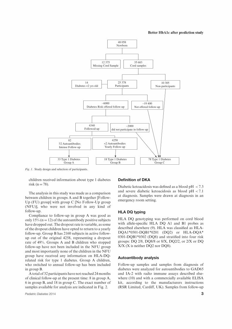
Better HbA1c after prediction study
48 058Newborn
12 375Missing Cord Sample
35 683Cord samples
25 378Participants
10 305 Non-participants
∼19 400 Not offered follow-up
∼6000Diabetes Risk offered follow-up
14Diabetes <2 yrs old
4340Followed-up
822 Autoantibodies
Intense Follow-up
4258<2 AutoantibodiesYearly Follow-up
∼2000did not participate in follow-up
18 Type 1 DiabetesGroup B
33 Type 1 DiabetesGroup A
78 Type 1 DiabetesGroup C
Fig. 1. Study design and selection of participants.
children received information about type 1 diabetesrisk (n = 78).
The analysis in this study was made as a comparisonbetween children in groups A and B together [Follow-Up (FU) group] with group C [No Follow-Up group(NFU)], who were not involved in any kind offollow-up.
Compliance to follow-up in group A was good asonly 15% (n = 12) of the autoantibody positive subjectshave dropped out. The dropout rate is variable, as someof the dropout children have opted to return to a yearlyfollow-up. Group B has 2160 subjects in active follow-up out of the original 4258, representing a dropoutrate of 49%. Groups A and B children who stoppedfollow-up have not been included in the NFU groupand most importantly none of the children in the NFUgroup have received any information on HLA-DQ-related risk for type 1 diabetes. Group A children,who switched to annual follow-up has been includedin group B.
A total of 32 participants have not reached 24 monthsof clinical follow-up at the present time: 8 in group A,6 in group B, and 18 in group C. The exact number ofsamples available for analysis are indicated in Fig. 2.
Definition of DKA
Diabetic ketoacidosis was defined as a blood pH < 7.3and severe diabetic ketoacidosis as blood pH < 7.1at diagnosis. Samples were drawn at diagnosis in anemergency room setting.
HLA DQ typing
HLA DQ genotyping was performed on cord bloodwith allele-specific HLA DQ A1 and B1 probes asdescribed elsewhere (9). HLA was classified as HLA-DQA1*0501-DQB1*0201 (DQ2) or HLA-DQA*0301-DQB1*0302 (DQ8) and stratified into four riskgroups: DQ 2/8, DQ8/8 or 8/X, DQ2/2, or 2/X or DQX/X (X is neither DQ2 nor DQ8).
Autoantibody analysis
Follow-up samples and samples from diagnosis ofdiabetes were analyzed for autoantibodies to GAD65and IA-2 with radio immune assays described else-where (10) and with a commercially available ELISAkit, according to the manufacturers instructions(RSR Limited, Cardiff, UK). Samples from follow-up
Pediatric Diabetes 2014 3

Lundgren et al.
24 monthsp <0.001
12 monthsp=0.009
6 monthsp=0.34
3 monthsp=0.12
Diagnosisp=0.006
Med
ian
HbA
1c (m
mol
/mol
)
90
80
70
60
50
40
FU n=51NFU n=77
FU n=45NFU n=74
FU n=45NFU n=74
FU n=43NFU n=70
FU n=33NFU n=56
Fig. 2. HbA1c levels at diagnosis and the first 2 yr after diagnosis in the FU and NFU groups. Solid line, no follow-up group; Dotted line,follow-up group.
visits and diabetes diagnosis were additionally ana-lyzed for autoantibodies to insulin (IAA) and radioimmune assay of the three amino acid variants of zinctransporter 8 (ZnT8RA, arginine 325 zinc transporter8 autoantibody; ZnT8WA, tryptophan 325 zinc trans-porter 8 autoantibody; ZnT8QA, glutamine 325 zinctransporter 8 autoantibody) autoantibodies. For IAAperformed before 2011 and all ZnT8 autoantibodiesthe method is described elsewhere (11). For IAA sam-ples analyzed after 2010, the following alteration tothe assay has been made: to the buffer used for incu-bation [TRIS-buffer (pH 8.0) with 1% (v/v) Tween 20]1% w/v bovine serum albumin was added to preventnon-specific binding. In the competitive assay, insteadof arbitrary units, U/mL has been calculated using astandard curve. The standard curve represented sevendifferent concentrations (3–358 U/mL) and the concen-tration was plotted against cpm values on a Log2 scale.
Data collection
Pediatricians at the pediatric clinics in the RegionSkane continuously reported children from the DiPiScohort when they developed diabetes. Additionalinformation was gained from on-going studies,including all children diagnosed in Skane (the Skanestudy) and Sweden (the Better Diabetes Diagnosisstudy) (12). Data regarding metabolic parameters,
symptoms at diagnosis and clinical post diagnosisfollow-up data on HbA1c and insulin doses wereretrieved from individual electronic patient records(Melior, Siemens AG, Berlin, Germany). Insulin doseswere analyzed as total units of insulin per kg and day.Follow-up data was recorded after diagnosis at 3, 6, 12,and 24 ± 1 month. The clinical care of the diagnosedpatients is ongoing, and some children with type 1diabetes have therefore yet to reach 24 months afterdiagnosis.
Statistical methods
Data were analyzed using SPSS version 21 (Spss inc,Chicago, IL, USA). Differences in categorical variableswere tested using Pearson’s chi-squared test or Fisher’sexact test when appropriate. Mann–Whitney U testwas used for continuous variables. Correlation analysisbetween HbA1c and insulin doses was performedusing Spearman’s rho. All HbA1c analyses wereperformed using IFCC (mmol/mol) and results werethen recalculated to NGSP (%). Missing data wasexcluded on a variable-by-variable basis to maximizeuse of the data. Bonferroni correction for multiplecomparisons was performed where applicable. P-values< 0.05 were considered statistically significant.
The Regional Ethics Review Board in Lund, Sweden,approved the study.
4 Pediatric Diabetes 2014

Better HbA1c after prediction study
Results
Baseline characteristics
The baseline characteristics of the 129 DiPiS childrendiagnosed with type 1 diabetes are summarizedin Table 1. The FU/NFU groups did not differin gender (p = 0.38) or mean age at diagnosis ofdiabetes (p = 0.45). Two children were negative forislet autoantibodies at diagnosis of diabetes (Table 1).One was antibody positive in control samples 1 yr afterdiagnosis and the other had a clinically typical type 1diabetes. The HLA risk genotypes DQ 2/8, DQ 8/8,DQ 8/X, DQ 2/2, and DQ 2/X were found in 97% inthe children with intense follow-up (group A), 100%in the group with some follow-up (group B), and 86%in the group (group C) without follow-up (Table 1).
Clinical characteristics at diagnosis
The presence of the following symptoms was recordedat diagnosis: polyuria, polydipsia, weight loss, andketoacidosis. The presence of at least one symptom wasless in the FU (84%) compared to the NFU group [97%;OR 0.14 (95% CI = 0.03–0.70; p = 0.014), Bonferronicorrected p-value 0.028; Table 2].
Diabetic ketoacidosis
The proportion of children presenting with diabeticketoacidosis (pH < 7.3) was lower in the FU (2%)compared to the NFU group [18%; OR 0.091 (95% CI= 0.01–0.80; p = 0.005) Bonferroni corrected p-value0.010). Regarding severe ketoacidosis (pH < 7.1) nosignificant differences could be seen with an incidence
of 2% in the FU and 5% in the NFU group. Noneof the patients enrolled in intense follow-up presentedwith diabetic ketoacidosis.
Hemoglobin A1c (HbA1c)
At the time of diabetes diagnosis, the FU group had alower median HbA1c, 9.2% (77 mmol/mol) comparedto 10.0% (87 mmol/mol) in the NFU group (p = 0.006)(Table 2). At 3 and 6 months after diagnosis nosignificant differences were observed. At 12 monthsafter diagnosis, the children in the FU group hadlower HbA1c, 7.0% (53 mmol/mol) compared to 7.4%(57 mmol/mol) in the NFU group (p = 0.009) (Table 2,Fig. 2). At 24 months after diagnosis, this differenceincreased with the FU group having median HbA1c7.0% (53 mmol/mol) compared to 7.8% (62 mmol/mol)in the NFU group (p < 0.001). All differencesremained statistically significant after correction formultiple comparisons (Table 3, Fig. 2). In the totalcohort at diagnosis, patients, with ketoacidosis (11.5%(102 mmol/mol) had a higher median HbA1c thanpatients without [9.6% (81 mmol/mol); p < 0.001].However, HbA1c did not differ between these twogroups during the first 2 yr after diagnosis (3 monthsp = 0.386; 6 months p = 0.080; 12 months p = 0.126; 24months p = 0.793).
Insulin dose and remission
Three months after diagnosis, children in the FU grouphad a lower total daily dose of insulin (0.47 U kg−1 d−1)compared to the NFU group (0.52 U kg−1 d−1;p = 0.026), while no differences were observed at 6, 12,
Table 1. Baseline characteristics of the DiPiS study groups at diagnosis
Study group in DiPiS
A B C
n 33 18 78Age at diagnosis (yr) Mean ± SD 6.5 ± 2.6 7.3 ± 3.2 6.5 ± 2.6Gender (n) Female 20 (61%) 9 (50%) 38 (49%)
Male 13 (39%) 9 (50%) 40 (51%)HLA-DQ (n) 2/8 15 (46%) 6 (33%) 34 (44%)
8/8, 8/X (X is not 2) 14 (42%) 9 (50%) 18 (23%)2/2, 2/X (X is not 8) 3 (9%) 3 (17%) 15 (19%)X/X (X is not 2 or 8) 1 (3%) 0 11 (14%)
Autoantibody pos. (n) GAD65A 24 (73%) 13 (72%) 52 (67%)IAA-A 11 (33%) 8 (44%) 33 (42%)IA2-A 28 (85%) 13 (72%) 51 (65%)
ZnT8RA 17 (52%) 8 (44%) 36 (46%)ZnT8WA 18 (55%) 8 (44%) 29 (37%)ZnT8QA 12 (36%) 5 (28%) 18 (23%)
Antibodycount (n) Negative 0 0 2 (3%)Single 4 (12%) 4 (22%) 12 (15%)
Multiple 27 (82%) 14 (78%) 60 (77%)Missing 2 (6%) 0 4 (5%)
Pediatric Diabetes 2014 5

Lundgren et al.
Table 2. Incidence of diabetes related symptoms at diagnosis in the FU and NFU group
Follow-up No follow-upn Incidence (%) Incidence (%) p* p† OR 95% CI
Symptoms 129 84 97 0.014 0.028 0.141 0.029–0.696Polydipsia 129 80 96 0.006 0.018 0.16 0.043–0.63Polyuria 129 76 96 0.001 0.003 0.13 0.035–0.49Weight loss 122 46 65 0.034 0.10 0.45 0.22–0.95
DKA 129 2 18 0.005 0.01 0.091 0.012–0.72Severe DKA 129 2 5 ns ns 0.50 0.051–4.94
DKA, diabetic ketoacidosis (pH < 7.3); Severe DKA, diabetic ketoacidosis (pH < 7.1).*p-Value of Mann–Whitney U test.†Bonferroni corrected p-value.
Table 3. HbA1c at onset and HbA1c and total daily dose of insulin for the 2 yr following diabetes diagnosis in the FU andNFU groups
Follow-Up No Follow-Upn Median (IQR) Median (IQR) p* p†
HbA1c at diagnosis % (mmol/mol) 128 9.2 (8.0–10.5) 10.0 (9.0–11.2) 0.006 0.03(77 (64–91)) (86 (75–99))
HbA1c, 3 months % (mmol/mol) 119 6.5 (6.1–7.0) 6.7 (6.2–7.5) 0.120 0.60(48 (43–53)) (50 (44–58))
HbA1c, 6 months % (mmol/mol) 119 6.6 (6.0–7.5) 7.1 (6.2–7.6) 0.344 > 1(49 (43–59)) (54 (44–60))
HbA1c, 12 months % (mmol/mol) 113 7.0 (6.5–7.4) 7.4 (6.5–7.4) 0.009 0.045(53 (48–57)) (57 (51–65))
HbA1c, 24 months % (mmol/mol) 89 7.0 (6.5–7.7) 7.8 (7.1–8.4) < 0.001 < 0.001(53 (48–61)) (62 (54–69))
TDD, 3 months U kg−1 d−1 125 0.47 (0.30–0.58) 0.52 (0.39–0.65) 0.026 0.13TDD, 6 months U kg−1 d−1 120 0.62 (0.41–0.76) 0.62 (0.41–0.76) 0.443 > 1TDD, 12 months U kg−1 d−1 113 0.75 (0.52–0.89) 0.75 (0.59–0.90) 0.511 > 1TDD, 24 months U kg−1 d−1 89 0.76 (0.63–0.93) 0.89 (0.63–0.93) 0.098 0.49
IQR, interquartile range; TDD, total daily dose of insulin.*p-Value of Mann–Whitney U test.†Bonferroni corrected p-value.
and 24 months after diagnosis (Table 3). No differencesin total daily insulin dose (TDD) were observed afterBonferroni correction for multiple analyses. A weakbut significant correlation between insulin dose andHbA1c was detected in the total study group 24 monthspost diagnosis (ρ = 0.376, p < 0.001). No differencesin remission, defined as TDD < 0.5 U kg−1 d−1, wereobserved between the groups at any time.
Discussion
The main findings in this study is that childrenparticipating in a prospective long-term follow-upbefore the diagnosis of type 1 diabetes are healthier atdiabetes diagnosis and have better metabolic controlin standard clinical care up to 2 yr after diagnosis,compared to children who have not been followed in astudy. Specifically, we found that children participatingin follow-up more often presented without any reporteddiabetes-related symptoms. Children in follow-up (FU)therefore had decreased rates of polydipsia, polyuria,and weight loss, between 15 and 20% less, than the
No follow-up (NFU) group. These findings are in linewith earlier diabetes prediction studies such as DPT-1,DAISY, and the BABYDIAB (5, 7, 13). The TEDDYstudy described 11.3% ketoacidosis, which was lowerthan similar longitudinal studies or registries (6). It wastherefore of interest that the NFU group in our studyhad similar rates of ketoacidosis at diagnosis comparedto data from the Swedish pediatric diabetes registryfrom 2012 (14), whereas our FU group had only 2%ketoacidosis at diagnosis. This very low frequency ofketoacidosis is in part explained by the fact that ourDiPiS children were not followed until they were 2 yrof age.
Reducing the incidence of ketoacidosis is importantas it carries a risk of mortality as well as reducedbeta-cell function (15–17). As DiPiS was designed tofollow children and give risk information from 2 yrof age, children diagnosed with diabetes before thisage (n = 14) were missed. It is known that children inthe youngest age group have a higher frequency ofdiabetic ketoacidosis at diagnosis (16, 18). It cannotbe excluded that our frequency of ketoacidosis in the
6 Pediatric Diabetes 2014

Better HbA1c after prediction study
NFU group might have been higher if the DiPiS studydesign would have included children from an earlierage. It is possible that the younger age group may havehad the greatest benefit of having parents, who wereinformed about the risk of diabetes in their child.
Our results implicate that children participatingin follow-up before diagnosis of type 1 diabeteshave better metabolic control up to 2 yr afterdiagnosis, measured as a lower HbA1c. As HbA1cis strongly associated to the risk of long-term diabetescomplications (19, 20), this is an important finding notpreviously described for a timeframe as long as 2 yrafter participating in a longitudinal follow-up study.Furthermore, our finding that HbA1c was improved inthe FU group was unexpected. We can only speculatewhy the study subjects showed an improved metaboliccontrol for up to 2 yr. One possibility is that the familieswere better prepared for type 1 diabetes treatmentand may have informed themselves in anticipationof a diagnosis. Another possibility is that the earlydiagnosis resulted in fewer metabolic abnormalities.This may have led to a prolonged period of partialremission due to better beta-cell function at diagnosis.Previous studies have also addressed psychologicalproblems related to early screening of infants andchildren, however, without finding serious adverseeffects (21, 22). In this study, we show a beneficialeffect to the participants with less severe status atdiagnosis and an improved metabolic control up to 2yr after diagnosis. These results may be important tothe recruitment of study subjects for future follow-upstudies.
A potential weakness of this study, was that therecording of data and the follow-up was not partof the original DiPiS protocol. For example, fastingor stimulated C-peptide at the time of diabetesdiagnosis could not be done. During follow-up afterdiagnosis, c-peptide sampling was not a part of theregular clinical follow-up. However, no differences inremission, defined as TDD < 0.5 U kg−1 d–1, couldbe seen between the FU and NFU groups. Also, thenumber of study participants with type 1 diabetes inthis study may be regarded as rather small. This has tobe taken into account when interpreting the data.
It could be argued that families willing to participatein the DiPiS study were more motivated, have a highereducational level or are more anxious about theirchild’s health. Socioeconomic data is only availablefor the 25 0000 families, who filled out the 2-monthquestionnaire but not for the approximately 10 000families who did not. Previous analyses in DiPiS havefound that families were less likely to participate ifthe child was born in a hospital in a large city, themother was either younger than 25 or older than 40yr, was premature or twin. Mothers with diabetes wereless likely to participate (4–6, 21). It is possible that
other socioeconomic factors influenced the outcomeafter diagnosis, which was outside of the scope of thisstudy.
This study represents a large part of the populationas cord blood samples were obtained from 80% of thechildren born during 4 yr in the southernmost part ofSweden. The incidence of diabetes in the study cohort is40/100 000 at the present time. The children diagnosedwith diabetes are all cared for by public healthcarein six pediatric clinics and all physicians are usingthe same electronic patient records. It was thereforepossible easily to access the patient charts. Differencesin care are presumed to be small since all centers adhereto national guidelines on the management of pediatrictype 1 diabetes and regular meetings are held in theregion to ensure equal care.
In conclusion, our study shows that the DiPiSchildren who were enrolled at 2 yr of age in alongitudinal study on the prediction of diabetes werediagnosed at an early stage of the disease as HbA1cwas lower and there were fewer symptoms including alower frequency of ketoacidosis. In addition, the DiPiSchildren had better metabolic control after diagnosiswhen subjected to standard clinical care, demonstratingsignificantly lower HbA1c levels 12 and 24 months afterdiagnosis. Screening at birth for type 1 diabetes geneticrisk and informing the parents may be sufficient toincrease the awareness of diabetes symptoms to permita diagnosis with less symptoms.
AcknowledgementsOther co-investigators in the DiPiS study group are CeciliaAndersson, Maria Ask, Jenny Bremer, Charlotte Brundin,Corrado Cilio, Carina Hansson, Gertie Hansson, Sten Ivarsson,Berglind Jonsdottir, Bengt Lindberg, Barbro Lernmark, ZelihaMestan, Anita Ramelius, Ingrid Wigheden, and Ulla-MarieCarlsson. We thank all the participating parents and children inthe DiPiS study.
Our research is supported in part by the SwedishResearch Council (grant no. 14064), Juvenile Diabetes ResearchFoundation, Wallenberg Foundation, Swedish ChildhoodDiabetes Foundation, Swedish Diabetes Association, NordiskInsulin Fund, National Institutes of Health (DK26190), SUSfunds, as Terry & Louise Gregg Diabetes in PregnancyAward from the American Diabetes Association, Lion ClubInternational, district 101-S, SUS foundations, and the SkaneCounty Council Foundation for Research and Development.
Conflict of interest
The authors declare that there is no duality of interestassociated with this manuscript.
Author Contributions
ML analyzed the data and wrote the manuscript.AS and CS were involved in early interpretation,collection, and analysis of data. IJ was involved in
Pediatric Diabetes 2014 7
Lundgren et al.
updating, collecting, and early interpretation of data.AC, EC, BJ, KL, AL, JN, and TV were involved instudy design, data collection, and critically revising themanuscript for important intellectual content. HELdesigned the study, was involved in data collection,interpreted data, and contributed to and edited themanuscript. All authors gave final approval of theversion to be published.
References
1. Redondo MJ, Fain PR, Eisenbarth GS. Genetics oftype 1A diabetes. Recent Prog Horm Res 2001: 56:69–89.
2. Barrett JC, Clayton DG, Concannon P et al.Genome-wide association study and meta-analysis findthat over 40 loci affect risk of type 1 diabetes. Nat Genet2009: 41: 703–707.
3. Notkins AL. Immunologic and genetic factors in type1 diabetes. J Biol Chem 2002: 277: 43545–43548.
4. Larsson HE, Larsson HE, Vehik KK, et al. Childrenfollowed in the TEDDY study are diagnosed with type1 diabetes at an early stage of disease. Pediatr Diabetes2014: 15: 118–126.
5. Winkler C, Schober E, Ziegler A-G, Holl RW.Markedly reduced rate of diabetic ketoacidosis atonset of type 1 diabetes in relatives screened for isletautoantibodies. Pediatr Diabetes 2012: 13: 308–313.
6. Larsson HE, Larsson HE, Vehik KK et al. Reducedprevalence of diabetic ketoacidosis at diagnosis of type 1diabetes in young children participating in longitudinalfollow-up. Diabetes Care 2011: 34: 2347–2352.
7. Barker JM, Goehrig SH, Barriga K et al. Clinicalcharacteristics of children diagnosed with type 1 diabetesthrough intensive screening and follow-up. DiabetesCare 2004: 27: 1399–1404.
8. Larsson K, Elding Larsson H, Cederwall E et al.Genetic and perinatal factors as risk for childhood type1 diabetes. Diabetes Metab Res Rev 2004: 20: 429–437.
9. Elding Larsson H, Lynch K, Lernmark B, HanssonG, Lernmark A, Ivarsson S-A. Relationship betweenincreased relative birthweight and infections duringpregnancy in children with a high-risk diabetes HLAgenotype. Diabetologia 2007: 50: 1161–1169.
10. Lynch K, Lernmark B, Merlo B, Cilio CM, IvarssonS-A, Lernmark A. Cord blood islet autoantibodies andseasonal association with the type 1 diabetes high-riskgenotype. J Perinatol 2008: 28: 211–217.
11. Andersson C, Andersson C, Vaziri-Sani F et al.Triple specificity of ZnT8 autoantibodies in relationto HLA and other islet autoantibodies in childhood andadolescent type 1 diabetes. Pediatr Diabetes 2013: 14:97–105.
12. Delli AJ, Vaziri-Sani F, Lindblad BB et al. Zinctransporter 8 autoantibodies and their association withSLC30A8 and HLA-DQ genes differ between immigrantand Swedish patients with newly diagnosed type 1diabetes in the better diabetes diagnosis study. Diabetes2012: 61: 2556–2564.
13. Triolo TM, Chase HP, Barker JM, DPT-1 StudyGroup. Diabetic subjects diagnosed through theDiabetes Prevention Trial-Type 1 (DPT-1) are oftenasymptomatic with normal A1C at diabetes onset.Diabetes Care 2009: 32: 769–773.
14. Swedish Childhood Diabetes Registry SWEDIA-BKIDS, Swedish Childhood Diabetes Registry SWEDI-ABKIDS Swediabkids Annual report 2012. Nationelltregister for barn och ungdomsdiabetes (2013) : 1–54.
15. Hara N, Alkanani AK, Ir D et al. The role of theintestinal microbiota in type 1 diabetes. Clin Immunol2013: 146: 112–119.
16. Edge JA, Hawkins MM, Winter DL, Dunger DB.The risk and outcome of cerebral oedema developingduring diabetic ketoacidosis. Arch Dis Child 2001: 85:16–22.
17. Rewers A, Chase HP, Mackenzie T et al. Predictorsof acute complications in children with type 1 diabetes.JAMA 2002: 287: 2511–2518.
18. Rewers A, Klingensmith GJ, Davis C et al. Presenceof diabetic ketoacidosis at diagnosis of diabetes mellitusin youth: the Search for Diabetes in Youth Study.Pediatrics 2008: 121: e1258–e1266.
19. Shalitin S, Phillip M. Which factors predict glycemiccontrol in children diagnosed with type 1 diabetes before6.5 years of age? Acta Diabetol 2012: 49: 355–362.
20. Svensson M, Eriksson JW, Dahlquist G. Earlyglycemic control, age at onset, and development ofmicrovascular complications in childhood-onset type 1diabetes: a population-based study in northern Sweden.Diabetes Care 2004: 27: 955–962.
21. Lernmark B, Elding Larsson H, Hansson G,Lindberg B, Lynch K, Sjoblad S. Parent responsesto participation in genetic screening for diabetes risk.Pediatr Diabetes 2004: 5: 174–181.
22. Ludvigsson J, Ludvigsson M, Sepa A. Screening forprediabetes in the general child population: maternalattitude to participation. Pediatr Diabetes 2001: 2:170–174.
8 Pediatric Diabetes 2014

MA
RK
US LU
ND
GR
EN
Preventing the Predictable: Type 1 diabetes in children: Risk factors and im
pact of participation in prospective follow-up 2017:165 9
7891
7619
5475
Lund UniversityDepartment of Clinical Sciences, Malmö
Lund University, Faculty of Medicine Doctoral Dissertation Series 2017:165
ISBN 978-91-7619-547-5ISSN 1652-8220
Preventing the PredictableType 1 diabetes in children: Risk factors and impact of participation in prospective follow-upMARKUS LUNDGREN FACULTY OF MEDICINE | LUND UNIVERSITY
This thesis focuses on several steps in the type 1 diabetes research process. We start by pre-senting data on early-life risk factors from the DiPiS and TEDDY studies relating to umbilical cord blood autoantibodies, early life stress and the use of analgesic antipyretics. We then present data on the impact of prospec-tive follow-up and study participation regar-ding status at diabetes diagnosis and during regular clinical follow-up. Finally, results from a clinical trial, DiAPREV-IT, on immune tole-rance with GAD-Alum in high-risk children are presented.
Preventing the Predictable
Markus Lundgren is a pediatric diabetologist at Kristianstad central hospital. His research focuses on the DiPiS, TEDDY and DiAPREV-IT studies at the unit for Diabetes and Celiac disease, Department of clinical sciences Malmö.