
Cyto-, myelo- and chemoarchitecture of the prefrontal cortex of the Cebus monkey
0270-6474/84/0408-2133$02.00/O The Journal of Neuroscience Copyright 0 Society for Neuroscience Vol. 4, No. 8, pp. 2133-2144 Printed in U.S.A. August 1984
QUANTITATIVE AUTORADIOGRAPHIC DISTRIBUTION OF L-[~H] GLUTAMATE-BINDING SITES IN RAT CENTRAL NERVOUS SYSTEM’ J. TIMOTHY GREENAMYRE, ANNE B. YOUNG,’ AND JOHN B. PENNEY
Neuroscience Program and Department of Neurology, University of Michigan, Ann Arbor, Michigan 48109
Received December 19,1983; Revised March 5,1984; Accepted March 8,1984
Abstract
Quantitative autoradiography was used to determine the distribution of L-[3H]glutamate-binding sites in the rat central nervous system. Autoradiography was carried out in the presence of Cl- and Ca*+ ions. Scatchard plots and Hill coefficients of_ glutamate binding suggested that glutamate was interacting with a single population of sites having a KD of about 300 nM and a capacity of 14.5 pmol/mg of protein. In displacement studies, ibotenate also appeared to bind to a single class of non-interacting sites with a KI of 28 PM. However, quisqualate displacement of [“HIglutamate binding revealed two well-resolved sites with K,s of 12 nM and 114 pM in striatum. These sites were unevenly distributed, representing different proportions of specific glutamate binding in different brain regions. The distribution of glutamate-binding sites correlated very well with the projection areas of putative glutamatergic pathways. This technique provides an extremely sensitive assay which can be used to gather detailed pharmacological and anatomical information about L-[“HIglutamate binding in the central nervous system.
Abundant electrophysiological and biochemical evidence sup- ports the role of glutamate, or a glutamate-like substance, as a major excitatory transmitter in the central nervous system (for recent reviews, see Watkins, 1978; Davies et al., 1979; Cotman et al., 1981; Cotman and Nadler, 1981; Watkins and Evans, 1981; Fagg and Foster, 1983; Zaczek et al., 1983). Electrophys- iologically, glutamate is almost universally excitatory, indica- ting the presence of glutamate receptors on most central nerv- ous system neurons. With the development of various gluta- mate analogues, including antagonists of excitatory amino acids, physiologists have proposed numerous glutamatergic pathways (Watkins and Evans, 1981; Fagg and Foster, 1983), as well as multiple receptor types (Cotman et al., 1981; Watkins and Evans, 1981; McLennan, 1981). Biochemical evidence also strongly suggests a transmitter function for glutamate. Gluta- mate is selectively located in neurons and nerve terminals (Kuhar and Snyder, 1970; Storm-Mathisen et al., 1983) and is transported into synaptosomes by a high-affinity, sodium-de- pendent uptake system (Wofsey et al, 1971; Balcar and John- ston, 1972; Divac et al., 1977). Glutamate is released in a calcium-dependent fashion upon depolarization of the neuronal terminals of excitatory pathways (Potashner, 1978; Sandoval and Cotman, 1978; Pearce and Dutton, 1981), and it binds with high affinity to neuronal membranes in dendritic zones (Mi-
’ This work was supported by National Science Foundation Grant BNS-8118765, United Cerebral Palsy Foundation Grant R-305-82, and National Institute of Mental Health Individual Predoctoral National Research Service Award 1 F31 MH08922-01 to J. T. G. We wish to thank Zane Hollingsworth for modifying the curve-fitting computer program to accept autoradiographic data.
* To whom correspondence should be addressed.
chaelis et al., 1974; Roberts, 1974; Foster and Roberts, 1978; Biziere et al., 1980; Baudry and Lynch, 1981a; Foster et al., 1981; Werling and Nadler, 1982; Greenamyre et al., 1983). By examining certain of these biochemical markers, such as high- affinity uptake and calcium-dependent release, in the projection areas of lesioned excitatory afferents, investigators have pro- posed glutamatergic pathways which are in good agreement with those suggested on physiological grounds (Watkins and Evans, 1981; Fagg and Foster, 1983).
The binding sites for L-glutamate have several characteristics suggesting that they are related to postsynaptic glutamate receptors (for reviews, see Baudry and Lynch, 1981b; Michaelis et al., 1981; Roberts and Sharif, 1981). L-[“HIGlutamate bind- ing is saturable and reversible, and is highest in dendritic zones and in membrane fractions enriched in synaptic junctions. Binding is inhibited by a variety of glutamate analogues in- cluding antagonists of glutamate-induced depolarization. After lesions of projection areas of putative glutamatergic pathways, glutamate binding is decreased (Biziere et al., 1980; Roberts et al., 1982), suggesting a postsynaptic location of the binding sites.
Attempts to describe the regional distribution of [“Hlgluta- mate-binding sites have been limited by available dissection methods and by the amount of tissue required for reliable binding assays and have yielded conflicting results (Biziere et al., 1980; Head et al., 1980; Baudry and Lynch, 1981a). Re- cently, in vitro autoradiographic techniques for examining the binding of [“HIglutamate to rat brain sections have been de- scribed (Greenamyre et al., 1983; Halpain et al., 1983; Mon- aghan et al., 1983). We report here a detailed description of the regional distribution of glutamate-binding sites determined in the presence of both chloride and calcium ions.
2133
2134 Greenamyre et al. Vol. 4, No. 8, Aug. 1984
Materials and Methods
Materials. L-[QH]Glutamic acid (45 Ci/mmol) was obtained from Amersham (Arlington Heights, IL). Ibotenic acid was purchased from Regis Chemical Co. (Morton Grove, IL). All other compounds were purchased from Sigma Chemical Co. (St. Louis, MO).
Tissue preparation. Sprague-Dawley male rats (150 to 300 gm) were decapitated, and the brains were quickly removed and blocked, mounted on a microtome chuck with Lipshaw embedding matrix, and frozen under powdered dry ice. Twenty-micrometer sections were cut on a Lipshaw cryostat and thaw-mounted onto subbed slides. Sections were washed for 30 min at 20°C in 50 mM Tris-HCl (pH 7.2), containing 2.5 mM CaClz, in order to remove endogenous glutamate, and then were blown dry with a stream of cool air (room temperature).
Autoradiography. In regional distribution studies, the tissue was incubated for 45 min at 2°C with 8 ml of 200 nM L-[3H]glutamate (diluted with unlabeled glutamate to a “working” specific activity of 4.5 Ci/mmol) in 50 mM Tris-HCl containing 2.5 mM Car&. Under these conditions, “zone A” conditions are maintained and less than 5% of free ligand is bound. In competition studies unlabeled drugs were included in the assay mixture. Concentrations of glutamate between 20 nM and 2.5 ELM were used in saturation experiments. Nonspecific binding was determined in the presence of 1 mM unlabeled glutamate and represented 5 to 15% of total binding at a [3H]glutamate concen- tration of 200 nM.
After the incubation, sections were rinsed three times with cold buffer, then rinsed twice with 2 ml of cold 2.5% glutaraldehyde in acetone, in order to dry the slides rapidly and minimize uneven disso- ciation during drying. The total rinse time was approximately 10 sec. (Because glutamate has a rapid dissociation rate and a rinse in buffer alone results in uneven dissociation during drying, a number of differ- ent rinse procedures were examined to minimize this problem. It was found that a rinse with 2.5% glutaraldehyde in acetone markedly improved the reproducibility of our results, without altering the char- acteristics or regional distribution of binding. The time from first contact with this solution until the slides were dry was 2 to 3 sec. Thus, there is less chance for the tritium label to move or dissociate during drying.) Slides were blown dry with warm air, placed in x-ray cassettes with appropriate radioactive standards (Pan et al., 1983), and apposed to Ultrofilm 3H (LKB).
After a 14- to 21-day exposure at 4”C, the film was developed in D- 19 for 3 min at 2O”C, fixed, and dried. The film was placed in a
photographic enlarger, and the optical densities of areas of film were determined with a computer-assisted microdensitometer located at the center of the enlarger’s image plane. Sixteen readings from each area were averaged, and the radioactivity was determined by a computer- generated polynomial regression analysis, which compared film densi- ties produced by the tissue sections with those produced by the radio- active standards.
Following exposure and development of the autoradiographs, the unfixed tissue sections were stained for Nissl substance with thionin. Photographs of the autoradiographs and the N&l-stained sections were made in mirror-image relation to each other. The Nissl-autora- diograph composites represent the same one-half of a single tissue section.
Results
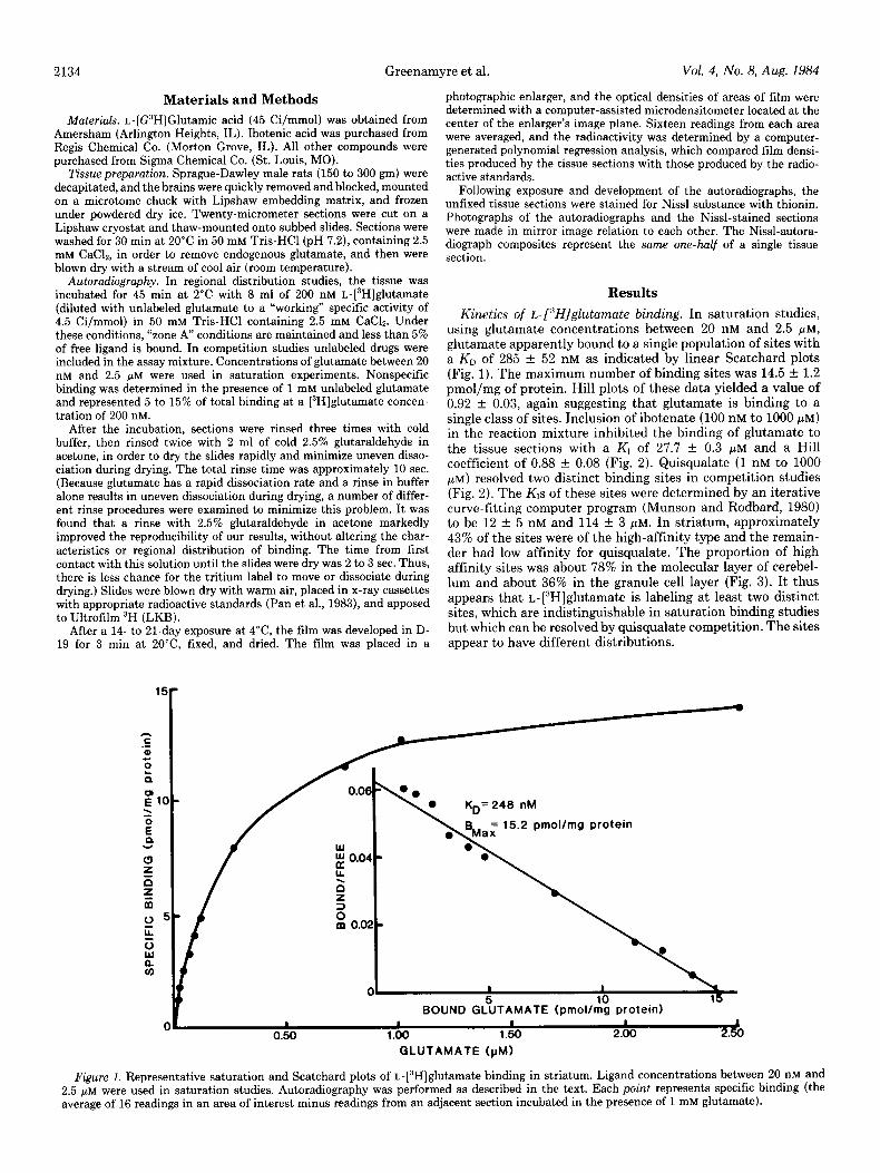
Kinetics of t-[“Hjglutamate binding. In saturation studies, using glutamate concentrations between 20 nM and 2.5 PM, glutamate apparently bound to a single population of sites with a KD of 285 f 52 nM as indicated by linear Scatchard plots (Fig. 1). The maximum number of binding sites was 14.5 f 1.2 pmol/mg of protein. Hill plots of these data yielded a value of 0.92 f 0.03, again suggesting that glutamate is binding to a single class of sites. Inclusion of ibotenate (100 nM to 1000 PM) in the reaction mixture inhibited the binding of glutamate to the tissue sections with a Kl of 27.7 f 0.3 FM and a Hill coefficient of 0.88 + 0.08 (Fig. 2). Quisqualate (1 nM to 1000 pM) resolved two distinct binding sites in competition studies (Fig. 2). The KIs of these sites were determined by an iterative curve-fitting computer program (Munson and Rodbard, 1980) to be 12 + 5 nM and 114 f 3 pM. In striatum, approximately 43% of the sites were of the high-affinity type and the remain- der had low affinity for quisqualate. The proportion of high affinity sites was about 78% in the molecular layer of cerebel- lum and about 36% in the granule cell layer (Fig. 3). It thus appears that L-[3H]glutamate is labeling at least two distinct sites, which are indistinguishable in saturation binding studies but which can be resolved by quisqualate competition. The sites appear to have different distributions.
6:
BOUND c?u*h114TE protein) I I I I 4
0.50 1.00 1.60 2.00 2.50
GLUTAMATE ()rM)
Figure 1. Representative saturation and Scatchard plots of L-[3H]glutamate binding in striatum. Ligand concentrations between 20 nM and 2.5 pM were used in saturation studies. Autoradiography was performed as described in the text. Each point represents specific binding (the average of 16 readings in an area of interest minus readings from an adjacent section incubated in the presence of 1 mM glutamate).

The Journal of Neuroscience Autoradiography of Glutamate Binding in Rat Brain 2135
+-GLUTAMATE
I I I I I I 9 8 7 8 5 4 3
- IOQ,~ EOMPETITOR]
Figure 2. Representative plots of inhibition of striatal [3H]glutamate binding by quisqualate (1 nM to 1000 PM), unlabeled glutamate (10 nM to 1000 PM), and ibotenate (100 nM to 1000 PM). Each point represents specific binding. The concentration of [3H]glutamate was 200 nM. Hill coefficients near 1 were calculated for ibotenate and unlabeled glutamate, and inhibition constants (Kr) for these compounds were determined by log-logit regression analysis using the formula: Kis = IC,,/(l + L/Kn). The KIS for quisqualate displacement were determined by an iterative curve-fitting computer program (Munson and Rodbard, 1980).
%R,= 22
Figure 3. Inhibition of [“HIglutamate binding by quisqualate in cerebellar molecular and granule cell layers. Points represent the average specific binding from four determinations (16 readings per determination) in two separate experiments. The concentration of [3H]glutamate was 200 nM. The proportion of sites in the molecular layer having a high affinity for quisqualate (%Rn) was 78. In the granule cell layer %RH was 36.
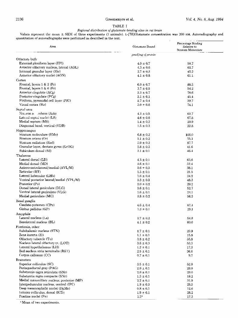
Regional distribution of L-[“H/glutamate-binding sites. The of the absolute amount of bound glutamate and the amount of binding of glutamate to rat brain sections exhibited a marked binding relative to that in stratum moleculare. Representative regional heterogeneity, with the highest levels found in the autoradiographs and Nissl-stained sections are shown in Figure stratum moleculare of the dentate gyrus. The distribution of 4; the composites represent the same side of the same section, glutamate-binding sites is summarized in Table I, both in terms as described under “Materials and Methods.”
2136 Greenamyre et al.
TABLE I
Vol. 4, No. 8, Aug. 1984
Regional distribution of glutamate-binding sites in rat brain
Values represent the mean f SEM of three experiments (3 animals). L-[3H]Glutamate concentration was 200 UM. Autoradiography and quantitation of autoradiographs were performed as described in the text.
Area Glutamate Bound Percentage Binding
Relative to Stratum Moleculare
Olfactory bulb External plexiform layer (EPl) Anterior olfactory nucleus, lateral (AOL)
Internal granular layer (IGr) Anterior olfactory nuclei (AON)
Cortex Frontal, layers 1 & 2 (Fr)
Frontal, layers 5 & 6 (Fr) Anterior cingulate (ACg) Posterior cingulate (PCg)
Piriform, pyramidal cell layer (PfC) Visual cortex (Str)
Septal area Nucleus ac mbens (Acb)
Lateral septai nuclei (LS) Medial septum (MS) Diagnonal band, vertical (VDB)
Hippocampus Stratum moleculare (SMo) Stratum oriens (Or)
Stratum radiatum (Rad) Granular layer, dentate gyrus (GrDG)
Subiculum dorsal (Sd)
Thalamus Lateral dorsal (LD) Medial dorsal (MD) Anteroventrolateral/medial (AVL/M)
Reticular (RT) Lateral habenular (LHb) Ventral posterior lateral/medial (VPL/M)
Posterior (PO) Dorsal lateral geniculate (DLG) Ventral lateral geniculate (VLG)
Medial geniculate (MG)
Basal ganglia Caudate-putamen (CPU) Globus pallidus (GP)
Amygdala
Lateral nucleus (La) Basolateral nucleus (BL)
Forebrain, other Subthalamic nucleus (STN) Zona incerta (ZI)
Olfactory tubercle (Tu) Nucleus lateral olfactory tr. (LOT) Lateral hypothalamus (LH) Bed nucleus stria terminalis (BST)
Corpus callosum (CC)
Brainstem
Superior colliculus (SC) Periaqueductal gray (PAG) Substantia nigra reticulata (SNr)
Substantia nigra compacta (SNc) Medial mammillary nucleus, posterior (MP) Interpeduncular nucleus, central (IPC)
Deep mesencephalic nuclei (DpMe) Inferior colliculus, dorsal (ICD) Pontine nuclei (Pn)
a Mean of two experiments.
4.0 f 0.7 58.7
4.3 f 0.6 62.7
2.7 f 0.3 40.2
4.1 + 0.8 61.1
6.0 + 0.7 88.5
3.7 + 0.5 54.2
5.3 + 0.7 78.6
3.1 + 0.1 45.4
2.7 + 0.4 39.7
5.0 + 0.6 74.1
4.3 + 0.5 63.7
4.6 + 0.6 67.8
1.4 f 0.2 20.9
1.5 f 0.3 22.8
6.8 f 0.2 100.0
5.1 f 0.2 75.1
5.9 + 0.2 87.7
2.8 + 0.3 41.6
3.1 + 0.1 46.4
4.3 f 0.1 63.6
3.6 f 0.1 52.4
2.6 + 0.3 38.1
1.5 f 0.1 21.5
1.6 + 0.4 24.2
3.3 + 0.3 48.3
2.0 + 0.2 29.2
3.6 + 0.1 52.7
1.6 + 0.1 24.1
3.8 + 0.2 56.3
4.6 + 0.4 67.4
1.3 + 0.1 19.1
3.7 + 0.2 54.8
4.1 + 0.2 60.0
1.7 f 0.1 23.9
1.1 + 0.1 15.8
3.8 + 0.2 55.8
3.6 r 0.3 53.3
1.2 + 0.1 17.3
2.5 + 0.1 36.6
0.7 f 0.1 9.7
3.5 + 0.1 52.0
2.0 + 0.1 29.0
2.0 + 0.1 29.0
1.3 f 0.1 19.2
2.2 + 0.1 31.9
1.9 + 0.3 28.5
0.8 f 0.1 12.6
1.9 + 0.1 28.2
1.2” 17.3
The Journal of Neuroscience Autoradiography of Glutamate Binding in Rat Brain
TABLE l-continued
2137
Area
Dorsal cochlear nucleus (DCo) Nucleus solitarius, medial (SolM) Hypoglossal nucleus (12) Inferior olivary nuclei (JO) Spinal nucleus of V, caudal (SP5C)
Cerebellum Molecular layer (Mel) Granule cell layer (Gr)
Spinal cord
Glutamate bound
pmol/mg ofprotein
1.7 f 0.3 1.7 + 0.1 0.7 + 0.1
1.5 + 0.2 1.4 + 0.3
Percentage Binding Relative to
Stratum Moleculare
24.9 25.4 10.6
22.6 21.2
3.3 + 0.2 49.0
2.1 + 0.2 26.3
Substantia gelatinosa, cervical (CSG) 2.6 + 0.4 38.0
Substantia gelatinosa, thoracic (TSG) 2.1 f 0.4 30.4
Substantia gelatinosa, lumbar (LSG) 2.8 f 0.4 40.8 Ventral horn, cervical (CVH) 0.7 f 0.2 10.4 Ventral horn, thoracic (TVH) 0.9 f 0.2 13.2
Ventral horn, lumbar (LVH) 0.8 f 0.2 11.8
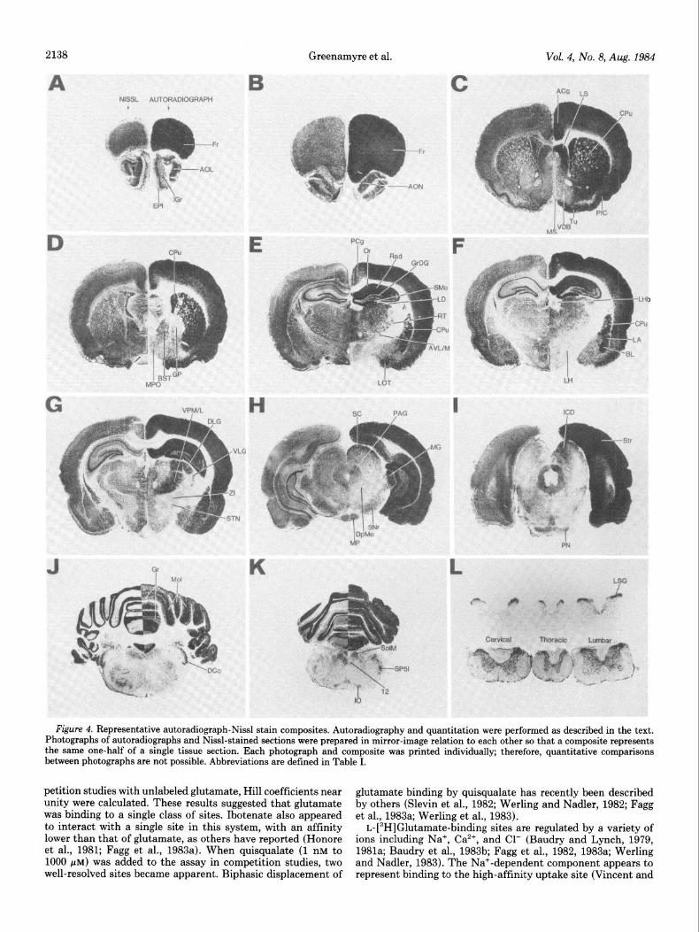
Forebrain. As can be seen in Figures 4 and 5, the amount of glutamate binding varies widely in the forebrain. The olfactory bulb nuclei and molecular layers exhibited relatively high levels of binding, whereas the glomerular layer did not bind glutamate above background levels. Cortical areas differed more than 2- fold in glutamate binding. The highest cortical binding was found in superficial frontal cortex, the lowest in the pyramidal cell layer of piriform cortex. It was a consistent finding that more glutamate bound to superficial layers of cortex than to deep layers, and to anterior cingulate cortex more than to posterior cingulate cortex.
in brainstem than in forebrain. The superficial gray of the superior colliculus had the most glutamate binding of any midbrain structure. There was a differential distribution of sites within the substantia nigra, with approximately 50% more binding in pars reticulata than in pars compacta. Intermediate levels of binding were found in periaqueductal gray matter, the mammillary body, the interpeduncular nucleus, and the inferior colliculus. Very little binding was found in deep mesencephalic nuclei.
The basal ganglia showed marked differences in the levels of bound glutamate, with the caudate-putamen having many more binding sites than the globus pallidus. Similarly, in the septal area there were large differences in glutamate binding in adja- cent structures. The lateral septal nuclei and the nucleus ac- cumbens bound approximately 3 times as much glutamate as the medial septum or vertical limb of the diagonal band.
Of the other brainstem nuclei where binding was measured, the most glutamate was bound in the medial nucleus of the solitary tract, followed closely by the dorsal cochlear nucleus, the inferior olivary nuclei, and the spinal nucleus of the trigem- inal nerve, pars caudalis. Slightly less binding was found in the pontine nuclei and even less glutamate bound to the hypoglossal nucleus. There was very little binding in pontine or medullary tegmentum.
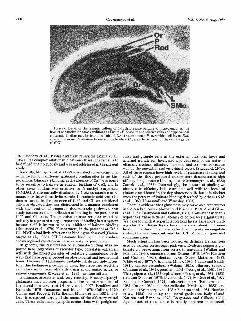
In the hippocampus, glutamate binds in a distinctive laminar pattern (Fig. 6), with highest levels in the stratum moleculare and much less in the granule cell layer and subiculum. Stratum radiatum and stratum oriens both showed a high density of sites. Subnuclei of the amygdala bound different amounts of glutamate, as can be seen in Figure 4F. The lateral and baso- lateral nuclei bind more glutamate than do other amygdaloid nuclei.
The molecular layer of cerebellum bound about one-half the amount of glutamate that was found in stratum moleculare in hippocampus. Forty percent less binding was found in the granule cell layer.
Spinal cord. Relatively high levels of glutamate were bound to substantia gelatinosa of the spinal cord at all levels. Uni- formly little binding was seen in the ventral horn of the spinal cord.
Various thalamic nuclei were readily distinguished by the amount of bound glutamate. The lateral dorsal nucleus bound the most glutamate, and the reticular and lateral habenular nuclei had relatively little glutamate binding. The ventral pos- terior thalamus (lateral and medial portions) stands out in sharp contrast to other posterior thalamic nuclei because of its high density of binding sites (Fig. 4G). Heavy glutamate binding was also found in medial and dorsal lateral geniculate bodies; the ventral lateral geniculate showed much less binding.
Discussion
In other forebrain regions, the subthalamus could be distin- guished from zona incerta by its higher level of binding. Both the olfactory tubercle and nucleus of the lateral olfactory tract had relatively high levels of binding sites. The bed nucleus of the stria terminalis bound an intermediate amount of gluta- mate, and the lowest levels of glutamate binding in the fore- brain were found in the hypothalamus and in the white matter of the corpus callosum.
Brainstem and cerebellum. In general, there was less binding
This report confirms earlier findings that quantitative auto- radiography may be used to examine the pharmacological spec- ificity and regional distribution of glutamate binding in central nervous system tissue sections (Greenamyre et al., 1983; Hal- pain et al., 1983; Monaghan et al., 1983). As previously reported (Greenamyre et al., 1983), the binding of glutamate to tissue sections is saturable and reversible, having a tXh of dissociation of 0.38 min. In the present study, glutamate bound to tissue with an affinity similar to that found in other autoradiographic studies as well as homogenate studies (Roberts and Sharif, 1981; Slevin and Coyle, 1981). Our earlier studies were carried out at 37°C (Greenamyre et al., 1983). Subsequently, it was found that the same pattern of binding was obtained at 2°C. At this temperature, total binding was at least as great as at 37”C, and nonspecific binding was reduced and more uniform. Saturation studies yielded linear Scatchard plots, and in com-
2138 Greenamyre et al. Vol. 4, No. 8, Aug. 1984
Figure 4. Representative autoradiograph-Nissl stain composites. Autoradiography and quantitation were performed as described in the text. Photographs of autoradiographs and Nissl-stained sections were prepared in mirror-image relation to each other so that a composite represents the same one-half of a single tissue section. Each photograph and composite was printed individually; therefore, quantitative comparisons between photographs are not possible. Abbreviations are defined in Table I.
petition studies with unlabeled glutamate, Hill coefficients near unity were calculated. These results suggested that glutamate was binding to a single class of sites. Ibotenate also appeared to interact with a single site in this system, with an affinity lower than that of glutamate, as others have reported (Honore et al., 1981; Fagg et al., 1983a). When quisqualate (1 nM to 1000 pM) was added to the assay in competition studies, two well-resolved sites became apparent. Biphasic displacement of
glutamate binding by quisqualate has recently been described by others (Slevin et al., 1982; Werling and Nadler, 1982; Fagg et al., 1983a; Werling et al., 1983).
L-[3H]Glutamate-binding sites are regulated by a variety of ions including Na+, Ca2’, and Cl- (Baudry and Lynch, 1979, 1981a; Baudry et al., 1983b; Fagg et al., 1982, 1983a; Werling and Nadler, 1983). The Na+-dependent component appears to represent binding to the high-affinity uptake site (Vincent and
The Journal of Neuroscience Autoradiography of Glutamate Binding in Rat Brain
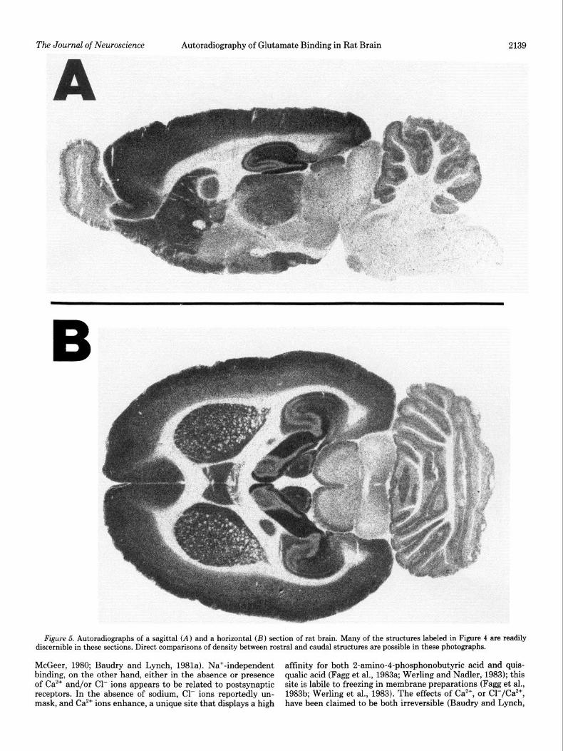
Figure 5. Autoradiographs of a sagittal (A) and a horizontal (B) section of rat brain. Many of the structures labeled in Figure 4 are readily discernible in these sections. Direct comparisons of density between rostra1 and caudal structures are possible in these photographs.
McGeer, 1980; Baudry and Lynch, 1981a). Na+-independent affinity for both 2-amino-4-phosphonobutyric acid and quis- binding, on the other hand, either in the absence or presence qualic acid (Fagg et al., 1983a; Werling and Nadler, 1983); this of Ca2+ and/or Cl- ions appears to be related to postsynaptic site is labile to freezing in membrane preparations (Fagg et al., receptors. In the absence of sodium, Cl- ions reportedly un- 1983b; Werling et al., 1983). The effects of Ca*+, or Cl-/Ca*+, mask, and Ca*+ ions enhance, a unique site that displays a high have been claimed to be both irreversible (Baudry and Lynch,
2140 Greenamyre et al. Vol. 4, No. 8, Aug. 1984
Figure 6. Detail of the laminar pattern of L-[“HIglutamate binding to hippocampus at the level of and under the same conditions as Figure 4F. Absolute and relative values of hippocampal glutamate binding may be found in Table I. Or, stratum oriens; P, pyramidal cell layer; Rad, stratum radiatum: L. stratum lacunosum-moleculare; Gr, granule cell layer of the dentate gyms (GrDG).
1979; Baudry et al., 1983a) and fully reversible (Mena et al., 1982). The complex relationship between these ions remains to be defined unambiguously and was not addressed in the present study.
Recently, Monaghan et al. (1983) described autoradiographic evidence for four different glutamate-binding sites in rat hip- pocampus. Glutamate binding in the absence of Ca2+ was found to be sensitive to kainate in stratum lucidum of CA3, and in other areas binding was sensitive to N-methyl-D-aspartate (NMDA). A site partially displaced by 1 FM quisqualate or LY- amino-3-hydroxy-5-methylisoxazole-4-propionic acid was also demonstrated. In the presence of Ca*+ and Cl- an additional site was observed that was distributed in a manner consistent with the location of proposed glutamatergic pathways. Our study focuses on the distribution of binding in the presence of Ca2+ and Cl- ions. The putative kainate receptor would be unlikely to represent a large proportion of binding in our system because Ca2+ is known to be an inhibitor of kainate binding (Beaumont et al., 1979). Furthermore, in the presence of Ca*+/ Cl-, NMDA had little effect on the binding we observed (Green- amyre et al., 1983). [3H]Glutamate binding, in our studies, shows regional variation in its sensitivity to quisqualate.
In general, the distribution of glutamate-binding sites re- ported here (regardless of receptor type) correlates extremely well with the projection sites of putative glutamatergic path- ways that have been proposed on physiological and biochemical bases. Because [3H]glutamate probably labels multiple recep- tors, this technique provides an assay for structures receiving excitatory input from afferents using acidic amino acids, or related compounds (Zaczek et al., 1983), as transmitters.
Glutamate, aspartate, and, very recently, N-acetylaspartyl- glutamate have all been proposed as the neurotransmitter of the lateral olfactory tract (Harvey et al., 1975; Bradford and Richards, 1976; Yamamoto and Matsui, 1976; Collins, 1979; Collins and Probett, 1981; ffrench-Mullen et al., 1983). This tract is composed largely of the axons of the olfactory mitral cells. These cells make synaptic connections with periglome-
rular and granule cells in the external plexiform layer and internal granule cell layer, and also with cells of the anterior olfactory nucleus, olfactory tubercle, and piriform cortex, as well as the amygdala and entorhinal cortex (Shepherd, 1979). All of these regions have high levels of glutamate binding and each of the three proposed transmitters demonstrates high affinity for glutamate-binding sites (Greenamyre et al., 1983; Zaczek et al., 1983). Interestingly, the pattern of binding we observed in olfactory bulb correlates well with the levels of glutamic acid found in the dog olfactory bulb, but it is distinct from the pattern of kainate binding described by others (Nadi et al., 1980; Unnerstall and Wamsley, 1983).
There is evidence that glutamate may serve as a transmitter in the cerebral cortex (Jaspar and Koyama, 1969; Abdul-Ghani et al., 1981; Baughman and Gilbert, 1981). Consistent with this hypothesis, there is dense labeling of cortex by [3H]glutamate. We have found that superficial cortical layers have more bind- ing sites than deeper layers have. There was about 75% more binding in anterior cingulate cortex than in posterior cingulate cortex; this has been confirmed by D. T. Monaghan (personal communication).
Much attention has been focused on defining transmitters used by various corticofugal pathways. Evidence supports glu- tamatergic projections from cortex to amygdala (Walker and Fonnum, 1983), cuneate nucleus (Stone, 1976, 1979; Rustioni and Cuenod, 1982), dentate gyrus (Storm-Mathisen, 1977; White et al., 1977; Wheal and Miller, 1980; Nadler and Smith, 1981), nucleus accumbens (Walaas, 1981), olfactory tubercle (Fonnum et al., 1981), pontine nuclei (Young et al., 1981, 1983; Thangnipon et al., 1983), spinal cord (Young et al., 1981,1983), striatum (Spencer, 1976; Divac et al., 1977; McGeer et al., 1977; Reubi and Cuenod, 1979), substantia nigra (Fonnum et al., 1981; Carter, 1982), superior colliculus (Kvale et al., 1983), and thalamus (Bromberg et al., 1981; Fonnum et al., 1981; Rustioni et al., 1982), including the lateral geniculate body (Lund- Karlsen and Fonnum, 1978; Baughman and Gilbert, 1981). Again, each of these areas is readily apparent in autoradi-
The Journal of Neuroscience Autoradiography of Glutamate Binding in Rat Brain 2141
ographs of glutamate binding. Equally important is the fact that nearby structures which receive little or no cortical input, such as the globus pallidus, show little glutamate binding. In this regard, it is noteworthy that the dorsal lateral geniculate body, which receives putative glutamatergic innervation from visual cortex, shows a high density of glutamate-binding sites. The ventral lateral geniculate body apparently does not receive glutamatergic cortical input and has little binding. The medial geniculate body shows dense labeling by [“HIglutamate and is known to receive a cortical projection (Shepherd, 1979), but there is not yet evidence to suggest that this input is glutama- tergic. Similarly, the subthalamic nucleus, which receives a neurochemically undefined cortical input (Kunzle, 1978), has more glutamate binding than does the surrounding zona in- certa. Studies which examined the glutamatergic innervation of the substantia nigra (Fonnum et al., 1981; Carter, 1982) did not differentiate pars compacta from pars reticulata. We found higher levels of binding in pars reticulata than in pars com- pacta.
It is widely accepted that glutamate or a similar substance serves as a neurotransmitter of both hippocampal afferents and efferents. The stratum moleculare of the dentate gyrus receives putative glutamatergic input from the entorhinal cortex via the perforant path (Storm-Mathisen, 1977; White et al., 1977; Wheal and Miller, 1980; Nadler and Smith, 1981) and from hippocampal CA4 cells (Cotman and Nadler, 1981; Storm- Mathisen, 1981). In our autoradiographic assay, this structure had the highest level of glutamate-binding sites of any brain region. In keeping with the proposed glutamatergic and/or aspartergic innervation of stratum oriens and stratum radiatum from commissural and Schaffer collateral fibers (Cotman and Nadler, 1981; Storm-Mathisen, 1981; Corradetti et al., 1983), these dendritic zones also show high levels of binding. The CA3 pyramidal cells project bilaterally through the fimbria and fornix to the lateral but not the medial septal nuclei (Swanson and Cowan, 1977). Consistent with the proposal by Zaczek et al. (1979) that this projection is glutamatergic, we found that the lateral septum is selectively and heavily labeled by gluta- mate. Walaas and Fonnum (1980) have proposed that projec- tions from subiculum to the nucleus of the diagonal band, bed nucleus of the stria terminalis, nucleus accumbens, mediobasal hypothalamus, and mammillary body may all use glutamate as a transmitter. All of these areas showed significant glutamate binding, although the levels of these sites were unevenly dis- tributed. Nucleus accumbens was labeled most heavily, followed by the bed nucleus of the stria terminalis, mammillary body, diagonal band nucleus, and hypothalamus. This order, based on density of binding sites, matches that of Walaas and Fon- num (1980) for absolute levels of glutamate uptake in these areas, suggesting a correlation between the density of presyn- aptic terminals (uptake sites) and postsynaptic receptors.
In the brainstem gray matter there was a 5-fold difference between the highest and lowest densities of binding sites. In general, binding was lower than in forebrain. Several brainstem nuclei have already been mentioned with respect to glutama- tergic innervation and glutamate binding and will not be dis- cussed further. It is interesting to note that some nuclei, such as the periaqueductal gray matter, interpeduncular nucleus, inferior colliculus, and the inferior olivary nuclei, show sub- stantial glutamate binding but, to date, lack any defined glu- tamatergic input. However, all of these nuclei, with the possible exception of the interpeduncular nucleus, receive afferents from cortex (Brodal, 1981). It has been suggested that glutamate may serve as the transmitter of certain primary afferents to brainstem nuclei. In particular, there is substantial evidence implicating glutamatergic function in the cochlear nucleus (Godfrey et al., 1977; Potashner, 1983; Altschuler et al., 1984; for a review, see Wenthold, 1981), in the nucleus of the tractus solitarius (Dietrich et al., 1981; Granata and Reis, 1983), and
in the spinal nucleus of the trigeminal nerve, pars caudalis (Salt and Hill, 1980,1982). Subdivisions of the cochlear nucleus were not specifically examined for relative density of glutamate- binding sites, but it was readily apparent that there was sub- stantial binding in the dorsal cochlear nucleus. There was also moderate binding in the nucleus solitarius and the caudal trigeminal nucleus (20 to 25% of the maximal binding found in the stratum moleculare). Of brainstem nuclei measured, the least binding was found in the deep mesencephalic nuclei and the hypoglossal nucleus. (Although there was even less binding in other brainstem regions, we were unable to define these areas anatomically in autoradiographs. Only measurements from pos- itively identified areas were included in this report.)
One of the best characterized putative glutamatergic path- ways is the granule cell/parallel fiber system of the cerebellum (Young et al., 1974; Hudson et al., 1976; Sandoval and Cotman, 1978). Granule cell axons project to the molecular layer of the cerebellum and, here, form the parallel fibers, which make putative glutamatergic synaptic connections with dendritic spines of Purkinje, basket, stellate, and Golgi cells. Further- more, it has been proposed that the quisqualate receptor is the major excitatory amino acid receptor type present in this region (Foster and Roberts, 1981; Crepe1 et al., 1982). Autoradiographs reveal that the molecular layer binds about 60% more glutamate than does the granule cell layer. Approximately 80% of the molecular layer-binding sites are of the high-affinity quisqual- ate type and 20% are the low-affinity type. Binding sites in the granule cell layer may be postsynaptic to climbing fiber termi- nals, which are also believed to use an excitatory amino acid as their transmitter (Rea et al., 1980; Wiklund et al., 1982; Foster and Roberts, 1983; Toggenburger et al., 1983). Alternatively, they may be postsynaptic to mossy fibers, which also may use excitatory amino acids as transmitters (Freeman et al., 1983). It is interesting to note that the granule cell layer apparently displays a pharmacological profile which differs from that in the molecular layer. Less than 40% of the binding sites in this layer have a high affinity for quisqualate.
In the spinal cord, most of the glutamate binding is confined to the substantia gelatinosa. It has been proposed, but remains controversial, that glutamate may serve as a transmitter of some primary afferents in the spinal cord (Graham et al., 1967; Roberts and Keen, 1973, 1974; Zieglgansberger and Puil, 1973; Johnson, 1977). Our findings are consistent with this hypoth- esis, but until further studies are carried out it cannot be stated with any certainty that these sites are postsynaptic to primary afferents. For example, it is known that intrinsic cells of the substantia gelatinosa and of lamina IV to VI are retrogradely labeled after D-[“Hlaspartate injections into cervical dorsal horn (Rustioni and Cuenod, 1982). It is also thought that excitatory amino acids may be neurotransmitters of spinal cord interneurons (Davidoff et al., 1967; Watkins and Evans, 1981; Watkins et al., 1981). We find that there is a significant but relatively low level of binding in the remaining lamina of the spinal cord.
White matter has very low levels of glutamate binding. However, because white matter has a greater self-absorbtion (attenuation) of fl particles than gray matter has (Alexander et al., 1981), we may have underestimated binding in those struc- tures with high white matter content. In areas of pure white matter, this underestimation would be at most 30%. Thus, the value of 0.7 pmol/mg of protein (10% of the binding found in stratum moleculare) obtained for corpus callosum may be as high as 1.0 pmol/mg of protein (15% of the binding found in stratum moleculare).
Autoradiography of L-[3H]glutamate binding provides a sen- sitive assay which can be used to obtain detailed pharmacolog- ical data from highly circumscribed, well defined brain regions. It permits assays that are beyond the resolution of the micro- dissection methods and tissue requirements of conventional
2142 Greenamyre et al. Vol. 4, No. 8, Aug. 1984
binding assays. The results of the present study demonstrate a striking correlation between glutamate-binding sites and the terminal fields of previously proposed glutamatergic pathways. Structures adjacent to those suspected of receiving glutama- tergic input have much less binding. This suggests that auto- radiography may provide an assay for projection areas of path- ways that use excitatory amino acids as neurotransmitters. This technique should help to better define the roles of excit- atory amino acids in the central nervous system.
References
Abdul-Ghani, A. S., J. Coutinho-Netto, and H. F. Bradford (1981) In uiuo superfusion methods and the release of glutamate. In Glutamate: Transmitter in the Central Neruous System, P. J. Roberts, J. Storm- Mathisen, and G. A. R. Johnston, eds., pp. 155-204, John Wiley & Sons, New York.
Alexander, G. M., R. J. Schwartzman, R. D. Bell, J. Yu, and A. Renthal (1981) Quantitative measurement of local cerebral metabolic rate for glucose utilizing tritiated 2-deoxyglucose. Brain Res. 223: 59-67.
Altschuler, R. A., R. J. Wenthold, A. M. Schwartz, W. J. Haser, N. P. Cruthoys, M. H. Parakkal, and J. Fex (1984) Immunocytochemical localization of glutaminase-like immunoreactivity in the auditory nerve. Brain Res. 291: 173-178.
Balcar, V. J., and G. A. R. Johnston (1972) The structural specificity of the high affinity uptake of L-glutamate and L-aspartate by rat brain slices. J. Neurochem. 19: 2657-2666.
Baudry, M., and G. Lynch (1979) Regulation of glutamate receptors by cations. Nature 282: 748-750.
Baudry, M., and G. Lynch (1981a) Characterization of two [aH]gluta- mate binding sites in rat hippocampal membranes. J. Neurochem. 36: 811-820.
Baudry, M., and G. Lynch (1981b) Hippocampal glutamate receptors. Mol. Cell. Biochem. 38: 5-18.
Baudry, M., K. Kramer, and G. Lynch (1983a) Irreversibility and time course of calcium stimulated [3H]glutamate binding to rat hippocam- pal membranes. Brain Res. 270: 142-145.
Baudry, M., R. Siman, E. K. Smith, and G. Lynch (198313) Regulation by calcium ions of glutamate receptor binding in hippocampal slices. Eur. J. Pharmacol. 90: 161-168.
Baughman, R. W., and C. D. Gilbert (1981) Aspartate and glutamate as possible neurotransmitters in the visual cortex. J. Neurosci. I: 427-439.
Beaumont, K., Y. Maurin, T. D. Reisine, J. Z. Fields, E. Spokes, E. D. Bird, and H. I. Yamamura (1979) Huntington’s disease and its model: Alterations in kainate binding. Life Sci. 24: 809-816.
Biziere, K., H. Thompson, and J. T. Coyle (1980) Characterization of specific, high-affinity binding sites for L-[3H]glutamic acid in rat brain membranes. Brain Res. 183: 421-433.
Bradford, H. F., and C. D. Richards (1976) Specific release of endoge- nous glutamate from pirifom cortex stimulated in uitro. Brain Res. 105: 168-172. -
Brodal, A. (1981) Neurological Anatomy in Relation to Clinical Medicine, Oxford Universitv Press. New York.
Bromberg, M. B., J. B. Penney, B. S. Stephenson, and A. B. Young (1981) Evidence for glutamate as the neurotransmitter of corticoth- alamic and corticorubral pathways. Brain Res. 215: 369-374.
Carter, C. J. (1982) Topographical distribution of possible glutama- tergic pathways from the frontal cortex to the striatum and substan- tia nigra in rats. Neuropharmacology 21: 379-383.
Collins, G. G. S. (1979) Effect of chronic bulbectomy on depth distri- bution of amino acid transmitter candidates in rat olfactory cortex. Brain Res. 171: 552-555.
Collins, G. G. S., and G. A. Probett (1981) Aspartate and not glutamate
Cotman, C. W., A. C. Foster, and T. Lanthorn (1981) An overview of glutamate as a neurotransmitter. Adv. Biochem. Psychopharmacol. 27: l-27.
Crepel, F. S., S. S. Dhanjal, and T. A. Sears (1982) Effect of glutamate, aspartate and related derivatives on cerebellar Purkinje cell dendrites in the rat: An in vitro study. J. Physiol. (Lond.) 329: 297-317.
Davidoff, R. A., L. T. Graham, R. P. Shank, R. Werman, and M. H. Aprison (1967) Changes in amino acid concentrations associated with loss of spinal interneurons. J. Neurochem. 14: 191-194.
Davies. J.. R. H. Evans. A. A. Francis. and J. C. Watkins (1979) Excitatory amino acid receptors and synaptic excitation in the mammalian central nervous system. J. Physibl. (Paris) 75: 641-644.
Dietrich, W. D., 0. H. Lowry, and A. D. Loewy (1981) The distribution of glutamate, GABA and aspartate in the nucleus tractus solitarius of the cat. Brain Res. 237: 254-260.
Divac. I.. F. Fonnum. and J. Storm-Mathisen (1977) High affinitv Y
uptake of glutamate in terminals of corticostriatal axons. Nature 266: 377-378.
Fagg, G. E., and A. C. Foster (1983) Amino acid neurotransmitters and their pathways in the mammalian central nervous system. Neuro- science 9: 701-719.
Fagg, G. E., A. C. Foster, E. E. Mena, and C. W. Cotman (1982) Chloride and calcium ions reveal a pharmacologically distinct pop- ulation of L-glutamate binding sites in synaptic membranes: Corre- spondence between biochemical and electrophysiological data. J. Neurosci. 2: 958-965.
Fagg, G. E., A. C. Foster, E. E. Mena, and C. W. Cotman (1983a) Chloride and calcium ions separate L-glutamate receptor populations in synaptic membranes. Eur. J. Pharmacol. 88: 105-110.
Fagg, G. E., E. E. Mena, D. T. Monaghan, and C. W. Cotman (1983b) Freezing eliminates a specific population of L-glutamate receptors in synaptic membranes. Neurosci. Lett. 38: 157-162.
ffrench-Mullen, M. H., R. Zaczek, K. J. Koller, J. T. Coyle, and D. 0. Carpenter (1983) Action of N-acetylaspartylglutamate on mamma- lian neurons. Sot. Neurosci. Abstr. 9: 444.
Fonnum, F., A. Soreide, I. Kvale, J. Walker, and I. Walaas (1981) Glutamate in cortical fibers. Adv. Biochem. Psychopharmacol. 27: 29-42.
Foster, A. C., and P. J. Roberts (1978) High affinity L-[3H]glutamate binding to postsynaptic receptor sites on rat cerebellar membranes. J. Neurochem. 31: 1467-1477.
Foster, G. A., and P. J. Roberts (1981) Stimulation of rat cerebellar guanosine 3’,5’-cyclic monophosphate (cyclic GMP) levels: Effects of amino acid antagonists. Br. J. Pharmacol. 74: 723-729.
Foster, G. A., and P. J. Roberts (1983) Neurochemical and pharmaco- logical correlates of inferior olive destruction in the rat: Attenuation of the events mediated by an endogenous glutamate-like substance. Neuroscience 8: 277-284.
Foster, A. C., E. E. Mena, G. E. Fagg, and C. W. Cotman (1981) Glutamate and aspartate binding sites are enriched in synaptic junctions isolated from rat brain. J. Neurosci. 1: 620-625.
Freeman, M. E., J. D. Lane, and J. E. Smith (1983) Turnover rates of amino acid neurotransmitters in regions of rat cerebellum. J. Neu- rochem. 40: 1441-1447.
Godfrey, A. A., J. A. Carter, S. J. Berger, 0. H. Lowry, and F. M. Matschinsky (1977) Quantitative histochemical mapping of candi- date transmitter amino acids in cat cochlear nucleus. J. Histochem. Cytochem. 25: 417-431.
Graham, L. T., R. P. Shank, R. Werman, and M. H. Aprison (1967) Distribution of some synaptic transmitter suspects in cat spinal cord: Glutamic acid, aspartic acid, y-aminobutyric acid, glycine and glu- tamine. J. Neurochem. 14: 465-472.
Granata, A. R., and D. J. Reis (1983) Release of [3H]L-glutamic acid (L-Glu) and [3H]D-aspartic acid (D-ASP) in the area of nucleus tractus solitarius in uiuo produced by stimulation of the vagus nerve. Brain
is the likely transmitter of the rat lateral olfactory tract fibers. Brain Res. 259: 77-93. Res. 209: 231-234. Greenamvre. J. T., A. B. Young. and J. B. Pennev (1983) Quantitative “_ -. _
Corradetti, R., G. Moneti, F. Moroni, G. Pepeu, and A. Wieraszko autoradiography of L-[3H]glutamate binding to rat brain. Neurosci. (1983) Electrical stimulation of the stratum radiatum increases the Lett. 37: 155-160. release and neosynthesis of aspartate, glutamate and y-aminobutyric Halpain, S., B. Parsons, and T. C. Rainbow (1983) Tritium-film auto- acid in rat hippocampal slices. J. Neurochem. 41: 1518-1525. radiography of sodium-independent glutamate binding sites in rat
Cotman, C. W.; and J. V. Nadler (1981) Glutamate and aspartate as brainy Eur. J. Pharmacol. S& 313-314. hippocampal transmitters: Biochemical and pharmacological evi- Harvey, J. A., C. N. Scholfield, L. T. Graham, and M. H. Aprison dence. In Glutamate: Transmitter in the Central Neruous System, P. (1975) Putative transmitters in denervated olfactory cortex. J. Neu- J. Roberts, J. Storm-Mathisen, and G. A. R. Johnston, eds., pp. 117- rochem. 24: 445-449. 154, John Wiley & Sons, New York. Head, R. A., G. Tunnicliff, and G. K. Matheson (1980) Glutamate
The Journal of Neuroscience Autoradiography of Glutan
receptor binding to cat central nervous system membranes. Can. J. Biochem. 58: 534-538.
Honore, T., J. Lauridsen, and P. Krogsgaard-Larsen (1981) Ibotenic acid analogues as inhibitors of [3H]glutamic acid binding to cerebel- lar membranes. J. Neurochem. 36: 1302-1304.
Hudson, D. B., T. Valcana, G. Bean, and P. S. Timiras (1976) Glutamic acid, a strong candidate as the neurotransmitter of the cerebellar granule cell. Neurochem. Res. 1: 73-81.
Jaspar, H. H., and I. Koyama (1969) Rate of release of amino acids from the cerebral cortex in the cat as affected by brainstem and thalamic stimulation. Can. J. Phvsiol. Pharmacol. 47: 889-905.
Johnson, J. L. (1977) Glutamic acidas a synaptic transmitter candidate in the dorsal sensory neuron: Reconsiderations. Life Sci. 20: 1637- 1644.
Kuhar, M. J., and S. H. Snyder (1970) The subcellular distribution of free ‘H-glutamic acid in rat cerebral cortical slices. J. Pharmacol. Exp. Ther. 171: 141-152.
Kunzle, H. (1978) An autoradiographic analysis of the efferent connec- tions from premotor and adjacent regions (areas 6 and 9) in Macaca jaxicularis. Brain Behav. Evol. 15: 185-234.
Kvale, I., V. M. Fosse, and F. Fonnum (1983) Development of neuro- transmitter parameters in lateral geniculate body, superior colliculus and visual cortex of the albino rat. Dev. Brain Res. 7: 137-145.
Lund-Karlsen, R., and F. Fonnum (1978) Evidence for glutamate as a neurotransmitter in the corticofugal pathways to the lateral genicu- late body and the superior colliculus in rats. Brain Res. 151: 457- 467.
McGeer, P. L., E. G. McGeer, U. Scherer, and K. Singh (1977) A glutamatergic corticostriatal path? Brain Res. 128: 369-373.
McLennan. H. (1981) On the nature of the receptors for various excitatory amino acids in the mammalian central nervous system. Adv. Biochem. Psychopharmacol. 27: 253-262.
Mena, E. E., G. E. Fagg, D. T. Monaghan, and C. W. Cotman (1982) Acidic amino acid receptor populations in synaptic membranes: Regulation by Cl- and Ca’+ ions. Sot. Neurosci. Abstr. 8: 878.
Michaelis. E. K.. M. L. Michaelis, and L. L. Bovarskv (1974) High- affinity glutamic acid binding to rat brain synaptic membranes. Biochim. Biophys. Acta 367: 338-348.
Michaelis, E. K., M. L. Michaelis, H. H. Chang, R. D. Grubbs, and D. R. Kuonen (1981) Molecular characteristics of glutamate receptors in the mammalian brain. Mol. Cell. Biochem. 38: 163-179.
Monaghan, D. T., V. R. Holets, D. W. Toy, and C. W. Cotman (1983) Anatomical distributions of four pharmacologically distinct 3H-~- glutamate binding sites. Nature 306: 176-179. -
Munson. P. J.. and D. Rodbard (1980) Lieand: A versatile computerized approach for characterization of ligand-binding systems. Anal. Biochem. 107: 220-239.
Nadi, N. S., J. D. Hirsch, and F. L. Margolis (1980) Laminar distribu- tion of putative neurotransmitter amino acids and ligand binding sites in the dog olfactory bulb. J. Neurochem. 34: 138-i46.
Nadler. J. V.. and E. M. Smith (1981) Perforant path lesion depletes glutamate content of fascia dentata synaptosomes. Neurosci.’ Lett. 25: 275-280.
Pan, H. S., K. A. Frey, A. B. Young, and J. B. Penney (1983) Changes in [3H]muscimol binding in substantia nigra, entopeduncular nu- cleus, globus pallidus, and thalamus after striatal lesions as demon- strated by quantitative receptor autoradiography. J. Neurosci. 3: 118991198.
Pearce, B. R., and G. R. Dutton (1981) K+-stimulated release of endogenous glutamate, GABA and other amino acids from neuron- and glia-enriched cultures of the rat cerebellum. FEBS Lett. 135: 215-218.
Potashner, S. J. (1978) Effects of tetrodotoxin, calcium and magnesium on the release of amino acids from slices of guinea pig cerebral cortex. J. Neurochem. 31: 187-195.
Potashner, S. J. (1983) Uptake and release of D-aspartate in the guinea pig cochlear nucleus. J. Neurochem. 41: 1094-1101.
Rea, M. A., W. J. McBride, and B. H. Brode (1980) Regional and synaptosomal levels of amino acid neurotransmitters in the 3-ace- tylpyridine deafferentated rat cerebellum. J. Neurochem. 34: 1106- 1108.
Reubi, J. C., and M. Cuenod (1979) Glutamate release in. vitro from corticostriatal terminals. Brain Res. 176: 185-188.
Roberts, P. J. (1974) Glutamate receptors in rat central nervous system. Nature 252: 399-401.
Roberts, P. J., and P. Keen (1973) Uptake of [‘%]glutamate into dorsal
nate Binding in Rat Brain 2143
and ventral roots of spinal nerves of the rat. Brain Res. 57; 234-238. Roberts, P. J., and P. Keen (1974) Effects of dorsal root section on
amino acids of rat spinal cord. Brain Res. 74: 333-337. Roberts, P. J., and N. A. Sharif (1981) Radioreceptor binding studies
with glutamate and aspartate. Adv. Biochem. Psychopharmacol. 27; 295-306.
Roberts, P. J., G. J. McBean, N. A. Sharif, and E. M. Thomas (1982) Striatal glutamatergic function: Modifications following specific le- sions. Brain Res. 235: 83-91.
Rustioni, A., and M. Cuenod (1982) Selective retrograde transport of D-aspartate in spinal interneurons and cortical neurons of rats. Brain Res. 236: 143-155.
Rustioni, A., R. Spreafico, S. Cheema, and M. Cuenod (1982) Selective retrograde labelling of somatosensory cortical neurons after 3H-~- aspartate injections in the ventrobasal complex of rats and cats. Neuroscience (Suppl.) 7: S183.
Salt, T. E., and R. G. Hill (1980) Excitatory amino acids as transmitter candidates of vibrissae afferent fibres to the rat trigeminal nucleus caudalis. Neurosci. Lett. 22: 183-187.
Salt, T. E., and R. G. Hill (1982) Differentiation of excitatory amino acid receptors in the rat caudal trigeminal nucleus: A microionto- phoretic study. Neuropharmacology 21: 385-390.
Sandoval, M. E., and C. W. Cotman (1978) Evaluation of glutamate as a neurotransmitter of cerebellar parallel fibers. Neuroscience 3: 199- 206.
Shepherd, G. M. (1979) The Synaptic Organization of the Bruin, Oxford University Press, New York.
Slevin, J. T., and J. T. Coyle (1981) Ontogeny of receptor binding sites for [3H]glutamic acid and [3H]kainic acid in the rat cerebellum. J. Neurochem. 37: 531-533.
Slevin, J., J. Collins, K. Lindsley, and J. T. Coyle (1982) Specific binding of [3H]glutamate to cerebellar membranes: Evidence for recognition site heterogeneity. Brain Res. 249: 353-360.
Spencer, H. J. (1976) Antagonism of cortical excitation of striatal neurons by glutamic acid diethyl ester: Evidence for glutamic acid as an excitatory transmitter in the rat striatum. Brain Res. 102: 91- 101.
Stone, T. W. (1976) Blockade by amino acid antagonists of neuronal excitation mediated by the pyramidal tract. J. Physiol. (Lond.) 257: 187-198.
Stone, T. W. (1979) Amino acids as neurotransmitters of corticofugal neurones in the rat: A comparison of glutamate and aspartate. Br. J. Pharmacol. 67: 545-551.
Storm-Mathisen, J. (1977) Glutamic acid and excitatory nerve endings: Reduction of glutamic acid uptake after axotomy. Brain Res. 120: 379-386.
Storm-Mathisen, J. (1981) Glutamate in hippocampal pathways. Adv. Biochem. Psychopharmacol. 27: 43-55.
Storm-Mathisen, J., A. K. Leknes, A. T. Bore, J. L. Vaaland, P. Edminson, F. S. Haug, and 0. P. Ottersen (1983) First visualization of glutamate and GABA in neurons by immunocytochemistry. Na- ture 301: 517-520.
Swanson, L. W., and W. M. Cowan (1977) An autoradiographic study of the organization of the efferent connections of the hippocampal formation in the rat. J. Comp. Neurol. 172: 49-84.
Thangnipon, W., T. Taxt, P. Brodal, and J. Storm-Mathisen (1983) The corticopontine projection: Axotomy-induced loss of high-affinity L-glutamate and D-aspartate uptake, but not of GABA uptake, glutamate decarboxylase or choline acetyltransferase, in the pontine nuclei. Neuroscience 8: 449-458.
Toggenburger, G., L. Wiklund, H. Henke, and M. Cuenod (1983) Release of endogenous and accumulated exogenous amino acids from slices of normal and climbing fiber-deprived rat cerebellar slices. J. Neurochem. 41: 1606-1613.
Unnerstall, J. R., and J. K. Wamsley (1983) Autoradiographic locali- zation of high-affinity [3H]kainic acid binding sites in the rat fore- brain. Eur. J. Pharmacol. 86: 361-371.
Vincent, S. R., and E. G. McGeer (1980) A comparison of sodium- dependent glutamate binding with high-affinity glutamate uptake in rat striatum. Brain Res. 184: 99-108.
Walaas, I. (1981) Biochemical evidence for overlapping neocortical and allocortical glutamate projections to the nucleus accumbens and rostra1 caudatoputamen in the rat brain. Neuroscience 6: 399-405.
Walaas, I., and F. Fonnum (1980) Biochemical evidence for glutamate as a transmitter in hippocampal efferents to the basal forebrain and hypothalamus in the rat brain. Neuroscience 5: 1691-1698.
2144 Greenamyre et al. Vol. 4, No. 8, Aug. 1984
Walker, J. E., and F. Fonnum (1983) Regional cortical glutamatergic Wiklund, L., G. Toggenburger, and M. Cuenod (1982) Aspartate: Pos- and aspartergic projections to the amygdala and thalamus of the rat. sible neurotransmitter in cerebellar climbing fibers. Science 216: 78- Brain Res. 267: 371-374. 80.
Watkins, J. C. (1978) Excitatory amino acids. In Kuinic Acid as a Tool in Neurobiology, E. G. McGeer, P. L. McGeer, and J. W. Olney, eds., pp. 37-69, Raven Press, New York.
Watkins, J. C., and R. H. Evans (1981) Excitatory amino acid neuro- transmitters. Annu. Rev. Pharmacol. Toxicol. 21: 165-204.
Watkins, J. C., J. Davies, R. H. Evans, A. A. Francis, and A. W. Jones
Wofsey, A. R., M. J. Kuhar, and S. H. Snyder (1971) A unique svnaptosomal fraction, which accumulates glutamic and aspartic acids, in brain tissue. Proc. Natl. Acad. Sci. U. S. A. 68: 1102-1106.
Yamamoto. C.. and S. Matsui (1976) Effect of stimulation of excitatory I I
nerve tract on release of glutamic acid from olfactory cortex slices in uiuo. J. Neurochem. 26:-487-491.
Young, A. B., M. L. Oster-Granite, R. M. Herndon, and S. H. Snyder (1974) Glutamic acid: Selective depletion by viral-induced granule cell loss in hamster cerebellum. Brain Res. 73: l-13.
Young, A. B., M. B. Bromberg, and J. B. Penney (1981) Decreased glutamate uptake in subcortical areas deafferented by sensorimotor cortical ablation in the cat. J. Neurosci. 1: 241-249.
Young, A. B., J. B. Penney, G. W. Dauth, M. B. Bromberg, and S. Gilman (1983) Glutamate or aspartate as a possible neurotransmitter of cerebral corticofugal fibers in the monkey. Neurology 33: 1513- 1516.
(1981) Pharmacology of receptors for excitatory amino acids. Adv. Biochem. Psvchonharmacol. 27: 263-273.
Wenthold, R. J. (1981) Glutamate and aspartate as neurotransmitters for the auditory nerve. Adv. Biochem. Psychopharmacol. 27: 69-78.
Werling, L. L., and J. V. Nadler (1982) Complex binding of L-[~H] glutamate to hippocampal synaptic membranes in the absence of sodium. J. Neurbchem. 38: 1050-1062.
Werling, L. L., and J. V. Nadler (1983) Multiple binding sites for L- [3H]glutamate on hippocampal synaptic membranes: Effects of cal- cium and chloride ions. Sot. Neurosci. Abstr. 9: 1186.
Werlinz. L. L.. K. A. Doman. and J. V. Nadler (1983) L-13H1Glutamate bind:ng to ‘hippocampal synaptic membranes: Two- binding sites discriminated by their differing affinities for quisqualate. J. Neuro- them. 41: 586-593.
Wheal, H. V., and J. J. Miller (1980) Pharmacological identification of acetylcholine and glutamate excitatory systems in the dentate gyrus of the rat. Brain Res. 182: 145-155.
White, W. F., J. V. Nadler, A. Hamberger, C. W. Cotman, and J. T. Cummins (1977) Glutamate as a transmitter of the hippocampal perforant path. Nature 270: 356-357.
Zaczek, R., J. C. Hedreen, and J. T. Coyle (1979) Evidence for a hippocampal-septal glutamatergic pathway in the rat. Exp. Neurol. 65: 145-156.
Zaczek, R., K. Koller, R. Cotter, D. Heller, and J. T. Coyle (1983) N- Acetylaspartylglutamate: An endogenous peptide with high affinity for a brain “glutamate” receptor. Proc. Natl. Acad. Sci. U. S. A. 60: 1116-1119.
Zieglgansberger, W., and E. A. Puil (1973) Actions of glutamic acid on spinal neurons. Exp. Brain Res. 17: 35-49.
Copyright © 2022 FDOKUMEN


![Autoradiographic visualisation of [ 3H]5-carboxamidotryptamine binding sites in the guinea pig and rat brain](https://static.fdokumen.com/doc/165x107/631db97fb5acdf8d60026115/autoradiographic-visualisation-of-3h5-carboxamidotryptamine-binding-sites-in.jpg)