
Proteomic analysis of the soluble fraction from human corneal fibroblasts with reference to ocular...
15
Proteomic Analysis of the Soluble Fraction from Human Corneal Fibroblasts with Reference to Ocular Transparency* Henrik Karring‡§, Ida B. Thøgersen§, Gordon K. Klintworth¶, Jan J. Enghild§, and Torben Møller-Pedersen‡ The transparent corneal stroma contains a population of corneal fibroblasts termed keratocytes, which are inter- spersed between the collagen lamellae. Under normal conditions, the keratocytes are quiescent and transpar- ent. However, after corneal injury the keratocytes become activated and transform into backscattering wound-heal- ing fibroblasts resulting in corneal opacification. At pres- ent, the most popular hypothesis suggests that particular abundant water-soluble proteins called enzyme-crystal- lins are involved in maintaining corneal cellular transpar- ency. Specifically, corneal haze development is thought to be related to low levels of cytoplasmic enzyme-crystallins in reflective corneal fibroblasts. To further investigate this hypothesis, we have used a proteomic approach to iden- tify the most abundant water-soluble proteins in serum- cultured human corneal fibroblasts that represent an in vitro model of the reflective wound-healing keratocyte phenotype. Densitometry of one-dimensional gels re- vealed that no single protein isoform exceeded 5% of the total water-soluble protein fraction, which is the qualifying property of a corneal enzyme-crystallin according to the current definition. This result indicates that wound-heal- ing corneal fibroblasts do not contain enzyme-crystallins. A total of 254 protein identifications from two-dimensional gels were performed representing 118 distinct proteins. Proteins protecting against oxidative stress and protein misfolding were prominent, suggesting that these pro- cesses may participate in the generation of cytoplasmic light-scattering from corneal fibroblasts. Molecular & Cellular Proteomics 3:660 – 674, 2004. The cornea, the only transparent connective tissue of the body, is responsible for 70% of the total refractive power of visible light in the eye. The corneal stroma (thickness 450 m) mainly consists of collagen lamellae formed by uniform fibrils composed of type I, III, and V collagen (1, 2). The uniform diameter (30 nm) and regular spacing of the colla- gen fibrils (60 nm between the centers) lead to destructive interference of light except in the forward direction and thus optical transparency of the extracellular matrix of the cornea (3, 4). The stroma also contains multiple layers of quiescent corneal fibroblasts, termed keratocytes, which are inter- spersed between the collagen lamellae. Under normal condi- tions, the quiescent keratocytes are transparent except for the nuclei when studied in vivo using scanning slit specular mi- croscopy (5), slit-lamp biomicroscopy, or in vivo confocal microscopy (6). This stealth-like invisibility indicates that the refractive index of the keratocyte cytoplasm and cellular pro- cesses is similar to that of the extracellular matrix of the corneal stroma (6). The keratocytes have a dynamic potential for proliferation and transformation. Thus, after injury of the cornea such as excimer laser keratectomy for the treatment of myopia, the keratocytes transform into spindle-shaped fibroblastic cells (7, 8). The corneal fibroblasts repopulate the damaged region by migration and proliferation and deposit disordered and nontransparent matrix components (9 –11). The damaged stroma is then remodelled through continuous degradation and synthesis of the extracellular matrix to recreate the highly ordered collagen structure (11, 12). In the latest phase of the wound-healing process, the corneal fibroblasts undergo a phenotype change into myofibroblasts, which are responsible for wound contraction (13–15). The wound-healing process reduces the optical clarity of the cornea due to an increase in the light-scattering from the cell bodies of the corneal fibro- blasts compared with that of keratocytes (7, 8, 16, 17). These results suggest that development of corneal cellular haze during wound-healing is caused in part by an increase in the backscatter of light from the cytoplasm of the stromal cells. The maintenance of ocular transparency and the develop- ment of opacities have been studied intensively in the ocular lens. The - and /-crystallins are the major water-soluble proteins of all vertebrate ocular lenses and are critical for the optical properties and transparency of this tissue. The -crys- tallins are members of the small heat shock protein family and have chaperone-like activity preventing the aggregation of proteins (18, 19). The /-crystallins are related to microbial From the ‡Department of Ophthalmology, Aarhus University Hos- pital, Nørrebrogade 44, 8000 Aarhus C, Denmark; the §Department of Molecular Biology, Science Park, University of Aarhus, Gustav Wieds Vej 10c, 8000 Aarhus C, Denmark; and the ¶Departments of Pathol- ogy and Ophthalmology, Duke University Medical Center, Durham, NC 27710 Received, January 31, 2004, and in revised form, March 30, 2004 Published, MCP Papers in Press, March 30, 2004, DOI 10.1074/mcp.M400016-MCP200 Research © 2004 by The American Society for Biochemistry and Molecular Biology, Inc. 660 Molecular & Cellular Proteomics 3.7 This paper is available on line at http://www.mcponline.org
Transcript of Proteomic analysis of the soluble fraction from human corneal fibroblasts with reference to ocular...
Proteomic Analysis of the Soluble Fractionfrom Human Corneal Fibroblasts withReference to Ocular Transparency*Henrik Karring‡§, Ida B. Thøgersen§, Gordon K. Klintworth¶, Jan J. Enghild§,and Torben Møller-Pedersen‡�
The transparent corneal stroma contains a population ofcorneal fibroblasts termed keratocytes, which are inter-spersed between the collagen lamellae. Under normalconditions, the keratocytes are quiescent and transpar-ent. However, after corneal injury the keratocytes becomeactivated and transform into backscattering wound-heal-ing fibroblasts resulting in corneal opacification. At pres-ent, the most popular hypothesis suggests that particularabundant water-soluble proteins called enzyme-crystal-lins are involved in maintaining corneal cellular transpar-ency. Specifically, corneal haze development is thought tobe related to low levels of cytoplasmic enzyme-crystallinsin reflective corneal fibroblasts. To further investigate thishypothesis, we have used a proteomic approach to iden-tify the most abundant water-soluble proteins in serum-cultured human corneal fibroblasts that represent an invitro model of the reflective wound-healing keratocytephenotype. Densitometry of one-dimensional gels re-vealed that no single protein isoform exceeded 5% of thetotal water-soluble protein fraction, which is the qualifyingproperty of a corneal enzyme-crystallin according to thecurrent definition. This result indicates that wound-heal-ing corneal fibroblasts do not contain enzyme-crystallins.A total of 254 protein identifications from two-dimensionalgels were performed representing 118 distinct proteins.Proteins protecting against oxidative stress and proteinmisfolding were prominent, suggesting that these pro-cesses may participate in the generation of cytoplasmiclight-scattering from corneal fibroblasts. Molecular &Cellular Proteomics 3:660–674, 2004.
The cornea, the only transparent connective tissue of thebody, is responsible for �70% of the total refractive power ofvisible light in the eye. The corneal stroma (thickness �450�m) mainly consists of collagen lamellae formed by uniform
fibrils composed of type I, III, and V collagen (1, 2). Theuniform diameter (�30 nm) and regular spacing of the colla-gen fibrils (�60 nm between the centers) lead to destructiveinterference of light except in the forward direction and thusoptical transparency of the extracellular matrix of the cornea(3, 4). The stroma also contains multiple layers of quiescentcorneal fibroblasts, termed keratocytes, which are inter-spersed between the collagen lamellae. Under normal condi-tions, the quiescent keratocytes are transparent except for thenuclei when studied in vivo using scanning slit specular mi-croscopy (5), slit-lamp biomicroscopy, or in vivo confocalmicroscopy (6). This stealth-like invisibility indicates that therefractive index of the keratocyte cytoplasm and cellular pro-cesses is similar to that of the extracellular matrix of thecorneal stroma (6).
The keratocytes have a dynamic potential for proliferationand transformation. Thus, after injury of the cornea such asexcimer laser keratectomy for the treatment of myopia, thekeratocytes transform into spindle-shaped fibroblastic cells(7, 8). The corneal fibroblasts repopulate the damaged regionby migration and proliferation and deposit disordered andnontransparent matrix components (9–11). The damagedstroma is then remodelled through continuous degradationand synthesis of the extracellular matrix to recreate the highlyordered collagen structure (11, 12). In the latest phase of thewound-healing process, the corneal fibroblasts undergo aphenotype change into myofibroblasts, which are responsiblefor wound contraction (13–15). The wound-healing processreduces the optical clarity of the cornea due to an increase inthe light-scattering from the cell bodies of the corneal fibro-blasts compared with that of keratocytes (7, 8, 16, 17). Theseresults suggest that development of corneal cellular hazeduring wound-healing is caused in part by an increase in thebackscatter of light from the cytoplasm of the stromal cells.
The maintenance of ocular transparency and the develop-ment of opacities have been studied intensively in the ocularlens. The �- and �/�-crystallins are the major water-solubleproteins of all vertebrate ocular lenses and are critical for theoptical properties and transparency of this tissue. The �-crys-tallins are members of the small heat shock protein family andhave chaperone-like activity preventing the aggregation ofproteins (18, 19). The �/�-crystallins are related to microbial
From the ‡Department of Ophthalmology, Aarhus University Hos-pital, Nørrebrogade 44, 8000 Aarhus C, Denmark; the §Department ofMolecular Biology, Science Park, University of Aarhus, Gustav WiedsVej 10c, 8000 Aarhus C, Denmark; and the ¶Departments of Pathol-ogy and Ophthalmology, Duke University Medical Center, Durham,NC 27710
Received, January 31, 2004, and in revised form, March 30, 2004Published, MCP Papers in Press, March 30, 2004, DOI
10.1074/mcp.M400016-MCP200
Research
© 2004 by The American Society for Biochemistry and Molecular Biology, Inc.660 Molecular & Cellular Proteomics 3.7This paper is available on line at http://www.mcponline.org
oxidative stress proteins (20) and have been implicated in thedevelopment of cataracts (21). The high concentration andoligomeric complex formations of the �- and �/�-crystallinsare believed to give the ocular lens its refractive index andoptical properties through short-range spatial orders (22).
Several studies of various vertebrates and invertebrateshave shown taxon-specific accumulations of water-solubleproteins in the lens and epithelial cells of the cornea. Theabundant taxon-specific proteins in the lens and cornea areoften similar or identical to metabolic enzymes and, therefore,termed enzyme-crystallins. Most mammals accumulate alde-hyde dehydrogenase 3 (ALDH3)1 in the corneal epithelium(23–26). Transketolase (TKT) is a major protein in the mouseand human corneal epithelium (27), and �-enolase is relativelyabundant in human, mouse, and chicken corneas (26). Isoci-trate dehydrogenase is overexpressed in the bovine cornealepithelium (28), while peptidyl-prolyl cis-trans isomerase andargininosuccinate lyase are found at high concentrations inthe corneal epithelium of chicken (26). Glutathione S-transfer-ase-related S-crystallin is abundant in squid cornea (26), whilezebrafish accumulates gelsolin and actin in the corneal epi-thelium (29). It has been suggested that proteins present at aconcentration exceeding 5% of the total water-soluble proteincontent of corneal cells should be defined as enzyme-crys-tallins (30). The function of the corneal enzyme-crystallins isnot clear, but it has been suggested that they serve catalyticas well as structural roles reminiscent to the �- and �/�-crystallins in the lens and thereby contribute to the transpar-ency and optical properties of the corneal cells.
TKT and ALDH1 are abundant proteins in rabbit keratocytes(17). These two proteins comprise about 30% of the totalwater-soluble protein content in freshly isolated keratocytesfrom normal rabbit corneas, but are reduced to less than 15%in light-scattering corneal fibroblasts (17). These results indi-cate an apparent correlation between cellular transparencyand the levels of enzyme-crystallins in stromal cells of thecornea. However, ALDH3A1-deficient mice (31) and TKT�/–
(32) mice show no corneal phenotype, indicating that theenzyme-crystallins are not required for corneal transparency.Other studies have indicated that the abundant water-solubleenzyme-crystallins protect the cornea against ultraviolet (UV)-induced oxidative injury. Thus, isocitrate dehydrogenase pre-vents lipid peroxidation and oxidative DNA damage (33), whileALDH3A1 detoxifies the cellular environment by removingtoxic aldehydes generated by lipid peroxidation (34, 35). Inaddition, ALDH3A1 may function as a UV-light filter in thecornea by directly absorbing UV radiation (36). However, de-
spite the various suggestions concerning the roles of theabundant water-soluble proteins in the corneal, it is reasona-ble to believe that maintenance of corneal transparency andthe development of cellular haze are dependent on the proteinexpression profile.
In the present study, the soluble proteome of cultured hu-man corneal fibroblasts was analyzed by one-dimensional(1D) and two-dimensional (2D) PAGE, and 118 of the mostabundant water-soluble proteins were identified by matrix-assisted laser desorption/ionization (MALDI) mass spectrom-etry (MS) or MALDI quadrupole time-of-flight (TOF) tandemmass spectrometry (MS/MS). The identified proteins are in-volved in processes such as protein folding and degradation,cell proliferation, differentiation, and apoptosis, metabolism,cytoskeleton organization and cell motility, protection againstoxidative stress, signal transduction, and secretion of pro-teins. Based on the identified proteins, we propose that oxi-dative stress may lead to protein unfolding that may partici-pate in the enhanced backscatter of light from cornealfibroblasts. Aggregation of irreversible oxidized proteins isknown to opacify the crystalline lens. Thus, it is possible thatdevelopment of corneal cellular haze during wound-healing issomehow reminiscent to the formation of an oxidative stress-induced opacity in the crystalline lens.
EXPERIMENTAL PROCEDURES
Cell Culture—Four human donor corneas (males of 39 and 51 yearsold and females of 66 and 73 years old) were obtained from theDanish Cornea Bank, Aarhus University Hospital, and dissected inHanks’ buffered saline solution (HBSS; Life Technologies, Inc., GrandIsland, NY). After removing the Descemet’s membrane-endothelialcomplex and the epithelium, the central stroma was punched from theendothelial side using a 7-mm trephine. The stromal buttons were cutinto 1-mm3 pieces and washed three times in 1 ml of HBSS beforebeing used for explants in 40-ml cell-culture flasks (Nunc, Roskilde,Denmark).
The corneal fibroblasts were cultured at 37 °C in a 5% CO2-humidified incubator in Dulbecco’s modified Eagle’s medium/NUTMIX F-12 (HAM) medium (Life Technologies, Inc.) containing 10%fetal bovine serum (Life Technologies, Inc.), 100 U/ml penicillin, 100�g/ml streptomycin (Life Technologies, Inc.), and 0.25 �g/ml fungi-zone (amphotericin B) (Life Technologies, Inc.). When the cultures hadmultiplied to confluence, the cells were trypsinized by 0.25% (w/v)trypsin/1 mM EDTA mixture (Life Technologies, Inc.) and transferred toa 250-ml cell-culture flask (first passage). Subsequently, confluentcultures were split in 1:4 and cells from the third passage werewashed in phosphate-buffered saline (PBS) Dulbecco’s (Life Technol-ogies, Inc.) before harvested by scraping using a cell scraper in PBSDulbecco’s. Cells were spun down at 1,000 � g for 10 min at 25 °Cand washed thoroughly three times in the same buffer. After washingand centrifugation, the corneal fibroblasts were resuspended in 0.1 �PBS Dulbecco’s diluted in water and containing 2 mM 1,10-phenan-throline (Sigma, St. Louis, MO), 40 �M trans-epoxysuccinyl-L-leucyl-amido(4-guanidino)-butane (E-64) (Sigma), and 2 mM pefabloc SC(Fluka, Buchs, Switzerland). Cells were lysed by sonication on ice,and the soluble fraction was isolated by centrifugation at 33,000 � gfor 15 min at 4 °C and stored at �80 °C.
1D and 2D PAGE—For 1D SDS-PAGE, the soluble fraction of thecorneal fibroblasts was mixed with SDS sample buffer containing 1
1 The abbreviations used are: aa, amino acid; ALDH, aldehydedehydrogenase; 1D, one-dimensional; 2D, two-dimensional; DTT, di-thiothreitol; ER, endoplasmic reticulum; IL, interleukin; MALDI, matrix-assisted laser desorption/ionization; MS, mass spectrometry; MS/MS, tandem mass spectrometry; PBS, phosphate-buffered saline;ROS, reactive oxygen species; TOF, time-of-flight; TKT, transketo-lase; Ub, ubiquitin; UV, ultraviolet.
Proteomic Analysis of Human Corneal Fibroblasts
Molecular & Cellular Proteomics 3.7 661
mM dithiothreitol (DTT) and run in 5–15% gradient gels using theglycine, 2-amino-2-methyl-1,3-propanediol/HCl system described byBury (37). Gels were Coomassie blue stained, and densitometry wasperformed on three samples.
For 2D PAGE, the soluble fraction of the corneal fibroblasts wasmixed with sample buffer (5 M urea, 2 M thiourea, 2% (w/v) 3-[(3-cholamidopropyl)-dimethylammonio]-1-propanesulfonate (Chaps)(Sigma), 2% (w/v) N-decyl-N,N-dimethyl-3-ammonio-1-propanesul-fonate (SB3–10) (Sigma), 10 mM DTT, 2 mM EDTA, 2 mM 1,10-phe-nanthroline, 40 �M E-64, 2 mM pefabloc SC, and 0.5% (v/v) carrierampholytes (Amersham Biosciences, Piscataway, NJ) of pH 4–7 or6–11 in accordance with the immobilized pH gradients) (38, 39) to afinal protein concentration of 0.5–1 mg/ml and incubated for 1 h at25 °C under rotation. Bromphenol blue was added to the samples toa final concentration of 10 �g/ml, and insoluble material was removedby centrifugation at 33,000 � g for 30 min at 25 °C. The solubilizedsample (350 �l) was loaded onto 18-cm Immobiline DryStrips (Amer-sham Biosciences) with pH ranges of 4.5–5.5, 5.5–6.7, 4–7, or 6–9and covered with DryStrip Cover Fluid (Amersham Biosciences). Afterincubation for 12–18 h at 25 °C, the isoelectric focusing was per-formed for 16–18 h using the IPGphor System I (Amersham Bio-sciences) and according to the protocol of Gorg et al. (40). Proteinswere focused in the first electrophoresis dimension for about 124 kVh.The proteins were reduced and alkylated by incubating the immobilizedpH gradient strips in 10 ml of reduction buffer (6 M urea, 50 mM Tris-HCl(pH 8.8), 30% (v/v) glycerol, 2% (v/v) SDS, and 6.5 mM DTT) at 25 °C for15 min under rotation and in 10 ml of alkylation buffer (6 M urea, 50 mM
Tris-HCl (pH 8.8), 30% (v/v) glycerol, 2% (v/v) SDS, and 10 mM iodo-acetamide) at 25 °C for 15 min under rotation, respectively.
For the second-dimensional electrophoresis, the strips were trans-ferred to 18 � 23.4-cm SDS-polyacrylamide gels (12.5%), coveredwith 0.5% agarose, and run in a Hoefer DALTTM tank (AmershamBiosciences) for 6 h at about 17,000 W per gel at 20 °C in runningbuffer containing 25 mM Tris, 192 mM glycine, and 0.1% (w/v) SDS.The gels were fixed overnight, and proteins were visualized by silver-staining as described for MS analysis (41). Gels were scanned andstored in 5% (v/v) acetic acid at 6 °C.
Sample Preparation for MS Analysis—2D gel spots of interest wereexcised and washed in 0.5 ml of water and incubated two times in 0.1ml of 50% (v/v) acetonitrile for 15 min. The gel plugs were dehydratedby incubation in 50 �l of acetonitrile for 15 min and equilibrated in 50�l of 0.1 M NH4HCO3 for 5 min before 50 �l of acetonitrile was added,and the samples were incubated for 15 min. After removal of thesupernatant, the gel plugs were lyophilized in a speed-vac for 10 min.The in-gel digestion was performed by adding 5 �l of 50 mM
NH4HCO3 containing 25 �g/ml sequencing-grade modified trypsinfrom porcine (Promega, Madison, WI) followed by incubation at 20 °Cfor 5 min. NH4HCO3 (15 �l of 50 mM) was added to the samplesfollowed by incubation at 37 °C for 16–18 h.
For MALDI-MS analysis, the resulting tryptic-digested peptideswere isolated using ZipTip P10 pipette tips (Millipore, Bedford, MA)and spotted onto the MALDI sample target using 0.5 �l of matrixsolution containing 70% (v/v) acetonitrile, 0.03% (v/v) trifluoroaceticacid (protein sequencer grade), and 0.4% (w/v) recrystallised�-cyano-4-hydroxy-cinnamic acid (Sigma).
Protein Identification by MALDI-MS or MS/MS—MALDI-TOF MS orMS/MS data was acquired using a Q-Tof Ultima Global instrument(Micromass/Waters Corp., Manchester, United Kingdom). The massspectrometer was calibrated over the range m/z 50–3000 using poly-ethylene glycol mixture (1.7 mg/ml PEG200, PEG400, PEG600,PEG1000, and PEG2000, and 0.28 mg/ml NaI in 50% (v/v) acetoni-trile). Each spectrum was calibrated using glu-fibrinopeptide B (mo-lecular weight � 1570.6774) (Sigma) as lock mass.
For peptide fingerprinting, mass spectra were acquired in the pos-
itive-ion mode over the range 800–3000 m/z. The mass list of pep-tides were used to search the Swiss-Prot/TrEMBL (42) or NCBInrprotein databases on a local Mascot server using search engineMascot software (Matrix Sciences, London, United Kingdom) (43).The searches were performed with a peptide mass tolerance of 50ppm, carbamidomethyl modification of cystein residues, and alloweda single missed tryptic cleavage. Only significant hits as defined byMascot probability analysis and with at least five matches of peptidemasses were accepted. Usually, the peptide mass accuracy waswithin 10 ppm.
Tandem mass spectrometry was performed for proteins not iden-tified by peptide fingerprinting. An abundant MS precursor ion wasselected and the MS/MS data were acquired. Argon was used as acollision gas, and the collision energy required for fragmentationranged from 50 to 120 V depending on the peptide mass. The MS/MSdata were calibrated by fixing the MS precursor ion to its m/z obtainedfrom MS. The resulting mass list of fragmented peptides was used tosearch the protein databases using the search engine Mascot software(Matrix Sciences) (43). The searches were performed with a peptidemass tolerance of 2 Da, MS/MS ion mass tolerance of 0.8 Da, carbam-idomethyl modification of cystein residues, and up to one missed cleav-age. For all identifications, human protein databases were used.
RESULTS
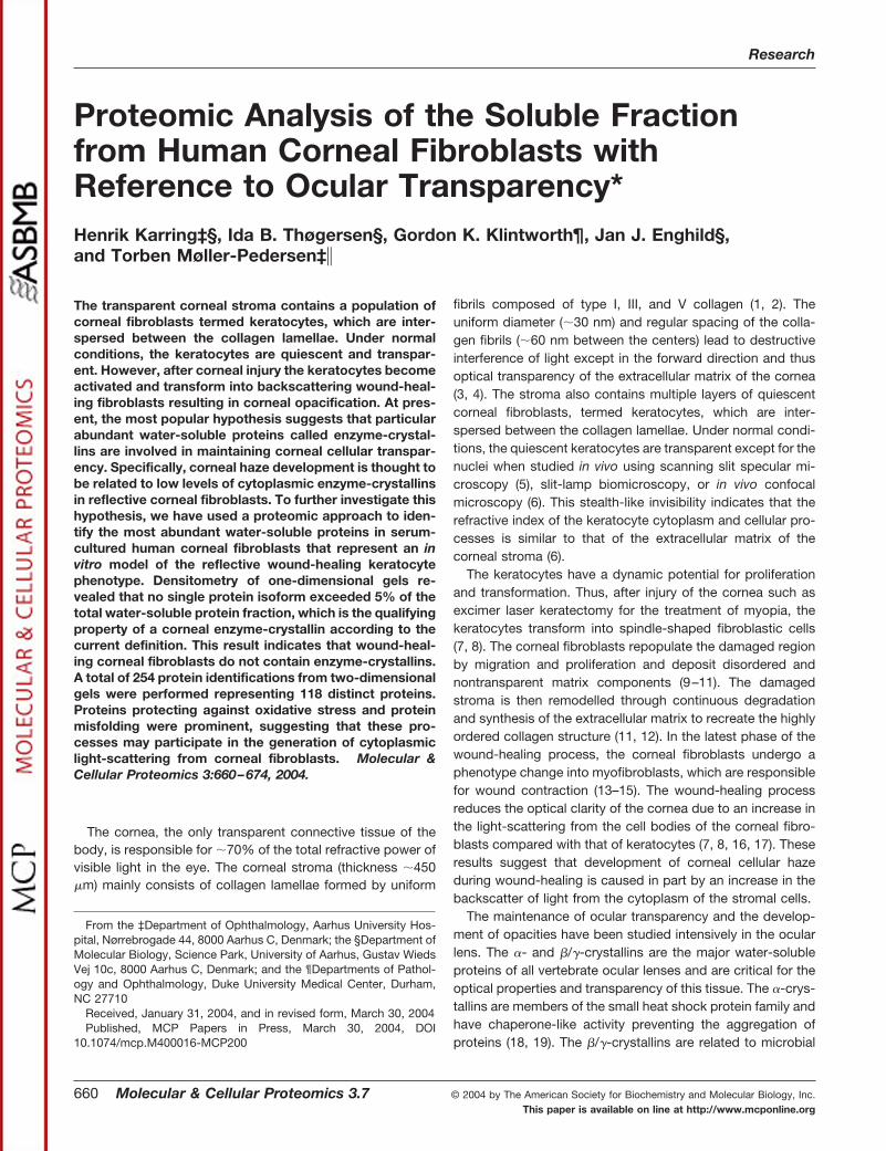
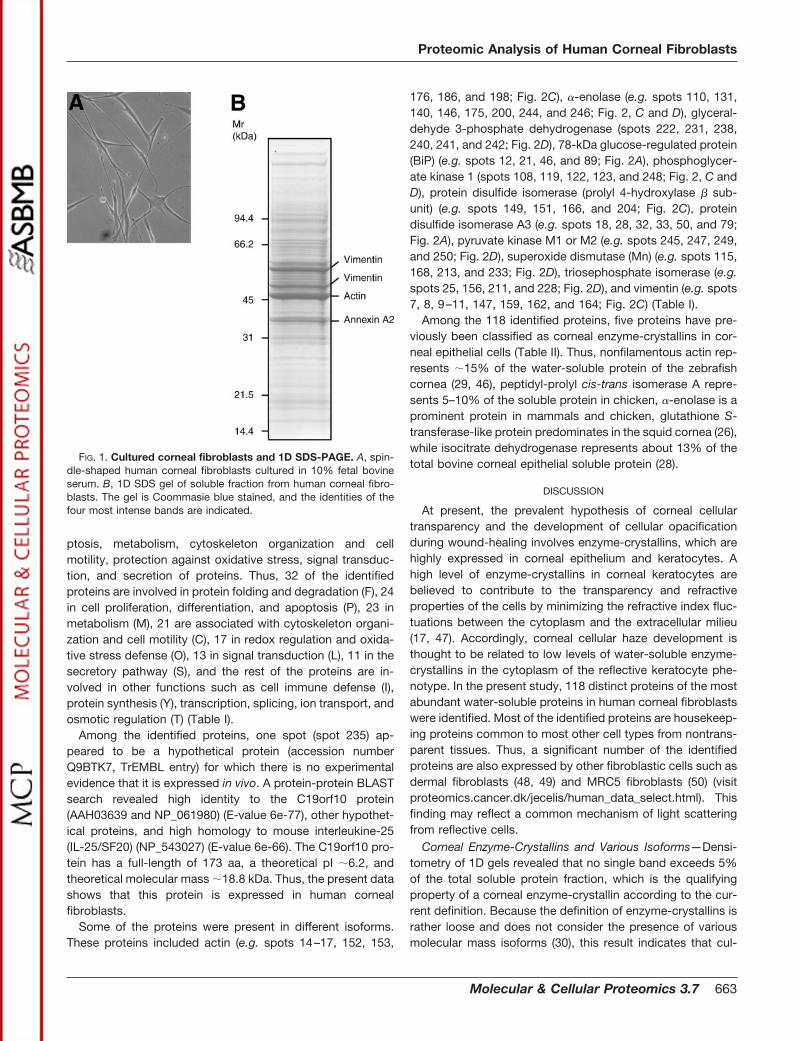
1D PAGE and Quantification by Densitometry—When trans-parent keratocytes are exposed to fetal bovine serum, theytransform into the reflective phenotype characterized by aspindle-shaped morphology and a fibroblastic cytoskeletonorganization (44, 45). Thus, cultured human corneal fibro-blasts of the third passage (Fig. 1A) were used as an in vitromodel system for the study of the corneal fibroblast solubleproteome. For initial analysis, the soluble fraction was ana-lyzed by 1D PAGE. Quantification by densitometry of 1D gelsshowed that no single protein isoform exceeded 2.5% of thetotal amount of protein in the soluble fraction. Identification ofthe four most intensive bands revealed that the most abun-dant proteins were actin, two different vimentin isoforms, andannexin A2. Thus, actin (�47 kDa) makes up 2.1 � 0.2% ofthe soluble protein content, while two vimentin isoforms of�59 and �51.3 kDa makes up 1.5 � 0.1 and 1.1 � 0.1%,respectively. Annexin A2 (�38 kDa) accounts for 1.0 � 0.2%of the total protein in the soluble fraction (Fig. 1B).
Proteome Study Using 2D PAGE—The soluble proteome ofthe corneal fibroblasts was further analyzed by 2D PAGEusing pH gradients 4–7 (Fig. 2A), 6–9 (Fig. 2B), 4.5–5.5 (Fig.2C), and 5.5–6.7 (Fig. 2D). No obvious differences in theprotein expression profiles among the donors were observed.All together, 254 gel spots were analyzed and identified byMALDI-MS or MS/MS. Thus, all spots indicated in Fig. 2 wereidentified by mass spectrometry. The identified proteins rep-resented 118 distinct proteins, which were categorized ac-cording to function and cellular localization as reported in thecurrent literature (Table I). In cases where it was obvious thatthe same spot had been analyzed on different gels, the spotswere given the same spot ID (Fig. 2, A–D). Most of the iden-tified proteins are involved in processes such as protein fold-ing and degradation, cell proliferation, differentiation, and apo-
Proteomic Analysis of Human Corneal Fibroblasts
662 Molecular & Cellular Proteomics 3.7
ptosis, metabolism, cytoskeleton organization and cellmotility, protection against oxidative stress, signal transduc-tion, and secretion of proteins. Thus, 32 of the identifiedproteins are involved in protein folding and degradation (F), 24in cell proliferation, differentiation, and apoptosis (P), 23 inmetabolism (M), 21 are associated with cytoskeleton organi-zation and cell motility (C), 17 in redox regulation and oxida-tive stress defense (O), 13 in signal transduction (L), 11 in thesecretory pathway (S), and the rest of the proteins are in-volved in other functions such as cell immune defense (I),protein synthesis (Y), transcription, splicing, ion transport, andosmotic regulation (T) (Table I).
Among the identified proteins, one spot (spot 235) ap-peared to be a hypothetical protein (accession numberQ9BTK7, TrEMBL entry) for which there is no experimentalevidence that it is expressed in vivo. A protein-protein BLASTsearch revealed high identity to the C19orf10 protein(AAH03639 and NP_061980) (E-value 6e-77), other hypothet-ical proteins, and high homology to mouse interleukine-25(IL-25/SF20) (NP_543027) (E-value 6e-66). The C19orf10 pro-tein has a full-length of 173 aa, a theoretical pI �6.2, andtheoretical molecular mass �18.8 kDa. Thus, the present datashows that this protein is expressed in human cornealfibroblasts.
Some of the proteins were present in different isoforms.These proteins included actin (e.g. spots 14–17, 152, 153,
176, 186, and 198; Fig. 2C), �-enolase (e.g. spots 110, 131,140, 146, 175, 200, 244, and 246; Fig. 2, C and D), glyceral-dehyde 3-phosphate dehydrogenase (spots 222, 231, 238,240, 241, and 242; Fig. 2D), 78-kDa glucose-regulated protein(BiP) (e.g. spots 12, 21, 46, and 89; Fig. 2A), phosphoglycer-ate kinase 1 (spots 108, 119, 122, 123, and 248; Fig. 2, C andD), protein disulfide isomerase (prolyl 4-hydroxylase � sub-unit) (e.g. spots 149, 151, 166, and 204; Fig. 2C), proteindisulfide isomerase A3 (e.g. spots 18, 28, 32, 33, 50, and 79;Fig. 2A), pyruvate kinase M1 or M2 (e.g. spots 245, 247, 249,and 250; Fig. 2D), superoxide dismutase (Mn) (e.g. spots 115,168, 213, and 233; Fig. 2D), triosephosphate isomerase (e.g.spots 25, 156, 211, and 228; Fig. 2D), and vimentin (e.g. spots7, 8, 9–11, 147, 159, 162, and 164; Fig. 2C) (Table I).
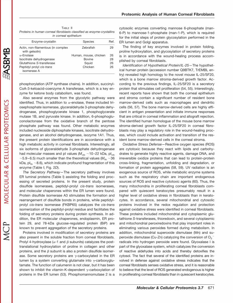
Among the 118 identified proteins, five proteins have pre-viously been classified as corneal enzyme-crystallins in cor-neal epithelial cells (Table II). Thus, nonfilamentous actin rep-resents �15% of the water-soluble protein of the zebrafishcornea (29, 46), peptidyl-prolyl cis-trans isomerase A repre-sents 5–10% of the soluble protein in chicken, �-enolase is aprominent protein in mammals and chicken, glutathione S-transferase-like protein predominates in the squid cornea (26),while isocitrate dehydrogenase represents about 13% of thetotal bovine corneal epithelial soluble protein (28).
DISCUSSION
At present, the prevalent hypothesis of corneal cellulartransparency and the development of cellular opacificationduring wound-healing involves enzyme-crystallins, which arehighly expressed in corneal epithelium and keratocytes. Ahigh level of enzyme-crystallins in corneal keratocytes arebelieved to contribute to the transparency and refractiveproperties of the cells by minimizing the refractive index fluc-tuations between the cytoplasm and the extracellular milieu(17, 47). Accordingly, corneal cellular haze development isthought to be related to low levels of water-soluble enzyme-crystallins in the cytoplasm of the reflective keratocyte phe-notype. In the present study, 118 distinct proteins of the mostabundant water-soluble proteins in human corneal fibroblastswere identified. Most of the identified proteins are housekeep-ing proteins common to most other cell types from nontrans-parent tissues. Thus, a significant number of the identifiedproteins are also expressed by other fibroblastic cells such asdermal fibroblasts (48, 49) and MRC5 fibroblasts (50) (visitproteomics.cancer.dk/jecelis/human_data_select.html). Thisfinding may reflect a common mechanism of light scatteringfrom reflective cells.
Corneal Enzyme-Crystallins and Various Isoforms—Densi-tometry of 1D gels revealed that no single band exceeds 5%of the total soluble protein fraction, which is the qualifyingproperty of a corneal enzyme-crystallin according to the cur-rent definition. Because the definition of enzyme-crystallins israther loose and does not consider the presence of variousmolecular mass isoforms (30), this result indicates that cul-
FIG. 1. Cultured corneal fibroblasts and 1D SDS-PAGE. A, spin-dle-shaped human corneal fibroblasts cultured in 10% fetal bovineserum. B, 1D SDS gel of soluble fraction from human corneal fibro-blasts. The gel is Coommasie blue stained, and the identities of thefour most intense bands are indicated.
Proteomic Analysis of Human Corneal Fibroblasts
Molecular & Cellular Proteomics 3.7 663
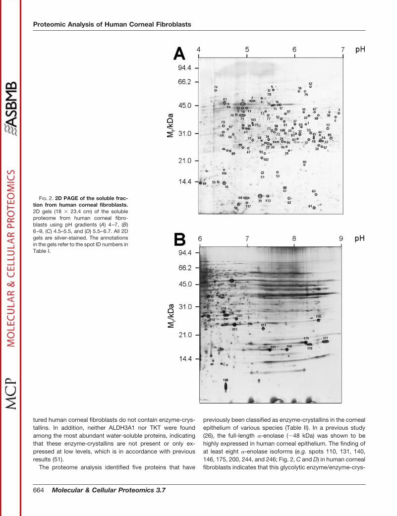
tured human corneal fibroblasts do not contain enzyme-crys-tallins. In addition, neither ALDH3A1 nor TKT were foundamong the most abundant water-soluble proteins, indicatingthat these enzyme-crystallins are not present or only ex-pressed at low levels, which is in accordance with previousresults (51).
The proteome analysis identified five proteins that have
previously been classified as enzyme-crystallins in the cornealepithelium of various species (Table II). In a previous study(26), the full-length �-enolase (�48 kDa) was shown to behighly expressed in human corneal epithelium. The finding ofat least eight �-enolase isoforms (e.g. spots 110, 131, 140,146, 175, 200, 244, and 246; Fig. 2, C and D) in human cornealfibroblasts indicates that this glycolytic enzyme/enzyme-crys-
FIG. 2. 2D PAGE of the soluble frac-tion from human corneal fibroblasts.2D gels (18 � 23.4 cm) of the solubleproteome from human corneal fibro-blasts using pH gradients (A) 4–7, (B)6–9, (C) 4.5–5.5, and (D) 5.5–6.7. All 2Dgels are silver-stained. The annotationsin the gels refer to the spot ID numbers inTable I.
Proteomic Analysis of Human Corneal Fibroblasts
664 Molecular & Cellular Proteomics 3.7
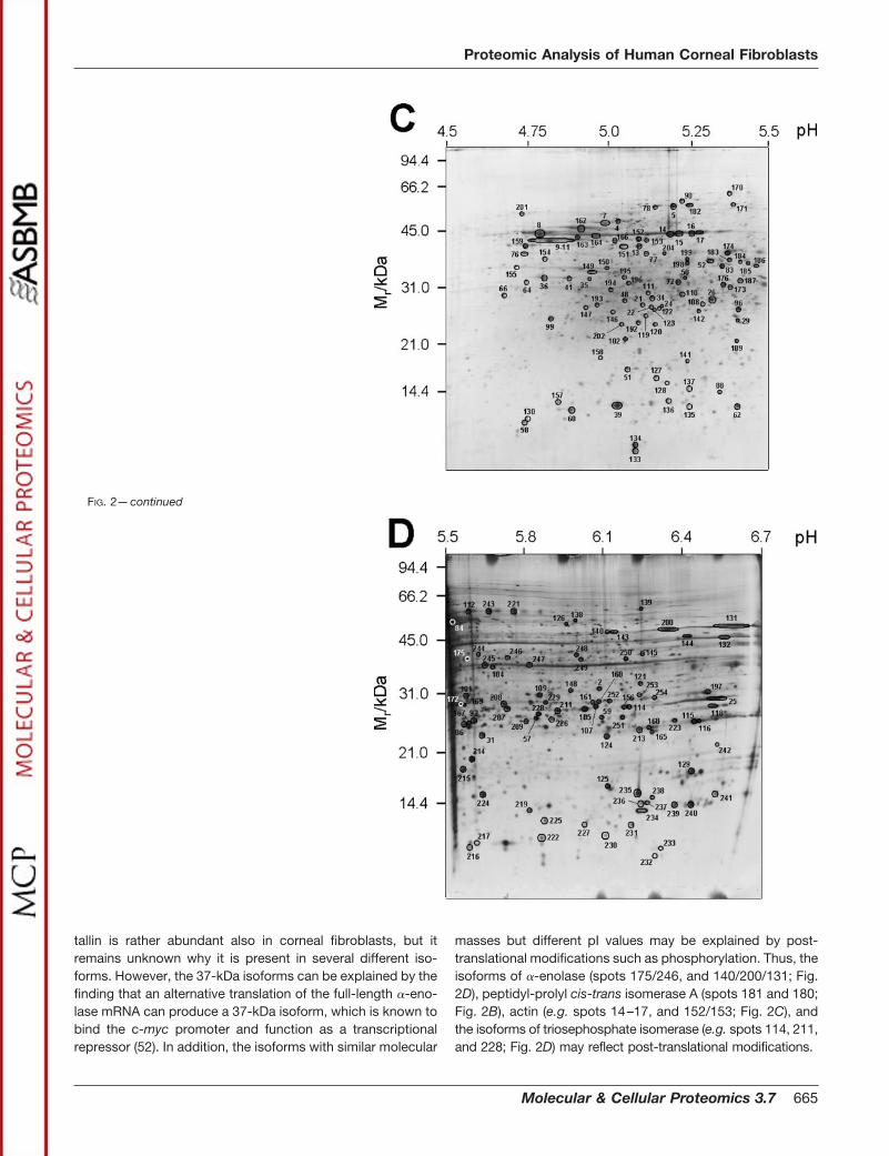
tallin is rather abundant also in corneal fibroblasts, but itremains unknown why it is present in several different iso-forms. However, the 37-kDa isoforms can be explained by thefinding that an alternative translation of the full-length �-eno-lase mRNA can produce a 37-kDa isoform, which is known tobind the c-myc promoter and function as a transcriptionalrepressor (52). In addition, the isoforms with similar molecular
masses but different pI values may be explained by post-translational modifications such as phosphorylation. Thus, theisoforms of �-enolase (spots 175/246, and 140/200/131; Fig.2D), peptidyl-prolyl cis-trans isomerase A (spots 181 and 180;Fig. 2B), actin (e.g. spots 14–17, and 152/153; Fig. 2C), andthe isoforms of triosephosphate isomerase (e.g. spots 114, 211,and 228; Fig. 2D) may reflect post-translational modifications.
FIG. 2—continued
Proteomic Analysis of Human Corneal Fibroblasts
Molecular & Cellular Proteomics 3.7 665
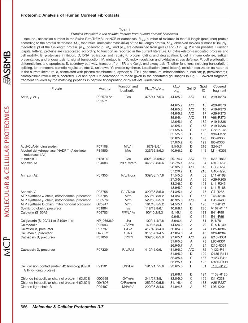
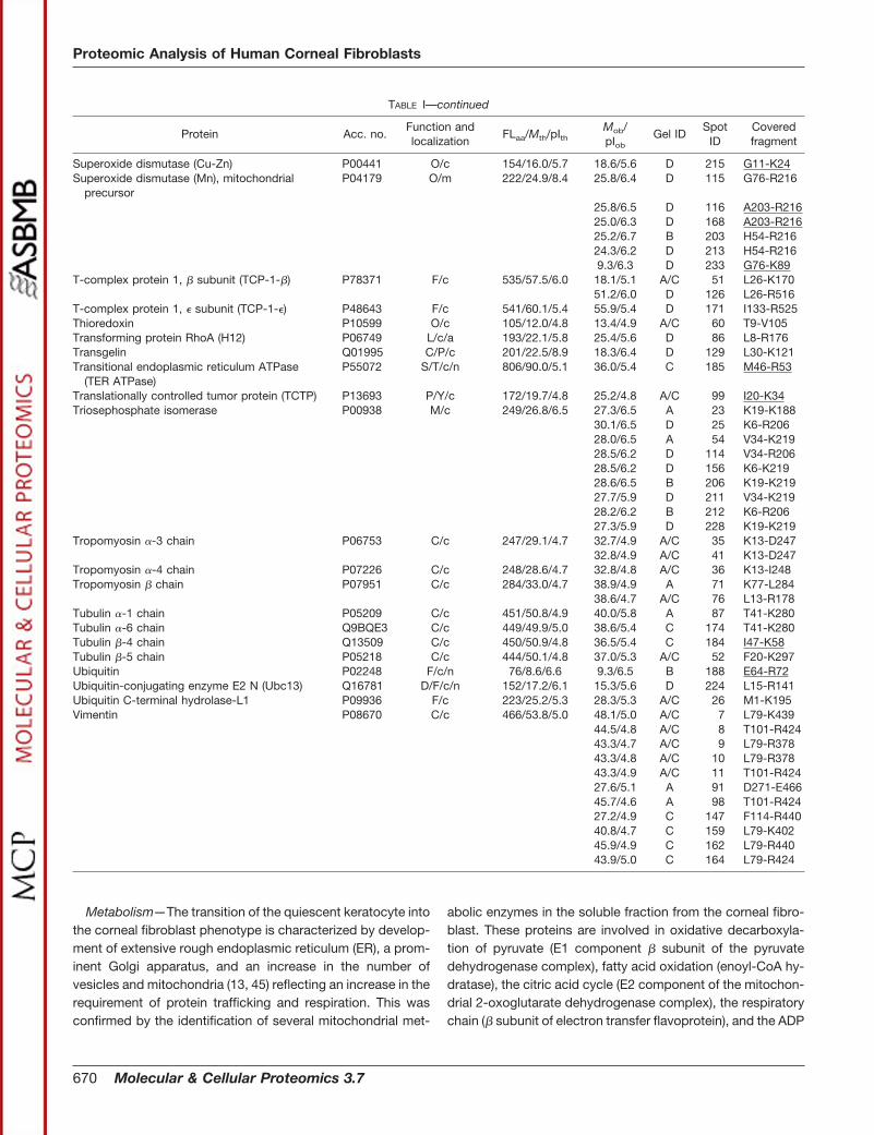
TABLE IProteins identified in the soluble fraction from human corneal fibroblasts
Acc. no., accession number in the Swiss-Prot/TrEMBL or NCBInr databases. FLaa, number of residues in the full-length (precursor) proteinaccording to the protein databases. Mth, theoretical molecular mass (kDa) of the full-length protein. Mob, observed molecular mass (kDa). pIth,theoretical pI of the full-length protein. pIob, observed pI. Mob and pIob are determined from gels C and D in Fig. 2 when possible. Function(capital letters), proteins are categorized according to function as reported in the current literature. C, cytoskeleton-associated proteins andcell motility; B, proteinase inhibition; D, DNA replication and repair; F, protein folding and degradation; I, cell immune defense, antigenpresentation, and endocytosis; L, signal transduction; M, metabolism; O, redox regulation and oxidative stress defense; P, cell proliferation,differentiation, and apoptosis; S, secretory pathway, transport from ER and Golgi, and exocytosis; T, other functions including transcription,splicing, ion transport, osmotic regulation, etc; U, unknown; Y, protein synthesis. Localization (small letters), cellular localization as reportedin the current literature. a, associated with plasma membrane; c, cytosol; e, ER; l, lysosome; m, mitochondrion; n, nuclear; p, peroxisome; r,sarcoplasmic reticulum; s, secreted. Gel and spot IDs correspond to those given in the annotated gel images in Fig. 2. Covered fragment,fragment covered by the matching peptides in peptide fingerprinting or by MS/MS (underlined).
Protein Acc. no.Function andlocalization
FLaa/Mth/pIthMob/pIob
Gel IDSpot
IDCoveredfragment
Actin, � or � P02570 orP02571
C/c 375/41.7/5.3 44.6/5.2 A/C 14 A19-K373
44.6/5.2 A/C 15 A29-K37344.6/5.3 A/C 16 A19-K37344.6/5.3 A/C 17 A29-K37335.5/5.4 A/C 83 V96-R37242.6/5.1 C 152 A19-K33642.5/5.1 C 153 A19-K33631.5/5.4 C 176 G63-K37335.5/5.5 C 186 V96-R37236.0/5.2 C 198 I85-K33637.0/5.2 C 199 I85-K336
Acyl-CoA-binding protein P07108 M/c/n 87/9.9/6.1 9.5/5.6 D 216 S2-K67Alcohol dehydrogenase [NADP�] (Aldo-keto
reductase 1A1)P14550 M/c 325/36.8/6.3 40.9/6.2 D 145 M14-K308
�-Actinin 1 P12814 C/c 892/103.5/5.2 29.1/4.7 A/C 66 I656-R863Annexin A1 P04083 P/L/T/c/a/n 346/38.8/6.6 28.7/5.1 A/C 34 Q10-R228
28.3/5.0 A/C 48 G30-R22837.2/6.2 B 218 Q10-R228
Annexin A2 P07355 P/L/T/c/a 339/38.7/7.6 17.5/5.6 A 53 L11-R16827.3/5.8 A 95 A29-R20533.3/6.2 D 121 L11-R24518.9/5.2 C 141 L11-R168
Annexin V P08758 P/L/T/c/a 320/35.8/5.0 34.3/5.1 A 75 G7-R285ATP synthase � chain, mitochondrial precursor P25705 M/m 553/59.8/9.2 14.3/4.8 C 157 T46-K194ATP synthase � chain, mitochondrial precursor P06576 M/m 529/56.5/5.3 48.9/5.0 A/C 4 L95-K480ATP synthase D chain, mitochondrial precursor O75947 M/m 161/18.5/5.2 24.5/5.1 C 120 T10-K121�2-microglobulin, precursor P01884 I/s 119/13.8/6.1 10.8/6.1 D 230 V102-K111Calcyclin (S100A6) P06703 P/F/L/r/n 90/10.2/5.3 9.1/5.1 C 133 E41-R55
9.9/5.1 C 134 E41-R55Calgizzarin (S100A14 or S100A11p) NP_066369 U/u 102/11.4/7.8 8.9/6.4 A 61 I4-K78Calmodulin P02593 L/S/P/c 149/16.8/4.1 14.6/4.0 A 49 E15-K149Calreticulin, precursor P27797 F/S/e 417/48.3/4.3 56.8/4.3 A 74 E25-K286Calumenin, precursor O43852 S/e/s 315/37.1/4.5 47.0/4.5 A 43 V28-K284Cathepsin B, precursor P07858 I/P/F/I 339/38.8/5.9 27.6/5.1 A/C 22 I210-R331
31.8/5.5 A 73 L80-R33126.9/5.7 A 94 I210-R331
Cathepsin D, precursor P07339 P/L/F/I/l 412/45.0/6.1 31.9/5.2 A/C 72 Y123-R41131.0/5.9 D 109 Q185-R41132.3/5.4 C 187 Y123-R41133.2/5.1 C 196 Q185-R411
Cell division control protein 42 homolog (G25KGTP-binding protein)
P21181 C/P/L/c 191/21.7/5.8 23.6/5.6 D 31 T108-R120
23.6/6.1 D 124 T108-R120Chloride intracellular channel protein 1 (CLIC1) O00299 O/T/n/c 241/27.3/5.1 32.8/5.0 C 195 I21-K238Chloride intracellular channel protein 4 (CLIC4) Q9Y696 C/P/c/m/n 253/29.0/5.5 31.1/5.4 C 173 A25-R227Clathrin light chain B P09497 M/I/c/a/l 229/25.3/4.6 31.0/4.5 A 69 L96-K204
Proteomic Analysis of Human Corneal Fibroblasts
666 Molecular & Cellular Proteomics 3.7
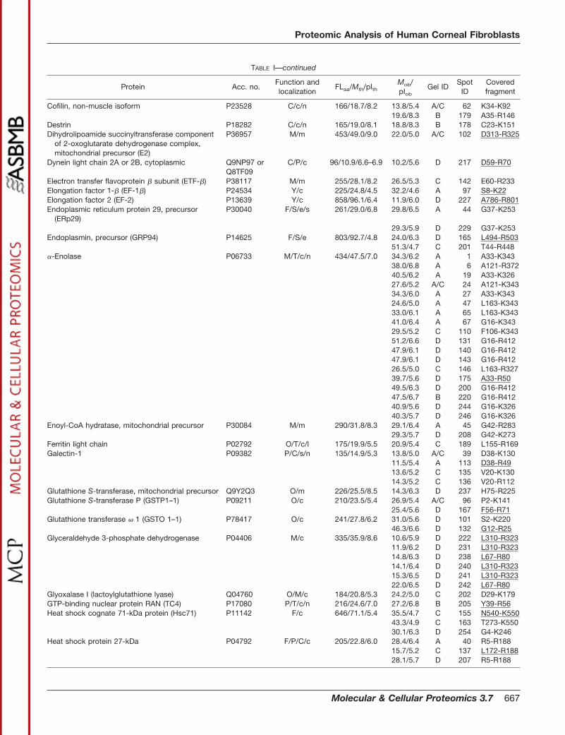
TABLE I—continued
Protein Acc. no.Function andlocalization
FLaa/Mth/pIthMob/pIob
Gel IDSpot
IDCoveredfragment
Cofilin, non-muscle isoform P23528 C/c/n 166/18.7/8.2 13.8/5.4 A/C 62 K34-K9219.6/8.3 B 179 A35-R146
Destrin P18282 C/c/n 165/19.0/8.1 18.8/8.3 B 178 C23-K151Dihydrolipoamide succinyltransferase component
of 2-oxoglutarate dehydrogenase complex,mitochondrial precursor (E2)
P36957 M/m 453/49.0/9.0 22.0/5.0 A/C 102 D313-R325
Dynein light chain 2A or 2B, cytoplasmic Q9NP97 orQ8TF09
C/P/c 96/10.9/6.6–6.9 10.2/5.6 D 217 D59-R70
Electron transfer flavoprotein � subunit (ETF-�) P38117 M/m 255/28.1/8.2 26.5/5.3 C 142 E60-R233Elongation factor 1-� (EF-1�) P24534 Y/c 225/24.8/4.5 32.2/4.6 A 97 S8-K22Elongation factor 2 (EF-2) P13639 Y/c 858/96.1/6.4 11.9/6.0 D 227 A786-R801Endoplasmic reticulum protein 29, precursor
(ERp29)P30040 F/S/e/s 261/29.0/6.8 29.8/6.5 A 44 G37-K253
29.3/5.9 D 229 G37-K253Endoplasmin, precursor (GRP94) P14625 F/S/e 803/92.7/4.8 24.0/6.3 D 165 L494-R503
51.3/4.7 C 201 T44-R448�-Enolase P06733 M/T/c/n 434/47.5/7.0 34.3/6.2 A 1 A33-K343
38.0/6.8 A 6 A121-R37240.5/6.2 A 19 A33-K32627.6/5.2 A/C 24 A121-K34334.3/6.0 A 27 A33-K34324.6/5.0 A 47 L163-K34333.0/6.1 A 65 L163-K34341.0/6.4 A 67 G16-K34329.5/5.2 C 110 F106-K34351.2/6.6 D 131 G16-R41247.9/6.1 D 140 G16-R41247.9/6.1 D 143 G16-R41226.5/5.0 C 146 L163-R32739.7/5.6 D 175 A33-R5049.5/6.3 D 200 G16-R41247.5/6.7 B 220 G16-R41240.9/5.6 D 244 G16-K32640.3/5.7 D 246 G16-K326
Enoyl-CoA hydratase, mitochondrial precursor P30084 M/m 290/31.8/8.3 29.1/6.4 A 45 G42-R28329.3/5.7 D 208 G42-K273
Ferritin light chain P02792 O/T/c/l 175/19.9/5.5 20.9/5.4 C 189 L155-R169Galectin-1 P09382 P/C/s/n 135/14.9/5.3 13.8/5.0 A/C 39 D38-K130
11.5/5.4 A 113 D38-R4913.6/5.2 C 135 V20-K13014.3/5.2 C 136 V20-R112
Glutathione S-transferase, mitochondrial precursor Q9Y2Q3 O/m 226/25.5/8.5 14.3/6.3 D 237 H75-R225Glutathione S-transferase P (GSTP1–1) P09211 O/c 210/23.5/5.4 26.9/5.4 A/C 96 P2-K141
25.4/5.6 D 167 F56-R71Glutathione transferase � 1 (GSTO 1–1) P78417 O/c 241/27.8/6.2 31.0/5.6 D 101 S2-K220
46.3/6.6 D 132 G12-R25Glyceraldehyde 3-phosphate dehydrogenase P04406 M/c 335/35.9/8.6 10.6/5.9 D 222 L310-R323
11.9/6.2 D 231 L310-R32314.8/6.3 D 238 L67-R8014.1/6.4 D 240 L310-R32315.3/6.5 D 241 L310-R32322.0/6.5 D 242 L67-R80
Glyoxalase I (lactoylglutathione lyase) Q04760 O/M/c 184/20.8/5.3 24.2/5.0 C 202 D29-K179GTP-binding nuclear protein RAN (TC4) P17080 P/T/c/n 216/24.6/7.0 27.2/6.8 B 205 Y39-R56Heat shock cognate 71-kDa protein (Hsc71) P11142 F/c 646/71.1/5.4 35.5/4.7 C 155 N540-K550
43.3/4.9 C 163 T273-K55030.1/6.3 D 254 G4-K246
Heat shock protein 27-kDa P04792 F/P/C/c 205/22.8/6.0 28.4/6.4 A 40 R5-R18815.7/5.2 C 137 L172-R18828.1/5.7 D 207 R5-R188
Proteomic Analysis of Human Corneal Fibroblasts
Molecular & Cellular Proteomics 3.7 667
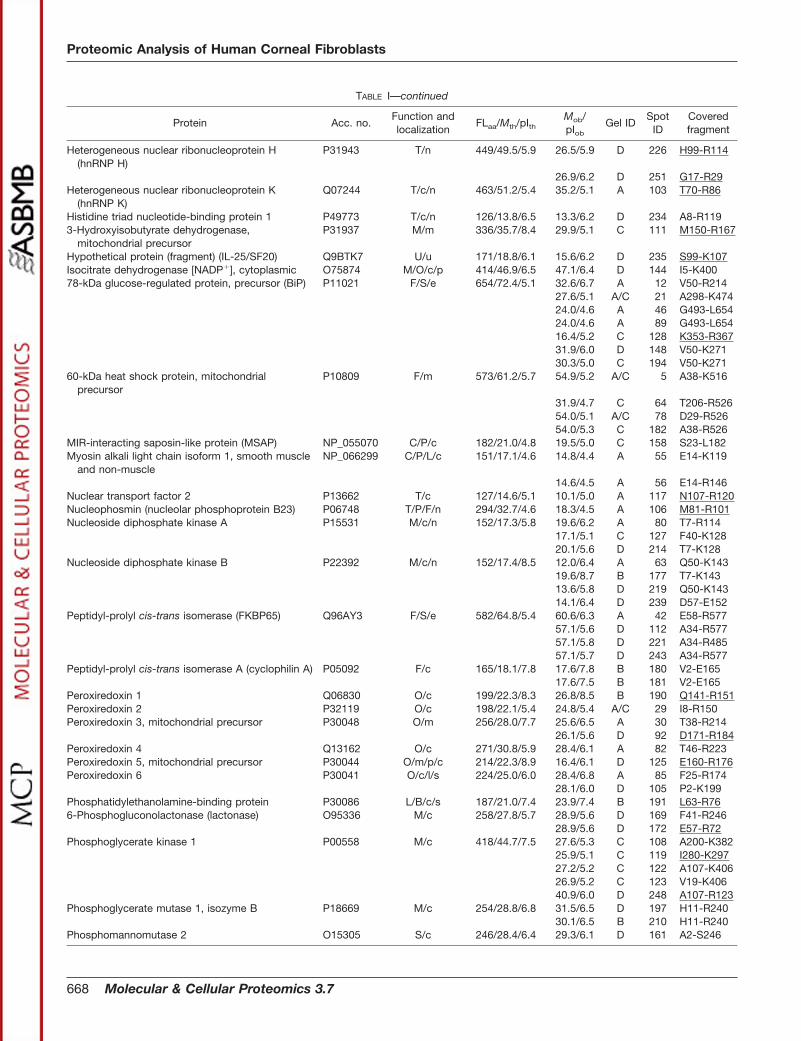
TABLE I—continued
Protein Acc. no.Function andlocalization
FLaa/Mth/pIthMob/pIob
Gel IDSpot
IDCoveredfragment
Heterogeneous nuclear ribonucleoprotein H(hnRNP H)
P31943 T/n 449/49.5/5.9 26.5/5.9 D 226 H99-R114
26.9/6.2 D 251 G17-R29Heterogeneous nuclear ribonucleoprotein K
(hnRNP K)Q07244 T/c/n 463/51.2/5.4 35.2/5.1 A 103 T70-R86
Histidine triad nucleotide-binding protein 1 P49773 T/c/n 126/13.8/6.5 13.3/6.2 D 234 A8-R1193-Hydroxyisobutyrate dehydrogenase,
mitochondrial precursorP31937 M/m 336/35.7/8.4 29.9/5.1 C 111 M150-R167
Hypothetical protein (fragment) (IL-25/SF20) Q9BTK7 U/u 171/18.8/6.1 15.6/6.2 D 235 S99-K107Isocitrate dehydrogenase [NADP�], cytoplasmic O75874 M/O/c/p 414/46.9/6.5 47.1/6.4 D 144 I5-K40078-kDa glucose-regulated protein, precursor (BiP) P11021 F/S/e 654/72.4/5.1 32.6/6.7 A 12 V50-R214
27.6/5.1 A/C 21 A298-K47424.0/4.6 A 46 G493-L65424.0/4.6 A 89 G493-L65416.4/5.2 C 128 K353-R36731.9/6.0 D 148 V50-K27130.3/5.0 C 194 V50-K271
60-kDa heat shock protein, mitochondrialprecursor
P10809 F/m 573/61.2/5.7 54.9/5.2 A/C 5 A38-K516
31.9/4.7 C 64 T206-R52654.0/5.1 A/C 78 D29-R52654.0/5.3 C 182 A38-R526
MIR-interacting saposin-like protein (MSAP) NP_055070 C/P/c 182/21.0/4.8 19.5/5.0 C 158 S23-L182Myosin alkali light chain isoform 1, smooth muscle
and non-muscleNP_066299 C/P/L/c 151/17.1/4.6 14.8/4.4 A 55 E14-K119
14.6/4.5 A 56 E14-R146Nuclear transport factor 2 P13662 T/c 127/14.6/5.1 10.1/5.0 A 117 N107-R120Nucleophosmin (nucleolar phosphoprotein B23) P06748 T/P/F/n 294/32.7/4.6 18.3/4.5 A 106 M81-R101Nucleoside diphosphate kinase A P15531 M/c/n 152/17.3/5.8 19.6/6.2 A 80 T7-R114
17.1/5.1 C 127 F40-K12820.1/5.6 D 214 T7-K128
Nucleoside diphosphate kinase B P22392 M/c/n 152/17.4/8.5 12.0/6.4 A 63 Q50-K14319.6/8.7 B 177 T7-K14313.6/5.8 D 219 Q50-K14314.1/6.4 D 239 D57-E152
Peptidyl-prolyl cis-trans isomerase (FKBP65) Q96AY3 F/S/e 582/64.8/5.4 60.6/6.3 A 42 E58-R57757.1/5.6 D 112 A34-R57757.1/5.8 D 221 A34-R48557.1/5.7 D 243 A34-R577
Peptidyl-prolyl cis-trans isomerase A (cyclophilin A) P05092 F/c 165/18.1/7.8 17.6/7.8 B 180 V2-E16517.6/7.5 B 181 V2-E165
Peroxiredoxin 1 Q06830 O/c 199/22.3/8.3 26.8/8.5 B 190 Q141-R151Peroxiredoxin 2 P32119 O/c 198/22.1/5.4 24.8/5.4 A/C 29 I8-R150Peroxiredoxin 3, mitochondrial precursor P30048 O/m 256/28.0/7.7 25.6/6.5 A 30 T38-R214
26.1/5.6 D 92 D171-R184Peroxiredoxin 4 Q13162 O/c 271/30.8/5.9 28.4/6.1 A 82 T46-R223Peroxiredoxin 5, mitochondrial precursor P30044 O/m/p/c 214/22.3/8.9 16.4/6.1 D 125 E160-R176Peroxiredoxin 6 P30041 O/c/l/s 224/25.0/6.0 28.4/6.8 A 85 F25-R174
28.1/6.0 D 105 P2-K199Phosphatidylethanolamine-binding protein P30086 L/B/c/s 187/21.0/7.4 23.9/7.4 B 191 L63-R766-Phosphogluconolactonase (lactonase) O95336 M/c 258/27.8/5.7 28.9/5.6 D 169 F41-R246
28.9/5.6 D 172 E57-R72Phosphoglycerate kinase 1 P00558 M/c 418/44.7/7.5 27.6/5.3 C 108 A200-K382
25.9/5.1 C 119 I280-K29727.2/5.2 C 122 A107-K40626.9/5.2 C 123 V19-K40640.9/6.0 D 248 A107-R123
Phosphoglycerate mutase 1, isozyme B P18669 M/c 254/28.8/6.8 31.5/6.5 D 197 H11-R24030.1/6.5 B 210 H11-R240
Phosphomannomutase 2 O15305 S/c 246/28.4/6.4 29.3/6.1 D 161 A2-S246
Proteomic Analysis of Human Corneal Fibroblasts
668 Molecular & Cellular Proteomics 3.7
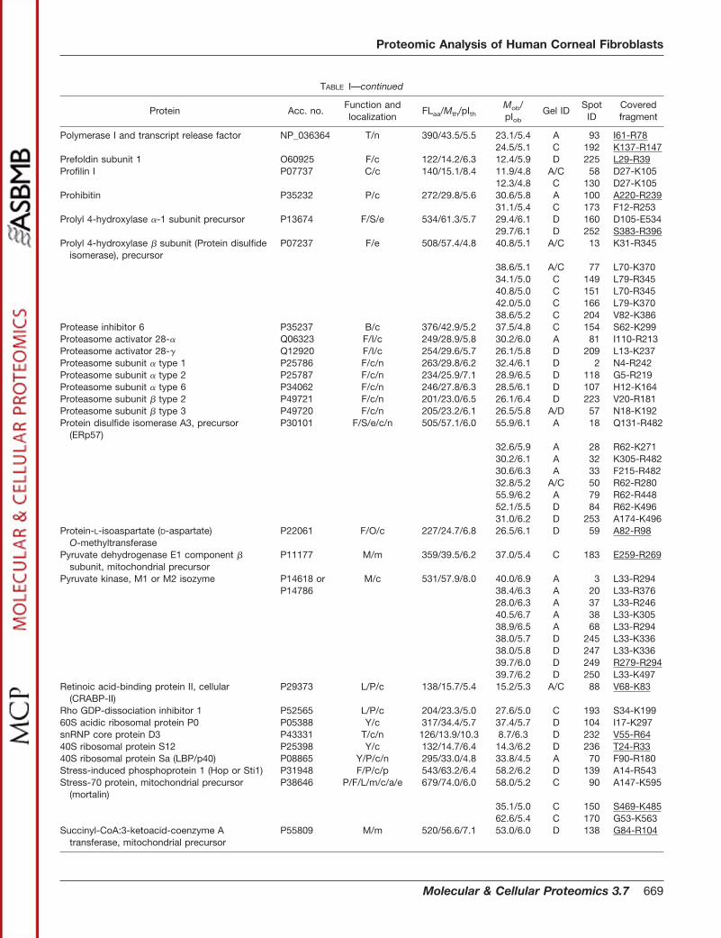
TABLE I—continued
Protein Acc. no.Function andlocalization
FLaa/Mth/pIthMob/pIob
Gel IDSpot
IDCoveredfragment
Polymerase I and transcript release factor NP_036364 T/n 390/43.5/5.5 23.1/5.4 A 93 I61-R7824.5/5.1 C 192 K137-R147
Prefoldin subunit 1 O60925 F/c 122/14.2/6.3 12.4/5.9 D 225 L29-R39Profilin I P07737 C/c 140/15.1/8.4 11.9/4.8 A/C 58 D27-K105
12.3/4.8 C 130 D27-K105Prohibitin P35232 P/c 272/29.8/5.6 30.6/5.8 A 100 A220-R239
31.1/5.4 C 173 F12-R253Prolyl 4-hydroxylase �-1 subunit precursor P13674 F/S/e 534/61.3/5.7 29.4/6.1 D 160 D105-E534
29.7/6.1 D 252 S383-R396Prolyl 4-hydroxylase � subunit (Protein disulfide
isomerase), precursorP07237 F/e 508/57.4/4.8 40.8/5.1 A/C 13 K31-R345
38.6/5.1 A/C 77 L70-K37034.1/5.0 C 149 L79-R34540.8/5.0 C 151 L70-R34542.0/5.0 C 166 L79-K37038.6/5.2 C 204 V82-K386
Protease inhibitor 6 P35237 B/c 376/42.9/5.2 37.5/4.8 C 154 S62-K299Proteasome activator 28-� Q06323 F/I/c 249/28.9/5.8 30.2/6.0 A 81 I110-R213Proteasome activator 28-� Q12920 F/I/c 254/29.6/5.7 26.1/5.8 D 209 L13-K237Proteasome subunit � type 1 P25786 F/c/n 263/29.8/6.2 32.4/6.1 D 2 N4-R242Proteasome subunit � type 2 P25787 F/c/n 234/25.9/7.1 28.9/6.5 D 118 G5-R219Proteasome subunit � type 6 P34062 F/c/n 246/27.8/6.3 28.5/6.1 D 107 H12-K164Proteasome subunit � type 2 P49721 F/c/n 201/23.0/6.5 26.1/6.4 D 223 V20-R181Proteasome subunit � type 3 P49720 F/c/n 205/23.2/6.1 26.5/5.8 A/D 57 N18-K192Protein disulfide isomerase A3, precursor
(ERp57)P30101 F/S/e/c/n 505/57.1/6.0 55.9/6.1 A 18 Q131-R482
32.6/5.9 A 28 R62-K27130.2/6.1 A 32 K305-R48230.6/6.3 A 33 F215-R48232.8/5.2 A/C 50 R62-R28055.9/6.2 A 79 R62-R44852.1/5.5 D 84 R62-K49631.0/6.2 D 253 A174-K496
Protein-L-isoaspartate (D-aspartate)O-methyltransferase
P22061 F/O/c 227/24.7/6.8 26.5/6.1 D 59 A82-R98
Pyruvate dehydrogenase E1 component �subunit, mitochondrial precursor
P11177 M/m 359/39.5/6.2 37.0/5.4 C 183 E259-R269
Pyruvate kinase, M1 or M2 isozyme P14618 or M/c 531/57.9/8.0 40.0/6.9 A 3 L33-R294P14786 38.4/6.3 A 20 L33-R376
28.0/6.3 A 37 L33-R24640.5/6.7 A 38 L33-K30538.9/6.5 A 68 L33-R29438.0/5.7 D 245 L33-K33638.0/5.8 D 247 L33-K33639.7/6.0 D 249 R279-R29439.7/6.2 D 250 L33-K497
Retinoic acid-binding protein II, cellular(CRABP-II)
P29373 L/P/c 138/15.7/5.4 15.2/5.3 A/C 88 V68-K83
Rho GDP-dissociation inhibitor 1 P52565 L/P/c 204/23.3/5.0 27.6/5.0 C 193 S34-K19960S acidic ribosomal protein P0 P05388 Y/c 317/34.4/5.7 37.4/5.7 D 104 I17-K297snRNP core protein D3 P43331 T/c/n 126/13.9/10.3 8.7/6.3 D 232 V55-R6440S ribosomal protein S12 P25398 Y/c 132/14.7/6.4 14.3/6.2 D 236 T24-R3340S ribosomal protein Sa (LBP/p40) P08865 Y/P/c/n 295/33.0/4.8 33.8/4.5 A 70 F90-R180Stress-induced phosphoprotein 1 (Hop or Sti1) P31948 F/P/c/p 543/63.2/6.4 58.2/6.2 D 139 A14-R543Stress-70 protein, mitochondrial precursor
(mortalin)P38646 P/F/L/m/c/a/e 679/74.0/6.0 58.0/5.2 C 90 A147-K595
35.1/5.0 C 150 S469-K48562.6/5.4 C 170 G53-K563
Succinyl-CoA:3-ketoacid-coenzyme Atransferase, mitochondrial precursor
P55809 M/m 520/56.6/7.1 53.0/6.0 D 138 G84-R104
Proteomic Analysis of Human Corneal Fibroblasts
Molecular & Cellular Proteomics 3.7 669
Metabolism—The transition of the quiescent keratocyte intothe corneal fibroblast phenotype is characterized by develop-ment of extensive rough endoplasmic reticulum (ER), a prom-inent Golgi apparatus, and an increase in the number ofvesicles and mitochondria (13, 45) reflecting an increase in therequirement of protein trafficking and respiration. This wasconfirmed by the identification of several mitochondrial met-
abolic enzymes in the soluble fraction from the corneal fibro-blast. These proteins are involved in oxidative decarboxyla-tion of pyruvate (E1 component � subunit of the pyruvatedehydrogenase complex), fatty acid oxidation (enoyl-CoA hy-dratase), the citric acid cycle (E2 component of the mitochon-drial 2-oxoglutarate dehydrogenase complex), the respiratorychain (� subunit of electron transfer flavoprotein), and the ADP
TABLE I—continued
Protein Acc. no.Function andlocalization
FLaa/Mth/pIthMob/pIob
Gel IDSpot
IDCoveredfragment
Superoxide dismutase (Cu-Zn) P00441 O/c 154/16.0/5.7 18.6/5.6 D 215 G11-K24Superoxide dismutase (Mn), mitochondrial
precursorP04179 O/m 222/24.9/8.4 25.8/6.4 D 115 G76-R216
25.8/6.5 D 116 A203-R21625.0/6.3 D 168 A203-R21625.2/6.7 B 203 H54-R21624.3/6.2 D 213 H54-R2169.3/6.3 D 233 G76-K89
T-complex protein 1, � subunit (TCP-1-�) P78371 F/c 535/57.5/6.0 18.1/5.1 A/C 51 L26-K17051.2/6.0 D 126 L26-R516
T-complex protein 1, � subunit (TCP-1-�) P48643 F/c 541/60.1/5.4 55.9/5.4 D 171 I133-R525Thioredoxin P10599 O/c 105/12.0/4.8 13.4/4.9 A/C 60 T9-V105Transforming protein RhoA (H12) P06749 L/c/a 193/22.1/5.8 25.4/5.6 D 86 L8-R176Transgelin Q01995 C/P/c 201/22.5/8.9 18.3/6.4 D 129 L30-K121Transitional endoplasmic reticulum ATPase
(TER ATPase)P55072 S/T/c/n 806/90.0/5.1 36.0/5.4 C 185 M46-R53
Translationally controlled tumor protein (TCTP) P13693 P/Y/c 172/19.7/4.8 25.2/4.8 A/C 99 I20-K34Triosephosphate isomerase P00938 M/c 249/26.8/6.5 27.3/6.5 A 23 K19-K188
30.1/6.5 D 25 K6-R20628.0/6.5 A 54 V34-K21928.5/6.2 D 114 V34-R20628.5/6.2 D 156 K6-K21928.6/6.5 B 206 K19-K21927.7/5.9 D 211 V34-K21928.2/6.2 B 212 K6-R20627.3/5.9 D 228 K19-K219
Tropomyosin �-3 chain P06753 C/c 247/29.1/4.7 32.7/4.9 A/C 35 K13-D24732.8/4.9 A/C 41 K13-D247
Tropomyosin �-4 chain P07226 C/c 248/28.6/4.7 32.8/4.8 A/C 36 K13-I248Tropomyosin � chain P07951 C/c 284/33.0/4.7 38.9/4.9 A 71 K77-L284
38.6/4.7 A/C 76 L13-R178Tubulin �-1 chain P05209 C/c 451/50.8/4.9 40.0/5.8 A 87 T41-K280Tubulin �-6 chain Q9BQE3 C/c 449/49.9/5.0 38.6/5.4 C 174 T41-K280Tubulin �-4 chain Q13509 C/c 450/50.9/4.8 36.5/5.4 C 184 I47-K58Tubulin �-5 chain P05218 C/c 444/50.1/4.8 37.0/5.3 A/C 52 F20-K297Ubiquitin P02248 F/c/n 76/8.6/6.6 9.3/6.5 B 188 E64-R72Ubiquitin-conjugating enzyme E2 N (Ubc13) Q16781 D/F/c/n 152/17.2/6.1 15.3/5.6 D 224 L15-R141Ubiquitin C-terminal hydrolase-L1 P09936 F/c 223/25.2/5.3 28.3/5.3 A/C 26 M1-K195Vimentin P08670 C/c 466/53.8/5.0 48.1/5.0 A/C 7 L79-K439
44.5/4.8 A/C 8 T101-R42443.3/4.7 A/C 9 L79-R37843.3/4.8 A/C 10 L79-R37843.3/4.9 A/C 11 T101-R42427.6/5.1 A 91 D271-E46645.7/4.6 A 98 T101-R42427.2/4.9 C 147 F114-R44040.8/4.7 C 159 L79-K40245.9/4.9 C 162 L79-R44043.9/5.0 C 164 L79-R424
Proteomic Analysis of Human Corneal Fibroblasts
670 Molecular & Cellular Proteomics 3.7
phosphorylation (ATP synthase chains). In addition, succinyl-CoA-3-ketoacid-coenzyme A transferase, which is a key en-zyme for ketone body catabolism, was found.
Also several enzymes from the glycolytic pathway wereidentified. Thus, in addition to �-enolase, these included tri-osephosphate isomerase, glyceraldehyde 3-phosphate dehy-drogenase, phosphoglycerate kinase 1, phosphoglyceratemutase 1B, and pyruvate kinase. In addition, 6-phosphoglu-conolactonase from the oxidative branch of the pentosephosphate pathway was found. Other metabolic enzymesincluded nucleoside diphosphate kinases, isocitrate dehydro-genase, and an alcohol dehydrogenase, isozyme 1A1. Thus,the present protein identifications are in accordance with ahigh metabolic activity in corneal fibroblasts. Interestingly, allsix isoforms of glyceraldehyde 3-phosphate dehydrogenasehad molecular masses and pI values (Mob �10–22 kDa, pIob
�5.9–6.5) much smaller than the theoretical values (Mth �36kDa, pIth �8.6), which indicate profound fragmentation of thisenzyme (Table I; Fig. 2D).
The Secretory Pathway—The secretory pathway involvesER luminal proteins (Table I) assisting the folding and proc-essing of secreted proteins. In the present study, proteindisulfide isomerases, peptidyl-prolyl cis-trans isomerases,and molecular chaperones within the ER lumen were found.Protein disulfide isomerase A3 stimulates the formation andrearrangement of disulfide bonds in proteins, while peptidyl-prolyl cis-trans isomerase (FKBP65) catalyzes the cis-transisomerization of the peptidyl-prolyl residue and facilitates thefolding of secretory proteins during protein synthesis. In ad-dition, the ER molecular chaperones, endoplasmin, ER pro-tein 29, and 78-kDa glucose-regulated protein (BiP) areknown to prevent aggregation of the secretory proteins.
Proteins involved in modification of secretory proteins arealso present in the soluble fraction from corneal fibroblasts.Prolyl 4-hydroxylase (�-1 and � subunits) catalyzes the post-translational hydroxylation of proline in collagen and otherproteins, and the � subunit is also a protein disulfide isomer-ase. Some secretory proteins are �-carboxylated in the ERlumen by a system converting glutamate into �-carboxyglu-tamate. The function of calumenin is not clear, but it has beenshown to inhibit the vitamin-K-dependent �-carboxylation ofproteins in the ER lumen (53). Phosphomannomutase 2 is a
cytosolic enzymes converting mannose 6-phosphate (man-6-P) to mannose-1-phosphate (man-1-P), which is requiredfor the initial steps of protein glycosylation performed in theER lumen and Golgi apparatus.
The finding of key enzymes involved in protein folding,proline hydroxylation, and glycosylation of secretory proteinsis in accordance with the wound-healing process accom-plished by corneal fibroblasts.
Identification of Hypothetical Protein/IL-25—The hypothet-ical human protein (accession number Q9BTK7, TrEMBL en-try) revealed high homology to the novel mouse IL-25/SF20,which is a bone marrow stroma-derived growth factor. Ac-cording to the previous findings, IL-25/SF20 is a secretoryprotein that stimulates cell proliferation (54, 55). Interestingly,recent reports have shown that both the corneal epitheliumand stroma contain a significant number of resident bonemarrow-derived cells such as macrophages and dendriticcells (56, 57). The bone marrow-derived cells are highly effi-cient in antigen presentation and initiate immune responsesthat are critical in corneal inflammation and allograft rejection.The identified human homologue of the mouse bone marrowstroma-derived growth factor IL-25/SF20 in corneal fibro-blasts may play a regulatory role in the wound-healing proc-ess, which could include activation and transition of the res-ident bone marrow-derived cells in the corneal stroma.
Oxidative Stress Defense—Reactive oxygen species (ROS)are cytotoxic because they react with lipids and carbohy-drates to generate highly reactive agents, damage DNA, andirreversible oxidize proteins that can lead to protein-proteincross-linking, fragmentation, unfolding and degradation, orformation of protein aggregates (58, 59). UV radiation is anexogenous source of ROS, while metabolic enzyme systemssuch as the respiratory chain are important endogenoussources of ROS and reactive cytotoxic agents (60). Thus, themany mitochondria in proliferating corneal fibroblasts com-pared with quiescent keratocytes presumably result in ahigher level of oxidative stress in fibroblasts than in kerato-cytes. In accordance, several mitochondrial and cytosolicproteins involved in the redox regulation and protectionagainst oxidative stress were identified in corneal fibroblasts.These proteins included mitochondrial and cytoplasmic glu-tathione S-transferases, thioredoxin, and several cytoplasmicand mitochondrial peroxiredoxins that play important roles ineliminating various peroxides formed during metabolism. Inaddition, mitochondrial superoxide dismutase (Mn) and su-peroxide dismutase (Cu-Zn) catalyzing the conversion of O2
�
radicals into hydrogen peroxide were found. Glyoxalase I ispart of the glyoxalase system, which catalyzes the conversionof reactive aldehydes into acids and thereby detoxifies thecytosol. The fact that several of the identified proteins are in-volved in defense against oxidative stress indicates that thecorneal fibroblasts senses oxidative stress, and it is reasonableto believe that the level of ROS generated endogenous is higherin proliferating corneal fibroblasts than in quiescent keratocytes.
TABLE IIProteins in human corneal fibroblasts classified as enzyme-crystallins
in corneal epithelium
Enzyme-crystallin Species Ref.
Actin, non-filamentous (in complexwith gelsolin)
Zebrafish 29
�-Enolase Human, mouse, chicken 26Isocitrate dehydrogenase Bovine 28Glutathione S-transferase Squid 26Peptidyl prolyl cis-trans
isomerase AChicken 26
Proteomic Analysis of Human Corneal Fibroblasts
Molecular & Cellular Proteomics 3.7 671
Protein Folding and Degradation—Molecular chaperonesand the protein degradation machinery are cellular protectionsystems that assist protein folding and prevent the accumu-lation of unfolded and damaged proteins. In the presentstudy, both cytosolic and mitochondrial proteins involved inproteins folding and degradation were identified.
Prefoldin is a cytosolic chaperone that delivers unfoldedprotein, principally actins and tubulins, to the cytosolic chap-eronin T-complex protein 1, which is essential for the foldingof these cytoskeleton proteins (61). Hsp27 is an ubiquitouslyexpressed member of the small heat shock proteins and hasbeen implicated in various functions including cellular resist-ance during heat shock, oxidative stress, cytokine treatment,and is involved in cytoskeleton organization. Stress-inducedphosphoprotein 1 is similar to the cytoplasmic yeast heatshock protein STI1 that is known to interact with hsp90 andhsp70 and regulate their ATPase activities. In addition, thecytosolic heat shock cognate 71-kDa protein was found. Mi-tochondrial 60-kDa heat shock protein is implicated in mito-chondrial protein import and prevents unfolding under mito-chondrial stress conditions, while mitochondrial stress-70protein may be a chaperone.
The ubiquitin (Ub)-proteasome system catalyzes the de-struction of unfolded or impaired proteins generated in cells.It has previously been shown that the Ub-proteasome systemis induced during the phenotypic transition from keratocyte tofibroblast and that the higher levels of free Ub, Ub-proteinconjugates, and the 26S proteasome in corneal fibroblastswere maintained during subculturing (51). Thus, the cornealfibroblast has a persisting pool of Ub conjugates targeted tothe 26S proteasome for degradation. These results are con-sistent with the present findings of several proteins from theUb-proteasome system including proteasome subunits, freeUb, Ub-conjugating enzyme, and Ub C-terminal hydrolase.The abundance of free Ub and proteasome subunits in sub-cultured corneal fibroblasts could indicate that the Ub-pro-teasome system in corneal fibroblasts is required for degra-dation of damaged or unfolded proteins rather than for proteindegradations associated with cell transformation and differ-entiation as suggested for TKT (51). Thus, the present resultsin combination with the previous findings might indicate thatthe requirement of protein degradation, which could includethe degradation of oxidized proteins, is higher in cornealfibroblasts than in keratocytes. However, certain oxidizedproteins and severe oxidized proteins are poor substrates ofthe proteasome, which can lead to accumulation of oxidizedprotein aggregates in intracytoplasmic inclusions as observedin some diseases (62, 63).
Perspectives—Increased knowledge on the various kerato-cyte phenotypes is important for understanding corneal phys-iology and pathophysiology. The biochemical mechanismsregulating cellular transparency of the cornea are not fullyunderstood. This study has applied a proteomic approach toidentify soluble cellular components in primary corneal fibro-
blasts that are responsible for the cellular haze observedfollowing corneal injury. Most of the identified proteins arecommon to other nontransparent fibroblastic cells from othertissues. The rather prominent finding of multiple proteins in-volved in oxidative stress defense, protein folding, and deg-radation in corneal fibroblasts provides the basis to proposethat protein unfolding induced by oxidative stress may par-ticipate in the backscattering of light from this keratocytephenotype. In that respect, it should be noted that oxidativestress is know to cause cataract owing to oxidation of lensproteins and formation of light-scattering, high-molecular-mass protein aggregates (64). Thus, it is possible that devel-opment of corneal cellular haze during wound-healing couldsomehow be reminiscent to the development of oxidativestress-induced opacity in the crystalline lens. Further studiesare needed to explore this hypothesis.
* This work was supported in part by grants from the NationalInstitutes of Health (RO1-EY12712), The Danish Medical ResearchCouncil, The Danish Association for Prevention of Eye Diseases andBlindness, The Novo Nordic Foundation, The Synoptik Foundation,Ingeniør August Frederik Erichsens Legat, Ingrid Munkholms Legat,The Hørslev Foundation, Fonden til Lægevidenskabens Fremme,Alice og Jørgen A. Rasmussens Memorial Grant, Jørgen BagenkopNielsens Myopia Foundation, The Research Foundation at the Uni-versity of Aarhus, The Danish Eye Research Foundation, The DanishMedical Association Research Foundation, Svend H.A. SchrødersFoundation, The Toyota Foundation, Jacob Madsens Foundation,Lars Andersens Foundation, Torben and Alice Frimodts Foundation,Fabrikant Einar Willumsens Mindelegat, The Anniversary Foundationfor King Christian IX and Queen Louise, Augustinus Foundation, HedeNielsen Foundation, and Apotekerfonden af 1991. The costs of pub-lication of this article were defrayed in part by the payment of pagecharges. This article must therefore be hereby marked “advertise-ment” in accordance with 18 U.S.C. Section 1734 solely to indicatethis fact.
� To whom correspondence should be addressed: Department ofOphthalmology, Aarhus University Hospital, Nørrebrogade 44, 8000Aarhus C, Denmark. Tel.: 45-89493227; Fax: 45-86121653; E-mail:[email protected].
REFERENCES
1. Marshall, G. E., Konstas, A. G., and Lee, W. R. (1991) Immunogold finestructural localization of extracellular matrix components in aged humancornea. II. Collagen types V and VI. Graefes Arch. Clin. Exp. Ophthalmol.229, 164–171
2. Birk, D. E., Fitch, J. M., Babiarz, J. P., Doane, K. J., and Linsenmayer, T. F.(1990) Collagen fibrillogenesis in vitro: interaction of types I and V colla-gen regulates fibril diameter. J. Cell Sci. 95, 649–657
3. Maurice, D. (1957) The structure and transparency of the cornea. J. Physiol.136, 263–286
4. Komai, Y., and Ushiki, T. (1991) The three-dimensional organization ofcollagen fibrils in the human cornea and sclera. Invest. Ophthalmol. Vis.Sci. 32, 2244–2258
5. Gallagher, B., and Maurice, D. (1977) Striations of light scattering in thecorneal stroma. J. Ultrastruct. Res. 61, 100–114
6. Møller-Pedersen, T. (2004) Keratocyte reflectivity and corneal haze. Exp.Eye Res. 78, 553–560
7. Møller-Pedersen, T., Cavanagh, H. D., Petroll, W. M., and Jester, J. V.(1998) Corneal haze development after PRK is regulated by volume ofstromal tissue removal. Cornea 17, 627–639
8. Møller-Pedersen, T., Li, H. F., Petroll, W. M., Cavanagh, H. D., and Jester,
Proteomic Analysis of Human Corneal Fibroblasts
672 Molecular & Cellular Proteomics 3.7
J. V. (1998) Confocal microscopic characterization of wound repair afterphotorefractive keratectomy. Invest. Ophthalmol. Vis. Sci. 39, 487–501
9. Cintron, C., Schneider, H., and Kublin, C. (1973) Corneal scar formation.Exp. Eye Res. 17, 251–259
10. Cintron, C., and Kublin, C. L. (1977) Regeneration of corneal tissue. Dev.Biol. 61, 346–357
11. Ross, R., Everett, N. B., and Tyler, R. (1970) Wound healing and collagenformation. J. Cell Biol. 44, 645–654
12. Cionni, R. J., Katakami, C., Lavrich, J. B., and Kao, W. W. (1986) Collagenmetabolism following corneal laceration in rabbits. Curr. Eye Res. 5,549–558
13. Jester, J. V., Rodrigues, M. M., and Herman, I. M. (1987) Characterizationof avascular corneal wound healing fibroblasts. New insights into themyofibroblast. Am. J. Pathol. 127, 140–148
14. Jester, J. V., Petroll, W. M., Barry, P. A., and Cavanagh, H. D. (1995)Expression of alpha-smooth muscle (�-SM) actin during corneal stromalwound healing. Invest. Ophthalmol. Vis. Sci. 36, 809–819
15. Welch, M. P., Odland, G. F., and Clark, R. A. (1990) Temporal relationshipsof F-actin bundle formation, collagen and fibronectin matrix assembly,and fibronectin receptor expression to wound contraction. J. Cell Biol.110, 133–145
16. Møller-Pedersen, T., Cavanagh, H. D., Petroll, W. M., and Jester, J. V.(1998) Neutralizing antibody to TGF� modulates stromal fibrosis but notregression of photoablative effect following PRK. Curr. Eye Res. 17,736–747
17. Jester, J. V., Møller-Pedersen, T., Huang, J., Sax, C. M., Kays, W. T.,Cavanagh, H. D., Petroll, W. M., and Piatigorsky, J. (1999) The cellularbasis of corneal transparency: Evidence for ’corneal crystallins’. J. CellSci. 112, 613–622
18. Horwitz, J. (1992) Alpha-crystallin can function as a molecular chaperone.Proc. Natl. Acad. Sci. U. S. A. 89, 10449–10453
19. Horwitz, J. (2003) Alpha-crystallin. Exp. Eye Res. 76, 145–15320. Wistow, G. (1993) Lens crystallins: Gene recruitment and evolutionary
dynamism. Trends Biochem. Sci 18, 301–30621. Graw, J. (1997) The crystallins: Genes, proteins and diseases. Biol. Chem.
378, 1331–134822. Delaye, M., and Tardieu, A. (1983) Short-range order of crystallin proteins
accounts for eye lens transparency. Nature 302, 415–41723. Abedinia, M., Pain, T., Algar, E. M., and Holmes, R. S. (1990) Bovine corneal
aldehyde dehydrogenase: The major soluble corneal protein with a pos-sible dual protective role for the eye. Exp. Eye Res. 51, 419–426
24. Verhagen, C., Hoekzema, R., Verjans, G. M., and Kijlstra, A. (1991) Identi-fication of bovine corneal protein 54 (BCP 54) as an aldehyde dehydro-genase. Exp. Eye Res. 53, 283–284
25. Cooper, D. L., Baptist, E. W., Enghild, J. J., Isola, N. R., and Klintworth,G. K. (1991) Bovine corneal protein 54K (BCP54) is a homologue of thetumor-associated (class 3) rat aldehyde dehydrogenase (RATALD). Gene98, 201–207
26. Cuthbertson, R. A., Tomarev, S. I., and Piatigorsky, J. (1992) Taxon-specificrecruitment of enzymes as major soluble proteins in the corneal epithe-lium of three mammals, chicken, and squid. Proc. Natl. Acad. Sci.U. S. A. 89, 4004–4008
27. Sax, C. M., Salamon, C., Kays, W. T., Guo, J., Yu, F. X., Cuthbertson, R. A.,and Piatigorsky, J. (1996) Transketolase is a major protein in the mousecornea. J. Biol. Chem. 271, 33568–33574
28. Sun, L., Sun, T. T., and Lavker, R. M. (1999) Identification of a cytosolicNADP�-dependent isocitrate dehydrogenase that is preferentially ex-pressed in bovine corneal epithelium. A corneal epithelial crystallin.J. Biol. Chem. 274, 17334–17341
29. Xu, Y. S., Kantorow, M., Davis, J., and Piatigorsky, J. (2000) Evidence forgelsolin as a corneal crystallin in zebrafish. J. Biol. Chem. 275,24645–24652
30. Piatigorsky, J. (1998) Gene sharing in lens and cornea: facts and implica-tions. Prog. Retin. Eye Res. 17, 145–174
31. Nees, D. W., Wawrousek, E. F., Robison, W. G., Jr., and Piatigorsky, J.(2002) Structurally normal corneas in aldehyde dehydrogenase 3a1-deficient mice. Mol. Cell. Biol. 22, 849–855
32. Xu, Z. P., Wawrousek, E. F., and Piatigorsky, J. (2002) Transketolasehaploinsufficiency reduces adipose tissue and female fertility in mice.Mol. Cell. Biol. 22, 6142–6147
33. Lee, S. M., Koh, H. J., Park, D. C., Song, B. J., Huh, T. L., and Park, J. W.
(2002) Cytosolic NADP�-dependent isocitrate dehydrogenase statusmodulates oxidative damage to cells. Free Radic. Biol. Med. 32,1185–1196
34. Pappa, A., Chen, C., Koutalos, Y., Townsend, A. J., and Vasiliou, V. (2003)Aldh3a1 protects human corneal epithelial cells from ultraviolet- and4-hydroxy-2-nonenal-induced oxidative damage. Free Radic. Biol. Med.34, 1178–1189
35. Pappa, A., Estey, T., Manzer, R., Brown, D., and Vasiliou, V. (2003) Humanaldehyde dehydrogenase 3A1 (ALDH3A1): Biochemical characterizationand immunohistochemical localization in the cornea. Biochem. J. 376,615–623
36. Mitchell, J., and Cenedella, R. J. (1995) Quantitation of ultraviolet light-absorbing fractions of the cornea. Cornea 14, 266–272
37. Bury, A. F. (1981) Analysis of protein and peptide mixtures. Evaluation ofthree sodium dodecyl sulfate-polyacrylamide gel electrophoresis buffersystems. J. Chromatogr. 213, 491–500
38. Rabilloud, T., Adessi, C., Giraudel, A., and Lunardi, J. (1997) Improvementof the solubilization of proteins in two-dimensional electrophoresis withimmobilized pH gradients. Electrophoresis 18, 307–316
39. Herbert, B. (1999) Advances in protein solubilisation for two-dimensionalelectrophoresis. Electrophoresis 20, 660–663
40. Gorg, A., Obermaier, C., Boguth, G., Harder, A., Scheibe, B., Wildgruber,R., and Weiss, W. (2000) The current state of two-dimensional electro-phoresis with immobilized pH gradients. Electrophoresis 21, 1037–1053
41. Shevchenko, A., Wilm, M., Vorm, O., and Mann, M. (1996) Mass spectro-metric sequencing of proteins silver-stained polyacrylamide gels. Anal.Chem. 68, 850–858
42. Boeckmann, B., Bairoch, A., Apweiler, R., Blatter, M. C., Estreicher, A.,Gasteiger, E., Martin, M. J., Michoud, K., O’Donovan, C., Phan, I., Pil-bout, S., and Schneider, M. (2003) The SWISS-PROT protein knowledge-base and its supplement TrEMBL in 2003. Nucleic Acids Res. 31,365–370
43. Perkins, D. N., Pappin, D. J., Creasy, D. M., and Cottrell, J. S. (1999)Probability-based protein identification by searching sequence data-bases using mass spectrometry data. Electrophoresis 20, 3551–3567
44. Jester, J. V., Barry, P. A., Lind, G. J., Petroll, W. M., Garana, R., andCavanagh, H. D. (1994) Corneal keratocytes: In situ and in vitro organi-zation of cytoskeletal contractile proteins. Invest. Ophthalmol. Vis. Sci.35, 730–743
45. Fini, E. M. (1999) Keratocyte and fibroblast phenotypes in the repairingcornea. Prog. Retin. Eye Res. 18, 529–551
46. Piatigorsky, J. (2001) Enigma of the abundant water-soluble cytoplasmicproteins of the cornea: the “refracton” hypothesis. Cornea 20, 853–858
47. Piatigorsky, J. (2000) Review: A case for corneal crystallins. J. Ocul. Phar-macol. Ther. 16, 173–180
48. Boraldi, F., Bini, L., Liberatori, S., Armini, A., Pallini, V., Tiozzo, R.,Ronchetti, I. P., and Quaglino, D. (2003) Normal human dermal fibro-blasts: Proteomic analysis of cell layer and culture medium. Electro-phoresis 24, 1292–1310
49. Boraldi, F., Bini, L., Liberatori, S., Armini, A., Pallini, V., Tiozzo, R., Pasquali-Ronchetti, I., and Quaglino, D. (2003) Proteome analysis of dermal fibro-blasts cultured in vitro from human healthy subjects of different ages.Proteomics 3, 917–929
50. Celis, J. E., Dejgaard, K., Madsen, P., Leffers, H., Gesser, B., Honore, B.,Rasmussen, H. H., Olsen, E., Lauridsen, J. B., Ratz, G., et al. (1990) TheMRC-5 human embryonal lung fibroblast two-dimensional gel cellularprotein database: Quantitative identification of polypeptides whose rel-ative abundance differs between quiescent, proliferating and SV40 trans-formed cells. Electrophoresis 11, 1072–1113
51. Stramer, B. M., Cook, J. R., Fini, M. E., Taylor, A., and Obin, M. (2001)Induction of the ubiquitin-proteasome pathway during the keratocytetransition to the repair fibroblast phenotype. Invest. Ophthalmol. Vis. Sci.42, 1698–1706
52. Feo, S., Arcuri, D., Piddini, E., Passantino, R., and Giallongo, A. (2000)ENO1 gene product binds to the c-myc promoter and acts as a tran-scriptional repressor: Relationship with Myc promoter-binding protein 1(MBP-1). FEBS Lett. 473, 47–52
53. Wallin, R., Hutson, S. M., Cain, D., Sweatt, A., and Sane, D. C. (2001) Amolecular mechanism for genetic warfarin resistance in the rat. FASEB J.15, 2542–2544
54. Tulin, E. E., Onoda, N., Nakata, Y., Maeda, M., Hasegawa, M., Nomura, H.,
Proteomic Analysis of Human Corneal Fibroblasts
Molecular & Cellular Proteomics 3.7 673
and Kitamura, T. (2001) SF20/IL-25, a novel bone marrow stroma-de-rived growth factor that binds to mouse thymic shared antigen-1 andsupports lymphoid cell proliferation. J. Immunol. 167, 6338–6347
55. Tulin, E. E., Onoda, N., Nakata, Y., Maeda, M., Hasegawa, M., Nomura, H.,and Kitamura, T. (2003) Letter of Retraction: SF20/IL-25, a novel bonemarrow stroma-derived growth factor that binds to mouse thymic sharedantigen-1 and supports lymphoid cell proliferation. J. Immunol. 170,1593
56. Brissette-Storkus, C. S., Reynolds, S. M., Lepisto, A. J., and Hendricks,R. L. (2002) Identification of a novel macrophage population in the normalmouse corneal stroma. Invest. Ophthalmol. Vis. Sci. 43, 2264–2271
57. Hamrah, P., Liu, Y., Zhang, Q., and Dana, M. R. (2003) The corneal stromais endowed with a significant number of resident dendritic cells. Invest.Ophthalmol. Vis. Sci. 44, 581–589
58. Berlett, B. S., and Stadtman, E. R. (1997) Protein oxidation in aging,disease, and oxidative stress. J. Biol. Chem. 272, 20313–20316
59. Ghezzi, P., and Bonetto, V. (2003) Redox proteomics: Identification ofoxidatively modified proteins. Proteomics 3, 1145–1153
60. Lenaz, G. (1998) Role of mitochondria in oxidative stress and ageing.Biochim. Biophys. Acta 1366, 53–67
61. Siegers, K., Bolter, B., Schwarz, J. P., Bottcher, U. M., Guha, S., and Hartl,F. U. (2003) TRiC/CCT cooperates with different upstream chaperones inthe folding of distinct protein classes. EMBO J. 22, 5230–5240
62. Grune, T., Merker, K., Sandig, G., and Davies, K. J. (2003) Selective deg-radation of oxidatively modified protein substrates by the proteasome.Biochem. Biophys. Res. Commun. 305, 709–718
63. Giasson, B. I., Duda, J. E., Murray, I. V., Chen, Q., Souza, J. M., Hurtig, H. I.,Ischiropoulos, H., Trojanowski, J. Q., and Lee, V. M. (2000) Oxidativedamage linked to neurodegeneration by selective �-synuclein nitration insynucleinopathy lesions. Science 290, 985–989
64. Lou, M. F. (2003) Redox regulation in the lens. Prog. Retin. Eye Res. 22,657–682
Proteomic Analysis of Human Corneal Fibroblasts
674 Molecular & Cellular Proteomics 3.7