Satellite tracking of sea turtles released after prolonged captivity periods
Prolonged and Effective Blockade of Tumor Necrosis Factor Activity Through Adenovirus-Mediated Gene...
6
-
Upload
independent -
Category
Documents
-
view
0 -
download
0
Transcript of Prolonged and Effective Blockade of Tumor Necrosis Factor Activity Through Adenovirus-Mediated Gene...
Proc. Natl. Acad. Sci. USAVol. 91, pp. 215-219, January 1994Immunology
Prolonged and effective blockade of tumor necrosis factor activitythrough adenovirus-mediated gene transferJ. KOLLS*, K. PEPPELt, M. SILVA, AND B. BEUTLERThe Howard Hughes Medical Institute and the Department of Internal Medicine, The University of Texas, Southwestern Medical Center, 5323 Harry HinesBoulevard, Dallas, TX 75235-9050
Communicated by Roger H. Unger, September 7, 1993
ABSTRACT A chimeric protein capable of binding andneutralizing tumor necrosis factor (TNF) and lymphotoxin wasexpressed in mice transduced with a replication-incompetentadenoviral vector into which a TNF inhibitor gene had beenengineered. Within 3 days following the injection of 109 infec-tious particles, the TNF inhibitor concentration exceeded 1mg/ml of plasma; this level of expression was maintained forat least 4 weeks, and detectable TNF inhibitory activity wasmeasured 6 weeks after injection of the recombinant virus.Introduction of the artificial gene produced a phenotypic effectcomparable to homozygous deletion of the 55-kDa TNF recep-tor, in that animals were rendered highly susceptible to infec-tion by Listeria monocytogenes, whereas control animals re-ceiving a replication-incompetent virus coding for (-galacto-sidase were capable of resisting Listeria challenge. Adenovirus-mediated transfer of a gene encoding a TNF inhibitor offers apractical means of imposing effective, long-term blockade ofTNF actinvty in vivo for investigational and therapeutic pur-poses.
The analysis of cytokine function, and the treatment ofcytokine-mediated disease states, have depended upon thedevelopment of techniques for long-term blockade of cyto-kine activity. Passive immunization has been used withsuccess as a means of neutralizing tumor necrosis factor(TNF) activity in animal models of sepsis (1-6), cancer (7),and chronic bacterial infection (8, 9). Such studies haverevealed that TNF is important to host defense againstListeria monocytogenes (9), Mycobacterium bovis (8), andCryptococcus neoformans (10), each of which replicatesintracellularly.However, passive immunization has several shortcomings
as a means of blocking TNF activity over long periods oftime. Among these, specific antibodies must be produced foreach animal species in which they are used; the antibodiesserve as immunogens and can only be administered for alimited period of time; and most antibodies raised againstTNF are incapable of neutralizing lymphotoxin, a relatedcytokine with an identical spectrum of biological activitiestransduced through binding to the same set of receptors asthose engaged by TNF (11-13). Moreover, the neutralizingpotential and other properties of antibodies are quite vari-able, and conclusions drawn from passive immunizationstudies cannot always be accepted as general and correct.Deletion of the TNF receptor genes has been achieved
(refs. 14 and 15; D. Goeddel, personal communication) andmay provide a means to study the function that TNF evolvedto serve in mice. However, studies of TNF action in spe-cialized strains of mice and in other mammalian species haveyet to be advanced by this achievement. Toward the devel-opment of an alternative and versatile means of selectivelyblocking the action of molecules that act through interaction
The publication costs of this article were defrayed in part by page chargepayment. This article must therefore be hereby marked "advertisement"in accordance with 18 U.S.C. §1734 solely to indicate this fact.
with the two known TNF receptors, we recently produced arecombinant protein capable of binding TNF and lympho-toxin and neutralizing their activity. This molecule, a fusionprotein formed by joining the human 55-kDa TNF receptorextracellular domain to a mouse IgG heavy chain, binds TNFat a 1:1 molar ratio, engaging two of its three receptor-bindingsites. In an effort to permit flexible, long-term neutralizationof TNF in animals of diverse species, we developed anadenoviral vector encoding the chimeric TNF inhibitor,transcriptionally driven by a cytomegalovirus (CMV) pro-moter.
MATERIALS AND METHODSGeneration of Recombinant Adenoviruses. The bivalent
TNF inhibitor cDNA (16) was subcloned (EcoRI -* Sal I) intopACCMV (obtained from Robert Gerard, University ofTexas Southwestern Medical Center), which contains (se-quentially) 1.3 map units of sequence taken from the left endof the adenovirus 5 (Ad5) genome, the CMV early promoter,the pUC19 polylinker, simian virus 40 splice and poly(A)signal sequences, and, finally, map units 9 through 17 of theAdS genome. The recombinant plasmid was cotransfectedinto the 293 cell packaging line together with the largeadenoviral plasmid pJM17 (obtained from Frank L. GrahamMcMaster University, Hamilton, Ontario, Canada) using acalcium phosphate coprecipitation method. As described byGraham and Prevec (16), 293 cells constitutively express theadenoviral ElA and E1B proteins and support the replicationof ElA-defective mutants. pJM17 supplies the remainder ofthe AdS genome, but its size exceeds the packaging limit foradenovirus. Adenoviral genomes formed by recombinationbetween the pJM17 vector and the pACCMV vector con-tained the inhibitor expression construct, were replicationdefective, and were effectively packaged to form infectiousvirions.
After cotransfection, cells were overlaid with Dulbecco'smodified Eagle's medium (DMEM) supplemented with 2%fetal bovine serum (FBS) (both from GIBCO), and 0.65%Noble agar (Difco). Plaques were picked 10-14 days aftercotransfection and propagated in 60-mm plates of 293 cells.Viral DNA was extracted (16) from the propagated plaquesand screened by Southern blotting. Conditioned medium wasscreened for TNF inhibitory activity (i.e., neutralization ofrecombinant murine TNF by the SKMEL-109 bioassay sys-tem) (17).
Abbreviations: TNF, tumor necrosis factor; CMV, cytomegalovirus;FBS, fetal bovine serum; CHO, Chinese hamster ovary; pfu, plaque-forming units; cfu, colony-forming units; Ad5, adenovirus 5.*Present address: Pulmonary Critical Care Medicine, LouisianaState University Medical Center, 1901 Perdido Street, Suite 3205,New Orleans, LA 70112-1393.
tPresent address: The Howard Hughes Medical Institute, DukeUniversity Medical Center, 384 Carl Building, Box 3705, Durham,NC 27710.
215
Proc. Natl. Acad. Sci. USA 91 (1994)
A second recombinant adenovirus, encoding ,3-galacto-sidase, was made by subcloning the lacZ gene (from pNf3;kindly provided by Grant MacGregor, Howard Hughes Med-ical Institute, Houston) (18) into pACCMV and cotransfect-ing with pJM17 as outlined above. Candidate plaques werescreened by infecting 293 cells, fixing with 4% paraformal-dehyde in phosphate-buffered saline (PBS), and staining for,B-galactosidase activity using a solution of 10 mMK4Fe(CN)6, 10 mM K4Fe3(CN)6, 2 mM MgCl2, (all fromSigma) and 5-bromo-4-chloro-3-indolyl 83-D-galactoside(United States Biochemical) at a concentration of 1 mg/ml inDulbecco's PBS.
Propagation and Purification ofRecombinant Adenoviruss.293 cells were grown in DMEM/F12 medium supplementedwith 5% FBS and 2% penicillin/streptomycin solution(GIBCO) in 15-cm plates or roller bottles and infected at amultiplicity of 5-10. After 36-48 hr, when the cytopathiceffect was complete, the cells were sedimented at 500 x g.They were then resuspended in 10 ml of DMEM/F12 me-dium, and the virus was released by freeze-thawing fivetimes. It was then purified by ultracentrifugation over a CsClgradient (16). Banded virus was recovered and spin-dialyzedover Sepharose CL6B (Pharmacia) and stored in aliquots at-80°C after the additior of bovine serum albumin to aconcentration of 100 p,g/ml. To titer the final preparation, analiquot of virus was serially diluted and assayed for ability toform plaques on 293 cell monolayers (16).
Assay of TNF Inhibitory Activity. Attempts to directlydemonstrate the TNF inhibitor in plasma by polyacrylamidegel electrophoresis were unsuccessful, probably because theinhibitor is glycosylated and therefore migrates as a broadband, overlapping with many other plasma proteins (17).Therefore, TNF inhibitory levels were measured in cellculture medium and in mouse plasma as described (17).Briefly, samples were serially diluted and 1 pl ofeach dilutionwas incubated in separate wells of a 96-well plate with murineTNF at a final concentration of 1.0 ng/ml. The incubationwas allowed to continue for 1 hr at 37°C in a final volume of150 p1, in the presence of cycloheximide at a concentrationof 100 p,g/ml. Seventy thousand SKMEL-109 cells were thenadded to each well and the incubation was continued over-night. The plates were then washed twice in 0.9%o NaCl andstained with crystal violet (0.5% in 30% methanol) for 5 min.The plates were then washed four more times. Cells that hadsurvived TNF treatment were quantitated by solubilizing thestain in 50% acetic acid. Optical density was determined at490 nm. One neutralizing unit was defined as the quantity ofinhibitor required to neutralize 150 pg of recombinant mouseTNF. Minimum estimates of plasma inhibitor concentrationswere calculated with reference to the concentration ofplasmarequired to effect complete inhibition of TNF activity-i.e.,complete neutralization of TNF by a given concentration ofplasma inhibitor reflected the presence of at least an equi-molar concentration of active inhibitor. Given the affimityconstant of the inhibitor/ligand couple (15, 18, 19), and theinitial concentration of TNF in the assay system (1 ng/ml),the approximation is a reasonably close one.
Iodination of Chimeric Inhibitor. The TNF inhibitor waspurified from conditioned medium of Chinese hamster ovary(CHO) cells permanently transfected with the inhibitor ex-pression construct (17). This was accomplished by NH4SO4precipitation followed by protein G and protein A chroma-tography (both from Pharmacia). Although the inhibitorfailed to bind to protein G-Sepharose, it bound avidly toprotein A-Sepharose in the presence of 3 M NaCl and waseluted using 0.01 M acetic acid and immediately neutralizedusing 0.05 M bicine (pH 8.8). Purified inhibitor was theniodinated using the lodo-Gen technique (20). Radiolabeledinhibitor was separated from unincorporated 125I by chroma-
tography over Bio-Gel P-6 (Bio-Rad) to yield a product witha specific activity of 1.5 x 10³ cpm/ng of inhibitor.
Pharmacokinetic Studies. Labeled inhibitor protein wasinjected (5 x 10⁵ cpm per mouse) via the orbital sinus, andheparinized plasma was obtained at 5 min and at 1, 3, 6, 24,60, and 72 hr after injection by bleeding from the tail. Threemicroliters of plasma obtained at each time point was elec-trophoresed through a 10% SDS/polyacrylamide gel (21).The gel was dried, exposed to a Europium screen for 24 hr,and scanned using a PhosphorImager (Molecular Dynamics).The radiolabeled protein in circulation was thus quantitated,and calculations of the volume of distribution and half-lifewere made assuming a two-compartment model (22).
L. monocytogenes Infection. L. monocytogenes organisms(ATCC no. 43251; obtained from Michael Bevin, HowardHughes Medical Institute, Seattle) were stored at -70°C at anapproximate titer of 2 x 10⁸ colony-forming units (cfu)/ml.Prior to administration, mice received 109 plaque-formingunits (pfu) of either adenovirus engineered to express 3-ga-lactosidase (control) or adenovirus engineered to express theTNF inhibitor. Forty-eight hours after inoculation of theviruses, mice were bled to confirm that high concentrationsofTNF inhibitor had been achieved in plasma. Seventy-twohours after inoculation, the bacteria were thawed and 2.4 x10⁴ organisms (LDso = 105 cfu in C57B/6 mice; MichaelBevan, personal communication) were administered by or-bital sinus injection. The titer of the bacterial inoculum wasconfirmed retrospectively by plating serial dilutions on tryp-tic soy agar (Difco). Four days after administration, animalswere sacrificed by CO2 narcosis. The liver and spleen fromeach animal were weighed and homogenized in PBS. Serialdilutions ofthe organ homogenates were plated on tryptic soyagar to determine cfu of Listeria per g of tissue.
Aninals. Mice ofthe strains BALB/c (for pharmacokineticstudies) and C57B/6 (for studies with Listeria infection) wereobtained from Sasco and from Harlan, respectively, andhoused in the University of Texas Southwestern MedicalCenter Animal Resource Center prior to the experimentsshown. All studies were carried out in accordance withrequirements of the institutional review board for animalexperimentation.
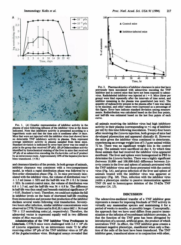
RESULTSPharmacokinetics of the TNF Inhibitor Produced by the
Adenoviral Construct. BALB/c mice were injected with 109,108, or 107 pfu of the TNF inhibitor virus. Plasma inhibitoryactivity, which was undetectable in samples obtained prior toinjection of the adenovirus, rose abruptly, peaking on day 2in mice that received 107 pfu and on day 8 in mice thatreceived 108 or 109 pfu (Fig. 1A). Between dosage groups,peak plasma concentrations were separated by approxi-mately one order of magnitude, reflecting the dose depen-dency of inhibitor production. Control mice that received 109pfu of virus encoding f3galactosidase had no detectableplasma inhibitor activity at any time point (data not shown).Approximately 10% of the hepatocytes of the control micewere transduced by this dose ofvirus, as indicated by stainingfor l3-galactosidase activity (Fig. 1B).A rapid decline of inhibitor production was observed by day
10 in the group ofmice inoculated with 108 pfu and by day 28-35in the group of mice inoculated with 109 pfu. Since the proteinencoded by the adenovirus contained a human receptor moiety,we considered it possible that antibody formation eventuated inrapid clearance of the recombinant protein. To investigatewhether this was so, we injected mice from the group that hadreceived 109 pfu and had ceased to express inhibitory activity inthe plasma for a period of 4 weeks, as well as control animalsthat had not been exposed to the recombinant protein, withpurified inhibitor labeled with 125I and analyzed the distribution
216 Immunology: Kolls et al.
Proc. Natl. Acad. Sci. USA 91 (1994) 217
-.0- io7 pfu-e- 10-8 pfu
I--10-9 pfu
Ei4-
Time, days
0 40 50Time, hr
FiG. 1. (A) Graphic representation of inhibitor activity in theplasma of mice following infusion of the inhibitor virus at the dosesindicated. Note that inhibitory activity is presented according to alogarithmic scale and that the time axis is nonlinear after 14 days.Mice that were not injected with the inhibitor virus (not shown) haveno detectable TNF inhibitory activity in plasma. Data representaverage inhibitory activity in plasma sampled from three mice.Standard deviation is indicated by error bars (error was too small toprint in the group that received 109 pfu). (B) P-Galactosidase activityidentified by histochemical staining of the liver in mice that received109 pfu of an adenovirus encoding the Escherichia coli lacZ productby an intravenous route. Approximately 10% ofthe hepatocytes havebeen transduced. (x50.)
and clearance kinetics ofthe protein. In both groups ofanimals,inhibitor clearance was consistent with a two-compartmentmodel, in which a rapid distribution phase was followed by afirst-order elimination phase (Fig. 2). In mice previously inoc-ulated with the inhibitor virus, the volume of distribution was 5
+ 1.5 ml (mean ± SD) and the half-life was 29 ± 0.1 hr (meanSD). In control control mice, the volume of distribution was
6.8 ± 1.5 ml, and the half-life was 36 ± 6.8 hr. The differencein half-life was thus small and beneath statistical significance (P> 0.05, Student's ttest). Therefore, we could not explain the fallin inhibitor levels on the basis of shortened half-life resultingfrom immunization and presume that production ofthe inhibitordeclines several weeks following viral transduction. Seventy-two hours after infection, similar inhibitor concentrations wereobserved in C57B/10 mice and in BALB/c mice (662 ± 36.6 and739 ± 115 neutralizing units/Al, respectively). Therefore, theadenoviral vector is expressed equally well in two differentstrains of Mus musculus.
Administration of the TNF Inhibitor Virus Predisposes toListeriosis. C57B/6 mice were challenged with 2.4 x 10⁴ cfuof Listeria organisms by an intravenous route 72 hr afterreceiving either 109 pfu of the TNF inhibitor virus or 109 pfuof the 3-galactosidase virus. Biological assays revealed that
FIG. 2. Pharmacokinetics ofinhibitor clearance in mice that havepreviously been inoculated with adenovirus encoding the TNFinhibitor and in control mice that have not been exposed to adeno-virus. Radiolabeled inhibitor was injected at t = 0. Mice (three pergroup) were bled repeatedly after the intervals of time noted, andinhibitor remaining in the plasma was quantitated (see text). Thequantity of radioactivity present in the plasma after 5 min was takento be maximal, and other values were expressed as a percentage ofthis figure. Error bars indicate standard deviation among measure-ments. Redistribution was calculated based on the first four points,and half-life was estimated based on the last four points of eachcurve.
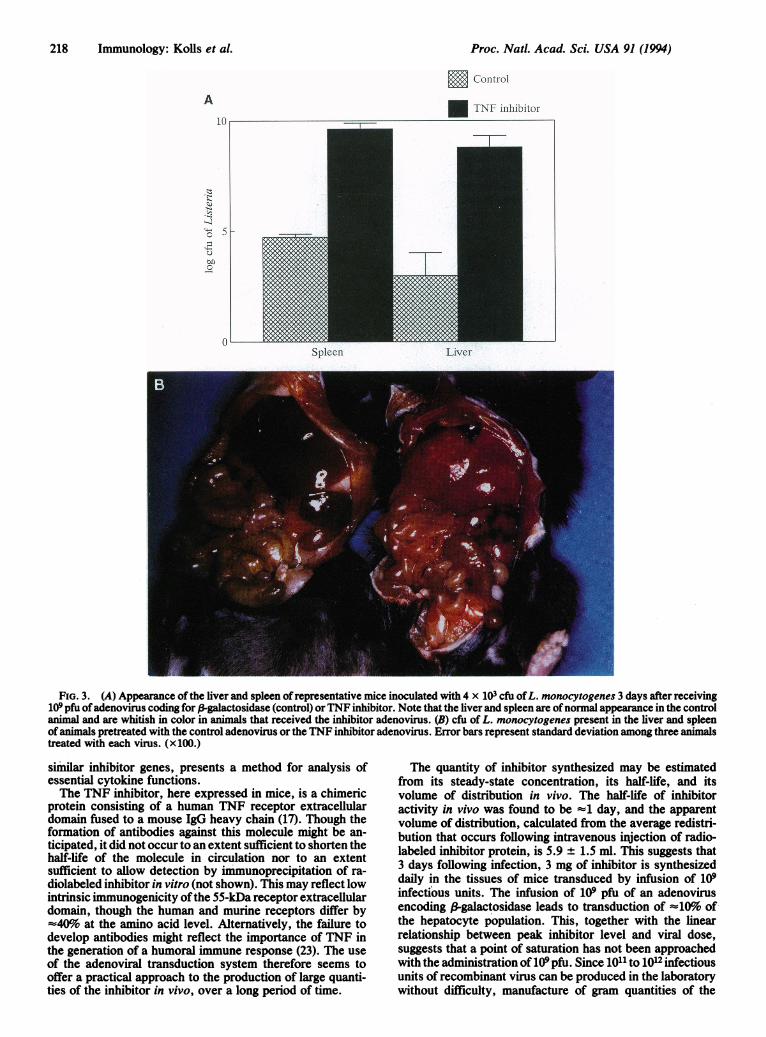
all animals receiving the inhibitor virus had high inhibitoryactivity in their plasma (corresponding to =1 mg of inhibitorper ml) by this time following inoculation. Twenty-four hoursafter receiving the Listeria injection, both groups ofmice haddeveloped piloerection and appeared clinically ill. Howeverthe mice given the inhibitor virus continued to deteriorate,experiencing an average weight loss of 3.5 g per animal within72 hr. There was no significant weight loss in the controlgroup. The animals were sacrificed on day 4, by which timethose animals that had received the inhibitor virus appearedmoribund. The liver and spleen were homogenized in PBS todetermine the Listeria burden. There was a highly significant(between 10,000- and 100,000-fold) difference between Lis-teria counts in the liver and spleen of animals pretreated withthe TNF inhibitor virus and those pretreated with the controlvirus (Fig. 3A), and gross infection of the liver and spleen ofanimals treated with the inhibitor virus was apparent atnecropsy (Fig. 3B). Thus, infection with the inhibitor virusyields a phenotype similar to passive immunization againstTNF (9) and to homozygous deletion of the 55-kDa TNFreceptor (14, 15).
DISCUSSIONThe adenovirus-mediated transfer of a TNF inhibitor generepresents a means for imposing blockade ofTNF activity invirtually any mammalian species and maintaining it for aperiod ofweeks to months, or possibly longer.-Conceptually,the approach that we have used differs from passive immu-nization orthe infusion ofrecombinant inhibitory proteins, inthat the function of the TNF gene has been abrogated byintroduction ofa second, artificial gene, the product ofwhichneutralizes TNF. In effect, the TNF inhibitor gene creates adominant negative phenotype, manifested when only a frac-tion of the cells of the host have been transduced. The TNFinhibitor gene, transduced alone or in conjunction with other,
Immunology: Kolls et al.
Proc. Natl. Acad. Sci. USA 91 (1994)
E Control
A* TNFI- inilhibitor
Spleen Liver
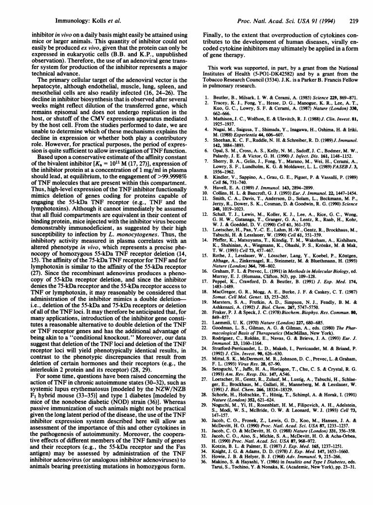
FiG. 3. (A) Appearance of the liver and spleen of representative mice inoculated with 4 x 10³ cfu ofL. monocytogenes 3 days after receiving109 pfu ofadenovirus coding for f-galactosidase (control) orTNF inhibitor. Note that the liver and spleen are ofnormal appearance in the controlanimal and are whitish in color in animals that received the inhibitor adenovirus. (B) cfu of L. monocytogenes present in the liver and spleenof animals pretreated with the control adenovirus or the TNF inhibitor adenovirus. Error bars represent standard deviation among three animalstreated with each virus. (x100.)
similar inhibitor genes, presents a method for analysis ofessential cytokine functions.The TNF inhibitor, here expressed in mice, is a chimeric
protein consisting of a human TNF receptor extracellulardomain fused to a mouse IgG heavy chain (17). Though theformation of antibodies against this molecule might be an-ticipated, it did not occur to an extent sufficient to shorten thehalf-life of the molecule in circulation nor to an extentsufficient to allow detection by immunoprecipitation of ra-diolabeled inhibitor in vitro (not shown). This may reflect lowintrinsic immunogenicity ofthe 55-kDa receptor extracellulardomain, though the human and murine receptors differ by-40%o at the amino acid level. Alternatively, the failure todevelop antibodies might reflect the importance of TNF inthe generation of a humoral immune response (23). The useof the adenoviral transduction system therefore seems tooffer a practical approach to the production of large quanti-ties of the inhibitor in vivo, over a long period of time.
The quantity of inhibitor synthesized may be estimatedfrom its steady-state concentration, its half-life, and itsvolume of distribution in vivo. The half-life of inhibitoractivity in vivo was found to be =1 day, and the apparentvolume of distribution, calculated from the average redistri-bution that occurs foilowing intravenous injection of radio-labeled inhibitor protein, is 5.9 ± 1.5 ml. This suggests that3 days following infection, 3 mg of inhibitor is synthesizeddaily in the tissues of mice transduced by infusion of 109infectious units. The infusion of 109 pfu of an adenovirusencoding f-galactosidase leads to transduction of =40% ofthe hepatocyte population. This, together with the linearrelationship between peak inhibitor level and viral dose,suggests that a point of saturation has not been approachedwith the administration of 109 pfu. Since 1011 to 1012 infectiousunits of recombinant virus can be produced in the laboratorywithout difficulty, manufacture of gram quantities of the
1.(
(-)
-.1
c )
()
218 Immunology: Kolls et al.
Proc. Natl. Acad. Sci. USA 91 (1994) 219
inhibitor in vivo on a daily basis might easily be attained usingmice or larger animals. This quantity of inhibitor could noteasily be produced ex vivo, given that the protein can only beexpressed in eukaryotic cells (B.B. and K.P., unpublishedobservation). Therefore, the use of an adenoviral gene trans-fer system for production of the inhibitor represents a majortechnical advance.The primary cellular target of the adenoviral vector is the
hepatocyte, although endothelial, muscle, lung, spleen, andmesothelial cells are also readily infected (16, 24-26). Thedecline in inhibitor biosynthesis that is observed after severalweeks might reflect dilution of the transferred gene, whichremains episomal and does not undergo replication in thehost, or shutoff of the CMV expression apparatus mediatedby the host cell. From the studies performed to date, we areunable to determine which of these mechanisms explains thedecline in expression or whether both play a contributoryrole. However, for practical purposes, the period of expres-sion is quite sufficient to allow investigation ofTNF function.Based upon a conservative estimate of the affinity constant
of the bivalent inhibitor [Ka = 1011 M (17, 27)], expression ofthe inhibitor protein at a concentration of 1 mg/ml in plasmashould lead, at equilibrium, to the engagement of >99.9998%of TNF molecules that are present within this compartment.Thus, high-level expression of the TNF inhibitor functionallymimics deletion of genes coding for proteins capable ofengaging the 55-kDa TNF receptor (e.g., TNF and thelymphotoxins). Although it cannot immediately be assumedthat all fluid compartments are equivalent in their content ofbinding protein, mice injected with the inhibitor virus becomedemonstrably immunodeficient, as suggested by their highsusceptibility to infection by L. monocytogenes. Thus, theinhibitory activity measured in plasma correlates with analtered phenotype in vivo, which represents a precise phe-nocopy of homozygous 55-kDa TNF receptor deletion (14,15). The affinity of the 75-kDa TNF receptor for TNF and forlymphotoxin is similar to the affinity of the 55-kDa receptor(27). Since the recombinant adenovirus produces a pheno-copy of 55-kDa receptor deletion, and since the inhibitordenies the 75-kDa receptor and the 55-kDa receptor access toTNF or lymphotoxin, it may reasonably be considered thatadministration of the inhibitor mimics a double deletion-i.e., deletion of the 55-kDa and 75-kDa receptors or deletionof all of the TNF loci. It may therefore be anticipated that, formany applications, introduction of the inhibitor gene consti-tutes a reasonable alternative to double deletion of the TNFor TNF receptor genes and has the additional advantage ofbeing akin to a "conditional knockout." Moreover, our datasuggest that deletion of the TNF loci and deletion of the TNFreceptor loci will yield phenotypically identical results, incontrast to the phenotypic discrepancies that result fromdeletion of certain hormones and their receptors (e.g., theinterleukin 2 protein and its receptor) (28, 29).For some time, questions have been raised concerning the
action ofTNF in chronic autoimmune states (30-32), such assystemic lupus erythematosus [modeled by the NZW/NZBF1 hybrid mouse (33-35)] and type 1 diabetes [modeled bymice of the nonobese diabetic (NOD) strain (36)]. Whereaspassive immunization of such animals might not be practicalgiven the long latent period of the disease, the use of the TNFinhibitor expression system described here will allow anassessment of the importance of this and other cytokines inthe pathogenesis of autoimmunity. Moreover, the coopera-tive effects of different members of the TNF family of genesand their receptors (e.g., the 55-kDa receptor and the Fasantigen) may be assessed by administration of the TNFinhibitor adenovirus (or analogous inhibitor adenoviruses) to
Finally, to the extent that overproduction of cytokines con-tributes to the development of human diseases, virally en-coded cytokine inhibitors may ultimately be applied in a formof gene therapy.
This work was supported, in part, by a grant from the NationalInstitutes of Health (5-PO1-DK42582) and by a grant from theTobacco Research Council (3534). J.K. is a Parker B. Francis Fellowin pulmonary research.
1. Beutler, B., Milsark, I. W. & Cerami, A. (1985) Science 229, 869-871.2. Tracey, K. J., Fong, Y., Hesse, D. G., Manogue, K. R., Lee, A. T.,
Kuo, G. C., Lowry, S. F. & Cerami, A. (1987) Nature (London) 330,662-666.
3. Mathison, J. C., Wolfson, E. & Ulevitch, R. J. (1988)J. Clin. Invest. 81,1925-1937.
4. Nagai, M., Saigusa, T., Shimada, Y., Inagawa, H., Oshima, H. & Iriki,M. (1988) Experientia 44, 606-607.
5. Sheehan, K. C. F., Ruddle, N. H. & Schreiber, R. D. (1989)J. Immunol.142, 3884-3893.
6. Opal, S. M., Cross, A. S., Keily, N. M., Sadoff, J. C., Bodmer, M. W.,Palardy, J. E. & Victor, G. H. (1990) J. Infect. Dis. 161, 1148-1152.
7. Sherry, B. A., Gelin, J., Fong, Y., Marano, M., Wei, H., Cerami, A.,Lowry, S. F., Lundholm, K. G. & Moldawer, L. L. (1989) FASEB J. 3,1956-1962.
8. Kindler, V., Sappino, A., Grau, G. E., Piguet, P. & VassaUli, P. (1989)Cell 56, 731-740.
9. Havell, E. A. (1989) J. Immunol. 143, 2894-2899.10. Collins, H. L. & Bancroft, G. J. (1993) Eur. J. Immunol. 22, 1447-1454.11. Smith, C. A., Davis, T., Anderson, D., Solam, L., Beckmann, M. P.,
Jerzy, R., Dower, S. K., Cosman,-D. & Goodwin, R. G. (1990) Science248, 1019-1023.
12. Schall, T. J., Lewis, M., Kolier, K. J., Lee, A., Rice, G. C., Wong,G. H. W., Gatanaga, T., Granger, G. A., Lentz, R., Raab, H., Kohr,W. J. & Goeddel, D. V. (1990) Cell 61, 361-370.
13. Loetscher, H., Pan, Y.-C. E., Lahm, H.-W., Gentz, R., Brockhaus, M.,Tabuchi, H. & Lesslauer, W. (1990) Cenl 61, 351-359.
14. Pfeffer, K., Matsuyama, T., Kundig, T. M., Wakeham, A., Kishihara,K., Shahinian, A., Wiegmann, K., Ohashi, P. S., Kr6nke, M. & Mak,T. W. (1993) Cell 73, 457-467.
15. Rothe, J., Lesslauer, W., Losscher, Lang, Y., Koebel, P., Kontgen,Althage, A., Zinkernagel, R., Steinmetz, M. & Bluethmann, H. (1993)Nature (London) 364, 798-802.
16. Graham, F. L. & Prevec, L. (1991) in Methods in Molecular Biology, ed.Murray, E. J. (Humana, Clifton, NJ), pp. 109-128.
17. Peppel, K., Crawford, D. & Beutler, B. (1991) J. Exp. Med. 174,1483-1489.
18. MacGregor, G. R., Mogg, A. E., Burke, J. F. & Caskey, C. T. (1987)Somat. Cell Mol. Genet. 13, 253-265.
19. Marsters, S. A., Frutkin, A. D., Simpson, N. J., Fendly, B. M. &Ashkenazi, A. (1992) J. Biol. Chem. 267, 5747-5750.
20. Fraker, P. J. & Speck, J. C. (1978) Biochem. Biophys. Res. Commun. 80,849-857.
21. Laemmli, U. K. (1970) Nature (London) 227, 680-685.22. Goodman, L. S., Gilman, A. G. & Gilman, A., eds. (1980) The Phar-
macological Basis of Therapeutics (MacMillan, New York).23. Rodrfguez, C., Roldin, E., Navas, G. & Brieva, J. A. (1993) Eur. J.
Immunol. 23, 1160-1164.24. Stratford-Perricaudet, L. D., Makeh, I., Perricaudet, M. & Briand, P.
(1992) J. Clin. Invest. 90, 626-630.25. Mittal, S. K., McDermott, M. R., Johnson, D. C., Prevec, L. & Graham,
F. L. (1993) Virus Res. 28, 67-90.26. Setoguchi, Y., Jaffe, H. A., Horiagon, T., Chu, C. S. & Crystal, R. G.
(1993) Am. Rev. Resp. Dis. 147, A546.27. Loetscher, H., Gentz, R., Zulauf, M., Lustig, A., Tabuchi, H., Schlae-
ger, E., Brockhaus, M., Gallati, H., Manneberg, M. & Lesslauer, W.(1991) J. Biol. Chem. 266, 18324-18329.
28. Schorle, H., Holtschke, T., Hilnig, T., Schimpl, A. & Horak, I. (1991)Nature (London) 352, 621-624.
29. Noguchi, M., Yi, H., Rosenblatt, H. M., Filipovich, A. H., Adelstein,S., Modi, W. S., McBride, 0. W. & Leonard, W. J. (1993) Cell 73,147-157.
30. Jacob, C. O., Fronek, Z., Lewis, G. D., Koo, M., Hansen, J. A. &McDevitt, H. 0. (1990) Proc. Natl. Acad. Sci. USA 87, 1233-1237.
31. Jacob, C. 0. & McDevitt, H. 0. (1988) Nature (London) 331, 356-358.32. Jacob, C. O., Aiso, S., Michie, S. A., McDevitt, H. 0. & Acha-Orbea,
H. (1990) Proc. Natl. Acad. Sci. USA 87, 968-972.33. Kotzin, B. L. & Palmer, E. (1987) J. Exp. Med. 165, 1237-1251.34. Knight, J. G. & Adams, D. D. (1978) J. Exp. Med. 147, 1653-1660.35. Howie, J. B. & Helyer, B. J. (1968) Adv. Immunol. 9, 215-266..36. Makino, S. & Hayashi, Y. (1986) in Insulitis and Type I Diabetes, eds.
animals bearing preexisting mutations in homozygous form.
Immunology: Kofls et al.
Tanii, S., Tochino, Y. & Nonaka, K. (Academic, New York), pp. 23-31.