Proconvulsive effect of hydrochlorothiazide in an in vitro rat seizure model
7
Iranian Journal of Basic Medical Sciences ijbms.mums.ac.ir Proconvulsive effect of hydrochlorothiazide in an in vitro rat seizure model Christoph Kellinghaus 1 *, Ali Gorji 2, 3, 4 1 Department of Neurology, Klinikum Osnabrück, Am Finkenhügel 1, 49076 Osnabrück, Germany 2 Institute of Neurophysiology, University of Münster, Robert-Koch-Str. 115, 48149 Münster, Germany 3 Department of Neurology and Department of Neurosurgery, University of Münster, Albert-Schweitzer-Campus A1, 48029 Münster, Germany 4 Shefa Neuroscience Center, Khatam Hospital, Rashid Yasemi St., Valiasr Ave, Tehran, Iran A R T I C L E I N F O A B S T R A C T Article type: Original article Objective(s): Protective effects of diuretics, particularly of hydrochlorothiazide (HCT), for the development of epilepsy have been described in vivo. However, its mechanism of action is unknown. Materials and Methods: Extracellular field potentials were recorded from the CA1- and CA3- subfields of the hippocampus of rats. Epileptiform discharges were induced by omission of Mg 2+ from the artificial cerebrospinal fluid (ACSF). HCT was added to the ACSF at a concentration of 2 mmol/l, 0.2 mmol/l or 0.02 mmol/l. Frequency, amplitude and duration of the epileptiform discharges were evaluated. Long-term potentiation (LTP) was induced with and without the presence of HCT (n=6; 2 mmol/l). In addition, rats were injected with HCT (n=4) or saline (n=2), and the brain tissue was analyzed using HPLC. Results: Application of 0.02, 0.2, and 2 mmol/l HCT accelerated the frequency of discharges by 50%, 91%, and 100%, respectively. The amplitude of burst discharges also increased by 9%, 54%, and 300%, and the duration of epileptiform discharges increased by 10%, 30% and 120%. All parameters returned close to the basal levels after 60min washout of the substance. HCT increased the electrical evoked potentials but did not affect the LTP in hippocampal tissues. There was no evidence of HCT in the rat brain after intraperitoneal injection. Conclusion: Exposure of hippocampal slices to HCT enhanced epileptiform activity in a dose- dependent manner. In addition, HCT does not seem to cross the blood brain barrier in rats. Thus, the anticonvulsive effect of HCT most likely is not through direct neuronal effect. Article history: Received: Dec 19, 2013 Accepted: Jul 14, 2014 Keywords: Blood-brain-barrier Diuretics Epilepsy Hippocampal slices ►Please cite this paper as: Kellinghaus Ch, Gorji A. Proconvulsive effect of hydrochlorothiazide in an in vitro rat seizure model. Iran J Basic Med Sci 2014; 17:860-866. Introduction Approximately 20 to 30% of the patients with epilepsy, ultimately develop refractory seizures that do not respond adequately to pharmacological treatment (1). These patients have only a small chance of obtaining seizure control with one of the established anticonvulsant drugs. Therefore, anticonvulsant drugs with different and new mechanisms of action are needed. One of the groups of drugs that have come into focus recently is the group of diuretics (2). Although the first reports of anticonvulsant effects of the loop diuretic furosemide date back nearly thirty years (3, 4), it was discovered only a decade ago that furosemide in high concentrations shows anticonvulsant properties in vitro (5). In the following, anticonvulsant effects of diuretics, particularly of furosemide, were shown using a variety of in vitro models of epilepsy (5-8). At the same time, there was epidemiological evidence that diuretic use could protect from the risk of new-onset seizures in a community-based sample of elderly patients (9). Most investigators have focused on furosemide, most likely because of its high diuretic potency and the availability of solutions for intravenous or intraperitoneal injections. This is in contrast to the fact that in the population-based study as well as in the maximum electroshock model, the protective effect of thiazide diuretics was stronger than of furosemide (9). In addition, it has been shown that HCT as well as other benzothiazide derivates potentiate the effect of low- dose phenytoin on the seizure threshold in the maximum electroshock model. The same study also showed that there is a tendency to elevate the seizure threshold by HCT alone (10). The mechanisms of anticonvulsant effect of HCT remain to be elucidated. Whereas, there is increasing evidence that furosemide has direct effect on neurons and glia via the electroneutral Na + -K + -2Cl- co-transporters (NKCC) that modulate intracellular chloride concentration and cell volume (11, 12). Thiazides do not have influence on the NKCC or the *Corresponding author: Christoph Kellinghaus. Department of Neurology, Klinikum Osnabrück, 49076 Osnabrück, Germany. Tel: +49-541-405-0; Fax: +49-541-405-6599; email: [email protected]
-
Upload
independent -
Category
Documents
-
view
6 -
download
0
Transcript of Proconvulsive effect of hydrochlorothiazide in an in vitro rat seizure model
Iranian Journal of Basic Medical Sciences
ijbms.mums.ac.ir
Proconvulsive effect of hydrochlorothiazide in an in vitro rat seizure model
Christoph Kellinghaus 1*, Ali Gorji 2, 3, 4
1 Department of Neurology, Klinikum Osnabrück, Am Finkenhügel 1, 49076 Osnabrück, Germany 2 Institute of Neurophysiology, University of Münster, Robert-Koch-Str. 115, 48149 Münster, Germany 3 Department of Neurology and Department of Neurosurgery, University of Münster, Albert-Schweitzer-Campus A1, 48029 Münster, Germany 4 Shefa Neuroscience Center, Khatam Hospital, Rashid Yasemi St., Valiasr Ave, Tehran, Iran
A R T I C L E I N F O A B S T R A C T
Article type: Original article
Objective(s): Protective effects of diuretics, particularly of hydrochlorothiazide (HCT), for the development of epilepsy have been described in vivo. However, its mechanism of action is unknown. Materials and Methods: Extracellular field potentials were recorded from the CA1- and CA3-subfields of the hippocampus of rats. Epileptiform discharges were induced by omission of Mg2+ from the artificial cerebrospinal fluid (ACSF). HCT was added to the ACSF at a concentration of 2 mmol/l, 0.2 mmol/l or 0.02 mmol/l. Frequency, amplitude and duration of the epileptiform discharges were evaluated. Long-term potentiation (LTP) was induced with and without the presence of HCT (n=6; 2 mmol/l). In addition, rats were injected with HCT (n=4) or saline (n=2), and the brain tissue was analyzed using HPLC. Results: Application of 0.02, 0.2, and 2 mmol/l HCT accelerated the frequency of discharges by 50%, 91%, and 100%, respectively. The amplitude of burst discharges also increased by 9%, 54%, and 300%, and the duration of epileptiform discharges increased by 10%, 30% and 120%. All parameters returned close to the basal levels after 60min washout of the substance. HCT increased the electrical evoked potentials but did not affect the LTP in hippocampal tissues. There was no evidence of HCT in the rat brain after intraperitoneal injection. Conclusion: Exposure of hippocampal slices to HCT enhanced epileptiform activity in a dose-dependent manner. In addition, HCT does not seem to cross the blood brain barrier in rats. Thus, the anticonvulsive effect of HCT most likely is not through direct neuronal effect.
Article history: Received: Dec 19, 2013 Accepted: Jul 14, 2014
Keywords: Blood-brain-barrier Diuretics Epilepsy Hippocampal slices
►Please cite this paper as: Kellinghaus Ch, Gorji A. Proconvulsive effect of hydrochlorothiazide in an in vitro rat seizure model. Iran J Basic Med Sci 2014; 17:860-866.
Introduction Approximately 20 to 30% of the patients with
epilepsy, ultimately develop refractory seizures that do not respond adequately to pharmacological treatment (1). These patients have only a small chance of obtaining seizure control with one of the established anticonvulsant drugs. Therefore, anticonvulsant drugs with different and new mechanisms of action are needed. One of the groups of drugs that have come into focus recently is the group of diuretics (2). Although the first reports of anticonvulsant effects of the loop diuretic furosemide date back nearly thirty years (3, 4), it was discovered only a decade ago that furosemide in high concentrations shows anticonvulsant properties in vitro (5). In the following, anticonvulsant effects of diuretics, particularly of furosemide, were shown using a variety of in vitro models of epilepsy (5-8). At the same time, there was epidemiological evidence that diuretic use could protect from the risk of new-onset seizures in a community-based sample of elderly patients (9).
Most investigators have focused on furosemide, most likely because of its high diuretic potency and the availability of solutions for intravenous or intraperitoneal injections. This is in contrast to the fact that in the population-based study as well as in the maximum electroshock model, the protective effect of thiazide diuretics was stronger than of furosemide (9). In addition, it has been shown that HCT as well as other benzothiazide derivates potentiate the effect of low-dose phenytoin on the seizure threshold in the maximum electroshock model. The same study also showed that there is a tendency to elevate the seizure threshold by HCT alone (10).
The mechanisms of anticonvulsant effect of HCT remain to be elucidated. Whereas, there is increasing evidence that furosemide has direct effect on neurons and glia via the electroneutral Na+-K+-2Cl- co-transporters (NKCC) that modulate intracellular chloride concentration and cell volume (11, 12). Thiazides do not have influence on the NKCC or the
*Corresponding author: Christoph Kellinghaus. Department of Neurology, Klinikum Osnabrück, 49076 Osnabrück, Germany. Tel: +49-541-405-0; Fax:
+49-541-405-6599; email: [email protected]
Kellinghaus and Gorji Proconvulsive effect of HCT in vitro
Iran J Basic Med Sci, Vol. 17, No. 11, Nov 2014
861
electroneutral K+-Cl- - cotransporters (KCC), either. However, it inhibits the carbonic anhydrase (13, 14). Carbonic anhydrase (CA) regulates the diffusion of HCO3- and protons across the cell membrane (2, 15) and its inhibition increases the hyperpolarizing effect of long-lasting GABA-A currents (16). Thus, the most favoured hypothesis is that the epidemiological effect of seizure protection by HCT intake may at least partially be an effect of CA inhibition (2).
No in vitro experiments investigating the anticonvulsant properties and mechanisms of thiazide diuretics in epilepsy models have been published so far. The aim of the present study was to evaluate whether hydrochlorothiazide (HCT), probably the most common used thiazide diuretic, plays anticonvulsant role in brain tissues in vitro. Furthermore, we investigated if HCT could pass blood-brain-barrier (BBB) to clear if its effect on CNS is a neuronal modulatory action or a systemic effect.
Materials and Methods Preparation of the slices and solutions
The experiments were performed on rat hippocampal slices. They have been performed according to national guidelines for animal research. The principles outlined in the Basel declaration have been considered when planning the experiment. After introduction of deep pentobarbital anesthesia, the animal was decapitated and the brain was removed. The hippocampi were dissected and cut into slices of 500 µm thickness. Then the slices were preincubated in artificial cerebrospinal fluid (ACSF) at 28°C for 1 hr under continuous equilibration with 5% CO2. The preincubation ACSF contained (in mmol/l): NaCl 124, KCl4, CaCl2 1.0, NaH2PO4 1.24, MgSO4 1.3, NaHCO3 26, and Glucose 10. After preincubation, CaCl2 was elevated to 2.0 mmol/l and the slices were transferred into an interphase-type recording chamber. In the experimental chamber, the temperature was increased to 32°C. During the experiments, the pH, the bath temperature and the flow rate (1.5 to 2 ml/min) remained constant. For the preparation of the zero-magnesium solution, we omitted MgSO4.
Hydrochlorothiazide (HCT) solution was prepared by adding dimethylsulfoxide (0.5 ml per mmol/l of the crystalline HCT substance) to achieve water solubility. Crystalline HCT was purchased by Sigma-Aldrich Germany, Munich, Germany.
Electrophysiological recordings
Extracellular field potential recordings were recorded with glass microelectrodes (Resistance 2-5 MΩ) in the stratum radiatum of the CA1 and CA3 hippocampal subregions connected with DC amplifiers. Extracellular recordings were obtained using a custom made differential amplifier (with band-pass filters at 0.5–10 kHz, sampling rate 0.3–100 Hz). The reference electrode and the connection to the
microelectrode were symmetrical Ag-Ag-KCl bridges. Field potentials were recorded by an ink-writer (capable to record between ± 10 V) as well as by a digital oscilloscope. Traces were digitized by Nicolet 460 digitizing oscilloscope (Nicolet Instruments) and analyzed with accompanying software.
Evoked field potentials and long-term potentiation (LTP)
A continuous single pulse of electrical stimulation was applied through a bipolar platinum electrode attached to the Schaffer collaterals of hippocampal slices. Evoked field excitatory post-synaptic potentials (fEPSP) were recorded in hippocampal CA1 region (stratum radiatum). The fEPSP was elicited by adjusting the intensity of stimulation to ∼50% of the maximum response (0.1-0.5 mA). The amplitude of fEPSP 1 m sec after the onset was measured for data analysis. In LTP experiments, the slice was sequentially stimulated once every min. Tetanic stimulation (100 Hz, for 1 sec, at baseline stimulation intensity) was operationally defined as the mean change in fEPSP amplitude for five stimuli given 30 min after tetanic stimulation compared with the mean fEPSP to five test pulses given immediately before the stimulation. Thus % potentiation= [(posttetanus amplitude/baseline amplitude) 1] 100. Tetanic stimulation was applied 60 min after application of HCT (2 mmol/l).
Experimental protocol
The protocol for the zero-magnesium model experiments consisted of four periods: 1) Superfusion with ACSF for 30 min to test for spontaneously occurring epileptiform field potentials (EFP; control period). Slices with spontaneously occurring EFP were excluded from further analysis. 2) Superfusion with zero-magnesium ACSF until there was a stable EFP frequency and amplitude for at least 20 min (baseline period). 3) Addition of HCT (2 mmol/l, n=5; 0.2 mmol/l, n=5; 0.02 mmol/l, n=5) to the magnesium-depleted ACSF for 60 min (HCT period). 4) Superfusion with zero-magnesium ACSF for 60 min (wash-out period).
EFP repetition rate was measured by the number of events per min. The EFP amplitude was measured peak-to-peak and EFP duration was determined as the width at half-maximal amplitude. Owing to the small group sizes that make normal distribution and variance homogeneity unlikely, we chose median and 25/75 percentiles for cohort description. The data were processed and analyzed using commercially available software (SPSS Version 12.1, Chicago, Ill., USA). Interval-and ordinal-scaled data were compared using the Wilcoxon test, and nominal-scaled data with the chi-square test or Fisher’s exact test (2x2 tables). Significance level was set at P<0.05 for two-tailed tests.
Proconvulsive effect of HCT in vitro Kellinghaus and Gorji
Iran J Basic Med Sci, Vol. 17, No. 11, Nov 2014
862
Brain tissue of 6 rats was used for HPLC analysis. Four animals were injected intraperitoneally (IP) with HCT (2 mmol/l, 5 ml), two animals only received saline. Two hr later, the animals were sacrificed with high doses of isoflurane followed by decapitation. The brain tissue was stored at -20 C for 12 to 48 hr until HPLC analysis.
HPLC was performed using a method developed by Zendelovska et al (17) for determination of HCT in human plasma. The system included a LiChrospher 100 RP8 column (125x5, 5 ) with the mobile phase consisting of 0.025 M phosphate buffer (adjusted to a pH of 5 with triethylamine) and acetonitile at a relation of 85/15, a pump, an UV-detector with variable wavelength and interface and dedicated software (LaChrom series, Merck-Hitachi, Darmstadt) running on a personal computer with Intel pentium III processor. The brain homogenate was centrifuged at 13000 rotations/min. The clear supernatant was transferred into an Eppendorf cap, and 40 l were injected into the HPLC system. The flow rate was 1ml/min, and the detection wave length was 230 nm.
Results The effect of HCT on 0-Mg2+-induced EFP
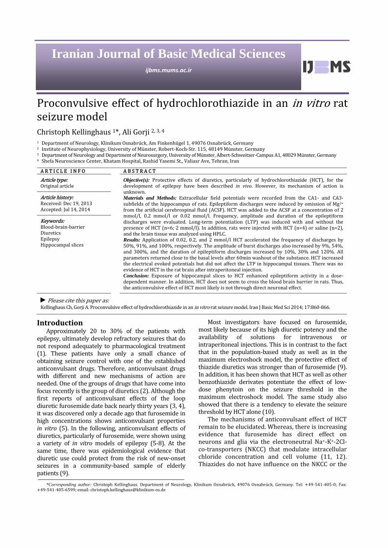
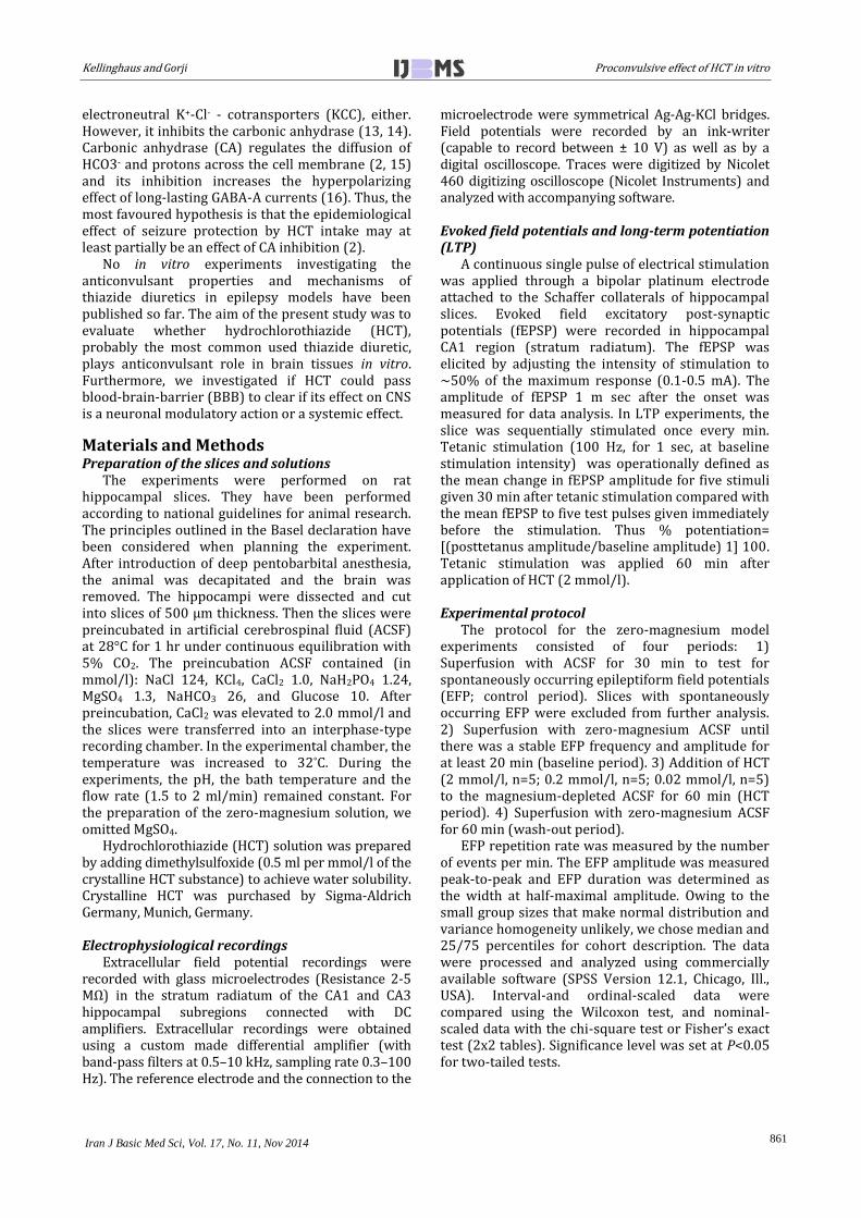
Only slices without spontaneous epileptiform discharges during control period were investigated. Application of Mg2+-free ACSF resulted in EFP after a median latency of 41 min (2 mmol/l HCT), 47 min
(0.2 mmol/l HCT) and 43 min (0.02 mmol/l HCT), respectively. After reaching a stable level over 15 min, median baseline repetition rate was 13/min (2 mmol/l HCT), 11/min (0.2 mmol/l HCT) and 10/min (0.02 mmol/l HCT) respectively. The differences were not significant. Addition of HCT to the bath resulted marked increase of repetition rate, amplitude and duration of the epileptiform field potentials (Figure 1). After 1 hr, the increase of the repetition rate was highest with 2 mmol/l HCT (median 26/min, 25-75 quartiles 21.5/min-38/min),
Figure 1. Extracellular field potential recordings from a rat hippocampal slice using the zero magnesium model; at baseline (a), after addition of HCT (b) and during wash-out (c)
Figure 2. Mean discharge repetition rate (a) and mean discharge amplitude of the field potential recordings of three groups of rats (n=5) using 0.02 mmol, 0.2 mmol and 2 mmol HCT solution
Kellinghaus and Gorji Proconvulsive effect of HCT in vitro
Iran J Basic Med Sci, Vol. 17, No. 11, Nov 2014
863
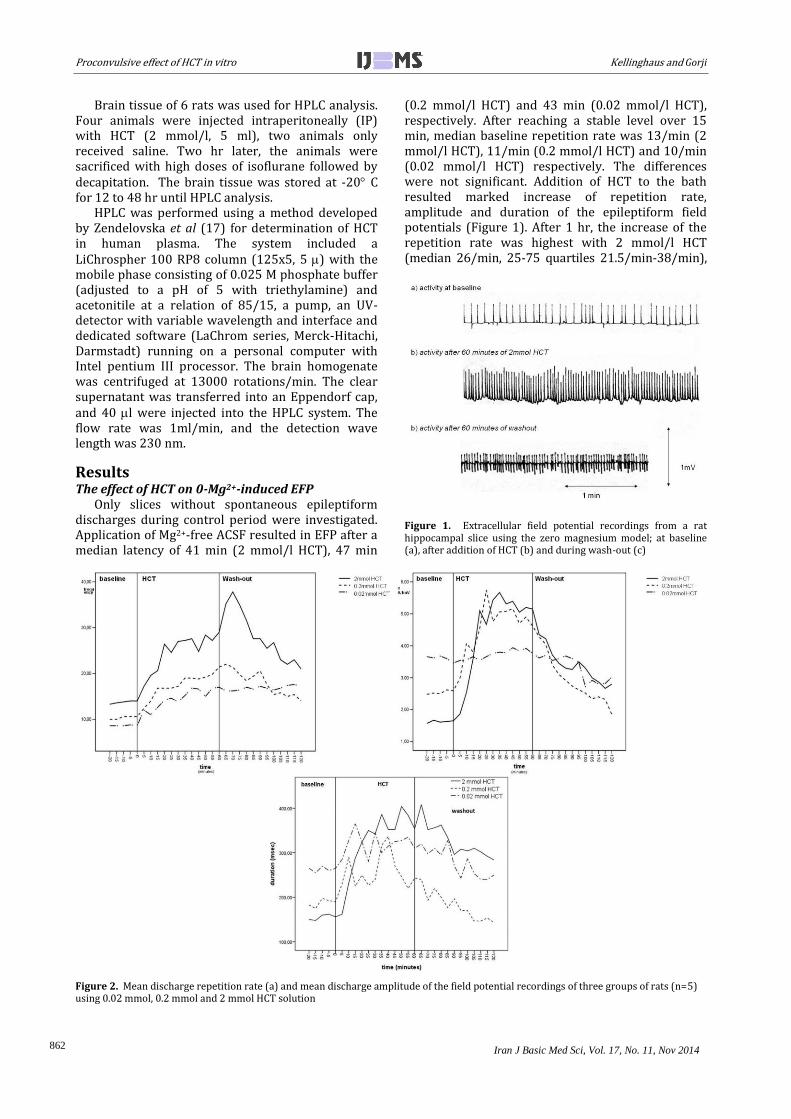
Figure 3. Long-term potentiation (LTP) of the evoked field excitatory post-synaptic potentials (fEPSP) in neocortical preparations. (a) Tetanic stimulation ((100 Hz, for 1 sec, at baseline stimulation intensity) produces a rapid and stable potentiation in the amplitude of the evoked field potentials, calculated as a percentage of baseline mean response amplitude. Solid and open triangles show the evoked fEPSP after application of hydrochlorothiazide (HCT, 2 mmol/l) and control, respectively. Arrow shows the time of tetanic stimulation, 60 min after application of HCT and artificial cerebrospinal fluid (control). The time points given refer to LTP induction. (b) Representative examples of the fEPSP before and after tetanic stimulation in control and HCT affected slices
slightly lower with 0.2 mmol/l HCT (median 21/min, 25-75 quartiles 18.5-24.5/min) and with 0.02 mmol HCT (median 15/min, 25-75 quartiles 14,5-20.5/min). The inter-group differences were statistically significant, as were the differences between baseline and 1 hr exposition within each group (P<0.05). The amplitude increased from a median of 0.18 mV to a median of 0.55 mV in the 2 mmol group, from a median of 0.24 mV to a median of 0.37 mV in the 0.2 mmol/l group, and from a median of 0.21 mV to a median of 0.23 mV in the 0.02 mmol/l group. Only the within-group differences of the 2 mmol/l and the 0.2 mmol/l groups were significant (P<0.05). Discharge duration also increased from 120 msec to 340 msec (median) in the 2 mM group, from 190 msec to 230 msec (median) in the 0.2 mmol/l group, and from 270 msec to 320 msec (median) in the 0.02 mmol/l group. Differences within groups, but not inter-group differences were statistically significant. After 60 min
of wash-out, repetition rate, discharge amplitude and discharge duration gradually returned to baseline levels (Figure 2). The effect of HCT on LTP
A conditioning tetanic stimulation was delivered to the Schaffer collaterals pathway followed by pulses with stimulation parameters identical to control values. The evoked fEPSP was stable for at least 30 min (with mean amplitude of 0.3 ± 0.03 mV) before application of tetanic stimulation (less than 10% variation; Figure 3). Administration of tetanic stimulation produced a rapid, stable, and lasting enhancement of the amplitude of the fEPSP in all tested preparations (n=6, 147±1.6% control; Figure 3 B). The potentiation rose within 2 to 3 min and stabilized within 5–10 min after the train of stimulations. Application of HCT (2 mmol/l; n = 8) 60 min before tetanic stimulation did not change LTP induction in all tested slices (146 ± 1.4% control, Figure 3B). Enhancement of LTP after application of HCT did not significantly differ from control slices (P<0.05).
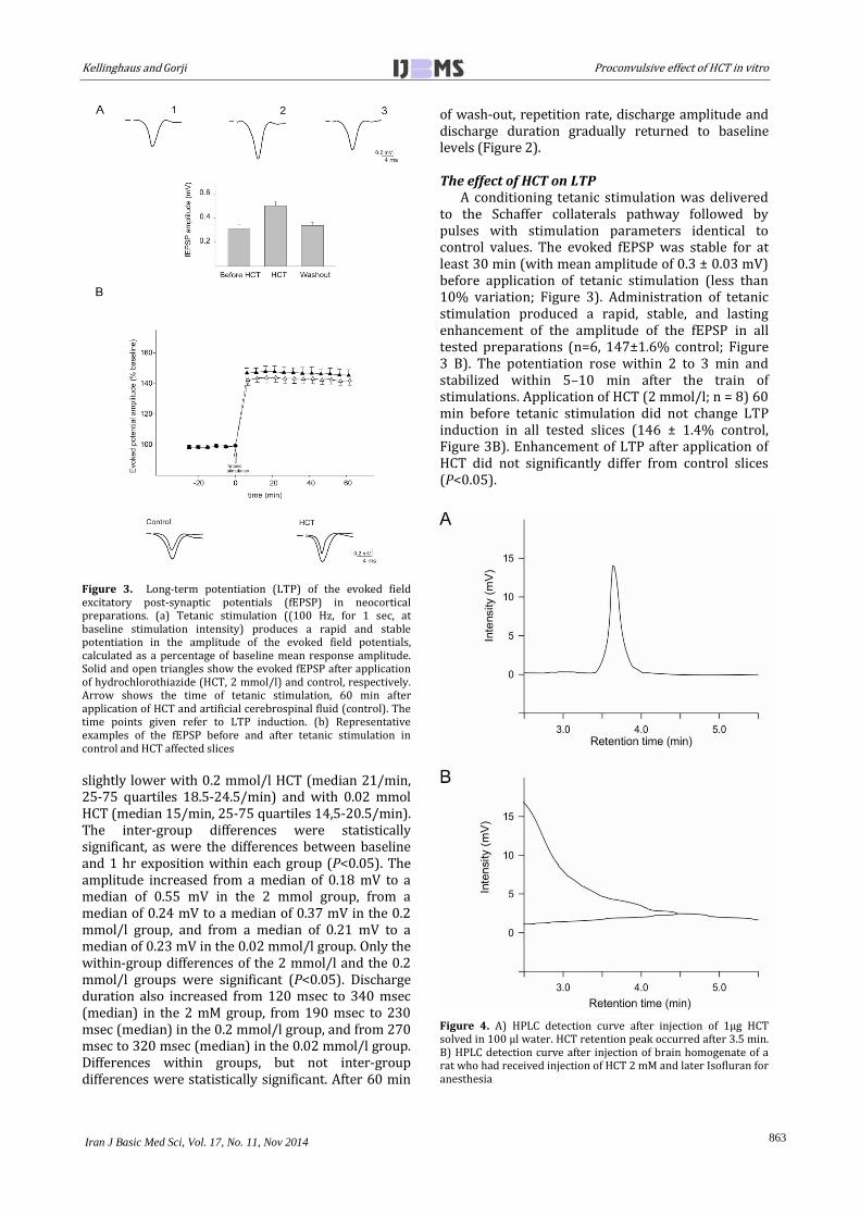
Figure 4. A) HPLC detection curve after injection of 1µg HCT solved in 100 µl water. HCT retention peak occurred after 3.5 min. B) HPLC detection curve after injection of brain homogenate of a rat who had received injection of HCT 2 mM and later Isofluran for anesthesia
Proconvulsive effect of HCT in vitro Kellinghaus and Gorji
Iran J Basic Med Sci, Vol. 17, No. 11, Nov 2014
864
Measurement of HCT concentration in the brain after IP injection
In HPLC, the peak of HCT appeared at a retention time of approximately 3.5 min. There was a linear dose-concentration curve 0.5 g and 100 g HCT. When brain homogenate of HCT-treated rats was injected, HPLC showed no significant peak at the HCT retention time (Figure 4). There was also no peak when the brain of rats injected with saline (control) was analyzed.
Discussion In contrast to our expectations, HCT increased
repetition rate, amplitude and duration of the epileptiform discharges elicited by omission of Mg2+ from the bath solution in a dose-dependent manner. HCT also increased the amplitude of electrically evoked field potentials, but did not have any influence on the LTP induced by tetanic stimulations applied to Schaffer collaterals. When intraperitoneally injected, HPLC analysis did not show evidence of HCT in the brain even at high doses.
Our recordings used the zero-Mg2+ model of epileptiform activity (18-20). This model initiates robust epileptiform activity in both normal and lesioned cortex by augmenting the NMDA-component of EPSP, without pharmacological blockade of synaptic receptors (i.e., GABAA receptors in the bicuculline model), and represents a model for pharmacoresistant status epilepticus. This model represents a well-controlled analysis of drug effects independent of systemic influences (18-20). This model quite closely imitates in vivo conditions because recurrent and lateral inhibition remains intact. In addition, low-magnesium activity induces post-synaptic calcium influx, resulting in activation of the same calcium dependent second messenger mechanisms that underlie activity-dependent neuronal plasticity (20).
It has been found that diazoxide, another benzothiazide diuretic, is a potent and reversible gating modifier of AMPA-type glutamate receptor channels (22) that inhibits the rapid desensitization of the response to a constant agonist concentration. The most potent agent of the group of thiazide desensitizers is cyclothiazide (CTZ) (23, 24) that recently has been proposed as an in vitro epilepsy model (25). Although there is chemical similarity between different benzothiazide diuretics, the effect on quisqualat-induced AMPA-desensitation has been shown to be quite variable. In concentrations of 500umol, HCT also potentiated the steady-state component of quisqualat-activated currents (23), but to a much smaller amount than cyclothiazide. It remains undetermined whether HCT also - like CTZ - increases pre-synaptic glutamate release (26) or directly inhibits GABA-A receptors (27).
LTP is a phenomenon in which a constant pre-synaptic high stimulation of excitatory amino acids
neuronal pathways results in prolonged enhanced post-synaptic responses. It is well established that NMDA receptors are a molecular detector of the coincidence of both the pre-synaptic release of glutamate and a post-synaptic depolarization at the origin of LTP induction (28). HCT did not affect LTP. In keeping with our data, it has been shown that the HCT (500 μM) potently inhibits non-NMDA receptors desensitization in cultured hippocampal neurons (23). Cyclothiazide (300 μM), a thiazide diuretic and an allosteric inhibitor of non-NMDA receptor desensitization, induced potentiated low Mg2+–induced seizure-like events recorded from the CA3 pyramidal layer of juvenile rat hippocampal slices (29). In line with our results, cyclothiazide administered intracerebral ventricle induced epileptiform activities from an initial of multiple evoked population spikes, progressed to spontaneous spikes and finally to highly synchronized burst activities in hippocampal CA1 neurons (30, 31). In contrast to in vivo studies, systemic administration of thiazides, including hydrochlorothiazide (100 mg), enhanced the anticonvulsant action of carbamazepine (32) and gabapentin (33) in the maximal electroshock seizure model in mice.
In our experiments, we could not find evidence that HCT does permeate BBB in the rat. This is in agreement with studies of dogs where dosages as high as 500 mg/kg administered twice daily did not result in measurable quantities (<3 mg/l) of the drug in CSF or brain (34). Unfortunately, there is no data available in the literature regarding the BBB permeability for HCT in humans. In addition, there is no evidence for central nervous effects of HCT even in the setting of overdose intoxication (35). One could argue that HCT may influence ion transport at the BBB without actually permeating it. However, HCT at a relatively high concentration of 1.5 mmol/l does not have any effect on the blood to brain sodium transport in vitro (36).
The anticonvulsant effect of HCT in vivo most likely is mediated via mechanisms based on effects outside the brain. It seems unlikely that the inhibition of the carbonic anhydrase plays a relevant role. Dashputra and colleagues found that there is no correlation between the effect on the carbonic anhydrase and the potentiation of the anticonvulsant effect of phenytoin in the maximal electroshock model in rats (10). Systemic acidosis seems equally unlikely to be related to the protective effect of HCT and other benzothiazides because acetazolamide raises the seizure threshold before there has been time for substantial bicarbonate loss (37). It is very unlikely that the protective effect of HCT in humans is due to enhancing the action of standard anticonvulsants because the population under study did not suffer from epilepsy (9, 38).
There are limitations to our study. First, we did

Kellinghaus and Gorji Proconvulsive effect of HCT in vitro
Iran J Basic Med Sci, Vol. 17, No. 11, Nov 2014
865
not investigate the effect of HCT in other in vitro models of epilepsy. Therefore, generalizability is limited. Moreover, we arbitrarily chose HCT concentrations ranging from 0.02 mmol/l to 2 mmol/l. We intended to look across a large window of possible concentrations. However, HCT concentration in plasma (or brain) of humans when exposed to HCT in clinical practice may be much lower (39). Thus, we may have missed relevant effects of concentrations below our lowest concentration, or between concentrations. In addition, the group sizes were rather small. However, the effects we have observed seem rather robust, which makes it unlikely that with larger groups we would have seen different results.
The protective effect of HCT and the other diuretics is rather mild in the epidemiological studies. Moreover, in the animal studies presented by Hesdorffer et al (9), HCT (as well as furosemide) was applied in doses that are 100 to 200 times higher than the doses used in clinical practice of hypertension treatment (HCT 165 mg/kg in rats vs. HCT 0.3-1 mg/kg in humans). Therefore, the use of diuretics as alternative convulsant agents cannot be recommended yet. More research is needed to further elucidate possible mechanisms of anticonvulsant activity.
Conclusion Although HCT seems to have anticonvulsive
properties on in vivo experimental models, exposure of hippocampal slices to HCT resulted in an enhancement of the epileptiform burst discharges. In addition, HCT at the same concentration did not cross the BBB in rats. Thus, the anticonvulsive effect of HCT most likely is not through direct neuronal effects.
Acknowledgment We thank Dr Hans-Gerd Gumbinger, Institute of
Pharmacology and Toxicology, University of Münster, for performing the HPLC analyses and for helpful comments.
References 1. Kwan P, Brodie MJ. Early identification of refractory epilepsy. N Engl J Med 2000; 342:314-319. 2. Staley KJ. Diuretics as Antiepileptic Drugs: Should We Go with the Flow?. Epilepsy Curr 2002; 2:35-38. 3. Ahmad S, Clarke L, Hewett AJ, Richens A. Controlled Trial of Frusemide As An Antiepileptic Drug in Focal Epilepsy. Br J Clin Pharmacol 1976; 3:621-625. 4. Ahmad S, Perucca E, Richens A. Effect of Frusemide, Mexiletine, (+)-Propranolol and 3 benzodiazepine drugs on interictal spike discharges in electroencephalograms of epileptic patients. Br J Clin Pharmacol 1977; 4:683-688. 5. Hochman DW, Baraban SC, Owens JW, Schwartzkroin PA. Dissociation of synchronization
and excitability in furosemide blockade of epileptiform activity. Science 1995; 270:99-102. 6. Gutschmidt KU, Stenkamp K, Buchheim K, Heinemann U, Meierkord H. Anticonvulsant actions of furosemide in vitro. Neuroscience 1999; 91:1471-1481. 7. Hochman DW, Schwartzkroin PA. Chloride-cotransport blockade desynchronizes neuronal discharge in the "epileptic" hippocampal slice. J Neurophysiol 2000; 83:406-417. 8. Margineanu DG, Klitgaard H. Can gap-junction blockade preferentially inhibit neuronal hypersynchrony vs. excitability? Neuropharmacology 2001; 41:377-383. 9. Hesdorffer DC, Stables JP, Hauser WA, Annegers JF, Cascino G. Are certain diuretics also anticonvulsants? Ann Neurol 2001; 50:458-462. 10. Dashputra PG, Khapre MD. Potentiation of the antiepileptic action of diphenylhydantoin by chlorothiazid congeners. Indian J Med Res 1970; 58:159-166. 11. Delpire E. Cation-chloride cotransporters in neuronal communication. News Physiol Sci 2000; 15:309-312. 12. Sung KW, Kirby M, McDonald MP, Lovinger DM, Delpire E. Abnormal GABAA receptor-mediated currents in dorsal root ganglion neurons isolated from Na-K-2Cl cotransporter null mice. J Neurosci 2000; 20:7531-7538. 13. Herbert V, Goodman LA, Gilman A. Pharmaco-logical basis of therapeutics. Pharmacological basis of therapeutics; 1965. 14. Mount DB, Hoover RS, Hebert SC. The molecular physiology of electroneutral cation-chloride cotransport. J Membr Biol 1997; 158:177-186. 15. Woodbury DM. Carbonic anhydrase inhibitors. Adv Neurol 1980; 27:617-633. 16. Staley KJ, Soldo BL, Proctor WR. Ionic mechanisms of neuronal excitation by inhibitory GABAA receptors. Science 1995; 269:977-981. 17. Zendelovska D, Stafilov T, Milosevski P. Development of solid-phase extraction method and its application for determination of hydrochlorothiazide in human plasma using HPLC. Biomed Chromatogr 2004; 18:71-76. 18. Avoli M, Louvel J, Pumain R, Olivier A. Seizure-like discharges induced by lowering [Mg2+]o in the human epileptogenic neocortex maintained in vitro. Brain Res 1987; 417:199-203. 19. Gorji A, Höhling JM, Madeja M, Straub H, Köhling R, Tuxhorn I, et al. Effect of levetiracetam on epileptiform discharges in human neocortical slices. Epilepsia 2002; 43:1480-1487. 20. Mody I, Lambert JD, Heinemann U. Low extracellular magnesium induces epileptiform activity and spreading depression in rat hippocampal slices. J Neurophysiol 1987; 57:869-888. 21. Müller M, Pape HC, Speckmann EJ, Gorji A. Effect of eugenol on spreading depression and epileptiform discharges in rat neocortical and hippocampal tissues. Neuroscience 2006; 140:743-751. 22. Yamada KA, Rothman SM. Diazoxide blocks glutamate desensitization and prolongs excitatory post-synaptic currents in rat hippocampal neurons. J Physiol 1992; 458:409-423.
Proconvulsive effect of HCT in vitro Kellinghaus and Gorji
Iran J Basic Med Sci, Vol. 17, No. 11, Nov 2014
866
23. Yamada KA, Tang CM. Benzothiadiazides inhibit rapid glutamate receptor desensitization and enhance glutamatergic synaptic currents. J Neurosci 1993; 13:3904-3915. 24. Zorumski CF, Yamada KA, Price MT, Olney JW. A benzodiazepine recognition site associated with the non-NMDA glutamate receptor. Neuron 1993; 10:61-67. 25. Qi J, Wang Y, Jiang M, Warren P, Chen G. Cyclothiazide induces robust epileptiform activity in rat hippocampal neurons both in vitro and in vivo. J Physiol 2006; 571:605-618. 26. Diamond JS, Jahr CE. Asynchronous release of synaptic vesicles determines the time course of the AMPA receptor-mediated EPSC. Neuron 1995; 15:1097-1107. 27. Deng L, Chen G. Cyclothiazide potently inhibits gamma-aminobutyric acid type A receptors in addition to enhancing glutamate responses. Proc Natl Acad Sci U S A 2003; 100:13025-13029. 28. Sourdet V, Debanne D. The role of dendritic filtering in associative long-term synaptic plasticity. Learn Mem 1999; 6:422-447. 29. Lasztoczi B, Kardos J. Cyclothiazide prolongs low [Mg2+]-induced seizure-like events. J Neurophysiol 2006; 96:3538-3544. 30. Kong S, Qian B, Liu J, Fan M, Chen G, Wang Y. Cyclothiazide induces seizure behavior in freely moving rats. Brain Res 2010; 1355:207-213. 31. Qian B, Sun Y, Wu Z, Wan L, Chen L, Kong S, et al. Epileptiform response of CA1 neurones to convulsant stimulation by cyclothiazide, kainic acid and pentylenetetrazol in anaesthetized rats. Seizure 2011; 20:312-319.
32. Lukawski K, Swiderska G, Czuczwar SJ. Effect of hydrochlorothiazide on the anticonvulsant action of antiepileptic drugs against maximal electroshock-induced seizures in mice. Pharmacol Rep 2012; 64:315-320. 33. Łukawski K, Swiderska G, Czuczwar SJ. Effect of combined treatment with diuretics and gabapentin on convulsive threshold in mice. Acta Pol Pharm 2013; 70:147-152. 34. Baer JE, Leidy HL, Brooks AV, Beyer KH. The physiological disposition of chlorothiazide (Diuril) in the dogs. J Pharmacol Exp Ther 1959; 125:295-302. 35. Flockhat DA, Milau-Daniel-Drici C, Drici MD. Nitroprusside, ACE inhibitors and selected antihypertensive agents. In: Haddad LM et al. Poisoning and drug overdose. Philadelphia: W.B. Saunders, 1, 1998:1041-1048. 36. Ennis SR, Ren XD, Betz AL. Mechanisms of sodium transport at the blood-brain barrier studied with in situ perfusion of rat brain. J Neurochem 1996; 66:756-763. 37. Millichap JG, Woodbury DM. Anticonvulsant potency and effects of brain carbonic anhydrase of acetazolamide and sulphonamide. J Pharmacol Exp Ther 1955; 113:39-47. 38. Hesdorffer DC, Hauser WA, Annegers JF, Rocca WA. Severe, uncontrolled hypertension and adult-onset seizures: a case-control study in Rochester, Minnesota. Epilepsia 1996; 37:736-741. 39. Beermann B, Groschinsky-Grind M, Rosen A. Absorption, metabolism, and excretion of hydrochlorothiazide. Clin Pharmacol Ther 1976; 19:531-537.