Preparation of nanostructured ruthenium doped titania for the photocatalytic degradation of...
25
Accepted Manuscript Original article Preparation of nanostructured ruthenium doped titania for the photocatalytic degradation of 2-chlorophenol under visible light Radwa A. Elsalamony, Sawsan A. Mahmoud PII: S1878-5352(12)00146-3 DOI: http://dx.doi.org/10.1016/j.arabjc.2012.06.008 Reference: ARABJC 670 To appear in: Arabian Journal of Chemistry Received Date: 19 January 2012 Accepted Date: 17 June 2012 Please cite this article as: R.A. Elsalamony, S.A. Mahmoud, Preparation of nanostructured ruthenium doped titania for the photocatalytic degradation of 2-chlorophenol under visible light, Arabian Journal of Chemistry (2012), doi: http://dx.doi.org/10.1016/j.arabjc.2012.06.008 This is a PDF file of an unedited manuscript that has been accepted for publication. As a service to our customers we are providing this early version of the manuscript. The manuscript will undergo copyediting, typesetting, and review of the resulting proof before it is published in its final form. Please note that during the production process errors may be discovered which could affect the content, and all legal disclaimers that apply to the journal pertain.
-
Upload
independent -
Category
Documents
-
view
0 -
download
0
Transcript of Preparation of nanostructured ruthenium doped titania for the photocatalytic degradation of...
Accepted Manuscript
Original article
Preparation of nanostructured ruthenium doped titania for the photocatalytic
degradation of 2-chlorophenol under visible light
Radwa A. Elsalamony, Sawsan A. Mahmoud
PII: S1878-5352(12)00146-3
DOI: http://dx.doi.org/10.1016/j.arabjc.2012.06.008
Reference: ARABJC 670
To appear in: Arabian Journal of Chemistry
Received Date: 19 January 2012
Accepted Date: 17 June 2012
Please cite this article as: R.A. Elsalamony, S.A. Mahmoud, Preparation of nanostructured ruthenium doped titania
for the photocatalytic degradation of 2-chlorophenol under visible light, Arabian Journal of Chemistry (2012), doi:
http://dx.doi.org/10.1016/j.arabjc.2012.06.008
This is a PDF file of an unedited manuscript that has been accepted for publication. As a service to our customers
we are providing this early version of the manuscript. The manuscript will undergo copyediting, typesetting, and
review of the resulting proof before it is published in its final form. Please note that during the production process
errors may be discovered which could affect the content, and all legal disclaimers that apply to the journal pertain.

1
Preparation of nanostructured ruthenium doped titania for the
photocatalytic degradation of 2-chlorophenol under visible light
Corresponding author: Dr. Radwa Abbas Elsalamony Researcher of physical chemistry, Process Design and Development Department, Egyptian Petroleum Research Institute, Cairo, Egypt. *[email protected] Dr. Sawsan Abdel Hady Mahmoud Researcher of physical chemistry, Process Design and Development Department, Egyptian Petroleum Research Institute, Cairo, Egypt. Address/ 1 Ahmed Elzomor Street- Nasr City- Cairo Phone number/ 02-24734983 Mobil number/ 01144753475 Fax numbers/ 02-22747847
2
Preparation of nanostructured ruthenium doped titania for the photocatalytic
degradation of 2-chlorophenol under visible light
Radwa A. Elsalamony, Sawsan A. Mahmoud
Egyptian Petroleum Research Institute, Cairo, Egypt.
Telephone numbers: 0020224734983 or 00201144753475
Email address: [email protected]
Abstract
Ru doped titania was prepared by impregnation method and examined for the
photocatalytic degradation of 2-chlorophenol at ambient conditions. Ru/TiO2
photocatalysts with metal loadings of 0.2, 0.4, 0.6 and 0.8 wt% were prepared and
characterized using TEM, XRD, FTIR, SBET and EDX analysis. The degradation of 2-
chlorophenol (2-CP) in the aqueous phase was investigated under irradiation at 254
nm, employing either photodegradation in the presence of titania, Ru doped titania or
photolysis, to compare the efficiency of these photoinduced advanced oxidation
techniques. Photocatalysis under visible irradiation was also investigated. The
removal efficiency arrived at 50% using 0.2% Ru/TiO2 catalyst.
Keywords: nanostructured Ru/TiO2, 2-chlorophenol, Photocatalytic degradation,
visible irradiation, chloride ions, acetate ions.
1. Introduction
Chlorophenols such as 2-chlorophenol (2-CP), 4-Chlorophenol (4-CP), 2,4-
dichlorophenol (2,4-DCP) and pentachlorophenol (PCP) represent important water
pollutants and have been named as priority pollutants by the USEPA [1].
However; 2-Chlorophenol is very hazardous in case of skin contact (irritant), of
ingestion, of inhalation and hazardous in case of eye contact (irritant). Severe over-
exposure can result in death. The stability of the C–Cl bond in halohydrocarbons is
responsible for their toxicity and persistence in the biological environment [2]. 2-
Chlorophenol is very toxic and poorly biodegradable [3]. A wastewater stream
containing 2-CP over 200 mgl−1 may not be treated effectively by direct biological
methods [1]. TiO2 photocatalysis is currently accepted as one of the most promising
technologies for the complete destruction and elimination of organic contaminants for
environmental protection purposes [4-6] .It is well known that the effectiveness of
TiO2 as a photocatalyst is very sensitive to its crystal phase, particle size, specific
area, and pore structure. Among the advantages of titania on other photocatalysts, its
excellent (photo) chemical stability, low cost and non-toxicity can be cited. However,

3
its wide band-gap energy (ca. 3.0 e.v for rutile and 3.2 e.v for anatase) means that
only 5% of solar spectrum is used. Moreover, TiO2 presents a relatively high electron-
hole recombination rate which is detrimental to its photoactivity. In this sense, doping
with metals or metal oxides could make a double effect:
(1) Firstly, it could reduce the band gap energy, thus shifting the absorption band to
the visible region.
(2) Secondly, metals could provoke a decrease in electron-hole recombination rate,
acting as electron traps.
There is considerable controversy on the effect of metal ions [7]. Several studies have
reported that incorporation of transition metal ions can effectively enhance the
efficiency of TiO2-based catalytic systems [8,9]. Furthermore, the noble metal
dopants can change the distribution of electrons because of its own catalytic
properties and effectively prevent the electron–hole recombinations, thereby enhance
the photocatalytic efficiency of TiO2. However, there is considerable controversy on
the effect of metal ions [7,10]. Also, the loading level plays an important role as
higher loading induces faster electron–hole recombination [11]. Hence, it is utmost
important to study the level of metal loading to exploit the maximum ability of noble
metal-doped TiO2 towards photoreduction.
Burns et al. [12] investigated the photocatalytic degradation of 2-CP using sol–gel
synthesized Nd-doped TiO2 under UV irradiation. They reported that doping TiO2
with Nd(III) reduced the photodegradation time due to the difference in the ionic radii
of Nd(III) and Ti(IV). Much larger substitutional Nd caused localized charge
perturbation and formation of oxygen vacancies which act as electron traps. Jung and
coworkers [13, 14] investigated the doping of transition metals such as Pd(II), Pt(IV),
Nd(III), and Fe(III) in TiO2 thin film synthesized by metallorganic chemical vapor
deposition method. They also demonstrated that Nd(III) doping improved the
photodegradation of 2-CP under UV irradiation. Barakat et al. [15] investigated the
photocatalytic degradation of 2-CP using Co- doped TiO2 nanoparticles under UV
irradiation. Their study revealed that the photocatalytic activity strongly depends on
Co doping concentration. The photodegradation process was optimized by using 10
mgl-1 Co-doped TiO2 with Co doping concentration of 0.036, after 3 h irradiation.
They also established that the presence of Co ions in the TiO2 structure caused a
significant absorption shift towards the visible region. The photodegradation
efficiency matched the maximum light absorption efficiency. Ru-doped visible light

4
responsive TiO2 was prepared to improve visible light absorption by [16]. The
photocatalytic activity of VLR Ru-doped TiO2 was investigated to remove
metsulfuron-methy (MSM) in aqueous phase. The photocatalytic activity of VLR
photocatalyst was significantly improved on the addition of Ru. The removal
efficiency increased from 40 to 80%. The removal efficiency of MSM was
proportional to the increasing Ru-doped TiO2 under visible light. However, beyond
the optimum concentration of 0.5gl-1, only a little improvement in the degradation of
MSM was found.
In the present investigation, Ru doped mesoporous titania prepared by impregnation
method with metal loadings of 0.2, 0.4, 0.6 and 0.8 wt% and characterized using
TEM, XRD, FTIR, SBET and EDX analysis. we carry out series of investigations on
the effect of Ru dopant level on TiO2 mesoporous support, which in turn is directly
related to its performance in the photocatalytic degradation of 2-CP. The study is also
intended to give a picture of the influence of catalyst loading, dose of Ru, phase
transformation (anatase to rutile) on photocatalytic reduction of 2-chlorophenol. As
doped TiO2 is UV active [17], we paid attention to improve activities in visible range
linking with Ru/doped catalysts.
2. Experimental
2.1. Preparation of the catalysts
For mesoporous TiO2 materials [18], 3.6 g of tetrabutyl titanate was dissolved in
absolute ethanol with a weight ratio of 1/7 under stirring. The resulting suspension
was stirred for 3 h at room temperature, and then 5 ml of deionized water was added
dropwise. The solution was stirred for an additional 2 h, followed by evaporation at
150oC. The obtained solid product was exhaustively washed with deionized water and
ethanol, dried at 80o C overnight, and then calcined at 500o C for 3 h.
Ru/TiO2 samples with Ru loading ranging from 0.2 to 0.8 % (w/w) using TiO2 was
prepared by incipient wet impregnation method [19] with an aqueous solution of
ruthenium trichloride trihydrate. An appropriate amount of the precursor, so as to
obtain the desired Ru concentration in TiO2, was mixed with TiO2 in distilled water.
The resulting slurry was subjected to continuous stirring at ambient temperature, and
subsequently dried in an oven at 110oC for 10 h. The solid residue was then crushed
and calcined in air for 5 h.
5
2.2. Photolysis and photocatalysis Experiments:-
500 cc of an aqueous solution containing 100 ppm of high purity 2-CP was subjected
to UV irradiation using a 6 watt lamp at a wavelength of 254 nm. All
photodegradation experiments were conducted in a batch reactor at pH 6. The UV
lamp was placed in a cooling silica jacket and placed in a jar containing the polluted
water. The catalyst was suspended in the solution with a magnetic stirrer at a
controlled reaction temperature of 25°C during the experimental period. At different
irradiation time intervals, samples of the irradiated water were withdrawn for analysis
using an HPLC chromatograph PerkinElmer (series 200) with photo-diode-array UV
detector and a C18 column. The mobile phase was acetonitrile/water (60:40) injected
in a rate of 1.0 ml min−1.
The concentration of Cl- and acetate ions produced in the solution during the
photodegradation was determined by Ione Chromatography (IC Dionex-pac) attached
with AS14 column of eluent consisted of 2: 7sodium carbonate: sodium bicarbonate
mixture and the flow rate was 1.2 ml/m. In all cases, air was bubbled through the
reaction mixture to ensure a constant dissolved O2 concentration (3×10⁻⁴ mol dm−3).
2.3. Physical characterization of the catalysts
N2 physisorption was utilized to study the effect of the preparation and pretreatment
process on the bulk Ru/TiO2 samples. Characteristics such as BET surface area, pore
size, and pore volume were examined. N2 physisorption studies were carried out in a
NOVA2000 gas sorption analyzer (Quantachrome Corporation) system. For each
measurement, the sample was degassed at 250oC for 3–4 h, and then analyzed at 77 K
(liquid N2 temperature). The surface areas and pore volumes were determined using
the BET (Brunauer–Emmett–Teller) method from the adsorption branch in the
relative pressure range of 0.05-0.35. The total pore volume of the samples was
calculated from the nitrogen uptake at P/Po = 0.95, using the BJH (Barrett–Joyner–
Halenda) method from the isothermal desorption data.
XRD analysis was performed to determine the effects of the Ru doping on the rutile
and anatase concentrations. X-ray powder diffraction (XRD) patterns were obtained
with a PANalytical X'Pert PRO diffractometer in reflection mode using CuKα
radiation over the scan range of 2θ between 20 and 80 at 295 K, in order to identify
the phase present.
FTIR were performed using ATI Mattson model Genesis Series (USA) Infrared
spectrophotometer adopting KBr technique. For all samples, the KBr technique was
6
carried out approximately in a quantitative manner since the weight of sample and
that of KBr, were always kept constant.
The topography and particle size of Ru/TiO2 was measured using JEOL transmission
electron microscopy (TEM) operating at an accelerating voltage of 120 Kv. The
structure resolution of microscope is 0.2 nm. Prior to the analysis, the catalyst sample
was ground into powder (using mortar and pestle) and then ultrasonically dispersed in
a solvent and placed on a carbon-coated copper grid. The sample was allowed to dry
before TEM analysis.
To confirm the presence of RuO2 attained on the TiO2, EDX analysis was carried out.
Energy Dispersive X-ray Spectroscopy (EDX or EDS) is a chemical microanalysis
technique used in conjunction with SEM. EDX analysis was used to characterize the
elemental composition of the TiO2.
3. Result and discussion
3.1. Characterization of Ru /TiO2
The crystallinity of the prepared samples was examined by XRD analysis. XRD
patterns of TiO2-blank, and Ru doped (0.2, 0.4, 0.6 and 0.8 wt %) TiO2 are shown in
Fig. 1. TiO2 is a mixture of anatase and rutile, The peaks at 2θ values of 25.3, 30.8,
37.8, 48.1, 53.9, 54.3, 56.6, 70.2 and 75.1, 82.7 were identified by comparison with
literature data and confirm that the particles are polycrystalline with an anatase
structure. Whereas; the peaks at 2θ values of 27.4, 36.1, 39.2, 41.3, 44.1, 62.8, 64.2,
69 were corresponding to rutile. The unsupported ruthenium oxide, which was
produced from the decomposition of RuCl3.xH2O, shows a pattern that matches the
RuO2 pattern in the JCPDS card indicating that after calcinations the Ru precursor
decomposed to produce RuO2. Therefore, RuO2 should be observed in the unreduced
Ru/TiO2. As can be seen in Fig. 1, TiO2 is a mixture of anatase and rutile, and most of
the RuO2 peaks are overlapped with rutile TiO2. This makes it difficult to identify
RuO2 from the TiO2 support with XRD. This RuO2 behavior is also observed by [20].
In Fig.2, the percentages of rutile were calculated from X-ray diffraction intensities.
The mass fraction of rutile, Xr, was determined by
[1]
Where, IR is the intensity of predominant rutile peak (110) (2θ =27.4o), and IA is the
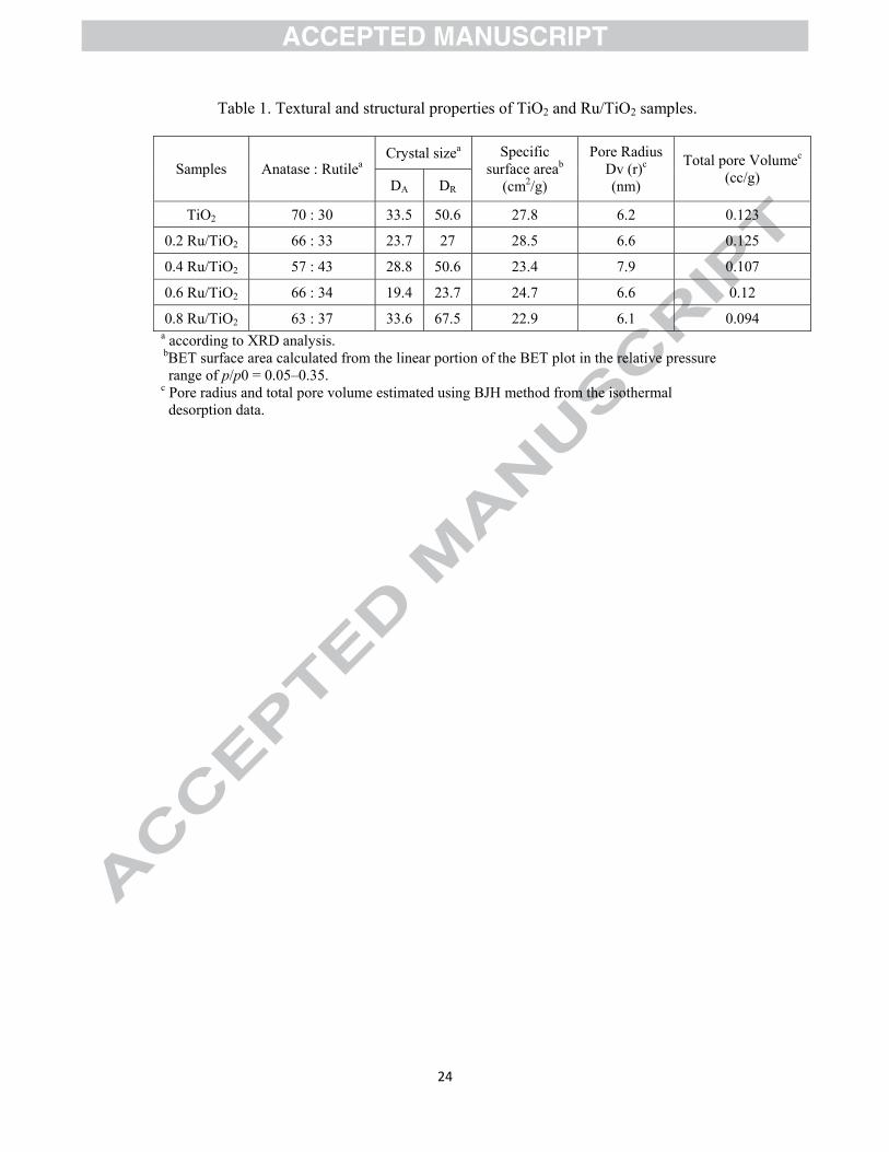
intensity of predominant anatase peak (101) (2θ =25.3o) [21]. The effect of metal
doped on TiO2 phase transformation (anatase to rutile) shown in Table (1). The
7
Ru/TiO2 samples showed a very similar isotherm to those of the TiO2-blank (type IV).
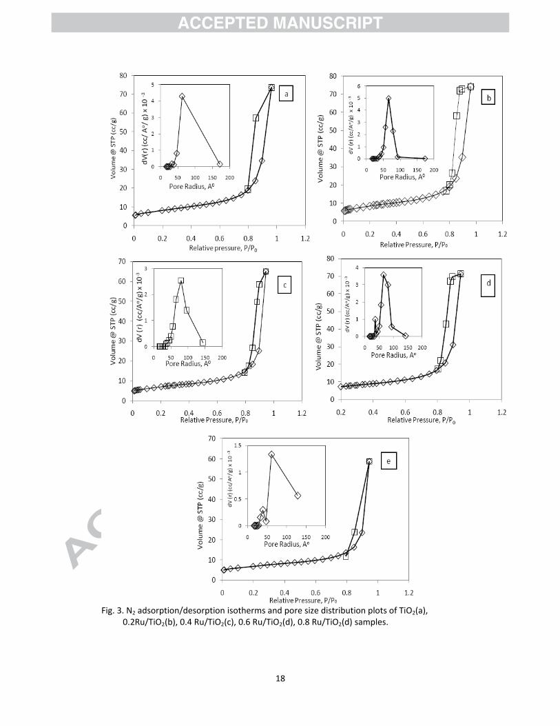
A representative adsorption–desorption hysteresis loop isotherm is shown in Fig. 3.
The small figure inserted in Fig. 3 is the pore size distribution calculated from
desorption data by the BJH method. The surface areas, average pore sizes, and pore
radius of the analyzed samples are summarized in Table 1. According to Brunauer's
classification samples are characterized by well-developed mesoporous structure [22].
The shape of the hysteresis loop is type H1closing at p/po ~0.6 according to the
IUPAC classification that usually associated with pores consisting of agglomerates or
compact of uniform spherical particles with regular arrays, whose pore size
distribution are normally narrow [23]. The least surface area of 22.9 m2/g for Ru/Ti
wt % 0.8, and the highest surface area of 28.5 cm2/g were obtained when Ru/Ti wt %
was 0.2. After introduction of the Ru precursor and calcination to produce RuO2 on
the support, the surface area and total pore volume of the support were reduced; as
metal loading increase from 0.4 to 0.8%; due to the presence of some RuO2 in the
pores.
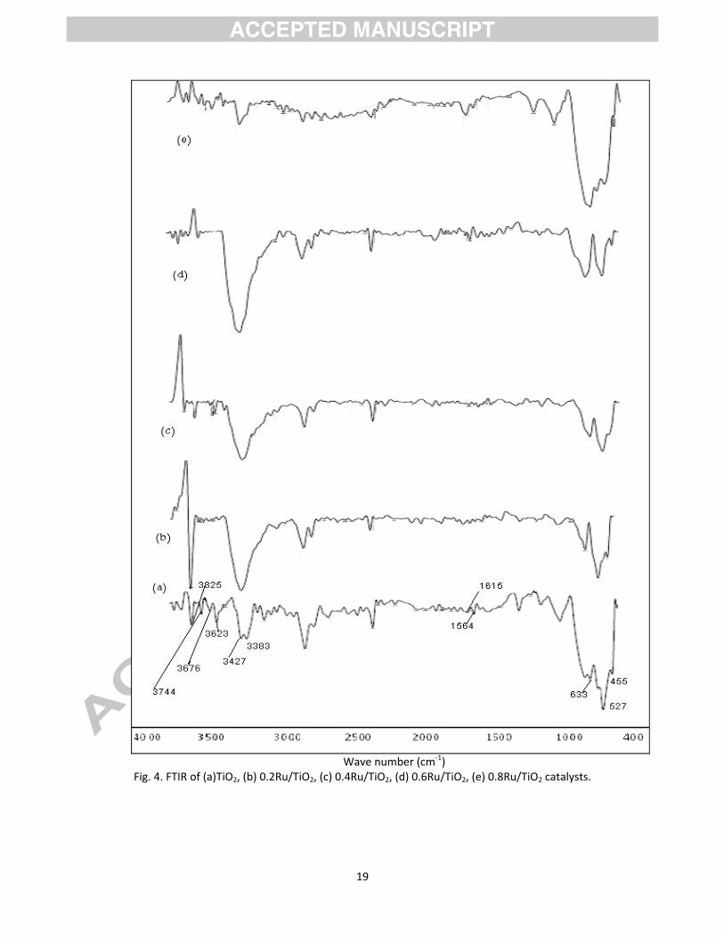
Fig. 4. shows a summary of Raman spectra of Ru/TiO2 catalysts. Structure of TiO2 is
confirmed in the FTIR spectrum. The peak of TiO2 anatase observed ca. 527 cm-1 (Ti–
O vibration), 633 cm-1, and 450 cm-1 corresponding to TiO2 rutile crystalline phase
were observed in all samples. The two bands appearing at 1564 and 1615 cm-1,
correspond to the stretching vibration of hydroxyl groups (O–H) and hydrogen-
bonded surface water molecules [24] in coordinated water, Ti–OH groups, or other
hydrated species. Bands at 3383, 3427 cm-1, are characteristic of, associated hydroxyl
groups. Additional stronger bands at 3623, 3676 cm-1 are characteristic of tetrahedral
coordinated vacancies, and designated Ti4+-OH. Two strong absorbance at 3744 and
3825 cm-1 are due to octahedral vacancies and designated Ti3+-OH [25]. Ru doped
TiO2 with different ratios of anatase to rutile, show that; the amount of surface
adsorbed water and hydroxyl groups has little relation with crystallite phases [26].
The TEM images of undoped and Ru-doped TiO2 are shown in Fig. 5. Blank TiO2 and
Ru-doped TiO2 showed regular round shaped particles. There was no significant
difference between two particles. This also previously confirmed from hysteresis loop
of these sample. Small dark spots seen are assumed as Ru particles and seem to be
non-uniformly distributed among the TiO2 particles with a particle size of
approximately 14 nm. Furthermore; Shen et al. [27] reported that Ru particles varied
in width from 1 to 14 nm with the majority of the particles having a width between 1

8
and 6 nm giving an average Ru particle size of 4.1 nm. This leads to spreading or
covering by the layer of TiO2.
A typical EDX pattern of the Ru/TiO2 is shown in Fig. 6. The elemental composition
of the samples was found to be 42.9% O, 56.9% Ti, 0.26% Ru for 0.2Ru/TiO2, and it
was 56% O, 43.4% Ti, 0.45% Ru for 0.4 Ru/TiO2catalyst. However, it was confirmed
the presence of RuO2 on TiO2.
3.2. Direct photolysis
Photolysis of 2-CP by 254 nm reduced its concentration, up to 43% transformation
after 60 min; as shown in Fig.7. Previously, Ragaini et al. [28] confirmed that 2-CP
was reasonably fast photolysis under irradiation at 254 nm.
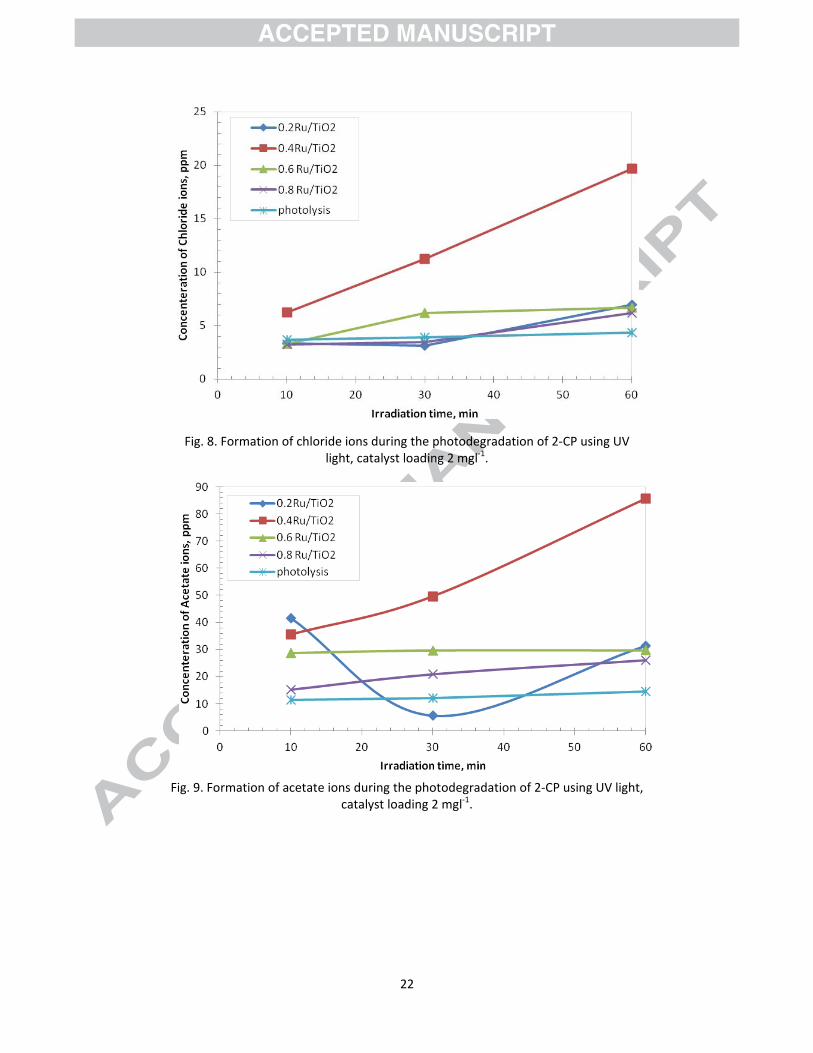
Fig. 8 shows the amount of chlorides ions formed on the photodegradation process as
a function of irradiation time. In photolysis; chloride ions reached 4ppm after 60 min
of irradiation time. This confirms that an efficient cleavage of the C-Cl bond occurs
from the electronically excited state of 2-CP produced by direct light absorption at
254 nm. Furthermore, Barakatet al. [15] reported the change in the pH is due to the
liberation of HCl during the degradation reaction of 2-CP. The reaction can be
represented mechanistically [29] and stoichiometrically [30] as follows:
Intermediate 2 HCl + 12 CO2 +4 H2O
On the other hand, Fig. 9 shows the production of acetate ions under photolysis of
2CP. The higher acetate concentration at initial time decreases the PH value and
encourages the formation of a positively charged TiO2 (TiOH2+) surface which cannot
provide hydroxyl groups needed for hydroxyl radical formation and consequently
degradation rate is decreased [15]. This also provided in our previous study on
photocatalytic degradation of 2,4,6-trichlorophenol using Ti-MCM-41 catalysts [31].
As a general, the rapid degradation of formed acetate ions may be the rate determine
step in photolysis of 2CP.
3.3. Photocatalytic degradation in the presence of blank TiO2
2-CP photocatalytic degradation was investigated under irradiation at 254 nm
wavelength in the presence of mesoporous titania particles (UV+TiO2). 2-CP
photodegradation clearly proceeded faster in the absence of TiO2 particles (Fig. 7).
This may appear surprising at first sight. However, a similar behavior has been
reported under irradiation either at 254 nm [32] or containing low wavelength
components [33], while in general a rate increase was observed upon photocatalyst

9
addition under irradiation at longer wavelengths [32]. Of course, concomitant direct
2-CP photolysis and photocatalysis on TiO2 occur under irradiation at 254 nm. The
fact that, with increasing the fraction of light absorbed (and scattered) by the
semiconductor particles, 2-CP underwent progressively slower degradation, indicates
that the photocatalytic degradation path was slower than direct 2-CP photolysis. By
contrast, photocatalytic conditions ensured faster mineralization. As shown in Fig. 7,
progressively higher percent mineralization was attained after 60 min irradiation in
the presence of photocatalyst.
3.4. Effect of semiconductor content
In order to attain optimum Ru dopant level, the target reaction was carried out on Ru
doped TiO2 with dopant level of 0.2–0.8 wt% using UV light and the results are
presented in Fig. 7. An interpretation of reactivity order is difficult since it is probably
the result of many factors: surface area, crystallinity, crystal size . . . [34]. An increase
in the content of these metals leads to a decrease in photocatalytic activity, while an
increase in the anatase content in titanium dioxide leads to an increase in such activity
[35]. The introduction of transition metals resulted in the change of the electronic
environment of TiO2. Based on the TEM results, Ru metals were adsorbed on the
surface of TiO2 as little island of metal particle that acts as the active sites of the
photocatalytic reaction [36] and may favor separating charge carriers efficiently,
inhibiting the recombination of electron–hole pairs, and ultimately causing the
enhancement of the reactivity. Barakata et al. [15] suggested that by irradiation of the
Co-doped TiO2, Co(III) ions work as electrons scavengers which may react with the
superoxide species and prevent the holes–electrons (h+/e-) recombination and
consequently increase the efficiency of the photo-oxidation.
As shown in Fig.7, The photodegradation efficiency of 2-CP increased with
increasing Ru concentration, reaching a maximum value of 61% with sample
containing 0.2 wt% Ru. A further increase in Ru concentration to 0.4 wt% resulted in
slightly increase in the photodegradation efficiency to 63%.
On the contrary, if the Ru content is low in the case of 0.2 Ru/Ti wt%, there are fewer
trapping sites available, thus reducing the activity. As a result; the activity of Ru/TiO2
was dependent on the doping concentration and 0.4 wt% Ru/TiO2 has turned out to be
the optimum Ru doping level for photocatalytic reduction of 2-chlorophenol; because
it has a high content of rutile as shown in Table 1. The enhanced light absorption
activity of the mixed phases, suggested the transfer of photo generated electrons from

10
a lower energy rutile to anatase electron site. This electron transfer would serve to
reduce the recombination rate of anatase by increasing the separation between the
electron and hole, resulting in greater catalytic reactivity [37]. Also, many catalytic
“hot spots” may be located at the solid-solid interface of anatase and rutile, which is
favorable for the improvement of photocatalytic activity [38]. However; Liu et al.,
[26] studied the UV and visible-light photocatalytic activity of nitrogen doped TiO2
with different ratios of anatase to rutile, and conclude that; nitrogen doped TiO2 with
a higher content of rutile can possess abundant surface water and hydroxyl groups,
and a high photocatalysis efficiency. The highest chloride formation in the case of
0.4Ru/TiO2 catalyst (Fig. 8) indicates the rapid cleavage of C-Cl bond. However;
constant increasing of acetate concentration (Fig. 9) providing the ability of this
catalyst in degradation of aromatic ring during photocatalytic process.
On the other hand; under irradiation at 254 nm, direct 2-CP photolysis appears to be
faster than photocatalytic degradation using 0.6 and 0.8% Ru/TiO2 catalysts. The
reason attributed to the detrimental activity of Ru loading beyond 0.4 wt% is the
increase in the recombination rate owing to the decrease in the distance between
trapping sites in a particle with the number of dopants (as shown in TEM images of
0.6, 0.8 Ru/TiO2catalysts). Also; this result is approved by [39] whereas; 0.5% iron
doped sample in the mixed phase catalyst decreased the UV light absorption as
compared to the undoped sample using the same preparation method, due to the
recombination sites of Fe2O3. Iwasaki et al. [40] and Choi et al. [41] also confirmed
the retarding effect of increasing Co(III) ion doping ranging from 0.004 to 0.14 on the
photocatalytic activity of TiO2 under UV light irradiation. Moreover, the results of
BET surface area could suggest that some photocatalytic active sites of the TiO2
surface are blocked by Ru particles due to decreasing the pore volume as doped metal
ratio increase beyond 0.4%, resulting in lower catalytic activity.
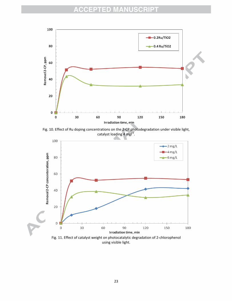
3. 5. Photocatalytic degradation using visible light
Effect of Ru doping concentrations on the 2-CP photodegradation under visible light
is shown in Fig.10. The photodegradation efficiency of 2-CP reaching a maximum
value of 53% with sample containing 0.2 wt% Ru. A further increase in Ru
concentration to 0.4 wt% resulted in decrease in the photodegradation efficiency to
34%. Besides, anatase and rutile ratio, the crystal size of catalyst particles and surface
area are also important factors for the photocatalytic activity. For 0.4% Ru/TiO2
catalyst, a crystal size of rutile is increased (Table 1). Its surface area is decreased and

11
the pore volume is decreased. Thus, the 0.2% Ru/TiO2 with higher anatase ratio
structure, larger surface area large pore volume and smaller crystal size has the best
photocatalytic oxidation efficiency in visible region. Pore volume is related to the
diffusion of pollutants and samples during the photocatalytic reaction. The higher the
pore volume of the samples, the diffusion between pollutants and samples becomes
rapid and fast [42].
Based on the observation of Ku et al., [43] study, the differences between the global
photocatalytic destruction rates of 2-CP by various forms of TiO2 particles might be
attributed to the difference of the surface area of TiO2, not to the difference of the
crystal properties in this study.
In addition, the activity have a direct correlation with the amount of •OH groups on
the surface [44]; as previously shown in FTIR spectrum, the 0.2 Ru/TiO2 catalyst has
high amount of •OH groups on the surface than 0.4 Ru/TiO2.
3. 6. Effect of 0.2 Ru/TiO2 loading
The photocatalytic degradation of 2-CP was carried out over a 0.2 Ru/TiO2 catalyst at
different loading of 2-6 mgl-1 in order to optimize the catalyst loading during the
visible irradiation process. A significant increase in degradation of 2-CP as catalyst
loading increase from 2 mgl-1 to 4 mgl-1; as illustrated in Fig. 11.
The decrease in the degradation of 2-CP above 4 mgl-1 may be attributed to the
scattering effect of light caused by the high concentration of photocatalyst. The
photodecomposition rates of pollutants are influenced by the active site and the
photoabsorption of the catalyst used. Adequate loading of the catalyst increases the
generation rate of electron/hole pairs for enhancing the degradation of pollutants.
However, addition of a high dose of the semiconductor decreases the light penetration
by the photocatalyst suspension and reduces the degradation rate [45].
4. Conclusion
From the study of the above work, the following conclusions can be drawn:
Enhancement of the photocatalytic activity of TiO2 for photocatalytic
degradation of 2-CP by doping Ru has been achieved.
The mineralization of 2-CP is demonstrated by production of chloride and
acetate ions.
The entry of Ru into the lattice of TiO2 is evidenced by XRD, SEM-EDX and
TEM studies.
12
0.4 wt% Ru/TiO2 has turned out to be the optimum Ru doping level for
photocatalytic reduction of 2-chlorophenol using UV light; because it has a
high content of rutile. The enhanced light absorption activity of the mixed
phases, suggested the transfer of photo generated electrons from a lower
energy rutile to anatase electron site. This electron transfer would serve to
reduce the recombination rate of anatase by increasing the separation between
the electron and hole, resulting in greater catalytic reactivity.
On the other hand; the 2% Ru/TiO2 with the optimum catalyst loading 4 mgl-1,
provided photodegradation efficiency of 2-CP reaching a maximum value of
53% using visible light. Where; the incorporation of Ru in TiO2 led to large
surface area and high pore volume values. This also led to form more electron
capture traps, which contribute to high separation efficiency of photogenerated
carriers.
References
[1] M.A. Callahan, M.W. Slimak, N.W. Gabel, I.P. May, C.F. Foeler, J.R. Freed,
P. Jennings, R.L.Durfee, F.C. Whitemore, B. Maestri, N.R. Mabey, B.R. Holt,
C. Gould, Water-related Environmental Fate of 129 Priority Pollutants, vol. II,
No. 84-1-8, EPA-440/4-79-029b, US Environmental Protection Agency,
Washington, DC, 1979.
[2] H. Roques, Chemical Water Treatment, VCH Verlag, Wienheim, Germany,
1996.
[3] K. R. Krijgsheld, A. van der Gen, Assessment of the impact of the emission of
certain organochlorine compounds on the aquatic environment: Part I:
Monochlorophenols and 2, 4-dichlorophenol, Chemosphere 15 (1986) 825-
860.
[4] M. Shen, Z. Y. Wu, H. Huang, Y. Du, Z. Zou, P. Yang, Carbon-doped anatase
TiO2 obtained from TiC for photocatalysis under visible light irradiation,
Mater. Lett. 60 (2006) 693– 697.
[5] B. Sun, P. G. Smirniotis, P. Boolchand, Visible Light Photocatalysis with
Platinized Rutile TiO2 for Aqueous Organic Oxidation, Langmuir 21 (2005)
11397–11403.
13
[6] J. Fernandez, J. Kiwi, J. Baeza, J. Freer, C. Lizama, H. D. Mansilla, Orange II
photocatalysis on immobilised TiO2: Effect of the pH and H2O2, Appl. Catal.
B: Environ. 48 (2004) 205–211.
[7] M. A. Fox, M. T. Dulay, Heterogeneous photocatalysis, Chem. Rev. 93 (1993)
341-357.
[8] I.-H. Tseng, W. C. Chang, J. C. S. Wu, Photoreduction of CO2 using sol–gel
derived titania and titania-supported copper catalysts, Appl. Catal. B: Environ.
[9] I.-H. Tseng, J. C. S. Wu, H.-Y. Chou, Effects of sol–gel procedures on the
photocatalysis of Cu/TiO2 in CO2 photoreduction, J. Catal. 221 (2004) 432-
440.
[10] V. W. Day, W. G. Klemperer, D. J. Main, Niobotungstate, Nb2W4O194- and
triphosphate, P3O93-, complexes of (cyclooctadiene)iridium(I): synthesis,
structure, and stability of tetra-n-butylammonium salts of
{[(C8H12)Ir]5(Nb2W4O19)2}3-,{[(C8H12)Ir]2H(Nb2W4O19)2}5-,and
[(C8H12)Ir(P3O9)]2-, Inorg. Chem. 29 (1990) 2345-2355.
[11] N. Jaffrezic-Renault, P. Pichat, A. Foissy, R. Mercier, Study of the effect of
deposited platinum particles on the surface charge of titania aqueous
suspensions by potentiometry, electrophoresis, and labeled-ion adsorption, J.
Phys. Chem. 90 (1986) 2733-2738.
[12] A. Burns, W. Li, C. Baker, S.I. Shah, Sol–gel synthesis and characterization
of neodymium-ion doped nanostructured titania thin film, Mater. Res. Soc.
Symp. Proc. 703 (2002) 193-198.
[13] O.-J. Jung, Synergistic Effect on the Photocatalytic Degradation of 2-
Chlorophenol Using TiO2 Thin Films Doped with Some Transition Metals in
Water, Bull. Korean Chem. Soc. 22 (2001) 1183-1191.
[14] O.J. Jung, S.H. Kim, K.H. Cheong, W. Li, S.I. Shah, Metallorganic Chemical
Vapor Deposition and Characterization of TiO2 Nanoparticles, Bull. Korean
Chem. Soc. 24 (2003) 49-54.
[15] M. A. Barakata, H. Schaeffera, G. Hayesa, S. Ismat-Shah, Photocatalytic
degradation of 2-chlorophenol by Co-doped TiO2 nanoparticles, Appl. Catal.
B: Environ. 57 (2005) 23–30.
[16] M. Senthilnanthan, D.P. Ho, S. Vigneswaran, H.H. Ngo, H.K. Shon, Visible
light responsive ruthenium-doped titanium dioxide for the removal of

14
metsulfuron-methyl herbcide in aqueous phase, Separation and Purification
Technology 75 (2010) 415–419.
[17] M. S. Nahar, K. Hasegawa, S. Kagaya, Photocatalytic degradation of phenol
by visible light-responsive iron-doped TiO2 and spontaneous sedimentation of
the TiO2 particles, Chemosphere, 65 (2006) 1976–1982.
[18] D. Huang, G.S. Luo, Y.J. Wang, Using phosphoric acid as a catalyst to control
the structures of mesoporous titanium dioxide materials, Micropor. Mesopor.
Mater. 84 (2005) 27–33.
[19] D. G. Blackmond, E. I. Ko, Preparation and characterization of well-defined
Ni/SiO2 catalysts, Appl. Catal. 13 (1984) 49-68.
[20] N. Venkatachalam, M. Palanichamy, B. Arabindoo, V. Murugesan, Enhanced
photocatalytic degradation of 4-chlorophenol by Zr4+ doped nano TiO2 , J.
Mol. Catal. A: Chem. 266 (2007) 158–165.
[21] G. B. Song, J. K. Liang, F. S. Liu, T. J. Peng, G. H. Rao, Preparation and
phase transformation of anatase–rutile crystals in metal doped TiO2/muscovite
nanocomposites, Thin Solid Films, 491 (2005) 110–116.
[22] F. Tomul and S. Balci, Characterization of Al, Cr-pillared clays and CO
oxidation. Appl. Clay Sci., 43 (2009) 13-20.
[23] G. Leofanti, M. Padovan, G. Tozzola and B. Venturelli, Surface area and pore
texture of catalysts, Catal. Today, 41 (1998) 207-219.
[24] J. R. S. Brownson, M. I. Tejedor-Tejedor, M. A. Anderson, Photoreactive
Anatase Consolidation Characterized by FTIR Spectroscopy, Chem. Mater. 17
(2005) 6304-6310.
[25] P. Madhu Kumar, S. Badrinarayanan, M. Sastry, Nanocrystalline TiO2 studied
by optical, FTIR and X-ray photoelectron spectroscopy: correlation to
presence of surface states, Thin Solid Films 358 (2000) 122-130
[26] G. Liu, X. Wang, Z. Chen, H.-M. Chen, G. Qing (Max) Lu, The role of crystal
phase in determining photocatalytic activity of nitrogen doped TiO2 , J.
Colloid and Interface Science, 329 (2009) 331–338.
[27] X. Shen, L.J. Garces, Y. Ding, K. Laubernds, R.P. Zerger, M. Aindow, E.J.
Neth, S.L. Suib, Behavior of H2 chemisorption on Ru/TiO2 surface and its
application in evaluation of Ru particle sizes compared with TEM and XRD
analyses, Appl. Catal. A: Gen. 335 (2008) 187–195.

15
[28] V. Ragaini, E. Selli, C. L. Bianchi, C. Pirola, Sono-photocatalytic degradation
of 2-chlorophenol in water: kinetic and energetic comparison with other
techniques, Ultrason. Sonochem. 8 (2001) 251–258.
[29] G.L. Puma, P.L. Yue, Effect of radiation wavelength on the rate of
photocatalytic oxidation of organic pollutants, Ind. Eng. Chem. Res. 41 (2002)
5594-5600.
[30] L. Rideh, A.Wehrer, D. Ronze, A. Zoulalian, Photocatalytic Degradation of 2-
Chlorophenol in TiO2 Aqueous Suspension: Modeling of Reaction Rate, Ind.
Eng. Chem. Res. 36 (1997) 4712-4718.
[31] A. K. Aboul-Gheit , S. M. Abdel-Hamid, S. A. Mahmoud, R. A. El-
Salamony, J. Valyon, M. R. Mihályi, Á. Szegedi, Mesoporous Ti-MCM-41
materials as photodegradation catalysts of 2,4,6-trichlorophenol in water, J
Mater Sci. (2011) 46:3319–3329.
[32] I. Ilisz, A. Dombi, K. Mogyorósi, A. Farkas, I. Dékány, Removal of 2-
chlorophenol from water by adsorption combined with TiO2 photocatalysis,
Appl. Catal. B: Environ. 39 (2002) 247–256.
[33] R.-A. Doong, C.-H. Chen, R. A. Maithreepala, S.-M. Chang, The influence of
pH and cadmium sulfide on the photocatalytic degradation of 2-chlorophenol
in titanium dioxide suspensions, Water Res. 35 (2001) 2873–2880.
[34] E. Piera, M. I. Tejedor-Tejedor, M. E. Zorn, M. A. Anderson, Relationship
concerning the nature and concentration of Fe(III) species on the surface of
TiO2 particles and photocatalytic activity of the catalyst, Appl. Catal. B 46
(2003) 671-685.
[35] A. S. Kovalenko, S. Ya. Kuchmii, T. F. Makovskaya, A. V. Korzhak, V. V.
Tsyrina, V. I. Yatskiv, and V. G. Ilin. Theoretical and Experimental
Chemistry, Vol. 39, No. 2, 2003.
[36] S. Ozkan, M.W. Kumthekar, G. Karakas, Characterization and temperature-
programmed studies over Pd/TiO2 catalysts for NO reduction with methane,
Catal. Today 40 (1998) 3-14.

16
[37] R. Bickley, T. Gonzalea-Carreno, J. Lees, A structural investigation of
titanium dioxide photocatalysts, J. Solid State Chem. 92 (1991) 178–190.
[38] G. H. Li, L. Chen, M. E. Graham, K. A. Gray, A comparison of mixed phase
titania photocatalysts prepared by physical and chemical methods: The
importance of the solid–solid interface, J. Mol. Catal. A: Chem. 275 (2007)
30–35.
[39] M. S. Nahar, J. Zhang, K. Hasegawa, S. Kagaya, S. Kuroda, Phase
transformation of anatase–rutile crystals in doped and undoped TiO2 particles
obtained by the oxidation of polycrystalline sulfide, Materials Science in
Semiconductor Processing 12 (2009) 168–174.
[40] M. Iwasaki, M. Hara, H. Kawada, H. Tada, S. Ito, Cobalt Ion-Doped TiO2
Photocatalyst Response to Visible Light, J. Colloid Interface Sci. 224 (2000)
202-204.
[41] W. Choi, A. Termin, M.R. Hofmann, Effects of Metal-Ion Dopants on the
Photocatalytic Reactivity of Quantum-Sized TiO2 Particles, Angew. Chem.
Int. Ed. Engl. 33 (1994) 1091-1092.
[42] B. Guo, H. Shen, K.Shu, Y. Zeng and W. Ning, The study of the relationship
between pore structure and photocatalysis of mesoporous TiO2, J. Chem. Sci.,
121 (2009) 317–321.
[43] Y. Ku, R.-M. Leu and K.-C. Lee, Decomposition of 2-chlorophenol in
aqueous solution by UV irradiation with the presence of titanium dioxide,
Wat. Res. 30 (1996) 2569-2578.
[44] S.-J. Tsai, and S. Cheng, Effect of TiO2 crystalline structure in photocatalytic
degradation of phenolic contaminants, Cat. Tod. 33 (1997) 227-237.
[45] A. Doongr, W. H. Chang, Photodegradation of parathion in aqueous titanium
dioxide and zero valent iron solutions in the presence of hydrogen peroxide, J.
Photochem. Photobiol. A: Chem. 116 (1998) 221-228.

17
Fig. 1. X‐ ray Diffraction spectra for (a) TiO2, (b) 0.1 Ru/TiO2, (c) 0.2 Ru/TiO2, (d)
0.4 Ru/TiO2, (e) 0.6 Ru/TiO2, (f) 0.6 Ru/TiO2.
Fig. 2. Rutile fraction of TiO2 in Ru/TiO2 catalusts as function of Ru ratio.
18
Fig. 3. N2 adsorption/desorption isotherms and pore size distribution plots of TiO2(a), 0.2Ru/TiO2(b), 0.4 Ru/TiO2(c), 0.6 Ru/TiO2(d), 0.8 Ru/TiO2(d) samples.
19
Wave number (cm‐1)
Fig. 4. FTIR of (a)TiO2, (b) 0.2Ru/TiO2, (c) 0.4Ru/TiO2, (d) 0.6Ru/TiO2, (e) 0.8Ru/TiO2 catalysts.

20
Fig. 5. TEM micrograph of (a) TiO2, (b) 0.2 Ru/TiO2, (b) 0.4 Ru/TiO2, (c) 0.6Ru/TiO2,(d) 0.8
Ru/TiO2,
21
Fig. 6. EDX spectrum of 0.2, 0.4 Ru/TiO2 catalysts.
Fig. 7. Effect of Ru doping concentrations on the 2‐CP photodegradation using UV
irradiation, catalyst loading 2 mgl‐1.
22
Fig. 8. Formation of chloride ions during the photodegradation of 2‐CP using UV
light, catalyst loading 2 mgl‐1.
Fig. 9. Formation of acetate ions during the photodegradation of 2‐CP using UV light,
catalyst loading 2 mgl‐1.
23
Fig. 10. Effect of Ru doping concentrations on the 2‐CP photodegradation under visible light,
catalyst loading 4 mgl‐1.
Fig. 11. Effect of catalyst weight on photocatalytic degradation of 2‐chlorophenol
using visible light.
24
Table 1. Textural and structural properties of TiO2 and Ru/TiO2 samples.
Crystal sizea
Samples Anatase : Rutilea DA DR
Specific surface areab
(cm2/g)
Pore Radius Dv (r)c (nm)
Total pore Volumec (cc/g)
TiO2 70 : 30 33.5 50.6 27.8 6.2 0.123
0.2 Ru/TiO2 66 : 33 23.7 27 28.5 6.6 0.125
0.4 Ru/TiO2 57 : 43 28.8 50.6 23.4 7.9 0.107
0.6 Ru/TiO2 66 : 34 19.4 23.7 24.7 6.6 0.12
0.8 Ru/TiO2 63 : 37 33.6 67.5 22.9 6.1 0.094 a according to XRD analysis. bBET surface area calculated from the linear portion of the BET plot in the relative pressure range of p/p0 = 0.05–0.35. c Pore radius and total pore volume estimated using BJH method from the isothermal desorption data.