Silkworm Pupa Oil and Sardine Oil as an Additional Energy ...
Upload
independentCategory
view
0download
0
Food Chemistry 120 (2010) 433–442
Contents lists available at ScienceDirect
Food Chemistry
journal homepage: www.elsevier .com/locate / foodchem
Preparation of eicosapentaenoic acid concentrates from sardine oil by Bacilluscirculans lipase
Kajal Chakraborty a,*, P. Vijayagopal a, Rekha D. Chakraborty a,b, K.K. Vijayan a
a Bioprospecting Section of Marine Biotechnology Division, Central Marine Fisheries Research Institute, Ernakulam North P.O., P.B. No. 1603, Cochin 682018, Kerala, Indiab Crustacean Fisheries Division, Central Marine Fisheries Research Institute, Ernakulam North P.O., P.B. No. 1603, Cochin 682018, Kerala, India
a r t i c l e i n f o
Article history:Received 27 May 2009Received in revised form 1 September 2009Accepted 13 October 2009
Keywords:LipaseBacillus circulansSardine oiln-3 PUFAAmide complexationArgentated chromatography
0308-8146/$ - see front matter � 2009 Elsevier Ltd. Adoi:10.1016/j.foodchem.2009.10.032
* Corresponding author. Tel.: +91 484 2394867; faxE-mail address: [email protected] (K. Chakrab
a b s t r a c t
An extracellular lipase derived from Bacillus circulans, isolated from marine macroalga, Turbinaraia cono-ides, was used to prepare n-3 polyunsaturated fatty acid (PUFA) concentrates from sardine oil triglycer-ides. The enzyme was purified 132-fold with specific activity of 386 LU/mg. The purified lipase was ableto enrich sardine oil with 37.7 ± 1.98% 20:5n-3 and 5.11 ± 0.14% 18:3n-3 in the triglyceride fraction after3 h of hydrolysis. Lower hydrophobic constants of n-3 fatty acids (18:3n-3log P = 5.65; 20:5n-3log P = 5.85,respectively) than n-6 (20:4n-6log P = 6.16) resulted in higher hydrolytic resistance of the former towardlipase, leading to their enrichment in the triglyceride fraction. Lipase-catalysed hydrolysis of sardine oilfor 3 h, followed by urea complexation, provided free fatty acids containing 51.3 ± 4.65% 20:5n-3. Thepurified methyl ester of 20:5n-3 (68.29 ± 2.15%) from the urea concentrate was attained by chromatog-raphy on argentated neutral alumina.
� 2009 Elsevier Ltd. All rights reserved.
1. Introduction
The long chain n-3 polyunsaturated fatty acids (PUFAs) with C20
acyl chain length, particularly eicosapentaenoic acid (EPA, 20:5n-3)and linolenic acid (LA, 18:3n-3), were reported to have variousimportant physiological roles in health, and in the inflammatoryprocess (Sahena et al., 2009). Whilst saturated and monounsatu-rated fatty acids may be synthesised in the body, PUFAs cannot besynthesised de novo due to the lack of essential enzymes requiredto synthesise PUFAs in adequate levels from precursor fatty acids,and therefore, must be externally supplied in the diet (Cahu, Guil-laume, Stephan, & Chim, 1994). The PUFAs are currently in demandin the pure form in nutraceutical and aquafeed formulations, andare being actively studied to understand their potential roles in hu-man health. There are reports of concentrates of PUFA from marinealga, fish oil (Guil-Guerrero, López-Martínez, Rincon-Cervera, &Campra-Madrid, 2007), and edible oils (López-Martínez, Campra-Madrid, & Guil-Guerrero, 2004; López-Martínez, Campra-Madrid,Rincón-Cervera, & Guil-Guerrero, 2005). Also fishery by-products/processing waste and low value catches have been utilised for con-centrating PUFA (Zuta, Simpson, Chan, & Phillips, 2003). A survey ofthe literature revealed a diverse range of methods for enrichmentand purification of LC-PUFAs. These included urea inclusioncomplexation, molecular distillation, iodolactonisation, low
ll rights reserved.
: +91 484 2394909.orty).
temperature fractional crystallisation, salt solubility methods, andliquid–liquid extraction–fractionation using aqueous AgNO3 solu-tion (Guil-Guerrero et al., 2007).
However, most of the existing chemical methods for purifyingindividual PUFAs and PUFA concentrates are non-selective for dif-ferent fatty acids. The substrate specificity of lipases (triacylglyc-erol acyl hydrolases) has been utilised for the enhancement ofPUFA content in fish oils by several groups (Can & Özçelik, 2005;Liu, Zhang, Hong, & Ji, 2007). It was found that lipases were effi-cient in hydrolytic and esterification reactions because of theirhigh stereospecificity and mild reaction conditions at low temper-ature and in organic solvents (Can & Özçelik, 2005). The uniquespecificity of lipases is utilised to selectively concentrate targetedfatty acids in the triacylglyceride fraction that can be readily ab-sorbed into plasma triacylglycerol.
Bacterial lipases are one of the predominant classes of lipasesfor the enhancement of PUFA content in fish oils, due to their un-ique thermostability. Lipases have been reported from Bacilluscoagulans (Kumar, Kikon, Upadhyay, Kanwar, & Gupta, 2005), B. cir-culans (Kademi, Ait-Abdelkader, & Fakhreddine, 2000), and B.licheniformis (Chakraborty & Paulraj, 2008a), that were used to con-centrate n-3 PUFAs. Novozym 435TM was used to selectively con-centrate targeted fatty acids in triglycerides due to its uniquesubstrate specificity towards PUFAs (Liu et al., 2007). Response sur-face methodology was used to determine optimal conditions forthe lipase-catalysed enrichment of hazelnut oil by incorporatingn-3 PUFAs from menhaden oil (Can & Özçelik, 2005). DHA-rich

434 K. Chakraborty et al. / Food Chemistry 120 (2010) 433–442
triglycerides were prepared from fish oil with lipases obtainedfrom Candida cylindracea and Chromobacterium viscosum (Tanaka,Hirano, & Funada, 1994).
PUFAs are found to be widely available in marine fish species.However, sardine (Sardinella longiceps) is a low-value fish, and itsoil is locally available as a by product. A higher concentration ofPUFAs (31.4%) particularly EPA (17.6%) in sardine oil is an addedadvantage. In this paper, we report methods to purify an extracel-lular lipase, to homogeneity, from culture broth of B. circulans, orig-inally isolated from marine macroalga Turbinaraia conoides. Thepresent paper is also directed to a process to prepare n-3 PUFA con-centrates by lipase-catalysed purification of triglycerides derivedfrom sardine oil, and further concentrating the target PUFAs byurea complexation and argentated column chromatographic meth-ods. Structure–activity relationship analyses, to correlate the lipaseactivity with physicochemical properties of different classes offatty acids, were used to elucidate the structural descriptors offatty acids (hydrophobic and steric) controlling lipolytic activity.
2. Materials and methods
2.1. Chemicals and instrumentation
The solvents used for sample preparation were of analyticalgrade (E-Merck, Darmstadt, Germany). Sephadex G-100 and othersupports of chromatography were from Sisco Research Laborato-ries (SRL, Mumbai, India). Amberlite IRA-410 (Cl� form) and bovineserum albumin were obtained from HiMedia (Mumbai, India);Electrophoresis grade acrylamide, bis-acrylamide, and low-rangemolecular marker proteins were procured from Bio-Rad Laborato-ries (Bio-Rad, Hercules, CA). All glassware was rinsed with CHCl3/CH3OH (2:l, v/v), and dried under N2. Standards of fatty acid methylester (Supelco TM 37 Component FAME Mix, Catalog No. 47885-U)and boron trifluoride/methanol (14% BF3 in CH3OH, w/v) were pro-cured from Sigma–Aldrich Chemical Co. Inc (St. Louis, MO). GLCdata were recorded on a Perkin-Elmer AutoSystem XL gas chro-matograph. Mass spectral assays were obtained using a Varian1200L single-quadrupole mass spectrometer instrument underelectron impact (70 eV) conditions.
2.2. Isolation of B. circulans from T. conoides and lipase production
For isolation of epiphytic bacteria, marine macroalga T. conoidessamples obtained from the Gulf of Mannar were rinsed with sterileseawater, and a small portion of the algal surface was scraped witha sterile swab and spread onto the plates of marine agar plates.After incubation at 37 �C for 24–48 h, all colonies were streakedonto the marine agar slants and maintained for further microbio-logical and biochemical identification. Standard sampling proce-dures were followed, and colonies were recovered from marineagar plates (Chakraborty & Paulraj, 2008a). The bacterial culturesobtained from the alga were thereafter grown on modified tribu-tyrin slants (0.3% meat extract, 0.5% peptone, 1% NaCl, 0.3% tribu-tyrin, and 0.01% CaCl2�2H2O) to detect the lipolytic activity.Amongst several epiphytic bacteria isolated from this alga, oneBacillus sp. (B. circulans) was used for production of lipase due toits higher lipolytic potential. The bacterial cultures belonging toB. circulans were inoculated in a 250 ml Erlenmeyer flask contain-ing nutrient broth (100 ml). The content was then incubated at37 �C for 24 h under shaking (150 rpm) to raise the inoculum forthe enzyme production. The seed culture (100 ml) (1% v/v) of B. cir-culans was grown in sterilised broth, containing (g l�1); NaNO3 3;K2HPO4 0.1; MgSO4�7H2O 0.5; KCl 0.5, FeSO4�7H2O 0.05, yeastextract 5.0, and microalgal paste (Isochrysis galabana, 5%, v/v) at37 �C in a 1000 ml Erlenmeyer flask with shaking (150 rpm) up
to 72 h. The culture broth was harvested at different time-intervals(0, 5, 10, 24, 29, 34, 48, 53, 58, and 72 h) to determine the optimumtime for maximum lipase production. The culture broth was clari-fied by centrifugation (10,000g for 20 min at 4 �C, Superspin R-V/FM Plasto Crafts, Plasto Crafts Mumbai) to recover the supernatantthat was filtered (0.2 l). This concentrated liquid, referred to asthe crude extracellular lipase solution, was used for furtherpurification.
2.3. Purification of lipase from B. circulans by ammonium sulphateprecipitation and chromatography
The crude lipase thus obtained was purified to homogeneity byammonium sulphate precipitation, Sephadex G-100 gel filtration,and Amberlite IRA-410 (Cl� form) anion-exchange chromatogra-phy. Solid ammonium sulphate was added to the crude enzyme ex-tract in increments of 5% until 70% saturation (w/v) with gentlestirring for 2 h at 4 �C, and allowed to stand for 4 h. The precipi-tates thus obtained were separated by centrifugation (10,000g for30 min, 4 �C), and the supernatant was fractionated with the slowaddition of ammonium sulphate to 70% saturation. The solutionwas stirred for 1 h at 4 �C, and allowed to stand overnight. The pre-cipitated proteins were collected by centrifugation at 10,000g(30 min, 4 �C), and the supernatant was discarded. The pellet wasdissolved in a minimum volume of Tris–HCl buffer (50 mM, pH8.0), and dialysed against the same buffer for 18 h to remove theresidual salts, and assayed for lipase activity (Winkler & Stuck-mann, 1979). The enzyme concentrate obtained from dialysiswas loaded onto a Sephadex G-100 gel filtration column(2.5 � 120 cm) at a flow rate of 0.5 ml/min. The column was equil-ibrated and eluted with Tris–HCl buffer (50 mM, pH 8.0) supple-mented with CaCl2 (1.0 mM). The eluant fractions showing lipaseactivity (obtained at the 22–30th fraction) were pooled together,and concentrated by lyophilisation. The lyophilised protein wasdissolved in distilled water (5 ml), and dialysed against Tris–HClbuffer (50 mM, pH 8.0). The concentrated protein was further chro-matographed on an Amberlite IRA-410 (Cl� form) column(1.5 � 15 cm) pre-equilibrated with Tris–HCl buffer (50 mM, pH8.0), and the bound proteins were eluted in Tris–HCl (50 mM, pH8.0) with a linear gradient of increasing concentration of NaCl(0–0.5 M) at a flow rate of 0.5 ml/min. The active fractions (ob-tained at the 35–40th fraction) were pooled, and analysed for li-pase activity. The lipase activity of the enzyme was estimatedspectrophotometrically (Varian Cary 50 Conc., USA), using 4-nitro-phenyl palmitate (4-NPP) as reported earlier (Winkler & Stuck-mann, 1979). One activity unit of lipase (LU) was defined aslmole of 4-NP released from hydrolysis of 4-NPP/ml/min by oneml of enzyme under standard assay conditions. The protein con-centration was determined by measuring the absorbance at590 nm (Varian Cary 50 Conc., USA) using bovine serum albumin(20–150 lg) as a standard (Bradford, 1976). The active enzymefractions were stored at 4 �C until used for polyacrylamide gel elec-trophoresis and further enzyme characterisation.
2.4. Determination of molecular weight of the lipase by denaturingpolyacrylamide gel electrophoresis
Polyacrylamide gel electrophoresis (PAGE) in the presence ofsodium dodecyl sulphate (0.1% w/v SDS–PAGE) was performedon 12% polyacrylamide gel (with 6% stacking gel), following theestablished procedure (Laemmli, 1970), and the apparent molecu-lar mass of proteins was determined with reference to thelow-range molecular mass markers (14.4–97.4 kDa, Bio-Rad Labo-ratories, Hercules CA). The molecular mass markers used wereb-phosphorylase (97.4 kDa), serum albumin (66.2 kDa), ovalbumin(45.0 kDa), carbonic anhydrase (31.0 kDa), trypsin inhibitor
K. Chakraborty et al. / Food Chemistry 120 (2010) 433–442 435
(21.5 kDa) and lysozyme (14.4 kDa). After migration, the gels werefixed using 7% (v/v) CH3COOH/CH3OH, and submitted to a cycle ofstaining/destaining with Coomassie Brilliant Blue R-250 (stainer)and 14% (v/v) CH3COOH/CH3OH (destainer), respectively. Molecu-lar mass was determined from the plots of log molecular mass(log M) vs. migration (Rf) for the series of protein standards. The ac-tive lipase fractions were identified by analytical SDS–PAGE of pro-teins, using 12% gels stained with Coomassie brilliant blue R-250 asdetailed elsewhere (Kim, Kwon, Yoon, Kim, & Kim, 2005). The gelswere rinsed with water and stored in acetic acid (7% v/v) (Lee,Chung, & Rhee, 1993) to develop a dark yellow colour. Gel filtrationchromatography with Sephadex G-75 was used to validate themolecular weight of lipase. The elution volume of the lipase wascompared with those of known protein standards (horse heartcytochrome C, 12.4 kDa; soybean trypsin inhibitor, 20.1 kDa; bo-vine serum albumin, 66 kDa; b-phosphorylase, 97.4 kDa), and themolecular masses of standard proteins were plotted against theirrespective ratios of elution volume to column void volume (Ve/V0) to draw the calibration curve, the column void volume (V0)being the elution volume of blue dextran (2000 kDa).
2.5. Preparation of n-3 polyunsaturated fatty acid concentrates bytime-course (1–6 h) B. circulans lipase-catalysed hydrolysis of sardineoil triglycerides
In this study, crude sardine oil from whole sardine was obtainedfrom a plant located in Cochin, India. The crude oil thus obtainedwas chilled and filtered to remove solid impurities. The contentwas further bleached by the addition of powdered activated char-coal (4%) to the oil under vacuum conditions, to remove colouredcompounds and other impurities (metals such as Ni and Fe) fromthe oil. The powder was then removed by filtration, and the prod-uct was treated for a short time at high temperature to remove lowmolecular weight contaminants (free fatty acids, volatile amines)and peroxides. Antioxidant (tertiary butyl hydroxyquinone 0.01%w/v) was added to the oil to arrest oxidation. The free flowing lightyellow-coloured oil thus obtained was stored under nitrogen at�20 �C, in a sealed dark amber glass container, until used.
Sardine oil triglycerides thus obtained were further stabilisedby adding phosphatidylcholine and a-tocopherol (0.1% each) andmetal complexing agents (ascorbic acid, 0.01%). The triglyceridesfrom sardine oil were cleaned up on neutral alumina, using n-hex-ane/EtOEt as the eluting solvent system (95:5, v/v), and extracted,following established method (Bligh & Dyer, 1959). The lipids thusobtained were further purified on a column of neutral alumina,using n-hexane/EtOEt (95/5, v/v). The concentrated triglycerideswere thereafter hydrolysed with lipases purified from B. circulans,and the total fatty acid content, at various time-intervals (1–6 h),was analysed. In brief, a reaction mixture containing triglycerides(100 ml, 0.01%, w/w tert-butyl hydroxyl quinone) in PIPES–NaOHbuffer (5 ml, 0.15 M, pH 7.0) was stabilised with Triton X-100(0.3%, v/v), and CaCl2 (0.5 ml, 100 mM), to which lipase was added(500 LU). The contents were placed in a screw-cap round-bottomflask (250 ml) containing glass beads, following the procedureadopted in an earlier experiment with suitable modification (Tana-ka et al., 1994). The round-bottom flask was flushed with N2 andincubated at 37 ± 1 �C under shaking (500 rpm). Samples (0.5 ml)from the reaction mixture were withdrawn periodically (1–6 h)for determining the lipase activity. Methanolic KOH (0.5 N,25 ml) solution was added to the mixture to neutralise the freefatty acids released during hydrolysis. Distilled water (500 ml)was added to the reaction mixture; the lower aqueous layer wasacidified to pH 1.0 with HCl (2 N, 20 ml), and the triacylglycerolswere extracted with n-hexane (100 ml � 2). The lower aqueouslayer was discarded, and the upper n-hexane layer, containing tri-acylglycerol, was further extracted with distilled water (50 ml � 3)
to remove free fatty acids (Chakraborty & Paulraj, 2008b). Theupper n-hexane layer was concentrated at 40 �C in vacuo, and theconcentrated glycerides were maintained under N2 at �20 �C priorto further use. The hydrolysis products of the reaction were mon-itored by thin-layer chromatography, using silica gel as adsorbentand CHCl3/Me2O/AcOH (80:20:0.5, v/v/v) as developing solventsystem, and visualised by exposure to iodine vapour. To obtainpure triacylglycerol from the n-hexane layer, the triacylglycerolswere separated on an alumina column as eluted with n-hexane/EtOEt (9:1, v/v). Free fatty acids from triacylglycerol, obtained aftersaponification (Metcalf, Schimtz, & Pleka, 1966), were derivatisedto their methyl esters and N-acyl pyrrolidides, following estab-lished procedure (Andersson, 1978) for gas–liquid chromato-graphic (GLC) and gas chromatographic–mass spectroscopic (GC–MS) analyses (Chakraborty & Paulraj, 2008b). Various physico-chemical descriptors, namely partition coefficient (log P) and bulkparameters (molar volume MV and parachor P) (Chakraborty &Devakumar, 2005; Chakraborty, Devakumar, Tomar, & Kumar,2003) have been used to understand the structural features ofthe fatty acids dictating lipolytic activity.
2.6. Urea complexation and argentated column chromatographicfractionation of individual PUFAs
The PUFAs were concentrated by urea-fatty acid complexation,as defined elsewhere (Guil-Guerrero et al., 2007). Briefly, the tri-glycerides were saponified to furnish free fatty acids (3 g), to whichurea (12 g) in aqueous MeOH (120 ml) was added, and the contentswere heated (60–65 �C) until a clear homogeneous solution wasobtained (Chakraborty & Paulraj, 2007). The urea complexes wereallowed to crystallise overnight at 4 �C. The mixture was then fil-tered, after which the methanolic solution of non-urea complexedfatty acids was evaporated and poured into HCl (5 ml, 1%, v/v). Themixture was further extracted with n-hexane (20 ml � 2), and thecombined organic layers were washed with water, and dried overanhydrous Na2SO4, before being evaporated. The resulting PUFAconcentrate obtained by urea complexation was dissolved inMeOH, and transesterified to furnish the FAMEs, which were ex-tracted with n-hexane, and concentrated under reduced pressureto yield a residue. The residue was dissolved in n-hexane (30 ml)to be applied onto the argentation chromatography column, andfor analysis by GC/GC–MS. An aliquot of crude methylated extract(25 mg) obtained in the transesterification step was dissolved in n-hexane (5 ml) and subjected to liquid chromatography over argen-tated neutral alumina as detailed elsewhere (Chakraborty & Paul-raj, 2007), and the cold-water jacketed column was eluted withn-hexane/Me2O and a step gradient, increasing the proportion ofMe2O (99:1 to 90:10, v/v) to provide ten fractions (FI-FX, 30 mleach). Hexane was used to remove the non-polar fraction, followedby Me2O to give the polar fraction. The fraction eluted by n-hex-ane/Me2O (9:1, v/v) was further separated using a stepwise gradi-ent system (n-hexane to EtOAc). Evaporation of solvents fromfractions, followed by silver-ion thin-layer chromatography(AgNO3/TLC, 5 cm � 20 cm) using n-hexane/EtOEt/AcOH(80:20:0.5, v/v/v), validated the purity of the individual fatty acids.The TLC bands were stained with 2,7-dichlorofluorescein in MeOH(0.1%, w/v), and examined under UV-light.
2.7. Derivatisation of fatty acids for gas chromatography (GC)/gaschromatography–mass spectrometry (GC–MS) analyses
The fatty acid composition of the PUFA concentrate was deter-mined as described elsewhere (Chakraborty & Paulraj, 2007).Briefly, the triglycerides were extracted by using CHCl3/MeOH/H2O (2:4:1, v/v/v), and saponified with alkaline reagent (0.5 NKOH/MeOH). The saponificable materials were extracted with
436 K. Chakraborty et al. / Food Chemistry 120 (2010) 433–442
petroleum ether:EtOEt (1:1, v/v) after removal of nonsaponificablematerials by solvent extraction (with n-hexane) and acidification(1 N HCl). A methylating mixture (14% BF3/CH3OH) was used totransesterify the saponificable material, yielding fatty acid methylesters (FAME) that were extracted with n-hexane/H2O. After re-moval of the aqueous layer, the n-hexane layer was concentratedin vacuo, reconstituted in petroleum ether, and stored at�20 �C un-til required for analyses. A Perkin-Elmer AutoSystem XL, Gas chro-matograph (Perkin-Elmer, USA) equipped with a flame ionisationdetector (FID) analysed the composition of the fatty acids. The col-umn used was an Elite-5 (crossbond 5% diphenyl–95% dimethylpolsiloxane) capillary column (30 m � 0.53 mm i.d., 0.50 lm filmthickness, Supelco, Bellfonte, PA). The oven temperature was heldat 110 �C for 1 min, and then increased to 250 �C at 30 �C/min,where it was held for 1.0 min, followed by an increase of 25 �C/min to 285 �C, where it was held for 2.0 min, until all peaks had ap-peared. The injector and detector were held at 285 and 290 �C,respectively. He was used as carrier gas at 3.0 cm/s flow rate. Theinjection volume was 1 ll. FAMEs were identified by comparisonof retention times with known standards (37 component FAMEMix, Supelco). The EI/GC–MS analyses were performed on a sin-gle-quadrupole mass spectrometer (Varian 1200L, USA) underelectron impact (EI, ionisation energy 70 eV) conditions, with anon-column injector set at 110 �C for confirmation of the fatty acidsidentification. FAMEs were derivatised to N-acyl pyrrolidides bycondensation of fatty acid methyl ester with a mixture of pyrroli-dine (1 ml) and acetic acid (0.1 ml) at 100 �C under reflux (2 h)for GC–MS analyses as reported earlier (Andersson, 1978). TheGC apparatus was equipped with a WCOT fused silica capillary col-umn of high polarity (DB-5; 30 m � 0.25 mm i.d., 0.39 mm o.d., and0.25 lm film thickness; Varian). The polymeric stationary phasewas non-polar (VF-5MS, 5% phenyl substituted methylsiloxane).The carrier gas was of ultra high purity He (99.99% purity) with aconstant flow rate of 1 ml/min. The injector and detector tempera-tures were maintained isothermal at 300 �C. The injection volumewas 1 ll. Samples were injected, in split (1:15) mode at 300 �C, intothe capillary column, similar to that used for the GC analyses, andthe oven was identically programmed. Ion source and transfer linewere kept at 300 �C. Mass spectra were analysed using VarianWorkstation (version 6.2) software. The recovered fatty acidmethyl esters (FAMEs) from urea fractionation were resolved byTLC (5 cm � 20 cm), pre-coated with silica gel and impregnatedwith AgNO3 following a reported method (Chakraborty & Paulraj,2007). TLC plates were developed twice in n-hexane/diethylether/acetic acid (80:20:0.5, v/v/v) to separate individual bands.The bands were stained with 2,7-dichlorofluorescein in alcohol(0.1%, w/v), and examined under UV-light.
2.8. Statistical analyses
The percentage composition of individual fatty acid methyl es-ters was expressed as mean ± standard deviation of three differentexperiments, and subjected to a one-way analysis of variance usingSPSS (version 10.0) software. Arc sin transformation was used priorto statistical analyses of FAME data expressed in percentages. Onthe basis of the significance of treatments, LSD at 5% level of signif-icance (p = 0.05) was computed.
3. Results and discussion
3.1. Preparation of crude lipase from B. circulans
Lipase-selective medium (rhodamine B-triolein agar) was usedto determine the lipase-producing capability of B. circulans isolatedfrom the marine macroalga T. conoides. The crude extracts obtained
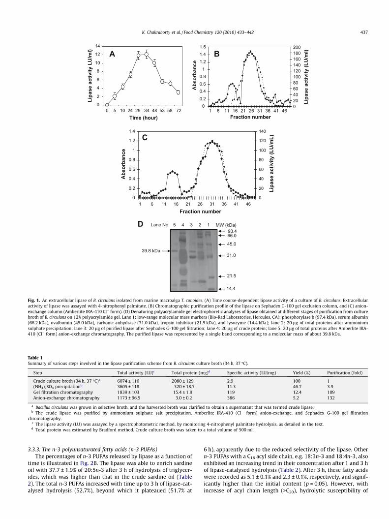
after 24 h of incubation exhibited a lipase activity of 7.3 ± 0.8 LU/ml. However, the crude extract obtained after 34 h of incubationrecorded the highest lipase activity (12.2 ± 1.1 LU/ml) (Fig. 1A),and was therefore used for further purification of the lipase. Earlierstudies indicated maximum lipase activities at the preset of loga-rithmic phase of bacterial growth (Schuepp, Kermasha, Michalski,& Morin, 1997).
3.2. Purification of B. circulans lipase and molecular studies
The results of purification profile of the extracellular lipase by B.circulans are summarised in Table 1. A 109-fold purification with aspecific activity of 119 LU/mg was attained after gel filtration onSephadex G-100 (Table 1). Highest lipase activity was apparentat the 26th fraction of the gel filtration (Fig. 1B). The enzymewas finally purified 132-fold, using Amberlite XRD-5 (Cl� form) an-ion-exchange chromatography, at the 32nd fraction, with a specificactivity of 386 LU/mg (Fig. 1C and Table 1). The homogeneity of thepurified lipase was checked by the presence of a single band corre-sponding to an apparent molecular mass of 39.8 kDa on SDS–PAGEgels, suggesting it to be a homomeric protein (Fig. 1D). Activitystaining confirmed the presence of the purified lipase. Gel filtrationchromatography, using Sephadex G-75, indicated the apparentmolecular weight of the lipase as 40.5 kDa.
3.3. Change in fatty acid composition as a function of time-course(1–6 h) lipase-catalysed hydrolysis of triglycerides from sardine oil
3.3.1. GeneralThe refined sardine oil was found to contain PUFAs, particularly
18:3n-3 (2.5 ± 0.5%), 20:5n-3 (17.6 ± 0.6%) and 22:6n-3 (8.2 ± 0.9%),along with other n-3 and n-6 PUFAs, as indicated in Table 2. The n-3 fatty acids contributed the major share (30.6%) with the n-3/n-6fatty acid ratio of 13.3. Amongst saturated fatty acids, 16:0 wasfound to be predominant, followed by 14:0, and 18:0 (Table 2).The sardine oil triglycerides were hydrolysed with lipases purifiedfrom B. circulans; the total fatty acid content of triglycerides at var-ious time-intervals of hydrolysis (1–6 h) were analysed, and the re-sults are indicated below.
3.3.2. Saturated (SFAs) and monounsaturated fatty acids (MUFAs)The fatty acid composition of sardine oil and those of enzyme
hydrolysates are given in Table 2. The lipase produced the highestdegree of hydrolysis for SFAs (47.2%), followed by MUFAs (25.9%),from their initial content after 3 h. The total concentration of SFAexhibited reductions of 15.1% and 52.2% after 1 and 3 h of hydroly-sis, respectively whereas, after prolonged hydrolysis (6 h), the pro-portion of SFAs to total fatty acids was found to be increased(18.5%) (Table 2). The total MUFA content was found to be reducedby 4.8% after 1 h of lipase-catalysed hydrolysis, and 21.9% after 3 h.However, the proportion of total MUFAs exhibited an increase(25.9%), even after 6 h of hydrolysis (Table 2). The decrease inthe contents of SFAs and MUFAs in the triglyceride mixture withthe progress of hydrolysis suggests that SFAs and MUFAs weremore easily hydrolysed by the lipase than those in triglyceridesthat contain polyunsaturated fatty acids, resulting in their enrich-ment in the triglyceride fraction (Fig. 2A). It was reported thatthose substrates containing D2–D7 isomers of 18:1n-9 were resis-tant to lipase-catalysed hydrolysis, resulting in higher concentra-tion of the fatty acid, and the discrimination was greatest for theD5 isomer (Heimermann, Holman, Gordon, Kowalyshyn, & Jensen,1973). The reduction in SFAs and MUFAs in the enzyme hydroly-sate appeared to be due to the higher selectivity of lipase for SFAsand MUFAs, thus furnishing a fatty acid concentrate with compar-atively lower content of these fatty acids (Chakraborty & Paulraj,2008b).
00.20.40.60.8
11.21.41.6
1 6 11 16 21 26 31 36 41 46Fraction number
Abs
orba
nce
020406080100120140160180200
Lipa
se a
ctiv
ity (L
U/m
l)
0
2
4
6
8
10
12
14
0 5 10 24 29 34 48 53 58 72Time (hour)
Lipa
se a
ctiv
ity L
U/m
l) A
0
0.2
0.4
0.6
0.8
1
1.2
1.4
1 6 11 16 21 26 31 36 41 46
Fraction number
Abs
orba
nce
0
20
40
60
80
100
120
140
Lipa
se a
ctiv
ity (L
U/m
L)
B
C
Lane No. 5 1234 MW (kDa)
14.4
21.5
31.0
45.0
66.093.4
39.8 kDa
D
Fig. 1. An extracellular lipase of B. circulans isolated from marine macroalga T. conoides. (A) Time course-dependent lipase activity of a culture of B. circulans. Extracellularactivity of lipase was assayed with 4-nitrophenyl palmitate. (B) Chromatographic purification profile of the lipase on Sephadex G-100 gel exclusion column, and (C) anion-exchange column (Amberlite IRA-410 Cl� form). (D) Denaturing polyacrylamide gel electrophoretic analyses of lipase obtained at different stages of purification from culturebroth of B. circulans on 12% polyacrylamide gel. Lane 1: low-range molecular mass markers (Bio-Rad Laboratories, Hercules, CA): phosphorylase b (97.4 kDa), serum albumin(66.2 kDa), ovalbumin (45.0 kDa), carbonic anhydrase (31.0 kDa), trypsin inhibitor (21.5 kDa), and lysozyme (14.4 kDa); lane 2: 20 lg of total proteins after ammoniumsulphate precipitation; lane 3: 20 lg of purified lipase after Sephadex G-100 gel filtration; lane 4: 20 lg of crude protein; lane 5: 20 lg of total proteins after Amberlite IRA-410 (Cl� form) anion-exchange chromatography. The purified lipase was represented by a single band corresponding to a molecular mass of about 39.8 kDa.
Table 1Summary of various steps involved in the lipase purification scheme from B. circulans culture broth (34 h, 37 �C).
Step Total activity (LU)c Total protein (mg)d Specific activity (LU/mg) Yield (%) Purification (fold)
Crude culture broth (34 h, 37 �C)a 6074 ± 116 2080 ± 129 2.9 100 1(NH4)2SO4 precipitationb 3605 ± 118 320 ± 18.7 11.3 46.7 3.9Gel filtration chromatography 1839 ± 103 15.4 ± 1.8 119 12.4 109Anion-exchange chromatography 1173 ± 96.5 3.0 ± 0.2 386 5.2 132
a Bacillus circulans was grown in selective broth, and the harvested broth was clarified to obtain a supernatant that was termed crude lipase.b The crude lipase was purified by ammonium sulphate salt precipitation, Amberlite IRA-410 (Cl� form) anion-exchange, and Sephadex G-100 gel filtration
chromatography.c The lipase activity (LU) was assayed by a spectrophotometric method, by monitoring 4-nitrophenyl palmitate hydrolysis, as detailed in the text.d Total protein was estimated by Bradford method. Crude culture broth was taken to a total volume of 500 ml.
K. Chakraborty et al. / Food Chemistry 120 (2010) 433–442 437
3.3.3. The n-3 polyunsaturated fatty acids (n-3 PUFAs)The percentages of n-3 PUFAs released by lipase as a function of
time is illustrated in Fig. 2B. The lipase was able to enrich sardineoil with 37.7 ± 1.9% of 20:5n-3 after 3 h of hydrolysis of triglycer-ides, which was higher than that in the crude sardine oil (Table2). The total n-3 PUFAs increased with time up to 3 h of lipase-cat-alysed hydrolysis (52.7%), beyond which it plateaued (51.7% at
6 h), apparently due to the reduced selectivity of the lipase. Othern-3 PUFAs with a C18 acyl side chain, e.g. 18:3n-3 and 18:4n-3, alsoexhibited an increasing trend in their concentration after 1 and 3 hof lipase-catalysed hydrolysis (Table 2). After 3 h, these fatty acidswere recorded as 5.1 ± 0.1% and 2.3 ± 0.1%, respectively, and signif-icantly higher than the initial content (p = 0.05). However, withincrease of acyl chain length (>C20), hydrolytic susceptibility of

Table 2Fatty acid composition (as weight %) of crude and lipase hydrolysate of sardine oil at three different time durations (1, 3, and 6 h) using purified lipase obtained from B. circulans,followed by urea complexation.
Fatty acids Sardine oil Fatty acid (as weight %) UA-fatty acida
1 h 3 h 6 h
Saturated fatty acids14:0 8.5 ± 0.2 8.3 ± 0.3 5.0 ± 0.1 7.4 ± 0.3 1.7 ± 0.216:0 18.1 ± 1.9 17.1 ± 0.2 9.3 ± 0.2 11.1 ± 0.1 0.7 ± 0.117:0 1.3 ± 0.0 ND ND ND ND18:0 2.1 ± 0.0 ND ND ND NDR SFA 30 25.4 14.3 18.5 2.4
Monounsaturated fatty acids16:1n-7 16.1 ± 1.8 15.4 ± 1.7 11.2 ± 1.0 8.3 ± 0.8 4.1 ± 0.218:1n-9 19.9 ± 2.7 19.0 ± 1.3 17.3 ± 1.6 17.4 ± 1.18 11.6 ± 1.117:1 0.4 ± 0.1 0.3 ± 0.1 0.2 ± 0.0 0.2 ± 0.05 ND20:1n-11 0.2 ± 0.0 0.1 ± 0.0 ND ND NDR MUFA 36.6 34.8 28.7 25.9 15.7
Polyunsaturated fatty acids18:2n-6 0.7 ± 0.1 0.5 ± 0.1 0.4 ± 0.1 0.4 ± 0.1 1.6 ± 0.118:3n-3 2.5 ± 0.5 3.3 ± 0.3 5.1 ± 0.1 4.9 ± 0.3 9.7 ± 0.518:4n-3 1.2 ± 0.3 1.3 ± 0.1 2.3 ± 0.1 2.3 ± 0.2 5.7 ± 0.220:4n-6 1.6 ± 0.0 0.1 ± 0.0 0.1 ± 0.0 0.1 ± 0.0 0.9 ± 0.120:5n-3 17.6 ± 0.6 23.0 ± 1.3 37.7 ± 1.9 37.1 ± 2.2 51.3 ± 4.722:5n-3 1.1 ± 0.2 0.5 ± 0.1 0.4 ± 0.1 0.4 ± 0.0 1.4 ± 0.322:6n-3 8.2 ± 0.9 7.8 ± 0.5 7.2 ± 0.3 6.5 ± 0.6 8.2 ± 0.7R PUFA 32.9 36.5 53.2 51.7 78.8n-3 PUFA 30.6 35.9 52.7 51.2 76.3n-6 PUFA 2.3 0.6 0.5 0.5 2.5n-3/n-6 13.3 59.8 105.4 102.4 30.5
LSD (p = 0.05) 1.2 0.9 1.6 2.1 1.8
a UA-fatty acid concentrate implies the urea concentrate of fatty acids. RSFA: total saturated fatty acids; RMUFA: total monounsaturated fatty acids; RPUFA: totalpolyunsaturated fatty acids; Data presented as mean values of three samples (means ± standard deviation). At the beginning of the hydrolysis (6 h) the lipases display asignificant preference for n-3 PUFAs containing 18–20 carbon atoms in the acyl chain. However, the resistance to release of SFAs and MUFAs was less as the hydrolysisreaction progressed. These values do not total 100% because minor fatty acids are not reported. ND implies non-detectable (or fatty acids present below 0.05%).
438 K. Chakraborty et al. / Food Chemistry 120 (2010) 433–442
the ester linkage of triglycerides (by the lipase) was found to in-crease as is evident from the 12.1% decrease of 22:6n-3 in the tri-glyceride fraction after 3 h of hydrolysis. The final 22:6n-3 contentin the glyceride mixture after 6 h hydrolysis was 6.5 ± 0.6%, i.e.,1.3% less than in the original sardine oil (Table 2). Similar resultshave been apparent from the reduced content of 22:5n-3 (Table2). Generally lipases are very specific in hydrolysing specific fattyacids with specific acyl chain length (e.g. C20 or C22 acyl chainlength) or specific classes of fatty acids (e.g. EPA with C20 acyl chainlength and DHA C22 acyl chain length). Since lipase reaction is veryspecific, some lipases (e.g. the B. circulans lipase in this study) canconcentrate a particular fatty acid (e.g. EPA, 20:5n-3), but may beineffective for concentrating other fatty acids (e.g. DHA 22:6n-3,which could not be enriched in this process). The aim of this studyis to enrich EPA (20:5n-3) from sardine oil. The bacterial lipasepurified from B. circulans is specific in hydrolysing fatty acids high-er than C20 n-3, e.g. DHA, thereby decreasing their concentration inthe triglyceride fraction. Earlier studies indicated the increase in n-3 fatty acids (63.7%) in the triacylglycerol after 6 h of hydrolysis byan extracellular lipase isolated from B. licheniformis (Chakraborty &Paulraj, 2008b). A 2-fold increase of DHA and EPA concentrationwas reported by Pseudomonas cepacia and Candida rugosa lipase-catalysed hydrolysis of Salmo salar L. oil (Sun, Pigott, & Herwig,2002). A microbial lipase isolated from C. antarctica was reportedto yield 93.5% PUFA with 25.7% EPA and 44.7% DHA from cod liveroil triglycerides (Medina et al., 1999). Hydrolysis by P. fluorescensHU380 lipase, followed by amide complexation of cod liver oil,was reported to yield 43.1% EPA and 7% DHA (Kojima, Sakuradani,& Shimizu, 2006). From these results it can be concluded that thebacterial lipase is specific in hydrolysing fatty acids higher thanC20 n-3. It is apparent that a particular fatty acid, e.g. 22:5n-3 or22:6n-3, released on hydrolysis is more abundant, resulting in de-crease of the triacylglycerol portion of fatty acids, thus suggesting
that the ester bond of the fatty acids are susceptible to hydrolysisby the lipase used in the present study. On the other hand, 20:5n-3and 18:3n-3 (having the C18 n-3 configuration) are more abundantin the triacylglycerol portion of fatty acids, thus suggesting that thelipase is less reactive towards that fatty acid’s ester bond (resistantto hydrolysis), resulting in their increase in the triglycerides (Cha-kraborty & Paulraj, 2008b). However, due to its acyl side chain andolefinic double bond specificity, this particular lipase obtainedfrom B. circulans is not able to concentrate DHA.
3.3.4. The n-6 polyunsaturated fatty acids (n-6 PUFAs)In general, the n-6 fatty acids exhibited a reduction in their con-
tent after lipase-catalysed hydrolysis. Amongst n-6 PUFAs, 18:2n-6was recorded to be 0.5 ± 0.1% after 1 h of lipase-catalysed hydroly-sis, and 0.4 ± 0.1% after 3 h, which were substantially lower thanthat in the crude sardine oil (Table 2). Similarly, the content of20:4n-6 in triglyceride was found to decrease by 94.3% after 1 hof hydrolysis, and 95.6% after 3 h (Table 2). The results apparentlysuggest that, amongst n-3 and n-6 fatty acids, the former are moreresistant to hydrolysis by the lipase used in this study, and the dis-crimination was the greatest for the C18–20 acyl chain-lengthedhomologues. However, after prolonged hydrolysis (9 h), the con-tent of n-6 fatty acids recorded a slight increase, presumably dueto the reduced selectivity of lipase. It is apparent that, after pro-longed hydrolysis, only a few target fatty acid ester bonds (n-6fatty acyl ester bonds) are available in the reaction mixture thatare susceptible to hydrolysis by a lipase. The microbial lipase caneven cleave bonds that are resistant (or nearly resistant) to hydro-lysis (C18–20 n-3 fatty acids like EPA) to maintain the dynamic equi-librium of the system. These results indicate that the resistance ofhydrolysis of this latter class of fatty acyl ester bond depends uponthe presence of other substrates (n-6 fatty acyl ester bonds and es-ters other than C18–C20 n-3 fatty acids) in the reaction mixture. An

A
B
C
0
10
20
30
40
50
60
1h 3h 6h
Pro
port
ions
of f
atty
ac
ids
in to
tal
(%)
SFAMUFAPUFA
0
5
10
15
20
25
30
35
40
1h 3h 6h
Prop
ortio
ns o
f fa
tty
acid
s in
tota
l (%
)
18:3n-3
18:4n-3
20:5n-3
22:6n-3
0
0.1
0.2
0.3
0.4
0.5
0.6
1h 3h 6h
Prop
ortio
ns o
f fa
tty
acid
s in
tota
l (%
) 18:2n-6
20:4n-6
Time of lipase-catalyzed hydrolysis (h)
Fig. 2. Per cent share of fatty acids in triglyceride fraction (as weight %) in total by B.circulans lipase-catalysed hydrolysis of sardine oil for different time-intervals(1–6 h). (A) Proportions of different classes of fatty acids (SFAs, MUFAs, and PUFAs)in triglyceride fraction of sardine oil with respect to time duration (1–6 h) of lipase-catalysed hydrolysis; (B) proportions of PUFAs with n-6 double bond, and (C) n-3double bond in triglyceride fraction of sardine oil with respect to time duration(1–6 h) of lipase-catalysed hydrolysis.
K. Chakraborty et al. / Food Chemistry 120 (2010) 433–442 439
earlier study reported that the content of 20:4n-6 in the glyceridemixture increased with the progress of hydrolysis by lipase puri-fied from B. licheniformis (Chakraborty & Paulraj, 2008b). Thiswas found to be due to fatty acid specificity of lipases toward PU-FAs with D5 unsaturated double bond (cis-5 olefinic bond posi-tioned at the number 5 carbon from the carboxyl end)(Chakraborty & Paulraj, 2008b). Earlier results indicated that the li-pase from B. licheniformis was unique in the resistance it encoun-tered with 20:5n-3 and 20:4n-6. This could be attributed to thepositional specificity of the enzyme at the olefinic double bondsof PUFAs. Lipases have been screened, from different sources, toselectively concentrate c-linolenic acid from B. officinalis and Echi-um fastuosum seed oil (López-Martínez et al., 2004).
3.4. Specificity of bacterial lipase towards different fatty acids andstructure–bioactivity relationship analyses
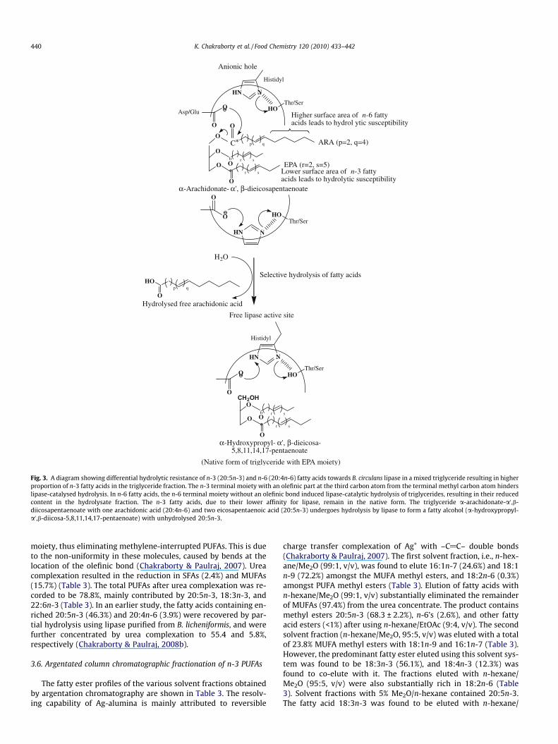
A direct hydrophobic effect, as exemplified by partition coeffi-cient log P and bulk parameters molar volume (MV) and parachor(P), appeared to dictate lipase-catalysed hydrolysis of the fatty acylester linkage of triglycerides. The n-3 fatty acids exhibited lower
hydrophobic activities (20:5n-3log P = 5.85) than did the n-6 fattyacids (20:4n-6log P = 6.16), resulting in greater hydrolytic suscepti-bility of the latter, leading to n-3 decrease in the triglyceride frac-tion. Similarly, the n-3 fatty acid 18:3n-3 showed higher hydrolyticresistance to lipase, evidently due to the lower bulk(MV = 301 cm3; P = 731 cm3). The fatty acid 22:5n-3 is compara-tively more susceptible to lipase-catalysed hydrolysis than is20:5n-3, apparently due to the slightly higher MV and P than inthe latter, thereby increasing the content of 20:5n-3 in the triglyc-eride fraction. The terminal double bond at the n-3 position (be-tween C15–16 moiety of 18:3n-3 and 20:5n-3) appeared to reducethe aliphatic chain (3.7041 A0) that apparently hinders the favour-able interaction of the aminoacyl residues in the active site. This, inturn, appeared to retain the native form of triglycerides containingn-3 fatty acids (Fig. 3). The fatty acid 20:5n-3 has a lower bulk thanhas 20:4n-6, as exemplified by its lower MV (321 cm3) and P(798 cm3) than the latter. It is apparent that an extended terminalaliphatic moiety (6.1025 A0) in the n-6 fatty acid homologues(18:2n-6 and 20:4n-6) results in favourable rearrangement of theaminoacyl residues in the enzyme active site leading to facilehydrolysis of ester bonds of triglycerides containing n-6 fatty acids(Fig. 3). These results indicated that B. circulans lipase is unique inits lack of activity on n-3 PUFAs. The decrease in the contents ofSFAs and MUFAs in the triglyceride fraction with the progress ofhydrolysis suggests that they are more easily hydrolysed by the li-pase than are n-3 fatty acids, resulting in enrichment of the latterin the triglyceride fraction. The bacterial lipase from B. circulans isspecific in hydrolysing fatty acids higher than C20 n-3, thereby con-centrating EPA (C20 n-3 fatty acid). The content of C22 fatty acids(22:5n-3) with an acyl chain length of 25.3461 A0 exhibited areduction in the triglyceride fraction after lipase-catalysed hydro-lysis. Similarly 22:6n-3 was reduced by 4.0% after 1 h of hydrolysis.However, after prolonged hydrolysis of triglycerides (3 and 6 h),the content of this fatty acid exhibited a reduction in the hydrolysisrate (12.1% and 20.6%, respectively) apparently due to the satura-tion of lipase activity. Such fatty acid selectivity of lipases, i.e.,hydrolysis favouring n-6 over n-3, and long-chain fatty acids(>C20) (22:6n-3) over 20:5n-3, may therefore be useful for fraction-ating the latter from sardine oil. It is noteworthy that the microbiallipase from B. circulans used in the present study was specific inhydrolysing n-6 fatty acids and n-3 fatty acids with >C20 acyl chainlength (22:6n-3) at the initial stage of hydrolysis (up to 6 h) of fattyacyl ester bonds. However, after prolonged hydrolysis, when only afew target fatty acid ester bonds (n-6 fatty acyl ester bonds and es-ters other than C18–20 n-3 fatty acids) are available in the enzymehydrolysate (that are susceptible to hydrolysis by a lipase), themicrobial lipase can even cleave bonds that are resistant to hydro-lysis (C18–20 n-3 fatty acids). An earlier study suggested that thefatty acid triacylglycerols containing D6 and D9 isomers weremore susceptible to lipolysis by B. licheniformis MTCC 6824 lipase,and the discrimination was the greatest for the D5 isomer (20:5n-3and 20:4n-6), followed by D4 double-bonded fatty acids such as22:6n-3 (Chakraborty & Paulraj, 2008b). Structure–activity studiesof lipases from Penicillium sp. and Rhizopus arrhizus were used topredict enzyme specificity leading to fatty acid concentration(Mukherjee, Kiewitt, & Hills, 1993). It was reported that fattyacids with the first double bond from the carboxyl end as a cis-4(22:6n-3), cis-6 (18:1n-12, 18:3n-6, 18:4n-3), or a cis-8 (20:3n-6) are strongly discriminated by the lipase (Mukherjee et al., 1993).
3.5. Preparation of PUFA concentrates by urea complexation
The per cent fatty acid composition obtained after urea com-plexation are illustrated in Table 2. Urea complexation of fattyacids was used to concentrate PUFAs from SFAs and MUFAs, whereurea occludes straight-chain SFAs and MUFAs in the hexagonal
CH2OHO
O O*
p q
r s
r s
Thr/SerHO
O
O
HN N
Histidyl
Oα-Hydroxypropyl- α', β-dieicosa-
5,8,11,14,17-pentaenoate
O
HO
Hydrolysed free arachidonic acid
Free lipase active site
H2O
Anionic hole
Thr/SerHOAsp/Glu
O
O
HN N
Histidyl
O
O O*
O*
Higher surface area of n-6 fattyacids leads to hydrol ytic susceptibility
p qC
O
r s
r s
Thr/SerHO
O
O
HN N
EPA (r=2, s=5)
ARA (p=2, q=4)
Lower surface area of n-3 fattyacids leads to hydrolytic susceptibilityO
α-Arachidonate- α', β-dieicosapentaenoate
Selective hydrolysis of fatty acids
(Native form of triglyceride with EPA moiety)
Fig. 3. A diagram showing differential hydrolytic resistance of n-3 (20:5n-3) and n-6 (20:4n-6) fatty acids towards B. circulans lipase in a mixed triglyceride resulting in higherproportion of n-3 fatty acids in the triglyceride fraction. The n-3 terminal moiety with an olefinic part at the third carbon atom from the terminal methyl carbon atom hinderslipase-catalysed hydrolysis. In n-6 fatty acids, the n-6 terminal moiety without an olefinic bond induced lipase-catalytic hydrolysis of triglycerides, resulting in their reducedcontent in the hydrolysate fraction. The n-3 fatty acids, due to their lower affinity for lipase, remain in the native form. The triglyceride a-arachidonate-a0 ,b-diicosapentaenoate with one arachidonic acid (20:4n-6) and two eicosapentaenoic acid (20:5n-3) undergoes hydrolysis by lipase to form a fatty alcohol (a-hydroxypropyl-a0 ,b-diicosa-5,8,11,14,17-pentaenoate) with unhydrolysed 20:5n-3.
440 K. Chakraborty et al. / Food Chemistry 120 (2010) 433–442
moiety, thus eliminating methylene-interrupted PUFAs. This is dueto the non-uniformity in these molecules, caused by bends at thelocation of the olefinic bond (Chakraborty & Paulraj, 2007). Ureacomplexation resulted in the reduction in SFAs (2.4%) and MUFAs(15.7%) (Table 3). The total PUFAs after urea complexation was re-corded to be 78.8%, mainly contributed by 20:5n-3, 18:3n-3, and22:6n-3 (Table 3). In an earlier study, the fatty acids containing en-riched 20:5n-3 (46.3%) and 20:4n-6 (3.9%) were recovered by par-tial hydrolysis using lipase purified from B. licheniformis, and werefurther concentrated by urea complexation to 55.4 and 5.8%,respectively (Chakraborty & Paulraj, 2008b).
3.6. Argentated column chromatographic fractionation of n-3 PUFAs
The fatty ester profiles of the various solvent fractions obtainedby argentation chromatography are shown in Table 3. The resolv-ing capability of Ag-alumina is mainly attributed to reversible
charge transfer complexation of Ag+ with –C@C– double bonds(Chakraborty & Paulraj, 2007). The first solvent fraction, i.e., n-hex-ane/Me2O (99:1, v/v), was found to elute 16:1n-7 (24.6%) and 18:1n-9 (72.2%) amongst the MUFA methyl esters, and 18:2n-6 (0.3%)amongst PUFA methyl esters (Table 3). Elution of fatty acids withn-hexane/Me2O (99:1, v/v) substantially eliminated the remainderof MUFAs (97.4%) from the urea concentrate. The product containsmethyl esters 20:5n-3 (68.3 ± 2.2%), n-6’s (2.6%), and other fattyacid esters (<1%) after using n-hexane/EtOAc (9:4, v/v). The secondsolvent fraction (n-hexane/Me2O, 95:5, v/v) was eluted with a totalof 23.8% MUFA methyl esters with 18:1n-9 and 16:1n-7 (Table 3).However, the predominant fatty ester eluted using this solvent sys-tem was found to be 18:3n-3 (56.1%), and 18:4n-3 (12.3%) wasfound to co-elute with it. The fractions eluted with n-hexane/Me2O (95:5, v/v) were also substantially rich in 18:2n-6 (Table3). Solvent fractions with 5% Me2O/n-hexane contained 20:5n-3.The fatty acid 18:3n-3 was found to be eluted with n-hexane/

Table 3Percentage composition (as weight %) of individual fatty acids in different solvent eluates by argentation chromatography using urea-fatty acid concentrate.
Fatty acids UA-fatty acid Eluants (solvent systems)
n-Hexane/Me2O (99:1, v/v) n-Hexane/Me2O (95:5, v/v) n-Hexane/EtOAc (9:4, v/v)
Saturated fatty acids14:0 ND ND ND ND16:0 0.7 ± 0.1 0.2 ± 0.0 ND ND17:0 ND ND ND ND18:0 1.7 ± 0.2 ND ND NDR SFA 2.4 0.2 0.00 0.00
Monounsaturated fatty acids16:1n-7 4.1 ± 0.2 24.6 ± 1.1 6.8 ± 0.1 0.1 ± 0.018:1n-9 11.6 ± 1.1 72.2 ± 2.2 16.8 ± 0.2 0.2 ± 0.117:1 ND 0.6 ± 0.0 0.1 ± 0.0 ND20:1n-11 ND ND ND NDR MUFA 15.7 97.4 23.7 0.3
Polyunsaturated fatty acids18:2n-6 1.6 ± 0.1 0.3 ± 0.1 5.3 ± 0.2 2.5 ± 0.118:3n-3 9.7 ± 0.5 0.3 ± 0.1 56.1 ± 3.2 13.9 ± 0.218:4n-3 5.7 ± 0.2 0.1 ± 0.0 12.3 ± 0.6 2.6 ± 0.120:4n-6 0.9 ± 0.1 0.1 ± 0.0 0.1 ± 0.0 0.1 ± 0.020:5n-3 51.3 ± 4.7 ND 0.3 ± 0.0 68.3 ± 2.222:5n-3 1.4 ± 0.3 ND ND 1.5 ± 0.222:6n-3 8.2 ± 0.7 ND 0.1 ± 0.0 8.7 ± 0.4R PUFA 78.8 0.8 74.2 97.6n-3 PUFA 76.3 0.4 68.8 95.0n-6 PUFA 2.5 0.4 5.4 2.6n-3/n-6 30.5 1.0 12.7 36.5
LSD (p = 0.05) 1.8 2.4 0.8 1.7
The notations are as indicated under Table 1. On the basis of the significance of treatments, LSD at the 5% level of significance (p = 0.05) was computed. Data are presented asmean values of three samples (means ± standard deviation). These values do not total 100% because minor fatty acids are not reported. ND implies non-detectable (or fattyacids present below 0.05%).
K. Chakraborty et al. / Food Chemistry 120 (2010) 433–442 441
Me2O (20:1, v/v) with a final purity of 56.1 ± 3.2%. The methyl esterof 22:6n-3, with an additional double bond, was found to co-elute(22:6n-3, 8.7 ± 0.4%) with 20:5n-3 in the fraction containing n-hex-ane/EtOAc (9:4, v/v) as eluting solvent system (Table 3). AgNO3–TLC was used to elucidate the progress of purification of PUFAs.The uppermost band (Rf: 0.80–86) of SFAs, and the second bandof MUFAs (Rf: 0.46–55) were apparent. FAME up to trienes(18:2n-6 and 18:3n-3) remained at the base of TLC chromatogram.These results are in agreement with the reported literature(Chakraborty & Paulraj, 2007).
4. Conclusions
The present study showed the changes in different classes offatty acid and composition as a function of time-course (1–6 h)of hydrolysis of triglycerides from sardine oil by a lipase purifiedfrom B. circulans that may be a potential enzyme source for concen-tration of n-3 PUFAs of acyl chain length shorter than C20. The n-3PUFAs (18:3n-3 and 20:5n-3) in the triglyceride mixture werefound to increase proportionally with the progress of hydrolysisup to 3 h; after that their concentration plateaued as reaction timeincreased beyond 3 h, due to reduced selectivity of the lipase. Itwas found that ester bonds of n-3 fatty acid homologues werehighly resistant to hydrolysis by lipase from B. circulans, resultingin their enrichment in fatty acid triglycerides. Structure–bioactiv-ity relationship analyses revealed a direct relationship of hydro-phobic and steric properties to lipase-catalysed hydrolytic effectson fatty acyl ester bonds of triglycerides. The urea complexationreaction raised 20:5n-3 and 18:3n-3 contents by 2.91-fold and3.92-fold, respectively, thus allowing further purification by argen-tated chromatography. The fractions eluted with n-hexane/Me2O(95:5, v/v) and n-hexane/EtOAc (9:4, v/v), by argentated chroma-tography furnished 18:3n-3 and 20:5n-3 methyl ester in final puri-ties of 56.1 ± 3.18% and 68.3 ± 2.15% of total fatty acids,
respectively. It can be concluded that a combination of B. circulanslipase-catalysed hydrolysis followed by urea fractionation and arg-entated chromatography, is a promising method for separating andconcentrating n-3 fatty acids like 18:3n-3 and 20:5n-3 from sar-dine oil. The lipase-catalysed reaction offers several benefits, suchas greater selectivity and milder reaction conditions. PUFA easilyundergoes decomposition in chemical processes, yielding undesir-able oxidation products and polymers. Modification of fats and oilscontaining PUFAs by lipase may be a process with prospects for en-zyme application on a commercial scale. This procedure for con-centrating n-3 PUFAs by B. circulans lipase would contribute tothe commercial application of lipase to obtain concentrated n-3fatty acids, particularly EPA from sardine oil.
Acknowledgements
The authors are thankful to the Dr. G. Syda Rao, Director, CMFRI,Cochin for providing necessary facilities to carry out the work.Thanks are due to Dr. Shibu M. Eappen, Scientist B, SophisticatedTest and Instrumentation Centre, Cochin University of Scienceand Technology, Kerala, for recording the mass spectral data.
References
Andersson, B. A. (1978). Mass spectrometry of fatty acid pyrrolidides. Progress in theChemistry of Fats and Other Lipids, 16, 279–308.
Bligh, E. G., & Dyer, W. J. (1959). A rapid method of total lipid extraction andpurification. Canadian Journal of Biochemistry and Physiology, 37, 911–917.
Bradford, M. (1976). A rapid and sensitive method for the quantification ofmicrogram quantities of protein utilizing the principle of protein–dye binding.Analytical Biochemistry, 72, 248–254.
Cahu, C. L., Guillaume, J. C., Stephan, G., & Chim, L. (1994). Influence ofphospholipids and highly unsaturated fatty acids on spawning rate and eggand tissue composition in Penaeus vannamei fed semipurified diets. Aquaculture,126, 159–170.
Can, A., & Özçelik, B. (2005). Enrichment of hazelnut oil with long-chain n-3 PUFAby lipase-catalyzed acidolysis: Optimization by response surface methodology.Journal of the American Oil Chemists Society, 82, 27–32.

442 K. Chakraborty et al. / Food Chemistry 120 (2010) 433–442
Chakraborty, K., & Devakumar, C. (2005). Quantitative structure–activityrelationship (QSAR) analysis as a tool to evaluate mode of action of chemicalhybridizing agents (CHAs) for wheat (Triticum aestivum L.). Journal ofAgricultural and Food Chemistry, 53, 3468–3475.
Chakraborty, K., Devakumar, C., Tomar, S. M. S., & Kumar, R. (2003). Synthesis andquantitative structure–activity relationships (QSAR) of oxanilates as chemicalhybridizing agents (CHAs) for wheat (Triticum aestivum L.). Journal ofAgricultural and Food Chemistry, 51, 992–998.
Chakraborty, K., & Paulraj, R. (2007). Eicosapentanoic acid enrichment from sardineoil by argentation chromatography. Journal of Agricultural and Food Chemistry,55, 7586–7595.
Chakraborty, K., & Paulraj, R. (2008a). An extra-cellular alkaline metallolipase fromBacillus licheniformis MTCC 6824: Purification and biochemical characterization.Food Chemistry, 109, 727–736.
Chakraborty, K., & Paulraj, R. (2008b). Enrichment of eicosapentaenoic acidfrom sardine oil with D5-olefinic bond specific lipase from Bacilluslicheniformis MTCC 6824. Journal of Agricultural and Food Chemistry, 56,1428–1433.
Guil-Guerrero, J. L., López-Martínez, J. C., Rincon-Cervera, M. A., & Campra-Madrid,P. (2007). One-step extraction and concentration of polyunsaturated fattyacids from fish liver. Journal of Agricultural and Food Chemistry, 84,357–361.
Heimermann, W. H., Holman, R. T., Gordon, D. T., Kowalyshyn, D. E., & Jensen, R. G.(1973). Effect of double bond position in octadecenoates upon hydrolysis bypancreatic lipase. Lipids, 8, 45–47.
Kademi, A., Ait-Abdelkader, N., & Fakhreddine, L. (2000). Purification andcharacterization of a thermostable esterase from the moderate thermophileBacillus circulans. Applied Microbiology and Biotechnology, 54, 173–179.
Kim, K. R., Kwon, D. Y., Yoon, S. H., Kim, W. Y., & Kim, K. H. (2005). Purification,refolding, and characterization of recombinant Pseudomonas fluorescens lipase.Protein Expression and Purification, 39, 124–129.
Kojima, Y., Sakuradani, E., & Shimizu, S. (2006). Different specificity of two types ofPseudomonas lipases for C20 fatty acids with a D5 unsaturated double bond andtheir application for selective concentration of fatty acids. Journal of Bioscienceand Bioengineering, 101, 496–500.
Kumar, S., Kikon, K., Upadhyay, A., Kanwar, S. S., & Gupta, R. (2005).Production, purification and characterization of lipase from thermophilic andalkalophilic Bacillus coagulans BTS-3. Protein Expression and Purification, 41,38–44.
Laemmli, U. K. (1970). Cleavage of structural protein during the assembly of thehead of bacteriophage T4. Nature, 227, 680–685.
Lee, Y. P., Chung, G. H., & Rhee, J. S. (1993). Purification and characterization ofPseudomonas fluorescens SIK W1 lipase expressed in Escherichia coli. Biochimicaet Biophysica Acta, 1169, 156–164.
Liu, S., Zhang, C., Hong, P., & Ji, H. (2007). Lipase-catalysed acylglycerol synthesis ofglycerol and n-3 PUFA from tuna oil: Optimisation of process parameters. FoodChemistry, 103, 1009–1015.
López-Martínez, J. C., Campra-Madrid, P., & Guil-Guerrero, J. L. (2004). c-Linolenicacid enrichment from Borago officinalis and Echium fastuosum seed oils and fattyacids by low temperature crystallization. Journal of Bioscience andBioengineering, 97, 294–298.
López-Martínez, J. C., Campra-Madrid, P., Rincón-Cervera, M. A., & Guil-Guerrero, J.L. (2005). Ecological and simultaneous seed oil extraction/saponification/gamma-linolenic acid concentration. European Journal of Lipid Science andTechnology, 107, 180–186.
Medina, A. R., Cerdán, L. E., Giménez, A. G., Páez, B. C., González, M. J. I., & Grima, E.M. (1999). Lipase-catalyzed esterification of glycerol and polyunsaturated fattyacids from fish and microalgae oils. Journal of Biotechnology, 70, 379–391.
Metcalf, L. D., Schimtz, A. A., & Pleka, J. R. (1966). Rapid preparation of fatty acidesters from lipids for gas chromatographic analyses. Analytical Chemistry, 38,514–515.
Mukherjee, K. D., Kiewitt, I., & Hills, M. J. (1993). Substrate specificities of lipases inview of kinetic resolution of unsaturated fatty acids. Applied Microbiology andBiotechnology, 40, 489–493.
Sahena, F., Zaidul, I. S. M., Jinap, S., Saari, N., Jahurul, H. A., Abbas, K. A., et al. (2009).PUFAs in fish: extraction, fractionation, importance in health. ComprehensiveReview of Food Science and Food Safety, 8, 59–74.
Schuepp, C., Kermasha, S., Michalski, M. C., & Morin, A. (1997). Production, partialpurification and characterisation of lipases from Pseudomonas fragi CRDA 037.Process Biochemistry, 32, 225–232.
Sun, T., Pigott, G. M., & Herwig, R. P. (2002). Lipase-assisted concentration of n-3polyunsaturated fatty acids from viscera of farmed Atlantic Salmon (Salmo salarL.). Journal of Food Science, 67, 130–136.
Tanaka, Y., Hirano, J., & Funada, T. (1994). Synthesis of docosahexaenoic acid-richtriglyceride with immobilized Chromobacterium viscosum lipase. Journal of theAmerican Oil Chemists Society, 71, 331–334.
Winkler, U. K., & Stuckmann, M. (1979). Glucogen, hyaluronate and some otherpolysaccharides greatly enhance the formation of exolipase by Serratiamarcescens. Journal of Bacteriology, 138, 663–670.
Zuta, C. P., Simpson, B. K., Chan, H. M., & Phillips, L. (2003). Concentrating PUFA frommackerel processing waste. Journal of the American Oil Chemists Society, 80,933–936.
Copyright © 2022 FDOKUMEN