Comparison of the effect of calcium gluconate and batroxobin on the release of transforming growth...
7
RESEARCH ARTICLE Open Access Comparison of the effect of calcium gluconate and batroxobin on the release of transforming growth factor beta 1 in canine platelet concentrates Raul F Silva 1,2* , Jorge U Carmona 2 and Cleuza MF Rezende 1 Abstract Background: The clinical use of autologous platelet concentrates (also known as platelet-rich plasma) on the field of regenerative therapy, in the last decade has been the subject of several studies especially in equine medicine and surgery. The objectives of this study was: 1) to describe and compare the cellular population in whole blood, lower fraction (A) and upper fraction (B) of platelet concentrates, 2) to measure and compare the transforming growth factor beta 1 (TGF-β 1 ) concentration in plasma and both platelet concentrates after be activated with calcium gluconate or batroxobin plus calcium gluconate and, 3) to determine correlations between cell counts in platelet concentrates and concentrations of TGF-β 1 . Blood samples were taken from 16 dogs for complete blood count, plasma collection and platelet concentrates preparation. The platelet concentrates (PC) were arbitrarily divided into two fractions, specifically, PC-A (lower fraction) and PC-B (upper fraction). The Platelet concentrates were analyzed by hemogram. After activated with calcium gluconate or batroxobin plus calcium gluconate, TGF-β 1 concentration was determined in supernatants of platelet concentrates and plasma. Results: There were differences statistically significant (P < 0.05) for the platelet count and leukocyte count and TGF-β 1 concentration between whole blood, plasma and both platelet concentrates. A significant correlation was found between the number of platelets in both platelet concentrates and TGF-β 1 concentration. Platelet collection efficiency was 46.34% and 28.16% for PC-A and PC-B, respectively. TGF-β 1 concentration efficiency for PC activated with calcium gluconate was 47.75% and 31.77%, for PC-A and PC-B, respectively. PC activated with batroxobin plus CG showed 46.87% and 32.24% for PC-A and PC-B, respectively. Conclusions: The methodology used in this study allows the concentration of a number of platelets and TGF-β 1 that might be acceptable for a biological effect for clinical or experimental use as a regenerative therapy in dogs. Keywords: Tube method, Platelet Rich-Plasma, Dog, Regenerative medicine Background The healing process is directed by complex biological mechanisms that involve many cells and proteins, such as cytokines and growth factors (GF), among others. Cellular and molecular interactions allow, under physio- logical conditions, the repair or regeneration of damaged tissues [1,2]. Platelets play a central role in the healing process. These cytoplasmic fragments not only have hemostatic properties [3], but also have pro-inflammatory, regulatory [4] and regenerative properties, which are mediated by interaction with other cells (neutrophils and endothelial cells), GF, chemokines and other regula- tory molecules [5]. Currently, there is a growing interest in the use of au- tologous platelet concentrates (PC), also known as plate- let rich plasma (PRP) for stimulating the healing process and promoting regeneration instead of reparation. Plate- let alpha granules [4,6] contain at least seven GF directly * Correspondence: [email protected] 1 Departamento de Clínica e Cirurgia Animal, Universidade Federal de Minas Gerais, Belo Horizonte, Brazil 2 Grupo de Investigación Terapia Regenerativa, Departamento de Salud Animal, Universidad de Caldas, Manizales, Colombia © 2012 Silva et al.; licensee BioMed Central Ltd. This is an Open Access article distributed under the terms of the Creative Commons Attribution License (http://creativecommons.org/licenses/by/2.0), which permits unrestricted use, distribution, and reproduction in any medium, provided the original work is properly cited. Silva et al. BMC Veterinary Research 2012, 8:121 http://www.biomedcentral.com/1746-6148/8/121
-
Upload
fundacionuniversitarialoslibertadores -
Category
Documents
-
view
2 -
download
0
Transcript of Comparison of the effect of calcium gluconate and batroxobin on the release of transforming growth...

Silva et al. BMC Veterinary Research 2012, 8:121http://www.biomedcentral.com/1746-6148/8/121
RESEARCH ARTICLE Open Access
Comparison of the effect of calcium gluconateand batroxobin on the release of transforminggrowth factor beta 1 in canine plateletconcentratesRaul F Silva1,2*, Jorge U Carmona2 and Cleuza MF Rezende1
Abstract
Background: The clinical use of autologous platelet concentrates (also known as platelet-rich plasma) on the fieldof regenerative therapy, in the last decade has been the subject of several studies especially in equine medicineand surgery. The objectives of this study was: 1) to describe and compare the cellular population in whole blood,lower fraction (A) and upper fraction (B) of platelet concentrates, 2) to measure and compare the transforminggrowth factor beta 1 (TGF-β1) concentration in plasma and both platelet concentrates after be activated withcalcium gluconate or batroxobin plus calcium gluconate and, 3) to determine correlations between cell counts inplatelet concentrates and concentrations of TGF-β1. Blood samples were taken from 16 dogs for complete bloodcount, plasma collection and platelet concentrates preparation. The platelet concentrates (PC) were arbitrarilydivided into two fractions, specifically, PC-A (lower fraction) and PC-B (upper fraction). The Platelet concentrateswere analyzed by hemogram. After activated with calcium gluconate or batroxobin plus calcium gluconate, TGF-β1concentration was determined in supernatants of platelet concentrates and plasma.
Results: There were differences statistically significant (P< 0.05) for the platelet count and leukocyte count andTGF-β1 concentration between whole blood, plasma and both platelet concentrates. A significant correlation wasfound between the number of platelets in both platelet concentrates and TGF-β1 concentration. Platelet collectionefficiency was 46.34% and 28.16% for PC-A and PC-B, respectively. TGF-β1 concentration efficiency for PC activatedwith calcium gluconate was 47.75% and 31.77%, for PC-A and PC-B, respectively. PC activated with batroxobin plusCG showed 46.87% and 32.24% for PC-A and PC-B, respectively.
Conclusions: The methodology used in this study allows the concentration of a number of platelets and TGF-β1that might be acceptable for a biological effect for clinical or experimental use as a regenerative therapy in dogs.
Keywords: Tube method, Platelet Rich-Plasma, Dog, Regenerative medicine
BackgroundThe healing process is directed by complex biologicalmechanisms that involve many cells and proteins, suchas cytokines and growth factors (GF), among others.Cellular and molecular interactions allow, under physio-logical conditions, the repair or regeneration of damagedtissues [1,2]. Platelets play a central role in the healing
* Correspondence: [email protected] de Clínica e Cirurgia Animal, Universidade Federal de MinasGerais, Belo Horizonte, Brazil2Grupo de Investigación Terapia Regenerativa, Departamento de SaludAnimal, Universidad de Caldas, Manizales, Colombia
© 2012 Silva et al.; licensee BioMed Central LtCommons Attribution License (http://creativecreproduction in any medium, provided the or
process. These cytoplasmic fragments not only havehemostatic properties [3], but also have pro-inflammatory,regulatory [4] and regenerative properties, which aremediated by interaction with other cells (neutrophilsand endothelial cells), GF, chemokines and other regula-tory molecules [5].Currently, there is a growing interest in the use of au-
tologous platelet concentrates (PC), also known as plate-let rich plasma (PRP) for stimulating the healing processand promoting regeneration instead of reparation. Plate-let alpha granules [4,6] contain at least seven GF directly
d. This is an Open Access article distributed under the terms of the Creativeommons.org/licenses/by/2.0), which permits unrestricted use, distribution, andiginal work is properly cited.
Silva et al. BMC Veterinary Research 2012, 8:121 Page 2 of 7http://www.biomedcentral.com/1746-6148/8/121
involved in the healing process. Of these proteins, trans-forming growth factor beta isoform 1 (TGF-β1) is ofpivotal importance for its actions on cell proliferation,angiogenesis and extracellular matrix deposition [7].Many substances could potentially be used for platelet
activation as a previous step for the clinical use PC.Platelet activation understood as a combination of fi-brinogen cleavage leading to fibrin mesh formation andexternalization of alpha granules containing growth fac-tors [8]. Arbitrarily, activating substances could are clas-sified according to their chemical structure in proteic,non-proteic forms and combination of both. Proteic ac-tivating substances include thrombin (either from bovineor autogenous sources) [9,10], and soluble collagen typeI [11] amongst others. Calcium salts, such as calciumchloride and calcium gluconate [9] are classified as non-proteic activating substances. Combination of both in-cludes batroxobin plus calcium gluconate [10].Some protocols for platelet activation include the use
of a calcium salt solely and others include the combin-ation of a calcium salt plus a proteic activating sub-stance. To date, there is no convincing data about whatis the best activating substance (in terms of GF concen-tration) for PC activation and more research is necessaryto know the effect of these substances (either alone orcombined) on the release of platelet main derived GF,such as TGF-β1.There is information on basic biological aspects and
clinical use of PC in musculoskeletal and soft tissue in-juries in human beings [12-15] and horses [16-18]. Thestudy presented here provides the description of a man-ual protocol for producing two kinds of canine PC ar-bitrarily classified as lower fraction (PC-A) and upperfraction (PC-B) and evaluates the effect of a non-proteicactivating substance (calcium gluconate -CG-) and the
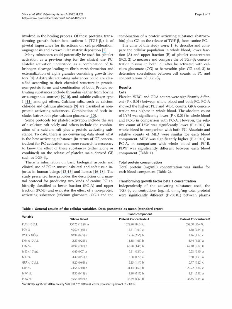
Table 1 General results of the cellular variables. Data present
VariableWhole Blood
PLT × 103/μL 330.75 (18.28) a
PCV % 45.50 (1.05) a
WBC× 103/μL 10.94 (0.77) a
LYM× 103/μL 2.27 (0.25) a
LYM % 20.97 (2.08) a
MID× 103/μL 0.49 (007) a
MID % 4.49 (0.55) a
GRA× 103/μL 8.20 (0.68) a
GRA % 74.54 (2.01) a
MPV (fL) 8.36 (0.18) a
PDW % 35.53 (0.47) a
Statistically significant differences by SNK test. a,b,c Different letters represent signifi
combination of a proteic activating substance (batroxo-bin) plus CG on the release of TGF-β1 from canine PC.The aims of this study were: 1) to describe and com-
pare the cellular population in whole blood, lower frac-tion (A) and upper fraction (B) of platelet concentrates(PC), 2) to measure and compare the of TGF-β1 concen-tration plasma in both PC after be activated with cal-cium gluconate (CG) or batroxobin plus CG and, 3) todetermine correlations between cell counts in PC andconcentrations of TGF-β1.
ResultsCellsPlatelet, WBC, and GRA counts were significantly differ-ent (P< 0.01) between whole blood and both PC. PC-Ashowed the highest PLT and WBC counts. GRA concen-tration was highest in whole blood. The absolute countof LYM was significantly lower (P< 0.01) in whole bloodand PC-B in comparison with PC-A. However, the rela-tive count of LYM was significantly lower (P< 0.01) inwhole blood in comparison with both PC. Absolute andrelative counts of MID were similar for each bloodcomponent. MPV was significantly higher (P< 0.01) inPC-A, in comparison with whole blood and PC-B.PDW was significantly different between each bloodcomponent (Table 1).
Total protein concentrationTotal protein (mg/mL) concentration was similar foreach blood component (Table 2).
Transforming growth factor beta 1 concentrationIndependently of the activating substance used, theTGF-β1 concentrations (ng/mL or ng/mg total protein)were significantly different (P< 0.01) between plasma
ed as mean (standard error)
Blood component
Platelet Concentrate-A Platelet Concentrate-B
1072.90 (84.01)b 652.00 (56.47)c
5.81 (1.01) a 1.58 (0.84) c
17.86 (2.56) b 4.46 (1.27) c
11.38 (1.63) b 3.44 (1.26) a
65.78 (3.41) b 67.18 (6.82) b
0.61 (0.21) a 0.23 (0.10) a
3.08 (0.79) a 3.60 (0.93) a
5.85 (1.11) b 0.77 (0.22) c
31.14 (3.60) b 29.22 (2.38) c
8.80 (0.17) b 8.31 (0.13) a
36.79 (0.37) b 35.45 (0.45) a
cant (P< 0.01).

Table 2 Concentration of transforming growth factor beta 1 in plasma and supernatants of platelet concentrates (PC).Data presented as mean (standard error)
Activating substance VariableBlood component
Plasma PC-A PC-B
Calcium Gluconate TGF-β1 (ng/mL) 13.7 (2.88) a 45.8 (5.43) b 30.5 (3.11) c
TP (mg/mL) 62.0 (0.96) a 62.3 (0.79) a 62.6 (1.06) a
TGF-β1 (ng/mg of TP) 0.22 (0.05) a 0.73 (0.09) b 0.49 (0.05) c
Batroxobin plus calcium gluconate TGF-β1 (ng/mL) 13.7 (2.88) a 44.9 (5.65) b 30.9 (3.20) c
TP (mg/mL) 62.0 (0.96) a 63.1 (1.25) a 64.7 (1.65) a
TGF-β1 (ng/mg of TP) 0.22 (0.05) a 0.72 (0.09) b 0.48 (0.05) c
Statistically significant differences by SNK test. a,b,c Different letters represent significant (P< 0.01).
Silva et al. BMC Veterinary Research 2012, 8:121 Page 3 of 7http://www.biomedcentral.com/1746-6148/8/121
and both PC. The highest TGF-β1 concentration wasfound in PC-A. No differences were noted between thetwo activating substances on the release of TGF-β1 ofboth PC (Table 2).
CorrelationsHighly significant correlations were found for PLT countand TGF-β1 concentration in both PC, independently ofthe activating substance used. For platelet concentratesactivated with CG the correlation coefficients were ρ=0.7(P< 0.01) and ρ= 0.75 (P< 0.01) for PC-A and PC-B, re-spectively. For platelet concentrates activated with batroxo-bin plus CG the correlation coefficients were ρ= 0.71(P< 0.01) and ρ= 0.75 (P< 0.01) for PC-A and PC-B,respectively.
Collection (concentration) efficiencyPlatelet collection efficiency was 46.34% and 28.16% forPC-A and PC-B, respectively. The combined collectionefficiency for both PC was 74.5%. Platelet concentrationwas 224.38% for PC-A and 97.13% for PC-B in compari-son with platelet counts in whole blood. Data of TGF-β1concentration efficiency for each PC after activation withCG and batroxobin plus CG is presented in Table 3.
DiscussionThis research describes a reliable method for producingplatelet concentrates and consequently for concentratingGF, such as TGF-β1 from canine blood. Some manual(tube) protocols [19-22] and a semi-automated method[23] have been described for producing PC in dogs.
Table 3 Concentration efficiency of transforming growthfactor beta 1 in supernatants of platelet concentrates (PC)
Activating substance VariableBlood component
PC-A PC -B PC-A+B
Calcium gluconate TGF-β1 (%) 47.8 31.8 165.57
Batroxobin plus calcium gluconate TGF-β1 (%) 46.9 32.2 158.22
Manual protocols were performed with either sodiumcitrate [19-21] or ACD-A [22] as anticoagulant. Thoseprotocols included simple and double centrifugationsteps for concentrating between 400 X 103 to 1300 X103 PLT/μL. However, platelets were counted manually(light microscopy) and did not report additional hema-tologic features of the resulting PC.Platelet concentration reached in the protocol de-
scribed here was slightly lower than a semi-automateddouble centrifugation method evaluated in dogs, whichpresented a median concentration of 1336 PLT X 103/μL[23]. However, this method is limited because it couldonly be used in medium at large breed dogs, since itrequires 60 mL of blood for PC preparation. The proto-col described here presents the advantage that PC is eas-ily obtained by using one centrifugation step with asmall volume of blood. This last situation is importantwhen pediatric patients or small breed dogs are treated.The size and weight of the blood cells, the relative
centrifugation forces (g) and time are factors that deter-mine the cellular and molecular characteristics of a PC.This concept is necessary for comparing the results ofthe research described here with other published studiesin human beings [12-15] and horses [16-18]. The proto-col described here permitted obtaining two kinds of dif-ferent PC. Platelet concentrate-A presented a higherconcentration of PLT/μL, WBC/μL and TGF-β1/mL (in-dependently of the activating substance used) in com-parison with PC-B. From a comparative point of viewboth canine and human blood present the same trendwhen centrifuged for PC preparation. However, equineblood requires a double centrifugation [24] for obtaininga PC with an acceptable quantity of PLT.There are a lot of controversies about the ideal num-
ber of concentrated platelets in a PC for its clinical usein human beings and horses. Some researches considerthe highest number of platelets concentrated in PRP toyield the best clinical results. This assumption couldemerge from experimental results observed in rabbitswhere higher platelet concentrations were better forosteointegration, than lower platelet concentration [25].

Silva et al. BMC Veterinary Research 2012, 8:121 Page 4 of 7http://www.biomedcentral.com/1746-6148/8/121
However, excellent clinical results have been observedin human beings [26] and horses [16] using PC with300–400 X 103 PLT/μL.Another fact that generates controversy is the presence
of leukocytes in PC. Some researches consider that WBCare a contaminant of PRP and possibly deleterious fortissues when treated, especially at high concentrations.However, others believe that WBC are important regula-tory cells contained in PRP and necessary for woundhealing [27]. To date, there are no studies that defini-tively elucidate the significance of leukocytes in PRP.However, when manual methods are used for producingPC in human beings and horses, leukocyte concen-trations are comparatively lower when semi-automatedmethods are used. Unfortunately, there is no data aboutWBC concentration in canine PC obtained by manual orsemi-automated methods. However, it is important tonote that platelet concentration of the protocol de-scribed here was not correlated with WBC concentra-tion. This situation is different for PC derived fromequine blood [24].Both PC obtained in this study permitted concentrate
two (PC-B) and three (PC-A) fold the concentration ofTGF-β1 respect to the basal concentration of this proteinin plasma. These findings suggest that plasma plateletswere scarcely activated at the moment of blood extrac-tion, since the MPV remained lower in whole blood incomparison with the same parameter in PC-A. However,although MPV value was statistically higher in PC-A incomparison with PC-B and whole blood, this plateletactivation parameter remained between normal rangevalues for canine platelets [28]. Maybe, the reason, whichMPV was higher in PC-A, is related to the large numberof concentrated PLT and WBC. A single centrifugationprocess produces cellular friction that could be most ac-tive toward the platelet fraction near to the erythrocytepackage. This situation has also been observed in equinePC obtained by simple and double centrifugation tubemethods [24].Plasma TGF-β1 concentrations of this study were quite
similar to the values described for this protein fromserum of two dogs (16.5 and 19.9 ng/mL) [29]. However,other research described plasma TGF-β1 concentrationranging from 0.193-0.598 ng/mL. That study included29 canine blood samples collected with EDTA by using adouble centrifugation protocol [30]. Plasma or serumTGF-β1 concentrations described in these two last stud-ies [29,30] could be influenced by methodological as-pects such as the use or not of anticoagulant and thetype of antibody used for TGF-β1 measurement. To note,this protein was measured in the present study with aspecific canine antibody for TGF-β1, whereas the studiesmentioned [29,30] used a human TGF-β1 antibody. How-ever, this is only an assumption and further studies are
necessary to validate the actual utility of human or ca-nine ELISA kits for canine TGF-β1 measurement.Plasma and both PC of this study presented higher
concentrations of TGF-β1 in comparison with the resultsof the same protein from autologous conditioned plasma(ACP) and plasma obtained with ACD-A from blood ofdogs [31]. In that research [31], a TGF-β1 mean concen-tration of 1.24 ± 0.59 ng/mL was obtained from ACPwith PLT counts ranging from 277–293.5 X 10³/μL. How-ever, the ELISA human kit used did not detect plasmaTGF-β1 concentration. In addition, no statistical correl-ation was found between the number of PLT concen-trated and the TGF-β1 concentrations [31]. This last resultwas different from the findings of the study describedhere; since strong correlations (70%) were noticed be-tween PLT counts and TGF-β1 concentration. The differ-ence between the results obtained in that study [31] andthe findings of the research presented here could berelated to the specificity of the antibody used for TGF-β1detection and because they used no activating sub-stances for stimulating the release of growth factorsfrom platelets.There is some controversy about the need of adding
activating substances to induce the release of GF con-tained in PLT. Some researchers think that activatingsubstances are not necessary when PC will be used as aninjection for the treatment of tendon and ligament le-sions or arthropaties [32]. They argue that connectivetissues are rich in collagen and that this autologous pro-tein is enough to induce platelet activation. Other re-searchers believe that the use of activating substances isnecessary to stimulate the massive release of growth fac-tors in the foci of the lesion and thus increasing thehealing process of the affected tissue [32]. However, todate there is no scientific information to determine if ac-tivating substances should be used before PC injection.Platelet activating substances are a necessary pre-
requisite for producing platelet gel from PC. Platelet gelsare used for covering large skin defects or for fillingbone defects either alone or combined with other bio-materials. Platelet gels could be produced from PC acti-vated with proteic and non-proteic substances or bycombination of both. Classically, bovine thrombin (ei-ther alone or in combination with a calcium salt) hasbeen used for platelet gel production. Thrombin inducesfibrin polymerization by removing fibrinopeptides A andB from the fibrin molecule. This protein also producesplatelet activation and massive release of growth factors.Some clinicians prefer not to use bovine thrombin forplatelet gel formation because this substance induces cross-reacting antibodies against coagulation factors V and XI [9].Batroxobin induces fibrin polymerization by remov-
ing only the fibrinopeptide A. This substance does notinduce platelet activation or immunological reactions
Silva et al. BMC Veterinary Research 2012, 8:121 Page 5 of 7http://www.biomedcentral.com/1746-6148/8/121
against coagulation factors [9]. The manufacturer rec-ommends the use of batroxobin plus calcium gluconatefor platelet gel formation. In the study presented here nodifferences were noticed on the release of TGF-β1 fromboth PC activated with CG or batroxobin plus CG.Macroscopically no differences were noted about thequality of the platelet gel formed or the time requiredfor clot formation. However, one limitation of this studyis that kinetics of gelation was not performed [9].Results of this study corroborate that batroxobin in-
duce negligible platelet activation and the addition ofCG was necessary to induce the TGF-β1 release. Thisphenomenon could be explained without the need ofusing an experimental group of PC activated only withbatroxobin, since TGF-β1 concentration was statisticallysimilar for both PC, independently of the activating sub-stance used, CG or batroxobin plus CG. This study islimited since TGF-β1 release only was measured at once.Further studies are necessary for knowing if the releasekinetics of this growth factor is time dependent or isrelated to an activating substance in particular.Collection and concentration efficiencies are two im-
portant aspects related with the capacity of and protocolor device for concentrating the most possible number ofplatelets and growth factors from a whole blood sample.The protocol described here for producing PC and conse-quently concentrating TGF-β1 was better than the cellularand molecular results obtained for canine ACP protocol[31] and the platelet concentration efficiency described fora semi-automated method for producing canine PRP [23].
ConclusionsIn summary, this study describes a simple and inexpen-sive manual tube protocol for producing two PC withdifferent cellular and molecular characteristics. Releaseof TGF-β1 from both PC at 2 h post-activation wasmainly dependent of CG. Further research is necessaryto determine the quality and strength of the platelet gelformed before PC activation with CG or batroxobin (ei-ther alone or in combination with CG). A study on thekinetics of release of TGF-β1 and other platelet storagegrowth factors from PC activated with several substancesduring several hours should be performed.Clinical studies are necessary to determine the actual
clinical utility of each PC produced with the protocol ofthis study. Aspects related with the cellular or molecularcomposition of PC possibly will determine the specificuse of each biodrug for a particular disease or tissue.The use of platelet activating substance also will deter-mine the specific clinical use of PC in dogs.
MethodsThe ethics committee of animal research of Federal Uni-versity of Minas Gerais approved this study.
AnimalsSixteen male mongrel dogs were used, with age rangebetween 16 to 24 months and 15 Kg of average weight,clinically healthy at time of blood collection and sero-logically negative for leishmaniasis and ehrlichiosis.
Preparation of platelet concentratesBlood collection was performed by puncturing the sa-phenous vein with a butterfly catheter 21 G (ShandongWeigao Group, Weihai, China). Blood was placed in8.5 mL tubes with 1.5 mL of ACD-A (trisodium citrate22 g/L, citric acid 8 g/L and dextrose 24.5 g/L) (BectonDickinson and Company, New Jersey, USA). Tubes werecentrifuged (SIGMA 3 K30, Osterode am Harz, Ger-many) at 191 g for 6 minutes. Arbitrarily, the plasmaderived from blood centrifugation was divided in twoequal fractions, PC-A and PC-B. PC-A (lower PC frac-tion) was considered as the first 50% of plasma next tothe packed cell volume (PCV) and PC-B (upper PC frac-tion) was the 50% of remaining plasma in the tube.
Cellular evaluationBlood samples and both PC were analyzed for hemo-gram by using an automated counting device by volu-metric impedance (Abacus Junior Vet, Budapest, Hungary).Each sample was analyzed by triplicate. The hematol-ogical parameters tested were hematocrit (PCV), plateletcount (PLT/μL), leukocyte count (WBC/μL), absolutecounts (cells/μL) and relative values (% cells) of lympho-cytes (LYM), monocytes (MID), neutrophils, eosinophilsand basophils (GRA), mean platelet volume (MPV fL)and platelet distribution width (PDW %).
Activation of platelet concentratesSamples of one mL from PC-A and PC-B were dividedinto aliquots of 500 μL and then activated with 50 μL ofCG 10% (Ropsohn Therapeutics Ltda, Bogotá, Colombia)or batroxobin (Plateltex, Praha, Czech Republic) recon-stituted with one mL of CG 10% (Ropsohn TherapeuticsLtda, Bogotá, Colombia). After activated, the samples werekept in incubation at room temperature (22°C) for twohours [24]. Fibrin clots in each PC sample were releasedfrom the tube walls and centrifuged at 1500 g for 10minutes. Further, others plasma samples were obtainedby the centrifugation protocol described above. Thesupernatants of activated PC and plasma samples were ali-quoted and frozen at - 80°C for later determination ofTGF-β1 concentration.
Determination of total proteinTotal protein (TP) concentration was measured by du-plicate in both PC and plasma by using the biuretmethod (Biosystems, Barcelona, Spain) in a semiauto-matic chemistry analyser (RT-1904CV, Nanjing, China).
Silva et al. BMC Veterinary Research 2012, 8:121 Page 6 of 7http://www.biomedcentral.com/1746-6148/8/121
This determination was performed to know the propor-tion of TGF-β1/TP released during PC activation.
Determination of the concentration of transforminggrowth factor beta 1Concentration of TGF-β1 (ng/mL) in plasma and bothPC were determined by ELISA sandwich, specificallydeveloped with antibodies against canine TGF-β1 (Mouse/Rat/Porcine/Canine TGF-β1, MB100B, R&D Systems,Minneapolis, USA). This protein had a mean detectionsensitivity of 4.6 pg/mL. ELISA was performed by dupli-cate for each sample according to the manufacturerinstructions. Reading (Biochrom, Anthos 2010, Cam-bridge, UK) was performed at 450 nm.
Statistically analysisData derived from this study presented normal distribu-tion (Shapiro-Wilk test, P> 0.05) and analyzed variableswere presented as mean and mean standard error. Com-parison between groups was performed using a one-wayANOVA and post-hoc par-wise comparisons were per-formed with a Student-Newman-Keuls (SNK) test. Cor-relations between TGF-β1 concentrations and cellulardata were performed using a Pearson (ρ) test. A value ofP ≤ 0.01 was accepted as statistical significant for allthe tests.
Collection (concentration) efficiencyPlatelet collection efficiency was determined by the for-mula: (PC volume x platelet count in the PC/whole bloodvolume x platelet count in whole blood) x 100 [33].TGF-β1 concentration efficiency was determined by
the formula:(concentration of TGF-β1 in PC (ng/mL) xvolume of PC/plasmaTGF-β1 concentration (ng/mL) xwhole blood volume) x 100 [34].
Authors’ contributionsRFS conceived of the study, performed the laboratory tests, performed thestatistical analysis and participated in the drafting of the manuscript. CMFRparticipated in the design and participated in the drafting of the manuscript.JUC coordinated the study, participated in the design and harmonized thedrafting of the manuscript. All authors read and approved the finalmanuscript.
AcknowledgementsThe authors thank Dr. Piero Borzini for his technical criticisms to thismanuscript and Andrea Vecchiato from Plateltex. We thank Coordenação deAperfeiçoamento de Pessoal de Nível Superior - CAPES and Fundação doAmparo à Pesquisa do Estado de Minas Gerais - FAPEMIG, Brazil.
Received: 3 May 2012 Accepted: 4 July 2012Published: 25 July 2012
References1. Enoch S, Leaper DJ: Basic science of wound healing. Surgery 2008,
26(2):31–37.2. Beldon P: Basic Science of wound healing. Surgery 2010, 28(9):409–412.3. Hartwig J, Italiano J: The birth of the platelet. J Thromb Haemost 2003,
1(7):1580–1586.
4. Mannaioni PF, Di Bello GM, Masini E: Platelets and inflammation: role ofplatelet-derived growth factor, adhesion molecules and histamine.Inflamm Res 1997, 46(1):4–18.
5. Anitua E, Andía I, Ardanza B, Nurden P, Nurden A: Autologous platelets as asource of proteins for healing and tissue regeneration. Thromb Haemost 2004,91(1):4–15.
6. Pelagalli A, Lombarda D, D’Angelo R, Della Morte R, Avallone L, Staiano N:Species variability in platelet aggregation response to different agonists.J Comp Pathol 2002, 127(2–3):126–132.
7. Lee KS, Wilson JJ, Rabago DP, Baer GS, Jacobson JA, Borrero CG: MusculoskeletalApplications of Platelet-Rich Plasma: Fad or Future? AJR Am J Roentgenol 2011,196(3):628–636.
8. Dohan DM, Choukroun J, Diss A, Dohan SL, Dohan AJ, Mouhyi J, Gogly B:Platelet-rich fibrin (PRF): a second-generation platelet concentrate. PartII: platelet-related biologic features. Oral Surg Oral Med Oral Pathol OralRadiol Endod 2006, 101(3):e45–e50.
9. Mazzucco L, Balbo V, Cattana E, Borzini P: Platelet-rich plasma and plateletgel preparation using Plateltex. Vox Sang 2008, 94(3):202–208.
10. Mazzucco L, Balbo V, Cattana E, Guaschino R, Borzini P: Not every PRP-gelis born equal. Evaluation of growth factor availability for tissues throughfour PRP-gel preparations: Fibrinet, RegenPRP-Kit, Plateltex and onemanual procedure. Vox Sang 2009, 97(2):110–118.
11. Jennings LK: Mechanisms of platelet activation: need for new strategiesto protect against platelet-mediated atherothrombosis. Thromb Haemost2009, 102(2):248–257.
12. Sánchez M, Azofra J, Anitua E, Andía I, Padilla S, Santisteban J, Mujika I:Plasma rich in growth factors to treat an articular cartilage avulsion: acase report. Med Sci Sports Exerc 2003, 35(10):1648–1652.
13. Alio JL, Abad M, Artola A, Rodriguez-Prats JL, Pastor S, Ruiz-Colecha J: Useof autologous platelet-rich plasma in the treatment of dormant cornealulcers. Ophthalmology 2007, 114(7):1286–1293.
14. Thor A, Franke-Stenport V, Johansson CB, Rasmusson L: Early bone formationin humans bone grafts treated with platelet-rich plasma: preliminaryhistomorphometric results. Int J Oral Maxillofac Surg 2007, 36(12):1164–1171.
15. Greppi N, Mazzucco L, Galetti G, Bona F, Petrillo E, Smacchia C, Raspollini E,Cossovich P, Caprioli R, Borzini P, Rebulla P, Marconi M: Treatment ofrecalcitrant ulcers with allogeneic platelet gel from pooled platelets inaged hypomobile patients. Biologicals 2011, 39(2):73–80.
16. Carmona JU, Argüelles D, Climent F, Prades M: Autologous plateletconcentrates as a treatment of horses with osteoarthritis: A preliminarypilot clinical study. J Equine Vet Sci 2007, 27(4):167–170.
17. Argüelles D, Carmona JU, Climent F, Muñoz E, Prades M: Autologousplatelet concentrates as a treatment for musculoskeletal lesions in fivehorses. Vet Rec 2008, 162(7):208–211.
18. Waselau M, Sutter WW, Genovese RL, Bertone AL: Intralesional injection ofplatelet-rich plasma followed by controlled exercise for treatment ofmidbody suspensory ligament desmitis in Standardbred racehorses.J Am Vet Med Assoc 2008, 232(10):1515–1520.
19. Choi BH, Zhu SJ, Kim BY, Huh SH, Lee SH, Jung JH: Effect of platelet-richplasma (PRP) concentration on the viability and proliferation of alveolarbone cells: an in vitro study. Int J Oral Maxillofac Surg 2005, 34(4):420–424.
20. Ferraz VCM, Ferrigno CRA, Schmaedecke A: Platelet concentration ofplatelet rich plasma from dog, obtained through three centrifugationspeeds. Braz J Vet Res Anim Sci 2007, 44(6):435–440.
21. You TM, Choi BH, Li J, Jung JH, Lee HJ, Lee SH, Jeong SM: The effect ofplatelet-rich plasma on bone healing around implants placed in bonedefects treated With Bio-oss: a pilot study in the dog tibia. Oral Surg OralMed Oral Pathol Oral Radiol Endod 2007, 103(4):e8–e12.
22. Li NY, Yuan RT, Chen T, Chen LQ, Jin XM: Effect of Platelet-Rich Plasma andLatissimus Dorsi Muscle Flap on Osteogenesis and Vascularization of Tissue-Engineered Bone in Dogs. J Oral Maxillofac Surg 2009, 67(9):1850–1858.
23. Thoesen MS, Vander-Berg-Foels WS, Stokol T, Rassnick KM, Jacobson MS,Kevi SB, Todhunter RJ: Use of a centrifugation-based, point-of-care devicefor production of canine autologous bone marrow and plateletconcentrates. Am J Vet Res 2006, 67(10):1655–1661.
24. Argüelles D, Carmona JU, Pastor J, Iborra A, Viñals L, Martínez P, Bach E,Prades M: Evaluation of single and double centrifugation tube methodsfor concentrating equine platelets. Res Vet Sci 2006, 81(2):237–245.
25. Weibrich G, Hansen T, Kleis W, Buch R, Hitzler WE: Effect of plateletconcentration in platelet-rich plasma on peri-implant bone regeneration.Bone 2004, 34(4):665–671.
Silva et al. BMC Veterinary Research 2012, 8:121 Page 7 of 7http://www.biomedcentral.com/1746-6148/8/121
26. Marx RE: Platelet-rich plasma: Evidence to support its usage. J OralMaxillofac Surg 2004, 62(4):489–496.
27. Park JE, Barbul A: Understanding the role of immune regulation in woundhealing. Am J Surg 2004, 187(5A):11–16.
28. Vagdatli E, Gounari E, Lazaridou E, Katsibourlia E, Tsikopoulou F, Labrianou I:Platelet distribution width: a simple, practical and specific marker ofactivation of coagulation. Hippokratia 2010, 14(1):28–32.
29. Vercelli A, Bellone G, Abate O, Emanuelli G, Cagnasso A: Expression oftransforming growth factor-beta isoforms in the skin, kidney, pancreasand bladder in a German shepherd dog affected by renalcystadenocarcinoma and nodular dermatofibrosis. J Vet Med A PhysiolPathol Clin Med 2003, 50(10):506–510.
30. Neumann S, Kaup FJ, Beardi B: Plasma concentration of transforminggrowth factor-beta1 and hepatic fibrosis in dogs. Can J Vet Res 2008,72(5):428–431.
31. Stief M, Gottschalk J, Ionita JC, Einspanier A, Oechtering G, Böttcher P:Concentration of platelets and growth factors in canine autologousconditioned plasma. Vet Comp Orthop Traumatol 2011, 24(2):122–1225.
32. Mishra A, Woodall J, Vieira A: Treating of tendon and muscle usingPlatelet-rich plasma. Clin Sports Med 2009, 28(1):113–125.
33. Weibrich G, Kleis WK, Buch R, Hitzler WE, Hafner G: The Harvest SmartPReP™ system versus the Friadent-Schütze platelet-rich plasma kit.Clin Oral Implants Res 2003, 14(2):233–239.
34. Weibrich G, Kleis WK, Hitzler WE, Hafner G: Comparison of the plateletconcentrate collection system with the plasma-rich-in growth factors kitto produce platelet rich plasma: a technical report. Int J Oral MaxillofacImplants 2005, 20(1):118–123.
doi:10.1186/1746-6148-8-121Cite this article as: Silva et al.: Comparison of the effect of calciumgluconate and batroxobin on the release of transforming growth factorbeta 1 in canine platelet concentrates. BMC Veterinary Research 20128:121.
Submit your next manuscript to BioMed Centraland take full advantage of:
• Convenient online submission
• Thorough peer review
• No space constraints or color figure charges
• Immediate publication on acceptance
• Inclusion in PubMed, CAS, Scopus and Google Scholar
• Research which is freely available for redistribution
Submit your manuscript at www.biomedcentral.com/submit