Mothers’ knowledge about fluoride therapy and fissure sealants
Upload
independentCategory
view
0download
0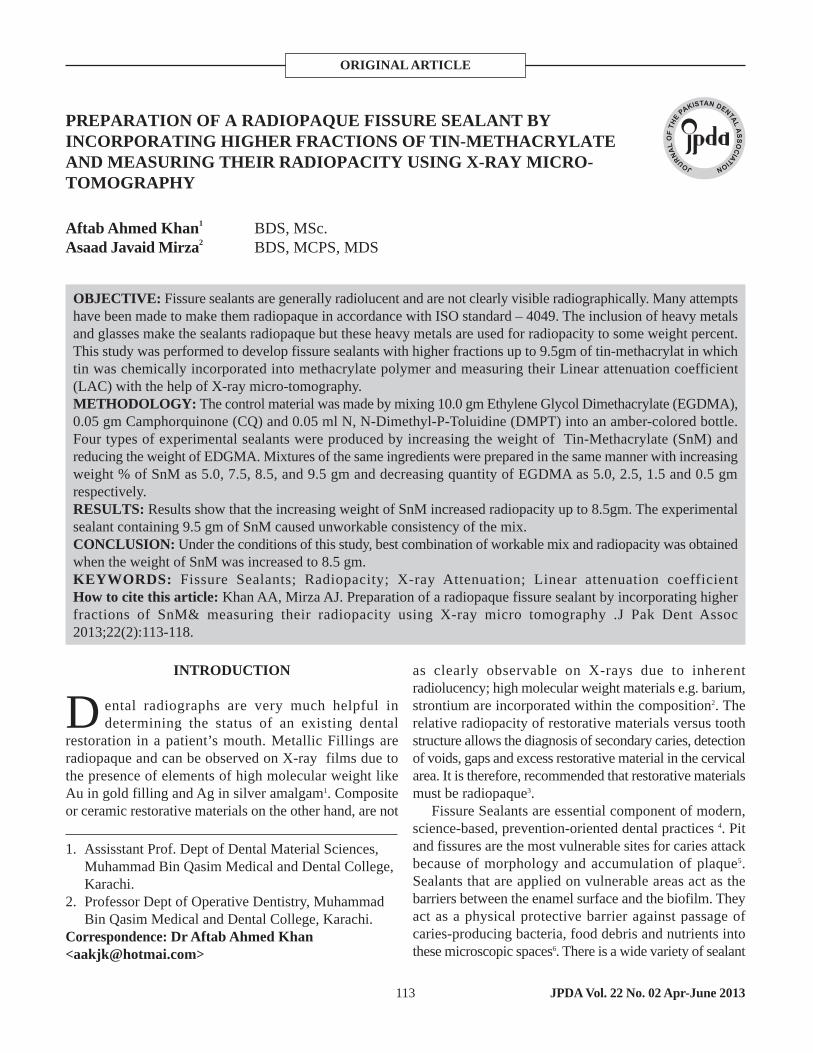
ORIGINAL ARTICLE
PREPARATION OF A RADIOPAQUE FISSURE SEALANT BYINCORPORATING HIGHER FRACTIONS OF TIN-METHACRYLATEAND MEASURING THEIR RADIOPACITY USING X-RAY MICRO-TOMOGRAPHY
INTRODUCTION
ental radiographs are very much helpful indetermining the status of an existing dental
restoration in a patient’s mouth. Metallic Fillings areradiopaque and can be observed on X-ray films due tothe presence of elements of high molecular weight likeAu in gold filling and Ag in silver amalgam1. Compositeor ceramic restorative materials on the other hand, are not
as clearly observable on X-rays due to inherentradiolucency; high molecular weight materials e.g. barium,strontium are incorporated within the composition2. Therelative radiopacity of restorative materials versus toothstructure allows the diagnosis of secondary caries, detectionof voids, gaps and excess restorative material in the cervicalarea. It is therefore, recommended that restorative materialsmust be radiopaque3.
Fissure Sealants are essential component of modern,science-based, prevention-oriented dental practices 4. Pitand fissures are the most vulnerable sites for caries attackbecause of morphology and accumulation of plaque5.Sealants that are applied on vulnerable areas act as thebarriers between the enamel surface and the biofilm. Theyact as a physical protective barrier against passage ofcaries-producing bacteria, food debris and nutrients intothese microscopic spaces6. There is a wide variety of sealant
JPDA Vol. 22 No. 02 Apr-June 2013113
OBJECTIVE: Fissure sealants are generally radiolucent and are not clearly visible radiographically. Many attemptshave been made to make them radiopaque in accordance with ISO standard – 4049. The inclusion of heavy metalsand glasses make the sealants radiopaque but these heavy metals are used for radiopacity to some weight percent.This study was performed to develop fissure sealants with higher fractions up to 9.5gm of tin-methacrylat in whichtin was chemically incorporated into methacrylate polymer and measuring their Linear attenuation coefficient(LAC) with the help of X-ray micro-tomography.METHODOLOGY: The control material was made by mixing 10.0 gm Ethylene Glycol Dimethacrylate (EGDMA),0.05 gm Camphorquinone (CQ) and 0.05 ml N, N-Dimethyl-P-Toluidine (DMPT) into an amber-colored bottle.Four types of experimental sealants were produced by increasing the weight of Tin-Methacrylate (SnM) andreducing the weight of EDGMA. Mixtures of the same ingredients were prepared in the same manner with increasingweight % of SnM as 5.0, 7.5, 8.5, and 9.5 gm and decreasing quantity of EGDMA as 5.0, 2.5, 1.5 and 0.5 gmrespectively.RESULTS: Results show that the increasing weight of SnM increased radiopacity up to 8.5gm. The experimentalsealant containing 9.5 gm of SnM caused unworkable consistency of the mix.CONCLUSION: Under the conditions of this study, best combination of workable mix and radiopacity was obtainedwhen the weight of SnM was increased to 8.5 gm.KEYWORDS: Fissure Sealants; Radiopacity; X-ray Attenuation; Linear attenuation coefficientHow to cite this article: Khan AA, Mirza AJ. Preparation of a radiopaque fissure sealant by incorporating higherfractions of SnM& measuring their radiopacity using X-ray micro tomography .J Pak Dent Assoc2013;22(2):113-118.
Aftab Ahmed Khan1
Asaad Javaid Mirza2BDS, MSc.BDS, MCPS, MDS
1. Assisstant Prof. Dept of Dental Material Sciences,Muhammad Bin Qasim Medical and Dental College,Karachi.
2. Professor Dept of Operative Dentistry, Muhammad Bin Qasim Medical and Dental College, Karachi.
Correspondence: Dr Aftab Ahmed Khan<[email protected]>
D

materials being used all over the world. The componentsof the sealants are similar to those of composite resinrestorative materials, which contain a cross-linked polymermatrix, particulate fillers and coupling agent for matrix-filler adhesion. Most sealants are either BisphenolMethacrylate resins or Urethane-Based products 7.
Fissure Sealants are highly helpful in preventing dentaldecays in the pits and fissures of teeth. However, it hasbeen seen that sealants are not widely used by dentists8.The reason for their under-use is difficulty to identify thesealant clinically or radiographically in a patient’s mouthbecause almost all of the commercially available fissuresealants are radiolucent9.
Many attempts have been made over the years to makefissure sealant radiopaque. The X-ray opacity of the dentalsealants is achieved by blending of x-ray opaque fillersinto the polymeric matrix. The blending of fillers withmatrix has many disadvantages as; discoloration of thematerial, washing out of fillers from the matrix, toxicityand sometimes radioactivity. Improper blending mayenhance radiopacity in an area of surplus accumulation offillers and vice versa 10.
An alternative approach is to use polymers in whichthe x-ray opaque element is chemically incorporated withinthe monomer unit 11. Anderson et al.12 investigated twopotential materials: Barium Methacrylate monomer, whichis in powder form at room temperature, and a TinMethacrylate monomer, which is liquid at roomtemperature. Their study found that Barium Methacrylatemonomer, when mixed with a Methacrylate diluent, didnot blend well when observed under X-raymicrotomography (XMT)13 but particles of high X-rayattenuation were formed. On the other hand, XMT imagesof the Tin Methacrylate material revealed that the mixturewas completely homogenous.
Anderson et al12 used maximum of 50% SnM in theirstudy. This study has been done to further develop novelradiopaque fissure sealants containing tin in higherconcentrations. When a radiopaque component, organictin is introduced in a molten state with methylmethacrylate,it reacts with the carbon bond of methylmethacrylatepolymer forming tributyltin methacrylate.
The aim of our study was to prepare fissure Sealantcontaining 7.5gm SnM, 8.5gm SnM and 9.5gm SnM andassess their radiopacity by using X-ray micro-tomographymachine.
METHODOLOGY:
This experiment was performed at the BiophysicsDepartment of Queen Mary University of London, UnitedKingdom. The radiopaque material used in this experimentwas Methacryloxytri-n-butyltin (SnM 6488, Lot # 2155-70-6, ABCR GmbH & Co. KG, Im Schlehert 10 D-76187Karlsruhe) present in liquid form.
Preparation of experimental sealant containing 0.0gmSnM (control sealant)
10.0 gm Ethylene Glycol Dimethacrylate (EGDMA inliquid form) of Aldrich chemical Co. Ltd, GillinghamDorset, England. Lot no. 97-90-5 was poured on a weighingboat with the help of a pipette and the precise weight wasmeasured on Explorer® (OHAUS Europe GmbHHeuwinkelstrasse 3 CH-8606 Nänikon Switzerland). Themeasured weight was poured into amber-colored bottle(amber-colored bottles were used to stop accidentalpolymerization) with cap on it. 0.05gm Camphorquinone(CQ in powder form) of Aldrich Chemical Company Inc,Ried str 2,Steinberg, Germany. Lot no.S12442-513. and0.05 ml N, N-Dimethyl-P-Toluidine (DMPT in liquid form)of Aldrich Chemical Company Inc, Ried str 2, Steinberg,Germany. Lot no. AQ05027HN.was added to the EGDMAin the bottle. There was no SnM in this sealant.
To mix all the constituents homogenously, a magneticstirrer (IKA LABORTECKNIC® Janke & Kunkel-Str.10,79219 Staufen, Germany) set at 500 rpm was used. Thematerial was mixed for 10 minutes in a dark room at roomtemperature.
Preparation of experimental sealant containing 5.0,7.5, 8.5, 9.5 gm SnM
Mixtures of the same ingredients were prepared in thesame manner with increasing weight of SnM as 5.0, 7.5,8.5, and 9.5 gm and decreasing quantity of EGDMA as5.0, 2.5, 1.5 and .05 gm respectively.
Following five compositions were prepared.
0.0 gm SnM + 10.0 gm EGDMA + 0.05 gm CQ + 0.05ml DMPT (0% SnM)5.0 gm SnM + 5.0 gm EGDMA + 0.05 gm CQ + 0.05 mlDMPT (50% SnM)7.5 gm SnM + 2.5 gm EGDMA + 0.05 gm CQ + 0.05 mlDMPT (75% SnM)8.5 gm SnM + 1.5 gm EGDMA + 0.05 gm CQ + 0.05 mlDMPT (85% SnM)
JPDA Vol. 22 No. 02 Apr-June 2013 114
Khan AA, Mirza AJ. Preparation of a radiopaque fissure sealant

9.5 gm SnM + 0.5 gm EGDMA + 0.05 gm CQ + 0.05 mlDMPT (95% SnM)
With the help of a disposable pipette, 01 ml of eachexperimental sealant from amber colored bottle was placedin a cuvette measuring 12.5x12.5x45 mm (fig. 1) andlight-cured for 20 seconds on each side using a curinglight (Heliolux DLX by VIVADENT manufacturingcompany, Amherst N.Y. 14228 USA). Figure 1 showingexperimental sealant in a cuvette.
The cuvettes were mounted onto the rotation axis of theXMT machine at room temperature of 25-27 °C. Tocalculate the radiopacity of experimental sealants the XMTmachine was run for about 8 to 10 hours at KeV 40 toobtain the raw data for the specimens in cuvettes(figure 2).
Visual AnalysisThe samples then were analyzed using Amira (TGS
Template Graphics Software Inc., USA). It is a computerprogram which gives 3-D imaging. It allowed visualizationof every single slice as well as viewing of the samplesfrom each angle.
Linear Absorption Coefficient (LAC):It describes the fraction of an x- ray beam that is
absorbed or scattered per unit thickness of an absorber. Tocalculate linear absorption coefficient of the experimentalsealants, a computerized software “XCOM” was used.This programme was developed by Dr.Graham Davis ofQueen Mary, University of London.
RESULTS:
Each slice was checked coronally, axially and sagitally forits radiopacity using Amira. Each of the SnM sealantseemed to be homogenous and in consistent form
JPDA Vol. 22 No. 02 Apr-June 2013115
Fig 1: experimental sealant in a cuvette
Fig 3: 50 gm snM Fig 4: 75 gm snM
Fig 5: 85 gm snM
Fig 2: cuvette mounted on rotational axis of XMT
Khan AA, Mirza AJ. Preparation of a radiopaque fissure sealant

throughout the sample. Axial Views of XMT images areshown in Fig. 3, 4 and 5. The figures show that radiopacityincreases with the increase in amount of SnM.The histograms reveal following information:
• Peak XMT voxels counts of control material (0.0gm SnM) corresponding to a linear absorption coefficient(LAC) of 0.22 cm-1 (Fig. 6).
• Peak XMT voxels counts of experimental sealant with5.0gm SnM corresponding to a LAC of 2.92 cm-1
(Fig.7).
• Peak XMT voxels counts of experimental sealant with7.50gm SnM corresponding to a LAC of 3.64 cm-1
(Fig. 8).• Peak XMT voxels counts of experimental sealant with
8.5gm SnM corresponding to a LAC of 4.36 cm-1 (Fig.9).
• Peak XMT voxels counts of experimental sealant with9.5gm SnM corresponding to a LAC of 4.76 cm-1 (Fig. 10 ).
The Amira analysis and histograms consistently show thatincreasing the amount of SnM increases the X-rayattenuation value thus increasing the opacity of the samplesfrom 0.0gm SnM to 9.5 gm.
Mixing of the SnM remained homogenous when 5.0,7.5 and 8.5gm of SnM was mixed with EGDMA. Theexperimental sealant prepared with 9.5gm SnM did notshow workable consistency as the quantity of resin matrixin the mixture fell short.
DISCUSSION:
Radio-opacity is a fundamental requirement forrestorative dental materials. ISO standard 4049 stipulatesthat the minimum radiopacity for a restorative materialshould be equal to or higher than its equivalent thicknessin aluminum14. Metallic dental materials like Amalgam,
116JPDA Vol. 22 No. 02 Apr-June 2013
Fig. 6: Histogram of control material (0.0gm SnM)
Fig. 7: Histogram of 5.0gm SnM
Fig. 8: Histogram of 7.5gm SnM
Fig. 9: Histogram of 8.5gm SnM
Fig. 9: Histogram of 8.5gm SnM
Fig. 10: Histogram of 9.5gm SnM
Fig. 9: Histogram of 8.5gm SnM
Fig. 10: Histogram of 9.5gm SnM
Khan AA, Mirza AJ. Preparation of a radiopaque fissure sealant

JPDA Vol. 22 No. 02 Apr-June 2013117
Gold, Cobalt Chromium, Stainless Steel have high radio-opacity15. None of the composite material has radio-opacitylike that of metallic materials because of absence of metallicelements in it. This Study has proven the radio-opacity ofSnM based sealants.
American Dental Association in its specification No.39 about Fissure Sealants classifies them according to theircuring method; chemical curing (type1) and light curing(type2). The specification No. 39 establishes that the curingtime for type 2 sealants should not be more than 60seconds16. Polymerization of experimental material in thecuvette showed that SnM has no adverse effect on thepolymerization time of the material.
It was also observed that higher the weight% of SnMin a sealant, lighter was the color of the sealant. Controlmaterial (without tin-methacrylate) was dark yellow incolor after polymerization.
During making the samples of experimental fissuresealant in different amount of weight percentages of SnM,it was noted that material was homogenous in consistency.All materials mixed into a homogenous consistency rapidlyexcept the one containing 9.5 gm of SnM. This sealantshowed consistency that was not workable. This may bedue to increased amount of tin methacrylate (9.5gm) mixedwith decreased amount of EGDMA (0.5gm) that left nocarbon bond vacant to react with tin. Moreover, itsmechanical strength hypothetically, must have beencompromised to be used as a fissure sealant.
One important property of resin based dental materialsis polymerization shrinkage. It was observed with nakedeye that the sealant which had more weight % of SnM hadno or negligible volume shrinkage against the sealantswith less weight% of SnM. The control material (withoutSnM) had greater degree of volumetric shrinkage. Thisshowed that Sn acts as filler in the sealant and it reducesthe shrinkage of the polymers.
It is mandatory for fissure sealant to have wearresistance17. If the sealant is without filler particles it wearsaway easily with the passage of time because of hydrophilicnature of most of the polymers that are used in restorativematerials18. Water penetrates into the restorative materialand gradually degrades the material. Presence of Sn willact as a filler particle and it will not only increase radio-opacity but also enhance its strength and wear resistance19.
The results of this study are consistent with earlierwork of Anderson et al who produced this novel material.Recommendations: We recommend further studiesinvestigating other aspects of this novel fissure sealantsuch as bond strength, retention rates, wear resistance,
biological reactions and bacterial interactions.
CONCLUSION:
Under the conditions of this study, best combinationof workable mix and radiopacity was obtained when theweight of SnM was increased to 8.5 gm.
REFERENCES:
1. Watts DC. Radiopacity vs. composition of some bariumand strontium glass composites. J. Dent.1987; 15: 38-43
2. Hara AT, Serra MC, Haiter-Neto F, Rodrigues JuniorAL. Radiopacity of esthetic restorative materials compared with human tooth structure. Am J Dent. 2001; 14: 383-386.
3. Akerboom HBM, Kreulen CM, van Amerongen WE,Mol A. Radiopacity of posterior composite resins, composite resin luting cements, and glass-ionomer lining cements. J Prosthet Dent 1993; 70: 351-355.
4. Rethman J. Trends in preventive care: caries risk assessment and indications for sealants. J. Am Dent Assoc. 2000; 131. 8S-12S
5. Kervanto-Seppälä S, Pietilä I, Meurman JH, KerosuoE. Pit and fissure sealants in dental public health - application criteria and general policy in Finland. BMCoral health 2009; 9:5: In: BioMed Central Ltd. Oral Health, 18.07.2011, Available from http://creativecommons.org/licences/by/2.0/ pit- and-fissure- sealants- in- dental- public- health–application-criteria- and- general- policy- in- Finland.
6. Pinkham, R., Paediatric Dentistry: Infancy through adolescent, Mosby; 1999. P.P.
7. Powers JM, Sakaguchi RL, Craig RG. Craig’s restorative dental materials. 12th Edition. Toronto: Mosby; 2006.
8. Farsi NM. The effect of education upon dentists' knowledge and attitude toward fissure sealants. Odonto-Stomatologie Tropicale 1999; 22: 27-32.
9. Seigal MD, Garcia AI, Kandray DP, Giljahn LK. Theuse of dental sealant by Ohio dentists. J Public HealthDent 1996; 56: 12-21.
10.Delaviz Y, Zhang ZX, Cabasso I, Smith J. Polym. Prepr. Amer.Chem.Soc. polym. Div 1989; 30: 215.
11.Ahir, VB, Foster J. Denture bases of X-ray opaque polymers. USA Patent # 3974104; http://www.freepatentonline.com/3974104.html
12.Anderson P, Ahmed Y, Patel MP, Davis GR, Braden
Khan AA, Mirza AJ. Preparation of a radiopaque fissure sealant

JPDA Vol. 22 No. 02 Apr-June 2013 118
M. X-ray microtomographic studies of novel radio-opaque polymeric materials for dental applications. Mat. Sci. Tech 2006; 22: 1094-1097.
13.Elliott JC, Dover SD. X-ray microtomography. J. Microscopy 1982; 126: 211-213
14.Dukic W, Delija B, Derossi D, Dadic I. Radiopacity ofcomposite dental materials using a digital X-ray system.Dent Mater J. 2012; Feb 3;31:47-53.
15.Tsuge T. Radiopacity of conventional, resin-modifiedglass ionomer, and resin-based luting materials. J OralSci. 2009; 51: 223-230.
16.Ansi/Ada : Proposed Specification No. 39 For Pit and Fissure Sealants, American National StandardsInstitute/American Dental Association.(2006)
17.Pintado MR, Conry JP, Douglas WH. Fissure sealant wear at 30 months: new evaluation criteria. J Dent.
1991; 19: 33–38.18.Osorio E, Osorio R, Davidenko N, Sastre R, Aguilar
JA, Toledano M. Polymerization kinetics and mechanicalcharacterization of new formulations of light-cured dental sealants. J Biomed Mater Res B Appl Biomater.2007 ; 80: 18-24.
19.Seyed Mostafa Mousavinasab (2011). Effects of FillerContent on Mechanical and Optical Properties of DentalComposite Resin, Metal, Ceramic and Polymeric Composites for Various Uses, Dr. John Cuppoletti (Ed.),ISBN: 978-953-307-353-8, InTech, DOI: 10.5772/21405. Available from: http://www.intechopen.com/books/metal-ceramic-and-polymeric-composites-for-various-uses/effects-of-filler-content-on-mechanical-and-optical-properties-of-dental-composite-resin.
Khan AA, Mirza AJ. Preparation of a radiopaque fissure sealant
Copyright © 2022 FDOKUMEN