Microtomography-based Pore Structure Modelling of Geologic Materials used as Building and Dimension...
9
Microtomography-based Pore Structure Modelling of Geologic Materials used as Building and Dimension Stones A. Maurício 1, a , C. Figueiredo 1,b , C. Alves 2,c , M.F. Pereira 1,d , L. Aires-Barros 1,e , J.A.N. Neto 3, f 1 Centro de Petrologia e Geoquímica (CEPGIST), Instituto Superior Técnico, Portugal 2 Centro de Investigação Geológica, Ordenamento e Valorização de Recursos Universidade do Minho, Portugal 3 Departamento de Geologia – Univ. Federal Ceará, Brasil a [email protected], b [email protected], c [email protected], d [email protected], e [email protected], f [email protected] Keywords: X-ray computed tomography, Stones, Porosity, Sulphate salt degradation Abstract. In this paper, an initial X-ray Computed Tomography study of sulphate salt degradation of two Portuguese Dimension Stones (“Semi-rijo” and “Mocacreme”) is presented, based on the Standard EN 12370 (1999). This study was performed using a high resolution X-ray Computed Tomography, a 3D X-ray microscopy non-destructive technique, in order to get representative digital information on 3D texture of the external surface and of the interior of visible light opaque objects for imaging and eventually measuring them, at resolutions in the μm range. This will produce spatial quantitative information map on the density distribution of the scanned samples, usually associated to different mineral phases and voids’ textures constituting the natural materials. Both types of stones are also being investigated by the combined application of classic methods: Optical Microscopy, Scanning Electron Microscopy and Mercury Injection Porosimetry (MICP). Most of those classic studies use mainly traditional 2D imaging techniques, none of these being able to produce the 3D resolution details that X-ray micro-tomography enables. In order to achieve future better qualitative and quantitative integrated models, it will be important to combine its non destructive and 3D characteristics results with those of 3D MICP models obtained for such complex materials. This enables to qualitatively and/or quantitatively assess the evolution and decay potential of different phases and voids (pores + fissures) textures in different environmental interaction conditions. So, in order to set-up more efficient forecasts of their engineering properties behaviour in a given environment this study is an essential initial complementary step to compare and integrate in near future studies all the advantages and disadvantages of the application of these classic and new methods, based respectively upon visual examination of the samples’ exteriors and on micro- tomographic image models of the samples’ interiors and surfaces, to these geologic materials. Introduction It is well known in the fields of Materials Science and Geosciences (Petrophysics) that natural stones decay as a result of interactions with the fluid environment they are in contact with. This is, generally, a very complex process combining mechanical, chemical, physical and biological aspects. This environment behaves, as a rule, stochastically, being influenced by meteorological and aerochemical factors, airborne and waterborne pollution and biological organisms, to name just a few. The environmental hazards that most materials from heritage sites face in different regions are widely recognized as a major hazard having significant social and cultural implications. The decay of the stones of these heritage sites constitutes a problem of conservation and/or restoration that worries government officials and the civil society alike. Decay is strongly conditioned by the nature, content and behaviour of the environmental aqueous solutions contaminating and percolating those Materials Science Forum Vols. 636-637 (2010) pp 1306-1312 Online available since 2010/Jan/12 at www.scientific.net © (2010) Trans Tech Publications, Switzerland doi:10.4028/www.scientific.net/MSF.636-637.1306 All rights reserved. No part of contents of this paper may be reproduced or transmitted in any form or by any means without the written permission of the publisher: Trans Tech Publications Ltd, Switzerland, www.ttp.net. (ID: 193.136.140.179-24/02/10,13:25:27)
-
Upload
independent -
Category
Documents
-
view
0 -
download
0
Transcript of Microtomography-based Pore Structure Modelling of Geologic Materials used as Building and Dimension...

Microtomography-based Pore Structure Modelling of Geologic Materials used as Building and Dimension Stones
A. Maurício 1, a, C. Figueiredo 1,b , C. Alves 2,c , M.F. Pereira1,d, L. Aires-Barros1,e, J.A.N. Neto3, f
1 Centro de Petrologia e Geoquímica (CEPGIST), Instituto Superior Técnico, Portugal 2 Centro de Investigação Geológica, Ordenamento e Valorização de Recursos
Universidade do Minho, Portugal 3 Departamento de Geologia – Univ. Federal Ceará, Brasil
[email protected], [email protected], c [email protected], d [email protected], [email protected], f [email protected]
Keywords: X-ray computed tomography, Stones, Porosity, Sulphate salt degradation
Abstract. In this paper, an initial X-ray Computed Tomography study of sulphate salt degradation of two Portuguese Dimension Stones (“Semi-rijo” and “Mocacreme”) is presented, based on the Standard EN 12370 (1999). This study was performed using a high resolution X-ray Computed Tomography, a 3D X-ray microscopy non-destructive technique, in order to get representative digital information on 3D texture of the external surface and of the interior of visible light opaque objects for imaging and eventually measuring them, at resolutions in the µm range. This will produce spatial quantitative information map on the density distribution of the scanned samples, usually associated to different mineral phases and voids’ textures constituting the natural materials. Both types of stones are also being investigated by the combined application of classic methods: Optical Microscopy, Scanning Electron Microscopy and Mercury Injection Porosimetry (MICP). Most of those classic studies use mainly traditional 2D imaging techniques, none of these being able to produce the 3D resolution details that X-ray micro-tomography enables. In order to achieve future better qualitative and quantitative integrated models, it will be important to combine its non destructive and 3D characteristics results with those of 3D MICP models obtained for such complex materials. This enables to qualitatively and/or quantitatively assess the evolution and decay potential of different phases and voids (pores + fissures) textures in different environmental interaction conditions. So, in order to set-up more efficient forecasts of their engineering properties behaviour in a given environment this study is an essential initial complementary step to compare and integrate in near future studies all the advantages and disadvantages of the application of these classic and new methods, based respectively upon visual examination of the samples’ exteriors and on micro-tomographic image models of the samples’ interiors and surfaces, to these geologic materials.
Introduction
It is well known in the fields of Materials Science and Geosciences (Petrophysics) that natural stones decay as a result of interactions with the fluid environment they are in contact with. This is, generally, a very complex process combining mechanical, chemical, physical and biological aspects. This environment behaves, as a rule, stochastically, being influenced by meteorological and aerochemical factors, airborne and waterborne pollution and biological organisms, to name just a few.
The environmental hazards that most materials from heritage sites face in different regions are widely recognized as a major hazard having significant social and cultural implications. The decay of the stones of these heritage sites constitutes a problem of conservation and/or restoration that worries government officials and the civil society alike. Decay is strongly conditioned by the nature, content and behaviour of the environmental aqueous solutions contaminating and percolating those
Materials Science Forum Vols. 636-637 (2010) pp 1306-1312Online available since 2010/Jan/12 at www.scientific.net© (2010) Trans Tech Publications, Switzerlanddoi:10.4028/www.scientific.net/MSF.636-637.1306
All rights reserved. No part of contents of this paper may be reproduced or transmitted in any form or by any means without the written permission of thepublisher: Trans Tech Publications Ltd, Switzerland, www.ttp.net. (ID: 193.136.140.179-24/02/10,13:25:27)
complex materials (composed of more or less randomly interconnected networks of, multiphased, multiscaled materials/voids (pores + cracks), they can also be considered as random heterogeneous materials). These solutions can have a very high chemical and/or physical/mechanical decay potential to damage those materials, greatly influencing their spatial microstructure (texture) and other chemical, physical and engineering properties like permeability, strength and durability.
Depending on largely unpredictable changes in the behaviour of the environment contacting the stone, solutions may percolate through the spectrum of the stone voids network, eventually chemically reacting with the stone matrix and modifying the overall system (voids (cracks + pores) network, matrix texture and composition and the percolating solutions). This way, ions may remain in solution or salts may precipitate and accumulate as water evaporates. It can be expected that such conditions are likely to induce oscillatory stresses contributing to the more or less complex behaviour of the decay processes, leading to distinct decay patterns not completely understood as yet [1-5].
In the Petrophysical context, for example, it is considered that the critical issue is not the formulation of constitutive or transport equations. The bottleneck of the research process lies on lack of adequate and reliable quantitative information [6]. However, although a growing and steady trend towards quantitative studies can be ascertained [1-8] driven by the pressure of several significant and powerful factors (socio-cultural, scientific, technologic, practical and economic) to get more reliable durability forecasts of heritage materials, most in-situ studies are still largely qualitative in nature. This is mainly because even the sophisticated non-destructive instrumentation (e.g.: nuclear magnetic resonance imaging, X-ray and ultrasound tomography, line profilometry…) recently created, can only be used for studying very small samples of porous materials, mainly in laboratory conditions [9].
This study aims to apply X-ray microtomography on natural building stones in order to better understand a kind of environmentally induced degradation phenomena, demonstrating the applicability potential of micro-tomography in providing new insights and guidance to prepare more systematic and detailed investigations on stone decay. This is because of its relatively easy 3-D information visualisation and quantification and non-destructive character, enabling integrated research for more rational design and control of salt induced decay of these geological materials.
Materials
For this study, two porous local Portuguese natural building stones have been selected because of their relatively high porosity and their almost pure (mono-) mineralogical composition. [10] state that “Semi-rijo” (S) and “Moca Creme” (M) are both calciclastic limestones and represent different facies exploited from the same Bathonian age (Middle Jurassic) “Valverde” formation belonging to the Calcareous Massif of Estremadura (Mesocenozoic Occidental Border of Portugal) [11]. It occupies an area of 900 km2 in the centre of Portugal, about 100 km north of Lisbon. It is a thick sequence of carbonate Mesozoic rocks, structurally elevated, that were deposited in a sedimentary basin along the continental Portuguese margin. In thin sections, (S) can be classified as fossiliferous pelmicrosparite/grainstone and (M) as biopelintrasparite/grainstone. Scanning Electron Microscopy (SEM) observations show that for both stones the pores may occur associated to sparry calcite cement, being intergranular and with fissure-like shape or within the micrite grains showing a more irregular shape.
Methods
In this paper, an initial micro-tomography study of sulphate salt degradation of two Portuguese Dimension Stones is presented, based on the Portuguese Standard NP EN 12370 (2001) [12]. The tomographic study was performed on two samples (M: 5.63×5.40×5.53mm3 and S: 6.20×5.7×5.73 mm3) using a compact desktop high resolution Skyscan 1172 microtomograph, for X-ray microscopy and micro-tomography (HRXCT). In principle, it consists of the combination of an X-ray shadow microscopic system and a computer with tomographic reconstruction and analysis
Materials Science Forum Vols. 636-637 1307
software. The Skyscan 1172 contains an X-ray micro-focus tube with high-voltage power supply, a specimen stage with precision manipulator, a 2-D X-ray CCD-camera connected to the frame-grabber and a Dual Pentium computer with colour monitor. In order to obtain high-resolution images, small samples are preferred. X-ray source and detector are fixed while the sample rotates around a stable vertical axis. Samples were scanned at a voltage of 100 kV with current intensity at 100 µA. A random movement of ten with a five frame averaging was chosen to minimise noise. The XX, YY spatial resolution was 6.76 µm with a 14 µm resolution on the ZZ axis. The parallelepiped shaped samples were cut from the same stone macro samples used for the complementary classic methods integrated study referred above. During information acquisition, X-ray radiographs are recorded at different angles during step-wise rotation between 0 and 180º around the vertical axis. The basic physical parameter quantified in each pixel of a CT-image is the linear attenuation coefficient. Beer’s law relates the intensity (I) of X-ray photons passing through the object with thickness h, with the incoming intensity (Io) and the attenuation coefficient (I) of the object:
I/Io= exp(-µh). (1)
If several absorbing materials are present, this relationship is summarised by:
I/Io= exp(-Σµihi). (2)
with index i referring to every type of material occurring in the X-ray beam. The linear attenuation coefficient µ depends on both electron density ρ and atomic number Z:
µ= ρ(a+bZ3.8/ E3.2 ). (3)
where a represents the nearly energy-independent Klein– Nishina coefficient and b is a constant. The first term stands for the Compton scattering, predominant at X-ray energies above 100 kV
(where the Skyscan 1172 does not operate), whereas the second accounts for the photoelectric absorption, which is more important at energies below 100 kV [13]. To obtain 3-D information from the images produced by the X-ray microtomograph, new reconstruction software had to be developed [13]. The images always contain a certain amount of noise that should be reduced. The X-ray microtomograph operation procedure was optimised to produce the best images by reducing artefacts like beam hardening, ring, star and line artefacts as much as possible [14]. To reduce these artefacts, an Al-filter is placed between the X-ray source and the object to absorb the low photon energies. Line artefacts are bright lines due to abnormally bright pixels in the detector during one radiograph. Very dense inclusions can create a secondary radiation resulting in star artefacts, also reduced by the Al-filter. Ring artefacts appear as circles centred on the rotation axis and are caused by detector inaccuracies. To minimise ring artefacts, a random movement is applied on the object, together with its active area on the detector [13]. After acquisition and correction, the image data can be qualitatively and/or quantitatively interpreted using home-made 3-D analysis software [13]. This software enables quantification of petrographic/petrophysical rock properties such as maximum opening, orientation, pore volumes and quantification of necks when they are visible in the samples. The pore size distribution is determined by the maximum opening, which is the biggest inscribed sphere of each pore. Erosion and dilation of the binary network with a spherical structural element determines this maximum opening. This paper applies a method that has been developed for employing microfocal-based HRXCT systems for static texture scanning characterization. It was designed to be usable on typical systems. [15].
Results and Examination
The sulphate salt stone degradation process, based on the Standard EN 12370 (1999) defines an artificially “weathering” process consisting of 15 steps. At the beginning and end of each step the same samples were successively, visually and microscopically observed, photographed and tomographically scanned, non-destructively.
For each sample the 3D texture information was reconstructed in about 980 slices along the ZZ axis with a resolution of about 14 µm. In order to see the evolution of each material, reconstructed
1308 Advanced Materials Forum V
slices of the bottom, the middle and the top of each sample were selected, corresponding as far as possible to the same slice of the sample, from step 1 and step 6 of the 15 steps cycle of the Standard.
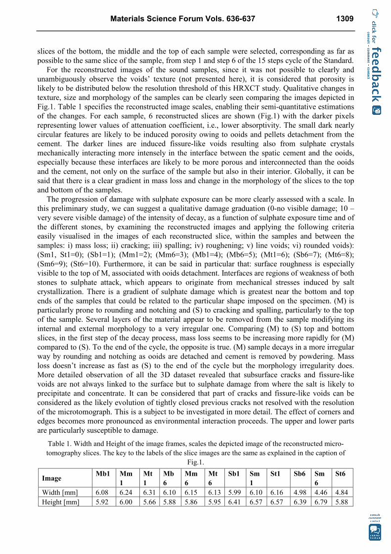
For the reconstructed images of the sound samples, since it was not possible to clearly and unambiguously observe the voids’ texture (not presented here), it is considered that porosity is likely to be distributed below the resolution threshold of this HRXCT study. Qualitative changes in texture, size and morphology of the samples can be clearly seen comparing the images depicted in Fig.1. Table 1 specifies the reconstructed image scales, enabling their semi-quantitative estimations of the changes. For each sample, 6 reconstructed slices are shown (Fig.1) with the darker pixels representing lower values of attenuation coefficient, i.e., lower absorptivity. The small dark nearly circular features are likely to be induced porosity owing to ooids and pellets detachment from the cement. The darker lines are induced fissure-like voids resulting also from sulphate crystals mechanically interacting more intensely in the interface between the spatic cement and the ooids, especially because these interfaces are likely to be more porous and interconnected than the ooids and the cement, not only on the surface of the sample but also in their interior. Globally, it can be said that there is a clear gradient in mass loss and change in the morphology of the slices to the top and bottom of the samples.
The progression of damage with sulphate exposure can be more clearly assessed with a scale. In this preliminary study, we can suggest a qualitative damage graduation (0-no visible damage; 10 – very severe visible damage) of the intensity of decay, as a function of sulphate exposure time and of the different stones, by examining the reconstructed images and applying the following criteria easily visualised in the images of each reconstructed slice, within the samples and between the samples: i) mass loss; ii) cracking; iii) spalling; iv) roughening; v) line voids; vi) rounded voids): (Sm1, St1=0); (Sb1=1); (Mm1=2); (Mm6=3); (Mb1=4); (Mb6=5); (Mt1=6); (Sb6=7); (Mt6=8); (Sm6=9); (St6=10). Furthermore, it can be said in particular that: surface roughness is especially visible to the top of M, associated with ooids detachment. Interfaces are regions of weakness of both stones to sulphate attack, which appears to originate from mechanical stresses induced by salt crystallization. There is a gradient of sulphate damage which is greatest near the bottom and top ends of the samples that could be related to the particular shape imposed on the specimen. (M) is particularly prone to rounding and notching and (S) to cracking and spalling, particularly to the top of the sample. Several layers of the material appear to be removed from the sample modifying its internal and external morphology to a very irregular one. Comparing (M) to (S) top and bottom slices, in the first step of the decay process, mass loss seems to be increasing more rapidly for (M) compared to (S). To the end of the cycle, the opposite is true. (M) sample decays in a more irregular way by rounding and notching as ooids are detached and cement is removed by powdering. Mass loss doesn’t increase as fast as (S) to the end of the cycle but the morphology irregularity does. More detailed observation of all the 3D dataset revealed that subsurface cracks and fissure-like voids are not always linked to the surface but to sulphate damage from where the salt is likely to precipitate and concentrate. It can be considered that part of cracks and fissure-like voids can be considered as the likely evolution of tightly closed previous cracks not resolved with the resolution of the microtomograph. This is a subject to be investigated in more detail. The effect of corners and edges becomes more pronounced as environmental interaction proceeds. The upper and lower parts are particularly susceptible to damage.
Table 1. Width and Height of the image frames, scales the depicted image of the reconstructed micro-tomography slices. The key to the labels of the slice images are the same as explained in the caption of
Fig.1.
Image Mb1 Mm
1 Mt1
Mb6
Mm6
Mt6
Sb1 Sm1
St1 Sb6 Sm6
St6
Width [mm] 6.08 6.24 6.31 6.10 6.15 6.13 5.99 6.10 6.16 4.98 4.46 4.84 Height [mm] 5.92 6.00 5.66 5.88 5.86 5.95 6.41 6.57 6.57 6.39 6.79 5.88
Materials Science Forum Vols. 636-637 1309
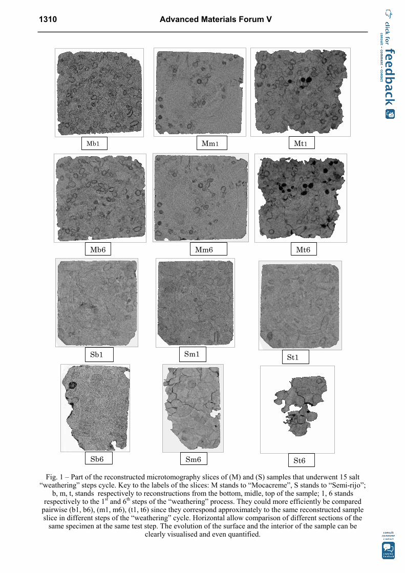
Fig. 1 – Part of the reconstructed microtomography slices of (M) and (S) samples that underwent 15 salt
“weathering” steps cycle. Key to the labels of the slices: M stands to “Mocacreme”, S stands to “Semi-rijo”; b, m, t, stands respectively to reconstructions from the bottom, midle, top of the sample; 1, 6 stands
respectively to the 1st and 6th steps of the “weathering” process. They could more efficiently be compared pairwise (b1, b6), (m1, m6), (t1, t6) since they correspond approximately to the same reconstructed sample slice in different steps of the “weathering” cycle. Horizontal allow comparison of different sections of the same specimen at the same test step. The evolution of the surface and the interior of the sample can be
clearly visualised and even quantified.
Mb1 Mm1 Mt1
Mb6 Mm6 Mt6
Sb1
Sb6 Sm6 St6
St1 Sm1
1310 Advanced Materials Forum V
Conclusion
For the studied stone samples HRXCT was an instrument to non-destructively monitor details of subsurface damage progression. Furthermore, it was important in revealing not only which stone decayed globally more rapidly, but also where and when the detailed progress of the size and morphology of decay, on the surface and inside the stone, in connection with the texture evolution of stone components (matter and voids), occurred. There are however some limitations to this methodology. In particular, at least: the small sample size that can be studied and its maximum resolution are limited to the µm scales.
Acknowledgments
This study was partially financed by Fundação para a Ciência e a Tecnologia (FCT), Project POCTI/CTA/44940/2002_PORENET, also with financial support from European Union FEDER and the state budget of the Portuguese Republic. We wish to thank Centro de Petrologia e Geoquímica/ IST/TU Lisbon subproject DECASTONE and the Centro de Investigação Geológica, Ordenamento e Valorização de Recursos of the Universidade do Minho, for all logistic facilities supporting this study.
References
[1] S. Lewin: The susceptibility of calcareous stones to salt decay. In Grafo (eds), Proc. of the 1st Intern. Symp. on the Conservation of Monuments in the Mediterranean Basin. Bari (Italy) Brescia. (1989).
[2] C.A. Price: Stone Conservation: an overview of current research. (Research in Conservation Series. Santa Monica, CA: Getty Conservation Institute 1996).
[3] M. Steiger and A. Zeunert: Crystallisation properties of salt mixtures: comparison of experimental results and model calculations. In Josef Riederer (ed.), Proc. of the 8th Intern. Congress on Deterioration and Conservation of Stone. Berlin (1996).
[4] R. Snethlage and E. Wendler: Saving our Cultural Heritage: The Conservation of Historical Stone Structures. Dahlen Workshop Reports, Chichester, UK: John Wiley & Sons Ltd (1997).
[5] R. Livingston: Influence of evaporite minerals on gypsum crusts and alveolar weathering. In Josef Riederer (ed.), Proc. of the 8th Intern. Congress on Deterioration and Conservation of Stone. Berlin (1996).
[6] L. Pel, H.P. Huinink, K. Kopinga: Ion transport and crystallization in inorganic building materials as studied by nuclear magnetic resonance. Applied Physics Letters 81 (15): (2002) 2893-2895.
[7] L.A. Rijniers, L. Pel, H.P. Huinink, K. Kopinga: Salt crystallization as damage mechanism in porous building materials – a nuclear magnetic resonance study. Magnetic Resonance Imaging 23: (2005) 273:276.
[8] A. Sawdy, and C. Price: Salt damage at Cleeve Abbey, England. Part I: A comparison of theoretical predictions and practical observations. Journal of Cultural Heritage 6 (2005) 125-135.
[9] A. Maurício, C.A.M. Figueiredo and L. Aires-Barros, in: Heritage Weathering and Conservation, edited by Fort, Alvarez de Buergo, Gomez-Heras & Vasquez-Calvo, Vol. 2, Madrid (2006), pp. 583-590.
[10] C. Figueiredo, R. Folha, A. Dionísio, A. Maurício, C. Alves, L. Aires-Barros, in: 7th Symp. Cons. Monuments Medit. Basin – Orléans- France (2007), pp.181-189.
Materials Science Forum Vols. 636-637 1311

[11] C.O.R.P. (CATÁLOGO DE ROCHAS OR.AME.TAIS PORTUGUESAS). 1983, 1984, 1985. Volumes I, II, III, Direcção-Geral de Geologia e Minas, Ministério da Indústria, Energia e Exportação.
[12] EN 12370, 1999. .atural stone test methods: Determination of resistance to salt crystallisation, Norma Portuguesa, IPQ (2001), 10 pp.
[13] V. Cnudde and P.J.S. Jacobs: Environmental Geology 46: 477-485 (2004).
[14] R.A. Ketcham and W.D.Carlson: Computer & Geosciences 27 (2001) 381-400.
[15] R.A. Ketcham and G.J. Iturrino: Journal of Hydrology 302 (2005) 92-106.
1312 Advanced Materials Forum V
Advanced Materials Forum V doi:10.4028/www.scientific.net/MSF.636-637 Microtomography-Based Pore Structure Modelling of Geologic Materials Used asBuilding and Dimension Stones doi:10.4028/www.scientific.net/MSF.636-637.1306 References[1] S. Lewin: The susceptibility of calcareous stones to salt decay. In Grafo (eds), Proc. ofthe 1st Intern. Symp. on the Conservation of Monuments in the Mediterranean Basin. Bari(Italy) Brescia. (1989). [2] C.A. Price: Stone Conservation: an overview of current research. (Research inConservation Series. Santa Monica, CA: Getty Conservation Institute 1996). [3] M. Steiger and A. Zeunert: Crystallisation properties of salt mixtures: comparison ofexperimental results and model calculations. In Josef Riederer (ed.), Proc. of the 8th Intern.Congress on Deterioration and Conservation of Stone. Berlin (1996). [4] R. Snethlage and E. Wendler: Saving our Cultural Heritage: The Conservation ofHistorical Stone Structures. Dahlen Workshop Reports, Chichester, UK: John Wiley & SonsLtd (1997). [5] R. Livingston: Influence of evaporite minerals on gypsum crusts and alveolarweathering. In Josef Riederer (ed.), Proc. of the 8th Intern. Congress on Deterioration andConservation of Stone. Berlin (1996). [6] L. Pel, H.P. Huinink, K. Kopinga: Ion transport and crystallization in inorganic buildingmaterials as studied by nuclear magnetic resonance. Applied Physics Letters 81 (15):(2002) 2893-2895.doi:10.1063/1.1512329 [7] L.A. Rijniers, L. Pel, H.P. Huinink, K. Kopinga: Salt crystallization as damagemechanism in porous building materials – a nuclear magnetic resonance study. MagneticResonance Imaging 23: (2005) 273:276. [8] A. Sawdy, and C. Price: Salt damage at Cleeve Abbey, England. Part I: A comparison oftheoretical predictions and practical observations. Journal of Cultural Heritage 6 (2005)125-135.doi:10.1016/j.culher.2005.03.003 [9] A. Maurício, C.A.M. Figueiredo and L. Aires-Barros, in: Heritage Weathering andConservation, edited by Fort, Alvarez de Buergo, Gomez-Heras & Vasquez-Calvo, Vol. 2,Madrid (2006), pp. 583-590. [10] C. Figueiredo, R. Folha, A. Dionísio, A. Maurício, C. Alves, L. Aires-Barros, in: 7th

Symp. Cons. Monuments Medit. Basin – Orléans- France (2007), pp.181-189. [11] C.O.R.P. (CATÁLOGO DE ROCHAS ORNAMENTAIS PORTUGUESAS). 1983, 1984,1985. Volumes I, II, III, Direcção-Geral de Geologia e Minas, Ministério da Indústria,Energia e Exportação. [12] EN 12370, 1999. Natural stone test methods: Determination of resistance to saltcrystallisation, Norma Portuguesa, IPQ (2001), 10 pp. [13] V. Cnudde and P.J.S. Jacobs: Environmental Geology 46: 477-485 (2004).doi:10.1007/s00254-004-1049-5 [14] R.A. Ketcham and W.D.Carlson: Computer & Geosciences 27 (2001) 381-400.doi:10.1016/S0098-3004(00)00116-3 [15] R.A. Ketcham and G.J. Iturrino: Journal of Hydrology 302 (2005) 92-106.doi:10.1016/j.jhydrol.2004.06.037