Functional analysis of the chemokine receptor CCR3 on airway epithelial cells
Upload
independentCategory
view
5download
0
10.1128/IAI.73.4.2515-2523.2005.
2005, 73(4):2515. DOI:Infect. Immun. Deborah Negrão-Correa and Mauro M. TeixeiraM. Barsante, Rodrigo Correa-Oliveira, Lúcia A. O. Fraga, Guabiraba, Cíntia A. J. Pereira, Flávia M. Carvalho, MicheleG. Souza, Adriana F. Silva, Remo C. Russo, Rodrigo Adriano L. S. Souza, Ester Roffê, Vanessa Pinho, Danielle Human and Experimental Schistosomiasis
inαMacrophage Inflammatory Protein 1Potential Role of the Chemokine
http://iai.asm.org/content/73/4/2515Updated information and services can be found at:
These include:
REFERENCEShttp://iai.asm.org/content/73/4/2515#ref-list-1at:
This article cites 53 articles, 24 of which can be accessed free
CONTENT ALERTS more»articles cite this article),
Receive: RSS Feeds, eTOCs, free email alerts (when new
http://journals.asm.org/site/misc/reprints.xhtmlInformation about commercial reprint orders: http://journals.asm.org/site/subscriptions/To subscribe to to another ASM Journal go to:
on April 4, 2014 by guest
http://iai.asm.org/
Dow
nloaded from
on April 4, 2014 by guest
http://iai.asm.org/
Dow
nloaded from

INFECTION AND IMMUNITY, Apr. 2005, p. 2515–2523 Vol. 73, No. 40019-9567/05/$08.00�0 doi:10.1128/IAI.73.4.2515–2523.2005Copyright © 2005, American Society for Microbiology. All Rights Reserved.
Potential Role of the Chemokine Macrophage Inflammatory Protein1� in Human and Experimental Schistosomiasis
Adriano L. S. Souza,1 Ester Roffe,1 Vanessa Pinho,1 Danielle G. Souza,1 Adriana F. Silva,2Remo C. Russo,1 Rodrigo Guabiraba,1 Cıntia A. J. Pereira,2 Flavia M. Carvalho,1
Michele M. Barsante,1 Rodrigo Correa-Oliveira,3 Lucia A. O. Fraga,4Deborah Negrao-Correa,2 and Mauro M. Teixeira1*
Laboratorio de Imunofarmacologia, Departamento de Bioqu¡mica e Imunologia,1 and Grupo Interdisciplinar de Estudosem Esquistossomose, Departamento de Parasitologia,2 Instituto de Ciencias Biologicas, Universidade Federal de Minas
Gerais, and Laboratorio de Imunologia Celular, Centro de Pesquisa Rene Rachou, Fundacao Oswaldo Cruz,3
Belo Horizonte, and Laboratorio de Imunologia, Universidade do Valedo Rio Doce, Montes Claros,4 Brazil
Received 7 July 2004/Returned for modification 19 August 2004/Accepted 11 November 2004
In human schistosomiasis, the concentrations of the chemokine macrophage inflammatory protein 1�(MIP-1�/CCL3) is greater in the plasma of patients with clinical hepatosplenic disease. The objective of thepresent study was to confirm the ability of CCL3 to detect severe disease in patients classified by ultrasonog-raphy (US) and to evaluate the potential role of CCL3 in Schistosoma mansoni-infected mice. CCL3 wasmeasured by enzyme-linked immunosorbent assay in the plasma of S. mansoni-infected patients. CCL3-deficient mice were infected with 25 cercariae, and various inflammatory and infectious indices were evaluated.The concentration of CCL3 was higher in the plasma of S. mansoni-infected than noninfected patients.Moreover, CCL3 was greater in those with US-defined hepatosplenic than with the intestinal form of thedisease. In CCL3-deficient mice, the size of the granuloma and the liver eosinophil peroxidase activity andcollagen content were diminished compared to wild-type mice. In CCL3-deficient mice, the worm burden after14 weeks of infection, but not after 9 weeks, was consistently smaller. The in vitro response of mesenteric lymphnode cells to antigen stimulation was characterized by lower levels of interleukin-4 (IL-4) and IL-10. CCL3 isa marker of disease severity in infected humans, and experimental studies in mice suggest that CCL3 may bea causative factor in the development of severe schistosomiasis.
Schistosomiasis is one of the most prevalent helminth dis-eases in the world and is caused by blood flukes of the Schis-tosoma genus (36). In infected individuals, the granulomatousinflammation in response to egg deposition in the liver in thecase of Schistosoma mansoni is the major pathological findingand accounts for most of the clinical symptoms. Worm ovipo-sition at 5 to 6 weeks poses a strong Th2 bias in the immuneresponse against infection (6) while also inhibiting the Th1component (12, 42). Nonetheless, the granulomatous responsethat is maximal during the first few weeks after initial oviposi-tion is also subjected to immunomodulation. Apart from in-terleukin-10 (IL-10) (29), other factors may also be involved inthis process. For example, recent literature suggests that, inanimal models, the granulomatous response that occurs atchronic time points is dependent on the soluble form of theIL-13 receptor �2 (27). In patients, failure in modulating theresponse might lead to the development severe schistosomiasislater in life (15). Indeed, a direct consequence of the persis-tence of an exacerbated immune response appears to be thedevelopment of large granulomatous reactions associated withintense collagen deposition (20) and the development of hepa-tosplenic schistosomiasis. Therefore, there is much scientific
interest in the understanding of the mechanisms and inflam-matory mediators underlying egg-induced granulomatous re-sponse, with the ultimate goal of proposing strategies to mod-ulate fibrosis. It is known, however, that prevention ofgranuloma formation may be dangerous, since lethality hasbeen reported in mice that fail to form granulomas (21).
A few studies have evaluated the role of chemokines in S.mansoni granuloma formation. Most of them have been car-ried out in mice by intravenous administration of egg antigen-conjugated beads (9). Overall, there is good evidence to sug-gest that chemokines play an important role in defining theleukocyte infiltrate and subsequent immune response that oc-curs around beads (9). For instance, the chemokines (32),macrophage inflammatory protein 1� (MIP-1�/CCL3) andregulated on activation and normal T-cell expressed and se-creted (RANTES/CCL5), are released during the granuloma-tous response and blocking of their action alters the size andother granuloma characteristics in a model of pulmonary em-bolization (10, 25, 26, 38). Studies with chemokine receptor-deficient mice also suggested a role for chemokines in thedevelopment of granulomas (9, 19, 41, 52).
Fewer studies have evaluated the role of chemokines forhuman schistosomiasis. A recent study from our group hasshown that concentrations of CCL5, eotaxin/CCL11, andCCL3 are elevated in the plasma in patients with chronicschistosomiasis. Importantly, patients with high concentrationsof CCL3 were more likely to develop hepatosplenomegaly
* Corresponding author. Mailing address: Laboratorio de Imuno-farmacologia, Departamento de Bioquımica e Imunologia, Instituto deCiencias Biologicas, Universidade Federal de Minas Gerais, BeloHorizonte, Brazil. Phone: 55-31-3499-2651. Fax: 55-31-3499-2651. E-mail: [email protected].
2515
on April 4, 2014 by guest
http://iai.asm.org/
Dow
nloaded from

(18). However, this assumption may have been biased, sinceorganomegaly was not confirmed by ultrasonography (US).Moreover, blockade of receptors for CCL3 was associated witha decrease in size of in vitro granuloma around antigen-con-jugated beads (18). In the present study, we wanted to confirmand extend our previous data, but now using patients classifiedby US from a different area of schistosomiasis endemicity. Wealso investigated likely roles of CCL3 in a murine model ofinfection that could account for differences in disease morbid-ity in patients. To this end, the course of S. mansoni infectionwas monitored in CCL3-deficient and wild-type mice.
MATERIALS AND METHODS
Blood collecting and outpatient clinical and US examination. Plasma sampleswere obtained from patients living in Boa Uniao and Nova Uniao, two adjacentlocalities, in Minas Gerais, Brazil. Study groups were composed of 32 intestinaldisease patients, 13 hepatosplenic patients, and 12 controls. The diameter of theportal and mesenteric veins, the sizes of the liver and spleen, and wall thickeningof central and peripheral branches of the portal vein were evaluated according toWorld Health Organization guidelines (1, 14, 22, 27). The criteria for inclusion in thehepatosplenic group was that patients had to present thickening of the portalbranches of �0.3 cm and hepatic and splenic organomegaly as shown by US per-formed with a portable Hitachi EUB-200 apparatus. This is an important aspect ofthe present study since sole clinical organomegaly is a bad predictor of diseaseoutcome (1). In our patient population, only grade I fibrosis (periportal thickeningof �0.3 and �0.5) was found, confirming previous observations that more severepresentations of the disease seem to be less prevalent probably due to the treatmentof infection (2, 3). Patients were treated for infection regardless of their adherenceto the study. Those younger than 5 years and those exhibiting liver damage sugges-tive of other diseases, such as steatosis, in the absence of pipe stem fibrosis, wereexcluded. The control group was composed of noninfected individuals living in thearea of endemicity and healthy volunteers from an area where the disease is notendemic. Negative results for schistosome infection were attested by three consec-utive parasitological examinations by the Kato-Katz method (24). All patients signedan informed consent for the use of blood samples.
Animals and Infection. Male and female C57BL/6 and CCL3-deficient mice(13) were obtained from the animal facility of the Centro de Pesquisas Ren,Rachou. The animals were 8 weeks old at the beginning of the experiment andkept at the animal facility of the Grupo Interdisciplinar de Estudos em Esquis-tossomose. Rodent chow and water were given ad libitum. LE strain S. mansonicercariae (50) released from Biomphalaria glabrata snails were suspended insaline 0.9% and subcutaneously injected (25 cercariae per animal) by an expe-rienced technician. Within the same experiment, all groups of mice, wild type orknockout, were infected with the same cercaria suspension. To assess parasiteburden, mice had to be subjected to mesenteric perfusion for collecting of adultworms. The latter procedure induces a degree of tissue damage and thus pre-vented the use of the tissues and blood from these animals for the measurementof other parameters. Thus, separate groups of animals were used to assess eitherparasitological or immunological parameters. Female mice were used for the para-sitological studies (egg and worm burdens), and male mice were used for all otherparameters described here. Infection-induced morbidity and mortality were ob-served in CCL3-deficient and wild-type mice throughout the experiment, whichlasted 9 or 14 weeks, and so represented the acute and chronic phases of theinfection. All experiments were repeated and, since the means of a given variablewere similar in these experiments, the results were pooled for presentation.
Determination of egg content in the feces. Animals were kept in individualgridded-floor cages the night prior to sacrifice. In each cage a piece of wetabsorbent paper kept feces humid. On the following morning, feces were col-lected, weighed, homogenized in 10% buffered formaldehyde, and stored. Twoaliquots of 100 �l each were counted by light microscopy, and the results areexpressed as number of eggs per gram of feces.
Recovering of worms from the portal system. Animals were sacrificed bycervical displacement, and viscera were exposed. A cut was made at the portalvein, and a needle conjugated to a perfusion pump was introduced into theabdominal aorta (45). Saline (0.9%) was perfused and collected in a Beckerapparatus. Decantation was allowed for 30 min, and the supernatant was dis-carded. Washing steps were performed until the supernatant was clear. Male andfemale worms were counted in a laboratory magnifier.
Determination of egg content in the tissues. The livers, lungs, intestines, andspleens of infected mice were weighted and solubilized in 5% KOH solution until
no sign of intact tissue was present. Tissue suspensions were centrifuged at 200� g for 1 min at room temperature, washed five times, and stored at 4°C. Eggsin aliquots of 100 �l were counted under a light microscope, and values wereextrapolated to the total organ content. The tissue egg retention index, a mea-sure of worm fecundity, was calculated as the sum of the absolute number of eggspresent in tissues divided by the number or female worms, regardless of whetherthey were found mated or not (17).
Differential blood cell count and tissue histology. Animals were sacrificedunder ether anesthesia, followed by cervical displacement, and 20 �l of blood wascollected from each brachial plexus. The blood was diluted in Turk’s solution fortotal cell count. A blood smear was prepared and stained with Giemsa andMay-Grunwald stains. Differential blood count of mononuclear cells, neutro-phils, and eosinophils was performed from a total of 300 cells. Fragments of liverwere removed, fixed in 10% buffered formaldehyde, and stained with eitherhematoxylin and eosin or Picrus Sirius (30). About 500 �l of blood were collectedfor the quantification of cytokines.
Cell culture. After sacrifice, animals not subjected to hepatic perfusion had theabdominal wall opened in sterile conditions. Mesenteric lymph nodes (MLN)were removed, and cells were isolated. The resultant cell suspension was dilutedto 107 cells/ml in HEPES-modified RPMI supplemented with potassium-peni-cillin at 10 IU/ml and 1 �g of gentamicin-containing 5% fetal calf serum/ml. Cellswere plated in the presence of concanavalin A (1 �g/ml), a nonspecific T-cellstimulator (11), and schistosome egg antigen (SEA) (5) or soluble adult wormantigenic preparation (SWAP), both at the concentration of 50 �g/ml. Thesupernatant was collected 48 h later for cytokine quantification.
Cytokine determination by ELISA. The concentration of cytokines in plasmaand culture supernatant was performed by sandwich enzyme-linked immunosor-bent assay (ELISA) as described elsewhere (31). The ELISA kits for mouse IL-4,IL-10, IL-13, gamma interferon (IFN-�), and human CCL3 were obtained fromR&D Systems. The antibody pairs and standards used in the assay for mouseIL-1 were kindly provided by Steve Poole. The specific recommendations of themanufacturers were followed. Human plasma samples were subjected to acid-saltprecipitation to avoid cross-reactivity in ELISA (18).
Determination of the activity of eosinophil peroxidase (EPO). The assay wasperformed as described previously (47). Briefly, 100 mg of the liver and intestineof the animals were weighted and homogenized in 1.9 ml of phosphate-bufferedsaline and centrifuged at 12,000 � g for 10 min. The supernatant was discarded,and the erythrocytes were lysed. The samples were then centrifuged, the super-natant was discarded, and the pellet was suspended in 1.9 ml of 0.5% hexade-cyltrimethyl ammonium bromide in phosphate-buffered saline, frozen threetimes in liquid nitrogen, and centrifuged at 4°C at 12,000 � g for 10 min. Thesupernatant was used in the enzymatic assay by the addition of an equal amountsubstrate (1.5 mM o-phenylenediamine–6.6 mM H2O2 in 0.075 mM Tris-HCl[pH 8]). The reaction was stopped with 50 �l of 1 M H2SO4, and the absorbancewas read at 492 nm.
Hydroxyproline determination. Fragments (100 mg) of liver and intestine wereremoved for hydroxyproline determination as an indirect measure of collagencontent (39). Briefly, tissues were homogenized in 0.9% saline, frozen, andlyophilized. The assay was performed with 20 mg of the lyophilized materialsubjected to alkaline hydrolysis in 300 �l of H2O plus 75 �l of 10 M NaOH at120°C for 20 min. An aliquot of 50 �l of the hydrolyzed tissue was added to 450�l of Chloramine T oxidizing reagent (0.056 M Chloramine T–n-propanol 10%in acetate-citrate buffer [pH 6.5]) and allowed to react for 20 min. A hydroxypro-line standard curve was prepared likewise. Color was developed by the additionof 500 �l of 1 M p-dimethylaminebenzaldehyde diluted in n-propanol–perchloricacid (2:1 [vol/vol]). The absorbance was read at 550 nm.
Morphometric analysis. Slides with sections of the liver were analyzed undera light microscope for the characterization of the inflammatory infiltrate and thecomposition of granulomas. For the quantitative analysis of granuloma area,images were captured with a digital camera (Optronics DEI-470) connected to amicroscope (Olympus IX70) and analyzed with Image ProPlus. For each infectedanimal, the first nine granulomas found in either of three liver sections meetingthe criteria of being isolated and of presenting a central viable egg were photo-graphed and analyzed. Granulomas in which more than one or no egg was visiblewere excluded from the analysis. The area of each of the nine granulomas wasmeasured three times and averaged.
Histological egg densitometry. Images covering 1,500 �m2 from liver sectionsof male wild-type and CCL3�/� mice were captured with a digital camera(Optronics DEI-470) connected to a microscope (Olympus IX70) with a magni-fication of � 100. The number of images taken was about 30 to 50 for eachanimal and was equal to the highest number of whole snapshots that could betaken without including serial slices in a given fragment of stained tissue. Theschistosome eggs per image were counted, and the average value was entered as
2516 SOUZA ET AL. INFECT. IMMUN.
on April 4, 2014 by guest
http://iai.asm.org/
Dow
nloaded from

an individual value for each animal. The final value is referred to here as the “eggdensitometric index” and was used to compare to egg counts obtained fromwhole-tissue digestions performed in female mice. As hydroxyproline contentwas only measured in livers and intestines of male mice, the egg densitometricindex was useful to calculate a ratio describing the collagen deposition per tissueegg content.
Statistical analysis. Results for mice are expressed as means the standarderror of the means (SEM) and were analyzed by analysis of variance, followed bythe Newman-Keuls multiple comparison test. MLN cultures were analyzed byusing an unpaired t test when detectable levels of cytokines were found only ininfected groups. Human data were analyzed by Kruskal-Wallis test, followed byDunn’s multicomparison test. A cutoff value of MIP-1� that differentiated pa-tients with or without hepatosplenomegaly was selected empirically. Relative riskwas calculated by using the Fisher exact test in a 2 � 2 contingency table.
RESULTS
Concentration of CCL3 in plasma of patients. To confirmand validate our previous finding that CCL3 marked a group ofpatients with severe disease (18), we evaluated the concentra-tions of CCL3 by sandwich ELISA in plasma samples of US-assessed schistosome-infected individuals. In agreement withprevious findings, infected patients had greater concentrationof CCL3 in plasma than noninfected individuals (P � 0.05).The concentrations of CCL3 were significantly higher inplasma of patients with organomegaly than in those withoutorganomegaly (P � 0.001, Fig. 1). The value of 400 pg/ml wasfound to be best to separate patients with or without organo-megaly. Indeed, infected patients with a concentration ofCCL3 of �400 pg/ml had a 14-fold greater chance of havingorganomegaly than those with lower concentrations of thischemokine (P � 0.001). However, there was no tendency forpatients with high levels of CCL3 within the intestinal group toexhibit higher levels of fibrosis (not shown).
Infection indices in CCL3-deficient and wild-type mice. Ini-tial experiments were designed to assess whether CCL3 defi-ciency interfered with worm burden and/or fecundity. No sta-tistically significant difference in the lethality rates of infectedmice was found, although percentages tended to be smaller inthe CCL-3-deficient (28.5%) than in the wild-type group(37.5%) at 14 weeks postinfection. Of the approximately 25injected cercariae, ca. 10 adult worms were recovered after
perfusion from either wild-type or CCL3-deficient mice at 9weeks postinfection (Fig. 2A). At 14 weeks, there was a similarnumber of worms in the wild-type group, but a we observed asignificant (P � 0.01) reduction in CCL3-deficient mice (Fig.2A). Despite changes in worm numbers, the tissue egg reten-tion index, as calculated by the total number of eggs trapped intissues divided by the total number of female worms, was equalin CCL3-deficient and wild-type mice during both the acuteand the chronic phases (Fig. 2B).
These results indicate that CCL3 deficiency does not alterworm fecundity. However, tissue- and phase-specific analysisreveals important differences in egg burdens calculated in fe-male mice through the digestion of whole tissues with 5%KOH solution. During both the acute and chronic phases, asmaller number of eggs per gram of feces was found in CCL3-deficient mice (Fig. 3A). At the chronic phase other tissue specificdifferences were found. A reduction in the absolute number ofeggs deposited in the liver (Fig. 3B, P � 0.05) and spleen (Fig. 3C,P � 0.05) was observed in the CCL3-deficient group. There wasno difference in the number of eggs in the intestines (Fig. 3D) orlungs (not shown). Confirming data on egg counts, the evaluationof egg number by histology demonstrated a smaller number of
FIG. 1. Chemokine levels in the plasma of schistosome-infectedpatients and control individuals. Individual concentrations are shownwith the median of each group expressed as a horizontal line. Patientswere classified as intestinal (n 32) or hepatosplenic (n 13) ac-cording to clinical and US examinations. A control group was com-posed of 12 individuals. Plasma samples were processed for elimina-tion of cross-reactivity and used for CCL3 determination by ELISA.The data were analyzed by the Kruskal-Wallis test, followed by Dunn’scomparison test.
FIG. 2. Total number of worms in the portal system and fecundityof S. mansoni harvested from wild-type or CCL3-deficient mice. Val-ues are the means the SEM of worms from perfusates of femalemice. (A) The male and female adult mated and unmated wormspresent in the portal system of infected wild-type (�; acute phase, n 6; chronic phase, n 12) or CCL3-deficient (o; acute phase, n 6;chronic phase, n 11) mice were counted after hepatic perfusion at 9(acute phase) or 14 (chronic phase) weeks after infection. (B) Theratio between the total number or eggs present in all analyzed tissues,i.e., lungs, liver, intestine, and spleen divided by the number of femaleworms gives the tissue egg retention index for each group. The num-bers of female worms are equal to those used to calculate the averagesshown in panel A. #, P � 0.01 as determined by the Newman-Keulstest for CCL3-deficient mice compared to wild-type ones at the samephase of infection.
VOL. 73, 2005 ROLE OF CCL3 IN SCHISTOSOMIASIS 2517
on April 4, 2014 by guest
http://iai.asm.org/
Dow
nloaded from
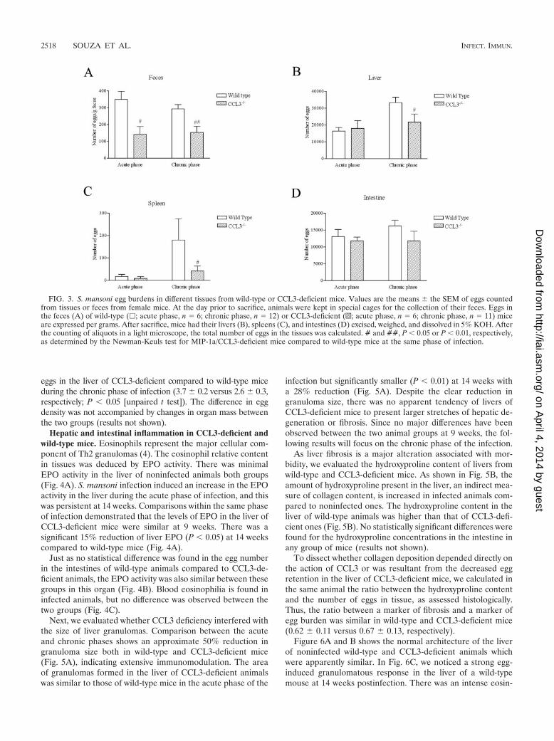
eggs in the liver of CCL3-deficient compared to wild-type miceduring the chronic phase of infection (3.7 0.2 versus 2.6 0.3,respectively; P � 0.05 [unpaired t test]). The difference in eggdensity was not accompanied by changes in organ mass betweenthe two groups (results not shown).
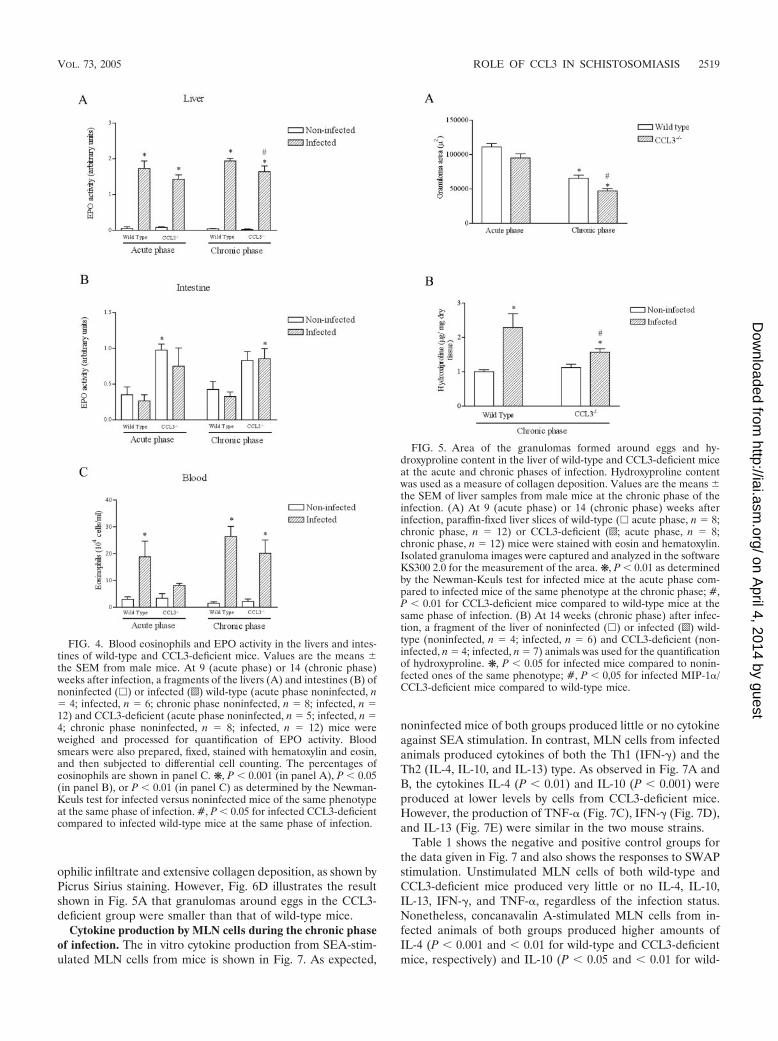
Hepatic and intestinal inflammation in CCL3-deficient andwild-type mice. Eosinophils represent the major cellular com-ponent of Th2 granulomas (4). The eosinophil relative contentin tissues was deduced by EPO activity. There was minimalEPO activity in the liver of noninfected animals both groups(Fig. 4A). S. mansoni infection induced an increase in the EPOactivity in the liver during the acute phase of infection, and thiswas persistent at 14 weeks. Comparisons within the same phaseof infection demonstrated that the levels of EPO in the liver ofCCL3-deficient mice were similar at 9 weeks. There was asignificant 15% reduction of liver EPO (P � 0.05) at 14 weekscompared to wild-type mice (Fig. 4A).
Just as no statistical difference was found in the egg numberin the intestines of wild-type animals compared to CCL3-de-ficient animals, the EPO activity was also similar between thesegroups in this organ (Fig. 4B). Blood eosinophilia is found ininfected animals, but no difference was observed between thetwo groups (Fig. 4C).
Next, we evaluated whether CCL3 deficiency interfered withthe size of liver granulomas. Comparison between the acuteand chronic phases shows an approximate 50% reduction ingranuloma size both in wild-type and CCL3-deficient mice(Fig. 5A), indicating extensive immunomodulation. The areaof granulomas formed in the liver of CCL3-deficient animalswas similar to those of wild-type mice in the acute phase of the
infection but significantly smaller (P � 0.01) at 14 weeks witha 28% reduction (Fig. 5A). Despite the clear reduction ingranuloma size, there was no apparent tendency of livers ofCCL3-deficient mice to present larger stretches of hepatic de-generation or fibrosis. Since no major differences have beenobserved between the two animal groups at 9 weeks, the fol-lowing results will focus on the chronic phase of the infection.
As liver fibrosis is a major alteration associated with mor-bidity, we evaluated the hydroxyproline content of livers fromwild-type and CCL3-deficient mice. As shown in Fig. 5B, theamount of hydroxyproline present in the liver, an indirect mea-sure of collagen content, is increased in infected animals com-pared to noninfected ones. The hydroxyproline content in theliver of wild-type animals was higher than that of CCL3-defi-cient ones (Fig. 5B). No statistically significant differences werefound for the hydroxyproline concentrations in the intestine inany group of mice (results not shown).
To dissect whether collagen deposition depended directly onthe action of CCL3 or was resultant from the decreased eggretention in the liver of CCL3-deficient mice, we calculated inthe same animal the ratio between the hydroxyproline contentand the number of eggs in tissue, as assessed histologically.Thus, the ratio between a marker of fibrosis and a marker ofegg burden was similar in wild-type and CCL3-deficient mice(0.62 0.11 versus 0.67 0.13, respectively).
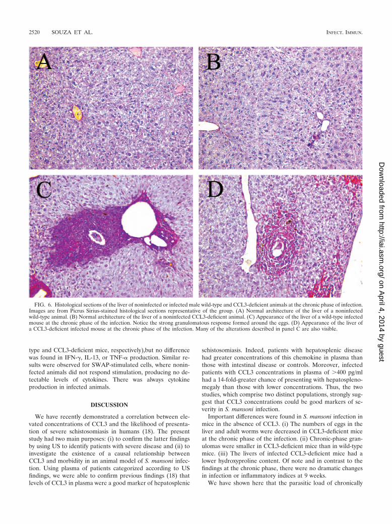
Figure 6A and B shows the normal architecture of the liverof noninfected wild-type and CCL3-deficient animals whichwere apparently similar. In Fig. 6C, we noticed a strong egg-induced granulomatous response in the liver of a wild-typemouse at 14 weeks postinfection. There was an intense eosin-
FIG. 3. S. mansoni egg burdens in different tissues from wild-type or CCL3-deficient mice. Values are the means the SEM of eggs countedfrom tissues or feces from female mice. At the day prior to sacrifice, animals were kept in special cages for the collection of their feces. Eggs inthe feces (A) of wild-type (�; acute phase, n 6; chronic phase, n 12) or CCL3-deficient (o; acute phase, n 6; chronic phase, n 11) miceare expressed per grams. After sacrifice, mice had their livers (B), spleens (C), and intestines (D) excised, weighed, and dissolved in 5% KOH. Afterthe counting of aliquots in a light microscope, the total number of eggs in the tissues was calculated. # and ##, P � 0.05 or P � 0.01, respectively,as determined by the Newman-Keuls test for MIP-1a/CCL3-deficient mice compared to wild-type mice at the same phase of infection.
2518 SOUZA ET AL. INFECT. IMMUN.
on April 4, 2014 by guest
http://iai.asm.org/
Dow
nloaded from
ophilic infiltrate and extensive collagen deposition, as shown byPicrus Sirius staining. However, Fig. 6D illustrates the resultshown in Fig. 5A that granulomas around eggs in the CCL3-deficient group were smaller than that of wild-type mice.
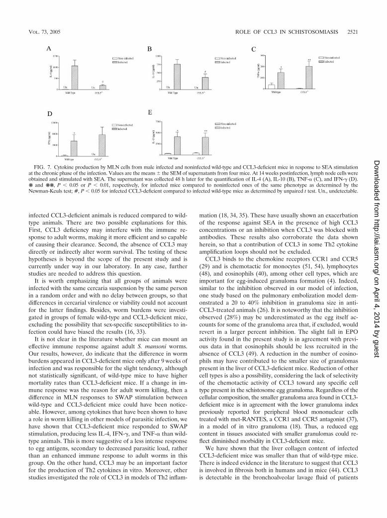
Cytokine production by MLN cells during the chronic phaseof infection. The in vitro cytokine production from SEA-stim-ulated MLN cells from mice is shown in Fig. 7. As expected,
noninfected mice of both groups produced little or no cytokineagainst SEA stimulation. In contrast, MLN cells from infectedanimals produced cytokines of both the Th1 (IFN-�) and theTh2 (IL-4, IL-10, and IL-13) type. As observed in Fig. 7A andB, the cytokines IL-4 (P � 0.01) and IL-10 (P � 0.001) wereproduced at lower levels by cells from CCL3-deficient mice.However, the production of TNF-� (Fig. 7C), IFN-� (Fig. 7D),and IL-13 (Fig. 7E) were similar in the two mouse strains.
Table 1 shows the negative and positive control groups forthe data given in Fig. 7 and also shows the responses to SWAPstimulation. Unstimulated MLN cells of both wild-type andCCL3-deficient mice produced very little or no IL-4, IL-10,IL-13, IFN-�, and TNF-�, regardless of the infection status.Nonetheless, concanavalin A-stimulated MLN cells from in-fected animals of both groups produced higher amounts ofIL-4 (P � 0.001 and � 0.01 for wild-type and CCL3-deficientmice, respectively) and IL-10 (P � 0.05 and � 0.01 for wild-
FIG. 4. Blood eosinophils and EPO activity in the livers and intes-tines of wild-type and CCL3-deficient mice. Values are the means the SEM from male mice. At 9 (acute phase) or 14 (chronic phase)weeks after infection, a fragments of the livers (A) and intestines (B) ofnoninfected (�) or infected (o) wild-type (acute phase noninfected, n 4; infected, n 6; chronic phase noninfected, n 8; infected, n 12) and CCL3-deficient (acute phase noninfected, n 5; infected, n 4; chronic phase noninfected, n 8; infected, n 12) mice wereweighed and processed for quantification of EPO activity. Bloodsmears were also prepared, fixed, stained with hematoxylin and eosin,and then subjected to differential cell counting. The percentages ofeosinophils are shown in panel C. ❋, P � 0.001 (in panel A), P � 0.05(in panel B), or P � 0.01 (in panel C) as determined by the Newman-Keuls test for infected versus noninfected mice of the same phenotypeat the same phase of infection. #, P � 0.05 for infected CCL3-deficientcompared to infected wild-type mice at the same phase of infection.
FIG. 5. Area of the granulomas formed around eggs and hy-droxyproline content in the liver of wild-type and CCL3-deficient miceat the acute and chronic phases of infection. Hydroxyproline contentwas used as a measure of collagen deposition. Values are the means the SEM of liver samples from male mice at the chronic phase of theinfection. (A) At 9 (acute phase) or 14 (chronic phase) weeks afterinfection, paraffin-fixed liver slices of wild-type (� acute phase, n 8;chronic phase, n 12) or CCL3-deficient (o; acute phase, n 8;chronic phase, n 12) mice were stained with eosin and hematoxylin.Isolated granuloma images were captured and analyzed in the softwareKS300 2.0 for the measurement of the area. ❋, P � 0.01 as determinedby the Newman-Keuls test for infected mice at the acute phase com-pared to infected mice of the same phenotype at the chronic phase; #,P � 0.01 for CCL3-deficient mice compared to wild-type mice at thesame phase of infection. (B) At 14 weeks (chronic phase) after infec-tion, a fragment of the liver of noninfected (�) or infected (o) wild-type (noninfected, n 4; infected, n 6) and CCL3-deficient (non-infected, n 4; infected, n 7) animals was used for the quantificationof hydroxyproline. ❋, P � 0.05 for infected mice compared to nonin-fected ones of the same phenotype; #, P � 0,05 for infected MIP-1�/CCL3-deficient mice compared to wild-type mice.
VOL. 73, 2005 ROLE OF CCL3 IN SCHISTOSOMIASIS 2519
on April 4, 2014 by guest
http://iai.asm.org/
Dow
nloaded from
type and CCL3-deficient mice, respectively),but no differencewas found in IFN-�, IL-13, or TNF-� production. Similar re-sults were observed for SWAP-stimulated cells, where nonin-fected animals did not respond stimulation, producing no de-tectable levels of cytokines. There was always cytokineproduction in infected animals.
DISCUSSION
We have recently demonstrated a correlation between ele-vated concentrations of CCL3 and the likelihood of presenta-tion of severe schistosomiasis in humans (18). The presentstudy had two main purposes: (i) to confirm the latter findingsby using US to identify patients with severe disease and (ii) toinvestigate the existence of a causal relationship betweenCCL3 and morbidity in an animal model of S. mansoni infec-tion. Using plasma of patients categorized according to USfindings, we were able to confirm previous findings (18) thatlevels of CCL3 in plasma were a good marker of hepatosplenic
schistosomiasis. Indeed, patients with hepatosplenic diseasehad greater concentrations of this chemokine in plasma thanthose with intestinal disease or controls. Moreover, infectedpatients with CCL3 concentrations in plasma of �400 pg/mlhad a 14-fold-greater chance of presenting with hepatospleno-megaly than those with lower concentrations. Thus, the twostudies, which comprise two distinct populations, strongly sug-gest that CCL3 concentrations could be good markers of se-verity in S. mansoni infection.
Important differences were found in S. mansoni infection inmice in the absence of CCL3. (i) The numbers of eggs in theliver and adult worms were decreased in CCL3-deficient miceat the chronic phase of the infection. (ii) Chronic-phase gran-ulomas were smaller in CCL3-deficient mice than in wild-typemice. (iii) The livers of infected CCL3-deficient mice had alower hydroxyproline content. Of note and in contrast to thefindings at the chronic phase, there were no dramatic changesin infection or inflammatory indices at 9 weeks.
We have shown here that the parasitic load of chronically
FIG. 6. Histological sections of the liver of noninfected or infected male wild-type and CCL3-deficient animals at the chronic phase of infection.Images are from Picrus Sirius-stained histological sections representative of the group. (A) Normal architecture of the liver of a noninfectedwild-type animal. (B) Normal architecture of the liver of a noninfected CCL3-deficient animal. (C) Appearance of the liver of a wild-type infectedmouse at the chronic phase of the infection. Notice the strong granulomatous response formed around the eggs. (D) Appearance of the liver ofa CCL3-deficient infected mouse at the chronic phase of the infection. Many of the alterations described in panel C are also visible.
2520 SOUZA ET AL. INFECT. IMMUN.
on April 4, 2014 by guest
http://iai.asm.org/
Dow
nloaded from
infected CCL3-deficient animals is reduced compared to wild-type animals. There are two possible explanations for this.First, CCL3 deficiency may interfere with the immune re-sponse to adult worms, making it more efficient and so capableof causing their clearance. Second, the absence of CCL3 maydirectly or indirectly alter worm survival. The testing of thesehypotheses is beyond the scope of the present study and iscurrently under way in our laboratory. In any case, furtherstudies are needed to address this question.
It is worth emphasizing that all groups of animals wereinfected with the same cercaria suspension by the same personin a random order and with no delay between groups, so thatdifferences in cercarial virulence or viability could not accountfor the latter findings. Besides, worm burdens were investi-gated in groups of female wild-type and CCL3-deficient mice,excluding the possibility that sex-specific susceptibilities to in-fection could have biased the results (16, 33).
It is not clear in the literature whether mice can mount aneffective immune response against adult S. mansoni worms.Our results, however, do indicate that the difference in wormburdens appeared in CCL3-deficient mice only after 9 weeks ofinfection and was responsible for the slight tendency, althoughnot statistically significant, of wild-type mice to have highermortality rates than CCL3-deficient mice. If a change in im-mune response was the reason for adult worm killing, then adifference in MLN responses to SWAP stimulation betweenwild-type and CCL3-deficient mice could have been notice-able. However, among cytokines that have been shown to havea role in worm killing in other models of parasitic infection, wehave shown that CCL3-deficient mice responded to SWAPstimulation, producing less IL-4, IFN-�, and TNF-� than wild-type animals. This is more suggestive of a less intense responseto egg antigens, secondary to decreased parasitic load, ratherthan an enhanced immune response to adult worms in thisgroup. On the other hand, CCL3 may be an important factorfor the production of Th2 cytokines in vitro. Moreover, otherstudies investigated the role of CCL3 in models of Th2 inflam-
mation (18, 34, 35). These have usually shown an exacerbationof the response against SEA in the presence of high CCL3concentrations or an inhibition when CCL3 was blocked withantibodies. These results also corroborate the data shownherein, so that a contribution of CCL3 in some Th2 cytokineamplification loops should not be excluded.
CCL3 binds to the chemokine receptors CCR1 and CCR5(29) and is chemotactic for monocytes (51, 54), lymphocytes(48), and eosinophils (40), among other cell types, which areimportant for egg-induced granuloma formation (4). Indeed,similar to the inhibition observed in our model of infection,one study based on the pulmonary embolization model dem-onstrated a 20 to 40% inhibition in granuloma size in anti-CCL3-treated animals (26). It is noteworthy that the inhibitionobserved (28%) may be underestimated as the egg itself ac-counts for some of the granuloma area that, if excluded, wouldrevert in a larger percent inhibition. The slight fall in EPOactivity found in the present study is in agreement with previ-ous data in that eosinophils should be less recruited in theabsence of CCL3 (49). A reduction in the number of eosino-phils may have contributed to the smaller size of granulomaspresent in the liver of CCL3-deficient mice. Reduction of othercell types is also a possibility, considering the lack of selectivityof the chemotactic activity of CCL3 toward any specific celltype present in the schistosome egg granuloma. Regardless of thecellular composition, the smaller granuloma area found in CCL3-deficient mice is in agreement with the lower granuloma indexpreviously reported for peripheral blood mononuclear cellstreated with met-RANTES, a CCR1 and CCR5 antagonist (37),in a model of in vitro granuloma (18). Thus, a reduced eggcontent in tissues associated with smaller granulomas could re-flect diminished morbidity in CCL3-deficient mice.
We have shown that the liver collagen content of infectedCCL3-deficient mice was smaller than that of wild-type mice.There is indeed evidence in the literature to suggest that CCL3is involved in fibrosis both in humans and in mice (44). CCL3is detectable in the bronchoalveolar lavage fluid of patients
FIG. 7. Cytokine production by MLN cells from male infected and noninfected wild-type and CCL3-deficient mice in response to SEA stimulationat the chronic phase of the infection. Values are the means the SEM of supernatants from four mice. At 14 weeks postinfection, lymph node cells wereobtained and stimulated with SEA. The supernatant was collected 48 h later for the quantification of IL-4 (A), IL-10 (B), TNF-� (C), and IFN-� (D).❋ and ❋❋, P � 0.05 or P � 0.01, respectively, for infected mice compared to noninfected ones of the same phenotype as determined by theNewman-Keuls test; #, P � 0.05 for infected CCL3-deficient compared to infected wild-type mice as determined by unpaired t test. Un., undetectable.
VOL. 73, 2005 ROLE OF CCL3 IN SCHISTOSOMIASIS 2521
on April 4, 2014 by guest
http://iai.asm.org/
Dow
nloaded from

with sarcoidosis and idiopathic pulmonary fibrosis (43). More-over, anti-CCL3 antibody treatment in mice inhibits bleomy-cin-induced inflammatory infiltrate and fibrosis (41). Besides,several reports have shown the role of IL-4 in granulomatousinflammation and its interference on various aspects of theresponse, including collagen synthesis (7, 8, 28). So, it is pos-sible that the inhibition of IL-4 production observed in CCL3-deficient mice could account for at least some of the observedinhibition of collagen deposition. From these evidences, itcould have been possible that CCL3 also affected the amountof collagen that is produced in the liver. However, unlike thepipe stem fibrosis found in humans, the granulomas containmost of the disease-induced collagen deposition in mice. Ac-cordingly, by calculating the ratio between the hydroxyprolinecontent and the liver egg content, we could observe that thedifference in fibrosis deposition was eliminated if the effect ofegg burdens was excluded. These data indicate that the re-duced collagen content of liver of CCL3-deficient mice wasresulted mainly from the decreased number of eggs present inthese animals. A direct effect of CCL3 on fibrogenesis in ourmodel could not be demonstrated but remains a possibility thatdeserves further investigation.
In summary, we have shown that CCL3 was elevated in theplasma of patients with US-defined hepatosplenomegaly andmarked a group under greater risk of developing severe disease.
We also showed that CCL3 deficiency is associated with de-creased morbidity in a murine model of infection. This may derivefrom distinct actions of this chemokine, i.e., one that results inreduced worm burdens and their associated consequences andwhose mechanism is unknown. One such associated consequence,whose importance is undisputed in schistosomiasis, is a reductionin collagen content that was demonstrated in CCL3-deficientmice. Second, CCL3 deficiency was associated with decreasedgranulomas, possibly reflecting a direct action of the chemokineon the various cell types that compose the granulomatous inflam-mation (46). Altogether, these results indicate that schistosomia-sis is milder in the absence of CCL3 and suggest that CCL3 maybe a causative factor in the development of severe schistosomiasisin humans.
ACKNOWLEDGMENTS
This investigation received financial support from the UNDP/WorldBank/WHO Special Programme for Research and Training in TropicalDiseases (A20748), the Conselho Nacional de Desenvolvimento Cien-tıfico e Tecnologico (CNPq-Brazil), the Fundacao de Amparo a Pes-quisas do Estado de Minas Gerais (FAPEMIG), and the Coordenacaode Aperfeicoamento de Pessoal de Nıvel Superior (CAPES-Brazil).
We thank Ary Correa Junior and Denise C. Carmona for help withthe morphological analysis and Valdineria Borges for technical labo-ratory assistance. We also thank Alberto Geraldo do Santos and Flo-rence Mara Rosa of the Grupo Interdisciplinar de Estudos em Esquis-tossomose for their valuable work in maintaining and providingcercariae for mouse infection studies.
REFERENCES
1. Abdel-Wahab, M. F., G. Esmat, A. Farrag, Y. A. el-Boraey, and G. T. Strick-land. 1992. Grading of hepatic schistosomiasis by the use of ultrasonography.Am. J. Trop. Med. Hyg. 46:403–408.
2. Andrade, Z. A. 1998. The situation of hepatosplenic schistosomiasis in Braziltoday. Mem. Inst. Oswaldo Cruz 93(Suppl. 1):313–316.
3. Barbosa, F. S., J. F. Goncalves, and M. C. Melo. 1995. Hepatosplenic formsof schistosomiasis mansoni in the interior of Northeastern Brazil. Cad. SaudePublica 11:325–331.
4. Bentley, A. G., B. L. Doughty, and S. M. Phillips. 1982. Ultrastructuralanalysis of the cellular response to Schistosoma mansoni. III. The in vitrogranuloma. Am. J. Trop. Med. Hyg. 31:1168–1180.
5. Carter, C. E., and D. G. Colley. 1978. An electrophoretic analysis of Schis-tosoma mansoni soluble egg antigen preparation. J. Parasitol. 64:285–290.
6. Cheever, A. W., K. F. Hoffmann, and T. A. Wynn. 2000. Immunopathology ofschistosomiasis mansoni in mice and men. Immunol. Today 21:465–466.
7. Cheever, A. W., M. E. Williams, T. A. Wynn, F. D. Finkelman, R. A. Seder,T. M. Cox, S. Hieny, P. Caspar, and A. Sher. 1994. Anti-IL-4 treatment ofSchistosoma mansoni-infected mice inhibits development of T cells andnon-B, non-T cells expressing Th2 cytokines while decreasing egg-inducedhepatic fibrosis. J. Immunol. 153:753–759.
8. Cheever, A. W., Y. Xu, J. G. Macedonia, T. Cox, S. Hieny, and A. Sher. 1992The role of cytokines in the pathogenesis of hepatic granulomatous diseasein Schistosoma mansoni-infected mice. Mem. Inst. Oswaldo Cruz 87(Suppl.4):81–85.
9. Chensue, S. W., N. W. Lukacs, T. Y. Yang, X. Shang, K. A. Frait, S. L.Kunkel, T. Kung, M. T. Wiekowski, J. A. Hedrick, D. N. Cook, A. Zingoni,S. K. Narula, A. Zlotnik, F. J. Barrat, A. O’Garra, M. Napolitano, and S. A.Lira. 2001. Aberrant in vivo T helper type 2 cell response and impairedeosinophil recruitment in CC chemokine receptor 8 knockout mice. J. Exp.Med. 193:573–584.
10. Chensue, S. W., K. S. Warmington, E. J. Allenspach, B. Lu, C. Gerard, S. L.Kunkel, and N. W. Lukacs. 1999. Differential expression and cross-regulatoryfunction of RANTES during mycobacterial (type 1) and schistosomal (type 2)antigen-elicited granulomatous inflammation. J. Immunol. 163:165–173.
11. Chilson, O. P., and A. E. Kelly-Chilson. 1989. Mitogenic lectins bind to theantigen receptor on human lymphocytes. Eur. J. Immunol. 19:389–396.
12. Coelho, P. M., P. Raso, R. T. de Mello, and N. H. Toppa. 1994. Schistosomamansoni in mice: modulation of granulomatous response after reinfectionand chemotherapeutic treatment. Rev. Soc. Bras. Med. Trop. 273:119–125.
13. Cook, D. N., M. A. Beck, T. M. Coffman, S. L. Kirby, J. F. Sheridan, I. B.Pragnell, and O. Smithies. 1995. Requirement of MIP-1� for an inflamma-tory response to viral infection. Science 269:1583–1585.
14. Doehring-Schwerdtfeger, E., C. Kaiser, J. Schlake, I. M. Abdel-Rahim, Q.Mohamed-Ali, J. Richter, D. Franke, R. Kardorff, M. Elsheikh, and J. H.
TABLE 1. In vitro cytokine production from MLN cells(chronic phase)
Cytokine Infectionstatusa
Mean productionb SEM (pg/ml) in:
Cell medium Concanavalin A SWAP
IL-4Wild type NI ND 6 2 ND
I ND 97 22‡ 150 58CCL3�/� NI ND 4 3 ND
I ND 98 9† 29 15
IL-10Wild type NI ND 61 7 ND
I ND 231 37* 244 81CCL3�/� NI ND 14 2 ND
I ND 306 85† 121 63
IL-13Wild type NI 300 7 353 37 411 31
I 30 8 2,886 558 1,208 301CCL3�/� NI 390 51 401 59 514 75
I 179 121 4,044 1,286* 1,291 524
IFN-�Wild type NI 123 72 7,049 5,799 ND
I 168 13 6,989 1,800 1,458 430CCL3�/� NI 11 8 1,277 586 ND
I 226 49 4,885 1,969 253 75
TNF-�Wild type NI 9 4 109 36 7 2
I 7 3 111 9 30 8*CCL3�/� NI 4 1 62 8 3 1
I 12 10 76 12 14 5
a NI, not infected; I, infected.b ND, not detectable. �, P � 0.05; †, P � 0.01; ‡, P � 0.001. P values were
determined by the Newman-Keuls test for male infected mice compared tononinfected mice of the same phenotype.
2522 SOUZA ET AL. INFECT. IMMUN.
on April 4, 2014 by guest
http://iai.asm.org/
Dow
nloaded from

Ehrich. 1992. Ultrasound versus clinical examination as indication for Schis-tosoma mansoni associated morbidity in children. Trop. Med. Parasitol.43:245–248.
15. Dunne, D. W., and E. J. Pearce. 1999. Immunology of hepatosplenic schis-tosomiasis mansoni: a human perspective. Microbes Infect. 1:533–560.
16. Eloi-Santos, S., N. J. Olsen, R. Correa-Oliveira, and D. G. Colley. 1992.Schistosoma mansoni: mortality, pathophysiology, and susceptibility differ-ences in male and female mice. Exp. Parasitol. 75:168–175.
17. el Ridi, R., T. Ozaki, T. Inaba, M. Ito, and H. Kamiya. 1997. Schistosomamansoni oviposition in vitro reflects worm fecundity in vivo: individual-,parasite age-, and host-dependent variations. Int. J. Parasitol. 27:381–387.
18. Falcao, P. L., R. Correa-Oliveira, L. A. Fraga, A. Talvani, A. E. Proudfoot, T. N.Wells, T. J. Williams, P. J. Jose, and M. M. Teixeira. 2002. Plasma concentra-tions and role of macrophage inflammatory protein 1� during chronic Schisto-soma mansoni infection in humans. J. Infect. Dis. 186:1696–1700.
19. Gao, J. L., T. A. Wynn, Y. Chang, E. J. Lee, H. E. Broxmeyer, S. Cooper, H. L.Tiffany, H. Westphal, J. Kwon-Chung, and P. M. Murphy. 1997. Impairedhost defense, hematopoiesis, granulomatous inflammation, and type 1-type 2cytokine balance in mice lacking CC chemokine receptor 1. J. Exp. Med.185:1959–1968.
20. Henri, S., C. Chevillard, A. Mergani, P. Paris, J. Gaudart, C. Camilla, H.Dessein, F. Montero, N. E. Elwali, O. K. Saeed, M. Magzoub, and A. J.Dessein. 2002. Cytokine regulation of periportal fibrosis in humans infectedwith Schistosoma mansoni: IFN-� is associated with protection against fibro-sis and TNF-� with aggravation of disease. J. Immunol. 169:929–936.
21. Hoffmann, K. F., A. W. Cheever, and T. A. Wynn. 2000. IL-10 and thedangers of immune polarization: excessive type 1 and type 2 cytokine re-sponses induce distinct forms of lethal immunopathology in murine schisto-somiasis. J. Immunol. 164:6406–6416.
22. Homeida, M., S. Ahmed, A. Dafalla, S. Suliman, I. Eltom, T. Nash, and J. L.Bennett. 1988. Morbidity associated with Schistosoma mansoni infection asdetermined by ultrasound: a study in Gezira, Sudan. Am. J. Trop. Med. Hyg.39:196–201.
23. Junqueira, L. C., G. Bignolas, and R. R. Brentani. 1979. Picrosirius stainingplus polarization microscopy, a specific method for collagen detection intissue sections. Histochem. J. 11:447–455.
24. Katz, N., A. Chaves, and J. Pellegrino. 1972. A simple device for quantitativestool thick-smear technique in schistosomiasis mansoni. Rev. Inst. Med.Trop. Sao Paulo 14:397–400.
25. Lukacs, N. W., S. W. Chensue, R. E. Smith, R. M. Strieter, K. Warmington,C. Wilke, and S. L. Kunkel. 1994. Production of monocyte chemoattractantprotein-1 and macrophage inflammatory protein-1� by inflammatory gran-uloma fibroblasts. Am. J. Pathol. 144:711–718.
26. Lukacs, N. W., S. L. Kunkel, R. M. Strieter, K. Warmington, and S. W.Chensue. 1993. The role of macrophage inflammatory protein 1 alpha inSchistosoma mansoni egg-induced granulomatous inflammation. J. Exp.Med. 177:1551–1559.
27. Mentink-Kane, M. M., A. W. Cheever, R. W. Thompson, D. M. Hari, N. B.Kabatereine, B. J. Vennervald, J. H. Ouma, J. K. Mwatha, F. M. Jones, D. D.Donaldson, M. J. Grusby, D. W. Dunne, and T. A. Wynn. 2004. IL-13receptor alpha 2 down-modulates granulomatous inflammation and prolongshost survival in schistosomiasis. Proc. Natl. Acad. Sci. USA 101:586–590.
28. Metwali, A., D. Elliott, A. M. Blum, J. Li, M. Sandor, R. Lynch, N. Noben-Trauth, and J. V. Weinstock. 1996. The granulomatous response in murineSchistosomiasis mansoni does not switch to Th1 in IL-4-deficient C57BL/6mice. J. Immunol. 157:4546–4553.
29. Montenegro, S. M., P. Miranda, S. Mahanty, F. G. Abath, K. M. Teixeira,E. M. Coutinho, J. Brinkman, I. Goncalves, L. A. Domingues, A. L.Domingues, A. Sher, and T. A. Wynn. 1999. Cytokine production in acuteversus chronic human schistosomiasis mansoni: the cross-regulatory role ofinterferon-gamma and interleukin-10 in the responses of peripheral bloodmononuclear cells and splenocytes to parasite antigens. J. Infect. Dis. 179:1502–1514.
30. Montes, G. S. 1996. Structural biology of the fibres of the collagenous andelastic systems. Cell Biol. Int. 20:15–27.
31. Morita, A., K. Shimosako, S. Kikuoka, Y. Taniguchi, M. Kitaura, K.Sasakura, M. Tamaki, T. Tsuji, H. Teraoka, O. Yoshie, T. Nakajima, and K.Hirai. 1999. Development of a sensitive enzyme-linked immunosorbent assayfor eotaxin and measurement of its levels in human blood. J. Immunol.Methods 226:159–167.
32. Murphy, P. M., M. Baggiolini, I. F. Charo, C. A. Hebert, R. Horuk, K.Matsushima, L. H. Miller, J. J. Oppenheim, and C. A. Power. 2000. Inter-national union of pharmacology. XXII. Nomenclature for chemokine recep-tors. Pharmacol. Rev. 52:145–176.
33. Nakazawa, M., M. R. Fantappie, G. L. Freeman, Jr., S. Eloi-Santos, N. J.Olsen, W. J. Kovacs, W. E. Secor, and D. G. Colley. 1997. Schistosoma
mansoni: susceptibility differences between male and female mice can bemediated by testosterone during early infection. Exp. Parasitol. 85:233–240.
34. Oliveira, D. M., D. N. Silva-Teixeira, and A. M. Goes. 1999. Evidence for nitricoxide action on in vitro granuloma formation through pivotal changes in MIP-1�and IL-10 release in human schistosomiasis. Nitric Oxide 3:162–171.
35. Oliveira, D. M., D. N. Silva-Teixeira, S. Gustavson, S. M. Oliveira, and A. M.Goes. 2000. Nitric oxide interaction with IL-10, MIP-1�, MCP-1 and RAN-TES over the in vitro granuloma formation against different Schistosomamansoni antigenic preparations on human schistosomiasis. Parasitology 120:391–398.
36. Pearce, E. J., and A. S. MacDonald. 2002. The immunobiology of schisto-somiasis. Nat. Rev. Immunol. 2:499–511.
37. Proudfoot, A. E., C. A. Power, A. J. Hoogewerf, M. O. Montjovent, F. Borlat,R. E. Offord, and T. N. Wells. 1996. Extension of recombinant humanRANTES by the retention of the initiating methionine produces a potentantagonist. J. Biol. Chem. 271:2599–2603.
38. Qiu, B., K. A. Frait, F. Reich, E. Komuniecki, and S. W. Chensue. 2001.Chemokine expression dynamics in mycobacterial (type 1) and schistosomal(type-2) antigen-elicited pulmonary granuloma formation. Am. J. Pathol.158:1503–1515.
39. Reddy, G. K., and C. S. Enwemeka. 1996. A simplified method for theanalysis of hydroxyproline in biological tissues. Clin. Biochem. 29:225–229.
40. Rot, A., M. Krieger, T. Brunner, S. C. Bischoff, T. J. Schall, and C. A.Dahinden. 1992. RANTES and macrophage inflammatory protein 1 alphainduce the migration and activation of normal human eosinophil granulo-cytes. J. Exp. Med. 176:1489–1495.
41. Shang, X., B. Qiu, K. A. Frait, J. S. Hu, J. Sonstein, J. L. Curtis, B. Lu, C.Gerard, and S. W. Chensue. 2000. Chemokine receptor 1 knockout inhibitsnatural killer cell recruitment and impairs type 1 cytokines in lymphoid tissueduring pulmonary granuloma formation. Am. J. Pathol. 157:2055–2063.
42. Sher, A., D. Fiorentino, P. Caspar, E. Pearce, and T. Mosmann. 1991.Production of IL-10 by CD4� T lymphocytes correlates with downregulationof Th1 cytokine synthesis in helminth infection. J. Immunol. 147:2713–2716.
43. Smith, R. E., R. M. Strieter, S. H. Phan, N. W. Lukacs, G. B. Huffnagle, C. A.Wilke, M. D. Burdick, P. Lincoln, H. Evanoff, and S. L. Kunkel. 1994.Production and function of murine macrophage inflammatory protein-1� inbleomycin-induced lung injury. J. Immunol. 153:4704–4712.
44. Smith, R. E., R. M. Strieter, K. Zhang, S. H. Phan, T. J. Standiford, N. W.Lukacs, and S. L. Kunkel. 1995. A role for C-C chemokines in fibrotic lungdisease. J. Leukoc. Biol. 57:782–787.
45. Smithers, S. R., and R. J. Terry. 1965. The infection of laboratory hosts withcercariae of Schistosoma mansoni and the recovery of the adult worms.Parasitology 55:695–700.
46. Standiford, T. J., M. W. Rolfe, S. L. Kunkel, J. P. Lynch III, M. D. Burdick,A. R. Gilbert, M. B. Orringer, R. I. Whyte, and R. M. Strieter. 1993. Mac-rophage inflammatory protein-1� expression in interstitial lung disease.J. Immunol. 151:2852–2863.
47. Strath, M., D. J. Warren, and C. J. Sanderson. 1985 Detection of eosinophilsusing an eosinophil peroxidase assay: its use as an assay for eosinophildifferentiation factors. J. Immunol. Methods 83:209–215,.
48. Taub, D. D., R. K. Conlon, A. Lloyd, J. J. Oppenheim, and D. J. Kelvin. 1993.Preferential migration of activated CD4� and CD8� T cells in response toMIP-1� and MIP-1�. Science 260:355–358.
49. Teixeira, M. M., T. J. Williams, and P. G. Hellewell. 1997 Description of anin vivo model for the assessment of eosinophil chemoattractants in themouse. Mem. Inst. Oswaldo Cruz 92(Suppl. 2):211–214.
50. Valadares, T. E., P. M. Coelho, J. Pellegrino, and I. B. Sampaio. 1981.Schistosoma mansoni: comparison of oviposition of the LE’ (Belo Hori-zonte), SP (Sao Paulo), and ST (Liberia) strains in mice. Rev. Inst. Med.Trop. Sao Paulo 23:1–5.
51. Wang, J. M., B. Sherry, M. J. Fivash, D. J. Kelvin, and J. J. Oppenheim.1993. Human recombinant macrophage inflammatory protein-1� and -� andmonocyte chemotactic and activating factor utilize common and uniquereceptors on human monocytes. J. Immunol. 150:3022–3029.
52. Warmington, K. S., L. Boring, J. H. Ruth, J. Sonstein, C. M. Hogaboam, J. L.Curtis, S. L. Kunkel, I. R. Charo, and S. W. Chensue. 1999. Effect of C-Cchemokine receptor 2 (CCR2) knockout on type-2 (schistosomal antigen-elicited) pulmonary granuloma formation: analysis of cellular recruitmentand cytokine responses. Am. J. Pathol. 154:1407–1416.
53. World Health Organization. 1990. Proposal for a practical guide to thestandardized use of ultrasound in the assessment of pathological changes.World Health Organization, Geneva, Switzerland.
54. Wolpe, S. D., G. Davatelis, B. Sherry, B. Beutler, D. G. Hesse, H. T. Nguyen,L. L. Moldawer, C. F. Nathan, S. F. Lowry, and A. Cerami. 1988. Macro-phages secrete a novel heparin-binding protein with inflammatory and neu-trophil chemokinetic properties. J. Exp. Med. 167:570–581.
Editor: J. F. Urban, Jr.
VOL. 73, 2005 ROLE OF CCL3 IN SCHISTOSOMIASIS 2523
on April 4, 2014 by guest
http://iai.asm.org/
Dow
nloaded from
Copyright © 2022 FDOKUMEN