Involvement of chemokine receptors in breast cancer metastasis
Upload
independentCategory
view
0download
0
The chemokine receptors CCR2 and CX3CR1 mediate monocyte/macrophage trafficking in kidney ischemia–reperfusion injury
Li Li1,2, Liping Huang1, Sun-Sang J. Sung1,2, Amy L. Vergis1, Diane L. Rosin3, C. EdwardRose Jr1, Peter I. Lobo1,2, and Mark D. Okusa1,2
1Division of Nephrology, Department of Medicine, University of Virginia, Charlottesville, Virginia, USA
2Center for Immunity, Inflammation and Regenerative Medicine, University of Virginia, Charlottesville,Virginia, USA
3Department of Pharmacology, University of Virginia, Charlottesville, Virginia, USA
AbstractChemokines and their receptors such as CCR2 and CX3CR1 mediate leukocyte adhesion andmigration into injured tissue. To further define mechanisms of monocyte trafficking during kidneyinjury we identified two groups of F4/80-positive cells (F4/80low and F4/80high) in the normal mousekidney that phenotypically correspond to macrophages and dendritic cells, respectively. Followingischemia and 3 h of reperfusion, there was a large influx of F4/80low inflamed monocytes, but notdendritic cells, into the kidney. These monocytes produced TNF-α, IL-6, IL-1 α and IL-12. Ischemicinjury induced in CCR2−/− mice or in CCR2+/+ mice, made chimeric with CCR2−/− bone marrow,resulted in lower plasma creatinine levels and their kidneys had fewer infiltrated F4/80low
macrophages compared to control mice. CX3CR1 expression contributed to monocyte recruitmentinto inflamed kidneys, as ischemic injury in CX3CR1−/− mice was reduced, with fewer F4/80low
macrophages than controls. Monocytes transferred from CCR2+/+ or CX3CR1+/− mice migrated intoreperfused kidneys better than monocytes from either CCR2−/− or CX3CR1−/− mice. Adoptivetransfer of monocytes from CCR2+/+ mice, but not CCR2−/− mice, reversed the protective effect inCCR2−/− mice following ischemia-reperfusion. Egress of CD11b+Ly6Chigh monocytes from bloodinto inflamed kidneys was CCR2- and CX3CR1-dependent. Our study shows that inflamed monocytemigration, through CCR2- and CX3CR1-dependent mechanisms, plays a critical role in kidney injuryfollowing ischemia reperfusion.
Keywordsinnate immunity; acute kidney injury; inflammation; chemokines; trafficking
Acute kidney injury induced by ischemia–reperfusion injury (IRI) is associated with highmorbidity and mortality and remains a critical clinical issue.1,2 Rapid accumulation ofneutrophils and monocyte/macrophages in the injured kidney is an essential feature of theinnate immune response in IRI.3–6 Monocyte/macrophages are heterogeneous.7,8 Neither theblood monocyte subset that mobilizes rapidly in response to IRI nor the mechanism ofmonocyte/macrophage trafficking is known.
Correspondence: Li Li, Division of Nephrology, Department of Medicine, University of Virginia Health System, University of Virginia,Box 800133, Charlottesville, Virginia 22908, USA. E-mail: [email protected] the authors declared no competing interests.
NIH Public AccessAuthor ManuscriptKidney Int. Author manuscript; available in PMC 2009 June 1.
Published in final edited form as:Kidney Int. 2008 December ; 74(12): 1526–1537. doi:10.1038/ki.2008.500.
NIH
-PA Author Manuscript
NIH
-PA Author Manuscript
NIH
-PA Author Manuscript

Two types of blood monocytes were recently identified in mice. Resident monocytes,7 havinga CD11b+ CCR2lowGr-1− Ly6C−CX3CR1high phenotype, migrate to uninjured tissues rapidlyafter emigration from bone marrow (BM)7 and differentiate into resident macrophages anddendritic cells (DCs). In contrast, a distinct inflamed monocyte subset with a CD11b+
CCR2highLy6Chigh Gr-1intCX3CR1low phenotype infiltrates infected tissue7–11 andcontributes to the development of atherosclerosis and inflammation;12 its function in kidneyinjury has not been investigated.
Chemokines are potent mediators of leukocyte cell adhesion and migration. C–C motifchemokine receptor 2 (CCR2) is expressed on a subset of monocytes that participates in defenseagainst infection and chronic inflammation.8,13 Monocyte chemoattractant protein-1 (MCP-1/CCL2) is the main ligand for CCR2,14 and the MCP-1/CCR2 signal pathway is important formonocyte recruitment to the site of inflammation.8,13 C-X3-C motif chemokine receptor 1(CX3CR1) and its ligand, fractalkine, are important in macrophage accumulation andinflammation15 and contribute to atherosclerosis.12,16–18
In the current study we test the hypothesis that trafficking of the blood inflamed monocytesubset to injured kidneys requires MCP-1/CCR2 and fractalkine/CX3CR1 and that theF4/80low macrophage population (derived from the blood inflamed monocytes) contributes tothe innate immune response to mediate kidney IRI.
RESULTSF4/80+ leukocytes are abundantly expressed in normal mouse kidneys and their numbersincrease rapidly following kidney IR
Flow cytometry and immunohistochemistry revealed that F4/80-positive (F4/80+) cells werethe most abundant resting leukocyte population, constituting ~50% of the CD45+ populationin normal kidneys (Figure S1a and b). We next examined the effect of kidney IR on the timecourse of F4/80+ infiltration relative to other leukocytes (Figure S1c). Although at baseline theF4/80+ cell population was the most abundant leukocyte subset, at 30 min of reperfusion therewas no increase in infiltration of F4/80+ cells above sham. At 1 h of reperfusion, the numberof F4/80+ cells (macrophages) increased significantly, peaking at 24 h, and persisting for atleast 7 days following reperfusion.
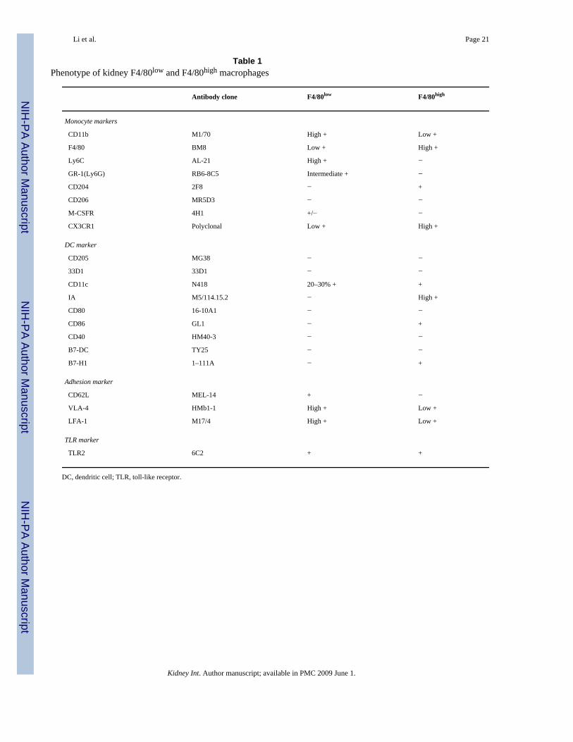
There are two macrophage subtypes in the kidney with distinct phenotypesFluorescence-activated cell sorter (FACS) analysis identified two subsets of F4/80+ cells innormal mouse kidneys, CD11blowF4/80high (referred to as F4/80high) andCD11bhighF4/80low (F4/80low) cells, based on the F4/80 and CD11b fluorescence intensity(Figure 1a). The F4/80high cells comprised ~40% of the total leukocyte (CD45+) populationand represented the major leukocyte population within normal mouse kidneys. Usingmonocyte, DC, chemokine receptor, adhesion and toll-like receptor (TLR) markers these twoF4/80+ subsets had distinct phenotypes (Figure 1b–d; Table 1). All F4/80low cells expressedCD62L+, Ly6Chigh, Gr-1int (Figure 2b), and CX3CR1low (Figure 2c), a phenotypecharacteristic of the blood inflamed monocyte.7,8 All F4/80high cells expressed CD11c+, MHCclass IIhigh (IAhigh), CD86+, and CX3CR1high (Figure 1c), a phenotype characteristic of DCs.Both subsets expressed lymphocyte function-associated antigen 1, very late antigen-4, TLR2,MD-1 (Figure 1d), and RP-105 (not shown). Therefore, F4/80high cells and F4/80low cells wereconsidered to represent kidney resident DCs and macrophages derived from the inflamedmonocyte pool, respectively.
As both kidney F4/80+ subsets express CX3CR1, we used CX3CR1+/GFP mice to trackmacrophages and DCs in normal kidney. Targeted deletion of CX3CR1 and replacement with
Li et al. Page 2
Kidney Int. Author manuscript; available in PMC 2009 June 1.
NIH
-PA Author Manuscript
NIH
-PA Author Manuscript
NIH
-PA Author Manuscript

the gene encoding green fluorescent protein (GFP) provides a useful means for followinglabeled cells and for studying the function of CX3CR1. GFP is expressed in allCX3CR1+/GFP and homozygous CX3CR1GFP/GFP circulating CD11b+F4/80+ cells.7,19,20Most of the kidney CX3CR1 GFP+ DCs are resident DCs and are distributed throughout thecortex (Figure 1e) and outer medulla (Figure 1f), consistent with a recent report.21 MHC II(IA) distribution was mainly in the kidney medulla (Figure 1f), compared to cortex (Figure1e), and most GFP+ cells expressed IA (Figure 1e–g) and F4/80 on the cell surface (Figure 1h).DCs comprised 85% of the GFP+ cells (Figure 1i). All of the CX3CR1+GFPhigh cells expressedCD11b, and most expressed CD11c and MHC II, markers for DCs (Figure 1i). Ly6C and Gr-1were expressed primarily on the GFPlow population (Figure 1j), which has the same phenotypeas the blood inflamed monocytes.7 Both GFP+ cell populations expressed F4/80 (Figure 1hand j). Kidney F4/80low leukocytes had a similar phenotype to GFPlow cells inCX3CR1+/GFP mice and represented kidney monocyte/macrophages, whereas the F4/80high
subset resemble CX3CR1+ GFPhigh cells and represented resident kidney DCs (Figure 1b andc).
Recruitment of F4/80low macrophage subset following kidney IRIFollowing 3 (Figure 2a and g) and 24 h (Figure 2b and h) of reperfusion FACS analysis showedan increase in the cell number of F4/80low macrophages. However, 3 (Figure 2a and g) and 24h (Figure 2b and h) after reperfusion there was no change in the number of F4/80high kidneyresident DCs.
Similar results were observed with Ly6Chigh and Gr-1int markers expressed on the bloodinflamed monocytes, at 3 (Figure 2c and e) and 24 h (Figure 2d and f). At 3 h followingreperfusion, recruited F4/80low macrophages were Ly6Chigh (Figure 2c), which representsimmature, newly arrived monocytes from BM.8 By 24 h, Ly6C expression was markedlydownregulated in the F4/80low monocytes (Figure 2d); the values for mean fluorescence indexwere 2243.3 ± 134.2 (n = 3) and 768.0 ± 47.4 (n = 3) for 3 and 24 h IRI, respectively (P <0.001). This finding suggests that either macrophages mature after migration to the inflamedkidney or mature macrophages infiltrate IRI kidneys. Similarly, the number ofF4/80lowGr-1int macrophages in kidneys increased at 3 h (Figure 2e) and was greater (relativeto sham) by 24 h of reperfusion (Figure 2f, right). The mean fluorescence index ofF4/80lowGr-1int decreased at 24 h compared to 3 h following kidney IRI, paralleling thefindings with Ly6C. Therefore, an abundance of infiltrating macro-phages, phenotypicallysimilar to circulating inflamed monocytes, was identified in reperfused kidneys.
Heterogeneity of proinflammatory cytokine production by CD11bhighF4/80low andCD11bhighF4/80high 24 h following kidney IRI
We used FACS analysis of intracellular cytokine production to determine functionaldifferences between F4/80low and F4/80high populations following kidney IRI. Gating onF4/80low and F4/80high (Figure 2b) macrophages from 24 h sham and IRI kidney, wedetermined expression of intracellular interleukin (IL)-1α, IL-6, IL-12p40/70, and tumor-necrosis factor (TNF)-α (Figure 3a and b). The proportion of F4/80low macrophages producingcytokines (% of total F4/80low population) following IRI was 4- to 8-fold greater for IRI relativeto sham (Figure 3a and c). In contrast, the percentage of F4/80high cells (Figure 3b and d) thatproduced IL-1α, IL-6, IL-12p40/70 was unchanged but TNF-α-producing cells increasedsignificantly following IRI compared to sham. Therefore, 24 h following reperfusion, FACSanalysis reveals functional differences between F4/80low and F4/80high populations.
Li et al. Page 3
Kidney Int. Author manuscript; available in PMC 2009 June 1.
NIH
-PA Author Manuscript
NIH
-PA Author Manuscript
NIH
-PA Author Manuscript

CCR2-deficient, but not MCP-1-deficient, signaling protects kidneys from IRIChemokine production attracts monocytes, T cells, and neutrophils to the reperfused kidney.Compared with sham mouse kidneys, expression of RANTES, MIP-2, IP-10, and MCP-1mRNA increased at 24 h after IRI (data not shown).
As CCR2/MCP-1 is an important signaling pathway in mediating monocyte/macrophagemigration, we performed kidney IRI in mice deficient in CCR2 (CCR2−/−) or its ligand, MCP-1(MCP-1−/−). Following 24 h of reperfusion, plasma creatinine levels increased significantly inCCR2+/+ but not in CCR2−/− mice (Figure 4a). To determine the contribution of CCR2expressed on bone marrow (BM)-derived cells to tissue injury following reperfusion, wegenerated BM chimeras by transferring BM-derived cells from CCR2−/− mice into BM-depleted CCR2+/+ mice (CCR2−/− → CCR2+/+). IRI led to an increase in plasma creatinine inCCR2+/+ → CCR2+/+ but not in CCR2−/− → CCR2+/+ chimeras (Figure 4b). Tubule cellnecrosis in the kidney outer medulla was lower in CCR2−/− mice following IRI compared toCCR2+/+ mice (acute tubular necrosis score: 0.5 ± 0.1 vs 4.5 ± 0.3, P < 0.0001) (Figure 4c andd) and in CCR2−/− → CCR2+/+ compared with CCR2+/+ → CCR2+ chimeras (acute tubularnecrosis score: 0.6 ± 0.4 vs 4.4 ± 0.3, P < 0.0001) (Figure 4e and f). In contrast, the kidneysof MCP-1−/− mice were not protected from IRI; plasma creatinine levels were 2.20 ± 0.13 (n= 5) and 0.27 ± 0.02 (n = 4) mg/100 ml for IRI and sham, respectively (P < 0.001) and moreF4/80low macrophages infiltrated injured tissue in MCP-1−/− mice. Although F4/80lowLy6C+
cell counts were similar between sham-operated WT and MCP−/− mice, F4/80low Ly6C+ cellcounts ( × 105 cells per g kidney) increased to a similar degree in WT (n = 3) and KO (n = 3)mice following IRI (2.42 ± 0.31 and 2.57 ± 0.12, respectively, P = NS). This is probably dueto the contribution of other MCP family members to kidney IRI.
We evaluated the function of CCR2 expression in mediating F4/80low macrophage recruitmentto injured kidneys. Following 24 h of reperfusion, CD11b+F4/80low macrophages increased inkidneys of CCR2+/+ mice and CCR2+/+ → CCR2+/+ chimeras (Figure 4g). However, theabsence of CCR2 attenuated the increase in F4/80low macrophages in CCR2−/− mice andCCR2−/− → CCR2+/+ chimeras (Figure 4g). Similar results were observed with other markersof F4/80lowLy6C+ macrophages and F4/80lowGr-1int macrophages (not shown). Interestingly,the number of F4/80high kidney resident DCs did not increase in CCR2+/+, CCR2−/−,CCR2+/+ → CCR2+/+, or CCR2−/− → CCR2+/+ mice following 24 h of reperfusion comparedto sham (data not shown). Thus, CCR2 expressed on BM-derived cells mediates F4/80low
monocyte/macrophage infiltration into reperfused kidneys.
The infiltration of neutrophils in kidneys subjected to IRI in relation to macrophage recruitmentwas also examined. In CCR2−/− and CCR2−/− → CCR2+/+ mice, neutrophils comigrated withmacrophages into injured kidneys. Kidney neutrophil cell number ( × 105 per g kidney)following IRI was 2.79 ± 0.23 (n = 4) and 10.87 ± 1.78 (n = 4) (P < 0.005) for CCR2−/− andCCR2+/+, respectively, and 3.46 ± 1.55 (n = 8) and 13.28 ± 2.31 (n = 10) (P < 0.005) forCCR2−/− → CCR2+/+ and CCR2+/+ → CCR2+/+, respectively. These results suggested thatCCR2 also mediated neutrophil recruitment to inflamed kidneys.
Deficiency of CX3CR1 reduces kidney IRICX3CR1 is another chemokine receptor mediating monocyte migration. Following kidney IRI,an increase in plasma creatinine levels was observed in CX3CR1+/GFP mice, but levels weresignificantly lower in CX3CR1-deficient CX3CR1GFP/GFP mice (Figure 5a). Tubule necrosisproduced by IRI was also attenuated in CX3CR1GFP/GFP compared with CX3CR1+/GFP mice(acute tubular necrosis score: 2.0 ± 0.74 vs 4.1 ± 0.22, P < 0.05) (Figure 5b).
Li et al. Page 4
Kidney Int. Author manuscript; available in PMC 2009 June 1.
NIH
-PA Author Manuscript
NIH
-PA Author Manuscript
NIH
-PA Author Manuscript
Monocyte infiltration of the reperfused kidney is CX3CR1 dependentTo test our hypothesis that monocyte recruitment to the reperfused kidney is CX3CR1dependent, we examined the kidney CX3CR1+ GFPlowGr-1+ and CX3CR1+ GFPlowLy6C+
macrophages in CX3CR1GFP/GFP (CX3CR1 KO) and CX3CR1+/GFP mice7 followingreperfusion (Figure 5c and d). We found increased CX3CR1+ GFPlow Gr-1+ macrophageinfiltration (cell number, × 105 per g kidney) into reperfused kidneys of CX3CR1+/GFP mice(1.64 ± 0.51 (n = 7) and 6.86 ± 1.35 (n = 11) for sham and IRI, respectively (P < 0.01)) but notin CX3CR1GFP/GFP mice (1.20 ± 0.28 (n = 6) and 2.21 ± 0.29 (n = 10) for sham and IRI,respectively (P > 0.05)). Similar results were found for CX3CR1+ GFPlow Ly6C+ macrophages(Figure 5d). The increase from sham following IRI (%) was 316.9 ± 117.5 (n = 5) and 78.8 ±39.5 for CX3CR1+/GFP and CX3CR1GFP/GFP (n = 9), respectively (P < 0.05). These resultsindicate that CX3CR1 is important in monocyte/macrophage recruitment following IRI.
Infiltrating F4/80low macrophages in reperfused kidneys were derived from a bloodCD11bhigh Ly6Chigh inflamed monocyte pool
Kidney-recruited F4/80low macrophages following IRI ex-pressed Ly6Chigh, Gr-1int andCD62L, markers characteristic of blood-derived inflamed monocytes (Figure S2). Thepercentage of CD11b+Ly6Chigh monocytes in blood increased as early as 3 h after kidney IRI(4.45 ± 0.85; n = 12) compared with sham (1.72 ± 0.28; n = 12) (P < 0.01).
Because CCR2 is necessary for monocyte trafficking into reperfused kidneys, we examinedthe function of CCR2 on the blood content of CD11b+ Ly6Chigh monocytes (Figure 6a) afterkidney IRI. We found increased blood CD11b+ Ly6-Chigh cells in 24 h IRI CCR2+/+ micecompared with sham but not in CCR2−/− mice. We also found that following IRI the percentage(and absolute cell counts; not shown) of blood CD11b+ Ly6Chigh monocytes increased in theCCR2+/+ → CCR2+/+ but not in the CCR2−/− → CCR2+/+ chimeras (Figure 6a). These resultssuggest that CCR2 deficiency leads to reduced influx of Ly6Chigh inflamed monocytes intothe reperfused kidney due to a reduced blood content of Ly6Chigh inflamed monocytes inCCR2−/− mice and CCR2−/− → CCR2+/+ chimeras.
We next examined whether the reduction of blood CD11b+ Ly6Chigh monocytes in CCR2−/−
mice was due to the retention of CD11b+ Ly6Chigh monocytes in BM. After 24 h of kidneyreperfusion, CCR2+/+ and CCR2−/− mice had a similar percentage of BM Ly6Chigh monocytes,and the percentage change of BM Ly6Chigh cells in CCR2+/+ → CCR2+/+ chimeras was similarto CCR2−/− → CCR2+/+ chimeras following IRI (Figure 6b). These findings demonstrated thatfollowing kidney IRI, blood CD11b+ Ly6Chigh monocytes are reduced in CCR2−/− mice andcould contribute to a diminished blood to kidney tissue gradient. This could contribute toreduced monocyte/macrophage infiltration and tissue protection observed in CCR2−/− micefollowing IRI.
CCR2 and CX3CR1 are necessary for inflamed monocyte migration to the IRI kidneyThe observed decrease in kidney infiltration of monocytes into reperfused kidneys may be dueto: (1) the reduced blood content of monocytes in the CCR2−/− mice (hence reduced gradientfrom blood to kidney) or (2) the necessity of CCR2 expression on monocytes to facilitatemigration from blood to the injured kidney. To distinguish between these two possibilities wetransferred the same number (1 × 10⁷) of carboxyfluorescein diacetate succinimidyl ester(CFSE)-labeled BM monocytes either from CCR2+/+ or CCR2−/− mice to CCR2−/− mice atthe onset of kidney IRI and tracked their infiltration into CCR2−/− kidneys subjected to IR(Figure 6c and d). CFSE-labeled CCR2+/+ monocytes were detected in reperfused kidneys andcomprised primarily the F4/80low population. There was a threefold greater influx of CFSE-labeled CCR2+/+ monocytes in CCR2−/− IRI kidney compared to CFSE-labeled CCR2−/−
monocytes. Similarly, we adoptively transferred CX3CR1GFP/GFP or CX3CR1+/GFP BM
Li et al. Page 5
Kidney Int. Author manuscript; available in PMC 2009 June 1.
NIH
-PA Author Manuscript
NIH
-PA Author Manuscript
NIH
-PA Author Manuscript

monocytes into WT mice (Figure 6e and f). CX3CR1+/GFP monocytes were detected inreperfused kidneys and com-prised primarily the F4/80low population. There was a 3.2-foldgreater influx of CX3CR1+/GFP monocytes into the reperfused kidneys of WTmice comparedto CX3CR1GFP/GFP monocytes.
The protected kidney function in CCR2−/− mice was reversed following adoptive transfer ofCCR2+/+ (1 × 10⁷) but not CCR2−/− monocytes. Creatinine level following IRI was 0.81 ±0.11 (n = 4) and 0.51 ± 0.03 (n = 6) mg/100 ml for CCR2−/− mice that received CCR2+/+ andCCR2−/− monocytes, respectively (P < 0.05). These data demonstrated that monocytesexpressing CCR2 represent inflamed monocytes that migrate to the IRI kidney and contributeto tissue injury in a CCR2-dependent manner.
The data from these two experiments provide strong evidence that the protection observedfollowing reperfusion in CCR2- and CX3CR1-deficient mice is due to a reduced CCR2- andCX3CR1-dependant infiltration of F4/80low macrophages and that the reduced infiltration intoreperfused kidneys is due to the combined effect of reduced emigration of monocytes fromBM (CCR2−/− mice) and reduced migration of monocytes into injured kidneys.
DISCUSSIONEarly infiltration of macrophages and neutrophils contributes to the innate immune responseof kidney IRI.4–6 Macrophages are a heterogeneous population, and two subsets of monocyte/macrophages have been identified: Ly6-Chigh CCR2+ Gr-1+ CX3CR1low andLy6ClowCCR2−Gr-1− CX3CR1high.11,12 In this study, we made two seminal observations.First, we showed that F4/80+ cells in normal kidneys consist of two subsets: (1) F4/80low
macrophages derived from the blood inflamed monocyte pool and (2) F4/80high populationphenotypically representing DCs. This conclusion is supported by the following: (1) the useof multiple cell-surface markers (Table 1; Figure 1) that can distinguish between two differenttypes of macrophages;7 (2) the two macrophage subsets exhibit functional differences;F4/80high cells do not produce cytokines to a large degree with the exception of TNF-α, whereasF4/80low cells produce an abundance of proinflammatory cytokines after IRI; and (3) theF4/80high population possesses characteristics of a professional antigen presenting cell (L. Liand M.D. Okusa, unpublished observation, 2008). Second, increased CD11b+
F4/80lowLy6ChighCCR2+ Gr-1+ CX3CR1low cells infiltrate IRI kidneys, and CCR2 andCX3CR1 mediate trafficking of inflamed monocytes from blood to kidneys. The number ofblood Ly6Chigh monocytes increased dramatically after IRI suggesting that this population ofcirculating inflamed monocytes may be the source of macrophages infiltrating injured kidneys.We provide a comprehensive analysis of trafficking of the inflamed monocyte subset fromblood to kidney, which leads to early initiation of the innate response following kidney IRI.
CCR2-dependent monocyte/macrophage trafficking following kidney IRIIn atherosclerosis, CCR2, CCR5, and CX3CR1 mediate monocyte/macrophage migration intoinflamed tissue12 and CCR2−/− mice were protected from Listeria monocytogenes infectiondue to an attenuation of monocyte egress from BM.22 Further, CCR2−/− mice were protectedfrom kidney IRI, and this protection was associated with reduced macrophage infiltration.23Our studies compliment these results and demonstrate a causal relation between CCR2-expressing monocyte/macrophages and tissue injury following kidney IRI. In the currentstudies, CCR2−/− mouse kidney protection was due to a reduction of blood monocytes.Although reduced infiltration of monocytes into the reperfused kidney may be due secondarilyto reduction in blood levels of monocytes and not to a primary defect in CCR2−/− monocyteresponse to chemokines, we do not believe that this is the case. Preventing the decrease inblood monocytes in CCR2−/− mice by transferring CCR2−/− monocytes did not facilitate theirentry in injured tissues. However, transfer of CCR2+/+ monocytes did facilitate macrophage
Li et al. Page 6
Kidney Int. Author manuscript; available in PMC 2009 June 1.
NIH
-PA Author Manuscript
NIH
-PA Author Manuscript
NIH
-PA Author Manuscript

infiltration and induce injury. In contrast in L. monocytogenes-infected mice, TNF-α-producingCCR2−/− monocytes infiltrated inflamed spleen,22 a finding that differs from our study butthat could be explained by differences in the model (septic vs aseptic IRI) and tissue (spleenvs kidney) examined in the two studies. Therefore, inflamed monocyte migration into thereperfused kidney is in part CCR2 dependent and contributes to early inflammation in kidneyIRI.
Previous studies examining the proinflammatory cytokines in IRI kidney have used PCR orenzyme-linked immunosorbent assay of whole kidney homogenate and suggested infiltratedmacrophages identified by immunohistochemistry as a likely source of cytokine production.Indeed, an increase in cytokine level in whole kidney may represent production by immune,epithelial, interstitial, or endothelial cells, and as such the cellular source of cytokines hasremained unknown. Our study is the first one to determine at a single immune cell(macrophage) proinflammatory cytokine production by using FACS intracellular staining.
CX3CL1/CX3CR1-mediated monocyte/macrophage trafficking in kidney IRICX3CL1/CX3CR1 is another pathway mediating monocyte migration. CX3CR1low Gr-1+
CCR2+ subsets are actively recruited to inflamed tissue and CX3CR1highGr-1− CCR2− subsetsmigrate to uninjured tissue and differentiate into resident macrophages and DCs.9 CX3CR1-deficient (CX3CR1GFP/GFP) mice are significantly protected from rejection of transplantedhearts.24 We found that CX3CR1−/− mouse kidneys were protected from IRI, confirmingstudies that employed a CX3CR1 blocking antibody,25 and this protection was associated withless GFPlowGr-1+ or GFPlowLy6C+ macrophage infiltration compared to the heterogeneousCX3CR1+/GFP mice. Thus CX3CR1 signaling mediates infiltration of inflamed monocytes intoinjured kidneys.
CCR2 and CX3CR1 may act cooperatively and/or independently as each may be responsiblefor specific mechanisms of monocyte trafficking in inflamed tissue. Tacke et al.12 found thatCCR2−Ly6Clow monocytes infiltrated atherosclerosis plaques less frequently compared withCCR2+ Ly6Chigh monocytes, and CCR2− Ly6Clow monocyte recruitment is CCR5 dependentand CCR2 independent, but CCR2+ Ly6Chigh monocyte migration is CX3CR1 dependent.
DCs participate in innate and adaptive immunity, autoimmunity, allograft, and host defense.26 F4/80high resident DCs are the predominant leukocyte subset, but the function of kidneyDCs in IRI is unknown completely. TNF-α production from kidney DCs at early time pointsfollowing IR was involved in kidney injury.27 We showed recently that kidney F4/80high DCsmediate OT-II T-cell proliferation by presenting OVA323-339 efficiently (L. Li and M.D.Okusa, unpublished observation, 2008) and present self-glycolipid to activate natural killer Tcells (NKT) by producing interferon-γ following reperfusion.28 IL-12 and IL-23 are importantcytokines secreted by activated DCs. IL-23 mediates neu-trophil recruitment and productionof related chemokines CXCL1, 2, and 529 and may also be involved in kidney IRI. Lastly, itshould be recognized that other macrophage subsets likely participate in repair of kidneyfollowing IRI.4 The alternative macrophage expressing the mannose receptor and activated byIL-4 and IL-13 likely participates in tissue repair.30
In conclusion, this study identified the profile of leukocyte subset influx in the early and latephases following reperfusion and defined two F4/80+ populations in the kidney: resident DCsand macrophages. CCR2 and CX3CR1 signaling pathways play critical roles in inflamedmonocyte recruitment in kidney IRI (Figure 7). Resident DCs participate in innate immunitythrough natural killer T-cell activation28 and modulate adaptive immunity.31 These studiesprovide new insight into mechanisms of early innate immune response to acute kidney IRI,concepts that may generalize to other organs and provide the foundation for therapeuticstrategies.
Li et al. Page 7
Kidney Int. Author manuscript; available in PMC 2009 June 1.
NIH
-PA Author Manuscript
NIH
-PA Author Manuscript
NIH
-PA Author Manuscript

MATERIALS AND METHODSMice and surgical protocol
We adhered to the NIH Guide for the Care and Use of Laboratory Animals. The AnimalResearch Committee of the University of Virginia approved all procedures and protocols. Weused C57BL/6 and Balb/c male background mice (~20 g, 6–8 weeks of age; Charles RiverLaboratories, Wilmington, MA, USA). Age-matched CCR2−/− mice (Balb/c background),CX3CR1+/GFP and CX3CR1GFP/GFP mice (C57BL/6 background) were bred in our animalfacility. Following anesthesia, bilateral flank incisions were performed as previously described,32 both kidney pedicles were exposed and cross-clamped for 32 min, then clamps were released(reperfusion) for different times. In sham-operated mice, kidney pedicles were exposed but notclamped.
Tissue processing and cell counting by FACSKidney cell pellets were prepared from sham and IRI mice and kidney leukocyte subset cellnumber was calculated as described.28 Kidney F4/80+ cell subsets were identified usingallophycocyanin (APC)-labeled rat anti-mouse F4/80 (BM8) and phycoerythrin (PE)-labeledrat anti-mouse CD11b (M1/70), I-A (M5/114.15.2), CD11c (N418), CD86 (GL1), TLR2 (6C2),Ly6C (AL-21; BD Bioscience, San Jose, CA, USA), Gr-1 (Ly6G) (RB6-8C5), CD62L(MEL-14), very late antigen-4 (HMb1-1), or lymphocyte function-associated antigen 1(M17/4) antibodies, or primary rabbit anti-mouse CX3CR1 antibody followed by PE-labeledgoat anti-rabbit IgG (Southern Biotech, Birmingham, AL, USA).
Fresh mouse blood and BM cells were obtained.14 Cell pellets were incubated with anti-CD11b-PE (M1/70), F4/80-APC (BM8) and Ly6C-FITC (AL-21). 7-AAD was used to excludedead cells. Flow cytometry data acquisition was performed on BD FACSCalibur and analyzedby Flowjo software 6.4 (Tree Star Inc., Ashland, OR, USA).
Intracellular staining of proinflammatory cytokinesFACS intracellular staining of proinflammatory cytokines was performed as described.28 PE-labeled anti-mouse IL-1α (ALF-161), IL-6 (MP5-20F3), IL-12p40/70 (C17.8), TNF-α (MP6-XT22), or isotype control antibody (5 µg/ml) was added to each sample after blocking withanti-mouse CD16/32 (10 mg/ml) for 20 min on ice. All of above mAbs, unless otherwise stated,were from eBioscience (San Diego, CA, USA).
Generation of chimeric miceTwo groups of chimeric mice were generated by transferring BM cells as described.33 (1) BMfrom CCR2−/− to CCR2+/+ mice (CCR2−/− → CCR2+/+) and (2) BM from CCR2+/+ toCCR2+/+ mice (CCR2+/+ → CCR2+/+). Chimeric mice were used 8 weeks after BM transfer.Reconstitution efficiency is nearly 90%.32,33
BM monocyte isolation and adoptive transferBM CFSE labeling and monocyte isolation was as described before.22 The enriched populationcomprised 90% CD11b+ cells. CFSE-labeled enriched monocytes (1 × 10⁷) were injected i.v.into CCR2−/− mice at the onset of kidney IRI.
HistochemistryKidneys were fixed for hematoxylin and eosin staining, and acute tubular necrosis was scoredin the outer stripe of the outer medulla as described previously.34 Sections were viewed bylight microscopy (Zeiss AxioSkop), and photographs were taken with a SPOT-RT camera(software version 3.3; Diagnostic Instruments, Sterling Heights, MI, USA).
Li et al. Page 8
Kidney Int. Author manuscript; available in PMC 2009 June 1.
NIH
-PA Author Manuscript
NIH
-PA Author Manuscript
NIH
-PA Author Manuscript
Immunofluorescence and confocal microscopyMouse kidney sections were fixed in 0.7% PLP (0.7% paraformaldehyde), 1.4% lysine, 0.2%sodium periodate in 0.1 M sodium phosphate buffer, pH7.4,32 incubated in 30% sucrose for 48h at 4 °C, embedded and frozen in Optimal Cutting Temperature compound (Ted Pella Inc.,Redding, CA, USA). Frozen sections (25 µm) were permeabilized with 0.3% Triton X-100,blocked with 10% goat serum and anti-CD16/32 antibody, and incubated for 1 h with Alexa647-labeled F4/80 (Invitrogen, Carlsbad, CA, USA) or IA (BioLegend, San Diego, CA, USA)or PE-labeled I-A (eBioscience, Carlsbad, CA, USA) antibodies (5 µg/ml). Images wereacquired using Zeiss LSM 510-UV confocal microscopy and processed using AdobePhotoshop (Adobe, San Jose, CA, USA).
StatisticsGraphPad Instat 3 (GraphPad Inc., San Diego, CA, USA) was used to analyze the data.Unpaired t-test, analysis of variance, and Tukey’s post hoc analysis were performed with P <0.05 indicating significance.
Supplementary MaterialRefer to Web version on PubMed Central for supplementary material.
ACKNOWLEDGMENTSThis work was supported in part by funds from NIH RO1DK56223, RO1DK62324, RO1 HL070065, R01 CA78400,and R21 AI059996. The authors are grateful to Dr Dan R. Littman (Skirball Institute, NYU Medical Center) forCX3CR1GFP/GFP and CX3CR1+/GFP mice, Dr William Kuziel (University of Texas, Austin) for the CCR2−/−mice; Dr Alaa S. Awad (University of Virginia) for helpful discussions and to Steven P. Song (University of Virginia)for composing Figure 7.
REFERENCES1. Thadhani R, Pascual M, Bonventre JV. Acute renal failure. N Engl J Med 1996;334:1448–1460.
[PubMed: 8618585]2. Jo SK, Rosner MH, Okusa MD. Pharmacologic treatment of acute kidney injury: why drugs haven’t
worked and what is on the horizon. Clin J Am Soc Nephrol 2007;2:356–365. [PubMed: 17699435]3. Bonventre JV, Weinberg JM. Recent advances in the pathophysiology of ischemic acute renal failure.
J Am Soc Nephrol 2003;14:2199–2210. [PubMed: 12874476]4. Li L, Okusa MD. Blocking the immune response in ischemic acute kidney injury: the role of adenosine
2A agonists. Nat Clin Pract Nephrol 2006;2:432–444. [PubMed: 16932478]5. Jo SK, Sung SA, Cho WY, et al. Macrophages contribute to the initiation of ischaemic acute renal
failure in rats. Nephrol Dial Transplant 2006;21:1231–1239. [PubMed: 16410269]6. Day YJ, Huang L, Ye H, et al. Renal ischemia-reperfusion injury and adenosine 2A receptor-mediated
tissue protection: role of macrophages. Am J Physiol Renal Physiol 2005;288:F722–F731. [PubMed:15561971]
7. Geissmann F, Jung S, Littman DR. Blood monocytes consist of two principal subsets with distinctmigratory properties. Immunity 2003;19:71–82. [PubMed: 12871640]
8. Sunderkotter C, Nikolic T, Dillon MJ, et al. Subpopulations of mouse blood monocytes differ inmaturation stage and inflammatory response. J Immunol 2004;172:4410–4417. [PubMed: 15034056]
9. Gordon S, Taylor PR. Monocyte and macrophage heterogeneity. Nat Rev Immunol 2005;5:953–964.[PubMed: 16322748]
10. Qu C, Edwards EW, Tacke F, et al. Role of CCR8 and other chemokine pathways in the migrationof monocyte-derived dendritic cells to lymph nodes. J Exp Med 2004;200:1231–1241. [PubMed:15534368]
11. Tacke F, Randolph GJ. Migratory fate and differentiation of blood monocyte subsets. Immunobiology2006;211:609–618. [PubMed: 16920499]
Li et al. Page 9
Kidney Int. Author manuscript; available in PMC 2009 June 1.
NIH
-PA Author Manuscript
NIH
-PA Author Manuscript
NIH
-PA Author Manuscript

12. Tacke F, Alvarez D, Kaplan TJ, et al. Monocyte subsets differentially employ CCR2, CCR5, andCX3CR1 to accumulate within atherosclerotic plaques. J Clin Invest 2007;117:185–194. [PubMed:17200718]
13. Charo IF, Ransohoff RM. The many roles of chemokines and chemokine receptors in inflammation.N Engl J Med 2006;354:610–621. [PubMed: 16467548]
14. Charo IF, Taubman MB. Chemokines in the pathogenesis of vascular disease. Circ Res 2004;95:858–866. [PubMed: 15514167]
15. Damas JK, Boullier A, Waehre T, et al. Expression of fractalkine (CX3CL1) and its receptor,CX3CR1, is elevated in coronary artery disease and is reduced during statin therapy. ArteriosclerThromb Vasc Biol 2005;25:2567–2572. [PubMed: 16224053]
16. Combadiere C, Potteaux S, Gao JL, et al. Decreased atherosclerotic lesion formation in CX3CR1/apolipoprotein E double knockout mice. Circulation 2003;107:1009–1016. [PubMed: 12600915]
17. McDermott DH, Fong AM, Yang Q, et al. Chemokine receptor mutant CX3CR1-M280 has impairedadhesive function and correlates with protection from cardiovascular disease in humans. J Clin Invest2003;111:1241–1250. [PubMed: 12697743]
18. Wallace GR, Vaughan RW, Kondeatis E, et al. A CX3CR1 genotype associated with retinal vasculitisin patients in the United Kingdom. Invest Ophthalmol Vis Sci 2006;47:2966–2970. [PubMed:16799040]
19. Jung S, Aliberti J, Graemmel P, et al. Analysis of fractalkine receptor CX(3)CR1 function by targeteddeletion and green fluorescent protein reporter gene insertion. Mol Cell Biol 2000;20:4106–4114.[PubMed: 10805752]
20. Palframan RT, Jung S, Cheng G, et al. Inflammatory chemokine transport and presentation in HEV:a remote control mechanism for monocyte recruitment to lymph nodes in inflamed tissues. J ExpMed 2001;194:1361–1373. [PubMed: 11696600]
21. Soos TJ, Sims TN, Barisoni L, et al. CX3CR1+ interstitial dendritic cells form a contiguous networkthroughout the entire kidney. Kidney Int 2006;70:591–596. [PubMed: 16760907]
22. Serbina NV, Pamer EG. Monocyte emigration from bone marrow during bacterial infection requiressignals mediated by chemokine receptor CCR2. Nat Immunol 2006;7:311–317. [PubMed: 16462739]
23. Furuichi K, Wada T, Iwata Y, et al. CCR2 signaling contributes to ischemia-reperfusion injury inkidney. J Am Soc Nephrol 2003;14:2503–2515. [PubMed: 14514728]
24. Robinson LA, Nataraj C, Thomas DW, et al. A role for fractalkine and its receptor (CX3CR1) incardiac allograft rejection. J Immunol 2000;165:6067–6072. [PubMed: 11086038]
25. Oh DJ, Dursun B, He Z, et al. Fractalkine receptor (CX3CR1) inhibition is protective against ischemicacute renal failure in mice. Am J Physiol Renal Physiol 2008;294:F264–F271. [PubMed: 18003857]
26. Koski GK, Cohen PA, Roses RE, et al. Reengineering dendritic cell-based anti-cancer vaccines.Immunol Rev 2008;222:256–276. [PubMed: 18364007]
27. Dong X, Swaminathan S, Bachman LA, et al. Resident dendritic cells are the predominant TNF-secreting cell in early renal ischemia-reperfusion injury. Kidney Int 2007;71:619–628. [PubMed:17311071]
28. Li L, Huang L, Sung SS, et al. NKT cell activation mediates neutrophil IFN-gamma production andrenal ischemia-reperfusion injury. J Immunol 2007;178:5899–5911. [PubMed: 17442974]
29. Chen Y, Langrish CL, McKenzie B, et al. Anti-IL-23 therapy inhibits multiple inflammatory pathwaysand ameliorates autoimmune encephalomyelitis. J Clin Invest 2006;116:1317–1326. [PubMed:16670771]
30. Gordon S. Alternative activation of macrophages. Nat Rev Immunol 2003;3:23–35. [PubMed:12511873]
31. Dong X, Swaminathan S, Bachman LA, et al. Antigen presentation by dendritic cells in renal lymphnodes is linked to systemic and local injury to the kidney. Kidney Int 2005;68:1096–1108. [PubMed:16105040]
32. Day YJ, Huang L, Ye H, et al. Renal ischemia-reperfusion injury and adenosine 2A receptor-mediatedtissue protection: the role of CD4+ T cells and IFN-gamma. J Immunol 2006;176:3108–3114.[PubMed: 16493070]
33. Day YJ, Huang L, McDuffie MJ, et al. Renal protection from ischemia mediated by A2A adenosinereceptors on bone marrow-derived cells. J Clin Invest 2003;112:883–891. [PubMed: 12975473]
Li et al. Page 10
Kidney Int. Author manuscript; available in PMC 2009 June 1.
NIH
-PA Author Manuscript
NIH
-PA Author Manuscript
NIH
-PA Author Manuscript

34. Faubel S, Ljubanovic D, Reznikov L, et al. Caspase-1-deficient mice are protected against cisplatin-induced apoptosis and acute tubular necrosis. Kidney Int 2004;66:2202–2213. [PubMed: 15569309]
Li et al. Page 11
Kidney Int. Author manuscript; available in PMC 2009 June 1.
NIH
-PA Author Manuscript
NIH
-PA Author Manuscript
NIH
-PA Author Manuscript

Figure 1. There are two F4/80+ macrophage subtypes in the kidney with distinct phenotypiccharacteristics (see also Table 1)(a) Four-color FACS analysis identified two F4/80+ populations in the normal C57BL/6 mousekidney: CD11bhighF4/80low and CD11blowF4/80high (oval circled). Further phenotyping ofthese two F4/80+ populations showed that the F4/80low cells expressed (c) CX3CR1low andonly this population, but not the F4/80high population, expressed (b) CD62L, Ly6Chigh, andGr-1int (oval circled). (c) There is an F4/80high subset (a, c) that expressed CX3CR1,high
CD11c+, CD86, and IAhigh and is referred to as a dendritic cell (DC) population. (d) Bothmacrophages (F4/80low) and DCs (F4/80high) expressed LFA-1, VLA-4, TLR2, and MD-1.(e, f) In kidney sections from C57BL/6 background CX3CR1+/GFP mice, GFP is expressed
Li et al. Page 12
Kidney Int. Author manuscript; available in PMC 2009 June 1.
NIH
-PA Author Manuscript
NIH
-PA Author Manuscript
NIH
-PA Author Manuscript
mainly on monocyte/macrophages and DCs, and many CX3CR1+GFP+ green fluorescing cellswere seen in the cortex (e) and medulla (f). (g, h) Higher magnification z-stack projectionimages of the kidney medulla viewed under a Zeiss LSM-510 confocal microscope showedCX3CR1-GFP-positive cells in green, PE-tagged IA-positive cells in red (g), Alexa 647-taggedF4/80 positive cells in red (h), and colocalization of the two fluorophores inCX3CR1+GFP+IA+ or CX3CR1+GFP+F4/80+ DCs in yellow, respectively; (g) z-stackprojection image of 14 optical slices at 0.35 mm intervals and (h) z-stack projection image of5 optical slices at 0.69 µm intervals. IA and F4/80 expression appears on the DC cell surface(arrowheads in g and h, respectively). Tubule lumen is represented by *. (i) Depiction ofGFPhigh cells that expressed CD11b+, CD11c+, and IA+, which are phenotypically DCs asidentified in (g, h). (j) Both CX3CR1+ GFPhigh and GFPlow cells expressed F4/80. However,compared with GFPhigh cells, GFPlow cells also expressed Gr-1 and Ly6C, which representmonocytes/macrophages. Cell populations of interest are indicated within ovals or in boxedregions.
Li et al. Page 13
Kidney Int. Author manuscript; available in PMC 2009 June 1.
NIH
-PA Author Manuscript
NIH
-PA Author Manuscript
NIH
-PA Author Manuscript

Figure 2. Recruitment of CD11bhighF4/80low inflamed monocyte subset following kidney IRIMouse kidneys were subjected to 32 min ischemia followed by (a, c, e) 3h or (b, d, f) 24 h ofreperfusion. All leukocytes from sham and IRI kidneys of equal weight were counted andevaluated by FACS (see ‘Materials and Methods’). Results clearly indicated that there was anincrease in total number of CD45+ leukocytes in IRI kidneys at (a, right) 3 h and (b, right) 24h when compared to respective sham-operated mice. The increase in leukocytes was mostlyfrom F4/80low, (a, b) CD11b+, (c, d) Ly6C+, (e, f) Gr-1+ cells, consistent with macrophagesderived from the inflamed monocytes subset. Summary of the cell numbers of macrophages(F4/80low) and DCs (F4/80high) in sham and IRI kidneys after (g) 3 h and (h) 24 h of reperfusion.Values are means ± s.e.; N = 7–10; **P < 0.01, ***P < 0.001; NS, not significant.
Li et al. Page 14
Kidney Int. Author manuscript; available in PMC 2009 June 1.
NIH
-PA Author Manuscript
NIH
-PA Author Manuscript
NIH
-PA Author Manuscript

Figure 3. Heterogeneity of proinflammatory cytokine production by CD11bhigh F4/80low andCD11bhigh F4/80high macrophages 24 h following kidney IRIRepresentative tracings from FACS analysis of expression of intracellular cytokines, includingIL-1α, IL-6, IL-12p40/70, and TNF-α, in the (a) F4/80low and (b) F4/80high populations ofmacrophages in mouse kidney after 24 h of reperfusion (defined by gating on CD45+
CD11bhigh F4/80low and CD11blowF4/80high macrophages, respectively, as shown in Figure2b) for sham operation (blue line), IRI (green line), or isotype control (red line). Max (%),percentage of F4/80low or F4/80high macrophages that are cytokine positive. Summary ofproinflammatory cytokine production by (c) F4/80low macrophages and (d) F4/80high
macrophages. Gating on the F4/80low population (c), the percentage of cells that producedIL-1α, IL-6, IL-12p40/70, and TNF-α increased significantly following IRI compared to sham(P < 0.05 for all IRI compared to sham groups). Values are mean ± s.e.; N = 3–6. Similarly,gating on the (d) F4/80high population, the percentage of cells that produced IL-1α, IL-6,IL-12p40/70 was unchanged following IRI compared to sham, but TNF-α increasedsignificantly following IRI compared to sham (P < 0.05). Values are mean ± s.e.; N = 3–6.
Li et al. Page 15
Kidney Int. Author manuscript; available in PMC 2009 June 1.
NIH
-PA Author Manuscript
NIH
-PA Author Manuscript
NIH
-PA Author Manuscript
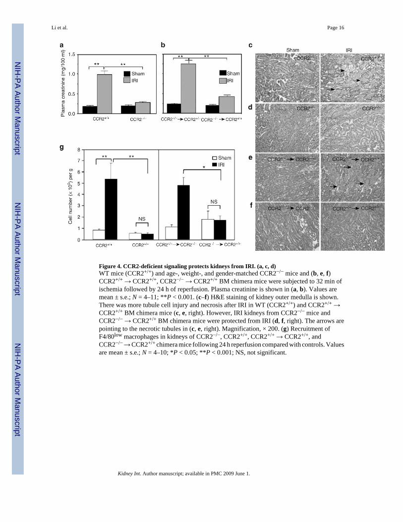
Figure 4. CCR2-deficient signaling protects kidneys from IRI. (a, c, d)WT mice (CCR2+/+) and age-, weight-, and gender-matched CCR2−/− mice and (b, e, f)CCR2+/+ → CCR2+/+, CCR2−/− → CCR2+/+ BM chimera mice were subjected to 32 min ofischemia followed by 24 h of reperfusion. Plasma creatinine is shown in (a, b). Values aremean ± s.e.; N = 4–11; **P < 0.001. (c–f) H&E staining of kidney outer medulla is shown.There was more tubule cell injury and necrosis after IRI in WT (CCR2+/+) and CCR2+/+ →CCR2+/+ BM chimera mice (c, e, right). However, IRI kidneys from CCR2−/− mice andCCR2−/− → CCR2+/+ BM chimera mice were protected from IRI (d, f, right). The arrows arepointing to the necrotic tubules in (c, e, right). Magnification, × 200. (g) Recruitment ofF4/80low macrophages in kidneys of CCR2−/−, CCR2+/+, CCR2+/+ → CCR2+/+, andCCR2−/− → CCR2+/+ chimera mice following 24 h reperfusion compared with controls. Valuesare mean ± s.e.; N = 4–10; *P < 0.05; **P < 0.001; NS, not significant.
Li et al. Page 16
Kidney Int. Author manuscript; available in PMC 2009 June 1.
NIH
-PA Author Manuscript
NIH
-PA Author Manuscript
NIH
-PA Author Manuscript

Figure 5. Kidneys of CX3CR1-deficient mice are protected from IRI with fewer infiltratedmacrophagesMouse kidneys were subjected to 32 min ischemia followed by 24 h of reperfusion. In C57BL/6 background CX3CR1+/GFP mice, one CX3CR1 allele was replaced by GFP, which has noeffect on CX3CR1 gene function, whereas two CX3CR1 alleles were replaced with GFP inCX3CR1GFP/GFP mice, resulting in a CX3CR1 knockout. (a) Plasma creatinine forCX3CR1+/GFP mice and CX3CR1GFP/GFP mice. Values are mean ± s.e.; N = 6–11; **P <0.001. (b) H&E staining of kidney sections in sham and IRI for CX3CR1+/GFP andCX3CR1GFP/GFP mice. Black arrows indicate necrotic tubule cells in the outer medulla of
Li et al. Page 17
Kidney Int. Author manuscript; available in PMC 2009 June 1.
NIH
-PA Author Manuscript
NIH
-PA Author Manuscript
NIH
-PA Author Manuscript

CX3CR1+/GFP mice after IRI. FACS analysis of kidney (c) GFPlow Gr-1+ and (d)GFPlowLy6C+.
Li et al. Page 18
Kidney Int. Author manuscript; available in PMC 2009 June 1.
NIH
-PA Author Manuscript
NIH
-PA Author Manuscript
NIH
-PA Author Manuscript

Figure 6. CCR2 and CX3CR1 are necessary for monocyte egress from BM (to blood) and fromblood (to tissue) following kidney IRIMouse kidneys were subjected to 32 min ischemia followed by 24 h reperfusion and leukocytesfrom (a) blood and (b) BM were quantiated by FACS. We gated on blood and BM SSClow
population and quantitated CD11bhighLy6Chigh inflamed monocytes. Values showCD11bhighLy6Chigh inflamed monocytes as a percentage of total leukocytes isolated after 24h IRI or sham operation from blood of CCR2+/+ (n = 9 and 6), CCR2−/− (n = 4 and 4),CCR2−/− → CCR2+/+ (n = 4 and 4) and CCR2+/+ → CCR2+/+ (n = 4 and 3) mice and fromBM of CCR2+/+ (n = 9 and 7), CCR2−/− (n = 8 and 4), CCR2−/− → CCR2+/+ (n = 7 and 4) andCCR2+/+ → CCR2+/+ (n = 8 and 6) mice. *P < 0.05; **P < 0.001. (c, d) CCR2−/− monocytes(Mo) and CCR2+/+ Mo were isolated from BM and labeled with CFSE. 1 × 10⁷ CFSE-labeledBM monocytes were adoptively transferred separately to the CCR2−/− mice at the onset of thesurgery. Infiltrated labeled cells are denoted by oval. (e, f) GFP-positive monocytes fromCX3CR1+/GFP and CX3CR1 KO (CX3CR1GFP/GFP) (1 × 10⁷) mice were adoptivelytransferred separately into C57BL/6 WT mice and mice were subjected to 24 h IRI.
Li et al. Page 19
Kidney Int. Author manuscript; available in PMC 2009 June 1.
NIH
-PA Author Manuscript
NIH
-PA Author Manuscript
NIH
-PA Author Manuscript

Figure 7. Trafficking of monocytes in ischemia/reperfusion injury (IRI) miceBM CD11b+ Ly6Chigh monocyte/monocyte precursor egress to the blood circulation is CCR2-dependent. (a) Some of the Ly6Chigh monocytes lose their CCR2 and Ly6C expression andare further characterized with CD62L−, Gr-1− and CX3CR1high. (b) These cells migrate tonormal noninflamed tissue rapidly after they are released in the blood and differentiate intotissue dendritic cells (DCs), which express CD11b+, CD11c+, IAhigh, CD86+ and F4/80high.(c) On the other hand, some of the monocytes continue to express CCR2+ and Ly6Chigh on thecell surface with the additional expression of CX3CR1low, Gr-1int, and CD62L+. (d) Theseinflamed monocytes respond to the gradient of chemokines (e) released from IRI kidneys. Inthe injured tissue, these macrophages derived from inflamed monocytes are characterized byCD62L+, Gr-1int, Ly6C+, and F4/80low expression. Infiltrated macrophages produce largeamounts of proinflammatory cytokines, which are involved in tissue injury.
Li et al. Page 20
Kidney Int. Author manuscript; available in PMC 2009 June 1.
NIH
-PA Author Manuscript
NIH
-PA Author Manuscript
NIH
-PA Author Manuscript
NIH
-PA Author Manuscript
NIH
-PA Author Manuscript
NIH
-PA Author Manuscript
Li et al. Page 21
Table 1Phenotype of kidney F4/80low and F4/80high macrophages
Antibody clone F4/80low F4/80high
Monocyte markers
CD11b M1/70 High + Low +
F4/80 BM8 Low + High +
Ly6C AL-21 High + −
GR-1(Ly6G) RB6-8C5 Intermediate + −
CD204 2F8 − +
CD206 MR5D3 − −
M-CSFR 4H1 +/− −
CX3CR1 Polyclonal Low + High +
DC marker
CD205 MG38 − −
33D1 33D1 − −
CD11c N418 20–30% + +
IA M5/114.15.2 − High +
CD80 16-10A1 − −
CD86 GL1 − +
CD40 HM40-3 − −
B7-DC TY25 − −
B7-H1 1–111A − +
Adhesion marker
CD62L MEL-14 + −
VLA-4 HMb1-1 High + Low +
LFA-1 M17/4 High + Low +
TLR marker
TLR2 6C2 + +
DC, dendritic cell; TLR, toll-like receptor.
Kidney Int. Author manuscript; available in PMC 2009 June 1.
Copyright © 2022 FDOKUMEN