Values of Customer Reliability - Australian Energy Regulator
Upload
independentCategory
view
5download
0
Cavin1; a Regulator of Lung Function and MacrophagePhenotypePraveen Govender1, Freddy Romero2, Dilip Shah2, Jesus Paez1, Shi-Ying Ding3, Libin Liu3, Adam Gower1,
Elizabeth Baez1, Sherif Shawky Aly1, Paul Pilch3, Ross Summer2*
1 The Pulmonary Center, Boston University School of Medicine, Boston, Massachusetts, United States of America, 2Center of Translational Medicine and Division of
Pulmonary and Critical Care Medicine, Thomas Jefferson University, Philadelphia, Pennsylvania, United States of America, 3Department of Biochemistry, Boston University
School of Medicine, Boston, Massachusetts, United States of America
Abstract
Caveolae are cell membrane invaginations that are highly abundant in adipose tissue, endothelial cells and the lung. Theformation of caveolae is dependent on the expression of various structural proteins that serve as scaffolding for thesemembrane invaginations. Cavin1 is a newly identified structural protein whose deficiency in mice leads to loss of caveolaeformation and to development of a lipodystrophic phenotype. In this study, we sought to investigate the functional role ofCavin1 in the lung. Cavin1 deficient mice possessed dramatically altered distal lung morphology and exhibited significantphysiological alterations, notably, increased lung elastance. The changes in distal lung architecture were associated withhypercellularity and the accumulation of lung macrophages. The increases in lung macrophages occurred without changesto circulating numbers of mononuclear cells and without evidence for increased proliferation. However, the increases inlung macrophages were associated with higher levels of macrophage chemotactic factors CXCL2 and CCL2 in BAL fluid fromCavin12/2 mice suggesting a possible mechanism by which these cells accumulate. In addition, lung macrophages fromCavin12/2 mice were larger and displayed measurable differences in gene expression when compared to macrophagesfrom wild-type mice. Interestingly, macrophages were also increased in adipose tissue but not in liver, kidney or skeletalmuscle from Cavin12/2 mice, and similar tissue specificity for macrophage accumulation was observed in lungs andadipose tissue from Caveolin12/2 mice. In conclusion, this study demonstrates an important role for Cavin1 in lunghomeostasis and suggests that caveolae structural proteins are necessary for regulating macrophage number andphenotype in the lung.
Citation: Govender P, Romero F, Shah D, Paez J, Ding S-Y, et al. (2013) Cavin1; a Regulator of Lung Function and Macrophage Phenotype. PLoS ONE 8(4): e62045.doi:10.1371/journal.pone.0062045
Editor: Stephania Ann Cormier, Louisiana State University Health Sciences Center, United States of America
Received November 16, 2012; Accepted March 16, 2013; Published April 25, 2013
Copyright: � 2013 Govender et al. This is an open-access article distributed under the terms of the Creative Commons Attribution License, which permitsunrestricted use, distribution, and reproduction in any medium, provided the original author and source are credited.
Funding: This work was supported by the National Institutes of Health (NIH) R01HL105490. The funders had no role in study design, data collection and analysis,decision to publish, or preparation of the manuscript.
Competing Interests: The authors have declared that no competing interests exist.
* E-mail: [email protected]
Introduction
The branched structure of the lung, culminating in alveolae,
provides the large surface area needed for efficient gas exchange in
this tissue. The maximization of the area involved in gas exchange
extends to the cellular level, in that type I epithelial cells and
endothelial cells are highly enriched in cell surface invaginations,
resulting in a functional enhancement of cell surface capacity [1].
These surface invaginations, also called caveolae, are not exclusive
to the lung. Most notably, caveolae are found in the highest
concentrations in adipose tissue, and are present in many other
tissues at lower levels [2]. The importance of caveolae in these
tissues is related less to their role in increasing surface area and
more to their ability to regulate diverse biological processes such as
glucose and lipid homeostasis, clathrin-independent pinocytosis,
and a wide-range of intracellular trafficking and signaling events.
The formation of caveolae is dependent on the production of
various structural proteins that serve as scaffolding for these
membrane invaginations. Genetic mutations in select structural
proteins lead to the absence of caveolae formation and de-
velopment of complex pathological phenotypes [3–7]. The most
recognized abnormalities associated with deficiency in these
proteins are metabolic derangements, and in mice and humans,
these include hyperglycemia, insulin resistance and lipodystrophy
[8–11]. However, it is also increasingly apparent from knockout
studies that caveolae carry out many other important functions
separate from their role in regulating metabolism [2,12].
In particular, caveolae have been shown to play a critical role in
orchestrating lung homeostasis. For example, targeted deletion of
either caveolin 1 or 2 proteins leads to a wide range of lung
abnormalities, including increased deposition of extracellular
matrix, dysregulated cell growth and proliferation, and impaired
pulmonary vascular function [3,4]. Although the mechanisms
leading to these diverse changes are poorly understood, changes to
epithelial, endothelial and fibroblast function are well-documen-
ted.
Recently, a new group of caveolar structural proteins, the
cavins, have been identified. This family of proteins, like the
caveolins, is essential for the biogenesis and function of caveolae
[13]. Included in this family is a 43-kDa cytoplasmic protein
known as PTRF (Polymerase I and Transcript Release Factor) or
Cavin1 (also known in past as Cav-P60) [14–17]. Like the
caveolins, Cavin1 is highly expressed in multiple tissues, including
PLOS ONE | www.plosone.org 1 April 2013 | Volume 8 | Issue 4 | e62045

the lung, but murine knockout studies have described only
lipodystrophic phenotypes [9]. Based on this limited knowledge
of Cavin1, the focus of this investigation was to understand the
functional role of this newly recognized caveolar protein in lung
homeostasis.
Materials and Methods
Ethics StatementAll experiments were carried out in accordance with the
recommendations in the Guide for the Care and Use of
Laboratory Animals of the National Institutes of Health and
study protocols were approved by Boston University’s Institutional
Animal Care and Use Committee (IACUC number AN-14683).
MiceStudies were performed on 2-month-old female mice. Cavin1
knockout (Cavin12/2) mice on a C57Bl/6 background were
generated as previously described [9]. Cavin12/2 mice and
controls were obtained by breeding Cavin1+/2. Cavin12/+ mice
yield small litters but surviving pups, like Cavin12/2 mice,
behave normally and have normal life expectancies when followed
to 1 year. We have previously reported that adult Cavin12/2
mice are leaner when compared to their litter mate controls [9].
Figure 1. Immunohistochemistry staining of lung for Cavin1. A) Cavin1 staining is absent from proximal lung (purple) but localizes to distalmurine lung (brown color). B) As expected, Cavin1 staining is absent from the lungs of Cavin12/2 mice. C-E) High power images of wild-type lungdemonstrate Cavin1 expression on blood vessel endothelium (solid arrows, C), capillaries (arrowheads, D), type-I pneumocytes (broken arrows, D),alveolar macrophages (asterisk, E). Staining is absent from type-II epithelial cells in distal airspaces (broken arrow, C). F) The specificity of Cavin1antibody was demonstrated by Western blot analysis.doi:10.1371/journal.pone.0062045.g001
Cavin1 Regulates Lung Homeostasis
PLOS ONE | www.plosone.org 2 April 2013 | Volume 8 | Issue 4 | e62045

Caveolin-1 knockout (Cav12/2) mice and age-matched controls
were purchased from The Jackson Laboratory (Bar Harbor, ME).
Lung Physiology MeasurementsIn an anesthetized mouse, an 18G tracheal cannula was inserted
into a surgically exposed trachea and ligated tightly. The mouse
was then placed on the FlexiVent mechanical ventilator (Scireq
Scientific Respiratory Equipment, Montreal, Quebec), and ven-
tilated at 300 breaths/min with positive-end expiratory pressure
set at 3 cmH2O. Baseline measurement of airway resistance and
lung elastance was measured as previously described [18].
Western Blot AnalysisWestern blot was performed using 20 mg of protein isolated
from homogenized lung tissue or macrophage cell lines (MH-S or
RAW 264.7 cells). Protein samples were separated by SDS-PAGE
and electrophoretically transferred to polyvinylidene difluoride
(PVDF) membranes. Membranes were incubated in phosphate-
buffered saline with 0.1% Tween 20 containing 10% nonfat dry
milk for 1 h at room temperature, followed by incubation with
primary rabbit antibody directed against murine Cavin1 (21st
Century Biochemicals; Marlboro, MA), CD45 (Abcam, Cam-
bridge, MA), GM-CSF (Abcam, Cambridge, MA) or b-actin (Cell
Signaling Technology; Beverly, MA). Horseradish peroxidase-
conjugated secondary antibodies (Sigma-Aldrich; St Louis, MO)
and an enhanced chemiluminescence (ECL) substrate kit (Perki-
nElmer Life Sciences, Waltham, MA) were used for detection.
ImmunohistochemistryFormalin-fixed tissues were prepared for frozen or paraffin
sectioning using standard techniques. For paraffin-embedded
tissues, wax was removed by solvents (Histo-Clear; National
Diagnostics, Atlanta, GA) and tissues were re-hydrated in graded
alcohols prior to antibody staining. For some antibodies, antigen
retrieval was required using a heated citric acid solution (Target
Retrieval Solution; Dako, Carpinteria, CA). For all sections,
hydrogen peroxide in methanol (3%, 15 min, 22uC) was used to
quench endogenous peroxidases and background staining was
reduced by blocking with 2% BSA. Primary antibodies used in
these studies included rabbit polyclonal anti-Cavin1 (21st Century
Biochemicals; Marlboro, MA), rat anti-mouse CD45 (BD
Biosciences no. 550539), rat anti-mouse Mac-3 (BD Biosciences
no. 550292), biotinylated rat anti-mouse NK1.1 (BioLegend clone
PK136), biotinylated rat anti-mouse B-220 (BD Biosciences
no. 553086), biotinylated rat anti-mouse CD-3 (BioLegend
no. 100303), or purified rat anti-mouse GR-1 (R&D systems no.
MAB1037). All primary antibodies were diluted in ranges from
1:100 to 1:500 and applied for 1 hour at RT. Biotinylated
antibodies were detected using an ABC kit (Vector Laboratories)
followed by addition of 3,39-diaminobenzadine. Cavin1, CD-45,
NK 1.1, Mac-3, and GR-1 were detected using an anti-rabbit
(Cavin1) or anti-rat secondary antibody kit (Vector Laboratories
Burlingame, CA) before exposure to 3,39-diaminobenzadine.
PCNA (Invitrogen Grand Island, NY) and TUNEL (R&D Systems
Minneapolis, MN) staining were performed using kits according to
manufacturer’s protocol.
Figure 2. Cavin1 deficiency leads to physiological and structural changes to lung. A) Airway resistance and lung elastance were increasedin lungs of Cavin12/2 mice (n = 6, both groups, 2 independent experiments). B) Low power images of H&E stained lungs (upper images) showarchitectural distortion in the lungs of Cavin12/2 mice. High power images (middle images) demonstrate hypercellularity (increased blue nuclei) inthe distal lung in Cavin12/2 mice. Trichrome staining (lower images) demonstrates increased deposition of collagen (blue) in the lungs of Cavin12/2 mice. C) Dry weight (n = 5, each group, p,0.05) and DNA content (n = 10, each group, p,0.001) are increased in the lungs of Cavin12/2 mice.Statistically significant differences were not observed in lung wet-to-dry ratios from wild-type and Cavin12/2 mice (n = 5, each group).doi:10.1371/journal.pone.0062045.g002
Cavin1 Regulates Lung Homeostasis
PLOS ONE | www.plosone.org 3 April 2013 | Volume 8 | Issue 4 | e62045

Tissue MorphometryQuantification of leukocytes was performed after antibody
staining in tissue sections. Evaluation was performed by two
independent investigators, one of who was blinded to the study
group. Mac3, CD4, NK1.1, B220 and Gr-1+ cells were quantified
in three histological sections from each mouse. Because of
structural changes in lungs of Cavin12/2mice, the quantification
of Mac3+ cells was performed by two methods. First, we calculated
the number of cells per high power field. Second, we evaluated the
number of Mac3+ cells per alveolus. For the latter analysis, 300
alveoli were counted per mouse in each group.
Lung DNA Content and Wet-to-dry RatiosDNA content and lung wet-to-dry ratios were determined per
published protocols [19,20].
ELISAThermo Scientific Nunc Maxisorp plates were coated with
primary antibody against CCL2 or CX3CL1 (R&D Systems
Minneapolis, MN) overnight at RT and then washed with PBS
and 0.5% Tween 20. Plates were blocked with 1% BSA in PBS for
1 h, followed by addition of samples for 2 h incubation at RT.
After washing, biotinylated secondary antibody was applied for
2 h, followed by streptavidin-HRP conjugate diluted at 1:200 with
the blocking reagent. Reaction was measured using endpoint
spectrometry after being developed with 0.01% tetramethylbenzi-
dine dissolved in DMSO and 0.5% hydrogen peroxide.
Real-time Quantitative PCRRNA was isolated from homogenized lungs using RNeasy Plus
Mini kit (Qiagen; Valencia, CA). Real-time quantitative PCR was
performed on cDNA converted from isolated RNA using the
Reverse Transcription System kit (Promega; Madison, WI). The
CFX96 TouchTM Real-Time PCR Detection System (BioRad;
Figure 3. Leukocytes are increased in the lungs of Cavin12/2mice. A) Transcript levels for structural and non-structural cells in lung of wild-type and Cavin12/2mice. Transcript levels for the hematopoietic marker CD45 are increased in the lungs of Cavin12/2mice. Statistically significantdifferences were not observed in transcript levels for SPC, CC-10, T1a, CD31 in the lungs of wild-type and Cavin12/2 mice (n = 5 or greater in allgroups, two replicates). B) CD45-positive cells are increased in the lungs of Cavin12/2 mice. (Top) Immunohistochemistry staining for mouse CD45.Brown straining represents CD45+ expressing cells. (Middle) Flow cytometry analysis of CD45 stained lung digests from wild-type and Cavin12/2mice (n = 6 wild-type and Cavin12/2 mice). Doublets, red blood cells and dead cells were excluded from analysis. (Bottom) Western blot analysis forCD45 in lungs from wild-type and Cavin12/2 mice.doi:10.1371/journal.pone.0062045.g003
Cavin1 Regulates Lung Homeostasis
PLOS ONE | www.plosone.org 4 April 2013 | Volume 8 | Issue 4 | e62045
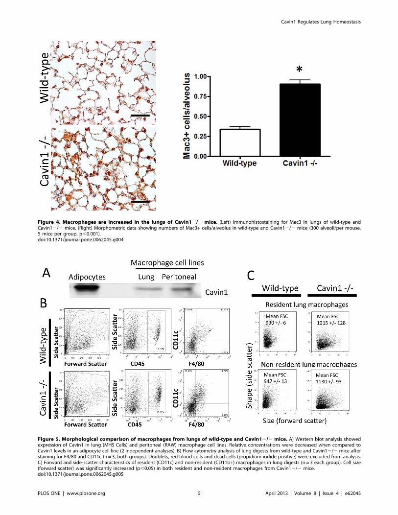
Figure 4. Macrophages are increased in the lungs of Cavin12/2 mice. (Left) Immunohistostaining for Mac3 in lungs of wild-type andCavin12/2 mice. (Right) Morphometric data showing numbers of Mac3+ cells/alveolus in wild-type and Cavin12/2 mice (300 alveoli/per mouse,5 mice per group, p,0.001).doi:10.1371/journal.pone.0062045.g004
Figure 5. Morphological comparison of macrophages from lungs of wild-type and Cavin12/2 mice. A) Western blot analysis showedexpression of Cavin1 in lung (MHS Cells) and peritoneal (RAW) macrophage cell lines. Relative concentrations were decreased when compared toCavin1 levels in an adipocyte cell line (2 independent analyses). B) Flow cytometry analysis of lung digests from wild-type and Cavin12/2 mice afterstaining for F4/80 and CD11c (n = 3, both groups). Doublets, red blood cells and dead cells (propidium iodide positive) were excluded from analysis.C) Forward and side-scatter characteristics of resident (CD11c) and non-resident (CD11b+) macrophages in lung digests (n = 3 each group). Cell size(forward scatter) was significantly increased (p,0.05) in both resident and non-resident macrophages from Cavin12/2 mice.doi:10.1371/journal.pone.0062045.g005
Cavin1 Regulates Lung Homeostasis
PLOS ONE | www.plosone.org 5 April 2013 | Volume 8 | Issue 4 | e62045
Hercules, CA) was used to measure gene expression for
granulocyte-monocyte colony-stimulating factor (GM-CSF), sur-
factant protein C (SPC), Clara cell 10 (CC10), T1-a, CD31 and
CD45 (Taqman gene expression probes, Applied Biosystems;
Carlsbad, CA). The 18S rRNA transcript was used to normalize
RNA concentration for each sample.
Flow Cytometry and Cell SortingSingle-cell suspensions of enzyme-digested lung tissue were
obtained using published protocols [21]. Cell suspensions were
immunostained with rat anti-mouse CD45, F4/80 and CD11c,
and in separate studies for CD11c and CD11b (BD Biosciences,
Franklin Lakes, NJ). Isotype controls were used in all studies.
Antibody stained cells were subjected to flow cytometry analysis.
Propidium iodide was used to exclude dead cells from analysis.
Bronchoalveolar Lavage and Peripheral Blood Cell CountsBronchoalveolar lavage was performed using previously de-
scribed methods [22]. The total number of peripheral blood
leukocytes was calculated using a hemocytometer and differential
counts were performed on blood smears after Diff-Quik staining.
Microarray analysisAll procedures were performed at the Boston University
Microarray Resource Facility. Briefly, the total RNA was isolated
using QIAGEN’s RNeasy kit (Qiagen, Valencia, CA) and sample
integrity was verified using RNA 6000 Pico Assay RNA chips run
in Agilent 2100 Bioanalyzer (Agilent Technologies, Palo Alto, CA).
Total RNA (5 ng) was reverse transcribed using Ovation Pico
WTA System V2 (Nugen, San Carlos, California). The obtained
SPIA-amplified cDNA was purified using Agencourt RNA clean
XP Purification Beads and fragmented (5 ng) and labeled with
biotin using the Encore Biotin Module (NuGEN, San Carlos,
California). SPIA-amplified cDNA and fragmented cDNA quality
controls were carried out by running an mRNA Pico assay in the
Agilent 2100 Bioanalyzer.
The labeled, fragmented DNA was hybridized to the Mouse
Gene 1.0 ST Array (Affymetrix, Santa Clara, CA) for 18 hours in
a GeneChip Hybridization oven 640 at 45uC with rotation
(60 rpm). The hybridized samples were washed and stained using
an Affymetrix fluidics station 450. After staining, microarrays were
immediately scanned using an Affymetrix GeneArray Scanner
3000 7G Plus (Affymetrix, Santa Clara, CA).
Raw Affymetrix CEL files were normalized to produce Entrez
Gene-identifier-specific expression values using the implementa-
tion of the Robust Multiarray Average (RMA) [23] in the affy
package [24] in the Bioconductor software suite (version 2.10.0)
[25] and an Entrez Gene-specific probeset mapping from
BrainArray (version 14.0.0) [26,27]. Genes were considered
expressed if their mean expression across all samples exceeded
the median expression of all genes across all samples. Differential
gene expression between wild type and Cavin 12/2 samples was
assessed using the empirical Bayes (moderated) t test from the
limma package [28]. All microarray analyses were performed
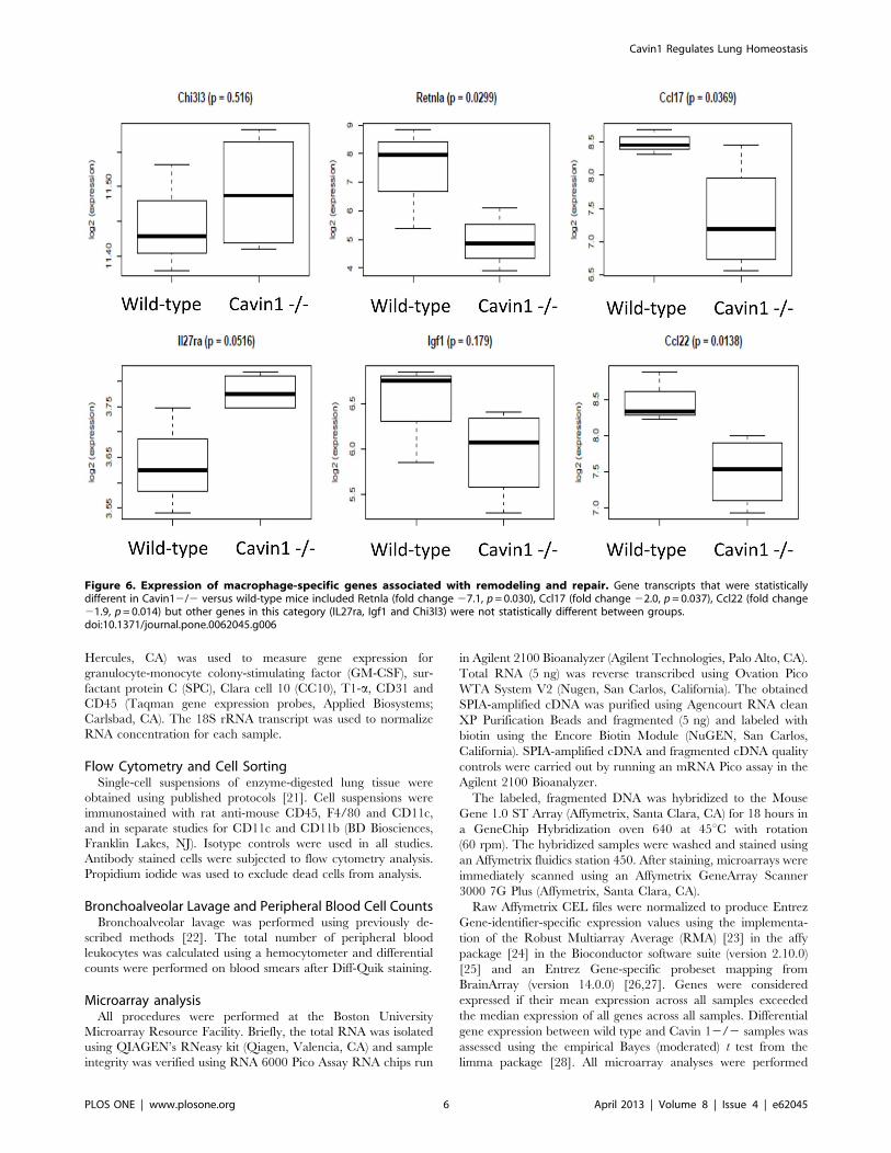
Figure 6. Expression of macrophage-specific genes associated with remodeling and repair. Gene transcripts that were statisticallydifferent in Cavin12/2 versus wild-type mice included Retnla (fold change 27.1, p= 0.030), Ccl17 (fold change 22.0, p= 0.037), Ccl22 (fold change21.9, p= 0.014) but other genes in this category (IL27ra, Igf1 and Chi3l3) were not statistically different between groups.doi:10.1371/journal.pone.0062045.g006
Cavin1 Regulates Lung Homeostasis
PLOS ONE | www.plosone.org 6 April 2013 | Volume 8 | Issue 4 | e62045

Figure 7. Gene Set Enrichment Analysis (GSEA). GSEA performed to identify Gene Ontology (GO) terms or Biocarta, KEGG or Reactomepathways that were differentially regulated between alveolar macrophages of wild-type and Cavin12/2 mice. Of the total 14 gene sets significantlyover-represented (FDR-corrected p,0.05) in Cavin1-deficient mice six of these sets, which are represented in this figure, were related to proliferativeprocesses, such as mitosis, DNA replication, and homologous recombination, and all were coordinately down-regulated in the Cavin12/2 mice.doi:10.1371/journal.pone.0062045.g007
Cavin1 Regulates Lung Homeostasis
PLOS ONE | www.plosone.org 7 April 2013 | Volume 8 | Issue 4 | e62045

using the R environment for statistical computing (version 2.12.0)
[29,30].
Gene Set Enrichment Analysis (GSEA)GSEA was used to identify biological terms, pathways and
processes that were coordinately up- or down-regulated with
respect to various comparisons [31]. A list of all Entrez Gene
identifiers interrogated by the array was ranked according to the
moderated t statistic computed between the wild-type and Cavin
12/2 samples, and this list was then used to perform a pre-
ranked GSEA analysis using the Entrez Gene versions of the
Biocarta, KEGG, Reactome, and Gene Ontology (GO) gene sets
obtained from the Molecular Signatures Database (MSigDB),
version 3.0 [32].
Statistical AnalysisStatistics were performed using GraphPad Prism 5.0 software.
Comparisons between groups were performed using a 2-tailed
unpaired Student t test or Mann Whitney U test. To control for
multiple comparisons in GSEA analysis, Benjamini-Hochberg-
False Discovery Rate (FDR) correction was applied. Significance
of correlation was determined using the Pearson’s test.
Results
Immunohistochemistry was performed on frozen sections in
order to localize Cavin1 in lung tissue. Staining demonstrated
Cavin1 to be absent from proximal airway epithelium but
abundantly expressed on large and small blood vessel endothelium
and throughout distal airspaces (brown stain, Fig. 1). High power
images of distal airspaces further localized Cavin1 protein to
alveolar macrophages and type I epithelial cells, but not to
cuboidal type II epithelial cells (Fig. 1C-E). As expected, staining
was not detectable in the lungs of Cavin12/2 mice (Fig. 1B).
Given the abundance of Cavin1 in lung, we investigated the
functional role for this protein by performing detailed analyses of
lungs from Cavin12/2 mice. Physiological measurements dem-
onstrated increased airways resistance and lung elastance in
Cavin12/2 mice (Fig. 2A), but the most dramatic finding came
from histological analyses, which showed markedly altered distal
lung morphology and significant thickening of the lung inter-
stitium in Cavin12/2 mice (Fig. 2B - upper and middle images).
Trichrome staining illustrated that lung interstitial thickening was
associated with an increase in collagen deposition (Fig. 2B – lower
images). Moreover, there were increased numbers of nuclei
detected within the lung interstitium of Cavin12/2 mice after
staining with hematoxylin (Fig. 2B – middle image), and this
hypercellular phenotype was consistent with the observed increase
in dry weight and DNA content in lungs of Cavin12/2 mice
(Fig. 2C).
To evaluate whether hypercellularity was attributable to
increases in specific cell populations, quantitative RT-PCR was
performed to screen for alterations in endothelial, epithelial and
leukocyte-specific markers in the lungs of wild-type and Cavin12/
2 mice (Fig. 3A). We did not identify any differences in transcript
levels for the proximal epithelial gene CC-10 or the distal type II
epithelial cell marker SP-C; however, non-significant increases
were observed in levels of the endothelial marker CD31 and the
distal type I epithelial marker T1a. Most notably, we found that
transcript levels for the hematopoietic marker CD45 were
significantly increased in lungs of Cavin12/2 mice.
Since differences in CD45 transcript levels were largely
unexpected, we sought to confirm whether the number of
leukocytes was in fact increased in the lungs of Cavin12/2 mice.
Antibody staining directed against CD45 identified increased
numbers of leukocytes in histological sections as did both flow
cytometry (CD45+ cells in digested lungs were 15% +/2 SEM
1.3% in wild-type mice and 27% +/21.4% SEM in Cavin12/2
mice, p,0.05) and Western blot analysis from digested lungs of
Cavin12/2 mice (Fig. 3B). Surprisingly, immunophenotyping in
tissue sections did not identify differences in cells expressing T-cell
(CD3), B-cell (B220), NK cells (NK1.1) or granulocyte (Gr-1)
specific markers (data not shown). However, significant increases
were observed in cells expressing the macrophage-marker Mac3 in
lungs of Cavin12/2 mice (Fig. 4). This selective increase in
Figure 8. Western blot analysis for granulocyte-macrophagecolony-stimulating factor (GM-CSF) in lungs from wild-typeand Cavin12/2 mice (same polyvinylidene difluoride (PVDF)membrane used in figure 3.doi:10.1371/journal.pone.0062045.g008
Figure 9. CCL2 and CX3CL1 are increased in the lungs of Cavin12/2 mice. Enzyme-linked immunosorbent assay for CCL2 and CX3CL1 inlungs of wild-type and Cavin12/2 mice (n = 4, each group, two replicates).doi:10.1371/journal.pone.0062045.g009
Cavin1 Regulates Lung Homeostasis
PLOS ONE | www.plosone.org 8 April 2013 | Volume 8 | Issue 4 | e62045

Mac3+ cells was also associated with a 3-fold rise in macrophages
in BAL fluid of Cavin12/2 mice (9.7+/213 x104 cells/lung wild-
type versus 23.7+/244 x104 cells/lung Cavin12/2 mice, n= 5
each group). Importantly, similar increases were not observed in
the number of mononuclear cells in the circulation of Cavin12/2
mice (3996+/2247 cells/mm3 wild-type versus 3899+/2436
cells/mm3 Cavin12/2 mice, n = 5 each group).
After confirming Cavin1 transcript expression in freshly isolated
alveolar macrophages (data not shown) and Cavin1 protein
expression in multiple macrophage cell lines (Fig. 5A), we
evaluated whether deficiency in Cavin1 was associated with
qualitative differences in lung macrophages. Flow cytometry
identified a trend toward increased F4/80+ CD11c+ cells in lung
digests of Cavin12/2 mice, but this did not reach statistical
significance (Fig. 5B). However, in separate studies, clear
differences were observed in the morphological characteristics of
macrophages recovered from lung digests during forward and side-
scatter analysis. As shown in Figure 5C, resident CD11c+macro-
phages and non-resident CD11b+monocyte/macrophage popula-
tions from lung digests of Cavin12/2 mice were on average
larger (forward scatter) when compared to those from wild-type
mice.
In addition to changes in physical characteristics, microarray
analysis demonstrated measurable differences in gene expression
between alveolar macrophages (isolated by BAL) from wild-type
and Cavin12/2 mice. The expression of 1393 genes was
significantly (nominal p,0.05) altered by Cavin1 deficiency (more
genes than would be expected by chance). Surprisingly, M1 and
M2 gene transcripts were either expressed at low levels or did not
change significantly (p.0.05) between wild-type and Cavin12/2
cells (data not shown); however, several macrophage-specific genes
associated with remodeling and repair were significantly down-
regulated in the Cavin 12/2 mice (Fig. 6) [33].
Gene Set Enrichment Analysis (GSEA) was then performed to
identify Gene Ontology (GO) terms or Biocarta, KEGG or
Reactome pathways that were differentially regulated between
alveolar macrophages of wild-type and Cavin12/2 mice. A total
of 14 gene sets were significantly over-represented (FDR-corrected
p,0.05) among genes that were up- or down-regulated in Cavin1-
deficient mice (Table S1). Six of these sets were related to
proliferative processes, such as mitosis, DNA replication, and
homologous recombination, and were coordinately down-regulat-
ed in the Cavin12/2 mice (Fig. 7). Genes that were frequently
represented among these gene sets included many key mediators of
cell cycle control, especially the Mcm (minichromosome mainte-
nance complex), Cdc (cell division cycle), Orc (origin recognition
complex) and Rfc (replication factor C) family members (Fig. 7).
Given that coordinate down-regulation of mitotic genes was
observed in macrophages from Cavin12/2 mice, we evaluated
whether these cells displayed any differences in proliferation
Figure 10. CD45 and Mac3 staining of tissues from wild-type, Cavin12/2 and Cav12/2 mice. A) Representative sections from CD45stained lung and adipose tissue from wild-type, Cavin12/2 and Cav12/2 mice (n = 4, all groups). B) Results of morphometric analyses on Mac3stained lung and adipose tissue sections from wild-type, Cavin12/2 and Cav12/2 mice (n = 4, all groups). Differences in Mac3+ cells were notobserved in liver, kidney or skeletal muscle in either group (data not shown).doi:10.1371/journal.pone.0062045.g010
Cavin1 Regulates Lung Homeostasis
PLOS ONE | www.plosone.org 9 April 2013 | Volume 8 | Issue 4 | e62045

relative to the wild-type macrophages. There was no difference in
proliferation as measured by PCNA staining, and consistent with
the microarray analysis, we detected significant decreases in both
transcript (data not shown) and protein levels (Fig. 8) of the
macrophage proliferation cytokine GM-CSF. Interestingly, these
findings suggest that maturation and function of alveolar
macrophages is likely to be impaired by GM-CSF deficiency in
Cavin12/2 mice.
Since macrophage gene expression and PCNA labeling
suggested that increased proliferation was unlikely to explain
macrophage accumulation in Cavin12/2 mice, we sought to
evaluate whether other mechanisms, such as increased recruitment
may have contributed to these findings. Apoptotic cell debris is
well-recognized as an important stimulus for macrophage re-
cruitment, but TUNEL staining failed to detect evidence for
increased apoptotic cell death in unchallenged lungs from
Cavin12/2 mice (data not shown). However, modest but
statistically significant increases were detected in the levels of
macrophage chemotactic factors CCL2 and CX3CL1 in BAL
fluid from Cavin12/2 mice (Fig. 9). Together, these findings
suggest that increased recruitment may contribute, at least in small
part, to macrophage accumulation in lungs of Cavin12/2 mice.
Finally, to evaluate whether alterations in macrophage homeo-
stasis were specific to either the lung or to Cavin1 deficiency,
immunostaining for leukocyte antigens CD45 and Mac3 was
performed in multiple tissues from wild-type, Cavin12/2 and
Cav12/2 mice. Increased staining for CD45+ and Mac3+ cells
was observed in lung and adipose tissue (Fig. 10), but not in liver,
kidney or skeletal muscle from knock-out mice (data not shown).
These findings highlight the importance of Cavin1 and Cav1 in
regulating macrophage homeostasis but suggest this property is
restricted to selective tissues (e.g. lung and adipose tissue).
Discussion
Caveolae are prominent morphological features of lung
endothelium and epithelium that are important for respiratory
function [34]. These properties are underscored by the dysfunc-
tional phenotype of the Cav1 deficient mouse that lacks caveolae,
shows thickening of the alveolar septa and respiratory functional
defects [4]. Interestingly, the Cav2 deficient mouse, a protein
thought of as the structural partner for Cav1 [35] exhibits a similar
pulmonary phenotype as does the Cav1 deficient animal, but it
shows no loss of caveolar structure [5]. With this context in mind,
we examined, for the first time, the pulmonary phenotype of the
Cavin1 knockout mouse and showed that Cavin1 is critical for
regulating lung homeostasis and that its deficiency leads to altered
structure and function of the lung. Most notable among these
changes were physiological (e.g. increased elastance) and bio-
chemical alterations (e.g. increased extracellular matrix) as well as
marked hypercellularity. While collectively these pathological
findings complement those described for lungs of Cav1 and
Cav22/2 mice, unique to this investigation was the observation
that leukocytes rather than structural cells contributed to lung
hypercellularity [3–5].
In previous reports, hypercellularity in lungs of Cav12/2 mice
was attributed to increases in Flk1+ ‘‘endothelial progenitors’’ [4].
Interestingly, upon detailed review of these publications, Flk1+cells were noted to possess morphological characteristics atypical
for endothelial cells (e.g. small and round) and found to reside in
locations similar to the CD45+ cells in our study [4]. These
findings, along with reports demonstrating low-level expression of
Flk1 on macrophages, support the notion that at least a subset of
‘‘endothelial progenitors’’ may in fact be immune cells [36–38].
Detailed characterization of lung leukocytes in Cavin12/2
mice found that increased cellularity was largely attributable to
macrophage infiltration, an event that has also been reported in
adipose tissue of mice lacking caveolae [39]. However, along with
changes in cell numbers, we also found that Cavin1 deficiency led
to a significantly altered lung macrophage phenotype. This was
most evident when comparing cell size during flow cytometry
analysis, but other qualitative changes were readily apparent
during gene expression profiling.
Despite identifying significant changes in macrophage gene
expression, we found that macrophage expression profiles did not
fit into classic M1 or M2 paradigms [33]. These findings are in
contrast to a recent study showing up-regulation of M2-related
genes in adipose-tissue derived macrophages from Cav12/2 mice
[39]. These differences between studies are multi-factorial and
likely relate to tissue, macrophage and structural protein related
factors. Another notable finding from our microarray analysis was
the observation that genes associated with repair and remodeling
were down-regulated in macrophages from Cavin12/2 mice.
Given that extracellular matrix deposition appeared to be
increased in the lungs of Cavin12/2 mice, we speculate that
down-regulation of these genes may be an adaptive response to the
already remodeled architecture in lungs these mice. Moreover,
these findings suggest that macrophages do not significantly
contribute to baseline fibrosis in these mice.
The mechanisms leading to lung macrophage accumulation in
Cavin12/2 mice cannot as yet be defined precisely. However,
increased numbers of macrophages in the distal airspaces of
Cavin12/2 mice indicated that cells were unlikely to be simply
trapped in the thickened interstitium of the lung. Additionally,
normal circulating numbers of mononuclear cells illustrated that
systemic alterations in hematopoiesis were unlikely to have
contributed significantly. Findings to suggest macrophage accu-
mulation in Cavin12/2 mice was due to increased cellular
recruitment were based on increased levels of macrophage
chemotactic factors CCL2 and CX3CL1 in BAL fluid from
Cavin12/2 mice. However, because increases in these chemo-
kines were quite modest, it seems unlikely that this mechanism
entirely explains the macrophage accumulation in lungs Ca-
vin12/2 mice. With this in mind, we speculate that impaired
egress could also play a role [40,41] since leukocytes utilize
caveolae on endothelial cell surfaces to enter and to exit tissues
[42].
While numerous reports describe a role for caveolae in
suppressing cell proliferation [3,43,44], the findings in our study
(i.e. absence of PNCA labeling and down-regulation of cell cycle
related genes) indicate that increased proliferation was not a major
contributor to macrophage accumulation in Cavin12/2 mice.
Although absence of proliferation was unexpected from Cavin1
deficiency, these findings are, however, consistent with published
studies on macrophages, which indicate these cells do not
proliferate in the lung under basal, non-stressed conditions [22].
Another important finding in this study was the observation that
transcript and protein levels for GM-CSF were markedly de-
creased in lungs from Cavin12/2 mice. GM-CSF is a 23 kD
glycoprotein that functions to promote monocyte growth and
differentiation but acts in lung to regulate not only macrophage
number and phenotype but also various critical functions such as
surfactant phospholipid catabolism and the clearance of pathogens
from the lung. [45–47] Although our study did not go on to
evaluate whether GM-CSF replacement therapy restores macro-
phage number and phenotype in lungs from Cavin12/2 mice, or
for that matter in adipose tissue, we speculate that its deficiency is
likely to play a mechanistic role in these findings.
Cavin1 Regulates Lung Homeostasis
PLOS ONE | www.plosone.org 10 April 2013 | Volume 8 | Issue 4 | e62045

Finally, while the focus of this paper was to investigate cavin1’s
role in the lung, it is worth emphasizing that alterations in
macrophage homeostasis were not restricted to this tissue. The
reasons for the selective increase in macrophages in lung and
adipose tissue are not obvious, but it seems logical to assume that
characteristics shared by these organs may explain the abnormal
macrophage accumulation in these tissues. Notably, one feature
shared by the lung and adipose tissue is their role in regulating
lipid regulation. Adipose tissue stores and releases lipids in
response to metabolic cues and lipid biosynthesis and clearance
are critical for maintaining the functional characteristics of the
lung’s surface lining fluid. Thus, we postulate, that altered lipid
metabolism may be a factor promoting macrophage accumulation
in lung and adipose tissue; a hypothesis supported by decreased
GM-CSF levels in lungs from Cavin12/2 mice [45–47].
In conclusion, this study is the first to describe a role for Cavin1
in the lung. Our findings indicate that targeted deletion of Cavin1
leads to a complex lung phenotype that includes alterations in
respiratory function as well as changes to lung macrophage
number and phenotype. We believe these findings expand our
understanding of the lung and implicate caveolar structural
proteins in the regulation of lung macrophage homeostasis.
Supporting Information
Table S1 Gene sets with significant enrichment in geneswhose expression is altered in Cavin 12/2 macro-phages. Gene Set Enrichment Analysis (GSEA) was performed
on 1,625 gene sets from Gene Ontology, KEGG, Reactome, and
Biocarta, and 14 of these gene sets were found to be significantly
enriched (FDR q ,0.05) among genes whose expression was up-
or down-regulated in Cavin 12/2 versus wild-type macrophages.
(DOCX)
Acknowledgments
We would like to thank Dr. William Cruikshank for his assistance in
performing physiological measurements and his thoughtful critiques of the
manuscript.
Author Contributions
Conceived and designed the experiments: PG SD LL PP RS. Performed
the experiments: PG SD LL EB SSA RS FR DS JP. Analyzed the data: PG
PP RS AG EB FR JP. Contributed reagents/materials/analysis tools: PP
RS JP FR. Wrote the paper: PG AG PP RS FR.
References
1. Gil J (1983) Number and distribution of plasmalemmal vesicles in the lung. Fed
Proc 42: 2414–2418.
2. Razani B, Woodman SE, Lisanti MP (2002) Caveolae: from cell biology to
animal physiology. Pharmacol Rev 54: 431–467.
3. Razani B, Engelman JA, Wang XB, Schubert W, Zhang XL, et al. (2001)
Caveolin-1 null mice are viable but show evidence of hyperproliferative and
vascular abnormalities. The Journal of biological chemistry 276: 38121–38138.
4. Drab M, Verkade P, Elger M, Kasper M, Lohn M, et al. (2001) Loss of caveolae,
vascular dysfunction, and pulmonary defects in caveolin-1 gene-disrupted mice.
Science 293: 2449–2452.
5. Razani B, Wang XB, Engelman JA, Battista M, Lagaud G, et al. (2002)
Caveolin-2-deficient mice show evidence of severe pulmonary dysfunction
without disruption of caveolae. Mol Cell Biol 22: 2329–2344.
6. Park DS, Woodman SE, Schubert W, Cohen AW, Frank PG, et al. (2002)
Caveolin-1/3 double-knockout mice are viable, but lack both muscle and non-
muscle caveolae, and develop a severe cardiomyopathic phenotype. Am J Pathol
160: 2207–2217.
7. Woodman SE, Park DS, Cohen AW, Cheung MW, Chandra M, et al. (2002)
Caveolin-3 knock-out mice develop a progressive cardiomyopathy and show
hyperactivation of the p42/44 MAPK cascade. The Journal of biological
chemistry 277: 38988–38997.
8. Razani B, Combs TP, Wang XB, Frank PG, Park DS, et al. (2002) Caveolin-1-
deficient mice are lean, resistant to diet-induced obesity, and show hypertrigly-
ceridemia with adipocyte abnormalities. The Journal of biological chemistry
277: 8635–8647.
9. Liu L, Brown D, McKee M, Lebrasseur NK, Yang D, et al. (2008) Deletion of
Cavin/PTRF causes global loss of caveolae, dyslipidemia, and glucose
intolerance. Cell Metab 8: 310–317.
10. Hayashi YK, Matsuda C, Ogawa M, Goto K, Tominaga K, et al. (2009) Human
PTRF mutations cause secondary deficiency of caveolins resulting in muscular
dystrophy with generalized lipodystrophy. The Journal of clinical investigation
119: 2623–2633.
11. Shastry S, Delgado MR, Dirik E, Turkmen M, Agarwal AK, et al. (2010)
Congenital generalized lipodystrophy, type 4 (CGL4) associated with myopathy
due to novel PTRF mutations. Am J Med Genet A 152A: 2245–2253.
12. Le Lay S, Kurzchalia TV (2005) Getting rid of caveolins: phenotypes of
caveolin-deficient animals. Biochim Biophys Acta 1746: 322–333.
13. Briand N, Dugail I, Le Lay S (2011) Cavin proteins: New players in the caveolae
field. Biochimie 93: 71–77.
14. Voldstedlund M, Vinten J, Tranum-Jensen J (2001) cav-p60 expression in rat
muscle tissues. Distribution of caveolar proteins. Cell Tissue Res 306: 265–276.
15. Vinten J, Voldstedlund M, Clausen H, Christiansen K, Carlsen J, et al. (2001) A
60-kDa protein abundant in adipocyte caveolae. Cell Tissue Res 305: 99–106.
16. Jansa P, Mason SW, Hoffmann-Rohrer U, Grummt I (1998) Cloning and
functional characterization of PTRF, a novel protein which induces dissociation
of paused ternary transcription complexes. Embo J 17: 2855–2864.
17. Vinten J, Johnsen AH, Roepstorff P, Harpoth J, Tranum-Jensen J (2005)
Identification of a major protein on the cytosolic face of caveolae. Biochim
Biophys Acta 1717: 34–40.
18. Morgan RK, McAllister B, Cross L, Green DS, Kornfeld H, et al. (2007)
Histamine 4 receptor activation induces recruitment of FoxP3+ T cells and
inhibits allergic asthma in a murine model. Journal of immunology 178: 8081–
8089.
19. Konter JM, Parker JL, Baez E, Li SZ, Ranscht B, et al. (2012) Adiponectin
attenuates lipopolysaccharide-induced acute lung injury through suppression of
endothelial cell activation. J Immunol 188: 854–863.
20. Inselman LS, Wapnir RA, Spencer H (1987) Obesity-induced hyperplastic lunggrowth. Am Rev Respir Dis 135: 613–616.
21. Summer R, Fiack CA, Ikeda Y, Sato K, Dwyer D, et al. (2009) Adiponectin
deficiency: a model of pulmonary hypertension associated with pulmonaryvascular disease. Am J Physiol Lung Cell Mol Physiol 297: L432–438.
22. Murphy J, Summer R, Wilson AA, Kotton DN, Fine A (2008) The prolonged
life-span of alveolar macrophages. American journal of respiratory cell andmolecular biology 38: 380–385.
23. Irizarry RA, Hobbs B, Collin F, Beazer-Barclay YD, Antonellis KJ, et al. (2003)
Exploration, normalization, and summaries of high density oligonucleotide arrayprobe level data. Biostatistics 4: 249–264.
24. Gautier L, Cope L, Bolstad BM, Irizarry RA (2004) affy–analysis of Affymetrix
GeneChip data at the probe level. Bioinformatics 20: 307–315.
25. Gentleman RC, Carey VJ, Bates DM, Bolstad B, Dettling M, et al. (2004)Bioconductor: open software development for computational biology and
bioinformatics. Genome Biol 5: R80.
26. Dai M, Wang P, Boyd AD, Kostov G, Athey B, et al. (2005) Evolving gene/transcript definitions significantly alter the interpretation of GeneChip data.
Nucleic Acids Res 33: e175.
27. University of Michigan Molecular and Behavioral Neuroscience Institute:Microarray Lab.
28. Smyth GK, editor (2005) Limma: linear models for microarray data. In:
’Bioinformatics and Computational Biology Solutions using R and Bioconduc-tor’. New York: Springer.
29. R Development Core Team (2011) R: A language and environment for
statistical computing. Vienna, Austria: R Foundation for Statistical Computing.
30. The R Project for Statistical Computing.
31. Subramanian A, Tamayo P, Mootha VK, Mukherjee S, Ebert BL, et al. (2005)
Gene set enrichment analysis: a knowledge-based approach for interpreting
genome-wide expression profiles. Proc Natl Acad Sci U S A 102: 15545–15550.
32. Subramanian A, Kuehn H, Gould J, Tamayo P, Mesirov JP (2007) GSEA-P:
a desktop application for Gene Set Enrichment Analysis. Bioinformatics 23:
3251–3253.
33. Mosser DM, Edwards JP (2008) Exploring the full spectrum of macrophage
activation. Nat Rev Immunol 8: 958–969.
34. Gosens R, Mutawe M, Martin S, Basu S, Bos ST, et al. (2008) Caveolae and
caveolins in the respiratory system. Curr Mol Med 8: 741–753.
35. Cohen AW, Hnasko R, Schubert W, Lisanti MP (2004) Role of caveolae and
caveolins in health and disease. Physiol Rev 84: 1341–1379.
36. Sharifi BG, Zeng Z, Wang L, Song L, Chen H, et al. (2006) Pleiotrophin inducestransdifferentiation of monocytes into functional endothelial cells. Arterioscler
Thromb Vasc Biol 26: 1273–1280.
37. Jeon SH, Chae BC, Kim HA, Seo GY, Seo DW, et al. (2007) Mechanismsunderlying TGF-beta1-induced expression of VEGF and Flk-1 in mouse
macrophages and their implications for angiogenesis. J Leukoc Biol 81: 557–566.
38. Yang ZF, Poon RT, Luo Y, Cheung CK, Ho DW, et al. (2004) Up-regulation ofvascular endothelial growth factor (VEGF) in small-for-size liver grafts enhances
Cavin1 Regulates Lung Homeostasis
PLOS ONE | www.plosone.org 11 April 2013 | Volume 8 | Issue 4 | e62045

macrophage activities through VEGF receptor 2-dependent pathway. Journal of
immunology 173: 2507–2515.39. Briand N, Le Lay S, Sessa WC, Ferre P, Dugail I (2011) Distinct roles of
endothelial and adipocyte caveolin-1 in macrophage infiltration and adipose
tissue metabolic activity. Diabetes 60: 448–453.40. Gerszten RE, Tager AM (2012) The monocyte in atherosclerosis–should I stay
or should I go now? N Engl J Med 366: 1734–1736.41. van Gils JM, Derby MC, Fernandes LR, Ramkhelawon B, Ray TD, et al. (2012)
The neuroimmune guidance cue netrin-1 promotes atherosclerosis by inhibiting
the emigration of macrophages from plaques. Nat Immunol 13: 136–143.42. Millan J, Hewlett L, Glyn M, Toomre D, Clark P, et al. (2006) Lymphocyte
transcellular migration occurs through recruitment of endothelial ICAM-1 tocaveola- and F-actin-rich domains. Nat Cell Biol 8: 113–123.
43. Torres VA, Tapia JC, Rodriguez DA, Parraga M, Lisboa P, et al. (2006)Caveolin-1 controls cell proliferation and cell death by suppressing expression of
the inhibitor of apoptosis protein survivin. J Cell Sci 119: 1812–1823.
44. Galbiati F, Volonte D, Liu J, Capozza F, Frank PG, et al. (2001) Caveolin-1
expression negatively regulates cell cycle progression by inducing G(0)/G(1)
arrest via a p53/p21(WAF1/Cip1)-dependent mechanism. Mol Biol Cell 12:
2229–2244.
45. Dranoff G, Crawford AD, Sadelain M, Ream B, Rashid A, et al. (1994)
Involvement of granulocyte-macrophage colony-stimulating factor in pulmonary
homeostasis. Science 264: 713–716.
46. Huffman JA, Hull WM, Dranoff G, Mulligan RC, Whitsett JA (1996)
Pulmonary epithelial cell expression of GM-CSF corrects the alveolar proteinosis
in GM-CSF-deficient mice. J Clin Invest 97: 649–655.
47. Uchida K, Beck DC, Yamamoto T, Berclaz PY, Abe S, et al. (2007) GM-CSF
autoantibodies and neutrophil dysfunction in pulmonary alveolar proteinosis.
N Engl J Med 356: 567–579.
Cavin1 Regulates Lung Homeostasis
PLOS ONE | www.plosone.org 12 April 2013 | Volume 8 | Issue 4 | e62045
Copyright © 2022 FDOKUMEN