The Short Non-Coding Transcriptome of the Protozoan Parasite Trypanosoma cruzi
Upload
independentCategory
view
1download
0
PE
CJa
b
c
d
a
ARRA
KTNCCE
1
clCdicScmo
hed
1d
Spectrochimica Acta Part A 79 (2011) 312–319
Contents lists available at ScienceDirect
Spectrochimica Acta Part A: Molecular andBiomolecular Spectroscopy
journa l homepage: www.e lsev ier .com/ locate /saa
otent 5-nitrofuran derivatives inhibitors of Trypanosoma cruzi growth:lectrochemical, spectroscopic and biological studies
. Maria Aravenaa, A. Claudio Oleaa,∗, Hugo Cerecettob, Mercedes Gonzálezb,uan Diego Mayac, Jorge Rodríguez-Becerrad
Departamento de Química Inorgánica y Analítica, Facultad de Ciencias Quimicas y Famacéuticas, Universidad de Chile, Olivos 1007, Independencia, Santiago, ChileGrupo de Química Medicinal, Laboratorio de Química Orgánica, Facultad de Ciencias-Facultad de Química, Universidad de la Republica, Montevideo, UruguayFacultad de Medicina, ICBO, Programa de Farmacología Clínica y Molecular, Universidad de Chile, Santiago, ChileDepartamento de Química, Facultad de Ciencias Básicas, Universidad Metropolitana de Ciencias de la Educación, Santiago, Chile
r t i c l e i n f o
rticle history:eceived 3 September 2010eceived in revised form 26 January 2011ccepted 7 February 2011
eywords:. cruziitrofuranytotoxicity
a b s t r a c t
Cyclic voltammetry and electron spin resonance techniques were used in the investigation of severalpotential antiprotozoal containing thiosemicarbazone and carbamate nitrofurans. In the electrochemicalbehaviour, a self-protonation process involving the nitro group was observed. The reactivity of the nitroanion radical for these derivatives with glutathione, a biological relevant thiol, was also studied in meansof cyclic voltammetry. These studies demonstrated that glutathione could react with radical species from5-nitrofuryl system. Furthermore, from the voltammetric results, some parameters of biological signifi-cance as E1
7 (indicative of the biological nitro anion radical formation), and KO2 (thermodynamic indicatorthe of oxygen redox cycling) have been calculated. We also evaluated the stability of the nitro anion radi-
yclic voltammetrySR
cal in terms of the dimerization constant (kd). The nitrofuran-free radicals from cyclic voltammetry werecharacterized by electron spin resonance. A clear dependence between both the thiosemicarbazone orcarbamate substructure and the length of the linker, furyl- or furylpropenyl-spacer, and the delocalizationof the unpaired electron was observed.
Through of biological assays we obtained important parameters that account for the selective anti-trypanosomal activity of these derivatives. The trypomastigote viability study showed that all derivatives
masti
are as active as in the epi. Introduction
According to WHO, infectious and parasitic diseases are majorauses of human diseases worldwide [1,2]. Together with malaria,eishmaniasis, and African trypanosomiasis (sleeping sickness),hagas’ disease (American trypanosomiasis) is a major cause ofeath and hardship in the impoverished regions of the develop-
ng world. It is the largest parasitic disease burden in the Americanontinent being endemic in 21 countries from southern Unitedtates to Argentine and Chile. The morbidity and mortality asso-iated with this disease in America are more than one order ofagnitude higher than those caused by malaria, schistosomiasis
r leishmaniasis [1–3].
An important number of pharmacological active compoundsave a nitro aromatic group in their molecular structures, whichxplains their biological activity [4,5]. Due to the ability of theserugs to be reduced at the nitro group, they are metabolized to the
∗ Corresponding author. Tel.: +56 2 9782834; fax: +56 2 7370567.E-mail address: [email protected] (A. Claudio Olea).
386-1425/$ – see front matter © 2011 Elsevier B.V. All rights reserved.oi:10.1016/j.saa.2011.02.007
gote form of the parasite in a doses dependent manner.© 2011 Elsevier B.V. All rights reserved.
corresponding amines via nitro radical, nitroso intermediates andhydroxylamine. Nevertheless, it is known that the biological activ-ity of nitro compounds is not due to the final product of reduction,but to the formation of intermediates, generally free radicals [6].No medication has been proven to be effective once the diseasehas progressed to later stages. Moreover, this pathology is treatedwith synthetic drugs such as Nifurtimox® (Nfx) and Benznidazole®
(Bnz), and two nitro-containing heterocycles. The chemotherapy isstill inadequate due to its undesirable side effects, including cardiacand/or renal toxicity. This explains the constant need for discover-ing and to investigate new and effective agents against Trypanosomecruci (T. cruzi) [7,8].
Early experiments carried out on one of the main drug usedin Chagas treatment, Nfx, suggest that intracellular reduction fol-lowed by redox cycling yielding reactive oxygen species (ROS) maybe their major mode of action against T. cruzi. These can cause
cellular damage directly by reacting with various biological macro-molecules, or indirectly by the generation of the highly reactivehydroxyl radical via Haber–Weis and Fenton reactions [9,10]. But,recently we demonstrated that ROS are not produced into the T.cruzi mainly a bioreductive process is involved [11]. Trypanothione
C. Maria Aravena et al. / Spectrochimica Acta Part A 79 (2011) 312–319 313
Table 1Studied anti-T. cruzi derivatives.
(b) (a)
()nN
+
O-
O
O
NNH
S
NH
CH2
ON
NH
O
O
N+
O-
O
()n
.
n Nitrofuran derivatives
(a) Nitro-thiosemicarbazone (b) Nitro-carbamates
0 5-NT1 5-NC1
1 5-NT2 5-NC2
-0,6-0,8-1,0-1,2-1,4
IVaIIIa
IIIcIVc
0.4 µA
E/ V
Vc
0,0-0,5-1,0-1,5-2,0
IVa
Vc IVcIIIcIIcIc
E/ V
1 µAIa
-0,6-0,8-1,0-1,2-1,4-1,6
IIIa
IVa
Vc
E/ V
0.2 µA
IIIc
b
c
a
F clic v0 ide lin0 ccesso sh line
rtdtts“Bitpri
TC
ig. 1. Example of cyclic voltammograms of 5-NT1 in DMSO, with 0.1 M TBAP, (a) cy.1 and 2.0 V/s. (b) Cyclic voltammogram using a sweep rate of 2.0 V/s: (i) solid w.5 mM; and (iii) dash line, derivative 5-NT1 in the presence of NaOH 1.0 mM. (c) Suf 2.0 V/s: (i) solid wide line, first cycle; (ii) solid thin line, second cycle; and (iii) da
eductase (TR) constitutes an attractive target for chemotherapeu-ic research in relation to Chagas’ and other trypanosomatid-causediseases. The two main notions for this are specificity and essen-iality. In effect, TR has not been found in the human host andhere are evidences suggesting that it is essential for parasiteurvival [12–14]. Many nitrofurans were previously found to besubversive” (turncoat/sabotage inhibitors) substrates of TR [15].esides the characteristic mentioned in the previous paragraph, it
s important to note that for the design of new chemotherapeu-ic alternatives, membrane sterol biosynthesis is one of the mostromising targets. T. cruzi like most pathogenic fungi and yeastsequires specific 24-alkyl sterols for cell viability and proliferationn all stages of its life cycle and cannot use the abundant supply
able 2yclic voltammetric parameters corresponding to the nitro moiety reduction of couple III
Compoundsa EpIIIc (V) EpIIIa (V) �E (V) E1/2
5-NT1 0.75 −0.69 0.06 −0.75-NT2 0.77 −0.69 0.08 −0.75-NC1 0.56 −0.52 0.04 −0.55-NC2 0.60 −0.57 0.03 −0.5Nfx −0.91 −0.85 0.06 −0.8
a Compounds: 1 × 10⁻³ M concentration compounds dissolved in DMSO using excess ob At 2 V/s.
oltammogram using a potential range of −1.4 to −0.4 V and a sweep rates betweene, derivative 5-NT1; (ii) solid thin line, derivative 5-NT1 in the presence of NaOHive sweeps voltammogram using a potential range of −2.0 to 0 V and a sweep rate, thirst cycle.
of cholesterol present in its mammalian hosts [16]. The T. cruziergosterol biosynthesis pathway has been chemically validated asa chemotherapeutic target at several steps [17], including squa-lene epoxidase (SE; EC 1.14.99.7) a microsomal mono-oxygenasethat catalyzes the conversion of squalene to 2,3-oxidosqualeneusing molecular oxygen [16]. Based in this precept we described 5-nitrofuryl containing heterocycles with excellent anti-T. cruzi activ-ities and sterol membrane biosynthesis inhibitory capacity [18,19].
In this paper we selected two of the most active agents from thisfamily of 5-nitrofuryl containing thiosemicarbazone (5-NT) [18]and 5-nitrofuryl containing carbamate (5-NC) [19] (Table 1).
We studied them electrochemically in aprotic solvent usingcyclic voltammetry (CV) technique and examined the interac-
c/IIIa vs saturated calomel electrode en DMSO.
(V) Ipa/Ipcb kd (M−1 s−1) E1
7 (V) KO2 (M−1 s−1)
2 0.61 14.9E+06 −0.149 7.94E+003 1.02 3.11E+05 −0.166 2.36E+004 1.00 0.90E+05 ≈0 5.97E−039 1.10 1.41E+05 −0.020 5.17E−038 1.01 7.23E+06 −0.287 1.7E+02
f PTBA as supporting electrolyte.

314 C. Maria Aravena et al. / Spectrochimica Acta Part A 79 (2011) 312–319
hanis
tgast
2
2
cb((s
2
bwVs
Fsd
Fig. 2. Proposed reduction mec
ion between the radical species generated from 5-NT/5-NC andlutathione (GSH). The nitro anion radical species were char-cterized using electron spin resonance spectroscopy (ESR) andimulation through WINEPR Simfonia. Besides, we tested the anti-ripomastigote activity on DM28 strain.
. Experimental
.1. Samples
Two different structural features were included in the studiedompounds (5-NT1,2 and 5-NC1,2): (1) thiosemicarbazone or car-amate 5-nitrofuryl-substituents (5-NT and 5-NC derivatives) and2) different lengths of the linker, furyl or furylpropenyl-spacern = 0 or n = 1, Table 1). Compounds 5-NT and 5-NC (Table 1) wereynthesized according to methods reported [18,19].
.2. Cyclic voltammetry
Dimethylsulfoxide (DMSO) was obtained from Aldrich. Tetra-utylamonium perchlorate (TBAP), used as supporting electrolyte,as obtained from Fluka. CV was carried out using a Metrohm 693A instrument with a 694 VA Stand convertor and a 693 VA Proces-or, in DMSO (c.a. 1.0 × 10⁻³ mol/L), under a nitrogen atmosphere
-0,6-0,8-1,0-1,2-1,4-1,6
IVa
IIIa
IIIc
E/ V
0.5 µA
Vc
ig. 3. Cyclic voltammogram in DMSO: (a) solid wide line, derivative 5-NT1; (b)olid thin line, derivative 5-NT1 in the presence of GSH 0.5 mM; and (c) dash line,erivative 5-NT1 in the presence of GSH 1.0 mM.
m of studied nitro derivatives.
at room temperature, with TBAP (c.a. 0.1 mol/L), using a three-electrode cell. A hanging mercury drop electrode (HMDE) was usedas the working electrode, a platinum wire as the auxiliary electrode,and saturated calomel (SCE) as the reference.
Using the Nicholson’s procedure [20] the Ipa/Ipc values(HRNO2/HRNO2
•−) was measured from each cyclic voltammogramwith scan rate between 0.1 and 2.0 V/s. Employing the theoreticalapproaches of Olmnested et al. [21] for dimerization, the Ipa/Ipc val-ues were inserted into a working curve to determine the parameterω, which is described by the equation ω = kdC0� where kd is thesecond-order rate constant for the dimerization of RNO2
•−, C0 isthe nitrocompound concentration and � = (E� − E1/2)�, where E� isthe switching potential, E1/2 is the half-wave potential and � is thescan rate.
We used Dimethylformamide (DMF) as a solvent to calculate theE1
7 value using the same wave described from CV in DMSO milieu,according to reference [22].
2.3. ESR spectroscopy
ESR spectra were recorded in the X band (9.85 GHz) using aBruker ECS 106 spectrometer with a rectangular cavity and 50 kHzfield modulation. The hyperfine splitting constants were estimatedto be accurate within 0.05 G. The nitroanion radicals were gener-ated by electrolytic reduction in situ under the same conditionsof temperature, atmosphere and concentrations stated at the CVexperiment. The ESR spectra were simulated using the programWINEPR Simfonia 1.25 version.
2.4. Biological assays
2.4.1. Epimastigote culture and growth inhibition assaysTrypanosoma cruzi Dm28c epimastigotes clone, were cultured,
at an initial density of 3 × 10⁶ parasites/mL, at 28 ◦C in monophasicDiamond’s culture milieu, supplemented with 4 �M Hemin, bovinefetal calf serum at a final concentration of 5% (v/v), and sodium peni-cillin 100 mg/mL and streptomycin 100 mg/mL. The assayed drugswere dissolved in 1% dimethylsulfoxide (DMSO) at variable doses.No toxic effect was attributable to DMSO at the final concentrationof 1%. All measurements were carried out in triplicate. Epimastigotegrowth was followed daily by nephelometry for 7 days. The neph-
elometry measurements were directly proportional to the numberof parasites in suspension.Since parasite growth is exponential, the data were analyzedaccording to a first order equation and the respective growth con-stant (k), corresponding to the slope, was calculated for controls
C. Maria Aravena et al. / Spectrochimica Acta Part A 79 (2011) 312–319 315
Table 3Experimental hyperfine splittings (Gauss) and g values for nitroanion radicals.
.
Compounds aN1 aN2 aN3 aH1 aH2 aH3a aH3b aH4 aH5 aH6 g
ai
2
smm2c
3rp
2ffpp1fvOR
Fr
5-NT1 1.54 6.24 1.57 1.08 1.085-NT2 1.54 6.24 1.57 1.08 1.085-NC1 11.74 0.21 0.21 2.00 1.105-NC2 4.42 0.78 1.20 1.25 2.71
nd parasites treated with different drug concentrations. The ICk50s defined as the drug concentration needed to lower the k by 50%.
.4.2. Cell cultures in vitro model of infection with T. cruziRAW 264.7 cells were cultured at 37 ◦C in humidified atmo-
phere and 5% CO2 at a density of 100,000 cell/cm2 in RPMIilieu supplemented with 0.22% sodium bicarbonate, strepto-ycin 100 mg/mL, penicillin 100 �g/mL and fetal calf serum 5%. At
4 h, infected with metacyclic trypomastigotes of T. cruzi Dm28clone, at reason of three trypomastigotes by macrophages.
After that time, the culture milieu was collected, centrifuged at000 rpm for 5 min, and the trypomastigote-containing pellet wasesuspended in RPMI supplemented with fetal bovine serum 5% andenicillin–streptomycin at a final density of 50 × 106 parasites/mL.
.4.2.1. Cell viability. The cell viability measurement was per-ormed by mitochondrial reduction of tetrazolium salts or MTT toormazan, which has a purple color whose absorbance is directlyroportional to the number of living cells in culture. The com-ounds were tested at five different doses, between 6.25 and
00 �M, besides the blank and controls. All samples were per-ormed in triplicate and from the dose–response curve the IC50alues were extracted. Nfx and Bnz were included in this assay.n culture plates of 96 wells, with a cell culture with 100 �L ofPMI milieu without phenol red and no supplement was added(a)
(a’) [G]
3445 3450 3455 3460 3465 3470 3475 3480 3485 34
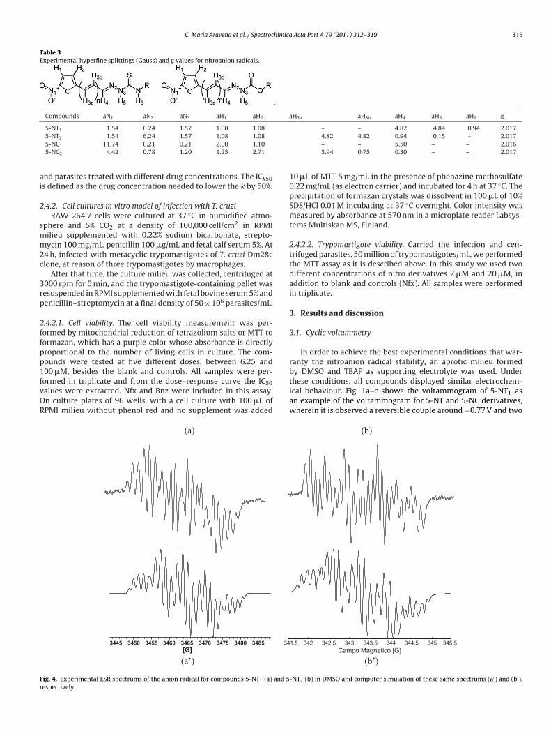
ig. 4. Experimental ESR spectrums of the anion radical for compounds 5-NT1 (a) and 5espectively.
– – 4.82 4.84 0.94 2.0174.82 4.82 0.94 0.15 – 2.017– – 5.50 – – 2.0163.94 0.75 0.30 – – 2.017
10 �L of MTT 5 mg/mL in the presence of phenazine methosulfate0.22 mg/mL (as electron carrier) and incubated for 4 h at 37 ◦C. Theprecipitation of formazan crystals was dissolvent in 100 �L of 10%SDS/HCl 0.01 M incubating at 37 ◦C overnight. Color intensity wasmeasured by absorbance at 570 nm in a microplate reader Labsys-tems Multiskan MS, Finland.
2.4.2.2. Trypomastigote viability. Carried the infection and cen-trifuged parasites, 50 million of trypomastigotes/mL, we performedthe MTT assay as it is described above. In this study we used twodifferent concentrations of nitro derivatives 2 �M and 20 �M, inaddition to blank and controls (Nfx). All samples were performedin triplicate.
3. Results and discussion
3.1. Cyclic voltammetry
In order to achieve the best experimental conditions that war-ranty the nitroanion radical stability, an aprotic milieu formed
by DMSO and TBAP as supporting electrolyte was used. Underthese conditions, all compounds displayed similar electrochem-ical behaviour. Fig. 1a–c shows the voltammogram of 5-NT1 asan example of the voltammogram for 5-NT and 5-NC derivatives,wherein it is observed a reversible couple around −0.77 V and two(b)
(b’)
1.5 342 342.5 343 343.5 344 344.5 345.5345
Campo Magnetico [G]
-NT2 (b) in DMSO and computer simulation of these same spectrums (a′) and (b′),

316 C. Maria Aravena et al. / Spectrochimica Acta Part A 79 (2011) 312–319
(b) (a)
339 339.5 340 340.5 341 341.5 342 342.5 343 343.5Campo Magnetico [G]
339 339.5 340 340.5 341 341.5 342 342.5 343 343.5Campo Magnetico [G]
F and 5r
iIioprattsasisNptT
(a’)
ig. 5. Experimental ESR spectrums of the anion radical for compounds 5-NC1 (a)espectively.
rreversible peaks around −0.84 V and −1.29 V. The first coupleIIc/IIIa (around −0.77 V, Table 2) corresponds to the electrochem-cal reduction of the nitro group with the consequent formationf the nitro-anion radical (HRNO2/HRNO2
•−) and the irreversibleeak (Vc) corresponds to the further reduction of the nitro-anionadical to the corresponding hydroxylamine derivative. In addition,n anodic peak appears around −0.84 V (IVa), which it is related tohe self-protonation reaction (see Fig. 1b). This process correspondso acid–base equilibrium in aprotic milieu, a typical behaviour ofelf-protonation phenomenon displayed by nitro-compounds withcidic moieties in their structures [23–26]. In order to assess theelf-protonation mechanism proposed (see Fig. 2), it was workedn the presence of increasing amounts of aqueous NaOH. Fig. 1b
hows two couples and one irreversible peak in the presence ofaOH, the first one described above (IIIc/IIIa) and the second cou-le, around −0.92 V (IVc/IVa), corresponding to the reduction ofhe anionic molecules generated (−RNO2/−RNO2•−, see Fig. 2).he couple (IIIc/IIIa) and the cathodic peak (Vc) gradually disap-
CONTR
OL
Nfx
/20E
-06M
5-NT1
/2E-0
6M
5-NT1/
20E-0
6M
0
10
20
30
40
50
Try
po
masti
go
tes V
iab
le (
millio
ns
/mL
)
Fig. 6. Trypomastigote viability assay using Dm2
(b’)
-NC2 (b) in DMSO and computer simulation of these same spectrums (a′) and (b′),
peared with increasing amounts of NaOH and appear the secondcouple (IVc/IVa). For this last, it was calculated Ipa/Ipc ratio usingthe Nicholson and Shain equation [20,27], this shows an increaseclose to unity with the addition of NaOH, which confirms the pro-posed mechanism (see Fig. 2). Moreover, Fig. 1c displays that thepeak IVc appears when successive sweeps were used. All studiedcompounds have acidic hydrogen in the side-chains and shown aself-protonation process.
On the other hand, the voltammogram of all compounds exhibitone anodic peak around −0.46 V (Ia), that could be associated to thereoxidation process of the hydroxylamine, obtained in the electro-chemical reduction (Vc), into nitroso derivative (see Fig. 2). Thecounterpart of this process (Ia) is observe in the voltammogram of
successive sweeps (Fig. 1c), which shows a cathodic peak (Ic) relateto the reduction process of the resulting nitroso derivative (coupleIc/Ia). In addition, it is observe a shoulder peak (IIc) which could beattributed to the reduction of the nitroso derivatives which comefrom the anionic molecule.CONTR
OL
Nfx
/20E
-06M
5-NT2
/2E-0
6M
5-NT2/
20E-0
6M
0
10
20
30
40
50
Try
po
masti
go
tes V
iab
le (
millio
ns/m
L)
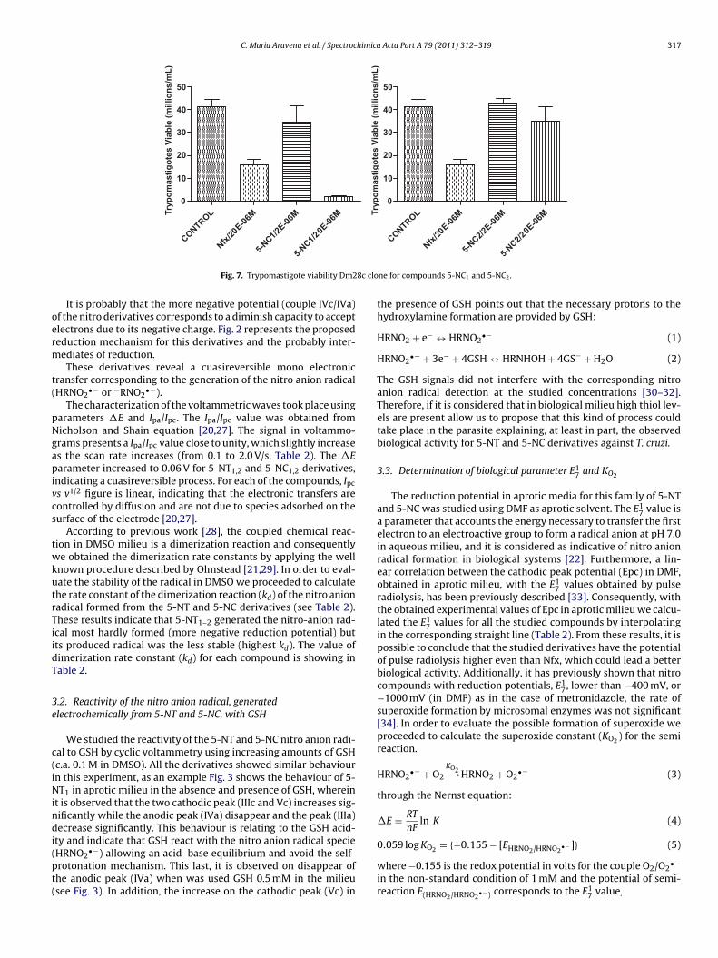
8c clone for compounds 5-NT1 and 5-NT2.
C. Maria Aravena et al. / Spectrochimica Acta Part A 79 (2011) 312–319 317
CONTR
OL
/20E
-06M
1/2E
-06M
/20E
-06M
0
10
20
30
40
50
Try
po
ma
sti
go
tes
Via
ble
(m
illio
ns
/mL
)
CONTR
OL
/20E
-06M
2/2E
-06M
/20E
-06M
0
10
20
30
40
50
Try
po
ma
sti
go
tes
Via
ble
(m
illio
ns
/mL
)
8c clo
oerm
t(
pNgapivcs
twkutrTiidT
3e
c(iNindi(pt(
Nfx
5-NC
5-NC1
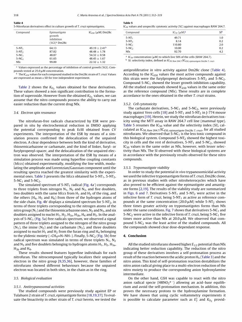
Fig. 7. Trypomastigote viability Dm2
It is probably that the more negative potential (couple IVc/IVa)f the nitro derivatives corresponds to a diminish capacity to acceptlectrons due to its negative charge. Fig. 2 represents the proposededuction mechanism for this derivatives and the probably inter-ediates of reduction.These derivatives reveal a cuasireversible mono electronic
ransfer corresponding to the generation of the nitro anion radicalHRNO2
•− or −RNO2•−).
The characterization of the voltammetric waves took place usingarameters �E and Ipa/Ipc. The Ipa/Ipc value was obtained fromicholson and Shain equation [20,27]. The signal in voltammo-rams presents a Ipa/Ipc value close to unity, which slightly increases the scan rate increases (from 0.1 to 2.0 V/s, Table 2). The �Earameter increased to 0.06 V for 5-NT1,2 and 5-NC1,2 derivatives,
ndicating a cuasireversible process. For each of the compounds, Ipc
s v1/2 figure is linear, indicating that the electronic transfers areontrolled by diffusion and are not due to species adsorbed on theurface of the electrode [20,27].
According to previous work [28], the coupled chemical reac-ion in DMSO milieu is a dimerization reaction and consequentlye obtained the dimerization rate constants by applying the well
nown procedure described by Olmstead [21,29]. In order to eval-ate the stability of the radical in DMSO we proceeded to calculatehe rate constant of the dimerization reaction (kd) of the nitro anionadical formed from the 5-NT and 5-NC derivatives (see Table 2).hese results indicate that 5-NT1–2 generated the nitro-anion rad-cal most hardly formed (more negative reduction potential) butts produced radical was the less stable (highest kd). The value ofimerization rate constant (kd) for each compound is showing inable 2.
.2. Reactivity of the nitro anion radical, generatedlectrochemically from 5-NT and 5-NC, with GSH
We studied the reactivity of the 5-NT and 5-NC nitro anion radi-al to GSH by cyclic voltammetry using increasing amounts of GSHc.a. 0.1 M in DMSO). All the derivatives showed similar behaviourn this experiment, as an example Fig. 3 shows the behaviour of 5-T1 in aprotic milieu in the absence and presence of GSH, wherein
t is observed that the two cathodic peak (IIIc and Vc) increases sig-ificantly while the anodic peak (IVa) disappear and the peak (IIIa)ecrease significantly. This behaviour is relating to the GSH acid-
ty and indicate that GSH react with the nitro anion radical specieHRNO2
•−) allowing an acid–base equilibrium and avoid the self-rotonation mechanism. This last, it is observed on disappear ofhe anodic peak (IVa) when was used GSH 0.5 mM in the milieusee Fig. 3). In addition, the increase on the cathodic peak (Vc) in
Nfx
5-NC
5-NC2
ne for compounds 5-NC1 and 5-NC2.
the presence of GSH points out that the necessary protons to thehydroxylamine formation are provided by GSH:
HRNO2 + e− ↔ HRNO2•− (1)
HRNO2•− + 3e− + 4GSH ↔ HRNHOH + 4GS− + H2O (2)
The GSH signals did not interfere with the corresponding nitroanion radical detection at the studied concentrations [30–32].Therefore, if it is considered that in biological milieu high thiol lev-els are present allow us to propose that this kind of process couldtake place in the parasite explaining, at least in part, the observedbiological activity for 5-NT and 5-NC derivatives against T. cruzi.
3.3. Determination of biological parameter E17 and KO2
The reduction potential in aprotic media for this family of 5-NTand 5-NC was studied using DMF as aprotic solvent. The E1
7 value isa parameter that accounts the energy necessary to transfer the firstelectron to an electroactive group to form a radical anion at pH 7.0in aqueous milieu, and it is considered as indicative of nitro anionradical formation in biological systems [22]. Furthermore, a lin-ear correlation between the cathodic peak potential (Epc) in DMF,obtained in aprotic milieu, with the E1
7 values obtained by pulseradiolysis, has been previously described [33]. Consequently, withthe obtained experimental values of Epc in aprotic milieu we calcu-lated the E1
7 values for all the studied compounds by interpolatingin the corresponding straight line (Table 2). From these results, it ispossible to conclude that the studied derivatives have the potentialof pulse radiolysis higher even than Nfx, which could lead a betterbiological activity. Additionally, it has previously shown that nitrocompounds with reduction potentials, E1
7, lower than −400 mV, or−1000 mV (in DMF) as in the case of metronidazole, the rate ofsuperoxide formation by microsomal enzymes was not significant[34]. In order to evaluate the possible formation of superoxide weproceeded to calculate the superoxide constant (KO2 ) for the semireaction.
HRNO2•− + O2
KO2−→HRNO2 + O2•− (3)
through the Nernst equation:
�E = RT
nFln K (4)
0.059 log KO2 = {−0.155 − [EHRNO2/HRNO2•− ]} (5)
where −0.155 is the redox potential in volts for the couple O2/O2•−
in the non-standard condition of 1 mM and the potential of semi-reaction E(HRNO2/HRNO2
•−) corresponds to the E17 value.
318 C. Maria Aravena et al. / Spectrochimica Acta Part A 79 (2011) 312–319
Table 45-Nitrofuran derivatives effect in culture growth of T. cruzi epimastigotes.
Compound Epimastigotegrowthinhibition(%GI)a Dm28c
ICk50 (�M) Dm28c
5-NT1 64.13 49.19 ± 2.41b
5-NT2 87.82 48.48 ± 1.785-NC1 48.87 54.32 ± 0.585-NC2 61.65 48.45 ± 1.67Nfx 99.00 22.32 ± 1.32
p
a
Ttae
3
pteuetfts(lrm5
tttttndyp(atraH
nene
3
3
Tu
Table 5IC50 values and unspecific cytotoxic activity (%C) against macrophages RAW 264.7.
Compound IC50 (�M)a SIb
5-NT1 49.71 1.05-NT2 8.14 0.25-NC1 110.80 2.05-NC2 10.00 0.2
a Values expressed as the percentage of inhibition of control growth (%GI). Com-ounds tested at 25.0 �M concentration.b The ICk50 values for each compound studied in the Dm28c strain of T. cruzi. Values
re expressed as mean ± SD for tree independent experiment.
Table 2 shows the KO2 values obtained for these derivatives.hese values showed a non significant contribution to the forma-ion of superoxide. However from the obtained KO2 values, we canssume that the nitro compounds possess the ability to carry outasier reduction than the current drug Nfx.
.4. Electron spin resonance
The nitrofuran-free radicals characterized by ESR were pre-ared in situ by electrochemical reduction in DMSO applyinghe potential corresponding to peak EcIII obtained from CVxperiments. The interpretation of the ESR by means of a sim-lation process confirmed the delocalization of the unpairedlectron. A clear dependence between both the kind of derivative,hiosemicarbazone or carbamate, and the kind of linker, furyl- orurylpropenyl-spacer, and the delocalization of the unpaired elec-ron was observed. The interpretation of the ESR by means of aimulation process was made using hyperfine coupling constantshfccs) obtained experimentally, modifying the line width, modu-ating the amplitude and Lorentzian/Gaussian component until theesulting spectra reached the greatest similarity with the experi-ental ones. Table 3 presents the hfccs obtained for 5-NT1, 5-NT2,
-NC1 and 5-NC2.The simulated spectrum of 5-NT1 radical (Fig. 4a′) corresponds
o three triplets from nitrogen N1, N2 and N3, and five doublets,wo doublets with the same hfccs from H1 and H2 of the ring, andhe others (H4, H5 and H6) from three of the hydrogen atoms ofhe side chain. Fig. 4b′ displays a simulated spectrum for 5-NT2, inerms of three triplets corresponding to the nitrogen atoms of theitro group (N1) and the thiosemicarbazone ones, N2 and N3, and sixoublets assigned to nuclei H1, H2, H3a, H3b, H4 and H5. In the anal-sis of 5-NC1 (Fig. 5a) free radicals spectrum, we observed a signalattern of three triplets assigned to the nitrogen of the nitro groupN1), the imine (N2) and the carbamate (N3), and three doubletsssigned to nuclei H1 and H2 from the furan ring and H4 belongingo the ylidenic moiety (–CH4 N–NH–). Finally, 5-NC2 (Fig. 5b) freeadical spectrum was simulated in terms of three triplets N1, N2nd N3 and five doublets belonging to hydrogen atoms H1, H2, H3a,3b and H4.
These results showed features hyperfine individuals for eachitrofuran. The nitrocompound typically localizes their unpairedlectron in the nitro group [9,35,36], however, these families ofitrofurans showed different behaviours because the unpairedlectron was located in both sites, in the chain as in the ring.
.5. Biological evaluation
.5.1. Antitrypanosomal activitiesThe studied compounds were previously study against EP or
ulahuen 2 strain of T. cruzi, epimastigote forms [18,19,37]. To eval-ate the bioactivity in other strain of T. cruzi herein, we tested the
Nfx 92.70 4.2
a IC50 concentration (�M) to which live 50% of the cells (RAW 264.7).b SI: selectivity index, defined as IC50, Raw 264.7/ICk50, epimastigote Dm28c T. cruzi .
antiproliferative in vitro activity against Dm28c clone (Table 4).According to the ICk50 values the most active compounds againstthis strain were the furylpropenyl derivatives 5-NT2 and 5-NC2.Compound 5-NC1 showed the lesser growth inhibition capability.All the studied compounds showed ICk50 values in the same orderas the reference compound (Nfx). These results are in completeaccordance to the ones obtained in the other T. cruzi strains.
3.5.2. Cell cytotoxicityThe carbazate derivatives, 5-NC1 and 5-NC2, were previously
study against Vero cells [18] and 5-NT1 and 5-NT2 in J-774 mousemacrophages [19]. Herein, we study the nitrofuran derivatives tox-icity using the MTT assay in RAW 264.7 cell line (mammal type).Table 5 resumes the IC50 value and the selectivity index (SI), cal-culated as IC50, Raw 264.7/ICk50, epimastigote Dm28c T. cruzi, for all studiednitrofurans. We observed that 5-NC1 is the less toxic compound inthis biological system. Compound 5-NT2 showed the highest toxi-city in cells and the rest of derivatives, 5-NT1 and 5-NC2, showedIC50 values in the same order as Nfx, however, with lesser selec-tivity than Nfx. The SI observed with these biological systems arein accordance with the previously results observed for these nitrocompounds.
3.5.3. Trypomastigote viabilityIn order to study the potential in vivo trypanosomicidal activity
we used the infective trypomastigote forms of T. cruzi, Dm28c clone,as in previous studies with other nitroheterocycles which havealso proved to be efficient against the epimastigote and amastig-ote forms [2,19]. The results of the viability study are summarizedin Figs. 6 and 7. Derivatives 5-NT1 and 5-NT2 were active in theinfective form of T. cruzi, being 5-NT1 as active as reference com-pounds at the same concentration (20.0 �M) while 5-NT2 showsthree times greater activity on trypomastigotes forms than Nfxunder the same conditions. Fig. 7 shows that derivatives 5-NC1 and5-NC2 were active in the infective form of T. cruzi, being 5-NC1 fivetimes more active than Nfx at 20.0 �M. We observed that com-pound 5-NC2 was the least active of the studied compounds. Allthe compounds showed clear dose-dependant response.
4. Conclusion
All the studied nitrofurans showed higher E1/2 potential than Nfxindicating better reduction capability. The reduction of the nitrogroup of these derivatives involves a self-protonation process asresult of the reaction between the acidic proton H5 (Table 3) and thenitro anion. This kind of self-protonation reaction destabilizes thenitro anion radical giving place to a multi-electron reduction of thenitro moiety to produce the corresponding anion hydroxylamineintermediate.
On the other hand, GSH was capable to react with the nitro
anion radical specie (HRNO2•−) allowing an acid–base equilib-rium and avoid the self-protonation mechanism. In addition, thisproves the necessary protons to the hydroxylamine formation.We have shown that using cyclic voltammetry experiments itis possible to calculate parameter such as E1
7 and KO2 provid-

himica
iwingmrathupdpN
A
I
R
[
[
[
[
[
[
[[
[
[
[[[[
[
[
[[[
[[[
[[[[35] C. Rigol, C. Olea-Azar, F. Mendizabal, L. Otero, D. Gambino, M. González, H.
C. Maria Aravena et al. / Spectroc
ng simple diagnostic criteria to select, in first screening, agentsith adequate biological performances. From the measurements
n DMF it was possible to obtain the E17 concluding that the
itroanion radical from these 5-nitrofurans could be biologicallyenerated via enzymatic reduction because the energy of its for-ation is similar or lower than the values for well-know enzymatic
educible drugs as Nfx and Bnz. Stable free radicals were gener-ted using electrochemical reductions at potentials correspondingo the first wave and characterized by ESR spectroscopy. Theyperfine pattern observed in these derivatives indicated that thenpaired electron is delocalized throughout the molecule. Try-anocidal activity of these 5-nitrofurans was achieved. All theerivatives showed interesting anti-T. cruzi properties against try-omastigotes with antiproliferative activities in the same order thatfx.
cknowledgements
This research was supported by FONDECYT 1071068 and CON-CYT scholarship Ph.D. RIDIMEDCHAG-CYTED is acknowledged.
eferences
[1] Available from: http://www.who.int/tdr/diseases/chagas/ (accessed 08.10.09).[2] J.D. Maya, S. Bollo, L. Núnez, J. Squella, A. Morello, Biochem. Pharmacol. 65
(2003) 999–1006.[3] J.D. Maya, B.K. Cassels, P. Iturriaga-Vásquez, J. Ferreira, M. Faúndez, N. Galanti,
A. Ferreira, A. Morello, Comp. Biochem. Physiol. A: Mol. Integr. Physiol. 146(2007) 601–620.
[4] H. Cerecetto, M. González, Mini Rev. Med. Chem. 8 (2008) 1355–1383.[5] H. Cerecetto, B. Mester, S. Onetto, G. Seaone, M. González, F. Zinola, Farmaco
47 (1992) 1207–1213.
[6] R. Docampo, S.N.J. Moreno, Rev. Infect. Dis. 6 (1984) 223–238.[7] H. Cerecetto, M. Gonzalez, Curr. Top. Med. Chem. 2 (2002) 1187–1213.[8] H. Cerecetto, M. González, Pharmaceuticals 3 (2010) 810–838.[9] S.N. Moreno, R.P. Mason, R. Docampo, J. Biol. Chem. 259 (1984) 5606–5611.10] C. Olea-Azar, C. Rigol, F. Mendizabal, A. Morello, J.D. Maya, C. Moncada, E. Cabr-era, R. Di Maio, M. González, H. Cerecetto, Free Radic. Res. 37 (2003) 993–1001.
[
[
Acta Part A 79 (2011) 312–319 319
11] M. Boiani, L. Piacenza, P. Hernández, L. Boiani, H. Cerecetto, M. González, A.Denicola, Biochem. Pharmacol. 79 (2010) 1736–1745.
12] C. Dumas, M. Ouellette, J. Tovar, M.L. Cunningham, A.H. Fairlamb, S. Tamar, M.Olivier, B. Papadopoulou, EMBO J. 16 (1997) 2590–2598.
13] S. Krieger, W. Schwarz, M.R. Ariyanayagam, A.H. Fairlamb, R.L. Krauth-Siegel,C. Clayton, Mol. Microbiol. 35 (2000) 542–552.
14] J. Tovar, M.L. Cunningham, A.C. Smith, S.L. Croft, A.H. Fairlamb, Proc. Natl. Acad.Sci. U.S.A. 95 (1998) 5311–5316.
15] G.B. Henderson, P. Urlich, A.H. Fairlamb, I. Rosenberg, M. Pereira, M. Sela, A.Cerami, Proc. Natl. Acad. Sci. U.S.A. 85 (1988) 5374–5378.
16] J.A. Urbina, R. Docampo, Expert Opin. Ther. Pat. 13 (2003) 661–669.17] J.C. Hinshaw, D.-Y. Suh, P. Garnier, F.S. Buckner, R.T. Eastman, S.P.T. Matsuda,
B.M. Joubert, I. Coppens, K.A. Joiner, S. Merali, T.E. Nash, E. Prestwich, J. Med.Chem. 46 (2003) 4240–4243.
18] A. Gerpe, I. Odreman-Nunez, P. Draper, L. Boiani, J.A. Urbina, M. González, H.Cerecetto, Bioorg. Med. Chem. 16 (2008) 569–577.
19] A. Gerpe, G. Alvarez, D. Benítez, L. Boiani, M. Quiroga, P. Hernández, M. Sortino, S.Zacchino, M. González, H. Cerecetto, Bioorg. Med. Chem. 17 (2009) 7500–7509.
20] S. Nicholson, Anal. Chem. 38 (1966) 1406.21] M.L. Olmnested, R.G. Hamilton, R.S. Nicholson, Anal. Chem. 41 (1969).22] P. Wardman, Environ. Health Perspect. 64 (1985) 309–320.23] C. Olea-Azar, H. Cerecetto, A. Gerpe, M. González, V.J. Arán, C. Rigol, L. Opazo,
Spectrosc. Lett. 63 (2005) 36–42.24] J.A. Bautista-Martínez, I. González, M. Aguilar-Martínez, Electrochim. Acta 49
(2004) 3403–3411.25] J. Carbajo, S. Bollo, L.J. Núnez-Vergara, A. Campero, J.A. Squella, J. Electroanal.
Chem. 531 (2002) 187–194.26] G. Kokkinidis, A. Kelaidopoulou, J. Electroanal. Chem. 414 (1996) 197–208.27] S. Nicholson, J. Shain, Anal. Chem. 36 (1964) 706–723.28] J. Arguello, L.J. Núnez-Vergara, S. Bollo, J.A. Squella, Bioelectrochemistry 69
(2006) 104–112.29] M.L. Olmnested, R.S. Nicholson, Anal. Chem. 41 (1969) 862–864.30] J.H. Tocher, D. Edwards, J. Biochem. Pharmacol. 50 (1995) 1367–1371.31] L.J. Nunez-Vergara, J.A. Squella, C. Olea-Azar, S. Bollo, P.A. Navarrete-Encina, J.C.
Sturm, Electrochim. Acta 45 (2000) 3555–3561.32] R. Kizek, J. Vacek, L. Trnkova, J. Jelen, Bioelectrochemistry 63 (2004) 31–36.33] A. Breccia, G. Berrilli, S. Roffia, Int. J. Radiat. Biol. 36 (1979) 85–89.34] M.H. Livertoux, P. Lagrange, A. Minn, Brain Res. 725 (1996) 207–216.
Cerecetto, Spectrochim. Acta A 61 (2005) 2933–2938.36] C. Rigol, C. Olea.Azar, F. Mendizabal, R. Briones, H. Cerecetto, M. Gonzalez, J.
Mol. Struct. THEOCHEM 770 (2006) 125–129.37] J. Rodríguez, A. Gerpe, G. Aguirre, U. Kemmerling, O. Piro, V.J. Arán, J.D. Maya, C.
Olea-Azar, M. González, H. Cerecetto, Eur. J. Med. Chem. 44 (2009) 1545–1553.
Copyright © 2022 FDOKUMEN