Post-mortem investigations on a leatherback turtle Dermochelys coriacea stranded along the Northern...
6
DISEASES OF AQUATIC ORGANISMS Dis Aquat Org Vol. 100: 71–76, 2012 doi: 10.3354/dao02479 Published August 13 INTRODUCTION The leatherback sea turtle Dermochelys coriacea (Vandelli, 1761) is a circumglobal species with the largest distribution of all reptiles (James et al. 2005). It is listed as critically endangered in the Interna- tional Union for Conservation of Nature Red List of Threatened Species (Sarti Martinez 2000) because of human impacts on its population health worldwide (Spotila et al. 1996). Besides loggerhead (Caretta caretta) and green (Chelonia mydas) turtles, which reproduce in the Mediterranean, leatherbacks are regularly present in this basin, most likely originat- ing from Atlantic populations (Casale et al. 2003). In particular, the number of animals stranded in the Adriatic Sea, mainly in the southeastern basin, dur- ing the summer seems to be higher than the Mediter- ranean average (Lazar et al. 2008). They are rarely reported to reach the northern coastline. Herein, we report the main findings of a post-mortem investiga- tion carried out on a leatherback sea turtle stranded along the northern Adriatic coastline. MATERIALS AND METHODS On 9 June 2009, an adult female leatherback sea turtle (total length: 178 cm; curved carapace length: 138 cm; mass: 199.5 kg) was found dead on the sandy beach of Lido di Venezia, Venice, Italy (45° 24’ 44’’ N, 12° 22’ 33’’ E). The preservation status of the carcass was good, and a detailed post-mortem examination © Inter-Research 2012 · www.int-res.com *Corresponding author. Email: [email protected] Post-mortem investigations on a leatherback turtle Dermochelys coriacea stranded along the Northern Adriatic coastline Lisa Poppi 1 , Annalisa Zaccaroni 2 , Daniela Pasotto 1 , Giorgia Dotto 1 , Federica Marcer 3 , Dino Scaravelli 2 , Sandro Mazzariol 1, * 1 Department of Comparative Biomedicine and Food Science, University of Padova, Viale dell’Università 16, Legnaro (PD) 35020, Italy 2 Department of Veterinary Medical Sciences, University of Bologna, Ozzano dell'Emilia, Bologna 40064, Italy 3 Department of Animal Medicine, Production and Health, University of Padova, Viale dell’Università 16, Legnaro (PD) 35020, Italy ABSTRACT: Leatherback sea turtles Dermochelys coriacea are regularly reported in the Mediter- ranean Sea but rarely reach the northern Adriatic Sea. In the summer of 2009, a well-preserved carcass of an adult female of this species was found dead along the coast of Lido di Venezia. A complete necropsy was carried out, along with evaluation of levels of tissue trace elements. The the post-mortem revealed acute severe bacterial gastroenteritis caused by Photobacterium damselae ssp. piscicida, an opportunistic agent that infected an apparently debilitated animal weakened by ingested plastic debris. High levels of heavy metals (Hg, Pb, Cd and As) found in the liver and kidneys might have contributed to the animal’s demise. These findings support previous indications that marine debris is one of the major threats to marine animals, particularly for criti- cally endangered species such as the leatherback turtle. KEY WORDS: Leatherback turtle · Photobacterium damselae ssp. piscicida · Marine debris · Adriatic Sea Resale or republication not permitted without written consent of the publisher This authors' personal copy may not be publicly or systematically copied or distributed, or posted on the Open Web, except with written permission of the copyright holder(s). It may be distributed to interested individuals on request.
Transcript of Post-mortem investigations on a leatherback turtle Dermochelys coriacea stranded along the Northern...
DISEASES OF AQUATIC ORGANISMSDis Aquat Org
Vol. 100: 71–76, 2012doi: 10.3354/dao02479
Published August 13
INTRODUCTION
The leatherback sea turtle Dermochelys coriacea(Vandelli, 1761) is a circumglobal species with thelargest distribution of all reptiles (James et al. 2005).It is listed as critically endangered in the Interna-tional Union for Conservation of Nature Red List ofThreatened Species (Sarti Martinez 2000) because ofhuman impacts on its population health worldwide(Spotila et al. 1996). Be sides loggerhead (Carettacaretta) and green (Chelonia mydas) turtles, whichreproduce in the Mediterranean, leatherbacks areregularly present in this basin, most likely originat-ing from Atlantic populations (Casale et al. 2003). Inparticular, the number of animals stranded in theAdriatic Sea, mainly in the southeastern basin, dur-
ing the summer seems to be higher than the Mediter-ranean average (Lazar et al. 2008). They are rarelyreported to reach the northern coastline. Herein, wereport the main findings of a post-mortem investiga-tion carried out on a leatherback sea turtle strandedalong the northern Adriatic coastline.
MATERIALS AND METHODS
On 9 June 2009, an adult female leatherback seaturtle (total length: 178 cm; curved carapace length:138 cm; mass: 199.5 kg) was found dead on the sandybeach of Lido di Venezia, Venice, Italy (45° 24’ 44’’ N,12° 22’ 33’’ E). The preservation status of the carcasswas good, and a detailed post-mortem examination
© Inter-Research 2012 · www.int-res.com*Corresponding author. Email: [email protected]
Post-mortem investigations on a leatherback turtle Dermochelys coriacea stranded along the
Northern Adriatic coastline
Lisa Poppi1, Annalisa Zaccaroni2, Daniela Pasotto1, Giorgia Dotto1, Federica Marcer3, Dino Scaravelli2, Sandro Mazzariol1,*
1Department of Comparative Biomedicine and Food Science, University of Padova, Viale dell’Università 16, Legnaro (PD) 35020, Italy
2Department of Veterinary Medical Sciences, University of Bologna, Ozzano dell'Emilia, Bologna 40064, Italy3Department of Animal Medicine, Production and Health, University of Padova, Viale dell’Università 16,
Legnaro (PD) 35020, Italy
ABSTRACT: Leatherback sea turtles Dermochelys coriacea are regularly reported in the Mediter-ranean Sea but rarely reach the northern Adriatic Sea. In the summer of 2009, a well-preservedcarcass of an adult female of this species was found dead along the coast of Lido di Venezia. Acomplete necropsy was carried out, along with evaluation of levels of tissue trace elements. Thethe post-mortem revealed acute severe bacterial gastroenteritis caused by Photobacteriumdamselae ssp. piscicida, an opportunistic agent that infected an apparently debilitated animalweakened by ingested plastic debris. High levels of heavy metals (Hg, Pb, Cd and As) found in theliver and kidneys might have contributed to the animal’s demise. These findings support previousindications that marine debris is one of the major threats to marine animals, particularly for criti-cally endangered species such as the leatherback turtle.
KEY WORDS: Leatherback turtle · Photobacterium damselae ssp. piscicida · Marine debris · Adriatic Sea
Resale or republication not permitted without written consent of the publisher
This authors' personal copy may not be publicly or systematically copied or distributed, or posted on the Open Web, except with written permission of the copyright holder(s). It may be distributed to interested individuals on request.
Dis Aquat Org 100: 71–76, 2012
was performed at the Faculty of Veterinary Medicineof Padova within 24 h of recovery of the stranded ani-mal. Tissue samples were preserved in 10% neutralbuffered formalin for histopathological examination,refrigerated for microbiological and parasitologicalexaminations or frozen for biomolecular and eco -toxicological investigations. For the histopathological examination, the tissues were embedded in paraffin,sectioned (4 µm thickness) and routinely stained withhematoxylin and eosin. Selected tissue sections fromthe main organs were also stained using dif ferent histochemical staining techniques (Periodic acid-Schiff, Gram and Giemsa staining) to detect biolo -gical agents. Danscher’s auto-metallographic tech-nique (Danscher & Moller-Madsen 1985) and therhodizonate method for lead salts were used on hepatic and renal tissue to detect inorganic mercuryand lead particles, respectively (Lillie 1954). Duringnecropsy, intestinal swabs were collected for standardmicrobiological analyses. The samples were inocu-lated onto sheep-blood agar (tryptic soy agar supple-mented with 5% defibrinated sheep blood) and incu-bated aerobically at 28 and 37°C for 24 to 48 h. Theisolated colonies were biochemically identified usingAPI 20NE (bioMérieux). To confirm the identity of theisolates, a molecular analysis was performed. The total DNA of the isolates was extracted by heating aselected colony at 100°C for 10 min and emulsifyingin 100 µl of sterile nuclease-free water; this was usedas a template for further molecular investigations.
Parasitological examinations were performed onall the major organs. The collected parasites werestored in 70% ethanol and studied by light micro -scopy after staining with borax-carmine and clarify-ing in Amman’s lactophenol.
Trace elements in the liver, kidney, skin and muscletissues were analyzed at the University of Bo logna,Italy. Subsamples (0.7 g) of each tissue were digestedwith a Milestone MLS 1200 Mega microwave ovenusing 4 ml of nitric acid and 1 ml of hydrogen per -oxide. Measurements of the concentrations of trace elements (namely, arsenic, lead, chromium, cop per,manganese, iron, selenium, zinc, cadmium, nickeland mercury), were performed by inductively coupledplasma optical emission spectrometry using a PerkinElmer Optima 2100 DV instrument coupled with aCETAC U5000AT+ ultrasound nebulizer for mercury.Two blanks were run during each set of analysis tocheck for purity of the trace elements, and the accuracy of the method was verified using reference materials (lyophilized mussel; CRM 278, Community Bureau of Reference, BCR, Brussels). All of the refer-ence material values were within the certified limits.
RESULTS
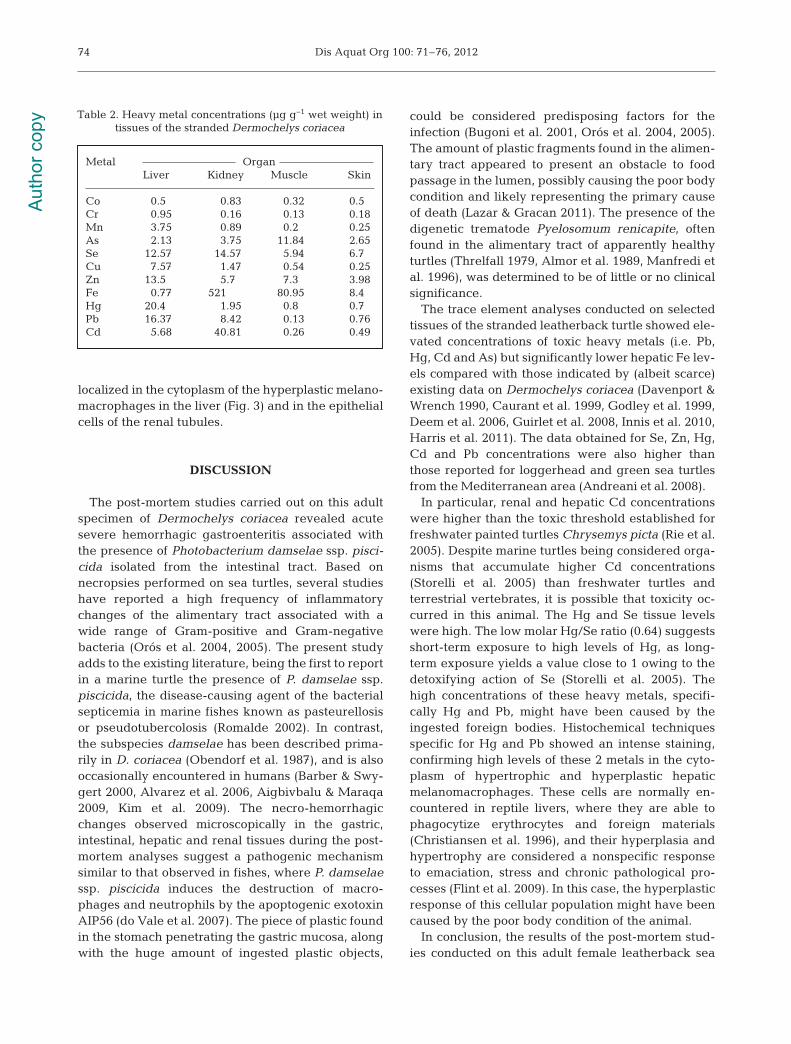
External examination revealed poor body condi-tion and bloody discharge from the mouth and thenostrils, along with epidermal bruises. In addition,severe oedema and several petechiae in the subcuta-neous tissues and the internal viscera were seen. Amoderate amount of sero-hemorrhagic fluid with fib-rin was found in the coelomatic cavity. Upon exami-nation of the gastrointestinal tract, acute severe dif-fuse hemorrhagic gastroenteritis was observed inassociation with the presence of a plastic fragmentperforating a fold of the gastric mucosa (Fig. 1). Asreported in Table 1, 14 different foreign bodies werefound in the alimentary tract, mainly represented byfloating plastic fragments, obstructing the intestinallumen (Fig. 1). Upon microscopic investigation, dif-fuse and severe hemorrhagic mucosal necrosis withlow-grade heterophilic inflammation was observedin the esophagus, stomach and upper intestine with
72
Fig. 1. Foreign bodies found during gross examination in thegastro-intestinal tract of the stranded Dermochelys coriacea.(A) Multiple pieces of plastic obstructing the intestinallumen associated with hemorrhages, ulceration and severediffuse edema of the enteric mucosa (arrow). (B) A rigid
plastic fragment perforating the gastric wall
Aut
hor c
opy
Poppi et al.: Post-mortem investigations on a leatherback turtle
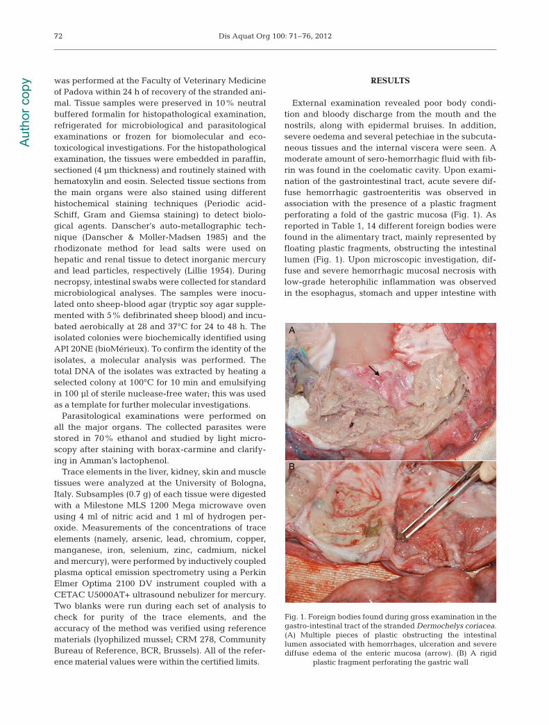
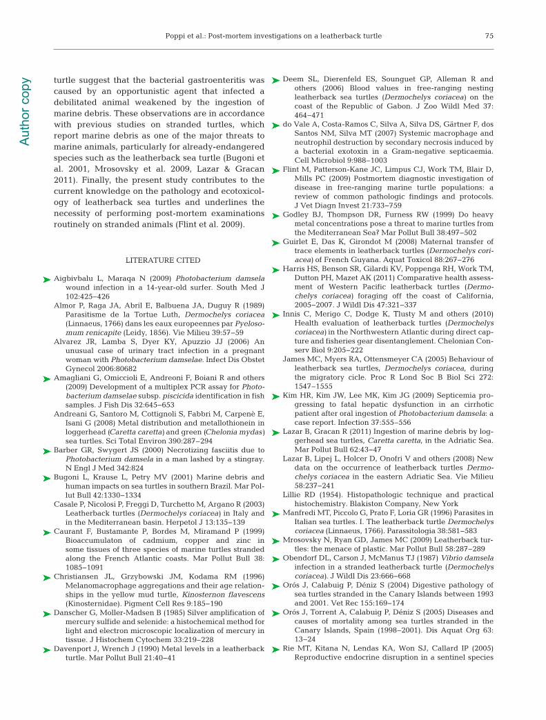
multiple foci of hepatic, renal and cardiac necrosisand moderate to severe melanomacrophage hyper-trophy and hyperplasia in the liver (Fig. 2). Chronicmild multifocal bronchopneumonia was diagnosedduring the histopathologic examination.
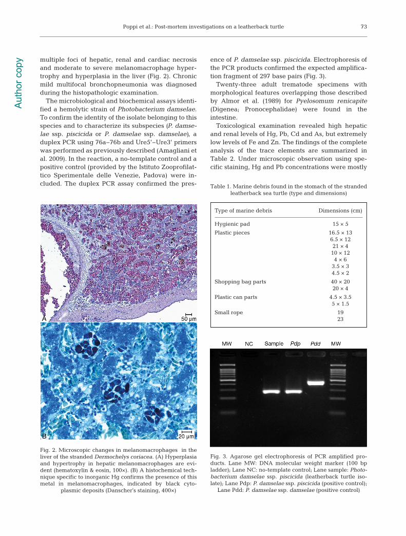
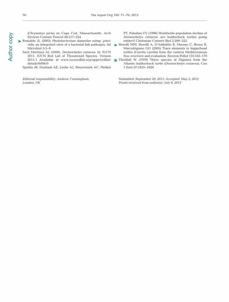
The microbiological and biochemical assays identi-fied a hemolytic strain of Photobacterium damselae.To confirm the identity of the isolate belonging to thisspecies and to characterize its subspecies (P. damse-lae ssp. piscicida or P. damselae ssp. damselae), aduplex PCR using 76a!76b and Ure5’!Ure3’ primerswas performed as previously described (Amagliani etal. 2009). In the reaction, a no-template control and apositive control (provided by the Istituto Zooprofilat-tico Sperimentale delle Venezie, Padova) were in -cluded. The duplex PCR assay confirmed the pres-
ence of P. damselae ssp. piscicida. Electrophoresis ofthe PCR products confirmed the expected amplifica-tion fragment of 297 base pairs (Fig. 3).
Twenty-three adult trematode specimens withmorphological features overlapping those describedby Almor et al. (1989) for Pyelosomum renicapite(Digenea; Pronocephalidae) were found in the intestine.
Toxicological examination revealed high hepaticand renal levels of Hg, Pb, Cd and As, but extremelylow levels of Fe and Zn. The findings of the completeanalysis of the trace elements are summarized inTable 2. Under microscopic observation using spe-cific staining, Hg and Pb concentrations were mostly
73
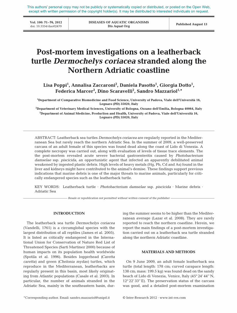
Type of marine debris Dimensions (cm)
Hygienic pad 15 " 5
Plastic pieces 16.5 " 136.5 " 1221 " 4
10 " 124 " 6
3.5 " 34.5 " 2
Shopping bag parts 40 " 2020 " 4
Plastic can parts 4.5 " 3.55 " 1.5
Small rope 1923
Table 1. Marine debris found in the stomach of the stranded leatherback sea turtle (type and dimensions)
Fig. 3. Agarose gel electrophoresis of PCR amplified pro -ducts. Lane MW: DNA molecular weight marker (100 bpladder); Lane NC: no-template control; Lane sample: Photo-bacterium damselae ssp. piscicida (leatherback turtle iso-late); Lane Pdp: P. damselae ssp. piscicida (positive control);
Lane Pdd: P. damselae ssp. damselae (positive control)
Fig. 2. Microscopic changes in melanomacrophages in theliver of the stranded Dermo chelys coriacea. (A) Hyperplasiaand hypertrophy in hepatic melanomacrophages are evi-dent (hematoxylin & eosin, 100"). (B) A histochemical tech-nique specific to inorganic Hg confirms the presence of thismetal in mela no macrophages, indicated by black cyto -
plasmic deposits (Danscher’s staining, 400")
Aut
hor c
opy
Dis Aquat Org 100: 71–76, 2012
localized in the cytoplasm of the hyperplastic melano -macrophages in the liver (Fig. 3) and in the epithelialcells of the renal tubules.
DISCUSSION
The post-mortem studies carried out on this adultspecimen of Dermochelys coriacea revealed acutesevere hemorrhagic gastroenteritis associated withthe presence of Photobacterium damselae ssp. pisci-cida isolated from the intestinal tract. Based onnecropsies performed on sea turtles, several studieshave reported a high frequency of inflammatorychanges of the alimentary tract associated with awide range of Gram-positive and Gram-negativebacteria (Orós et al. 2004, 2005). The present studyadds to the existing literature, being the first to reportin a marine turtle the presence of P. damselae ssp.piscicida, the disease-causing agent of the bacterialsepticemia in marine fishes known as pasteurellosisor pseudotubercolosis (Romalde 2002). In contrast,the subspecies damselae has been described prima-rily in D. coriacea (Obendorf et al. 1987), and is alsooccasionally encountered in humans (Barber & Swy -gert 2000, Alvarez et al. 2006, Aigbivbalu & Maraqa2009, Kim et al. 2009). The necro-hemorrhagicchanges observed microscopically in the gastric,intestinal, hepatic and renal tissues during the post-mortem analyses suggest a pathogenic mechanismsimilar to that observed in fishes, where P. damselaessp. piscicida induces the destruction of macro-phages and neutrophils by the apoptogenic exotoxinAIP56 (do Vale et al. 2007). The piece of plastic foundin the stomach penetrating the gastric mucosa, alongwith the huge amount of ingested plastic objects,
could be considered predisposing factors for theinfection (Bugoni et al. 2001, Orós et al. 2004, 2005).The amount of plastic fragments found in the alimen-tary tract appeared to present an obstacle to foodpassage in the lumen, possibly causing the poor bodycondition and likely representing the primary causeof death (Lazar & Gracan 2011). The presence of thedigenetic trematode Pyelosomum renicapite, oftenfound in the alimentary tract of apparently healthyturtles (Threlfall 1979, Almor et al. 1989, Manfredi etal. 1996), was determined to be of little or no clinicalsignificance.
The trace element analyses conducted on selectedtissues of the stranded leatherback turtle showed ele-vated concentrations of toxic heavy metals (i.e. Pb,Hg, Cd and As) but significantly lower hepatic Fe lev-els compared with those indicated by (albeit scarce)existing data on Dermochelys coriacea (Davenport &Wrench 1990, Caurant et al. 1999, Godley et al. 1999,Deem et al. 2006, Guirlet et al. 2008, Innis et al. 2010,Harris et al. 2011). The data obtained for Se, Zn, Hg,Cd and Pb concentrations were also higher thanthose reported for loggerhead and green sea turtlesfrom the Mediterranean area (Andreani et al. 2008).
In particular, renal and hepatic Cd concentrationswere higher than the toxic threshold established forfreshwater painted turtles Chrysemys picta (Rie et al.2005). Despite marine turtles being considered orga -nisms that accumulate higher Cd concentrations(Storelli et al. 2005) than freshwater turtles and terrestrial vertebrates, it is possible that toxicity oc -curred in this animal. The Hg and Se tissue levelswere high. The low molar Hg/Se ratio (0.64) suggestsshort-term exposure to high levels of Hg, as long-term exposure yields a value close to 1 owing to thedetoxifying action of Se (Storelli et al. 2005). Thehigh concentrations of these heavy metals, specifi-cally Hg and Pb, might have been caused by theingested foreign bodies. Histochemical techniquesspecific for Hg and Pb showed an intense staining,confirming high levels of these 2 metals in the cyto-plasm of hypertrophic and hyperplastic hepaticmelano macrophages. These cells are normally en -coun tered in reptile livers, where they are able tophagocytize erythrocytes and foreign materials(Christiansen et al. 1996), and their hyperplasia andhypertrophy are considered a nonspecific responseto emaciation, stress and chronic pathological pro-cesses (Flint et al. 2009). In this case, the hyperplasticresponse of this cellular population might have beencaused by the poor body condition of the animal.
In conclusion, the results of the post-mortem stud-ies conducted on this adult female leatherback sea
74
Metal OrganLiver Kidney Muscle Skin
Co 0.5 0.83 0.32 0.5Cr 0.95 0.16 0.13 0.18Mn 3.75 0.89 0.2 0.25As 2.13 3.75 11.84 2.65Se 12.57 14.57 5.94 6.7Cu 7.57 1.47 0.54 0.25Zn 13.5 5.7 7.3 3.98Fe 0.77 521 80.95 8.4Hg 20.4 1.95 0.8 0.7Pb 16.37 8.42 0.13 0.76Cd 5.68 40.81 0.26 0.49
Table 2. Heavy metal concentrations (µg g!1 wet weight) in tissues of the stranded Dermochelys coriacea
Aut
hor c
opy
Poppi et al.: Post-mortem investigations on a leatherback turtle
turtle suggest that the bacterial gastroenteritis wascaused by an opportunistic agent that infected adebilitated animal weakened by the ingestion ofmarine debris. These observations are in accordancewith previous studies on stranded turtles, whichreport marine debris as one of the major threats tomarine animals, particularly for already-endangeredspecies such as the leatherback sea turtle (Bugoni etal. 2001, Mrosovsky et al. 2009, Lazar & Gracan2011). Finally, the present study contributes to thecurrent knowledge on the pathology and ecotoxicol-ogy of leatherback sea turtles and underlines thenecessity of performing post-mortem examinationsroutinely on stranded animals (Flint et al. 2009).
LITERATURE CITED
Aigbivbalu L, Maraqa N (2009) Photobacterium damselawound infection in a 14-year-old surfer. South Med J102: 425!426
Almor P, Raga JA, Abril E, Balbuena JA, Duguy R (1989)Parasitisme de la Tortue Luth, Dermochelys coriacea(Linnaeus, 1766) dans les eaux europeennes par Pyeloso-mum renicapite (Leidy, 1856). Vie Milieu 39: 57!59
Alvarez JR, Lamba S, Dyer KY, Apuzzio JJ (2006) Anunusual case of urinary tract infection in a pregnantwoman with Photobacterium damselae. Infect Dis ObstetGynecol 2006:80682
Amagliani G, Omiccioli E, Andreoni F, Boiani R and others(2009) Development of a multiplex PCR assay for Photo-bacterium damselae subsp. piscicida identification in fishsamples. J Fish Dis 32: 645!653
Andreani G, Santoro M, Cottignoli S, Fabbri M, Carpenè E,Isani G (2008) Metal distribution and metallothionein inloggerhead (Caretta caretta) and green (Chelonia mydas)sea turtles. Sci Total Environ 390:287–294
Barber GR, Swygert JS (2000) Necrotizing fasciitis due toPhotobacterium damsela in a man lashed by a stingray.N Engl J Med 342: 824
Bugoni L, Krause L, Petry MV (2001) Marine debris andhuman impacts on sea turtles in southern Brazil. Mar Pol-lut Bull 42: 1330!1334
Casale P, Nicolosi P, Freggi D, Turchetto M, Argano R (2003)Leatherback turtles (Dermochelys coriacea) in Italy andin the Mediterranean basin. Herpetol J 13: 135!139
Caurant F, Bustamante P, Bordes M, Miramand P (1999)Bioaccumulaton of cadmium, copper and zinc insome tissues of three species of marine turtles strandedalong the French Atlantic coasts. Mar Pollut Bull 38: 1085!1091
Christiansen JL, Grzybowski JM, Kodama RM (1996)Melanomacrophage aggregations and their age relation-ships in the yellow mud turtle, Kinosternon flavescens(Kinosternidae). Pigment Cell Res 9: 185!190
Danscher G, Moller-Madsen B (1985) Silver amplification ofmercury sulfide and selenide: a histochemical method forlight and electron microscopic localization of mercury intissue. J Histochem Cytochem 33: 219!228
Davenport J, Wrench J (1990) Metal levels in a leatherbackturtle. Mar Pollut Bull 21: 40!41
Deem SL, Dierenfeld ES, Sounguet GP, Alleman R and others (2006) Blood values in free-ranging nesting leatherback sea turtles (Dermochelys coriacea) on thecoast of the Republic of Gabon. J Zoo Wildl Med 37: 464!471
do Vale A, Costa-Ramos C, Silva A, Silva DS, Gärtner F, dosSantos NM, Silva MT (2007) Systemic macrophage andneutrophil destruction by secondary necrosis induced bya bacterial exotoxin in a Gram-negative septicaemia.Cell Microbiol 9: 988!1003
Flint M, Patterson-Kane JC, Limpus CJ, Work TM, Blair D,Mills PC (2009) Postmortem diagnostic investigation ofdisease in free-ranging marine turtle populations: areview of common pathologic findings and protocols.J Vet Diagn Invest 21: 733!759
Godley BJ, Thompson DR, Furness RW (1999) Do heavymetal concentrations pose a threat to marine turtles fromthe Mediterranean Sea? Mar Pollut Bull 38: 497!502
Guirlet E, Das K, Girondot M (2008) Maternal transfer oftrace elements in leatherback turtles (Dermochelys cori-acea) of French Guyana. Aquat Toxicol 88: 267!276
Harris HS, Benson SR, Gilardi KV, Poppenga RH, Work TM,Dutton PH, Mazet AK (2011) Comparative health assess-ment of Western Pacific leatherback turtles (Dermo -chelys coriacea) foraging off the coast of California,2005!2007. J Wildl Dis 47: 321!337
Innis C, Merigo C, Dodge K, Tlusty M and others (2010)Health evaluation of leatherback turtles (Dermochelyscoriacea) in the Northwestern Atlantic during direct cap-ture and fisheries gear disentanglement. Chelonian Con-serv Biol 9: 205!222
James MC, Myers RA, Ottensmeyer CA (2005) Behaviour ofleatherback sea turtles, Dermochelys coriacea, duringthe migratory cicle. Proc R Lond Soc B Biol Sci 272:1547–1555
Kim HR, Kim JW, Lee MK, Kim JG (2009) Septicemia pro-gressing to fatal hepatic dysfunction in an cirrhoticpatient after oral ingestion of Photobacterium damsela: acase report. Infection 37: 555!556
Lazar B, Gracan R (2011) Ingestion of marine debris by log-gerhead sea turtles, Caretta caretta, in the Adriatic Sea.Mar Pollut Bull 62: 43!47
Lazar B, Lipej L, Holcer D, Onofri V and others (2008) Newdata on the occurrence of leatherback turtles Dermo -chelys coriacea in the eastern Adriatic Sea. Vie Milieu58: 237!241
Lillie RD (1954). Histopathologic technique and practicalhistochemistry. Blakiston Company, New York
Manfredi MT, Piccolo G, Prato F, Loria GR (1996) Parasites inItalian sea turtles. I. The leatherback turtle Dermo chelyscoriacea (Linnaeus, 1766). Parassitologia 38: 581!583
Mrosovsky N, Ryan GD, James MC (2009) Leatherback tur-tles: the menace of plastic. Mar Pollut Bull 58: 287!289
Obendorf DL, Carson J, McManus TJ (1987) Vibrio damselainfection in a stranded leatherback turtle (Dermochelyscoriacea). J Wildl Dis 23: 666!668
Orós J, Calabuig P, Déniz S (2004) Digestive pathology ofsea turtles stranded in the Canary Islands between 1993and 2001. Vet Rec 155: 169!174
Orós J, Torrent A, Calabuig P, Déniz S (2005) Diseases andcauses of mortality among sea turtles stranded in theCanary Islands, Spain (1998!2001). Dis Aquat Org 63: 13!24
Rie MT, Kitana N, Lendas KA, Won SJ, Callard IP (2005)Reproductive endocrine disruption in a sentinel species
75A
utho
r cop
y
Dis Aquat Org 100: 71–76, 2012
(Chrysemys picta) on Cape Cod, Massachusetts. ArchEnviron Contam Toxicol 48: 217!224
Romalde JL (2002) Photobacterium damselae subsp. pisci-cida: an integrated view of a bacterial fish pathogen. IntMicrobiol 5: 3!9
Sarti Martinez AL (2000). Dermochelys coriacea. In: IUCN2011. IUCN Red List of Threatened Species. Version2011.1 Available at www.iucnredlist.org/apps/redlist/details/6494/0
Spotila JR, Dunham AE, Leslie AJ, Steyermark AC, Plotkin
PT, Paladino FV (1996) Worldwide population decline ofDermochelys coriacea: are leatherback turtles goingextinct? Chelonian Conserv Biol 2: 209!222
Storelli MM, Storelli A, D’Addabbo R, Marano C, Bruno R,Marcotrigiano GO (2005) Trace elements in loggerheadturtles (Caretta caretta) from the eastern MediterraneanSea: overview and evaluation. Environ Pollut 135: 163!170
Threlfall W (1979) Three species of Digenea from theAtlantic leatherback turtle (Dermochelys coriacea). CanJ Zool 57: 1825!1829
76
Editorial responsibility: Andrew Cunningham, London, UK
Submitted: September 20, 2011; Accepted: May 2, 2012Proofs received from author(s): July 6, 2012
Aut
hor c
opy