Poly(lactic-co-glycolic acid) Bone Scaffolds with Inverted Colloidal Crystal Geometry
12
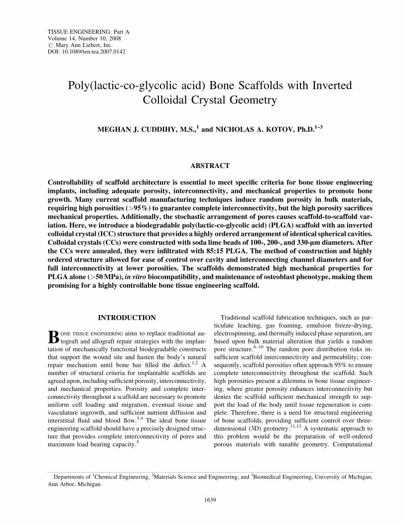
Poly(lactic-co-glycolic acid) Bone Scaffolds with Inverted Colloidal Crystal Geometry MEGHAN J. CUDDIHY, M.S., 1 and NICHOLAS A. KOTOV, Ph.D. 1–3 ABSTRACT Controllability of scaffold architecture is essential to meet specific criteria for bone tissue engineering implants, including adequate porosity, interconnectivity, and mechanical properties to promote bone growth. Many current scaffold manufacturing techniques induce random porosity in bulk materials, requiring high porosities (>95%) to guarantee complete interconnectivity, but the high porosity sacrifices mechanical properties. Additionally, the stochastic arrangement of pores causes scaffold-to-scaffold var- iation. Here, we introduce a biodegradable poly(lactic-co-glycolic acid) (PLGA) scaffold with an inverted colloidal crystal (ICC) structure that provides a highly ordered arrangement of identical spherical cavities. Colloidal crystals (CCs) were constructed with soda lime beads of 100-, 200-, and 330-lm diameters. After the CCs were annealed, they were infiltrated with 85:15 PLGA. The method of construction and highly ordered structure allowed for ease of control over cavity and interconnecting channel diameters and for full interconnectivity at lower porosities. The scaffolds demonstrated high mechanical properties for PLGA alone (>50 MPa), in vitro biocompatibility, and maintenance of osteoblast phenotype, making them promising for a highly controllable bone tissue engineering scaffold. INTRODUCTION B ONE TISSUE ENGINEERING aims to replace traditional au- tograft and allograft repair strategies with the implan- tation of mechanically functional biodegradable constructs that support the wound site and hasten the body’s natural repair mechanism until bone has filled the defect. 1,2 A number of structural criteria for implantable scaffolds are agreed upon, including sufficient porosity, interconnectivity, and mechanical properties. Porosity and complete inter- connectivity throughout a scaffold are necessary to promote uniform cell loading and migration, eventual tissue and vasculature ingrowth, and sufficient nutrient diffusion and interstitial fluid and blood flow. 3,4 The ideal bone tissue engineering scaffold should have a precisely designed struc- ture that provides complete interconnectivity of pores and maximum load bearing capacity. 5 Traditional scaffold fabrication techniques, such as par- ticulate leaching, gas foaming, emulsion freeze-drying, electrospinning, and thermally induced phase separation, are based upon bulk material alteration that yields a random pore structure. 6–10 The random pore distribution risks in- sufficient scaffold interconnectivity and permeability; con- sequently, scaffold porosities often approach 95% to ensure complete interconnectivity throughout the scaffold. Such high porosities present a dilemma in bone tissue engineer- ing, where greater porosity enhances interconnectivity but denies the scaffold sufficient mechanical strength to sup- port the load of the body until tissue regeneration is com- plete. Therefore, there is a need for structural engineering of bone scaffolds, providing sufficient control over three- dimensional (3D) geometry. 11,12 A systematic approach to this problem would be the preparation of well-ordered porous materials with tunable geometry. Computational Departments of 1 Chemical Engineering, 2 Materials Science and Engineering, and 3 Biomedical Engineering, University of Michigan, Ann Arbor, Michigan. TISSUE ENGINEERING: Part A Volume 14, Number 10, 2008 # Mary Ann Liebert, Inc. DOI: 10.1089/ten.tea.2007.0142 1639
Transcript of Poly(lactic-co-glycolic acid) Bone Scaffolds with Inverted Colloidal Crystal Geometry
Poly(lactic-co-glycolic acid) Bone Scaffolds with Inverted
Colloidal Crystal Geometry
MEGHAN J. CUDDIHY, M.S.,1 and NICHOLAS A. KOTOV, Ph.D.1–3
ABSTRACT
Controllability of scaffold architecture is essential to meet specific criteria for bone tissue engineeringimplants, including adequate porosity, interconnectivity, and mechanical properties to promote bonegrowth. Many current scaffold manufacturing techniques induce random porosity in bulk materials,requiring high porosities (>95%) to guarantee complete interconnectivity, but the high porosity sacrificesmechanical properties. Additionally, the stochastic arrangement of pores causes scaffold-to-scaffold var-iation. Here, we introduce a biodegradable poly(lactic-co-glycolic acid) (PLGA) scaffold with an invertedcolloidal crystal (ICC) structure that provides a highly ordered arrangement of identical spherical cavities.Colloidal crystals (CCs) were constructed with soda lime beads of 100-, 200-, and 330-lm diameters. Afterthe CCs were annealed, they were infiltrated with 85:15 PLGA. The method of construction and highlyordered structure allowed for ease of control over cavity and interconnecting channel diameters and forfull interconnectivity at lower porosities. The scaffolds demonstrated high mechanical properties forPLGA alone (>50 MPa), in vitro biocompatibility, and maintenance of osteoblast phenotype, making thempromising for a highly controllable bone tissue engineering scaffold.
INTRODUCTION
BONE TISSUE ENGINEERING aims to replace traditional au-
tograft and allograft repair strategies with the implan-
tation of mechanically functional biodegradable constructs
that support the wound site and hasten the body’s natural
repair mechanism until bone has filled the defect.1,2 A
number of structural criteria for implantable scaffolds are
agreed upon, including sufficient porosity, interconnectivity,
and mechanical properties. Porosity and complete inter-
connectivity throughout a scaffold are necessary to promote
uniform cell loading and migration, eventual tissue and
vasculature ingrowth, and sufficient nutrient diffusion and
interstitial fluid and blood flow.3,4 The ideal bone tissue
engineering scaffold should have a precisely designed struc-
ture that provides complete interconnectivity of pores and
maximum load bearing capacity.5
Traditional scaffold fabrication techniques, such as par-
ticulate leaching, gas foaming, emulsion freeze-drying,
electrospinning, and thermally induced phase separation, are
based upon bulk material alteration that yields a random
pore structure.6–10 The random pore distribution risks in-
sufficient scaffold interconnectivity and permeability; con-
sequently, scaffold porosities often approach 95% to ensure
complete interconnectivity throughout the scaffold. Such
high porosities present a dilemma in bone tissue engineer-
ing, where greater porosity enhances interconnectivity but
denies the scaffold sufficient mechanical strength to sup-
port the load of the body until tissue regeneration is com-
plete. Therefore, there is a need for structural engineering
of bone scaffolds, providing sufficient control over three-
dimensional (3D) geometry.11,12 A systematic approach to
this problem would be the preparation of well-ordered
porous materials with tunable geometry. Computational
Departments of 1Chemical Engineering, 2Materials Science and Engineering, and 3Biomedical Engineering, University of Michigan,
Ann Arbor, Michigan.
TISSUE ENGINEERING: Part AVolume 14, Number 10, 2008# Mary Ann Liebert, Inc.DOI: 10.1089/ten.tea.2007.0142
1639

image-based design and fabrication processes, such as solid
free-form (SFF) fabrication, provide precise control over
scaffold architecture, including interconnectivity and geom-
etry, but are limited in useable materials and have a mini-
mum feature size of 100 mm.4,11,13,14 A method of producing
scaffolds with highly controlled architectural properties on
multiple scales would greatly advance scaffold design for
bone tissue engineering.
Inverted colloidal crystal (ICC) scaffolds have emerged
recently as a highly organized 3D environment for cell
growth.15–20 ICC scaffolds possess a hexagonal close-
packed (HCP) geometry, which can be described as closely
packed spherical cavities arranged in a hexagonal crystal
lattice. The HCP arrangement offers a uniform cellular en-
vironment for differentiation and growth, as well as an ideal
setting for understanding the role of cell-to-cell and cell-to-
scaffold interactions, and for computer modeling of scaffold
properties.18 Additionally, HCP geometry guarantees inter-
connectivity between all neighboring spherical cavities, and
the maximum theoretical porosity for HCP structures is
74%. The high interconnectivity and low porosity of hy-
drogel HCP structures are 10 to 1,000 times stiffer than
predicted for bulk material alteration techniques in poly-
ethylene glycol hydrogel scaffolds.20 ICC structures can be
prepared from any bulk material, as long as a suitable pair of
solvents is found: one to dissolve the mold without dis-
turbing the bulk material and vice versa. Ideally, specific
design criteria, such as mechanical properties, degradation
rate, and growth factor release rate, can be met by adjusting
scaffold material and the size of the spheres that compose the
mold.
Recently, our group has produced ICC scaffolds com-
posed of sol-gel and polyacrylamide hydrogel.15–19 Several
cell types were shown to attach to and proliferate on these
structures, including human hepatic, bone marrow stromal,
thymic epithelial, and monocyte cells and mouse fibroblastic
and T-cells. The challenges related to cell attachment typical
for hydrogels were solved using layer-by-layer coating of
their surfaces.16,21 The ICC structure offers some obvious
advantages for bone tissue engineering such as a high degree
of structural control, invariably complete interconnectivity
of cavities, 3D geometry resembling the morphology of
trabecular bone, and the possibility of achieving high me-
chanical strength. Other tangible advantages, which might
come into play in future research, are the convenience for
tissue development simulation and incorporation of osteo-
conductive and osteoinductive nanocolloids. From a fun-
damental point of view, the simplicity of ICC preparation
and the high degree of structural control provide excellent
opportunities for understanding and manipulating cell cul-
ture on the scaffold.
Here, we introduce ICC scaffolds from a biodegradable
polymer often used in constructing bone implants. The key
parameters examined here are the ability to create highly
ordered poly(lactic-co-glycolic acid) (PLGA) ICC scaffolds,
the potential to control the diameter of the cavities and the
channels connecting the cavities, the compressive modulus of
the scaffolds, and the biocompatibility of scaffolds in vitro.
MATERIALS AND METHODS
Preparation of colloidal crystals
One gram of uniformly sized soda lime microspheres
(Duke Scientific Corporation, Palo Alto, CA) were added to
ethylene glycol (Sigma, St. Louis, MO) in a dropper bottle.
Molds were assembled by securing a glass Pasteur pipette to
a glass vial (7-mm inner diameter) so that the pipette tip
hung centered within the vial. Each mold was filled with
ethylene glycol and positioned in an ultra-sonic bath. Two
drops of the microsphere solution was dropped into the pi-
pette every 15 min, so the microspheres slowly settled onto
the bottom of the vial and assembled into a colloidal crystal
(CC) under gentle sonication. After the desired volume of
microspheres was added to the mold, the pipette was de-
tached, and the microspheres and vial were left under soni-
cation for an additional hour. The vial was then heated at
1608C overnight to evaporate all ethylene glycol before
heating for 3 h to anneal the microspheres together, forming
a solid CC. CCs were constructed of 100-, 200-, and 330-mm
microspheres to yield ICCs with three different cavity sizes.
The annealing temperatures were adjusted in two sepa-
rate experiments. In the first experiment, the annealing
temperature was varied to explore the controllability of the
diameter of interconnecting channels. Here, 100-mm mi-
crospheres were annealed at 6608C, 6708C, 6808C, and
6908C; 200-mm microspheres were annealed at 6708C,
6808C, 6908C, and 7008C; 330-mm microspheres were an-
nealed at 6908C, 7008C, and 7108C, and 7208C. Based on the
results from the first experiment, six combinations of mi-
crosphere size and annealing temperatures were chosen to
produce scaffolds with different-sized cavities and inter-
connecting channels for cell culture. Microspheres with a
100-mm diameter were annealed at 6708C and 6908C, and
200- and 330-mm microspheres were annealed at 6908C and
7008C. These temperatures were chosen based on stability of
CCs and the variety of resulting channel sizes.
Preparation of ICC scaffolds
CCs were removed from vials and placed into a 0.1-g/mL
solution of 85:15 PLGA (Lactel Absorbable Polymers,
Pelham, AL) in methylene chloride. The CCs were centri-
fuged for 10 min at 5900 rpm to infiltrate the PLGA solution.
The infiltrated CCs were allowed to dry at room temperature
overnight and then placed under vacuum for 24 h. The edges
of each infiltrated CC were scraped lightly with a razor blade
to remove surface PLGA and expose the microspheres.
Next, the infiltrated CCs were stirred in a 5% hydrofluoric
acid (HF) solution for 2 days, changing HF solution once,
and then stirred in a 1.0 N hydrochloric acid (HCl) solution
for 1 day. The HF/HCl cycle was repeated, rinsing with
1640 CUDDIHY AND KOTOV

distilled water¼ between cycles. To ensure that all HF was
rinsed from the scaffolds, resulting ICCs were washed in
phosphate buffered saline (PBS), changing solution until the
pH remained stable at 7.4.
Characterization of ICCs
The order, overall structure, and diameter of intercon-
necting channels were characterized for scaffolds of 100-,
200-, and 330-mm cavity diameters. The HCP order of CCs
for each microsphere size was observed using scanning
electron microscopy (SEM). In the first annealing tempera-
ture experiment, ICCs were characterized using SEM to
observe overall order and how the interconnecting channel
size varied with microsphere annealing temperature. For
each microsphere size, two ICCs resulting from each mi-
crosphere size and annealing temperature combination were
examined, and 10 interconnecting channels were measured
on each scaffold. The diameters of interconnecting chan-
nels were analyzed using SEM images using the public do-
main NIH Image program (developed at the U.S. National
Institutes of Health and available t http://rsb.info.nih.gov/
nih-image/).
Mechanical properties
Four ICCs constructed from 100-, 200-, and 330-mm mi-
crospheres with annealing temperatures of 6708C, 6908C,
and 7008C, respectively, were mechanically tested in com-
pression using a 100Q Universal Test System Mechanical
Properties Tester (TestResources Inc, Shakopee, MN). ICCs
were tested in a dry state at room temperature. The speci-
mens were compressed at a rate of 0.01 mm/s, and the
compressive modulus was defined as the slope of the linear
portion of the stress–strain curve.
Cell culture
Human fetal osteoblast (hFOB) cell line 1.19 (CRL-
11372) was purchased from American Tissue Culture
Corporation (ATCC, Manassas, VA) and grown as re-
commended by ATCC in 45% Ham’s F12 medium, 45%
Dulbecco’s modified Eagle medium, and 10% fetal bovine
serum (Gibco, Frederick, MD) supplemented with 1% an-
tibiotic. Cells were grown at 378C and 5% carbon dioxide
(CO2), with the medium changed every 2 to 3 days.
Cell loading and culture on scaffolds
Twelve ICCs, 7 mm in diameter and 1.2 mm in height,
were constructed for each of the six cavity size and annealing
temperature combinations mentioned above. Scaffolds were
sterilized and wetted with ethanol for 1 h. Scaffolds were
then centrifuged twice in fresh PBS at 900 rpm for 5 min and
then placed in a well of a 12-well plate. hFOB 1.19 cells were
stained with trypan blue (Sigma) and counted before being
suspended in medium and distributed into wells at 4.6�104
cells/scaffold. Cells and scaffolds were incubated at 378Cand 5% CO2. Three scaffolds of each cavity size and an-
nealing temperature were removed at 1, 5, 9, and 12 days for
viability staining, and three scaffolds were removed for
double-stranded DNA (dsDNA) quantification.
In a second set of cell culture experiments aimed at
quantifying and examining the osteoblast phenotype, 20
scaffolds of the following cavity sizes and annealing tem-
peratures were used: 100 mm, 6708C; 200 mm, 6908C;
330 mm, 7008C. Scaffolds were prepared as described above
with ethanol and PBS treatment and then placed into wells of
a 96-well plate before cells were seeded at densities of
2.0�104 cells/scaffold. Scaffolds were inserted to 96-well
plates because the scaffolds would cover the entire bottom
area of the wells, increasing the consistency of cell seeding
into the scaffolds rather than onto the bottom of the well
plate. Cells were incubated at 378C and 5% CO2. After 1
day, scaffolds were transferred to a 24-well plate to increase
the amount of medium surrounding cells. Two scaffolds
were removed after 3 h, and six scaffolds were removed on
days 1, 4, and 7. At all time points, two scaffolds were used
for F-actin and collagen type I staining, and during the later
three time points, four scaffolds were used for dsDNA and
alkaline phosphatase (ALP) activity.
Staining and confocal microscopy for viability,
F-actin, and collagen type I
Cells on each of the three scaffolds removed at each time
point for viability assessment were stained with 2 mM of
calcein AM and 4 mM of ethidium homodimer-1 using a
Live/Dead Viability/Cytotoxicity Kit (Invitrogen Corpora-
tion, Carlsbad, CA).
Scaffolds for F-actin and collagen type I staining were
removed at each time point, fixed with 4% formaldehyde for
10 min at room temperature, rinsed in PBS twice, permea-
blized with 0.1% Triton X-100 for 3 to 5 min, rinsed twice
more with PBS, and then immediately treated for visuali-
zation of collagen or F-actin. Cells in scaffolds used for F-
actin staining were incubated for 30 min with rhodamine
phalloidin (Invitrogen Corporation). Scaffolds used for an-
tibody immunohistological staining of collagen were stored
in a 40-mg/mL solution of biotin-labeled polyclonal goat
anti-collagen type I (Millipore, Billerica, MA) at 48Covernight. Samples were rinsed twice in PBS and incu-
bated in a 10-mg/mL solution of fluorescein isothiocyanate–
streptavidin (MP Biomedicals, Solon, OH) for 1 h at 378C.
All samples were viewed immediately or stored at 48Cprotected from light and were examined using a Leica TCS
SP2 confocal laser scanning microscope with 10� and 20�objectives.
Scanning electron microscopy
Samples for SEM were fixed in 2%-volume glutaral-
dehyde solution overnight and then washed in sodium
PLGA BONE SCAFFOLDS WITH INVERTED COLLOIDAL CRYSTAL GEOMETRY 1641

cacodylate solution for 1 h. Samples were dehydrated
through an ethanol series with concentrations of 20%, 50%,
70%, 90%, and 100%, rinsing in each for 20 min. They were
then freeze-dried and gold-coated. SEM observations were
performed using a Nova Nanolab Dualbeam Focussed Ion
Beam Workstation (FEI Company, Hillsboro, OR) and SEM
at 15 kV.
dsDNA quantification
Scaffolds slated for dsDNA quantification were washed
with PBS, sonicated in 500 mL of passive lysis buffer (Pro-
mega Corporation, Madison, WI), and stored at�708C until
quantification, when samples were thawed and centri-
fuged for 10 min at 10,000 rpm. Of the supernatant, 50 mL
was prepared for dsDNA quantification using Quant-iT
PicoGreen dsDNA Reagent (Invitrogen Corporation), ac-
cording to the manufacturer’s protocol. Sample fluorescence
was measured at 520 nm using excitation at 480 nm on a
Synergy 2 Multi-Detection Microplate Reader (Biotek,
Winooski, VT).
ALP activity
ALP activity was assessed using a colorimetric endpoint
assay measuring the conversion of colorless 4-nitrophenyl
phosphate (p-NPP) (Sigma) to 4-nitrophenol (p-NP ) (Sigma)
using ALP.22 Samples were lysed and centrifuged as de-
scribed above. Of the supernatant, 50 mL was used to quan-
tify dsDNA, and 10 mL was incubated with 40 mL of p-NPP
(20 mM) for 30 min at 378C. The reaction was stopped by
adding 50 mL of 0.1 N sodium hydroxide, and sample absor-
bance was determined at 405 nm on a microplate reader. The
activity of enzyme present was quantified by comparison
with a standard curve. ALP activity was normalized to cell
number using dsDNA assay results.
Statistical analysis
In all graphs, data are represented as the average of
the indicated sample sizes� standard deviations. A Student
t-test was performed to determine statistical significance
( p< 0.05).
RESULTS
Characterization of ICC scaffolds
The chosen range of annealing temperatures was selected
to provide a CC that was stable enough to be handled and
provide interconnectivity between adjacent microspheres.
The upper limit of the temperature range was chosen so
that microspheres were not completely melted, which
would inhibit the infiltration of PLGA in spaces between
microspheres.
Highly ordered CCs were assembled from uniformly sized
soda lime microspheres 100, 200, and 330 mm in diameter
(Fig. 1). For all three sizes, microspheres were arranged in an
HCP array, as observed in the structure cross-sections. After
infiltration with PLGA, beads were completely removed
using HF/HCl, resulting in ICC structures with a degree of
FIG. 1. Scanning electron mi-
croscopy images of colloidal
crystals made from microspheres
of diameter (A) 100mm, (B)
200 mm, and (C) 330 mm. Scale
bar¼ 200 mm.
FIG. 2. Scanning electron mi-
croscopy images of poly(lactic-co-
glycolic acid)inverted colloidal
crystals resulting from colloidal
crystals of microspheres of diam-
eter (A) 100 mm, (B) 200 mm, and
(C) 330mm. Scale bar¼ 200mm.
1642 CUDDIHY AND KOTOV

order similar to that of the CC templates (Fig. 2). The cav-
ities were uniform in size and showed interconnectivity with
all adjacent cavities.
Channel size control using annealing temperature
To explore the degree of control over the diameter of
interconnecting channels using CC annealing temperature,
each microsphere size was annealed at four temperatures.
Several 100-mm CCs annealed at temperatures higher than
6908C were found to anneal to the vial that served as the CC
mold and could not be removed for PLGA infiltration. It was
assumed that annealing at temperatures higher than 6908Ccaused excessive melting of 100-mm microspheres. The
edges of 200-mm CCs annealed at temperatures lower than
6708C crumbled when removed from the vial mold; it is
likely that lower temperatures did not yield sufficient an-
nealing to provide a secure structure for handling. For 330-
mm microspheres, annealing temperatures of 6708C and
6808C were attempted, but the resulting CCs were not stable
enough to be handled without breaking; thus it was decided
that higher temperatures would be explored. Additionally,
because 330-mm CCs annealed at 7208C were melted and
cracked, ICCs were not constructed at this temperature.
ICCs from CCs of each microsphere diameter and annealing
temperature were characterized using SEM. The interstitial
channel diameters were measured to ensure sufficient size to
allow cell migration throughout the scaffold. Figure 3 shows
the average diameters for ICCs resulting from each micro-
sphere size and annealing temperature.
Mechanical properties
The compressive moduli of PLGA ICCs resulting from
each microsphere size are displayed in Table 1. The com-
pressive moduli of ICCs with 100- and 200-mm cavity di-
ameters are slightly higher than those with 330-mm cavities.
dsDNA quantification
Two annealing temperatures were chosen for each cav-
ity size to determine the effects of cavity and channel di-
ameter on osteoblast cell culture. dsDNA quantification for
each of these six scaffold cavity and annealing tempera-
ture sizes is displayed in Figure 4 for up to 12 days. There
was a trend toward more cells with larger cavity diameters,
and proliferation did not seem significant over the culture
period.
FIG. 3. Interconnecting channel diameters for inverted colloidal
crystals with 100-, 200-, and 330-mm cavities with varying mi-
crosphere annealing temperatures.
FIG. 4. Double strand DNA present in scaffolds (n¼ 3) of dif-
ferent cavity and interconnecting space diameters for culture up to
12 days. The legend indicates the following: cavity diameter (mm),
interconnecting space diameter (mm). (*Statistically significant
( p< 0.05) compared with 100, 39 for the indicated culture period;
**Statistically significant ( p< 0.05) compared with 100, 42 for the
indicated culture period; ***Statistically significant ( p< 0.05)
compared with 200, 67 for the indicated culture period; ****Sta-
tistically significant ( p< 0.05) compared with 200, 85 for the in-
dicated culture period; *****Statistically significant ( p< 0.05)
compared with 330, 105 for the indicated culture period.)
Table 1. COMPRESSIVE MODULUS OF POLY(LACTIC-CO-GLYCOLIC ACID) INVERTED COLLOIDAL CRYSTALS CONSTRUCTED USING 100-,
200-, AND 330-Mm MICROSPHERES AT THE INDICATED ANNEALING TEMPERATURES (N¼ 4)
100mm, 6708C 200mm, 6908C 330mm, 7008CCompressive Modulus ( MPa) 55.24� 17.57 63.63� 14.47 54.80� 30.98
PLGA BONE SCAFFOLDS WITH INVERTED COLLOIDAL CRYSTAL GEOMETRY 1643

Biocompatibility of PLGA ICC scaffolds
Confocal microscopy of hFOB 1.19 cultures on scaffolds
1, 5, 9, and 12 days after seeding onto the scaffolds revealed
attachment, viability, and proliferation of human osteoblasts
for up to 12 days on the scaffolds, as displayed in Figure 5.
Most cells remained viable for the duration of the culture
period. SEM images (Fig. 6) reveal variety in cell morpho-
logies, ranging from fully spread and attached cells to
rounded cells with less area of attachment. Confocal mi-
croscopy images of F-actin staining using rhodamine phal-
loidin for cell cultures of 3 h and 1 and 4 days are shown
in Figure 7. F-actin staining reveals that, at 3 h, cells are
rounded, probably because they have not attached to the
scaffold, and most F-actin is localized near the cell mem-
brane. After 1 day, cells have attached to the scaffold and
show more uniform F-actin distribution, because their cy-
toskeletal structure is spread, indicating interaction and
contact with the scaffolds.
Maintenance of osteoblast phenotype
Anti-collagen antibody staining is shown in Figure 8.
Collagen type I antibody staining did not reveal any col-
lagen in the structure at day 1, but by day 4, the presence
of collagen was observed within or outside of the cells,
indicating an osteoblast phenotype. ALP activity for each
scaffold cavity size at 1, 4, and 7 days is shown in Figure 9.
ALP activity, however, seems slightly higher for larger
cavity sizes, and an upregulation was observed over time.
FIG. 5. Confocal microscopy
images of human fetal osteoblast
cell line 1.19 culture on inverted
colloidal crystals with cavity si-
zes and annealing temperatures
of (A) 100 mm, 6708C; (B)
200 mm, 7008C; (C) 330 mm,
7008C; (D) 100mm, 6908C; (E)
200 mm, 7008C; and (F) 330mm,
7008C. A–C are images of cul-
tures after 1 day, and D–F are
images taken after 12 days of
culture. Scale bar¼ 200mm.
Color images available online at
www.liebertonline.com/ten.
FIG. 6. Scanning electron mi-
croscopy images of human fetal
osteoblast cell line 1.19 on in-
verted colloidal crystals demon-
strating variety of cell
morphologies. Cavity diameters
and annealing temperatures for
pictured scaffolds are (A)
200 mm, 6908C and (B) 330 mm,
6908C. Both images are after 5
days of culture.
1644 CUDDIHY AND KOTOV

DISCUSSION
The internal architecture of a scaffold defines the funda-
mental characteristics of the construct. For example, the
geometry of a scaffold define the surface area for cell mi-
gration and proliferation, void volume for tissue and vas-
cular invasion in vivo, and bulk scaffold (i.e., mechanical)
properties.23–26 Control over scaffold internal architecture is
important to meet specific design criteria.27,28 Principally,
we aimed to prove control over scaffold cavity size and the
size of the interconnecting channels between cavities, which
directly relate to surface area, void volume, and mechanical
properties.
Microsphere size, HCP arrangement, and CC annealing
temperature primarily control the cavity and interconnecting
channel sizes of ICCs. Unlike hydrogel, PLGA does not
significantly expand into cavities when microspheres are
removed; therefore, the microsphere diameter accurately
defines the resulting cavity diameter. The combination of
HCP structure and microsphere annealing provides full in-
terconnectivity throughout the ICC scaffold. The highly
ordered structure of CCs guarantees connectivity of each
cavity to 12 neighboring cavities. Annealing not only gives
stability to the CC structure, but also provides the sites of
connection between cavities in ICCs. Annealing tempera-
ture regulation is essential to ensure complete PLGA infil-
tration and scaffold interconnectivity and to regulate the
interconnecting channel size.
Pore size is of interest in controlling tissue growth, par-
ticularly bone, into scaffolds.29,30 In vivo, it is generally
believed that bone ingrowth and vascularization are maxi-
mized over fibrous or connective tissue ingrowth in scaffolds
in pore diameters greater than 100 mm;31,32 however, this has
been debated, with research indicating that smaller pore
diameters encourage bone growth.33 Others have shown that
bone growth occurs via chondrogenesis in 100-mm pores
FIG. 7. Confocal microscopy images of human fetal osteoblast cell line 1.19 on inverted colloidal crystals stained for actin after
(A) 3 h (inset, greater magnification of bottommost cell in image), (B) 1 day, and (C) 4 days. Color images available online at www
.liebertonline.com/ten.
FIG. 8. Confocal microscopy images of human fetal osteoblast cell line 1.19 on inverted colloidal crystals stained for collagen type I after
4 days of culture. (A) Scale bar¼ 40mm. (B) and (C) Scale bar¼ 80mm. Color images available online at www.liebertonline.com/ten.
PLGA BONE SCAFFOLDS WITH INVERTED COLLOIDAL CRYSTAL GEOMETRY 1645

within hydroxyapatite scaffolds, yet with 350-mm pores,
osteogenesis occurs directly.34–36 In vitro, it has been shown
that larger pore sizes correlate with earlier osteoblast dif-
ferentiation.26 There is a need for a systematic method of
varying pore diameters to determine optimal bone growth
conditions.
In this study, diameters of 100, 200, and 330 mm were
chosen to demonstrate the ability to construct PLGA ICCs
with a range of cavity diameters that would support nutrient
and waste diffusion and convection in vitro and in vivo, as
well as tissue and vascular in-growth in vivo. Additionally,
architectures in this range have been shown to promote
osteoblast or bone growth.37,38
For each cavity size, the interconnecting channels were
large enough to facilitate cell migration throughout the
scaffold. Although there is some overlap in the size of in-
terconnecting channels at different annealing temperatures,
there is an obvious trend of higher annealing temperature
causing larger interconnecting channel diameters. This is
important for this type of scaffold because this experiment
demonstrates that the diameter of the cavities and the di-
ameter of the channels connecting them can be controlled
during the preparation of the scaffold. More melting of mi-
crospheres at higher temperatures, which provides a greater
void space between cavities, causes the larger channel di-
ameters. A slightly greater range of channel diameters exists
for each temperature with 330-mm microspheres. This is
most likely due to the greater size distribution of 330-mm
microspheres (4.9%) than with 100-mm (3.1%) and 200-mm
(2.9%) microspheres, which will cause less-ordered packing
and a less consistent degree of contact between microspheres
in CCs. Additionally, the larger microsphere size causes
faster settling of microspheres during CC construction, which
may hinder ordered packing, despite sonication. In each
case, the range of interconnecting channel diameters may
also be attributed to uneven heat distribution throughout
the CC. Adjusting the time of annealing may increase the
control over degree of annealing to decrease the variation
in interconnecting channel diameters. Despite the range of
interconnecting channel diameters, the general linear in-
crease in channel diameter with higher annealing tempera-
ture demonstrates an additional level of control over ICC
scaffold properties that has not been demonstrated in many
other scaffold fabrication techniques.
The targeted functional outcome of bone tissue engi-
neering is to produce a scaffold–cell construct that has me-
chanical properties supporting the loads of daily life.39
Overall, the ICC geometry possesses an advantage over
traditional porous scaffolds because of its complete inter-
connectivity at lower porosities. The compressive moduli
are greater than those of other porous PLGA scaffolds fab-
ricated using a stochastic arrangement of porogens,40,41 In
the compressive moduli of the scaffolds are in the range
of other polymer scaffolds fabricated using SFF, which can
be designed to meet specific mechanical properties.23,42
Considering that the majority of current scaffolds are now
composed of composite materials to improve mechanical
properties, the ICC scaffolds have great potential to approach
the mechanical properties of bone. Better mechanical prop-
erties may be obtained using additional infiltration steps or
infiltrating with a stronger biodegradable composite mate-
rial. An additional infiltration step for the 330-mm or larger
CCs may be especially advantageous in obtaining morpho-
logical similarity to trabecular bone, which has trabeculae of
80 to 280 mm in thickness and more than 450 mm of sepa-
ration between trabeculae.43
Confocal microscopy images reveal that cells were able
to attach and inhabit the ICC scaffolds for up to 12 days,
and the abundance of calcein AM–stained cells (green)
indicate that viability was strong. Cell survival over several
weeks is especially important to establish because of the
harsh acid treatment required to remove the microspheres.
Figure 5E and F shows how cells have begun to coat the
cavity walls and appear to have infiltrated beyond the outer
layer of cavities.
The variety of cell morphologies displayed in Figure 6
indicates that, although cells adhere to the PGLA ICC scaf-
fold, cell adhesion could be improved to achieve more con-
sistently spread morphologies. Greater cell attachment will
also encourage cell migration within the scaffold, yielding a
more uniform population throughout the structure. F-actin
staining revealed that the rounded morphologies with actin
localized to the cell membrane were demonstrated at times
close to seeding and that flattened morphologies with a
uniform cytoskeleton were observed after 1 day. The roun-
ded morphologies observed in SEM images may be cells that
have recently attached, migrated, or replicated. Still, colla-
gen was not observed 3 h or 1 day after seeding, indicating
that surface modification that enhances or speeds cell
spreading may be beneficial in increasing bone production
within the scaffold.
FIG. 9. Alkaline phosphatase activity measured in scaffolds
(n¼ 4), normalized according to double-strand DNA levels in
scaffolds. (*Statistically significant ( p< 0.05) compared with day
1. **Statistically significant ( p< 0.05) compared with days 1 and
4 for the indicated cavity diameter.)
1646 CUDDIHY AND KOTOV

dsDNA quantification revealed a trend that more cells
could be found on scaffolds of larger pore sizes, probably
because, as the pore sizes and interconnecting channel sizes
increase, the cavity openings on the surface of the scaffold
are larger, leading to greater cell seeding efficiency. Ad-
ditionally, there was a trend toward greater ALP activity
with greater cavity sizes, as well as greater ALP activity over
time. ALP activity was normalized to dsDNA, indicating
that cells were more active over time and that the increased
activity was not due to greater numbers of cells present.
Because more cells could be seeded into larger pores, per-
haps their proximity within spherical pores signal osteo-
genesis. Additionally, it is likely that the larger pore sizes
allow for greater circulation of nutrients, signaling factors,
and metabolites.
Because the quantity of dsDNA did not increase greatly
over time for most cavity and interconnecting channel sizes
(Fig. 4), it is likely that cells were in a differentiated state,
rather than a proliferative state. Low proliferation, increas-
ing ALP activity over time, and the presence of collagen
type I are important parameters to consider, particularly
when using the hFOB 1.19 pre-osteoblast cell line, because
these parameters are indicators of osteoblast differentia-
tion and mineralization.22,44,45 Because this cell line has
a temperature-dependent phenotype, meaning that, at 33.58C,
cells rapidly replicate and that, at 39.58C, cells display little
or no cell division.46 In these studies, it was shown that, at
the incubation temperature used, 378C, there was low cell
replication, as well as factors indicative of bone production.
These studies support conclusions that cavity sizes larger
than 100 mm are ideal for bone growth, but further study
must be performed. Still, one must also remember that with
larger pore sizes, mechanical properties are likely to de-
crease. In vivo studies may be more indicative of effects of
scaffold feature sizes, because larger quantities of cells,
tissues, and circulating fluids are in contact with the scaf-
fold than during in vitro studies.
CONCLUSIONS
A new geometry of biodegradable ICC scaffolds was
demonstrated, with consistent geometries and cavity sizes of
100, 200, and 330 mm. These studies indicated trends that
larger pore sizes may increase cell growth and bone pro-
duction. PLGA ICC scaffolds have great potential for bone
tissue engineering applications because they possess a reg-
ular structure for good mechanical properties and uniform
cell loading and retention. The spherical cavities also pro-
vide maximal surface area and variable diameters to support
tissue and vascular invasion.
The ICC structure has broad applications in bone tissue
engineering in that it can be incorporated into molds with
more complex geometries designed to fit anatomical shapes,
for robust mechanical properties, or with architectures ideal
for tissue invasion. For example, the ICC structure can be
incorporated into molds designed with complex internal
architecture for better mechanical functionality and made
using SFF fabrication methods. This incorporation can take
advantage of the mechanical and geometric functionality of
the SFF scaffold while increasing its surface area and to-
pography. For this application, CC construction with smaller
microspheres might be desired to facilitate greater order
when depositing microspheres into molds with their own
internal architecture while providing larger pores in between
struts with ICC internal geometries.
ACKNOWLEDGMENTS
This work was supported by the National Institutes of
Health (NIH) grant 5R01EB007350-02. The authors would
like to thank Dr. Michael Solomon and the Solomon group
(University of Michigan, Ann Arbor, MI) for their assis-
tance in using the confocal scanning laser microscope.
REFERENCES
1. Langer, R., and Vacanti, J.P. Tissue engineering. Science.
260, 920, 1993.
2. Laurencin, C.T., Ambrosio, A.M., Borden, M.D., and Cooper,
J.A., Jr. Tissue engineering: orthopedic applications. Annu
Rev Biomed Eng. 1, 19, 1999.
3. Liu, X., and Ma, P.X. Polymeric scaffolds for bone tissue
engineering. Ann Biomed Eng. 32, 477, 2004.
4. Taboas, J.M., Maddox, R.D., Krebsbach, P.H., and Hollister,
S.J. Indirect solid free form fabrication of local and global
porous, biomimetic and composite 3D polymer-ceramic scaf-
folds. Biomaterials. 24, 181, 2003.
5. Lin, C.Y., Schek, R.M., Mistry, A.S., Shi, X., Mikos, A.G.,
Krebsbach, P.H., and Hollister, S.J. Functional bone engi-
neering using ex vivo gene therapy and topology-optimized,
biodegradable polymer composite scaffolds. Tissue Eng. 11,
1589, 2005.
6. Temenoff, J.S., and Mikos, A.G. Formation of highly po-
rous biodegradable scaffolds for tissue engineering. Electron
J Biotechnol. 3, 114, 2000.
7. Dunn, J.C., Chan, W.Y., Cristini, V., Kim, J.S., Lowengrub,
J., Singh, S., and Wu, B.M. Analysis of cell growth in three-
dimensional scaffolds. Tissue Eng. 12, 705, 2006.
8. Jones, J.R., Ehrenfried, L.M., and Hench, L.L. Optimising
bioactive glass scaffolds for bone tissue engineering. Bio-
materials. 27, 964, 2006.
9. Li, W.J., Tuli, R., Okafor, C., Derfoul, A., Danielson, K.G.,
Hall, D.J., and Tuan, R.S. A three-dimensional nanofibrous
scaffold for cartilage tissue engineering using human mes-
enchymal stem cells. Biomaterials. 26, 599, 2005.
10. Nam, Y.S., and Park, T.G. Biodegradable polymeric micro-
cellular foams by modified thermally induced phase separa-
tion method. Biomaterials. 20, 1783, 1999.
11. Hollister, S.J. Porous scaffold design for tissue engineering.
Nat Mater. 4, 518, 2005.
12. Rezwan, K., Chen, Q.Z., Blaker, J.J., and Boccaccini, A.R.
Biodegradable and bioactive porous polymer/inorganic
PLGA BONE SCAFFOLDS WITH INVERTED COLLOIDAL CRYSTAL GEOMETRY 1647

composite scaffolds for bone tissue engineering. Biomaterials.
27, 3413, 2006.
13. Yang, S., Leong, K.F., Du, Z., and Chua, C.K. The design of
scaffolds for use in tissue engineering. Part II. Rapid proto-
typing techniques. Tissue Eng. 8, 1, 2002.
14. Lee, J.W., Cuddihy, M.J., and Kotov, N.A. 3D cell culture
matrices: state of the art. Tissue Eng Part B. 14, 61, 2008.
15. Kotov, N.A., Liu, Y., Wang, S., Cumming, C., Eghtedari, M.,
Vargas, G., Motamedi, M., Nichols, J., and Cortiella, J. In-
verted colloidal crystals as three-dimensional cell scaffolds.
Langmuir. 20, 7887, 2004.
16. Lee, J.W., Shanbhag, S., and Kotov, N.A. Inverted colloidal
crystals as three-dimensional microenvironments for cellular
co-cultures. J Mater Chem. 16, 3558, 2006.
17. Liu, Y., Wang, S., Lee, J.W., and Kotov, N.A. A floating self-
assembly route to colloidal crystal templates for 3D cell
scaffolds. Chem Mater. 17, 4918, 2005.
18. Shanbhag, S., Woo Lee, J., and Kotov, N. Diffusion in three-
dimensionally ordered scaffolds with inverted colloidal crystal
geometry. Biomaterials. 26, 5581, 2005.
19. Zhang, Y., Wang, S., Eghtedari, M., and Kotov, N.A. Inverted
colloidal crystal hydrogel matrices as three-dimensional cell-
scaffolds. Adv Funct Mater. 15, 725, 2005.
20. Stachowiak, A.N., Bershteyn, A., Tzatzalos, E., and Irvine,
D.J. Bioactive hydrogels with an ordered cellular structure
combine interconnected macroporosity and robust mechanical
properties. Adv Mater. 17, 399, 2005.
21. Jan, E., and Kotov, N.A. Successful differentiation of mouse
neural stem cells on layer-by-layer assembled single-walled
carbon nanotube composite. Nano Lett. 7, 1123, 2007.
22. Manolagas, S., Burton, D., and Deftos, L. 1,25-
dihydroxyvitamin d3 stimulates the alkaline phosphatase
activity of osteoblast-like cells. J Biol Chem. 256, 7115,
1981.
23. Williams, J.M., Adewunmi, A., Schek, R.M., Flanagan, C.L.,
Krebsbach, P.H., Feinberg, S.E., Hollister, S.J., and Das, S.
Bone tissue engineering using polycaprolactone scaffolds
fabricated via selective laser sintering. Biomaterials. 26,
4817, 2005.
24. Mathieu, L.M., Mueller, T.L., Bourban, P.E., Pioletti, D.P.,
Muller, R., and Manson, J.A. Architecture and properties of
anisotropic polymer composite scaffolds for bone tissue en-
gineering. Biomaterials. 27, 905, 2006.
25. Gomes, M.E., Holtorf, H.L., Reis, R.L., and Mikos, A.G.
Influence of the porosity of starch-based fiber mesh scaffolds
on the proliferation and osteogenic differentiation of bone
marrow stromal cells cultured in a flow perfusion bioreactor.
Tissue Eng. 12, 801, 2006.
26. Holtorf, H.L., Datta, N., Jansen, J.A., and Mikos, A.G. Scaffold
mesh size affects the osteoblastic differentiation of seeded
marrow stromal cells cultured in a flow perfusion bioreactor.
J Biomed Mater Res A. 74, 171, 2005.
27. Chen, V.J., and Ma, P.X. The effect of surface area on the
degradation rate of nano-fibrous poly(l-lactic acid) foams.
Biomaterials. 27, 3708, 2006.
28. Cao, Y., Mitchell, G., Messina, A., Price, L., Thompson, E.,
Penington, A., Morrison, W., O’Connor, A., Stevens, G.,
and Cooper-White, J. The influence of architecture on deg-
radation and tissue ingrowth into three-dimensional poly
(lactic-co-glycolic acid) scaffolds in vitro and in vivo. Bio-
materials. 27, 2854, 2006.
29. Zeltinger, J., Sherwood, J.K., Graham, D.A., Mueller, R., and
Griffith, L.G. Effect of pore size and void fraction on cellular
adhesion, proliferation, and matrix deposition. Tissue Eng. 7,
557, 2001.
30. Karageorgiou, V., and Kaplan, D. Porosity of 3D bioma-
terial scaffolds and osteogenesis. Biomaterials. 26, 5474,
2005.
31. Hulbert, S.F., Young, F.A., Mathews, R.S., Klawitter, J.J.,
Talbert, C.D., and Stelling, F.H. Potential of ceramic materi-
als as permanently implantable skeletal prostheses. J Biomed
Mater Res. 4, 433, 1970.
32. Klenke, F.M., Liu, Y., Yuan, H., Hunziker, E.B., Siebenrock,
K.A., and Hofstetter, W. Impact of pore size on the vascu-
larization and osseointegration of ceramic bone substitutes
in vivo. J Biomed Mater Res A. 2007 Sep 26 [Epub ahead of
print].
33. Itala, A.I., Ylanen, H.O., Ekholm, C., Karlsson, K.H., and
Aro, H.T. Pore diameter of more than 100 microm is not
requisite for bone ingrowth in rabbits. J Biomed Mater Res.
58, 679, 2001.
34. Kuboki, Y., Jin, Q., Kikuchi, M., Mamood, J., and Takita, H.
Geometry of artificial ECM: sizes of pores controlling phe-
notype expression in bmp-induced osteogenesis and chon-
drogenesis. Connect Tissue Res. 43, 529, 2002.
35. Kuboki, Y., Jin, Q., and Takita, H. Geometry of carriers
controlling phenotypic expression in bmp-induced osteogen-
esis and chondrogenesis. J Bone Joint Surg Am. 83-A Suppl
1, S105, 2001.
36. Flautre, B., Descamps, M., Delecourt, C., Blary, M.C., and
Hardouin, P. Porous HA ceramic for bone replacement: role
of the pores and interconnections—experimental study in the
rabbit. J Mater Sci Mater Med. 12, 679, 2001.
37. Ruhe, P.Q., Hedberg-Dirk, E.L., Padron, N.T., Spauwen,
P.H., Jansen, J.A., and Mikos, A.G. Porous poly(dl-lactic-
co-glycolic acid)/calcium phosphate cement composite for
reconstruction of bone defects. Tissue Eng. 12, 789, 2006.
38. Zinger, O., Zhao, G., Schwartz, Z., Simpson, J., Wieland, M.,
Landolt, D., and Boyan, B. Differential regulation of osteo-
blasts by substrate microstructural features. Biomaterials. 26,
1837, 2005.
39. Goldstein, S.A. Tissue engineering: functional assessment and
clinical outcome. Ann N Y Acad Sci. 961, 183, 2002.
40. Ma, P.X., and Choi, J.W. Biodegradable polymer scaffolds
with well-defined interconnected spherical pore network.
Tissue Eng. 7, 23, 2001.
41. Shea, L.D., Wang, D., Franceschi, R.T., and Mooney,
D.J. Engineered bone development from a pre-osteoblast
cell line on three-dimensional scaffolds. Tissue Eng. 6, 605,
2000.
42. Mondrinos, M.J., Dembzynski, R., Lu, L., Byrapogu, V.K.,
Wootton, D.M., Lelkes, P.I., and Zhou, J. Porogen-based
solid freeform fabrication of polycaprolactone-calcium phos-
phate scaffolds for tissue engineering. Biomaterials. 27, 4399,
2006.
43. Hildebrand, T., Laib, A., Muller, R., Dequeker, J., and
Ruegsegger, P. Direct three-dimensional morphometric anal-
ysis of human cancellous bone: microstructural data from
1648 CUDDIHY AND KOTOV
spine, femur, iliac crest, and calcaneus. J Bone Miner Res. 14,
1167, 1999.
44. Kang, M.I., Lee, W.Y., Oh, K.W., Han, J.H., Song, K.H.,
Cha, B.Y., Lee, K.W., Son, H.Y., Kang, S.K., and Kim,
C.C. The short-term changes of bone mineral metabo-
lism following bone marrow transplantation. Bone. 26, 275,
2000.
45. Wang, H., Li, Y., Zuo, Y., Li, J., Ma, S., and Cheng, L.
Biocompatibility and osteogenesis of biomimetic nano-
hydroxyapatite/polyamide composite scaffolds for bone tissue
engineering. Biomaterials. 28, 3338, 2007.
46. Harris, S.A., Enger, R.J., Riggs, B.L., and Spelsberg, T.C.
Development and characterization of a conditionally immor-
talized human fetal osteoblastic cell line. J Bone Miner Res.
10, 178, 1995.
Address reprint requests to:
Nicholas Kotov, Ph.D.
2300 Hayward Street
3074 H.H. Dow Building
Ann Arbor, MI 48109
E-mail: [email protected]
Received: May 19, 2007
Accepted: February 20, 2008
PLGA BONE SCAFFOLDS WITH INVERTED COLLOIDAL CRYSTAL GEOMETRY 1649