Pharmaceutical stability of colloidal saccharated iron oxide ...
Upload
independentCategory
view
5download
0
Published: September 22, 2011
r 2011 American Chemical Society 4711 dx.doi.org/10.1021/nl202390s |Nano Lett. 2011, 11, 4711–4717
LETTER
pubs.acs.org/NanoLett
Plasmon Dynamics in Colloidal Cu2�xSe NanocrystalsFrancesco Scotognella,*,#,† Giuseppe Della Valle,#,† Ajay Ram Srimath Kandada,#,† Dirk Dorfs,‡
Margherita Zavelani-Rossi,† Matteo Conforti,§ Karol Miszta,‡ Alberto Comin,^ Kseniya Korobchevskaya,^
Guglielmo Lanzani,†,|| Liberato Manna,‡ and Francesco Tassone||
†Dipartimento di Fisica, Istituto di Fotonica e Nanotecnologie CNR, Politecnico di Milano, Piazza Leonardo da Vinci 32,20133 Milano, Italy‡Istituto Italiano di Tecnologia, Via Morego 30, 16163 Genova, Italy§CNISM and Dipartimento di Ingegneria dell’Informazione, Universit�a di Brescia, Via Branze 38, 25123 Brescia, Italy
)CNST of IIT@POLIMI, Via Pascoli 70/3, 20133 Milano, Italy^Nanophysics Unit, Istituto Italiano di Tecnologia, Via Morego 30, 16163 Genova GE, Italy
bS Supporting Information
Theoretical and experimental research on the optical featuresexhibited by metallic nanosystems has paved the way to
extensive applications in several fields, from surface-enhancedspectroscopies to biological and chemical nanosensing.1 Suchdevelopments are primarily due to the localized surface plasmonresonance (LSPR), which results in intense optical absorptionand scattering as well as subwavelength localization of largeelectrical fields in the vicinity of the nano-object.2�10 Theplasmonic response of these metallic nanostructures is stronglydependent on the type of metal of which they are made, on thedielectric function of the surrounding medium, and on theparticle shape and size. Nanoparticles of several metals, such asAg, Au, Cu, and Pt, exhibiting plasmonic response in theultraviolet and in the visible regions of the spectrum have beensuccessfully synthesized in the past decades,11�15 and elongatedmetallic nanoparticles have been shown to have plasmonicresponse in the near-infrared, due to excitation of the long-itudinal plasmon mode.16�18
In addition to “traditional” metal nanoparticles, a consistenteffort has been made in the last years in the synthesis of heavily
doped semiconductor nanocrystals so as to achieve metallicbehavior and eventually a tunable plasmonic response. In parti-cular, research has focused on nanocrystals of copper chalcogen-ide, mainly Cu2S and Cu2Se.
19�26 It is also established that inthese materials Cu can exist in stoichiometric ratios considerablylower than 2:1 with respect to Se or S; i.e., there can be a largenumber of copper vacancies, which results in a large number offree carriers (holes) in the valence band, hence in a “self-doping”of the material. The influence of this heavy doping on the opticalresponse in the infrared region (IR) of copper chalcogenides hasbeen a subject of interest very recently, although the observationof IR response from these materials dates several decades back.Already in 1973, Gorbachev and Putilin observed a plasmon bandin the reflectivity of p-type copper selenide and copper telluridethin films.27 Around 10 years ago, Yumashev et al. andGurin et al.investigated oxidized Cu2Se nanoparticles embedded into
Received: July 13, 2011Revised: September 20, 2011
ABSTRACT: The optical response of metallic nanostructuresafter intense excitation with femtosecond-laser pulses hasrecently attracted increasing attention: such response is domi-nated by ultrafast electron�phonon coupling and offers thepossibility to achieve optical modulation with unprecedentedterahertz bandwidth. In addition to noble metal nanoparticles,efforts have been made in recent years to synthesize heavilydoped semiconductor nanocrystals so as to achieve a plasmonicbehavior with spectrally tunable features. In this work, westudied the dynamics of the localized plasmon resonanceexhibited by colloidal Cu2‑xSe nanocrystals of 13 nm in dia-meter and with x around 0.15, upon excitation by ultrafast laserpulses via pump�probe experiments in the near-infrared, with∼200 fs resolution time. The experimental results were interpretedaccording to the two-temperature model and revealed the existence of strong nonlinearities in the plasmonic absorption due to themuch lower carrier density of Cu2�xSe compared to noble metals, which led to ultrafast control of the probe signal with modulationdepth exceeding 40% in transmission.
KEYWORDS: Plasmon resonance, semiconductor nanocrystals, pump�probe, copper selenide, two temperature model, opticalswitching

4712 dx.doi.org/10.1021/nl202390s |Nano Lett. 2011, 11, 4711–4717
Nano Letters LETTER
glasses, and they noticed too an IR band, which however wasattributed to transitions of electrons trapped in long-lived deeptrap states in the glass matrix.28,29
In 2009, Zhao et al. prepared Cu2�xS nanocrystals withdifferent values of x and clearly demonstrated how this had astrong influence on their IR plasmonic response.19 Early thisyear, Luther et al.24 and Dorfs et al.26 reported detailed studieson the plasmonic properties of Cu2�xS nanoparticles24 andCu2�xSe nanoparticles.26 Both groups demonstrated that thegradual oxidation of particles with an initial 2:1 Cu:S(Se)stoichiometry increased the degree of copper deficiency “x”,thus yielding Cu2�xS nanoparticles. As a consequence of thisoxidation, an IR band emerged and increased in intensity andblue-shifted in energy depending on the extent of copperdeficiency. Dorfs et al.26 additionally showed the reversible tuningof the IR plasmonic response of Cu2�xSe nanocrystals via theirgradual oxidation, either under air or by stepwise addition of aCe(IV) complex and reduction, upon exposure to a Cu(I)complex, until the Cu:Se stoichiometry could be restored to 2:1.
In addition to these developments of materials, in the very lastyears the study of the dynamical response of plasmonic nanos-tructures has led to the emergence of the field of ultrafast activeplasmonics.30 It has been recently reported that direct opticalexcitation of the metal by intense femtosecond laser pulsesinduces an ultrafast modulation of plasmonic signals, whichuncovers the unprecedented possibility to achieve terahertzmodulation bandwidth, i.e., a rate at least 5 orders of magnitudefaster than existing technologies.30 A quantitative investigation ofsuch dynamical features is therefore critical for the developmentof a new generation of ultrafast nanodevices. Pump�probespectroscopy, giving access to the electronic excitation andsubsequent relaxation processes in the material, is to date themost suitable tool for the experimental study of the ultrafastdynamical features exhibited by plasmonic nanostructures. So far,several noble metal structures have been investigated, includingspherical nanoparticles31�33 and nanorods,34 and all these stud-ies have found that the underlying physical phenomena areactually similar to those observed in bulk (i.e., thin film) metallicsystems.35,36When the pump pulse is in the infrared region of thespectrum, the initial Fermi distribution of electrons in theconduction band is strongly perturbed by pump absorption;the pump pulse creates energetic electrons that are not in thermalequilibrium and within a few hundred femtoseconds a new Fermidistribution is reached via strong electron�electron scattering,resulting in a thermalized electron gas with higher temperaturethan the lattice (hot electrons). Subsequently, within the followingfew picoseconds the electron gas cools down by releasing itsexcess energy to the lattice through electron�phonon coupling.Ultimately, within hundreds of picoseconds, the nanoparticlereleases its energy to the environment,17 with heat conduction tothe surface of the nanoparticles provided by phonon�phononcoupling. While all these processes have been studied in noblemetal nanoparticles, not much is known instead in similarprocesses in the recently developed nanoparticles of heavilyself-doped copper chalcogenides.
In this work, we studied the optical response of colloidalCu2�xSe nanocrystals to ultrafast laser pulses, recorded viapump�probe experiments in the near-infrared with ∼200 fsresolution time, and specifically investigated the hole�phononcoupling dynamics taking place on the picosecond time scale.The experimental results were interpreted according to thetwo-temperature model already developed for the theoretical
investigation of more conventional metallic systems.35 Wefound relevant differences in these Cu2�xSe nanocrystals withrespect to the more conventional nanoparticles of noble metals,due to the much lower carrier density in the present particles.This translates into much larger carrier temperatures andstrong nonlinearities in the plasmonic absorption of Cu2�xSenanocrystals.
The Cu2�xSe nanocrystals considered in the present studywere synthesized and then controllably oxidized upon exposureto air as described in detail by Dorfs et al.,26 until x = 0.15 wasreached, so their final composition was Cu1.85Se. The opticalresponse exhibited by these nanocrystals under continuous waveexcitation with near-infrared light is plasmonic in nature and canbe quantitatively interpreted according to the quasi-static ap-proximation of the Mie theory.26 More precisely, the dipolarabsorption and scattering cross sections of a Cu2�xSe nanopar-ticle of radius R , 2πc/ω are respectively given by
σA ¼ 4πkR3 ImεðωÞ � εmεðωÞ þ 2εm
� �ð1Þ
σS ¼ 83πk4R6
����� εðωÞ � εmεðωÞ þ 2εm
�����2
where εm = nm2 is the dielectric constant of the environmental
medium, k = nmω/c (being ω the optical frequency) and ε =ε1(ω) + iε2(ω) is the Cu2�xSe dielectric function provided bythe Drude model
ε1 ¼ ε∞ � ω2P
ω2 þ Γ2 ð2Þ
ε2 ¼ ω2PΓ
ωðω2 þ Γ2Þwith ε∞ the high frequency dielectric permittivity,ωP the plasmafrequency of the free carriers of the system, and Γ the free carrier
Figure 1. Experimental steady-state optical extiction spectrum of asolution of Cu1.85Se nanocrystals dissolved in toluene (solid black line)and theoretically computed extinction efficiency (dashed red line)from eqs 1 and 2 (see text for parameters). The arrows indicate thewavelength of the optical pulses in the pump�probe experiments. Theinset reports a transmission electron microscopy (TEM) image of thenanocrystals.

4713 dx.doi.org/10.1021/nl202390s |Nano Lett. 2011, 11, 4711–4717
Nano Letters LETTER
damping (i.e., the inverse of the carrier relaxation time). Weremark that the background dielectric constant ε∞ is due to thepresence of interband transitions at higher photon energy.
Previous results by Dorfs et al.26 showed that, depending onthe degree of Cu deficiency (i.e., “x” in Cu2�xSe), different materialdielectric functions ε are retrieved, resulting in a plasmon resonance(at frequencyω0 approximately given by ε1(ω0) =�2εm) that blue-shifts with increasing x. For the present study we selected Cu1.85Senanocrystals dissolved in toluene (refractive index nm = 1.497, εm =2.24), which were characterized by a relatively narrow plasmonicresonance (about 0.56 eV), with an intense peak around 1050 nm,as shown in Figure 1, which reports the optical extinction spectrumof a solution of Cu1.85Se nanocrystals in toluene (with estimatedparticle concentration N = 13 � 1013 cm�3) placed in a 0.5 mmthick quartz cuvette. Figure 1 also reports the total extinction crosssectionσE =σA +σS ofCu1.85Se nanoparticles computed from eqs 1and 2 with Drude parameters ε∞ = 10,ωP = 6.76� 1015 rad/s, andΓ = 6.64 � 1014 rad/s.
The pump probe setup was based on a commercial Ti:sapphire amplified laser system delivering 150 fs pulses at1 kHz repetition rate at a central wavelength of 800 nm. Afraction of the beamwas used to pump a non-collinear parametricamplifier (NOPA) to generate pulses in the near-infrared.37,38
We tuned the NOPA to obtain pump pulses in correspondencewith the plasmonic resonance of the sample (∼1050 nm, 1.19 eVwith a bandwidth of 40�45 nm). The probe pulses were
produced by focusing the fundamental beam into a 2 mm thicksapphire plate in order to generate a stable white light super-continuum. A long pass filter with cut-on wavelength at 820 nmwas used to filter out the residual fundamental and the visiblecomponents of the probe pulses. The pump and probe beamswere focused onto the sample with a spot size of 200 μm. Thepump�probe setup employed a computer-controlled opticalmultichannel analyzer and the measured signal is a map of thechirp-free differential transmissionΔT/T = (Ton�Toff)/Toff as afunction of the pump�probe time delay for different probewavelengths; Ton and Toff are the probe spectra transmitted bythe excited and unperturbed samples, respectively.
Figure 2 reports the temporal dynamics of the relativedifferential transmission (ΔT/T) probed at 900 nm(Figure 2a) and at 1300 nm (Figure 2b), obtained by excitingthe nanocrystals with different pump fluences. At 900 nm, theexperimental results revealedΔT/T > 0 right after the absorptionof the pump beam, with a maximum value of about 40% underthe maximum pump fluence of 4.45 mJ/cm2, whereas at 1300 nmΔT/T < 0 was observed, with maximum (negative) value ofabout�3% under a pump fluence of 1.87 mJ/cm2. In both cases,a monotonic and fast decrease of the signal was then observedwithin a few picoseconds (Figure 2a,b) followed by a muchslower (nanosecond time scale) decay (see Figure 2c for thesignal probed at 900 nm), leading to complete recovery of theinitial condition (before pump arrival) within a few nanoseconds.
It is worth noting that the maximum ΔT/T exhibited byCu1.85Se nanocrystals exceeds by at least 1 order of magnituedthe ΔT/T reported in metallic systems for comparable pumpfluences (cf. refs 33 and 35).
To tentatively describe the nonlinear optical features asreported in Figure 2, we applied the theoretical methods devel-oped for metals, since we investigated the wavelength regiondominated by the metallic behavior of the system. The pump�probe dynamics of metallic systems can be ascribed to thevariations attained by the metal dielectric function induced bypump absorption.35,36 As explained before, the picoseconddynamics is related to the cooling of the hot electron gasgenerated by absorption of the pump energy. Therefore, thetime evolution of the systemwithin the picosecond time scale canbe modeled as a result of a heat transfer between a thermalizedgas of carriers at temperature TC and the lattice at a lowertemperature TL, via carrier�phonon scattering process. The so-called two temperature model (TTM) quantitatively accountsfor such transfer according to the coupled equations35
γTCdTC
dt¼ � GðTC � TLÞ þ PAðtÞ
CLdTL
dt¼ GðTC � TLÞ � GLðTL � T0Þ
8>><>>: ð3Þ
where γTC is the heat capacity of the carrier gas, being γ the so-called carrier heat capacity constant, CL is the heat capacity of thelattice, G is the carrier�lattice coupling factor, GL is thelattice�environment coupling factor, T0 is the environmentaltemperature (which is assumed constant) and PA(t) is the pumppower density absorbed in the volume of the metallic system.Once the temperature dynamics induced by the pump pulse isdetermined from eq 3, the optical response of the systems to theprobe pulse can then be retrieved from the temperature depen-dence of the dielectric function of the material and from theelectromagnetic boundary conditions due to the particular
Figure 2. Differential transmission signal from a solution of Cu1.85Senanocrystals dissolved in toluene for short-time dynamics at (a) 900 nmand at (b) 1300 nm probe wavelength and (c) long-time dynamics at900 nm. The experimental results (solid lines) are compared withnumerical calculations (dotted lines) for the different incident pumpfluences.

4714 dx.doi.org/10.1021/nl202390s |Nano Lett. 2011, 11, 4711–4717
Nano Letters LETTER
geometry of the system, which in the case of a small sphericalparticle is described by eq 1.
The effects of the carrier and lattice temperatures on thedielectric function also depend on the detailed band structure ofthe material and on the energy of the probe photon. It is wellaccepted that Cu2�xSe behaves as a p-type degenerate semicon-ductor with a partially filled valence band.26,39 We reportin Figure 3a a tentative and qualitative sketch of the bandstructure deduced from spectroscopic investigations reportedin the literature for cubic berzelianite Cu2�‑xSe.
According to Al-Mamun et al.,42 the Cu2�xSe system with x =0.15 exhibits two interband optical transitions, a direct transitionat 2.1 eV and an indirect transition at 1.3 eV. It is thus expectedthat the pump photon at 1.19 eV energy is absorbed by anintraband process alone similarly to what happens in noblemetals in the near-infrared, and no indirect interband transitionsare expected to occur. The subsequent heating of the carrier gasresults in a smearing of the Fermi distribution and gives rise to amodulation of the interband transition probability for the probelight at 1.38 and 0.95 eV (Figure 3a), which results in amodulation Δε∞ of the ε∞ parameter in the Drude dielectricfunction of eq 2.
Given that the largest contribution to ε∞ at the probedwavelength originates from the direct interband transitions,having an edge at much lower wavelengths, we understand that
Δε∞ is basically real. In metals, it is well-known that thismodulation is proportional to the carrier excess energy,33,36
and thus it scales quadratically with the carrier temperature TC
Δε∞ ¼ ηT2c ð4Þ
with η being a fitting parameter.The subsequent heating of the lattice also results in a
modulation of ε∞, which is however negligible in our case, andin a modulation of the ωp and Γ related to the free carriers. Bymeasuring the steady-state optical extiction spectrum of theCu1.85Se nanocrystals at different temperatures, we found thatthe variation ofωp with TL is negligible, whereas the broadeningΓ increases linearly with TL as in metallic systems36
Γ ¼ Γ0 þ βðTL � T0Þ ð5Þwhere Γ0 is the room temperature carrier damping and β aconstant parameter. The effect on the extinction cross section isillustrated in Figure 3b. Comparing experimental results withnumerical computation from quasi-static formulas of eqs 1 and 2,we estimated β = 1.8 � 1012 rad s�1 K�1 (see SupportingInformation, Figure S2).
The previous discussion provides a qualitative understandingof the dynamics observed in the experimental data reported inFigure 2. The increase of the carrier temperature TC leads to ared-shift of the plasmonic resonance through the increase of thereal part of the dielectric function ε∞, whereas the increase of thelattice temperature TL leads to a broadening of the plasmonicresonance, through the increase of the free carrier damping.The red shift of the plasmonic resonance enhances (reduces)the transmissivity of the probe which is in the blue tail (red tail) ofthe extinction spectrum (Figure 2a,b). The broadening of thespectrum enhances the transmissivity for probe wavelengths inthe central part of the spectrum and reduces it both in the blue aswell as in the red tails (cf. Figure 3b).
Clearly, the fast decay of the differential transmission occur-ring within the first few picoseconds after the excitation is relatedto the fast decrease of the carrier temperature, while the followingtail, lasting for several hundreds of picoseconds, is related to theslower cooling of the lattice.
In order to draw a quantitative comparison with the experi-ment, the theoretical differential transmission was computed asΔT/T = exp(�ΔσENL)� 1,ΔσE being the variation attained bythe extinction cross section according to the quasi-static formulasof eq 1, and the temperature-dependent dielectric function ofCu2�xSe given by
εðTC,TLÞ ¼ ε∞ þ Δε∞ðTCÞ � ω2P
ω2 þ Γ2ðTLÞ
þ iω2
PΓðTLÞωðω2 þ Γ2ðTLÞÞ
ð6Þ
with TC and TL provided by numerical solution of the TTM, andG, η, and GL for the long time-scale dynamics, as fittingparameters. In doing so, we assumed a heat capacity of the latticeCL = 2.72 � 106 J m�3 K�1, according to the experimental datareported in ref 41 for α-Cu2Se, whereas the heat capacityconstant of the carriers was estimated as γ = rγAu =3.2 J m�3 K�2, with γAu = 63 J m�3 K�2 being the heat capacityconstant of the free electrons in Au and r the ratio betweenCu2�xSe carrier density (estimated, from the plasma frequency
Figure 3. (a) Tentative sketch of band diagram in Cu1.85Se for opticaltransitions in the visible. The smearing of the electron distribution f(E)due to intraband pump absorption and the subsequent carrier tempera-ture (TC) increase is also illustrated. (b) Extinction cross section ofCu1.85Se nanospheres at different lattice temperatures (TL) resultingfrom the linear temperature dependence of the free carrier damping Γ(shown in the inset) as experimentally determined from temperaturedependent spectroscopic measurements (see Supporting Information,Figure S2).

4715 dx.doi.org/10.1021/nl202390s |Nano Lett. 2011, 11, 4711–4717
Nano Letters LETTER
and hole effectivemass at room temperature, to beNC=ωP2ε0mh/
e2 = 3 � 1021 cm�3) and Au carrier density (5.9 � 1022 cm�3).Given the incident pump fluence F, the absorbed pump power
density PA(t) to be employed in the TTM calculations wasestimated according to the formula
PAðtÞ ¼ffiffiffiffiffiffiffiffiffiffiffiffiffi4 lnð2Þ
π
rAðFÞ F
NVLT0
� �exp � 4 lnð2Þt2
T02
!
ð7Þwhere V is the volume of the spherical nanoparticle, T0 = 150 fs isthe pump pulse duration (fwhm), and A(F) is the measuredpump absorption, reported in Figure 4b. Note that despite of the92% absorption estimated from the low power continuous wavetransmission measurements (corresponding to the 1.1 peakoptical density reported in Figure 1), pump pulse excitation withfemtosecond laser beams induces saturation of the absorptionstarting from pump fluences as low as a few hundred μJ/cm2.
The numerical solution of the TTMmodel with the best fittingparameters is reported in Figure 4a, whereas the correspondingnumerically computed differential transmissions are shown inpanels a and b of Figure 2 (for the picosecond time scale at 900and 1300 nm, respectively) and in Figure 2c (for the long timescale at 900 nm). From the numerical solutions we extracted acarrier�phonon coupling factor G = 1.2 � 1016 W m�3 K�1. It
must be noted that this estimation is representative of the hightemperature range of a thousand kelvin explored in the experi-ments and provides aG factor 1 order of magnitude lower than inAu in the same temperature range.44 Since the G factor isproportional to the carrier density,44 this lowerG is in agreementwith the lower carrier density in Cu1.85Se compared to Au. Wealso estimated η = 7� 10�9 K�2, see eq 4, which is about 5 timeslower than what is found for silver structures (films) at 900 nmprobe wavelength.36
The numerical simulation is in good agreement with theexperiment at low pump fluences, while at the highest fluenceof 4.45 mJ/cm2 it disagrees for the short time-scale dynamics(compare solid and dotted red curves in Figure 2a). Actually, forsuch a high fluence a large carrier excess energy is produced, asshown by the large effective carrier temperature predicted withthe TTM, approaching 104 K (see red curves in Figure 4a). Inthese conditions, similarly to what happens in noble metals, thenonthermalized carriers, initially excited by the intense pumpbeam, start significantly contributing to the optical response ofthe system within the first few hundred femtoseconds of thedynamics, which is not well described with the empirical formulaused in this work, reported in eq 4.
At long delays, the significant residual optical response resultsfrom a larger dependence of the Drude broadening Γ on thelattice temperature compared to metals. Gorbachev and Putilin,for thin films of similar composition and in the same temperaturerange explored in our experiments, found a strong red-shift of theplasma frequency with increasing lattice temperature.27 This shiftwas attributed to a large increase of the average carrier mass withincreasing lattice temperature (based on the assumption that thecarrier density remained constant, despite no direct measure-ment of the carrier density with temperature was reported). Thismust be in turn related to a change of the electronic bandstructure, thus of the crystal structure, which is is well-known tofeature a complex phase diagram for copper selenide. In parti-cular, in the case of bulk films Cu2�xSe can crystallize in a varietyof phases, i.e., cubic, tetragonal40,41 (although the tetragonalphase is rarely reported for Cu:Se stoichiometry deviating from2:1), andmonoclinic. In this respect, with increasing temperaturein a range around 300�360 K the crystal first undergoes asecond-order transition from the low-temperature “ordered”monoclinic phase to a partially disordered cubic phase and thena first-order transition from this phase to the cubic disorderedphase.4 Here the progressive loss of order is referred to that ofvacancies and Cu occupancies in the various sites/sublattices.The transitions have also been identified in marked changes ofthe electronic and superionic conductivity,45,46 the formerchange clearly related to a change of the band structure.
Since these phase transitions occur in a temperature range thatis partially probed by our experiments on the Cu2�xSe nano-particles discussed here, we recorderd a series of X-ray powderdiffraction patterns on a sample of these nanocrystals (kept undervacuum to prevent oxidation), in a temperature range from roomtemperature up to 200 �C (473 K). All the recorded patterns, alsoat room temperature, clearly indicated that the nanoparticleshave cubic phase (see Supporting Information, Figure S1, fordetails) and no phase transition is observed. Therefore, weconclude that the absence of red shift of the plasma frequencyin our experiment could be related to the absence of the highertemperature first-order transition for the nanoparticles, com-pared to the bulk films. Nevertheless, we expect that even innanocrystals the underlying increase with temperature of the
Figure 4. (a) Carrier (hole) and lattice temperature numericallycomputed from the TTM of eq 3. (b) Measured absorption (at1045 nm pump wavelength) as a function of the incident pulse fluenceshowing saturation starting from few hundred μJ/cm2. (c) Interbanddielectric permittivityΔε∞ = ηTC
2. Fitted parameters:G = 1.2� 1016Wm�3 K�1; GL = 2 � 1016 W m�3 K�1; η = 7 � 10�9 K�2.
4716 dx.doi.org/10.1021/nl202390s |Nano Lett. 2011, 11, 4711–4717
Nano Letters LETTER
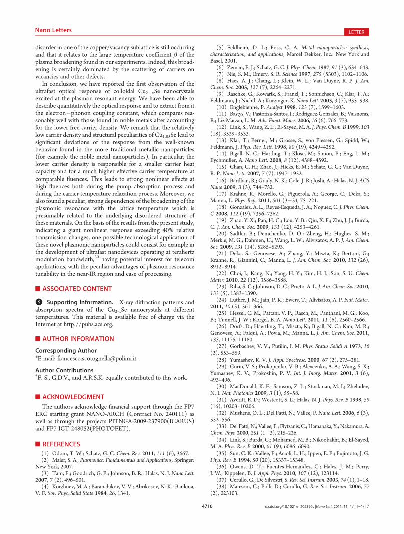
disorder in one of the copper/vacancy sublattice is still occurringand that it relates to the large temperature coefficient β of theplasma broadening found in our experiments. Indeed, this broad-ening is certainly dominated by the scattering of carriers onvacancies and other defects.
In conclusion, we have reported the first observation of theultrafast optical response of colloidal Cu2�xSe nanocrystalsexcited at the plasmon resonant energy. We have been able todescribe quantitatively the optical response and to extract from itthe electron�phonon coupling constant, which compares rea-sonably well with those found in noble metals after accountingfor the lower free carrier density. We remark that the relativelylow carrier density and structural peculiarities of Cu1.85Se lead tosignificant deviations of the response from the well-knownbehavior found in the more traditional metallic nanoparticles(for example the noble metal nanoparticles). In particular, thelower carrier density is responsible for a smaller carrier heatcapacity and for a much higher effective carrier temperature atcomparable fluences. This leads to strong nonlinear effects athigh fluences both during the pump absorption process andduring the carrier temperature relaxation process. Moreover, wealso found a peculiar, strong dependence of the broadening of theplasmonic resonance with the lattice temperature which ispresumably related to the underlying disordered structure ofthesematerials. On the basis of the results from the present study,indicating a giant nonlinear response exceeding 40% relativetransmission changes, one possible technological application ofthese novel plasmonic nanoparticles could consist for example inthe development of ultrafast nanodevices operating at terahertzmodulation bandwidth,30 having potential interest for telecomapplications, with the peculiar advantages of plasmon resonancetunability in the near-IR region and ease of processing.
’ASSOCIATED CONTENT
bS Supporting Information. X-ray diffraction patterns andabsorption spectra of the Cu2-xSe nanocrystals at differenttemperatures. This material is available free of charge via theInternet at http://pubs.acs.org.
’AUTHOR INFORMATION
Corresponding Author*E-mail: [email protected].
Author Contributions#F. S., G.D.V., and A.R.S.K. equally contributed to this work.
’ACKNOWLEDGMENT
The authors acknowledge financial support through the FP7ERC starting grant NANO-ARCH (Contract No. 240111) aswell as through the projects PITNGA-2009-237900(ICARUS)and FP7-ICT-248052(PHOTOFET).
’REFERENCES
(1) Odom, T. W.; Schatz, G. C. Chem. Rev. 2011, 111 (6), 3667.(2) Maier, S. A., Plasmonics: Fundamentals and Applications; Springer:
New York, 2007.(3) Tam, F.; Goodrich, G. P.; Johnson, B. R.; Halas, N. J. Nano Lett.
2007, 7 (2), 496–501.(4) Korzhuev, M. A.; Baranchikov, V. V.; Abrikosov, N. K.; Bankina,
V. F. Sov. Phys. Solid State 1984, 26, 1341.
(5) Feldheim, D. L.; Foss, C. A. Metal nanoparticles: synthesis,characterization, and applications; Marcel Dekker, Inc.: New York andBasel, 2001.
(6) Zeman, E. J.; Schatz, G. C. J. Phys. Chem. 1987, 91 (3), 634–643.(7) Nie, S. M.; Emery, S. R. Science 1997, 275 (5303), 1102–1106.(8) Haes, A. J.; Chang, L.; Klein, W. L.; Van Duyne, R. P. J. Am.
Chem. Soc. 2005, 127 (7), 2264–2271.(9) Raschke, G.; Kowarik, S.; Franzl, T.; Sonnichsen, C.; Klar, T. A.;
Feldmann, J.; Nichtl, A.; Kurzinger, K. Nano Lett. 2003, 3 (7), 935–938.(10) Englebienne, P. Analyst 1998, 123 (7), 1599–1603.(11) Bastys, V.; Pastoriza-Santos, I.; Rodriguez-Gonzalez, B.; Vaisnoras,
R.; Liz-Marzan, L. M. Adv. Funct. Mater. 2006, 16 (6), 766–773.(12) Link, S.; Wang, Z. L.; El-Sayed, M. A. J. Phys. Chem. B 1999, 103
(18), 3529–3533.(13) Klar, T.; Perner, M.; Grosse, S.; von Plessen, G.; Spirkl, W.;
Feldmann, J. Phys. Rev. Lett. 1998, 80 (19), 4249–4252.(14) Bigall, N. C.; Hartling, T.; Klose, M.; Simon, P.; Eng, L. M.;
Eychmuller, A. Nano Lett. 2008, 8 (12), 4588–4592.(15) Chan, G. H.; Zhao, J.; Hicks, E. M.; Schatz, G. C.; Van Duyne,
R. P. Nano Lett. 2007, 7 (7), 1947–1952.(16) Bardhan, R.; Grady, N. K.; Cole, J. R.; Joshi, A.; Halas, N. J.ACS
Nano 2009, 3 (3), 744–752.(17) Krahne, R.; Morello, G.; Figuerola, A.; George, C.; Deka, S.;
Manna, L. Phys. Rep. 2011, 501 (3�5), 75–221.(18) Gonzalez, A. L.; Reyes-Esqueda, J. A.; Noguez, C. J. Phys. Chem.
C 2008, 112 (19), 7356–7362.(19) Zhao, Y. X.; Pan, H. C.; Lou, Y. B.; Qiu, X. F.; Zhu, J. J.; Burda,
C. J. Am. Chem. Soc. 2009, 131 (12), 4253–4261.(20) Sadtler, B.; Demchenko, D. O.; Zheng, H.; Hughes, S. M.;
Merkle, M. G.; Dahmen, U.; Wang, L. W.; Alivisatos, A. P. J. Am. Chem.Soc. 2009, 131 (14), 5285–5293.
(21) Deka, S.; Genovese, A.; Zhang, Y.; Miszta, K.; Bertoni, G.;Krahne, R.; Giannini, C.; Manna, L. J. Am. Chem. Soc. 2010, 132 (26),8912–8914.
(22) Choi, J.; Kang, N.; Yang, H. Y.; Kim, H. J.; Son, S. U. Chem.Mater. 2010, 22 (12), 3586–3588.
(23) Riha, S. C.; Johnson, D. C.; Prieto, A. L. J. Am. Chem. Soc. 2010,133 (5), 1383–1390.
(24) Luther, J. M.; Jain, P. K.; Ewers, T.; Alivisatos, A. P. Nat. Mater.2011, 10 (5), 361–366.
(25) Hessel, C. M.; Pattani, V. P.; Rasch, M.; Panthani, M. G.; Koo,B.; Tunnell, J. W.; Korgel, B. A. Nano Lett. 2011, 11 (6), 2560–2566.
(26) Dorfs, D.; Haertling, T.; Miszta, K.; Bigall, N. C.; Kim, M. R.;Genovese, A.; Falqui, A.; Povia, M.; Manna, L. J. Am. Chem. Soc. 2011,133, 11175–11180.
(27) Gorbachev, V. V.; Putilin, I. M. Phys. Status Solidi A 1973, 16(2), 553–559.
(28) Yumashev, K. V. J. Appl. Spectrosc. 2000, 67 (2), 275–281.(29) Gurin, V. S.; Prokopenko, V. B.; Alexeenko, A. A.; Wang, S. X.;
Yumashev, K. V.; Prokoshin, P. V. Int. J. Inorg. Mater. 2001, 3 (6),493–496.
(30) MacDonald, K. F.; Samson, Z. L.; Stockman, M. I.; Zheludev,N. I. Nat. Photonics 2009, 3 (1), 55–58.
(31) Averitt, R. D.; Westcott, S. L.; Halas, N. J. Phys. Rev. B 1998, 58(16), 10203–10206.
(32) Muskens, O. L.; Del Fatti, N.; Vallee, F.Nano Lett. 2006, 6 (3),552–556.
(33) Del Fatti, N.; Vallee, F.; Flytzanis, C.; Hamanaka, Y.; Nakamura, A.Chem. Phys. 2000, 251 (1�3), 215–226.
(34) Link, S.; Burda, C.; Mohamed, M. B.; Nikoobakht, B.; El-Sayed,M. A. Phys. Rev. B 2000, 61 (9), 6086–6090.
(35) Sun, C. K.; Vallee, F.; Acioli, L. H.; Ippen, E. P.; Fujimoto, J. G.Phys. Rev. B 1994, 50 (20), 15337–15348.
(36) Owens, D. T.; Fuentes-Hernandez, C.; Hales, J. M.; Perry,J. W.; Kippelen, B. J. Appl. Phys. 2010, 107 (12), 123114.
(37) Cerullo, G.; De Silvestri, S. Rev. Sci. Instrum. 2003, 74 (1), 1–18.(38) Manzoni, C.; Polli, D.; Cerullo, G. Rev. Sci. Instrum. 2006, 77
(2), 023103.

4717 dx.doi.org/10.1021/nl202390s |Nano Lett. 2011, 11, 4711–4717
Nano Letters LETTER
(39) Abdullaev, G. B.; Aliyarova, Z. A.; Asadov, G. A. Phys. StatusSolidi 1967, 21, 461–464.(40) Al Mamun, A. B.; Islam, M. O.; Bhuiyan, A. H. J. Mater. Sci.:
Mater. Electron. 2005, 16 (5), 263–268.(41) Glazov, V. M.; Pashinkin, A. S.; Fedorov, V. A. Inorg. Mater.
2000, 36 (7), 641–652.(42) Lin, Z.; Zhigilei, L. V.; Celli, V. Phys. Rev. B 2008, 77 (7),
075133.(43) Kogut, A. N.; Mel’nik, A. I.; Mikolaichuk, A. G.; Romanishin,
B. M. Russ. Phys. J. 1973, 16 (8), 1113–1116.(44) De Montreuil, L. Econ. Geol. 1975, 70 (2), 384–387.(45) Garba, E. J. D.; Jacobs, R. L. Physica 1986, 138B, 253.(46) Vu�ci�c, Z.; Milat, O.; Horvati�c, V.; Ogorelec, Z. Phys. Rev. B
1981, 24, 5398.
Copyright © 2022 FDOKUMEN