Ascorbic acid metabolism during bilberry (Vaccinium myrtillus L.) fruit development

Toxicology Letters 133 (2002) 47–57
Plasmid DNA damage caused by methylated arsenicals,ascorbic acid and human liver ferritin
Sarfaraz Ahmad a, Kirk T. Kitchin b,*, William R. Cullen c
a Mercer Uni�ersity School of Medicine, Di�ision of Basic Medical Sciences, 1550 College Street, Macon, GA 31207, USAb En�ironmental Carcinogenesis Di�ision, National Health and En�ironmental Effects Research Laboratory, US En�ironmental
Protection Agency, MD-68, Research Triangle Park, NC 27711, USAc Chemistry Department, Uni�ersity of British Columbia, Vancou�er, Canada V6T 1Z1
Abstract
Both dimethylarsinic acid (DMA(V)) and dimethylarsinous acid (DMA(III)) release iron from human liver ferritin(HLF) with or without the presence of ascorbic acid. With ascorbic acid the rate of iron release from HLF byDMA(V) was intermediate (3.37 nM/min, P�0.05) and by DMA(III) was much higher (16.3 nM/min, P�0.001).No pBR322 plasmid DNA damage was observed from in vitro exposure to arsenate (iAs(V)), arsenite (iAs(III)),monomethylarsonic acid (MMA(V)), monomethylarsonous acid (MMA(III)) or DMA(V) alone. DNA damage wasobserved following DMA(III) exposure; coexposure to DMA(III) and HLF caused more DNA damage; considerablyhigher amounts of DNA damage was caused by coexposure of DMA(III), HLF and ascorbic acid. Diethylenetri-aminepentaacetic acid (an iron chelator), significantly inhibited DNA damage. Addition of catalase (which canincrease Fe2+ concentrations) further increased the plasmid DNA damage. Iron-dependent DNA damage could bea mechanism of action of human arsenic carcinogenesis. © 2002 Elsevier Science Ireland Ltd. All rights reserved.
Keywords: Arsenic; Iron; Human liver ferritin; DNA damage; Reactive oxygen species; Dimethylarsinous acid, DMA(III)
www.elsevier.com/locate/toxlet
1. Introduction
Arsenic is one of the most important globalenvironmental toxicants. Arsenic commonly existsin +3 and +5 valence states and in a number ofinorganic and organic forms. There is clear evi-dence from studies in humans that arsenic expo-sures are associated with cancer of the skin, lungs,liver, kidney, and bladder. Although themetabolism of inorganic arsenic is fairly well un-derstood, the precise biochemical mechanism ofarsenic-induced carcinogenicity in humans is notknown. Both in vivo and in vitro studies showthat trivalent arsenical forms are more toxic thanthe corresponding pentavalent forms (Ahmad et
Abbre�iations: iAs(V), arsenate; iAs(III), arsenite; DMA (V),dimethylarsinic acid; DMA(III), dimethylarsinous acid;DTPA, diethylenetriaminepentaacetic acid; EDTA, ethylenedi-aminetetraacetic acid; HLF, human liver ferritin; L, linearform of plasmid pBR322 DNA (after a double-strandedbreak); MMA (V), monomethylarsonic acid; MMA(III),monomethylarsonous acid; OC, opencoiled plasmid pBR322DNA (after a single-stranded break); ROS, reactive oxygenspecies; SC, supercoiled plasmid pBR322 DNA; SOD, super-oxide dismutase.
* Corresponding author. Tel.: +1-919-541-7502; fax: +1-919-541-0694.
E-mail address: [email protected] (K.T. Kitchin).
0378-4274/02/$ - see front matter © 2002 Elsevier Science Ireland Ltd. All rights reserved.
PII: S0 378 -4274 (02 )00079 -6

S. Ahmad et al. / Toxicology Letters 133 (2002) 47–5748
al., 2000a; Styblo et al., 2000; Petrik et al., 2000).In biological systems pentavalent arsenicals arereduced to trivalency in the presence of endoge-nous reducing agents. Earlier it was believed thatmethylation of the inorganic form of arsenic(iAs(V) or iAs(III)) is a detoxification pathway.But studies from our laboratory and others haveshown that some methylated arsenicals are evenmore cytotoxic and genotoxic than inorganic ar-senic (Brown et al., 1997; Ahmad et al., 1999;Petrik et al., 2000; Mass et al., 2001).
The free radical theory of arsenic carcinogenesishas been gaining acceptance (Yamanaka et al.,1991; Yamanaka and Okada, 1994; Ahmad et al.,2000a; Kitchin, 2001). Exposure to arsenic maygenerate reactive oxygen species (ROS) in vivo(such as dimethylarsenic radical ((CH3)2As�),dimethylarsenic peroxyl radicals ((CH3)2AsOO�),superoxide anion, singlet oxygen and hydroxylradicals) and be responsible for cellular toxicityand/or carcinogenicity (Yamanaka and Okada,1994; Barchowsky et al., 1996; Lynn et al., 1998).Earlier we reported that acute exposure to methy-lated arsenicals led to a significant decrease incellular glutathione in rats (Brown et al., 1997)and mice (Ahmad et al., 1999). Glutathione deple-tion may be correlated with ROS-mediated oxida-tive stress. We also have reported varying degreesof DNA damage caused by arsenicals— in ratlungs by DMA(V) (Brown et al., 1997), in mouseliver by DMA(V) (Ahmad et al., 1999) and in anin vitro system containing calf thymus DNA andbleomycin by DMA(III) (Ahmad et al., 2000a).Iron is both an essential element and the mostabundant transition metal found in biological sys-tems because of its important role in major bio-logical reactions such as the oxygen transport,energy production, toxification and detoxificationreactions (Thiel, 1987). The iron storage protein,ferritin, plays a key role in iron storage, utiliza-tion and metabolism. Despite the essential natureof iron, it is toxic when present in excess concen-trations in cells. Free iron causes redox cycling,production of ROS and oxidative stress.
In this study we further demonstrate the inter-action between DMA(V) or DMA(III) with HLF.We also investigated the role of iron released fromHLF by arsenic species in generating ROS andinducing significant plasmid DNA damage.
2. Materials and methods
2.1. Materials
pBR322 plasmid DNA was initially purchasedfrom three different suppliers and evaluated inorder to select a plasmid DNA source containingas few nicks (open circular (OC) form) as possi-ble. ICN Biochemicals (Aurora, OH) human liverferritin (purified) and pBR322 plasmid DNA werethen purchased and utilized. Agarose, ferrozine(3 - (2 - pyridyl) - 5,6 - bis(4 - phenyl - sulfonic acid) -1,2,4-triazine), ascorbic acid, superoxide dismu-tase (SOD) (bovine erythrocyte), catalase (bovineliver), ferrous sulfate, sodium azide, ethylenedi-aminetetraacetic acid (EDTA) and diethylenetri-aminepentaacetic acid (DTPA) were purchasedfrom Sigma Chemical Co. (St. Louis, MO).Sodium arsenate (certified A.C.S. grade), sodiumarsenite (certified grade) and potassium iodidewere obtained from Fisher Scientific Company.Other important materials were sodiummonomethylarsonic acid (MMA(V)) (from Chem-ical Services, West Chester, PA), dimethylarsinicacid (DMA(V)) (from Ansul Corp., Weslaco, TX)and D-mannitol (from Aldrich Chemical Co., Mil-waukee, WI). Methylarsine oxide (the tetramer(CH3AsIIIO)4; (MMA(III)) (a source of MMA(III)in solution)) and iododimethylarsine ((CH3)2
AsIIII; (DMA(III))) were synthesized using previ-ously described methods (Cullen et al., 1984; Sty-blo et al., 1997). Identity and purity of thesynthesized arsenicals were confirmed using 1H-NMR, mass spectrometry and hydride generationatomic absorption spectrophotometry. All otherchemicals were of analytical grade quality or bet-ter and used without further purification.
2.2. Iron mobilization assay
Iron release from HLF by DMA(V) andDMA(III) was investigated with or without ascor-bic acid. Release of iron from HLF under aerobicconditions utilized a previously described assayprocedure (Ahmad et al., 2000a; Double et al.,1998; Monteiro and Winterbourn, 1989). The as-say system (total volume 150 �l) contained HLF(2 �g), ferrozine (0.5 mM), DMA(V)/DMA(III)

S. Ahmad et al. / Toxicology Letters 133 (2002) 47–57 49
(each 10 mM) with or without ascorbic acid (250�M) in 10 mM phosphate buffered saline (pH7.4). The reaction was initiated by the addition ofHLF and time-dependent formation of Fe(fer-rozine)3]2+-complex was measured at 562 nm in amicrotiter plate. The amount of iron releasedfrom HLF is expressed as nM Fe2+ released perminute, using ferrous sulfate as an iron standard.The rate of iron release was linear from 0–15 min.The iron release measurements (nM/min) weretaken only during this initial linear phase ofreaction.
2.3. Clea�age of plasmid pBR322 DNA
Electrophoresis was carried out in 1% agarosegels (Sambrook et al., 1989) as previously de-scribed (Ahmad et al., 2000b; Di Mascio et al.,2000). Depending on the protocol, plasmidpBR322 DNA was treated with either of six dif-ferent arsenicals with or without the presence ofHLF, ascorbic acid, the iron chelator (DTPA)and oxyradical scavengers for the specific periodof time at 37 °C. Briefly, the incubation mixture intotal volume of 50 �l contained: 500 ng pBR322DNA, arsenic species (0–1.0 mM), HLF (10 �g),ascorbic acid (25 �M), DTPA (5 mM) or oxyrad-ical scavengers (such as SOD (150 units), catalase(50 units), D-mannitol (5 mM), potassium iodide(5 mM), sodium azide (5 mM)) in 10 mM phos-phate-buffered saline (pH 7.4). The contents weremixed well and incubated at 37 °C in a water bathfor 2 h. Then 5 �l of gel loading solution contain-ing 40 �M EDTA, 0.05% bromophenol bluetracking dye and 50% (v/v) sucrose was addedand the whole reaction mixture was loaded into a1% agarose gel for electrophoresis for 3 h at 50mA current. The agarose gels contained 0.5 �g/mlethidium bromide. The DNA bands were visual-ized under UV light and photographs were takenusing a Kodak Digital 1D Image Analysis Soft-ware (Version 3.0). Figs. 1–6 are representativeagarose gels taken from two to four separatereplicates of each experiment performed on sepa-rate days.
In our experience, pBR322 plasmid DNA al-ways contains some DNA in the OC form al-though it is normally free of linear form (L)
Fig. 1. Agarose gel electrophoresis of plasmid pBR322 DNAafter treatment with arsenic species (1 mM each) alone. Lane(a) control DNA; lane (b) plus iAs(V); lane (c) plus iAs(III);lane (d) plus MMA(V); lane (e) plus MMA(III); lane (f) plusDMA(V); lane (g) plus DMA(III) and lane (h) plus EcoRIenzyme. Experimental conditions are described under Section2.
DNA. Thus, after agarose gel electrophoresis,even control lanes of pBR322 plasmid DNA showsome amount of OC DNA (for example see lane aof Fig. 1). In order to better demonstrate theappearance of L form of DNA (e.g. lane e of Fig.6), 500 ng of DNA was loaded into each lane foragarose electrophoresis. The presence of L formDNA is particularly germane to clastogenicitywhich has been a frequent finding of arsenic expo-sures both in vitro and in vivo. This high a degreeof DNA loading (500 ng) makes both the super-coiled (SC) and OC bands appear fairly thick andbright. The basis of an experimental finding of
Fig. 2. Agarose gel electrophoresis of plasmid pBR322 DNAafter treatment with arsenic species (1 mM each) and HLF (10�g). Lane (a and h) control DNA with HLF; lane (b) plusiAs(V); lane (c) plus iAs(III); lane (d) plus MMA(V); lane (e)plus MMA(III); lane (f) plus DMA(V); lane (g) plusDMA(III). Experimental conditions are described under Sec-tion 2.

S. Ahmad et al. / Toxicology Letters 133 (2002) 47–5750
Fig. 3. Agarose gel electrophoresis of plasmid pBR322 DNAafter treatment with arsenic species (1 mM each), HLF (10 �g)and ascorbic acid (25 �M). Lane (a and h) control DNA withHLF; lane (b) plus iAs(V) and ascorbic acid; lane (c) plusiAs(III) and ascorbic acid; lane (d) plus MMA(V) and ascorbicacid; lane (e) plus MMA(III) and ascorbic acid; lane (f) plusDMA(V) and ascorbic acid; lane (g) plus DMA(III) andascorbic acid. Experimental conditions are described underSection 2.
Fig. 5. Effects of DTPA on plasmid pBR322 DNA damage inthe presence of DMA(III) (1 mM), HLF (10 �g) and ascorbicacid (25 �M). Lane (a and h) control DNA with HLF; lane (b)plus DMA(III) and DTPA (5 mM); lane (c) plus ascorbic acidand DTPA (5 mM); lane (d) plus DMA(III) and ascorbic acid;lane (e) plus DMA(III), ascorbic acid and DTPA (1 mM); lane(f) plus DMA(III), ascorbic acid and DTPA (5 mM); lane (g)plus DTPA (5 mM). Experimental conditions are describedunder Section 2.
increased OC or L form of plasmid DNA was thequalitative, visual comparison of the size andintensities of the OC or L DNA band.
2.4. Statistical analysis
Statistical analysis of the raw data employedanalysis of variance (ANOVA). For a statistical
test of possible chemical synergy, estimation of anadditivity surface of raw data was done by theinteraction test of a 2×2 factorial design analysisof variance (i.e. a design for two chemicals). Avalue of P�0.05 was considered to be statisticallysignificant.
Fig. 6. Effects of different ROS scavengers on plasmidpBR322 DNA damage in the presence of DMA(III) (1.0 mM),HLF (10 �g) and ascorbic acid (25 �M). Lane (a) controlDNA; lane (b) plus HLF; lane (c) plus DMA(III), ascorbicacid and HLF; lane (d) plus SOD (150 units), DMA(III),ascorbic acid and HLF; lane (e) plus catalase (50 units),DMA(III), ascorbic acid and HLF; lane (f) plus mannitol (5mM), DMA(III), ascorbic acid and HLF; lane (g) plus potas-sium iodide (5 mM), DMA(III), ascorbic acid and HLF; lane(h) plus sodium azide (5 mM), DMA(III), ascorbic acid andHLF. Experimental conditions are described under Section 2.
Fig. 4. Agarose gel electrophoresis of plasmid pBR322 DNAafter treatment with increasing concentration of DMA(III)(0–1 mM), HLF (10 �g) and ascorbic acid (25 �M). Lane (aand h) control DNA with HLF; lane (b) plus DMA(III) (1mM); lane (c) plus ascorbic acid; lane (d) plus DMA(III) (0.25mM) and ascorbic acid; lane (e) plus DMA(III) (0.5 mM) andascorbic acid; lane (f) plus DMA(III) (0.75 mM) and ascorbicacid; lane (g) plus DMA(III) (1 mM) and ascorbic acid.Experimental conditions are described under Section 2.
S. Ahmad et al. / Toxicology Letters 133 (2002) 47–57 51
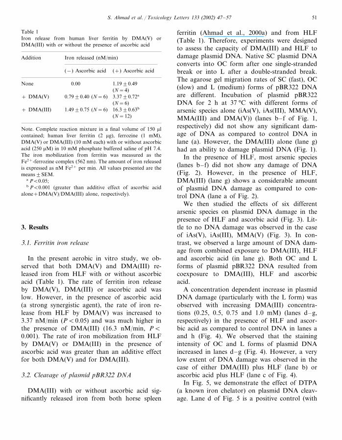
Table 1Iron release from human liver ferritin by DMA(V) orDMA(III) with or without the presence of ascorbic acid
Iron released (nM/min)Addition
(−) Ascorbic acid (+) Ascorbic acid
0.00None 1.19�0.49(N=4)
+ DMA(V) 3.37�0.72a0.79�0.40 (N=6)(N=6)
1.49�0.75 (N=6)+ DMA(III) 16.3�0.63b
(N=12)
Note. Complete reaction mixture in a final volume of 150 �lcontained; human liver ferritin (2 �g), ferrozine (1 mM),DMA(V) or DMA(III) (10 mM each) with or without ascorbicacid (250 �M) in 10 mM phosphate buffered saline of pH 7.4.The iron mobilization from ferritin was measured as theFe2+-ferrozine complex (562 nm). The amount of iron releasedis expressed as nM Fe2+ per min. All values presented are themeans�SEM.
a P�0.05;b P�0.001 (greater than additive effect of ascorbic acid
alone+DMA(V)/DMA(III) alone, respectively).
ferritin (Ahmad et al., 2000a) and from HLF(Table 1). Therefore, experiments were designedto assess the capacity of DMA(III) and HLF todamage plasmid DNA. Native SC plasmid DNAconverts into OC form after one single-strandedbreak or into L after a double-stranded break.The agarose gel migration rates of SC (fast), OC(slow) and L (medium) forms of pBR322 DNAare different. Incubation of plasmid pBR322DNA for 2 h at 37 °C with different forms ofarsenic species alone (iAs(V), iAs(III), MMA(V),MMA(III) and DMA(V)) (lanes b– f of Fig. 1,respectively) did not show any significant dam-age of DNA as compared to control DNA inlane (a). However, the DMA(III) alone (lane g)had an ability to damage plasmid DNA (Fig. 1).
In the presence of HLF, most arsenic species(lanes b– f) did not show any damage of DNA(Fig. 2). However, in the presence of HLF,DMA(III) (lane g) shows a considerable amountof plasmid DNA damage as compared to con-trol DNA (lane a of Fig. 2).
We then studied the effects of six differentarsenic species on plasmid DNA damage in thepresence of HLF and ascorbic acid (Fig. 3). Lit-tle to no DNA damage was observed in the caseof iAs(V), iAs(III), MMA(V) (Fig. 3). In con-trast, we observed a large amount of DNA dam-age from combined exposure to DMA(III), HLFand ascorbic acid (in lane g). Both OC and Lforms of plasmid pBR322 DNA resulted fromcoexposure to DMA(III), HLF and ascorbicacid.
A concentration dependent increase in plasmidDNA damage (particularly with the L form) wasobserved with increasing DMA(III) concentra-tions (0.25, 0.5, 0.75 and 1.0 mM) (lanes d–g,respectively) in the presence of HLF and ascor-bic acid as compared to control DNA in lanes aand h (Fig. 4). We observed that the stainingintensity of OC and L forms of plasmid DNAincreased in lanes d–g (Fig. 4). However, a verylow extent of DNA damage was observed in thecase of either DMA(III) plus HLF (lane b) orascorbic acid plus HLF (lane c of Fig. 4).
In Fig. 5, we demonstrate the effect of DTPA(a known iron chelator) on plasmid DNA cleav-age. Lane d of Fig. 5 is a positive control (with
3. Results
3.1. Ferritin iron release
In the present aerobic in vitro study, we ob-served that both DMA(V) and DMA(III) re-leased iron from HLF with or without ascorbicacid (Table 1). The rate of ferritin iron releaseby DMA(V), DMA(III) or ascorbic acid waslow. However, in the presence of ascorbic acid(a strong synergistic agent), the rate of iron re-lease from HLF by DMA(V) was increased to3.37 nM/min (P�0.05) and was much higher inthe presence of DMA(III) (16.3 nM/min, P�0.001). The rate of iron mobilization from HLFby DMA(V) or DMA(III) in the presence ofascorbic acid was greater than an additive effectfor both DMA(V) and for DMA(III).
3.2. Clea�age of plasmid pBR322 DNA
DMA(III) with or without ascorbic acid sig-nificantly released iron from both horse spleen

S. Ahmad et al. / Toxicology Letters 133 (2002) 47–5752
HLF, DMA(III) and ascorbic acid but withoutDTPA) and shows maximum DNA damage.However, the presence of DTPA in the reactionmixture (1 mM in lane e or 5 mM in lane f) alongwith HLF, DMA(III) and ascorbic acid signifi-cantly decreased the DNA damage. DTPA signifi-cantly diminished the plasmid DNA cleavage bychelating the iron released from HLF. Lane b ofFig. 5 (HLF and DMA(III)) shows some DNAdamage even in the presence of 5 mM DTPAwhen compared to DNA in lanes a and h whichlack DMA(III). This may be due to direct interac-tion between DMA(III) and DNA. The DNA inlanes c (HLF, ascorbic acid and DTPA) and laneg (HLF and DTPA) did not show any DNAdamage (Fig. 5).
Finally, we showed the effects of different ROSscavengers on plasmid DNA damage caused byiron released from HLF by DMA(III) and ascor-bic acid (Fig. 6). Superoxide dismutase (SOD) andcatalase remove superoxide anions and hydrogenperoxide, respectively. Mannitol and potassiumiodide reduce hydroxyl radical concentration.Sodium azide is a singlet oxygen scavenger. InFig. 6, lanes a and b were control lanes whichcontain only plasmid DNA or DNA with HLF,respectively. Lane c served as a positive controland contained the target DNA, DMA(III), HLFand ascorbic acid without any ROS scavengers.Inhibition of plasmid DNA damage by SOD (150units) was small (Fig. 6, lane d). However, in thepresence of catalase (50 units) (lane e) the degreeof DNA damage was actually increased to a quitesevere amount (particularly of the L form). Weobserved some DNA remaining at the origin inlane e. Crosslinking of pBR322 DNA into largermolecular weight species may have occurred inlane e. In these experiments, catalase increased thedegree of DNA damage found by decreasing thehydrogen peroxide concentration which in turnsallows ferrous iron concentration to increase (Ah-mad et al., 2000a). The hydroxy radical scavenger,mannitol (5 mM) (lane f) did not clearly show anyprotection against DNA damage. However,potassium iodide (lane g) and sodium azide (laneh) (each 5 mM) showed a demonstrable degree ofprotection from DNA damage.
4. Discussion
Arsenic is commonly found in the environmentand is a known human carcinogen (NRC, 1999;IARC, 1987). Generally, biomethylation of inor-ganic arsenicals has been considered a majordetoxification pathway. However, recent biochem-ical studies showed that some methylated arseni-cals (MMA(III) and DMA(III)) are even moretoxic than the parent iAs(V) and iAs(III) (Stybloet al., 2000; Petrik et al., 2000). Our lab studiesalso indicate that DMA may induce oxidativestress in Sprague–Dawley rats (Brown et al.,1997) and in B6C3F1 mice (Ahmad et al., 1999).The US EPA has classified DMA as a probablehuman carcinogen (Group B-2) on the basis offeeding studies in B6C3F1 mice and Fischer F344rats (US EPA, 1994). DMA has been identified asa complete carcinogen in rat urinary bladder,mouse fibrosarcomas (US EPA, 1994) and in K6/ODC transgenic mouse skin (Chen et al., 2000).DMA is a promoter of carcinogenesis in mouselung (Yamanaka et al., 1996), and in rat liver,kidney, urinary bladder and thyroid gland (Ya-mamoto et al., 1995).
Recent publications demonstrate the involve-ment of free radicals in arsenic-mediated cellulartoxicity (Barchowsky et al., 1996; Lynn et al.,1998). According to Yamanaka and Okada(1994), metabolism of DMA causes the formationof either dimethylarsenic radical, dimethylarsenicperoxyl radical, superoxide anion or hydroxylradicals in vivo. Our previous in vitro studiesshowed that all arsenic forms release iron fromferritin to some extent (Ahmad et al., 2000a).However, methylated arsenicals (particularlyDMA(III)) release a larger amount of iron fromferritin as compared to other arsenic species.Wang (1996) has shown increased iron content inblood, urine and hair samples of arsenic-exposedpatients suffering from blackfoot disease (gan-grene of several peripheral areas such as toes, feetand fingers due to high arsenic exposure). Lee andHo (1995) have noted a dose-dependent inductionof heme oxygenase and ferritin content in sodiumarsenite treated human fibroblast cells. Thus, therelease of iron from the enzymatic action of hemeoxygenase may cause the induction of ferritin (the

S. Ahmad et al. / Toxicology Letters 133 (2002) 47–57 53
principal iron storage protein) synthesis. Arsenictreatment has caused release of heme iron in ratfibroblasts (Eisentein et al., 1991), increased hemeoxygenase activity in rat liver (Brown andKitchin, 1996) and kidney (Kitchin et al., 1999)and induction of metallothionein-II and ferritin HmRNA level in HeLa cells (Guzzo et al., 1994).Earlier studies showed that high body iron storesand dietary iron intakes are associated with can-cer (Toyokuni, 1996; Diwan et al., 1997).
No previous pharmacodynamic studies of pos-sible interactions between arsenic species andtransition metals (particularly iron) were found.We first reported that methylated arsenicals (par-ticularly DMA(V) and DMA(III)) reduced andmobilized the iron from horse spleen ferritin aloneor in the presence of ascorbic acid (Ahmad et al.,2000a). In the present in vitro studies we observedthat DMA(V) and DMA(III) significantly re-leased iron from HLF and that DMA(III) causediron-mediated plasmid DNA damage. The DNAdamage by DMA(III) was synergistically in-creased in the presence of ascorbic acid. This maybe due to an increased amount of iron releasefrom HLF by DMA(III) in the presence of ascor-bic acid (Ahmad et al., 2000a). Recently, theprotein ferritin has been shown to exist in thenuclear as well as the cytoplasmic compartment(Pountney et al., 1999; Thompson et al., 2000).We noted that the presence of an iron chelator(DTPA) significantly diminished the plasmidDNA cleavage (Fig. 5). This could be due tochelation of free iron (released from HLF) byDTPA. This indicates that iron released fromHLF by DMA(III) and ascorbic acid plays a rolein the cleavage of plasmid DNA.
It is well known that ‘‘free’’ iron causes ROSformation and catalyzes deleterious oxidation re-actions which damage DNA, lipid and protein(Aust et al., 1985). Strictly speaking, iron is neverreally free as it is normally chelated with endoge-nous ligands. We studied the effects of differentROS scavengers on plasmid DNA damage causedby iron released from HLF by DMA(III) andascorbic acid. Potassium iodide reduced to somedegree the amount of DNA damage, indicatingsome involvement of hydroxyl radicals in thereaction.
Antagonism of DNA damage was observedwith SOD, potassium iodide and sodium azide.Antagonism by sodium azide indicates significantinvolvement of singlet oxygen in this iron medi-ated plasmid DNA damage caused by exposure toDMA(III), HLF and ascorbic acid. Completeprotection was not achieved by any of these indi-vidual ROS scavengers. This indicates that besidesthe suspected free radical species (1) there may besome other reactive intermediate(s) present or (2)a direct interaction between DMA(III) and DNAmay occur.
The human genome is vulnerable to damageand/or mutation by ROS (Meneghini and Martin,1993). ROS (superoxide anion, hydrogen perox-ide, singlet oxygen and hydroxyl radical) directlyor indirectly cause a broad range of DNA dam-age. Evidence from our studies for the involve-ment of particular free radicals in arseniccarcinogenesis includes: free iron (ferrous) (Table1, Figs. 5 and 6 of this study; tables 1–4 ofAhmad et al., 2000a), superoxide anion (Fig. 6 ofthis study), hydrogen peroxide (Fig. 6), hydroxylradical (Fig. 6) and singlet oxygen (Fig. 6). Singletoxygen can be generated by the reaction of super-oxide with hydrogen peroxide (Mao et al., 1995).Evidence for the involvement of particular freeradicals in arsenic carcinogenesis from the studiesof others includes free radicals generated by expo-sure to either iAs(III) or iAs(V) (Liu et al., 2000),dimethylarsenic radical (Yamanaka et al., 1989,1990), dimethylarsenic peroxyl radical, (Tezuka etal., 1993; Yamanaka et al., 1990, 1995), hydroxylradical (Fukushima, personal communication),superoxide anion (Barchowsky et al., 1999; Ya-manaka et al., 1991) and hydrogen peroxide (Bar-chowsky et al., 1999; Yamanaka and Okada,1994).
All of the five human organs with high riskratios for arsenic induced cancer have one ormore factors which contribute to free radical me-diated carcinogenesis (Table 2). After exposure tosome form of arsenic, experimental animals havealso developed cancer in each of these five organs.Summaries of arsenic, ferritin, ascorbic acid, glu-tathione, oxygen concentrations and blood flow inthese five mammalian organs susceptible to ar-senic carcinogenesis are included in Table 2. Ac-

S. Ahmad et al. / Toxicology Letters 133 (2002) 47–5754
Tab
le2
Ars
enic
,fe
rrit
in,
asco
rbic
acid
,gl
utat
hion
e,ox
ygen
conc
entr
atio
nan
dbl
ood
flow
infiv
em
amm
alia
nor
gans
inw
hich
arse
nic-
med
iate
dca
ncer
has
been
repo
rted
inhu
man
san
dex
peri
men
tal
anim
als
Liv
erK
idne
yO
rgan
sSk
inL
ung
Bla
dder
Low
Low
Hig
hF
erri
tin
conc
entr
atio
nL
owL
owM
ediu
mL
owH
igh
(cyt
ochr
omes
P-4
50,
Low
Non
-fer
riti
nir
onM
ediu
m(h
eme
oxyg
enas
ehe
me
oxyg
enas
ein
duct
ion
indu
ctio
n)co
ncen
trat
ion
and
othe
rno
n-fe
rrit
in(h
eme-
prot
ein,
free
iron
)ir
on)
Hig
hD
MA
,M
MA
Per
sist
ent
arse
nic
leve
lsP
ersi
sten
tar
seni
cle
vels
Hig
hM
MA
and
DM
AA
rsen
icco
ncen
trat
ion,
Hig
hM
MA
and
DM
Aex
posu
re.
expo
sure
from
urin
e.pe
rsis
tenc
ean
dex
posu
re.
As(
III)
C�
MM
A(V
)(M
MA
(III
)or
DM
A(I
II)
met
abol
ism
coul
dbe
aca
rcin
ogen
icM
MA
(III
)C�
DM
A(V
)sp
ecie
sin
hum
anbl
adde
r)H
igh
Low
Low
Hig
hH
igh
End
ogen
ous
redu
cing
agen
ts(s
uch
asas
corb
icac
idor
GSH
)L
owL
owL
owO
xyge
npa
rtia
lpr
essu
reH
igh
Low
Yes
(++
+)a
Yes
(+)
Yes
(+)
Yes
(++
+)
Ars
enic
-med
iate
dca
ncer
Yes
(++
+)
inhu
man
sD
MA
acts
asa
prom
oter
DM
Aac
tsas
atu
mor
DM
Aac
tsas
atu
mor
DM
Aac
tsas
atu
mor
DM
A(V
)-m
edia
ted
DM
Aac
tsas
atu
mor
prom
oter
inra
tspr
omot
erin
mic
e.pr
omot
erin
rats
prom
oter
and
aco
mpl
ete
and
aco
mpl
ete
canc
erin
carc
inog
enin
K6/
OD
Cex
peri
men
tal
anim
als
carc
inog
enin
rats
.tr
ansg
enic
mic
e.1
42
1N
umbe
rof
favo
rabl
e3
fact
ors
for
RO
S-m
edia
ted
arse
nic
carc
inog
enes
is
aT
henu
mbe
rof
(+)
indi
cate
sth
eri
skra
tio
ofar
seni
cca
rcin
ogen
esis
inhu
man
s.

S. Ahmad et al. / Toxicology Letters 133 (2002) 47–57 55
cumulation of inorganic arsenic and its methyl-ated metabolites have been reported in liver,spleen, kidneys, lungs, urinary bladder and skin inthe first 24 h after arsenic administration in ani-mals and man (Vallee et al., 1960; Yamauchi andYamamura, 1983; Vahter et al., 1984; Hughes etal., 1994).
Each of these five organs is quite different inthe way that arsenic exposure could result incarcinogenesis via iron-mediated ROS. For exam-ple, the lung has high blood flow, high oxygenpartial pressure, high concentration of reducingagents and persistent arsenic levels after chronicarsenic exposure. The bladder has high DMA andMMA exposure from urine. DMA(III) could be acarcinogenic arsenic species in human bladder.The bladder, as best we know today, lacks arsenicmethylation enzyme activity but possesses theability to reduce pentavalent arsenic to trivalentforms. Sampayo-Reyes et al. (2000) recentlyquantitated MMA(V) reductase activity in ham-ster testis, kidney, skin, heart, lung, liver, spleen,bladder and brain homogenates. Brain had thehighest MMA(V) reductase activity. Bladder, atarget tissue for arsenic carcinogenesis, had thesecond highest MMA(V) reductase activity (twicethat of any of the remaining seven tissues) (Sam-payo-Reyes et al., 2000). The same authors alsodetected MMA(III) and DMA(III) in the liver asin vivo metabolites of iAs(V). The skin has persis-tent arsenic levels after chronic arsenic exposureprobably due to skin’s high keratin content. Theliver has several factors favoring ROS-mediatedarsenic carcinogenesis (Table 2), but a lower de-gree of carcinogenic response in both animals andman. This may be because (i) there are manyfactors other than just DNA damage and initialmutation that are required for multi-stage car-cinogenesis and (ii) human liver is quite resistantto carcinogenicity in general.
Based on the results of this and a previousstudy (Ahmad et al., 2000a), we suggest thatmethylated arsenicals and ascorbic acid cause ironrelease from HLF, iron-dependent ROS forma-tion in vivo and ROS-induced DNA damage. ThisDNA damage could be a mechanism of action ofarsenic carcinogenesis in humans. However, thisROS-mode of action of arsenic and ferritin re-
leased iron does not exclude participation of otherpathways of arsenic carcinogenesis (Kitchin,2001).
5. Disclaimer
This manuscript has been reviewed in accor-dance with the policy of the National Health andEnvironmental Effects Research Laboratory, USEnvironmental Protection Agency, and approvedfor publication. Approval does not signify thatthe contents necessarily reflect the views and poli-cies of the Agency, nor does mention of tradenames of commercial products constitute endorse-ment or recommendation for use.
Acknowledgements
This work was partially supported by a grantfrom the National Research Council, Washing-ton, DC, to a postdoctoral fellow (Sarfaraz Ah-mad) at the US Environmental ProtectionAgency, North Carolina.
References
Ahmad, S., Anderson, W.L., Kitchin, K.T., 1999. Dimethyl-arsinic acid effects on DNA damage and oxidative stressrelated biochemical parameters in B6C3F1 mice. CancerLett. 139, 129–135.
Ahmad, S., Kitchin, K.T., Cullen, W.R., 2000a. Arsenic spe-cies that cause release of iron from ferritin and generationof activated oxygen. Arch. Biochem. Biophys. 382, 195–202.
Ahmad, S., Agrawal, R., Agrawal, D.K., Rao, G.S., 2000b.Bioreactivity of glutathionyl hydroquinone with implica-tions to benzene toxicity. Toxicology 150, 31–39.
Aust, S.D., Morehouse, L.A., Thomas, C.E., 1985. Role ofmetals in oxygen radical reaction. J. Free Rad. Biol. Med.1, 3–25.
Barchowsky, A., Dudek, E.J., Treadwell, M.D., Wetterhahn,K.E., 1996. Arsenic induces oxidant stress and NF-kappa-B activation in cultured aortic endothelial cells. Free Rad.Biol. Med. 21, 783–790.
Barchowsky, A., Klei, L.R., Dudek, E.J., Swartz, H.M.,James, P.E., 1999. Stimulation of reactive oxygen, but notreactive nitrogen species, in vascular endothelial cells ex-posed to low levels of arsenite. Free. Rad. Biol. Med. 27,1405–1412.

S. Ahmad et al. / Toxicology Letters 133 (2002) 47–5756
Brown, J.L., Kitchin, K.T., 1996. Arsenic, but not cadmium,induces ornithine decarboxylase and heme oxygenase activ-ity in rat liver—relevance to arsenic carcinogenesis. CancerLett. 98, 227–231.
Brown, J.L., Kitchin, K.T., George, M., 1997. Dimethylarsinicacid treatment alters six different rat biochemical parame-ters: relevance to arsenic carcinogenesis. Teratog. Car-cinog. Mutag. 17, 71–84.
Chen, Y., Megosh, L.C., Gilmour, S.K., Sawicki, J.A.,O’Brien, T.G., 2000. K6/ODC transgenic mice as a sensi-tive model for carcinogen identification. Toxicol. Lett. 116,27–35.
Cullen, W.R., McBride, B.C., Reglinski, J., 1984. The reactionof methylarsenicals with thiols: some biological implica-tions. J. Inorg. Biochem. 21, 179–194.
Di Mascio, P., Teixeira, P.C., Onuki, J., Mereiros, M.H.G.,Dornemann, D., Douki, T., Cadet, J., 2000. DNA damageby 5-aminolevulinic and 4,5-dioxovaleric acids in the pres-ence of ferritin. Arch. Biochem. Biophys. 373, 368–374.
Diwan, B.A., Kasprazak, K.S., Anderson, L.M., 1997. Promo-tion of dimethylbenz[a]anthracene-initiated mammary car-cinogenesis by iron in female Sprague–Dawley rats.Carcinogenesis 18, 1757–1762.
Double, K.L., Maywald, M., Schmittel, M., Riederer, P.,Gerlach, M., 1998. In vitro studies of ferritin iron releaseand neurotoxicity. J. Neurochem. 70, 2492–2499.
Eisentein, R.S., Garcia, M.D., Pettingell, W., Munro, H.N.,1991. Regulation of ferritin and heme oxygenase synthesisin rat fibroblasts by different forms of iron. Proc. Natl.Acad. Sci. USA 88, 688–692.
Guzzo, A., Karatzios, C., Diorio, C., Dubow, M.S., 1994.Metallothionein-II and ferritin H mRNA levels are in-creased in arsenite-exposed HeLa cells. Biochem. Biophys.Res. Commun. 205, 590–595.
Hughes, M.F., Menache, M., Thompson, D.J., 1994. Dose-de-pendent disposition of sodium arsenate in mice followingacute oral exposure. Fund. Appl. Toxicol. 22, 80–89.
International Agency for Research on Cancer (IARC), 1987.In: IARC Monograph on the Evaluation of CarcinogenicRisks to Humans: Overall Evaluations of Carcinogenicity:An update of IARC Monographs 1–42 (Suppl. 7), Interna-tional Agency for Research on Cancer, Lyon, France, pp.100–106.
Kitchin, K.T., 2001. Recent advances in arsenic carcinogene-sis: modes of action, animal model systems and methylatedarsenic metabolites. Toxicol. Appl. Pharmacol. 172, 249–261.
Kitchin, K.T., Del Razo, L.M., Brown, J.L., Anderson, W.L.,Kenyon, E.M., 1999. An integrated pharmacokinetic andpharmacodynamic study of arsenite action. 1. Heme oxyge-nase induction in rats. Teratog. Carcinogen. Mutag. 19,385–402.
Lee, T.C., Ho, I.C., 1995. Modulation of cellular antioxidantdefense activities by sodium arsenite in human fibroblasts.Arch. Toxicol. 69, 498–504.
Liu, J., Kadiiska, M., Liu, Y., Qu, W., Mason, R.P., Waalkes,M.P., 2000. Acute arsenic-induced free radical production
and oxidative stress-related gene expression in mice. Toxi-cologist 54, 280–281.
Lynn, S., Shiung, J.N., Gurr, J.R., Jan, K.Y., 1998. Arsenitestimulates poly(ADP-ribosylation) by generation of nitricoxide. Free Rad. Biol. Med. 24, 442–449.
Mao, Y., Zang, L., Shi, X., 1995. Singlet oxygen generation inthe superoxide reaction. Biochem. Mol. Biol. Int. 36, 227–232.
Mass, M.J., Tennant, A., Roop, B., Kundu, K., Brock, K.,Kligerman, A., Demarini, D., Wang, C., Cullen, W.,Thomas, D., Styblo, M., 2001. Methylated arsenic(III)species react directly with DNA and are potential proxi-mate or ultimate genotoxic forms of arsenic. Toxicologist60, Abstract 358.
Meneghini, R., Martin, E.L., 1993. Hydrogen peroxide andDNA damage. In: Halliwell, B., Auroma, O.I. (Eds.),DNA and Free Radicals. Prentice Hall Publisher, NewJersey, pp. 83–93.
Monteiro, H.P., Winterbourn, C.C., 1989. Release of ironfrom ferritin by divicine, isouramil, acid-hydrolyzed vicine,and dialuric acid and inhibition of lipid peroxidation.Arch. Biochem. Biophys. 271, 536–545.
National Research Council, 1999. Mechanisms of Toxicity, In:Arsenic in Drinking Water. National Academy Press Pub-lisher, Washington, DC, pp. 194–196.
Petrik, J.S., Ayala-Fierro, F., Cullen, W.R., Carter, D.E.,Aposhian, H.V., 2000. Monomethylarsonous acid(MMA(III) is more toxic than arsenite in chang humanhepatocytes. Toxicol. Appl. Pharmacol. 163, 203–207.
Pountney, D., Trugnan, G., Bourgeois, M., Beaumont, C.,1999. The identification of ferritin in the nucleus of K562cells, and investigation of a possible role in the transcrip-tional regulation of adult beta-globin gene expression. J.Cell Sci. 112, 825–831.
Sambrook, J., Fritsch, E.F., Maniatis, T., 1989. Isolation ofbacteriophage and plasmid DNA. In: Sambrook, J.,Fritsch, E.F., Maniatis, T. (Eds.), Molecular Cloning: ALaboratory Manual, second ed. Cold Spring Harbor Labo-ratory Press, Cold Spring Harbor, NY, pp. 86–96.
Sampayo-Reyes, A., Zakharyan, R.A., Healy, S.M.,Aposhian, H.V., 2000. Monomethylarsonic acid reductaseand monomethylarsonous acid in hamster tissue. Chem.Res. Toxicol. 13, 1181–1186.
Styblo, M., Del Razo, L.M., Vega, L., Germolec, D.R.,LeCluyse, E.L., Hamilton, G.A., Reed, W., Wang, C.,Cullen, W.R., Thomas, D.J., 2000. Comparative toxicity oftrivalent and pentavalent inorganic and methylated arseni-cals in rat and human cells. Arch. Toxicol. 74, 289–299.
Styblo, M., Serves, S.V., Cullen, W.R., Thomas, D.J., 1997.Comparative inhibition of yeast glutathione reductase byarsenicals and arsenothiols. Chem. Res. Toxicol. 10, 27–33.
Tezuka, M., Hanioka, K., Yamanaka, K., Okada, S., 1993.Gene damage induced in human alveolar type II (L-132)cells by exposure to dimethylarsinic acid. Biochem. Bio-phys. Res. Commun. 191, 1178–1183.

S. Ahmad et al. / Toxicology Letters 133 (2002) 47–57 57
Thiel, E.C., 1987. Ferritin: structure, gene regulation andcellular function in animals, plants and microorganisms.Ann. Rev. Biochem. 56, 289–315.
Thompson, K., Ye, Z., Connor, J.R., 2000. Ferritin is anuclear protein that responds to cell stress and protectsDNA. Toxicologist 54, 417 Abstract c1964.
Toyokuni, S., 1996. Iron-induced carcinogenesis: The role ofredox regulation. Free Rad. Biol. Med. 20, 553–566.
US EPA, 1994. Carcinogenicity peer review of cacodylic acid.Memorandum from S. Malish and E. Rinde to C. Giles-Parker and J. Elllenberger. US EPA, Office of Pesticidesand Toxic substances, Washington, DC.
Vahter, M., Marafante, E., Dencker, L., 1984. Tissue distribu-tion and retention of 74As-dimethylarsinic acid in mice andrats. Arch. Environ. Contam. Toxicol. 13, 259–264.
Vallee, B.L., Ulmer, D.D., Wacker, W.E.C., 1960. Arsenictoxicology and biochemistry. A.M.A. Indust. Health 21,132–151.
Wang, C.T., 1996. Concentration of arsenic, selenium, zinc,iron and copper in the urine of blackfoot disease patientsat different clinical stages. Eur. J. Clin. Chem. Clin.Biochem. 34, 493–497.
Yamamoto, S., Konishi, Y., Matsuda, T., Murai, T., Shibata,M.A., Matsui, Y.I., Otani, S., Kuroda, K., Endo, G.,Fukushima, S., 1995. Cancer induction by an organicarsenic compound, dimethylarsinic acid (cacodylic acid), inF344/DuCrj rats after pretreatment with five carcinogens.Cancer Res. 55, 1271–1276.
Yamanaka, K., Okada, S., 1994. Induction of lung-specificDNA damage by metabolically methylated arsenics via theproduction of free radicals. Environ. Health Perspect. 102
(Suppl 3), 37–40.Yamanaka, K., Hasegawa, A., Sawamura, R., Okada, S.,
1991. Cellular response to oxidative damage in lung in-duced by the administration of dimethylarsinic acid, amajor metabolite of inorganic arsenics, in mice. Toxicol.Appl. Pharmacol. 108, 205–213.
Yamanaka, K., Hayashi, H., Kato, K., Hasegawa, A., Okada,S., 1995. Involvement of preferential formation ofapurinic/apyrimidinc sites in dimethylarsenic-inducedDNA strand breaks and DNA-protein crosslinks in cul-tured alveolar epithelial cells. Biochem. Biophys. Res.Commun. 207, 244–249.
Yamanaka, K., Hoshino, M., Okamoto, M., Sawamura, R.,Hasegawa, A., Okada, S., 1989. Dimethylated arsenicsinduce DNA strand breaks in lung via the production ofactive oxygen in mice. Biochem. Biophys. Res. Commun.165, 43–50.
Yamanaka, K., Hoshino, M., Okamoto, M., Sawamura, R.,Hasegawa, A., Okada, S., 1990. Induction of DNA dam-age by dimethylarsine, a metabolite of inorganic arsenics,is for the major part likely due to its peroxyl radical.Biochem. Biophys. Res. Commun. 168, 58–64.
Yamanaka, K., Ohtsubo, K., Hasegawa, A., Hayashi, H.,Ohji, H., Kanisawa, M., Okada, S., 1996. Exposure todimethylarsinic acid, a main metabolite of inorganic arsen-ics, strongly promotes tumorigenesis initiated by 4-ni-troquinoline-1-oxide in the lungs of mice. Carcinogenesis17, 767–770.
Yamauchi, H., Yamamura, Y., 1983. Concentration andchemical species of arsenic in human tissue. Bull. Environ.Contam. Toxicol. 31, 267–270.
Copyright © 2022 FDOKUMEN