Original Article Polyisoprenylated methylated protein methyl esterase overexpression and...
19
Am J Cancer Res 2014;4(2):116-134 www.ajcr.us /ISSN:2156-6976/ajcr0000259 Original Article Polyisoprenylated methylated protein methyl esterase overexpression and hyperactivity promotes lung cancer progression Felix Amissah, Randolph Duverna, Byron J Aguilar, Rosemary A Poku, Gebre-Egziabher Kiros, Nazarius S Lamango College of Pharmacy and Pharmaceutical Sciences, Florida A&M University, Tallahassee, Florida 32307, USA Received January 20, 2014; Accepted February 15, 2014; Epub March 1, 2014; Published March 15, 2014 Abstract: The involvement of hyperactive polyisoprenylated proteins in cancers has stimulated the search for drugs to target and suppress their excessive activities. Polyisoprenylated methylated protein methyl esterase (PMPMEase) inhibition has been shown to modulate polyisoprenylated protein function. For PMPMEase inhibition to be effective against cancers, polyisoprenylated proteins, the signaling pathways they mediate and/or PMPMEase must be over- expressed, hyperactive and be involved in at least some cases of cancer. PMPMEase activity in lung cancer cells and its expression in lung cancer cells and cancer tissues were investigated. PMPMEase was found to be overexpressed and significantly more active in lung cancer A549 and H460 cells than in normal lung fibroblasts. In a tissue micro- array study, PMPMEase immunoreactivity was found to be significantly higher in lung cancer tissues compared to the normal controls (p < 0.0001). The mean scores ± SEM were 118.8 ± 7.7 (normal), 232.1 ± 25.1 (small-cell lung carcinomas), 352.1 ± 9.4 (squamous cell carcinomas), 311.7 ± 9.8 (adenocarcinomas), 350.0 ± 24.2 (papillary adenocarcinomas), 334.7 ± 30.1 (adenosquamous carcinomas), 321.9 ± 39.7 (bronchioloalveolar carcinomas), and 331.3 ± 85.0 (large-cell carcinomas). Treatment of lung cancer cells with L-28, a specific PMPMEase inhibitor, resulted in concentration-dependent cell death (EC 50 of 8.5 μM for A549 and 2.8 μM for H460 cells). PMPMEase inhibition disrupted actin filament assembly, significantly inhibited cell migration and altered the transcription of cancer-related genes. These results indicate that elevated PMPMEase activity spur cell growth and migration, imply- ing the possible use of PMPMEase as a protein biomarker and drug target for lung cancer. Keywords: Polyisoprenylation, esterase, Ras, lung cancer, isoprenylation, methylation, nanostring, monomeric G-proteins Introduction Lung cancer is the second most prevalent form of cancer in the United States and is ranked as the leading cause of cancer-related deaths both in the United States and around the world [1, 2]. In 2013, an estimated 228,190 new cases of lung cancer will be diagnosed and 159,480 will die from the disease (26% of all female cancer deaths and 28% of all male can- cer death) in the United States alone [1]. Lung cancer is the most commonly diagnosed can- cer as well as the leading cause of cancer deaths in males worldwide [1, 3]. The increased incidence of lung cancer, especially in the United States and other developed countries, has been linked mainly to smoking and urban- ization-related environmental pollution [4, 5]. Of the two main histological categories of lung cancer, small-cell lung cancer (SCLC) is less prevalent (14%) although it is the more aggres- sive form, with a 5-year survival rate of only 6% compared to 17% for the non-small-cell lung cancer (NSCLC) [5]. NSCLC comprises 80-85% of lung cancer cases which are categorized as adenocarcinoma, squamous cell and large cell anaplastic carcinomas [6]. Treatment options, which include surgical resection, radiotherapy and/or chemotherapy, depend on the clinical stage and other patient factors [7, 8]. With only 15.9% overall 5-year survival rate for all stages of lung cancer combined, there is a need to search for other, more effective management options [3, 5, 9].
Transcript of Original Article Polyisoprenylated methylated protein methyl esterase overexpression and...

Am J Cancer Res 2014;4(2):116-134www.ajcr.us /ISSN:2156-6976/ajcr0000259
Original ArticlePolyisoprenylated methylated protein methyl esterase overexpression and hyperactivity promotes lung cancer progression
Felix Amissah, Randolph Duverna, Byron J Aguilar, Rosemary A Poku, Gebre-Egziabher Kiros, Nazarius S Lamango
College of Pharmacy and Pharmaceutical Sciences, Florida A&M University, Tallahassee, Florida 32307, USA
Received January 20, 2014; Accepted February 15, 2014; Epub March 1, 2014; Published March 15, 2014
Abstract: The involvement of hyperactive polyisoprenylated proteins in cancers has stimulated the search for drugs to target and suppress their excessive activities. Polyisoprenylated methylated protein methyl esterase (PMPMEase) inhibition has been shown to modulate polyisoprenylated protein function. For PMPMEase inhibition to be effective against cancers, polyisoprenylated proteins, the signaling pathways they mediate and/or PMPMEase must be over-expressed, hyperactive and be involved in at least some cases of cancer. PMPMEase activity in lung cancer cells and its expression in lung cancer cells and cancer tissues were investigated. PMPMEase was found to be overexpressed and significantly more active in lung cancer A549 and H460 cells than in normal lung fibroblasts. In a tissue micro-array study, PMPMEase immunoreactivity was found to be significantly higher in lung cancer tissues compared to the normal controls (p < 0.0001). The mean scores ± SEM were 118.8 ± 7.7 (normal), 232.1 ± 25.1 (small-cell lung carcinomas), 352.1 ± 9.4 (squamous cell carcinomas), 311.7 ± 9.8 (adenocarcinomas), 350.0 ± 24.2 (papillary adenocarcinomas), 334.7 ± 30.1 (adenosquamous carcinomas), 321.9 ± 39.7 (bronchioloalveolar carcinomas), and 331.3 ± 85.0 (large-cell carcinomas). Treatment of lung cancer cells with L-28, a specific PMPMEase inhibitor, resulted in concentration-dependent cell death (EC50 of 8.5 μM for A549 and 2.8 μM for H460 cells). PMPMEase inhibition disrupted actin filament assembly, significantly inhibited cell migration and altered the transcription of cancer-related genes. These results indicate that elevated PMPMEase activity spur cell growth and migration, imply-ing the possible use of PMPMEase as a protein biomarker and drug target for lung cancer.
Keywords: Polyisoprenylation, esterase, Ras, lung cancer, isoprenylation, methylation, nanostring, monomeric G-proteins
Introduction
Lung cancer is the second most prevalent form of cancer in the United States and is ranked as the leading cause of cancer-related deaths both in the United States and around the world [1, 2]. In 2013, an estimated 228,190 new cases of lung cancer will be diagnosed and 159,480 will die from the disease (26% of all female cancer deaths and 28% of all male can-cer death) in the United States alone [1]. Lung cancer is the most commonly diagnosed can-cer as well as the leading cause of cancer deaths in males worldwide [1, 3]. The increased incidence of lung cancer, especially in the United States and other developed countries, has been linked mainly to smoking and urban-
ization-related environmental pollution [4, 5]. Of the two main histological categories of lung cancer, small-cell lung cancer (SCLC) is less prevalent (14%) although it is the more aggres-sive form, with a 5-year survival rate of only 6% compared to 17% for the non-small-cell lung cancer (NSCLC) [5]. NSCLC comprises 80-85% of lung cancer cases which are categorized as adenocarcinoma, squamous cell and large cell anaplastic carcinomas [6]. Treatment options, which include surgical resection, radiotherapy and/or chemotherapy, depend on the clinical stage and other patient factors [7, 8]. With only 15.9% overall 5-year survival rate for all stages of lung cancer combined, there is a need to search for other, more effective management options [3, 5, 9].
PMPMEase promotes lung cancer progression
117 Am J Cancer Res 2014;4(2):116-134
Polyisoprenylated proteins play important roles in carcinogenesis [10, 11]. Such proteins that include the Ras superfamily [11, 12] are essen-tial regulators of cell proliferation, differentia-tion, motility, apoptosis, intracellular signaling and cytoskeletal organization [13, 14]. Indeed, the members of the Ras superfamily of small GTP-binding proteins mediate signaling path-ways that regulate metastasis in cancer cells [10, 15]. Studies have shown that up to 30% of lung adenocarcinomas are associated with K-Ras mutations [16, 17]. Altered expression of the Ras proteins seen mostly in smokers fre-quently occurs in lung adenocarcinomas [18]. Efficient localization of Ras to the inner surface of plasma membranes and its expression of biological activity is dependent on polyisopre-nylation followed by carboxymethylation at the carboxyl-terminal [19]. Enzymes of the polyiso-prenylation pathway have thus been widely investigated for the development of anticancer agents [13, 20, 21]. Farnesyltransferase inhibi-tors such as Tipifarnib and Lonafarnib were evaluated in clinical trials for anticancer treat-ment but found to be less effective [22, 23].
Polyisoprenylated protein methyl transferase (PPMTase) has also been the target of antican-cer drug development efforts [24]. Although the research targeting the polyisoprenylation path-way has been geared mainly towards finding treatments for cancers with aberrantly active Ras proteins, the fact that cancer-promoting changes also occur upstream of the Ras pro-teins in the growth signaling pathway imply that effective anti-Ras agents may also be useful for cancers with hyperactive growth factors and their receptors. The epidermal growth factor and/or its receptors are hyperactive in cancers due either to overexpression and/or gain-in-function mutations [25]. In non-small cell lung cancer, 43-89% of the cases overexpress epi-dermal growth factor receptor (EGFR) or harbor hyperactive mutated forms [26].
Polyisoprenylated methylated protein methyl esterase (PMPMEase, EC 3.1.1.1) hydrolyzes the ester products of PPMTase at the terminal only reversible step of the pathway [27]. PMPMEase thus appears pivotal to the pro-cessing and functioning of polyisoprenylated proteins. PMPMEase as polyisoprenylation-dependent esterase was first identified in our laboratory [28] and its unique ability to selec-tively hydrolyze polyisoprenylated substrates
was demonstrated with a series of substrates and cholinesterase enzymes [29]. We previ-ously determined that PMPMEase might be a key regulator of cell growth using polyisoprenyl-ated sulfonyl fluoride inhibitors which caused significant morphological changes and degen-eration of human neuroblastoma cells [30]. Amissah and co-workers further demonstrated that inhibition of PMPMEase with polyunsatu-rated fatty acids (PUFAs) also causes cancer cell death [31, 32]. The notion that PMPMEase activity correlates with cell viability also stems from the evidence that cyclooxygenase 2 (COX-2) is overexpressed in most tumors [33, 34], cancer risks are lowered due to the consump-tion of foods rich in PUFAs [35] and/or the long term use of non-steroidal anti-inflammatory drugs [36] coupled with the findings that PMPMEase and cell viability are both inhibited by PUFAs but not prostaglandins [31, 32]. That PMPMEase or human carboxyesterase 1 (CES1) activity is indeed important for polyiso-prenylated protein metabolism, activity and function, has recently been confirmed in stud-ies with PMPMEase siRNA on RhoA methylation status, activation, and its effects on F-actin and cell morphology [37]. We thus hypothe-sized that PMPMEase is overexpressed and therefore hyperactive at least in some cases of lung cancer. As such, it would be a suitable can-didate for further investigation as an early/com-panion diagnostic biomarker and target for therapeutic drug development. Therefore, the overexpression of PMPMEase in 88.3% of the over 400 lung cancer cases strongly supports a link between PMPMEase hyperactivity and lung cancer and that targeting this enzyme for spe-cific inhibition constitutes a valid and novel strategy to clinically manage lung cancer.
Materials and methods
Cell culture
Human lung fibroblasts (WI-38) cells and human lung cancer (A549 and H460) cells were purchased from, authenticated and certified by American Type Culture Collection (Manassas, VA). WI-38 cells were cultured in Minimum Essential Medium (Invitrogen, Carlsbad, CA), A549 cells were cultured in F12 Kaighn’s Medium (Invitrogen, Carlsbad, CA) and H460 cells were cultured in RPMI 1640 (Invitrogen, Carlsbad, CA). All media were supplemented with 10% heat-inactivated fetal bovine serum
PMPMEase promotes lung cancer progression
118 Am J Cancer Res 2014;4(2):116-134
(Invitrogen, Carlsbad, CA), 100 U/ml penicillin and 100 μg/ml streptomycin (Invitrogen, Carlsbad, CA). The cultures were incubated at 37°C in 5% CO2/95% humidified air.
Cell viability assays
The lung cell lines (WI-38, A549 and H460) were seeded at a density of 2 × 10⁴ per well in 96-well tissue culture plates and allowed to attach overnight at 37°C in 5% CO2/95% humid-ified air. The cells were then treated with 2-trans, trans-farnesylthioethanesulfonyl fluo-ride (L-28) which was synthesized as previously described [30]. L-28 (1-200 µM) was dissolved in acetone (solvent, final concentration of 1% in wells). Control cells were treated with 1% of acetone. Identical amounts of L-28 were used to supplement the samples at 24 h for the 48 h exposure and 24 and 48 h for the 72 h expo-sure. CellTiter-Blue Cell Viability Assay kit (Promega, Madison, WI) was used to determine the cell viability as previously described [32]. Cell viability was expressed as the percentage of the fluorescence in the treated cells relative to that of the controls. Cell proliferation assay was also conducted on the lung cancer cells by exploring the cell viability before the initial treatment and after final treatment. The differ-ences between the initial and final viabilities were quantified as the cell proliferation. In another experiment to determine cell recovery after treatment, media were removed from treated cells and replaced with drug-free growth media for 48 h to observe whether degenerating cells could be revived. Cell viabil-
ity was measured and expressed as the per-centage of the fluorescence in the treated cells relative to that of the controls.
Effect of PMPMEase inhibition on clonogenic cell survival
Clonogenic cell survival was evaluated by con-centration-survival fractions as determined by a colony-forming assay. Exponential growing cells were seeded into 6-well culture plates at a density of 1 × 10³ cells/well and left at 37°C for 24 h to attach. L-28 (5 and 10 µM) or 1 μL of acetone (carrier solution) were then added to triplicate wells. Identical amounts of L-28 were used to supplement the samples at 24 h and 48 h for 72 h exposure. After 72-h incubation, plates were washed twice in serum-free medi-um and incubated in fresh drug-free medium containing 10% (v/v) fetal bovine serum for an additional 7 days. The resulting colonies were fixed with a 10:1 (v/v) mixture of methanol and acetic acid. These were stained with 1% crystal violet and the number of colonies containing > 50 cells were counted. Cell survival following L-28 exposure was expressed as the percent-age of control survival.
Western blot analysis
Whole-cell lysates were prepared from lung cancer cells in modified immunoprecipitation buffer (150 mM NaCl, 50 mM Tris-HCl, 50 mM NaF, 5 mM EDTA, 0.5% [wt/vol] sodium deoxy-cholate, and 1% Triton X-100) containing phos-phatase and protease inhibitors. Lysates con-taining 50 µg of protein were mixed with SDS-PAGE sample buffer, placed in boiling water bath for 5 min and separated by SDS-PAGE. Purified porcine PMPMEase (1.6 µg) was applied to one of the lanes as a positive control. Resolved proteins were transferred onto nitro-cellulose membranes, and the membranes were blocked with 3% BSA solution for 1 hour at room temperature and incubated at 4°C over-night with rabbit polyclonal human carboxyles-terase 1 antibody (Santa Cruz biotechnology, CA). Membranes were then incubated with alkaline phosphatase-conjugated goat anti-rab-bit secondary antibody for 1 h at room temper-ature. Immunoreactivity was detected using nitro-blue tetrazolium and 5-bromo-4-chloro-3’-indolyphosphate solution (Santa Cruz bio-technology, CA) as an alkaline phosphatase substrate.
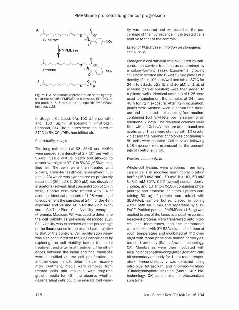
Figure 1. A. Schematic representation of the hydroly-sis of the specific PMPMEase substrate, RD-PNB, to the product. B. Structure of the specific PMPMEase inhibitor, L-28.
PMPMEase promotes lung cancer progression
119 Am J Cancer Res 2014;4(2):116-134
PMPMEase activity in lung cancer cells
PMPMEase activity was assayed using a previ-ously established method [29] incorporating the specific PMPMEase substrate, N-(4-nitrobenzoyl)-S-trans, trans-farnesyl-L-cysteine methyl ester synthesized in our laboratory as previously described [29] (Figure 1A). The hydrolysis of the substrate to product is sensi-tive to the PMPMEase specific inhibitor, L-28 (Figure 1B) [30]. Cells were cultured in 175 cm2 vented culture flasks at 37°C in 5% CO2/95% humidified air to confluence. The cells were washed with phosphate buffered saline, lysed with Triton-X 100 (0.1%) in 100 mM Tris-HCl, pH 7.4 containing EDTA (1 mM). Aliquots of the resulting lysate were incubated with N-(4-nitrobenzoyl)-S-trans, trans-farnesyl-L-cysteine methyl ester substrate (1 mM) in a total reac-tion volume of 100 µL at 37°C for 3 h and ana-lyzed by HPLC as previously described [29]. The total protein concentrations were determined using the Bradford reagent. To establish the effect L-28 on PMPMEase activity in the lung cells, aliquots of the resulting lysate were pre-incubated for 10 min with L-28 (0.1-1000 µM) followed by assay for residual enzymatic activity.
Apoptosis assay
The mode of cell death induced by L-28 was determined by the modified EB/OA staining method [31, 38]. A549 cells were seeded onto 24-well plates at a density of 1.5 × 10⁵ cells and incubated at 37°C in 5% CO2/95% humidi-fied air. These were then treated with L-28 (0-10 μM) and incubated for 48 h at 37°C before staining with EB/AO. A solution containing both EB and AO was added to each well to a final concentration of 10 μg/mL and incubated in the dark for 15 min. The cells were viewed under a Zeiss microscope using 480/30 nm excitation filter. Images of the cells were cap-tured with an Olympus DP70 Camera. The con-ventional EB/AO staining was also employed to quantify apoptosis [38, 39]. Briefly, A549 cells treated with L-28 (0-10 μM) and incubated for 48 h at 37°C were harvested by transferring the media into 15 ml tubes. The rest of the cells were detached with Dulbecco’s PBS containing 1 mM EDTA and added to the transferred media. Cells were pelleted by centrifugation at 130 g for 5 minutes, washed with 1 mL of cold PBS and the cell pellets re-suspended in 25 μL
of cold PBS and 2 μL EB/AO dye mix was added. Stained cell suspensions (10 μL) were placed on clean microscope slides and covered with coverslips. The cells were viewed and counted under the microscope at 40 × magnification using 480/30 nm excitation filter. The counts were conducted in triplicates with a minimum of 100 cells counted per replicate.
Effect of PMPMEase inhibition on lung cancer cell migration
Scratch assay was conducted to determine the effect of L-28 on A549 cell motility. The cells were seeded at a density of 2.5 × 10⁵ in a 24 well plate. After 24 h, the medium was removed and replaced with medium containing 10% FBS. When the cells reached full confluence, the monolayers were wounded by scratching the surface as uniformly and straight as possi-ble using a sterile 10 μL pipette tip at least three times. The cells were washed once with complete media to remove detached cells fol-lowed by the acquisition of the original image of the wound under a microscope. The wounded cell monolayer was then incubated with the cell culture medium containing an appropriate con-centration of L-28 (0.5-1.0 µM). Images of the cells invading the scratch were captured with an Olympus DP70 Camera at indicated time points (0, 6, and 12 h). The pictures were ana-lyzed and the width of the wounds measured using NIH ImageJ software (http://rsb.info.nih.gov/ij/). The difference between the widths is taken as the migration distance.
Effect of PMPMEase inhibition on F-actin fila-ments organization and cellular localization of PMPMEase and Ras proteins
To visualize the effect of L-28 on F-actin organi-zation and localization of PMPMEase, K-ras, H-ras and N-ras, immunofluorescence analyses were performed on L-28-treated A549 cells in 4-well Lab-tek II chambers (Nalge Nunc International, NY). The cells were fixed in 3.7% paraformaldehyde (Sigma, MO) in PBS for 15 min at room temperature. After washing with PBS, the cells were permeabilized with 0.1% Triton X-100 (Sigma, MO) for 5 min on ice and blocked with 1% BSA in PBS for 1 h at 4°C. To visualize F-actin organization, phalloidin (Biotium, CA) and propidium iodide (Sigma, St. Louis, MO) fluorescence staining were per-formed. To study the localization of PMPMEase,
PMPMEase promotes lung cancer progression
120 Am J Cancer Res 2014;4(2):116-134
K-ras, H-ras and N-ras, the cells were incubat-ed with the respective rabbit polyclonal anti-bodies against PMPMEase, K-Ras, H-ras or N-ras (Santa Cruz Biotechnology, CA) in 1% BSA in PBS overnight. After washing in PBS, the cells were incubated with flourescein isothiocy-anate (FITC)-conjugated goat anti-antibody (Santa Cruz Biotechnology, CA) for 1 h at room temperature in a humidified chamber. The cells were washed with PBS, stained with DAPI (Sigma, St. Louis, MO) at a concentration of 0.1 μg/ml in PBS and mounted on microscope slides. These were then viewed on a Zeiss fluo-rescent microscope and the images captured with an Olympus DP70 Camera.
Effect of PMPMEase inhibition on the expres-sion of cancer-related genes
Lung cancer A549 and H460 cells were seeded at a density of 1 × 10⁵ per well in 24-well cul-ture plates and allowed to attach overnight at 37°C in 5% CO2/95% humidified air. The cells were then treated with either solvent (control) or L-28 at 2 µM and 5 µM. After 48 h of treat-ment, the cells were detached with trypsin, washed once with PBS, counted and 10000 cells were lysed in 5 µL RLT buffer (Qiagen, Valencia, CA). The analysis for the relative expression of cancer-related genes was con-ducted using the nCounter GX Human Cancer References Kit for profiling cancer-related genes (NanoString Technologies, Seattle, WA) at the Oncogenomics Core Facility of the Sylvester Comprehensive Cancer Center University of Miami Miller School of Medicine. The raw nanoString counts were analyzed using the nSolver Software (NanoString Technologies, Seattle, WA) as previously described [40]. Briefly, the data were normalized using positive control spikes to correct for any such experi-mental variables between the samples as dif-ferences in efficiency in hybridization, purifica-tion or binding. This normalization is conducted on all nanoString counts by multiplying them with a normalization factor. The normalization factor is calculated by averaging the sum of the counts for all positive hybridization controls for each sample divided by the sum for each sam-ple. The normalization factor within the required range was between 0.3-3. In addition, the data were normalized for RNA content using house-keeping genes and multiplying all counts by a calculated normalization factor. The normaliza-tion is optimized using multiple housekeeping
genes that include those with high and low expression levels (CLTC, GAPDH, GUSB, HPRT1, PGK1, TUBB). This normalization factor is cal-culated by the average of the geometric mean of all the housekeeping gene counts for each sample divided by the geometric mean of each sample. Also, the average of negative controls consisting of eight codes with no transcript rep-resenting background noise was subtracted from the normalized gene counts. Multi Experiment Viewer (MeV v4.6.2) was employed to cluster the data sets to obtain the heat maps resulting from the numerical fold-change expression levels compared to the control. Heat maps were obtained by clustering based on Manhattan Distance. Prior to clustering, the data was filtered using criteria set such that only data values ≥ 3-fold changes were employed. A total of 47 genes passed the filter-ing criteria and were used for the heat map analyses. Color scale limits were set at “-3.0, 0.0, 3.0”, meaning that the brightest red repre-sents ≥ 3-fold upregulation relative to the con-trols, the brightest green represents ≥ 3-fold downregulation, and black represents no change. The 47 genes were further grouped based on their main functions, and bar graphs indicating their relative expression levels were obtained using Graphpad Prism version 4.0 for Windows (San Diego, CA).
PMPMEase expression in lung cancer tissues
The expression of PMPMEase in lung cancer tissues was determined using immunohisto-chemical analysis as previously described [41] using human lung cancer and normal adjacent tissue microarrays (TMAs) comprising a total of 416 cores from 416 distinct cases. The tissues used were supplied by, and the immunohisto-chemistry conducted at US Biomax (Rockville, MD).
Statistical analysis
All results were expressed as the means ± S.E.M. The concentration-response curves were obtained by plotting the percent inhibition against the log of the inhibitor concentrations. Nonlinear regression plots were generated using Graphpad Prism version 4.0 for Windows (San Diego, CA). From these, the concentra-tions that inhibit 50% of the activity (IC50) were calculated. The tissue microarrays data were analyzed by one-way ANOVA using SAS 9.2 soft-ware (SAS Institute, Cary NC). Statistical differ-
PMPMEase promotes lung cancer progression
121 Am J Cancer Res 2014;4(2):116-134
ences between normal and cancer tissues were determined using Bonferroni›s procedure for multiple comparisons. The Bonferroni pro-cedure takes into account the number of means to be compared and is more conservative than other multiple comparison methods. P-values of less than 0.05 were considered statistically significant.
Results
PMPMEase inhibition suppresses lung cancer cell viability
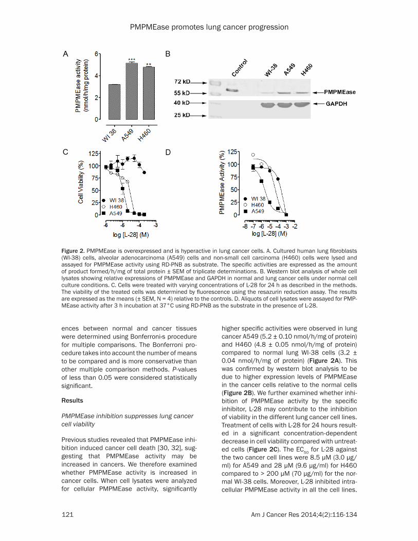
Previous studies revealed that PMPMEase inhi-bition induced cancer cell death [30, 32], sug-gesting that PMPMEase activity may be increased in cancers. We therefore examined whether PMPMEase activity is increased in cancer cells. When cell lysates were analyzed for cellular PMPMEase activity, significantly
higher specific activities were observed in lung cancer A549 (5.2 ± 0.10 nmol/h/mg of protein) and H460 (4.8 ± 0.05 nmol/h/mg of protein) compared to normal lung WI-38 cells (3.2 ± 0.04 nmol/h/mg of protein) (Figure 2A). This was confirmed by western blot analysis to be due to higher expression levels of PMPMEase in the cancer cells relative to the normal cells (Figure 2B). We further examined whether inhi-bition of PMPMEase activity by the specific inhibitor, L-28 may contribute to the inhibition of viability in the different lung cancer cell lines. Treatment of cells with L-28 for 24 hours result-ed in a significant concentration-dependent decrease in cell viability compared with untreat-ed cells (Figure 2C). The EC50 for L-28 against the two cancer cell lines were 8.5 µM (3.0 µg/ml) for A549 and 28 µM (9.6 µg/ml) for H460 compared to > 200 µM (70 µg/ml) for the nor-mal WI-38 cells. Moreover, L-28 inhibited intra-cellular PMPMEase activity in all the cell lines.
Figure 2. PMPMEase is overexpressed and is hyperactive in lung cancer cells. A. Cultured human lung fibroblasts (WI-38) cells, alveolar adenocarcinoma (A549) cells and non-small cell carcinoma (H460) cells were lysed and assayed for PMPMEase activity using RD-PNB as substrate. The specific activities are expressed as the amount of product formed/h/mg of total protein ± SEM of triplicate determinations. B. Western blot analysis of whole cell lysates showing relative expressions of PMPMEase and GAPDH in normal and lung cancer cells under normal cell culture conditions. C. Cells were treated with varying concentrations of L-28 for 24 h as described in the methods. The viability of the treated cells was determined by fluorescence using the resazurin reduction assay. The results are expressed as the means (± SEM, N = 4) relative to the controls. D. Aliquots of cell lysates were assayed for PMP-MEase activity after 3 h incubation at 37°C using RD-PNB as the substrate in the presence of L-28.

PMPMEase promotes lung cancer progression
122 Am J Cancer Res 2014;4(2):116-134
However, the inhibition was relatively higher in the cancer cells (IC50 values of 2.5 µM or 0.88 µg/ml for A549 and 41 µM or 14 µg/ml for H460) compared to 520 µM (180 µg/ml) for the normal WI-38 cells (Figure 2D). When the effect of PMPMEase inhibition on cell proliferation was determined, L-28 significantly inhibited the proliferation of both A549 (Figure 3A) and H460 cells (Figure 3B). In order to better under-stand the long term effect of PMPMEase inhibi-tion on cancer cell viability, cells previously exposed to varying concentrations of L-28 for 72 h were further cultured for 48 h. Withdrawal of treatment with L-28 did not result in renewed increases in cell viability (Figure 3C). A clono-genic assay was used to assess the differences in reproductive viability between human lung cancer cells treated with L-28 and untreated control cells. The clonogenic deaths of A549
and H460 cells after incubation with either 5 or 10 μM of L-28 at 37°C are shown in Figure 3D. Treatment with either 5 or 10 μM of L-28 for 72 h reduced the clonogenic survival of A549 cells to about 43.4% and 15.5%, and H460 cells to about 13.5% and 0.2%, respectively. These results indicate that the lung cancer cells exposed to PMPMEase inhibitors are unlikely to recover from the degeneration effects.
The AO/EB method was used to determine the mode of L-28-induced cytotoxicity as previously described [31, 38]. Fluorescence light micros-copy coupled with the differential uptake of the fluorescent DNA-binding dyes, EB and AO, was used for its simplicity, rapidity, and accuracy [38]. AO permeated the live control cells as well as cells exposed to lower concentrations of L-28 turning their nuclei green. Cells treated
Figure 3. L-28 inhibits lung cancer cell proliferation. Cultured human lung cancer (A) A549 and (B) H460 cells were treated with varying concentrations of L-28 for 72 h as described in the methods. Cell viability was determined before the initial treatment and then after the final treatment by fluorescence using the resazurin reduction assay. Cell proliferation was calculated as the difference in cell viability between the initial and final readings. (C) L-28 in-duces irreversible lung cancer cell degeneration. Cultured human lung cancer (A549 and H460) cells were treated with varying concentrations of L-28 for 72 h as described in the methods. The treatment medium was replaced with normal growth medium and cultured for a further 48 h. The cell viability was determined by fluorescence using the resazurin reduction assay. The results are expressed as the means (± SEM, N = 4) relative to the controls. (D)Clonogenic cell death caused by L-28. Exponentially growing cells in culture were treated with L-28 (5 or 10 μM) for 72 h, washed, cultured for a further 7 days at 37°C, and clonogenic survival was determined. The results are expressed as the means (± SEM, N = 4) relative to the controls. Significance (***p < 0.001) was determined by Student’s t-test.

PMPMEase promotes lung cancer progression
123 Am J Cancer Res 2014;4(2):116-134
used wound-healing assay [42] to characterize the cell migration response in A549 cells. The number of cells involved in the wound sealing as well as the mean distance of migration were evaluated after 6 h and 12 h. Control untreated A549 cells migrated into the wound area faster, reaching 40% and 85% sealing at 6 and 12 h after wound scratch, respectively (Figure 5A and 5B). The number of cells in the control that migrated into the wound area after 6 and 12 h were 61 ± 5.0 and 250 ± 31, respectively. Treatment of A549 cells with 0.5 µM of L-28 sig-nificantly decreased the number of cells that
with L-28 fluoresced red due to EB uptake fol-lowing the loss of cytoplasmic membrane integ-rity. L-28 induced apoptosis in a concentration dependent manner as depicted in Figure 4. Taken together, these results indicate that higher levels of PMPMEase likely contribute to lung cancer progression thereby making PMPMEase a viable target for anticancer drug development.
PMPMEase inhibition attenuates cell migration
To examine the effect of L-28 on lung cancer A549 cell migration, we employed the widely
Figure 4. L-28 induces apoptosis of human lung cancer A549 cells. Cells were treated with L-28 (5 and 10 μM) for 48 h and stained with acridine orange and ethidium bromide (AO/EB, 10 μg/ml) as described in the methods. When stained with AO/EB, live cells with normal nuclei (n) appear green; apoptotic cells (a) with condensed or fragmented chromatin in the nuclei appear orange. A. Shows pictures of cells taken with bright field focus. B. Indicates AO/EB overlay pictures obtained from the simplified morphological observation. C. Shows quantification of apoptotic A549 cells (shown as percentages) using the conventional method of staining.

PMPMEase promotes lung cancer progression
124 Am J Cancer Res 2014;4(2):116-134
120.6 ± 3.1 µm (control) to 73.5 ± 3.5 µm (0.5 µM) and 71.7 ± 3.7 µm (1 µM), respectively after 12 h (p < 0.001). These results indicate that inhibition of PMPMEase inhibits wound-induced cell migration.
PMPMEase inhibition disrupts actin cytoskel-eton organization
Polyisoprenylated monomeric G-proteins such as Ras and Rho family of G proteins play impor-tant roles in regulating migration, differentia-
migrated into the wound area to 1.3 ± 0.3 and 75 ± 9.0 after 6 and 12 h, respectively (p < 0.001). A further decrease in the number of migrated cells was observed with 1.0 µM of L-28 treatment (1.0 ± 0.5 and 69 ± 5.0) after 6 and 12 h, respectively (p < 0.001). The rate of migration as indicated by migration distance was also significantly inhibited by L-28 com-pared with that of control untreated cells at each of the time points examined (Figure 5A and 5C). L-28 significantly decreased the dis-tance of migration into the wound area from
Figure 5. Wound healing and cell migration assays. A. Cell motility in wound healing assay. A uniform scratch was made in 90% confluent monolayer culture of A549 cells and the extent of closure was monitored under microscope and photographed. Representative images of three independent experiments done in duplicate are shown. Results of three independent experiments were plotted. Migrating cells within the scratch were quantified and the distance of migration measured using NIH ImageJ software (n = 3 individual experiments). B. Treatment with L-28 (0.5 µM and 1 µM) significantly inhibited the number of migrated cells compared with the control solvent-treated cells. C. Over the same incubation time, the cells in the control groups migrated farther, achieving a complete or near closure of the wounds. The results are expressed as the means (± SEM, N = 5) relative to the controls. Significance (***p < 0.001) was determined by Student’s t-test.
PMPMEase promotes lung cancer progression
125 Am J Cancer Res 2014;4(2):116-134
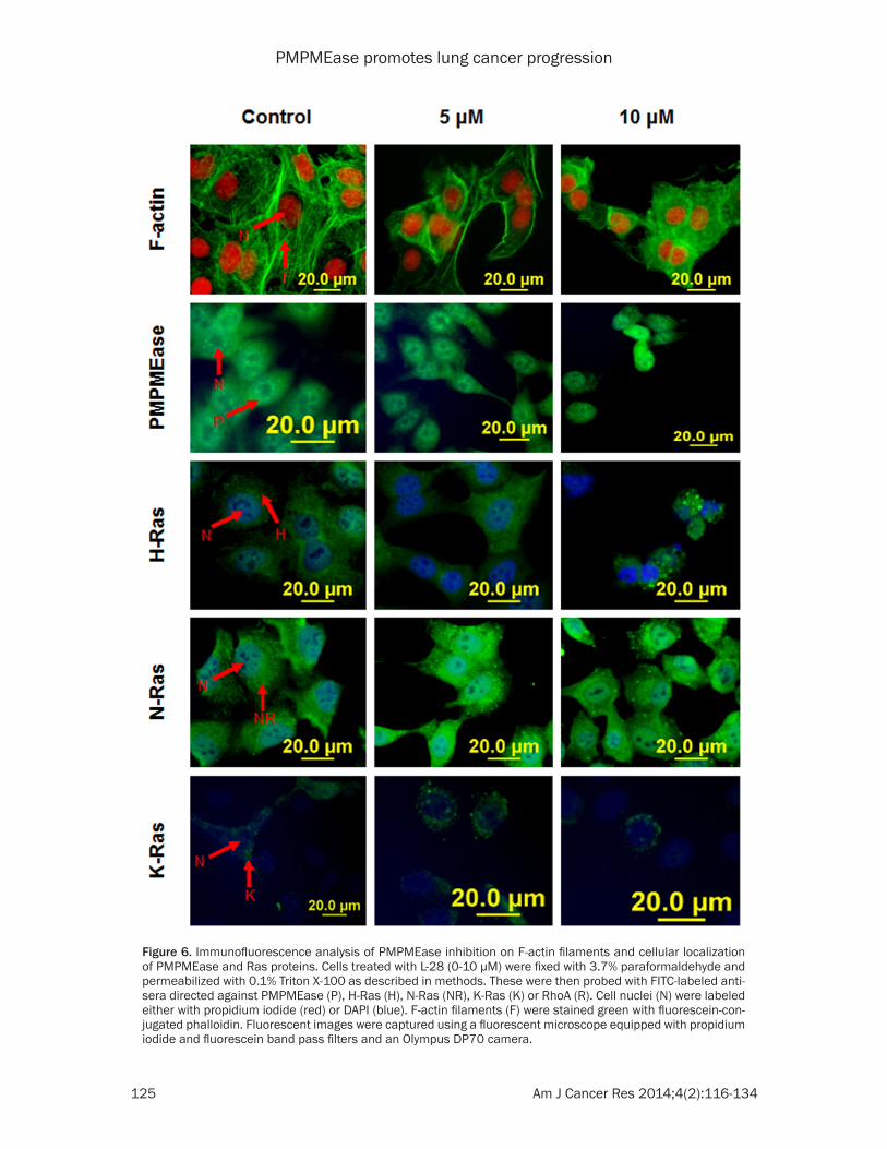
Figure 6. Immunofluorescence analysis of PMPMEase inhibition on F-actin filaments and cellular localization of PMPMEase and Ras proteins. Cells treated with L-28 (0-10 µM) were fixed with 3.7% paraformaldehyde and permeabilized with 0.1% Triton X-100 as described in methods. These were then probed with FITC-labeled anti-sera directed against PMPMEase (P), H-Ras (H), N-Ras (NR), K-Ras (K) or RhoA (R). Cell nuclei (N) were labeled either with propidium iodide (red) or DAPI (blue). F-actin filaments (F) were stained green with fluorescein-con-jugated phalloidin. Fluorescent images were captured using a fluorescent microscope equipped with propidium iodide and fluorescein band pass filters and an Olympus DP70 camera.

PMPMEase promotes lung cancer progression
126 Am J Cancer Res 2014;4(2):116-134
Figure 7. nCounter analysis of the effect of L-28 on cancer-related genes in A549 and H460 cells. Cells were treated with L-28 (2 µM and 5 µM) for 72 h. Isolated RNA was purified and subjected to nCounter measurements for transcripts of cancer-related genes. Data were normalized based on seven house-keeping genes showing stable expression levels in both cell lines. Genes with fold changes of ≥ 3 or ≤ 3 are shown. For these differentially regulated genes, their fold change values in the treated cells versus control conditions were obtained. A. Heat map of the fold change in gene expression values are shown. MultiExperimentViewer (MeV v4.6.2) was used for hierarchical clustering by average linkage clustering based on Manhattan distance (red indicates upregulated genes; green indicates downregulated genes). B. Bar graph of fold change in expression values of selected genes.
PMPMEase promotes lung cancer progression
127 Am J Cancer Res 2014;4(2):116-134
genes were similarly altered in both A549 and H460 cell lines, (Figure 7A). This subset of genes were further grouped, based on their principal functions, into (1) DNA repair/tran-scription, (2) cell cycle regulation, (3) cell growth, proliferation, differentiation and sur-vival, (4) cell adhesion and migration and (5) immune response (Figure 7B). In general, L-28 robustly activated the expression of pro-apop-totic and cell cycle arrest genes (BCL6, CDKN1A, IGFBP3, MLL, ETS1, TGFBI, TNFRSF-
tion, survival and proliferation [43]. We deter-mined whether PMPMEase inhibition by L-28 affects actin cytoskeleton organization which plays a vital role in cell shape, adhesion and migration [44]. We also aimed to determine the effect of PMPMEase inhibition on the cellular localization of PMPMEase, N-Ras, K-Ras, and H-Ras in A549 cells. Treatment of cells with PMPMEase inhibitor did not appear to alter nei-ther the levels nor the distribution of PMPMEase as well as N-Ras, K-Ras and H-Ras in the A549
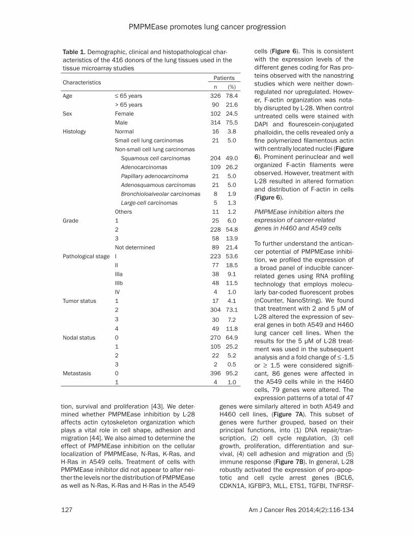
Table 1. Demographic, clinical and histopathological char-acteristics of the 416 donors of the lung tissues used in the tissue microarray studies
CharacteristicsPatientsn (%)
Age ≤ 65 years 326 78.4> 65 years 90 21.6
Sex Female 102 24.5Male 314 75.5
Histology Normal 16 3.8Small cell lung carcinomas 21 5.0Non-small cell lung carcinomas Squamous cell carcinomas 204 49.0 Adenocarcinomas 109 26.2 Papillary adenocarcinoma 21 5.0 Adenosquamous carcinomas 21 5.0 Bronchioloalveolar carcinomas 8 1.9 Large-cell carcinomas 5 1.3Others 11 1.2
Grade 1 25 6.02 228 54.83 58 13.9Not determined 89 21.4
Pathological stage I 223 53.6II 77 18.5IIIa 38 9.1IIIb 48 11.5IV 4 1.0
Tumor status 1 17 4.12 304 73.13 30 7.24 49 11.8
Nodal status 0 270 64.91 105 25.22 22 5.23 2 0.5
Metastasis 0 396 95.21 4 1.0
cells (Figure 6). This is consistent with the expression levels of the different genes coding for Ras pro-teins observed with the nanostring studies which were neither down-regulated nor upregulated. Howev- er, F-actin organization was nota-bly disrupted by L-28. When control untreated cells were stained with DAPI and flourescein-conjugated phalloidin, the cells revealed only a fine polymerized filamentous actin with centrally located nuclei (Figure 6). Prominent perinuclear and well organized F-actin filaments were observed. However, treatment with L-28 resulted in altered formation and distribution of F-actin in cells (Figure 6).
PMPMEase inhibition alters the expression of cancer-related genes in H460 and A549 cells
To further understand the antican-cer potential of PMPMEase inhibi-tion, we profiled the expression of a broad panel of inducible cancer-related genes using RNA profiling technology that employs molecu-larly bar-coded fluorescent probes (nCounter, NanoString). We found that treatment with 2 and 5 μM of L-28 altered the expression of sev-eral genes in both A549 and H460 lung cancer cell lines. When the results for the 5 μM of L-28 treat-ment was used in the subsequent analysis and a fold change of ≤ -1.5 or ≥ 1.5 were considered signifi-cant, 86 genes were affected in the A549 cells while in the H460 cells, 79 genes were altered. The expression patterns of a total of 47

PMPMEase promotes lung cancer progression
128 Am J Cancer Res 2014;4(2):116-134
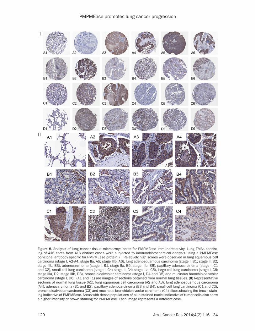
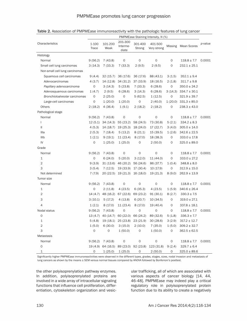
PMPMEase immunoreactivity. Figure 8 shows representative images of normal lung and lung cancer tissues. The data indicate that increas-ing levels of PMPMEase expression are associ-ated with tumors (Figure 8I). We observed pre-dominantly peri-nuclear cytoplasmic staining indicative of PMPMEase protein expression in the lung cells (Figure 8II) consistent with the endoplasmic reticulum localization of the enzyme [28, 45]. In the control cores consisting of normal lung tissues, staining was present in a few normal bronchial and glandular epithelial cells. However, in general, only trace to weak immunostaining was observed in the normal lung tissues. Solid tumors with deeply stained epithelial cells were displayed especially in most of the SCC, ASC, BAC, AC and PAC. Scattered patterns of staining were observed among the LCC and the SCLC tumor cells, with positive areas spread out throughout the cores. Significant differences in PMPMEase immuno-reactivity intensities between the normal tis-sues and the different lung tumor categories were observed when the stained sections were analyzed (p = 0.0002 - < 0.0001) (Table 2). The mean scores ± SEM were 118.8 ± 7.7 (normal), 232.1 ± 25.1 (small-cell lung carcinomas), 352.1 ± 9.4 (squamous cell carcinomas), 311.7 ± 9.8 (adenocarcinomas), 350.0 ± 24.2 (papil-lary adenocarcinomas), 334.7 ± 30.1 (adeno-squamous carcinomas), 321.9 ± 39.7 (bron-chioloalveolar carcinomas), and 331.3 ± 85.0 (large-cell carcinomas). Very high PMPMEase expression (score = 401-500) were observed in 43.1% of SCC. The mean PMPMEase immuno-reactivity score was significantly high even at early-stage (stages I and II) lesions, with pro-gressively increasing staining intensities (Figure 8, Table 2). Although no specific trend was observed when the data were analyzed according to pathological stages, grades, tumor size, nodal status and metastases, there were significant differences when compared to the normal tissues regardless of the parameter under consideration.
Discussion
Previous studies revealed the death of cancer cells treated with polyisoprenylated small mol-ecule inhibitors of PMPMEase [30] or PUFAs [32]. Despite this apparent dependency of can-cer cell viability on PMPMEase activity, the pos-sible role of PMPMEase in cancer progression has not received similar research attention as
10B, RARA, SIAH1, and CEBPA), while anti-apoptotic genes as well as genes promoting cell proliferation (BIRC5, CDC2, E2F1, PCNA, MYBL2, CCNA2 and CCND3) were downregu-lated. Notably, the expression of the transcrip-tion factor and cell cycle progression gene, MYBL2 was severely suppressed in both H460 and A549 cell lines. The death receptor genes, TNFRSF10B and PPARG were both upregulat-ed. Also, genes involved in lung development (CTNNB1, PDGFA, PTHLH and FGF2) were upregulated whilst CTGF was downregulated by L-28 treatment. In addition, L-28 induced the upregulation of genes for the growth factors, FGF2, MET, AREG, IGFBP3, and EGFR, as well as their receptors. Genes involved in integrin signaling were either upregulated (LICAM, ITGB1 and PLAUR) or downregulated (CTGF). L-28 treatment induced the upregulation of CTNNB1, CD44, MET and CDKN1A. Moreover, genes that either code for ligands (AREG, FGF2, MET, IL6, L1F), receptors (EGFR, PLAUR) and downstream signaling mediators (BRAF, EPS8) in receptor tyrosine kinase pathways were upregulated. These data suggest that the observed effects of L-28 on the cells involve significant changes that impact signaling path-ways involved in cell proliferation, differentia-tion, apoptosis survival, adhesion and migra- tion.
PMPMEase is overexpressed in lung cancer
The results showing that PMPMEase activities and protein levels are higher in lung cancer ver-sus normal cells and the observation that PMPMEase inhibition in these cells induces apoptosis and the altered expression of cancer-related genes led us to examine TMAs consist-ing of various lung cancer tissues and normal lung tissues for the relative expression of PMPMEase. The demographic and histopatho-logical characteristics of the tissues donors for the TMAs are shown in Table 1. The ages of the patients ranged from 27 to 77 years and most (76.25%) were males. As shown in Table 1, there were 416 total cases out of which 16 were normal tissues and 21 were SCLC. The NSCLC tumors comprised 204 squamous cell carcinomas (SCC), 109 adenocarcinomas (AC), 21 papillary adenocarcinomas (PAC), 21 adeno-squamous carcinomas (ASC), 8 bronchioloalve-olar carcinomas (BAC), 5 large-cell carcinomas (LCC) and 11 other tumors. In general, all the lung cancers showed intense intracellular
PMPMEase promotes lung cancer progression
129 Am J Cancer Res 2014;4(2):116-134
Figure 8. Analysis of lung cancer tissue microarrays cores for PMPMEase immunoreactivity. Lung TMAs consist-ing of 416 cores from 416 distinct cases were subjected to immunohistochemical analysis using a PMPMEase polyclonal antibody specific for PMPMEase protein. (I) Relatively high scores were observed in lung squamous cell carcinoma (stage I, A2-A4; stage IIa, A5; stage IIIb, A6), lung adenosquamous carcinoma (stage I, B1; stage II, B2; stage IIIb, B3), adenocarcinoma (stage I, B1; stage IIa, B5; stage IIIb, B6), papillary adenocarcinoma (stage I, C1 and C2), small cell lung carcinoma (stage I, C4; stage II, C4; stage IIIa, C5), large cell lung carcinoma (stage I, C6; stage IIIa, D2; stage IIIb, D3), bronchioloalveolar carcinoma (stage I, D4 and D5) and mucinous bronchioloalveolar carcinoma (stage I, D6). (A1 and F1) are images of sections obtained from normal lung tissues. (II) Representative sections of normal lung tissue (A1), lung squamous cell carcinoma (A2 and A3), lung adenosquamous carcinoma (A4), adenocarcinoma (B1 and B2), papillary adenocarcinoma (B3 and B4), small cell lung carcinoma (C1 and C2), bronchioloalveolar carcinoma (C3) and mucinous bronchioloalveolar carcinoma (C4) slices showing the brown stain-ing indicative of PMPMEase. Areas with dense populations of blue-stained nuclei indicative of tumor cells also show a higher intensity of brown staining for PMPMEase. Each image represents a different case.
PMPMEase promotes lung cancer progression
130 Am J Cancer Res 2014;4(2):116-134
ular trafficking, all of which are associated with various aspects of cancer biology [14, 44, 46-48]. PMPMEase may indeed play a critical regulatory role in polyisoprenylated protein function due to its ability to create a negatively
the other polyisoprenylation pathway enzymes. In addition, polyisoprenylated proteins are involved in a wide array of intracellular signaling functions that influence cell proliferation, differ-entiation, cytoskeleton organization and vesic-
Table 2. Association of PMPMEase immunoreactivity with the pathologic features of lung cancer
Characteristics
PMPMEase Staining Intensity, N (%)
p-value1-100Trace
101-200Weak
201-300Interme-
diate
301-400Strong
401-500Very strong Missing Mean Scores
Histology Normal 9 (56.2) 7 (43.8) 0 0 0 0 118.8 ± 7.7 0.0001 Small cell lung carcinomas 3 (14.3) 7 (33.3) 7 (33.3) 2 (9.5) 2 (9.5) 0 232.1 ± 25.1
Non-small cell lung carcinomas
Squamous cell carcinomas 9 (4.4) 32 (15.7) 36 (17.6) 36 (17.6) 88 (43.1) 3 (1.5) 352.1 ± 9.4 Adenocarcinomas 4 (3.7) 14 (12.8) 34 (31.2) 37 (33.9) 18 (16.5) 2 (1.8) 311.7 ± 9.8 Papillary adenocarcinoma 0 3 (14.3) 5 (23.8) 7 (33.3) 6 (28.6) 0 350.0 ± 24.2 Adenosquamous carcinomas 1 (4.7) 2 (9.5) 6 (28.6) 3 (14.3) 6 (28.6) 3 (14.3) 334.7 ± 30.1 Bronchioloalveolar carcinomas 0 2 (25.0) 0 5 (62.5) 1 (12.5) 0 321.9 ± 39.7 Large-cell carcinomas 0 1 (20.0) 1 (20.0) 0 2 (40.0) 1 (20.0) 331.3 ± 85.0 Others 2 (18.2) 4 (36.4) 1 (9.1) 2 (18.2) 2 (18.2) 0 238.3 ± 43.0Pathological stage
Normal 9 (56.2) 7 (43.8) 0 0 0 0 118.8 ± 7.7 0.0001 I 12 (5.1) 34 (14.3) 55 (23.2) 58 (24.5) 73 (30.8) 5 (2.1) 334.2 ± 8.3 II 4 (5.3) 14 (18.7) 19 (25.3) 18 (24.0) 17 (22.7) 3 (4.0) 305.0 ± 14.0 IIIa 2 (5.3) 7 (18.4) 5 (13.2) 8 (21.1) 15 (39.5) 1 (2.6) 342.6 ± 22.5 IIIb 1 (2.1) 9 (19.1) 11 (23.4) 8 (17.0) 18 (38.3) 0 333.0 ± 17.8 IV 0 1 (25.0) 1 (25.0) 0 2 (50.0) 0 325.0 ± 89.0
Grade
Normal 9 (56.2) 7 (43.8) 0 0 0 0 118.8 ± 7.7 0.0001 1 0 6 (24.0) 5 (20.0) 3 (12.0) 11 (44.0) 0 333.0 ± 27.2 2 9 (3.9) 31 (13.6) 46 (20.2) 56 (24.6) 86 (37.7) 1 (0.4) 348.8 ± 8.0 3 3 (5.4) 7 (12.5) 19 (33.9) 17 (30.4) 10 (17.9) 0 312.9 ± 15.0 Not determined 7 (7.9) 20 (22.5) 19 (21.3) 16 (18.0) 19 (21.3) 8 (9.0) 262.9 ± 13.9Tumor size
Normal 9 (56.2) 7 (43.8) 0 0 0 0 118.8 ± 7.7 0.0001 1 0 2 (11.8) 4 (23.5) 6 (35.3) 4 (23.5) 1 (5.9) 340.6 ± 26.4 2 14 (4.7) 48 (16.2) 67 (22.6) 69 (23.2) 91 (30.1) 8 (2.7) 330.3 ± 7.5
3 3 (10.1) 5 (17.2) 4 (13.8) 6 (20.7) 10 (34.5) 0 319.0 ± 27.1
4 1 (2.1) 8 (17.0) 11 (23.4) 8 (17.0) 19 (40.4) 0 337.8 ± 18.1
Nodal status 9 (56.2) 7 (43.8) 0 0 0 0 118.8 ± 7.7 0.0001 0 13 (4.7) 40 (14.7) 60 (22.0) 66 (24.2) 89 (32.6) 5 (1.8) 336.3 ± 7.7 1 5 (4.8) 19 (18.1) 25 (23.8) 23 (21.9) 30 (28.6) 3 (2.9) 317.2 ± 12.7 2 1 (5.0) 6 (30.0) 3 (15.0) 2 (10.0) 7 (35.0) 1 (5.0) 309.2 ± 32.7 3 0 0 1 (50.0) 0 1 (50.0) 0 362.5 ± 62.5Metastasis
Normal 9 (56.2) 7 (43.8) 0 0 0 0 118.8 ± 7.7 0.0001 0 19 (4.9) 64 (16.5) 89 (23.0) 92 (23.8) 123 (31.8) 9 (2.4) 329.7 ± 6.4 1 0 1 (25.0) 1 (25.0) 0 2 (50.0) 0 325.0 ± 89.6Significantly higher PMPMEase immunoreactivities were observed in the different types, grades, stages, sizes, nodal invasion and metastasis of lung cancers as shown by the means ± SEM versus normal tissues compared by ANOVA followed by Bonferroni’s posttest.
PMPMEase promotes lung cancer progression
131 Am J Cancer Res 2014;4(2):116-134
ligands, particularly AREG, which together play important roles in the pathogenesis of lung cancer. EGFR signaling pathways play pertinent roles in evading apoptosis and promoting sur-vival which are the hallmarks of cancer cells [51]. The key signaling pathways initiated by EGFR through its Grb2/Sos1 associations involve Ras proteins which then stimulates the mitogen-activated protein kinase pathway. Ras activation is subsequently involved in cell growth, proliferation, motility, survival and transformation [52]. Experimental and clinical evidence indicates that overexpression or mutation of EGFR mediates tumor resistance to both chemotherapy and especially radiother-apy [53]. Upregulation of trophic factors and their signaling mediators in the current study likely indicates a feedback survival effort due to impaired downstream mediator(s). Although RAS genes, as well as Ras protein localization, were not significantly altered, inhibition of PMPMEase likely affected the methylated/demethylated ratios the proteins, impacting their conformations and altering their interac-tions with downstream mediators in the signal-ing pathway.
Inhibition of PMPMEase was also associated with significant disruption of F-actin organiza-tion as well as inhibition of cell motility. These may be attributed to defective processing of polyisoprenylated Rho family of small GTPases such as Rac1, RhoA and Cdc42 that control the dynamics of actin cytoskeleton [44]. The gene expression study also revealed changes in the expression of some pertinent genes (FGF2, MET, PLAUR, ITGB1 and TGFB1) necessary for chemotaxis, receptors signaling, cell adhesion and migration upon treatment of the cells with L-28. Genes such as CTNNB1, MET, CDKN1A and CD44 are associated with β-catennin, which is located at cell-cell adherens junction where it links cadherens to actin cytoskeleton [54, 55]. Also, the expression of genes such as CTGF, LICAM, ITGB1 and PLAUR which are asso-ciated with integrin and its signaling, were affected. Integrin regulates cytoskeleton orga-nization necessary for cell polarity, migration, proliferation [54]. Integrins also function as transmembrane linkers to the cytoskeleton required for the cells to grip the extracellular matrix [54]. These changes are therefore con-sistent with the observed inhibition of cell migration due to PMPMEase inhibition.
charged carboxylate ion with the propensity to induce conformational changes near the poly-isoprenyl moiety that is critical for protein-pro-tein interactions [46]. That PMPMEase expres-sion and activity is important for cell growth was suggested by previous work from this labo-ratory in which PMPMEase inhibition induced cancer cell death [30, 31, 41]. Cushman et al., using a knockdown approach in a recent study identified CES 1 (PMPMEase), as a specific esterase whose function is important for the methylation status as well as activation of RhoA and cell morphology. The elevated PMPMEase activity and expression in the lung cancer cells compared to the normal lung fibroblasts sug-gests that overexpression of PMPMEase may indeed spur cell growth.
This study shows that PMPMEase inhibition is associated with decrease in cell viability through the induction of apoptosis and the inhi-bition of cell proliferation and survival. These were further confirmed by the gene expression studies that revealed the downregulation of anti-apoptotic genes as well as the upregula-tion of pro-apototic and death receptor path-way and cell cycle arrest genes. TNFRSF10B can be activated by the tumor necrosis factor-related apoptosis-inducing ligand, TNFSF10/TRAIL/APO-2L, and mediates in apoptosis sig-nal transduction. Downregulation of DNA repair genes such as TYMS and TOP2A through inhibi-tion of PMPMEase activity indicates the dys-regulation of DNA damage response. Since DNA damage response is essential to genomic integrity and prevention of carcinogenesis [49], failure to repair DNA damage can lead to genomic instability and cell death that enhanc-es chemotherapy benefit. A large body of evi-dence from pre-clinical and clinical studies indi-cates that BRCA1 is central to DNA damage repair response [50]. It is therefore not surpris-ing to observe that its downregulation in the current study is associated with loss of cell viability. Moreover, since cell cycle checkpoints are essential for maintaining genomic integrity, the observation that cell cycle arrest genes are upregulated while cell cycle progression genes are downregulated is consistent with the observed effects of PMPMEase inhibition on cell viability.
In addition to harboring K-RAS mutations, A549 cells are known to express activated EGFR as a result of constitutive upregulation of EGFR

PMPMEase promotes lung cancer progression
132 Am J Cancer Res 2014;4(2):116-134
Conclusion
In summary, PMPMEase activity and expres-sion is elevated in lung cancer. Inhibition of PMPMEase activity induces lung cancer cell apoptosis. This makes it a suitable biomarker that can be developed into a procedure for the early/companion diagnosis of lung cancer. Potent and specific inhibitors of PMPMEase could eventually be developed as a new class of targeted therapies for breast cancer.
Acknowledgements
This project was supported by the National Center for Research Resources and the National Institute of Minority Health and Health Disparities of the National Institutes of Health through Grant Number 8 G12 MD007582-28 and 2 G12 RR003020.
Disclosure of conflict of interest
The authors declare no conflict of interest.
Address correspondence to: Dr. Nazarius Saah Lamango, College of Pharmacy and Pharmaceutical Sciences, Florida A&M University, Tallahassee, FL 32307, USA. Tel: 850-412-7377; Fax: 850-599-3347; E-mail: [email protected]
References
[1] Siegel R, Naishadham D and Jemal A. Cancer statistics, 2013. CA Cancer J Clin 2013; 63: 11-30.
[2] Jemal A, Bray F, Center MM, Ferlay J, Ward E and Forman D. Global cancer statistics. CA Cancer J Clin 2013; 61: 69-90.
[3] Jemal A, Bray F, Center MM, Ferlay J, Ward E and Forman D. Global cancer statistics. CA Cancer J Clin 2011; 61: 69-90.
[4] Boffetta P and Nyberg F. Contribution of envi-ronmental factors to cancer risk. Br Med Bull 2003; 68: 71-94.
[5] American Cancer Society. Cancer Facts & Fig-ures 2011. Atlanta: American Cancer Society; 2011.
[6] Traynor AM and Schiller JH. Systemic treat-ment of advanced non-small cell lung cancer. Drugs Today (Barc) 2004; 40: 697-710.
[7] Travis WD and Rekhtman N. Pathological diag-nosis and classification of lung cancer in small biopsies and cytology: strategic management of tissue for molecular testing. Semin Respir Crit Care Med 2011; 32: 22-31.
[8] Ganti AK, Huang CH, Klein MA, Keefe S and Kelley MJ. Lung cancer management in 2010. Oncology (Williston Park) 2011; 25: 64-73.
Tissue microarrays continue to play a promi-nent role in the identification of disease bio-markers and validation of putative therapeutic targets [56, 57]. Positive PMPMEase immuno-reactivity in various types of lung cancer strong-ly suggests that PMPMEase hyperactivity may play crucial roles in cancer etiology and/or pro-gression. This finding is in strong agreement not only with studies showing the cancer cell death associated with PMPMEase inhibition but also the well documented roles that aber-rant polyisoprenylated protein function plays in carcinogenicity [16, 18] and cell migration [14, 48]. Taken together, it can be concluded that PMPMEase is overexpressed in lung cancer where its hyperactivity likely spurs cancer cell growth. Therefore, the enzyme assay and/or protein immunodetection methods could be developed to serve in the diagnosis or compan-ion diagnosis of lung cancer. The enzyme may further serve as a target for the development of a novel class of drugs to combat lung cancer. L-28, a polyisoprenylated small molecule inhibi-tor of PMPMEase was shown to irreversibly inhibit isolated PMPMEase activity as well as activities in cultured cancer cells resulting in cell death [30]. Our current results show that L-28 has the potential to inhibit lung cancer cell viability through inhibition of PMPMEase acti- vity.
This and previous studies suggest that PMPMEase activity likely plays a vital role not only in cancers but also in degenerative disor-ders. Experimental conditions which favor poly-isoprenylated protein methylation induce Parkinson disease-like effects in animals [58, 59]. Inhibition of PMPMEase by pesticides [59] and PUFAs may replicate a hypermethylation scenario and cell apoptosis observed with spe-cific PMPMEase inhibition [30]. Although some pesticides [59, 60] but not PUFAs have been implicated in human Parkinsonism, both class-es of compounds may constitute environmen-tal and endogenous risk factors for Parkinson’s disease. Aberrant geranylgeranyl transferase leading to defective polyisoprenylated proteins accounts for the macular degeneration observed in choroideremia [61]. These demon-strate that while targeted inhibition of PMPMEase may hold promise in the control of cell proliferation in lung cancer, the degree of enzyme inhibition must be carefully determined to avoid the detrimental consequences that may result from excessive inhibition.
PMPMEase promotes lung cancer progression
133 Am J Cancer Res 2014;4(2):116-134
[24] Kloog Y, Zatz M, Rivnay B, Dudley PA and Mar-key SP. Nonpolar lipid methylation-identifica-tion of nonpolar methylated products synthe-sized by rat basophilic leukemia cells, retina and parotid. Biochem Pharmacol 1982; 31: 753-759.
[25] Chen HX, Cleck JN, Coelho R and Dancey JE. Epidermal growth factor receptor inhibitors: current status and future directions. Curr Probl Cancer 2009; 33: 245-294.
[26] Gupta R, Dastane AM, Forozan F, Riley-Portu-guez A, Chung F, Lopategui J and Marchevsky AM. Evaluation of EGFR abnormalities in pa-tients with pulmonary adenocarcinoma: the need to test neoplasms with more than one method. Mod Pathol 2009; 22: 128-133.
[27] Casey PJ. Biochemistry of protein prenylation. J Lipid Res 1992; 33: 1731-1740.
[28] Oboh OT and Lamango NS. Liver prenylated methylated protein methyl esterase is the same enzyme as Sus scrofa carboxylesterase. J Biochem Mol Toxicol 2008; 22: 51-62.
[29] Lamango NS, Duverna R, Zhang W and Ablordeppey SY. Porcine Liver Carboxylester-ase Requires Polyisoprenylation for High Affin-ity Binding to Cysteinyl Substrates. Open En-zym Inhib J 2009; 2: 12-27.
[30] Aguilar B, Amissah F, Duverna R and Lamango NS. Polyisoprenylation potentiates the inhibi-tion of polyisoprenylated methylated protein methyl esterase and the cell degenerative ef-fects of sulfonyl fluorides. Curr Cancer Drug Targets 2011; 11: 752-762.
[31] Amissah F, Poku RA, Aguilar BJ, Duverna R, Abonyo BO and Lamango NS. Polyisoprenylat-ed Methylated Protein Methylesterase: The Ful-crum of Chemopreventive Effects of NSAIDs and PUFAs. In: editors. Nova Science Publish-ers; 2013. pp: 119-144.
[32] Amissah F, Taylor S, Duverna R, Ayuk-Takem LT and Lamango NS. Regulation of polyisoprenyl-ated methylated protein methyl esterase by polyunsaturated fatty acids and prostaglan-dins. Eur J Lipid Sci Technol 2011; 113: 1321-1331.
[33] Half E, Tang XM, Gwyn K, Sahin A, Wathen K and Sinicrope FA. Cyclooxygenase-2 expres-sion in human breast cancers and adjacent ductal carcinoma in situ. Cancer Res 2002; 62: 1676-1681.
[34] Kim YB, Kim GE, Cho NH, Pyo HR, Shim SJ, Chang SK, Park HC, Suh CO, Park TK and Kim BS. Overexpression of cyclooxygenase-2 is as-sociated with a poor prognosis in patients with squamous cell carcinoma of the uterine cervix treated with radiation and concurrent chemo-therapy. Cancer 2002; 95: 531-539.
[35] Moyad MA. An introduction to dietary/supple-mental omega-3 fatty acids for general health
[9] Siegel R, Ward E, Brawley O and Jemal A. Can-cer statistics, 2011: the impact of eliminating socioeconomic and racial disparities on pre-mature cancer deaths. CA Cancer J Clin 2011; 61: 212-236.
[10] Schubbert S, Shannon K and Bollag G. Hyper-active Ras in developmental disorders and cancer. Nat Rev Cancer 2007; 7: 295-308.
[11] Sebti SM. Protein farnesylation: implications for normal physiology, malignant transforma-tion, and cancer therapy. Cancer Cell 2005; 7: 297-300.
[12] McTaggart SJ. Isoprenylated proteins. Cell Mol Life Sci 2006; 63: 255-267.
[13] Winter-Vann AM and Casey PJ. Post-prenyl-ation-processing enzymes as new targets in oncogenesis. Nat Rev Cancer 2005; 5: 405-412.
[14] Bifulco M. Role of the isoprenoid pathway in ras transforming activity, cytoskeleton organi-zation, cell proliferation and apoptosis. Life Sci 2005; 77: 1740-1749.
[15] Flotho C, Kratz C and Niemeyer CM. Targeting RAS signaling pathways in juvenile myelo-monocytic leukemia. Curr Drug Targets 2007; 8: 715-725.
[16] Mills NE, Fishman CL, Scholes J, Anderson SE, Rom WN and Jacobson DR. Detection of K-ras oncogene mutations in bronchoalveolar lavage fluid for lung cancer diagnosis. J Natl Cancer Inst 1995; 87: 1056-1060.
[17] Ridanpaa M, Karjalainen A, Anttila S, Vainio H and Husgafvelpursiainen K. Genetic altera-tions in p53 and k-ras in lung-cancer in rela-tion to histopathology of the tumor and smok-ing history of the patient. Int J Oncol 1994; 5: 1109-1117.
[18] Rodenhuis S and Slebos RJ. Clinical signifi-cance of ras oncogene activation in human lung cancer. Cancer Res 1992; 52: 2665s-2669s.
[19] Clarke S, Vogel JP, Deschenes RJ and Stock J. Posttranslational modification of the Ha-ras oncogene protein: evidence for a third class of protein carboxyl methyltransferases. Proc Natl Acad Sci U S A 1988; 85: 4643-4647.
[20] Ghobrial IM and Adjei AA. Inhibitors of the ras oncogene as therapeutic targets. Hematol On-col Clin North Am 2002; 16: 1065-1088.
[21] Wiemer AJ, Hohl RJ and Wiemer DF. The inter-mediate enzymes of isoprenoid metabolism as anticancer targets. Anticancer Agents Med Chem 2009; 9: 526-542.
[22] Mazieres J, Pradines A and Favre G. Perspec-tives on farnesyl transferase inhibitors in can-cer therapy. Cancer Lett 2004; 206: 159-167.
[23] Agrawal AG and Somani RR. Farnesyltransfer-ase inhibitor as anticancer agent. Mini Rev Med Chem 2009; 9: 638-652.

PMPMEase promotes lung cancer progression
134 Am J Cancer Res 2014;4(2):116-134
[51] McKenzie S and Kyprianou N. Apoptosis eva-sion: the role of survival pathways in prostate cancer progression and therapeutic resis-tance. J Cell Biochem 2006; 97: 18-32.
[52] Shields JM, Pruitt K, McFall A, Shaub A and Der CJ. Understanding Ras: ‘it ain’t over ‘til it’s over’. Trends Cell Biol 2000; 10: 147-154.
[53] Milas L, Raju U, Liao Z and Ajani J. Targeting molecular determinants of tumor chemo-radio-resistance. Semin Oncol 2005; 32: S78-81.
[54] Hood JD and Cheresh DA. Role of integrins in cell invasion and migration. Nat Rev Cancer 2002; 2: 91-100.
[55] Shibamoto S, Hayakawa M, Takeuchi K, Hori T, Oku N, Miyazawa K, Kitamura N, Takeichi M and Ito F. Tyrosine phosphorylation of beta-catenin and plakoglobin enhanced by hepato-cyte growth factor and epidermal growth factor in human carcinoma cells. Cell Adhes Com-mun 1994; 1: 295-305.
[56] Braunschweig T, Chung JY and Hewitt SM. Tis-sue microarrays: bridging the gap between re-search and the clinic. Expert Rev Proteomics 2005; 2: 325-336.
[57] Brennan DJ, Kelly C, Rexhepaj E, Dervan PA, Duffy MJ and Gallagher WM. Contribution of DNA and tissue microarray technology to the identification and validation of biomarkers and personalised medicine in breast cancer. Can-cer Genomics Proteomics 2007; 4: 121-134.
[58] Lamango NS, Ayuk-Takem LT, Nesby R, Zhao WQ and Charlton CG. Inhibition mechanism of S-adenosylmethionine-induced movement def-icits by prenylcysteine analogs. Pharmacol Bio-chem Behav 2003; 76: 433-442.
[59] Lamango NS, Koikov L, Duverna R, Abonyo BO and Hubbard JB. Non-cholinergic organophos-phorus toxicity: possible mechanism involving the protein prenylation pathway. In: Webster LR, editors. Neurotoxicity Syndromes. New York: Nova Science Publishers, Inc.; 2007. pp: 37-68.
[60] Moretto A and Colosio C. Biochemical and toxi-cological evidence of neurological effects of pesticides: the example of Parkinson’s dis-ease. Neurotoxicology 2011; 32: 383-391.
[61] Seabra MC, Ho YK and Anant JS. Deficient ge-ranylgeranylation of Ram/Rab27 in choroider-emia. J Biol Chem 1995; 270: 24420-24427.
and prevention: part I. Urol Oncol 2005; 23: 28-35.
[36] Rayyan Y, Williams J and Rigas B. The role of NSAIDs in the prevention of colon cancer. Can-cer Invest 2002; 20: 1002-1011.
[37] Cushman I, Cushman SM, Potter PM and Casey PJ. Control of RhoA Methylation by Car-boxylesterase I. J Biol Chem 2013; 288: 19177-19183.
[38] Ribble D, Goldstein NB, Norris DA and Shell-man YG. A simple technique for quantifying apoptosis in 96-well plates. BMC Biotechnol 2005; 5: 12.
[39] Cohen JJ. Apoptosis. Immunol Today 1993; 14: 126-130.
[40] Jeselsohn RM, Werner L, Regan MM, Fatima A, Gilmore L, Collins LC, Beck AH, Bailey ST, He HH, Buchwalter G, Brown M, Iglehart JD, Rich-ardson A and Come SE. Digital quantification of gene expression in sequential breast cancer biopsies reveals activation of an immune re-sponse. PLoS One 2013; 8: e64225.
[41] Amissah F, Duverna R, Aguilar BJ, Poku RA and Lamango NS. Polyisoprenylated methylated protein methyl esterase is both sensitive to curcumin and overexpressed in colorectal can-cer: implications for chemoprevention and treatment. Biomed Res Int 2013; 2013: 416534.
[42] Liang CC, Park AY and Guan JL. In vitro scratch assay: a convenient and inexpensive method for analysis of cell migration in vitro. Nat Protoc 2007; 2: 329-333.
[43] Adjei AA. Blocking oncogenic Ras signaling for cancer therapy. J Natl Cancer Inst 2001; 93: 1062-1074.
[44] Hall A. Rho GTPases and the actin cytoskele-ton. Science 1998; 279: 509-514.
[45] Munro S and Pelham HR. A C-terminal signal prevents secretion of luminal ER proteins. Cell 1987; 48: 899-907.
[46] Boguski MS and McCormick F. Proteins regu-lating Ras and its relatives. Nature 1993; 366: 643-654.
[47] Bourne HR, Sanders DA and McCormick F. The GTPase superfamily: a conserved switch for di-verse cell functions. Nature 1990; 348: 125-132.
[48] Cushman I and Casey PJ. RHO methylation matters: a role for isoprenylcysteine carboxyl-methyltransferase in cell migration and adhe-sion. Cell Adh Migr 2011; 5: 11-15.
[49] Truong LN and Wu X. Prevention of DNA re-replication in eukaryotic cells. J Mol Cell Biol 2011; 3: 13-22.
[50] Wu J, Lu LY and Yu X. The role of BRCA1 in DNA damage response. Protein Cell 2010; 1: 117-123.