An esterase from Thermus thermophilus HB27 with hyper-thermoalkalophilic properties: purification,...
12
This article appeared in a journal published by Elsevier. The attached copy is furnished to the author for internal non-commercial research and education use, including for instruction at the authors institution and sharing with colleagues. Other uses, including reproduction and distribution, or selling or licensing copies, or posting to personal, institutional or third party websites are prohibited. In most cases authors are permitted to post their version of the article (e.g. in Word or Tex form) to their personal website or institutional repository. Authors requiring further information regarding Elsevier’s archiving and manuscript policies are encouraged to visit: http://www.elsevier.com/copyright
-
Upload
independent -
Category
Documents
-
view
1 -
download
0
Transcript of An esterase from Thermus thermophilus HB27 with hyper-thermoalkalophilic properties: purification,...
This article appeared in a journal published by Elsevier. The attached
copy is furnished to the author for internal non-commercial research
and education use, including for instruction at the authors institution
and sharing with colleagues.
Other uses, including reproduction and distribution, or selling or
licensing copies, or posting to personal, institutional or third party
websites are prohibited.
In most cases authors are permitted to post their version of the
article (e.g. in Word or Tex form) to their personal website or
institutional repository. Authors requiring further information
regarding Elsevier’s archiving and manuscript policies are
encouraged to visit:
http://www.elsevier.com/copyright
Author's personal copy
Journal of Molecular Catalysis B: Enzymatic 70 (2011) 127–137
Contents lists available at ScienceDirect
Journal of Molecular Catalysis B: Enzymatic
journa l homepage: www.e lsev ier .com/ locate /molcatb
An esterase from Thermus thermophilus HB27 with hyper-thermoalkalophilicproperties: Purification, characterisation and structural modelling
P. Fucinosa, L. Pastranaa, A. Sanrománb, M.A. Longob, J.A. Hermosoc, M.L. Rúaa,!
a Department of Analytical and Food Chemistry, University of Vigo, Campus of Ourense, As Lagoas, 32004 Ourense, Spainb Department of Chemical Engineering, University of Vigo, Campus Lagoas-Marcosende, 36310 Vigo, Spainc Grupo de Cristalografía Macromolecular y Biología Estructural, Instituto Química-Física Rocasolano, CSIC, Serrano 119, 28006 Madrid, Spain
a r t i c l e i n f o
Article history:Received 6 September 2010Received in revised form 24 February 2011Accepted 25 February 2011Available online 5 March 2011
Keywords:Thermus thermophilusCell-bound esteraseThermostabilityPurificationStructure
a b s t r a c t
A membrane-associated esterase (E34Tt) was detected in Thermus thermophilus HB27. The enzyme waspurified to homogeneity in a three-step protocol. Detergent (CHAPS) above the CMC was found to beessential to solubilise the enzyme from cell membranes as well as for maintaining activity and stability.
By using mass fingerprinting, peptides were found to share identity with the YP 004875 protein, whichwas annotated as putative esterase in the genome analysis of T. thermophilus HB27, although experimen-tal evidence was lacking. No homology was detected with any known lipase or esterase. However, acomparison with the high-scored sequences from a BLASTp search identified the consensus sequence forlipases/esterases between amino acids 157 and 161 (Gly-Cys-Ser159-Ala-Gly). Further inhibition assayswith E600 confirmed that Ser159 was involved in the catalytic mechanism.
The monomeric enzyme had a molecular mass of 34 kDa and exhibited esterase activity with pref-erence for medium chain-length esters (C10). E34Tt was noticeable for its high thermal stability; theoptimal reaction temperature was higher than 80 "C and the half-life of thermal inactivation at 85 "Cwas 135 min, which makes it even more thermostable than some hyperthermophilic esterases. Theseproperties convert E34Tt into a very attractive enzyme for biotechnological purposes.
A theoretical structural model was constructed using as template a prolyl oligopeptidase from Susscrofa, and a putative catalytic triad (Ser159, Glu255 and His293) with high similarity to the template wasidentified.
© 2011 Elsevier B.V. All rights reserved.
1. Introduction
Enzymatic transformations are more specific and can be per-formed in milder conditions with a lower generation of by-productsthan chemical catalysis. As consequence, enzymes are gainingpopularity in the current chemical industry as economically andenvironmentally friendly catalysts, and can be used to replacechemical steps with low environmental efficiency [1]. Biocata-lysts can be implemented in a broad range of industrial processes,including detergent manufacturing, paper processing, agrochem-icals, bioplastics food technology, and synthesis or modificationof biologically active molecules for diagnostics and research [2].Therefore, the demand for novel enzymes with better activity andstability properties increases exponentially [3].
Thermal stability is one of the most sought-after characteristicsin new enzymes. In this regard, extreme thermophiles (microor-ganisms living at temperatures above 70 "C) are an interesting
! Corresponding author. Tel.: +34 988 387067; fax: +34 988 387001.E-mail address: [email protected] (M.L. Rúa).
source of stable enzymes as, besides thermo-resistance, ther-mozymes frequently present an unusual resistance towards anumber of chemical and physical denaturing agents [4], whichmake them suitable for harsh industrial conditions where conven-tional enzymes lose their function.
Amongst novel enzymes isolated from thermophilic microor-ganisms, hydrolases, and particularly lipases and esterases areexperiencing an increasing demand. Lipases (EC 3.1.1.3) andesterases (EC 3.1.1.1) catalyse the cleavage of ester bounds inaqueous media and the reverse reaction in organic solvents. Bothlipolytic enzymes have relevant applications in food, dairy, deter-gent, biofuel, and pharmaceutical industries. In particular, theseenzymes are interesting due to their broad substrate specificity,high regio- and enantioselectivity, and ability to synthesise activechiral compounds [5,6].
Several lipase- and esterase-producing thermophiles have beenstudied (see reviews by Atomi [7], Egorova and Antranikian [8],Levisson et al. [9] or Salameh and Wiegel [10]). However, theirbiotechnological potential has not been extensively exploited,mainly due to the lack of knowledge about their production, purifi-cation systems, and catalytic properties.
1381-1177/$ – see front matter © 2011 Elsevier B.V. All rights reserved.doi:10.1016/j.molcatb.2011.02.017
Author's personal copy
128 P. Fucinos et al. / Journal of Molecular Catalysis B: Enzymatic 70 (2011) 127–137
In relation with this, the thermophilic bacterium Thermus ther-mophilus HB27 is becoming a model organism for biotechnologyand genetic engineering of extremophilic microorganisms [11].The genus Thermus, with a temperature of growth ranging from45 "C to 85 "C, belongs to one of the oldest branches of bac-terial evolution and forms a phylum together with the genusDeinococcus. It most likely represents an evolutionary intermedi-ate between today’s Gram-positive and Gram-negative bacteria[12]. The genome sequences of two T. thermophilus strains, HB8and HB27, are available [13]. Chromosomes are highly conservedwith an identity of 94%, but variations are found, predominantly incell envelope structures [14]. Several species of the genus Thermusare able to form, under certain environmental conditions, rotundbodies (also called multicellular bodies), i.e., a variable numberof cells surrounded by a membrane formed by the fusion of theindividual external membranes of each cell [15]. Castán et al. [16]demonstrated that the internal volume of the multicellular bodiesconstitutes a hypertrophic periplasmic space with a specific pro-file of soluble proteins (different from that of the cytoplasm andthe membrane), which could be isolated by breaking the externalmembrane through repeated pipetting of cell suspensions.
These thermophiles produce a number of enzymes with consid-erable biotechnological interest such as proteases, phosphatases,catalases, several DNA processing enzymes, etc. [17,18]. However,little information is available on the lipolytic activities of Thermusspecies. In previous works the authors demonstrated the abilityof several Thermus strains to produce enzymes active on lipolyticsubstrates [19].
T. thermophilus HB27 produces two extracellular enzymes withlipase/esterase activity and molecular weights of 34 and 62 kDa[20]. By zymography, the same enzymes were identified in the cyto-plasm [21] and the periplasm of T. thermophilus HB27, although the62 kDa-esterase was the predominant in the latter location [22].Growth and esterase secretion was improved by modifying cul-ture conditions such as temperature, mineral composition of themedium or reactor configuration [21,23–25]. However, purificationwas hampered by difficulties due to the low abundance of theseenzymes and their tendency to aggregate [20].
Previous studies [26] and our own results [27] indicated thatT. thermophilus produce additional cell-bound lipolytic activity asmajor fraction. Interestingly, the same 34 and 62 kDa esteraseswere associated to this fraction.
Taking advantage of that, in this paper we report on theanalytical-scale purification of the 34-kDa protein (E34Tt) and char-acterisation for its biochemical and catalytic properties, includingstructural modelling. Wild-type E34Tt was remarkable for its ele-vated thermophilic and alkalophilic character. Tryptic digestioncoupled to peptide mass fingerprinting of the enzyme was carriedout, revealing identity with the putative esterase YP 004875 fromT. thermophilus HB27.
2. Materials and methods
2.1. Materials
T. thermophilus HB27 was kindly supplied by Dr. Berenguerfrom the Universidad Autónoma de Madrid (Spain). p-Nitrophenylesters, sodium cholate, Triton X-100, Coomassie BrilliantBlue R-250, Fast Red, !-naphthyl acetate, and CHAPS (3-[(3-cholamidopropyl)dimethylammonio]-1-propanesulfonate) werefrom Sigma (St. Louis, MO, USA). PD-10 columns and Butyl-Sepharose CL-4B gel were from GE Healthcare (Sweden). Molecularweight markers for electrophoresis were obtained from Bio-Rad(Richmond, CA, USA). All other chemicals used were of the purestgrade available.
2.2. Microorganisms and medium
Both, seed culture and cultivation in a 6-L bioreactor of T. ther-mophilus HB27, were made in MT-Burgas medium [23], containingper litre of spring thermal water (As Burgas, Galicia, Spain): trypti-case 8 g, yeast extract 4 g and NaCl 3 g. The final pH was adjusted to7.5. The microorganisms were grown in this medium (MT-Burgas)during 24 h. Cells were harvested by centrifugation; vacuum driedand the pellets kept at #40 "C until they were used as inoculum.
2.3. Cultivation
Cultivation was carried out in a 6-L BIOFLO III bioreactor (NewBrunswick Scientific, USA) filled up with 5 L of MT-Burgas mediumunder the following operation conditions: temperature 70 "C, stir-ring rate 70 rpm and air-flow rate of 3.5 L/min. The culture wasstarted with a 2% (v/v) inoculum of a 24 h culture and maintainedfor 30 h until the culture was in the stationary phase.
2.4. Preparation of cell extracts and cell fractions
5-L of a 30 h-culture (0.5 g/L of biomass) were centrifuged at lowspeed (4500 $ g, 10 min, 4 "C). In order to release the intercellularcontent of multicellular bodies (i.e., the periplasmic fraction) theprocedure reported by Fucinos et al. [21] was followed; the recov-ered biomass was submitted to a freeze–thaw cycle (#40 "C/48 h),method that has been showed to be as effective as the pipettingprotocol proposed by Castán et al. [16]. After thawing, the biomasswas suspended in an aqueous solution of 1% (w/v) CHAPS in 50 mMsodium phosphate buffer (pH 7.0), obtaining a solution (500 mL)that constituted the crude extract.
2.5. Purification
All chromatographic steps were run on a Pharmacia FPLC Purifi-cation System at room temperature.
2.5.1. Precipitation stepsThermal precipitation. The crude enzyme solution was subjected
to thermal precipitation at 85 "C for 35 min obtaining a white rub-bery low-density pellet that could not be completely removed bycentrifugation at (35,000 $ g, 30 min, 4 "C). A subsequent step offiltration through nylon membranes (0.2 "m) allowed its removal.
Ethanol/ether precipitation. Three volumes of an ice-cold mix-ture of ethanol and diethyl ether (1:1, v/v) were slowly added tothe filtered solution under continuous stirring. The mixture waskept stirring for 30 min. The precipitate was collected by centrifuga-tion (17,500 $ g, 10 min, 4 "C) dissolved in sodium phosphate buffer(0.05 M, pH 7.0) and dialysed overnight at room temperature in thesame buffer.
2.5.2. Chromatographic stepsHydrophobic interaction chromatography. The chromatography
was run on a Butyl-Sepharose CL-4B column (2.6 cm $ 8.5 cm, vol-ume 45 mL) equilibrated with 25 mM sodium phosphate buffer (pH7.0). Elution was performed at a flow rate of 1.5 mL/min in twosteps: (1) 1 mM sodium phosphate buffer (pH 7.0); (2) 1% (w/v)CHAPS in 1 mM sodium phosphate buffer (pH 7.0); in this case,after elution with 1 column volume, the flow was stopped and thecolumn allowed to stand at room temperature for 2 h in order topromote weakening of the protein–ligand interactions.
Gel filtration chromatography. A HiPrep 16/60 Sephacryl S-100HR column was equilibrated with 50 mM sodium phosphate buffer(pH 7.0) containing 0.15 M NaCl. The elution was performed ata flow rate of 0.5 mL/min and fractions of 1.5 mL were collected.The column was calibrated with aprotinin (6.5 kDa), cytochrome

Author's personal copy
P. Fucinos et al. / Journal of Molecular Catalysis B: Enzymatic 70 (2011) 127–137 129
C (12.4 kDa), carbonic anhydrase (29 kDa), bovine serum albumin(66 kDa) and blue dextran (2000 kDa).
2.6. Electrophoresis and zymogram analysis
SDS-polyacrylamide gel electrophoresis (SDS–PAGE) was per-formed with a 10% separating gel on a vertical slab mini gelapparatus (Model SE 250; Amersham Biosciences) at 40 mA for 2 h.The samples, diluted with electrophoresis sample buffer contain-ing 4% (w/v) SDS, were boiled for 5 min. After migration, gels werestained for protein detection with Coomassie Blue Brilliant R-250following standard procedures.
Detection of hydrolytic activity on the SDS–PAGE gels was car-ried out following procedures previously described [20]. Briefly,the gels were washed for 20 min at 65 "C in a solution of 20 mMTris–HCl buffer (pH 8.0) containing 0.5% (w/v) Triton X-100. Theesterase activity was detected with !-naphthyl acetate at 65 "C.After the reaction, the gels were briefly subjected to a cycle of stain-ing/destaining with Coomassie Blue Brilliant R-250 in order to allowmolecular weight determination. Broad range SDS–PAGE molecularweight standards from Bio-Rad were used. Proteins included were:myosin (200 kDa), #-galactosidase (116.25 kDa), phosphorylase B(97.4 kDa), bovine serum albumin (66.2 kDa), ovalbumin (45 kDa),carbonic anhydrase (31 kDa), trypsin inhibitor (21.5 kDa), lisozyme(14.4 kDa), and aprotinin (6.5 kDa).
2.7. N-terminal sequencing of the purified enzyme
The purified lipase was subjected to SDS–PAGE gel, blotted ontopolyvinylidene difluoride (PVDF) membranes (Millipore, Bedford,MA, USA) and stained with Coomassie Blue Brilliant R-250. Theband of 34 kDa was excised from the membrane and used for N-terminal sequencing by the Servei de Proteòmica i Bioinformàtica(SePBio) of the Universitat Autònoma de Barcelona (Barcelona,Spain).
2.8. Peptide-mass fingerprinting of the purified enzyme
The 34-kDa protein isolated by SDS–PAGE was digested withtrypsin and subjected to MALDI-TOF-MS analysis in the Centro deGenómica y Proteómica of the Universidad Complutense de Madrid(Madrid, Spain). For Peptide Mass Fingerprint analysis, the MASCOTsearch engine was used (http://www.matrixscience.com) [28].
2.9. In silico methods
Homologous sequences were identified through NCBI BLASTpsearches (http://www.ncbi.nlm.nih.gov/blast/) [29]. The conserveddomains were analysed with CD-Search (http://www.ncbi.nlm.nih.gov/Structure/cdd/wrpsb.cgi) [30]. N-terminal sequence analysisof T. thermophilus HB27 enzyme was performed using the SignalP3.0 Server (http://www.cbs.dtu.dk/services/SignalP/) [31] and Pho-bius (http://phobius.cbr.su.se) [32]. The alignments amongst theamino-acid sequences were made using the Geneious Pro 4.5.5software (Biomatters Ltd.). A three dimensional structure of E34Ttwas modelled using the 3D-PSSM protein fold recognition server(http://www.sbg.bio.ic.ac.uk/servers/3dpssm) [33].
2.10. Analytical methods 44
Cell growth was monitored spectrophotometrically at 600 nm,and converted to dry weight from a standard curve.
Protein was measured using the detergent-compatible BCAprotein assay (Pierce, Rockford, IL) in accordance with the manufac-turer’s instructions. Bovine serum albumin was used as standard.
The lipolytic activity was determined spectrophotometricallyusing 2.5 mM p-nitrophenyl dodecanoate as substrate [20], at pH8, 65 "C and 10 min of reaction time. One activity unit was definedas the amount of enzyme that produced 1 "mol of p-nitrophenolper minute under standard assay conditions. The activities wereexpressed in U/mL. All measurements were carried out in triplicate.
2.11. Biochemical characterisation
The activity–pH dependence was determined at 65 "C using p-nitrophenyl dodecanoate as substrate. Buffers used were: 100 mMsodium citrate/citric acid (pH 3–5.5), 100 mM Tris/maleic (pH6.1–8.0), Tris/HCl (pH 7.5–9.0), all containing 40 mM CaCl2. The pHof the different buffer solutions was adjusted at 65 "C.
The activity–temperature dependence was determined in therange 40–85 "C in 100 mM Tris/HCl buffer (pH 8.5) containing40 mM CaCl2. The buffer solutions were adjusted at pH 8.5 at eachassayed temperature.
Thermal inactivation was studied at 85 "C using samples of thepure enzyme solution diluted (100-fold) in 50 mM phosphate buffer(pH 7.0) and in 50 mM phosphate buffer (pH 7.0) containing 1%CHAPS. Aliquots (200 "L) were placed in PCR tubes that were closedand sealed with Teflon before introduced into a thermostatisedwater bath (Multi Temp II, GE Healthcare, Sweden). At regular times(0, 1, 3, 5, 7 and 10 h), aliquots were withdrawn, quickly cooleddown on ice and centrifuged (14,000 $ g, 15 s) in Mini Spin Plus(Eppendorf AG, Hamburg, Germany). The obtained supernatantswere kept frozen at #40 "C until assaying the residual activity withthe standard assay.
The substrate specificity was studied by following the hydrolysisof different p-nitrophenyl esters: decanoate (pNPC10), dode-canoate (pNPC12), tetradecanoate (pNPC14) and hexadecanoate(pNPC16), all at a final concentration of 2.5 mM in the assay. Activitywas measured at 65 "C in 100 mM Tris/HCl buffer (pH 8.5) contain-ing 40 mM CaCl2.
All measurements were carried out in triplicate.
2.12. Combined effect of pH and Temperature on the activity andstability of the recombinant enzymes
A second-order rotatable design, based on five levels and twovariables [34], was used to study the combined effect of pH andtemperature on the activity and stability of E34Tt. The design con-sisted in 18 experiments with four (22) factorial points, four axialpoints to form a central composite design with ˛ =
%2 and eight
centre points for replication. The range and codification of the vari-ables are shown in Table 3. Experimental data were fitted to thefollowing empirical model with the percentage of lipolytic activityas dependent variable:
Lipolytic activity (%) = b0+b1pH + b2T + b12pHT +b11pH2+b22T2
Signification of coefficients of each model was evaluated by Stu-dent’s t-test (˛ = 0.05). Non-significant parameters were removed,and best-fit parameters recalculated. Consistency of the model wastested by Fisher’s F-test (˛ = 0.05), using the following mean squaresratios:
The model is acceptable ifF1 = Model/Experimental error F1 & Fnum
den
F2 = Lack of fitting/Experimentalerror F2 ' Fnum
den
2.13. E600 inhibition
The enzyme solution was diluted in 50 mM phosphate buffer(pH 7) containing 150 mM NaCl and 4% (v/v) acetonitrile. Diethyl
Author's personal copy
130 P. Fucinos et al. / Journal of Molecular Catalysis B: Enzymatic 70 (2011) 127–137
Table 1Summary of the purification of E34Tt from T. thermophilus HB27.
Purification step Total units (U)a Yield (%) Specific activity (U/mg) Purification (fold)
Crude extract 1069.2 100 0.83 1.0Thermal treatmentb 962.25 90 1.26 1.5Ethanol/ether precipitationc 909.4 85.1 1.90 2.3Butyl-Sepharose CL-4B
Elution with CHAPS 621.2 58.1 84.16 100.8Sephacryl S-100 HR
E34Tt 386.5 36.2 316.21 378.7
a Lipolytic activity was determined using p-nitrophenyl laurate as substrate (65 "C, pH 8.0).b Activity assayed after filtration.c Activity assayed after dialysis.
p-nitrophenyl phosphate (E600), from a stock solution preparedin acetonitrile, was directly added up to a final concentration of4 mM to ensure a large excess over the enzyme concentration(10,500-fold excess, in molar units). After 30 min incubation at20 "C, aliquots were withdrawn and the remaining activity deter-mined following the standard assay. Control experiments in whichthe inhibitor was omitted were performed.
3. Results and discussion
3.1. Identification and extraction of cell-bound lipolytic activity
Previous studies [26] and our own results [27] indicated thatT. thermophilus produces cell-bound lipolytic activity as majorfraction. We developed a methodology to isolate the periplas-mic soluble proteins consisting on submitting the biomass froma 5 L-culture of T. thermophilus HB27, in the stationary phase, to afreeze–thaw cycle. The suspension was centrifuged to separate theperiplasmic fraction from the cells. Then, several agents commonlyused for the isolation of peripheral (Na2CO3, urea, NaCl, 2-propanol)and integral membrane proteins (cholic acid derivatives, Triton X-100, Tween 20, Brij 35) were screened to extract the cell-boundactivity. Best results were achieved using detergents and particu-larly 1% (w/v) CHAPS dissolved in 25 mM sodium phosphate buffer(pH 7.5).
A significant proportion of the lipolytic activity produced wasisolated from the cell-associated protein fraction. This extractrepresented a 45% (1069.2 U) of the total lipolytic activity (extra-cellular, intracellular, periplasmic and cell-bound), which justifiesthe use this fraction rather than the extracellular extract (25%).Cell-bound esterases from other organisms have been describedsuch as Streptomyces chrysomallus [35], Paenibacillus sp. BP-23 [36],Pseudomonas aeruginosa [37], Trichosporon species (DSMZ 11829)[38].
Zymogram analysis of the CHAPS-solubilised extract indicatedthe presence of the same two lipolytic enzymes (34 and 62 kDa,respectively), detected in the extracellular extracts of the microor-ganism. In the periplasmic space, the 62-kDa protein was thepredominant [22].
3.2. Purification of cell-bound lipolytic activity
Purification of the cell-bound esterases has been hampered bydifficulties due to its low abundance and relative instability. In addi-tion, the enzymes showed a very hydrophobic character and easilyform aggregates during purification. To overcome these difficultiesseveral treatments of the crude extract were assayed:
3.2.1. Precipitation stepsIn a previous work, we showed the high thermostability of
extracellular esterases from T. thermophilus [20]. As the zymo-gram analysis indicated that the cell-bound esterases were likely
the same as those described in the extracellular extracts of themicroorganism, we submitted 500 mL (1069.2 U) of a CHAPS-solubilised extract to a thermo-precipitation at high temperature(85 "C, 30 min) to promote the precipitation of contaminant pro-teins. The pellet formed was removed by centrifugation (35,000 $ g,30 min, 4 "C), followed by filtration through a Millipore nylon filter(0.2 "m). Activity recovery after this step was 90%. This treatmentwas followed by an ethanol/ether precipitation that yielded an85.1% of the initial units (Table 1). A 2.3-fold increment in thepurification factor was achieved after the two precipitation steps.
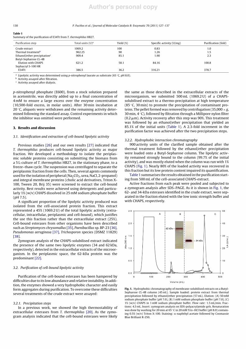
3.2.2. Hydrophobic interaction chromatography900 activity units of the clarified sample obtained after the
thermal treatment followed by the ethanol/ether precipitationwere loaded onto a Butyl-Sepharose column. The lipolytic activ-ity remained strongly bound to the column (99.7% of the initialactivity), and was mostly eluted when the column was run with 1%CHAPS (Fig. 1). Nearly 80% of the loaded activity was recovered inthis fraction but its low protein content impaired its quantification.
Table 1 summarises the results obtained in the purification start-ing from 500 mL of the cell-associated CHAPS-extract.
Active fractions from each peak were pooled and subjected toa zymogram analysis after SDS–PAGE. As it is shown in Fig. 1, the62- and 34-kDa esterases identified in the crude extract, were sep-arated in the fraction eluted with the low ionic strength buffer andwith CHAPS, respectively.
Fig. 1. Hydrophobic chromatography of membrane-solubilised extracts on a Butyl-Sepharose CL-4B column (45 mL). Sample loaded: protein extract from thermalprecipitation followed by ethanol/ether precipitation (57 mL). Elution: (A) 50 mMsodium phosphate buffer (pH 7.0), (B) 1 mM sodium phosphate buffer (pH 7.0), (C)1% (w/v) CHAPS in 1 mM sodium phosphate buffer. Flow rate: 1.5 mL/min. Frac-tions: 4.5 mL. Insert: zymogram analysis on SDS-polyacrylamide gels. Renaturationwas done by washing for 20 min at 65 "C in 20 mM Tris–HCl buffer (pH 8.0) contain-ing 0.5% (w/v) Triton X-100. Staining: !-naphthyl acetate followed by CoomassieBlue Brilliant R-250.

Author's personal copy
P. Fucinos et al. / Journal of Molecular Catalysis B: Enzymatic 70 (2011) 127–137 131
Table 2Parametric estimations and regression coefficients of a first-order model (1) and a parallel biexponential model (2) applied to the thermal deactivation of E34Tt from T.thermophilus HB27, incubated at 85 "C with detergent concentrations above (1%, w/v CHAPS) and below (0.01%, w/v CHAPS) the CMC.
Incubation Model t1/2 (h) k R km (h#1) ka (h#1) r2
1% (w/v) CHAPS (1) 2.25 0.31 – – –0.01% (w/v) CHAPS (1) 0.45 1.51 – – – 0.989
(2) 0.32 – 0.87 2.68 0.05 >0.999
3.2.3. Gel filtration chromatographyThe gel filtration analysis of the concentrated CHAPS-eluted
samples from the Butyl Sepharose column showed that only27% of the loaded activity was recovered in the void volume(>1.5 $ 106 Da), indicating that it was present in the form of ahigh molecular weight aggregates. When the pooled active frac-tions were mixed with CHAPS until 1% (w/v)-concentration wasreached, the activity yield could be restored up to nearly 100%after 2 h incubation at room temperature. This result indicatedthat the aggregation involved the active site of the enzyme, andthe reversible character of the phenomenon. The purification ofmicrobial lipases is frequently a difficult process due to the pres-ence of such aggregates, often caused by the presence of lipidsused to induce the enzyme production by the microorganism [39],or simply due to hydrophobic interactions amongst the enzymemolecules [40,41].
Taking into account these observations, a new chromatographywas carried out including 1%-CHAPS into the equilibration buffer.The obtained chromatographic profile is shown in Fig. 2. Nearly allthe loaded activity (86%) was recovered in an elution volume corre-sponding to 39.5 kDa which was in agreement with that determinedby denaturing electrophoresis (34 kDa) and deduced from the pri-mary sequence (36.4 kDa), while a second protein peak was in theflow-through fraction (Table 1). The enzyme was purified 379-foldwith a final specific activity of 316 U/mg.
The purity of the purified enzyme was checked by SDS–PAGE(inset in Fig. 2). Activity staining with !-naphthyl acetate and FastRed of the SDS-polyacrylamide gels resulted in one major red bandwith a molecular mass of 34 kDa (lane 1). In addition, the gelsshowed three minor bands with high molecular masses (62, 85,and 108 kDa). These bans are probably caused by self-aggregationof the purified esterase as they mostly disappeared by increasing
Fig. 2. Gel filtration chromatography of membrane-solubilised extracts on aSephacryl S-100 HR column (120 mL). Sample loaded: CHAPS-eluted fraction fromhydrophobic chromatography (1.2 mL). Elution: 50 mM sodium phosphate buffer(pH 7.0), containing 0.15 M NaCl and 1% (w/v) CHAPS. Flow rate: 0.5 mL/min.Fractions: 1.5 mL. Insert: zymogram analysis on SDS-polyacrylamide gels. Samplesloaded with a sample buffer containing a standard SDS concentration (4%, lane1),and double SDS concentration (8%, lane 2). Renaturation was done by washing for20 min at 65 "C in 20 mM Tris–HCl buffer (pH 8.0) containing 0.5% (w/v) Triton X-100.Staining: !-naphthyl acetate followed by Coomassie Blue Brilliant R-250.
the concentration of the denaturant in the sample buffer (8% SDS;lane 2).
3.3. Sequence analysis of native E34Tt
Proteins were separated by SDS–PAGE, blotted onto apolyvinylidene fluoride (PVDF) membrane and stained with Pon-ceau. Subsequently a piece of membrane containing a protein bandat 34-kDa was excised and subjected to N-terminal sequencing.However no specific sequence was determined, suggesting theexistence of a post-translational modification at the N-terminus[42]. The partially purified sample was then separated again by SDS-electrophoresis, the gel was stained with colloidal Coomassie BlueG-250, and the gel slice containing the 34-kDa protein was excised.The protein was treated with trypsin and resulting fragments wereanalysed by MALDI-TOF-MS. Spectra obtained were interpretedthrough the GPS Explorer software (Applied Biosystems, USA) withan integrated MASCOT search engine (Matrix Science, UK) that wasused for protein identification.
Peptide-mass fingerprinting was carried out and data searchedagainst NCBI databases. A total of 21 tryptic digest peptides wereidentified, whose masses and sequences matched to the theo-retical peptides resulting from in silico digestion of a putativeesterase from T. thermophilus HB27 (accession number YP 004875;gi|46199208), covering 49% of the entire sequence (329 amino acidresidues).
A molecular mass of 36.445 kDa and a pI of 8.37 were deducedfor this protein. In addition, the predicted enzyme sequence possesan N-terminal extension that has the requisites to be a transmem-brane helix: highly hydrophobic and a length of 20–25 amino acidresidues. Nonetheless, sequence analysis using the program SignalP3.0 predicts a signal peptide with a probability of 1.0, the cleavagesite of the signal peptide being between residues 16 (alanine) and17 (glutamine). Phobius program, considered superior over SignalP3.0 when it comes to prediction of transmembrane helices close tothe N-terminus [31], also predicts a secretory protein.
However, we also found that once E34Tt is solubilised frommembranes and purified, the detergent concentration (CHAPS) can-not be drastically decreased without loss of activity or stability.This fact strongly points to the association of this enzyme withmembranes.
Another interesting aspect invoked the possibility that themembrane association of E34Tt is connected to its transport out ofthe cell, the membrane-bound form being only a transitory stage.In fact, we detect this enzyme by SDS-electrophoresis followed byzymogram analysis in the culture medium of the microorganism[20].
BLASTp analysis revealed similarities to other hypothetical pro-teins and putative esterases. The highest identities being with aputative esterase from Thermus aquaticus Y51MC23 (ZP 03135254,329 aa), the hypothetical protein TTHA1268 from T. thermophilusHB8 (YP 144534, 214 aa), the V-type ATP synthase subunit fromT. thermophilus HB8 (YP 144536, 142 aa), and a putative pecti-nacetylesterase from Sorangium cellulosum (YP 001611310, 365aa). Conserved domains present in the encoded protein were alsoanalysed using the NCBI Conserved Domain Search, which revealeda possible pectin acetylesterase family domain (PAE), despite the
Author's personal copy
132 P. Fucinos et al. / Journal of Molecular Catalysis B: Enzymatic 70 (2011) 127–137
putative pectinacetylesterase from Sorangium cellulosum presentedthe lower identity amongst the four high-scored sequences in theBLASTp search.
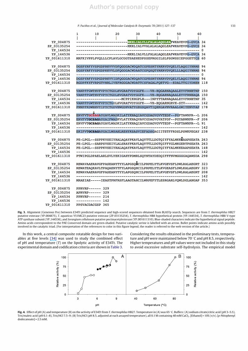
In order to further analyse the primary structural features ofE34Tt, multiple sequence alignments were performed using theGeneious Pro software with the four closely related sequences(Fig. 3). The analysis revealed that the PAE domain is common tothe five sequences.
The V-type ATP synthase subunit from T. thermophilus HB8(YP 144536, 142 aa) showed a very high similarity (97%) withthe N-terminal fragment of E34Tt (up to amino acid residue137). Interestingly, sequence from protein TTHA1268 from T. ther-mophilus HB8 matches (94% identity) the C-terminal fragment ofE34Tt (residues 133–329), starting at the end of the ATP syn-thase domain and goes up to the end of E34Tt protein sequence(residues 133–329); the putative esterase from the related organ-ism T. aquaticus Y51MC23 showed also an identity of 85%.
Despite the fact that no homology was detected with any lipaseor esterase described so far, the classical pentapeptide signaturemotif was identified between amino acids 157 and 161 (Gly-Cys-Ser159-Ala-Gly), which contains the putative catalytic serine. Toverify that E34Tt was indeed a serine hydrolase, a sample of enzymewas treated with the inhibitor E600, which binds covalently to ser-ine residues. The experiment was conducted in molar excess ofthe inhibitor ([E600]/[E34Tt] = 10,500), and in the presence of 1%(w/v) CHAPS to avoid enzyme aggregation. In these conditions, arapid and complete inhibition was observed after 30 min incuba-tion at room temperature, which clearly indicated that a serine wasinvolved in the catalytic mechanism.
3.4. Biochemical characterisation
3.4.1. Effect of pH and temperature on the enzyme activityThe pH and temperature dependence of E34Tt lipolytic activ-
ity were studied using p-nitrophenyl dodecanoate as substrate.pH–activity assays were conducted between pH 3 and 9, at 65 "C(determinations at higher pH values were hindered due to the spon-taneous hydrolysis of the substrate).
Contrary to most of the known esterases, which display an opti-mal activity in the range 5.5–6.5, E34Tt showed a maximum atpH 8.5 (Fig. 4A), more similar to that of lipases (maximally activeat 8.0–9.0) [43]. In addition, nearly 90% of the activity was stillretained at pH 9. This value is lower than the optimum for thethermostable lipases from microorganisms such as Geobacillus sp.(pH 9.0) [44] or Pseudomonas sp. (pH 11.0) [45]. However, thealkalophilicity of E34Tt is similar than the reported one for the ther-moalkalophilic lipases from Bacillus thermocatenulatus (pH 8.0–9.5)[41] and Bacillus sp. RSJ-1 (pH 8.0–9.0) [46], and even higher thanthose of some archaea such Pyrococcus horikoshii (pH 7.0) [47].
The temperature–activity dependence was measured in therange 40–80 "C. Results obtained (Fig. 4B) indicated an optimal tem-perature equal or above 80 "C (higher temperatures could not beassayed due to the elevated substrate self-hydrolysis).
The optimal catalytic temperature for E34Tt is at least 10 "Chigher than the optimal temperature for the growth of T. ther-mophilus HB27 (70 "C), as previously reported by [48], and it is alsohigher than the optimal temperature of some other esterases fromthermophilic microorganisms like Bacillus licheniformis (45 "C) [49],Bacillus circulans (60 "C) [50] or Thermoanaerobacter tengcongensis(70 "C) [51]. Moreover, this optimum is similar to that reported foresterases produced by hyperthermophilic organisms as Pyrobacu-lum calidifontis VA1 (90 "C) [52].
3.4.2. ThermostabilityThermal stability was assayed at 85 "C in presence of 1%
(w/v) CHAPS (above the CMC [53]). In these conditions, E34Tt
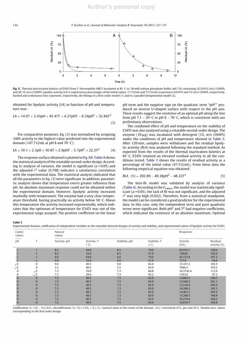
was extremely stable, maintaining residual activity after 10 h ofincubation (Fig. 5). In order to describe the deactivation kinetic,experimental data were adjusted to a first-order model:
LA(t) = LA0 e#kt (1)
where LA(t) represents the lipolytic activity at time t expressed aspercentage of the initial lipolytic activity (LA0), and k is the deacti-vation constant.
Deactivation in presence of 1% (w/v) CHAPS showed a good fit tothe first-order model (r2 = 0.992; Table 2), which predicted a half-life of 135 min. This makes E34Tt even more thermostable thansome esterases from hyperthermophiles such as Pyrococcus furiosus(t1/2 of 120 min at 75 "C) [54], Thermotoga maritima (t1/2 of 30 minat 80 "C) [55] or Thermococcus litoralis (t1/2 of 60 min at 70 "C) [56].In addition, no changes were observed in the deactivation kineticsat 85 "C when incubations were performed in presence of 1 mMEDTA (data not shown). Thus, Ca2+ or equivalent ions are not likelyto play a role in the thermostability of E34Tt.
To check the necessity of a micellar system to maintain thestability at high temperatures, thermal deactivation was also stud-ied at 0.01% (w/v) CHAPS concentration (below the CMC [53]). Inabsence of micelles, the enzyme underwent a 70% activity lossinstantly after dilution in an aqueous buffer when it was preparedfrom a concentrated sample containing 1% (w/v) CHAPS (17.5 U/mLvs. 73.5 U/mL). In other words, to maintain an active (native) confor-mation, the enzyme needs a micro-environment whose structuralarrangement resembles the membrane bilayer. Moreover, in thiscase, deactivation did not follow a first-order kinetic, especially atlong incubation times, in which the model (1) underestimated theresidual activity.
This behaviour could be explained by the presence of aggrega-tion forms with different activity and thermal stability, i.e., nativeforms (less stable in these conditions) and aggregated forms (morestable, although less active due to the inaccessibility of the sub-strate and/or to the scarcity of enzymes in native state being partof the aggregate). Thus, the activity measured at each time wouldbe the sum of the residual activity of monomers and aggregates.A mathematical approach to this phenomenon can be formulatedby using a parallel biexponential model based on that proposed byAymard and Belarbi [57] for the deactivation kinetics of enzymesmixtures:
LA(t) = LA0R e#kmt + LA0(1 # R) e#kat (2)
where R is the monomeric/total E34Tt ratio, and km and ka thedeactivation constants for the monomeric and aggregated forms,respectively.
As Table 2 and Fig. 5 show, the biexponential model provided agood fitting (r2 > 0.999), and the predicted half-life for E34Tt incu-bated with 0.01% (w/v) CHAPS was 0.32 h. Results also suggest thatthe aggregated form might have a thermal stability 50-fold higherthan the monomer.
3.4.3. Combined effect of pH and temperature on the lipolyticactivity and stability
Enzyme catalytic properties are commonly evaluated usingone-factor-at-a-time experiments. This methodology is easy tounderstand and useful for comparative purposes (most of thereported works followed this approach). However, one-factor-at-a-time experiments ignore interactions and may lead to misleadingconclusions. In this regard, experimental design methodologies[34] are more efficient than one-factor-at-a-time. Design of Exper-iments (DOE) uses statistical and mathematical techniques toevaluate the combined effect of factors instead of single factors atdifferent times. These approaches have been successfully appliedin the production [44,58], purification [59], characterisation [60],and application [61,62] of lipases and esterases.
Author's personal copy
P. Fucinos et al. / Journal of Molecular Catalysis B: Enzymatic 70 (2011) 127–137 133
Fig. 3. Alignment (Geneious Pro) between E34Tt predicted sequence and high-scored sequences obtained from BLASTp search. Sequences are from T. thermophilus HB27putative esterase (YP 004875), T. aquaticus Y51MC23 putative esterase (ZP 03135254), T. thermophilus HB8 hypothetical protein (YP 144534), T. thermophilus HB8 V-typeATP synthase subunit (YP 144536), and Sorangium cellulosum putative pectinacetylesterase (YP 001611310). Blue-shaded characters indicate the hypothetical signal peptide.Amino acids correspondent to the PAE Conserved domain are green-shaded. Putative catalytic serine is labelled with an arrow. Bullet points indicate amino acids possiblyinvolved in the catalytic triad. (For interpretation of the references to color in this figure legend, the reader is referred to the web version of the article.)
In this work, a central composite rotatable design for two vari-ables at five levels [34] was used to study the combined effectof pH and temperature (T) on the lipolytic activity of E34Tt. Theexperimental domain and codification criteria are shown in Table 3.
Considering the results obtained in the preliminary tests, tempera-ture and pH were maintained below 70 "C and pH 8.5, respectively.Higher temperatures and pH values were not included in this studyto avoid excessive substrate self-hydrolysis. The empirical model
Fig. 4. Effect of pH (A) and temperature (B) on the activity of E34Tt from T. thermophilus HB27. Temperature in (A) was 65 "C. Buffers: (A) sodium citrate/citric acid (pH 3–5.5),Tris/maleic acid (pH 6.1–8), Tris/HCl 7.5–9; (B) Tris/HCl (pH 8.5, adjusted at each assayed temperature), all 0.1 M containing 40 mM CaCl2. [Ethanol] = 10% (v/v). [p-Nitophenyldodecanoate] = 2.5 mM.
Author's personal copy
134 P. Fucinos et al. / Journal of Molecular Catalysis B: Enzymatic 70 (2011) 127–137
Fig. 5. Thermal deactivation kinetics of E34Tt from T. thermophilus HB27 incubated at 85 "C in: 50 mM sodium phosphate buffer (pH 7.0) containing (A) 0.01% (w/v) CHAPS,and (B) 1% (w/v) CHAPS. Lipolitic activity (LA) is expressed as percentages of the initial values, 17.5 U/mL and 73.5 U/mL in presence of 0.01% and 1% (w/v) CHAPS, respectively.Dashed and continuous lines represent, respectively, the fittings to a first-order model (1) and to a parallel biexponential model (2).
obtained for lipolytic activity (LA) as function of pH and tempera-ture was:
LA = 14.97 + 3.43pH + 45.47T + 4.27pHT # 8.34pH2 + 32.84T2
(3)
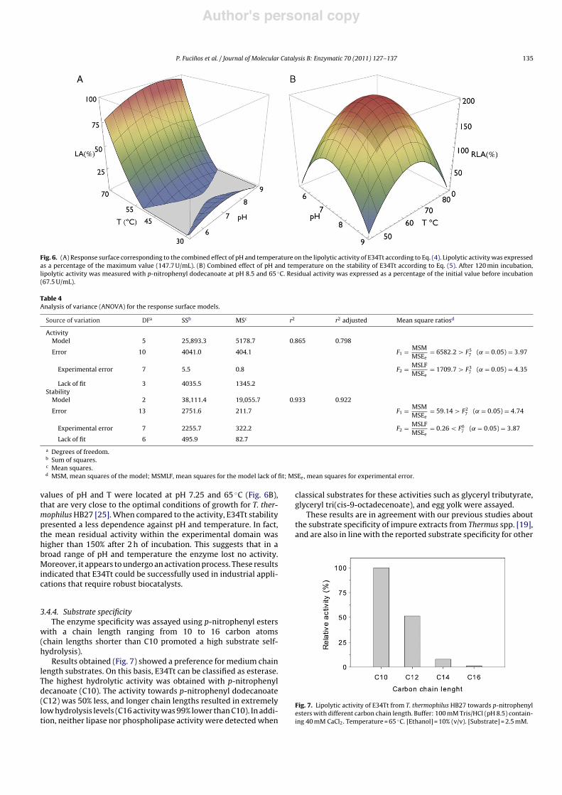
For comparative purposes, Eq. (3) was normalised by assigning100% activity to the highest value predicted into the experimentaldomain (147.7 U/mL at pH 8 and 70 "C):
LA = 10.1 + 2.3pH + 30.8T + 2.9pHT # 5.7pH2 + 22.2T2 (4)
The response surface obtained is plotted in Fig. 6A. Table 4 showsthe statistical analysis of the rotatable second-order design. Accord-ing to analysis of variance, the model is significant (˛ = 0.05) andthe adjusted r2 value (0.798) indicates a satisfactory correlationwith the experimental data. The statistical analysis indicated thatall the parameters in Eq. (3) were significant. In addition, paramet-ric analysis shows that temperature exerts greater influence thanpH. An absolute maximum response could not be obtained withinthe experimental domain. However, lipolytic activity increasedmarkedly with temperature. The enzyme had a very clear temper-ature threshold, having practically no activity below 50 "C. Abovethis temperature the activity increased exponentially, which indi-cates that the optimum of temperature for E34Tt was out of theexperimental range assayed. The positive coefficient on the linear
pH term and the negative sign on the quadratic term “pH2” pro-duced an inverse U-shaped surface with respect to the pH axis.These results suggest the existence of an optimal pH along the linefrom pH 7.1 – 29 "C to pH 8 – 70 "C, which is consistent with ourpreliminary observations.
The combined effect of pH and temperature on the stability ofE34Tt was also analysed using a rotatable second-order design. Theenzyme (10 "g) was incubated with detergent (1%, w/v CHAPS)under the conditions of pH and temperature showed in Table 3.After 120 min, samples were withdrawn and the residual lipoly-tic activity (RLA) was analysed following the standard method. Asexpected from the results of the thermal inactivation kinetics at85 "C, E34Tt retained an elevated residual activity in all the con-ditions tested. Table 3 shows the results of residual activity as apercentage of the initial value (67.5 U/mL). From these data thefollowing empirical equation was obtained:
RLA (%) = 202.89 # 49.38pH2 # 48.22T2 (5)
The best-fit model was validated by analysis of variance(Table 4). According to the Fvalues, the model was statistically signif-icant (˛ = 0.05), the lack of fit was not significant, and the adjustedr2 was very high (0.922). Therefore, from a statistical standpoint,the model can be considered a good predictor for the experimentaldata. In this case, only the independent term and pure quadraticterms were significant. Both pH2 and T2 had negative coefficients,which indicated the existence of an absolute maximum. Optimal
Table 3Experimental domain, codification of independent variables in the rotatable factorial designs of activity and stability, and experimental values of lipolytic activity for E34Tt.
Codedvalues
Naturalvalues
Responses
pH T Activity–pH Activity–T("C)
Stability–pH Stability–T("C)
Activity(U/L)
Residualactivity (%)
1 1 8.5 64.0 8.5 79.0 62,935.7 120.61 #1 8.5 35.0 8.5 51.0 1464.8 102.0
#1 1 6.0 64.0 6.0 79.0 45,127.8 107.2#1 #1 6.0 35.0 6.0 51.0 737.0 94.9%
2 0 9.0 49.5 9.0 65.0 13,337.2 102.9#%
2 0 5.5 49.5 5.5 65.0 7049.3 103.50
%2 7.3 70.0 7.3 84.8 18,3742.4 113.9
0 #%2 7.3 29.0 7.3 45.2 1363.8 97.2
0 0 7.3 49.5 7.3 65.0 14,885.3 228.20 0 7.3 49.5 7.3 65.0 13,566.2 201.10 0 7.3 49.5 7.3 65.0 15,139.4 205.40 0 7.3 49.5 7.3 65.0 14,286.2 185.30 0 7.3 49.5 7.3 65.0 14,401.2 205.40 0 7.3 49.5 7.3 65.0 15,266.5 184.80 0 7.3 49.5 7.3 65.0 16,210.4 228.20 0 7.3 49.5 7.3 65.0 16,010.7 184.8
Codification: Vc = (Vn # V0)/!Vn; decodification: Vn = V0 + (!Vn $ Vc). Vn = natural value in the centre of the domain; !Vn = increment of Vn per unit of Vc . Shaded area: valuescorresponding to the first order design.
Author's personal copy
P. Fucinos et al. / Journal of Molecular Catalysis B: Enzymatic 70 (2011) 127–137 135
Fig. 6. (A) Response surface corresponding to the combined effect of pH and temperature on the lipolytic activity of E34Tt according to Eq. (4). Lipolytic activity was expressedas a percentage of the maximum value (147.7 U/mL). (B) Combined effect of pH and temperature on the stability of E34Tt according to Eq. (5). After 120 min incubation,lipolytic activity was measured with p-nitrophenyl dodecanoate at pH 8.5 and 65 "C. Residual activity was expressed as a percentage of the initial value before incubation(67.5 U/mL).
Table 4Analysis of variance (ANOVA) for the response surface models.
Source of variation DFa SSb MSc r2 r2 adjusted Mean square ratiosd
ActivityModel 5 25,893.3 5178.7 0.865 0.798
Error 10 4041.0 404.1 F1 = MSMMSEe
= 6582.2 > F57 (˛ = 0.05) = 3.97
Experimental error 7 5.5 0.8 F2 = MSLFMSEe
= 1709.7 > F37 (˛ = 0.05) = 4.35
Lack of fit 3 4035.5 1345.2Stability
Model 2 38,111.4 19,055.7 0.933 0.922
Error 13 2751.6 211.7 F1 = MSMMSEe
= 59.14 > F27 (˛ = 0.05) = 4.74
Experimental error 7 2255.7 322.2 F2 = MSLFMSEe
= 0.26 < F67 (˛ = 0.05) = 3.87
Lack of fit 6 495.9 82.7
a Degrees of freedom.b Sum of squares.c Mean squares.d MSM, mean squares of the model; MSMLF, mean squares for the model lack of fit; MSEe, mean squares for experimental error.
values of pH and T were located at pH 7.25 and 65 "C (Fig. 6B),that are very close to the optimal conditions of growth for T. ther-mophilus HB27 [25]. When compared to the activity, E34Tt stabilitypresented a less dependence against pH and temperature. In fact,the mean residual activity within the experimental domain washigher than 150% after 2 h of incubation. This suggests that in abroad range of pH and temperature the enzyme lost no activity.Moreover, it appears to undergo an activation process. These resultsindicated that E34Tt could be successfully used in industrial appli-cations that require robust biocatalysts.
3.4.4. Substrate specificityThe enzyme specificity was assayed using p-nitrophenyl esters
with a chain length ranging from 10 to 16 carbon atoms(chain lengths shorter than C10 promoted a high substrate self-hydrolysis).
Results obtained (Fig. 7) showed a preference for medium chainlength substrates. On this basis, E34Tt can be classified as esterase.The highest hydrolytic activity was obtained with p-nitrophenyldecanoate (C10). The activity towards p-nitrophenyl dodecanoate(C12) was 50% less, and longer chain lengths resulted in extremelylow hydrolysis levels (C16 activity was 99% lower than C10). In addi-tion, neither lipase nor phospholipase activity were detected when
classical substrates for these activities such as glyceryl tributyrate,glyceryl tri(cis-9-octadecenoate), and egg yolk were assayed.
These results are in agreement with our previous studies aboutthe substrate specificity of impure extracts from Thermus spp. [19],and are also in line with the reported substrate specificity for other
Fig. 7. Lipolytic activity of E34Tt from T. thermophilus HB27 towards p-nitrophenylesters with different carbon chain length. Buffer: 100 mM Tris/HCl (pH 8.5) contain-ing 40 mM CaCl2. Temperature = 65 "C. [Ethanol] = 10% (v/v). [Substrate] = 2.5 mM.
Author's personal copy
136 P. Fucinos et al. / Journal of Molecular Catalysis B: Enzymatic 70 (2011) 127–137
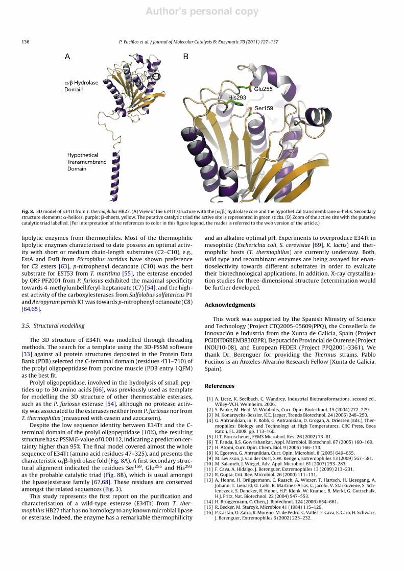
Fig. 8. 3D model of E34Tt from T. thermophilus HB27. (A) View of the E34Tt structure with the (!/#) hydrolase core and the hypothetical transmembrane !-helix. Secondarystructure elements: !-helices, purple; #-sheets, yellow. The putative catalytic triad the active site is represented in green sticks. (B) Zoom of the active site with the putativecatalytic triad labelled. (For interpretation of the references to color in this figure legend, the reader is referred to the web version of the article.)
lipolytic enzymes from thermophiles. Most of the thermophiliclipolytic enzymes characterised to date possess an optimal activ-ity with short or medium chain-length substrates (C2–C10), e.g.,EstA and EstB from Picrophilus torridus have shown preferencefor C2 esters [63], p-nitrophenyl decanoate (C10) was the bestsubstrate for EST53 from T. maritima [55], the esterase encodedby ORF PF2001 from P. furiosus exhibited the maximal specificitytowards 4-methylumbelliferyl-heptanoate (C7) [54], and the high-est activity of the carboxylesterases from Sulfolobus solfataricus P1and Aeropyrum pernix K1 was towards p-nitrophenyl octanoate (C8)[64,65].
3.5. Structural modelling
The 3D structure of E34Tt was modelled through threadingmethods. The search for a template using the 3D-PSSM software[33] against all protein structures deposited in the Protein DataBank (PDB) selected the C-terminal domain (residues 431–710) ofthe prolyl oligopeptidase from porcine muscle (PDB entry 1QFM)as the best fit.
Prolyl oligopeptidase, involved in the hydrolysis of small pep-tides up to 30 amino acids [66], was previously used as templatefor modelling the 3D structure of other thermostable esterases,such as the P. furiosus esterase [54], although no protease activ-ity was associated to the esterases neither from P. furiosus nor fromT. thermophilus (measured with casein and azocasein).
Despite the low sequence identity between E34Tt and the C-terminal domain of the prolyl oligopeptidase (10%), the resultingstructure has a PSSM E-value of 0.00112, indicating a prediction cer-tainty higher than 95%. The final model covered almost the wholesequence of E34Tt (amino acid residues 47–325), and presents thecharacteristic !/#-hydrolase fold (Fig. 8A). A first secondary struc-tural alignment indicated the residues Ser159, Glu255 and His293
as the probable catalytic triad (Fig. 8B), which is usual amongstthe lipase/esterase family [67,68]. These residues are conservedamongst the related sequences (Fig. 3).
This study represents the first report on the purification andcharacterisation of a wild-type esterase (E34Tt) from T. ther-mophilus HB27 that has no homology to any known microbial lipaseor esterase. Indeed, the enzyme has a remarkable thermophilicity
and an alkaline optimal pH. Experiments to overproduce E34Tt inmesophilic (Escherichia coli, S. cerevisiae [69], K. lactis) and ther-mophilic hosts (T. thermophilus) are currently underway. Both,wild type and recombinant enzymes are being assayed for enan-tioselectivity towards different substrates in order to evaluatetheir biotechnological applications. In addition, X-ray crystallisa-tion studies for three-dimensional structure determination wouldbe further developed.
Acknowledgments
This work was supported by the Spanish Ministry of Scienceand Technology (Project CTQ2005-05609/PPQ), the Consellería deInnovación e Industria from the Xunta de Galicia, Spain (ProjectPGIDIT06REM38302PR), Deputación Provincial de Ourense (ProjectINOU10-08), and European FEDER (Project PPQ2001-3361). Wethank Dr. Berenguer for providing the Thermus strains. PabloFucinos is an Ánxeles-Alvarino Research Fellow (Xunta de Galicia,Spain).
References
[1] A. Liese, K. Seelbach, C. Wandrey, Industrial Biotransformations, second ed.,Wiley-VCH, Weinheim, 2006.
[2] S. Panke, M. Held, M. Wubbolts, Curr. Opin. Biotechnol. 15 (2004) 272–279.[3] M. Konarzycka-Bessler, K.E. Jaeger, Trends Biotechnol. 24 (2006) 248–250.[4] G. Antranikian, in: F. Robb, G. Antranikian, D. Grogan, A. Driessen (Eds.), Ther-
mophiles: Biology and Technology at High Temperatures, CRC Press, BocaRaton, FL, 2008, pp. 113–160.
[5] U.T. Bornscheuer, FEMS Microbiol. Rev. 26 (2002) 73–81.[6] T. Panda, B.S. Gowrishankar, Appl. Microbiol. Biotechnol. 67 (2005) 160–169.[7] H. Atomi, Curr. Opin. Chem. Biol. 9 (2005) 166–173.[8] K. Egorova, G. Antranikian, Curr. Opin. Microbiol. 8 (2005) 649–655.[9] M. Levisson, J. van der Oost, S.W. Kengen, Extremophiles 13 (2009) 567–581.
[10] M. Salameh, J. Wiegel, Adv. Appl. Microbiol. 61 (2007) 253–283.[11] F. Cava, A. Hidalgo, J. Berenguer, Extremophiles 13 (2009) 213–231.[12] R. Gupta, Crit. Rev. Microbiol. 26 (2000) 111–131.[13] A. Henne, H. Brüggemann, C. Raasch, A. Wiezer, T. Hartsch, H. Liesegang, A.
Johann, T. Lienard, O. Gohl, R. Martinez-Arias, C. Jacobi, V. Starkuviene, S. Sch-lenczeck, S. Dencker, R. Huber, H.P. Klenk, W. Kramer, R. Merkl, G. Gottschalk,H.J. Fritz, Nat. Biotechnol. 22 (2004) 547–553.
[14] H. Brüggemann, C. Chen, J. Biotechnol. 124 (2006) 654–661.[15] R. Becker, M. Starzyk, Microbios 41 (1984) 115–129.[16] P. Castán, O. Zafra, R. Moreno, M. de Pedro, C. Vallés, F. Cava, E. Caro, H. Schwarz,
J. Berenguer, Extremophiles 6 (2002) 225–232.
Author's personal copy
P. Fucinos et al. / Journal of Molecular Catalysis B: Enzymatic 70 (2011) 127–137 137
[17] A.A. Pantazaki, A.A. Pritsa, D.A. Kyriakidis, Appl. Microbiol. Biotechnol. 58(2002) 1–12.
[18] E.E. Lioliou, A.A. Pantazaki, D.A. Kyriakidis, Microb. Cell. Fact. 3 (2004) 5.[19] A. Domínguez, A. Sanromán, P. Fucinos, M. Rúa, L. Pastrana, M. Longo, Biotech-
nol. Lett. 26 (2004) 705–708.[20] P. Fucinos, C.M. Abadín, A. Sanromán, M.A. Longo, L. Pastrana, M.L. Rúa, J.
Biotechnol. 117 (2005) 233–241.[21] P. Fucinos, A. Domínguez, M. Sanromán, M. Longo, M. Rúa, L. Pastrana, Biotech-
nol. Prog. 21 (2005) 1198–1205.[22] P. Fucinos, An integrated study of the lipases from Thermus thermophilus HB27:
conditions of production, cellular localisation, purification and characterisa-tion, PhD Thesis, University of Vigo, Ourense, Spain, 2007.
[23] P. Fucinos, M. Rúa, M. Longo, M. Sanromán, L. Pastrana, Process Biochem. 43(2008) 1383–1390.
[24] A. Dominguez, L. Pastrana, M.A. Longo, M.L. Rua, M.A. Sanroman, Biochem. Eng.J. 26 (2005) 95–99.
[25] A. Domínguez, P. Fucinos, M. Rúa, L. Pastrana, M. Longo, M. Sanromán, EnzymeMicrob. Technol. 40 (2007) 187–194.
[26] S. Sigurgisladottir, M. Konraosdottir, A. Jonsson, J. Kristjansson, E. Matthiasson,Biotechnol. Lett. 15 (1993) 361–366.
[27] F.J. Deive, E. Carvalho, L. Pastrana, M.L. Rúa, M.A. Longo, M.A. Sanroman, Biore-sour. Technol. 100 (2009) 3630–3637.
[28] D.N. Perkins, D.J. Pappin, D.M. Creasy, J.S. Cottrell, Electrophoresis 20 (1999)3551–3567.
[29] S.F. Altschul, T.L. Madden, A.A. Schäffer, J. Zhang, Z. Zhang, W. Miller, D.J. Lipman,Nucleic Acids Res. 25 (1997) 3389–3402.
[30] A. Marchler-Bauer, S.H. Bryant, Nucleic Acids Res. 32 (2004) W327–331.[31] J.D. Bendtsen, H. Nielsen, G. von Heijne, S. Brunak, J. Mol. Biol. 340 (2004)
783–795.[32] L. Käll, A. Krogh, E.L. Sonnhammer, J. Mol. Biol. 338 (2004) 1027–1036.[33] L. Kelley, R. MacCallum, M. Sternberg, J. Mol. Biol. 299 (2000)
499–520.[34] G.E.P. Box, J.S. Hunter, W.G. Hunter, Statistics for Experimenters: Design,
Innovation, and Discovery, 2nd ed., John Wiley & Sons, Inc., Hoboken, NJ,2005.
[35] R. Berger, M. Hoffmann, U. Keller, J. Bacteriol. 180 (1998) 6396–6399.[36] N. Prim, A. Blanco, J. Martínez, F. Pastor, P. Diaz, Res. Microbiol. 151 (2000)
303–312.[37] S. Wilhelm, A. Gdynia, P. Tielen, F. Rosenau, K. Jaeger, J. Bacteriol. 189 (2007)
6695–6703.[38] J. Vakhlu, S. Johri, V. Verma, S. Koul, R. Parshad, S. Taneja, G. Qazi, Enzyme
Microb. Technol. 37 (2005) 330–339.[39] M. Pernas, C. López, L. Pastrana, M. Rúa, J. Biotechnol. 84 (2001) 163–174.[40] N.H. Schlieben, K. Niefind, D. Schomburg, Protein Expr. Purif. 34 (2004)
103–110.[41] M.L. Rúa, C. Schmidt-Dannert, S. Wahl, A. Sprauer, R.D. Schmid, J. Biotechnol.
56 (1997) 89–102.
[42] K. Miyazaki, Extremophiles 9 (2005) 415–425.[43] P. Fojan, P.H. Jonson, M.T. Petersen, S.B. Petersen, Biochimie 82 (2000)
1033–1041.[44] Y.R. Abdel-Fattah, N.A. Soliman, A.A. Gaballa, S.A. Sabry, A.I. El-Diwany, Acta
Microbiol. Pol. 51 (2002) 353–366.[45] P. Rathi, S. Bradoo, R. Saxena, R. Gupta, Biotechnol. Lett. 22 (2000) 495–
498.[46] R. Sharma, S. Soni, R. Vohra, L. Gupta, J. Gupta, Process Biochem. 37 (2002)
1075–1084.[47] Y. Feng, Y. Joh, K. Ishikawa, H. Ishida, S. Ando, T. Yamagaki, H. Nakanishi, S. Cao,
I. Matsui, Y. Kosugi, J. Am. Oil Chem. Soc. 77 (2000) 1147–1152.[48] T. Oshima, K. Imahori, Int. J. Syst. Bacteriol. 24 (1974) 102–112.[49] E. Alvarez-Macarie, V. Augier-Magro, J. Baratti, Biosci. Biotechnol. Biochem. 63
(1999) 1865–1870.[50] A. Kademi, N. Ait-Abdelkader, L. Fakhreddine, J.C. Baratti, J. Mol. Catal. B: Enzym.
10 (2000) 395–401.[51] J. Zhang, J. Liu, J. Zhou, Y. Ren, X. Dai, H. Xiang, Biotechnol. Lett. 25 (2003)
1463–1467.[52] Y. Hotta, S. Ezaki, H. Atomi, T. Imanaka, Appl. Environ. Microbiol. 68 (2002)
3925–3931.[53] M. Pernas, L. Pastrana, P. Fucinos, M. Rúa, J. Phys. Org. Chem. 22 (2009) 508–514.[54] R. Almeida, S. Alquéres, A. Larentis, S. Rössle, A. Cardoso, W. Almeida, P. Bisch,
T. Alves, O. Martins, Enzyme Microb. Technol. 39 (2006) 1128–1136.[55] S. Kakugawa, S. Fushinobu, T. Wakagi, H. Shoun, Appl. Microbiol. Biotechnol.
74 (2007) 585–591.[56] M.R. Singleton, J.A. Littlechild, in: M. Adams, R.M. Kelly (Eds.), Methods in Enzy-
mology: Hyperthermophilic Enzymes, Academic Press, San Diego, 2001, pp.394–403.
[57] C. Aymard, A. Belarbi, Enzyme Microb. Technol. 27 (2000) 612–618.[58] X. Ren, D. Yu, S. Han, Y. Feng, Bioresour. Technol. 97 (2006) 2345–2349.[59] E. Kamimura, O. Medieta, M. Rodrigues, F. Maugeri, Biotechnol. Appl. Biochem.
33 (2001) 153–159.[60] M.D. Benaiges, M. Alarcoın, P. Fucinos, P. Ferrer, M. Rúa, F. Valero, Biotechnol.
Prog. 26 (2010) 1252–1258.[61] L. Peng, X. Xu, H. Mu, C. Høy, J. Adler-Nissen, Enzyme Microb. Technol. 31 (2002)
523–532.[62] C. Shieh, H. Liao, C. Lee, Bioresour. Technol. 88 (2003) 103–106.[63] M. Hess, M. Katzer, G. Antranikian, Extremophiles 12 (2008) 351–364.[64] Y. Park, S.Y. Choi, H. Lee, Biochim. Biophys. Acta (BBA): Gen. Subjects 1760
(2006) 820–828.[65] R. Gao, Y. Feng, K. Ishikawa, H. Ishida, S. Ando, Y. Kosugi, S. Cao, J. Mol. Catal. B:
Enzym. 24–25 (2003) 1–8.[66] V. Fülöp, Z. Böcskei, L. Polgár, Cell 94 (1998) 161–170.[67] K.E. Jaeger, B.W. Dijkstra, M.T. Reetz, Annu. Rev. Microbiol. 53 (1999) 315–351.[68] S. Hari Krishna, N. Karanth, Catal. Rev. Sci. Eng. 44 (2002) 499–591.[69] O. López-López, P. Fucinos, L. Pastrana, M. Rúa, M. Cerdán, M. González-Siso, J.
Biotechnol. 145 (2010) 226–232.