Gold nanoparticle synthesis using the thermophilic bacterium Thermus scotoductus SA-01 and the...
11
1 23 Gold Bulletin The journal of gold science, technology and applications e-ISSN 2190-7579 Gold Bull DOI 10.1007/s13404-014-0147-8 Gold nanoparticle synthesis using the thermophilic bacterium Thermus scotoductus SA-01 and the purification and characterization of its unusual gold reducing protein Mariana Erasmus, Errol Duncan Cason, Jacqueline van Marwijk, Elsabé Botes, Mariekie Gericke & Esta van Heerden
Transcript of Gold nanoparticle synthesis using the thermophilic bacterium Thermus scotoductus SA-01 and the...

1 23
Gold BulletinThe journal of gold science, technologyand applications e-ISSN 2190-7579 Gold BullDOI 10.1007/s13404-014-0147-8
Gold nanoparticle synthesis using thethermophilic bacterium Thermusscotoductus SA-01 and the purificationand characterization of its unusual goldreducing proteinMariana Erasmus, Errol Duncan Cason,Jacqueline van Marwijk, Elsabé Botes,Mariekie Gericke & Esta van Heerden
1 23
Your article is published under the Creative
Commons Attribution license which allows
users to read, copy, distribute and make
derivative works, as long as the author of
the original work is cited. You may self-
archive this article on your own website, an
institutional repository or funder’s repository
and make it publicly available immediately.
ORIGINAL PAPER
Gold nanoparticle synthesis using the thermophilic bacteriumThermus scotoductus SA-01 and the purificationand characterization of its unusual gold reducing protein
Mariana Erasmus & Errol Duncan Cason &
Jacqueline van Marwijk & Elsabé Botes &Mariekie Gericke & Esta van Heerden
# The Author(s) 2014. This article is published with open access at SpringerLink.com
Abstract Nanoparticles are very important materials forimplementing nanotechnology in diverse areas and are abundantin nature as living organisms operate at a nanoscale. As nano-particles exhibit interesting size- and shape-dependent physicaland chemical properties, the synthesis of uniform nanoparticleswith controlled sizes and shapes is of great importance.Nanoparticles are the end products of a wide variety of physical,chemical and biological processes, some of which are novel andradically different and others of which are quite commonplace.The ability to produce nanoparticles with specific shapes andcontrolled sizes could result in interesting new applications thatcan potentially be utilized in areas such as optics, electronics andthe biomedical field. In the present study, we have demonstratedthe ability of the thermophilic bacterium Thermus scotoductus
SA-01 to synthesize gold nanoparticles and determined theeffect of the physico-chemical parameters on particle synthesis.Furthermore, a protein purified from this bacterium is shown tobe capable of reducing HAuCl4 to form elemental nanoparticlesin vitro. The protein was purified to homogeneity and identifiedthrough N-terminal sequencing as an ABC transporter, peptide-binding protein. It is speculated that this protein reduces Au(III)through an electron shuttle mechanism involving a cysteinedisulphide bridge. Through manipulation of physico-chemicalparameters, it was possible to vary nanoparticles in terms ofnumber, shape and size. This is the first report of a transporterprotein from a thermophile with the ability to produce nanopar-ticles in vitro thus expanding the limited knowledge aroundbiological gold nanoparticle synthesis.
Keywords Gold nanoparticles . Thermophilic . Goldreduction . Biological synthesis . ABC transporter
Introduction
Colloidal gold nanoparticles have attracted the interestof scientists for over 400 years, and the synthesis there-of includes reduction of chloroauric acid by reductants;use of these reductants produces particles of relativelyuniform size, which are generally spherical [1–3]. Inorder to control synthesis of these particles, the meth-odology by which they are produced should be under-stood. The means by which microorganisms achievechanges in metal speciation and mobility are essentialgears of the biogeochemical cycles of metals, as well asfor other elements [4]. Microorganisms can interact withmetals in a variety of ways [5], [6–8] and biomineral-ization occurs through one of two major pathways,namely biologically induced biomineralization (BIM) or
Electronic supplementary material The online version of this article(doi:10.1007/s13404-014-0147-8) contains supplementary material,which is available to authorized users.
M. Erasmus : E. D. Cason : J. van Marwijk : E. Botes :E. van Heerden (*)Department of Microbial, Biochemical and Food Biotechnology,Faculty of Natural and Agricultural Sciences, University of the FreeState, Bloemfontein 9300, South Africae-mail: [email protected]
M. Erasmuse-mail: [email protected]
E. D. Casone-mail: [email protected]
J. van Marwijke-mail: [email protected]
E. Botese-mail: [email protected]
M. GerickeBiotechnology Division, Mintek, Private Bag X3015,Randburg 2125, South Africae-mail: [email protected]
Gold BullDOI 10.1007/s13404-014-0147-8
boundary-organized biomineralization (BOB) [9]. Whenthe BIM pathway is followed, the organism has littlecontrol over deposition of mineral particles and theseparticles are often characterized by poor crystallinity,broad particle size distribution and non-specific crystalmorphology [10–12]. Alternatively, when the BOB path-way is followed, the organism could exercise controlover the nucleation and growth of particles. These nano-particles are developed under the direct regulatory con-trol of the organism and synthesized at a specific loca-tion within the cell and only under certain conditions[11–13].
The ability to produce gold nanoparticles with predictableshapes and sizes under controlled conditions could result ininteresting new applications that can potentially be utilized inareas such as optics, electronics and biomedicine [14–17]. Thebenefits of using a biological synthesis system rangefrom the predictable production of uniform metal nano-particles due to the highly structured activities of mi-crobial cells to environmentally friendly productionmethods and the ability to produce nanoparticles withunique shapes and composition [18–20]. It is thereforeimportant to gain an understanding of the mechanism ofgold accumulation and reduction by bacterial cells on amolecular and cellular level. Identification and analysisof the biopolymers involved in these processes couldpotentially allow for a process in a cell-free environ-ment, where the size and shape of particles can beregulated to some extent by specific proteins/enzymes[21]. This would also be advantageous from a processpoint of view, since it would eliminate the need toharvest the nanoparticles formed within the cells, sim-plifying the process and lowering operation costs.
Different hypotheses have been formulated about themechanisms involved in gold reduction and nanoparticle syn-thesis, and these are all based on enzymes with metal reduc-tase (oxidoreduction) abilities [22, 23]. However, the focus onoxidoreductases has overlooked the concept of ‘metabolicpromiscuity’, where a single protein can have a wide rangeof functions [24, 25] resulting in a lack of knowledgeconcerning the mechanisms and capabilities of other non-reductase proteins that are still capable of gold reduction andnanoparticle formation [24, 25].
In this paper, the interaction between Thermusscotoductus SA-01 and gold chloride ions was evaluatedon both a biomass level, where biomass refers to thewhole cells of T. scotoductus SA-01, and by using apurified protein that was identified to be capable ofregulating gold nanoparticle synthesis. The effect ofthe physico-chemical parameters on particle morphologywas assessed, and it was determined whether theseparameters can be manipulated to produce size- andshape-specific nanoparticles.
Experimental details
Microorganisms and growth
T. scotoductus SA-01 was always grown from a glycerolstock, plated out onto TYG-agar plates (5 g L−1 tryptone,3 g L−1 yeast extract, 1 g L−1 glucose and 18 g L−1 bacterio-logical agar) and grown for 24 h. A 20–24-h-old culture wasused to prepare the pre-inoculum (a loop full of culture in50 ml TYG in a 250-ml Erlenmeyer flask) and grown untilmid-exponential growth phase (~8 h). The inoculum was alsogrown to mid-exponential growth phase (~8 h) and was pre-pared by adding 10ml of the pre-inoculum to 90ml TYG (in a500-ml Erlenmeyer flask). Fivemillilitres of the inoculumwasadded to 95 ml TYG (in a 500-ml Erlenmeyer flask) for thegrowth experiments. One-millilitre samples were withdrawnover time (every 2 h until 16 h, and then at 20 and 24 h), andthe optical density (OD) was measured at 600 nm.
The correlation between growth phase and gold reductionwas evaluated by collecting biomass at different time intervalsas described before. The biomass was washed three times andresuspended in a 1:20 w/v ratio in 50 mM acetate buffer, pH5.0 [21]. Au(III), to a final concentration of 2 mM, was addedand the concentration determined by using a spectrophoto-metric method adapted from Melwanki and co-workers [26].
Biomass was obtained as described previously to be used aswhole cell catalysts and exposed to varying physico-chemicalparameters. The effect of pH was evaluated using a pH rangedfrom 3.6, 5.5 (50 mM sodium acetate buffer) and 7.4 (50 mMsodium phosphate buffer) to 9.0 (50 mM borax buffer), whilegold ion concentration was evaluated for the range starting from0 to 0.01 M. The temperature range stretched from 30 to 65 °Cand the exposure time from 1 to 48 h.
Šlouf and co-workers [27] indicated that gold nanoparticlesexhibit pink/purple red colours, which arises due to excitationof surface plasmon vibrations in the gold nanoparticles. UV–Vis spectroscopy can be used to record the surface plasmonband of the gold nanoparticles resulting in visual confirmation(a shift from a yellow to a pink/purple colour) indicative ofgold nanoparticle production [28]. Samples were withdrawnover time and a UV–Vis spectrum (400–700 nm) was obtain-ed using a Beckman DU-800 spectrophotometer, to evaluatethe plasmon band associated with gold nanoparticles. Thesesamples were also analysed by transmission electron micros-copy (TEM), as described below.
Cell-free extraction and protein purification
T. scotoductus SA-01 was grown to the late exponential growthphase [29], biomass harvested by centrifugation (6,000×g;15 min; 4 °C) and cell pellets washed three times with 50 mMsodium phosphate buffer (pH 7.4). Cell-free extracts were pre-pared according to adapted methods of Gaspard and co-workers
Gold Bull
(this implied that the cells were broken to release the proteinsfrom within the cell) [30]. The soluble fraction was size fraction-ated and concentrated using an Amicon stirred cell fitted with aUF 30 MWCO membrane (Osmonics Inc.). The retentate wasdialysed (SnakeSkin 7000 MWCO; Thermo Scientific) against50 mM sodium phosphate buffer (pH 7.4) to remove reducingagents such as organic acids and carbohydrates. The filtrate wasapplied to a Super Q-Toyopearl column (TOSOH) in 50 mMsodium phosphate buffer (pH 7.4). The non-binding proteinfraction, which was able to form nanoparticles in the presenceof HAuCl4, was pooled and dialyzed against 50 mM sodiumacetate buffer (pH 5.0). The dialysate was applied to a SP-Toyopearl column (TOSOH) and eluted with a 0–1 M NaClgradient. Active fractions were pooled, dialysed against 50 mMsodium phosphate buffer (pH 7.4) and reapplied to the SP-Toyopearl column (TOSOH). Bound proteins were eluted witha linear 0–0.3 M NaCl gradient, active fractions pooled anddialysed against 50 mM sodium phosphate buffer (pH 7.4) andapplied to a Blue-Sepharose CL-6B column (Sigma-Aldrich).Proteins were eluted with a linear 0–0.6 M NaCl gradient, activefractions pooled and dialysed against 50 mM sodium phosphatebuffer (pH 7.4). Ammonium sulphate was added to 1 M andapplied to a Phenyl-Toyopearl column (TOSOH) pre-equilibrated with 50 mM sodium phosphate buffer (pH 7.4)containing 1 M (NH4)2SO4. Bound proteins were eluted with alinear 1–0 M (NH4)2SO4 gradient, active fractions pooled anddialysed against 50 mM sodium phosphate buffer (pH 7.4). Thesample was concentrated to ~3 ml and applied to a SephacrylS200HR column (Sigma-Aldrich) (2.5×65 cm). Proteins wereeluted with 50mM sodium phosphate buffer (pH 7.4) containing50 mM NaCl at 1 ml min−1. All liquid chromatography proce-dures were performed on an Äkta Prime Purification System(Amersham Biosciences).
Cloning and expression of protein
N-terminal sequencing of the purified protein was performedby automated Edman degradation with an Applied Biosystems477A gas-phase sequencer (Foster City, CA) at the ProteinChemistry Facility of the Centro de InvestigacionesBiológicas (CSIC; Madrid, Spain). From the obtained se-quence, primers were designed and the target gene was PCRamplified with 2 mM MgCl2, 0.8 mM dNTPs, 2.5 U Super-Therm polymerase, 0.2μMof both the forward (ABC_F_Nde–5′ CATATG AGA AAA GTA GGC AAG CTG GCT 3′) andreverse (ABC_R_Eco – 5′ GAA TTC TTA CTT GAC GGAAAG AGC GTA 3′) primers and 50 ng gDNA template. Thegene was cloned into pET-28b(+) and the construct transformedinto Escherichia coli Rosetta-Gami 2 (DE3) competent cells(Novagen) for expression. The transformants were inoculatedinto antibiotic-containing LB media (8 g L−1 tryptone, 5 g L−1
yeast extract, 5 g L−1 NaCl and 30 μg L−1 kanamycin) andcultured until an OD600 of 0.8 was reached before IPTG was
added to a final concentration of 1 mM to induce expression.The cells were incubated a further 4 h before being harvested,washed (50 mM sodium phosphate buffer, pH 7.4) anddisrupted by ultrasonic treatment. The fractions were separatedby ultra-centrifugation (100,000×g; 90 min; 4 °C). Purificationof the protein was achieved with poly-histidine tag affinitychromatography using a HisTrap FF column (GE Healthcare)according to the manufacturer’s instruction and an imidazolegradient ranging from 20 to 500 mM (100 ml; flow rate,5 ml min−1). Fractions were examined by Coomassie stainingfollowing SDS-PAGE [31, 32], and those containing proteinsof approximately 70 kDa were pooled and further fractionatedby size exclusion chromatography at a flow rate of 1 ml min−1
on Sephacryl S200HR (Sigma-Aldrich). Active fractions werepooled and dialyzed against 50 mM sodium phosphate buffer(pH 7.4), with Snakeskin-Pleated dialysis tubing (10000MWCO, Thermo Scientific) at 4 °C with two 2 L bufferchange. Protein concentrations were determined using theBCATM Protein Assay Kit (PIERCE–Thermo Scientific).
Gold reduction and nanoparticle synthesis using cell-freeextracts and purified protein
Proteins present in cell-free extracts were routinely assayedfor gold reducing ability by incubating the sample with 2 mMHAuCl4 in 50 mM sodium phosphate buffer (pH 7.4) at 65 °Cfor 24 h. Nanoparticle synthesis using the purified protein wasperformed under the same reaction conditions but with4.6 μM sodium dithionite [33] added as a reducing agent[34]. In a living cell, natural reductants are present that arecapable of reducing co-factors and in this case the disulphidebridge present in the protein, but these are absent in thepurified protein fractions; thus, the reductant facilitates thereduced state of the protein. The sodium dithionite reducesthe disulphide bridge in the protein, therefore enabling theprotein to reduce the gold ions.
To assess the influence on the size and shape of nanopar-ticles, reaction mixtures were prepared as follows: HAuCl4was added to 0.5, 2, 10 and 20 mM; protein concentrationswere varied from 10–100 μg ml−1 and sodium dithionite from0–100 μM. Incubation times ranged from 2 to 48 h, temper-atures from 30–75 °C and pH ranged from 3.6, 5.5 (sodiumacetate buffer) and 7.4 (sodium phosphate buffer) to 9.0(borax buffer). Formation of a pink/purple colour indicatedgold nanoparticle production [27], which was confirmed withTEM analysis, as described below.
Transmission electron microscopy
Biomass samples were prepared for TEM analysis by sepa-rating the whole cells from the liquid by centrifugation. Thebiomass was washed twice in 0.1 M phosphate buffer, pH 7.0and fixed overnight in 3 % glutaraldehyde solution, prepared
Gold Bull
in sodium phosphate buffer, pH 7.0. Recovering and washingof the biomass were done by centrifugation. The biomass wasenrobed in 1 % agar and dehydrated in a graded acetone-waterseries. Infiltration and embedding were accomplished byspurr-epoxy resin, followed by two changes of spurr. Blockswere polymerized at 70 °C for 8 h and the embedded materialsectioned with an ultra-microtome, yielding sections of ap-proximately 0.2 μm which were mounted on copper grids.
For the cell-free extracts and the purified protein, a drop ofthe sample was placed onto carbon-coated formvar grids;excess sample was removed after a minute using blottingpaper, and the grids were air-dried before analysis. The ap-propriate controls were evaluated without protein or sodiumdithionite to compensate for chemical reduction.
Electron micrographs were taken with either a Philips CM100 or 200-kV Philips CM 20 TEM [35]. Elemental analysiswas carried out using energy-dispersive X-ray spectroscopy(EDS) attached to the TEM.
X-ray diffraction
X-ray diffraction (XRD) analysis was conducted by theGeology Department at the University of the Free State. Thedata was collected at 40 kVand 30mAwith a Siemens D 8000diffractometer using CuKά radiation.
Results and discussion
Nanoparticle formation using T. scotoductus SA-01 wholecells
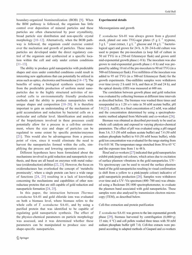
T. scotoductus SA-01 is unique to South Africa and can reducea number of compounds as electron acceptors through
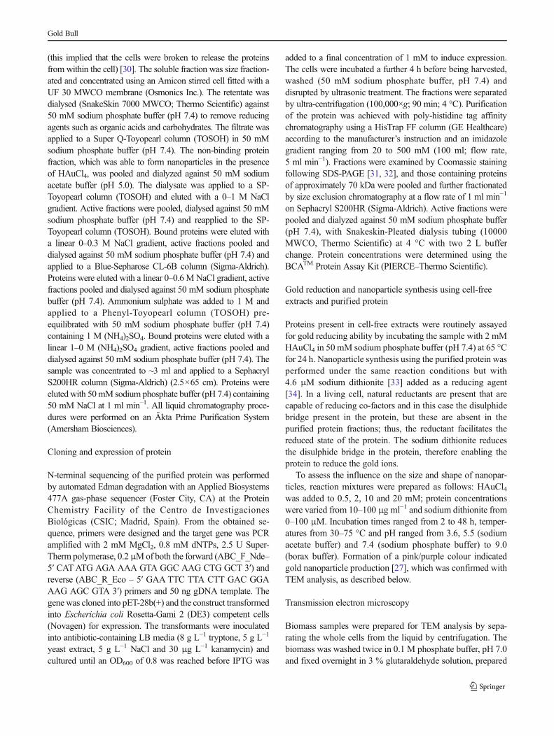
dissimilatory pathways. These compounds include Fe(III),Mn(IV), Co(III)-EDTA, Cr(VI) and U(VI) [36]. The Au(III)reducing capability of T. scotoductus SA-01 cells in differentgrowth phases illustrated that maximum Au(III) removal orreduction was reached after 8 h, which corresponds to the lateexponential—early stationary growth phase (Fig. 1a). Whenthese cells were incubated in the presence of Au(III), ultra-thinsection transmission electronmicroscopy (TEM) revealed thatthe gold wasmainly deposited on the outer wall layer (Fig. 1b)and elemental analysis, using EDS, showed that the particlesdo consist of gold (Fig. 2a); also present in vast amounts arecopper and carbon which can be attributed to the copper gridand the cell biomass. Since nanoparticle synthesis was asso-ciated with the biomass, it may be concluded that the cellsmight have a higher degree of control over the particle mor-phology as the BOB pathway of mineralization is observed[10–12]. XRD analysis on the biomass exposed to gold ionsgave an XRD pattern of the gold precipitated in or on thebacterial cells showing distinct peaks corresponding to the(111); (200) and (220) Bragg reflections of gold [37](Fig. 2b) which confirms the EDS results.
Effect of the physico-chemical parameters on nanoparticleformation
Various physico-chemical parameters were evaluatedusing whole cell biomass to determine their effect ongold reduction and nanoparticle formation. These param-eters included pH, temperature, exposure time and goldion concentration. The particle formation was confirmedvisually through colour formation (Supplementaryfigure 1c), peaks observed on the UV–Vis (400–700 nm) spectra (Supplementary figures 1a and b) andTEM analysis. An increase in the size of nanoparticlesresults in a change of colour, from pink, to purple, to
0 4 8 12 16 20 24 280.01
0.1
1
10
0
20
40
60
Time (h)
Abs
orba
nce
600n
m % A
u removed
500 nma b
Fig. 1 Interactions of Thermus scotoductus SA-01 with Au(III). aGrowth curve for T. scotoductus SA-01, showing the correlation betweengrowth phase (square) and Au(III) removal (triangle). b Thin section
TEMmicrograph of T. scotoductus SA-01 exposed to 2 mMHAuCl4; themicrograph shows the presence of gold deposited along the outer walllayer
Gold Bull
blue, due to the surface plasmon vibrations of the goldnanoparticles [38].
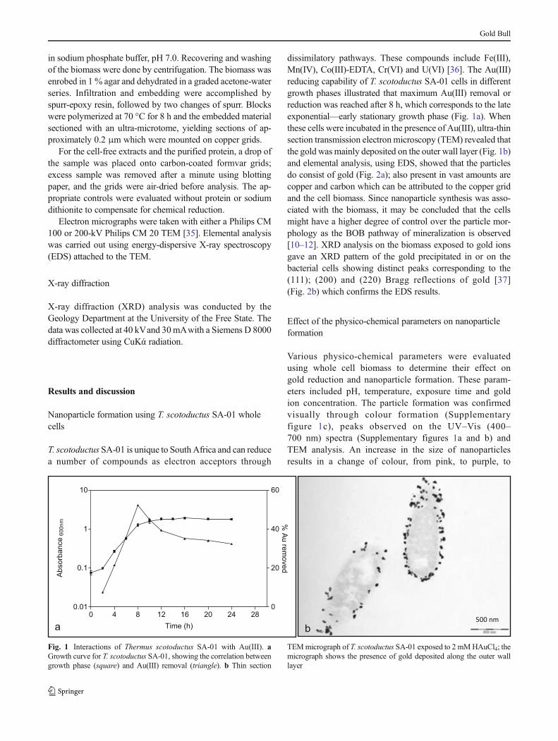
The effect of pH on the production of nanoparticles indi-cated that a lower pH was more conducive to the formation oflarger particles while a higher pH resulted in the formation ofsmall particles (Fig. 3a, b). Although pH does affect the size,shape and location of the particles, contrary to suggestions byHe and co-workers, particle shape and distribution could notbe controlled at this level [21]. In Fig. 3a, the formation ofoctahedral gold is observed and this points to mineral diagen-esis, while intracellular gold reduction leads to cell death(Supplementary figure 2) versus Fig. 3b where immobiliza-tion is focused in the cell envelope. Temperature and exposuretime had no significant effect on particle morphology butinfluenced the amount and rate of particle formation. Lowertemperatures and shorter incubation periods resulted in fewerparticles formed where an increase in temperature and longerincubation periods resulted in more and slightly larger parti-cles. These findings agree with the physico-chemical proper-ties of nanoparticles as discussed by Gericke and Pinches [39]who stated that the rate of particle formation can be correlatedwith the incubation temperature where an increase in temper-ature will allow faster particle formation and the rate ofreduction can influence the type of particle formed.
Figure 4 illustrates the effect of gold ion concentration onparticle formation. At concentrations exceeding 5 mM (forexample 10 mM seen in Fig. 4b), the cells are ruptured by thenanostructures formed and are released into the environmentand all means of controlling particle morphology is lost. Goldion concentrations lower than 5 mM (for example 0.5 mM asseen in Fig. 4a) result in the formation of smaller particleswhich are associated with the biomass. The effect of gold ionconcentration on particle formation at various concentrationscan be seen in supplementary Fig. 3a to e.
Nanoparticles from cell-free extracts
Incubation of soluble proteins >30 kDa with HAuCl4 resultedin the formation of nanoparticles and also showed a correla-tion between Au concentration and types of nanoparticlessynthesized (Fig. 5). Similar observations were also made byLi and co-workers [15], Shankar and co-workers [17] andWeiand co-workers [40] where it was demonstrated that goldnanosheets with triangular, hexagonal or truncated shapeswere dependent on the reductant to Au ion ratio. A mixtureof small amorphous particles and nebula-like formations wereobserved at low Au(III) concentrations (Fig. 5a), but withsome particles with specific shapes at 5 mM (Supplementary
a b
Fig. 2 a EDS analysis and b XRD pattern recorded of T. scotoductus SA-01 cells incubated in the presence of 2 mM Au(III)
a400 nm b400 nm
Fig. 3 TEM micrographsillustrating the effect of pH a 3.6and b 9.5 on particle formationusing whole cell biomass
Gold Bull
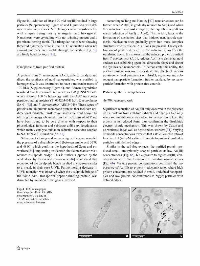
Figure 4a). Addition of 10 and 20mMAu(III) resulted in largeparticles (Supplementary Figure 4b and Figure 5b), with def-inite crystalline surfaces. Morphologies were nanosheet-like,with shapes being mostly triangular and hexagonal.Nanosheets were crystalline with no twinning present and aprominent herring motif. The triangular nanosheets showingthreefold symmetry were in the {111} orientation (data notshown), and dark lines visible through the crystals (Fig. 5b)are likely bend contours [41].
Nanoparticles from purified protein
A protein from T. scotoductus SA-01, able to catalyse anddirect the synthesis of gold nanoparticles, was purified tohomogeneity. It was determined to have a molecular mass of~70 kDa (Supplementary Figure 5), and Edman degradationresolved the N-terminal sequence as GPQDNSLVIGASwhich showed 100 % homology with the ABC transporterpeptide-binding protein (YP_004203474) from T. scotoductusSA-01 [42] and T. thermophilus (AEG34049). These types ofproteins are ubiquitous membrane proteins that facilitate uni-directional substrate translocation across the lipid bilayer byutilizing the energy obtained from the hydrolysis of ATP andhave been found to be very diverse with respect to theirphysiological function and substrate unlike oxidoreductaseswhich mainly catalyse oxidation-reduction reactions coupledto NADP/NAD+ utilization [43–45].
Subsequent cloning and sequencing of the gene revealedthe presence of a disulphide bond (between amino acid 337Cand 481C) which confirms the hypothesis of Scott and co-workers [34], implicating an electron shuttle mechanism via areduced disulphide bridge. This is further supported by thework done by Cason and co-workers [46] who found thatreduction of the disulphide bonds resulted in electron transferto a metal, in their case U(VI). Furthermore, a decrease inU(VI) reduction was observed when the disulphide bridge ofthe same ABC transporter peptide-binding protein wasdisrupted by mutation of the genes involved.
According to Tang and Hamley [47], nanostructures can beformed when Au(III) is gradually reduced to Au(I), and whenthis reduction is almost complete, the equilibrium shift to-wards reduction of Au(I) to Au(0). This, in turn, leads to theformation of nucleation sites that initiates nanoparticle syn-thesis. Nucleation sites gradually grow into more complexstructures when sufficient Au(I) ions are present. The crystal-lization of gold is directed by the reducing as well as thestabilizing agent. It is shown that the reduced protein, purifiedfrom T. scotoductus SA-01, reduces Au(III) to elemental goldand acts as a stabilizing agent that directs the shape and size ofthe synthesized nanoparticle. To demonstrate this ability, thepurified protein was used to evaluate the effects of variousphysico-chemical parameters on HAuCl4 reduction and sub-sequent nanoparticle formation, further validated by no nano-particle formation with protein-free controls.
Particle synthesis manipulations
Au(III): reductant ratio
Significant reduction of Au(III) only occurred in the presenceof the proteins from cell-free extracts and once purified onlywhen sodium dithionite was added to the reaction to keep theprotein in its reduced form, thus confirming the disulphideelectron shuttle mechanism. This was shown by Cason andco-workers [46] as well as Scott and co-workers [34]. Varyingdithionite concentrations revealed that a stoichiometric ratio ofless than 1:1 (4.6 μM sodium dithionite to protein) resulted inparticles with defined edges.
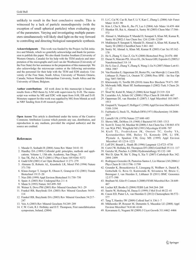
Similar to the cell-free extracts, the purified protein pro-duced small, amorphously shaped particles at low Au(III)concentrations (Fig. 6a), but exposure to higher Au(III) con-centrations led to the formation of plate-like nanostructures(Fig. 6b). Varying protein concentrations confirmed the im-portance of Au(III) to protein (reductant) ratio, where highprotein concentrations resulted in small, undefined nanoparti-cles and low protein concentrations in bigger particles withdefined edges.
a1000 nm b1000 nm
Fig. 4 TEM micrographsillustrating the effect of Au(III)concentration a 0.5 and (b)10 mM on particle formationusing whole cell biomass
Gold Bull
At high reductant to Au(III) ratios, gold ions are preferen-tially committed to particle initiation (nucleation) rather thanparticle growth [47] resulting in higher numbers of smallparticles. However, at lower reductant to Au(III) ratios, fewernucleation sites are available for particle initiation whichallows for bigger particles to grow as there is less competitionfor available gold ions [33, 48]. Thus, the less nucleation sitesare available, and the bigger particles can grow as there will beless competition for gold ions to act either as part of thenucleation process or to grow the size of the particle.
Incubation time, temperature and pH
Nanoparticles were already formed after only 8 h of incubation,but longer reaction times (up to 48 h) only resulted in highernumbers of particles and did not have a significant effect onparticle size. Since Au(III) reduction and eventual nanoparticlesynthesis are time-dependent reactions, longer incubation times
allow for the reaction equilibrium to shift towards Au(I) reduc-tion and formation of more nucleation sites. With longer incu-bation times, more nucleation sites can be occupied leading tohigher numbers of particles [47, 48].
Temperature had no effect on the size of the nanoparticles,but did influence particle shape. Incubation at lower temper-ature resulted in fewer particles but with defined edges(Fig. 7a), while particles produced at 75 °C were mostlyspherical (Fig. 7b). The overall effect of temperature is likelyin influencing the nucleation seed, rather than particle growth[49].
Lower pH values were more conducive to producingnanoplate-like structures with a larger aspect ratio (Fig. 8aand Supplementary Figure 6a), while incubation at higher pHresulted in more small spherical particles (SupplementaryFigure 6b). Particles with defined edges were formed at pH7.4 (Fig. 8b). Different pH values regulate the effective protonconcentration of the solution, which in turn controls
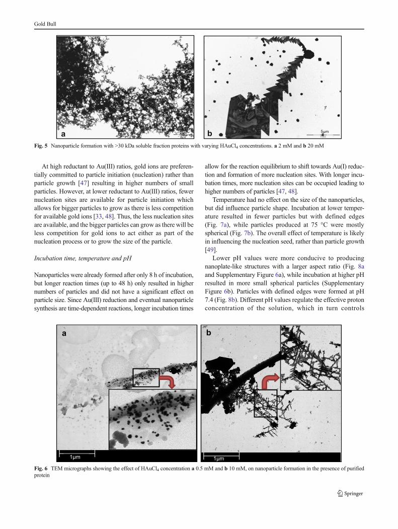
5µmba 5μm
Fig. 5 Nanoparticle formation with >30 kDa soluble fraction proteins with varying HAuCl4 concentrations. a 2 mM and b 20 mM
1µm 1µm
a b
Fig. 6 TEM micrographs showing the effect of HAuCl4 concentration a 0.5 mM and b 10 mM, on nanoparticle formation in the presence of purifiedprotein
Gold Bull

nanoparticle morphology rather than affecting nucleation andparticle initiation N.
Conclusions
T. scotoductus SA-01, the thermophilic bacterium used in thisstudy, has the ability to reduce Au(III) and produce nanopar-ticles, making it a suitable candidate for the production ofnanoparticles.
In this study, it was found that the physico-chemical pa-rameters have a definite influence on particle size with lowerpH and higher temperatures resulting in larger particles and incontrast, higher pH and lower temperatures will producesmaller particles. However, these processes are difficult tocontrol and a predefined morphology is still unobtainable atthis time. Microorganisms are very complex structures with avariety of metabolic functions which all contribute towards
the proper working of the cell. Many of these activities couldbe involved in the reduction of gold and the formation ofnanoparticles, but a better understanding of the mechanismmight be obtained if the protein/s involved in these reactions iselucidated. Gold reduction primarily occurred in the cell en-velope which is strong evidence for a gold ‘specific’ reductionprocess, hinting at a location for further investigation.
An ABC transporter, peptide-binding protein ofT. scotoductus SA-01, able to reduce and synthesize goldnanoparticles was purified to homogeneity. Even though thistype of protein is not a classical oxidoreductase, a cysteine–disulphide bridge electron shuttle mechanism is likely in-volved in reducing Au(III). Moreover, the protein also actsas nucleation seed sites that initiate and direct nanoparticlesynthesis. Through manipulation of physico-chemical param-eters, it is clear that particle formation can be influenced interms of size, shape and number of particles formed. However,since biological Au(III) reduction and nanoparticle synthesisis a complex process, manipulations of single parameters are
b
1µm 1µm
aFig. 7 TEM micrographs ofnanoparticles formed at a 42 °Cand b 75 °C with purified protein
ba
1µm 1µm
Fig. 8 TEM micrographs of nanoparticles formed at varying pH. a pH 5.5 and b pH 7.4 with purified protein
Gold Bull
unlikely to result in the best conclusive results. This iswitnessed by a lack of particle monodispersity (with theexception of small spherical particles) when evaluating anyof the parameters. Varying and investigating multiple param-eters simultaneously will likely shed light on the way forwardto controlling and directing biological nanoparticle synthesis.
Acknowledgments This work was funded by the Project AuTek initia-tive and Mintek, which we gratefully acknowledge and thank for permis-sion to publish this paper. We also thank Gordon Southam (University ofWestern Ontario, Canada) for his help with the TEM analysis and inter-pretation of the micrographs and Liesl van der Westhuizen (University ofthe Free State) for her assistance in the preparation of this manuscript. Wealso acknowledge the microscope centres at the following universitiesand the personnel for their help with the TEM and EDS analysis: Uni-versity of the Free State, South Africa; University of Western Ontario,Canada; Nelson Mandela Metropolitan University, South Africa and theUniversity of Ghent, Belgium.
Author contributions All work done in this manuscript is based onresults from a PhD thesis by JvM with supervision by EvH. The manu-script was written by ME and EDC with input from JvM, EB and EvH.Monetary support for this work was supplied byMG fromMintek as wellas NRF funding from EvH research grants.
Open Access This article is distributed under the terms of the CreativeCommons Attribution License which permits any use, distribution, andreproduction in any medium, provided the original author(s) and thesource are credited.
References
1. Masala O, Seshadri R (2004) Annu Rev Mater 34:41–812. Handley DA (1989) Colloidal gold: principles, methods and appli-
cations. Volume 1, 13th edn. Academic, San Diego, 333. Sau TK, Pal A, Pal T (2001) J Phys Chem 105:9266–92724. Gadd GM (2001) Curr Opin Biotechnol 11:271–2795. Ahmann D, Roberts AL, Krumholz LR, Morel FM (1994) Nature
371:7506. Klaus-Joerger T, Joerger R, Olsson E, Granqvist CG (2001) Trends
Biotechnol 19:15–207. Nies DH (1999) Appl Environ Biotechnol 51:730–7508. Spain A (2003) Rev Undergrad Res 2:1–69. Mann S (1993) Nature 365:499–505
10. Weiner S, Dove PM (2003) Rev Mineral Geochem 54:1–2911. Frankel RB, Bazylinski DA (2003) Rev Mineral Geochem 54:95–
11412. Frankel RB, Bazylinski DA (2003) Rev Mineral Geochem 54:217–
24713. Veis A (2003) Rev Mineral Geochem 54:249–28914. C.W. Corti, R.J. Holliday and D.T. Thompson, First nanofabrication
symposium, Ireland, (2004)
15. Li C, Cai W, Cao B, Sun F, Li Y, Kan C, Zhang L (2006) Adv FunctMater 16:83–90
16. Kim J, Cha S, Shin K, Jho JY, Lee J (2004) Adv Mater 16:459–46417. Shankar SS, Rai A, Ahmad A, Sastry M (2005) Chem Mat 17:566–
57218. Ahmad A, Mukherjee P, Mandal D, Senapati S, Khan MI, Kumar R,
Sastry M (2002) J Am Chem Soc 124:12108–1210919. Mukherjee P, Senapati S, Mandal D, Ahmad A, Khan MI, Kumar R,
Sastry M (2002) ChemBioChem 5:461–46320. Sastry M, Ahmad A, Khan MI, Kumar R (2003) Curr Sci 85:162–
17021. He S, Zhang Y, Guo Z, Gu N (2008) Biotechnol Prog 24:476–48022. DuranN,Marcato PD, AlvesOL, De SouzaGIH, Esposito E (2005) J
Nanobiotechnol 3:8–1423. He S, Guo Z, ZhangY, Zhang S,Wang J, Gu N (2007)Mater Lett 61:
3984–398724. Van Heerden E, Opperman DJ, Bester PA, vanMarwijk J, Cason ED,
Litthauer D, Piater LA, Onstott TC (2008) Proc SPIE - Int Soc OptEng 7097:70970S
25. Khersonsky O, Tawfik DS (2010) Annu Rev Biochem 79:471–50526. Melwanki MB, Masti SP, Seetharamappa J (2002) Turk J Chem 26:
17–2227. Šlouf M, Kužel R, Matej Z (2006) Krist Suppl 23:319–32428. Lazarides AA, Schatz GC (2000) J Phys Chem B 104:460–46729. Opperman DJ, van Heerden E (2007) J Appl Microbiol 103:1907–
191330. Gaspard S, Vazquez F, Holliger C (1998) Appl EnvironMicrobiol 64:
3188–319431. Fairbanks G, Steck TL,Wallach DFH (1971) Biochemistry 10:2606–
261732. Lamelli UK (1970) Nature 227:680–68533. Showe MK, DeMoss JA (1968) J Bacteriol 95:1305–131334. Scott D, Toney M, Muzikar M (2008) J Am Chem Soc 130:865–87435. van Wyk PWJ, Wingfield MJ (1991) Mycologia 83:698–70736. Kieft TL, Fredrickson JK, Onstott TC, Gorby YA,
Kostandarithes HM, Bailey TJ, Kennedy DW, Li SW,Plymale A, Spadoni CM, Gray MS (1999) Appl EnvironMicrobiol 65:1214–1221
37. Leff DV, Brandt L, Heath JR (1996) Langmuir 12:4723–473038. Corti CW, Holliday RJ, Thompson DT (2002) Gold Bull 35:111–11739. Gericke M, Pinches A (2006) Hydrometallurgy 83:132–14040. Wei D, Qian W, Shi Y, Ding S, Xia Y (2007) Carbohydr Res 342:
2494–249941. Rodriguez-Gonzalez B, Pastoriza-Santos I, Liz-Marzan LM (2006) J
Phys Chem B 110:11796–1179942. Gounder K, Brzuszkiewicz E, Liesegang H, Wollherr A, Daniel R,
Gottschalk G, Reva O, Kumwenda B, Srivastava M, Bricio C,Berenguer J, van Heerden E, Litthauer D (2011) BMC Genomics12:577–590
43. Braibant M, Gilot P, Content J (2000) FEMSMicrobiol Rev 24:449–467
44. Locher KP, Broths E (2004) FEBS Lett 564:264–26845. Saurin W, Hofnung M, Dassa E (1999) J Mol Evol 48:22–4146. Cason ED, Piater LA, van Heerden E (2012) Chemosphere 86:572–
57747. Tang T, Hamley IW (2009) Colloid Surf A 336:1–748. Mikheenko IP, Rousset M, Dementin S, Macaskie LE (2008) Appl
Environ Microbiol 74:6144–614649. Kawamura G, Nogami M (2009) J Cryst Growth 311:4462–4466
Gold Bull