Physically-Constrained Diffeomorphic Demons for the Estimation of 3D Myocardium Strain from Cine-MRI
10
Physically-Constrained Diffeomorphic Demons for the Estimation of 3D Myocardium Strain from Cine-MRI T. Mansi 1 , J-M. Peyrat 1 , M. Sermesant 1 , H. Delingette 1 , J. Blanc 2 , Y. Boudjemline 2 , and N. Ayache 1 1 INRIA-M´ editerran´ ee, Asclepios Project, Sophia Antipolis, France 2 Assistance Publique Hˆopitaux de Paris, Necker-Enfants Malades, Paris, France Abstract. Analysing heart motion provides crucial insights on the con- dition of the cardiac function. Tagged-MRI and 2D-strain ultrasound enable quantitative assessment of the myocardium strain. But estimat- ing 3D myocardium strain from cine-MRI remains attractive: cine-MRI is widely available and it yields detailed 3D+t anatomical images. This paper presents an image-based method to estimate myocardium strain from clinical short-axis cine-MRI. To recover non-apparent cardiac mo- tions, we improve the diffeomorphic demons, a non-linear registration algorithm, by adding two physical constraints. First, myocardium near- incompressibility is ensured by constraining the deformations to be di- vergence free. Second, myocardium elasticity is modelled using smooth vector filters. The proposed physically-constrained demons are compared with the diffeomorphic demons and evaluated in a healthy subject against tagged MRI. The method is also tested on a patient with congenital pulmonary valve regurgitations and compared with 2D-strain measure- ments. In both cases, obtained results correlate well with ground truth. This method may become a useful tool for cardiac function evaluation. 1 Introduction Analysing heart motion provides crucial insights on the condition and efficiency of the cardiac function. Myocardium strain can now be quantitatively assessed through tagged-MRI or ultrasound modalities such as 2D speckle tracking (also known as 2D-strain). Nevertheless, despite of its accuracy, tagged-MRI is still experimental [1] and while 2D-strain becomes available in clinical environment, it only provides partial evaluation of the myocardium [2]. Estimating the my- ocardium strain from clinical gated cine-MRI constitutes an attractive alterna- tive. Cine-MRI is widely available and yields detailed 3D anatomical images of the beating heart with constant image quality through time. However, esti- mating strain from these images may result inaccurate since only the apparent motion can be extracted and slice thickness is usually large. Prior knowledge about cardiac dynamics is thus required.
-
Upload
independent -
Category
Documents
-
view
2 -
download
0
Transcript of Physically-Constrained Diffeomorphic Demons for the Estimation of 3D Myocardium Strain from Cine-MRI
Physically-Constrained Diffeomorphic Demonsfor the Estimation of 3D Myocardium Strain
from Cine-MRI
T. Mansi1, J-M. Peyrat1, M. Sermesant1, H. Delingette1, J. Blanc2,Y. Boudjemline2, and N. Ayache1
1 INRIA-Mediterranee, Asclepios Project, Sophia Antipolis, France2 Assistance Publique Hopitaux de Paris, Necker-Enfants Malades, Paris, France
Abstract. Analysing heart motion provides crucial insights on the con-dition of the cardiac function. Tagged-MRI and 2D-strain ultrasoundenable quantitative assessment of the myocardium strain. But estimat-ing 3D myocardium strain from cine-MRI remains attractive: cine-MRIis widely available and it yields detailed 3D+t anatomical images. Thispaper presents an image-based method to estimate myocardium strainfrom clinical short-axis cine-MRI. To recover non-apparent cardiac mo-tions, we improve the diffeomorphic demons, a non-linear registrationalgorithm, by adding two physical constraints. First, myocardium near-incompressibility is ensured by constraining the deformations to be di-vergence free. Second, myocardium elasticity is modelled using smoothvector filters. The proposed physically-constrained demons are comparedwith the diffeomorphic demons and evaluated in a healthy subject againsttagged MRI. The method is also tested on a patient with congenitalpulmonary valve regurgitations and compared with 2D-strain measure-ments. In both cases, obtained results correlate well with ground truth.This method may become a useful tool for cardiac function evaluation.
1 Introduction
Analysing heart motion provides crucial insights on the condition and efficiencyof the cardiac function. Myocardium strain can now be quantitatively assessedthrough tagged-MRI or ultrasound modalities such as 2D speckle tracking (alsoknown as 2D-strain). Nevertheless, despite of its accuracy, tagged-MRI is stillexperimental [1] and while 2D-strain becomes available in clinical environment,it only provides partial evaluation of the myocardium [2]. Estimating the my-ocardium strain from clinical gated cine-MRI constitutes an attractive alterna-tive. Cine-MRI is widely available and yields detailed 3D anatomical imagesof the beating heart with constant image quality through time. However, esti-mating strain from these images may result inaccurate since only the apparentmotion can be extracted and slice thickness is usually large. Prior knowledgeabout cardiac dynamics is thus required.
2
Various methods have been proposed to estimate myocardium deformationfrom cine-MRI [3–7]. Some approaches drive an image registration algorithmwith biomechanical finite-element models that simulate the myocardium passiveproperties [3, 4]. These methods successfully estimate left ventricle myocardiumstrain. Nonetheless, they rely on specific biomechanical parameters that are dif-ficult to determine for a given patient. Besides, such models may not applyanymore in diseases. Purely image-driven methods have thus been proposed [5–7]. In [5], the authors use a B-spline Free-Form Deformation (FFD) algorithmregularised using myocardium contours that are manually delineated on all timeframes. The results are consistent with tagged MRI but requiring manual con-tours for all time frames may hamper using this approach in clinical settings.In [6], Bistoquet et al. present an FFD-like algorithm that ensures myocardiumnear-incompressibility [8]. The similarity criterion is penalised when the Jacobianof the deformation is significantly different from 1 and the dense deformationfield is estimated by using divergence-free radial basis functions. However, my-ocardium is tracked at the nodes of the epicardium and endocardia surfaces only,the inner deformations being interpolated from the surfaces without consideringpossible textural hints. In [7], the authors estimate the cardiac motion using aspatio-temporal model based on FFD and Kalman filters, under the assumptionof periodic motion. However, the proposed approach does not take guarantee my-ocardium biomechanical properties and it has been validated on 2D+t imagesonly.
This paper presents an image-based method to estimate myocardium strainfrom clinical short-axis cine-MRI. Our approach relies on diffeomorphic demons [9],an efficient non-linear registration algorithm. Diffeomorphic non-linear registra-tion [9–11] aims at finding a smooth, invertible, one-to-one mapping betweentwo images, which is suited for cardiac motion analysis. Because diffeomor-phic demons rely on image data exclusively, we introduce in the algorithm twophysical constraints to recover plausible non-apparent cardiac motions: i) my-ocardium near-incompressibility [8] and ii) elasticity. Integrating incompressibil-ity in non-linear registration algorithms has been widely studied for contrast-enhanced image registration [12–14]. Incompressibility can be modelled throughthe hard constraint Jacobian determinant of the deformation field equals 1 [12,13], or through the divergence-free approximation [6, 14]. In our approach, near-incompressibility is obtained by projecting the velocity and deformation fieldsonto the space of divergence-free vector fields. The second physical constraintwe consider is myocardium elasticity, modelled with an elastic-like regularisa-tion. Although myocardium tissue is found visco-elastic, it can be reasonablymodelled as elastic when it is analysed at the heart beat time-scale [8]. In thisway, our approach is purely image-based, it evaluates the motion everywhere inthe images, it does not need any mesh model and it requires very few image-based parameters. The Lagrangian finite strain tensor is finally calculated fromthe recovered deformation fields. The method is evaluated on a healthy subject,against tagged MRI, and on a patient suffering from congenital pulmonary valveregurgitations, against 2D-strain ultrasound.
3
2 Methods
2.1 Non-Linear Image Registration Using Diffeomorphic Demons(DD) Algorithm
Non-linear image registration aims at providing a transformation φ(x) = x +u(x), where u(x) is the displacement vector field, that aligns a template imageT (x) with a reference image R(x). The underlying principle lies in minimisingan energy E(φ) that comprises a similarity criterion D(T, R, φ) and a regulariserR(φ) ensuring smooth transformations and integrating prior knowledge.
Diffeomorphic demons (DD) algorithm [9] minimises E(φ) iteratively throughtwo decoupled steps. A correspondence field c that matches the points betweenthe two images, with a controlled uncertainty parametrised by σ2
x, is introducedin the energy: E(c,u) = 1/σ2
i ‖R − T c‖2 + 1/σ2x ‖u− c‖2 + 1/σ2
T ‖∇u‖2.In this equation, σ2
i is a parameter related to the noise in the images and σ2T
compromises between the image data term and the regularisation.First, the correspondence field c is updated using the diffeomorphic update
rule c ← u exp(v). exp(v) is the exponential operator defined in the Log-Euclidian framework and v is the smooth stationary velocity field that resultsfrom the minimisation of the correspondence energy:
Ecorr(v) =1σ2
i
‖R− T u exp(v)‖2 +1σ2
x
‖u− u exp(v)‖2 (1)
Derivating Ecorr yields v = −(R − T (Id + u))/(‖J‖2 + σ2i /σ2
x) J , with J =(∇R+∇(T (Id+u)))/2 and σi = |R−T (Id+ c)| (see [9] for further details).
Second, the displacement u that satisfies the regulariser R(u), knowing thenew correspondence field c, is computed. The second part of the demons energyE(c,u), Ereg(c,u) = 1/σ2
x ‖u− c‖2 + 1/σ2T ‖∇u‖2, is minimised with respect to
u, which amounts to convoluting c = u exp(v) with a Gaussian kernel.
2.2 Adding Physical Constraints to the Diffeomorphic Demons
Diffeomorphic demons (DD) algorithm is purely driven by image intensities.Therefore, it can hardly recover longitudinal shortening and circumferentialtwisting, which are poorly apparent in short-axis cine-MRI. To better estimatethese displacements, we improve the algorithm by adding two constraints relatedto the myocardium tissue properties: near-incompressibility and elasticity.
Incompressibility Constraint Contrary to [13], the incompressibility con-straint is decoupled from the energy minimisation to take advantage of the two-step minimisation process offered by the demons. Since DD relies on velocityfields, fluid-dynamics laws are used to model incompressibility [14]. However,slight volume drifts may remain as the Gaussian regularisation does not pre-serve incompressibility. Positions are hence corrected at the end of the demonsloop to further reduce volume variations.
4
Velocity Correction: Myocardium incompressibility can be interpreted as con-servation of myocardium mass throughout the cardiac cycle [8]. Therefore, themass continuity equation ∂ρ/∂t + div(ρv) = 0 applies, where ρ is the mass den-sity, v is the velocity and t is the time. Assuming ρ constant and uniform, theconservation of mass becomes conservation of volume and writes as div(v) = 0.
Consequently, the incompressibility property is integrated in the DD by con-straining the search space of the velocity fields v to the divergence-free vectorspace. Given the velocity field v computed by Equation (1), we solve the La-grangian problem [15]:
minv∈f∈L2(Ω) | div f = 0
maxp∈H1
0
(12‖v − v‖2 +
∫
Ω
p div v)
(2)
In this equation, Ω is an open subset of R3 (the image space), H10(Ω) is the
Sobolev space associated to Ω and p is the Lagrange multiplier related to thedivergence-free constraint. Derivating Equation (2) gives the optimal condition:
v = v − grad p (3)
under the constraint div v = 0. Equation (3) is the closed-form expression ofthe divergence-free projector Π(v) = v− grad p, where p is obtained by solvingthe Poisson equation 4p = div(v) under 0-Dirichlet boundary conditions [15].
Position Correction: Because the Gaussian regularisation may alter the obtainedincompressibility, we perform a second adjustment to correct the remaining vol-ume drifts after deformation.
For each point x of the image space, the Jacobian determinant J(x) =det(Id + ∇u(x)) relates to the local volume change due to the transformationφ(x) = x + u(x). Hence, the volume is locally preserved if the non-linear con-straint J(x) = 1 is satisfied. However, since myocardium is nearly incompress-ible (5% of volume variation is usually observed [6, 8]) and the displacementsprovided by the DD are smooth and derive from divergence-free velocities, thefirst-order approximation of J(x) = 1, i.e. div(u) = 0, can be used to correct theremaining volume drifts. As a result, the positions are corrected like the veloci-ties. At each iteration of the demons algorithm, the displacement field u is madedivergence-free by using the above-mentioned projector Π (Equation (3)). It isworth stressing that the displacements are projected to reduce remaining volumedrifts, whereas the velocities are projected as a result of the mass-conservationlaw.
Elastic-Like Regularisation Myocardium is commonly assumed to be visco-elastic. However, at the time-scale of the heart beat, one can reasonably approx-imate the visco-elasticity property by elasticity [8]. In our approach, elasticity ismodelled as regularisation. We substitute the diffusion-like regularisation, whichis unable to model elastic deformations, by an elastic regulariser. Linear elasticregularisers have been widely studied in the past [16]. Yet, they are suitable for
5
small displacements only and can present discontinuities in the derivatives ofthe resulting deformations, which is not desirable for our application. To over-come these limitations, we regularise the deformation field using an infinite-orderisotropic differential quadratic form [17]. Such regulariser behaves like elasticfilters, while ensuring smooth deformations. Minimising Ereg(c,u) with this reg-ulariser amounts to convoluting the deformation field u by the 3D isotropicseparable filter:
Gσ,κ(u) =1
(σ√
2π)3(1 + κ)
(Id +
κ
σ2uuT
)exp
(uT u2σ2
)(4)
which can be performed very efficiently by using recursive Gaussian filter [17].In Equation (4), σ is the standard deviation of the Gaussian function and κ is aparameter that acts as a Poisson ratio coefficient. The higher is κ, the stiffer isthe deformation.
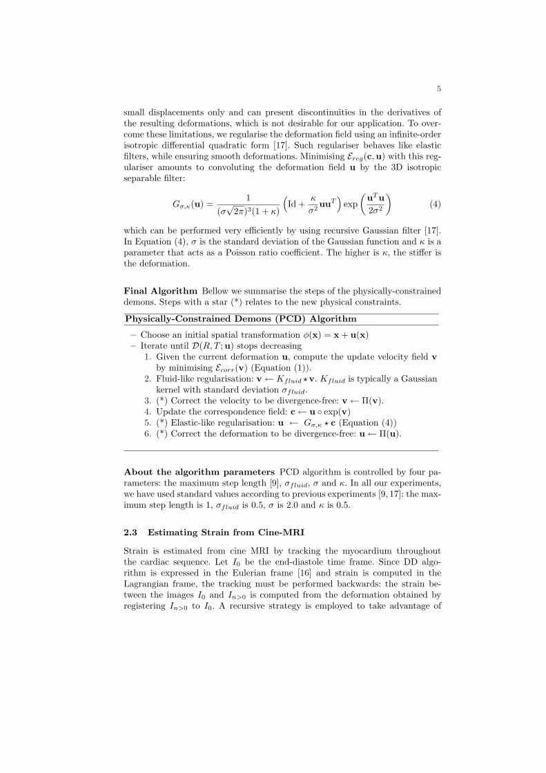
Final Algorithm Bellow we summarise the steps of the physically-constraineddemons. Steps with a star (*) relates to the new physical constraints.
Physically-Constrained Demons (PCD) Algorithm
– Choose an initial spatial transformation φ(x) = x + u(x)– Iterate until D(R, T ;u) stops decreasing
1. Given the current deformation u, compute the update velocity field vby minimising Ecorr(v) (Equation (1)).
2. Fluid-like regularisation: v← Kfluid ?v. Kfluid is typically a Gaussiankernel with standard deviation σfluid.
3. (*) Correct the velocity to be divergence-free: v← Π(v).4. Update the correspondence field: c← u exp(v)5. (*) Elastic-like regularisation: u ← Gσ,κ ? c (Equation (4))6. (*) Correct the deformation to be divergence-free: u← Π(u).
About the algorithm parameters PCD algorithm is controlled by four pa-rameters: the maximum step length [9], σfluid, σ and κ. In all our experiments,we have used standard values according to previous experiments [9, 17]: the max-imum step length is 1, σfluid is 0.5, σ is 2.0 and κ is 0.5.
2.3 Estimating Strain from Cine-MRI
Strain is estimated from cine MRI by tracking the myocardium throughoutthe cardiac sequence. Let I0 be the end-diastole time frame. Since DD algo-rithm is expressed in the Eulerian frame [16] and strain is computed in theLagrangian frame, the tracking must be performed backwards: the strain be-tween the images I0 and In>0 is computed from the deformation obtained byregistering In>0 to I0. A recursive strategy is employed to take advantage of
6
the frame-by-frame registration accuracy and to reduce tracking errors due tothe changing appearance of the trabecula, papillary muscles and neighbouringorgans. Having the spatial transformation φIn−1→I0 , we first compute φIn→In−1
and then we estimate the transformation φIn→I0 by taking as initialisation thecomposed transformation φIn→In−1 φIn−1→I0 . Finally, Lagrangian finite straintensor E = 1/2 (∇u +∇uT +∇uT∇u) is computed from the recovered defor-mation φ(x) = x + u(x). Radial, circumferential and longitudinal strains areestimated in the heart prolate coordinate system [18].
3 Experiments and Results
The method is tested on two subjects: a healthy adult and a young patient suf-fering from congenital pulmonary valve regurgitations. In the two experiments,heart motion is recovered from gated short-axis cine-MRI. In the first exper-iment, the resulting 3D deformations are compared with tagged MRI. In thesecond experiment, the deformations are compared with ultra-sound 2D-strain.
Because we are mainly interested in myocardium strain and to minimisethe computational overhead, diffeomorphic demons are constrained only insidea mask that delineates the cardiac muscle. As the projector Π and the elasticsmoothing are decoupled from the minimisation, appearing as additive terms inthe algorithm (Equations (3) and (4)), they can be added inside the mask and ig-nored outside (where diffeomorphic demons are still applied). It should be notedhowever that at the mask boundaries, the incompressibility constraint is not en-sured due to the boundary conditions of the projector Π. Furthermore, discon-tinuities may appear at the mask boundaries when computing the displacementderivatives. These limitations are addressed by dilating the myocardium maskby one voxel, thus ensuring optimal strain estimation within the myocardium.At last, we need to segment the myocardium at the end-diastole time frame onlyowing to the Eulerian formulation of the demons algorithm, contrary to previousapproaches which require delineations for all the cardiac sequence frames [3, 5].
3.1 Left Ventricle Myocardium Strain in a Healthy Subject
Retrospective gated steady-state free precession (SSFP) cine MR and taggedMR (complementary spatial modulation of magnetisation CSPAMM) images ofa healthy subject are acquired in the short-axis view covering the mid-part ofboth ventricles using 1.5T MR scanner (Achieva, Philips Medical System) (seven10mm-thick slices; 1.37x1.37mm in-plane resolution, 30 frames). No longitudinalcine-MRI are available. Visual inspection of the images revealed no spatial mis-registration. To improve registration performances, the resolution of the cine-MRimages is made isotropic at the in-plane resolution.
First, the incompressibility constraint is evaluated by computing the Jacobiandeterminant of the recovered myocardium deformation at each frame of thecardiac sequence. Without any constraint, mean Jacobian equals 1.09 ± 0.07(mean ± SD), maximum deviation from unity: 0.22. Adding the constraints
7
yields J(umyo) = 0.99±0.02 (mean ± SD) with a maximum deviation from unityof 0.05, which relates to the observed 5% of myocardium volume variation [8].
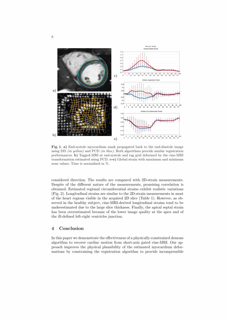
Second, registered images provided by the physically-constrained demons(PCD) are compared with those provided by the diffeomorphic demons (DD).Available myocardium mask at end-systole is registered back to the end-diastoleimage using the two methods. Qualitative evaluation is satisfying (Fig. 1a).Hausdorff distance between the two registered masks is 2.23mm and averageHausdorff distance is 0.02mm, which is less than the through-plane image res-olution. The physical constraints do not qualitatively decrease the registrationperformances.
Next, radial, longitudinal and circumferential strains are computed for the sixequatorial AHA zones using DD and PCD. In this experiment, we only considerthe equatorial zones since at the basal and apical regions the incompressibility as-sumptions may be violated due to the truncated acquisition. Fig. (1c-e) illustratethe global strains with minimum and maximum zone values. DD mainly recoversthe visible radial motion of the heart, resulting in over-estimated radial strains,implausible positive longitudinal strains and weak circumferential strains. On theother hand, the physical constraints enable recovering non-apparent motions byreorienting the estimated myocardium deformations to ensure incompressibility.Radial strain is closer to clinical observations and more homogeneous acrossthe myocardium [18], thus suggesting that the physical constraints also per-form global regularisation. Longitudinal strain is clinically plausible yet slightlyunder-estimated with respect to reported values, mainly because of the coarsethrough-plane resolution. Circumferential strain is improved but remains closeto DD estimations. A probable reason is the low temporal resolution, circumfer-ential motion occurring in few time frames.
Finally, the deformations recovered from the short-axis cine-MRI using PCDare qualitatively compared with short-axis tagged-MRI, exhibiting promisingresults (Fig. 1b).
3.2 Left-Ventricle Myocardium Strain in a Patient with ChronicPulmonary Valve Regurgitations
In the second experiment we evaluate our method on a patient (age=10) withcongenital pulmonary valve regurgitations. SSFP cine MRI of the heart are ac-quired in the short-axis view covering the entirety of both ventricles (10 9.6mm-thick slices; 1.02x1.02mm in-plane resolution; 25 frames) using a 1.5T MR scan-ner (Avanto, Siemens Medical Systems, Erlangen, Germany). No longitudinalcine-MRI are available. Visual inspection of the images revealed no spatial mis-registration. Finally, the images are made isotropic to improve registration per-formances. 2D-strain measurements are performed in the short-axis and the 4-chamber views (frame-rate: 80) using Automatic Functional Imaging (Vivid7,General Electrics, Vingmed Ultrasound), as described in [2].
Lagrangian finite strain tensors are estimated from the cine-MRI. Longitu-dinal (Ll) and circumferential (Lc) lengthening are computed according to therelation LV =
√1 + EV V + 1, where E is the strain tensor and V ∈ l, c is the
8
a)
b)
c)
d)
e)
Fig. 1. a) End-systole myocardium mask propagated back to the end-diastole imageusing DD (in yellow) and PCD (in blue). Both algorithms provide similar registrationperformances. b) Tagged-MRI at end-systole and tag grid deformed by the cine-MRItransformation estimated using PCD. c-e) Global strain with maximum and minimumzone values. Time is normalised in %.
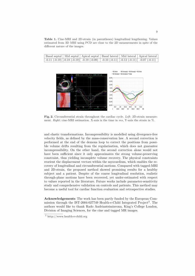
considered direction. The results are compared with 2D-strain measurements.Despite of the different nature of the measurements, promising correlation isobtained. Estimated regional circumferential strains exhibit realistic variations(Fig. 2). Longitudinal strains are similar to the 2D-strain measurements in mostof the heart regions visible in the acquired 2D slice (Table 1). However, as ob-served in the healthy subject, cine-MRI-derived longitudinal strains tend to beunderestimated due to the large slice thickness. Finally, the apical septal strainhas been overestimated because of the lower image quality at the apex and ofthe ill-defined left-right ventricles junction.
4 Conclusion
In this paper we demonstrate the effectiveness of a physically-constrained demonsalgorithm to recover cardiac motion from short-axis gated cine-MRI. Our ap-proach improves the physical plausibility of the estimated myocardium defor-mations by constraining the registration algorithm to provide incompressible
9
Table 1. Cine-MRI and 2D-strain (in parentheses) longitudinal lengthening. Valuesestimated from 3D MRI using PCD are close to the 2D measurements in spite of thedifferent nature of the images.
Basal septal Mid septal Apical septal Basal lateral Mid lateral Apical lateral
-0.11 (-0.19) -0.18 (-0.19) -0.19 (-0.09) -0.10 (-0.11) -0.12 (-0.11) -0.07 (-0.11)
Fig. 2. Circumferential strain throughout the cardiac cycle. Left : 2D-strain measure-ment. Right : cine-MRI estimation. X-axis is the time in ms, Y-axis the strain in %.
and elastic transformations. Incompressibility is modelled using divergence-freevelocity fields, as defined by the mass-conservation law. A second correction isperformed at the end of the demons loop to correct the positions from possi-ble volume drifts resulting from the regularisation, which does not guaranteeincompressibility. On the other hand, the second correction alone would nothave been sufficient since it only approximates the strong volume-preservingconstraint, thus yielding incomplete volume recovery. The physical constraintsreorient the displacement vectors within the myocardium, which enables the re-covery of longitudinal and circumferential motions. Compared with tagged-MRIand 2D-strain, the proposed method showed promising results for a healthysubject and a patient. Despite of the coarse longitudinal resolution, realisticthrough-plane motions have been recovered, yet under-estimated with respectto values reported in the literature. Future works include parameter-sensitivitystudy and comprehensive validation on controls and patients. This method maybecome a useful tool for cardiac function evaluation and retrospective studies.
Acknowledgements The work has been partly funded by the European Com-mission through the IST-2004-027749 Health-e-Child Integrated Project3. Theauthors would like to thank Rado Andriantsimiavona, King’s College London,Division of Imaging Sciences, for the cine and tagged MR images.3 http://www.health-e-child.org
10
References
1. McVeigh, E.: Regional myocardial function. Cardiology Clinics 16(2) (1998) 189–206
2. Teske, A., De Boeck, B., Melman, P., Sieswerda, G., Doevendans, P., Cramer, M.:Echocardiographic quantification of myocardial function using tissue deformationimaging, a guide to image acquisition and analysis using tissue doppler and speckletracking. Cardiovascular Ultrasound 5 (2007) 27
3. Papademetris, X., Sinusas, A., Dione, D., Constable, R., Duncan, J.: Estimating3D strain from 4D cine-MRI and echocardiography: In-vivo validation. In: MICCAI2000, Springer (2000) 678–686
4. Veress, A., Gullberg, G., Weiss, J.: Measurement of Strain in the Left Ventricleduring Diastole with cine-MRI and Deformable Image Registration. Journal ofBiomechanical Engineering 127 (2005) 1195
5. Feng, W., Denney, T., Lloyd, S., Dell’Italia, L., Gupta, H.: Contour regularizedleft ventricular strain analysis from cine MRI. In: Biomedical Imaging: From Nanoto Macro, 2008. ISBI 2008. 5th IEEE International Symposium on. (2008) 520–523
6. Bistoquet, A., Oshinski, J., Skrinjar, O.: Myocardial deformation recovery fromcine MRI using a nearly incompressible biventricular model. Medical Image Anal-ysis 12(1) (2008) 69–85
7. Delhay, B., Clarysse, P., Magnin, I.: Locally Adapted Spatio-temporal DeformationModel for Dense Motion Estimation in Periodic Cardiac Image Sequences. In:FIMH. Volume 4466., Springer (2007) 393
8. Glass, L., Hunter, P., McCulloch, A.: Theory of Heart: Biomechanics, Biophysics,and Nonlinear Dynamics of Cardiac Function. Springer-Verlag (1991)
9. Vercauteren, T., Pennec, X., Perchant, A., Ayache, N.: Non-parametric diffeo-morphic image registration with the demons algorithm. In: Proc. MICCAI 2007.Volume 7., Springer (2007) 319–326
10. Dupuis, P., Grenander, U., Miller, M.: Variational problems on flows of diffeo-morphisms for image matching. Quaterly of Applied Mathematics 56(3) (1998)587–600
11. Ashburner, J.: A fast diffeomorphic image registration algorithm. Neuroimage38(1) (2007) 95–113
12. Rohlfing, T., Maurer Jr, C., Bluemke, D., Jacobs, M.: Volume-preserving nonrigidregistration of MR breast images using free-form deformation with an incompress-ibility constraint. Medical Imaging, IEEE Transactions on 22(6) (2003) 730–741
13. Haber, E., Modersitzki, J.: Numerical methods for volume preserving image regis-tration. Inverse Problems 20(5) (2004) 1621–1638
14. Saddi, K.A., Chefd’hotel, C., Cheriet, F.: Large deformation registration ofcontrast-enhanced images with volume-preserving constraint. In: Proceedings ofSPIE Medical Imaging. Volume 6512., SPIE (2007)
15. Simard, P.Y., Mailloux, G.E.: A projection operator for the restoration ofdivergence-free vector fields. IEEE Transactions on Pattern Analysis and MachineIntelligence 10(2) (1988) 248–256
16. Modersitzki, J.: Numerical Methods for Image Registration. Oxford UniversityPress (2004)
17. Cachier, P., Ayache, N.: Isotropic energies, filters and splines for vectorial regular-ization. J. of Math. Imaging and Vision 20(3) (2004) 251–265
18. Moore, C., Lugo-Olivieri, C., McVeigh, E., Zerhouni, E.: Three-dimensional systolicstrain patterns in the normal human left ventricle: Characterization with taggedMR imaging. Radiology 214(2) (2000) 453–466