Children at high-familial risk for Eating Disorders: study of ...
Upload
independentCategory
view
2download
0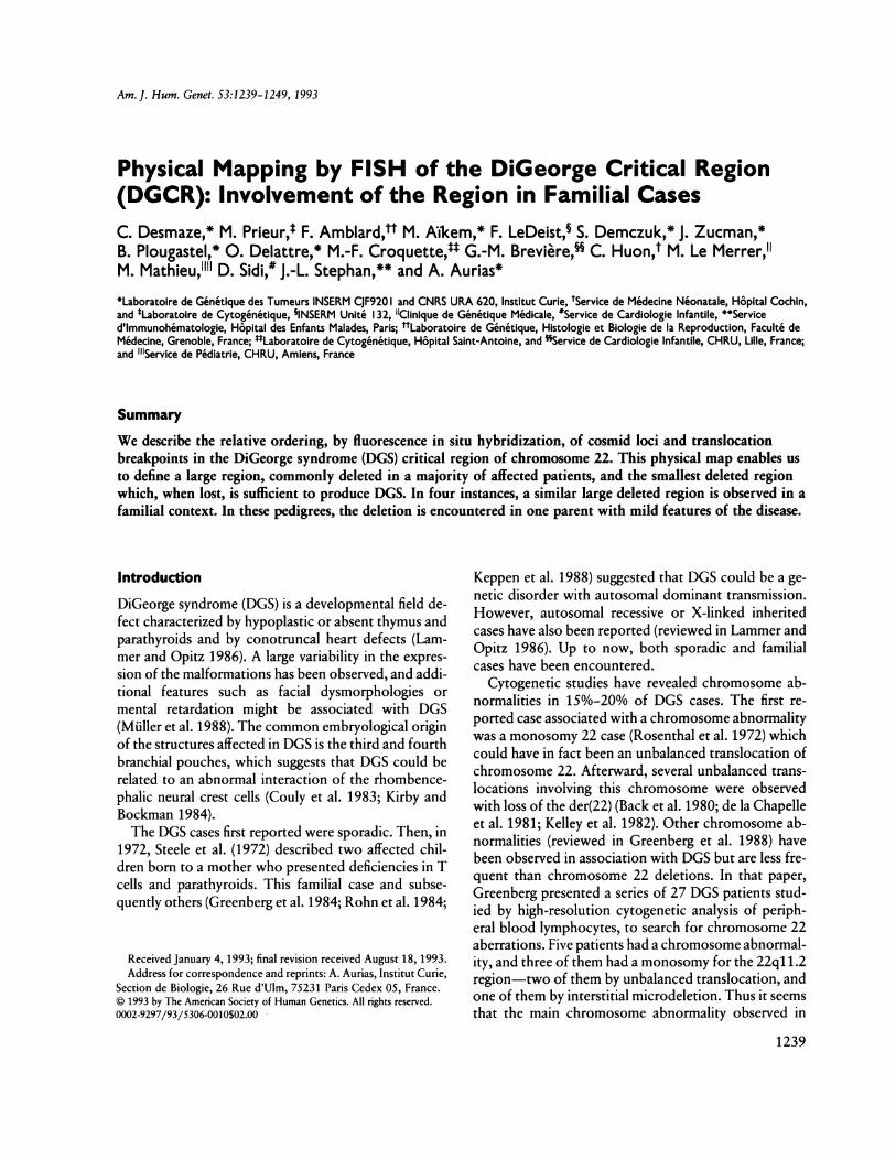
Am.J. Hum. Genet. 53:1239-1249, 1993
Physical Mapping by FISH of the DiGeorge Critical Region(DGCR): Involvement of the Region in Familial CasesC. Desmaze,* M. Prieur,* F. Amblard,tt M. ANkem,* F. LeDeist,§ S. Demczuk,* J. Zucman,*B. Plougastel,* 0. Delattre,* M.-F. Croquette,** G.-M. Breviere,§ C. Huon,t M. Le Merrer,11M. Mathieu,1111 D. Sidi,# J.-L. Stephan,** and A. Aurias*
*Laboratoire de Genetique des Tumeurs INSERM CJF920 1 and CNRS URA 620, Institut Curie, tService de Medecine Neonatale, H6pital Cochin,and $Laboratoire de Cytogenetique, SINSERM Unite 132, I"Clinique de Genetique Medicale, *Service de Cardiologie Infantile, "Serviced'lmmunohematologie, H6pital des Enfants Malades, Paris; tLaboratoire de Genetique, Histologie et Biologie de la Reproduction, Faculte deMedecine, Grenoble, France; #Laboratoire de Cytogenetique, H6pital Saint-Antoine, and "Service de Cardiologie Infantile, CHRU, Lille, France;and 111Service de Pediatrie, CHRU, Amiens, France
Summary
We describe the relative ordering, by fluorescence in situ hybridization, of cosmid loci and translocationbreakpoints in the DiGeorge syndrome (DGS) critical region of chromosome 22. This physical map enables us
to define a large region, commonly deleted in a majority of affected patients, and the smallest deleted regionwhich, when lost, is sufficient to produce DGS. In four instances, a similar large deleted region is observed in a
familial context. In these pedigrees, the deletion is encountered in one parent with mild features of the disease.
Introduction
DiGeorge syndrome (DGS) is a developmental field de-fect characterized by hypoplastic or absent thymus andparathyroids and by conotruncal heart defects (Lam-mer and Opitz 1986). A large variability in the expres-sion of the malformations has been observed, and addi-tional features such as facial dysmorphologies ormental retardation might be associated with DGS(Muller et al. 1988). The common embryological originof the structures affected in DGS is the third and fourthbranchial pouches, which suggests that DGS could berelated to an abnormal interaction of the rhombence-phalic neural crest cells (Couly et al. 1983; Kirby andBockman 1984).The DGS cases first reported were sporadic. Then, in
1972, Steele et al. (1972) described two affected chil-dren born to a mother who presented deficiencies in Tcells and parathyroids. This familial case and subse-quently others (Greenberg et al. 1984; Rohn et al. 1984;
Received January 4, 1993; final revision received August 18, 1993.Address for correspondence and reprints: A. Aurias, Institut Curie,
Section de Biologie, 26 Rue d'Ulm, 75231 Paris Cedex 05, France.© 1993 by The American Society of Human Genetics. All rights reserved.0002-9297/93/5306-0010$02.00
Keppen et al. 1988) suggested that DGS could be a ge-netic disorder with autosomal dominant transmission.However, autosomal recessive or X-linked inheritedcases have also been reported (reviewed in Lammer andOpitz 1986). Up to now, both sporadic and familialcases have been encountered.
Cytogenetic studies have revealed chromosome ab-normalities in 15%-20% of DGS cases. The first re-ported case associated with a chromosome abnormalitywas a monosomy 22 case (Rosenthal et al. 1972) whichcould have in fact been an unbalanced translocation ofchromosome 22. Afterward, several unbalanced trans-locations involving this chromosome were observedwith loss of the der(22) (Back et al. 1980; de la Chapelleet al. 1981; Kelley et al. 1982). Other chromosome ab-normalities (reviewed in Greenberg et al. 1988) havebeen observed in association with DGS but are less fre-quent than chromosome 22 deletions. In that paper,Greenberg presented a series of 27 DGS patients stud-ied by high-resolution cytogenetic analysis of periph-eral blood lymphocytes, to search for chromosome 22aberrations. Five patients had a chromosome abnormal-ity, and three of them had a monosomy for the 22ql 1.2region-two of them by unbalanced translocation, andone of them by interstitial microdeletion. Thus it seemsthat the main chromosome abnormality observed in
1239
Desmaze et al.
DGS is a deletion in the 22ql1.2 region, the conse-
quence of which could be a deletion for a number ofgenes, leading some authors to propose that DGSwould be a contiguous gene syndrome (Schmickel1986; Emanuel 1988). In contrast, Augusseau et al.(1986) described a DGS patient with an apparently bal-anced translocation t(2;22)(q14.1;q11.2) and no addi-tional chromosome abnormality. In the same way, no
correlation has been established between the severity ofthe pathology and the size of the deletion (Scambler et
al. 1991a). According to these observations, the hy-pothesis that DGS is a contiguous gene syndromeshould be readdressed.
In a majority of DGS cases, no cytogenetic abnormali-ties are observed; therefore, further molecular investi-gations have been performed. The first reports withgene dosage analysis and/or RFLP studies have beenpublished by Carey et al. (1990) and Fibison et al.(1990). Demonstration of loss of sequences in the22ql 1.2 region has been first provided by Scambler et
al. (1991a).In a previous study, we performed fluorescence in
situ hybridizations (FISH) on metaphases from periph-eral blood lymphocytes of a series of 10 DGS patients
(Desmaze et al. 1993). We probed this series with a
cosmid clone, Sc11.1, corresponding to a locus,D22S139, lying in 22ql1.2 (Carey et al. 1990), and dem-onstrated that all of the DGS patients studied were
hemizygous at this locus. Among them, two had a mi-crodeletion in 22q11.2 detectable by high-resolutionchromosome analysis, and the eight others had a nor-
mal karyotype.All these molecular results lead authors to consider
that a genetic etiology for DGS would include a majorsequence lying in the 22q11.2 region. In the present
study, we report on the organization, by means of bothunicolor FISH translocation breakpoint mapping andbicolor FISH, of three other loci that map near theScll.1 loci and that are always lost in the DGS patientswe have studied. We also report on four new familialcases of deletions associated with DGS.
Patients, Material, and Methods
Patients and Cell LinesThe patients were essentially ascertained through
their congenital heart defects. Of a large series of 50DGS patients (authors' unpublished data), a familialcontext was suspected in five instances.
Family A. -The proband, a 1-mo-old female, has mi-
crognathia and mild hypocalcemia. There is no thymic
shadow, but T lymphocytes are normal. There is nocongenital heart defect. Her brother died of typicalDGS (interrupted aortic arch, absence of thymus, hypo-calcemia, and low T-cell count) 1 year before. Themother has neonatal hypocalcemia, a mildly dysmor-phic facial appearance (mild micrognathia) evokingDGS, and mild mental retardation. The father is alsoslightly dysmorphic and has mild mental retardation.
Family B.-The proband, a 2-year-old girl, has aninterrupted aortic arch, mental retardation, no thymicshadow, a low T-lymphocyte count, and dysmorphicfeatures, including micrognathia, bilateral epicanthusfolds, and low-set ears. The mother is mildly retardedand has mildly dysmorphic features of DGS (i.e., mi-crognathia and low-set ears). She is now pregnant. Thefetus is a male with an interrupted aortic arch and a lowT-lymphocyte count. The father is normal.
Family C.-The proband is an 8-year-old male with aventricular septal defect and pulmonary atresia. Calce-mia is normal. The thymus is present, and T-cell countsare normal. The patient has typical DGS facial dysmor-phies, mild mental retardation, and hypernasal speech.The older brother has undergone an operation for anaortic arch anomaly. This brother has mild mental re-tardation with dysmorphic features including retro-gnathia, ear anomalies, and short uvula. His calcemia isnormal. The thymus is present. T-lymphocyte countsare normal. The two parents are normal, but the fatheris very slightly dysmorphic (prominent nose and dis-crete micrognathia), and a systematic dosage revealedmild hypocalcemia.
Family D.-The proband is a woman with dysmor-phic features (micrognathia, prominent nose, and smallauricles) closely resembling those observed in the velo-cardio-facial syndrome (VCFS) (Shprintzen et al. 1978)and with a cleft uvula but no congenital heart defect.Her two deceased children had typical DGS but werenot analyzed in our study. The older child presented atbirth with pulmonary atresia and ventricular septal de-fect, a hypoplastic thymus, microstomia, ear anomalies,and hypertelorism. The younger child presented withtruncus arteriosus, ventricular septal defect, and absentthymus. Dysmorphic features are noted in the clinicaldata but without details. The husband is normal.
Family E.-The proband is a newborn male withmildly dysmorphic features (micrognathia, small auri-cles, and bulbous nose), hypocalcemia, and a heartmurmur. Further investigations failed to confirm a con-genital heart defect. The father appears normal. Themother presented at birth with hypocalcemia but with-out any other pathological sign.
1240
Physical Mapping of the DGCR
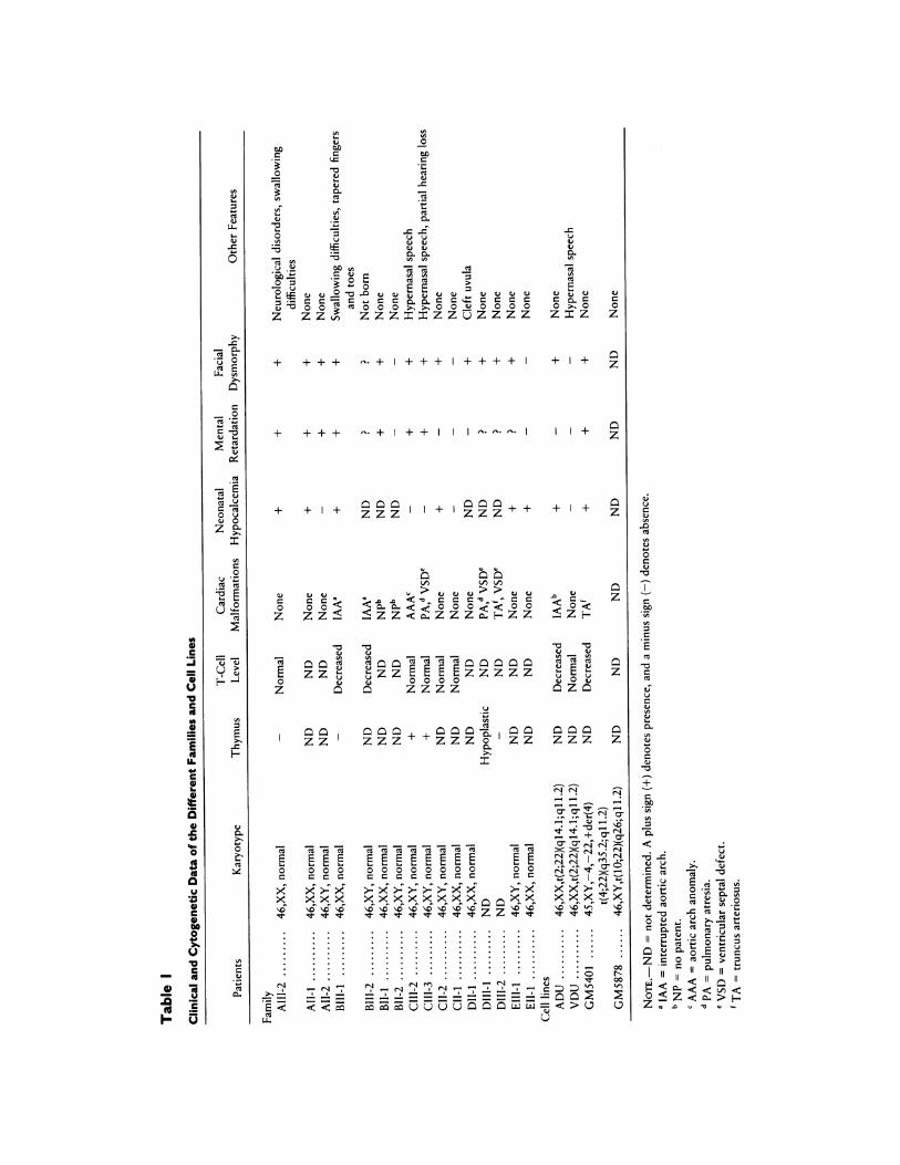
Cell LinesGM5401 (Greenberg et al. 1984) and GM5878 (Kel-
ley et al. 1982; Cannizzaro and Emanuel 1985) are celllines obtained from the NIGMS Human Genetic Mu-tant Cell Repository (Camden). The first cell line wasestablished from a DGS patient with an unbalancedt(4;22)(q35.2;qll.2) translocation, and the second cellline was established from the father of a DGS patientwith an unbalanced t(10;22)(q26;qll.2) translocation.This father carries the balanced translocation.ADU and VDU are lymphoblastoid cell lines from a
DGS proband and her mother, respectively. Except forhypernasal speech, the mother is normal. Both themother and the daughter present the same reciprocaland apparently balanced t(2;22)(q14;qll.2) transloca-tion (Augusseau et al. 1986). Karyotypes and clinicaldetails for all the patients and all the cell lines are sum-marized in table 1.
Hybrid Cell LinesFour hybrid cell lines were used as a somatic cell
hybrid mapping panel. (1) EYE F3A6 (Van Keuren et al.1987) retained the entire chromosome 22. (2) 33-11-Tg(Geurts van Kessel et al. 1980) was obtained from ahuman cell carrying a reciprocal t(X;22) translocation.The hybrid cell line retained the derivative X chromo-some. (3) ALE has been derived from the lymphoblas-toid cell line ICB100 which carries a recurrentt(11;22)(q23.1;qll.2) translocation (Zhang et al. 1990).The hybrid cell line kept the der(11). (4) AJO has beenderived from the lymphoblastoid cell line ICB101and retained the der(4) of a constitutional t(4;22)(p16.2;qll.2) translocation. The breakpoints of thethree last hybrid cell lines allow us to define four subre-gions in the proximal part of band 22ql 1.2 (Delattre etal. 1991).
FISHCytogenetic preparations.-Metaphases and prome-
taphases were obtained from cell lines or phytohe-magglutinin-stimulated lymphocytes by using standardmethods. GO-G1 interphase nuclei were obtained fromnormal donors' unstimulated blood lymphocytes.
Molecular probes.-All probes used for FISH werecosmid clones. Scll.1, described in Halford et al.(1993), was provided by P. Scambler. This cosmid cloneprobes two independent loci on chromosome 22 (Hal-ford et al. 1993; Scambler 1993), designated in our re-sults as "Scll.la" for the telomeric locus and as"Scll.lb" for the centromeric locus. From earlierwork, we had also demonstrated by FISH that these
two loci are recurrently deleted on one of the twochromosomes 22 in DGS patients (Desmaze et al. 1993).We have localized the corresponding loci between thebreakpoints of the t(X;22) translocation observed in the33-11-Tg cell line and that of the recurrent t(11;22)translocation observed in the ALE cell line (Delattre etal. 1991). This localization is in good agreement withthe one reported elsewhere by Sharkey et al. (1992) andHalford et al. (1993).We have searched for additional sequences localized
in the same subregion which corresponds to the Di-George critical region (DGCR) (Scambler et al. 1991a;Driscoll et al. 1992a; Scambler 1993). Using an ex-tended panel of somatic cell hybrids which included the33-1 1-Tg and ALE cell lines, we mapped three indepen-dent loci in the DGCR. They correspond to the K1429locus (D22S138) (Carey et al. 1990), to a NotI linkingclone, NotS4 (provided by H. Vissing) isolated from achromosome 22-specific cosmid library, and to the ca-techol-o-methyltransferase (COMT) locus. This genehas previously been located on chromosome 22 be-tween the breakpoints of the GL5 cell line and that ofthe t(10;22) translocation observed in the GM5878 cellline (Grossman et al. 1992). Cosmid probes 48F8 and10OC10-containing D22S138 and the COMT loci, re-spectively-were obtained by the screening of the cos-mid library LL22NC01 constructed in Livermore fromflow-sorted chromosomes of the human lymphoblas-toid cell line GM131. Their localization and that of theNot54 cosmid were confirmed to be in band 22q11.2by means of FISH on control prometaphases (data notshown). A cosmid, 26H9, containing the locus D22S9,was obtained from the same library. This locus is local-ized in the subregion immediately centromeric to thet(X;22) breakpoint observed in the 33-11-Tg cell line.Another cosmid, cosDs, probes a locus known to belocalized distal to the recurrent t(11;22) breakpoint (S.Demczuk, unpublished data) and was used because ofits good hybridization signal in interphase nuclei.ZNF70 was previously localized in the region immedi-ately distal to the AJO breakpoint (Aubry et al. 1992).
Hybridization and immunodetection.-Cosmid DNAswere extracted by using the alkaline lysis procedure(Sambrook et al. 1989). DNA was labeled either by bio-tin-14-dATP by using the Bionick kit (BRL, Gaithers-burg) or by digoxygenin-11-dUTP by using a nick-translation kit (Boehringer Mannheim) according to therecommendations of the suppliers. A total of 40-100ng of labeled DNA was mixed with about 100-foldsonicated human DNA in 20 pl of hybridization buffer(2 X SSC [1 X SSC = 0.15 M NaCl, 0.015 M Na citrate],
1241
n0
U~~~~
0(
0 I mc cZ
U0EE "U -a U U
co 0.X
;._ r- 4. C r- rQQ
_
-uZZ Z Z Z Ucc Z Z a
ZZZzm ZZZZ:I:ZZ ZZZZ Z =Z
+ + + ++ + + + + + + +
+ + ++ .+ + + - - -
+ + + a c +z z z
az aaiu u)
'I
> u v /v > > u ur le .0.0 < U U U = U U
0 O < 0<C 0. < <00 0<< 0 0z-<zz < 0 zzz . -zz
U0z
+ + az
z
Ic cc + + + +
< O-
az
az
E a a v E YE NE YE a E QjS. z z w w z z w I. w w z z z z z S. w
0 U 0 0 0 u u
z c ~zzzziU
C.0 a a a a a a a X a a
_- -
CZ CZ cmE E E$.. ;o o o
X_ rXX X X
D
<. c
E; E E E E;E E E;o o o o o o o o
X X X X X X X X
N z
can aZZZ Z
0-X X--+0-
E E >Nt, A<
X X^ X X X^~07 04 90 90 < sD
* ~~~~~~00
.r.U 00
. U
.
0 -
-a C
000
U C
D
U
0.
U
CZ
0
UC
a0
00
UUrUC 0 CZ
_s
* 0.
Z .0V:e
U(A
U0
0O
Z._
0b
ES
tL
-c
H
0._0
U
0.
cAc-
U
0z
iJ
=I',c
U'
IL0
v
la
0
4aU
0
.C(U4U0
I--
Cm
E<
cr
Physical Mapping of the DGCR
40 mM NaH2PO4, 50% deionized formamide, 10%dextran sulphate, 0.1% SDS, 1% Denhardt's solution).Slides were pretreated with 100 gg RNase A/ml (Sigma,St. Louis) for 1 h at 370C, then dehydrated through aseries of ethanol baths (50%, 75%, and 100%) and dena-tured for 2-3 min at 70'C in 2 X SSC 70% formamide.The hybridization mixture was denatured by boiling
for 10 min and was ice cooled and spotted onto thecytogenetic spreads. The slides were incubated over-night at 420C in a moist chamber under a plastic cover-slip. They were rinsed twice in 50% formamide 2 X SSCand then in 2 baths of 2 X SSC, at 420C each. After apreincubation in PBT (PBS, 0.1% Tween 20, 0.1%BSA), the immunodetection was performed in twosteps for unicolor FISH or in four steps for bicolorFISH. The biotinylated probes were revealed by a goatanti-biotin antibody, dilution 1/100 (Vector, Burlin-game) and a by fluorescein-conjugated anti-goat anti-body, dilution 1/400 (Biosys, Compiegne), and the di-goxygenin-labeled probes were revealed by a mouseanti-digoxygenin antibody and a rhodamine-conju-gated anti-mouse antibody, dilutions 1/100 and 1/10,respectively (Boehringer Mannheim). The slides werecounterstained with propidium iodide and weremounted in an antifade solution (Johnson andNogueira 1981).
Microscopic observations and data analyses.-Theslides were observed on an Aristoplan Leitz microscopewith a standard FITC filter combination for unicolorexperiments and a double-band pass filter (Omega Op-tical, Brattleboro) for bicolor experiments. Photo-graphs were taken with Kodak Ektachrome 400 film.For bicolor experiments, each cosmid probe was testedon interphase nuclei from a normal donor in order toestimate the hybridization efficiencies, defined as theratio of observed number of labeled loci:theoreticalnumber of probed loci. These efficiencies are 0.83,0.87, 0.79, 0.68, and 0.94 for Sc11.1, 10OC10, NotS4,48F8, and cosDs cosmid probes, respectively. All nucleiwith three discrete single signals were scored, and thedistribution was compared with a random distribution(X2 test).ResultsPhysical Mapping of the DGCRTwo different approaches were developed-translo-
cation breakpoint mapping and loci ordering by bicolorFISH. With regard to the former, we have performedunicolor FISH on a series of translocations involvingband 22ql 1.2. We have confirmed by FISH that thebreakpoint of the t(2;22) observed in the ADU cell line
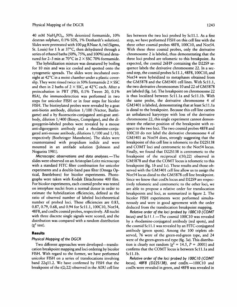
lies between the two loci probed by Scll.1. As a firststep, we have performed FISH on this cell line with thethree other cosmid probes 48F8, 10OC10, and NotS4.With these three cosmid probes, only the derivativechromosome 2 is labeled, thus demonstrating that thethree loci probed are telomeric to this breakpoint. Asexpected, the cosmid 26H9 containing the D22S9 se-quence labels the derivative chromosome 22. In a sec-ond step, the cosmid probes Scl1.1, 48F8, 10C10, andNotS4 were hybridized to metaphases obtained fromthe GM5878 and the GM5401 cell lines. With Scll.1,the two derivative chromosomes 10 and 22 of GM5878are labeled (fig. la). The breakpoint on chromosome 22is thus localized between Scll.la and Scll.lb. Withthe same probe, the derivative chromosome 4 ofGM5401 is labeled, demonstrating that at least Scll.lais distal to the breakpoint. Because this cell line carriesan unbalanced karyotype with loss of the derivativechromosome 22, this single experiment cannot demon-strate the relative position of the breakpoint with re-spect to the two loci. The two cosmid probes 48F8 and10OC10 do not label the derivative chromosome 4 ofGM5401 as NotS4 does, strongly suggesting that thebreakpoint of this cell line is telomeric to the D22S138and COMT loci and centromeric to the NotS4 locus.Finally, we found that D22S138 is centromeric to thebreakpoint of the reciprocal t(10;22) observed inGM5878 and that the COMT locus is telomeric to thisbreakpoint (fig. lb and 1c). These results and those ob-served with the GM5401 cell line allow us to assign theNotS4 locus distal to the GM5878 cell line breakpoint.Since we know that cosDs locus and D22S9 are respec-tively telomeric and centromeric to the other loci, weare able to propose a relative order for translocationbreakpoints and loci, as summarized in figure 2. Thebicolor FISH experiments were performed simulta-neously and were in good agreement with the orderdeduced from the translocation breakpoint mapping.
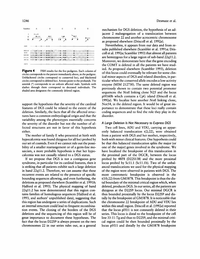
Relative order of the loci probed by I QOC 10 (COMTlocus) and Sc I I. I.-The cosmid 1OOC10 was revealedby a rhodamine-conjugated antibody (red spots), andthe cosmid Sc1 1.1 was revealed by an FITC-conjugatedantibody (green spots). Among the 100 triplets ob-served, 76 were of the green-red-green type, and 24were of the green-green-red type (fig. 3a). This distribu-tion is clearly not random (X2 = 14.5, P = .0001) andconfirms that the COMT locus is between Scll.la andScll.lb.
Relative order of the loci probed by I QOC10 (COMTlocus), 48F8 (D22S 138), and cosDs.-100C10 andcosDs were revealed in green, and 48F8 was revealed in
1243
Desmaze et al.
red. Among the 100 triplets analyzed, 85 were green-
green-red, and 15 were green-red-green (X2 = 28, P= .0001), confirming the localization of D22S138 out-
side the interval COMT-cosDs loci (fig. 3b). Probablybecause of either a peculiar configuration of the chro-matin or a polymorphism in the population, the same
technique performed for the loci probed by Not54 and
Figure I FIlSH- pertornmed on mietaph ases from the (CA\158-78cell line whlichI carries a baIlancdILi tr(I);2)l(.;Ll 1.) tran slocatiOn.a1. (OSiiiid probe Sc libeled(I r( 10) anL] der(22) (thinla1nottvs), aswell a-s II e norimal chLroImosomc 2 (thickene aiirrow). , )ro be 481:8-labCeId deir(221) (thin arro.L,a' wll as thle normial chromilosolieCithnikencd arr?'K' C,(COSTInId Ir-e1. 10 labeled-11Cder1) (thinal-On':, as\I ela-s heInrmalI I 1"IItCh S()IIlIWh Osome 2C2 b(-t kioked azrrowi).
Sc11.1 on four independent donors gave clear-cut dif-ferent ordering from one to the other.
Analysis of the PatientsAll the probands were studied with high-resolution
R-banding (850-bands stage). No deletion was sus-pected in any of these patients.
1244
Physical Mapping of the DGCR
LULUNE
G-o2D-
t2 t(492)
176f. ... W.,
..1
1^-- 1 Largest dued region
region
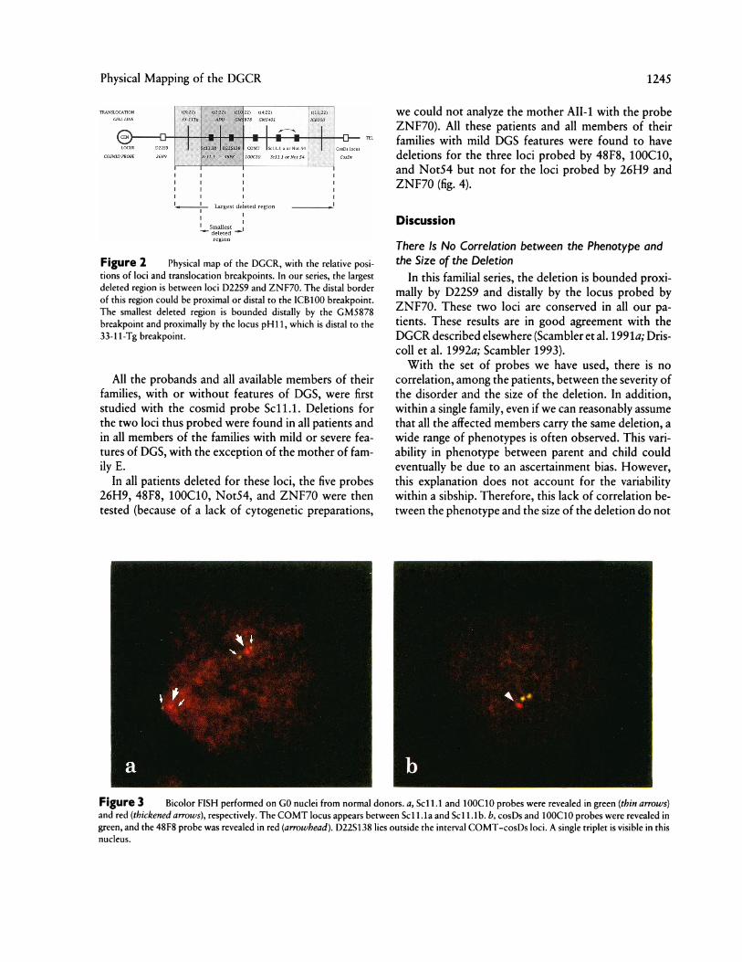
Figure 2 Physical map of the DGCR, with the relative posi-tions of loci and translocation breakpoints. In our series, the largestdeleted region is between loci D22S9 and ZNF70. The distal borderof this region could be proximal or distal to the ICB100 breakpoint.The smallest deleted region is bounded distally by the GM5878breakpoint and proximally by the locus pH11, which is distal to the33-1 1-Tg breakpoint.
All the probands and all available members of theirfamilies, with or without features of DGS, were firststudied with the cosmid probe Sc11.1. Deletions forthe two loci thus probed were found in all patients andin all members of the families with mild or severe fea-tures of DGS, with the exception of the mother of fam-ily E.
In all patients deleted for these loci, the five probes26H9, 48F8, 10OC10, NotS4, and ZNF70 were thentested (because of a lack of cytogenetic preparations,
we could not analyze the mother AII-1 with the probeZNF70). All these patients and all members of theirfamilies with mild DGS features were found to havedeletions for the three loci probed by 48F8, 10OC10,and Not54 but not for the loci probed by 26H9 andZNF70 (fig. 4).
Discussion
There Is No Correlation between the Phenotype andthe Size of the Deletion
In this familial series, the deletion is bounded proxi-mally by D22S9 and distally by the locus probed byZNF70. These two loci are conserved in all our pa-tients. These results are in good agreement with theDGCR described elsewhere (Scambler et al. 1991a; Dris-coll et al. 1992a; Scambler 1993).With the set of probes we have used, there is no
correlation, among the patients, between the severity ofthe disorder and the size of the deletion. In addition,within a single family, even if we can reasonably assumethat all the affected members carry the same deletion, awide range of phenotypes is often observed. This vari-ability in phenotype between parent and child couldeventually be due to an ascertainment bias. However,this explanation does not account for the variabilitywithin a sibship. Therefore, this lack of correlation be-tween the phenotype and the size of the deletion do not
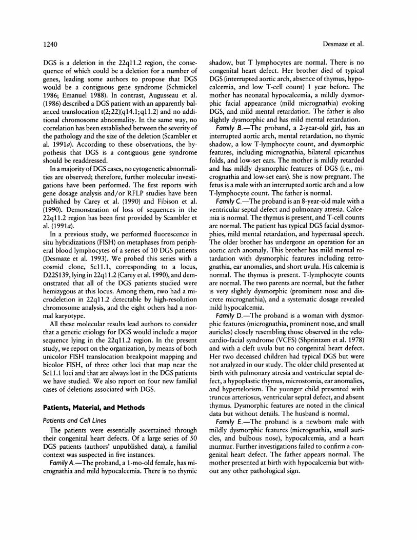
Figure 3 Bicolor FISH performed on GO nuclei from normal donors. a, Scl1.1 and 10OC10 probes were revealed in green (thin arrows)and red (thickened arrows), respectively. The COMT locus appears between Scd1 .la and Scd1 lb. b, cosDs and 10OC10 probes were revealed ingreen, and the 48F8 probe was revealed in red (arrowhead). D22S138 lies outside the interval COMT-cosDs loci. A single triplet is visible in thisnucleus.
1245
Desmaze et al.
B C D
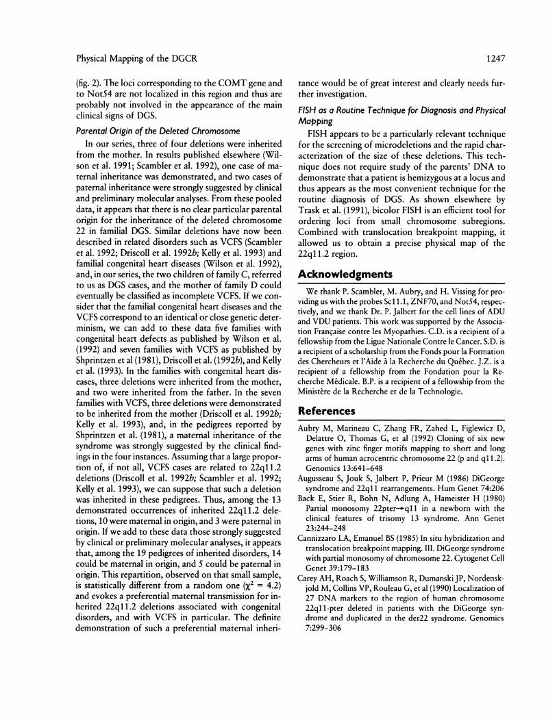
Figure 4 FISH results for the five pedigrees. Each column ofcircles corresponds to the patient immediately above, in the pedigree.Unblackened circles correspond to conserved loci, and blackenedcircles correspond to deleted loci. Arrows point to the probands. Theasterisk (*) corresponds to an unborn affected male. Symbols withslashes through them correspond to deceased individuals. Theshaded area designates the commonly deleted region.
support the hypothesis that the severity of the cardinalfeatures of DGS could be related to the extent of thedeletion. Similarly, the facts that all the affected struc-tures have a common embryological origin and that thevariability among the phenotypes essentially concerns
the severity of the disorder but not the number of af-fected structures are not in favor of this hypothesiseither.The mother of family E who presented at birth with
hypocalcemia was found dizygous for all loci probed byour set of cosmids. Even ifwe cannot rule out the possi-bility of a smaller rearrangement or of a germ-line mo-saicism, a more probable hypothesis is that her hypo-calcemia was not causally related to a DGS status.
If we propose that DGS is not a contiguous gene
syndrome, in particular for its cardinal features, then itis striking that all patients exhibit such a large deletionin band 22q11.2. Therefore, we can assume that theserecurrent events are related to the presence of specificbounding sequences allowing, and even furthering, thedeletions as proposed elsewhere (Scambler et al. 1991b;Halford et al. 1993). The physical mapping of band22ql 1.2 has now demonstrated that this region con-
tains families of homologous sequences (Halford et al.1993, and authors' unpublished data), suggesting thatthis region has undergone a series of duplications. Suchan internal structure could lead to frequent recombina-tion events. The cloning of the borders of the DGSdeletions and the sequencing of this region will be ofgreat importance to document these hypotheses. Thefact that the locus D22S9 is always present on the twochromosomes 22 in our series rules out, as a general
mechanism for DGS deletion, the hypothesis of an ad-jacent 2 malsegregation of a translocation betweenchromosome 22 and another acrocentric chromosomeas proposed elsewhere (Driscoll et al. 1992a).
Nevertheless, it appears from our data and from re-
sults published elsewhere (Scambler et al. 1991a; Dris-coll et al. 1992a; Scambler 1993) that almost all patientsare hemizygous for a large region of sub-band 22ql 1.2.Moreover, we demonstrate here that the gene encodingthe COMT is deleted in all the patients we have stud-ied. As proposed elsewhere (Scambler 1993), deletionof this locus could eventually be relevant for some clin-ical minor aspects of DGS and related disorders, in par-
ticular when the conserved allele encodes a low-activityenzyme (MIM 212730). The same deleted region was
previously shown to contain two potential promoter
sequences: the NotI linking clone N25 and the locuspH160b which contains a CpG island (Driscoll et al.1992a). We localize here another NotI linking clone,Not54, in the deleted region. It would be of great im-portance to demonstrate that these loci really containcoding sequences and to find the role they play in thedisorder.
A Large Deletion Is Not Necessary to Express DGSTwo cell lines, ADU and VDU, carrying an appar-
ently balanced translocation t(2;22), were obtainedfrom a patient with DGS and her mother, respectively,both with minor clinical features. One hypothesis couldbe that this balanced translocation splits the major (orone of the major) genes involved in the syndrome. Wehave localized the breakpoint of this translocation inthe proximal part of the DGCR, between the locusprobed by 48F8 (D22S138) and the more proximallocus probed by Sc11.1 (Scl1.lb). Two of the unbal-anced translocations we used for the physical mappingof the region were observed in patients with DGS. Themore centromeric breakpoint is observed in thet(10;22) from GM5878. This breakpoint is thus the dis-tal boundary of the minimal critical region which, whendeleted, produces DGS. In our series, all the patients are
dizygous at the D22S9 locus. Our minimal DGCR isthus bounded proximally by the locus D22S9 and dis-tally by the breakpoint of GM5878. It is noticeable thatthe chromosome 22 breakpoint of ADU and VDU lieswithin this small region. Driscoll et al. (1992a) reportedthat the locus pH11 is not constantly deleted in theirseries. This locus is distal to the breakpoint of the cellline 33-1 1-Tg and thus to D22S9, and the minimal criti-cal region could be then bounded proximally by thelocus pH11 and distally by the GM5878 breakpoint
Famdiies A
I
m .00->i,2639 -O---O------O-~
A.0-=SCIIlb :'~48FS100 CIO ;ilSSSCILlaNOT 54
ZNF70 -O - - O
0 0
0 a 0
1246
Physical Mapping of the DGCR 1247
(fig. 2). The loci corresponding to the COMT gene andto Not54 are not localized in this region and thus areprobably not involved in the appearance of the mainclinical signs of DGS.
Parental Origin of the Deleted ChromosomeIn our series, three of four deletions were inherited
from the mother. In results published elsewhere (Wil-son et al. 1991; Scambler et al. 1992), one case of ma-ternal inheritance was demonstrated, and two cases ofpaternal inheritance were strongly suggested by clinicaland preliminary molecular analyses. From these pooleddata, it appears that there is no clear particular parentalorigin for the inheritance of the deleted chromosome22 in familial DGS. Similar deletions have now beendescribed in related disorders such as VCFS (Scambleret al. 1992; Driscoll et al. 1992b; Kelly et al. 1993) andfamilial congenital heart diseases (Wilson et al. 1992),and, in our series, the two children of family C, referredto us as DGS cases, and the mother of family D couldeventually be classified as incomplete VCFS. If we con-sider that the familial congenital heart diseases and theVCFS correspond to an identical or close genetic deter-minism, we can add to these data five families withcongenital heart defects as published by Wilson et al.(1992) and seven families with VCFS as published byShprintzen et al (1981), Driscoll et al. (1992b), and Kellyet al. (1993). In the families with congenital heart dis-eases, three deletions were inherited from the mother,and two were inherited from the father. In the sevenfamilies with VCFS, three deletions were demonstratedto be inherited from the mother (Driscoll et al. 1992b;Kelly et al. 1993), and, in the pedigrees reported byShprintzen et al. (1981), a maternal inheritance of thesyndrome was strongly suggested by the clinical find-ings in the four instances. Assuming that a large propor-tion of, if not all, VCFS cases are related to 22ql 1.2deletions (Driscoll et al. 1992b; Scambler et al. 1992;Kelly et al. 1993), we can suppose that such a deletionwas inherited in these pedigrees. Thus, among the 13demonstrated occurrences of inherited 22q11.2 dele-tions, 10 were maternal in origin, and 3 were paternal inorigin. If we add to these data those strongly suggestedby clinical or preliminary molecular analyses, it appearsthat, among the 19 pedigrees of inherited disorders, 14could be maternal in origin, and 5 could be paternal inorigin. This repartition, observed on that small sample,is statistically different from a random one (X2 = 4.2)and evokes a preferential maternal transmission for in-herited 22q11.2 deletions associated with congenitaldisorders, and with VCFS in particular. The definitedemonstration of such a preferential maternal inheri-
tance would be of great interest and clearly needs fur-ther investigation.
FISH as a Routine Technique for Diagnosis and PhysicalMappingFISH appears to be a particularly relevant technique
for the screening of microdeletions and the rapid char-acterization of the size of these deletions. This tech-nique does not require study of the parents' DNA todemonstrate that a patient is hemizygous at a locus andthus appears as the most convenient technique for theroutine diagnosis of DGS. As shown elsewhere byTrask et al. (1991), bicolor FISH is an efficient tool forordering loci from small chromosome subregions.Combined with translocation breakpoint mapping, itallowed us to obtain a precise physical map of the22q11.2 region.
AcknowledgmentsWe thank P. Scambler, M. Aubry, and H. Vissing for pro-
viding us with the probes Scl 1.1, ZNF70, and NotS4, respec-tively, and we thank Dr. P. Jalbert for the cell lines of ADUand VDU patients. This work was supported by the Associa-tion Franpaise contre les Myopathies. C.D. is a recipient of afellowship from the Ligue Nationale Contre le Cancer. S.D. isa recipient of a scholarship from the Fonds pour la Formationdes Chercheurs et l'Aide a la Recherche du Quebec. J.Z. is arecipient of a fellowship from the Fondation pour la Re-cherche Medicale. B.P. is a recipient of a fellowship from theMinistere de la Recherche et de la Technologie.
ReferencesAubry M, Marineau C, Zhang FR, Zahed L, Figlewicz D,
Delattre 0, Thomas G, et al (1992) Cloning of six newgenes with zinc finger motifs mapping to short and longarms of human acrocentric chromosome 22 (p and qll.2).Genomics 13:641-648
Augusseau S, Jouk S, Jalbert P, Prieur M (1986) DiGeorgesyndrome and 22ql 1 rearrangements. Hum Genet 74:206
Back E, Stier R, Bohn N, Adlung A, Hameister H (1980)Partial monosomy 22pter- ql1 in a newborn with theclinical features of trisomy 13 syndrome. Ann Genet23:244-248
Cannizzaro LA, Emanuel BS (1985) In situ hybridization andtranslocation breakpoint mapping. III. DiGeorge syndromewith partial monosomy of chromosome 22. Cytogenet CellGenet 39:179-183
Carey AH, Roach S, Williamson R, Dumanski JP, Nordensk-jold M, Collins VP, Rouleau G, et al (1990) Localization of27 DNA markers to the region of human chromosome22q11-pter deleted in patients with the DiGeorge syn-drome and duplicated in the der22 syndrome. Genomics7:299-306
1248 Desmaze et al.
Couly G, Lagrue A, Griscelli C (1983) Le syndrome de Di-George: neurocristopathie rhombencephalique exemplaire.Rev Stomatol Chir Maxillofac 84:103-108
de la Chapelle A, Herva R, Koivisto M, Aula P (1981) A dele-tion in chromosome 22 can cause DiGeorge syndrome.Hum Genet 57:253-256
Delattre 0, Azambuja CJ, Aurias A, Zucman J, Peter M,Zhang F, Hors-Cayla MC, et al (1991) Mapping of humanchromosome 22 with a panel of somatic cell hybrids. Ge-nomics 9:721-727
Desmaze C, Scambler P, Prieur M, Halford S, Sidi D, Le DeistF, Aurias A (1993) Routine diagnosis of DiGeorge syn-drome by fluorescent in situ hybridization. Hum Genet90:663-665
Driscoll DA, Budarf ML, Emanuel BS (1992a) A genetic etiol-ogy for DiGeorge syndrome: consistent deletions and mi-crodeletions of 22ql 1. Am J Hum Genet 50:924-933
Driscoll DA, Spinner NB, Budarf ML, McDonald-McGinnDM, Zackai EH, Goldberg RB, Shprintzen RJ, et al (1992b)Deletions and microdeletions of 22ql 1.2 in velo-cardio-fa-cial syndrome. Am J Med Genet 44:261-268
Emanuel BS (1988) Molecular cytogenetics: toward dissec-tion of the contiguous gene syndromes. Am J Hum Genet43:575-578
Fibison WJ, Budarf M, McDermid H, Greenberg F, EmanuelBS (1990) Molecular studies of DiGeorge syndrome. Am JHum Genet 46:888-895
Geurts van Kessel AHM, Westerveld A, DeGroot PG, MeeraKP, Hagemeijer A (1980) Regional localisation of genescoding for human ACO2, ARSA and NAGA on chromo-some 22. Cytogenet Cell Genet 28:169-172
Greenberg F, Crowder WE, Paschall V, Colon-Linares J, Lu-bianski B, Ledbetter DH (1984) Familial DiGeorge syn-drome and associated partial monosomy of chromosome22. Hum Genet 65:317-319
Greenberg F, Elder FFB, Haffner P, Northrup H, LedbetterDH (1988) Cytogenetic findings in a prospective series ofpatients with DiGeorge anomaly. Am J Hum Genet43:605-611
Grossman MH, Emanuel BS, Budarf ML (1992) Chromo-somal mapping of the human catechol-o-methyltransferasegene to 22q11.1- oq11.2. Genomics 12:822-825
Halford S, Lindsay E, Nayudu M, Carey AH, Baldini A,Scambler P (1993) Low-copy-number repeat sequencesflank the DiGeorge/velo-cardio-facial syndrome loci at22q11. Hum Mol Genet 2:191-196
Johnson GD, Nogueira Araujo GM de C (1981) A simplemethod of reducing the fading of immunofluorescenceduring microscopy. J Immunol Method 43:349-350
Kelley RI, Zackai EH, Emanuel BS, Kistenmacher M, Green-berg F, Punnett HH (1982) The association of the Di-George anomaly with partial monosomy of chromosome22. J Pediatr 101:197-200
Kelly D, Goldberg R, Wilson D, Lindsay E, Carey A, Good-ship J, Burn J, et al (1993) Confirmation that the velo-car-dio-facial syndrome is associated with haplo-insufficiency
of genes at chromosome 22ql 1. Am J Med Genet 45:308-312
Keppen LD, Fasules JW, Burks AW, Gollin SM, Sawyer JR,Miller CH (1988) Confirmation of autosomal dominanttransmission of the DiGeorge malformation complex. J Pe-diatr 113:506-508
Kirby ML, Bockman DE (1984) Neural crest and normal de-velopment: a new perspective. Anat Rec 209:1-6
Lammer EJ, Opitz JM (1986) The DiGeorge anomaly as adevelopmental field defect. Am J Med Genet, Suppl 2:113-127
Muller W, Peter HH, Wilken M, JUppner H, Kallfelz HC,Krohn HP, Miller K, et al (1988) The DiGeorge syndrome.I. Clinical evaluation and course of partial and completeforms of the syndrome. Eur J Pediatr 147:496-502
Rohn RD, Leffell MS, Leadem P, Johnson D, Rubio T,Emanuel BS (1984) Familial third-fourth pharyngeal pouchsyndrome with apparent autosomal dominant transmis-sion. J Pediatr 105:47-51
Rosenthal IM, Bocian M, Krmpotic E (1972) Multiple anoma-lies including thymic aplasia associated with monosomy22. Pedriatr Res 6:358
Sambrook J, Fritsch EF, Maniatis T (eds) (1989) Molecularcloning: a laboratory manual, 2d ed. Cold Spring HarborLaboratory, Cold Spring Harbor, NY
Scambler PJ (1993) Deletions of human chromosome 22 andassociated birth defects. Curr Opin Genet Dev 3:432-437
Scambler PJ, Carey AH, Wyse RKH, Roach S, Dumanski JP,Nordenskjold M, Williamson R (1991a) Microdeletionswithin 22ql 1 associated with sporadic and familial Di-George syndrome. Genomics 10:201-206
Scambler P. Halford S, Wadey R, Lindsay E, Kelly D, Du-manski J, Nordenskjold M, et al (1991b) Duplicated se-quences within 22q11 and their relationship to gene dele-tion syndromes. Second International Workshop on theMapping of Human Chromosome 22, Montebello, Can-ada, September 10-13
Scambler PJ, Kelly D, Lindsay E, Williamson R, Goldberg R,Shprintzen R, Wilson DI, et al (1992) Velo-cardio-facialsyndrome associated with chromosome 22 deletions en-compassing the DiGeorge locus. Lancet 339:1138-1139
Schmickel RD (1986) Contiguous gene syndromes: a compo-nent of recognizable syndromes. J Pediatr 109:231-241
Sharkey AM, McLaren L, Carroll M, Fantes J, Green D, Wil-son D, Scambler PJ, et al (1992) Isolation of anonymousDNA markers for human chromosome 22ql 1 from a flow-sorted library, and mapping using hybrids from patientswith DiGeorge syndrome. Hum Genet 89:73-78
Shprintzen RJ, Goldberg RB, Levin ML, Sidoti EJ, BerkmanMD, Argamaso RV, Young D (1978) A new syndrome in-volving cleft palate, cardiac anomalies, typical facies, andlearning disabilities: velo-cardio-facial syndrome. Cleft Pal-ate J 15:56-62
Shprintzen RJ, Goldberg RB, Young D, Wolford L (1981)
Physical Mapping of the DGCR 1249
The velo-cardio-facial syndrome: a clinical and genetic anal-ysis. Pediatrics 67:167-172
Steele RW, Limas C, Thurman GB, Schuelein M, Bauer H,Bellanti JA (1972) Familial thymic aplasia: attempted re-constitution with fetal thymus in a millipore diffusionchamber. N Engl J Med 287:787-791
Trask BJ, Massa H, Kenwrick S, Gitschier J (1991) Mappingof human chromosome Xq28 by two-color fluorescence insitu hybridization of DNA sequences to interphase cell nu-clei. Am J Hum Genet 48:1-15
Van Keuren ML, Hart IM, Kao FT, Neve RL, Bruns GAP,Kurnit DM, Patterson D (1987) A somatic cell hybrid witha single human chromosome 22 corrects the defect in the
CHO mutant (Ade-I) lacking adenylosuccinate activity.Cytogenet Cell Genet 44:142-147
Wilson DI, Cross IE, Goodship JA, Coulthard S, Carey AH,Scambler PJ, Bain HH, et al (1991) DiGeorge syndromewith isolated aortic coarctation and isolated ventricularseptal defect in three sibs with a 22ql 1 deletion of mater-nal origin. Br Heart J 66:308-312
Wilson DI, Goodship JA, Burn J, Cross IE, Scambler PJ(1992) Deletions within chromosome 22ql 1 in familial con-genital heart disease. Lancet 340:573-575
Zhang FR, Aurias A, Delattre 0, Stern MR, Benitez J,Rouleau G, Thomas G (1990) Mapping of human chromo-some 22 by in situ hybridization. Genomics 7:319-324
Copyright © 2022 FDOKUMEN