
PhD School in Chemical Sciences Multitasking Fe3O4@Cu ...
204
Faculty of Mathematical, Physical and Natural Sciences PhD School in Chemical Sciences Cycle XXXI Multitasking Fe 3 O 4 @Cu@Au and Fe 3 O 4 @SiO 2 nanoparticles for biomedical applications PhD Supervisor: PhD Student: Prof. Mario Barteri Ginevra Berardi PhD Coordinator: Prof. Osvaldo Lanzalunga 2018
-
Upload
khangminh22 -
Category
Documents
-
view
1 -
download
0
Transcript of PhD School in Chemical Sciences Multitasking Fe3O4@Cu ...

Faculty of Mathematical, Physical and Natural
Sciences
PhD School in Chemical Sciences Cycle XXXI
Multitasking Fe3O4@Cu@Au and
Fe3O4@SiO2 nanoparticles for
biomedical applications
PhD Supervisor: PhD Student:
Prof. Mario Barteri Ginevra Berardi
PhD Coordinator:
Prof. Osvaldo Lanzalunga
2018
i
TABLE OF CONTENTS
1. GENERAL INTRODUCTION ... . . . . . . . . . . . . . . . . . . . . . . . . . . . . . . . . . . . . 1
Nanoparticles and Nanomedicine ....................................................... 2
1.1.1 General features of NPs ............................................................................ 5
1.1.2 Multitasking NPs ....................................................................................... 6
Magnetic nanoparticles ........................................................................ 8
1.2.1 Magnetite nanoparticles and superparamagnetism ............................... 8
Biomedical applications of MNPs ..................................................... 11
1.3.1 In vivo imaging ......................................................................................... 12
1.3.2 Therapy .................................................................................................... 13
1.3.3 In vitro diagnostics ................................................................................... 14
2. AIM AND STRUCTURE OF THIS THESIS ... . . . . . . . . . . . . . 17
Objectives and scientific strategy ...................................................... 18
Thesis structure .................................................................................. 19
3. SYNTHESIS AND PHYSICOCHEMICAL
CHARACTERIZATION OF Fe3O4@Cu@Au AND
Fe3O4@SiO2 NANOSYSTEMS ... . . . . . . . . . . . . . . . . . . . . . . . . . . . . . . . . . . . . . 21
Introduction ........................................................................................ 22
3.1.1 Core-shell nanoparticles ......................................................................... 22
Chapter objectives .............................................................................. 28
Results and discussion ........................................................................ 30
3.3.1 Fe3O4@Cu@Au MNPs ............................................................................ 30
3.3.2 Fe3O4@SiO2 MNPs .................................................................................. 37

ii
Chapter contributions ........................................................................ 42
Acknowledgments ............................................................................... 42
Materials and methods ....................................................................... 42
4. Fe3O4@Cu@Au NANOPARTICLES LOADING ACTIVE
TARGETING LIGANDS AND IN VITRO EXPERIMENTS
ON HUMAN BREAST CANCER CELL LINES ... . . . . . . . . . . . . 45
Introduction ........................................................................................ 46
4.1.1 Tumour targeting using nanosystems .................................................... 46
4.1.2 Folic acid and methotrexate ................................................................... 49
4.1.3 Folate receptors and cancer cells ........................................................... 50
Chapter objectives .............................................................................. 52
Results and discussion ........................................................................ 54
4.3.1 Functionalization with lipoic acid .......................................................... 54
4.3.2 Functionalization with poly-L-lysine ...................................................... 56
4.3.3 Functionalization with FA or MTX ....................................................... 58
4.3.4 In vitro experiments on human breast cancer cell lines ....................... 62
4.3.5 Ball milling process ................................................................................. 66
Chapter contributions ........................................................................ 68
4.4.1 Future perspectives ................................................................................. 68
Acknowledgments ............................................................................... 69
Materials and methods ....................................................................... 69
5. Fe3O4@Cu@Au NANOPARTICLES COATED WITH
MACROMOLECULES FOR THE ENGULFMENT OF
HUMAN IMMUNE CELLS.... . . . . . . . . . . . . . . . . . . . . . . . . . . . . . . . . . . . . . . . . . 73
iii
Introduction ........................................................................................ 74
5.1.1 Transferrin............................................................................................... 74
5.1.2 Streptavidin ............................................................................................. 75
5.1.3 Cell-mediated delivery: Trojan Horse Approach ................................. 76
Chapter objectives .............................................................................. 79
Results and discussion ........................................................................ 80
5.3.1 Functionalization of MNPs-LA-NHS with macromolecules ................ 80
5.3.2 In vitro biological experiments on human immune cells ...................... 84
Chapter contributions ........................................................................ 89
5.4.1 Future perspectives ................................................................................. 89
Acknowledgments ............................................................................... 90
Materials and methods ....................................................................... 90
6. Fe3O4@SiO2-LIPODEPSIPEPTIDE NANOSYSTEMS
WITH ANTIMICROBIAL ACTIVITY ... . . . . . . . . . . . . . . . . . . . . . . . . . 93
Introduction ........................................................................................ 94
6.1.1 Lipodepsipeptides from Pseudomonas syringae pv. syringae ............... 94
Chapter objectives .............................................................................. 98
Results and discussion ........................................................................ 99
6.3.1 Surface activation of Fe3O4@SiO2 MNPs with APTES ..................... 100
6.3.2 Functionalization with poly-L-aspartic acid ........................................ 101
6.3.3 Conjugation of syringomycin E or syringopeptin 22A ....................... 103
6.3.4 In vitro growth inhibition assays on R. pilimanae ............................... 107
6.3.5 In vitro internalization in human immune cells .................................. 110
Chapter contributions ...................................................................... 114
iv
6.4.1 Future perspectives ............................................................................... 114
Acknowledgments ............................................................................. 115
Materials and methods ..................................................................... 115
7. ANTIBODY-FUNCTIONALIZED MAGNETIC
NANOPARTICLES FOR THE DETECTION OF
EPINEPHRINE ... . . . . . . . . . . . . . . . . . . . . . . . . . . . . . . . . . . . . . . . . . . . . . . . . . . . . . . . . . 119
Introduction ...................................................................................... 120
7.1.1 Epinephrine ........................................................................................... 120
7.1.2 Immunoassays and immunosensors ..................................................... 124
Chapter objectives ............................................................................ 131
Results and discussion ...................................................................... 132
7.3.1 Development of the indirect competitive ELISA ................................ 132
7.3.2 Development of the direct competitive ELISA ................................... 141
7.3.3 Synthesis and characterization of Ab-MNPs ...................................... 145
7.3.4 Development of the direct competitive magneto-ELISA ................... 149
7.3.5 Development of the electrochemical magneto-immunosensors ......... 152
Chapter contributions ...................................................................... 156
7.4.1 Future perspectives ............................................................................... 156
Materials and methods ..................................................................... 157
8. ANNEX: NANOPARTICLES-MEDIATED OXIDATION
OF EPI .. . . . . . . . . . . . . . . . . . . . . . . . . . . . . . . . . . . . . . . . . . . . . . . . . . . . . . . . . . . . . . . . . . . . . . . 165
Introduction ...................................................................................... 166
8.1.1 Oxidation mechanism of EPI................................................................ 166
Chapter objectives ............................................................................ 167

v
Results and discussion ...................................................................... 169
8.3.1 Evaluation of the oxidative ability of Ab334-MNPs ........................... 169
8.3.2 Evaluation of the selectivity of Ab334-MNPs ..................................... 172
Chapter contributions ...................................................................... 174
8.4.1 Future perspectives ............................................................................... 174
Materials and methods ..................................................................... 175
9. CONCLUSIONS OF THIS THESIS ... . . . . . . . . . . . . . . . . . . . . . . . . 177
Conclusions ....................................................................................... 178
10. ACRONYMS AND ABBREVIATIONS ... . . . . . . . . . . . . . . . . . 181
10.1 Acronyms and abbreviations ............................................................. 182
11. BIBLIOGRAPHY ... . . . . . . . . . . . . . . . . . . . . . . . . . . . . . . . . . . . . . . . . . . . . . . . . 185

1. GENERAL INTRODUCTION

1. GENERAL INTRODUCTION
2
NANOPARTICLES AND NANOMEDICINE
Nanotechnology is a modern research field related to the design, development and
use of materials at the nanoscale, which involves several scientific areas, including
biology and chemistry. Nanomaterials have peculiar chemical and physical
properties, different from those of the corresponding bulk materials, therefore they
have attracted great interest. As shown in Figure 1.1-1 from ref. 1, the dimensions
of nanomaterials allow them to easily interact with cells and subcellular elements,
enabling the action on these biological systems. Therefore, in medicine and life
science these materials are helpful and innovative tools; in fact, by using them
new and more powerful strategies and applications can be performed, since they
can solve several problems related to standard approaches.2 In this context, the
term “nanomedicine” has been introduced to indicate this branch of
nanotechnology, dedicated to the development of nanomaterials for medical
applications. Nanomaterials are suitable for in vitro diagnostics, in vivo imaging
or therapy; moreover, some nanosystems are able to combine the therapeutic
application with the diagnostic aspect, improving the quality and the sensitivity of
imaging techniques, thus resulting promising theranostic tools for several
pathologies.3,4
Figure 1.1-1: Length scale from 1 nm to 100 μm comparing nanomaterials to cells and subcellular
elements (from ref. 1).
1. GENERAL INTRODUCTION
3
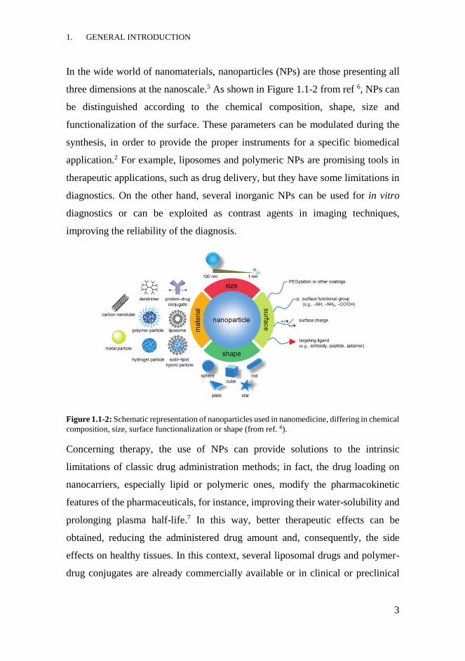
In the wide world of nanomaterials, nanoparticles (NPs) are those presenting all
three dimensions at the nanoscale.5 As shown in Figure 1.1-2 from ref 6, NPs can
be distinguished according to the chemical composition, shape, size and
functionalization of the surface. These parameters can be modulated during the
synthesis, in order to provide the proper instruments for a specific biomedical
application.2 For example, liposomes and polymeric NPs are promising tools in
therapeutic applications, such as drug delivery, but they have some limitations in
diagnostics. On the other hand, several inorganic NPs can be used for in vitro
diagnostics or can be exploited as contrast agents in imaging techniques,
improving the reliability of the diagnosis.
Figure 1.1-2: Schematic representation of nanoparticles used in nanomedicine, differing in chemical
composition, size, surface functionalization or shape (from ref. 6).
Concerning therapy, the use of NPs can provide solutions to the intrinsic
limitations of classic drug administration methods; in fact, the drug loading on
nanocarriers, especially lipid or polymeric ones, modify the pharmacokinetic
features of the pharmaceuticals, for instance, improving their water-solubility and
prolonging plasma half-life.7 In this way, better therapeutic effects can be
obtained, reducing the administered drug amount and, consequently, the side
effects on healthy tissues. In this context, several liposomal drugs and polymer-
drug conjugates are already commercially available or in clinical or preclinical
1. GENERAL INTRODUCTION
4
trials: for instance AmBisome® (Gilead Sciences, USA), a liposomal formulation
of amphotericin B, or Prothecan® (Enzon Pharmaceuticals, USA), a PEG–
camptothecin nanoconjugate, for the treatment of fungal and protozoal infections
or various cancers, respectively.2
Conversely, inorganic NPs such as those made of Au or iron oxide are exploited
for in vivo imaging techniques. Their optical or magnetic features make them good
contrast agents for imaging techniques, providing more spatial and temporal
information and increasing the sensitivity of the diagnostic tool. Moreover, their
joining may often enable multimodal imaging combining different techniques. For
instance, Au NPs can be exploited as contrast agents in X-ray imaging and
computed tomography (CT) while iron oxide superparamagnetic NPs in magnetic
resonance imaging (MRI).8-10
Concerning in vitro diagnostics, inorganic NPs like Au NPs or magnetic NPs are
the most promising.11 The nanoparticle-based diagnostics can offer advantages for
the detection of specific markers, opening new frontiers in the diagnosis of several
pathologies, including cancer, neurological disorders and infectious diseases.12
Through adequate surface functionalization with macromolecules, especially
enzymes and antibodies, interesting bioanalytical applications of NPs can be
achieved, improving the sensitivity of the analytical methods or innovating them.
For example, these kinds of NPs have been commonly employed in biosensors
and immunoassays.13,14 For instance, Au NPs have been used for detecting
biologically relevant molecules in optical and electrochemical analytical
methods.15,16 Magnetic NPs, instead, have been utilized preferentially for
detection and bio-separation of analytes or specific cell populations, like stem
cells or pathogenic bacteria. In fact, by exploiting their magnetic properties, the
application of an external magnetic field allows their efficient separation from a
complex biological matrix.17,18

1. GENERAL INTRODUCTION
5
1.1.1 General features of NPs
NPs show peculiar chemical and physical characteristics which differentiate them
from the corresponding bulk material. These properties can be divided into three
groups: the surface-dependent, the dimension-related and those deriving from the
quantum-size effect.1
The extended surface in the small volume of NPs leads to high surface-to-volume
ratio that affects many of their properties. In fact, compared to 3D materials the
increase in the number of surface atoms, characterized by binding weaker than the
internal atoms, produces changes in the physicochemical properties, such as
melting point and chemical reactivity. Other features like magnetic ones change,
instead, according to the dimensions of the material. For example, ferromagnetic
and ferrimagnetic materials exhibit superparamagnetic behaviour when their size
is nanoscale.19 Finally, the size-dependent quantum effects are related to the
change in the electronic levels of nanomaterials, which become discrete sets
instead of a continuum as in the bulk counterparts. Variations in the optical
properties of Au NPs are due to this phenomenon.20,21
The presence of special intrinsic properties in NPs explains why they have had
great success in the nanotechnological field and, in particular, in nanomedicine.
Moreover, the characteristics of NPs can be adapted to obtain more specific
nanosystems for a selected biomedical application through the surface coating
with chemically different materials and/or the functionalization with biomolecules
or pharmaceuticals.22

1. GENERAL INTRODUCTION
6
1.1.2 Multitasking NPs
In the last years, the scientific community is making considerable efforts to design
and develop multifunctional nanosystems in order to improve and extend the
applicability of the NPs in a theranostic view. A multitasking NP is defined as a
system capable of carrying out different functions at the same time; for instance,
presenting proper functionalities, a single nanoplatform can be suitable for
diagnostic and therapeutic purposes.23 The introduction of these functionalities
can be performed during the synthesis of NPs through an appropriate modification
of their surface with inorganic compounds or organic molecules or
macromolecules which leads to the combination of the specific physical,
chemical, biological or pharmaceutical features deriving from each component.
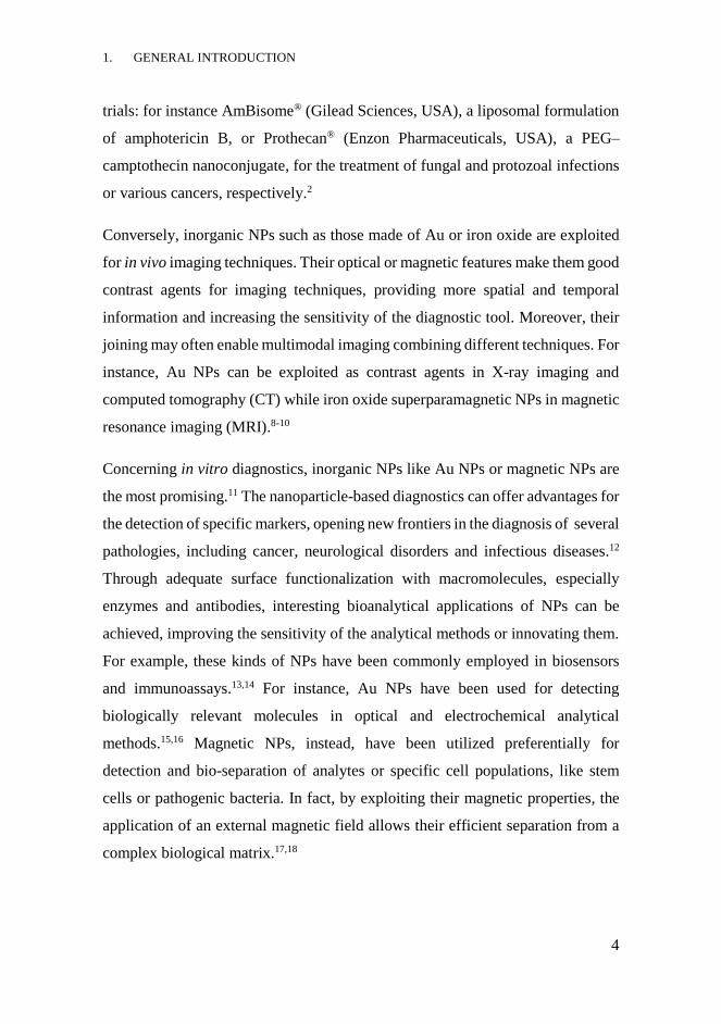
As shown in Figure 1.1-3 from ref. 24, a general multitasking nanosystem for
theranostics contains both functionalities for diagnostic and for therapeutic
purposes. For example, the presence of an inorganic material such as Au or iron
oxide which have peculiar optical or magnetic properties make the NPs suitable
in medical imaging techniques (MRI or optical imaging). Otherwise, the
incorporation of elements or compounds presenting radioactivity or fluorescence,
such as radionuclides, organic fluorescent probes or lanthanides, can help
monitoring the nanosystems during in vivo diagnostic applications.25 Often, a
multimodal imaging using different techniques can also be possible.
At the same time, the presence of biologically or pharmacologically active
compounds, for instance drugs or biomolecules, allows the use of the multitasking
nanosystem as a therapeutic tool. Moreover, the addition of macromolecules or
small molecules, like antibodies or targeting ligands, able to interact with specific
biological target, can selectively address NPs into the pathological area.3,24
1. GENERAL INTRODUCTION
7
Figure 1.1-3: General representation of a multitasking nanosystem for theranostic applications
(from ref. 24). The nanocore can be coated by inorganic or organic shells and functionalized with
molecules or macromolecules acting as targeting, therapeutic or diagnostic agents.
Concerning in vitro diagnostic, the combination of components with peculiar
physical properties, like Au NPs or iron-oxide NPs, with molecules or
macromolecules characterized by high-recognition features, like antibodies or
specific ligands, can offer advantages in the analytical method. For instance,
multitasking NPs can improve the analytical features of the technique, like
sensitivity and specificity; they can allow the performance of multistep procedures
in easier way, facilitating the sample preparation or reducing the time of the
analysis.26,27
1. GENERAL INTRODUCTION
8
MAGNETIC NANOPARTICLES
Among the NPs, the magnetic ones can offer many solutions in nanomedicine,
facilitating, innovating or integrating standard procedures. Being sensitive to the
magnetic field, they can be helpful for in vitro and in vivo diagnostic techniques,
but also in therapeutic applications such as drug delivery and hyperthermia.28,29
The combination of these NPs with other materials, such as Au NPs, liposomes or
polymers, or their modification with biologically relevant molecules are the main
strategies to fulfil the need of efficient medical tools. Engineered magnetic NPs
can therefore represent promising instruments in diagnostics and theranostics and
may contribute to approach the personalized medicine.25,26,30
1.2.1 Magnetite nanoparticles and superparamagnetism
Among the most interesting magnetic nanoparticles (MNPs), iron oxides NPs are
the best known and, in particular, those made of magnetite (Fe3O4) are considered
the most promising for biomedical applications thanks to their special features.
The crystal structure of Fe3O4, as shown in Figure 1.2-1 from ref. 31, consists in a
cubic inverse spinel structure in which O atoms form a close-packed cubic lattice
while the interstitial tetrahedral sites are occupied by Fe3+ ions and the octahedral
ones by Fe3+ and Fe2+ ions.
The synthetic strategies commonly used to produce Fe3O4 MNPs are physical
methods, such as gas-phase deposition and electron beam lithography, or wet
chemical preparations, for instance sol-gel synthesis or chemical co-
precipitation.32-34 The last one is the simplest and most efficient synthesis method
to obtain Fe3O4 MNPs. In the absence of surface coatings or functionalization,
Fe3O4 MNPs are unstable since they tend to oxidize and aggregate. The
transformation in hematite (α-Fe2O3) or maghemite (ϒ-Fe2O3) or the increase of

1. GENERAL INTRODUCTION
9
the particle dimensions produce changes in the magnetic properties of the
nanomaterials, limiting or compromising their biomedical use. 34
Figure 1.2-1: On the left: schematic representation of the inverse spinel structure of magnetite,
highlighting Fe and O atoms in red and blue, respectively (from ref. 31). Fe3+ ions occupied the
interstitial octahedral and tetrahedral sites while Fe2+ ions only the octahedral ones. On the right:
representation of the magnetization M vs applied field H plot for materials showing different
magnetic properties (from ref. 28).
1.2.1.1 Magnetic properties
The materials can be classified in paramagnetic, diamagnetic or
ferromagnetic/ferrimagnetic depending on their response to an applied external
magnetic field (H), and according to the value of the magnetic susceptibility (χ).
This parameter represents the proportional constant between the magnetization of
the material (M) and H, according to the equation M= χH. The values of χ > 0
characterize paramagnetic materials, χ < 0 the diamagnetic ones while χ >> 0 the
ferro/ferrimagnetic materials.
Figure 1.2-1 from ref. 28 shows the relation between M and H. From the graph M
vs H, the parameters saturation magnetization (Msat), remanence (Mr) and
coercivity (Hc) can be obtained. Msat corresponds to the highest M value
obtainable during the application of H. Mr represents the M value once H is absent
while Hc is the intensity of the applied H required to reduce Msat to zero.
1. GENERAL INTRODUCTION
10
Among the magnetic properties, ferromagnetism is considered a cooperative
behaviour, since only a certain number of atoms bound together can display it.
These materials possess unpaired electrons spins which align spontaneously in the
same direction (ferromagnetic) or in the opposite one (antiferromagnetic),
exhibiting magnetization in the absence of a magnetic field. When the number of
the antiparallel aligned magnetic moments is not equal, an overall magnetization
is present, and the material is classified as ferrimagnetic. Concerning Fe3O4, its
ferrimagnetic character derives from the antiferromagnetic coupling between the
Fe3+ ions which allows the magnetic moments of Fe2+ ions to generate the
magnetization of the unit cell. Ferromagnetic and ferrimagnetic materials exhibit
permanent magnetization once H is removed and they are characterized by the
presence of the hysteresis loop into the M vs H representation. However, when
the dimensions of the particles decrease up to a particular domain, each particle
comprises a single magnetic domain and the magnetic behaviour changes from
ferro/ferrimagnetism to superparamagnetism. For instance, in MNPs a size < 20
nm is required to have superparamagnetism while 128 nm is the critical dimension
for forming multi-domain structures in spherical MNPs which leads to the
decrease in the Hc value.35 The main difference between ferro/ferrimagnetic and
superparamagnetic material is that the second does not remain magnetized once
the external magnetic field is removed, like a paramagnetic one.
Superparamagnetic materials do not show magnetic hysteresis and their value of
magnetic susceptibility is higher than that of paramagnetic materials and quite
similar to ferromagnetic ones.19
1. GENERAL INTRODUCTION
11
BIOMEDICAL APPLICATIONS OF MNPs
The pharmaceutical and biomedical applications of Fe3O4 MNPs cover all the
medical fields. The general features of NPs and the peculiar magnetic properties,
biocompatibility and low cytotoxicity of Fe3O4 MNPs make them suitable for
diagnostic, imaging and/or therapy. For in vivo applications they should have low
dimensions and narrow size distribution, to avoid the rapid clearance, and high
magnetization.2,36 In this context, superparamagnetism is fundamental to avoid
magnetically-driven particle agglomeration, after the removal of the magnetic
field; in fact, the presence of aggregates can lead to occlusion of capillary vessels
and dangerous side effects. To improve the chemical stability and
biocompatibility of Fe3O4 MNPs surface functionalization is often required.37
Engineered Fe3O4 MNPs are thus innovating standard medical strategies,
improving their sensitivity and reliability, and are enabling new applications,
previously unimaginable, especially in therapeutic treatments.
As depicted in Figure 1.3-1 from ref. 38, Fe3O4 MNPs are providing their benefits
also in in vitro diagnostics in several bioanalytical methods, like biosensing
techniques, in the separation and sample-enrichment of analytes or in the
immobilization of macromolecules.18,26,39 In vivo Fe3O4 MNPs are used as contrast
agents in MRI, while their high potential as drug delivery systems or in
hyperthermia has also been studied.29,36 They can be used alone or in combination
with other nanomaterials, allowing the joining of different properties and the
widening of their applicability.40,41 The biomedical applications of Fe3O4 MNPs
are described in more details in the following sections.

1. GENERAL INTRODUCTION
12
Figure 1.3-1: Schematic representation of biomedical applications of magnetic nanoparticles (from
ref. 38). MNPs can be exploited in therapeutic applications like magnetic hyperthermia and drug
delivery or in in vivo imaging as contrast agents for MRI. Their main uses in in vitro diagnostics
concern cell labelling and magnetic separation and immobilization of macromolecules.
1.3.1 In vivo imaging
A proper and reliable imaging of internal organs is fundamental for allowing the
further appropriate medical treatments; in this area, MRI is one of the mainly used
imaging techniques since it produces images with good temporal and spatial
resolution without performing invasive procedures. In fact, MRI is very helpful in
discriminating between healthy and affected tissue in case of several pathologies,
related especially to soft tissues and cartilages. MRI is based on the achievement
of relaxation signals of proton spins corresponding to water molecules in the
human body, after the excitation by radiofrequency waves in a strong magnetic
field.
Contrast agents, generally based on paramagnetic materials such as Gd, are used
for clarifying the collected images and promoting a better diagnosis. Fe3O4 MNPs
1. GENERAL INTRODUCTION
13
are providing the development of a new generation of contrast agents; in fact,
thanks to their superparamagnetic behaviour these MNPs are able to generate
strong contrast effect and consequently excellent sensitivity in T2-weighted
images (T2= spin-spin relaxation time), improving image resolution.9,39,42 In this
context, several formulations of polymer-coated/stabilized superparamagnetic
nanoparticles of iron oxides (SPION) are already commercially available as MRI
contrast agents, for instance Resovist® (Bayer HealthCare Pharmaceuticals, USA)
or Feridex I.V.® (AMAG Pharmaceuticals Inc., USA) for liver, spleen and bone
marrow imaging.42
1.3.2 Therapy
Concerning therapeutic applications, Fe3O4 MNPs offer the possibility to achieve
stimuli-sensitive drug nanocarriers because of the magnetic field responsiveness
typical of these nanosystems. In fact, by applying an extracorporeal magnetic field
the remote magnetic focusing of the engineered Fe3O4 MNPs to a desired area can
be achieved.43 The simultaneous presence of active targeting agents can help
facilitating the interaction with affected tissue or cell. To develop a good drug
delivery system, the engineering of Fe3O4 MNPs is generally required; in this way
high drug loading can be obtained together with the improvement of
physicochemical and biological features of Fe3O4 MNPs, for instance their
chemical stability and half-life.38 Drug delivery systems based on engineered
Fe3O4 MNPs or incorporating them have been developed for magnetically guided
chemotherapy, radioimmunotherapy, photodynamic therapy or
magnetofection.40,44,45 Due to the opsonization phenomenon, the intravenous
administration of these nanosystems limits their applicability since it leads to rapid
clearance, as happened for several kinds of NPs.46,47 A more recent strategy,
known as cell-mediated delivery, consists in exploiting immune cells loaded with
engineered NPs, often functionalized with drugs, for transporting and delivering
1. GENERAL INTRODUCTION
14
the nanosystems in the pathological area.48 Moreover, it has been demonstrated
that cells engulfed with Fe3O4 MNPs acquired magnetic field responsiveness.49
In cancer therapy, Fe3O4 MNPs can be exploited also for magnetic hyperthermia;
this therapeutic strategy consists in killing cancer cells by exposing them to high
temperatures (42-46 °C).41,50 In this case, Fe3O4 MNPs represent the source of heat
since they can be heated by alternating magnetic field. When Fe3O4 MNPs are
located in the tumour area, the exposition to an external alternating magnetic field
generate heat locally in the tumour, thus causing the preferential death of
malignant cells. The heat production depends on several factors such as the
physicochemical properties of Fe3O4 MNPs or their functionalization or
parameters of the magnetic field. Several examples for thermal treatment of
different kinds of cancer can be found in the literature.29,51,52
Finally, the combination of drug delivery and hyperthermia results in better
therapeutic treatments while the matching of one or both of these strategies with
the use in MRI imaging, make Fe3O4 MNPs highly powerful theranostic
tools.40,50,53,54
1.3.3 In vitro diagnostics
The main applications of Fe3O4 MNPs in in vitro diagnostics concern cell labelling
and separation, detection of microorganisms or biologically-relevant molecules,
protein immobilization, bioanalysis and immunoassays.12,55 For instance, the
selective recognition of a specific kind of cell by Fe3O4 MNPs allows the further
magnetic separation from a complex matrix; in this way it is possible to prepare
enriched samples suitable for other analysis.56 For performing the detection or the
labelling of cells, microorganisms or other molecules, Fe3O4 MNPs have to be
chemically modified introducing a functionality able to recognize the target.
Antibodies (Ab) are very commonly used for this purpose thanks to their selective

1. GENERAL INTRODUCTION
15
binding capacity. In this context, several Fe3O4 MNPs-Ab have been developed
for the in vitro recognition and separation of cancer cells, stem cells or pathogenic
bacteria or biochemicals, such as proteins or small molecules.57-60
Thanks to the possible easy and quick recovery, Fe3O4 MNPs immobilizing
enzymes or proteins have been developed without dramatically affecting the
catalytic activity or the structure of the macromolecules. Moreover, the general
features of NPs improve the sensitivity of these enzyme-immobilized platforms,
reducing also the operational costs.61,62 Finally, in medical and clinical diagnostics
Fe3O4 MNPs can be used as solid support also for the immobilization of
biomarkers or antibodies able to detect specific ligands. Fe3O4 MNPs-Ab can
therefore be exploited in immunoassays, such as Enzyme Linked ImmunoSorbent
Assay (ELISA) or NPs-based bio-barcode assays, or in biosensors for the
detection and quantification of small molecules or biomolecules in complex
biological matrices.63-65


2. AIM AND STRUCTURE OF THIS
THESIS
2. AIM AND STRUCTURE OF THIS THESIS
18
Objectives and scientific strategy
The main aim of this thesis has been the development of new multifunctional
superparamagnetic nanosystems for biomedical applications in the field of therapy
and in vitro diagnostics. Starting from the favourable features of Fe3O4 MNPs,
which make them already suitable for several purposes, the design of core-shell
structured nanosystems has been accomplished to increase the potential
applicability. In fact, covering the superparamagnetic core with inert and
biocompatible inorganic materials leads to an increase in the chemical stability of
the entire nanosystem. Moreover, in this way the surface functionalization can be
easily carried out according to the chemical features of the external shell.
Two kinds of core-shell nanosystems have been firstly developed in this research
work and then engineered depending on the planned end use. In fact, the
introduction of other functionalities on the MNPs deriving from organic coatings,
and the loading of bioactive molecules or macromolecules, like proteins or drugs,
lead to the production of more efficient and specific nanosystems towards the
biological targets.
Thus, the specific objectives proposed in this research work are:
- The development of core-shell nanosystems made of Fe3O4@Cu@Au
MNPs and Fe3O4@SiO2 MNPs as the starting points for the production of
engineered multitasking MNPs for different biomedical applications.
- The achievement of new nanosystems loading chemotherapeutic drugs or
antimicrobial compounds for therapeutic purposes and the in vitro
evaluation of their activity on cancer cell lines or microbial cultures.
- The study of internalization of differently coated MNPs into human
immune cells for contributing on investigating the therapeutic approach
based on cell-mediated drug delivery.
2. AIM AND STRUCTURE OF THIS THESIS
19
- The development of innovative immunoassays and immunosensors
involving MNPs-Ab for in vitro diagnostic purposes.
Thesis structure
After the brief general introduction presented in chapter 1 on MNPs for
biomedical purposes, the production and physicochemical characterization of the
two nanosystems Fe3O4@Cu@Au MNPs and Fe3O4@SiO2 MNPs, on which this
thesis is based, are explained in chapter 3. The several engineered nanosystems
developed starting from one or both of the basic inorganic nanostructures and their
in vitro applications are described in the following chapters.
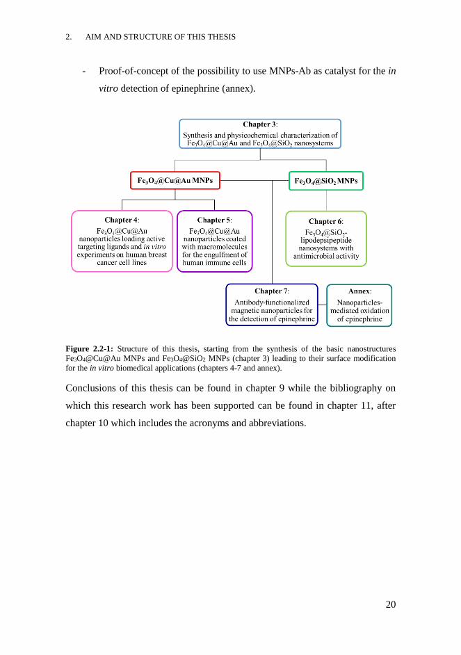
As shown in Figure 2.2-1 the central sections of this thesis deal with:
- The development of Fe3O4@Cu@Au nanosystems loading the bioactive
targeting molecules, folic acid and methotrexate, and the in vitro
interaction with the human breast cancer cell lines MCF-7 and MDA-MB-
231 (chapter 4).
- The coating of Fe3O4@Cu@Au MNPs with homo-polypeptides or
proteins and the evaluation of their uptake by monocyte-differentiated
macrophages (chapter 5).
- The functionalization of Fe3O4@SiO2 MNPs with two bacterial secondary
metabolites, syringomycin E and syringopeptin 22A, and the study of the
antimicrobial activity on yeast cultures and the internalization into
monocyte-differentiated macrophages (chapter 6).
- The evaluation of new immunoreagents for the detection of epinephrine
by standard ELISA assays and the implementation of the diagnostic
platforms using MNPs-Ab for the development of magneto-ELISAs and
amperometric magneto-immunosensors (chapter 7).
2. AIM AND STRUCTURE OF THIS THESIS
20
- Proof-of-concept of the possibility to use MNPs-Ab as catalyst for the in
vitro detection of epinephrine (annex).
Figure 2.2-1: Structure of this thesis, starting from the synthesis of the basic nanostructures
Fe3O4@Cu@Au MNPs and Fe3O4@SiO2 MNPs (chapter 3) leading to their surface modification
for the in vitro biomedical applications (chapters 4-7 and annex).
Conclusions of this thesis can be found in chapter 9 while the bibliography on
which this research work has been supported can be found in chapter 11, after
chapter 10 which includes the acronyms and abbreviations.
3. SYNTHESIS AND
PHYSICOCHEMICAL
CHARACTERIZATION OF
Fe3O4@Cu@Au AND Fe3O4@SiO2
NANOSYSTEMS

3. SYNTHESIS AND PHYSICOCHEMICAL CHARACTERIZATION OF Fe3O4@Cu@Au
AND Fe3O4@SiO2 NANOSYSTEMS
22
INTRODUCTION
This chapter deals with the development of the nanosystems used in this thesis,
corresponding to Fe3O4@Cu@Au MNPs and Fe3O4@SiO2 MNPs. The limits of
using bare Fe3O4 MNPs, deriving from their vulnerability to oxidation and
spontaneous aggregation process, can be minimized planning a core-shell
structure. The ideas behind the development of these inorganic nanosystems, in
addition to the synthetic processes and their physicochemical characterization are
illustrated in this section.
3.1.1 Core-shell nanoparticles
The development of nanosystems with a core-shell structure extends the use of
the NPs due to the improvement of some of their features.66 This structure is
characterized by the presence of an internal nucleus (core) and one or more layers
made of other materials (shell). These kinds of NPs are able to greatly improve
the chemical stability or biocompatibility of the material constituting the core,
thus expanding their applicability in the biomedical field. The choice of the shell
composition generally depends to the end use of the NP.24
Concerning the core-shell MNPs, Fe3O4 almost always constitutes the core of the
system and it is enclosed inside one or more shells of organic or inorganic
materials, as shown in Figure 3.1-1. The advantages
of a core-shell magnetic nanostructure derive from
the possibility to combine the properties of the core,
in this case superparamagnetism, with other
physicochemical features of the external material.67
The outer coating preserves Fe3O4 from the
oxidation of Fe2+ ions, that would alter the structure
Figure 3.1-1: General scheme
of a Fe3O4 MNPs with core-
shell structure.
3. SYNTHESIS AND PHYSICOCHEMICAL CHARACTERIZATION OF Fe3O4@Cu@Au
AND Fe3O4@SiO2 NANOSYSTEMS
23
and thus the magnetic properties. By modulating the chemical composition of the
coating, it is possible to improve the stability and dispersibility of Fe3O4 MNPs,
also minimizing the formation of aggregates. The presence of shells made of
inorganic compounds can add or enhance the properties of the entire nanosystem,
such as the optical, the catalytic or the electrical ones.37 Furthermore, by using
proper coating materials the drug loading capacity or the biocompatibility of the
nanosystem can be improved, facilitating the more effective interaction with
biological systems and providing more powerful drug delivery systems.68
Conversely, the presence of a coating material having peculiar properties, such as
radioactivity, fluorescence or absorption, makes those nanosystems active in more
than one medical diagnostic technique.69-71 Among the materials selected for
covering Fe3O4 MNPs, gold and silica are the most commonly used due to the
chemical inertia and biocompatibility. These materials provide the stability of the
MNPs in solution, limiting their spontaneous aggregation, and help to covalently
bind various molecules or biomolecules suitable for the final biomedical
applications.38
3.1.1.1 Silica shell
The chemical inertness and stability are interesting features of silica (SiO2),
making it an optimal material for covering Fe3O4 MNPs. Compared to the bare
magnetite nanosystems, silica-coated magnetite MNPs (Fe3O4@SiO2 MNPs) are
much more stable in solution since the shell protects them from oxidation and
limits their aggregation process; in fact, silica coating is able to shield the
magnetic dipole interactions and increase the surface negative charges on Fe3O4
MNPs, enhancing their electrostatic repulsion.37
Although SiO2 NPs, especially those made of mesoporous SiO2 72, are very
promising materials for drug delivery or generally in nanomedicine, the
combination with a superparamagnetic core offers advantages in focusing,
3. SYNTHESIS AND PHYSICOCHEMICAL CHARACTERIZATION OF Fe3O4@Cu@Au
AND Fe3O4@SiO2 NANOSYSTEMS
24
monitoring and revealing them during in vivo administration.73 Fe3O4@SiO2
MNPs can be synthesized by different procedures among which the Stöber method
is the most commonly used for the achievement of an homogeneous silica shell.74
Furthermore, the presence of a SiO2 layer facilitates the surface modification of
the Fe3O4@SiO2 MNPs with heterobifunctional molecules exploitable as linkers
for the conjugation of biologically relevant compounds. In fact, the presence of
surface silanol groups ensures high reactivity towards alcohols or silane coupling
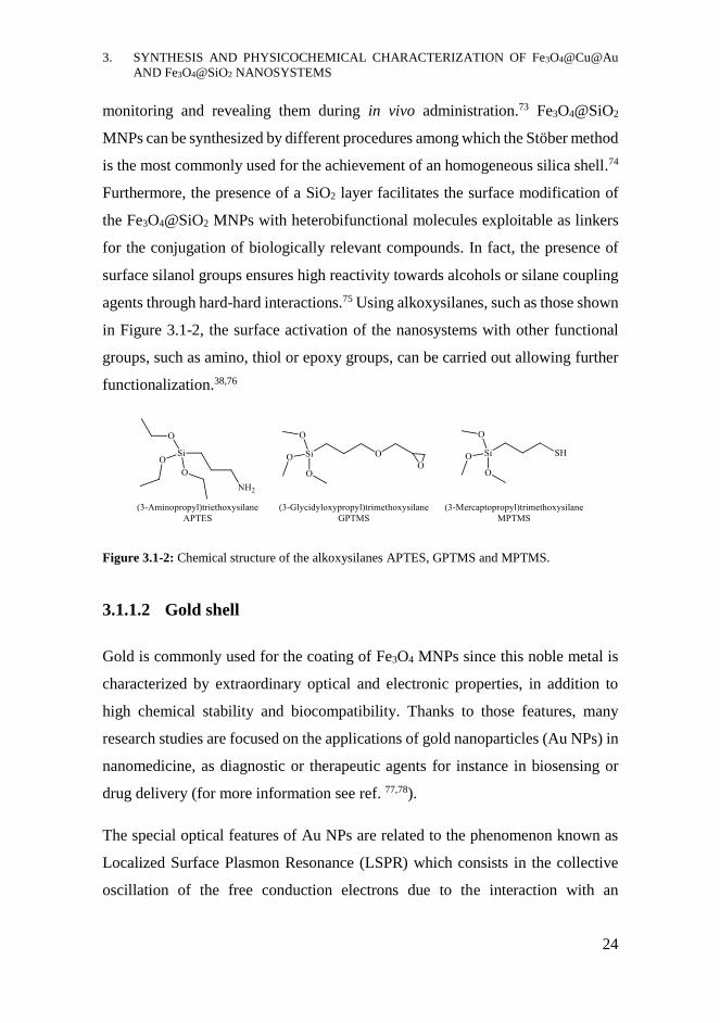
agents through hard-hard interactions.75 Using alkoxysilanes, such as those shown
in Figure 3.1-2, the surface activation of the nanosystems with other functional
groups, such as amino, thiol or epoxy groups, can be carried out allowing further
functionalization.38,76
Figure 3.1-2: Chemical structure of the alkoxysilanes APTES, GPTMS and MPTMS.
3.1.1.2 Gold shell
Gold is commonly used for the coating of Fe3O4 MNPs since this noble metal is
characterized by extraordinary optical and electronic properties, in addition to
high chemical stability and biocompatibility. Thanks to those features, many
research studies are focused on the applications of gold nanoparticles (Au NPs) in
nanomedicine, as diagnostic or therapeutic agents for instance in biosensing or
drug delivery (for more information see ref. 77,78).
The special optical features of Au NPs are related to the phenomenon known as
Localized Surface Plasmon Resonance (LSPR) which consists in the collective
oscillation of the free conduction electrons due to the interaction with an
3. SYNTHESIS AND PHYSICOCHEMICAL CHARACTERIZATION OF Fe3O4@Cu@Au
AND Fe3O4@SiO2 NANOSYSTEMS
25
electromagnetic wave. The oscillation frequency normally occurs in the visible
region and its changes can be monitored following the corresponding strong
absorption band.20 This phenomenon is very sensitive to the composition, the size,
the shape and the environment of Au NPs and, for these reasons, it is commonly
used for detecting and studying surface binding processes.79 Moreover, the optical
properties of Au are exploited for a promising therapeutic application for cancer
therapy known as phototherapy, based on the local heating upon excitation of the
surface plasmon oscillations, which can cause cancer cell death.80
Thus, the coating of the F3O4 core with an Au layer produces an enhancement of
the stability and inertness of Fe3O4, preventing its oxidation and maintaining the
superparamagnetic properties. Moreover, the presence of a Au shell gives the
possibility to match the magnetic properties of the core with the optical features
of Au, leading to nanosystems suitable for theranostics; for instance, NPs active
at the same time in MRI, hyperthermia and photothermal therapy can be
achieved.80 In addition to the improvement of biocompatibility, the Au shell
ensures the surface modification of the nanosystem via gold-thiol chemistry.38
3.1.1.3 Functionalization of gold shell
The Au shell offers the opportunity to easily functionalize the NPs exploiting soft-
soft interactions, occurring between the soft acid Au and the soft base S.81 This
interaction leads to stable bonds (binding energy of S-Au bond = 45 kcal/mol)
ensuring the covalent but quite easily breakable anchorage of different kinds of
molecules.82 The self-assembled monolayers (SAMs) of thiolate-Au can be
achieved by using monothiol (RSH), thioether (RSR’) or disulfide (RSSR’)
molecules containing alkyl or aryl groups, which general structure is reported in
Figure 3.1-3. SAMs may also be made of two or more linkers with different
functional groups, enabling further functionalization with several kinds of
molecules and biomolecules.83

3. SYNTHESIS AND PHYSICOCHEMICAL CHARACTERIZATION OF Fe3O4@Cu@Au
AND Fe3O4@SiO2 NANOSYSTEMS
26
Figure 3.1-3: General structure of the organosulfur compound thiol, thioether, disulfide and cyclic
disulfide. The general structure of thiol-terminated polyethylene glycol is also reported.
Thiol-terminated polyethylene glycol (thiol-PEG), which structure is shown in
Figure 3.1-3, is one of the most commonly used polymeric coating for the
functionalization of Au shell, since it leads to an increase of the in vitro and in
vivo stability of the NPs.84 The PEGylation of the nanosystems improves the
dispersibility and the biocompatibility, avoiding the non-specific binding to
protein and cells present in the vascular system during the in vivo administration
of the NPs.85 The use of heterobifunctional PEG molecules containing, on one
side, the thiol group and, on the other side, another functional group, like amino
or carboxyl group, enables to form covalent bonds with both the Au surface and
other biomolecules or bioactive compounds, such as enzymes, antibodies or drugs.
Mixed SAMs made of heterobifunctional-PEG molecules alternating reactive and
non-reactive groups, for instance thiol-PEG containing -COOH or -OCH3, can be
a good option for the further functionalization with proteins; in fact, the reduction
of the surface charge density may promote the binding of the macromolecule.86
The functionalization of Au surface can also be performed with short-length thiols
or sulfides, for instance a heterobifunctional molecule carrying a thiol and a
carboxylic acid group. In this case, choosing molecules containing a disulfide
group instead of a thiol can avoid the formation of disordered and unstable
structures. Moreover, cyclic disulfide can produce a more stable coating compared
to acyclic disulfides since the anchorage occurs through two S atoms.87
3. SYNTHESIS AND PHYSICOCHEMICAL CHARACTERIZATION OF Fe3O4@Cu@Au
AND Fe3O4@SiO2 NANOSYSTEMS
27
3.1.1.4 Fe3O4@Au nanoparticles
Although several examples of Fe3O4@Au nanosystems can be found in the
literature, the production of core-shell Fe3O4@Au MNPs with a uniform and
homogeneous Au layer is not easy.88,89 This problem arises from the fact that Au
provides a defective and incomplete coating of Fe3O4; in fact, Au reacts with soft
ligands while the magnetic core exposes the hard -OH groups that have low
affinity for Au. For these reasons, the Au deposition on the iron oxide surface is
extremely challenging since the noble metal tends to form separated NPs. The
direct synthesis of core-shell Fe3O4@Au MNPs has been achieved by a reverse-
micelle approach, which present some limitations due to low yield and poor
reproducibility.90,91
An interesting strategy to overcome these problems and obtain a uniform coating
was developed in the research group in which this thesis was carried out,
depositing an intermediate Cu shell between the Fe3O4 core and Au layer.
3.1.1.5 Copper shell
Cu has high chemical affinity for Au, as proved by their ability to give alloys, that
may guarantee the deposition of a homogeneous Au layer on Fe3O4 MNPs, and it
can easily be deposited on magnetite surface by simple redox reactions.92,93 Thus,
the Cu layer on one hand protects Fe3O4 from oxidation and, on the other hand,
can be exploited for the Au deposition through a direct redox reaction. Moreover,
the presence of Cu enhances some physicochemical features of the nanosystems,
especially in terms of catalytic ability.94
Therefore, the development of core-shell nanosystems made of Fe3O4@Cu or the
production of superparamagnetic NPs loading copper ions can provide advantages
in biomedical applications. In fact, nanosystems able to act as contrast agents in
both MRI and positron emission tomography (PET) can be achieved using the
3. SYNTHESIS AND PHYSICOCHEMICAL CHARACTERIZATION OF Fe3O4@Cu@Au
AND Fe3O4@SiO2 NANOSYSTEMS
28
mixture of 63Cu and the radionuclide 64Cu. The integration of these techniques
allows to highly improve the diagnosis of pathologies, in pre-clinical and clinical
applications, since it matches the high resolution of MRI with the sensitivity of
PET.70,95
CHAPTER OBJECTIVES
Since the central aim of this thesis is the development of new superparamagnetic
nanosystems for biomedical applications, the synthesis and physicochemical
characterization of the basic inorganic structures have been the first steps to
address.
The nucleus made of Fe3O4 is the central part of two kinds of core-shell
nanosystems differing in the materials that constitute the external shells. The core-
shell structure has been selected in order to protect Fe3O4 from oxidation,
maintaining its superparamagnetic features, and to improve the chemical stability
and biocompatibility of the nanosystems. The choice of producing two kinds of
nanosystems instead of one derives from the possibility to easily achieve different
surface functionalization exploiting the specific chemical reactivity of the
materials constituting the shells. In fact, selecting, from one hand, the noble metal
Au and, from the other hand, the inert material SiO2, the surface functionalization
can be based on soft-soft or hard-hard interactions, respectively. In this way it is
possible to covalently attach molecules bringing completely different functional
groups, avoiding their derivatization, and also to modulate the strength of the bond
between MNPs and surface coating depending on the biomedical end use.
3. SYNTHESIS AND PHYSICOCHEMICAL CHARACTERIZATION OF Fe3O4@Cu@Au
AND Fe3O4@SiO2 NANOSYSTEMS
29
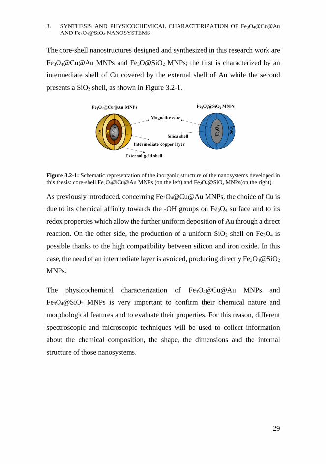
The core-shell nanostructures designed and synthesized in this research work are
Fe3O4@Cu@Au MNPs and Fe3O@SiO2 MNPs; the first is characterized by an
intermediate shell of Cu covered by the external shell of Au while the second
presents a SiO2 shell, as shown in Figure 3.2-1.
Figure 3.2-1: Schematic representation of the inorganic structure of the nanosystems developed in
this thesis: core-shell Fe3O4@Cu@Au MNPs (on the left) and Fe3O4@SiO2 MNPs(on the right).
As previously introduced, concerning Fe3O4@Cu@Au MNPs, the choice of Cu is
due to its chemical affinity towards the -OH groups on Fe3O4 surface and to its
redox properties which allow the further uniform deposition of Au through a direct
reaction. On the other side, the production of a uniform SiO2 shell on Fe3O4 is
possible thanks to the high compatibility between silicon and iron oxide. In this
case, the need of an intermediate layer is avoided, producing directly Fe3O4@SiO2
MNPs.
The physicochemical characterization of Fe3O4@Cu@Au MNPs and
Fe3O4@SiO2 MNPs is very important to confirm their chemical nature and
morphological features and to evaluate their properties. For this reason, different
spectroscopic and microscopic techniques will be used to collect information
about the chemical composition, the shape, the dimensions and the internal
structure of those nanosystems.
3. SYNTHESIS AND PHYSICOCHEMICAL CHARACTERIZATION OF Fe3O4@Cu@Au
AND Fe3O4@SiO2 NANOSYSTEMS
30
RESULTS AND DISCUSSION
The experimental results concerning the synthetic strategies for the achievement
of Fe3O4@Cu@Au MNPs and Fe3O4@SiO2 MNPs and their physicochemical
characterization by means of spectroscopic and microscopic techniques are
presented and discussed in this section.
3.3.1 Fe3O4@Cu@Au MNPs
3.3.1.1 Synthesis of the core: Fe3O4 MNPs
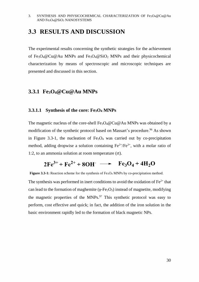
The magnetic nucleus of the core-shell Fe3O4@Cu@Au MNPs was obtained by a
modification of the synthetic protocol based on Massart’s procedure.96 As shown
in Figure 3.3-1, the nucleation of Fe3O4 was carried out by co-precipitation
method, adding dropwise a solution containing Fe2+/Fe3+, with a molar ratio of
1:2, to an ammonia solution at room temperature (rt).
The synthesis was performed in inert conditions to avoid the oxidation of Fe2+ that
can lead to the formation of maghemite (γ-Fe2O3) instead of magnetite, modifying
the magnetic properties of the MNPs.97 This synthetic protocol was easy to
perform, cost effective and quick; in fact, the addition of the iron solution in the
basic environment rapidly led to the formation of black magnetic NPs.
Figure 3.3-1: Reaction scheme for the synthesis of Fe3O4 MNPs by co-precipitation method.
3. SYNTHESIS AND PHYSICOCHEMICAL CHARACTERIZATION OF Fe3O4@Cu@Au
AND Fe3O4@SiO2 NANOSYSTEMS
31
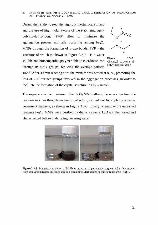
During the synthetic step, the vigorous mechanical stirring
and the use of high molar excess of the stabilizing agent
polyvinylpirrolidone (PVP) allow to minimize the
aggregation process normally occurring among Fe3O4
MNPs through the formation of μ-oxo bonds. PVP – the
structure of which is shown in Figure 3.3-2 - is a water
soluble and biocompatible polymer able to coordinate iron
through its C=O groups, reducing the average particle
size.98 After 30 min reacting at rt, the mixture was heated at 80°C, promoting the
loss of -OH surface groups involved in the aggregation processes, in order to
facilitate the formation of the crystal structure in Fe3O4 nuclei.
The superparamagnetic nature of the Fe3O4 MNPs allows the separation from the
reaction mixture through magnetic collection, carried out by applying external
permanent magnets, as shown in Figure 3.3-3. Finally, to remove the unreacted
reagents Fe3O4 MNPs were purified by dialysis against H2O and then dried and
characterized before undergoing covering steps.
Figure 3.3-3: Magnetic separation of MNPs using external permanent magnets. After few minutes
from applying magnets the black solution containing MNPs (left) becomes transparent (right).
Figure 3.3-2:
Chemical structure of
polyvinylpirrolidone

3. SYNTHESIS AND PHYSICOCHEMICAL CHARACTERIZATION OF Fe3O4@Cu@Au
AND Fe3O4@SiO2 NANOSYSTEMS
32
3.3.1.2 Physicochemical characterization of Fe3O4 MNPs
Since the development and applications of magnetic nanosystems based on
Fe3O4@Cu@Au MNPs is the central topic of the research group led by Prof.
Mario Barteri, in which this thesis was carried out, several experimental data on
the physicochemical characterization of this kind of nanosystem had already been
collected and/or published.99-101
The magnetic properties of Fe3O4 MNPs studied by means of vibrating sample
magnetometer (VSM) were reported in a previous doctoral thesis, confirming the
superparamagnetic features of these MNPs, since the measured Ms was of 71.6 ±
0.2 emu/g, comparable to the reference value of ≈ 80 emu/g.99 The analysis
performed by means of atomic force microscopy (AFM) demonstrated the
nanometric dimensions and spherical shape of Fe3O4 MNPs, showing an average
diameter of 15 nm, but also the presence of larger aggregates.
The X-ray powder diffraction (XRPD) measurements made it possible to study
the crystal nature of the Fe3O4 MNPs. As shown in the diffractogram in Figure
3.3-4, the peaks belonging to Fe3O4 crystals were identified, using reference data
(JCPDS file No. 88-0315), and the corresponding Miller indices were reported.
The broadness of the diffraction peaks may be related to the nanometric size of
the particles while the significant background noise may be due to the presence of
a not-negligible portion of amorphous materials.
3. SYNTHESIS AND PHYSICOCHEMICAL CHARACTERIZATION OF Fe3O4@Cu@Au
AND Fe3O4@SiO2 NANOSYSTEMS
33
Figure 3.3-4: XRPD spectrum of Fe3O4 MNPs including Miller indices corresponding to Fe3O4.
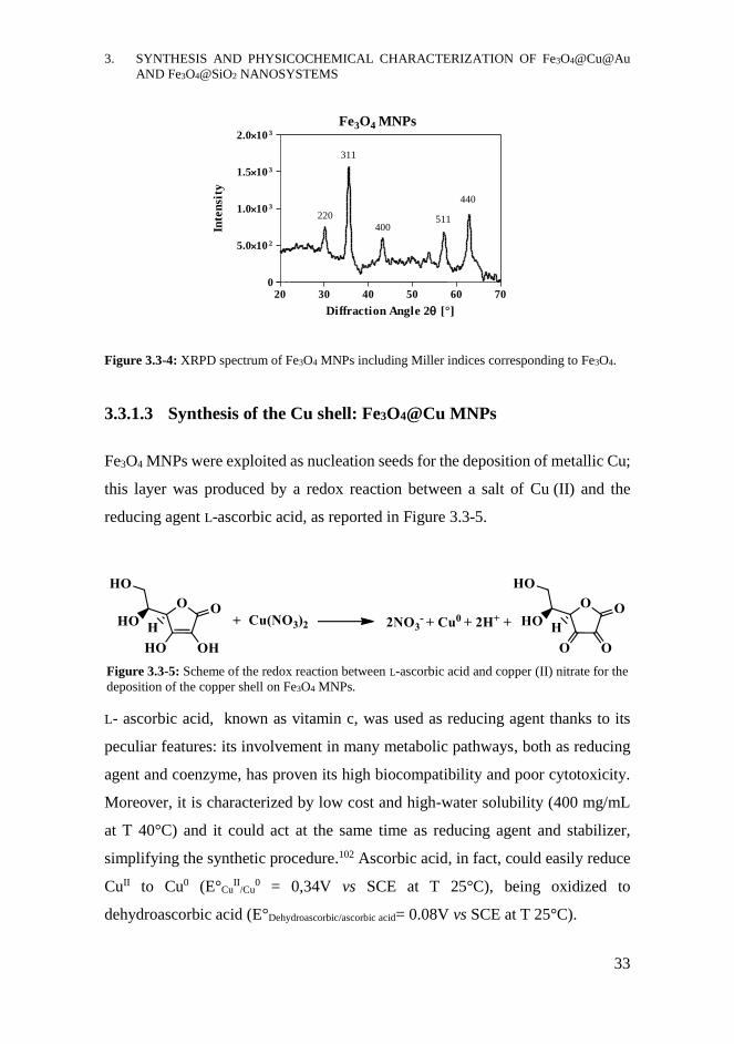
3.3.1.3 Synthesis of the Cu shell: Fe3O4@Cu MNPs
Fe3O4 MNPs were exploited as nucleation seeds for the deposition of metallic Cu;
this layer was produced by a redox reaction between a salt of Cu (II) and the
reducing agent L-ascorbic acid, as reported in Figure 3.3-5.
L- ascorbic acid, known as vitamin c, was used as reducing agent thanks to its
peculiar features: its involvement in many metabolic pathways, both as reducing
agent and coenzyme, has proven its high biocompatibility and poor cytotoxicity.
Moreover, it is characterized by low cost and high-water solubility (400 mg/mL
at T 40°C) and it could act at the same time as reducing agent and stabilizer,
simplifying the synthetic procedure.102 Ascorbic acid, in fact, could easily reduce
CuII to Cu0 (E°CuII
/Cu0 = 0,34V vs SCE at T 25°C), being oxidized to
dehydroascorbic acid (E°Dehydroascorbic/ascorbic acid= 0.08V vs SCE at T 25°C).
Fe3O4 MNPs
20 30 40 50 60 700
5.010 2
1.010 3
1.510 3
2.010 3
220
311
400511
440
Diffraction Angle 2 [°]
Inte
nsi
ty
Figure 3.3-5: Scheme of the redox reaction between L-ascorbic acid and copper (II) nitrate for the
deposition of the copper shell on Fe3O4 MNPs.
3. SYNTHESIS AND PHYSICOCHEMICAL CHARACTERIZATION OF Fe3O4@Cu@Au
AND Fe3O4@SiO2 NANOSYSTEMS
34
The reaction was mechanically stirred for 1 h at 50 °C and Fe3O4@Cu MNPs were
purified by dialysis against H2O and finally dried. The same coating protocol was
performed twice in order to improve the homogeneity of the Cu layer.
3.3.1.4 Physicochemical characterization of Fe3O4@Cu MNPs
The physicochemical characterization of Fe3O4@Cu MNPs was performed by
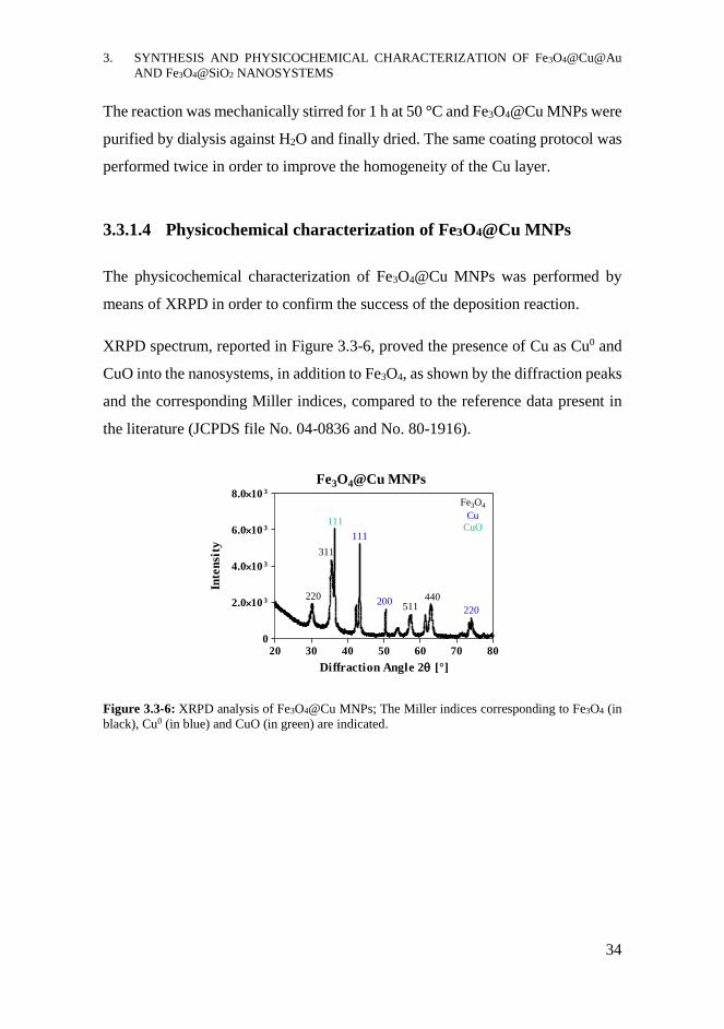
means of XRPD in order to confirm the success of the deposition reaction.
XRPD spectrum, reported in Figure 3.3-6, proved the presence of Cu as Cu0 and
CuO into the nanosystems, in addition to Fe3O4, as shown by the diffraction peaks
and the corresponding Miller indices, compared to the reference data present in
the literature (JCPDS file No. 04-0836 and No. 80-1916).
Figure 3.3-6: XRPD analysis of Fe3O4@Cu MNPs; The Miller indices corresponding to Fe3O4 (in
black), Cu0 (in blue) and CuO (in green) are indicated.
Fe3O4@Cu MNPs
20 30 40 50 60 70 800
2.010 3
4.010 3
6.010 3
8.010 3
220
Fe3O4
Cu
CuO
311
111
111
200511
440
220
Diffraction Angle 2 [°]
Inte
nsi
ty
3. SYNTHESIS AND PHYSICOCHEMICAL CHARACTERIZATION OF Fe3O4@Cu@Au
AND Fe3O4@SiO2 NANOSYSTEMS
35
3.3.1.5 Synthesis of the Au shell: Fe3O4@Cu@Au MNPs
As previously described, the Cu layer in Fe3O4@Cu MNPs allowed the deposition
of Au through a direct redox reaction between the tetrachloroauric acid, containing
Au3+ ions, and the Cu0 shell, as reported in Figure 3.3-7.
The Au3+ is characterized by a strong oxidative power (E° Au3+/Au0 = 1.52 V at T
25°C) and it can easily react with Cu0, consuming the copper shell and releasing
Cu2+ in solution. It was important to neutralize the strong acidity of HAuCl4,
reaching pH 6,5 by adding saturated bicarbonate solution, in order to avoid the
degradation and solubilization of Fe3O4@Cu MNPs. The reaction mixture was
mechanically stirred 45 min at 60°C and, after washing with H2O, a second
coating was carried out to improve the thickness of the layer, reaching to a more
homogeneous Au shell.
For the second deposition, L-ascorbic acid was used as the reducing agent thanks
to its peculiar features previously described. The reaction, reported in Figure
3.3-8, was stirred for 2 h at 60°C in bicarbonate buffer solution, neutralizing the
solution as previously performed, and using high excess of vitamin c.103
Figure 3.3-8: Scheme of the redox reaction between L-ascorbic acid and chloroauric acid for the
second deposition of the gold shell on Fe3O4@Cu MNPs.
After magnetic separation, the Fe3O4@Cu@Au MNPs were purified by dialysis
against H2O, dried and characterized by spectroscopic and microscopic
techniques.
Figure 3.3-7: Scheme of the direct redox reaction between the copper shell and tetrachloroauric
acid for the deposition of the Au layer on Fe3O4@Cu MNPs.
3. SYNTHESIS AND PHYSICOCHEMICAL CHARACTERIZATION OF Fe3O4@Cu@Au
AND Fe3O4@SiO2 NANOSYSTEMS
36
3.3.1.6 Physicochemical characterization of Fe3O4@Cu@Au MNPs
Fe3O4@Cu@Au MNPs were characterized by means of XRPD, AFM and
transmission electron microscopy (TEM).
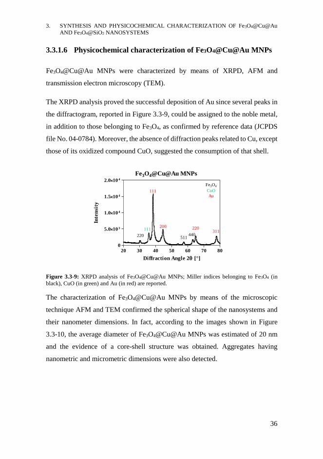
The XRPD analysis proved the successful deposition of Au since several peaks in
the diffractogram, reported in Figure 3.3-9, could be assigned to the noble metal,
in addition to those belonging to Fe3O4, as confirmed by reference data (JCPDS
file No. 04-0784). Moreover, the absence of diffraction peaks related to Cu, except
those of its oxidized compound CuO, suggested the consumption of that shell.
Figure 3.3-9: XRPD analysis of Fe3O4@Cu@Au MNPs; Miller indices belonging to Fe3O4 (in
black), CuO (in green) and Au (in red) are reported.
The characterization of Fe3O4@Cu@Au MNPs by means of the microscopic
technique AFM and TEM confirmed the spherical shape of the nanosystems and
their nanometer dimensions. In fact, according to the images shown in Figure
3.3-10, the average diameter of Fe3O4@Cu@Au MNPs was estimated of 20 nm
and the evidence of a core-shell structure was obtained. Aggregates having
nanometric and micrometric dimensions were also detected.
Fe3O4@Cu@Au MNPs
20 30 40 50 60 70 800
5.010 3
1.010 4
1.510 4
2.010 4
Fe3O4
CuO
Au
220
111
200
511440
220311
111
Diffraction Angle 2 [°]
Inte
nsi
ty

3. SYNTHESIS AND PHYSICOCHEMICAL CHARACTERIZATION OF Fe3O4@Cu@Au
AND Fe3O4@SiO2 NANOSYSTEMS
37
Figure 3.3-10: TEM (on the left) and AFM (on the right) analysis of Fe3O4@Cu@Au MNPs,
confirming the nanometer dimension and the core-shell structure of the nanosystems.
3.3.2 Fe3O4@SiO2 MNPs
In order to produce the other kind of nanosystem, Fe3O4 MNPs were exploited as
nucleation seeds for the deposition of SiO2 shell. As previously described in
section 3.1.1.1, SiO2 was selected as the coating agent because of its excellent
biocompatibility and chemical stability. Moreover, the surface functionalization
of Fe3O4@SiO2 MNPs could be carried out taking advantages from the strong
hard-hard interactions among silicon and other compounds, especially silanes;
using heterobifunctional silanes the conjugation of molecules or biomolecules of
interest can be achieved.104
3.3.2.1 Synthesis of silica shell: Fe3O4@SiO2 MNPs
The deposition of the SiO2 shell on Fe3O4 MNPs was carried out by the sol-gel
process, known as Stöber method.74 This synthetic strategy concerned the
polymerization of tetraethoxysilane (TEOS) in alcohol solution using ammonia as
catalyst, as reported in Figure 3.3-11.

3. SYNTHESIS AND PHYSICOCHEMICAL CHARACTERIZATION OF Fe3O4@Cu@Au
AND Fe3O4@SiO2 NANOSYSTEMS
38
Figure 3.3-11: Scheme of the polymerization reaction of TEOS for the deposition of SiO2 shell on
Fe3O4 MNPs.
The reaction was performed stirring Fe3O4 MNPs for 3 h at rt in TEOS solution.
The mechanism consists in the hydrolysis of TEOS in isopropyl alcohol followed
by the condensation step on the surface of Fe3O4 MNPs, leading to the formation
of the SiO2 shell. Fe3O4@SiO2 MNPs were separated from the reaction mixtures
through magnetic collection, washed several times first with alcohol and then with
H2O and finally dried. Two kinds of Fe3O4@SiO2 MNPs differing in the thickness
of the shell were synthesized; these MNPs were produced performing the
reactions with different amounts of TEOS, using three-times the concentration of
TEOS in one reaction compared to the other.105
3.3.2.2 Physicochemical characterization of Fe3O4@SiO2 MNPs
Fe3O4@SiO2 MNPs were characterized by means of spectroscopic and
microscopic techniques. The chemical characterization was achieved performing
Fourier-transform infrared spectroscopy (FT-IR) and XRPD analysis while the
morphological study was carried out by using AFM and TEM.
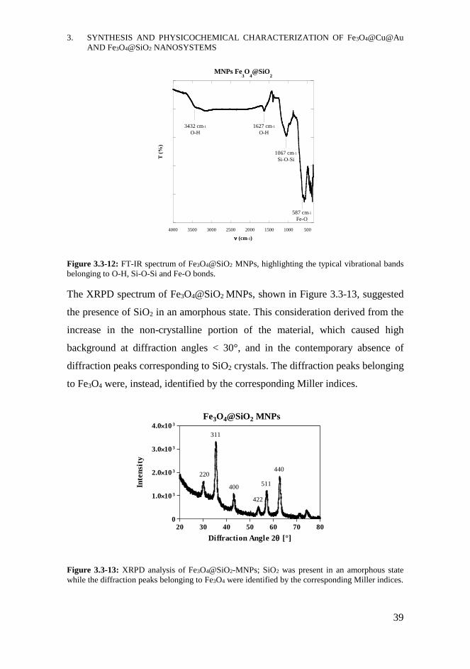
The FT-IR spectrum of Fe3O4@SiO2 MNPs, shown in Figure 3.3-12, confirmed
the presence of silica in the inorganic nanostructure in addition to the iron oxide
since the typical vibrational bands of SiO2 and Fe3O4 could be clearly recognized.
In fact, the intense vibrational bands centred at ν ≈ 1067 cm-1 and ν ≈ 587 cm-1
could be assigned to the stretching vibrations of Si-O-Si and Fe-O bond,
respectively. The broad band at ν ≈ 3430 cm-1 and that at ν ≈ 1630 cm-1 could,
instead, be ascribed to the O-H vibrations of adsorbed H2O.
3. SYNTHESIS AND PHYSICOCHEMICAL CHARACTERIZATION OF Fe3O4@Cu@Au
AND Fe3O4@SiO2 NANOSYSTEMS
39
Figure 3.3-12: FT-IR spectrum of Fe3O4@SiO2 MNPs, highlighting the typical vibrational bands
belonging to O-H, Si-O-Si and Fe-O bonds.
The XRPD spectrum of Fe3O4@SiO2 MNPs, shown in Figure 3.3-13, suggested
the presence of SiO2 in an amorphous state. This consideration derived from the
increase in the non-crystalline portion of the material, which caused high
background at diffraction angles < 30°, and in the contemporary absence of
diffraction peaks corresponding to SiO2 crystals. The diffraction peaks belonging
to Fe3O4 were, instead, identified by the corresponding Miller indices.
Figure 3.3-13: XRPD analysis of Fe3O4@SiO2-MNPs; SiO2 was present in an amorphous state
while the diffraction peaks belonging to Fe3O4 were identified by the corresponding Miller indices.
5001000150020002500300035004000
MNPs Fe3O
4@SiO
2
T (
%)
(cm-1)
1067 cm-1
Si-O-Si
587 cm-1
Fe-O
1627 cm-1
O-H
3432 cm-1
O-H
Fe3O4@SiO2 MNPs
20 30 40 50 60 70 800
1.010 3
2.010 3
3.010 3
4.010 3
220
311
400
422
511
440
Diffraction Angle 2 [°]
Inte
nsi
ty
3. SYNTHESIS AND PHYSICOCHEMICAL CHARACTERIZATION OF Fe3O4@Cu@Au
AND Fe3O4@SiO2 NANOSYSTEMS
40
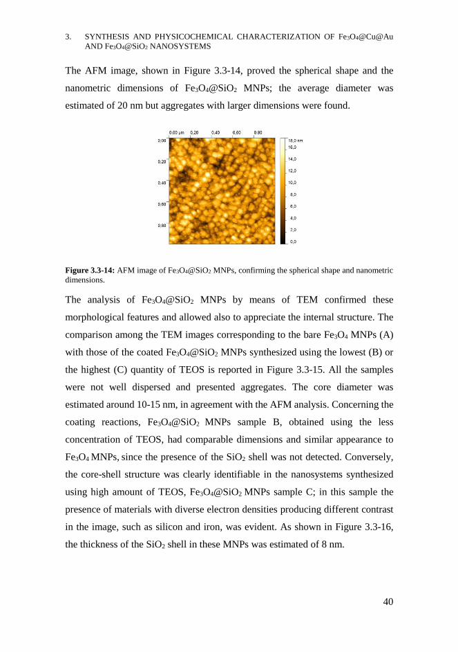
The AFM image, shown in Figure 3.3-14, proved the spherical shape and the
nanometric dimensions of Fe3O4@SiO2 MNPs; the average diameter was
estimated of 20 nm but aggregates with larger dimensions were found.
Figure 3.3-14: AFM image of Fe3O4@SiO2 MNPs, confirming the spherical shape and nanometric
dimensions.
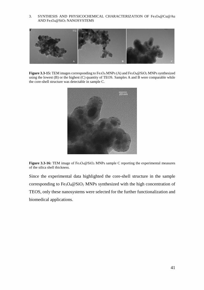
The analysis of Fe3O4@SiO2 MNPs by means of TEM confirmed these
morphological features and allowed also to appreciate the internal structure. The
comparison among the TEM images corresponding to the bare Fe3O4 MNPs (A)
with those of the coated Fe3O4@SiO2 MNPs synthesized using the lowest (B) or
the highest (C) quantity of TEOS is reported in Figure 3.3-15. All the samples
were not well dispersed and presented aggregates. The core diameter was
estimated around 10-15 nm, in agreement with the AFM analysis. Concerning the
coating reactions, Fe3O4@SiO2 MNPs sample B, obtained using the less
concentration of TEOS, had comparable dimensions and similar appearance to
Fe3O4 MNPs, since the presence of the SiO2 shell was not detected. Conversely,
the core-shell structure was clearly identifiable in the nanosystems synthesized
using high amount of TEOS, Fe3O4@SiO2 MNPs sample C; in this sample the
presence of materials with diverse electron densities producing different contrast
in the image, such as silicon and iron, was evident. As shown in Figure 3.3-16,
the thickness of the SiO2 shell in these MNPs was estimated of 8 nm.
3. SYNTHESIS AND PHYSICOCHEMICAL CHARACTERIZATION OF Fe3O4@Cu@Au
AND Fe3O4@SiO2 NANOSYSTEMS
41
Figure 3.3-15: TEM images corresponding to Fe3O4 MNPs (A) and Fe3O4@SiO2 MNPs synthesized
using the lowest (B) or the highest (C) quantity of TEOS. Samples A and B were comparable while
the core-shell structure was detectable in sample C.
Figure 3.3-16: TEM image of Fe3O4@SiO2 MNPs sample C reporting the experimental measures
of the silica shell thickness.
Since the experimental data highlighted the core-shell structure in the sample
corresponding to Fe3O4@SiO2 MNPs synthesized with the high concentration of
TEOS, only these nanosystems were selected for the further functionalization and
biomedical applications.
3. SYNTHESIS AND PHYSICOCHEMICAL CHARACTERIZATION OF Fe3O4@Cu@Au
AND Fe3O4@SiO2 NANOSYSTEMS
42
CHAPTER CONTRIBUTIONS
- Core-shell superparamagnetic Fe3O4@Cu@Au MNPs and Fe3O4@SiO2
MNPs were synthesized and characterized by means of different
spectroscopic and microscopic techniques. The chemical composition and
the crystal structure were confirmed by XRPD and/or FT-IR analysis
while data about the nanometric dimensions, shape and internal structure
were collected by AFM and TEM analysis.
ACKNOWLEDGMENTS
Dr. Alessandro Latini and Dr. Carmen Cavallo of Sapienza University of Rome
for XRPD. Dr Francesca Anna Scaramuzzo of Sapienza University of Rome for
AFM. Dr. Sergio Brutti of University of Basilicata for TEM. Prof. Maria Pia
Donzello of Sapienza University of Rome for FTIR.
MATERIALS AND METHODS
Reagents, materials and equipment: All the chemical reagents were purchased by
Sigma-Aldrich (Merck KGaA, Darmstadt, Germany) and used without further
purification. Permanent magnets made of Neodymium-Iron-Boron alloy, having
a magnetic field of 0.3 T, were used for magnetic separations. Sonication was
performed with UNISET-AC characterized by 45 W power and 38 KHz maximum
frequency. The diffractometer RIGAKU-MINIFLEX II, with a θ-2θ Bragg-
Brentano geometry and Cu anti-cathode (Cu Kα1 λ= 1.5405 Å), was used for
XRPD analysis on solid MNPs. Infrared spectra were recorded from 200 to 4000
cm-1 using the spectrometer VARIAN 660-IR FT-IR and preparing the IR tablets
3. SYNTHESIS AND PHYSICOCHEMICAL CHARACTERIZATION OF Fe3O4@Cu@Au
AND Fe3O4@SiO2 NANOSYSTEMS
43
mixing 1 mg of sample with 200 mg of anhydrous KBr. AFM analysis were
performed using the microscope MULTIMODE VEECO equipped with the
software Nanoscope IIIa; the topographical images of the sample, deposited as
diluted aqueous solution on Si slices, were collected in tapping mode using a
RTESP Bruker tip (nominal parameters r = 8mm, f = 300 kHz, k = 40 N/m) and
recorded with a 512x512 pixel resolution, corrected by polynomial background
filters using the software Gwyddion 2.31. TEM images were collected with the
microscope Philips ECM-200 KV.
Synthesis of the magnetite core - Fe3O4 MNPs: 200 mL of an aqueous solution
containing (NH4)2Fe(SO4)2*6H2O (3.8 mmol), Fe2(SO4)3 (3.8 mmol) and PVP
(MW 10 000, 19.1 mmol) were added dropwise in argon atmosphere to 600 mL
of NH4OH (0.7 M) dissolving the same amount of PVP. After 30 min reacting at
rt under mechanical stirring, the mixture was heated at 80°C and stirred 30 min.
Once cooled, Fe3O4 MNPs were magnetically separated from the mixture and
dialyzed 24 h against bi-distilled H2O. After drying under vacuum, about 700 mg
of Fe3O4 MNPs were obtained.
Synthesis of the Cu shell - Fe3O4@Cu MNPs: 100 mg of Fe3O4 MNPs (0.43 mmol)
were dispersed in 48 mL of bi-distilled H2O and sonicated 10 min. The molar ratio
between Fe3O4 : CuII used was 1.5:1. 10 mL of an aqueous solution of Cu(NO3)2
(0.29 mmol) and, after 5 min, 2 mL of an aqueous solution of L-ascorbic acid (0.58
mmol) were added dropwise to the dispersion. The reaction was mechanically
stirred for 1 h at 50°C. Fe3O4@Cu MNPs were magnetically separated, washed
with bi-distilled H2O and then used for the second covering.
The second covering reaction was performed as the previous one, using the same
amount of L-ascorbic acid and changing the molar ratio between Fe3O4 and CuII
to 4:1. After 1 h stirring at 50°C, the product Fe3O4@Cu MNPs was washed with
saturated NaCl solution and then dialysed 24 h against bi-distilled H2O and finally
dried in the stove overnight at 120°C.
3. SYNTHESIS AND PHYSICOCHEMICAL CHARACTERIZATION OF Fe3O4@Cu@Au
AND Fe3O4@SiO2 NANOSYSTEMS
44
Synthesis of the Au shell - Fe3O4@Cu@Au MNPs: 100 mg of Fe3O4@Cu MNPs
(0.82 mmol of Cu) were dispersed in 45 mL of bi-distilled H2O. 5 mL of a aqueous
solution of HAuCl4*3H2O (0.21 mmol), neutralized to pH 6.5 with saturated
NaHCO3 solution, were added dropwise to the dispersion. The mixture was
mechanically stirred for 45 min at 60°C. Fe3O4@Cu@Au MNPs were
magnetically separated from the mixture, washed with saturated NaHCO3 solution
and then bi-distilled H2O and finally covered with the second Au shell.
For the second gold deposition reaction, Fe3O4@Cu@Au MNPs achieved from
the first covering reaction were dispersed in 43 mL of bi-distilled H2O and 5 mL
of a neutralized aqueous solution of HAuCl4*3H2O (0.10 mmol, pH 6.5) were
added to the dispersion. After adding 2 mL of an aqueous solution of L-ascorbic
acid (0.50 mmol) the mixture was mechanically stirred at 60°C for 2 h.
Fe3O4@Cu@Au MNPs were collected by magnetic separation, washed with
EDTA solution (1 mM; pH 7) and then dialysed 24 h against bi-distilled H2O.
Fe3O4@Cu@Au MNPs were desiccated overnight in the stove at 120°C.
Synthesis of the SiO2 shell - Fe3O4@SiO2 MNPs: 100 mg of Fe3O4 MNPs were
dispersed in 6 mL of bi-distilled H2O and sonicated for 15 min. Under mechanical
stirring, 20 mL of a solution of TEOS in isopropyl alcohol (at the concentration
of 1 mg/mL or 2,5 mg/mL) and 1 mL of NH4OH (0.7 M) were added slowly to
the dispersion. After reacting 3h at rt, Fe3O4@SiO2 MNPs were magnetically
separated from the solution, washed with isopropyl alcohol and bi-distilled H2O
and then dried overnight under vacuum.
4. Fe3O4@Cu@Au NANOPARTICLES
LOADING ACTIVE TARGETING
LIGANDS AND IN VITRO
EXPERIMENTS ON HUMAN BREAST
CANCER CELL LINES
4. Fe3O4@Cu@Au NANOPARTICLES LOADING ACTIVE TARGETING LIGANDS AND
IN VITRO EXPERIMENTS ON HUMAN BREAST CANCER CELL LINES
46
INTRODUCTION
The development of new nanosystems suitable for theranostic applications in
cancer treatment is the focus of the research work presented in this chapter. The
basic metallic structure of Fe3O4@Cu@Au MNPs has been used for the
conjugation of bioactive molecules also known to be active targeting ligands, like
the vitamin folic acid and the chemotherapeutic drug methotrexate. To reach this
goal, the preliminary activation of the Au surface with molecules suitable for the
further modification has been carried out. To evaluate the therapeutic effect of the
synthesized nanosystems, biological studies on breast cancer cell lines have been
performed in collaboration with the research group led by Prof. Laura Masuelli at
the Department of Experimental Medicine - Sapienza University of Rome.
4.1.1 Tumour targeting using nanosystems
Since the spread of cancer is rapidly increasing, causing millions of deaths
worldwide, the development of new therapeutic strategies based on NPs and the
improvement of the imaging techniques can contribute to early diagnosis and
more efficient treatments, preventing the lethal outcomes of cancer.106-108 Several
kinds of nanoparticles, especially liposomes and polymeric nanoparticles, have
already been approved or submitted to clinical trials for the treatment of several
primary tumours and/or metastasis: for instance Myocet® (Sopherion
Therapeutics Inc., USA), non-PEGylated liposomes loading Doxorubicin, or
NK105, PEG-poly(aspartic acid) carriers loading Paclitaxel (Phase III)109 have
been proposed for the treatment of breast cancer.107
Fe3O4 MNPs can also be useful for cancer therapy and imaging, as previously
described in section 1.3. In addition to their exploitation as contrast agents in MRI
or in hyperthermia, Fe3O4 MNPs can be functionalized in order to carry proper
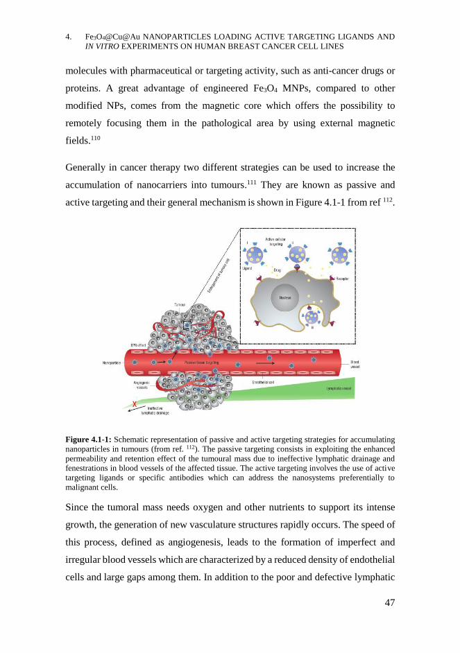
4. Fe3O4@Cu@Au NANOPARTICLES LOADING ACTIVE TARGETING LIGANDS AND
IN VITRO EXPERIMENTS ON HUMAN BREAST CANCER CELL LINES
47
molecules with pharmaceutical or targeting activity, such as anti-cancer drugs or
proteins. A great advantage of engineered Fe3O4 MNPs, compared to other
modified NPs, comes from the magnetic core which offers the possibility to
remotely focusing them in the pathological area by using external magnetic
fields.110
Generally in cancer therapy two different strategies can be used to increase the
accumulation of nanocarriers into tumours.111 They are known as passive and
active targeting and their general mechanism is shown in Figure 4.1-1 from ref 112.
Figure 4.1-1: Schematic representation of passive and active targeting strategies for accumulating
nanoparticles in tumours (from ref. 112). The passive targeting consists in exploiting the enhanced
permeability and retention effect of the tumoural mass due to ineffective lymphatic drainage and
fenestrations in blood vessels of the affected tissue. The active targeting involves the use of active
targeting ligands or specific antibodies which can address the nanosystems preferentially to
malignant cells.
Since the tumoral mass needs oxygen and other nutrients to support its intense
growth, the generation of new vasculature structures rapidly occurs. The speed of
this process, defined as angiogenesis, leads to the formation of imperfect and
irregular blood vessels which are characterized by a reduced density of endothelial
cells and large gaps among them. In addition to the poor and defective lymphatic
4. Fe3O4@Cu@Au NANOPARTICLES LOADING ACTIVE TARGETING LIGANDS AND
IN VITRO EXPERIMENTS ON HUMAN BREAST CANCER CELL LINES
48
drainage of the tumoral area, these fenestrations allow the passive diffusion of
nanomaterials into the pathological area. This phenomenon is known as Enhanced
Permeation and Retention effect (EPR) and is commonly exploited for improving
accumulation of nanocarriers into tumoral tissue, providing an increase in the
local distribution of drugs.111,113 However, EPR cannot be exploited for all kinds
of tumours since it depends on several features, such as the position and the size
of the tumour or the pore dimensions of the fenestrations; for instance, tumours
presenting necrotic areas characterized by hypoxic environment cannot be reached
by exploiting this passive accumulation phenomenon and, moreover, EPR is often
compromised by the increased intratumor pressure.114
The use of specific ligands for cancer cell which can act as targeting molecules
can help overcoming the limitations of the passive drug-delivery strategy.115 The
active targeting compounds include specific antibodies, proteins, nucleic acids,
peptides or other small molecules such as vitamins. They can actively interact
with some elements, such as surface molecules or receptors, present in the affected
organ, tissue or cell. Thanks to this specific interaction, the drug delivery
nanosystems loading active targeting ligands are preferentially directed to the
pathological tissues, enabling a more efficient therapeutic activity. Furthermore,
if the active targeting concerns a membrane receptor able to trigger the
internalization process and that is overexpressed in cancer cells, the uptake of the
functionalized nanosystem can be facilitated.116 Some examples of active
targeting molecules are antibodies or their fragments consisting in the
hypervariable region, proteins such as transferrin, peptides with RGD sequence
(arginine-glycine-aspartic acid) or vitamins like folic acid.117,118

4. Fe3O4@Cu@Au NANOPARTICLES LOADING ACTIVE TARGETING LIGANDS AND
IN VITRO EXPERIMENTS ON HUMAN BREAST CANCER CELL LINES
49
4.1.2 Folic acid and methotrexate
Folic acid (FA) is an essential vitamin belonging to
the B group (vitamin B9). As shown in Figure 4.1-2,
a pteridine ring (in black), a p-aminobenzoic acid
moiety (PABA, in red) and a γ- glutamic acid residue
(γ-Glu, in blue) compose its chemical structure. In
nature it can be found in several forms differing in the
oxidation state of the pteridine ring, or in the N(5) or
N(10) substituents or in the number of glutamate
residues; all these compounds belong to the folate
family. Folates are converted by the enzyme dihydrofolate reductase (DHFR) in
the active form tetrahydrofolate (THF), using NADPH as cofactor. THF acts as
coenzyme in many biochemical processes concerning the one-carbon transfer at
different oxidation state, for example in the metabolism of purine, pyrimidines
and some AA. Due to its biological importance, a deficiency of FA is correlated
to pathologies affecting especially nervous system, such as spina bifida.119
Due to the involvement of DHFR in cell growth, several chemotherapeutic drugs
have this enzyme as their biological target. Among those, methotrexate (MTX) is
a folate structural analogue since -NH2 group is present in the pteridine ring
instead of -OH and -CH3 instead of -H in the PABA
moiety, as shown in Figure 4.1-3. These structural
differences allow MTX to act as antagonist of FA
(antifolate), binding to the active site of DHFR and
inhibiting it.120 This process leads to a leakage of
THF, blocking the nucleic acid synthesis and causing
cell death. Due to its cytotoxic activity, MTX is
commonly used for the treatment of several kinds of
cancer, especially lung and breast cancer.121,122
Figure 4.1-2: Chemical
structure of folic acid; the
pteridine ring, the p-
aminobenzoic acid and the ϒ-
glutamic acid residue are
highlighted respectively in
black, red and blue.
Figure 4.1-3: Chemical
structure of methotrexate; the
pteridine ring, PABA and γ-Glu
are highlighted in black, red
and blue respectively, while the
substituents differing from FA
are circled.
4. Fe3O4@Cu@Au NANOPARTICLES LOADING ACTIVE TARGETING LIGANDS AND
IN VITRO EXPERIMENTS ON HUMAN BREAST CANCER CELL LINES
50
4.1.3 Folate receptors and cancer cells
The need of FA occurs particularly in rapidly dividing cells, including cancer
cells, and for this reason the antifolates are of great interest in cancer therapy. In
this context, the sensitivity of some cancer cell lines to antifolate drugs derives
from the overexpression of folate receptors on their cellular membranes, needed
for allowing high uptake of FA to support their rapid growth.123
The absorption of folates occurs in the intestinal villi thanks to specific folate
transporters: folate receptors (FRs), reduced folate carriers (RFC) and proton
coupled folate transporters are involved in this mechanism.124 Folate receptors are
trans-membrane proteins anchored by glycosylphosphatidylinositol molecules,
existing in different isoforms and showing nanomolar binding affinity for FA. The
α isoform of FRs (α-FR) has become an important tumoral marker since it is
overexpressed in many tumours affecting especially brain, liver, ovary and
breast.125 Although it is mainly present in macrophages, it is almost absent or
hardly accessible in healthy organs, such as liver, lung and placenta. Moreover,
the amount of folate receptors seems to increase during the worsening of the stage
of the cancer.126 This protein is thus becoming an interesting biological target for
the selective treatment of some tumours.123 Because of the high structural
similarity between FA and MTX, this drug is able to bind to RFC and FR with
lower affinity than FA but entering into the binding site with the identical
geometry and orientation of FA.127
Once bound FA or an antifolate, the folate receptors transport the ligand in
intracellular environment by a receptor-mediated endocytosis. As shown in Figure
4.1-4 from ref. 128, after the receptor-ligand complex formation the internalization
occurs through the intracellular compartment called endosome. The endosome
acidification produces the detachment of the folate from the protein, since the
binding is pH dependent and, consequently, its release in the cytoplasmic
environment together with the recycling of the receptor.128

4. Fe3O4@Cu@Au NANOPARTICLES LOADING ACTIVE TARGETING LIGANDS AND
IN VITRO EXPERIMENTS ON HUMAN BREAST CANCER CELL LINES
51
Figure 4.1-4: Mechanism of the folate receptor-mediated endocytosis exploited as active targeting
strategy in cancer treatment (from ref. 128). The interaction between the folate-loaded system and the
membrane folate receptors triggers the internalization process which starts with the production of
the endosomes. Once reached the cytoplasm, the acidification of the endosome causes the
intracellular release of the folate-functionalized system and the recycling of the folate receptors.
However, some cancer cells often show resistance to antifolate drug since they are
able to develop a defence mechanism, such as the amplification of the gene
encoding for DHFR or the decrease in the receptor-mediated transport
mechanism.129 To better exploit the promising features of folate active targeting,
the conjugation of FA or antifolates to nanocarriers seems to be a good strategy;
in fact, combining the peculiar features of NPs with those of folates the
accumulation of the nanosystems in targeted tissues can be improved together
with the internalization into malignant cells.118 In this way, the nanosystems can
be able to perform a more selective cytotoxic action limiting the side effects on
healthy tissues. Several metal and polymeric nanosystems have been synthesized
loading FA for targeting cancer cells 130,131 or loading MTX and using it both as
targeting and therapeutic agent.127,132 Examples of MNPs functionalized with FA
or MTX for cancer theranostics can be found in the literature, generally exploiting
the magnetic core for MRI or performing multimodal strategies.54,133
The design and development of new nanosystems able to overcome the cellular
defence strategies, exploiting the active targeting ability of FA or MTX and being
effective in cancer treatment remain interesting research topics.
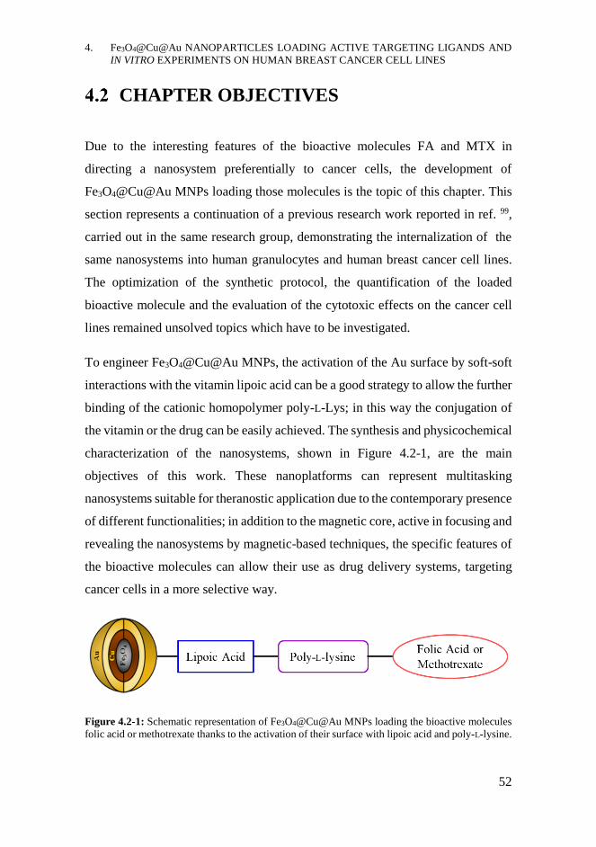
4. Fe3O4@Cu@Au NANOPARTICLES LOADING ACTIVE TARGETING LIGANDS AND
IN VITRO EXPERIMENTS ON HUMAN BREAST CANCER CELL LINES
52
CHAPTER OBJECTIVES
Due to the interesting features of the bioactive molecules FA and MTX in
directing a nanosystem preferentially to cancer cells, the development of
Fe3O4@Cu@Au MNPs loading those molecules is the topic of this chapter. This
section represents a continuation of a previous research work reported in ref. 99,
carried out in the same research group, demonstrating the internalization of the
same nanosystems into human granulocytes and human breast cancer cell lines.
The optimization of the synthetic protocol, the quantification of the loaded
bioactive molecule and the evaluation of the cytotoxic effects on the cancer cell
lines remained unsolved topics which have to be investigated.
To engineer Fe3O4@Cu@Au MNPs, the activation of the Au surface by soft-soft
interactions with the vitamin lipoic acid can be a good strategy to allow the further
binding of the cationic homopolymer poly-L-Lys; in this way the conjugation of
the vitamin or the drug can be easily achieved. The synthesis and physicochemical
characterization of the nanosystems, shown in Figure 4.2-1, are the main
objectives of this work. These nanoplatforms can represent multitasking
nanosystems suitable for theranostic application due to the contemporary presence
of different functionalities; in addition to the magnetic core, active in focusing and
revealing the nanosystems by magnetic-based techniques, the specific features of
the bioactive molecules can allow their use as drug delivery systems, targeting
cancer cells in a more selective way.
Figure 4.2-1: Schematic representation of Fe3O4@Cu@Au MNPs loading the bioactive molecules
folic acid or methotrexate thanks to the activation of their surface with lipoic acid and poly-L-lysine.
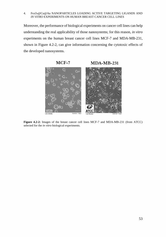
4. Fe3O4@Cu@Au NANOPARTICLES LOADING ACTIVE TARGETING LIGANDS AND
IN VITRO EXPERIMENTS ON HUMAN BREAST CANCER CELL LINES
53
Moreover, the performance of biological experiments on cancer cell lines can help
understanding the real applicability of those nanosystems; for this reason, in vitro
experiments on the human breast cancer cell lines MCF-7 and MDA-MB-231,
shown in Figure 4.2-2, can give information concerning the cytotoxic effects of
the developed nanosystems.
Figure 4.2-2: Images of the breast cancer cell lines MCF-7 and MDA-MB-231 (from ATCC)
selected for the in vitro biological experiments.
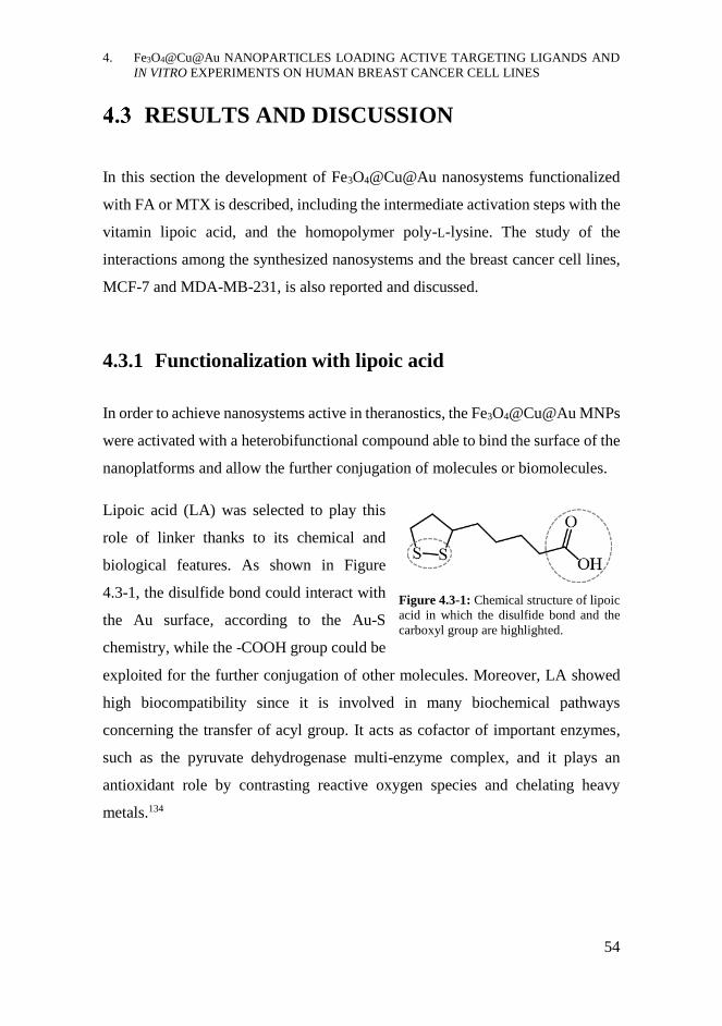
4. Fe3O4@Cu@Au NANOPARTICLES LOADING ACTIVE TARGETING LIGANDS AND
IN VITRO EXPERIMENTS ON HUMAN BREAST CANCER CELL LINES
54
RESULTS AND DISCUSSION
In this section the development of Fe3O4@Cu@Au nanosystems functionalized
with FA or MTX is described, including the intermediate activation steps with the
vitamin lipoic acid, and the homopolymer poly-L-lysine. The study of the
interactions among the synthesized nanosystems and the breast cancer cell lines,
MCF-7 and MDA-MB-231, is also reported and discussed.
4.3.1 Functionalization with lipoic acid
In order to achieve nanosystems active in theranostics, the Fe3O4@Cu@Au MNPs
were activated with a heterobifunctional compound able to bind the surface of the
nanoplatforms and allow the further conjugation of molecules or biomolecules.
Lipoic acid (LA) was selected to play this
role of linker thanks to its chemical and
biological features. As shown in Figure
4.3-1, the disulfide bond could interact with
the Au surface, according to the Au-S
chemistry, while the -COOH group could be
exploited for the further conjugation of other molecules. Moreover, LA showed
high biocompatibility since it is involved in many biochemical pathways
concerning the transfer of acyl group. It acts as cofactor of important enzymes,
such as the pyruvate dehydrogenase multi-enzyme complex, and it plays an
antioxidant role by contrasting reactive oxygen species and chelating heavy
metals.134
Figure 4.3-1: Chemical structure of lipoic
acid in which the disulfide bond and the
carboxyl group are highlighted.
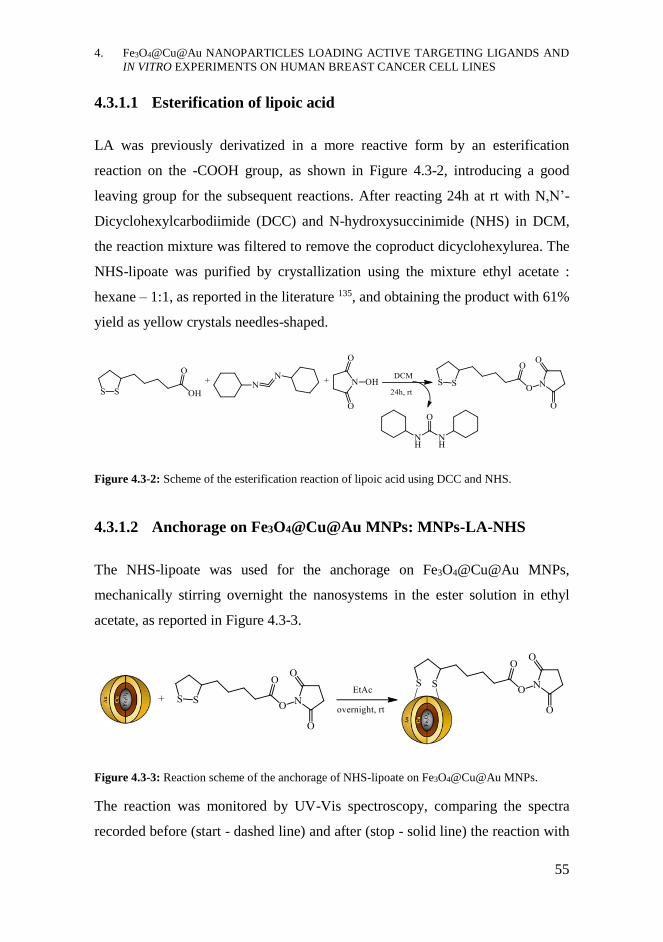
4. Fe3O4@Cu@Au NANOPARTICLES LOADING ACTIVE TARGETING LIGANDS AND
IN VITRO EXPERIMENTS ON HUMAN BREAST CANCER CELL LINES
55
4.3.1.1 Esterification of lipoic acid
LA was previously derivatized in a more reactive form by an esterification
reaction on the -COOH group, as shown in Figure 4.3-2, introducing a good
leaving group for the subsequent reactions. After reacting 24h at rt with N,N’-
Dicyclohexylcarbodiimide (DCC) and N-hydroxysuccinimide (NHS) in DCM,
the reaction mixture was filtered to remove the coproduct dicyclohexylurea. The
NHS-lipoate was purified by crystallization using the mixture ethyl acetate :
hexane – 1:1, as reported in the literature 135, and obtaining the product with 61%
yield as yellow crystals needles-shaped.
Figure 4.3-2: Scheme of the esterification reaction of lipoic acid using DCC and NHS.
4.3.1.2 Anchorage on Fe3O4@Cu@Au MNPs: MNPs-LA-NHS
The NHS-lipoate was used for the anchorage on Fe3O4@Cu@Au MNPs,
mechanically stirring overnight the nanosystems in the ester solution in ethyl
acetate, as reported in Figure 4.3-3.
Figure 4.3-3: Reaction scheme of the anchorage of NHS-lipoate on Fe3O4@Cu@Au MNPs.
The reaction was monitored by UV-Vis spectroscopy, comparing the spectra
recorded before (start - dashed line) and after (stop - solid line) the reaction with
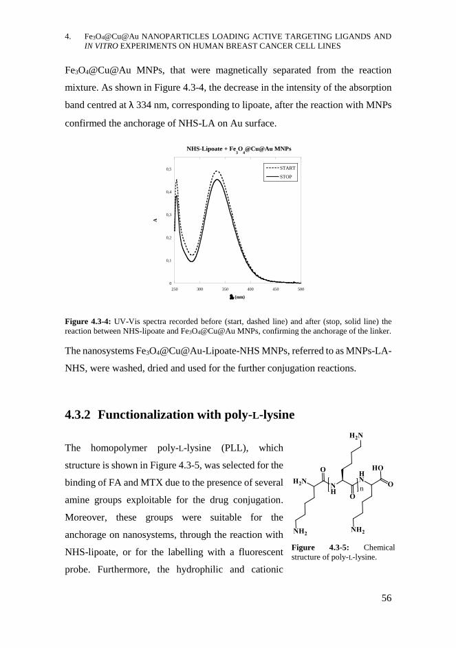
4. Fe3O4@Cu@Au NANOPARTICLES LOADING ACTIVE TARGETING LIGANDS AND
IN VITRO EXPERIMENTS ON HUMAN BREAST CANCER CELL LINES
56
Fe3O4@Cu@Au MNPs, that were magnetically separated from the reaction
mixture. As shown in Figure 4.3-4, the decrease in the intensity of the absorption
band centred at λ 334 nm, corresponding to lipoate, after the reaction with MNPs
confirmed the anchorage of NHS-LA on Au surface.
Figure 4.3-4: UV-Vis spectra recorded before (start, dashed line) and after (stop, solid line) the
reaction between NHS-lipoate and Fe3O4@Cu@Au MNPs, confirming the anchorage of the linker.
The nanosystems Fe3O4@Cu@Au-Lipoate-NHS MNPs, referred to as MNPs-LA-
NHS, were washed, dried and used for the further conjugation reactions.
4.3.2 Functionalization with poly-L-lysine
The homopolymer poly-L-lysine (PLL), which
structure is shown in Figure 4.3-5, was selected for the
binding of FA and MTX due to the presence of several
amine groups exploitable for the drug conjugation.
Moreover, these groups were suitable for the
anchorage on nanosystems, through the reaction with
NHS-lipoate, or for the labelling with a fluorescent
probe. Furthermore, the hydrophilic and cationic
0
0,1
0,2
0,3
0,4
0,5
250 300 350 400 450 500
NHS-Lipoate + Fe3O
4@Cu@Au MNPs
START
STOP
A
(nm)
Figure 4.3-5: Chemical
structure of poly-L-lysine.
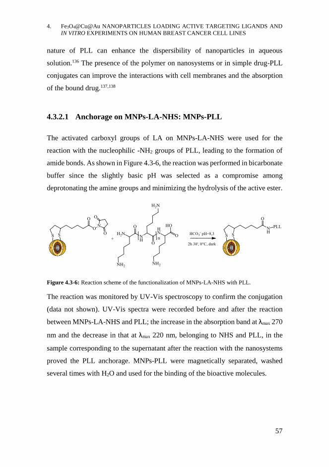
4. Fe3O4@Cu@Au NANOPARTICLES LOADING ACTIVE TARGETING LIGANDS AND
IN VITRO EXPERIMENTS ON HUMAN BREAST CANCER CELL LINES
57
nature of PLL can enhance the dispersibility of nanoparticles in aqueous
solution.136 The presence of the polymer on nanosystems or in simple drug-PLL
conjugates can improve the interactions with cell membranes and the absorption
of the bound drug.137,138
4.3.2.1 Anchorage on MNPs-LA-NHS: MNPs-PLL
The activated carboxyl groups of LA on MNPs-LA-NHS were used for the
reaction with the nucleophilic -NH2 groups of PLL, leading to the formation of
amide bonds. As shown in Figure 4.3-6, the reaction was performed in bicarbonate
buffer since the slightly basic pH was selected as a compromise among
deprotonating the amine groups and minimizing the hydrolysis of the active ester.
Figure 4.3-6: Reaction scheme of the functionalization of MNPs-LA-NHS with PLL.
The reaction was monitored by UV-Vis spectroscopy to confirm the conjugation
(data not shown). UV-Vis spectra were recorded before and after the reaction
between MNPs-LA-NHS and PLL; the increase in the absorption band at λmax 270
nm and the decrease in that at λmax 220 nm, belonging to NHS and PLL, in the
sample corresponding to the supernatant after the reaction with the nanosystems
proved the PLL anchorage. MNPs-PLL were magnetically separated, washed
several times with H2O and used for the binding of the bioactive molecules.

4. Fe3O4@Cu@Au NANOPARTICLES LOADING ACTIVE TARGETING LIGANDS AND
IN VITRO EXPERIMENTS ON HUMAN BREAST CANCER CELL LINES
58
4.3.3 Functionalization with FA or MTX
The bioactive molecules FA and MTX were first activated by an esterification
reaction and then conjugated to the free amine groups of PLL bound to the
nanosystems.
4.3.3.1 Esterification of FA and MTX
The activation of the -COOH groups of the two molecules was achieved by an
esterification reaction with 1-Ethyl-3-(3-dimethylaminopropyl)carbodiimide
(EDC) and NHS, in order to obtain the isolable esters NHS-AF and NHS-MTX.
The reactions, shown in Figure 4.3-7, were performed in anhydrous DMSO to
dissolve the bioactive molecules, using triethylamine (Et3N) as the catalyst. They
were carried out overnight at rt in closed systems to avoid the evaporation Et3N
and protected from light due to the photosensitivity of FA and MTX.
Figure 4.3-7: Scheme of the esterification reaction of FA or MTX using EDC and NHS.
The NHS-esters were purified by precipitation adding ethyl ether to the
concentrated DMSO solutions.139 The yellow-orange precipitates were filtered,
washed with ACN to remove the DMSO and dried under vacuum. Unfortunately,
this purification method did not remove completely the unreacted FA and MTX
which remained in significant amount in the final product (from 1H NMR analysis
NHS-FA:FA ratio 1:1, NHS-MTX:MTX ratio 2:1). However, the conjugation on
PLL was performed without further purification.
4. Fe3O4@Cu@Au NANOPARTICLES LOADING ACTIVE TARGETING LIGANDS AND
IN VITRO EXPERIMENTS ON HUMAN BREAST CANCER CELL LINES
59
The first idea was to try binding NHS-FA and NHS-MTX directly on free PLL to
allow a better quantification of the loaded drug. This strategy was assessed using
NHS-FA, since it was more easily available. The reactions were performed
solubilizing the reagents in DMSO and using different concentrations of FA (FA
moles at 1.5%, 5%, 10% and 20% of PLL moles,
calculated on Lys monomers). The solutions were
stirred overnight at rt and protected from light to avoid
FA photodegradation. Unfortunately, after drying the
reaction mixture, the solubilization with H2O of all the
conjugates PLL-FA led to completely insoluble
orange compounds with macroscopic tubular shape,
as those shown in in Figure 4.3-8.
Further investigation by 1D and 2D 1H and 13C NMR, like DOSY, TOCSY and
NOESY were performed dispersing PLL-FA conjugates in H2O:D2O mixture at
90:10 ratio (data not shown). From DOSY experiments DMSO and H2O
molecules resulted involved in the formation of the secondary structure of the
PLL-FA conjugate but remaining unstructured since they had logD values < logD
of PLL and FA (logD = -8.92 for DMSO, -8.56 for H2O, -10,1 for PLL and -9.7
for FA). From TOCSY analysis two spin systems for the PABA moiety of FA
were found, justifiable by the presence of a fraction of FA covalently bound to
PLL and another interacting only by stacking. NOESY experiments did not show
dipolar correlations among 1H of FA and PLL. All these studies suggested a
complex secondary structure based on β-sheets, but the structural complexity of
the PLL-FA conjugates needed further investigation by solid state techniques.
However, the insolubility of PLL-FA in all the solvent mixtures tried, differing in
polarity, pH or ionic strength, drastically limited their use. For this reason, NHS-
FA and NHS-MTX were conjugated on PLL already bound to the nanosystems,
avoiding the solubilization problems.
Figure 4.3-8: Photo of tubular
structures corresponding to
PLL-FA conjugate, achieved
using 10% of FA.
4. Fe3O4@Cu@Au NANOPARTICLES LOADING ACTIVE TARGETING LIGANDS AND
IN VITRO EXPERIMENTS ON HUMAN BREAST CANCER CELL LINES
60
4.3.3.2 Conjugation on nanoparticles: MNPs-PLL-FA and MNPs-
PLL-MTX
The free amine groups of PLL, anchored on the nanosystems, were used for the
reaction with the NHS-esters of FA or MTX. The reactions, reported in Figure
4.3-9, were performed dispersing MNPs-PLL in a solution of NHS-AF or NHS-
MTX in DMSO, stirring the mixture overnight at rt and protected from light.
Figure 4.3-9: Reaction scheme of the functionalization of MNPs-PLL with NHS-FA or NHS-MTX.
Since UV-Vis spectroscopy was used for monitoring the reaction and quantify the
drug loading, Et3N was needed to maintain stable the protonation degree of the
pteridine moiety of the bioactive molecules during the reaction; in this way it was
possible to avoid shifts and profile changes in the absorption bands of FA and
MTX in the UV-Vis spectra.140 The UV-Vis spectra, shown in Figure 4.3-10,
demonstrated the conjugation of the bioactive molecules on MNPs-PLL. In fact,
the significant decrease in the absorption bands corresponding to FA or MTX in
the spectra recorded after the reaction with MNPs-PLL (stop - solid line)
compared to the starting solutions of NHS-AF or NHS-MTX (start - dashed line)
proved the functionalization of the nanosystems. The loading was therefore
estimated in 0.8 mg FA or 0.6 mg of MTX on 50 mg of MNPs, using ε values
obtained by calibration curves of NHS-FA and NHS-MTX solutions, dissolved in
the same solvent of the reaction.

4. Fe3O4@Cu@Au NANOPARTICLES LOADING ACTIVE TARGETING LIGANDS AND
IN VITRO EXPERIMENTS ON HUMAN BREAST CANCER CELL LINES
61
Figure 4.3-10: UV Vis spectra recorded before (start, dashed line) and after (stop, solid line) the
reactions between MNPs-PLL and NHS-esters of folic acid (left) or methotrexate (right) which
confirmed the binding of the bioactive molecules due to the decrease in their absorption bands.
After magnetic separation and washings, the MNPs-PLL-FA and MNPs-PLL-
MTX nanosystems were characterized by several spectroscopic techniques. FT-
IR and fluorescence spectra of the MNPs, reported in Figure 4.3-11, confirmed
the conjugation of the bioactive molecules; in the FT-IR spectra, in fact,
characteristic vibrational bands belonging to FA, MTX and PLL, especially those
assigned to amide bonds and pteridine moiety, were detected and the
corresponding frequencies were indicated. The strong vibrational band around ν
570 cm-1 belonged to Fe-O-Fe bond of the magnetite core.
Furthermore, the fluorescence spectra of MNPs-PLL-AF and MNPs-PLL-MTX,
recorded using λexc at 278 nm, showed the emission bands corresponding to the
PABA and pteridine moieties of the bioactive molecules, at λmax at 350 nm and
λmax at 440-460 nm, respectively.141

4. Fe3O4@Cu@Au NANOPARTICLES LOADING ACTIVE TARGETING LIGANDS AND
IN VITRO EXPERIMENTS ON HUMAN BREAST CANCER CELL LINES
62
Figure 4.3-11: Top: FT-IR spectra corresponding to MNPs-PLL-FA (left) and MNPs-PLL-MTX
(right). The peaks of the vibrational bands belonging to the bioactive molecules were indicated.
Bottom: fluorescence spectra, recorded using λexc at 278 nm, of to MNPs-PLL-FA (left) and MNPs-
PLL-MTX (right). The emission bands correspond to the PABA and pteridine moieties of FA and
MTX.
4.3.4 In vitro experiments on human breast cancer cell lines
Once achieved the MNPs-PLL-FA and MNPs-PLL-MTX, in vitro biological
experiments on cancer cell lines were performed. As introduced previously in
section 4.1.3, FA or MTX are considered active targeting ligands for nanosystems
due to the overexpression of folate receptors in many cancer cells; in fact, they
might help in directing the functionalized nanoplatforms towards cancer cells,
accumulating inside them and preserving the healthy ones.
4. Fe3O4@Cu@Au NANOPARTICLES LOADING ACTIVE TARGETING LIGANDS AND
IN VITRO EXPERIMENTS ON HUMAN BREAST CANCER CELL LINES
63
The stabilized human breast cancer cell lines MCF-7 and MDA-MB-231 were
selected for the in vitro studies on the biological effects of the nanosystems. Both
those cell lines correspond to breast adenocarcinoma and literature data confirm
their upregulation of α-FR expression.142 According to previous results, achieved
in the same research group in which this thesis was developed, these cell lines
were sensitive to MTX, especially MDA-MB-231, as demonstrated by cell
viability assay. Moreover, the internalization of those nanosystems was
qualitatively confirmed by fluorescence microscopy experiments since the
nanosystems loading FA and MTX, labelled with the fluorescent probe Texas Red
(TR), were located in intracellular environment compared to the nanosystems
functionalized with PLL-TR only.99 Starting from those experimental data, in vitro
biological experiments on these cell lines were carried out in order to evaluate the
cytotoxic effects of functionalized MNPs by performing cell proliferation assays.
MCF-7 and MDA-MB-231 were incubated with MNPs-PLL-MTX, MNPs-PLL-
AF or MNPs-PLL upon different experimental conditions, such as cell amount,
incubation time, culture medium and nanoparticles-administration method, and
Sulforhodamine B proliferation assays were performed.143 Cells incubated with
free MTX and FA were used as controls, in addition to the negative control
consisting of not treated cells. According to the loading quantification of MTX on
MNPs, the comparable amount of free MTX, corresponding to 2 μg/mL, was
selected for the assays.
The proliferation assays were performed plating 10 000 cells/well or 25 000
cells/well in DMEM and, the next day, incubating them for 24 h or 48 h with the
nanosystems. Cells were treated with different amounts of functionalized
nanoparticles dispersed in medium, both filtered or not filtered on 0.45 μm filters.
A medium without folate, RPMI w/o FA, was selected to remove the presence of
basal FA and facilitate the internalization process of MNPs-PLL-FA or MNPs-
PLL-MTX due to the vitamin cell need.133 Since cells might suffered for the
absence of FA, the complete RPMI medium was also used. The cell incubation

4. Fe3O4@Cu@Au NANOPARTICLES LOADING ACTIVE TARGETING LIGANDS AND
IN VITRO EXPERIMENTS ON HUMAN BREAST CANCER CELL LINES
64
with MNPs dispersed in PBS was selected as other experimental condition. Serum
percentage was also assessed from 0.1% to 10%; low FBS concentration
minimized the interactions among nanoparticles and proteins while the high one
corresponded to the standard concentration ensuring a good cell viability.
The experiments carried out without filtrating the MNPs presented many
problems due to early cell death; in fact, the nanometric and micrometric
aggregates had deadly effects on cells, causing their disruption, as shown in Figure
4.3-12. For this reason, the filtration of the MNPs solutions was necessary;
however, although this process ensured cell viability, enabling the performance of
the proliferation assay, it led to low and unknown drug concentrations in the
samples of MNPs, hindering the comparison with free drug.
Figure 4.3-12: Images of MCF-7 and MDA-MB-231 before (left) and after (right) the treatment
with not-filtered solutions of MNPs.
The best, but not promising, results achieved from these in vitro experiments are
reported in Figure 4.3-13. These results corresponded to Sulforhodamine B assays
performed on MCF-7 and MDA-MB-231 after 24 h of treatment in three
experimental conditions. Cells were incubated with different volumes of filtrated
solutions of MNPs-PLL or MNPs-PLL-MTX (50 μL or 100 μL) in three media:
in PBS, replacing the medium with DMEM complemented with 10% FBS after
incubating for 3h, or in RPMI w/o folate + 0.1% FBS or in complete RPMI (w/o
the pH indicator phenol red) + 1% FBS. Untreated cells and samples incubated
with MTX 2 μg/mL or FA 0.2 μg/mL were used as controls. According to the
experimental results, a slight cytotoxic effect of MNPs was detected on both cell
lines but without significant differences among cell samples treated with MNPs-
PLL and those incubated with MNPs-PLL-MTX. Unfortunately, also free MTX
4. Fe3O4@Cu@Au NANOPARTICLES LOADING ACTIVE TARGETING LIGANDS AND
IN VITRO EXPERIMENTS ON HUMAN BREAST CANCER CELL LINES
65
for 24 h in the same medium was not effective in causing cell death at the
concentration of 2 μg/mL since cell viabilities were comparable to those achieved
in samples treated with FA 0.2 μg/mL. These data suggested that drug amount in
filtered samples was indeed too low to produce cytotoxic effects. These not
promising results suggested the need of finding a strategy to reduce the
aggregation process of MNPs, to avoid filtration, since it affected the successful
performance of the in vitro experiments.
Figure 4.3-13: Results of the 24 h Sulforhodamine B proliferation assays on MCF-7 (top) and
MDA-MB-231 (bottom). The experimental conditions corresponded to untreated cells (ctr) and
cells incubated with free MTX, FA or MNPs-PLL and MNPs-PLL-MTX at two concentrations in
volume. The treatments were performed in PBS, replacing after 3 h the medium with DMEM + 10%
FBS, or in RPMI w/o folic acid (RPMI w/o FA + 0.1% FBS) or w/o phenol red (RPMI w/o PhR +
1% FBS).
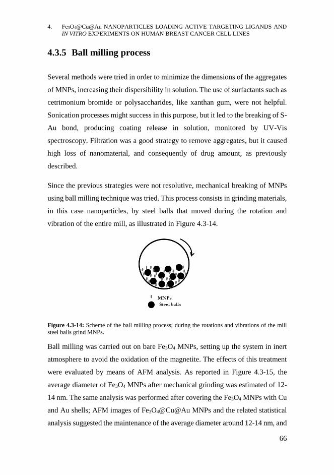
4. Fe3O4@Cu@Au NANOPARTICLES LOADING ACTIVE TARGETING LIGANDS AND
IN VITRO EXPERIMENTS ON HUMAN BREAST CANCER CELL LINES
66
4.3.5 Ball milling process
Several methods were tried in order to minimize the dimensions of the aggregates
of MNPs, increasing their dispersibility in solution. The use of surfactants such as
cetrimonium bromide or polysaccharides, like xanthan gum, were not helpful.
Sonication processes might success in this purpose, but it led to the breaking of S-
Au bond, producing coating release in solution, monitored by UV-Vis
spectroscopy. Filtration was a good strategy to remove aggregates, but it caused
high loss of nanomaterial, and consequently of drug amount, as previously
described.
Since the previous strategies were not resolutive, mechanical breaking of MNPs
using ball milling technique was tried. This process consists in grinding materials,
in this case nanoparticles, by steel balls that moved during the rotation and
vibration of the entire mill, as illustrated in Figure 4.3-14.
Figure 4.3-14: Scheme of the ball milling process; during the rotations and vibrations of the mill
steel balls grind MNPs.
Ball milling was carried out on bare Fe3O4 MNPs, setting up the system in inert
atmosphere to avoid the oxidation of the magnetite. The effects of this treatment
were evaluated by means of AFM analysis. As reported in Figure 4.3-15, the
average diameter of Fe3O4 MNPs after mechanical grinding was estimated of 12-
14 nm. The same analysis was performed after covering the Fe3O4 MNPs with Cu
and Au shells; AFM images of Fe3O4@Cu@Au MNPs and the related statistical
analysis suggested the maintenance of the average diameter around 12-14 nm, and
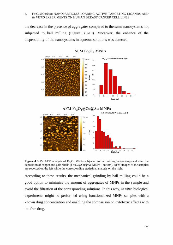
4. Fe3O4@Cu@Au NANOPARTICLES LOADING ACTIVE TARGETING LIGANDS AND
IN VITRO EXPERIMENTS ON HUMAN BREAST CANCER CELL LINES
67
the decrease in the presence of aggregates compared to the same nanosystems not
subjected to ball milling (Figure 3.3-10). Moreover, the enhance of the
dispersibility of the nanosystems in aqueous solutions was detected.
Figure 4.3-15: AFM analysis of Fe3O4 MNPs subjected to ball milling before (top) and after the
deposition of copper and gold shells (Fe3O4@Cu@Au MNPs - bottom). AFM images of the samples
are reported on the left while the corresponding statistical analysis on the right.
According to these results, the mechanical grinding by ball milling could be a
good option to minimize the amount of aggregates of MNPs in the sample and
avoid the filtration of the corresponding solutions. In this way, in vitro biological
experiments might be performed using functionalized MNPs samples with a
known drug concentration and enabling the comparison on cytotoxic effects with
the free drug.
4. Fe3O4@Cu@Au NANOPARTICLES LOADING ACTIVE TARGETING LIGANDS AND
IN VITRO EXPERIMENTS ON HUMAN BREAST CANCER CELL LINES
68
CHAPTER CONTRIBUTIONS
- The Fe3O4@Cu@Au MNPs were used for the development of
nanosystems carrying the active targeting molecules FA or MTX; to
enable the binding of the bioactive molecules, the previous activation of
the Au surface with LA and the conjugation of PLL were performed. The
drug loading quantification was carried out by means of UV-Vis
spectroscopy while the physicochemical characterization of the
nanosystems was achieved by FT-IR and fluorescence techniques.
- Since MTX is commonly used as chemotherapeutic drug, the cytotoxic
effects of functionalized nanosystems were evaluated on the breast cancer
cell lines MCF-7 and MDA-MB-231. Sulforhodamine B proliferation
assays suggested a slight cytotoxicity of MNPs, regardless of the presence
of the drug. The experimental evidences could be explained as the drug
amount was too low to produce toxic effects due to the necessary filtration
of the MNPs dispersions. Aggregates, in fact, dramatically affected the
success of the experiments, causing early cell death.
- To minimize the presence of big aggregates in the samples, ball milling
treatment may be a solution.
4.4.1 Future perspectives
- Functionalize the Fe3O4@Cu@Au MNPs, subjected to ball milling
process, with the bioactive molecules FA and MTX and try to improve
the drug loading, optimizing the reaction conditions.
- Perform the in vitro experiments without filtrating the MNPs samples thus
knowing the drug concentration and comparing the effects with free drug.
4. Fe3O4@Cu@Au NANOPARTICLES LOADING ACTIVE TARGETING LIGANDS AND
IN VITRO EXPERIMENTS ON HUMAN BREAST CANCER CELL LINES
69
ACKNOWLEDGMENTS
Dr. Fabio Sciubba and Prof. Maurizio Delfini for NMR studies. Prof. Maria Pia
Donzello for FTIR analysis. Prof. Laura Masuelli for the biological experiments.
Dr. Maria Assunta Navarra and Dr. Luca Farina for ball milling process. Dr.
Francesca Anna Scaramuzzo for AFM analysis. All of them are working at
Sapienza University of Rome.
MATERIALS AND METHODS
Reagents, materials and equipment: All the chemical reagents were purchased by
Sigma-Aldrich (Merck KGaA, Darmstadt, Germany) and used without further
purification. All the material used in in vitro biological experiments were
purchased from Corning (Corning, USA). Permanent magnets made of
Neodymium-Iron-Boron alloy, having a magnetic field of 0.3 T, were used for
magnetic separations. Sonication was performed with UNISET-AC characterized
by 45 W power and 38 KHz maximum frequency. Infrared spectra were recorded
from 200 to 4000 cm-1 by the spectrometer VARIAN 660-IR FT-IR, preparing the
IR tablets mixing 1 mg of sample with 200 mg of anhydrous KBr. Steady-state
fluorescence spectra were collected by Varian Cary Eclypse spectrofluorometer,
equipped with the Xe lamp using Suprasil® Quartz cuvette. NMR analysis were
performed by means of the BRUKER AVANCE 400 MHz spectrometer, using
TSP (stock solution in D2O at 20 mM) as the chemical shift reference. AFM
analysis were performed using the microscope MULTIMODE VEECO equipped
with the software Nanoscope IIIa; the topographical images of the sample,
deposited on Si slice, were collected working in tapping mode using a RTESP
Bruker tip (nominal parameters r = 8mm, f = 300 kHz, k = 40 N/m) and recorded
with a 512x512 pixel resolution, corrected by polynomial background filters using

4. Fe3O4@Cu@Au NANOPARTICLES LOADING ACTIVE TARGETING LIGANDS AND
IN VITRO EXPERIMENTS ON HUMAN BREAST CANCER CELL LINES
70
the software Gwyddion 2.31. A Vibromulino MM400 (Retsch, Haan, Germany)
was used for the ball milling process.
Esterification of Lipoic Acid: NHS-Lipoate: Lipoic acid (4.9 mM) and N-
hydroxysuccinimide (NHS 2 eq, 9.8 mM) were dissolved at rt in 50 mL of
anhydrous DCM and N,N’-Dicyclohexylcarbodiimide (DCC 2 eq, 9.8 mM)
was added after cooling the mixture at 4°C. After stirring 6 h at rt, 1 eq of NHS
and 1 eq DCC were added to the solution. After 24 h reacting, the
dicyclohexylurea was removed filtered the solution and the work up with a
Na2CO3 (x3) and NaCl saturated solution (x1) was performed. The organic
phase was dried and the crystallization with a mixture of ethyl acetate : hexane
- 1:1 was carried out. The final product NHS-Lipoate was achieved as yellow
crystals needles-shaped with 61% yield. 1H NMR (300 MHz, CDCl3, 25°C):
δ=3.47 (m, 1H, H-3), δ=3.04 (m, 2H, H-1), δ=2.72 (s, 4H, H-8, H-9), δ=2.52 (t,
2H, H-7), δ=2.35 (m, 1H, H-2), δ=1.82 (m, 1H, H-2), δ=1.64 (m, 4H, H-4, H-5),
δ=1.44 (m, 2H, H-6).
Functionalization with NHS-Lipoate: MNPs-LA-NHS: 100 mg of Fe3O4@Cu@Au
MNPs were dispersed in 10 mL of NHS-Lipoate in EtAc at 1 mg/mL. After
reacting overnight at rt, MNPs-LA-NHS were magnetically collected. UV-Vis
spectra of the solutions corresponding to the reaction supernatant, centrifuged 10
4. Fe3O4@Cu@Au NANOPARTICLES LOADING ACTIVE TARGETING LIGANDS AND
IN VITRO EXPERIMENTS ON HUMAN BREAST CANCER CELL LINES
71
min at 10 000 rpm, and to an aliquot of NHS-Lipoate starting solution, maintained
in the same reaction conditions, were recorded. MNPs-LA-NHS were washed
with EtAc and then dried under vacuum.
Functionalization with poly-L-Lysine: MNPs-PLL: 100 mg of MNPs-LA-NHS
were dispersed in 10 mL of a solution of PLL*HBr at 1 mg/mL in HCO3- buffer
(0.1 M, pH 8.3, filtered on 0.2 μm). After mechanical stirring 2 h 30 min at 4°C,
MNPs-PLL were magnetically collected, washed several times with bi-distilled
H2O and dried under vacuum.
Esterification of FA and MTX: NHS-FA and NHS-MTX
EDC (3 eq, 0.6 mmol) and NHS (3 eq, 0.6 mmol) were added to a solution of
MTX or FA (0.2 mmol) dissolved in 5 mL of anhydrous DMSO containing 4%
Et3N. The reaction was stirred overnight at rt protected from light. After
concentrating the solution under vacuum heating at 40°C, ethyl ether was added
dropwise to the solution under stirring, in order to precipitate the NHS-ester. The
solution was then filtered under vacuum washing the precipitate with CH3CN
(ACN) and finally drying under vacuum, achieving orange-yellow products. 1H
NMR (400 MHz, d6 DMSO, 25°C) spectra of NHS-FA and NHS-MTX were
compared to those of FA and MTX monitoring the shifts of the reference doublets
corresponding to 1H of PABA moieties, at δ 7.73 ppm (2H) and δ 6.73 ppm (2H)
for FA and at δ 7.62 ppm (2H) and δ 6.76 ppm (2H) for MTX.
Functionalization with NHS-FA or NHS-MTX: MNPs-PLL-FA and MNPs-PLL-
MTX: 5 mL of a solution of NHS-MTX or NHS-FA at 1 mg/mL in anhydrous
DMSO containing 4% Et3N were added to 5 mL containing 50 mg of MNPs-PLL
in the same solvent. The reaction was carried out at rt overnight in systems closed
and protected from light. The next day, MNPs-PLL-FA or MNPs-PLL-MTX were
magnetically collected and UV-Vis spectra of the solutions corresponding to the
reaction supernatants, centrifuged 10 min at 10000 rpm, and to the aliquot of the
NHS-MTX or NHS-FA starting solution, maintained in the reaction conditions,

4. Fe3O4@Cu@Au NANOPARTICLES LOADING ACTIVE TARGETING LIGANDS AND
IN VITRO EXPERIMENTS ON HUMAN BREAST CANCER CELL LINES
72
were recorded. The drug loading was calculated as the average Δc corresponding
to A values at λ 269 nm, 300 nm and 386 nm for MTX and λ 288 nm and 326 nm
for FA. The ε at the same λ were obtained using calibration curves of NHS-MTX
and NHS-FA dissolved in the DMSO + 4% Et3N. For NHS-FA ε288=4.04 mg-1 mL
cm-1 and ε360=1.83 mg-1 mL cm-1 while for NHS-MTX ε269=4.28 mg-1 mL cm-1,
ε300=3.48 mg-1 mL cm-1 and ε386=1.35 mg-1 mL cm-1 were used. The estimated
loading was 0.6 mg MTX on 50 mg MNPs-PLL-MTX and 0.8 mg of FA on 50
mg MNPs-PLL-FA. MNPs-PLL-MTX and MNPs-PLL-FA were washed several
times with bi-distilled H2O and dried under vacuum.
In vitro experiments on MCF-7 and MDA-MB-231 - Sulforhodamine B
proliferation assays: 25 000 cells/well in DMEM supplemented with 10% FBS
and 1% pen strep were plated in 96-wells plate for 24 h. MNPs-PLL and MNPs-
PLL-MTX were dispersed at 2 mg/mL in sterile PBS buffer, in RPMI 1640 w/o
folate + 0.1 % FBS or in RPMI 1640 w/o Phenol Red + 1% FBS. The MNPs
dispersions were filtered on 0.45 μm filters and 200 μL of the 1:2 or 1:4 dilutions
in the corresponding medium were added to the plate. FA at 0.2 μg/mL and MTX
at 2 μg/mL solutions in the same RPMI media or in DMEM + 10% FBS were used
as controls, in addition to untreated cells in the four media. Cells were incubated
at 37°C and 5% CO2; only in the sample treated with MNPs in PBS the buffer was
replaced by DMEM + 10% FBS after 3 h incubating. After 24 h of treatment,
Sulforhodamine B proliferation assay was performed as described in the standard
protocol143 and the A values at λ=492 nm and 620 nm were measured. The
corresponding ΔA is related to cell viability, considering 100% the ctr sample.
Ball Milling Process: Fe3O4 MNPs were subjected to ball milling treatment,
assembling the system in a glove box in inert atmosphere. 1,44 g of Fe3O4 MNPs
and 3 steel balls, with a total weigh of 12.5g were mixed. 8 grinding cycles, lasting
1 h each, were performed working at ν 25 Hz.
5. Fe3O4@Cu@Au NANOPARTICLES
COATED WITH MACROMOLECULES
FOR THE ENGULFMENT OF HUMAN
IMMUNE CELLS
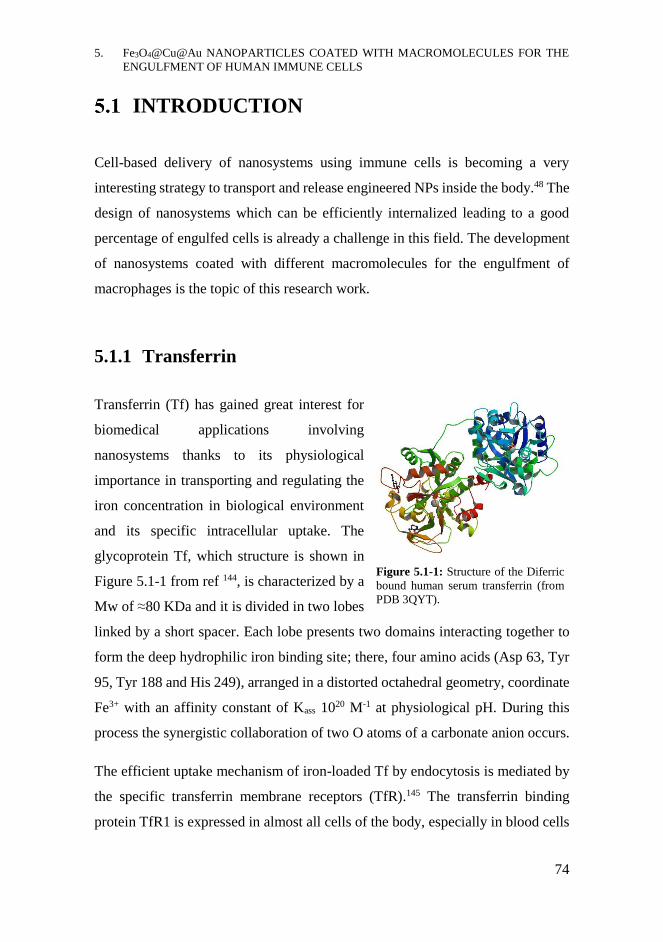
5. Fe3O4@Cu@Au NANOPARTICLES COATED WITH MACROMOLECULES FOR THE
ENGULFMENT OF HUMAN IMMUNE CELLS
74
INTRODUCTION
Cell-based delivery of nanosystems using immune cells is becoming a very
interesting strategy to transport and release engineered NPs inside the body.48 The
design of nanosystems which can be efficiently internalized leading to a good
percentage of engulfed cells is already a challenge in this field. The development
of nanosystems coated with different macromolecules for the engulfment of
macrophages is the topic of this research work.
5.1.1 Transferrin
Transferrin (Tf) has gained great interest for
biomedical applications involving
nanosystems thanks to its physiological
importance in transporting and regulating the
iron concentration in biological environment
and its specific intracellular uptake. The
glycoprotein Tf, which structure is shown in
Figure 5.1-1 from ref 144, is characterized by a
Mw of ≈80 KDa and it is divided in two lobes
linked by a short spacer. Each lobe presents two domains interacting together to
form the deep hydrophilic iron binding site; there, four amino acids (Asp 63, Tyr
95, Tyr 188 and His 249), arranged in a distorted octahedral geometry, coordinate
Fe3+ with an affinity constant of Kass 1020 M-1 at physiological pH. During this
process the synergistic collaboration of two O atoms of a carbonate anion occurs.
The efficient uptake mechanism of iron-loaded Tf by endocytosis is mediated by
the specific transferrin membrane receptors (TfR).145 The transferrin binding
protein TfR1 is expressed in almost all cells of the body, especially in blood cells
Figure 5.1-1: Structure of the Diferric
bound human serum transferrin (from
PDB 3QYT).

5. Fe3O4@Cu@Au NANOPARTICLES COATED WITH MACROMOLECULES FOR THE
ENGULFMENT OF HUMAN IMMUNE CELLS
75
such as macrophages, and it is often upregulated in rapidly proliferating cells,
including in cancer cells.146 For this reason, Tf-drug conjugates or nanosystems
functionalized with Tf can more easily reach and enter into their biological targets
thanks to the active targeting ability of Tf.147 Several examples of nanosystems
including transferrin can be found in the literature. For instance, polymeric or lipid
nanosystems loading drugs, DNA or radioisotopes, complexed or conjugated to
Tf, can be actively directed to the pathological area for therapeutic applications
while metal nanoparticles loading Tf can be used for targeting and imaging brain
tumours, such as glioma.117,148,149
5.1.2 Streptavidin
Streptavidin (Stv) is a protein commonly used
in biological and biotechnological applications
due to its high affinity for biotinylated
molecules. In fact, together with Avidin, Stv is
classified as a biotin-binding protein.150
Produced by the bacterium Streptomices
avidinii, Stv is a homo-tetramer able to bind
with high affinity four molecules of biotin, also
known as vitamin H, per mole of protein. This
binding occurs by the strongest non-covalent interaction known in nature, leading
to the formation of Stv-biotin complex characterized by Kd value of ≈10-15 M. As
shown in Figure 5.1-2 from ref 151, the secondary structure of Stv is characterized
by high amount of β-sheets, involved in the formation of the biotin binding site.
Due to this extraordinary affinity, Stv has been therefore exploited in many
biotechnological and nanobiotechnological applications based on the binding of
biotinylated molecules and macromolecules;152,153 Stv-biotin complexes are
commonly used in several bioanalytical techniques, especially immunoassays and
Figure 5.1-2: Structure of Streptavidin
complexed with the peptide FSHPQNT
(from PDB 1VWA).
5. Fe3O4@Cu@Au NANOPARTICLES COATED WITH MACROMOLECULES FOR THE
ENGULFMENT OF HUMAN IMMUNE CELLS
76
biosensors.154-156 Moreover, the derivatization of drugs or proteins, especially
enzymes or antibody, with biotin can ensure the strong binding to Stv-coated
nanosystems which can be employed for diagnostic or therapeutic purposes.157-159
5.1.3 Cell-mediated delivery: Trojan Horse Approach
Cell-mediated delivery of nanoparticles is a new and promising option in addition
to the standard passive and active targeting strategies, already described in section
4.1.1. It is based on the exploitation of immune cells, especially neutrophils,
monocytes and macrophages, for the transport and delivery of engineered
nanoplatforms inside the body.160,161 In this way, the overcoming of several limits
of the other drug delivery strategies especially reaching deep affected tissues, can
be achieved. In fact, in addition to their high innate biocompatibility and stealth
features, immune cells are naturally able to reach all the tissues in the organism,
also crossing hardly permeable barriers, such as the brain-blood barrier.162 They
are involved in many fundamental physiological processes: in fact, the organism
surveillance and defence against external pathogens, such as bacteria and viruses,
and the elimination of cellular debris and dead cells depend on them. Since many
of these cells are also directly involved in accelerating or contrasting several
pathological processes, especially cancer, infections and inflammations, their use
as carriers of medicated nanosystems can be a good strategy to achieve a more
efficient in loco drug delivery.163-165
Generally, the loading of nanosystems on cells can be achieved by covalent
modification or non-covalent interactions among cell membrane and NPs or by
exploiting the phagocytic activity of some kinds of immune cells that leads to the
internalization of the nanosystems.166 The internalization of engineered
nanoparticles in ex vivo cells, especially monocytes and neutrophils, extracted
from the patient and incubated with the medicated nanoparticles, produce MNPs-
loaded cells. These cells can act as Trojan Horses once reintroduced in the patient,

5. Fe3O4@Cu@Au NANOPARTICLES COATED WITH MACROMOLECULES FOR THE
ENGULFMENT OF HUMAN IMMUNE CELLS
77
migrating to the pathological focus thanks to innate chemoattraction, reaching for
instance the hypoxic areas of tumours.167 Once there, they can release their cargo
by physiological or external inputs.168 This promising strategy reported in Figure
5.1-3, is known as Trojan Horse approach and it can help improving the
effectiveness of therapeutic treatments based on nanotechnological approaches.167
Macrophages, albeit adherent cells, can also be selected as Trojan horses thanks
to their high phagocytic activity.
Figure 5.1-3: General scheme of the Trojan Horse approach based on the ex vivo incubation of
immune cells with engineered nanoparticles and their migration into pathological area by natural
chemoattraction mechanism once reintroduced in the patient.
Notwithstanding the great advantages, this technique presents some limits; for
instance, the need of loading high number of cells in order to achieve a huge
population of cells migrating to the pathological focus. Moreover, the drug must
remain active inside the immune cell, without undergoing degradation, and be
released once reached the target.160

5. Fe3O4@Cu@Au NANOPARTICLES COATED WITH MACROMOLECULES FOR THE
ENGULFMENT OF HUMAN IMMUNE CELLS
78
5.1.3.1 Macrophages
Macrophages, together with neutrophils and monocytes, may be suitable for
internalizing engineered nanosystems as they possess innate phagocytic capability
that they use for their physiological actions.169 Macrophages are located in almost
all tissues and they derive from monocytes. Monocytes, instead, are circulating
cells which can differentiate into macrophages once crossed the endothelial
barrier and penetrated deeply in the organs. Two phenotypes of macrophages can
be distinguished depending on their metabolism: the activated pro-inflammatory
(M1) and the activated anti-inflammatory (M2) macrophages. These cells are
involved in several diseases, especially cancer and inflammation disorders,
playing different roles depending on their polarization; in fact, M1 can exhibit
tumoricidal activities while M2 tumorigenic ones.
Due to their physiological and/or pathogenic roles and their phagocytic ability,
macrophages have been selected for imaging and therapeutic applications using
nanoparticles.170,171 Several engineered nanoparticles have been incorporated into
macrophages via phagocytosis, thus exploiting these engulfed cells as Trojan
Horses. For instance, Au nanoshells, NPs or liposomes loading doxorubicin were
successfully internalized in macrophages or monocyte-differentiated
macrophages for in vitro or in vivo applications for cancer treatments.172,173
Moreover, in this context the accumulation of macrophages carrying
functionalized MNPs into deep tumour sites by applying pulsed magnetic field
has been demonstrated, since the cells acquired magnetic field responsiveness.174
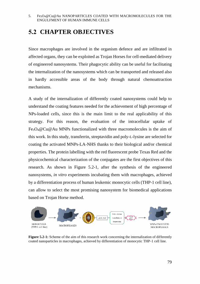
5. Fe3O4@Cu@Au NANOPARTICLES COATED WITH MACROMOLECULES FOR THE
ENGULFMENT OF HUMAN IMMUNE CELLS
79
CHAPTER OBJECTIVES
Since macrophages are involved in the organism defence and are infiltrated in
affected organs, they can be exploited as Trojan Horses for cell-mediated delivery
of engineered nanosystems. Their phagocytic ability can be useful for facilitating
the internalization of the nanosystems which can be transported and released also
in hardly accessible areas of the body through natural chemoattraction
mechanisms.
A study of the internalization of differently coated nanosystems could help to
understand the coating features needed for the achievement of high percentage of
NPs-loaded cells, since this is the main limit to the real applicability of this
strategy. For this reason, the evaluation of the intracellular uptake of
Fe3O4@Cu@Au MNPs functionalized with three macromolecules is the aim of
this work. In this study, transferrin, streptavidin and poly-L-lysine are selected for
coating the activated MNPs-LA-NHS thanks to their biological and/or chemical
properties. The protein labelling with the red fluorescent probe Texas Red and the
physicochemical characterization of the conjugates are the first objectives of this
research. As shown in Figure 5.2-1, after the synthesis of the engineered
nanosystems, in vitro experiments incubating them with macrophages, achieved
by a differentiation process of human leukemic monocytic cells (THP-1 cell line),
can allow to select the most promising nanosystem for biomedical applications
based on Trojan Horse method.
Figure 5.2-1: Scheme of the aim of this research work concerning the internalization of differently
coated nanoparticles in macrophages, achieved by differentiation of monocytic THP-1 cell line.
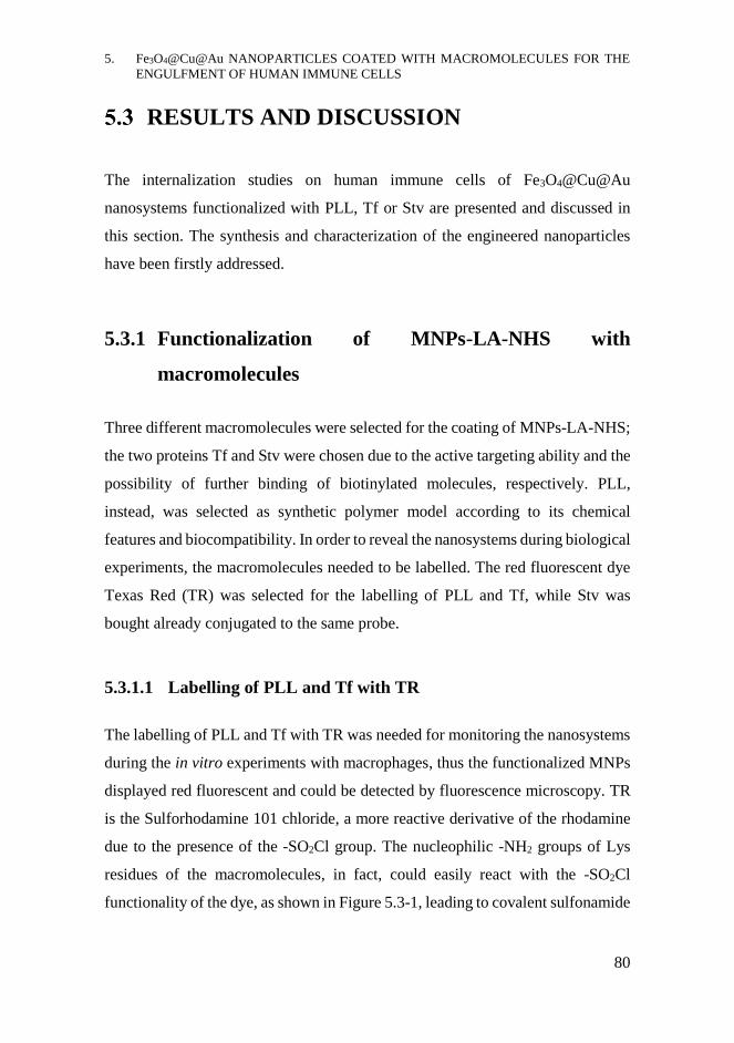
5. Fe3O4@Cu@Au NANOPARTICLES COATED WITH MACROMOLECULES FOR THE
ENGULFMENT OF HUMAN IMMUNE CELLS
80
RESULTS AND DISCUSSION
The internalization studies on human immune cells of Fe3O4@Cu@Au
nanosystems functionalized with PLL, Tf or Stv are presented and discussed in
this section. The synthesis and characterization of the engineered nanoparticles
have been firstly addressed.
5.3.1 Functionalization of MNPs-LA-NHS with
macromolecules
Three different macromolecules were selected for the coating of MNPs-LA-NHS;
the two proteins Tf and Stv were chosen due to the active targeting ability and the
possibility of further binding of biotinylated molecules, respectively. PLL,
instead, was selected as synthetic polymer model according to its chemical
features and biocompatibility. In order to reveal the nanosystems during biological
experiments, the macromolecules needed to be labelled. The red fluorescent dye
Texas Red (TR) was selected for the labelling of PLL and Tf, while Stv was
bought already conjugated to the same probe.
5.3.1.1 Labelling of PLL and Tf with TR
The labelling of PLL and Tf with TR was needed for monitoring the nanosystems
during the in vitro experiments with macrophages, thus the functionalized MNPs
displayed red fluorescent and could be detected by fluorescence microscopy. TR
is the Sulforhodamine 101 chloride, a more reactive derivative of the rhodamine
due to the presence of the -SO2Cl group. The nucleophilic -NH2 groups of Lys
residues of the macromolecules, in fact, could easily react with the -SO2Cl
functionality of the dye, as shown in Figure 5.3-1, leading to covalent sulfonamide
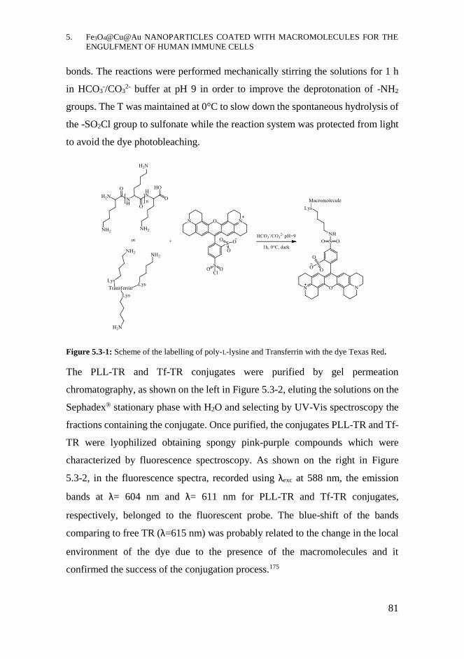
5. Fe3O4@Cu@Au NANOPARTICLES COATED WITH MACROMOLECULES FOR THE
ENGULFMENT OF HUMAN IMMUNE CELLS
81
bonds. The reactions were performed mechanically stirring the solutions for 1 h
in HCO3-/CO3
2- buffer at pH 9 in order to improve the deprotonation of -NH2
groups. The T was maintained at 0°C to slow down the spontaneous hydrolysis of
the -SO2Cl group to sulfonate while the reaction system was protected from light
to avoid the dye photobleaching.
Figure 5.3-1: Scheme of the labelling of poly-L-lysine and Transferrin with the dye Texas Red.
The PLL-TR and Tf-TR conjugates were purified by gel permeation
chromatography, as shown on the left in Figure 5.3-2, eluting the solutions on the
Sephadex® stationary phase with H2O and selecting by UV-Vis spectroscopy the
fractions containing the conjugate. Once purified, the conjugates PLL-TR and Tf-
TR were lyophilized obtaining spongy pink-purple compounds which were
characterized by fluorescence spectroscopy. As shown on the right in Figure
5.3-2, in the fluorescence spectra, recorded using λexc at 588 nm, the emission
bands at λ= 604 nm and λ= 611 nm for PLL-TR and Tf-TR conjugates,
respectively, belonged to the fluorescent probe. The blue-shift of the bands
comparing to free TR (λ=615 nm) was probably related to the change in the local
environment of the dye due to the presence of the macromolecules and it
confirmed the success of the conjugation process.175
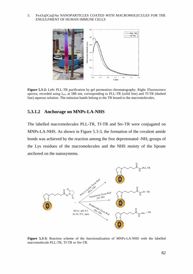
5. Fe3O4@Cu@Au NANOPARTICLES COATED WITH MACROMOLECULES FOR THE
ENGULFMENT OF HUMAN IMMUNE CELLS
82
Figure 5.3-2: Left: PLL-TR purification by gel permeation chromatography. Right: Fluorescence
spectra, recorded using λexc at 588 nm, corresponding to PLL-TR (solid line) and Tf-TR (dashed
line) aqueous solution. The emission bands belong to the TR bound to the macromolecules.
5.3.1.2 Anchorage on MNPs-LA-NHS
The labelled macromolecules PLL-TR, Tf-TR and Stv-TR were conjugated on
MNPs-LA-NHS. As shown in Figure 5.3-3, the formation of the covalent amide
bonds was achieved by the reaction among the free deprotonated -NH2 groups of
the Lys residues of the macromolecules and the NHS moiety of the lipoate
anchored on the nanosystems.
Figure 5.3-3: Reaction scheme of the functionalization of MNPs-LA-NHS with the labelled
macromolecule PLL-TR, Tf-TR or Stv-TR.
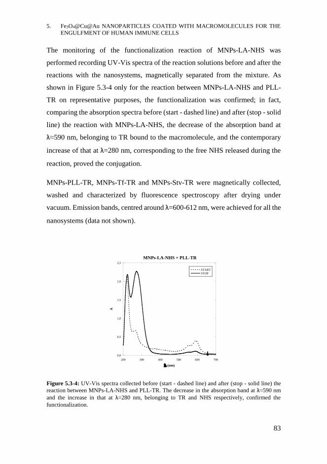
5. Fe3O4@Cu@Au NANOPARTICLES COATED WITH MACROMOLECULES FOR THE
ENGULFMENT OF HUMAN IMMUNE CELLS
83
The monitoring of the functionalization reaction of MNPs-LA-NHS was
performed recording UV-Vis spectra of the reaction solutions before and after the
reactions with the nanosystems, magnetically separated from the mixture. As
shown in Figure 5.3-4 only for the reaction between MNPs-LA-NHS and PLL-
TR on representative purposes, the functionalization was confirmed; in fact,
comparing the absorption spectra before (start - dashed line) and after (stop - solid
line) the reaction with MNPs-LA-NHS, the decrease of the absorption band at
λ≈590 nm, belonging to TR bound to the macromolecule, and the contemporary
increase of that at λ=280 nm, corresponding to the free NHS released during the
reaction, proved the conjugation.
MNPs-PLL-TR, MNPs-Tf-TR and MNPs-Stv-TR were magnetically collected,
washed and characterized by fluorescence spectroscopy after drying under
vacuum. Emission bands, centred around λ=600-612 nm, were achieved for all the
nanosystems (data not shown).
Figure 5.3-4: UV-Vis spectra collected before (start - dashed line) and after (stop - solid line) the
reaction between MNPs-LA-NHS and PLL-TR. The decrease in the absorption band at λ≈590 nm
and the increase in that at λ=280 nm, belonging to TR and NHS respectively, confirmed the
functionalization.
0,0
0,5
1,0
1,5
2,0
2,5
200 300 400 500 600 700
MNPs-LA-NHS + PLL-TR
START
STOP
A
(nm)
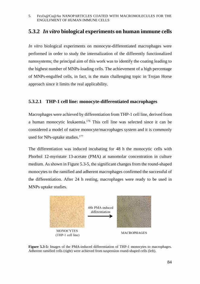
5. Fe3O4@Cu@Au NANOPARTICLES COATED WITH MACROMOLECULES FOR THE
ENGULFMENT OF HUMAN IMMUNE CELLS
84
5.3.2 In vitro biological experiments on human immune cells
In vitro biological experiments on monocyte-differentiated macrophages were
performed in order to study the internalization of the differently functionalized
nanosystems; the principal aim of this work was to identify the coating leading to
the highest number of MNPs-loading cells. The achievement of a high percentage
of MNPs-engulfed cells, in fact, is the main challenging topic in Trojan Horse
approach since it limits the real applicability.
5.3.2.1 THP-1 cell line: monocyte-differentiated macrophages
Macrophages were achieved by differentiation from THP-1 cell line, derived from
a human monocytic leukaemia.176 This cell line was selected since it can be
considered a model of native monocyte/macrophages system and it is commonly
used for NPs-uptake studies.177
The differentiation was induced incubating for 48 h the monocytic cells with
Phorbol 12-myristate 13-acetate (PMA) at nanomolar concentration in culture
medium. As shown in Figure 5.3-5, the significant changes from the round-shaped
monocytes to the ramified and adherent macrophages confirmed the successful of
the differentiation. After 24 h resting, macrophages were ready to be used in
MNPs uptake studies.
Figure 5.3-5: Images of the PMA-induced differentiation of THP-1 monocytes to macrophages.
Adherent ramified cells (right) were achieved from suspension round-shaped cells (left).

5. Fe3O4@Cu@Au NANOPARTICLES COATED WITH MACROMOLECULES FOR THE
ENGULFMENT OF HUMAN IMMUNE CELLS
85
5.3.2.2 MNPs-internalization experiments
Macrophages were incubated with MNPs-PLL-TR, MNPs-Tf-TR or MNPs-Stv-
TR assessing several parameters such as incubation time, culture medium and
mood of administration of nanosystems. The incubation times were varied from
15 min to 6 h, to obtain uptake information on short and long time. Good results
were achieved incubating cells for 2 h, since shorter times led to low uptake while
longer time to the worsening of cell conditions. One or several administrations of
MNPs-macromolecules were performed; the repeated administrations slightly
enhanced the MNPs internalization but causing higher cell stress, revealed by cell
detachment. Incubation medium was also assessed; MNPs-macromolecules were
dispersed in PBS buffer or in RPMI w/o PhR, complemented with 1% FBS only
for long incubation times. The culture medium was finally preferred to maintain
a good cell viability. Nanoparticles were administered to the cells as unfiltered or
filtered dispersions, achieving better results in the second case; the filtration on 3
μm filters allowed to maintain a high concentration of nanosystems removing the
bigger aggregates which affected cell viability.
The best results confirming the internalization of the MNPs-macromolecules were
achieved incubating monocyte-differentiated macrophages for 2 h at 37°C in 5%
CO2, administering the filtered dispersion of nanosystems only once. Untreated
cells, incubated in the same conditions as the treated ones, were used as negative
controls. Different methods or techniques were employed for the characterization
of MNPs-engulfed macrophages.
A simple, quick and reliable evidence of the MNPs internalization was achieved
by Prussian Blue Staining. In this way it was possible to highlight the presence of
iron deposits inside cells. In fact, as shown in Figure 5.3-6 for macrophages
incubated with MNPs-Tf-TR, intracellular blue spots, absent in the control
sample, demonstrated the presence of the nanosystems inside the cells, which
were identified by nuclear staining with neutral red. The ferric ions of Fe3O4 core,

5. Fe3O4@Cu@Au NANOPARTICLES COATED WITH MACROMOLECULES FOR THE
ENGULFMENT OF HUMAN IMMUNE CELLS
86
reacted with potassium ferrocyanide forming a deeply blue coloured complex
ferric ferrocyanide known as Prussian Blue. This staining could work regardless
of MNPs coating, since the positivity derived from the magnetic core of the
nanosystems.
Figure 5.3-6: Prussian Blue staining of monocyte-derived cells untreated (ctr on left) or incubated
with MNPs-Tf-TR (on right). Intracellular blue spots, corresponding to ferric ferrocyanide complex,
proved the presence of iron deposits produced by internalized nanoparticles.
A qualitative evidence, not reported, was achieved by applying an external
permanent magnet under the cell plates after incubating macrophages with MNPs-
macromolecules and removing the not internalized nanoparticles by several
washings. During this experiment, some cells moved following the magnetic field
and accumulating around the magnet. This phenomenon could be explained by the
presence of magnetic materials inside the macrophages, corresponding to
internalized MNPs, which acquired magnetic field responsiveness.49
By confocal fluorescent microscopy the evidence of MNPs-macromolecules
uptake was achieved. Fluorescence images of fixed samples of untreated cells or
cells incubated with the differently coated nanosystems are shown in Figure 5.3-7.
Z-stack acquisitions were performed on each sample, virtually sectioning the
entire cell volume in planes and collecting fluorescent images from each plane.
Here, the images corresponding to a central plane (23-27/40 planes) were
reported.
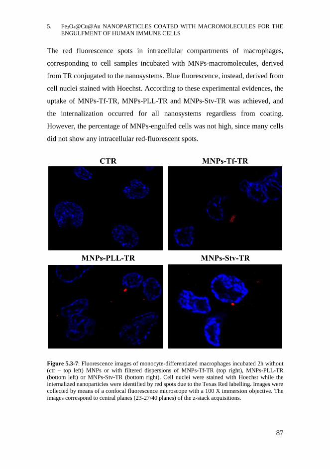
5. Fe3O4@Cu@Au NANOPARTICLES COATED WITH MACROMOLECULES FOR THE
ENGULFMENT OF HUMAN IMMUNE CELLS
87
The red fluorescence spots in intracellular compartments of macrophages,
corresponding to cell samples incubated with MNPs-macromolecules, derived
from TR conjugated to the nanosystems. Blue fluorescence, instead, derived from
cell nuclei stained with Hoechst. According to these experimental evidences, the
uptake of MNPs-Tf-TR, MNPs-PLL-TR and MNPs-Stv-TR was achieved, and
the internalization occurred for all nanosystems regardless from coating.
However, the percentage of MNPs-engulfed cells was not high, since many cells
did not show any intracellular red-fluorescent spots.
Figure 5.3-7: Fluorescence images of monocyte-differentiated macrophages incubated 2h without
(ctr – top left) MNPs or with filtered dispersions of MNPs-Tf-TR (top right), MNPs-PLL-TR
(bottom left) or MNPs-Stv-TR (bottom right). Cell nuclei were stained with Hoechst while the
internalized nanoparticles were identified by red spots due to the Texas Red labelling. Images were
collected by means of a confocal fluorescence microscope with a 100 X immersion objective. The
images correspond to central planes (23-27/40 planes) of the z-stack acquisitions.
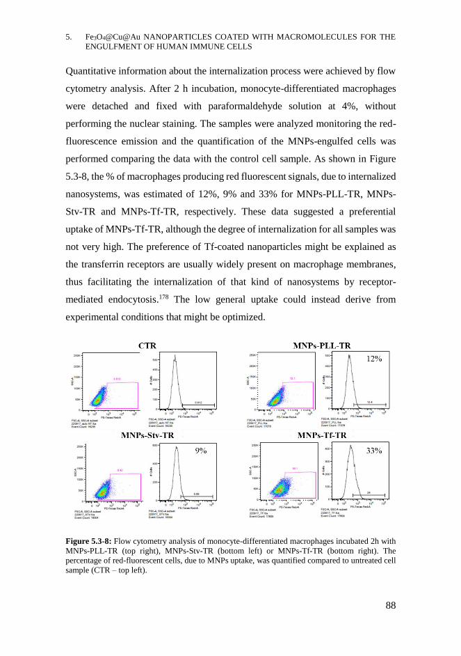
5. Fe3O4@Cu@Au NANOPARTICLES COATED WITH MACROMOLECULES FOR THE
ENGULFMENT OF HUMAN IMMUNE CELLS
88
Quantitative information about the internalization process were achieved by flow
cytometry analysis. After 2 h incubation, monocyte-differentiated macrophages
were detached and fixed with paraformaldehyde solution at 4%, without
performing the nuclear staining. The samples were analyzed monitoring the red-
fluorescence emission and the quantification of the MNPs-engulfed cells was
performed comparing the data with the control cell sample. As shown in Figure
5.3-8, the % of macrophages producing red fluorescent signals, due to internalized
nanosystems, was estimated of 12%, 9% and 33% for MNPs-PLL-TR, MNPs-
Stv-TR and MNPs-Tf-TR, respectively. These data suggested a preferential
uptake of MNPs-Tf-TR, although the degree of internalization for all samples was
not very high. The preference of Tf-coated nanoparticles might be explained as
the transferrin receptors are usually widely present on macrophage membranes,
thus facilitating the internalization of that kind of nanosystems by receptor-
mediated endocytosis.178 The low general uptake could instead derive from
experimental conditions that might be optimized.
Figure 5.3-8: Flow cytometry analysis of monocyte-differentiated macrophages incubated 2h with
MNPs-PLL-TR (top right), MNPs-Stv-TR (bottom left) or MNPs-Tf-TR (bottom right). The
percentage of red-fluorescent cells, due to MNPs uptake, was quantified compared to untreated cell
sample (CTR – top left).
5. Fe3O4@Cu@Au NANOPARTICLES COATED WITH MACROMOLECULES FOR THE
ENGULFMENT OF HUMAN IMMUNE CELLS
89
CHAPTER CONTRIBUTIONS
- A study on the preferential uptake of differently coated nanosystems by
phagocytic cells was performed by in vitro experiments incubating
monocyte-differentiated macrophages, achieved THP-1 cell line, with
Fe3O4@Cu@Au MNPs functionalized with the macromolecules PLL, Tf
and Stv. By labelling the macromolecules with TR, the internalization of
the nanosystems was confirmed by fluorescence microscopy, in addition
to the Prussian Blue Staining used for revealing iron intracellular deposits.
The MNPs-uptake was successfully achieved, preferentially for MNPs-
Tf-TR, leading to 33% of engulfed cells as measured by flow cytometry.
- MNPs-Tf-TR might be promising nanosystems for cell-mediated drug
delivery purposes based on Trojan Horse method.
5.4.1 Future perspectives
- Optimize the experimental conditions in order to enhance the loading and
assess the stability, cytotoxicity and degradation of the internalized
nanosystems.
- Try to internalize the nanosystems into other cell lines, including native
phagocytic cells and evaluate the response of MNPs-engulfed cells to
external magnetic fields in order to exploit magnets for their collection
and focusing.
- Functionalize the MNPs-Tf nanosystems with drugs or explore other
molecules or macromolecules more specific for activated macrophages
and evaluate the real applicability of monocytes-derived macrophages as
Trojan Horses for the delivery of bioactive compounds to malignant cells
in vitro and in vivo.
5. Fe3O4@Cu@Au NANOPARTICLES COATED WITH MACROMOLECULES FOR THE
ENGULFMENT OF HUMAN IMMUNE CELLS
90
ACKNOWLEDGMENTS
Prof. Laura Masuelli of Sapienza University of Rome for Prussian blue staining.
Dr. Valeria de Turris, Dr. Giovanna Peruzzi and Prof. Giancarlo Ruocco of Centre
for Life Nanoscience@Sapienza– Istituto Italiano di Tecnologia - for confocal
fluorescence microscopy and flow cytometry analysis.
MATERIALS AND METHODS
Reagents, materials and equipment: All the chemical reagents were purchased by
Sigma-Aldrich (Merck KGaA, Darmstadt, Germany) and used without further
purification. All the material used in in vitro biological experiments were
purchased from Corning (Corning, USA). Sephadex G-25 fine was used as
stationary phase in gel permeation chromatography. Permanent magnets made of
Neodymium-Iron-Boron alloy, having a magnetic field of 0.3 T, were used for
magnetic separations. Sonication was performed with UNISET-AC characterized
by 45 W power and 38 KHz maximum frequency. Fluorescence spectra were
collected by Varian Cary Eclypse spectrofluorometer, equipped with the Xe lamp.
Prussian Blue staining images were collected by Olympus BX53 equipped with
an 100x oil immersion objectives lens. The fluorescence confocal microscopy and
the flow cytometer were at the Centre for Life Nanoscience IIT@Sapienza. The
fluorescence images were collected z-stack mode acquiring 40 planes, 0.2 μm
distant one from each other, by means of the inverted microscope Olympus X73
equipped with 100x oil immersion objectives lens. The flow cytometer BD
LSRFortessa® with a green laser at 532 nm (600 LP) was used for the
quantification of the MNPs-loaded macrophages, counting ≈17000 events for
each sample.
5. Fe3O4@Cu@Au NANOPARTICLES COATED WITH MACROMOLECULES FOR THE
ENGULFMENT OF HUMAN IMMUNE CELLS
91
Labelling of poly-L-Lysine and Transferrin: PLL-TR and TF-TR: 20 mg of PLL
or Human Apo-Transferrin dissolved at the concentration of 0.1 mg/mL in
carbonate buffer (0.1 M, pH 9) were mixed with 1 mg of TR dissolved in 50 μL
of ACN. After 1 h stirring the reactions in dark at 0°C, the purification was
performed by gel permeation chromatography on Sephadex® G 25-fine stationary
phase eluting the solutions with bi-distilled H2O. Fractions containing PLL-TR or
TF-TR, selected by UV-Vis spectra, were collected and lyophilized. Pink-purple
products were achieved with 67 and 72% yield.
Conjugation of the macromolecules on MNPs-LA-NHS:MNPs-PLL-TR, MNPs-
Tf-TR and MNPs-Stv-TR: 100 mg of MNPs-LA-NHS were dispersed in 10 mL of
PLL-TR, TF-TR or STV-TR solutions at 0.5 mg/mL in bicarbonate buffer (0.1 M,
pH 8.3). After 2h 30 min mechanically stirring the solutions at 4°C protecting
from light, MNPs were magnetically separated and UV-Vis spectra of the
supernatants before and after the reactions were collected. MNPs-PLL-TR,
MNPs-Tf-TR and MNPs-Stv-TR were washed several times with bi-distilled H2O
and dried under vacuum.
PMA-induced differentiation of THP-1 cell line: Monocyte-differentiated
macrophages: 106 monocytes/well of THP-1 cell line in RPMI 1640 w/o PhR,
supplemented with 10% FBS and 1% Pen Strep, were plated in 6-wells plate and
treated with PMA 100 ng/mL (2mL/well). After 48h incubating at 37°C and 5%
CO2, cells were washed with PBS (3x 1mL/well) and incubated in complete RMPI
w/o PMA. After 24h resting monocyte-differentiated macrophages were used for
internalization experiments.
MNPs internalization: Monocyte-differentiated macrophages were incubated with
MNPs-PLL-TR, MNPs-Tf-TR or MNPs-Stv-TR dispersions in RPMI w/o PhR
and w/o FBS (2 mL/well), at starting concentrations of 1 mg/mL, filtered on 3 μm
filters. Cells incubated in the same condition w/o MNPs were used as negative
5. Fe3O4@Cu@Au NANOPARTICLES COATED WITH MACROMOLECULES FOR THE
ENGULFMENT OF HUMAN IMMUNE CELLS
92
controls. After 2h incubating, not internalized MNPs were removed by three
washings with PBS.
Confocal Fluorescence Microscopy analysis: Cells were fixed for 10 min at rt
with paraformaldehyde and sucrose solution at 4% in PBS (1mL/well). After 3
washings with PBS, cells were treated 10 min with glycine aqueous solution (200
mM, 1 mL/well) and, after washings with PBS, the cell nuclei were stained with
Hoechst solution (5 μg/mL in bi-distilled H2O) for 5 min at rt. Coverslips,
contained in each well, were mounted on slides with a glycerol solution at 90% in
PBS and confocal fluorescence analysis were performed.
Prussian Blue staining: After fixing cells in cooled MeOH, 2 mL of an aqueous
solution containing potassium ferrocyanide (4%) and HCl solution (4%) were
added to each well (1mL/well). After 15 min, three washings with bi-distilled H2O
were performed and cell nuclei were stained for 2 min with Nuclear Red aqueous
solution at 2%. After washings with bi-distilled H2O, coverslips were mounted on
slides using Entellan® mounting medium and microscopic images were collected.
Quantification of MNPs-engulfed macrophages by flow cytometry: 2 mL of
Accutase® solution were added to each well. After incubating 20 min, detached
cells were moved to falcon tubes and centrifuged 10 min at 1200 rpm. Pellets were
dispersed in 200 μL of paraformaldehyde solution at 4% in PBS and flow
cytometry analysis were performed.

6. Fe3O4@SiO2-LIPODEPSIPEPTIDE
NANOSYSTEMS WITH
ANTIMICROBIAL ACTIVITY
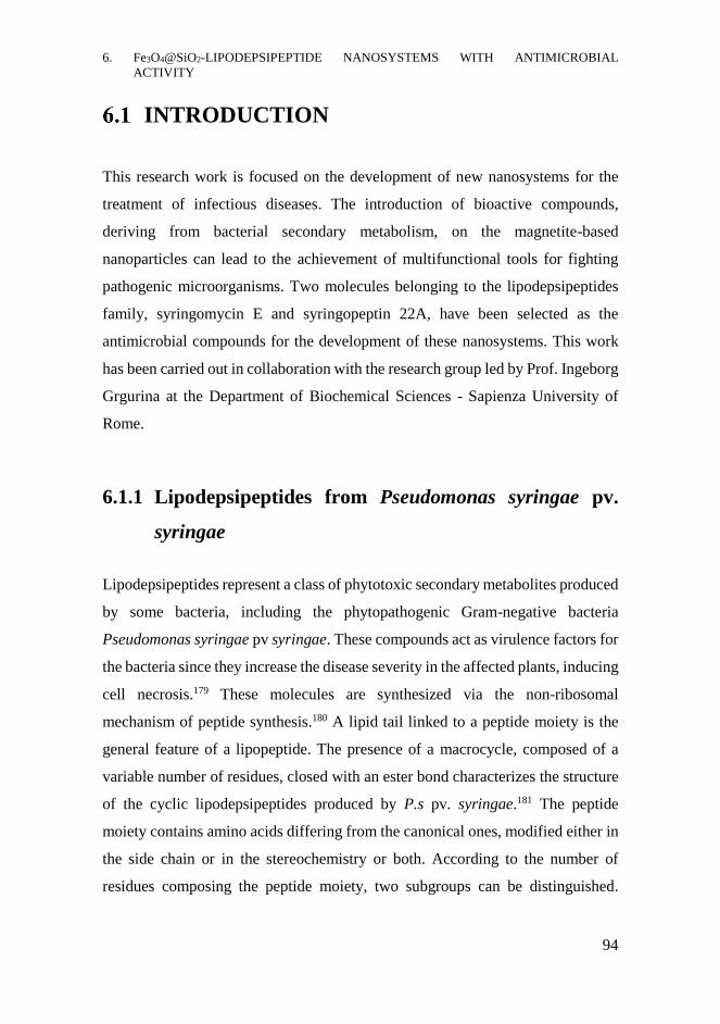
6. Fe3O4@SiO2-LIPODEPSIPEPTIDE NANOSYSTEMS WITH ANTIMICROBIAL
ACTIVITY
94
INTRODUCTION
This research work is focused on the development of new nanosystems for the
treatment of infectious diseases. The introduction of bioactive compounds,
deriving from bacterial secondary metabolism, on the magnetite-based
nanoparticles can lead to the achievement of multifunctional tools for fighting
pathogenic microorganisms. Two molecules belonging to the lipodepsipeptides
family, syringomycin E and syringopeptin 22A, have been selected as the
antimicrobial compounds for the development of these nanosystems. This work
has been carried out in collaboration with the research group led by Prof. Ingeborg
Grgurina at the Department of Biochemical Sciences - Sapienza University of
Rome.
6.1.1 Lipodepsipeptides from Pseudomonas syringae pv.
syringae
Lipodepsipeptides represent a class of phytotoxic secondary metabolites produced
by some bacteria, including the phytopathogenic Gram-negative bacteria
Pseudomonas syringae pv syringae. These compounds act as virulence factors for
the bacteria since they increase the disease severity in the affected plants, inducing
cell necrosis.179 These molecules are synthesized via the non-ribosomal
mechanism of peptide synthesis.180 A lipid tail linked to a peptide moiety is the
general feature of a lipopeptide. The presence of a macrocycle, composed of a
variable number of residues, closed with an ester bond characterizes the structure
of the cyclic lipodepsipeptides produced by P.s pv. syringae.181 The peptide
moiety contains amino acids differing from the canonical ones, modified either in
the side chain or in the stereochemistry or both. According to the number of
residues composing the peptide moiety, two subgroups can be distinguished.
6. Fe3O4@SiO2-LIPODEPSIPEPTIDE NANOSYSTEMS WITH ANTIMICROBIAL
ACTIVITY
95
Lipodepsinonapeptides present nine residues involved in the formation of the
lactone macrocycle; they include syringomycins (SRs), syringotoxin,
syringostatin and pseudomycin. The other group is characterized by a peptide
moiety of 19 to 25 residues and includes syringopeptins (SPs). Most of the AA
have hydrophobic features and the lactone macrocycle is composed of eight
residues.180,181
Lipodepsipeptides have attracted great interest for medical applications due to
their high biological activity against several pathogenic and non-pathogenic
microorganisms; particularly, these compounds have shown fungicidal and
bactericidal activity.182 The peculiar amphiphilic character of the
lipodepsipeptides is the basis of the antimicrobial activity since it allows the
interaction with cell membranes; the insertion of lipopeptides into the lipid bilayer
causes pore formation, leading consequently to membrane depolarization and cell
death.183 The haemolytic activity of red blood cells of some of these compounds,
especially SRs, limits the in vivo applicability; however, this phenomenon
probably does not represent a serious health risk due to the enzymatic degradation
of SRs during the digestive process which reduces their bloodstream
circulation.184
6.1.1.1 Syringomycin E
Syringomycins belong to the cyclic lipodepsinonapeptide class since they are
composed of a lactone macrocycle, containing nine amino acids, bound to a
hydrophobic 3-hydoxy fatty acid. The length of the lipid tail differentiates three
forms of SRs, named SRA1, SRE and SRG; SRE represents the most abundant
one and it is characterized by the 3-hydroxydodecanoyl side chain bound to the
N-terminal L-Ser residue. This AA is also involved in the macrocycle formation
since it is linked by an ester bond with the C-terminal L-Thr(4-Cl) residue, which
plays an important role in the biological activity, but it is sensitive to hydrolysis.
6. Fe3O4@SiO2-LIPODEPSIPEPTIDE NANOSYSTEMS WITH ANTIMICROBIAL
ACTIVITY
96
As shown in the SRE structure depicted in Figure 6.1-1, several D-residues and
modified amino acids form the peptide moiety, such as the 2,4-diaminobutyric
acid (Dab), 2,3-dehydroaminobutyric acid (z-Dhb) and L-3-hydroxyaspartic acid
(L-Asp (3-OH)).
Figure 6.1-1: Chemical structure of syringomycin E, highlighting in red the lactone.
The antimicrobial activity of syringomycin is due to its insertion into the cell
membrane which causes the formation of cell pores, freely permeable to
monovalent and bivalent cations. Particularly, an increased flux of K+, H+ and Ca2+
has been demonstrated. This process produces both the collapse of the pH gradient
through cell membrane and the activation of intracellular signalling pathways,
consequently leading to cell death.185 The ion-pore forming properties of SRE are
influenced by the amount and the type of sterols in the target membrane and they
are triggered by the aggregation process of SRE monomers in six-membered self-
assembled structures which form the membrane channels.186
Concerning the antimicrobial activity, SRE has shown high antifungal activity
towards filamentous fungi, such as Geotrichum candidum, and yeasts, such as
Rhodotorula pilimanae; it also causes growth-inhibitory effects against the
clinically important fungi Candida spp and Aspergillus spp.187

6. Fe3O4@SiO2-LIPODEPSIPEPTIDE NANOSYSTEMS WITH ANTIMICROBIAL
ACTIVITY
97
6.1.1.2 Syringopeptin 22A
Syringopeptins are characterized by a peptide moiety made up of 22 (SP22) or 25
(SP25) amino acids, predominantly Val and Ala. The lipid tail of 3-hydroxylated
fatty acid chain, containing 10 or 12 carbon atoms, differentiates the two forms of
SP22, respectively referred to as A and B. In SP22A, whose structure is shown in
Figure 6.1-2, the N-terminal residue z-Dhb is acylated by the 3-hydroxydecanoic
acid while the C-terminal Tyr residue is involved in the formation of the eight-
membered lactone ring with a D-allo-threonine (D-a-Thr) residue.
Figure 6.1-2: Structure of syringopeptin 22A highlighting the lactone in the macrocycle.
The basic mechanism of the biological activity of SP22A is similar to that of SRE:
the amphiphilic character of SP22A allows the insertion into the cell membrane,
forming pores and promoting the transmembrane ionic flux, especially Ca2+,
which causes cell death.188 SP22A displays antibiotic activity against several
Gram-positive bacteria and fungi, for instance Bacillus megaterium and Botrytis
cinerea, presenting some differences in the antimicrobial spectrum as compared
to SRE. Actually, SP22A is a very interesting bioactive compound since it shows
antimycobacterial properties inhibiting the growth of Mycobacterium smegmatis,
which is a non-pathogenic surrogate of Mycobacterium tuberculosis.188,189
6. Fe3O4@SiO2-LIPODEPSIPEPTIDE NANOSYSTEMS WITH ANTIMICROBIAL
ACTIVITY
98
CHAPTER OBJECTIVES
The development of new tools with antimicrobial effects is always a challenge in
the perspective of fighting pathogenic and resistant microorganisms. Due to their
biological activities, lipodepsipeptides can be interesting compounds for this
purpose.182 However, their chemical lability, due to the presence of the lactone
macrocyclic moiety, and their haemolytic action have limited their medical
applications. The immobilization of these compounds on nanoparticles could help
to protect them and increase their stability. Moreover, the conjugation to
nanoparticles could enhance the biological activity of antimicrobial drugs thanks
to the physicochemical properties of nanomaterials, as already demonstrated for
other bacterial antimicrobial peptides.190 Thus, a lower concentration of the
bioactive molecules can be utilized, consequently reducing the side effects.
Furthermore, the peculiar properties of MNPs make them interesting carriers of
the antimicrobial compounds for the achievement of multitasking nanosystems.
For instance, these nanoparticles can be used in vitro for targeting and separating
the microorganisms while in vivo they might allow the magnetic focusing into the
infected area, enabling the imaging and the treatment of the infection.
For these reasons, the development of new nanosystems based on Fe3O4@SiO2
MNPs functionalized with the two compounds, SRE and SP22A is the main aim
of this research work. Moreover, there are no examples in the literature concerning
the conjugation of these compounds on nanosystems, despite their interesting and
promising biological activity.
To perform the conjugation of SRE and SP22A exploiting their peptide moiety,
Fe3O4@SiO2 MNPs can be first activated with the amino silane APTES and the
polyelectrolyte poly-L-aspartic acid. The antimicrobial activity of the
functionalized nanosystems can be estimated by preliminary in vitro growth
inhibition assays on yeast cultures of Rhodotorula pilimanae.

6. Fe3O4@SiO2-LIPODEPSIPEPTIDE NANOSYSTEMS WITH ANTIMICROBIAL
ACTIVITY
99
At the same time, the internalization of the engineered nanosystems into human
immune cells might be an interesting objective. In fact, although immune cells are
involved in defending the organism, destroying the pathogens by engulfing them,
several microorganisms can survive inside those cells.191 Hidden into the immune
cells, pathogens become hard to treat by using standard therapy. The uptake of
medicated nanoparticles inside the infected immune cells can facilitate the
therapeutic applications, resulting a good strategy for fighting pathogens.192 In this
context, preliminary internalization experiments on monocyte-differentiated
macrophages of MNPs-lipopeptides can help paving the way for treating
pathogens directly inside the cells where they are hidden.
Figure 6.2-1 shows a schematic representation of the aims of this research work.
Figure 6.2-1: Representation of the nanoparticles Fe3O4@SiO2 MNPs activated with APTES and
poly-L-aspartic acid, functionalized with syringomycin E and syringopeptin 22A and used for in
vitro experiments on R. pilimanae cell culture and monocyte-differentiated macrophages.
RESULTS AND DISCUSSION
The design and development of new promising nanosystems based on
Fe3O4@SiO2 MNPs for therapeutic applications towards fungal infections are
illustrated in this section. The production of the nanosystems loading two
bioactive molecules belonging to the lipodepsipeptide family represented the
starting point of this research work. The in vitro biological experiments on fungal
cell cultures and monocyte-differentiated macrophages allowed to evaluate the
antimicrobial activity and the potential applicability of the nanosystems.
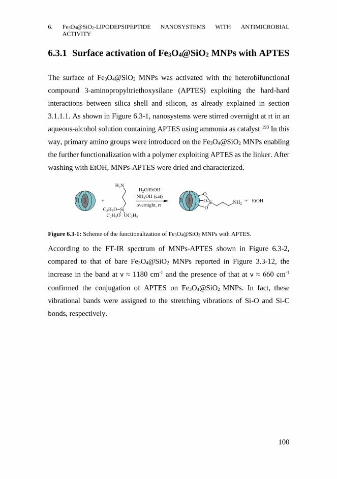
6. Fe3O4@SiO2-LIPODEPSIPEPTIDE NANOSYSTEMS WITH ANTIMICROBIAL
ACTIVITY
100
6.3.1 Surface activation of Fe3O4@SiO2 MNPs with APTES
The surface of Fe3O4@SiO2 MNPs was activated with the heterobifunctional
compound 3-aminopropyltriethoxysilane (APTES) exploiting the hard-hard
interactions between silica shell and silicon, as already explained in section
3.1.1.1. As shown in Figure 6.3-1, nanosystems were stirred overnight at rt in an
aqueous-alcohol solution containing APTES using ammonia as catalyst.193 In this
way, primary amino groups were introduced on the Fe3O4@SiO2 MNPs enabling
the further functionalization with a polymer exploiting APTES as the linker. After
washing with EtOH, MNPs-APTES were dried and characterized.
Figure 6.3-1: Scheme of the functionalization of Fe3O4@SiO2 MNPs with APTES.
According to the FT-IR spectrum of MNPs-APTES shown in Figure 6.3-2,
compared to that of bare Fe3O4@SiO2 MNPs reported in Figure 3.3-12, the
increase in the band at ν ≈ 1180 cm-1 and the presence of that at ν ≈ 660 cm-1
confirmed the conjugation of APTES on Fe3O4@SiO2 MNPs. In fact, these
vibrational bands were assigned to the stretching vibrations of Si-O and Si-C
bonds, respectively.
Fe3O
4
SiO
2
Fe3O
4
SiO
2

6. Fe3O4@SiO2-LIPODEPSIPEPTIDE NANOSYSTEMS WITH ANTIMICROBIAL
ACTIVITY
101
Figure 6.3-2: FT-IR spectrum of MNPs-APTES in which the vibrational bands were assigned to Si-
O, Si-C and Fe-O bonds.
6.3.2 Functionalization with poly-L-aspartic acid
After the surface activation, MNPs-APTES were functionalized with the
polyelectrolyte poly-L-aspartic acid (PLA). By introducing anionic groups, the
colloidal features of the nanoparticles could be increased, enhancing their water
dispersibility and minimizing the aggregation process among nanoparticles.
Moreover, PLA could improve the biocompatibility and biodegradability of the
entire nanosystem, being a low cytotoxic polymer.194,195 Compared to the
modification with single carboxylic acid molecules, the presence of several -
COOH groups increased the numbers of suitable binding sites for the modification
of MNPs with the bioactive molecules. The carboxyl groups could, therefore, be
exploited for both the anchorage on nanoparticles and the functionalization with
the lipodepsipeptides. Moreover PLA resulted an interesting polymer for
biomedical application both as metal chelator and drug carrier, alone or in
combination with other polymers like PEG and poly-ethylene oxide.196-198
5001000150020002500300035004000
Fe3O
4@SiO
2-APTES MNPs
T (
%)
(cm-1)
1180 cm-1
Si-O-Si
562 cm-1
Fe-O
3530 cm-1
O-H
664 cm-1
Si-C

6. Fe3O4@SiO2-LIPODEPSIPEPTIDE NANOSYSTEMS WITH ANTIMICROBIAL
ACTIVITY
102
As shown in Figure 6.3-3, the reaction was performed stirring the MNPs-APTES
in PLA solution overnight at rt. The activation of the -COOH groups of PLA with
EDC/NHS was carried out in PBS solution at slightly basic pH to improve the
deprotonation of APTES ammine groups, thus facilitating amide bonds formation.
Figure 6.3-3: Reaction scheme of the functionalization of MNPs-APTES with PLA activating the -
COOH with EDC/NHS.
The physicochemical characterization of the MNPs-PLA was performed by means
of FT-IR. Analysing the spectrum shown in Figure 6.3-4, in addition to the
vibrational bands belonging to the inorganic structure of the nanosystems, those
at ν 3270 cm-1, 1739 cm-1 and 1515 cm-1 could be assigned to the -OH and C=O
groups of the free carboxylic groups and of the amide bonds, confirming the
conjugation of the polymer.
Figure 6.3-4: FT-IR spectrum of poly-L-aspartic acid, addressing the vibrational bands to the
corresponding bonds.
Fe3O
4
SiO
2
Fe3O
4
SiO
2
5001000150020002500300035004000
MNPs-APTES-PLA
T (
%)
(cm-1)
1180 cm-1
Si-O-Si 562 cm-1
Fe-O
1515 cm-1
N-C=O
3270 cm-1
O-H
664 cm-1
Si-C
1739 cm-1
C=O

6. Fe3O4@SiO2-LIPODEPSIPEPTIDE NANOSYSTEMS WITH ANTIMICROBIAL
ACTIVITY
103
6.3.3 Conjugation of syringomycin E or syringopeptin 22A
SRE and SP22A were selected for the modification of the nanoparticles due to
their antibiotic activities; in fact, these lipodepsipeptides could act as therapeutic
agents for the treatment of bacterial or fungal infections. Combining these
properties with the diagnostic tool represented by the magnetic core of the
Fe3O4@SiO2 MNPs, multifunctional nanosystems could therefore be developed.
SRE and SP22A were supplied by the group of Prof. Grgurina.
The peptide moieties of both lipodepsipeptides contained basic Dab residues, that
could be exploited for the conjugation on MNPs-PLA. Since the functionalization
reaction should be performed at basic pH, preliminary studies were planned to
evaluate the stability of the compounds in these experimental conditions. In fact,
the hydrolysis of the macrocyclic lactone and/or of the L-Thr (4-Cl) residue at
basic pH could occur, compromising the antimicrobial activity of SRE and
SP22A.181
6.3.3.1 Evaluation of SRE and SP22A chemical stability
The maintenance of the cyclic structure of SRE and SP22A in conditions similar
to the experimental ones (120 min, pH 8, T 0°C), was evaluated by means of
circular dichroism (CD) technique. In fact, a variation of the conformation of these
compounds, derived from the lactone hydrolysis, could cause modifications in the
CD spectrum due to the change in the intramolecular conformational constrains.
CD spectrum, recorded in the far-UV region, of SP22A aqueous solution
maintained at 4°C is shown in Figure 6.3-5; it is characterized by optical bands at
λmin 213 nm and λmax 233 nm. CD spectrum of SP22A - start (black line)
corresponded to the starting aqueous solution of SP22A at pH 6.5. After increasing
the pH to 8, CD spectra were recorded at times ranging from 0 min to 3 days.
Analysing the experimental results, no significant differences could be found in

6. Fe3O4@SiO2-LIPODEPSIPEPTIDE NANOSYSTEMS WITH ANTIMICROBIAL
ACTIVITY
104
the CD spectra corresponding to SP22A solutions until 24h at pH 8. This result
shows that the native conformation of SP22A in solution was maintained. Slight
modification of the curve profile could be seen comparing the samples before
(SP22A – start) and after (SP22A – 0 min) the pH increase; these modifications
could be related to the change in the ionic strength 199 or to the beginning of the
formation of tiny amounts of the open form of the peptide, confirmed by
microbiological assays. After three days at pH 8, the CD spectrum of SP22A
changed significantly, suggesting the opening of the lactone ring.
Figure 6.3-5: CD spectra of a SP22A solution maintained at 4°C recorded before (black line) and
after increasing the pH. Blue line corresponded to SP22A – 0 min, acquired upon the addition of the
buffer. The other CD spectra were recorded at 30 min (green line), 60 min (red line), 90 min (purple
line), 120 min (orange line), 24 h (light blue line) and 3 days (light green line). The slight
modifications in the CD spectrum of SP22A at 0 min compared to the start sample were due to the
change of solvent while the variation of the curve profile in the CD spectrum of SP22A – 3 days
was related to the lactone hydrolysis.
Conformational changes of SRE were evaluated in a similar way by means of CD
spectra recorded in the far-UV region, maintaining the SRE solution at 4°C and
increasing the pH to 8. The spectra, shown in Figure 6.3-6, were characterized by
a broad negative band centered at λmin 230 nm, in agreement with data reported in
the literature.200 The starting solution (pH 6.5) corresponding to sample SRE start
produced a slightly different curve compared to those obtained after adding PBS
buffer, due to the change of the solvent, as already reported in the literature for
-10
-5
0
5
10
210 220 230 240 250 260
SP22A
SP22A - start
SP22A- 0 min
SP22A - 30min
SP22A - 60 min
SP22A- 90 min
SP22A - 120 min
SP22A - 24 h
SP22A - 3 days
(
md
eg c
m-1)
(nm)
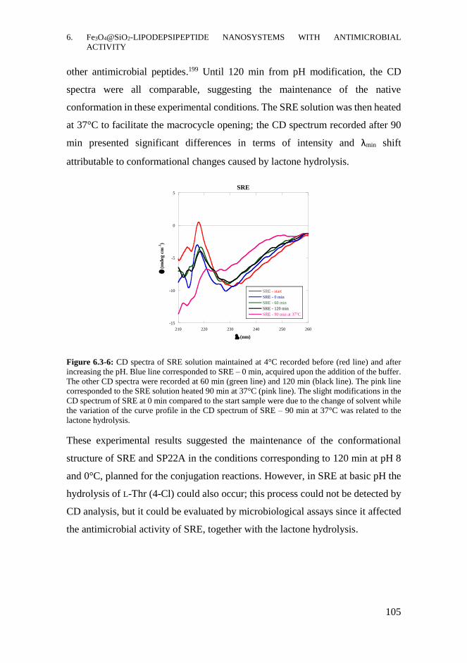
6. Fe3O4@SiO2-LIPODEPSIPEPTIDE NANOSYSTEMS WITH ANTIMICROBIAL
ACTIVITY
105
other antimicrobial peptides.199 Until 120 min from pH modification, the CD
spectra were all comparable, suggesting the maintenance of the native
conformation in these experimental conditions. The SRE solution was then heated
at 37°C to facilitate the macrocycle opening; the CD spectrum recorded after 90
min presented significant differences in terms of intensity and λmin shift
attributable to conformational changes caused by lactone hydrolysis.
Figure 6.3-6: CD spectra of SRE solution maintained at 4°C recorded before (red line) and after
increasing the pH. Blue line corresponded to SRE – 0 min, acquired upon the addition of the buffer.
The other CD spectra were recorded at 60 min (green line) and 120 min (black line). The pink line
corresponded to the SRE solution heated 90 min at 37°C (pink line). The slight modifications in the
CD spectrum of SRE at 0 min compared to the start sample were due to the change of solvent while
the variation of the curve profile in the CD spectrum of SRE – 90 min at 37°C was related to the
lactone hydrolysis.
These experimental results suggested the maintenance of the conformational
structure of SRE and SP22A in the conditions corresponding to 120 min at pH 8
and 0°C, planned for the conjugation reactions. However, in SRE at basic pH the
hydrolysis of L-Thr (4-Cl) could also occur; this process could not be detected by
CD analysis, but it could be evaluated by microbiological assays since it affected
the antimicrobial activity of SRE, together with the lactone hydrolysis.
-15
-10
-5
0
5
210 220 230 240 250 260
SRE
SRE - start
SRE - 0 min
SRE - 60 min
SRE - 120 min
SRE - 90 min at 37°C
(
md
eg c
m-1
)
(nm)

6. Fe3O4@SiO2-LIPODEPSIPEPTIDE NANOSYSTEMS WITH ANTIMICROBIAL
ACTIVITY
106
For these reasons, the further confirmation of the stability of SRE and SP22A was
achieved by evaluating the antimicrobial effect of the solutions, used for CD
analysis, by biological in vitro experiments on yeast cultures of R. pilimanae.
6.3.3.2 Functionalization of nanosystems with SRE and SP22A
The functionalization of the nanosystems was therefore carried out in a one-step
reaction, stirring the MNPs-PLA with EDC/NHS solution, for the activation of -
COOH groups, and SRE or SP22A solution, as shown in Figure 6.3-7. The
reactions were performed in PBS solution at pH 8, maintaining the T at 0°C for 2
h, to allow the formation of amide bonds. The covalent bonds were formed in
addition to the spontaneous ionic interactions occurring among negatively charged
MNPs-PLA and positively charged lipodepsipeptides.
Figure 6.3-7: Scheme of the functionalization of MNPs-PLA with SRE or SP22A activating the -
COOH groups with EDC and NHS.
After separating the nanosystems, the solutions corresponding to the supernatants
and the following three washings with H2O were used for the further
quantification of the loading. The products MNPs-SRE and MNPs-SP22A were
finally dried under vacuum. The quantification of the immobilization yields, by
evaluating the residual amount of the lipodepsipeptides in the supernatants was
performed by the group of Prof. Grgurina. Briefly, the supernatants were applied
on a Sep-Pak C₁₈ Cartridge conditioned according to the manufacturer’s
instructions. EDC and NHS were removed eluting with the mixture MeOH:H2O
5:95 containing 0.1% TFA while the SP22A and SRE were eluted with the mixture
acetonitrile:isopropyl alcohol 80:20 containing 0.1% TFA. Their amounts were
estimated by means of UV-Vis spectroscopy using ε280=1280 M-1 cm-1 and
ε240=9940 M-1 cm-1 for SP22A and SRE, respectively. The immobilization yields
Fe3O
4
SiO
2
Fe3O
4
SiO
2

6. Fe3O4@SiO2-LIPODEPSIPEPTIDE NANOSYSTEMS WITH ANTIMICROBIAL
ACTIVITY
107
resulted in 36.5% for MNPs-SP22A and 55% MNPs-SRE, corresponding to 36
μg SP22A/mg of MNPs-SP22A and 18 μg SRE/mg of MNPs-SRE.
6.3.3.3 Evaluation of the covalently and ionically bound SRE and
SP22A
To study the nature of the binding among the lipodepsipeptides and the
nanosystems, MNPs-SRE and MNPs-SP22A were treated with KCl 0.2 M. In this
way, the estimation of the amount of covalently bound and electrostatically
interacting lipodepsipeptides can be achieved; in fact, the fraction of SRE and
SP22A bound by ionic interactions could be detached using the high ionic strength
solution. After the treatment, MNPs-SRE and MNPs-SP22A were separated and
the lipodepsipeptides present in the supernatants were quantified by UV-Vis
spectroscopy, as previously described. The ionically bound amount was estimated
of 1% and 25% for SP22A and SRE respectively.
6.3.4 In vitro growth inhibition assays on R. pilimanae
The antimicrobial effects of MNPs-SRE and MNPs-SP22A were evaluated by in
vitro growth inhibition assays on R. pilimanae cell cultures. This yeast is
commonly used to assess the antifungal activity of the compounds since it is a
non-pathogenic microorganism easily to growth. The experiments, performed by
the group of Prof. Grgurina, were carried out plating the yeast in Petri dishes
covered by PDA medium. The plates were treated with serial 1:1 dilutions of SRE
or SP22A, free or bound to the nanosystems, or of MNPs-PLA and the yeast
cultures were growth for 48 h at 25°C. For the assays, concentrations of free
antibiotic compounds comparable to those of SRE and SP22A bound to the
nanosystems, quantified as described in section 6.3.3.2, were used. Moreover,
calibration curves correlating the diameter of the inhibition halos with the

6. Fe3O4@SiO2-LIPODEPSIPEPTIDE NANOSYSTEMS WITH ANTIMICROBIAL
ACTIVITY
108
concentration of free SRE and SP22A were used for estimating the antibiotic
activity of the nanosystems.
The results of the growth inhibition assays on R. pilimanae are shown in Figure
6.3-8. The bare nanoparticles MNPs-PLA did not produce any antimicrobial effect
on yeast growth, proving the biocompatibility of these nanosystems. The dilutions
of free SRE and free SP22A caused growth inhibition and also MNPs-SRE and
MNPs-SP22A inhibited yeast growth, especially the first nanosystem. In fact,
around the deposition points of free compounds or nanoparticles, yeast colonies
were not able to growth, as demonstrated by the presence of inhibition halos. Since
the absence of antimicrobial effects of MNPs-PLA, the antibiotic activity of
MNPs-SRE and MNPs-SP22A could only be derived from the bound
lipodepsipeptides. These experimental evidences, on one hand, confirmed the
success of the functionalization process of the nanosystems and, on the other hand,
proved their antifungal activity.
Comparing the inhibition halos produced by MNPs-SRE with those of free SRE,
an improvement in the antifungal activity was achieved after immobilization; in
fact, serial dilutions of MNPs-SRE, up to the ninth one, caused growth inhibition
while the free SRE displayed antimicrobial activity up to the seventh serial
dilution. Once bound to the nanoparticles the antibiotic activity of SRE was three
times increased. This evidence agreed with the already reported in vitro
enhancement of the biological effects for other bacterial antimicrobial peptides
after conjugation on nanoparticles, for instance vancomycin.190 In fact, the
extended surface area of NPs allowed to concentrate the bioactive molecules,
increasing its local amount, which could act in a more effective way, resulting in
the enhancement of the antimicrobial activity compared to the free compound.
Concerning MNPs-SP22A, the antimicrobial activity on R. pilimanae was not so
evident as for MNPs-SRE, since only the most concentrated solution, deposited
on the point M in the plate, produced growth inhibition effects. Compared to free
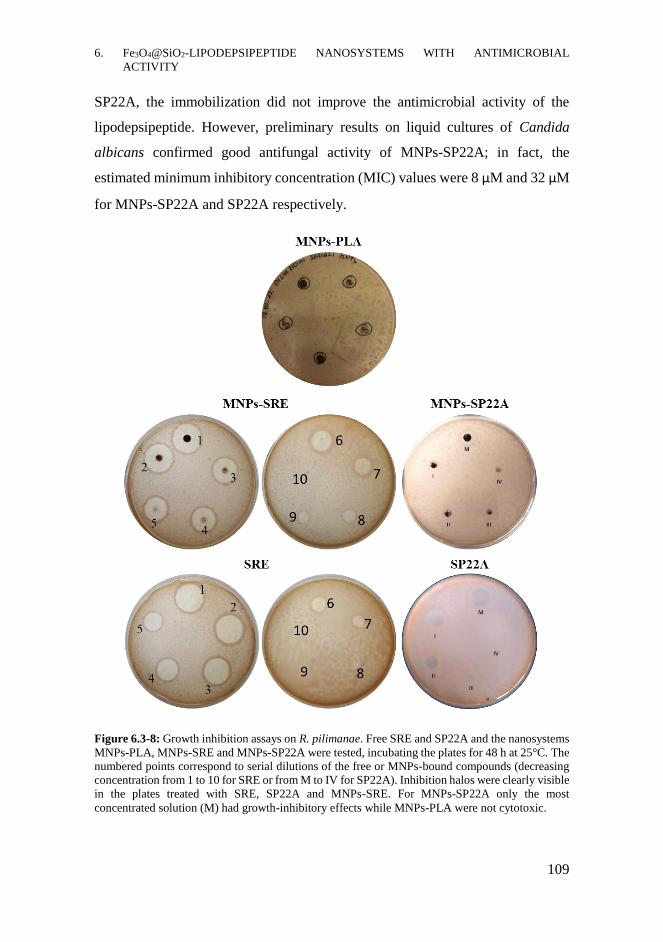
6. Fe3O4@SiO2-LIPODEPSIPEPTIDE NANOSYSTEMS WITH ANTIMICROBIAL
ACTIVITY
109
SP22A, the immobilization did not improve the antimicrobial activity of the
lipodepsipeptide. However, preliminary results on liquid cultures of Candida
albicans confirmed good antifungal activity of MNPs-SP22A; in fact, the
estimated minimum inhibitory concentration (MIC) values were 8 μM and 32 μM
for MNPs-SP22A and SP22A respectively.
Figure 6.3-8: Growth inhibition assays on R. pilimanae. Free SRE and SP22A and the nanosystems
MNPs-PLA, MNPs-SRE and MNPs-SP22A were tested, incubating the plates for 48 h at 25°C. The
numbered points correspond to serial dilutions of the free or MNPs-bound compounds (decreasing
concentration from 1 to 10 for SRE or from M to IV for SP22A). Inhibition halos were clearly visible
in the plates treated with SRE, SP22A and MNPs-SRE. For MNPs-SP22A only the most
concentrated solution (M) had growth-inhibitory effects while MNPs-PLA were not cytotoxic.
6. Fe3O4@SiO2-LIPODEPSIPEPTIDE NANOSYSTEMS WITH ANTIMICROBIAL
ACTIVITY
110
Supernatants and washings, collected during the functionalization reactions, were
also assessed by microbiological assays on R. pilimanae. The results confirmed
the presence of unreacted bioactive molecules in the reaction supernatants and
their absence in the washing solutions; in fact, the first produced yeast growth
inhibition, while the second did not show antimicrobial effects.
The antifungal activity of MNPs-SRE could derive from lipodepsipeptide both
ionically interacting and covalently bound to MNPs-PLA. After the detachment
of the ionically bound SRE treating the nanosystems with KCl solution, the
MNPs-SRE were still active. Even if covalent functionalization ensured higher
stability to the nanosystems, avoiding the undesired release of the bioactive
molecules in physiological environment, the ionically interacting fraction could
be more easily accessible; this binding, in fact, could provide the compound
readily available for the interactions with membranes, allowing a more effective
performance of the antimicrobial activity.
Therefore, MNPs-SRE and MNPs-SP22A showed interesting antimicrobial
activity paving the way to new promising nanosystems for the treatment of fungal
infections. According to the biological effects of SRE and SP22A against other
microorganisms, in addition to the imaging properties of the magnetite core, those
nanosystems might be suitable for both diagnosis and treatment of other kinds of
infection diseases, involving for instance bacteria or mycobacteria.
6.3.5 In vitro internalization in human immune cells
Considering that MNPs-SP22A displayed antifungal activity it was reasonable to
hypothesize that the applicability of these bioactive nanosystems could be
extended to other microorganisms, including pathogenic ones. Since some
microorganisms, once phagocyted by immune cells like macrophages, are able to
survive inside them in a latent state, the internalization of medicated nanoparticles
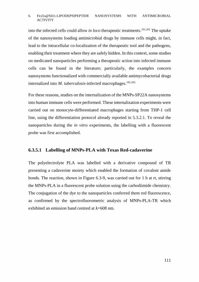
6. Fe3O4@SiO2-LIPODEPSIPEPTIDE NANOSYSTEMS WITH ANTIMICROBIAL
ACTIVITY
111
into the infected cells could allow in loco therapeutic treatments.191,201 The uptake
of the nanosystems loading antimicrobial drugs by immune cells might, in fact,
lead to the intracellular co-localization of the therapeutic tool and the pathogens,
enabling their treatment where they are safely hidden. In this context, some studies
on medicated nanoparticles performing a therapeutic action into infected immune
cells can be found in the literature; particularly, the examples concern
nanosystems functionalized with commercially available antimycobacterial drugs
internalized into M. tuberculosis-infected macrophages.192,202
For these reasons, studies on the internalization of the MNPs-SP22A nanosystems
into human immune cells were performed. These internalization experiments were
carried out on monocyte-differentiated macrophages starting from THP-1 cell
line, using the differentiation protocol already reported in 5.3.2.1. To reveal the
nanoparticles during the in vitro experiments, the labelling with a fluorescent
probe was first accomplished.
6.3.5.1 Labelling of MNPs-PLA with Texas Red-cadaverine
The polyelectrolyte PLA was labelled with a derivative compound of TR
presenting a cadaverine moiety which enabled the formation of covalent amide
bonds. The reaction, shown in Figure 6.3-9, was carried out for 1 h at rt, stirring
the MNPs-PLA in a fluorescent probe solution using the carbodiimide chemistry.
The conjugation of the dye to the nanoparticles conferred them red fluorescence,
as confirmed by the spectrofluorometric analysis of MNPs-PLA-TR which
exhibited an emission band centred at λ=608 nm.

6. Fe3O4@SiO2-LIPODEPSIPEPTIDE NANOSYSTEMS WITH ANTIMICROBIAL
ACTIVITY
112
Figure 6.3-9: Reaction scheme of the labelling of MNPs-PLA with Texas Red-cadaverine.
After the labelling, SP22A was conjugated on MNPs-PLA-TR following the
protocol previously described in section 6.3.3.2. SP22A was selected instead of
SRE for its well-known cytotoxic effect towards M. smegmatis, belonging to
Mycobacteriaceae family. This group contains several pathogens hard to treat,
particularly M. tuberculosis which is able to hide and survive inside macrophages.
6.3.5.2 Internalization in monocyte-differentiated macrophages
In vitro biological experiments were performed to demonstrate the uptake of
MNPs-PLA-TR and MNPs-SP22A-TR by monocyte-differentiated macrophages.
THP-1 cell line was first differentiated treating the monocytes with PMA, as
described in section 5.3.2.1. The macrophages were then incubated for 2 h at 37°C
with dispersions of MNPs-PLA-TR or MNPs-PLA-SP22A-TR, filtered on 3 μm
filters. The filtration was performed in order to remove big aggregates of
nanosystems which can endanger cell vitality. Untreated cells, maintained in the
same experimental conditions, were used as negative control. After incubation,
cells were washed several times with buffer solution and fixed with
paraformaldehyde solution. After staining cell nuclei, cells were analysed by
confocal fluorescence microscopy.
Fe3O
4
SiO
2
Fe3O
4
SiO
2
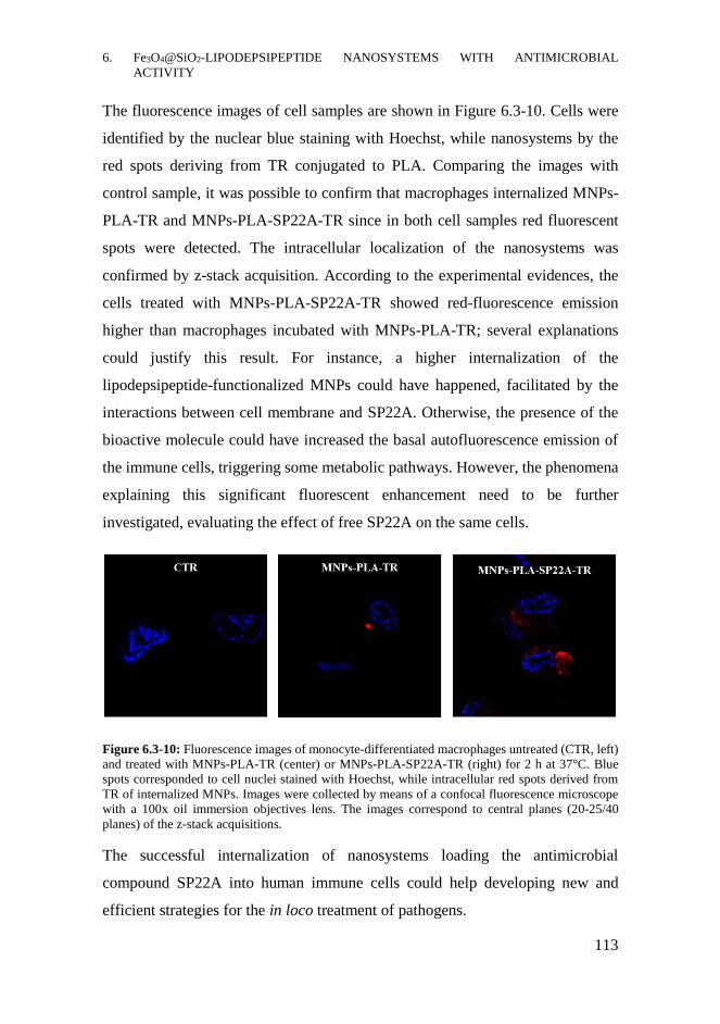
6. Fe3O4@SiO2-LIPODEPSIPEPTIDE NANOSYSTEMS WITH ANTIMICROBIAL
ACTIVITY
113
The fluorescence images of cell samples are shown in Figure 6.3-10. Cells were
identified by the nuclear blue staining with Hoechst, while nanosystems by the
red spots deriving from TR conjugated to PLA. Comparing the images with
control sample, it was possible to confirm that macrophages internalized MNPs-
PLA-TR and MNPs-PLA-SP22A-TR since in both cell samples red fluorescent
spots were detected. The intracellular localization of the nanosystems was
confirmed by z-stack acquisition. According to the experimental evidences, the
cells treated with MNPs-PLA-SP22A-TR showed red-fluorescence emission
higher than macrophages incubated with MNPs-PLA-TR; several explanations
could justify this result. For instance, a higher internalization of the
lipodepsipeptide-functionalized MNPs could have happened, facilitated by the
interactions between cell membrane and SP22A. Otherwise, the presence of the
bioactive molecule could have increased the basal autofluorescence emission of
the immune cells, triggering some metabolic pathways. However, the phenomena
explaining this significant fluorescent enhancement need to be further
investigated, evaluating the effect of free SP22A on the same cells.
Figure 6.3-10: Fluorescence images of monocyte-differentiated macrophages untreated (CTR, left)
and treated with MNPs-PLA-TR (center) or MNPs-PLA-SP22A-TR (right) for 2 h at 37°C. Blue
spots corresponded to cell nuclei stained with Hoechst, while intracellular red spots derived from
TR of internalized MNPs. Images were collected by means of a confocal fluorescence microscope
with a 100x oil immersion objectives lens. The images correspond to central planes (20-25/40
planes) of the z-stack acquisitions.
The successful internalization of nanosystems loading the antimicrobial
compound SP22A into human immune cells could help developing new and
efficient strategies for the in loco treatment of pathogens.

6. Fe3O4@SiO2-LIPODEPSIPEPTIDE NANOSYSTEMS WITH ANTIMICROBIAL
ACTIVITY
114
CHAPTER CONTRIBUTIONS
- Nanosystems loading the lipodepsipeptide SRE and SP22A were
developed for the first time, conjugating the bioactive molecules to
Fe3O4@SiO2 MNPs activated with APTES and PLA.
- The immobilization of SRE and SP22A did not cause the loss of their
biological activity since the MNPs-SRE and MNPs-SP22A produced
growth inhibitory effects on R. pilimanae cell cultures; actually, the
immobilization of SRE on MNPs-PLA increased three-times the
antimicrobial activity, compared to free SRE.
- MNPs- SP22A labelled with the fluorescent probe TR-cadaverine were
internalized by monocyte-differentiated macrophages, paving the way to
promising treatments with medicated nanoparticles against pathogens
directly inside cells.
6.4.1 Future perspectives
- Evaluate the antimicrobial activity of MNPs-SRE and MNPs-SP22A
against other microorganisms, such as pathogenic bacteria, to extend the
applicability of these novel nanosystems. Estimate the MIC values by
microbiological assays in liquid batch cultures.
- Perform internalization experiments of MNPs-SP22A and free SP22A by
monocyte-differentiated macrophages to understand the internalization
mechanisms and evaluate the release/degradation of the nanosystems
inside the cells.
6. Fe3O4@SiO2-LIPODEPSIPEPTIDE NANOSYSTEMS WITH ANTIMICROBIAL
ACTIVITY
115
ACKNOWLEDGMENTS
Prof. Ingeborg Grgurina and her group at Sapienza University of Rome for
microbiological experiments. Prof. Maria Pia Donzello at Sapienza University of
Rome for FTIR analysis. Dr. Valeria de Turris of centre for life
nanoscience@Sapienza – Istituto Italiano di Tecnologia – for confocal
fluorescence microscopy.
MATERIALS AND METHODS
Reagents, materials and equipment: All the chemical reagents were purchased by
Sigma-Aldrich (Merck KGaA, Germany) and used without further purification.
All the material used during in vitro biological experiments were purchased from
Corning (Corning, USA). Permanent magnets made of Neodymium-Iron-Boron
alloy, having a magnetic field of 0.3 T, were used for magnetic separations. The
spectropolarimeter Jasco J715 was utilized for the circular dichroism experiments,
using Suprasil® quartz cell for kinetic measurement and acquiring the spectra with
Spectra ManagerTM software. Steady-state fluorescence spectra were collected by
Cary Eclypse (Varian) spectrofluorometer, equipped with the Xe lamp using
Suprasil® quartz cell. Sep-Pak C₁₈ Cartridge was supplied by Waters (USA). The
experimental data were elaborated with the software Kaleidagraph (Synergy
Software, USA). The fluorescence confocal analysis was performed at the Centre
for Life Nanoscience IIT@Sapienza by means of the inverted microscope
Olympus X73 equipped with 100X oil immersion objectives lens. The
fluorescence images were collected by z-stack mode acquiring 40 planes divided
by 0.2 μm, and were elaborated by ImageJ software.
6. Fe3O4@SiO2-LIPODEPSIPEPTIDE NANOSYSTEMS WITH ANTIMICROBIAL
ACTIVITY
116
Surface activation with APTES: MNPs-APTES: 400 mg of Fe3O4@SiO2 MNPs
were dispersed in 16 mL of bi-distilled H2O and 40 mL of EtOH dissolving 4 mL
of APTES (17 mmol) and 40 μL of NH4OH were added to the dispersion. The
mixture was mechanically stirred overnight at rt. MNPs-APTES were
magnetically separated, washed with EtOH and H2O and dried under vacuum.
Conjugation of PLA:MNPs-PLA: 400 mg of MNPs-APTES were dispersed in 20
mL of PBS (0.1 M pH 8). 20 mg of PLA were dissolved in NaOH and diluted to
10 mL with PBS (0.1 M pH 6) containing EDC (0.3 mmol) and NHS (0.3 mmol).
The reaction was mechanically stirred overnight at rt. MNPs-PLA were
magnetically separated, washed with H2O and dried under vacuum.
CD studies on SRE and SP22A: CD spectrum of 1 mL of an aqueous solution of
SP22A (0.36 mg/mL in bi-distilled H2O) at 4°C. After mixing with 1 mL of PBS
(0.2 M pH 8) CD spectra were recorded in the far-UV range at time ranging from
0 min to 3 days maintaining the solution at 4°C.
CD analysis of 1 mL of an aqueous solution of SRE (0.125 mg/mL in bi-distilled
H2O) at 4°C was performed. After the dilution with 1 mL of PBS (0.2 M pH 8)
CD spectra were recorded in the far-UV range at time ranging from 0 min to 2 h
maintaining the solution at 4°C. The solution was then heated at 37°C and after
90 min a CD spectrum was recorded.
Conjugation of SRE or SP22A: MNPs-SRE and MNPs-SP22A: 40 mg of MNPs-
PLA were dispersed in 2.4 mL of PBS (0.2 M pH 8) dissolving EDC (4.6* 10-5
mol) and NHS (4.6* 10-5 mol). 2.4 mL of an aqueous solution of SRE (1.2 mg)
were added to the dispersion and the mixture was mechanically stirred for 2 h at
0°C. After magnetic separation, 3 washings with bi-distilled H2O were performed
(1 mL each) and the MNPs-PLA-SRE were finally dried under vacuum.

6. Fe3O4@SiO2-LIPODEPSIPEPTIDE NANOSYSTEMS WITH ANTIMICROBIAL
ACTIVITY
117
The same protocol was used for the conjugation of SP22A, dispersing the MNPs-
PLA in 1.5 mL of PBS containing EDC/NHS and adding 1.5 mL of the SP22A (4
mg) aqueous solution.
Quantification of SRE or SP22A on MNPs-SRE and MNPs-SP22A: Sep-Pak C₁₈
Cartridge was used for the chromatographic purification of the supernatants of the
functionalization reaction of MNPs-PLA with SRE or SP22A. The cartridge was
conditioned with MeOH:H2O 90:10 containing 0.1% TFA (9 mL) and then
equilibrated with H2O + 0.1% TFA solution (3x6 mL). The supernatants (3
aliquots of 1mL) were eluted with the desalting solution (MeOH:H2O 5:95
containing 0.1% TFA, 3x3 mL) to remove EDC and NHS while the
lipodepsipeptides were eluted with the solvent mixture ACN:isopropanol 80:20
containing 0.1% TFA (3x3 mL). The quantification of the lipodepsipeptides
present in the eluted fractions was performed by UV-Vis spectroscopy using
ε280=1280 M-1 cm-1 and ε240=9940 M-1 cm-1 for SP22A and SRE, respectively.
Treatment with KCl solution: 11 mg of MNPs-SP22A or MNPs-SRE were treated
for 5 min with 800 μL of KCl 0.2 M and centrifuged for 15 min at 14 000 rpm at
4°C, repeating three times the procedure. The ionically bound SP22A or SRE were
quantified in the supernatants by UV-Vis spectroscopy using ε280=1280 M-1 cm-1
and ε240=9940 M-1 cm-1 for SP22A and SRE, respectively.
Labelling with Texas Red-cadaverine: MNPs-PLA-TR: 100 mg of MNPs-PLA
were stirred for 30 min at rt in 5 mL of PBS (0.1 M pH 6) dissolving EDC (8.7*
10-5 mol) and NHS (8.7* 10-5 mol). After magnetic separation the supernatant was
removed and replaced by 5 mL of PBS (0.1 M, pH 8) containing TR-cadaverine
(7.2*10-7 mol in 100 μL ACN). After 1h stirring at rt in dark, the MNPs-PLA-TR
were magnetically collected, washed first with DMF and then several times with
bi-distilled H2O and dried under vacuum.
6. Fe3O4@SiO2-LIPODEPSIPEPTIDE NANOSYSTEMS WITH ANTIMICROBIAL
ACTIVITY
118
PMA-induced differentiation of THP-1 cell line: the same protocol reported in
section 5.6 was used to obtain adherent macrophages.
MNPs internalization: Monocyte-differentiated macrophages were incubated with
MNPs-PLA-TR and MNPs-PLA-SP22A-TR dispersions in PBS at starting
concentrations of 1 mg/mL, filtered on 3 μm filters. Cells incubated in the same
condition w/o MNPs were used as negative controls. After incubating 2 h at 37°C,
not internalized MNPs were removed by three washings with PBS. Cells were
fixed for 10 min at rt with paraformaldehyde and sucrose solution at 4% in PBS
(500 μL/well). After 3 washings with PBS, cells were treated 10 min with glycine
aqueous solution (200 mM, 500 μL/well) and, after washings with PBS, cell nuclei
were stained with Hoechst solution (5 μg/mL in bi-distilled H2O) for 5 min at rt.
Coverslips, present in each well, were mounted on slides with a glycerol solution
(90% in PBS) and confocal fluorescence analysis was performed.

7. ANTIBODY-FUNCTIONALIZED
MAGNETIC NANOPARTICLES FOR
THE DETECTION OF EPINEPHRINE
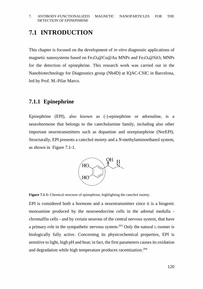
7. ANTIBODY-FUNCTIONALIZED MAGNETIC NANOPARTICLES FOR THE
DETECTION OF EPINEPHRINE
120
INTRODUCTION
This chapter is focused on the development of in vitro diagnostic applications of
magnetic nanosystems based on Fe3O4@Cu@Au MNPs and Fe3O4@SiO2 MNPs
for the detection of epinephrine. This research work was carried out in the
Nanobiotechnology for Diagnostics group (Nb4D) at IQAC-CSIC in Barcelona,
led by Prof. M.-Pilar Marco.
7.1.1 Epinephrine
Epinephrine (EPI), also known as (-)-epinephrine or adrenaline, is a
neurohormone that belongs to the catecholamine family, including also other
important neurotransmitters such as dopamine and norepinephrine (NorEPI).
Structurally, EPI presents a catechol moiety and a N-methylaminoethanol system,
as shown in Figure 7.1-1.
Figure 7.1-1: Chemical structure of epinephrine, highlighting the catechol moiety.
EPI is considered both a hormone and a neurotransmitter since it is a biogenic
monoamine produced by the neuroendocrine cells in the adrenal medulla -
chromaffin cells - and by certain neurons of the central nervous system, that have
a primary role in the sympathetic nervous system.203 Only the natural L-isomer is
biologically fully active. Concerning its physicochemical properties, EPI is
sensitive to light, high pH and heat; in fact, the first parameters causes its oxidation
and degradation while high temperature produces racemization.204
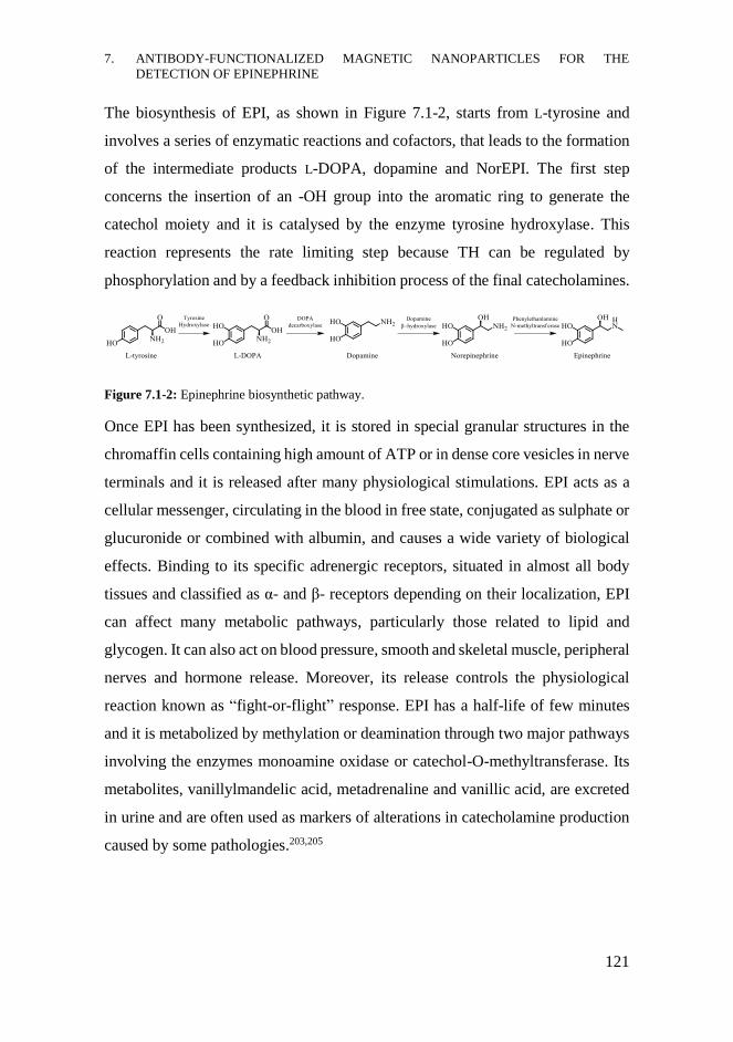
7. ANTIBODY-FUNCTIONALIZED MAGNETIC NANOPARTICLES FOR THE
DETECTION OF EPINEPHRINE
121
The biosynthesis of EPI, as shown in Figure 7.1-2, starts from L-tyrosine and
involves a series of enzymatic reactions and cofactors, that leads to the formation
of the intermediate products L-DOPA, dopamine and NorEPI. The first step
concerns the insertion of an -OH group into the aromatic ring to generate the
catechol moiety and it is catalysed by the enzyme tyrosine hydroxylase. This
reaction represents the rate limiting step because TH can be regulated by
phosphorylation and by a feedback inhibition process of the final catecholamines.
Figure 7.1-2: Epinephrine biosynthetic pathway.
Once EPI has been synthesized, it is stored in special granular structures in the
chromaffin cells containing high amount of ATP or in dense core vesicles in nerve
terminals and it is released after many physiological stimulations. EPI acts as a
cellular messenger, circulating in the blood in free state, conjugated as sulphate or
glucuronide or combined with albumin, and causes a wide variety of biological
effects. Binding to its specific adrenergic receptors, situated in almost all body
tissues and classified as α- and β- receptors depending on their localization, EPI
can affect many metabolic pathways, particularly those related to lipid and
glycogen. It can also act on blood pressure, smooth and skeletal muscle, peripheral
nerves and hormone release. Moreover, its release controls the physiological
reaction known as “fight-or-flight” response. EPI has a half-life of few minutes
and it is metabolized by methylation or deamination through two major pathways
involving the enzymes monoamine oxidase or catechol-O-methyltransferase. Its
metabolites, vanillylmandelic acid, metadrenaline and vanillic acid, are excreted
in urine and are often used as markers of alterations in catecholamine production
caused by some pathologies.203,205
7. ANTIBODY-FUNCTIONALIZED MAGNETIC NANOPARTICLES FOR THE
DETECTION OF EPINEPHRINE
122
7.1.1.1 Clinical importance of epinephrine
Many diseases and altered physiological conditions are associated with elevated
excretion of catecholamines, including EPI. It is known that the production of EPI
increases in stressful conditions and, for this reason, it has been termed as an
“emergency” hormone and it can be used for monitoring the stress level of a
patient.
Several important diseases are correlated with alterations in EPI levels in
biological fluids; the normal endogenous levels of EPI for healthy people are very
low, <0.5 nM (100 pg/mL) in plasma and <190 nmol/day in 24h-urine.206-208
Variations in its concentrations are caused by many factors such as, for instance,
stress, exercise, smoking, pain and hypertension. The measurement of adrenaline
levels is very helpful for the diagnosis of some pathologies, particularly
catecholamine-secreting tumours. In fact, pheochromocytoma, paraganglioma,
ganglioneuroma and neuroblastoma are tumours of the adrenal medulla normally
associated with hyperproduction and excess of catecholamines in plasma and
urine, including EPI and its metabolites. Some of these tumours are malignant and
can lead to death, especially neuroblastoma for childhood.209 The determination
of biological levels of catecholamines, including EPI, is also important for the
diagnosis of some neurodegenerative diseases, particularly Parkinson’s disease,
or mental disorders. Moreover, elevated excretion of EPI is associated with
cardiovascular disease, for instance hypertension that can be related to the
presence of pheochromocytoma. EPI has also an important role as drug; in fact, it
is normally used to treat anaphylaxis and cardiac arrest.207
Since EPI triggers a variety of biological effects on all the body tissues and is
involved in some important pathologies, its quantitative determination in
biological fluids, such as plasma and urine, has become very important for clinical
diagnosis.
7. ANTIBODY-FUNCTIONALIZED MAGNETIC NANOPARTICLES FOR THE
DETECTION OF EPINEPHRINE
123
7.1.1.2 Detection methods for EPI in the literature
Due to the low concentration of EPI in biological fluids, very sensitive analytical
methods are required for its detection. Chromatographic techniques are the most
sensitive techniques since high-performance liquid chromatography (HPLC) with
electrochemical or fluorescence detection or HPLC coupled to tandem mass
spectrometry (LC-MS/MS) can reach limit of detection (LOD) values ranging
from pM to nM.207,210,211 Electrochemical methods are mainly used for the
detection of EPI thanks to the possibility of exploiting the electroactive behaviour
of the catecholamine at certain potentials.212 One limitation of this kind of readout
is the presence of other compounds interfering during the detection process,
especially ascorbic acid or drugs, such as paracetamol. Modifications of the
electrode can improve the sensitivity of the electrochemical detection achieving
LOD values ranging from nM to μM.213
For these reasons, sample pre-treatments, pre-concentration or derivatization are
often needed. Liquid-liquid or solid-phase extractions with C18 column or
Sephadex® G-10, cation-exchange resin and alumina are some of the techniques
used for non-selective preparation of the sample.207,214 More selective strategies
concern the use of boronated gels or other platforms immobilizing boronic acid
since this compound is able to form a stable complex with the catechol moiety of
the catecholamines, allowing their almost selective extraction. Biosensors,
especially based on amperometric detection, are also used for the detection of EPI;
the immobilization of one or more enzymes able to oxidize the analyte - such as
Horseradish Peroxidase (HRP) - and amplify the signal are used for the
quantification of the analyte, leading to LOD values of few μM.215
Improvements in pre-treatment steps or in the detection methods are coming from
nanotechnology. Some examples have been reported in the literature.
Derivatization or modification of common measurement electrodes with
nanomaterials, like noble metal nanoparticles or carbon nanotubes, are improving
7. ANTIBODY-FUNCTIONALIZED MAGNETIC NANOPARTICLES FOR THE
DETECTION OF EPINEPHRINE
124
the sensitivity of electrochemical detection, leading to LOD around 2-5 µM.216,217
The use of functionalized nanoparticles for rapid and more selective extraction of
the analyte has also been studied; for instance, MNPs modified with boronic acid
derivatives are interesting tools to help the selective extraction process, achieving
LOD around few ng/mL.218,219 A more recent example concerns the use of
magnetic nanoparticles functionalized with supramolecular molecules that could
be used for loading EPI.220 Also fluorescence detection of EPI can be more easily
achieved using nanosystems; for instance, the quantification can be carried out
monitoring the inhibition of the Fe3O4 nanoparticle-mediated production of a
fluorescent compound in the presence of the analyte, leading to LOD values of
few nM.221
7.1.2 Immunoassays and immunosensors
An immunochemical technique is based on the specific recognition phenomena
between an antibody and an epitope, due to high chemical affinity that leads to the
binding process. Usually, labels are needed in order to detect and monitor the
immunochemical interaction, except for some label-free biosensors based on
surface plasmon resonance (SPR) or impedance.222,223 Chemiluminescent,
fluorescent or enzymatic labels, like HRP or alkaline phosphatase, are the most
common tools for revealing the antibody-epitope affinity. Among these
bioanalytical methods, the ELISA and the immunosensors are widely used.
Concerning immunosensors, they are compact analytical devices in which the
immunochemical interaction is detected and converted to an electrical signal by
means of a transducer.224 According to the sensing principle, the immunosensors
can be distinguished in electrochemical, optical or piezoelectrical ones. In fact,
the antibody-epitope binding can produce electroactive species or changes in the
optical properties or in the total mass of the system; by means of a proper
transducer, all these signals are finally converted in electric ones.
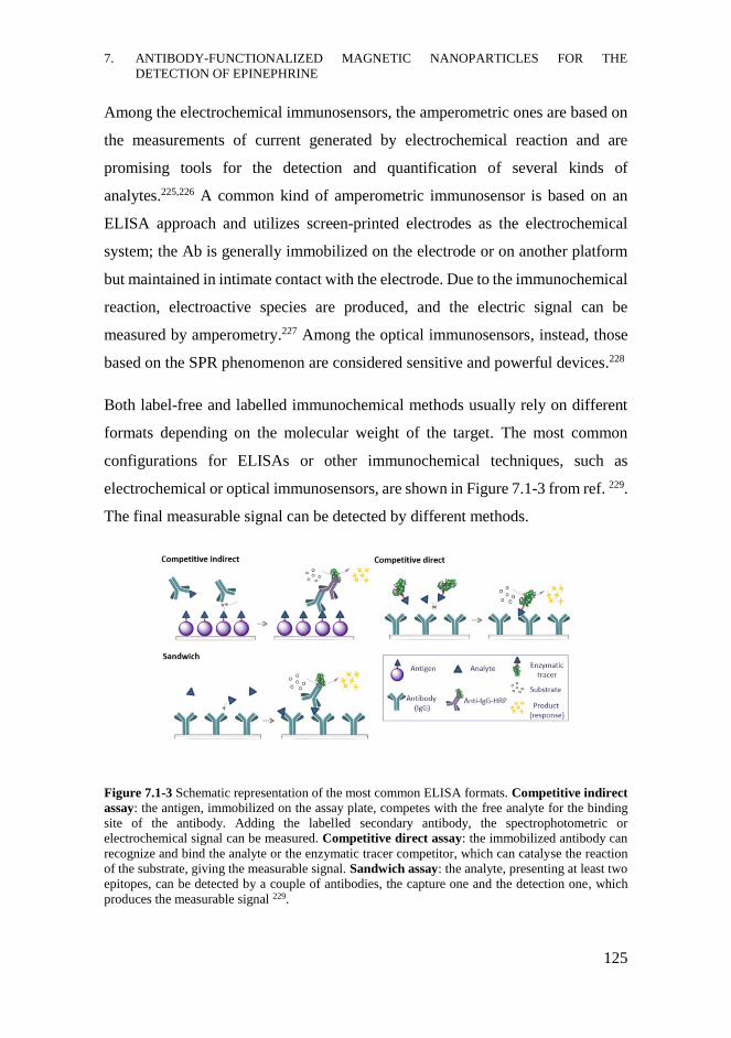
7. ANTIBODY-FUNCTIONALIZED MAGNETIC NANOPARTICLES FOR THE
DETECTION OF EPINEPHRINE
125
Among the electrochemical immunosensors, the amperometric ones are based on
the measurements of current generated by electrochemical reaction and are
promising tools for the detection and quantification of several kinds of
analytes.225,226 A common kind of amperometric immunosensor is based on an
ELISA approach and utilizes screen-printed electrodes as the electrochemical
system; the Ab is generally immobilized on the electrode or on another platform
but maintained in intimate contact with the electrode. Due to the immunochemical
reaction, electroactive species are produced, and the electric signal can be
measured by amperometry.227 Among the optical immunosensors, instead, those
based on the SPR phenomenon are considered sensitive and powerful devices.228
Both label-free and labelled immunochemical methods usually rely on different
formats depending on the molecular weight of the target. The most common
configurations for ELISAs or other immunochemical techniques, such as
electrochemical or optical immunosensors, are shown in Figure 7.1-3 from ref. 229.
The final measurable signal can be detected by different methods.
Figure 7.1-3 Schematic representation of the most common ELISA formats. Competitive indirect
assay: the antigen, immobilized on the assay plate, competes with the free analyte for the binding
site of the antibody. Adding the labelled secondary antibody, the spectrophotometric or
electrochemical signal can be measured. Competitive direct assay: the immobilized antibody can
recognize and bind the analyte or the enzymatic tracer competitor, which can catalyse the reaction
of the substrate, giving the measurable signal. Sandwich assay: the analyte, presenting at least two
epitopes, can be detected by a couple of antibodies, the capture one and the detection one, which
produces the measurable signal 229.

7. ANTIBODY-FUNCTIONALIZED MAGNETIC NANOPARTICLES FOR THE
DETECTION OF EPINEPHRINE
126
Kinds of immunochemical assay:
1. For high molecular weight analytes, like proteins or whole bacteria, sandwich
ELISA format is the most commonly used. Since the analyte presents at least two
different epitopes and its dimensions are quite big, the detection can be achieved
using a couple of antibodies: the capture antibody that is immobilized on the plate,
and the detection antibody, that is added free in solution and produces the
measurable signal proportional to the amount of the analyte, allowing its
quantification.
2. For low molecular weight analytes, such as EPI, the sandwich format cannot be
used. The detection can be achieved only using another molecule bigger than the
analyte, that competes with it for the binding site of the antibody. Due to the
presence of the competitor (generally named antigen (Ag)), that is usually a
bioconjugate formed by a hapten and a protein, these immunochemical assays are
referred to as competitive assays. Generally, a hapten can be defined as a small
and not immunogenic molecule, with physicochemical properties similar to the
target analyte, which can be bound to a different carrier protein according to the
purpose. For the immunization step, highly immunogenic proteins like Keyhole
Limpet hemocyanin (KLH) or Horseshoe Crab hemocyanin (HCH) are selected,
while for the development of the immunochemical assays, other macromolecules
such as proteins, polymers and enzymes are generally used.
The competitive immunoassays can be divided in direct and indirect formats. In
the first one the competitor is a hapten covalently attached to a label, referred to
as tracer, able to produce the signal. The indirect competitive assay requires the
addition of a labelled secondary antibody, specific for the constant fraction of the
primary antibody that produces the measurable signal.
Therefore, in the direct format the signal derives from the tracer while in the
indirect one it is produced by the labelled secondary antibody. For these reasons,
in both direct and indirect competitive immunochemical assays, the signal is

7. ANTIBODY-FUNCTIONALIZED MAGNETIC NANOPARTICLES FOR THE
DETECTION OF EPINEPHRINE
127
inversely proportional to the concentration of the analyte. This occurs because the
analyte and the antigen compete for the binding site of the antibody, depending
on their affinity constants (Kaff). The difference between the Kaff value of the
analyte and the competitor determines the preferential formation of the analyte-
antibody or competitor-antibody complex. Kaff-analyte much higher than Kaff-competitor
means low amount of antibody binding the competitor and finally producing the
measurable signal. For this reason, it is important to design and use competitors
having Kaff similar to that of the analyte, in order to ensure a good competition
between the Ag and the analyte, obtaining a significant signal.
The representation of the logarithm of the analyte concentration in front of the
signal gives the typical sigmoidal curve of a competitive assay, as reported in
Figure 7.1-4. Fitting the curve with a four-parameters equation, the analytical
features of the immunochemical assay are obtained.
Figure 7.1-4: Inhibition curve typical of both direct and indirect competitive ELISA assays,
measured by spectrophotometric (left side) or amperometric (right) techniques. In the curve fitting
equation, the four parameters are IC50 (half maximal inhibitory concentration), Ymax (maximum
value of absorbance or current), Ymin (minimum value of absorbance or current) and hillslope (slope
of the linear part of the curve).
7. ANTIBODY-FUNCTIONALIZED MAGNETIC NANOPARTICLES FOR THE
DETECTION OF EPINEPHRINE
128
7.1.2.1 Immunochemical methods involving magnetic nanoparticles
Nanotechnology can bring improvements in the analytical features of
immunochemical methods and practical advantages in the correlated experimental
protocols too. The introduction of nanotechnology in bioanalytical techniques, in
fact, can be very helpful to improve the reliability and the selectivity of
immunoassays in the detection of some analytes, particularly those characterized
by low molecular weight.230 Using NPs, the performances of immunochemical
devices can be enhanced thanks to the peculiar physicochemical features of
nanomaterials. For instance, the high surface area can lead to the achievement of
nanoplatforms with a high local quantity of biomolecules, such as antibodies,
increasing the interactions with the analyte and facilitating the biorecognition
process.
Among nanosystems exploitable in immunochemical assays, MNPs have received
great attentions.231 In fact, functionalizing MNPs with an antibody that
specifically recognize an analyte might be a good idea to obtain a nanoplatform
able to perform both the detection and the separation of the analyte from the
matrix. The specific biorecognition element, as the antibody is, can be combined
to the magnetic core of the nanosystems; after the analyte-antibody interaction,
the application of the external magnetic field can lead to the separation and
concentration of the target analyte. As reported in the literature, magnetic
nanosystems have been used in immunochemical biosensors and assays for the
detection of different kinds of analytes such as proteins, bacterial epitopes, viral
antigens and small analytes like toxins, vitamins, biomarkers or drugs in complex
matrices.65,228,232,233
For the immunochemical methods based on MNPs for the detection of small
analytes the direct competitive format is easier to perform. As shown in Figure
7.1-5, the experimental procedure consists in mixing the MNPs functionalized
with the appropriate specific Ab with the analyte and the enzymatic tracer. After
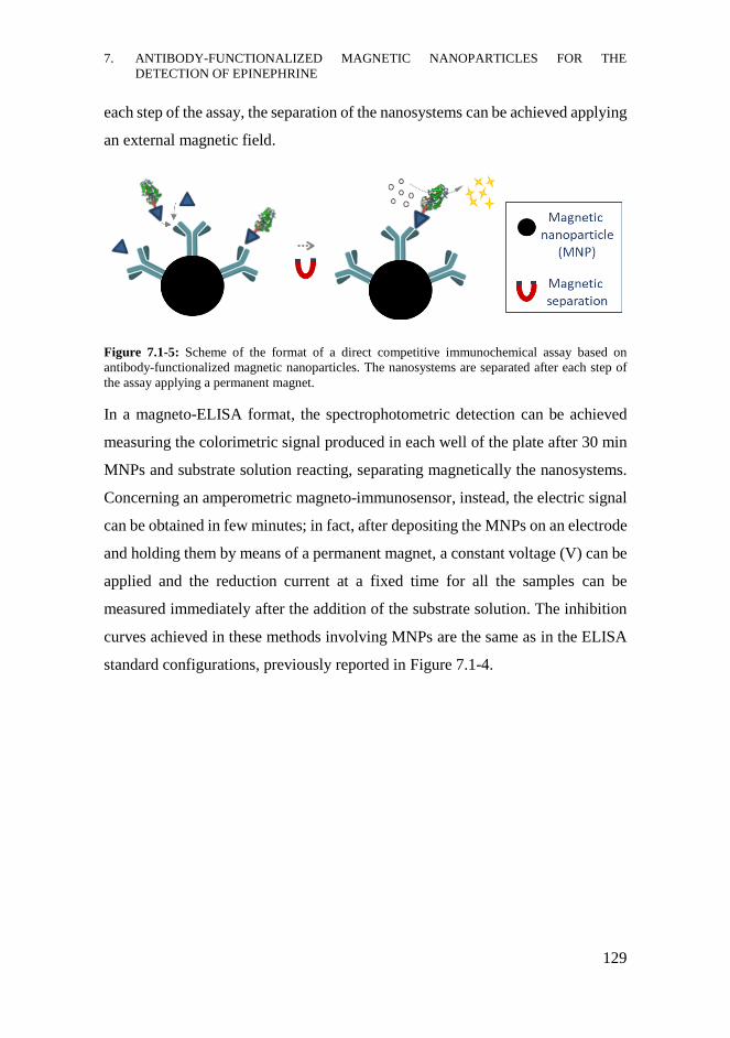
7. ANTIBODY-FUNCTIONALIZED MAGNETIC NANOPARTICLES FOR THE
DETECTION OF EPINEPHRINE
129
each step of the assay, the separation of the nanosystems can be achieved applying
an external magnetic field.
Figure 7.1-5: Scheme of the format of a direct competitive immunochemical assay based on
antibody-functionalized magnetic nanoparticles. The nanosystems are separated after each step of
the assay applying a permanent magnet.
In a magneto-ELISA format, the spectrophotometric detection can be achieved
measuring the colorimetric signal produced in each well of the plate after 30 min
MNPs and substrate solution reacting, separating magnetically the nanosystems.
Concerning an amperometric magneto-immunosensor, instead, the electric signal
can be obtained in few minutes; in fact, after depositing the MNPs on an electrode
and holding them by means of a permanent magnet, a constant voltage (V) can be
applied and the reduction current at a fixed time for all the samples can be
measured immediately after the addition of the substrate solution. The inhibition
curves achieved in these methods involving MNPs are the same as in the ELISA
standard configurations, previously reported in Figure 7.1-4.

7. ANTIBODY-FUNCTIONALIZED MAGNETIC NANOPARTICLES FOR THE
DETECTION OF EPINEPHRINE
130
7.1.2.2 Available immunoassays for EPI
Immunochemical assays for the detection of EPI are not the most commonly used
methods and are low discussed in the literature. Notwithstanding, many ELISA
kits are commercially available for the determination of EPI alone or in
combination with NorEPI. These competitive immunochemical assays are based
on rabbit-antibodies recognizing the N-acyl derivative of EPI; therefore, the
analyte needs to be first extracted using a cis-diol-specific affinity gel, then
acylated to N-acyl adrenaline and finally enzymatically converted into N-acyl
metanephrine. Although these assays are very sensitive, achieving for instance for
Abnova EPI ELISA Kit (KA1882) LOD values < 1 ng/mL in urine and 0.01
pg/mL in plasma, they require a complex sample treatment.
Another example of commercially available ELISA kit (CatCombi ELISA – IBL
International) is based on a sandwich format (rabbit anti-N-acyl metanephrine and
alkaline phosphatase labelled antibody against biotin) able to detect both
adrenaline and noradrenaline, after derivatization and biotinylating steps
(sensitivity 0.3 ng/mL).234
It is described in the literature a patent concerning an immunochemical assay for
the detection of EPI and NorEPI; it is based on an oxidation procedure converting
the analytes into compounds that are not bound by the antibody. Therefore, the
quantification can be achieved performing the immunoassay before and after the
oxidation reaction.235
Immunochemical methods for EPI offer advantages compared to standard
chromatographic techniques; they are easier to perform and more accessible to the
laboratories since they required cheaper equipment. Moreover, they allow the
simultaneous analysis of many samples or the contemporary detection of different
kinds of analytes through multiplexed platforms.
7. ANTIBODY-FUNCTIONALIZED MAGNETIC NANOPARTICLES FOR THE
DETECTION OF EPINEPHRINE
131
CHAPTER OBJECTIVES
Considering the biological importance of the neurohormone EPI and its key role
in many physiological and pathological conditions, the development of a
sensitive, simple and rapid immunochemical method for its detection, avoiding
sample pre-treatment and analyte derivatization, can be very useful.
The combination of the magnetic properties of MNPs, with the recognition ability
of an Ab and the advantages of an electrochemical sensor might lead to the
development of powerful analytical tools for this diagnostic purpose. The
presence of an Ab able to recognize the free analyte without performing any
modification or treatment could ensure the capture and the selective binding of
the analyte, while the magnetic core of the nanoparticles could facilitate the
separation and concentration steps. Moreover, a readout technique based on
electrochemical detection could bring rapidity, reliability and simplicity to this
kind of diagnostic system.
For all these reasons, the development of amperometric magneto-immunosensors
for the detection of EPI is the aim of this research project. The diagnostic tools
will be based on Fe3O4@Cu@Au MNPs and Fe3O4@SiO2 MNPs functionalized
with an antibody able to recognize and bind EPI. Rapidity, high simplicity and
good sensitivity will represent the features of this analytical tool.

7. ANTIBODY-FUNCTIONALIZED MAGNETIC NANOPARTICLES FOR THE
DETECTION OF EPINEPHRINE
132
RESULTS AND DISCUSSION
In this section the experimental results concerning to the development of different
kinds of immunochemical assays for the detection of EPI are presented and
discussed. The evaluation of the immunoreagents, performed by standard ELISA
configurations, was the starting point for the development of the MNPs-based
amperometric immunosensors.
7.3.1 Development of the indirect competitive ELISA
7.3.1.1 Immunoreagents evaluation
EPI is a low molecular weight compound not immunogenic by itself. Thus, the
preparation of an immunizing hapten is required. This hapten must be
conveniently attached to an immunogenic protein to stimulate the immune
response and to produce the specific antibodies against the selected molecule. The
specific antibodies against EPI had been produced in the Nb4D group.
Particularly, the designed and synthesized immunizing hapten for EPI had a
nucleophilic group (X) at the spacer arm which was conjugated to the
immunogenic protein HCH using N-succinimidyl iodoacetate (SIA) as a
heterobifunctional crosslinker agent. The resulting bioconjugate HCH-SIA-EPI,
known as immunogen, was used for immunizing three female New Zealand
rabbits obtaining three different antisera: As334, As335 and As336. These
antisera contain the pool of polyclonal antibodies developed against all the
epitopes present in the immunogen.
In order to characterize the antisera produced against EPI, the preparation of
competitor bioconjugates is required, as previously described. These compounds
consist in binding the immunizing hapten, or other chemical compounds

7. ANTIBODY-FUNCTIONALIZED MAGNETIC NANOPARTICLES FOR THE
DETECTION OF EPINEPHRINE
133
structurally related with the target analyte known as competitor haptens, to carrier
proteins different to the one used for immunizing. In this case, the immunizing
hapten of EPI and a competitor hapten derived from NorEPI, which was
chemically modified in the same way, were covalently attached to Bovine Serum
Albumin (BSA) instead of HCH. The conjugates containing the hapten of EPI
were considered homologous competitor conjugates, while those incorporating
the hapten of NorEPI were heterologous ones. The degree of homology or
heterology depends, on one hand, on the structure of the competitor hapten, and
on the other hand, on the crosslinker agent used for the preparation of the
bioconjugates. For some conjugates, a different heterobifunctional crosslinker
agent was used, the N-succinimidyl 3-maleimidopropionate (N-SMP), increasing
the structural differences between the immunogen and the corresponding
competitor. Thus, four competitor conjugates, previously prepared in the Nb4D
group, were selected for the development of the immunochemical assay for the
detection of EPI. They were BSA-SIA-EPI, BSA-(N-SMP)-EPI, BSA-SIA-
NorEPI and BSA-(N-SMP)-NorEPI and their structure is reported in Figure 7.3-1.
Figure 7.3-1: Structure of the homologous competitor conjugates BSA-SIA-EPI and BSA-(N-
SMP)-EPI and heterologous ones BSA-SIA-NorEPI and BSA-(N-SMP)-NorEPI. The general
structures of the haptens for EPI and NorEPI, in which X corresponds to a nucleophilic functional
group, are highlighted in blue.
These conjugates were also previously characterized by spectrometric techniques,
such as matrix assisted laser desorption ionization-time of flight mass
spectrometry (MALDI-TOF-MS). The hapten density of the bioconjugates varied
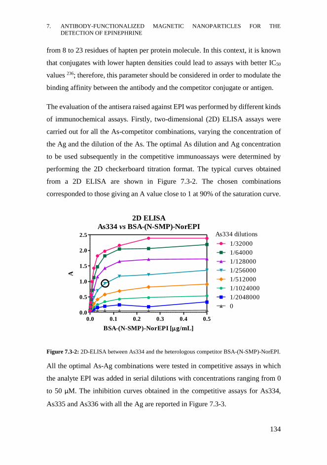
7. ANTIBODY-FUNCTIONALIZED MAGNETIC NANOPARTICLES FOR THE
DETECTION OF EPINEPHRINE
134
from 8 to 23 residues of hapten per protein molecule. In this context, it is known
that conjugates with lower hapten densities could lead to assays with better IC50
values 236; therefore, this parameter should be considered in order to modulate the
binding affinity between the antibody and the competitor conjugate or antigen.
The evaluation of the antisera raised against EPI was performed by different kinds
of immunochemical assays. Firstly, two-dimensional (2D) ELISA assays were
carried out for all the As-competitor combinations, varying the concentration of
the Ag and the dilution of the As. The optimal As dilution and Ag concentration
to be used subsequently in the competitive immunoassays were determined by
performing the 2D checkerboard titration format. The typical curves obtained
from a 2D ELISA are shown in Figure 7.3-2. The chosen combinations
corresponded to those giving an A value close to 1 at 90% of the saturation curve.
Figure 7.3-2: 2D-ELISA between As334 and the heterologous competitor BSA-(N-SMP)-NorEPI.
All the optimal As-Ag combinations were tested in competitive assays in which
the analyte EPI was added in serial dilutions with concentrations ranging from 0
to 50 μM. The inhibition curves obtained in the competitive assays for As334,
As335 and As336 with all the Ag are reported in Figure 7.3-3.
0.0 0.1 0.2 0.3 0.4 0.50.0
0.5
1.0
1.5
2.0
2.5
2D ELISA
As334 vs BSA-(N-SMP)-NorEPI
1/32000
1/64000
1/128000
1/256000
1/512000
1/1024000
1/2048000
0
As334 dilutions
BSA-(N-SMP)-NorEPI [g/mL]
A
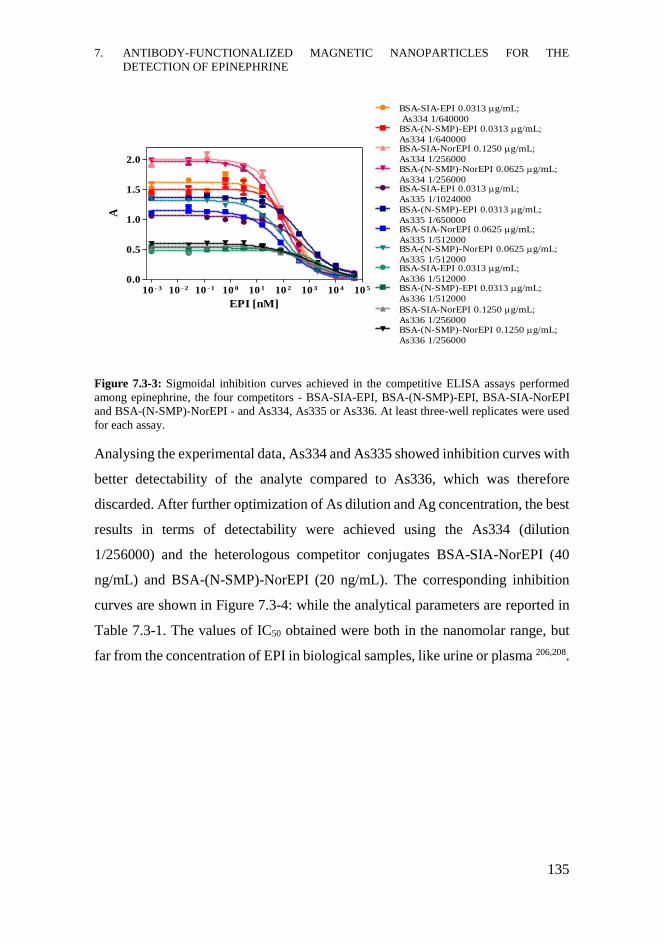
7. ANTIBODY-FUNCTIONALIZED MAGNETIC NANOPARTICLES FOR THE
DETECTION OF EPINEPHRINE
135
Figure 7.3-3: Sigmoidal inhibition curves achieved in the competitive ELISA assays performed
among epinephrine, the four competitors - BSA-SIA-EPI, BSA-(N-SMP)-EPI, BSA-SIA-NorEPI
and BSA-(N-SMP)-NorEPI - and As334, As335 or As336. At least three-well replicates were used
for each assay.
Analysing the experimental data, As334 and As335 showed inhibition curves with
better detectability of the analyte compared to As336, which was therefore
discarded. After further optimization of As dilution and Ag concentration, the best
results in terms of detectability were achieved using the As334 (dilution
1/256000) and the heterologous competitor conjugates BSA-SIA-NorEPI (40
ng/mL) and BSA-(N-SMP)-NorEPI (20 ng/mL). The corresponding inhibition
curves are shown in Figure 7.3-4: while the analytical parameters are reported in
Table 7.3-1. The values of IC50 obtained were both in the nanomolar range, but
far from the concentration of EPI in biological samples, like urine or plasma 206,208.
10 - 3 10 - 2 10 - 1 10 0 10 1 10 2 10 3 10 4 10 5
0.0
0.5
1.0
1.5
2.0BSA-(N-SMP)-NorEPI 0.0625 g/mL;
As334 1/256000
BSA-SIA-EPI 0.0313 g/mL;
As334 1/640000BSA-(N-SMP)-EPI 0.0313 g/mL;
As334 1/640000BSA-SIA-NorEPI 0.1250 g/mL;
As334 1/256000
BSA-SIA-EPI 0.0313 g/mL;
As335 1/1024000BSA-(N-SMP)-EPI 0.0313 g/mL;
As335 1/650000BSA-SIA-NorEPI 0.0625 g/mL;
As335 1/512000BSA-(N-SMP)-NorEPI 0.0625 g/mL;
As335 1/512000BSA-SIA-EPI 0.0313 g/mL;
As336 1/512000BSA-(N-SMP)-EPI 0.0313 g/mL;
As336 1/512000
BSA-SIA-NorEPI 0.1250 g/mL;
As336 1/256000BSA-(N-SMP)-NorEPI 0.1250 g/mL;
As336 1/256000
EPI [nM]
A
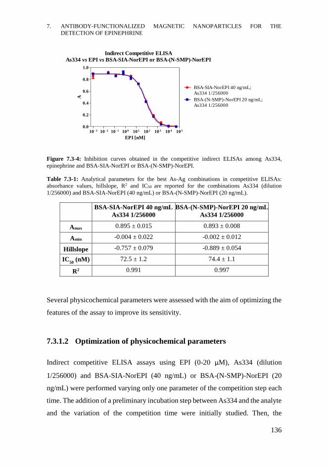
7. ANTIBODY-FUNCTIONALIZED MAGNETIC NANOPARTICLES FOR THE
DETECTION OF EPINEPHRINE
136
Figure 7.3-4: Inhibition curves obtained in the competitive indirect ELISAs among As334,
epinephrine and BSA-SIA-NorEPI or BSA-(N-SMP)-NorEPI.
Table 7.3-1: Analytical parameters for the best As-Ag combinations in competitive ELISAs:
absorbance values, hillslope, R2 and IC50 are reported for the combinations As334 (dilution
1/256000) and BSA-SIA-NorEPI (40 ng/mL) or BSA-(N-SMP)-NorEPI (20 ng/mL).
BSA-SIA-NorEPI 40 ng/mL
As334 1/256000
BSA-(N-SMP)-NorEPI 20 ng/mL
As334 1/256000
Amax 0.895 ± 0.015 0.893 ± 0.008
Amin -0.004 ± 0.022 -0.002 ± 0.012
Hillslope -0.757 ± 0.079 -0.889 ± 0.054
IC50
(nM) 72.5 ± 1.2 74.4 ± 1.1
R2 0.991 0.997
Several physicochemical parameters were assessed with the aim of optimizing the
features of the assay to improve its sensitivity.
7.3.1.2 Optimization of physicochemical parameters
Indirect competitive ELISA assays using EPI (0-20 μM), As334 (dilution
1/256000) and BSA-SIA-NorEPI (40 ng/mL) or BSA-(N-SMP)-NorEPI (20
ng/mL) were performed varying only one parameter of the competition step each
time. The addition of a preliminary incubation step between As334 and the analyte
and the variation of the competition time were initially studied. Then, the
10 - 3 10 - 2 10 - 1 10 0 10 1 10 2 10 3 10 4 10 5
0.0
0.2
0.4
0.6
0.8
1.0
BSA-SIA-NorEPI 40 ng/mL;
As334 1/256000
BSA-(N-SMP)-NorEPI 20 ng/mL;
As334 1/256000
EPI [nM]
A
Indirect Competitive ELISA
As334 vs EPI vs BSA-SIA-NorEPI or BSA-(N-SMP)-NorEPI
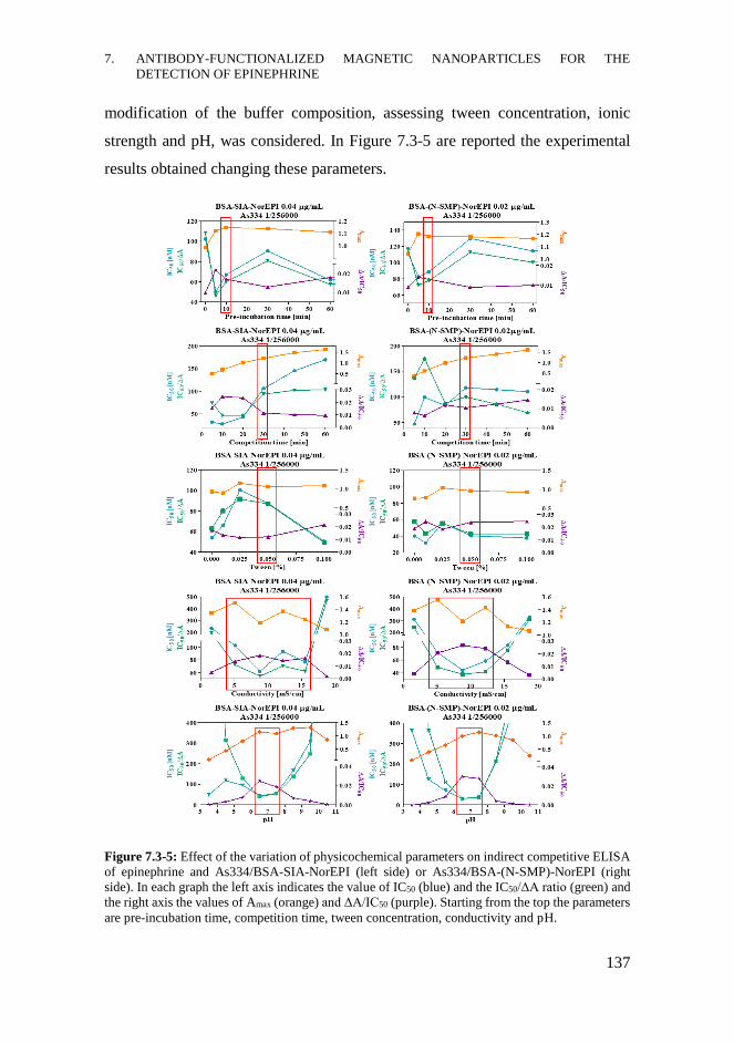
7. ANTIBODY-FUNCTIONALIZED MAGNETIC NANOPARTICLES FOR THE
DETECTION OF EPINEPHRINE
137
modification of the buffer composition, assessing tween concentration, ionic
strength and pH, was considered. In Figure 7.3-5 are reported the experimental
results obtained changing these parameters.
Figure 7.3-5: Effect of the variation of physicochemical parameters on indirect competitive ELISA
of epinephrine and As334/BSA-SIA-NorEPI (left side) or As334/BSA-(N-SMP)-NorEPI (right
side). In each graph the left axis indicates the value of IC50 (blue) and the IC50/ΔA ratio (green) and
the right axis the values of Amax (orange) and ΔA/IC50 (purple). Starting from the top the parameters
are pre-incubation time, competition time, tween concentration, conductivity and pH.
7. ANTIBODY-FUNCTIONALIZED MAGNETIC NANOPARTICLES FOR THE
DETECTION OF EPINEPHRINE
138
The effect of the incorporation of a pre-incubation step of the As334 with EPI was
evaluated from 5 min to overnight. According to the experimental results, the
addition of a short pre-incubation step (10 min) improved the detectability of the
immunoassay, comparing to the absence of pre-incubation (0 min). Overnight pre-
incubation resulted in analyte degradation and, consequently, in the worsening of
the analytical parameters of the immunoassay.
The competition time was assessed from 5 min to 60 min; short competition times
caused a decrease in the maximum colorimetric signal, compared to long ones.
Combining the 10 min preincubation step with competition, the best results were
obtained maintaining the competition time at 30 min, as in the starting protocol.
The effect of changing the tween concentration was evaluated, using PBS
solutions containing Tween 20 from 0% to 0.1%. Tween caused some variations
in the analytical parameters of the immunoassay; however, considering that IC50
values were all in the nanomolar range, this parameter was maintained at 0.05%.
Ionic strength was assessed using PBST solutions with a conductivity value
ranging from 5 mS/cm to 95 mS/cm. At conductivity higher than the reference
value of 16 mS/cm there was a loss of the colorimetric signal and for this reason,
the lower conductivity range from 2 mS/cm to 19 mS/cm was assessed. Both the
immunoassays showed high stability at low conductivity and good detectability,
particularly working in the range 5-16 mS/cm.
pH of the competition buffer was assessed using PBST solutions ranging from pH
2.5 to pH 11.5 and performing the ELISA experiments without the pre-incubation
step. pH altered dramatically the detectability of the immunoassays in terms of
both IC50 and Amax, probably due to the degradation of the analyte.204 The stability
of the immunoassays might be guaranteed working at almost neutral pH (6.5-7.5).
According to all experimental results an improvement on the IC50 value was
achieved introducing a short pre-incubation step (10 min) and maintaining all the
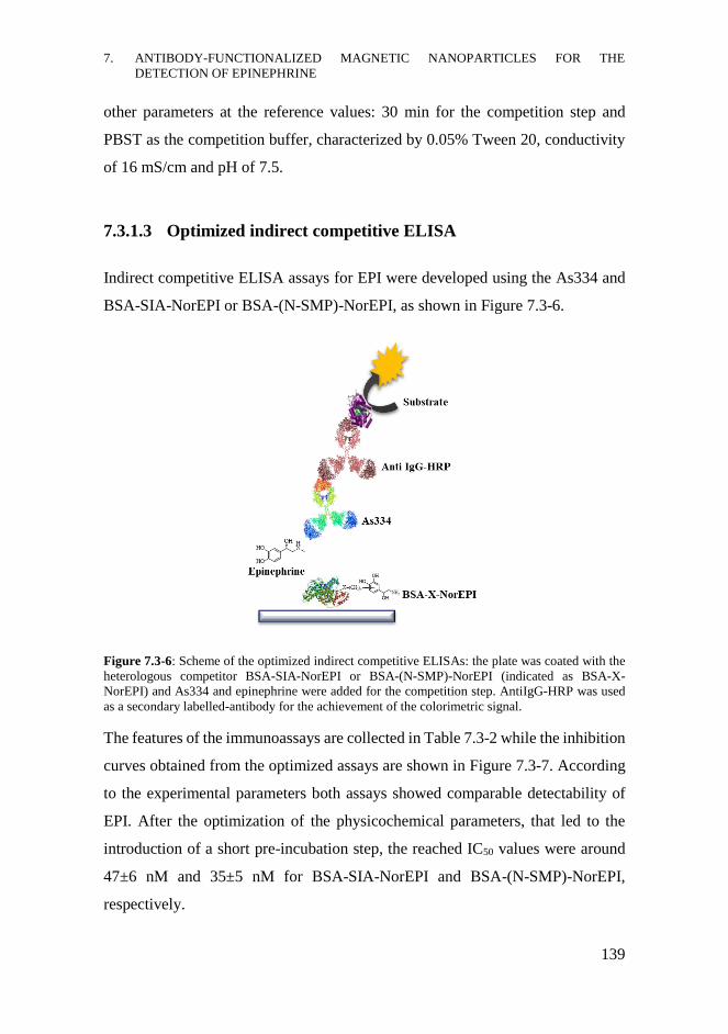
7. ANTIBODY-FUNCTIONALIZED MAGNETIC NANOPARTICLES FOR THE
DETECTION OF EPINEPHRINE
139
other parameters at the reference values: 30 min for the competition step and
PBST as the competition buffer, characterized by 0.05% Tween 20, conductivity
of 16 mS/cm and pH of 7.5.
7.3.1.3 Optimized indirect competitive ELISA
Indirect competitive ELISA assays for EPI were developed using the As334 and
BSA-SIA-NorEPI or BSA-(N-SMP)-NorEPI, as shown in Figure 7.3-6.
Figure 7.3-6: Scheme of the optimized indirect competitive ELISAs: the plate was coated with the
heterologous competitor BSA-SIA-NorEPI or BSA-(N-SMP)-NorEPI (indicated as BSA-X-
NorEPI) and As334 and epinephrine were added for the competition step. AntiIgG-HRP was used
as a secondary labelled-antibody for the achievement of the colorimetric signal.
The features of the immunoassays are collected in Table 7.3-2 while the inhibition
curves obtained from the optimized assays are shown in Figure 7.3-7. According
to the experimental parameters both assays showed comparable detectability of
EPI. After the optimization of the physicochemical parameters, that led to the
introduction of a short pre-incubation step, the reached IC50 values were around
47±6 nM and 35±5 nM for BSA-SIA-NorEPI and BSA-(N-SMP)-NorEPI,
respectively.
7. ANTIBODY-FUNCTIONALIZED MAGNETIC NANOPARTICLES FOR THE
DETECTION OF EPINEPHRINE
140
Figure 7.3-7: Inhibition curves of the optimized indirect competitive ELISAs among As334,
epinephrine and BSA-SIA-NorEPI or BSA-(N-SMP)-NorEPI.
Table 7.3-2: Immunoassay features of the optimized indirect competitive ELISAs for epinephrine;
information concerning immunoreagents, experimental parameters, Amax, Amin, R2, IC50, LOD and
working range (WR) are summarized.
Experimental
parameters BSA-SIA-NorEPI BSA-(N-SMP)-NorEPI
Ag concentration
[ng/mL] 40 20
As334 dilution 1/256000 1/256000
Preincubation time 10 min 10 min
Competition time 30 min 30 min
Tween concentration 0.05 % 0.05 %
Conductivity [mS/cm] 5-16 5-12
pH 6.5-7.5 6.5-7.5
Amax 1.11 ± 0.04 1.07 ± 0.07
Amin 0.05 ± 0.01 0.04 ± 0.01
R2 0.998 0.996
IC50 [nM]
IC50 [μg/L]
47 ± 6
8.6 ± 1.0
35 ± 5
6.3 ± 0.7
LOD [nM] 2.1 ± 0.6 1.5 ± 0.3
WR [nM] 5-311 4-254
Optimized Indirect Competitive ELISA
As334 vs EPI vs BSA-SIA-NorEPI or BSA-(N-SMP)-NorEPI
10 - 2 10 - 1 10 0 10 1 10 2 10 3 10 4 10 5
0.0
0.5
1.0
1.5
BSA-SIA-NorEPI 40 ng/mL;
As334 1/256000
BSA-(N-SMP)-NorEPI 20 ng/mL;
As334 1/256000
EPI [nM]
A
7. ANTIBODY-FUNCTIONALIZED MAGNETIC NANOPARTICLES FOR THE
DETECTION OF EPINEPHRINE
141
7.3.2 Development of the direct competitive ELISA
Once the immunoassay for the detection of EPI in an indirect competitive format
was developed and optimized, the assay in a direct format was carried out, taking
into account the immunoreagents and physicochemical conditions previously
selected.
The first step was to address the preparation, characterization and
immunochemical evaluation of the required enzymatic tracer (ET), HRP-(N-
SMP)-NorEPI, which would be finally used for the immunoassays involving
MNPs. As shown in Figure 7.3-8, the direct competitive ELISA assay was based
on the competition between the analyte EPI and the conjugate HRP-(N-SMP)-
NorEPI for the binding site of As334, adsorbed on the assay plate. The oxidation
of the substrate solution was catalysed by the ET, avoiding the addition of the
labelled secondary antibody and thus shortening the assay time.
Figure 7.3-8: Scheme of the direct competitive ELISA for epinephrine: EPI and HRP-(N-SMP)-
NorEPI competed for binding to As334, coating the plate. The oxidation of the substrate was
catalysed by the enzymatic tracer that produced the colorimetric signal.
7. ANTIBODY-FUNCTIONALIZED MAGNETIC NANOPARTICLES FOR THE
DETECTION OF EPINEPHRINE
142
7.3.2.1 Enzymatic tracer HRP-(N-SMP)-NorEPI
HRP-(N-SMP)-NorEPI was selected as the ET for the direct ELISA according to
the results obtained in the indirect competitive immunoassay, in which the most
heterologous competitor conjugate BSA-(N-SMP)-NorEPI showed the best
performance.
The ET was prepared in a two-step reaction, as shown in Figure 7.3-9. The first
step consisted in the modification of the amino group of Lys residues with the
linker N-SMP, forming amide bonds and introducing maleimide groups.237
According to the structure of this enzyme, only two Lys residues are accessible
enough to be derivatized. After the reaction, HRP-(N-SMP) conjugate was
purified from the excess of linker by size exclusion chromatography on an ÄKTA
prime system, eluting the sample through a Sephadex® G-25 column equilibrated
with ultrapure H2O. The introduction of the maleimide group on the enzyme
allowed to conjugate the hapten of NorEPI, already synthesized and characterized
in the Nb4D group. The second step consisted in the Michael addition of the
nucleophilic functional group of the hapten (X) to the electrophilic double bond
of the maleimide moiety on the enzymatic conjugate, leading to the formation of
covalent bonds.
Figure 7.3-9: Reaction scheme for the preparation of HRP-(N-SMP)-NorEPI; activation of Lys
residues with N-SMP (step 1) and Michael addition of the hapten of NorEPI (step 2).
7. ANTIBODY-FUNCTIONALIZED MAGNETIC NANOPARTICLES FOR THE
DETECTION OF EPINEPHRINE
143
After purification by dialysis with PBS and finally H2O, the ET was characterized
by MALDI-TOF-MS analysis to determine its hapten density. The comparison
among the mass spectra, reported in Figure 7.3-10, of the starting reagent HRP (in
blue), the intermediate conjugate HRP-(N-SMP) (in red) and the final product
HRP-(N-SMP)-NorEPI (in green) confirmed the derivatization of the enzyme
with the linker and the conjugation of the NorEPI hapten. The densities of linker
and hapten were, respectively, 2 and 1.5 for each enzyme macromolecule.
Figure 7.3-10: MALDI-TOF-MS analysis of HRP (in blue), HRP-(N-SMP) (in red) and HRP-(N-
SMP)-NorEPI (in green) with the corresponding molecular weights.
7.3.2.2 Direct competitive ELISA
Once prepared the ET, 2D direct ELISA was performed both to assure the catalytic
activity of HRP after conjugation process, and to optimize the As dilution and
tracer concentration. The combination As334 1/12000 and HRP-(N-SMP)-
NorEPI with a concentration of 8 ng/mL was selected for the competitive assay
with EPI (0-50 μM).
In Figure 7.3-11 the competitive curve of the direct ELISA is compared to those
obtained in the indirect formats while the experimental parameters of all these
immunoassays are summarized in Table 7.3-3. The IC50 and LOD values and the
working range of the direct ELISA were in the nanomolar range. The sensitivity
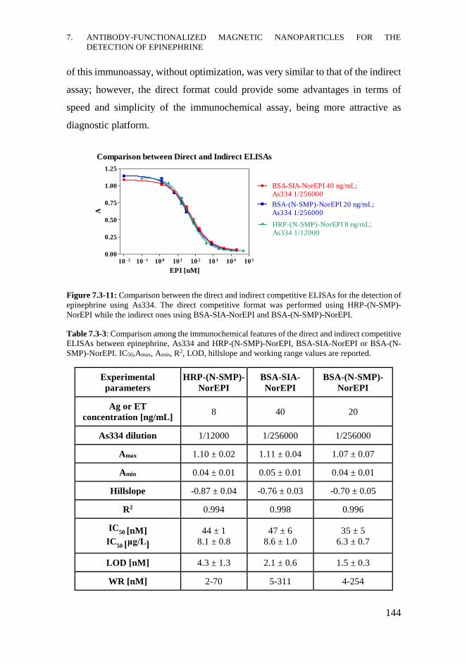
7. ANTIBODY-FUNCTIONALIZED MAGNETIC NANOPARTICLES FOR THE
DETECTION OF EPINEPHRINE
144
of this immunoassay, without optimization, was very similar to that of the indirect
assay; however, the direct format could provide some advantages in terms of
speed and simplicity of the immunochemical assay, being more attractive as
diagnostic platform.
Figure 7.3-11: Comparison between the direct and indirect competitive ELISAs for the detection of
epinephrine using As334. The direct competitive format was performed using HRP-(N-SMP)-
NorEPI while the indirect ones using BSA-SIA-NorEPI and BSA-(N-SMP)-NorEPI.
Table 7.3-3: Comparison among the immunochemical features of the direct and indirect competitive
ELISAs between epinephrine, As334 and HRP-(N-SMP)-NorEPI, BSA-SIA-NorEPI or BSA-(N-
SMP)-NorEPI. IC50,Amax, Amin, R2, LOD, hillslope and working range values are reported.
Experimental
parameters
HRP-(N-SMP)-
NorEPI
BSA-SIA-
NorEPI
BSA-(N-SMP)-
NorEPI
Ag or ET
concentration [ng/mL] 8 40 20
As334 dilution 1/12000 1/256000 1/256000
Amax 1.10 ± 0.02 1.11 ± 0.04 1.07 ± 0.07
Amin 0.04 ± 0.01 0.05 ± 0.01 0.04 ± 0.01
Hillslope -0.87 ± 0.04 -0.76 ± 0.03 -0.70 ± 0.05
R2 0.994 0.998 0.996
IC50 [nM]
IC50 [μg/L]
44 ± 1
8.1 ± 0.8
47 ± 6
8.6 ± 1.0
35 ± 5
6.3 ± 0.7
LOD [nM] 4.3 ± 1.3 2.1 ± 0.6 1.5 ± 0.3
WR [nM] 2-70 5-311 4-254
Comparison between Direct and Indirect ELISAs
10 - 2 10 - 1 10 0 10 1 10 2 10 3 10 4 10 5
0.00
0.25
0.50
0.75
1.00
1.25
BSA-(N-SMP)-NorEPI 20 ng/mL;
As334 1/256000
BSA-SIA-NorEPI 40 ng/mL;
As334 1/256000
HRP-(N-SMP)-NorEPI 8 ng/mL;
As334 1/12000
EPI [nM]
A
7. ANTIBODY-FUNCTIONALIZED MAGNETIC NANOPARTICLES FOR THE
DETECTION OF EPINEPHRINE
145
7.3.3 Synthesis and characterization of Ab-MNPs
Since the levels of EPI in biological fluids are in the picomolar range, as reported
in the literature, the introduction of sample preconcentration could be very
useful.207 In this way, it could be possible to achieve a concentration of the analyte
that might be measurable by direct or indirect immunochemical assays. The idea
of preconcentrating the sample could be achieved by different methods, as
explained in the introduction. In this context, one strategy might be using
nanosystems able to recognize the analyte and then facilitate its separation from
the mixture by a simple procedure.
For this purpose, MNPs-Ab could be a good solution, providing in one single
platform the recognition element, the antibody, and the separating feature, the
magnetic core. For this reason, Fe3O4@SiO2 MNPs and Fe3O4@Cu@Au MNPs
functionalized with the Ab - immunoglobulins G (IgGs) proteins - able to detect
EPI were synthesized and characterized.
7.3.3.1 Antibody purification
The antiserum As334 was purified in order to obtain only the IgGs fraction for the
functionalization of MNPs. The standard purification protocol for antibody
consisted in few steps, as reported in the literature.238,239 The pre-purification step
by ammonium sulphate precipitation was used to separate immunoglobulins from
other serum proteins, while the affinity chromatographic purification was needed
to achieve purified IgGs in buffer solution, eluting the solution of
immunoglobulins through a Protein A column. The elution of the IgGs solution
through a desalting column provided the purified antibody, named Ab334, that
was finally lyophilized and used for the functionalization of the nanosystems.
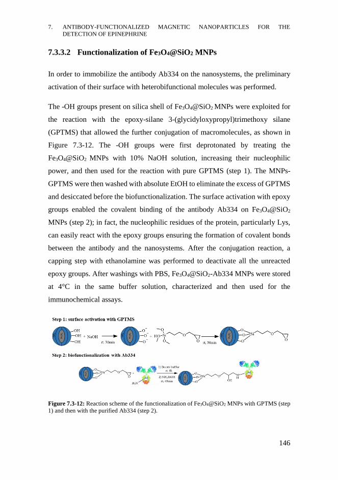
7. ANTIBODY-FUNCTIONALIZED MAGNETIC NANOPARTICLES FOR THE
DETECTION OF EPINEPHRINE
146
7.3.3.2 Functionalization of Fe3O4@SiO2 MNPs
In order to immobilize the antibody Ab334 on the nanosystems, the preliminary
activation of their surface with heterobifunctional molecules was performed.
The -OH groups present on silica shell of Fe3O4@SiO2 MNPs were exploited for
the reaction with the epoxy-silane 3-(glycidyloxypropyl)trimethoxy silane
(GPTMS) that allowed the further conjugation of macromolecules, as shown in
Figure 7.3-12. The -OH groups were first deprotonated by treating the
Fe3O4@SiO2 MNPs with 10% NaOH solution, increasing their nucleophilic
power, and then used for the reaction with pure GPTMS (step 1). The MNPs-
GPTMS were then washed with absolute EtOH to eliminate the excess of GPTMS
and desiccated before the biofunctionalization. The surface activation with epoxy
groups enabled the covalent binding of the antibody Ab334 on Fe3O4@SiO2
MNPs (step 2); in fact, the nucleophilic residues of the protein, particularly Lys,
can easily react with the epoxy groups ensuring the formation of covalent bonds
between the antibody and the nanosystems. After the conjugation reaction, a
capping step with ethanolamine was performed to deactivate all the unreacted
epoxy groups. After washings with PBS, Fe3O4@SiO2-Ab334 MNPs were stored
at 4°C in the same buffer solution, characterized and then used for the
immunochemical assays.
Figure 7.3-12: Reaction scheme of the functionalization of Fe3O4@SiO2 MNPs with GPTMS (step
1) and then with the purified Ab334 (step 2).
7. ANTIBODY-FUNCTIONALIZED MAGNETIC NANOPARTICLES FOR THE
DETECTION OF EPINEPHRINE
147
7.3.3.3 Functionalization of Fe3O4@Cu@Au MNPs
To conjugate the antibody on Fe3O4@Cu@Au MNPs, a covalent approach
involving heterobifunctional linkers was used instead of the easier but unstable
protein absorption strategy.
Exploiting the chemical affinity between Au and thiol groups, already described
in section 3.1.1.3, the surface of this kind of magnetic nanoparticles was first
activated with a mixed SAM based on heterobifunctional thiol-PEG molecules,
that were finally used for the conjugation of the Ab334, as shown in Figure 7.3-13.
A mixture of two kinds of linear PEG linkers, HS-PEG-COOH (average
Mn=7500) and HS-PEG-OCH3 (average Mn=6000) was used to achieve the
mixed SAM. The presence of the thiol groups ensured the stable anchorage on Au
surface, while the -COOH groups were exploited for the further conjugation of
biomolecules through amide bonds. The use of two kinds of linkers instead of one,
in a relative molar ratio HS-PEG-COOH : HS-PEG-OCH3 of 1:3, decreased the
negative charge density on the surface of nanoparticles, spacing them and
promoting the biofunctionalization step. The anchorage of the linkers was
performed under inert conditions in order to minimize the thiol-oxidation process,
stirring the solution overnight at rt (step 1). Activated Fe3O4@Cu@Au-PEG
MNPs were magnetically recovered, washed and then dried.
For the biofunctionalization with Ab334, the well-known carbodiimide chemistry
was used; the -COOH groups were activated by an EDC/NHS solution and used
for the reaction with Lys residues of the antibody, allowing its covalent binding
through amide bonds formation (step 2). After the capping reaction with
ethanolamine to deactivate all the unreacted reagents, the Fe3O4@Cu@Au-Ab334
MNPs were stored at 4°C in PBS.
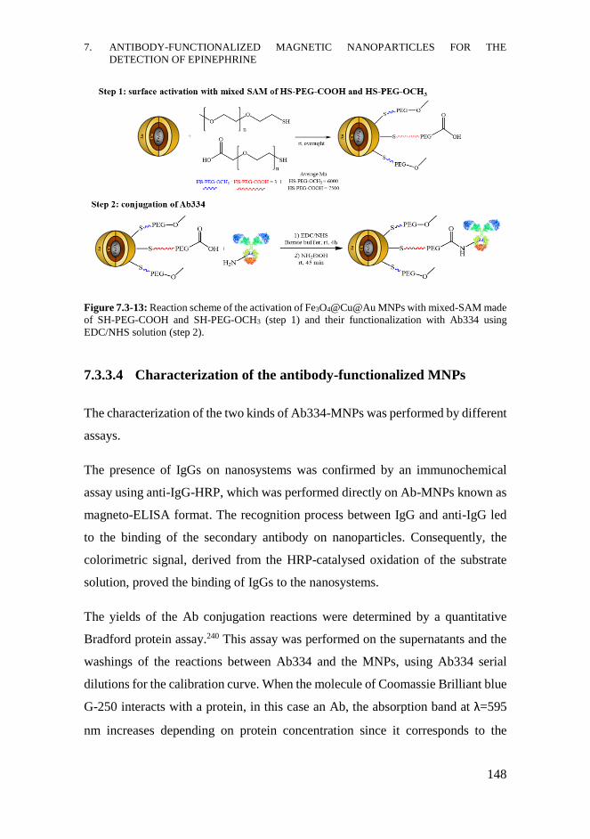
7. ANTIBODY-FUNCTIONALIZED MAGNETIC NANOPARTICLES FOR THE
DETECTION OF EPINEPHRINE
148
Figure 7.3-13: Reaction scheme of the activation of Fe3O4@Cu@Au MNPs with mixed-SAM made
of SH-PEG-COOH and SH-PEG-OCH3 (step 1) and their functionalization with Ab334 using
EDC/NHS solution (step 2).
7.3.3.4 Characterization of the antibody-functionalized MNPs
The characterization of the two kinds of Ab334-MNPs was performed by different
assays.
The presence of IgGs on nanosystems was confirmed by an immunochemical
assay using anti-IgG-HRP, which was performed directly on Ab-MNPs known as
magneto-ELISA format. The recognition process between IgG and anti-IgG led
to the binding of the secondary antibody on nanoparticles. Consequently, the
colorimetric signal, derived from the HRP-catalysed oxidation of the substrate
solution, proved the binding of IgGs to the nanosystems.
The yields of the Ab conjugation reactions were determined by a quantitative
Bradford protein assay.240 This assay was performed on the supernatants and the
washings of the reactions between Ab334 and the MNPs, using Ab334 serial
dilutions for the calibration curve. When the molecule of Coomassie Brilliant blue
G-250 interacts with a protein, in this case an Ab, the absorption band at λ=595
nm increases depending on protein concentration since it corresponds to the

7. ANTIBODY-FUNCTIONALIZED MAGNETIC NANOPARTICLES FOR THE
DETECTION OF EPINEPHRINE
149
protein-bound anionic form of the dye. As reported in Table 7.3-4, the conjugation
of the Ab334 to nanosystems was successfully achieved, leading to reaction yields
of 66.1% and 85.5% for Fe3O4@SiO2-Ab334 MNPs and Fe3O4@Cu@Au-Ab334
MNPs, respectively.
Table 7.3-4: Experimental results of Bradford protein assays on the solutions (supernatant +
washings) corresponding to Ab334-conjugation reactions. For each nanosystem the concentration
of Ab334 in solution (μg/mL), the amount of Ab334 bound to MNPs - expressed as concentration
(μg/mL) or referred to a mg of MNPs (μg/mg) - and the conjugation yield (%) are reported.
MNPs
Free
Ab334
[μg/mL]
Bound
Ab334
[μg/mL]
Bound Ab334
on MNPs mg
[μg/mg]
Reaction
yield
[%]
Fe3O4@SiO2-Ab334 169.32 330.69 8.55 66.1
Fe3O4@Cu@Au-Ab334 72.38 427.62 6.61 85.5
According to these experimental data, immunochemical magneto-assays based on
MNPs-Ab-334 were developed in order to assess the recognition activity of the
conjugated antibody towards EPI and to produce new and sensitive MNPs-based
immunoassays.
7.3.4 Development of the direct competitive magneto-
ELISA
Magneto-immunochemical assays were developed using the new synthesized and
characterized Fe3O4@Cu@Au-Ab334 MNPs and Fe3O4@SiO2-Ab334 MNPs.
The ability of Ab334 to recognize and interact with the analyte or its competitors
could have been affected by the conjugation process; structural modification of
the antibody, in fact, could have been occurred during the reaction with
nanoparticles. For these reasons, preliminary 2D direct magneto-ELISA assays
were carried out in order to confirm the recognition ability of the Ab and to select
7. ANTIBODY-FUNCTIONALIZED MAGNETIC NANOPARTICLES FOR THE
DETECTION OF EPINEPHRINE
150
the optimal combination of MNPs/ET for the further competitive assay. The 2D
magneto-ELISA assays were performed varying the quantity of Ab-334
functionalized magnetic nanoparticles, from 0 to 50 μg/well, and the
concentration of HRP-(N-SMP)-NorEPI from 0 to 0.2 μg/mL. All the magneto-
ELISA experiments were carried out following the protocol of the direct ELISA
format; however, after each step of the assay the MNPs were magnetically
separated from the solution, placing the plate on a magnetic rack, and manually
washed. After the stopping reaction, the supernatants were moved to another plate
and the colorimetric signal was measured at λ=450 nm, excluding the MNPs to
avoid possible interferences.
The experimental evidences confirmed the activity of the Ab334 in detecting the
ET, disproving the possible modification of the Ab due to the conjugation to
MNPs. These assays allowed also the selection of the optimal combinations of
MNPs quantity and ET concentration for the competitive assays, corresponding
to 25 μg/well for Fe3O4@Cu@Au-Ab334 MNPs, 6 μg/well for Fe3O4@SiO2-
Ab334 MNPs and 60 ng/mL for HRP-(N-SMP)-NorEPI.
According to the inhibition curves obtained in the competitive assays, shown in
Figure 7.3-14, EPI was able to compete with the enzymatic tracer for the binding
site of Ab334 anchored to the surface of the nanosystems. As reported in Table
7.3-5, the detectability of the magnetic-immunoassays was comparable to that
obtained using classic direct ELISA assays, especially in the case of
Fe3O4@Cu@Au-Ab334 MNPs. The IC50 and LOD values, in fact, were in the
nanomolar range.
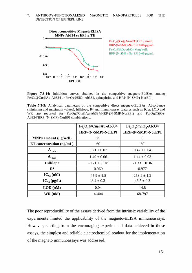
7. ANTIBODY-FUNCTIONALIZED MAGNETIC NANOPARTICLES FOR THE
DETECTION OF EPINEPHRINE
151
Figure 7.3-14: Inhibition curves obtained in the competitive magneto-ELISAs among
Fe3O4@Cu@Au-Ab334 or Fe3O4@SiO2-Ab334, epinephrine and HRP-(N-SMP)-NorEPI.
Table 7.3-5: Analytical parameters of the competitive direct magneto-ELISAs. Absorbance
(minimum and maximum values), hillslope, R2 and immunoassay features such as IC50, LOD and
WR are reported for Fe3O4@Cu@Au-Ab334/HRP-(N-SMP-NorEPI) and Fe3O4@SiO2-
Ab334/HRP-(N-SMP)-NorEPI combinations.
Fe
3O
4@Cu@Au-Ab334
HRP-(N-SMP)-NorEPI
Fe3O
4@SiO
2-Ab334
HRP-(N-SMP)-NorEPI
MNPs amount (μg/well) 25 6
ET concentration (ng/mL) 60 60
A min
0.21 ± 0.07 0.42 ± 0.04
A max
1.49 ± 0.06 1.44 ± 0.03
Hillslope -0.71 ± 0.18 -1.33 ± 0.36
R2 0.969 0.977
IC50
(nM)
IC50
(μg/L)
45.9 ± 1.5
8.4 ± 0.3
253.9 ± 1.2
46.5 ± 0.3
LOD (nM) 0.04 14.8
WR (nM) 4-404 68-797
The poor reproducibility of the assays derived from the intrinsic variability of the
experiments limited the applicability of the magneto-ELISA immunoassays.
However, starting from the encouraging experimental data achieved in those
assays, the simplest and reliable electrochemical readout for the implementation
of the magneto immunoassays was addressed.
Direct competitive MagnetoELISA
MNPs-Ab334 vs EPI vs TE
10 - 3 10 - 2 10 - 1 10 0 10 1 10 2 10 3 10 4 10 5
0.0
0.5
1.0
1.5
2.0Fe3O4@Cu@Au-Ab334 25 g/well;
HRP-(N-SMP)-NorEPI 0.06 g/mL
Fe3O4@SiO2-Ab334 6 g/well;
HRP-(N-SMP)-NorEPI 0.06 g/mL
EPI [nM]
A
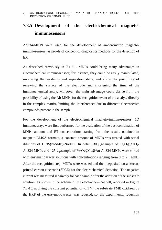
7. ANTIBODY-FUNCTIONALIZED MAGNETIC NANOPARTICLES FOR THE
DETECTION OF EPINEPHRINE
152
7.3.5 Development of the electrochemical magneto-
immunosensors
Ab334-MNPs were used for the development of amperometric magneto-
immunosensors, as proofs of concept of diagnostics methods for the detection of
EPI.
As described previously in 7.1.2.1, MNPs could bring many advantages in
electrochemical immunosensors; for instance, they could be easily manipulated,
improving the washings and separation steps, and allow the possibility of
renewing the surface of the electrode and shortening the time of the
immunochemical assay. Moreover, the main advantage could derive from the
possibility of using the Ab-MNPs for the recognition event of the analyte directly
in the complex matrix, limiting the interferences due to different electroactive
compounds present in the sample.
For the development of the electrochemical magneto-immunosensors, 1D
immunoassays were first performed for the evaluation of the best combination of
MNPs amount and ET concentration; starting from the results obtained in
magneto-ELISA formats, a constant amount of MNPs was treated with serial
dilutions of HRP-(N-SMP)-NorEPI. In detail, 30 μg/sample of Fe3O4@SiO2-
Ab334 MNPs and 125 μg/sample of Fe3O4@Cu@Au-Ab334 MNPs were stirred
with enzymatic tracer solutions with concentrations ranging from 0 to 2 μg/mL.
After the recognition step, MNPs were washed and then deposited on a screen-
printed carbon electrode (SPCE) for the electrochemical detection. The negative
current was measured separately for each sample after the addition of the substrate
solution. As shown in the scheme of the electrochemical cell, reported in Figure
7.3-15, applying the constant potential of -0.1 V, the substrate TMB oxidized by
the HRP of the enzymatic tracer, was reduced; so, the experimental reduction
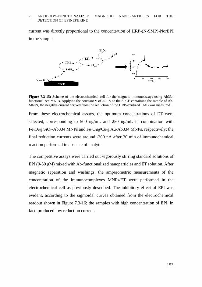
7. ANTIBODY-FUNCTIONALIZED MAGNETIC NANOPARTICLES FOR THE
DETECTION OF EPINEPHRINE
153
current was directly proportional to the concentration of HRP-(N-SMP)-NorEPI
in the sample.
Figure 7.3-15: Scheme of the electrochemical cell for the magneto-immunoassays using Ab334
functionalized MNPs. Applying the constant V of -0.1 V to the SPCE containing the sample of Ab-
MNPs, the negative current derived from the reduction of the HRP-oxidized TMB was measured.
From these electrochemical assays, the optimum concentrations of ET were
selected, corresponding to 500 ng/mL and 250 ng/mL in combination with
Fe3O4@SiO2-Ab334 MNPs and Fe3O4@Cu@Au-Ab334 MNPs, respectively; the
final reduction currents were around -300 nA after 30 min of immunochemical
reaction performed in absence of analyte.
The competitive assays were carried out vigorously stirring standard solutions of
EPI (0-50 μM) mixed with Ab-functionalized nanoparticles and ET solution. After
magnetic separation and washings, the amperometric measurements of the
concentration of the immunocomplexes MNPs/ET were performed in the
electrochemical cell as previously described. The inhibitory effect of EPI was
evident, according to the sigmoidal curves obtained from the electrochemical
readout shown in Figure 7.3-16; the samples with high concentration of EPI, in
fact, produced low reduction current.
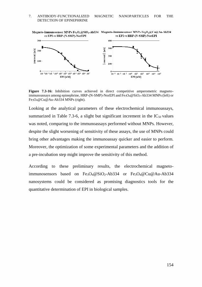
7. ANTIBODY-FUNCTIONALIZED MAGNETIC NANOPARTICLES FOR THE
DETECTION OF EPINEPHRINE
154
Figure 7.3-16: Inhibition curves achieved in direct competitive amperometric magneto-
immunoassays among epinephrine, HRP-(N-SMP)-NorEPI and Fe3O4@SiO2-Ab334 MNPs (left) or
Fe3O4@Cu@Au-Ab334 MNPs (right).
Looking at the analytical parameters of these electrochemical immunoassays,
summarized in Table 7.3-6, a slight but significant increment in the IC50 values
was noted, comparing to the immunoassays performed without MNPs. However,
despite the slight worsening of sensitivity of these assays, the use of MNPs could
bring other advantages making the immunoassay quicker and easier to perform.
Moreover, the optimization of some experimental parameters and the addition of
a pre-incubation step might improve the sensitivity of this method.
According to these preliminary results, the electrochemical magneto-
immunosensors based on Fe3O4@SiO2-Ab334 or Fe3O4@Cu@Au-Ab334
nanosystems could be considered as promising diagnostics tools for the
quantitative determination of EPI in biological samples.
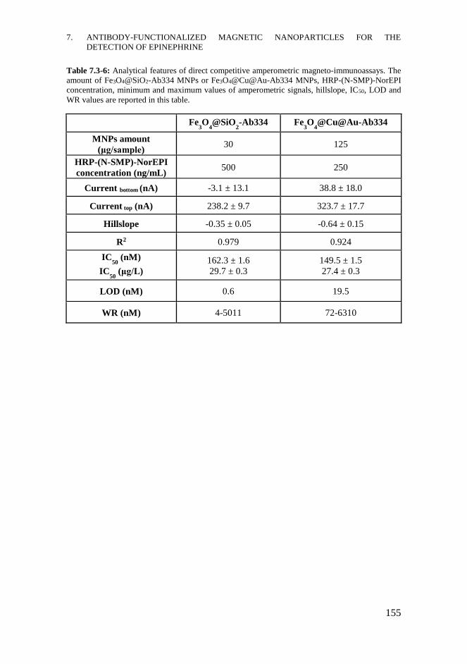
7. ANTIBODY-FUNCTIONALIZED MAGNETIC NANOPARTICLES FOR THE
DETECTION OF EPINEPHRINE
155
Table 7.3-6: Analytical features of direct competitive amperometric magneto-immunoassays. The
amount of Fe3O4@SiO2-Ab334 MNPs or Fe3O4@Cu@Au-Ab334 MNPs, HRP-(N-SMP)-NorEPI
concentration, minimum and maximum values of amperometric signals, hillslope, IC50, LOD and
WR values are reported in this table.
Fe3O
4@SiO
2-Ab334 Fe
3O
4@Cu@Au-Ab334
MNPs amount
(μg/sample) 30 125
HRP-(N-SMP)-NorEPI
concentration (ng/mL) 500 250
Current bottom (nA) -3.1 ± 13.1 38.8 ± 18.0
Current top (nA) 238.2 ± 9.7 323.7 ± 17.7
Hillslope -0.35 ± 0.05 -0.64 ± 0.15
R2 0.979 0.924
IC50
(nM)
IC50
(μg/L) 162.3 ± 1.6 29.7 ± 0.3
149.5 ± 1.5 27.4 ± 0.3
LOD (nM) 0.6 19.5
WR (nM) 4-5011 72-6310
7. ANTIBODY-FUNCTIONALIZED MAGNETIC NANOPARTICLES FOR THE
DETECTION OF EPINEPHRINE
156
CHAPTER CONTRIBUTIONS
- New immunochemical assays for the detection of EPI were developed.
Indirect competitive ELISA assays demonstrated high sensitivity in the
detection of the analyte achieving IC50 values in the nanomolar range (6-
9 μg/L), using As334 as antiserum and BSA-(N-SMP)-NorEPI or BSA-
SIA-NorEPI as competitors. Comparable analytical parameters were
obtained by the direct competitive ELISA format using the enzymatic
tracer HRP-(N-SMP)-NorEPI (IC50 9-10 μg/L).
- New MNPs-Ab based on Fe3O4@SiO2-Ab334 MNPs and
Fe3O4@Cu@Au-Ab334 MNPs were synthesized and characterized.
Different immunochemical assays were developed using these
nanosystems, such as magneto-ELISAs and electrochemical magneto-
immunosensors, achieving good sensitivity and low detectability limits
(IC50 27-30 μg/L), especially in the amperometric immunochemical
assays. These nanosystems might be used both for the recognition and
binding process of the analyte, due to the immobilized specific antibody,
and for its separation and concentration from complex matrices, thanks to
the properties of MNPs. The amperometric magneto-immunosensors
might be considered promising diagnostics tools for the detection of EPI.
7.4.1 Future perspectives
- Optimize the experimental procedures of the amperometric magneto-
immunosensor assays including, for instance, a pre-incubation step
between MNPs and EPI.
- Evaluate the matrix effects of plasma and urine samples in the
immunochemical assays and magneto-assays, and assess other analytical
parameters (specificity, reproducibility and accuracy studies).
7. ANTIBODY-FUNCTIONALIZED MAGNETIC NANOPARTICLES FOR THE
DETECTION OF EPINEPHRINE
157
MATERIALS AND METHODS
Reagents, materials and equipment: The chemical reagents used were purchased
by Sigma-Aldrich (Merck KGaA, Darmstadt, Germany). ÄKTATM prime,
provided with HiTrapTM Protein A HP column or HiTrapTM Desalting column (GE
Helthcare, Uppsala, Sweden), was used for chromatographic purification with the
corresponding software Prime View and Prime View Evaluation. OrDial D14
dialysis membranes (molecular weight cut off 12000-14000 Da) were purchased
from Orange Scientific (Braine-l’Alleud, Belgium). The matrix-assisted laser
desorption ionization time-of-flight mass spectrometer (MALDI-TOF-MS) was a
Bruker autoflex III Smartbeam spectrometer (Billerica, Massachusetts). The pH
and the conductivity of all buffers and solutions were measured with a pH-meter
pH 540 GLP and a conductimeter LF 340, respectively (WTW, Weilheim,
Germany). Polystyrene microtiter plates were purchased from Nunc (Maxisorp,
Roskilde, Denmark). Dilution plates were purchased from Nirco (Barberà del
Vallés, Spain). Washing steps of the immunochemical assays were performed on
a Biotek ELx465 (Biotek Inc.). 96-well plate magnetic separation rack (CORTEX
Biochem, WI, USA) was used for magnetic separation steps. Absorbances were
read on a SpectramaxPlus (Molecular Devices, Sunnyvale, CA, USA) using the
software SoftmaxPro v4.7 (Molecular Devices). Screen Printed Carbon
Electrodes DRP-C110 (DropSens, Asturias, Spain) were connected to the
potentiostat COMPACTSTAT.e Mobile Electrochemistry controlled by
IviumSoftTM Electrochemistry Software (IVIUM Technologies, Eindhoven, The
Netherlands). All the experimental data were analysed using the software
GraphPad Prism 5.03 (GraphPad Software Inc., San Diego, CA, USA) and
competitive curves were fitted with a four-parameters logistic equation.
Buffers: Unless otherwise indicated, phosphate buffer saline (PBS) is 0.01 M
phosphate buffer in a 0.8% saline solution, pH 7.5. Coating buffer is a 0.05 M
carbonate-bicarbonate buffer, pH 9.6. PBST is PBS with 0.05% Tween 20, pH
7. ANTIBODY-FUNCTIONALIZED MAGNETIC NANOPARTICLES FOR THE
DETECTION OF EPINEPHRINE
158
7.5. Citrate buffer is 0.04 M sodium citrate, pH 5.5. The substrate solution
contains 0.01% 3,3’,5,5’-tetramethylbenzidine (TMB) and 0.004% H2O2 in citrate
buffer. Borate buffer is 0.2 M boric acid/sodium borate, pH 8.7.
Non-competitive indirect ELISA: The recognition and binding abilities of the
antisera (As334-336) with the different antigens (BSA-SIA-EPI, BSA-(N-SMP)-
EPI, BSA-SIA-NorEPI or BSA-(N-SMP)-NorEPI) were evaluated by two-
dimensional titration assays. 2D-assays were carried out based on the
measurement of the binding of serial dilutions of the antisera (1/1000 to
1/2048000, and zero) against different concentrations of the antigens (serial 1:2
dilutions of 1 μg/mL solution, and zero). The microtiter plate was coated with Ag
dilutions in coating buffer (100 μL/well) and incubated overnight at 4°C. The next
day, the plate was washed four times with PBST (300 μL/well) and the As
dilutions were added to the wells (100 μL/well). After incubating 30 min at 25°C,
the plate was washed again and a solution of anti-IgG-HRP (1/6000 in PBST) was
added to the plate (100 μL/well). After 30 min incubating at rt, the plate was
washed again and incubated in the dark after the addition of substrate solution
(100 μL/well). The colorimetric reaction was stopped after 30 min at rt with 4 N
H2SO4 (50 μL/well), and the absorbance was read at λ=450 nm. From these
experiments, optimum concentrations for coating antigens and antisera dilutions
were chosen to generate around 0.7-1 units of absorbance.
Competitive indirect ELISA: The microtiter plate was coated with the antigen, at
the proper selected concentration in coating buffer (100 μL/well), and incubated
overnight at 4 °C, covered with adhesive plate sealers. The next day, the plate was
washed four times with PBST (300 μL/well), and the solutions of EPI (stock
solution 10 mM in DMSO, serial dilutions from 50 μM to 0 nM in PBST) were
added (50 μL/well), followed by the solution of antisera As334 (double
concentration of the selected dilution in PBST, 50 μL/well). After 30 min at 25°C,
the plate was washed four times with PBST (300 μL/well), and a solution of anti-
IgG-HRP (1/6000 in PBST) was added to the plate (100 μL/well) and incubated
7. ANTIBODY-FUNCTIONALIZED MAGNETIC NANOPARTICLES FOR THE
DETECTION OF EPINEPHRINE
159
for 30 min at rt. The plate was washed again and then incubated in dark after the
addition of the substrate solution (100 μL/well). The colorimetric reaction was
stopped after 30 min at rt with 4 N H2SO4 (50 μL/well), and the absorbance was
read at λ=450 nm. The standard curves were fitted to a four-parameter equation
according to the following formula: y = (A − B/[1 − (x/C)D] + B, where A is the
maximal absorbance, B is the minimum absorbance, C is the concentration
producing 50% of the maximal absorbance, and D is the slope at the inflection
point of the sigmoid curve. Unless otherwise indicated, all the data presented for
the immunochemical assays correspond to the average of at least three wells
replicates.
Physicochemical parameters optimization: Different physicochemical parameters
were assessed in the indirect competitive ELISA assays; the study included the
variation of pre-incubation time (overnight – 0 min), competition time (60 – 0
min), Tween 20 concentration (0.1% - 0%), conductivity (96 – 2 mS/cm) and pH
(11.5 – 2.5).
HRP-(N-SMP)-NorEPI synthesis: HRP was dissolved in PBS 10 mM pH=7.5
(5.04 mg in 474 μL) and N-SMP (0.65 mg in 30 μL DMF), ten-fold Lys
equivalents, was added to the enzymatic solution, achieving a solution at 10
mg/mL in HRP. The reaction mixture was stirred for 4 h at rt in dark and then
purified by ÄKTATM chromatographic system eluting the solution through
desalting columns by Milli-Q H2O. The intermediate product HRP-(N-SMP) was
mixed with NorEPI hapten (10 eq in DMF) and the reaction was performed in
PBS 10 mM stirring in dark for 90 min at rt and then overnight at 4°C. The protein
conjugate was purified by dialysis against 0.5 mM PBS (4×5 L) and Milli-Q H2O
(1×5 L) and was stored at 4°C. Working aliquots were prepared in 0.01 M PBS at
1 mg/mL and stored at 4 °C.
Hapten and linker densities of the bioconjugates were estimated by measuring the
MW of the native protein and of the conjugates by MALDI-TOF-MS. BSA was
7. ANTIBODY-FUNCTIONALIZED MAGNETIC NANOPARTICLES FOR THE
DETECTION OF EPINEPHRINE
160
utilized as internal standard for calibrating the instrument, using the mixture
HRP:BSA = 4:1 (40 μL HRP 10 mg/mL+10 μL BSA 10 mg/mL in H2O) as the
reference protein sample. Each mass spectrum was obtained by crystallization of
the matrix (sinapinic acid, 2 μL of 10 mg/mL in a 70:30 solution of ACN / H2O
and 0.1% in HCOOH), followed by the addition of 2 μL of sample or 2 μL of the
reference protein sample (diluted 1:2 with 0 ACN + 0.2% HCOOH). Finally, after
evaporation of the solution deposited on the plate, 2 μL of the matrix were added
again. The hapten densities (δ hapten) were calculated according to the following
equation: [MWconjugate-MWHRP]/MWhapten or linker.
2D Direct ELISA: The microtiter plate was coated with As334 dilutions in coating
buffer (1/1000 to 1/1024000, and zero, 100 μL/well) and incubated overnight at
4°C. The following day, the plate was washed four times with PBST (300
μL/well), and serial 1:2 dilutions of HRP-(N-SMP)-NorEPI in PBST (starting
solution 1 μg/mL in PBST, and zero) were added to the wells (100 μL/well). After
stirring 30 min at rt in dark, the plate was washed again, and the substrate solution
was added (100 μL/well). After stirring 30 min at rt in dark, the colorimetric
reaction was stopped adding 4 N H2SO4 (50 μL/well), and the absorbance was
read at λ=450 nm. From these experiments, optimum As334 dilution and
enzymatic tracer concentration were chosen to generate around 0.7-1 units of
absorbance.
Competitive direct ELISA: The microtiter plate was coated with As334 dilutions
(1/12000 and 1/16000) in coating buffer (100 μL/well), incubated overnight at 4
°C. The next day, the plate was washed four times with PBST (300 μL/well), and
the solutions of EPI (stock solution at 10 mM in DMSO, serial 1:5 dilutions from
50 μM to 0 nM in PBST) were added (50 μL/well), followed by the solution of
HRP-(N-SMP)-NorEPI in PBST (32 and 16 ng/mL; 50 μL/well). After stirring 30
min at rt in dark, the plate was washed four times with PBST (300 μL/well) and
the substrate solution was added (100 μL/well). The colorimetric reaction was
stopped with 4 N H2SO4 (50 μL/well) after stirring 30 min at rt in dark, and the
7. ANTIBODY-FUNCTIONALIZED MAGNETIC NANOPARTICLES FOR THE
DETECTION OF EPINEPHRINE
161
absorbance was read at λ=450 nm. The inhibition curves were fitted as previously
described.
As334 purification: A saturated solution of (NH4)2SO4 was slowly added (50 μL
each time) to three eppendorf containing 500 μL of As334 under stirring at 4°C,
until reaching 45% of saturation. After stirring 4 h at 4°C, the solutions were
centrifuged (10 min at 14000 rpm) and, once removed the supernatants, white
precipitates were obtained. After solubilization with PBS 10 mM (300 μL/
sample), sample solutions were filtered on 0,2 μm filters and then purified by
AKTATM, provided with Protein A column, eluting the solution with binding
buffer (PBS 20 mM pH 7) and then with citrate buffer (0.1 M pH 4). The IgGs
fraction of Ab334 was eluted with Milli-Q H2O through the desalting column and
finally lyophilized.
Synthesis of Fe3O4@SiO2-Ab334 MNPs: 100 mg of Fe3O4@SiO2 were stirred 30
min at rt with 2.4 mL of NaOH 10%. After washing with MilliQ H2O (x3) and
60% EtOH (x3), 1.2 mL of pure GPTMS was added to the MNPs. After stirring
30 min at rt, the Fe3O4@SiO2-GPTMS MNPs were washed three times with
absolute EtOH and finally dried under vacuum.
1 mL of a stock solution of purified Ab334 (1 mg/mL in borate buffer 0.1 M pH
8.5) was added to 50 mg of Fe3O4@SiO2-GPTMS MNPs dispersed in 1 mL of the
same buffer solution and the reaction mixture was stirred 4 h at rt. Fe3O4@SiO2-
Ab334 were washed three times with the same buffer solution and then stirred
with 1 mL of ethanolamine solution (0.1 M in PBS 10 mM). After 45 min, the
Fe3O4@SiO2-Ab334 MNPs were washed with PBS 10 mM and stored at 4°C in
the same buffer at a concentration of 50 mg/mL.
Synthesis of Fe3O4@Cu@Au-Ab334 MNPs: 4 mL of an aqueous solution
containing a mixture of HS-PEG-OCH3 15 mM (2 mL, average Mn 6000, linear)
and HS-PEG-COOH 5 mM (2 mL, average Mn 7500, linear) were added to 100
mg of Fe3O4@Cu@Au MNPs in 96 mL H2O under N2 flux. The solution was
7. ANTIBODY-FUNCTIONALIZED MAGNETIC NANOPARTICLES FOR THE
DETECTION OF EPINEPHRINE
162
divided in ten fractions that were stirred overnight at rt. The PEG-activated
nanoparticles were magnetically collected, washed several times with MilliQ H2O
and dried.
50 mg of Fe3O4@Cu@Au-PEG MNPs were dispersed in 1 mL of EDC/NHS
solution in 0.1 M borate buffer pH 8.5 (500 μL EDC 0.5 M + 500 μL NHS 0.4
M). After adding 1 mL of Ab334 stock solution (at 1 mg/mL in borate buffer), the
mixture was stirred 4 h at rt. The Fe3O4@Cu@Au-Ab334 MNPs were
magnetically collected, washed with the same buffer solution (1mL, x3) and
dispersed in 1 mL of ethanolamine solution (0.1 M in PBS 10 mM). After 45 min
of capping step, MNPs were washed three times with PBS 10 mM and then stored
at 4°C in the same buffer at a concentration of 50 mg/mL.
Bradford protein assay: Serial 1:2 dilutions of Ab334 80 μg/mL in borate buffer
were used for the calibration curve (160 μL/well) . The supernatants of the MNPs-
functionalization reactions were diluted in borate buffer to obtain a theoretical
concentration of 40 μg/mL (160 μL/well) while the washings solutions were used
without further dilutions (160 μL/well). Adding 40 μL/well of Coomassie Brilliant
Blue solution, the UV-Vis analysis was performed after 10 min measuring the
absorbance at λ=595 nm. Comparing the A values of the samples to the calibration
curve, the quantification of the free Ab334 and consequently of MNPs-bound
Ab334 was performed.
Magneto-ELISA between MNPs and anti-IgG: MNPs-Ab334 dispersions in
PBST, after 2 min sonicating, were added to the assay plate at serial 1:2 dilutions
ranging from 50 μg/well to zero (100 μL/well). After magnetically separating the
MNPs using the magnetic plate rack and removing the supernatants, 1/6000
dilution of anti-IgG-HRP in PBST was added (100uL/well). The plate was stirred
30 min at rt, MNPs were magnetically separated and three washings with PBST
were performed. 100 μL/well of substrate solution were added to the plate and,
after 30 min stirring at rt in dark, the colorimetric reaction was stopped adding 4

7. ANTIBODY-FUNCTIONALIZED MAGNETIC NANOPARTICLES FOR THE
DETECTION OF EPINEPHRINE
163
N H2SO4 (50 μL/well). MNPs were separated and the absorbance was read at
λ=450 nm after moving the supernatants to a different dilution plate.
Direct magneto-ELISA: MNPs-Ab334 dispersions in PBST, after 2 min
sonicating, were added to the assay plate at 1:2 serial dilutions ranging from 50
μg/well to zero (50 μL/well). 50 μL/well of HRP-(N-SMP)-NorEPI serial
dilutions in PBST (from 0.4 μg/mL to zero) were added to the plate. After 30 min
stirring at rt in dark, MNPs were magnetically collected using the magnetic plate
rack and the assay plate was washed three times with PBST (100 μL/well). The
substrate addition and the stopping reaction steps were performed as described in
the previous protocol.
Direct competitive magneto-ELISA: Fe3O4@Cu@Au-Ab334 MNPs in PBST,
after 2 min sonicating, were added to the assay plate at 25 μg/well (50 μL/well)
while Fe3O4@SiO2-Ab334 at 6 μg/well. 25 μL/well of EPI dilutions in PBST
(from 50 μM to 0) and 25 μL/well of HRP-(N-SMP)-NorEPI 60 ng/mL in PBST
were added to the plate. After 30 min stirring at rt in dark, MNPs were
magnetically separated using the magnetic plate rack and the plate was washed
three times with PBST (100 μL/well). The substrate addition and the stopping
reaction steps were performed as previously described.
Amperometric magneto immunosensor direct assay: 125 μg/sample of
Fe3O4@Cu@Au-Ab334 MNPs or 30 μg/sample of Fe3O4@SiO2-Ab334 MNPs
(100 μL/sample in PBST) were mixed with 1:2 serial dilutions of HRP-(N-SMP)-
NorEPI (from 2 μg/mL in PBST to zero, 100 μL/sample) in eppendorf tubes. After
30 min stirring in dark at rt, MNPs were magnetically separated and washings
with PBST were performed (3x100μL/sample). MNPs were dispersed in 160 μL
of citrate buffer and 40 μL were put on the SPCE, holding them applying an
external magnet, and used for each amperometric measurement. V=-0.1V was
applied and, at t = 60 s, 10 μL of the substrate solution were added to the sample.

7. ANTIBODY-FUNCTIONALIZED MAGNETIC NANOPARTICLES FOR THE
DETECTION OF EPINEPHRINE
164
Current values measured at t=200 s, after resting the corresponding values at t=50
s, were plotted in front of [ET].
Amperometric magneto immunosensor direct competitive assay: 125 μg/sample
of Fe3O4@Cu@Au-Ab334 MNPs (100 μL/sample in PBST) and 0.5 μg/mL of
HRP-(N-SMP)-NorEPI (50 μL/sample in PBST) or 30 μg/sample of Fe3O4@SiO2-
Ab334 MNPs (100 μL/sample in PBST) and 1 μg/mL of HRP-(N-SMP)-NorEPI
(50 μL/sample in PBST) were mixed in eppendorf tubes with 50 μL/sample of
EPI serial dilutions in PBST (from 50 μM to zero). After 30 min stirring in dark
at rt, MNPs were magnetically separated and washings with PBST were
performed (3x100 μL/sample). The substrate addition and the amperometric
detection were performed as previously described. Current values measured at
t=200 s, after resting the corresponding values at t=50 s, were plotted in front of
[EPI].

8. ANNEX: NANOPARTICLES-
MEDIATED OXIDATION OF EPI

8. NANOPARTICLES-MEDIATED OXIDATION OF EPI
166
INTRODUCTION
This annex deals with the proposal of using Fe3O4-based nanosystems as
multifunctional nanoplatforms for the detection of EPI exploiting its MNPs-
mediated oxidation process. This research work was carried out in the Nb4D
group.
8.1.1 Oxidation mechanism of EPI
The oxidation mechanism of EPI consists in two redox reactions separated by an
intermediate cyclization reaction, as reported in Figure 8.1-1.213 The catechol
moiety of EPI is oxidised into the corresponding orto-quinone compound. Then,
an 1,4 Michael addition can occur, depending on the pH, transforming adrenaline
o-quinone into leucoadrenochrome; at pH > 3, in fact, the secondary amine group
is partially deprotonated, reacting as nucleophile group by an intramolecular
cyclization and producing the stable corresponding five-membered ring.
Leucoadrenochrome can therefore undergo to another oxidation of the catechol
group, producing adrenochrome which can carry out a polymerization process,
producing melanin molecules.212,241
This mechanism can be catalyzed in the same way by using electrochemical
techniques, oxidoreductase enzymes like HRP, and also some kinds of
nanomaterials, such as SiO2 NPs.242,243
Since the oxidation products show characteristic absorption bands, especially the
red-colored final compound adrenochrome (λmax=490 nm), the UV-Vis
spectroscopy can be used for monitoring this oxidation pathway.244

8. NANOPARTICLES-MEDIATED OXIDATION OF EPI
167
Figure 8.1-1: Oxidation pathway of epinephrine. The oxidation of the catechol moiety produces
adrenaline o-quinone, that can undergo to an 1,4 Michael addition leading to leucoadrenochrome.
Another oxidation produces adrenochrome which can polymerize into melanin molecules.
CHAPTER OBJECTIVES
The combination of the well-known catalytic properties of MNPs with their
surface functionalization with Ab able to recognize EPI might pave the way for
the development of powerful multimodal diagnostic tools.
For these reasons, the idea behind this annex is using MNPs-Ab for the
recognition, separation and measurement of EPI using analytical techniques. The
Fe3O4@Cu@Au-Ab334 MNPs and Fe3O4@SiO2-Ab334 MNPs might be very
promising nanosystems for this purpose; in fact, they combine the antibody able
to detect EPI, as demonstrated in the previous chapter, with the superparamagnetic

8. NANOPARTICLES-MEDIATED OXIDATION OF EPI
168
core sensitive to the application of an external magnetic field. However, their
oxidative power towards EPI should already be demonstrated.
The aim of this annex, as shown in Figure 8.2-1, is to evaluate the ability of
Fe3O4@Cu@Au MNPs and Fe3O4@SiO2 MNPs, functionalized with the anti-EPI
Ab334 to oxidize adrenaline to adrenochrome, performing also a comparative
study with MNPs functionalized with the Ab186, a pre-immune not specific
antibody. In this way it might be possible to demonstrate, as proof of concept, the
powerful potential applicability of MNPs-Ab334 as multitasking diagnostic tools
for the recognition, separation and quantification of EPI.
Figure 8.2-1: Scheme of the aim of the annex: a study of the oxidative ability of Fe3O4@Cu@Au
and Fe3O4@SiO2 nanosystems, functionalized with Ab334 (anti-EPI Ab) towards epinephrine,
compared to other nanosystems functionalized with Ab186 (not specific Ab).
8. NANOPARTICLES-MEDIATED OXIDATION OF EPI
169
RESULTS AND DISCUSSION
In this section the experimental results concerning the study of the oxidative
ability of Fe3O4@Cu@Au-PEG-Ab334 MNPs and Fe3O4@SiO2-GPTMS-Ab334
MNPs towards EPI are presented. The evaluation of the selectivity of these
nanosystems is also reported by a comparative study with the same nanoparticles
functionalized with the pre-immune not specific antibody Ab186.
8.3.1 Evaluation of the oxidative ability of Ab334-MNPs
The study of the oxidative ability towards EPI of the MNPs-Ab334, previously
synthesized and characterized as reported in chapter 7, was first assessed.
Fe3O4@Cu@Au-Ab334 MNPs or Fe3O4@SiO2-Ab334 MNPs were dispersed in
EPI solutions in citrate buffer and H2O2 was added to trigger the oxidation
reaction. The same samples of MNPs and EPI without adding H2O2 were used as
negative controls, together with the blank of EPI and H2O2 without the MNPs.
After stirring the samples for 20 min in dark, a significant and clearly visible
increase in red colour was observed only in the solutions containing both MNPs
and H2O2, as qualitatively shown in Figure 8.3-1. The same experiment was
performed mixing MNPs with only H2O2, without achieving any colour variation
in the samples. The change in the solutions from colourless to red was a strong
proof that EPI (colourless) was oxidized to adrenochrome (red). These evidences
were also an interesting signal of the key role of the MNPs as reaction promoters.
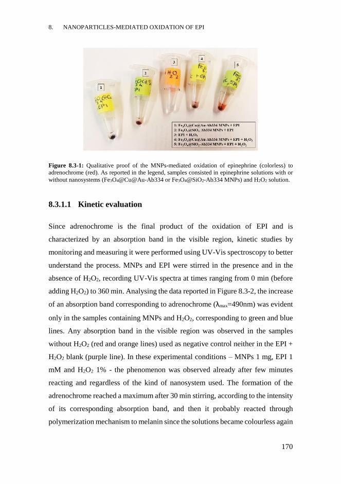
8. NANOPARTICLES-MEDIATED OXIDATION OF EPI
170
Figure 8.3-1: Qualitative proof of the MNPs-mediated oxidation of epinephrine (colorless) to
adrenochrome (red). As reported in the legend, samples consisted in epinephrine solutions with or
without nanosystems (Fe3O4@Cu@Au-Ab334 or Fe3O4@SiO2-Ab334 MNPs) and H2O2 solution.
8.3.1.1 Kinetic evaluation
Since adrenochrome is the final product of the oxidation of EPI and is
characterized by an absorption band in the visible region, kinetic studies by
monitoring and measuring it were performed using UV-Vis spectroscopy to better
understand the process. MNPs and EPI were stirred in the presence and in the
absence of H2O2, recording UV-Vis spectra at times ranging from 0 min (before
adding H2O2) to 360 min. Analysing the data reported in Figure 8.3-2, the increase
of an absorption band corresponding to adrenochrome (λmax=490nm) was evident
only in the samples containing MNPs and H2O2, corresponding to green and blue
lines. Any absorption band in the visible region was observed in the samples
without H2O2 (red and orange lines) used as negative control neither in the EPI +
H2O2 blank (purple line). In these experimental conditions – MNPs 1 mg, EPI 1
mM and H2O2 1% - the phenomenon was observed already after few minutes
reacting and regardless of the kind of nanosystem used. The formation of the
adrenochrome reached a maximum after 30 min stirring, according to the intensity
of its corresponding absorption band, and then it probably reacted through
polymerization mechanism to melanin since the solutions became colourless again
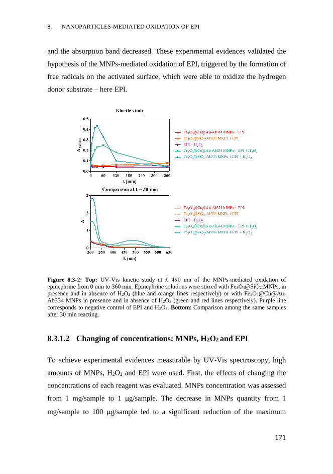
8. NANOPARTICLES-MEDIATED OXIDATION OF EPI
171
and the absorption band decreased. These experimental evidences validated the
hypothesis of the MNPs-mediated oxidation of EPI, triggered by the formation of
free radicals on the activated surface, which were able to oxidize the hydrogen
donor substrate – here EPI.
Figure 8.3-2: Top: UV-Vis kinetic study at λ=490 nm of the MNPs-mediated oxidation of
epinephrine from 0 min to 360 min. Epinephrine solutions were stirred with Fe3O4@SiO2 MNPs, in
presence and in absence of H2O2 (blue and orange lines respectively) or with Fe3O4@Cu@Au-
Ab334 MNPs in presence and in absence of H2O2 (green and red lines respectively). Purple line
corresponds to negative control of EPI and H2O2. Bottom: Comparison among the same samples
after 30 min reacting.
8.3.1.2 Changing of concentrations: MNPs, H2O2 and EPI
To achieve experimental evidences measurable by UV-Vis spectroscopy, high
amounts of MNPs, H2O2 and EPI were used. First, the effects of changing the
concentrations of each reagent was evaluated. MNPs concentration was assessed
from 1 mg/sample to 1 μg/sample. The decrease in MNPs quantity from 1
mg/sample to 100 μg/sample led to a significant reduction of the maximum
8. NANOPARTICLES-MEDIATED OXIDATION OF EPI
172
intensity of the colorimetric signal, which was reached after long reaction time
(40-60 min). Lower quantities of MNPs than 100 μg/sample did not produce
measurable UV-Vis signals. H2O2 concentration was assessed from 3% to 0.01 %;
the increase in H2O2 concentration from 1% to 3% had not a significant effect
since it could be considered already in excess. Below 1%, the signal was not
measurable. EPI solutions at a concentration ranging from 1 nM to 1 mM were
analysed. The use of μM and nM EPI solutions produced an amount of
adrenochrome too low for UV-Vis detection. For this reason, other analytical
techniques, for instance electrochemical ones, should be probably used in order to
detect samples with low concentrations of analyte (EPI < 1mM).
8.3.2 Evaluation of the selectivity of Ab334-MNPs
In order to evaluate the possibility to carry out a selective oxidative process, a pre-
incubation step between MNPs and EPI was introduced. For this purpose,
different kinds of MNPs-Ab were selected; MNPs loading Ab334 and MNPs
functionalized with Ab186 were used. Fe3O4@Cu@Au-Ab186 MNPs and
Fe3O4@SiO2-Ab186 MNPs were synthesized in the same way as the
corresponding MNPs-Ab334, as reported in 7.3.3, conjugating the purified pre-
immune Ab186 already available in the Nb4D group. This antibody did not show
specificity and for this reason it was selected, in opposition to the specific Ab334.
The four kinds of nanosystems at 1 mg/sample, Fe3O4@Cu@Au-Ab (334 or 186)
MNPs and Fe3O4@SiO2-Ab (334 or 186) MNPs were stirred with 1 mM EPI
solution in citrate buffer for 15 min. This pre-incubation step was performed to
promote the binding of EPI to the Ab-MNPs without triggering its oxidation.
MNPs were then washed several times with citrate buffer to remove the unbound
EPI and finally H2O2 1% solution was added to each sample. A solution containing
only EPI and H2O2 was used as negative control. After 20 min stirring, the UV-
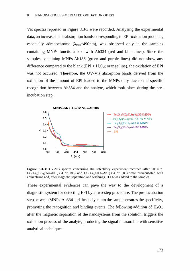
8. NANOPARTICLES-MEDIATED OXIDATION OF EPI
173
Vis spectra reported in Figure 8.3-3 were recorded. Analysing the experimental
data, an increase in the absorption bands corresponding to EPI-oxidation products,
especially adrenochrome (λmax=490nm), was observed only in the samples
containing MNPs functionalized with Ab334 (red and blue lines). Since the
samples containing MNPs-Ab186 (green and purple lines) did not show any
difference compared to the blank (EPI + H2O2; orange line), the oxidation of EPI
was not occurred. Therefore, the UV-Vis absorption bands derived from the
oxidation of the amount of EPI loaded to the MNPs only due to the specific
recognition between Ab334 and the analyte, which took place during the pre-
incubation step.
Figure 8.3-3: UV-Vis spectra concerning the selectivity experiment recorded after 20 min.
Fe3O4@Cu@Au-Ab (334 or 186) and Fe3O4@SiO2-Ab (334 or 186) were preincubated with
epinephrine and, after magnetic separation and washings, H2O2 was added to the samples.
These experimental evidences can pave the way to the development of a
diagnostic system for detecting EPI by a two-step procedure. The pre-incubation
step between MNPs-Ab334 and the analyte into the sample ensures the specificity,
promoting the recognition and binding events. The following addition of H2O2,
after the magnetic separation of the nanosystems from the solution, triggers the
oxidation process of the analyte, producing the signal measurable with sensitive
analytical techniques.
MNPs-Ab334 vs MNPs-Ab186
300 350 400 450 500 550 6000.0
0.1
0.2
0.3
0.4
0.5
0.6Fe3O4@Cu@Au-Ab334MNPs
Fe3O4@Cu@Au-Ab186 MNPs
Fe3O4@SiO2-Ab334 MNPs
Fe3O4@SiO2-Ab186 MNPs
EPI
(nm)
A
8. NANOPARTICLES-MEDIATED OXIDATION OF EPI
174
CHAPTER CONTRIBUTIONS
- The nanosystems based on Fe3O4@Cu@Au and Fe3O4@SiO2 MNPs were
able to promote the oxidation of the catecholamine EPI mediated by H2O2,
- Using MNPs functionalized with a specific antibody raised against EPI,
like Ab334, it was possible to achieve a selective method for binding,
magnetically separating and detecting the analyte. Through a two-step
procedure, consisting in a pre-incubation between MNPs-Ab334 and EPI
followed by the addition of H2O2, the selective oxidation of EPI bound to
MNPs-Ab334 could be achieved.
8.4.1 Future perspectives
- Study the MNPs-mediated oxidation of EPI using more sensitive
analytical techniques, such as electrochemical ones, evaluating also the
activity of bare MNPs. Optimize some physicochemical parameters such
as the incubation time, buffer solution or concentration of MNPs.
- Validate the two-step procedure in standard complex solutions and
biological matrices, such as plasma or urine.

8. NANOPARTICLES-MEDIATED OXIDATION OF EPI
175
MATERIALS AND METHODS
Reagents, materials and equipment: The chemical reagents used were purchased
by Sigma-Aldrich (Merck KGaA, Darmstadt, Germany). The pre-immune
antiserum As186 was already present in Nb4D group and ÄKTATM prime,
provided with HiTrapTM Protein A HP column or HiTrapTM Desalting column (GE
Helthcare, Uppsala, Sweden), was used for its purification with the corresponding
software Prime View and Prime View Evaluation. The pH and the conductivity
of all buffers and solutions were measured with a pH-meter pH 540 GLP and a
conductometer LF 340, respectively (WTW, Weilheim, Germany). UV-Vis
spectra were recorded from 200 to 1000 nm by means of SpectramaxPlus
(Molecular Devices, Sunnyvale, CA, USA) with the software SoftmaxPro v4.7
(Molecular Devices) using dilution plates as sample holders, purchased from
Nirco (Barberà del Vallés, Spain). All the experimental data were analysed using
the software GraphPad Prism 5.03 (GraphPad Software Inc., San Diego, USA).
Kinetic evaluation: two samples of Fe3O4@Cu@Au-Ab334 (1 mg/sample) and 2
samples of Fe3O4@SiO2-Ab334 MNPs (1 mg/sample) were stirred at 25°C in
Eppendorf tube with 1 mM EPI solution (180 μL/sample) in citrate buffer (0.04
M pH 5.5). UV-Vis spectra were recorded in a dilution plate before and after the
addition of H2O2 10 % solution in citrate buffer (20 μL/sample) or only citrate
buffer (20 μL/sample) from 5 min to 360 min. A solution of EPI 1 mM and H2O2
10 % without MNPs was used as blank.
Changing of concentrations: MNPs
90 μL/sample of EPI 1mM solution in citrate buffer were stirred with MNPs-
Ab334 (Fe3O4@Cu@Au-Ab334 or Fe3O4@SiO2-Ab334 MNPs) at concentrations
of 1000, 100, 10 or 1 μg/sample. After adding 10 μL of H2O2 10 % in citrate buffer
8. NANOPARTICLES-MEDIATED OXIDATION OF EPI
176
UV-Vis spectra were recorded in a dilution plate at 20, 40 and 90 min. Samples
of EPI/H2O2 and MNPs-Ab334/EPI/citrate buffer were used as blank.
Changing of concentrations: H2O2
90 μL/sample of EPI 1mM solution in citrate buffer were stirred with 100
μg/sample of MNPs-Ab334 (Fe3O4@Cu@Au-Ab334 or Fe3O4@SiO2-Ab334
MNPs). After adding 10 μL of H2O2 solution in citrate buffer (at concentrations
of 30%, 10%, 1% or 0.1%) UV-Vis spectra were recorded in a dilution plate after
20, 40 and 60 min. Samples of EPI/H2O2 30% and MNPs-Ab334/EPI/citrate
buffer were used as blank.
Changing of concentrations: EPI
90 μL/sample of EPI solutions in citrate buffer, at concentrations ranging from 1
mM to 1 nM, were stirred with 100 μg/sample of MNPs-Ab334 (Fe3O4@Cu@Au-
Ab334 or Fe3O4@SiO2-Ab334 MNPs). After adding 10 μL of H2O2 10 % solution
in citrate buffer, UV-Vis spectra were recorded in a dilution plate after 20 and 60
min. Samples of EPI 1 mM/ H2O2 10% and MNPs-Ab334/citrate buffer/H2O2 10%
were used as blank.
Selectivity evaluation: 1 mg of Fe3O4@Cu@Au or Fe3O4@SiO2 MNPs
functionalized with Ab334 or Ab186 were stirred for 15 min with 100 μL/sample
of EPI 0.9 mM in citrate buffer (0.04 M, pH 5.5). After magnetic separation,
MNPs were washed twice with citrate buffer and then dispersed in H2O2 1% (100
μL/sample). A solution of EPI 1 mM (90 μL) and H2O2 10% (10μL), added after
15 min, was used as blank. After 20 min stirring, UV-Vis spectra of the samples
were recorded in a dilution plate (100 μL sample/well).

9. CONCLUSIONS OF THIS THESIS
9. CONCLUSIONS OF THIS THESIS
178
Conclusions
Among the superparamagnetic nanoparticles suitable for biomedical applications,
new engineered nanosystems have been developed in this research work.
- Two basic core-shell structures made of Fe3O4@Cu@Au or Fe3O4@SiO2
MNPs have been designed, synthesized and characterized by means of
different physicochemical techniques, proving their nanometric size and
core-shell structure.
- The surface activation exploiting the physicochemical features of the
external inorganic shell, and the functionalization with polymers, proteins
or bioactive molecules have been carried out according to the planned
biomedical end use.
- The internalization of differently coated MNPs into monocyte-
differentiated macrophages have been achieved, preferentially for the
nanosystems loading Tf. These results, together with the possibility of
further functionalization with drugs and combined with the ability of Tf
in targeting cancer cells, make these nanosystems favourable for using in
the Trojan Horse approach.
- The lipodepsipeptides SRE and SP22A have been loaded, for the first
time, on nanoparticles without losing their biological activities. The
confirmation of the in vitro antimicrobial activity on the yeast R.
pilimanae and the uptake by monocyte-differentiated macrophages make
the MNPs-SRE and MNPs-SP22A promising tools for a new-generation
strategy for monitoring and treating infectious diseases.
- The development of the direct and indirect competitive ELISA
immunoassays for the in vitro detection of EPI has been firstly addressed,
reaching good sensitivity and detectability parameters. Amperometric
magneto-immunosensors involving antibody-functionalized MNPs have
been developed, avoiding analyte modification, reaching IC50 and LOD

9. CONCLUSIONS OF THIS THESIS
179
values in the nanomolar range. These diagnostic platforms, including
those based on the engineered nanosystems, could prove to be efficient
and sensitive alternatives to the standard analytical methods used for
detecting EPI in biological samples.
- The ability of antibody-functionalized MNPs to detect EPI and promote
its H2O2-mediated oxidation have been demonstrated, opening the
possibility to use them as diagnostic tools for the recognition, separation
and analysis of EPI.
The scientific results achieved during this research work using the nanosystems
based on Fe3O4@Cu@Au and Fe3O4@SiO2 MNPs might contribute to know and
develop new diagnostic tools and/or therapeutic strategies.


10. ACRONYMS AND ABBREVIATIONS
10. ACRONYMS AND ABBREVIATIONS
182
10.1 ACRONYMS AND ABBREVIATIONS
A Absorbance
AA Amino Acid
Ab Antibody
Ab186 Antibody 186 after purification
Ab334 Antibody 334 after purification
ACN Acetonitrile
AFM Atomic Force Microscopy
Ag Antigen
APTES (3-AminoPropyl)TriethoxySilane
As Antiserum
As334, As335, As336 Antiserum 334, 335, 336
Au NPs Gold nanoparticles
BSA Bovine Serum Albumin
CD Circular dichroism
CSIC Spanish National Research Council
CT Computer Tomography
Da Dalton
Dab 2,4-diaminobutyric acid
DCC N,N’-dicyclohexylcarbodiimide
DHFR DiHydroFolate Reductase
DMEM Dulbecco's Modified Eagle's medium
DMF N,N-dimethylformamide
DOSY Diffusion Ordered SpectroscopY
EDC-HCl 1-Ethyl-3-(3-dimethylaminopropyl)carbodiimide hydrochloride
ELISA Enzyme-Linked Immunosorbent Assay
EPI (-)-EPInephrine
EPR Enhanced Permeability and Retention Effect
ET Enzymatic Tracer
FA Folic Acid
FBS Fetal Bovine Serum
FRs Folate Receptors
FT-IR Fourier-Transform Infrared Spectroscopy
GPTMS (3-Glycidyloxypropyl)trimethoxysilane
Hc Coercivity
HCH Horseshoe Crab Hemocyanin
HRP Horseradish Peroxidise
IC10 Concentration in which the signal is 90 % inhibited
IC20 Concentration in which the signal is 80 % inhibited
IC50 Concentration in which the signal is 50 % inhibited
10. ACRONYMS AND ABBREVIATIONS
183
IC80 Concentration in which the signal is 20 % inhibited
IC90 Concentration in which the signal is 10 % inhibited
IgG Immunoglobulin G
IQAC Institute of Advanced Chemistry of Catalonia
LA Lipoic Acid
LOD Limit Of Detection
LSPR Localized Surface Plasmon Resonance
MALDI-TOF-MS Matrix Assisted Laser Desorption Ionization - Time of Flight
Mass Spectrometry
MIC Minimum Inhibitory Concentration
MNPs Magnetic NanoParticles
MNPs-Ab186 Fe3O4@Cu@Au-PEG-Ab186 or Fe3O4@SiO2-GPTMS-Ab186
MNPs-Ab334 Fe3O4@Cu@Au-PEG-Ab334 or Fe3O4@SiO2-GPTMS-Ab334
MNPs-APTES Fe3O4@SiO2-APTES MNPs
MNPs-GPTMS Fe3O4@SiO2-GPTMS
MNPs-LA-NHS Fe3O4@Cu@Au-LA-NHS MNPs
MNPs-PEG Fe3O4@Cu@Au-PEG
MNPs-PLA Fe3O4@SiO2-APTES-PLA MNPs
MNPs-PLA-SP22A-TR Fe3O4@SiO2-APTES-PLA-cadaverineTR-SP22A MNPs
MNPs-PLA-TR Fe3O4@SiO2-APTES-PLA-cadaverineTR MNPs
MNPs-PLL Fe3O4@Cu@Au-LA-PLL
MNPs-PLL-FA Fe3O4@Cu@Au-LA-PLL-FA
MNPs-PLL-MTX Fe3O4@Cu@Au-LA-PLL-MTX
MNPs-PLL-TR Fe3O4@Cu@Au-LA-PLL-TR MNPs
MNPs-SP22A Fe3O4@SiO2-APTES-PLA-SP22A MNPs
MNPs-SRE Fe3O4@SiO2-APTES-PLA-SRE MNPs
MNPs-Stv-TR Fe3O4@Cu@Au-LA-Stv-TR
MNPs-Tf Fe3O4@Cu@Au-LA-Tf
MNPs-Tf-TR Fe3O4@Cu@Au-LA-Tf-TR
Mr Remanence
MRI Magnetic Resonance Imaging
Msat Saturation Magnetization
MTX Methotrexate
MW Molecular weight
Nb4D Nanobiotechnology for Diagnostics Group
NHS N-HydroxySuccinimide
NOESY Nuclear Overhauser Effect Spectroscopy
NorEPI (-)-NorEPIneprhine
NPs Nanoparticles
N-SMP N-succinimidyl 3-maleimidopropionate
P.s. pv. syringae Pseudomonas syringae pv. syringae
PAb Polyclonal Antibody

10. ACRONYMS AND ABBREVIATIONS
184
PABA p-AminoBenzoic Acid
PBS Phosphate Buffered Saline solution
PBST Phosphate Buffered Saline Tween-20 solution
PEG PolyEthylene Glycol
PEG-SH Thiolated poly(ethylene)glycol
PET Positron Emission Tomography
PhR Phenol Red
PLA Poly-L-Aspartic acid
PLA-TR Poly-L-Aspartic acid-Texas Red cadaverine conjugate
PLL Poly-L-Lysine
PLL-TR Poly-L-Lysine-Texas Red conjugate
PMA Phorbol 12-Myristate 13-Acetate
PVP PolyVinylPyrrolidone
R. pilimanae Rhodotorula pilimanae
RFC Reduced Folate Carriers
RPMI Roswell Park Memorial Institute medium
rt Room temperature
SAM Self-Assembled Monolayer
SIA N-Succinimidyl IodoAcetate
SP22A Syringopeptin 22A
SPCE Screen Printed Carbon Electrode
SPION SuperParamagnetic Iron Oxide Nanoparticles
SPs Syringopeptins
SPR Surface Plasmon Resonance
SRE Syringomycin E
SRs Syringomycins
Stv Streptavidin
TEM Transmission Electron Microscopy
TEOS TetraEthyl Orthosilicate
Tf Transferrin
TFA Trifluoroacetic acid
TfRs Transferrin Receptors
Tf-TR Transferrin-Texas Red conjugate
THF TetraHydroFolate
TMB 3,3’,5,5’-TetraMethylBenzidine
TOCSY TOtal Correlated SpectroscopY
TR Texas Red
TSP Trimethylsilyl propionate
V Voltage
VSM Vibrating Sample Magnetometer
XRPD X-Ray Powder Diffraction
α-FR α isoform of the Folate Receptor

11. BIBLIOGRAPHY
11. BIBLIOGRAPHY
186
(1) H. Goesmann; C. Feldmann. Nanoparticulate functional materials. Angewandte Chemie
International Edition 2010, 49, 1362-1395.
(2) L. Zhang; F.X. Gu; J.M. Chan; A.Z. Wang; R.S. Langer; O.C. Farokhzad. Nanoparticles
in Medicine: Therapeutic Applications and Developments. Clinical Pharmacology &
Therapeutics 2008, 83, 761-769.
(3) E.K. Lim; T. Kim; S. Paik; S. Haam; Y.M. Huh; K. Lee. Nanomaterials for Theranostics:
Recent Advances and Future Challenges. Chemical reviews 2015, 115, 327-394.
(4) A.M. Nyström; Wooley, K. L. The Importance of Chemistry in Creating Well-Defined
Nanoscopic Embedded Therapeutics: Devices Capable of the Dual Functions of Imaging and
Therapy. Accounts of Chemical Research 2011, 44, 969-978.
(5) R. Singh; H.S. Nalwa. Medical Applications of Nanoparticles in Biological Imaging, Cell
Labeling, Antimicrobial Agents, and Anticancer Nanodrugs. Journal of Biomedical
Nanotechnology 2011, 7, 489-503.
(6) T. Sun; Y.S. Zhang; B. Pang; D.C. Hyun; M. Yang; Y. Xia. Engineered nanoparticles for
drug delivery in cancer therapy. Angewandte Chemie International Edition 2014, 53, 12320-
12364.
(7) N. Larson; H. Ghandehari. Polymeric conjugates for drug delivery. Chem. Mat. 2012, 24,
840-853.
(8) R. Popovtzer; A. Agrawal; N.A. Kotov; A. Popovtzer; J. Balter; T.E. Carey; R. Kopelman.
Targeted Gold Nanoparticles enable Molecular CT Imaging of Cancer Nano Letters 2008, 8, 4593-
4596.
(9) H.B. Na; I.C. Song; T. Hyeon. Inorganic Nanoparticles for MRI Contrast Agents.
Advanced Materials 2009, 21, 2133-2148.
(10) L.E. Cole; R.D. Ross; J.M.R. Tilley; T. Vargo-Gogola; R.K. Roeder. Gold nanoparticles
as contrast agents in x-ray imaging and computed tomography. Nanomedicine 2015, 10, 321-341.
(11) H.M.E. Azzazy; M.M.H. Mansour. In vitro diagnostic prospects of nanoparticles. Clinica
Chimica Acta 2009, 403, 1-8.
(12) X. Chi; D. Huang; Z. Zhao; Z. Zhou; Z. Yin; J. Gao. Nanoprobes for in vitro diagnostics
of cancer and infectious diseases. Biomaterials 2012, 33, 189-206.
(13) N. Sanvicens; C. Pastells; N. Pascual; M.P. Marco. Nanoparticle-based biosensors for
detection of pathogenic bacteria. TrAC Trends in Analytical Chemistry 2009, 28, 1243-1252.
(14) G.V. Kurlyandskaya; D.S. Portnov; I.V. Beketov; A. Larrañaga; A.P. Safronov; I. Orue;
A.I. Medvedev; A.A. Chlenova; M.B. Sanchez-Ilarduya; A. Martinez-Amesti; A.V. Svalov.
Nanostructured materials for magnetic biosensing. Biochimica et Biophysica Acta (BBA) -
General Subjects 2017, 1861, 1494-1506.
(15) K. Saha; S.S. Agasti; C. Kim; X. Li; V.M. Rotello. Gold Nanoparticles in Chemical and
Biological Sensing. Chemical reviews 2012, 112, 2739-2779.
(16) D.R. Viswambari; M. Doble; R.S. Verma. Nanomaterials for early detection of cancer
biomarker with special emphasis on gold nanoparticles in immunoassays/sensors. Biosensors and
Bioelectronics 2015, 68, 688-698.
(17) M. Hasanzadeh; N. Shadjou; M. De la Guardia. Iron and iron-oxide magnetic
nanoparticles as signal-amplification elements in electrochemical biosensing. TrAC Trends in
Analytical Chemistry 2015, 72, 1-9.
(18) I. Vasconcelos; C. Fernandes. Magnetic solid phase extraction for determination of drugs
in biological matrices. TrAC Trends in Analytical Chemistry 2017, 89, 41-52.
(19) S. Bedanta; W. Kleemann. Supermagnetism. The Journal of Physics D: Applied Physics
2009, 42, 013001.
(20) C.L. Nehl; J.H. Hafner. Shape-dependent plasmon resonances of gold nanoparticles.
Journal of Materials Chemistry 2008, 18, 2415-2419.
(21) K.K. Lance; E. Coronado; L.L. Zhao; G.C. Schatz. The Optical Properties of Metal
Nanoparticles: The Influence of Size, Shape, and Dielectric Environment. The Journal of Physical
Chemistry B 2003, 107, 668-677.
(22) C.C. Berry. Progress in functionalization of magnetic nanoparticles for applications in
biomedicine. Journal of Physics D: Applied Physics 2009, 42, 224003.
11. BIBLIOGRAPHY
187
(23) G. Bao; S. Mitragotri; S. Tong. Multifunctional nanoparticles for drug delivery and
molecular imaging. Annual review of biomedical engineering 2013, 15, 253-282.
(24) K. Chatterjee; S. Sarkar; K. Jagajjanani Rao; S. Paria. Core/shell nanoparticles in
biomedical applications. Advaces in Colloid and Interface Science 2014, 209, 8-39.
(25) C. Fang; M. Zhang. Multifunctional magnetic nanoparticles for medical imaging
applications. Journal of Materials Chemistry 2009, 19, 6258-6266.
(26) S.K. Yen; P. Padmanabhan; S.T. Selvan. Multifunctional iron oxide nanoparticles for
diagnostics, therapy and macromolecule delivery. Theranostics 2013, 3, 986-1003.
(27) W. Zhou; X. Gao; D. Liu; X. Chen. Gold Nanoparticles for In Vitro Diagnostics. Chemical
reviews 2015, 115, 10575-10636.
(28) M. Arruebo; R. Fernández-Pacheco; M.R. Ibarra; J. Santamaría. Magnetic nanoparticles
for drug delivery. Nano Today 2007, 2, 22-32.
(29) M. Banobre-Lopez; A. Teijeiro; J. Rivas. Magnetic nanoparticle-based hyperthermia for
cancer treatment. Reports of Practical Oncology and Radiotherapy 2013, 18, 397-400.
(30) H. Lu; M. Zhang; Y. Yang; Q. Huang; T. Fukuda; Z. Wang; Y. Shen. A bioinspired
multilegged soft millirobot that functions in both dry and wet conditions. Nature communications
2018, 9, 3944.
(31) S.M. Taimoory; J.F. Trant; A. Rahdar; M. Aliahmad; F. Sadeghfar; M. Hashemzaei.
Importance of the Inter-Electrode Distance for the Electrochemical Synthesis of Magnetite
Nanoparticles: Synthesis, Characterization, Computational Modelling, and Cytotoxicity. e-
Journal of Surface Science and Nanotechnology 2017, 15, 31-39.
(32) J. Xu; H. Yang; W. Fu; K. Du; Y. Sui; J. Chen; Y. Zeng; M. Li; G. Zou. Preparation and
magnetic properties of magnetite nanoparticles by sol–gel method. Journal of Magnetism and
Magnetic Materials 2007, 309, 307-311.
(33) A. Taebin; K.J. Hun; Y. Hee-Man; L.J. Woo; K. Jong-Duk. Formation Pathways of
Magnetite Nanoparticles by Coprecipitation Method. The Journal of Physical Chemistry C 2012,
116, 6069-6076.
(34) L.H. Reddy; J.L. Arias; J. Nicolas; P. Couvreur. Magnetic Nanoparticles: Design and
Characterization, Toxicity and Biocompatibility, Pharmaceutical and Biomedical Applications.
Chemical reviews 2012, 112, 5818-5878.
(35) Q. Li; C.W. Kartikowati; S. Horie; T. Ogi; T. Iwaki; K. Okuyama. Correlation between
particle size/domain structure and magnetic properties of highly crystalline Fe3O4 nanoparticles.
Scientific Reports 2017, 7, 9894.
(36) T.K. Jain; J. Richey; M. Strand; D.L. Leslie-Pelecky; C.A. Flask; V. Labhasetwar.
Magnetic nanoparticles with dual functional properties: drug delivery and magnetic resonance
imaging. Biomaterials 2008, 29, 4012-4021.
(37) W. Wu; C.Z. Jiang; V.A. Roy. Designed synthesis and surface engineering strategies of
magnetic iron oxide nanoparticles for biomedical applications. Nanoscale 2016, 8, 19421-19474.
(38) R.A. Bohara; N.D. Thorat; S.H. Pawar. Role of functionalization: strategies to explore
potential nano-bio applications of magnetic nanoparticles. RSC Advances 2016, 6, 43989-44012.
(39) C. Rümenapp; B. Gleich; A. Haase. Magnetic Nanoparticles in Magnetic Resonance
Imaging and Diagnostics. Pharmaceutical Research 2012, 29, 1165-1179.
(40) N. Griffete; J. Fresnais; A. Espinosa; C. Wilhelm; A. Bee; C. Menager. Design of magnetic
molecularly imprinted polymer nanoparticles for controlled release of doxorubicin under an
alternative magnetic field in athermal conditions. Nanoscale 2015, 7, 18891-18896.
(41) A. Espinosa; R. Di Corato; J. Kolosnjaj-Tabi; P. Flaud; T. Pellegrino; C. Wilhelm. Duality
of Iron Oxide Nanoparticles in Cancer Therapy: Amplification of Heating Efficiency by Magnetic
Hyperthermia and Photothermal Bimodal Treatment. ACS nano 2016, 10, 2436-2446.
(42) H. Ittrich; K. Peldschus; N. Raabe; M. Kaul; G. Adam. Superparamagnetic Iron Oxide
Nanoparticles in Biomedicine: Applications and Developments in Diagnostics and Therapy.
Fortschr Röntgenstr 2013, 185, 1149–1166
(43) M. Bietenbeck; A. Florian; C. Faber; U. Sechtem; A. Yilmaz. Remote magnetic targeting
of iron oxide nanoparticles for cardiovascular diagnosis and therapeutic drug delivery: where are
we now? International journal of nanomedicine 2016, 11, 3191-3203.
11. BIBLIOGRAPHY
188
(44) R. Mejias; S. Perez-Yague; L. Gutierrez; L.I. Cabrera; R. Spada; P. Acedo; C.J. Serna;
F.J. Lazaro; A. Villanueva; P. Morales Mdel; D.F. Barber. Dimercaptosuccinic acid-coated
magnetite nanoparticles for magnetically guided in vivo delivery of interferon gamma for cancer
immunotherapy. Biomaterials 2011, 32, 2938-2952.
(45) A.K. Silva; C. Menager; C. Wilhelm. Magnetic drug carriers: bright insights from light-
responsive magnetic liposomes. Nanomedicine 2015, 10, 2797-2799.
(46) M.M. Yallapu; N. Chauhan; S.F. Othman; V. Khalilzad-Sharghi; M.C. Ebeling; S. Khan;
M. Jaggi; S.C. Chauhan. Implications of protein corona on physico-chemical and biological
properties of magnetic nanoparticles. Biomaterials 2015, 46, 1-12.
(47) D.E. Owens III; N.A. Peppas. Opsonization, biodistribution, and pharmacokinetics of
polymeric nanoparticles. International journal of pharmaceutics 2006, 307, 93-102.
(48) L. Pang; C. Zhang; J. Qin; L. Han; R. Li; C. Hong; H. He; Wang, J. A novel strategy to
achieve effective drug delivery: exploit cells as carrier combined with nanoparticles. Drug delivery
2017, 24, 83-91.
(49) M. Muthana; S.D. Scott; N. Farrow; F. Morrow; C. Murdoch; S. Grubb; N. Brown; J.
Dobson; C.E. Lewis. A novel magnetic approach to enhance the efficacy of cell-based gene
therapies. Gene therapy 2008, 15, 902-910.
(50) C.A. Quinto; P. Mohindra; S. Tong; G. Bao. Multifunctional superparamagnetic iron oxide
nanoparticles for combined chemotherapy and hyperthermia cancer treatment. Nanoscale 2015, 7,
12728-12736.
(51) B. Sanz; M.P. Calatayud; T.E. Torres; M.L. Fanarraga; M.R. Ibarra; G.F. Goya. Magnetic
hyperthermia enhances cell toxicity with respect to exogenous heating. Biomaterials 2017, 114,
62-70.
(52) D. Shi; M.E. Sadat; A.W. Dunn; D.B. Mast. Photo-fluorescent and magnetic properties of
iron oxide nanoparticles for biomedical applications. Nanoscale 2015, 7, 8209-8232.
(53) B. Ding; S. Shen; L. Wu; X. Qi; H. Ni; Y. Ge. Doxorubicin-loaded Fe3O4@SiO2
Nanoparticles as Magnetic Targeting Agents for Combined Photothermal-chemotherapy of
Cancer. Chemistry Letters 2015, 44, 858-860.
(54) Z. Gao; X. Liu; G., D.; F. Zhou; L. Zhang; Q. Wang; J. Lu. Fe3O4@mSiO2-FA-CuS-PEG
Nanocomposites for Magnetic Resonance Imaging and Targeted Chemo-photothermal Synergistic
Therapy of Cancer Cells. Dalton Transactions 2016, 45, 13456-13465.
(55) S. Elingarami; M. Liu; Z. Li; N. He. Surface-Engineered Magnetic Nanoparticles for
Molecular Detection of Infectious Agents and Cancer. J. Nanosci. Nanotechnol. 2013, 13, 3204-
3213.
(56) H. Gu; K. Xu; C. Xu; B. Xu. Biofunctional magnetic nanoparticles for protein separation
and pathogen detection. Chemical communications 2006, 0, 941-949.
(57) A.L. Chun. Nanoparticles offer hope for TB detection. Nature Nanotechnology 2009, 4,
1-2.
(58) H. Xu; Z.P. Aguilar; L. Yang; M. Kuang; H. Duan; Y. Xiong; H. Wei; A. Wang. Antibody
conjugated magnetic iron oxide nanoparticles for cancer cell separation in fresh whole blood.
Biomaterials 2011, 32, 9758-9765.
(59) M. Lewin; N. Carlesso; C.H. Tung; X.W. Tang; D. Cory; D.T. Scadden; R. Weissleder.
Tat peptide-derivatized magnetic nanoparticles allow in vivo tracking and recovery of progenitor
cells. Nature Biotechnology 2000, 18, 410-414.
(60) Y. Chen; Y. Xianyu; Y. Wang; X. Zhang; R. Cha; J. Sun; X. Jiang. One-Step Detection of
Pathogens and Viruses: Combining Magnetic Relaxation Switching and Magnetic Separation.
ACS nano 2015, 9, 3184-3191.
(61) K. Atacan; B. Çakıroğlu; M. Özacar. Improvement of the stability and activity of
immobilized trypsin on modified Fe3O4 magnetic nanoparticles for hydrolysis of bovine serum
albumin and its application in the bovine milk. Food Chemistry 2016, 212, 460-468.
(62) Y.T. Zhu; X.Y. Ren; Y.M. Liu; Y. Wei; L.S. Qing; X. Liao. Covalent immobilization of
porcine pancreatic lipase on carboxyl-activated magnetic nanoparticles: Characterization and
application for enzymatic inhibition assays. Materials Science and Engineering C: Materials for
Biological Applications 2014, 38, 278-285.

11. BIBLIOGRAPHY
189
(63) M. Broto; J.P. Salvador; R. Galve; M.P. Marco. Biobarcode assay for the oral
anticoagulant acenocoumarol. Talanta 2018, 178, 308-314.
(64) D.M. Bruls; T.H. Evers; J.A.H. Kahlman; P.J.W. Van Lankvelt; M. Ovsyanko; E.G.M.
Pelssers; J.J.H.B. Schleipen; F.K. De Theije; C.A. Verschuren; T. Van der Wijk; J.B.A. Van Zon;
W.U. Dittmer; A.H.J. Immink; J.H. Nieuwenhuis; M.W.J. Prins. Rapid integrated biosensor for
multiplexed immunoassays based on actuated magnetic nanoparticles. Lab on a Chip 2009, 9,
3504-3510.
(65) D.G. Pinacho; F. Sánchez-Baeza; M.I. Pividori; M.P. Marco. Electrochemical Detection
of Fluoroquinolone Antibiotics in Milk Using a Magneto Immunosensor. Sensors 2014, 14,
15965-15980.
(66) R.G. Chaudhuri; S. Paria. Core/shell nanoparticles: classes, properties, synthesis
mechanisms, characterization, and applications. Chemical reviews 2012, 112, 2373-2433.
(67) B. Kalska-Szostko; U. Wykowska; D. Satuła. Magnetic nanoparticles of core–shell
structure. Colloids and Surfaces A: Physicochemical and Engineering Aspects 2015, 481, 527-
536.
(68) J.R. McCarthy; R. Weissleder. Multifunctional magnetic nanoparticles for targeted
imaging and therapy. Advanced drug delivery reviews 2008, 60, 1241-1251.
(69) O. Chen; L. Riedemann; F. Etoc; H. Herrmann; M. Coppey; M. Barch; C.T. Farrar; J.
Zhao; O.T. Bruns; H. Wei; P. Guo; J. Cui; R. Jensen; Y. Chen; D.K. Harris; J.M. Cordero; Z.
Wang; A. Jasanoff; D. Fukumura; R. Reimer; M. Dahan; R.K. Jain; M.G. Bawendi. Magneto-
fluorescent core-shell supernanoparticles. Nature Communication 2014, 5, 5093.
(70) X. Yang; H. Hong; J.J. Grailer; I.J. Rowland; A. Javadi; S.A. Hurley; Y. Xiao; Y. Yang;
Y. Zhang; R.J. Nickles; W. Cai; D.A. Steeber; S. Gong. cRGD-functionalized, DOX-conjugated,
and 64Cu-labeled superparamagnetic iron oxide nanoparticles for targeted anticancer drug delivery
and PET/MR imaging. Biomaterials 2011, 32, 4151-4160.
(71) J. Xie; K. Chen; J. Huang; S. Lee; J. Wang; J. Gao; X. Li; X. Chen. PET/NIRF/MRI triple
functional iron oxide nanoparticles. Biomaterials 2010, 31, 3016-3022.
(72) L. Maggini; I. Cabrera; A. Ruiz-Carretero; E.A. Prasetyanto; E. Robinet; L. De Cola.
Breakable mesoporous silica nanoparticles for targeted drug delivery. Nanoscale 2016, 8, 7240-
7247.
(73) Y. Wang; Q. Zhao; N. Han; L. Bai; J. Li; J. Liu; E. Che; L. Hu; Q. Zhang; T. Jiang; S.
Wang. Mesoporous silica nanoparticles in drug delivery and biomedical applications.
Nanomedicine: Nanotechnology, Biology and Medicine 2015, 11, 313-327.
(74) W. Stöber; A. Fink; E. Bohn. Controlled growth of monodisperse silica spheres in the
micron size range. Journal of Colloid and Interface Science 1968, 26, 62-69.
(75) T.G. Waddell; D.E. Leyden; M.T. DeBello. The nature of organosilane to silica-surface
bonding. The Journal of the American Chemical Society 1981, 103, 5303-5307.
(76) C. Zhang; B. Wängler; B. Morgenstern; H. Zentgraf; M. Eisenhut; H. Untenecker; R.
Krüger, R. H.; C. Seliger; W. Semmler; F. Kiessling. Silica- and Alkoxysilane-Coated Ultrasmall
Superparamagnetic Iron Oxide Particles: A Promising Tool To Label Cells for Magnetic
Resonance Imaging. Langmuir : the ACS journal of surfaces and colloids 2007, 23, 1427-1434.
(77) M.C. Daniel; D. Astruc. Gold nanoparticles: assembly, supramolecular chemistry,
quantum-size-related properties, and applications toward biology, catalysis, and nanotechnology.
Chemical reviews 2004, 104, 293-346.
(78) E.C. Dreaden; A.M. Alkilany; X. Huang; C.J. Murphy; M.A. El-Sayed. The golden age:
gold nanoparticles for biomedicine. Chemical Society Reviews 2012, 41, 2740-2779.
(79) S. Eustis; M.A. El-Sayed. Why gold nanoparticles are more precious than pretty gold:
Noble metal surface plasmon resonance and its enhancement of the radiative and nonradiative
properties of nanocrystals of different shapes. Chemical Society Reviews 2006, 35, 209-217.
(80) A. Espinosa; M. Bugnet; G. Radtke; S. Neveu; G.A. Botton; C. Wilhelm; A. Abou-Hassan.
Can magneto-plasmonic nanohybrids efficiently combine photothermia with magnetic
hyperthermia? Nanoscale 2015, 7, 18872-18877.
11. BIBLIOGRAPHY
190
(81) E. Pensa; E. Cortés; G. Corthey; P. Carro; C. Vericat; M.H. Fonticelli; G. Benítez; A.A.
Rubert; R.C. Salvarezza. The Chemistry of the Sulfur–Gold Interface: In Search of a Unified
Model. Accounts of Chemical Research 2012, 45, 1183-1192.
(82) R.G. Nuzzo; B.R. Zegarski; L.H. Dubois. Fundamental Studies of the Chemisorption of
Organosulfur Compounds on Au(111). Implications for Molecular Self-Assembly on Gold
Surfaces. The Journal of the American Chemical Society 1987, 109, 733-740.
(83) S. Rana; A. Bajaj; R. Mout; V.M. Rotello. Monolayer coated gold nanoparticles for
delivery applications. Advanced drug delivery reviews 2012, 64, 200-216.
(84) J. Manson; D. Kumar; B.J. Meenan; D. Dixon. Polyethylene glycol functionalized gold
nanoparticles: the influence of capping density on stability in various media. Gold Bulletin 2011,
44, 99-105.
(85) D. Kim; S. Park; J.H. Lee; Y.Y. Jeong; S. Jon. Antibiofouling Polymer-Coated Gold
Nanoparticles as a Contrast Agent for in Vivo X-ray Computed Tomography Imaging. The
Journal of the American Chemical Society 2007, 129, 7661-7665.
(86) Y.T. Liu; L. Yan; L.M. Sun; H.Q. Li; H.H. Li: Mixed short-chain PEG self-assembled
monolayer preparation and immobilization of protein, 2014; Vol. 42. pp. 69-74.
(87) R.L. Letsinger; R. Elghanian; G. Viswanadham; C.A. Mirkin. Use of a Steroid Cyclic
Disulfide Anchor in Constructing Gold Nanoparticles-Oligonucleotide Conjugates. Bioconjugate
Chemistry 2000, 11, 289-291.
(88) J. Luo; Q. Fan; M. Suzuki; I.S. Suzuki; M.H. Engelhard; Y. Lin; N.Kim; J.Q. Wang; C.J.
Zhong. Monodispersed Core−Shell Fe3O4@Au Nanoparticles. The Journal of Physical Chemistry
B 2005, 109, 21593-21601.
(89) Y. Xing; Y.Y. Jin; J.C. Si; M.L. Peng; X.F. Wang; C. Chen; Y.L. Cui. Controllable
synthesis and characterization of Fe3O4/Au composite nanoparticles. Journal of Magnetism and
Magnetic Materials 2015, 380, 150-156.
(90) H. Maleki; A. Simchi; M. Imani; B.F.O. Costa. Size-controlled synthesis of
superparamagnetic iron oxide nanoparticles and their surface coating by gold for biomedical
applications. Journal of Magnetism and Magnetic Materials 2012, 324, 3997-4005.
(91) S.V. Salihov; Y.A. Ivanenkov; S.P. Krechetov; M.S. Veselov; N.V. Sviridenkova; A.G.
Savchenko; N.L. Klyachko; Y.I. Golovin; N.V. Chufarova; E.K. Beloglazkina; A.G. Majouga.
Recent advances in the synthesis of Fe3O4@Au core/shell nanoparticles. Journal of Magnetism
and Magnetic Materials 2015, 394, 173-178.
(92) H. Cai; N. Zhu; Y. Jiang; P. He; Y. Fang. Cu@Au alloy nanoparticle as oligonucleotides
labels for electrochemical stripping detection of DNA hybridization. Biosensors and
Bioelectronics 2003, 18, 1311-1319.
(93) P. Li; L. Li; Y. Zhao; L. Sun; Y. Zhang. Selective binding and magnetic separation of
histidine-tagged proteins using Fe3O4/Cu-apatite nanoparticles. Journal of inorganic biochemistry
2016, 156, 49-54.
(94) M. Tang; S. Zhang; X. Li; X. Pang; H. Qiu. Fabrication of magnetically recyclable
Fe3O4@Cu nanocomposites with high catalytic performance for the reduction of organic dyes and
4-nitrophenol. Materials Chemistry and Physics 2014, 148, 639-647.
(95) D. Patel; A. Kell; B. Simard; B. Xiang; H.Y. Lin; G. Tian. The cell labeling efficacy,
cytotoxicity and relaxivity of copper-activated MRI/PET imaging contrast agents. Biomaterials
2011, 32, 1167-1176.
(96) R. Massart. Preparation of Aqueous Magnetic Liquids in Alkaline and Acidic Media. IEEE
Transactions on Magnetics 1981, 17, 1247-1248.
(97) K.J. Gallagher; W. Feitknecht; U. Mannweiler. Mechanism of Oxidation of Magnetite to
γ-Fe2O3. Nature 1968, 217, 1118.
(98) K. Seo; K. Sinha; E. Novitskaya; O.A. Graeve. Polyvinylpyrrolidone (PVP) effects on iron
oxide nanoparticle formation. Materials Letters 2018, 215, 203-206.
(99) F. De Angelis. Multitasking Nanoparticles for Theranostics: from the Synthesis to in Vitro
Applications. Sapienza Università di Roma, 2016.
(100) D. Passeri; C. Dong; M. Reggente; L. Angeloni; M. Barteri; F.A. Scaramuzzo; F. De
Angelis; F. Marinelli; F. Antonelli; F. Rinaldi; C. Marianecci; M. Carafa; A. Sorbo; D. Sordi; I.W.
11. BIBLIOGRAPHY
191
Arends; M. Rossi. Magnetic force microscopy: quantitative issues in biomaterials. Biomatter
2014, 4, e29507.
(101) F. De Angelis; G. Berardi; F.A. Scaramuzzo; M. Liberatore; M. Barteri. Internalisation of
core-shell superparamagnetic nanoparticles into human granulocytes. International Journal of
Nanotechnology 2016, 13, 659-666.
(102) J. Xiong; Y. Wang; Q. Xue; X. Wu. Synthesis of highly stable dispersions of nanosized
copper particles using l-ascorbic acid. Green Chemistry 2011, 13, 900-904.
(103) M.N. Nadagouda; R.S. Varma. A Greener Synthesis of Core (Fe, Cu)-Shell (Au, Pt, Pd,
and Ag) using ascorbic acid. Crystal Growth & Design 2007, 7, 2582-2587.
(104) M. Sonmez; M. Georgescu; L. Alexandrescu; D. Gurau; A. Ficai; D. Ficai; E. Andronescu.
Synthesis and Applications of Fe3O4/SiO2 Core-Shell Materials. Current Pharmaceutical Design
2015, 21, 5324-5335.
(105) H.L. Ding; Y.X. Zhang; S. Wang; J.M. Xu; S.C. Xu; G.H. Li. Fe3O4@SiO2 Core/Shell
Nanoparticles: The Silica Coating Regulations with a Single Core for Different Core Sizes and
Shell Thicknesses. Chem. Mat. 2012, 24, 4572-4580.
(106) R.L. Siegel; K.D. Miller; A. Jemal. Cancer Statistics, 2017. CA: a cancer journal for
clinicians 2017, 67, 7-30.
(107) V. Sanna; N. Pala; M. Sechi. Targeted therapy using nanotechnology: focus on cancer.
International journal of nanomedicine 2014, 9, 467-483.
(108) L. Jinxia. Nanotechnology-based platform for early diagnosis of cancer. Science Bulletin
2015, 60, 488-490.
(109) A Phase III Study of NK105 in Patients With Breast Cancer.
https://clinicaltrials.gov/ct2/show/record/NCT01644890.
(110) O.L. Gobbo; K. Sjaastad; M.W. Radomski; Y. Volkov; A. Prina-Mello. Magnetic
Nanoparticles in Cancer Theranostics. Theranostics 2015, 5, 1249-1263.
(111) N. Bertrand; J. Wu; X. Xu; N. Kamaly; O.C. Farokhzad. Cancer Nanotechnology: The
impact of passive and active targeting in the era of modern cancer biology. Advanced drug delivery
reviews 2014, 66, 2-25.
(112) D. Peer; J.M. Karp; S. Hong; O.C. Farokhzad; R. Margalit; R. Langer. Nanocarriers as an
emerging platform for cancer therapy. Nature Nanotechnology 2007, 2, 751-760.
(113) K. Greish: Enhanced Permeability and Retention (EPR) Effect for Anticancer
Nanomedicine Drug Targeting. In Cancer Nanotechnology: Methods and Protocols; Grobmyer,
S. R., Moudgil, B. M., Eds.; Humana Press: Totowa, NJ, 2010; pp 25-37.
(114) J. Fang; H. Nakamura; H. Maeda. The EPR effect: Unique features of tumor blood vessels
for drug delivery, factors involved, and limitations and augmentation of the effect. Advanced drug
delivery reviews 2011, 63, 136-151.
(115) M. Wang; M. Thanou. Targeting nanoparticles to cancer. Pharmacological Research
2010, 62, 90-99.
(116) S. Xu; B.Z. Olenyuk; C.T. Okamoto; S.F. Hamm-Alvarez. Targeting receptor-mediated
endocytotic pathways with nanoparticles: rationale and advances. Advanced drug delivery reviews
2013, 65, 121-138.
(117) C.H.J. Choi; C.A. Alabi; P. Webster; M.E. Davis. Mechanism of active targeting in solid
tumors with transferrin-containing gold nanoparticles. Proceedings of the National Academy of
Sciences USA 2010, 107, 1235-1240.
(118) Y. Lu; P.S. Low. Folate-mediated delivery of macromolecular anticancer therapeutic
agents☆. Advanced drug delivery reviews 2012, 64, 342-352.
(119) M. Lucock. Folic Acid: Nutritional Biochemistry, Molecular Biology, and Role in Disease
Processes. Molecular Genetics and Metabolism 2000, 71, 121-138.
(120) A.S. Wibowo; M. Singh; K.M. Reeder; J.J. Carter; A.R.Kovach; W. Meng; M. Ratnam;
F. Zhang; III, C. E. D. Structures of human folate receptors reveal biological trafficking states and
diversity in folate and antifolate recognition. Proceedings of the National Academy of Sciences
USA 2013, 110, 15180-15188.

11. BIBLIOGRAPHY
192
(121) J. Jolivet; R.L. Schilsky; B.D. Bailey; J.C. Drake; B.A. Chabner. Synthesis, Retention, and
Biological Activity of Methotrexate Polyglutamates in Cultured Human Breast Cancer Cells. The
Journal of Clinical Investigation 1982, 70, 351-360.
(122) B.A. Chabner; T.G. Roberts Jr. Chemotherapy and the war on cancer. Nature Reviews
Cancer 2005, 5, 65-72.
(123) A. Cheung; H.J. Bax; D.H. Josephs; K.M. Ilieva; G. Pellizzari; J. Opzoomer; J.
Bloomfield; M. Fittali; A. Grigoriadis; M. Figini; S. Canevari; J.F. Spicer; A.N. Tutt; S.N.
Karagiannis. Targeting folate receptor alpha for cancer treatment. Oncotarget 2016, 7, 52553-
52574.
(124) F. M. Sirotnak; B. Tolner. Carrier-mediated membrane transport of folates in mammalian
cells. Annual Reviews of Nutrition 1999, 19, 91-122.
(125) N. Parker; M.J. Turk; E. Westrick; J.D. Lewis; P.S. Low; C.P. Leamon. Folate receptor
expression in carcinomas and normal tissues determined by a quantitative radioligand binding
assay. Analytical Biochemistry 2005, 338, 284-293.
(126) L.E. Kelemen. The role of folate receptor alpha in cancer development, progression and
treatment: cause, consequence or innocent bystander? International Journal of Cancer 2006, 119,
243-250.
(127) P.T. Wong; S.K. Choi. Mechanisms and Implications of Dual-Acting Methotrexate in
Folate-Targeted Nanotherapeutic Delivery. International Journal of Molecular Sciences 2015, 16,
1772-1790.
(128) S. Wang; P.S. Low. Folate-mediated targeting of antineoplastic drugs, imaging agents,
and nucleic acids to cancer cells. Journal of Controlled Release 1998, 53, 39-48.
(129) Y. Takemura; H. Kobayashi; H. Miyachi. Cellular and molecular mechanisms of
resistance to antifolate drugs: new analogues and approaches to overcome the resistance.
International Journal of Hematology 1997, 66, 459-477.
(130) L. Liu; K.T. Yong; I. Roy; W.C. Law; L. Ye; J. Liu; J. Liu; R. Kumar; X. Zhang; P.N.
Prasad. Bioconjugated pluronic triblock-copolymer micelle-encapsulated quantum dots for
targeted imaging of cancer: in vitro and in vivo studies. Theranostics 2012, 2, 705-713.
(131) A. Sulistio; J. Lowenthal; A. Blencowe; M.N. Bongiovanni; L. Ong; Sally L. Gras; X.
Zhang; G.G. Qiao. Folic Acid Conjugated Amino Acid-Based Star Polymers for Active Targeting
of Cancer Cells. Biomacromolecules 2011, 12, 3469-3477.
(132) S.S. Abolmaali; A.M. Tamaddon; R. Dinarvand. A review of therapeutic challenges and
achievements of methotrexate delivery systems for treatment of cancer and rheumatoid arthritis.
Cancer chemotherapy and pharmacology 2013, 71, 1115-1130.
(133) N. Kohler; C. Sun; A. Fichtenholtz; J. Gunn; C. Fang; M. Zhang. Methotrexate-
immobilized poly(ethylene glycol) magnetic nanoparticles for MR imaging and drug delivery.
Small 2006, 2, 785-792.
(134) G.Ph. Biewenga; G.R.M.M. Haenen; A. Bast. The Pharmacology of the Antioxidant
Lipoic Acid. General Pharmacology 1997, 29, 315-331.
(135) C. Nativi; C. Ghelardini; G. La Marca; E. Dragoni. Compounds with both analgesic and
anti-hyperalgesic efficacy WO/2011/080725A1.
(136) D.I. Gittins; F. Caruso. Tailoring the Polyelectrolyte Coating of Metal Nanoparticles. The
Journal of Physical Chemistry B 2001, 105, 6846-6852.
(137) H.J.P. Ryser; W.C. Shen. Conjugation of methotrexate to poly(l-lysine) increases drug
transport and overcomes drug resistance in cultured cells. Proceedings of the National Academy
of Sciences USA 1978, 78, 3867-3870.
(138) X. Wang; F. Wei; A. Liu; L. Wang; J.C. Wang; L. Ren; W. Liu; Q. Tu; L. Li; J. Wang.
Cancer stem cell labeling using poly(L-lysine)-modified iron oxide nanoparticles. Biomaterials
2012, 33, 3719-3732.
(139) R.J. Lee; P.S. Low. Delivery of Liposomes into Cultured KB Cells via Folate Receptor-
mediated Endocytosis. The Journal of Biological Chemistry 1994, 269, 3198-3204.
(140) C. Lorente; A.H. Thomas. Photophysics and Photochemistry of Pterins in Aqueous
Solution. Accounts of Chemical Research 2006, 39, 395-402.
11. BIBLIOGRAPHY
193
(141) A.H. Thomas; C. Lorente; A.L. Capparelli; M. Raj Pokhrel; A.M. Braun; E. Oliveros.
Fluorescence of pterin, 6-formylpterin, 6-carboxypterin and folic acid in aqueous solution: pH
effect. Photochemical & Photobiological Sciences 2002, 1, 421-426.
(142) R. Meier; T.D. Henning; S. Boddington; S. Tavri; S. Arora; G. Piontek; M. Rudelius; C.
Corot; H.E. Daldrup-Link. Breast Cancers: MR Imaging of Folate-Receptor Expression with the
Folate-Specific Nanoparticle P1133 Radiology 2010, 255, 527-535.
(143) V. Vichai; K. Kirtikara. Sulforhodamine B colorimetric assay for cytotoxicity screening.
Nature Protocols 2006, 1, 1112-1116.
(144) N. Yang; H. Zhang; M. Wang; Q. Hao; H. Sun. Iron and bismuth bound human serum
transferrin reveals a partially-opened conformation in the N-lobe. Scientific Reports 2012, 2, 999.
(145) Z.M. Qian; H. Li; H. Sun; K. Ho. Targeted Drug Delivery via the Transferrin Receptor-
Mediated Endocytosis Pathway. Pharmacological reviews 2002, 54, 561-587.
(146) T. Inoue; P.G. Cavanaugh; P.A. Steck; N. Brunner; G.L. Nicolson. Differences in
Transferrin Response and Numbers of Transferrin Receptors in Rat and Human Mammary
Carcinoma Lines of Different Metastatic Potentials. Journal of Cellular Physiology 1993, 156,
212-217.
(147) T.R. Daniels; E. Bernabeu; J.A. Rodríguez; S. Patel; M. Kozman; D.A. Chiappetta; E.
Holler; J.Y. Ljubimova; G. Helguera; M.L. Penichet. The transferrin receptor and the targeted
delivery of therapeutic agents against cancer. Biochimica et Biophysica Acta (BBA) - General
Subjects 2012, 1820, 291-317.
(148) Z. Shao; J. Shao; B. Tan; S. Guan; Z. Liu; Z. Zhao; F. He; J. Zhao. Targeted lung cancer
therapy: preparation and optimization of transferrin-decorated nanostructured lipid carriers as
novel nanomedicine for co-delivery of anticancer drugs and DNA. International journal of
nanomedicine 2015, 10, 1223-1233.
(149) Y. Han; Y. Zhang; D. Li; Y. Chen; J. Sun; F. Kong. Transferrin-modified nanostructured
lipid carriers as multifunctional nanomedicine for codelivery of DNA and doxorubicin.
International journal of nanomedicine 2014, 9, 4107-4116.
(150) L. Chaiet; F.J. Wolf. The properties of streptavidin, a biotin-binding protein produced by
Streptomycetes. Archives of Biochemistry and Biophysics 1964, 106, 1-5.
(151) B.A. Katz; R.T. Cass. In Crystals of Complexes of Streptavidin with Peptide Ligands
Containing the HPQ Sequence the pKa of the Peptide Histidine Is Less than 3.0 The Journal of
Biological Chemistry 1997, 272, 13220-13228.
(152) M. Wilchek; E.A. Bayer. The avidin-biotin complex in immunology. Immunology Today
1984, 5, 39-43.
(153) E.P. Diamandis; T.K. Christopoulos. The Biotin-(Strept)Avidin System: Principlesand
Applicationsin Biotechnology. Clinical Chemistry 1991, 37, 625-636.
(154) N. Nath; A. Chilkoti. A Colorimetric Gold Nanoparticle Sensor To Interrogate
Biomolecular Interactions in Real Time on a Surface. Analytical Chemistry 2002, 74, 504-509.
(155) J. Wu; Y. Chen; M. Yang; Y. Wang; C. Zhang; M. Yang; J. Sun; M. Xie; X. Jiang.
Streptavidin-biotin-peroxidase nanocomplex-amplified microfluidics immunoassays for
simultaneous detection of inflammatory biomarkers. Analytica Chimica Acta 2017, 982, 138-147.
(156) L. Wang; Y. Zhang; X. Gao; Z. Duan; S. Wang. Determination of Chloramphenicol
Residues in Milk by Enzyme-Linked Immunosorbent Assay: Improvement by
Biotin−Streptavidin-Amplified System. Journal of Agricultural and Food Chemistry 2010, 58,
3265-3270.
(157) Y. Li; Y. Zhang; L. Jiang; P.K. Chu; Y. Dong; Q. Wei. A sandwich-type electrochemical
immunosensor based on the biotin- streptavidin-biotin structure for detection of human
immunoglobulin G. Scientific Reports 2016, 6, 22694.
(158) H.L. Liu; C.H. Sonn; J.H. Wu; K.M. Lee; Y.K. Kim. Synthesis of streptavidin-FITC-
conjugated core–shell Fe3O4-Au nanocrystals and their application for the purification of CD4+
lymphocytes. Biomaterials 2008, 29, 4003-4011.
(159) R. De Palma; C. Liu; F. Barbagini; G. Reekmans; K. Bonroy; W. Laureyn; G. Borghs; G.
Maes. Magnetic Particles as Labels in Bioassays: Interactions between a Biotinylated Gold

11. BIBLIOGRAPHY
194
Substrate and Streptavidin Magnetic Particles. The Journal of Physical Chemistry C 2007, 111,
12227-12235.
(160) F. Pierigè; S. Serafini; L. Rossi; M. Magnani. Cell-based drug delivery. Advanced drug
delivery reviews 2008, 60, 286-295.
(161) C. Eyileten; K. Majchrzak; Z. Pilch; K. Tonecka; J. Mucha; B. Taciak; K. Ulewicz; K.
Witt; A. Boffi; M. Krol; T. P. Rygiel. Immune Cells in Cancer Therapy and Drug Delivery.
Mediators of Inflammation 2016, 2016, 13 pages.
(162) M.R. Choi; R. Bardhan; K.J. Stanton-Maxey; S. Badve; H. Nakshatri; K.M. Stantz; N.
Cao; N.J. Halas; Clare, S. E. Delivery of nanoparticles to brain metastases of breast cancer using
a cellular Trojan horse. Cancer Nanotechnology 2012, 3, 47-54.
(163) A.C. Anselmo; S. Mitragotri. Cell-Mediated Delivery of Nanoparticles: Taking Advantage
of Circulatory Cells to Target Nanoparticles. Journal of Controlled Release 2014, 28, 531-541.
(164) H. Dou; C.J. Destache; J.R. Morehead; R.L. Mosley; M.D. Boska; J. Kingsley; S.
Gorantla; L. Poluektova; J.A. Nelson; M. Chaubal; J. Werling; J. Kipp; B.E. Rabinow; H.E.
Gendelman. Development of a macrophage-based nanoparticle platform for antiretroviral drug
delivery. Blood 2006, 108, 2827-2835.
(165) A.M. Brynskikh; Y. Zhao; R.L. Mosley; S. Li; M.D. Boska; N.L. Klyachko; A.V.
Kabanov; H.E. Gendelman; E.V. Batrakova. Macrophage delivery of therapeutic nanozymes in a
murine model of Parkinson’s disease. Nanomedicine 2010, 5, 379-396.
(166) X. Dong; D. Chu; Z. Wang. Leukocyte-mediated Delivery of Nanotherapeutics in
Inflammatory and Tumor Sites Theranostics 2017, 7, 751-763.
(167) M.R. Choi; K.J. Stanton-Maxey; J.K. Stanley; C.S. Levin; R. Bardhan; D. Akin; S. Badve;
J. Sturgis; J.P. Robinson; R. Bashir; N.J. Halas; S.E. Clare. A Cellular Trojan Horse for Delivery
of Therapeutic Nanoparticles into Tumors. Nano Letters 2007, 7, 3759-3765.
(168) J. Xue; Z. Zhao; L. Zhang; L. Xue; S. Shen; Y. Wen; Z. Wei; L.Wang; L. Kong; H. Sun;
Q.Ping; R. Mo; C. Zhang. Neutrophil-mediated anticancer drug delivery for suppression of
postoperative malignant glioma recurrence. Nature Nanotechnology 2017, 12, 692-700.
(169) E.V. Batrakova; H.E. Gendelman; A.V. Kabanov. Cell-mediated drug delivery. Expert
Opinion on Drug Delivery 2011, 8, 415-433.
(170) S.K. Patel; J. M. Janjic. Macrophage Targeted Theranostics as Personalized Nanomedicine
Strategies for Inflammatory Diseases. Theranostics 2015, 5, 150-172.
(171) Vinogradov, S.; Warren, G.; Wei, X. Macrophages associated with tumors as potential
targets and therapeutic intermediates. Nanomedicine 2014, 9, 695-707.
(172) J. Choi; H.Y. Kim; E.J. Ju; J. Jung; J. Park; H.K. Chung; J.S. Lee; J.S. Lee; H.J. Park; S.Y.
Song; S.Y. Jeong; E.K. Choi. Use of macrophages to deliver therapeutic and imaging contrast
agents to tumors. Biomaterials 2012, 33, 4195-4203.
(173) L. Pang; J. Qin; L. Han; W. Zhao; J. Liang; Z. Xie; P. Yang; J. Wang. Exploiting
macrophages as targeted carrier to guide nanoparticles into glioma. Oncotarget 2016, 7, 37081-
37091.
(174) M. Muthana; A.J. Kennerley; R. Hughes; E. Fagnano; J. Richardson; M. Paul; C.
Murdoch; F. Wright; C. Payne; M.F. Lythgoe; N. Farrow; J. Dobson; J. Conner; J.M. Wild; C.
Lewis. Directing cell therapy to anatomic target sites in vivo with magnetic resonance targeting.
Nature communications 2015, 6, 8009-8019.
(175) M. Gonçalves; T. Sameiro. Fluorescent Labeling of Biomolecules with Organic Probes.
Chemical reviews 2009, 109, 190-212.
(176) S. Tsuchiya; M. Yamabe; Y. Yamaguchi; Y. Kobayashi; T. Konno; K. Tada.
Establishment And Characterization Of A Human Acute Monocytic Leukemia Cell Line (Thp-1).
International Journal of Cancer 1980, 26, 171-176.
(177) J. Auwerx. The human leukemia cell line, THP-1 : A multifacetted model for the study of
monocyte-macrophage differentiation. Experientia 1991, 47, 22-31.
(178) R. Andreesen; J. Osterholz; H. Bodemann; K.J. Bross; U. Costabel; G.W. Löhr.
Expression of transferrin receptors and intracellular ferritin during terminal differentiation of
human monocytes. Blut 1984, 49, 195-202.
11. BIBLIOGRAPHY
195
(179) N.S. Iacobellis; P. Lavermicocca; I. Grgurina; M. Simmaco; A. Ballio. Phytotoxic
properties of Pseudomonas syringae pv. syringae toxins. Physiological and Molecular Plant
Pathology 1992, 40, 107-116.
(180) C. L. Bender; F. Alarcon-Chaidez; D.C. Gross. Pseudomonas syringae Phytotoxins: Mode
of Action, Regulation, and Biosynthesis by Peptide and Polyketide Synthetases. Microbiology and
Molecular Biology Reviews 1999, 63, 266-292.
(181) I. Grgurina: Lipopeptide Secondary Metabolites from the Phytopathogenic Bacterium
Pseudomonas syringae. In Advances in Microbial Toxin Research and its Biotechnological
Exploitation; Rajeev K. Upadhayay, K. A., Ed.; Plenum Publisher: New York, 2002.
(182) N. Bionda; J.P. Pitteloud; P. Cudic. Cyclic lipodepsipeptides: a new class of antibacterial
agents in the battle against resistant bacteria. Future medicinal chemistry 2013, 5, 1311-1330.
(183) M.L. Hutchison; D.C. Gross. Lipopeptide Phytotoxins Produced by Pseudomonas
syringae pv. syringae: Comparison of the Biosurfactant and Ion Channle-Forming Activities of
Syringopeptin and Syringomycin. Molecular Plant-Microbe Interactions 1997, 10, 347-354.
(184) A. Fiore; J.M. Laparra; R. Farre; M.R. Fullone; I. Grgurina; M. Gallo; V. Fogliano.
Lipodepsipeptides from Pseudomonas syringae Are Partially Proteolyzed and Are Not Absorbed
by Humans: An In Vitro Study. Jorunal of Food Protection 2008, 71, 979-985.
(185) V.V. Malev; L.V. Schagina; P.A. Gurnev; J.Y. Takemoto; E.M. Nestorovich; S.M.
Bezrukov. Syringomycin E Channel: A Lipidic Pore Stabilized by Lipopeptide? Biophysical
Journal 2002, 82, 1985-1994.
(186) A.M. Feigin; L.V. Schagina; J.Y. Takemoto; J.H. Teeter; J.G. Brand. The effect of sterols
on the sensitivity of membranes to the channel-forming antifungal antibiotic, syringomycin E.
Biochimica et Biophysica Acta (BBA) - Biomembranes 1997, 1324, 102-110.
(187) K.N. Sorensen; K.H. Kim; J.Y. Takemoto. In Vitro Antifungal and Fungicidal Activities
and Erythrocyte Toxicities of Cyclic Lipodepsinonapeptides Produced by Pseudomonas syringae
pv. syringae. Antimicrobial agents and chemotherapy 1996, 40, 2710-2713.
(188) M.F. Bensaci; P.A. Gurnev; S.M. Bezrukov; J.Y. Takemoto. Fungicidal Activities and
Mechanisms of Action of Pseudomonas syringae pv. syringae Lipodepsipeptide Syringopeptins
22A and 25A. Frontiers in microbiology 2011, 2, 216-222.
(189) I. Grgurina; M. Bensaci; G. Pocsfalvi; L. Mannina; O. Cruciani; A. Fiore; V. Fogliano;
K.N. Sorensen; J.Y. Takemoto. Novel cyclic lipodepsipeptide from Pseudomonas syringae pv.
lachrymans strain 508 and syringopeptin antimicrobial activities. Antimicrobial agents and
chemotherapy 2005, 49, 5037-5045.
(190) H. Gu; P.L. Ho; E. Tong; L. Wang; B. Xu. Presenting Vancomycin on Nanoparticles to
Enhance Antimicrobial Activities. Nano Letters 2003, 3, 1261-1263.
(191) E.N. Houben; L. Nguyen; J. Pieters. Interaction of pathogenic mycobacteria with the host
immune system. Current opinion in microbiology 2006, 9, 76-85.
(192) D.L. Clemens; B.Y. Lee; M. Xue; C.R. Thomas; H. Meng; D. Ferris; A.E. Nel; J.I. Zink;
M.A. Horwitz. Targeted intracellular delivery of antituberculosis drugs to Mycobacterium
tuberculosis-infected macrophages via functionalized mesoporous silica nanoparticles.
Antimicrobial agents and chemotherapy 2012, 56, 2535-2545.
(193) Y. Liu; Y. Li; X.M. Li; T. He. Kinetics of (3-aminopropyl)triethoxylsilane (APTES)
silanization of superparamagnetic iron oxide nanoparticles. Langmuir : the ACS journal of
surfaces and colloids 2013, 29, 15275-15282.
(194) S.M. Thombre; B.D. Sarwade. Synthesis and Biodegradability of Polyaspartic Acid: A
Critical Review. Journal of Macromolecular Science, Part A 2005, 42, 1299-1315.
(195) N. Sadeghiani; L.S. Barbosa; L.P. Silva; R.B. Azevedo; P.C. Morais; Z.G.M. Lacava.
Genotoxicity and inflammatory investigation in mice treated with magnetite nanoparticles surface
coated with polyaspartic acid. Journal of Magnetism and Magnetic Materials 2005, 289, 466-468.
(196) E. Gutierrez; T.C. Miller; J.R. Gonzalez-Redondo; J.A. Holcombe. Characterization of
Immobilized Poly-L-aspartate as a Metal Chelator. Environmental Science & Technology 1999,
33, 1664-1670.
(197) A. Lavasanifar; J. Samuel; G.S. Kwon. Poly(ethylene oxide)-block-poly(L-amino acid)
micelles for drug delivery. Avanced Drug Delivery Reviews 2002, 54, 169-190.

11. BIBLIOGRAPHY
196
(198) J. Vega-Chacon; M.I.A. Arbelaez; J.H. Jorge; R.F. Marques; M. Jafelicci Jr. pH-
responsive poly(aspartic acid) hydrogel-coated magnetite nanoparticles for biomedical
applications. Materials Science and Engineering C: Materials for Biological Applications 2017,
77, 366-373.
(199) J.R. Brannon; J.L. Thomassin; S. Gruenheid; H. Le Moual. Antimicrobial Peptide
Conformation as a Structural Determinant of Omptin Protease Specificity. Journal of bacteriology
2015, 197, 3583-3591.
(200) E. Vaillo; A. Ballio; P. Luisi; R.M. Thomas. The Spectroscopic Properties of the
Lipodepsipeptide, Syringomycin E. Biopolymers 1992, 32, 1317-1326.
(201) S. Gelperina, K. K., M.D. Iseman and L. Heifets. The potential advantages of nanoparticle
drug delivery systems in chemotherapy of tuberculosis. American journal of respiratory and
critical care medicine 2005, 172, 1487-1490.
(202) Y.V. Anisimova; S.I. Gelperina; C.A. Peloquin; L.B. Heifets. Nanoparticles as
antituberculosis drugs carriers: effect on activity against Mycobacterium tuberculosis in human
monocyte-derived macrophages. Journal of Nanoparticle Research 2000, 2, 165-171.
(203) W.R. Butt: Hormone Chemistry: Steroid and miscellaneous hormones; 2nd ed.; Ellis
Horwood Ltd: Chichester, 1976; Vol. 2.
(204) L. Bonhomme; D. Benhamou; C. France; E. Comoy; V. France; N. Preaux; C. France.
Stability of Epinephrine in Alkalinized Solutions. Annals of Emergency Medicine 1990, 19, 1242-
1244.
(205) G. Eisenhofer; I.J. Kopin; D.S. Goldstein. Catecholamine metabolism: a contemporary
view with implications for physiology and medicine. Pharmacological reviews 2004, 56, 331-349.
(206) T. Deboch; D.A. McGrowder; S.S. Budall. Indexing of Urinary Catecholamines and
Metanephrines by Urinary Creatinine Levels in the Diagnosis of Phaeochromocytoma. Trends in
Medical Research 2006, 1, 113-121.
(207) R.T Peaston; C. Weinkove. Measurement of catecholamines and their metabolites. Annals
of Clinical Biochemistry 2004, 41, 17-38.
(208) Y.C. Kudva; A.M. Sawka; W.F. Young Jr. The laboratory diagnosis of adrenal
pheochromocytoma: the Mayo Clinic experience. The Journal of Clinical Endocrinology &
Metabolism 2003, 88, 4533-4539.
(209) M.M. Fung; O.H. Viveros; D.T. O'Connor. Diseases of the adrenal medulla. Acta
Physiologica (Oxf) 2008, 192, 325-335.
(210) J. Bicker; A. Fortuna; G. Alves; A. Falcão. Liquid chromatographic methods for the
quantification of catecholamines and their metabolites in several biological samples—A review.
Analytica Chimica Acta 2013, 768, 12-34.
(211) G. Eisenhofer; M. Peitzsch; B.C McWhinney. Impact of LC-MS/MS on the laboratory
diagnosis of catecholamine-producing tumors. TrAC Trends in Analytical Chemistry 2016, 84,
106-116.
(212) M.D. Hawley; S.V. Tatawawadi; S. Piekarski; R.N. Adams. Electrochemical Studies of
the Oxidation Pathways of Catecholamines. The Journal of the American Chemical Society 1967,
89, 447-450.
(213) R.N. Goyal; A.R.S. Rana; H. Chasta. Electrochemical and peroxidase-catalyzed oxidation
of epinephrine. Electrochimica Acta 2012, 59, 492-498.
(214) M. Tsunoda. Recent advances in methods for the analysis of catecholamines and their
metabolites. Analytical and bioanalytical chemistry 2006, 386, 506-514.
(215) O.D. Leite; K.O. Lupetti; O. Fatibello-Filho; I.C. Vieira; A. De M. Barbosa. Synergic
effect studies of the bi-enzymatic system laccase peroxidase in a voltammetric biosensor for
catecholamines. Talanta 2003, 59, 889-896.
(216) E. Wierzbicka; M. Szultka-Młyńska; B. Buszewski; G.D. Sulk. Epinephrine sensing at
nanostructured Au electrode and determination its oxidative metabolism. Sensors and Actuators
B: Chemical 2016, 237, 206-215.
(217) N.G. Mphuthi; A.S. Adekunle; E.E. Ebenso. Electrocatalytic oxidation of Epinephrine and
Norepinephrine at metal oxide doped phthalocyanine/MWCNT composite sensor. Scientific
Reports 2016, 6, 26938.
11. BIBLIOGRAPHY
197
(218) M. Saraji; A. Shahvar. Selective micro solid-phase extraction of epinephrine,
norepinephrine and dopamine from human urine and plasma using aminophenylboronic acid
covalently immobilized on magnetic nanoparticles followed by high-performance liquid
chromatography-fluorescence detection. Analytical Methods 2016, 8, 830-839.
(219) L. Jiang; Y. Chen; Y. Luo; Y. Tan; M. Ma; B. Chen; Q. Xie; X. Luo. Determination of
catecholamines in urine using aminophenylboronic acid functionalized magnetic nanoparticles
extraction followed by high-performance liquid chromatography and electrochemical detection.
Journal of separation science 2015, 38, 460-467.
(220) C. Tudisco; F. Bertani; M.T. Cambria; F. Sinatra; E. Fantechi; C. Innocenti; C.
Sangregorio; E. Dalcanale; G.G. Condorelli. Functionalization of PEGylated Fe3O4 magnetic
nanoparticles with tetraphosphonate cavitand for biomedical application. Nanoscale 2013, 5,
11438-11446.
(221) C.H. Liu; C.J. Yu; W.L. Tseng. Fluorescence assay of catecholamines based on the
inhibition of peroxidase-like activity of magnetite nanoparticles. Analytica Chimica Acta 2012,
745, 143-148.
(222) R. Sharma; S.E. Deacon; D. Nowak; S.E. George; M.P. Szymonik; A.A.S. Tang; D.C.
Tomlinson; A.G. Davies; M.J. McPherson; C. Walti. Label-free electrochemical impedance
biosensor to detect human interleukin-8 in serum with sub-pg/ml sensitivity. Biosensors and
Bioelectronics 2016, 80, 607-613.
(223) U. Eletxigerra; J. Martinez-Perdiguero; R. Barderas; J.M. Pingarrón; S. Campuzano; S.
Merino. Surface plasmon resonance immunosensor for ErbB2 breast cancer biomarker
determination in human serum and raw cancer cell lysates. Analytica Chimica Acta 2016, 905,
156-162.
(224) C. Karunakaran; M. Pandiaraj; P. Santharaman: Chapter 4 - Immunosensors. In Biosensors
and Bioelectronics; Karunakaran, C., Bhargava, K., Benjamin, R., Eds.; Elsevier, 2015; pp 205-
245.
(225) C. Zhou; D. Liu; L. Xu; Q. Li; J. Song; S. Xu; R. Xing; H. Song. A sensitive label–free
amperometric immunosensor for alpha-fetoprotein based on gold nanorods with different aspect
ratio. Scientific Reports 2015, 5, 9939.
(226) H.M. Nassef; L. Civit; A. Fragoso; C.K. O’Sullivan. Amperometric Immunosensor for
Detection of Celiac Disease Toxic Gliadin Based on Fab Fragments. Analytical Chemistry 2009,
81, 5299-5307.
(227) K.K. Mistry; K. Layek; A. Mahapatra; C. RoyChaudhuri; H. Saha. A review on
amperometric-type immunosensors based on screen-printed electrodes. Analyst 2014, 139, 2289-
2311.
(228) X. Liu; Y. Hu; S. Zheng; Y. Liu; Z. He; F. Luo. Surface plasmon resonance immunosensor
for fast, highly sensitive, and in situ detection of the magnetic nanoparticles-enriched Salmonella
enteritidis. Sensors and Actuators B: Chemical 2016, 230, 191-198.
(229) A. Sanchis Villariz. New Immunochemical Approaches for Multiplexed Diagnostics.
Universitat de Barcelona, 2018.
(230) N. Sanvicens; I. Mannelli; J.P. Salvador; E. Valera; M.P. Marco. Biosensors for
pharmaceuticals based on novel technology. TrAC Trends in Analytical Chemistry 2011, 30, 541
-553.
(231) V. Urbanova; M. Magro; A. Gedanken; D. Baratella; F. Vianello; R. Zboril.
Nanocrystalline Iron Oxides, Composites, and Related Materials as a Platform for
Electrochemical, Magnetic, and Chemical Biosensors. Chem. Mat. 2014, 26, 6653-6673.
(232) A. Muriano; D.G. Pinacho; V. Chabottaux; J.M. Diserens; B. Granier; S. Stead; F. Sanchez
Baeza; M.I. Pividori; M.P. Marco. A portable electrochemical magnetoimmunosensor for
detection of sulfonamide antimicrobials in honey. Analytical and bioanalytical chemistry 2013,
405, 7885-7895.
(233) X. Liu; L. Li; Y.Q. Liu; X.B. Shi; W.J. Li; Y. Yang; L.G. Mao. Ultrasensitive detection
of deltamethrin by immune magnetic nanoparticles separation coupled with surface plasmon
resonance sensor. Biosensors and Bioelectronics 2014, 59, 328-334.
11. BIBLIOGRAPHY
198
(234) J. Westermann; W. Hubl; N. Kaiser; L. Salewski. Simple, Rapid and Sensitive
Determination of Epinephrine and Norepinephrine in Urine and Plasma by Non-competitive
Enzyme Immunoassay, compared with HPLC Method. Clinical Laboratoy 2002, 48, 61-71.
(235) R.W. Avenia; B. Pecherer. Immunoassay for catecholamines.
(236) O.B. Torres; R. Jalah; K.C. Rice; F. Li; J.F. Antoline; M.R. Iyer; A.E. Jacobson; M.N.
Boutaghou; C.R. Alving; G.R. Matyas. Characterization and optimization of heroin hapten-BSA
conjugates: method development for the synthesis of reproducible hapten-based vaccines.
Analytical and bioanalytical chemistry 2014, 406, 5927-5937.
(237) S. Yoshitake; M. Imagawa; E. Ishikawa; Y. Niitsu; I. Urushizaki; M. Nishiura; R.
Kanazawa; H. Kurosaki; S. Tachibana; N. Nakazawa; H. Ogawa. Mild and Efficient Conjugation
of Rabbit Fab' and Horseradish Peroxidase Using a Maleimide Compound and Its Use for Enzyme
Immunoassay. The Journal of Biochemistry 1982, 92, 1413-1424.
(238) S. Hober; K. Nord; M. Linhult. Protein A chromatography for antibody purification.
Journal of Chromatography B: Analytical Technologies in the Biomedical and Life Sciences 2007,
848, 40-47.
(239) J.E. Liddell: Antibody Purification by Ammonium Sulfate Precipitation; John Wiley &
Sons, Ltd, 2003.
(240) M.M. Bradford. A rapid and sensitive method for the quantitation of microgram quantities
of protein utilizing the principle of protein-dye binding. Analytical Biochemistry 1976, 72, 248-
254.
(241) J.D. Bu'Lock. The Formation of Melanin from Adrenochrome. Journal of the Chemical
Society 1961, 0, 52-58.
(242) Z. Tao; G. Wang; J. Goodisman; A. Tewodros. Accelerated Oxidation of Epinephrine by
Silica Nanoparticles. Langmuir : the ACS journal of surfaces and colloids 2009, 25, 10183-10188.
(243) S. Adak; U. Bandyopadhyay; D. Bandyopadhyay; R.K. Banerjee. Mechanism of
Horseradish Peroxidase Catalyzed Epinephrine Oxidation: Obligatory Role of Endogenous O2-
and H2O2. Biochemistry 1998, 37, 16922-16933.
(244) S. Siva; G. Venkatesh; A.M. Prabhu; R.K. Sankaranarayanan; N. Rajendiran. Absorption
and fluorescence spectral characteristics of norepinephrine epinephrine isoprenaline methyl dopa
terbutaline and orciprenaline drugs. Physics and Chemistry of Liquids 2012, 50, 434-452.