Perspectives on tissue interactions in development and disease
18
Current Molecular Medicine 2010, 10, 95-112 95 1566-5240/10 $55.00+.00 © 2010 Bentham Science Publishers Ltd. Perspectives on Tissue Interactions in Development and Disease D.W. Strand*, O.E. Franco, D. Basanta, A.R.A. Anderson and S.W. Hayward Department of Urologic Surgery, Vanderbilt University Medical Center, 1161 21st Ave. South, AA-1309 Medical Center North, Nashville, TN 37232, USA Abstract: From the morphogenetic movements of the three germ layers during development to the reactive stromal microenvironment in cancer, tissue interactions are vital to maintaining healthy organ morphologic architecture and function. The stromal compartment is thought to be complicit in tumor progression and, as such, represents an opportune target for disease therapies. However, recent developments in our understanding of the diversity of the stromal compartment and the lack of appropriate models to study its relevance in human disease have limited our further understanding of the role of tissue interactions in tumor progression. The failure of any model to fully recapitulate the complexities of systemic biology continues to create a higher imperative for incorporating various perspectives into a broader understanding for the ultimate goal of designing interventional therapies. Understanding this potential, this review examines the biological models used to study stromal-epithelial interactions and includes an attempt to incorporate behavioral terminology to define and mathematically model ecological relationships in stromal-epithelial interactions. In addition, the current attempt to incorporate these diverse ecological perspectives into in silico mathematical models through cross-disciplinary coordination is reviewed, which will provide a fresh perspective on defining cell group behavior and tissue ecology in disease and hopefully lead to the generation of new hypotheses to be empirically validated. Keywords: Tissue interaction, models, development, disease, stroma, epithelia, organ. INTRODUCTION The word organ is derived from the Latin word for instrument and refers to a group of tissues responsible for a specific function. Organs include tissue groups such as skin, teeth and bones as well as the more widely appreciated structures such as livers and hearts. Tissues are a specialized group of cells performing a common function (e.g., the secretory epithelial parenchymal cells of glandular organs). Beginning in the 1950s, developmental biologists began combining rudimentary tissues from developing embryos and observed that interactions between these tissues were responsible for organ morphogenesis. These experiments showed that epithelia did not branch in the absence of mesenchyme [1-10] and that mesenchyme removed from heterologous locations could either induce organ-specific epithelial differentiation or, be permissively induced by epithelia, depending on the organ and its developmental stage [11]. It was eventually concluded that most organs result from the interactions of multiple tissue groups [12]. The origin of the interacting tissues that comprise adult organs can be traced to the three germ layers that comprise the early vertebrate embryo: mesoderm, ectoderm and endoderm Fig. (1). Cells of each germ layer differentiate as they migrate through the primitive streak to form the gut tube in a conserved morphogenetic process called gastrulation [13]. The *Address correspondence to the author at the Department of Urologic Surgery, Vanderbilt University Medical Center, 1161 21st Ave. South, AA-1309 Medical Center North, Nashville, TN 37232, USA; Tel: (615) 343 5984; Fax: (615) 322 8990; E-mail: [email protected] ectoderm largely gives rise to the epithelium of the external skin and its derivatives such as sweat, mammary and preputial glands. The neural tube and neural crest and their derivatives are also of ectodermal origin. The endoderm will account for all of the epithelial elements of the esophagus, lungs, stomach, intestines, thyroid, thymus, pancreas, and liver as well as much of the urogenital tract. In triploblasts, the mesoderm that supplies the mesenchymal cells surrounding the epithelia of each primitive organ is formed through an epithelial to mesenchymal transition of the ectoderm. Mesoderm provides the instructive signals responsible for glandular patterning and differentiation [14]. Some of the pathways utilized to induce growth and development during organogenesis are detrimentally activated in diseased adult cells and have consequently prompted the comparison of some diseases to a reawakening of developmental programs [15, 16]. The accumulated data on organogenesis suggest that tissue interactions in organ development are both reciprocal and sequential and that, given the similarities in morphological induction, related molecular mechanisms are in control of early development of many organs [12]. The molecular machinery mediating the processes of organogenesis were not discovered until long after the original descriptive work was performed. With the help of transgenic and molecular technologies, regulatory developmental mechanisms have now been described for many transcription factors, growth factors, extracellular matrix proteins and matrix degrading enzymes [17]. Additionally, in organs such as the prostate and breast, sex steroid hormones have been
Transcript of Perspectives on tissue interactions in development and disease
Current Molecular Medicine 2010, 10, 95-112 95
1566-5240/10 $55.00+.00 © 2010 Bentham Science Publishers Ltd.
Perspectives on Tissue Interactions in Development and Disease
D.W. Strand*, O.E. Franco, D. Basanta, A.R.A. Anderson and S.W. Hayward
Department of Urologic Surgery, Vanderbilt University Medical Center, 1161 21st Ave. South, AA-1309 Medical Center North, Nashville, TN 37232, USA
Abstract: From the morphogenetic movements of the three germ layers during development to the reactive
stromal microenvironment in cancer, tissue interactions are vital to maintaining healthy organ morphologic
architecture and function. The stromal compartment is thought to be complicit in tumor progression and, as
such, represents an opportune target for disease therapies. However, recent developments in our
understanding of the diversity of the stromal compartment and the lack of appropriate models to study its
relevance in human disease have limited our further understanding of the role of tissue interactions in tumor
progression. The failure of any model to fully recapitulate the complexities of systemic biology continues to
create a higher imperative for incorporating various perspectives into a broader understanding for the ultimate
goal of designing interventional therapies. Understanding this potential, this review examines the biological
models used to study stromal-epithelial interactions and includes an attempt to incorporate behavioral
terminology to define and mathematically model ecological relationships in stromal-epithelial interactions. In
addition, the current attempt to incorporate these diverse ecological perspectives into in silico mathematical
models through cross-disciplinary coordination is reviewed, which will provide a fresh perspective on defining
cell group behavior and tissue ecology in disease and hopefully lead to the generation of new hypotheses to be
empirically validated.
Keywords: Tissue interaction, models, development, disease, stroma, epithelia, organ.
INTRODUCTION
The word organ is derived from the Latin word for instrument and refers to a group of tissues responsible for a specific function. Organs include tissue groups such as skin, teeth and bones as well as the more widely appreciated structures such as livers and hearts. Tissues are a specialized group of cells performing a common function (e.g., the secretory epithelial parenchymal cells of glandular organs). Beginning in the 1950s, developmental biologists began combining rudimentary tissues from developing embryos and observed that interactions between these tissues were responsible for organ morphogenesis. These experiments showed that epithelia did not branch in the absence of mesenchyme [1-10] and that mesenchyme removed from heterologous locations could either induce organ-specific epithelial differentiation or, be permissively induced by epithelia, depending on the organ and its developmental stage [11]. It was eventually concluded that most organs result from the interactions of multiple tissue groups [12].
The origin of the interacting tissues that comprise adult organs can be traced to the three germ layers that comprise the early vertebrate embryo: mesoderm, ectoderm and endoderm Fig. (1). Cells of each germ layer differentiate as they migrate through the primitive streak to form the gut tube in a conserved morphogenetic process called gastrulation [13]. The
*Address correspondence to the author at the Department of Urologic Surgery, Vanderbilt University Medical Center, 1161 21st Ave. South, AA-1309 Medical Center North, Nashville, TN 37232,
USA; Tel: (615) 343 5984; Fax: (615) 322 8990; E-mail: [email protected]
ectoderm largely gives rise to the epithelium of the external skin and its derivatives such as sweat, mammary and preputial glands. The neural tube and neural crest and their derivatives are also of ectodermal origin. The endoderm will account for all of the epithelial elements of the esophagus, lungs, stomach, intestines, thyroid, thymus, pancreas, and liver as well as much of the urogenital tract. In triploblasts, the mesoderm that supplies the mesenchymal cells surrounding the epithelia of each primitive organ is formed through an epithelial to mesenchymal transition of the ectoderm. Mesoderm provides the instructive signals responsible for glandular patterning and differentiation [14].
Some of the pathways utilized to induce growth and development during organogenesis are detrimentally activated in diseased adult cells and have consequently prompted the comparison of some diseases to a reawakening of developmental programs [15, 16]. The accumulated data on organogenesis suggest that tissue interactions in organ development are both reciprocal and sequential and that, given the similarities in morphological induction, related molecular mechanisms are in control of early development of many organs [12]. The molecular machinery mediating the processes of organogenesis were not discovered until long after the original descriptive work was performed. With the help of transgenic and molecular technologies, regulatory developmental mechanisms have now been described for many transcription factors, growth factors, extracellular matrix proteins and matrix degrading enzymes [17]. Additionally, in organs such as the prostate and breast, sex steroid hormones have been
96 Current Molecular Medicine, 2010, Vol. 10, No. 1 Strand et al.
shown to regulate morphogenesis as well as maintain adult epithelial cell differentiation via regulation of transcriptionally active receptors [18, 19]. The effects of altered hormone levels on differentiation and of altered stromal matrix deposition on morphological architecture have consequently provided a link between the mechanisms of development and late-stage disease through a disruption of the programmed interactions of tissues in adult organs [20, 21].
The loss of homeostatic interactions between the tissues of an organ during wound repair, infection and disease can be attributed to a loss of the positional information established during development, addressed in Section I, and also to the alteration of numerous molecules by modulating cellular subpopulations in the adult organ, which will be addressed in Section II. Our limited understanding of the causative roles of distinct tissues in disease progression, particularly the many cellular subpopulations of the stromal compartment, has led to a focus on the contributions of signaling and matrix molecules that are derived from the differentiation, recruitment or expansion of individual cell groups in the tumor microenvironment. As an
alternative to an in vivo perspective, the alteration of multiple, sometimes redundant molecular pathways can be condensed and understood as a set of phenotypes or behaviors, which can be incorporated into an in silico mathematical model of cellular interactions. Here we present a historical perspective of the study of tissue interactions and phenotypes in the development and disease of organs in order to more fully appreciate the role and potential manipulation of the stromal compartment for preventing disease progression. Furthermore, in an attempt to bridge the communication gap between models used by biologists (who study molecular detail) and mathematicians (whose models are built mainly on cell phenotypes) to study tissue interactions, the potential relationships between different cell types in a tumor microenvironment are discussed as behaviors in an ecosystem. In doing so, it is hoped that a foundation will be provided for the construction of new ideas and models to further aid our understanding of the role of tissue interactions in disease progression and to rationally approach the development of therapeutics targeting the microenvironment of benign and malignant proliferative diseases.
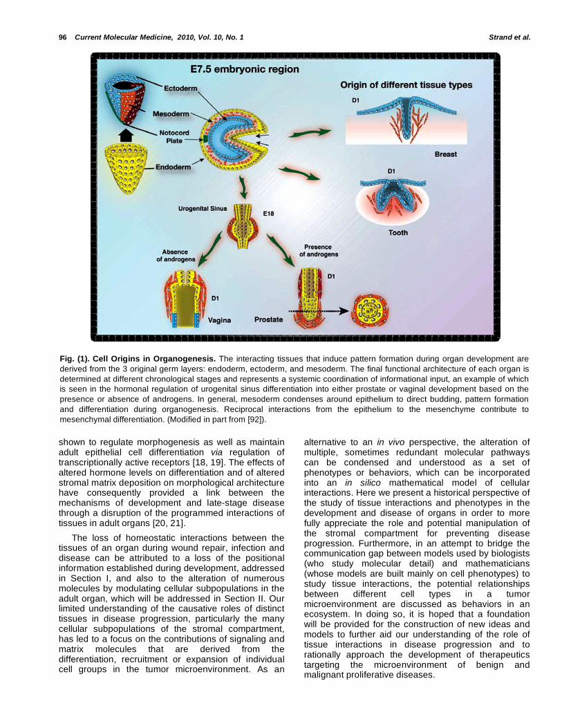
Fig. (1). Cell Origins in Organogenesis. The interacting tissues that induce pattern formation during organ development are
derived from the 3 original germ layers: endoderm, ectoderm, and mesoderm. The final functional architecture of each organ is
determined at different chronological stages and represents a systemic coordination of informational input, an example of which
is seen in the hormonal regulation of urogenital sinus differentiation into either prostate or vaginal development based on the
presence or absence of androgens. In general, mesoderm condenses around epithelium to direct budding, pattern formation
and differentiation during organogenesis. Reciprocal interactions from the epithelium to the mesenchyme contribute to
mesenchymal differentiation. (Modified in part from [92]).
Perspectives on Tissue Interactions in Development and Disease Current Molecular Medicine, 2010, Vol. 10, No. 1 97
I. STROMAL-EPITHELIAL INTERACTIONS IN ORGANOGENESIS
In organogenesis epithelial and mesenchymal cells derived from different germ layers interact spatiotemporally to induce a program of genetic and epigenetic tissue patterning and cellular differentiation, ultimately resulting in functional organs. During organogenesis, a population of stem cells become arranged into a specific three-dimensional space and differentiate into organ-specific epithelia through the inductive and morphogenetic mechanisms of the interactive gene products from different tissue groups [22].
As noted above, partitioning of the undifferentiated cells of the epiblast during gastrulation forms three germ layers: ectoderm, mesoderm and endoderm. Morphogenetic movements of the endodermal cells along the primitive streak form the gut tube from which each differentiated alimentary organ will form after budding and branching [23]. Epithelial cells migrating through the primitive streak display increased motility and an epithelial to mesenchymal transition due to a downregulation of E-cadherin [14, 24]. In successive steps, regional differences of the nascent mesenchymal cells provide the instructions for the differentiation and branching of endodermal evaginations into architectural patterns specific to each organ [25-27]. Although several transcription factors identify the presumptive regions of organogenesis, studies suggest that the regional fate of the endoderm can be re-specified when associated with heterotypic mesoderm denoting an inherent instructive role for mesenchyme [28].
The foundation for the field of mammalian mesenchymal-epithelial interactions was primarily laid in the 1950s by Grobstein’s seminal work on the development of the mouse submandibular gland, but included many other systems such as skin, kidney, gut, lung, tooth, thymus and thyroid [3]. During the same period, Le Douarin had recombined chick endoderm with somatopleural mesenchyme and surmised that endoderm had self-differentiative and inductive properties when she found that various digestive tissues developed and the mesenchyme differentiated into organ-specific connective tissue at the site of implantation [29]. Eventually, it was discovered using organ cultures that only a few of the differentiated intestinal epithelia developed cell autonomously, while differentiation of the majority of cell types and ultimate glandular formation required the presence of mesenchyme [27, 30, 31]. Thesleff who made many personal contributions to the field of tooth development [32], noted that mesenchymal cells condense next to organ-specific epithelium during the early developmental stages of most organs, and that these cells remain as stromal cells that support epithelial morphogenesis and differentiation in the adult organ [12].
Mesenchyme derived from different tissues at different stages of development elicits different effects.
As a result two broad classes of mesenchymal-stromal interaction with epithelium have been recognized. These are permissive and instructive effects. An instructive effect elicits a new program of markers in the epithelium as specified by the mesenchyme, as is found in the process of organogenesis. A permissive effect promotes expression of a previously determined developmental program already specified in the epithelium as is demonstrated when adult fibroblasts are combined with epithelium derived from a different organ, resulting in a maintenance of the original epithelial differentiation [25].
Early work by Kollar in tooth and skin development showed that mesoderm-derived dental and dermal papilla induced epithelial morphogenesis at specific embryonic stages [33-35]. However, these interactions were shown to be reciprocal and sequential since dental epithelium from early tooth germs also had the ability to instruct non-dental neural crest-derived mesenchyme, which demonstrated a shift in instructive potential as development progressed [36].
After these original demonstrations of mesenchymal-epithelial interactions in tooth and skin, the demonstration that other organs developed by similar mechanisms was soon to follow. Work by Cunha, a student of Kollar, was the first to show the reciprocal and sequential determination of urogenital organs including vagina, uterus and prostate through mesenchymal-epithelial interactions [37, 38]. As an added level of complexity, pioneering work by Cunha and Chung using tissue recombinants composed of androgen receptor-negative prostate epithelium and androgen receptor-positive urogenital mesenchyme showed that fetal androgens regulated normal prostatic epithelial cell cytodifferentiation and branching through androgen receptor activation in the mesenchyme [39, 40]. Subsequent heterotypic recombination experiments using adult bladder epithelium with urogenital mesenchyme demonstrated that even adult epithelium from a different organ could be induced to differentiate into prostatic epithelium by androgen sensitive fetal mesenchyme [41] potentially suggesting activation of a subpopulation of stem cells. Furthermore, the newly differentiated epithelium was demonstrated to direct the surrounding mesenchyme to become smooth muscle through a sequentially reciprocal paracrine induction [42]. In the adult prostate high levels of circulating androgens are thought to act through the prostatic smooth muscle to maintain epithelial differentiation and growth quiescence [43]. Epithelial apoptosis resulting from androgen ablation has also been shown to be due to a failure to occupy stromal rather than epithelial AR. In addition, an analogous paracrine system of steroidal control has since been demonstrated for estrogens and progesterone in the female genital tract [44, 45].
To separate epithelial glands from their surrounding stromal compartment and provide instruction for architectural arrangement [46], most epithelial glands secrete a layer of collagens, glycoproteins,
98 Current Molecular Medicine, 2010, Vol. 10, No. 1 Strand et al.
proteoglycans and hyaluronic acid deposited in the basal lamina [8, 47, 48]. Moreover, the glands can deposit a basal lamina without the help of mesenchyme although whether this is the same as a natural basement membrane is debatable [49]. Early work on the submandibular gland suggested that the morphology of the glandular epithelium is maintained by balancing the rates of synthesis and degradation of the glycosaminoglycan content of its basal lamina. Concordantly, rapid degradation of the laminar glycosaminoglycans by mesenchyme allowed epithelial growth and expansion implying that the basal lamina provided positional cues that direct morphogenesis [4, 50].
The role of the basal lamina and extracellular matrix in glandular function has been described in a number of branching organs such as the intestine [51], lung [17] and breast [52]. The breast is unusual in that most of the tissue remodeling occurs postnatally. Studies show that the regulation of mammary gland branching is dependent on the types and amounts of ECM as demonstrated by inhibiting or increasing either collagens or glycosaminoglycan deposition [47]. In coculture studies of colon morphology, well-differentiated colon epithelial cells form a monolayer on fibroblasts and deposit laminin-1 and collagen type IV at the heterotypic cell interface. Knockdown of laminin-1 in the epithelia resulted in a lack of basement membrane formation and an alteration of epithelial polarity and differentiation suggesting its role as the scaffold for basal lamina assembly [53].
Although the total number of glands within an organ may vary per organism, the architectural pattern of each gland is necessarily consistent for specific secretory functions. As historically shown in the prostate and breast, the architectural assembly of epithelia into specific patterns is induced and maintained by ECM and stromal signals through morphogenetic mechanisms [54]. A classic example of the distinction between morphogenetic and inductive mechanisms is shown in prostate recapitulation experiments where fibroblasts induce epithelial patterning without affecting cell states [18, 22]. The subsequent differentiation of epithelia into characteristically tall columnar prostate-specific secretory cells is accomplished through inductive mechanisms linked to the transmission of androgen signaling through fibroblasts [18].
In summary, organ development can be thought of as a programmed establishment of mutualism through architecture. Given the aforementioned data on the roles of mesenchymal cells and ECM in directing epithelial patterning in development, key questions garnering more attention are concerned with the effects of changing stromal-epithelial interactions on tumorigenesis. If organs are seen as cellular ecosystems that fluctuate according to the availability of resources, then the individual and group effects of mutations that perturb tissue interactions can be predicted using the statistical rigor of ecology, a new
perspective which will be addressed below. However, to achieve a proper understanding of tissue responses in chronic disease, it is first necessary to understand how tissues maintain homeostasis and how tissues repair wounding.
II. STROMAL-EPITHELIAL INTERACTIONS IN ADULT ORGANS
A. Homeostasis
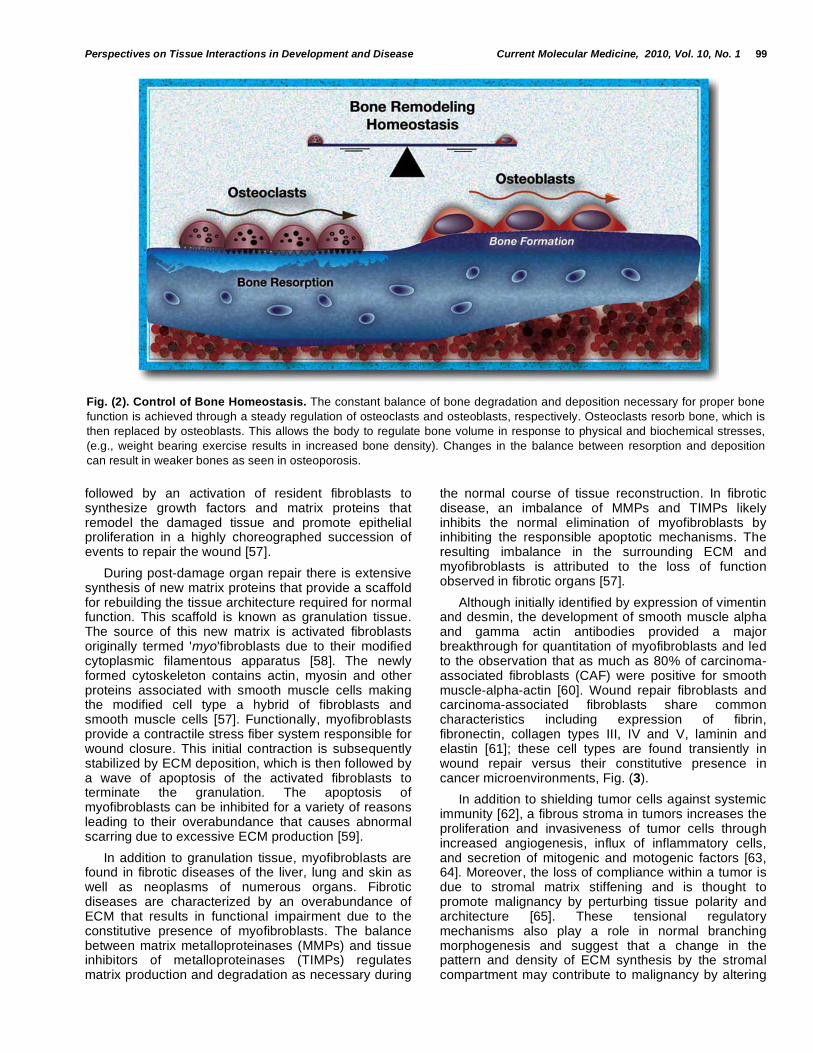
It is now understood that tissues perform a constant balancing act of proliferation, differentiation and death in order to maintain tissue structure and function. These functions require constant choreography, informed by cellular architecture and paracrine interactions. Some of the best-studied mechanisms of organ homeostasis have been accomplished in the field of bone remodeling. By identifying the role of osteoclasts and osteoblasts in bone degradation and deposition, respectively, much has been learned about the extent of continuous activity required for the maintenance of tissue function, which in the case of bone includes both bone microfracture repair and mineral homeostasis (reviewed in [55]) Fig. (2).
The architectural organization of cells is imperative for proper function so one of the keys to maintaining proper differentiation and function is to reestablish appropriate cell interactions and patterns through a programmed modulation of cell populations and matrix deposition, a process partially regulated by the inflammatory response. It is generally thought that the inflammatory system evolved to maintain homeostasis; however, the differences between acute inflammatory responses triggered by infection or tissue injury versus chronic inflammatory responses triggered by tissue stress or malfunction associated with disease are now becoming clearer. For a more detailed understanding of the role of inflammatory system tissue surveillance on homeostasis, the reader is referred to an excellent review [56].
B. Stromal-Epithelial Interactions in Tissue Repair and Cancer
The stroma is composed of all the non-epithelial components of an organ and is the extrinsic source of epithelial resources. Cell types within stromal tissue include fibroblasts, smooth muscle cells, nerves and blood vessels in addition to resident and migratory inflammatory cells. To better comprehend the complex reciprocity between stromal and epithelial compartments in a constitutively unregulated disease environment, interactions arranged during the development of tissue architecture and maintained through wound repair in adult tissues must be correctly understood. An insult that produces damage in adult organs induces a programmed, coordinated response of multiple tissues in a chronologically regulated, self-limited manner. In general, trauma or infection elicits a recruitment of inflammatory cells into the damaged tissue to clear the wound of cellular debris. This is
Perspectives on Tissue Interactions in Development and Disease Current Molecular Medicine, 2010, Vol. 10, No. 1 99
followed by an activation of resident fibroblasts to synthesize growth factors and matrix proteins that remodel the damaged tissue and promote epithelial proliferation in a highly choreographed succession of events to repair the wound [57].
During post-damage organ repair there is extensive synthesis of new matrix proteins that provide a scaffold for rebuilding the tissue architecture required for normal function. This scaffold is known as granulation tissue. The source of this new matrix is activated fibroblasts originally termed 'myo'fibroblasts due to their modified cytoplasmic filamentous apparatus [58]. The newly formed cytoskeleton contains actin, myosin and other proteins associated with smooth muscle cells making the modified cell type a hybrid of fibroblasts and smooth muscle cells [57]. Functionally, myofibroblasts provide a contractile stress fiber system responsible for wound closure. This initial contraction is subsequently stabilized by ECM deposition, which is then followed by a wave of apoptosis of the activated fibroblasts to terminate the granulation. The apoptosis of myofibroblasts can be inhibited for a variety of reasons leading to their overabundance that causes abnormal scarring due to excessive ECM production [59].
In addition to granulation tissue, myofibroblasts are found in fibrotic diseases of the liver, lung and skin as well as neoplasms of numerous organs. Fibrotic diseases are characterized by an overabundance of ECM that results in functional impairment due to the constitutive presence of myofibroblasts. The balance between matrix metalloproteinases (MMPs) and tissue inhibitors of metalloproteinases (TIMPs) regulates matrix production and degradation as necessary during
the normal course of tissue reconstruction. In fibrotic disease, an imbalance of MMPs and TIMPs likely inhibits the normal elimination of myofibroblasts by inhibiting the responsible apoptotic mechanisms. The resulting imbalance in the surrounding ECM and myofibroblasts is attributed to the loss of function observed in fibrotic organs [57].
Although initially identified by expression of vimentin and desmin, the development of smooth muscle alpha and gamma actin antibodies provided a major breakthrough for quantitation of myofibroblasts and led to the observation that as much as 80% of carcinoma-associated fibroblasts (CAF) were positive for smooth muscle-alpha-actin [60]. Wound repair fibroblasts and carcinoma-associated fibroblasts share common characteristics including expression of fibrin, fibronectin, collagen types III, IV and V, laminin and elastin [61]; these cell types are found transiently in wound repair versus their constitutive presence in cancer microenvironments, Fig. (3).
In addition to shielding tumor cells against systemic immunity [62], a fibrous stroma in tumors increases the proliferation and invasiveness of tumor cells through increased angiogenesis, influx of inflammatory cells, and secretion of mitogenic and motogenic factors [63, 64]. Moreover, the loss of compliance within a tumor is due to stromal matrix stiffening and is thought to promote malignancy by perturbing tissue polarity and architecture [65]. These tensional regulatory mechanisms also play a role in normal branching morphogenesis and suggest that a change in the pattern and density of ECM synthesis by the stromal compartment may contribute to malignancy by altering
Fig. (2). Control of Bone Homeostasis. The constant balance of bone degradation and deposition necessary for proper bone
function is achieved through a steady regulation of osteoclasts and osteoblasts, respectively. Osteoclasts resorb bone, which is
then replaced by osteoblasts. This allows the body to regulate bone volume in response to physical and biochemical stresses,
(e.g., weight bearing exercise results in increased bone density). Changes in the balance between resorption and deposition
can result in weaker bones as seen in osteoporosis.
100 Current Molecular Medicine, 2010, Vol. 10, No. 1 Strand et al.
the differentiation signals derived from homeostatic stromal-epithelial interactions [52, 66].
Myofibroblasts are absent from most normal homeostatic tissues, but are found in most cancer microenvironments. In various tumor models, fibroblasts support angiogenesis and tumorigenesis suggesting a specific role for the stromal microenvironment in providing tumor cell resources [67-69]. To determine the effect of activated fibroblasts in cancer microenvironments, CAFs have been harvested from human tissues and then grafted in combination with benign or carcinoma cell lines to determine their effect on tumor cell growth and invasiveness. In multiple cancer types, CAFs have been shown to promote tumorigenesis [70-74]. An example that CAFs can be tumor-promoting was demonstrated by recombining patient-derived CAFs with initiated, but non-tumorigenic prostate epithelial cells in a tissue-recombination sub-renal capsule graft. After a few months, the graft was removed and the epithelial population re-cultured. When the epithelia were subsequently grown alone in the kidney capsule, they were found to be tumorigenic suggesting a previous activation by CAFs [75, 76].
Schor was one of the first to provide evidence that the fibroblasts adjacent to carcinomas were fundamentally different than normal stroma and that these changes were implicated in neoplastic progression [77]. He also noted that these changes only occurred in a subset of the resident fibroblasts and posited his clonal expansion hypothesis which suggests that the changing ratio of stromal subtypes is
tumor-promoting [78, 79]. The work of Bissel's group was the first to identify the presence of myofibroblasts in breast cancer [62]. The identification of myofibroblasts in prostate cancer [80, 81] subsequently led to the study of their role in disease progression and the idea that an increased ratio of myofibroblasts to normal fibroblasts was predictive of disease recurrence [82]. Most cancer models that study the effect of the stromal microenvironment on tumorigenesis use clonal populations of fibroblasts. However, it is now recognized that the tumor microenvironment is composed of heterogeneous stromal populations using markers such as FSP-1, NG2, PDGFR , and collagen type I [83], which forces the reconsideration of the modeling of tumor microenvironments discussed in Section III.
The origin and genetic mutation of CAFs has been the subject of some debate [84, 85]. Studies show that subpopulations of CAFs can originate from EMT [86], circulating fibrocytes [87] and bone marrow-derived stem cells [88]. Evidence suggests that CAFs surrounding tumor epithelia have sustained somatic genetic alterations including LOH and copy number loss [89]; however, studies in breast and ovarian tumor stroma suggest that altered gene expression patterns responsible for the tumor-promoting phenotype of certain CAFs are more likely due to alternative (possibly epigenetic) mechanisms [90, 91].
In wound repair and in vitro, the myofibroblast phenotype is induced by TGF- and fibronectin from inflammatory- and stromal cells [92, 93]. Significantly, the growth factor TGF- can induce both indigenous
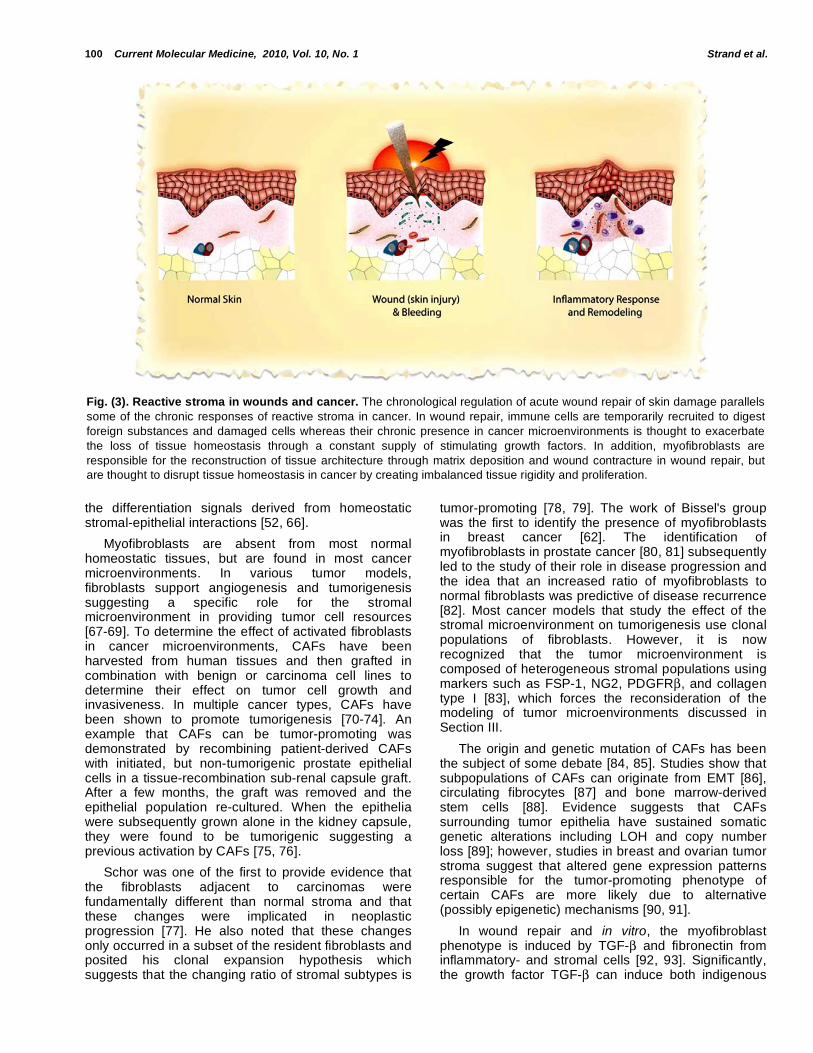
Fig. (3). Reactive stroma in wounds and cancer. The chronological regulation of acute wound repair of skin damage parallels
some of the chronic responses of reactive stroma in cancer. In wound repair, immune cells are temporarily recruited to digest
foreign substances and damaged cells whereas their chronic presence in cancer microenvironments is thought to exacerbate
the loss of tissue homeostasis through a constant supply of stimulating growth factors. In addition, myofibroblasts are
responsible for the reconstruction of tissue architecture through matrix deposition and wound contracture in wound repair, but
are thought to disrupt tissue homeostasis in cancer by creating imbalanced tissue rigidity and proliferation.
Perspectives on Tissue Interactions in Development and Disease Current Molecular Medicine, 2010, Vol. 10, No. 1 101
and recruited cells to become myofibroblasts implicating TGF- as a key cytokine in tissue repair. Concordantly, wound repair studies in Smad3-null mice demonstrate an acceleration of epithelial closure but a reduction in inflammatory cell recruitment and matrix deposition [94]. These data highlight the coordination of multiple cellular activities by TGF- in tissue repair and provide insight into the diverse roles TGF- plays on each cell type in cancer microenvironments.
The net effect of TGF- signaling in tumor microenvironments is still poorly understood and often seems contradictory when examined at various stages of tissue differentiation or on isolated tissue subgroups. Whether TGF- will induce cytostasis or apoptosis in normal epithelia depends on the intensity of the proliferative activity and poorly understood microenvironmental determinants [95]. The context of cell differentiation also dictates the effect of TGF- on the diseased tissue. For example, stromal cell TGF- production instills ER-negative breast cancer cells with the ability to form metastases in the lung due to their increased secretion of ANGPTL4 and subsequent increased permeability of lung capillaries [96]. Stromal production of TGF- by prostate CAFs increases the tumorigenicity and invasiveness of initiated prostate epithelia by blocking Smad3 nuclear translocation due to increased Akt signaling [97, 98]. Moreover, TGF- ’s transformation of mesenchymal cells to myofibroblasts has been shown to be pro-tumorigenic through its downstream regulation of angiogenic and chemotactic proteins like FGF-2, CTGF and CXCL12 [90, 99, 100]. It is now accepted that the cytostatic effect of TGF- is tumor-inhibitory early in tumorigenesis [95, 101]; however, mutations in its cognate receptor, T RII or in a subset of the pathways activated by TGF- result in increased survival, EMT, motility, invasiveness and metastasis [98, 102-106]. Unfortunately, despite the assumed therapeutic potential, systemic inhibitors of TGF- have yielded conflicting data [107-111] underscoring the need for better models of TGF- 's complex roles. For a review of the progress of chemotherapeutic targeting of CAFs, see [85].
In addition to fibroblast activation, in both wounds and tumors there is a chemotactic recruitment of specific leukocyte populations whose functions are to memorize and digest any foreign microorganisms or damaged cells. This complement of inflammatory cells includes monocytes, dendritic cells, neutrophils, mast cells and T cells, which produce several growth factors and chemokines (e.g. TGF- 1, FGF-2, TNF- and IL-1) that profoundly affect the phenotype and function of endothelial, epithelial and fibroblast cells. For a detailed outline of the role of specific chemokine/chemokine receptor and inflammatory cell responses in wound repair and cancer, the reader is referred to some excellent reviews [112, 113].
In summary, tumor growth, like wound repair, reflects a complex interplay of multiple tissues [114]. The homeostatic relationships of tissues and cells that are altered during carcinogenesis have been studied
using a number of different empirical methodologies; however, limitations regarding the systemic integration of tissue subtypes in most models necessitate the need for classifying each type of relationship in tumor microenvironments. A better understanding of the fundamental nature of both cellular and tissue interactions has been attempted using terminology borrowed from ecologists who describe the balance of organismal niches in terms of behavioral relationships. Although there are recognized differences in ecological and cellular environments, the following section will describe a recent effort to characterize the relationships of emergent tumor tissues in ecological terms in order to more fully understand the systemic integration of the altered cell and tissue relationships found in tumors.
C. The Ecology of Cancer
As embryonic cells divide and develop into adult tissues, they undergo a process of programmed differentiation and group arrangement that creates a homeostatic paracrine exchange of resources. When used in reference to adult organs, the term homeostatic reflects an activity rather than a status, as described previously for bone remodeling. The scale of homeostasis can reflect cell-cell interactions or systemic integration. The result is a resilient maintenance of niche balance due to a fluctuating availability of resources. The robustness of this mechanism is quite remarkable and is the result of evolutionary processes [115]. Both genetic and epigenetic alterations alter the homeostatic balance and transform the cohesive group of cells that make a tissue into a small ecosystem of individuals, in which different cell phenotypes compete and sometimes cooperate for available space and resources in order to survive and proliferate [116, 117].
The process of cancer initiation is still not fully understood and remains a controversial topic [118-120]. While a few tumors develop very rapidly, most develop over many years or decades. The former are likely to be highly aggressive from an early stage and have little reliance on microenvironmental influences [121]. The latter typically require mutations conferring on tumor cells the capacity to overcome the multiple intrinsic and extrinsic mechanisms that multicellular organisms possess that prevent the breakdown of homeostasis. Mutations that cause aberrant cell proliferation are essential to tumor formation. However, most DNA damage is quickly repaired by multiple intrinsic mechanisms [122]. In addition, because somatic cells are constantly dependent on homeostatic signals, mutations that disrupt an epithelial cell's interaction with the basal lamina [123] or an adjacent cell [124] can trigger apoptosis and replicative senescence. Innate immunity has also been suggested to play an important role in the suppression of cancer progression [125] since a variety of cancers arise in immunosuppressed individuals. These barriers offer a protection against tumor initiation and progression but
102 Current Molecular Medicine, 2010, Vol. 10, No. 1 Strand et al.
also act as a selective force that promotes somatic evolution of different clonal subspecies [126].
The idea that the local environment is important for the growth and metastasis of cancerous cells dates back to the late 19th century with Paget's well-known seed-soil hypothesis [127]. Cancer can be viewed as a phenotypic selection of multiple advantageous traits by a specific environment rather than an expansion of individual mutant genotypes, which may explain why certain mutant genotypes present as disease only in select organs or why changes in immigrants’ disease incidence are reflective of their new environment [128]. In tumor biology, as well as in evolutionary biology, selection is applied at the phenotypic level, and, as will be discussed below, cellular phenotypes in tumors are the result, not just of individual genotypes, but of the integration of the genotype with the microenvironment. By looking at cancer microenvironments through an ecological lens, it is possible to subcategorize environmental phenotypes in addition to screening for individual mutations. Research has subsequently focused on the role of the microenvironment in either the induction or selection of cumulative epithelial mutations [129].
Tumor cells typically acquire a number of traits as the neoplasm progresses to malignancy. These traits have been described by Hanahan and Weinberg [130] and include self-sufficiency in growth signals, insensitivity to anti-growth signals, evasion of apoptosis, angiogenesis, limitless replicative potential and the capability of invasion and metastasis. A variant of the list, proposed by Gatenby and Gillies, also includes resistance to acid-mediated toxicity and abnormally high glucose uptake [126]. Although it is normally considered that a sufficient number of cells in the tumor have to acquire all six traits, some researchers have recently hypothesized that some of these traits could be acquired by a reduced part of the tumor and that the benefits of the traits would be shared by the tumor as a whole [131].
Having the same fundamental principles (differential fitness and inheritable variation), there are some significant differences between organismal and tumor cellular evolution. For one, reproduction in a tumor, in contrast to many of the most commonly studied ecosystems, is asexual [132]. Further differences include the fact that phenotypic variation can be due to somatic mutations as well as epigenetic alterations and genomic instability and not due to recombination [132]. Other challenges to the study of tumors as micro ecosystems affected by somatic evolution include the fact that many important evolutionary parameters have not yet been measured in cancer. Determining mutation rates, fitness effects of mutations, generation times, population structure or the selective effect of cancer therapies [117] will be challenging work for biologists and will go a long way in the quest to characterize cancer evolution. The perspective of tumors as micro ecosystems poses new challenges to cancer researchers but also allows them the use of
powerful mathematical tools that ecologists and evolutionary scientists have used to study evolutionary dynamics and to describe interdependence and interactions between tumor cells themselves and between the tumor cells and the stroma.
Ecological mathematical tools used by ecologists that can be used to address cancer include game theory, evolution of cooperation and network theory. Game Theory (GT), initially introduced to study human behavior and economy [133], was later expanded, as Evolutionary Game Theory, to study how interactions between individuals in the same species determine the evolutionary dynamics of the population [134]. It is only in the last ten years that mathematical oncologists started using evolutionary game theory to study somatic evolution in cancer [135]. Using GT requires formally defining the relevant tumor cell phenotypes that might be present during tumor evolution and the interplay between these phenotypes in relation to a fitness payoff. This fitness is a measure of the ability of a tumor cell phenotype to survive and grow when living in a polyclonal tumor. GT has been successfully used to generate hypotheses to explain the emergence of phenotypes capable of unconstrained growth [136], avoiding apoptosis [137], acidifying the microenvironment [137, 138], promoting angiogenesis [137, 139] and invasion [140].
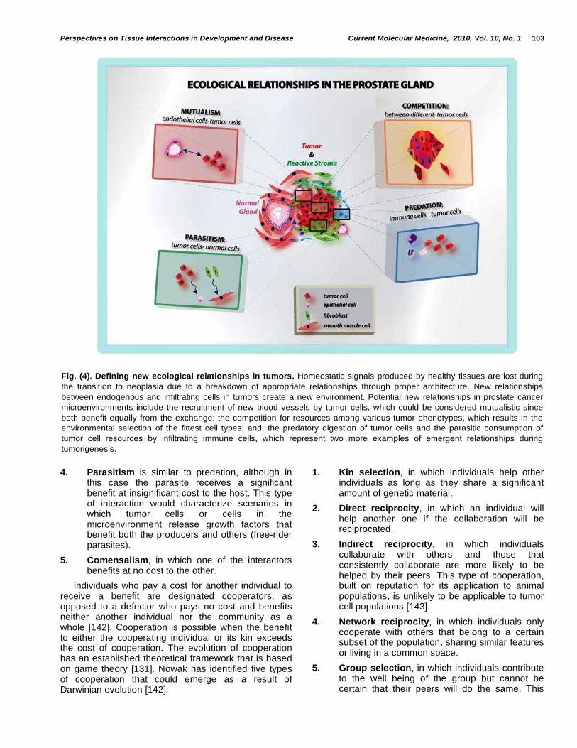
It is commonly assumed that in an ecosystem with limited resources, nutrients and space for growth, the only relevant type of interaction between individuals would be competition. Still there are numerous examples of cooperation within and across species and societies [141], and some researchers suspect cooperation within cancer ecosystems [131]. Ecological interactions can be defined according to the fitness benefit to the participants as: competition, predation, parasitism, mutualism and commensalism. All these types of interactions can be observed, to some degree, in a tumor [117] Fig. (4).
1. Mutualism, in which both interactors benefit equally. This would describe the types of interaction between cells in a healthy tissue in a multicellular organism. In a neoplasm, the interactions between tumor cells and stromal fibroblasts could also be seen as mutualistic since both get a fitness advantage from their interactions [114].
2. Competition, in which each interacting partner has a negative effect on the others. When genetic mutations or cellular damage disrupt mutualistic exchange, tumor cells start competing with other tumor and non-tumor cells for space and nutrients.
3. Predation, in which one of the interactors, the predator, obtains a small benefit, whereas the other, the prey, receives a significant detriment to its fitness. In a tumor it could be said that the tumor cells are the prey and the immune system is the predator.
Perspectives on Tissue Interactions in Development and Disease Current Molecular Medicine, 2010, Vol. 10, No. 1 103
4. Parasitism is similar to predation, although in this case the parasite receives a significant benefit at insignificant cost to the host. This type of interaction would characterize scenarios in which tumor cells or cells in the microenvironment release growth factors that benefit both the producers and others (free-rider parasites).
5. Comensalism, in which one of the interactors benefits at no cost to the other.
Individuals who pay a cost for another individual to receive a benefit are designated cooperators, as opposed to a defector who pays no cost and benefits neither another individual nor the community as a whole [142]. Cooperation is possible when the benefit to either the cooperating individual or its kin exceeds the cost of cooperation. The evolution of cooperation has an established theoretical framework that is based on game theory [131]. Nowak has identified five types of cooperation that could emerge as a result of Darwinian evolution [142]:
1. Kin selection, in which individuals help other individuals as long as they share a significant amount of genetic material.
2. Direct reciprocity, in which an individual will help another one if the collaboration will be reciprocated.
3. Indirect reciprocity, in which individuals collaborate with others and those that consistently collaborate are more likely to be helped by their peers. This type of cooperation, built on reputation for its application to animal populations, is unlikely to be applicable to tumor cell populations [143].
4. Network reciprocity, in which individuals only cooperate with others that belong to a certain subset of the population, sharing similar features or living in a common space.
5. Group selection, in which individuals contribute to the well being of the group but cannot be certain that their peers will do the same. This
Fig. (4). Defining new ecological relationships in tumors. Homeostatic signals produced by healthy tissues are lost during
the transition to neoplasia due to a breakdown of appropriate relationships through proper architecture. New relationships
between endogenous and infiltrating cells in tumors create a new environment. Potential new relationships in prostate cancer
microenvironments include the recruitment of new blood vessels by tumor cells, which could be considered mutualistic since
both benefit equally from the exchange; the competition for resources among various tumor phenotypes, which results in the
environmental selection of the fittest cell types; and, the predatory digestion of tumor cells and the parasitic consumption of
tumor cell resources by infiltrating immune cells, which represent two more examples of emergent relationships during
tumorigenesis.
104 Current Molecular Medicine, 2010, Vol. 10, No. 1 Strand et al.
type of cooperation works if the cooperating individuals get enough benefit of being part of a group that, thanks to the contribution of the cooperators, has a better chance of survival and growth.
Axelrod and colleagues have used this framework to study how tumor cells could cooperate to overcome the microenvironmental hurdles to tumor growth and progression [131]. Specifically, they have identified a number of areas in which this collaboration could happen: by sharing growth factors such as VEGF that promote angiogenesis, by sharing growth factors such as TGF- that allow tumor cells to keep growing, and by collaborating with stromal fibroblasts in ways that promote motility.
A third relevant mathematical tool that could be used to describe the structure and robustness of an oncological ecosystem is network theory. Network theory, also known as graph theory, has been used to study physical and telecommunication phenomena [144] but is also a widely used tool in ecology and systems biology. In systems biology, it has proven to be popular to describe such things as metabolic or protein-interaction networks as well as other cell molecules [144]. In ecology, one of the main uses is the characterization of food webs [145]. Food webs describe the feeding relationships between species in an ecosystem. Once the key species in the ecosystem have been identified and their relationships established, the resulting network could be studied to find out the importance of one particular species in the ecosystem to the survival of others as well as to investigate the robustness, scalability and evolvability of the network. This approach has yet to be exploited in cancer research but could potentially be used to describe a tumor ecosystem with the goal of identifying potential targets of therapy in the tumor and in the stroma.
Tumors can be considered in modified ecological terms, which allow for mathematical evaluation of the relationships between cell types and tissues within organs, and in the context of the host. Fig. (4) demonstrates known relationships between various cell types in the prostate, which could potentially be used to establish predictable outcomes based on population dynamics of cell types with definable behaviors. The ability of biologists to empirically provide to mathematicians statistically reliable data on the effects on organ function of individual cell phenotypes (behaviors) will facilitate the design of prognostically useful models for clinicians.
III. MODELS OF TISSUE INTERACTION
A. In Vitro Models of Stromal-Epithelial Interaction
In vitro models of stromal-epithelial interactions include two-dimensional (2D), three-dimensional (3D) and organ culture. The availability of multiple manipulatable cell lines makes monolayer 2D culture a useful tool for quickly generating data on various
cellular functions. The paracrine effects of clonal stromal and epithelial cells on each other can be modeled by using conditioned media in which secreted proteins from one cell support the growth of other cell types. However, co-culture using tissue culture inserts maintains a constant paracrine communication between stromal and epithelial cells supporting some aspects of cellular differentiation, which allows one to test the effects of putative modifiers of growth and differentiation between stromal and epithelial cells [146]. The disadvantages include the lack of direct contact between cells of different tissues and the inability to model tissue morphogenesis.
The major advantage of 3D cultures is the ability to study the arrangement of individual cells into tissue architecture seen during normal development or, alternatively, to study the distortion of the cellular polarization and normal tissue architecture displayed by tumor cells [147]. By culturing in the presence of laminin-rich Matrigel, a natural substrate composed of reconstituted basal membrane from the Engelbreth-Holm-Swarm tumor, cell lines as well as primary cells from different organs can reconstitute the formation of a differentiated phenotype (reviewed in [148]). Three-dimensional models have historically been monotypic although 3D skin models have combined keratinocytes, fibroblasts and cancer cells to simulate human melanoma [149]. To study the effect of fibroblasts on epithelial organization, some models use cell culture inserts pre-loaded with stromal cells that sit suspended above the matrix/epithelial cell mixture [150]. Unfortunately, this method fails to model the physical interaction of stromal and epithelial cells and the effect of stromal matrix regulation. Moreover, models of heterotypic cell-cell interactions have yet to be fully developed for multiple organ systems.
Although 3D cultures have been successful in cells from different organs (reviewed in [151]), much of the information regarding epithelial glandular arrangement has been obtained from studies in breast [152]. The pioneering work of Bissell and colleagues has demonstrated the ability of mammary epithelium to recreate acinar formation and production of milk proteins [153]. The presence of laminin and collagen type IV in the basement membrane (BM) could induce responsiveness to hormones and expression of mammary-specific genes in epithelial cells compared to those containing collagen I or fibronectin [154]. Laminin-rich BM triggered epithelial cell polarization allowing them to organize into structures resembling normal mammary acini that respond to lactogenic hormones by secreting milk proteins into the lumen [155]. In contrast, carcinoma-derived mammary epithelial cells were unable to polarize and instead formed disorganized, hyperplastic colonies. This assay has proven to be useful in distinguishing normal and cancer-derived breast epithelial cells [156].
ECM stiffness is commonly variable in normal tissues (e.g. breast vs. bone or early vs. late wound healing). In addition to manipulating gene expression in
Perspectives on Tissue Interactions in Development and Disease Current Molecular Medicine, 2010, Vol. 10, No. 1 105
normal cells or in tumor cells to study carcinogenesis in 3D, the composition of the matrix in 3D cultures has demonstrated dramatic effects on cell polarity and invasiveness [65, 66, 129, 157, 158]. The control of cell behavior by the adjacent matrix led some to question whether malignant cells in an activated microenvironment could be suppressed when replaced into a normal microenvironment. The ability to revert the malignant phenotype of tumor cells subsequently suggested that tissue phenotype is dominant over cellular genotype [159].
Another commonly used approach in developmental biology is organ culture, which makes use of organ rudiments harvested ex vivo and cultured in vitro. The advantage of this technique is that normal stromal/epithelial/ECM interactions are retained. Organ culture has been particularly valuable for studies that examine the effects of steroid hormones and growth factors on development, or induction of angiogenesis and resistance to chemotherapeutic drugs in tumors [18, 160-163]. Disadvantages include the inability to separate endogenous from exogenous signaling in addition to limitations in the size of the specimen (< 1mm thick), which has to be thin enough to allow proper oxygenation and nutrition of the interior of the explant.
Although 3D systems have an obvious advantage over 2D cultures, they are variable in their recapitulation of normal in vivo tissue conditions as they lack proper vasculature and normal transport of small molecules, a host immune response and other cell interactions. Such cultures merely represent a short-term assessment of progression. As such, 3D systems cannot completely replicate the complexities of the in vivo environment. Therefore, interpreting results generated from 3D models must be weighed in the light of these limitations; however, such results also provide a rapid method for hypothesis testing that will likely produce valuable questions that require further validation in vivo.
B. In Vivo Models of Stromal-Epithelial Interaction
Two major issues have plagued modeling reciprocal interactions between the stroma and the epithelium in vivo: the lack of organ- and tissue-specific promoters for driving transgenes in the stroma and the sometimes drastic differences between the stromal compartments of parallel organs in mice and humans. Historically, most of the attention has focused on the epithelium of tumors, which led to the development of promoters for specific targeting of epithelial tissue. Much less is known about the control of gene expression in the various cells types comprising the tumor microenvironment. Moreover, the organ-specific targeting of stromal genes has not yet been developed, making the study of stromal effects on various epithelial phenotypes difficult in an intact animal. Some progress has been made, however, and a few examples of the study of tumor stroma in genetically engineered mice (GEM) are warranted.
Genetically Engineered Mouse Models
Transgenic mouse models represent a useful tool for evaluating the fundamental spatio-temporal and cumulative mechanisms of malignancies, in addition providing a proper testing ground for therapeutics. In these animal models, tumors are derived from the manipulation of individual genes in specific (predominantly epithelial) tissues although the specificity of expression is sometimes dubious due to leaky expression in other cell types [164] or the inability to express a gene in the full complement of the desired cells. The ability to manipulate multiple genes within the same animal remains a technological constraint. This limitation is currently overcome through the cross breeding of various lineages; however, the generation and maintenance of multiple transgenic lineages is prohibitively time-consuming and costly for most complex experiments.
Cre-Lox recombination using cell type-specific promoters to drive expression of the Cre recombinase allows for spatially restricted gene expression to the compartment (tissue or organ) of interest. Promoters such as collagen type 1 alpha 1 and 2, FSP-1/S100A1, vimentin, alpha-smooth muscle actin, SM22/transgelin and SMMHC have been used to target gene deletion or expression in osteoblasts, fibroblasts, myofibroblasts and smooth muscle cells, each of which are major components of the stromal compartment [165-170].
The use of the FSP-1 promoter has enabled the study of the role of stromal cells in many tissues [171-173]. However, FSP-1 is expressed in only a fraction (30-50%) of mature stromal cells. As with other familiar stromal promoters such as vimentin or SM22, the disadvantage of using the FSP-1 promoter for stromal targeting is the lack of organ specificity. Alteration of genes that are pivotal in early development, as might be achieved using a combination of FSP1-Cre with a floxed gene, can result in early mortality, often precluding proper analysis of the organ of interest. To circumvent this problem, new strategies using more restrictive expression of the transgene in fibroblasts have been developed. Zheng and colleagues generated a transgenic mouse in which collagen type I alpha 2, a gene only expressed in fibroblasts that produce collagen type I, was linked to a Cre-ER fusion protein [170]. This technique provides a tight regulation of Cre activity allowing both temporal and tissue-specific control of gene regulation. Tamoxifen-dependent constructs are well-suited for post-natal activation of Cre activity; however, only a few Cre-ER models are currently available for targeting the stromal compartment.
One of the best examples of an in vivo model demonstrating stromal-epithelial interactions was reported by Bhowmick et al. A conditional mesenchymal (stromal) transforming growth factor-beta receptor 2 (Tgfbr2) knockout mouse was generated using FSP-1 Cre-mediated recombination. This mouse displayed a prostatic phenotype consistent with prostatic intraepithelial neoplasia, a precursor of
106 Current Molecular Medicine, 2010, Vol. 10, No. 1 Strand et al.
prostatic adenocarcinoma, and invasive squamous cell carcinoma of the forestomach by six weeks of age [165]. This study clearly demonstrates that loss of a critical signaling component in the stroma results in increased epithelial proliferation and in the promotion of invasive carcinomas in a tissue-specific manner. Because Tgfbr2
fspko mice die at about 6 weeks of age,
studies have focused on initiating factors involved in tumorigenesis, which means that these mice have been less useful for studying subsequent tumor progression.
Tumor progression is a dynamic process in which each aberrant change has a potential impact on the biology of the affected cell and its surroundings, which then creates new selective pressures that direct the evolution of the cancer. Several reports have recently identified mutations of tumor-suppressor genes, including p53, in the stromal compartment of advanced human carcinomas [174-176]. Although oncogenic stress has previously been shown to induce p53 responses in epithelial cells, Hill and colleagues explored the non-autonomous loss of stromal p53 on tumor evolution [177]. They demonstrated in a mouse model of prostatic carcinoma that suppression of epithelial Rb by expression of a fragment of the SV40 T antigen induces downregulation of p53 in stromal fibroblasts through an unknown paracrine mechanism. The loss of epithelial p53 was suggested to create a selective pressure that results in subsequent loss of p53 in a sub-population of stromal cells. The initial decrease in fibroblast proliferation was followed by selective evolution of a highly proliferative p53-deficient subpopulation of stromal cells. Moreover, these p53-deficient stromal cells non-autonomously increase the selective pressure against p53 in the epithelium suggesting that selection of p53-deficient fibroblasts is advantageous for tumor progression. It is not known whether this selection by epithelial cells occurs in all stromal cells or only in a specific lineage of cells bearing other mutations or epigenetic alterations. Accordingly, the validation of these observations in other cancer models will expand our understanding of the selective evolution of common stromal mutations relevant to human cancers.
In addition to facilitating the study of various cell genotypes, in vivo models have facilitated the study of the role of the extracellular matrix in tumor progression. Studies in mouse models in which stromal MMPs are either upregulated or suppressed have revealed the roles of these proteases during tumor progression. For example, in MMP-2 and MMP-9 knockout mice, a lower rate of tumor incidence and decreased growth of implanted neuroblastoma and ovarian cancer is observed compared to tumor implants into wild-type mice [178, 179]. The contribution of MMPs to tumor metastasis has also been demonstrated by injection of melanoma cells into MMP-2 and -9 knockout mice, which resulted in a decrease in the number of metastatic nodules compared to the wild-type hosts [180]. Despite some pro-tumorigenic effects of MMPs, recent studies have suggested protective and
antitumorigenic effects. In particular, these arise from proteases expressed by infiltrating inflammatory cells, which highlights the importance of using immunocompetent models for studying tumor progression in the context of MMPs. The failure of clinical trials that used “promising” inhibitors of MMPs based on in vitro data emphasizes the need for a better understanding of the complex interactions of MMPs in vivo during cancer progression [112].
Tissue Rescue by Transplantation
Manipulation of critical genes that result in embryonic or neonatal lethality of the transgenic or gene knockout mouse model is a major problem. In these cases it is often possible to “rescue” the organ of interest by dissection from the fetal or neonatal animal and subsequently growing the rescued tissue under the renal capsule of a syngeneic or immunocompromised host. The resultant organ explant can be used to examine the effects of the specific genetic change on organogenesis and carcinogenesis. Such tissues can also be used as a source of material for generating tissue recombinants for further study. A recent example of an optimal situation for tissue rescue is the study of the Tgfbr2
fspko mouse, which develops cancer and dies
at 7 weeks. In order to study post-pubertal, androgen-regulated prostatic tissue in these mice, the investigators rescued prostate tissue for further modification and then allografted the modified tissue under the renal capsule of either castrated or uncastrated immunocompromised mice [181]. The ability to rescue this tissue for allografting allowed the investigators to study the molecular mechanisms of androgen ablation on the carcinogenic effect of Tbr2ko prostate fibroblasts.
Tissue Recombination
The suggestion of genetic and epigenetic mutations in tumor stromal cells from clinical samples suggests that the stroma co-evolves with cancer cells [182, 183]. However, the lack of organ-specific stromal promoters remains a technological problem for the use of transgenic animals in the study of stromal contribution to tumor progression. Although transgenic mouse models have used promoter-regulated Cre recombinase to target the stroma of multiple tissues, these commonly result in mosaic expression patterns [165, 184]. Accordingly, the components of the stromal compartment that are frequently heterogeneous in normal tissues and organs are underrepresented. The ability to mutate the stroma in current autochthonous tumor mouse models will facilitate the study of stromal contribution during tumorigenesis and/or tumor progression.
Perhaps the most flexible alternative in vivo model for studying stromal-epithelial interactions is tissue recombination (TR) with subsequent xenografting. TR provides a valuable alternative to the labor- and cost-intensive generation of transgenic mice for the in vivo study of gene function within a specific tissue compartment. Tissue recombination has been a widely
Perspectives on Tissue Interactions in Development and Disease Current Molecular Medicine, 2010, Vol. 10, No. 1 107
used technique for examining different aspects of development. Briefly, tissue recombinants are made by mixing stromal or mesenchymal cells with epithelial cells within a matrix (most commonly type I collagen). These recombinants can then be grafted beneath the renal capsule of a young adult rodent host and grown for various periods of time (as long as a year). The renal capsule site is commonly chosen because the kidney has a rich vasculature that makes a suitable environment for the growth and survival of tissues.
TR has many advantages and has demonstrated faithful replication of key aspects of both development and carcinogenesis [185-187]. The inherent plasticity of the model allows for targeted modifications to all or some cells specifically within the epithelial and stromal tissues. Another advantage is that recombinants can be made using stromal and epithelial cells coming from the same (homotypic) or different (heterotypic) tissue as well as same (homospecific) or different (heterospecific) species. Another benefit of TR is that in vitro genetic manipulation of the cells (stromal or epithelial) can be performed before implantation. This approach was pioneered by Thompson to study malignant and benign disease progression in the prostate [188]. More recently, the use of lentiviral delivery systems has allowed either overexpression or downregulation of a gene of interest prior to grafting [97, 189].
Disadvantages of TR include the relative lack of metastasis as a desired endpoint. Also, recombinants are commonly grafted into immunocompromised mice (athymic or SCID) meaning that many effects of an intact immune system are lost. However, implantation into syngeneic hosts is possible when appropriate cells are available. Of note, another major disadvantage to studying the effects of a modified stromal compartment on epithelial morphogenesis and tumorigenesis is the lack of a mesenchymal cell line capable of inducing prostate glandular formation in vivo. At present, freshly harvested rat urogenital mesenchyme is recombined with prostate epithelia to induce glandular formation, but genetic manipulation of this mesenchyme is currently unavailable due to the inability to generate a cell line that retains inductivity. Therefore, the TR model must be reconsidered in order to mimic stromal heterogeneity and still maintain morphological integrity. Possible solutions include using UGM from trangenic mice or mixing rat UGM with genetically modified human stromal cell lines.
Although the grafting of tissue recombinations of human cells into various mouse organs fails to incorporate a fully intact immune response due to the necessity of using an immunocompromised host, the lower cost and the ability to control gene expression in multiple tissues and incorporate multiple cell lineages makes tissue recombination an attractive, modular alternative to the generation and crossing of multiple transgenic mouse models. In addition, sophisticated and controlled scenarios using inducible genes allows for the mutation of different compartments at different
time points in order to study the temporo-spatial contribution of each type of cell composing each tissue during cancer progression.
It is universally acknowledged that each model has its limitations and abilities and should therefore be utilized to address specific questions. The identification of genes and cells critical for tumor progression within the stroma, and the development of more sophisticated in vivo mouse models that mimic as many aspects of human normal and tumor biology as possible should prove valuable for discovering new methodologies for the detection and treatment of cancer.
Finally, many biological models of disease follow a reductionist approach by using clonal populations of cells overexpressing a particular protein thought to be involved in cancerous growth. While this type of genetic parsing is informative, it hardly accounts for either the multiplicity of aberrant gene interactions or the group behavior demonstrated by tissues. In essence, what has been learned recently is that while only a portion of the cells in diseased tissues contains mutations [83], the whole group becomes affected. Accordingly, the failure of reductionist models to recapitulate the net biology in complex systems has provided incentive for the development of systems biology genre discussed below.
C. New Perspectives: Mathematical Models of Tissue Interaction
During the course of this review we hope that it has become clear that there are a multitude of approaches to studying the development and progression of cancer. Not only do these approaches consider different aspects of the disease but they also consider fundamentally different scales: from genes to proteins to signaling pathways, to cells, tissues, organs and ultimately the organism. How can we hope to understand the complex interactions that occur across these scales when we have not fully understood them even at an individual scale? The dynamic complex ecosystem that is cancer must be understood within a theoretical framework that easily allows one to consider and study all relevant agents, interactions and scales while at the same time permitting isolation of key variables and processes. The very definition of a dynamical system implies that small changes in one variable can lead to large changes in another. Using mathematical and computational models it is possible to both integrate multiple interacting variables and, in addition, bridge distinct spatial and temporal scales.
Mathematical models of cancer have been developed for more than 60 years and yet, not until today has their value become recognized. This is partially due to the release of computational constraints and partially to a shift in attitudes of both mathematicians and biologists. The reductionist approach has taught us a great deal about the initial steps in cancer development and subsequent progression and has great potential as a tool to aid the

108 Current Molecular Medicine, 2010, Vol. 10, No. 1 Strand et al.
tailoring of patient-specific treatments. But given the diversity of favorite genes, proteins or pathways and their limited therapeutic success, many biologists are beginning to accept that we need to integrate our knowledge and start to look at cancer in a whole new way. However, this should not be confused with the holism of the past as we specifically need a quantitative holism (i.e. qolism) [190]. Mathematicians were trapped in the formalism of theory and analysis such that the models they built were, with some exceptions, more interesting as mathematical curiosities than as tools capable of giving biological insight. With the emergence of a new breed of mathematical modelers, who are not afraid of computation or mechanism, and the desire for qolistic approaches, the time is now ripe for an integrated understanding of cancer therapy [191-194].
One of the key aspects of in silico models (mathematical/computational models) is their ability to characterize biological systems using multiple dynamic (possibly changing in space and time) variables in a mechanistic fashion. This key characteristic gives us the ability to change individual variables or parameters, in a precise manner, and examine their effects on the whole system. This often leads to the emergence of a behavior that is a product only of the system and would otherwise have been very difficult to observe or understand, especially if only component parts of the system are considered in isolation.
Over the last ten years or so many mathematical models of tumor growth, both temporal and spatiotemporal, have appeared in the research literature (see [195] for a review). Deterministic reaction-diffusion equations have been used to model the spatial spread of tumors in the form of invading waves of cancer cells [196-198]. The growth of tumors has also been more realistically modeled in 2 and 3 spatial dimensions [199, 200]. Whilst all these models are able to capture the tumor structure at the tissue level, they fail to describe the tumor at the cellular level. On the other hand, single-cell-based models provide such a description and allow for a more realistic stochastic approach at both the cellular and subcellular levels. Several different discrete models of tumor growth have been developed recently. For a review on many different single-cell-based models applied to tumor growth and other biological problems see [201].
The integration of multiple variables (changing in space and time) across a range of scales has only recently become possible with the development of multiscale modeling approaches (see [202]). Multiscale models are often hybrid in nature in that they couple a discrete representation of the cellular population (such as tumor and stromal cells) with continuous representations of microenvironmental factors (such as ECM, growth factor, nutrient). Hybrid approaches such as the hybrid discrete continuum model of Anderson and colleagues [203, 204], the evolutionary hybrid cellular automata model of Gerlee [205] and the Immersed Boundary model of Rejniak [206, 207] have
specifically focused on the tumor/microenvironment interface and how it affects the morphological and evolutionary aspects of a growing tumor.
One of the outstanding fundamental questions in cancer biology is how to directly link the current wealth of genetic data, not only with clinical outcome, but crucially with the cellular phenotype. The so-called genotype-phenotype mapping is at the heart of our understanding of many of the engines that drive tumor growth and progression; however, it has to some extent been ignored. This is in no small part due to the sheer complexity of the processes involved: thousands of genes result in the regulation of a multitude of proteins, which interact and signal within complex pathways to drive a cellular response of an individual cell that modulates its signaling in response to microenvironmental cues and signals from other cells. Different aspects of this mapping have been considered in isolation, with some significant progress being made in regards to specific pathways involved in cancer [208, 209]. However, a major issue with pathway models is the over complexity and parameter redundancy. A new approach needs to be developed if we want to try to bridge the genotype to phenotype chasm. Network models offer one possible stepping stone [205, 210] and these can be very specific or more generalized, but the key to their success will be raw experimental data to parameterize them. This is no easy task and will most likely require the development of novel experimental protocols by integrated teams of scientists focused on this goal.
It cannot be stressed enough that mathematical models are only as good as the data upon which they are based. Therefore, in silico models need to be validated and the hypotheses they generate need to be empirically tested - the outcome of which may require model refinement or radical change. One thing that is certain is that there is no single ideal mathematical or computational model of cancer just as there is no single ideal biological model of cancer. Each have their limitations but this diversity of approaches now has a means of integration [211], which can only improve our current understanding of the cancer progression and treatment.
Table 1 summarizes some of the advantages and disadvantages of in vitro, in vivo and in silico models of disease.
CONCLUSION
This review has attempted to provide a broadened perspective of the available models to be chosen for the specific question to be addressed and to incorporate adaptive terminology to the interactive stromal-epithelial relationships in order to find common ground between multiple disciplines attempting to build on existing models. Mathematical modeling has the potential to contribute as much or even more so to cancer biology as to fields to which it has been used extensively such as physics. The key is the successful
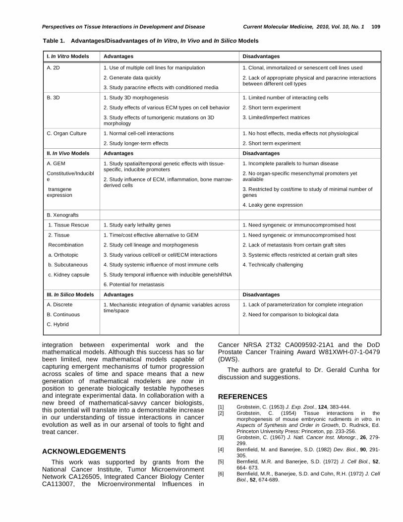
Perspectives on Tissue Interactions in Development and Disease Current Molecular Medicine, 2010, Vol. 10, No. 1 109
integration between experimental work and the mathematical models. Although this success has so far been limited, new mathematical models capable of capturing emergent mechanisms of tumor progression across scales of time and space means that a new generation of mathematical modelers are now in position to generate biologically testable hypotheses and integrate experimental data. In collaboration with a new breed of mathematical-savvy cancer biologists, this potential will translate into a demonstrable increase in our understanding of tissue interactions in cancer evolution as well as in our arsenal of tools to fight and treat cancer.
ACKNOWLEDGEMENTS
This work was supported by grants from the National Cancer Institute, Tumor Microenvironment Network CA126505, Integrated Cancer Biology Center CA113007, the Microenvironmental Influences in
Cancer NRSA 2T32 CA009592-21A1 and the DoD Prostate Cancer Training Award W81XWH-07-1-0479 (DWS).
The authors are grateful to Dr. Gerald Cunha for discussion and suggestions.
REFERENCES
[1] Grobstein, C. (1953) J. Exp. Zool., 124, 383-444.
[2] Grobstein, C. (1954) Tissue interactions in the morphogenesis of mouse embryonic rudiments in vitro. in Aspects of Synthesis and Order in Growth, D. Rudnick, Ed.
Princeton University Press: Princeton, pp. 233-256. [3] Grobstein, C. (1967) J. Natl. Cancer Inst. Monogr., 26, 279-
299.
[4] Bernfield, M. and Banerjee, S.D. (1982) Dev. Biol., 90, 291-305.
[5] Bernfield, M.R. and Banerjee, S.D. (1972) J. Cell Biol., 52,
664- 673. [6] Bernfield, M.R., Banerjee, S.D. and Cohn, R.H. (1972) J. Cell
Biol., 52, 674-689.
Table 1. Advantages/Disadvantages of In Vitro, In Vivo and In Silico Models
I. In Vitro Models Advantages Disadvantages
A. 2D 1. Use of multiple cell lines for manipulation
2. Generate data quickly
3. Study paracrine effects with conditioned media
1. Clonal, immortalized or senescent cell lines used
2. Lack of appropriate physical and paracrine interactions between different cell types
B. 3D
1. Study 3D morphogenesis
2. Study effects of various ECM types on cell behavior
3. Study effects of tumorigenic mutations on 3D morphology
1. Limited number of interacting cells
2. Short term experiment
3. Limited/imperfect matrices
C. Organ Culture 1. Normal cell-cell interactions
2. Study longer-term effects
1. No host effects, media effects not physiological
2. Short term experiment
II. In Vivo Models Advantages Disadvantages
A. GEM
Constitutive/Inducible
transgene expression
1. Study spatial/temporal genetic effects with tissue-specific, inducible promoters
2. Study influence of ECM, inflammation, bone marrow-derived cells
1. Incomplete parallels to human disease
2. No organ-specific mesenchymal promoters yet available
3. Restricted by cost/time to study of minimal number of genes
4. Leaky gene expression
B. Xenografts
1. Tissue Rescue 1. Study early lethality genes 1. Need syngeneic or immunocompromised host
2. Tissue
Recombination
a. Orthotopic
b. Subcutaneous
c. Kidney capsule
1. Time/cost effective alternative to GEM
2. Study cell lineage and morphogenesis
3. Study various cell/cell or cell/ECM interactions
4. Study systemic influence of most immune cells
5. Study temporal influence with inducible gene/shRNA
6. Potential for metastasis
1. Need syngeneic or immunocompromised host
2. Lack of metastasis from certain graft sites
3. Systemic effects restricted at certain graft sites
4. Technically challenging
III. In Silico Models Advantages Disadvantages
A. Discrete
B. Continuous
C. Hybrid
1. Mechanistic integration of dynamic variables across time/space
1. Lack of parameterization for complete integration
2. Need for comparison to biological data
110 Current Molecular Medicine, 2010, Vol. 10, No. 1 Strand et al.
[7] Bernfield, M.R., Banerjee, S.D., Koda, J.E. and Rapraeger,
A.C. (1984) Remodeling of the basement membrane as a mechanism of morphogenetic tissue interactions; in The Role of Extracellular Matrix in Development; Trelstad, R.L. Ed.
A.R. Liss: New York, pp. 545-596. [8] Bernfield, M.R., Cohn, R.H. and Banerjee, S.D. (1973) Am.
Zool., 13, 1067-1083.
[9] Wessells, N.K. (1970) J. Exp. Zool., 175, 455-466. [10] Sakakura, T., Nishizuka, Y. and Dawe, C.J. (1976) Science,
194, 1439-1441.
[11] Saxén, L. (1977) Directive versus permissive induction: a working hypothesis. In: Cell and Tissue Interactions; Burger, J.W.; Lash, M.M. Eds. Raven Press: New York, pp 1-10.
[12] Thesleff, I., Vaahtokari, A. and Partanen, A.M. (1995) Int. J. Dev. Biol., 39, 35-50.
[13] Spring, J., Yanze, N., Middel, A.M., Stierwald, M., Groger, H.
and Schmid, V. (2000) Dev. Biol., 228, 363-375. [14] Hay, E.D. (2005) Dev. Dyn., 233, 706-720. [15] Pierce, G. and Wallace, C. (1971) Cancer Res., 31, 127-134.
[16] McNeal, J.E. (1978) Invest. Urol., 15, 340-345. [17] Hogan, B.L. (1999) Cell, 96, 225- 233. [18] Hayward, S.W., Del Buono, R., Deshpande, N. and Hall, P.A.
(1992) J. Cell Sci., 102, 361-372. [19] Risbridger, G.P., Wang, H., Frydenberg, M. and Cunha, G.
(2001) Endocrinology, 142, 2443-2450.
[20] Page-McCaw, A., Ewald, A.J. and Werb, Z. (2007) Nat. Rev. Mol. Cell Biol., 8, 221-233.
[21] Risbridger, G.P., Almahbobi, G.A. and Taylor, R.A. (2005) Cell Tissue Res., 322, 173-181.
[22] Salazar-Ciudad, I., Jernvall, J. and Newman, S.A. (2003) Development, 130, 2027-2037.
[23] Wells, J.M. and Melton, D.A. (1999) Annu. Rev. Cell Dev.
Biol., 15, 393-410. [24] Cano, A., Perez-Moreno, M.A., Rodrigo, I., Locascio, A.,
Blanco, M.J., del Barrio, M.G., Portillo, F. and Nieto, M.A.
(2000) Nat. Cell Biol., 2, 76-83. [25] Haffen, K., Kedinger, M. and Simon-Assmann, P. (1987) J.
Pediatr. Gastroenterol. Nutr., 6, 14-23.
[26] Kedinger, M., Simon-Assmann, P.M., Grenier, J.F. and Haffen, K. (1981) Dev. Biol., 86, 339-347.
[27] Yasugi, S. and Mizuno, T. (1974) Dev. Biol., 174, 107-116.
[28] Grapin-Botton, A. and Melton, D.A. (2000) Trends Genet., 16, 124-130.
[29] Le Douarin, N., Bussonnet, C. and Chaumont, F. (1968) Ann.
Embryol. Morphol., 1, 29-39. [30] Yasugi, S. (1979) Dev. Growth Differ., 21, 343-348. [31] Yasugi, S. and Mizuno, T. (1978) Dev. Growth and Differ.,
20, 261-267. [32] Thesleff, I. and Hurmerinta, K. (1981) Differentiation, 18, 75-
88.
[33] Kollar, E.J. (1970) J. Invest. Dermatol., 55, 374-378. [34] Kollar, E.J. and Baird, G.R. (1969) J. Embryol. Exp.
Morphol., 21, 131-148.
[35] Kollar, E.J. and Baird, G.R. (1970) J. Embryol. Exp. Morph., 24, 173-186.
[36] Mina, M. and Kollar, E.J. (1987) Arch. Oral Biol., 32, 123-
127. [37] Cunha, G.R. (1972) Anat. Rec., 172, 529-542. [38] Cunha, G.R. (1976) J. Exp. Zool., 196, 361-370.
[39] Cunha, G.R. and Chung, L.W. (1981) J. Steroid Biochem., 14, 1317- 1324.
[40] Chung, L.W. and Cunha, G.R. (1983) Prostate, 4(5), 503-
511. [41] Cunha, G.R., Fujii, H., Neubauer, B.L., Shannon, J.M.,
Sawyer, L.M. and Reese, B.A. (1983) J. Cell Biol., 96, 1662-
1670. [42] DiSandro, M.J., Li, Y., Baskin, L.S., Hayward, S. and Cunha,
G. (1998) J. Urol., 160, 1040-1046; discussion 1079.
[43] Cunha, G.R., Donjacour, A.A., Hayward, S.W., Grossfeld, G.D., Thomson, A.A., Marker, P.C. and Dahiya, R. (2002) Role of Stroma. In: Prostate Cancer; Carroll, P.R.; Grossfeld,
G.D., Eds. BC Decker, Inc: Hamilton, pp. 60-81.
[44] Buchanan, D.L., Setiawan, T., Lubahn, D.L., Taylor, J.A.,
Kurita, T., Cunha, G.R., and Cooke, P.S. (1998) Endocrinology, 140, 484-491.
[45] Kurita, T., Wang, Y.Z., Donjacour, A.A., Zhao, C., Lydon,
J.P., O'Malley, B.W., Isaacs, J.T., Dahiya, R. and Cunha, G.R. (2001) Cell Death Differ., 8, 192-200.
[46] Adams, J.C. and Watt, F.M. (1993) Development, 117, 1183-
1198. [47] Wicha, M.S., Liotta, L.A., Vonderhaar, B.K. and Kidwell, W.R.
(1980) Dev. Biol., 80, 253-256.
[48] Liotta, L., Rao, C. and Wewer, U. (1988) Annu. Rev. Biochem., 55, 1037-1057.
[49] Banerjee, S.D., Cohn, R.H. and Bernfield, M.R. (1977) J. Cell
Biol., 73, 445-463. [50] Smith, R.L. and Bernfield, M. (1982) Dev. Biol., 94, 378-390. [51] Simon-Assmann, P., Bouziges, F., Haffen, A.K. and
Kedinger, M. (1988) Development, 102, 339-347. [52] Fata, J.E., Werb, Z. and Bissell, M.J. (2004) Breast Cancer
Res., 6, 1-11.
[53] De Arcangelis, A., Neuville, P., Boukamel, R., Lefebvre, O., Kedinger, M. and Simon-Assmann, P. (1996) J. Cell Biol., 133, 417-430.
[54] Cunha, G.R. (1994) Cancer, 74, 1030-1044. [55] Pivonka, P., Zimak, J., Smith, D.W., Gardiner, B.S., Dunstan,
C.R., Sims, N.A., John Martin, T. and Mundy, G.R. (2008)
Bone, 43, 249- 263. [56] Medzhitov, R. (2008) Nature, 454, 428-435. [57] Desmouliere, A., Chaponnier, C. and Gabbiani. (2005)
Wound Repair Regen., 13, 7-12.
[58] Gabbiani, G., Ryan, G.B. and Majne, G. (1971) Experientia, 27, 549-50.
[59] Gabbiani, G. (2003) J. Pathol., 200, 500-503.
[60] Skalli, O., Ropraz, P., Trzeciak, A., Benzonana, G., Gillessen, D. and Gabbiani, G. (1986) J. Cell Biol., 103, 2787-2796.
[61] Dvorak, H.F. (1986) N. Engl. J. Med., 315, 1650-1659. [62] Ronnov-Jessen, L., Petersen, O.W. and Bissell, M.J. (1996)
Physiol. Rev., 76, 69-125.
[63] Schor, S.L., Schor, A.M., Grey, A.M., and Rushton, G. (1988) J. Cell Sci., 90, 391-399.
[64] Tlsty, T.D. and Coussens, L.M. (2006) Annu. Rev. Pathol., 1,
119-150. [65] Paszek, M.J., Zahir, N., Johnson, K.R., Lakins, J.N.,
Rozenberg, G.I., Gefen, A., Reinhart-King, C.A., Margulies,
S.S., Dembo, M., Boettiger, D., Hammer, D.A. and Weaver, V.M. (2005) Cancer Cell, 8, 241-254.
[66] Kass, L., Erler, J.T., Dembo, M. and Weaver, V.M. (2007) Int.
J. Biochem. Cell Biol., 39, 1987-1994. [67] Chung, L.W. (1991) Cancer Metastasis Rev., 10, 263-274. [68] Tuxhorn, J.A., McAlhany, S.J., Dang, T.D., Ayala, G.E., and
Rowley, D.R. (2002) Cancer Res., 62, 3298-307. [69] Tlsty, T.D. (2001) Semin. Cancer Biol., 11, 97-104. [70] Orimo, A., Gupta, P.B., Sgroi, D.C., Arenzana-Seisdedos, F.,
Delaunay, T., Naeem, R., Carey, V.J., A.L. Richardson, and Weinberg, R.A. (2005) Cell, 121, 335-348.
[71] Hwang, R.F., Moore, T., Arumugam, T., Ramachandran, V.,
Amos, A. Rivera, K.D., Ji, B., Evans, D.B., and Logsdon, C.D. (2008) Cancer Res., 68, 918-926.
[72] Olumi, A.F., Grossfeld, G.D., Hayward, S.W., Carroll, P.R.,
Tlsty, T.D., and Cunha, G.R. (1999) Cancer Res., 59, 5002-5011.
[73] Mueller, M.M. and Fusenig, N.E. (2002) Differentiation, 70,
486-497. [74] Ohuchida, K., Mizumoto, K., Murakami, M., Qian, L.W., Sato,
N., Nagai, E., Matsumoto, K., Nakamura, T. and Tanaka, M.
(2004) Cancer Res., 64, 3215-3222. [75] Hayward, S.W., Wang, Y., Cao, M., Hom, Y.K., Zhang, B.,
Grossfeld, G.D., Sudilovsky, D. and Cunha, G.R. (2001)
Cancer Res., 61, 8135-8142. [76] Phillips, J.L., Hayward, S.W., Wang, Y., Vasselli, J.,
Pavlovich, C., Padilla-Nash, H., Pezullo, J.R., Ghadimi, B.M.,
Grossfeld, G.D., Rivera, A., Linehan, W.M., Cunha, G.R. and Ried, T. (2001) Cancer Res., 61, 8143-8149.
[77] Schor, S.L. and Schor, A.M. (1987) Exp. Cell Biol., 55, 11-17.

Perspectives on Tissue Interactions in Development and Disease Current Molecular Medicine, 2010, Vol. 10, No. 1 111
[78] Schor, S.L. and Schor, A.M. (1987) BioEssays, 7, 200-204.
[79] Schor, S.L., Schor, A.M. and Rushton, G. (1988) J. Cell Sci., 90, 401-407.
[80] Hayward, S.W., Rosen, M.A. and Cunha, G.R. (1997) Br. J.
Urol., 79, 18-26. [81] Tuxhorn, J.A., Ayala, G.E., Smith, M.J., Smith, V.C., Dang,
T.D. and Rowley, D.R. (2002) Clin. Cancer Res., 8, 2912-
2923. [82] Ayala, G., Tuxhorn, J.A., Wheeler, T.M., Frolov, A., Scardino,
P.T., Ohori, M., Wheeler, M., Spitler, J. and Rowley, D.R.
(2003) Clin. Cancer Res., 9, 4792-4801. [83] Sugimoto, H., Mundel, T.M., Kieran, M.W. and Kalluri, R.
(2006) Cancer Biol. Ther., 5, 1640-1646.
[84] Haviv, I., Polyak, K., Qiu, W., Hu, M. and Campbell, I. (2009) Cell Cycle, 8, 589-595.
[85] Ostman, A. and Augsten, M. (2009) Curr. Opin. Genet. Dev.,
19, 67-73. [86] Radisky, D.C., Kenny, P.A. and Bissell, M.J. (2007) J. Cell
Biochem., 101(4), 830-839.
[87] Abe, R., Donnelly, S.C., Peng, T., Bucala, R. and Metz, C.N. (2001) J. Immunol., 166, 7556-7562.
[88] Karnoub, A.E., Dash, A.B., Vo, A.P., Sullivan, A., Brooks,
M.W., Bell, G.W., Richardson, A.L., Polyak, K., Tubo, R. and Weinberg, R.A. (2007) Nature, 449, 557-63.
[89] Weber, F., Xu, Y., Zhang, L., Patocs, A., Shen, L., Platzer, P.
and Eng, C. (2007) JAMA, 297, 187-195. [90] Allinen, M., Beroukhim, R., Cai, L., Brennan, C., Lahti-
Domenici, J., Huang, H., Porter, D., Hu, M., Chin, L., Richardson, A., Schnitt, S., Sellers, W.R. and Polyak, K.
(2004) Cancer Cell, 6, 17-32. [91] Qiu, W., Hu, M., Sridhar, A., Opeskin, K., Fox, S., Shipitsin,
M., Trivett, M., Thompson, E.R., Ramakrishna, M., Gorringe,
K.L., Polyak, K., Haviv, I., and Campbell, I.G. (2008) Nat. Genet., 40, 650-655.
[92] Desmouliere, A., Geinoz, A., Gabbiani, F. and Gabbiani, G.
(1993) J. Cell Biol., 122, 103-111. [93] Serini, G., Bochaton-Piallat, M.L., Ropraz, P., Geinoz, A.,
Borsi, L., Zardi, L. and Gabbiani, G. (1998) J. Cell Biol., 142,
873-881. [94] Ashcroft, G.S., Yang, X., Glick, A.B., Weinstein, M., Letterio,
J.L., Mizel, D.E., Anzano, M., Greenwell-Wild, T., Wahl, S.M.,
Deng, C. and Roberts, A.B. (1999) Nat. Cell Biol., 1, 260-266.
[95] Massague, J. (2008) Cell, 134, 215-230.
[96] Padua, D., Zhang, X.H., Wang, Q., Nadal, C., Gerald, W.L., Gomis, R.R. and Massague, J. (2008) Cell, 133, 66-77.
[97] Ao, M., Franco, O.E., Park, D., Raman, D., Williams, K. and
Hayward, S.W. (2007) Cancer Res., 67, 4244-4253. [98] Ao, M., Williams, K., Bhowmick, N.A. and Hayward, S.W.
(2006) Cancer Res., 66, 8007-8016.
[99] Yang, F., Strand, D.W. and Rowley, D.R. (2008) Oncogene, 27, 450-459.
[100] Yang, F., Tuxhorn, J.A., Ressler, S.J., McAlhany, S.J., Dang,
T.D. and Rowley, D.R. (2005) Cancer Res., 65, 8887-8895. [101] Derynck, R., Akhurst, R.J. and Balmain, A. (2001) Nat.
Genet., 29, 117-19.
[102] Levy, L. and Hill, C.S. (2006) Cytokine Growth Factor Rev., 17, 41-58.
[103] Tang, B., Vu, M., Booker, T., Santner, S.J., Miller, F.R.,
Anver, M.R. and Wakefield, L.M. (2003) J. Clin. Invest., 112, 1116-1124.
[104] Wakefield, L.M. and Roberts, A.B. (2002) Curr. Opin. Genet.
Dev., 12, 22-29. [105] Muraoka-Cook, R.S., Shin, I., Yi, J.Y., Easterly, E., Barcellos-
Hoff, M.H., Yingling, J.M., Zent, R. and Arteaga, C.L. (2006)
Oncogene, 25, 3408-3423. [106] Muraoka, R.S., Dumont, N., Ritter, C.A., Dugger, T.C.,
Brantley, D.M., Chen, J., Easterly, E., Roebuck, L.R., Ryan,
S., Gotwals, P.J., Koteliansky, V. and Arteaga, C.L. (2002) J. Clin. Invest., 109, 1551- 1559.
[107] Dumont, N. and Arteaga, C.L. (2003) Cancer Cell, 3, 531-
536. [108] Yingling, J.M., Blanchard, K.L. and Sawyer, J.S. (2004) Nat.
Rev. Drug Discov., 3, 1011-1022.
[109] Pennison, M. and Pasche, B. (2007) Curr. Opin. Oncol., 19,
579-585. [110] Arteaga, C.L. (2006) Curr. Opin. Genet. Dev., 16, 30-37. [111] Biswas, S., Criswell, T.L., Wang, S.E. and Arteaga, C.L.
(2006) Clin. Cancer Res., 12, 4142-4146. [112] Coussens, L.M. and Werb, Z. (2002) Nature, 420, 860-867. [113] Raman, D., Baugher, P.J., Thu, Y.M. and Richmond, A.
(2007) Cancer Lett., 256, 137-165. [114] Mueller, M.M. and Fusenig, N.E. (2004) Nat. Rev. Cancer, 4,
839- 849.
[115] Basanta, D., Miodownik, M. and Baum, B. (2008) PLoS Comput. Biol., 4(3), e1000030.
[116] Nowell, P.C. (1976) Science, 194, 23-28.
[117] Merlo, L.M., Pepper, J.W., Reid, B.J. and Maley, C.C. (2006) Nat. Rev. Cancer, 6, 924- 935.
[118] Loeb, L.A. (1991) Cancer Res., 51, 3075-3079.
[119] Lengauer, C., Kinzler, K. and Vogelstein, B. (1998) Nature, 396, 643-649.
[120] Tomlinson, I. and Bodmer, W. (1999) Nat. Med., 5, 11-12.
[121] Loberg, R.D., Bradley, D.A., Tomlins, S.A., Chinnaiyan, A.M. and Pienta, K.J. (2007) CA. Cancer J. Clin., 57, 225-241.
[122] Kraemer, K.H. (1997) Proc. Natl. Acad. Sci. USA, 94, 11-14.
[123] Grossmann, J. (2002) Apoptosis, 7, 247-260. [124] Foty, R.A. and Steinberg, M.S. (2004) Int. J. Dev. Biol., 48,
397-409.
[125] Bru, A., Albertos, S., Garcia-Asenjo, J.A.L. and Bru, I. (2004) Phys. Rev. Lett., 92, 238101.
[126] Gatenby, R.A. and Gillies, R.J. (2008) Nat. Rev. Cancer, 8, 56-61.
[127] Paget, S. (1889) Lancet, 1, 571-573. [128] Vineis, P., Matullo, G. and Manuguerra, M. (2003) J.
Epidemiol. Comm. Health, 57, 89-95.
[129] Anderson, A.R., Weaver, A.M., Cummings, P.T. and Quaranta, V. (2006) Cell, 127, 905-915.
[130] Hanahan, D. and Weinberg, R.A. (2000) Cell, 100, 57-70.
[131] Axelrod, R., Axelrod, D. and Pienta, K. (2006) Proc. Natl. Asad. Sci., 103, 13474-13479.
[132] Crespi, B. and Summers, K. (2005) Trends Ecol. Evol., 20,
545-552. [133] von Neumann, J. and Morgernstern, O. (1953) Theory of
games and economic behaviour. Princeton University Press,
USA, Princeton, NJ. [134] Hofbauer, J. and Sigmund, K. (1998) Euolationary games
and population dynamics. Cambridge University Press.
[135] Basanta, D. and Deutsch, A. (2008) A game theoretical perspective on the somatic evolution of cancer. In: Selected topics in Cancer Modeling, Bellomo, N.; Chaplain, M. and de
Angelis, E., Ed. Birkhauser. [136] Gatenby, R.A. and Vincent, T.L. (2003) Cancer Res., 63,
6212-6220.
[137] Tomlinson, I.P.M. and Bodmer, W.F. (1997) Br. J. Cancer, 75, 157-160.
[138] Basanta, D., Simon, M., Hatzikirou, H. and Deutsch, A.
(2008) Cell Prolif., 41, 980-987. [139] Bach, L.A., xBentzen, A., Alsner, J. and Christiansen, F.B.
(2001) Eur. J. Cancer, 37, 2116-2120.
[140] Basanta, D., Hatzikirou, H. and Deutsch, A. (2008) Eur. J. Phys. B., 63, 393-397.
[141] Axelrod, A. (1997) Princeton, NJ: Princeton University Press.
[142] Nowak, M.A. (2006) Science, 314, 1560-1563. [143] Nowak, M. and Sigmund, K. (2005) Nature, 437, 1291-1298. [144] Barabási, A.-L. and Oltvai, Z.N. (2004) Nat. Rev. Gen., 5,
101-13. [145] Schoener, T.W. (1989) Ecology, 70(6), 1559-1589. [146] Habib, F.K., Ross, M. and Bayne, C.W. (2000) Prostate
Suppl., 9, 15-20. [147] Yamada, K.M. and Cukierman, E. (2007) Cell, 130, 601- 610. [148] Kleinman, H.K. and Martin, G.R. (2005) Semin. Cancer Biol.,
15, 378-386. [149] Smalley, K.S., Lioni, M. and Herlyn, M. (2006) In Vitro Cell,
Dev. Biol. Anim., 42, 242-247.
[150] Lang, S.H., Stark, M., Collins, A., Paul, A.B., Stower, M.J. and Maitland, N.J. (2001) Cell Growth Differ., 12, 631-640.
112 Current Molecular Medicine, 2010, Vol. 10, No. 1 Strand et al.
[151] Schmeichel, K.L. and Bissell, M.J. (2003) J. Cell Sci., 116,
2377-2388. [152] Bissell, M.J. and Radisky, D. (2001) Nat. Rev. Cancer, 1, 46-
54.
[153] Lee, E.Y., Lee, W.H., Kaetzel, C.S., Parry, G. and Bissell, M.J. (1985) Proc. Natl. Acad. Sci. USA, 82, 1419-1423.
[154] Roskelley, C.D., Desprez, P.Y. and Bissell, M.J. (1994) Proc.
Natl. Acad. Sci. USA, 91, 12378-12382. [155] Barcellos-Hoff, M.H., Aggeler, J., Ram, T.G. and Bissell, M.J.
(1989) Development, 105, 223-235.
[156] Petersen, O.W., Ronnov-Jessen, L., Weaver, V.M. and Bissell, M.J. (1998) Adv. Cancer Res., 75, 135-161.
[157] Cukierman, E., Pankov, R., Stevens, D.R. and Yamada, K.M.
(2001) Science, 294, 1708-1712. [158] Weaver, V.M., Fischer, A.H., Peterson, O.W. and Bissell,
M.J. (1996) Biochem. Cell Biol., 74, 833-851.
[159] Weaver, V.M., Petersen, O.W., Wang, F., Larabell, C.A., Briand, P., Damsky, C. and Bissell, M.J. (1997) J. Cell Biol., 137, 231-245.
[160] Gahwiler, B.H., Capogna, M., Debanne, D., McKinney, R.A. and Thompson, S.M. (1997) Trends Neurosci., 20, 471- 477.
[161] Lipschutz, J.H., Foster, B.A. and Cunha, G.R. (1997)
Prostate, 32, 35-42. [162] Sakai, T., Larsen, M. and Yamada, K.M. (2003) Nature, 423,
876-881.
[163] Sugimura, Y., Foster, B.A., Hom, Y.K., Rubin, J.S., Finch, P.W., Aaronson, S.A., Hayashi, N., Kawamura, J. and Cunha, G.R. (1996) Int. J. Dev. Biol., 40, 941-951.
[164] Tani, Y., Suttie, A., Flake, G.P., Nyska, A., and Maronpot,
R.R. (2005) Vet. Pathol., 42, 306-314. [165] Bhowmick, N.A., Chytil, A., Plieth, D. Gorska, A.E., Dumont,
N., Shappell, S., Washington, M.K., Neilson, E.G. and
Moses, H.L. (2004) Science, 303, 848-851. [166] Dacquin, R., Starbuck, M., Schinke, T. and Karsenty, G.
(2002) Dev. Dyn., 224(2), 245-251.
[167] Kuhbandner, S., Brummer, S., Metzger, D., Chambon, P., Hofmann, F. and Feil, R. (2000) Genesis, 28, 15-22.
[168] Miwa, T., Koyama, T. and Shirai. M. (2000) Genesis, 26, 136-138.
[169] Regan, C.P., Manabe, I. and Owens, G.K. (2000) Circ. Res., 87, 363-369.
[170] Zheng, B., Zhang, Z., Black, C.M., de Crombrugghe, B. and Dentonl, C.P. (2002) Am. J. Pathol., 160, 1609- 1617.
[171] Iwano, M., Fischer, A., Okada, H., Plieth, D., Xue, C., Danoff,
T.M. and Neilson, E.G. (2001) Mol. Ther., 3, 149-159. [172] Iwano, M., Plieth, D.T., Danoff, M. Xue, C., Okada, H. and
Neilson, E.G. (2002) J. Clin. Invest., 110, 341-350.
[173] Klingelhofer, J., Ambartsumian, N.S. and Lukanidin, E.M. (1997) Dev. Dyn., 210, 87-95.
[174] Fukino, K., Shen, L., Matsumoto, S., Morrison, C.D., Mutter,
G.L. and Eng, C. (2004) Cancer Res., 64, 7231-7236. [175] Kurose, K., Gilley, K., Matsumoto, S., Watson, P.H., Zhou,
X.P. and Eng, C. (2002) Nat. Genet., 32, 355-357.
[176] Paterson, R.F., Ulbright, T.M., MacLennan, G.T., Zhang, S., Pan, C.X., Sweeney, C.J., Moore, C.R., Foster, R.S., Koch, M.O., Eble, J.N. and Cheng, L. (2003) Cancer, 98, 1830-
1836. [177] Hill, R., Song, Y., Cardiff, R.D. and Van Dyke, T. (2005) Cell,
123, 1001-1011.
[178] Chantrain, C.F., Shimada, H., Jodele, S., Groshen, S., Ye, W., Shalinsky, D.R., Werb, Z., Coussens, L.M. and DeClerck, Y.A. (2004) Cancer Res., 64, 1675-1686.
[179] Huang, S., Van Arsdall, M., Tedjarati, S., McCarty, M., Wu, W., Langley, R. and Fidler, I.J. (2002) J. Natl. Cancer Inst., 94, 1134-1142.
[180] Itoh, T., Tanioka, M., Matsuda, H., Nishimoto, H., Yoshioka, T., Suzuki, R. and Uehira, M. (1999) Clin. Exp. Metastasis, 17, 177-181.
[181] Placencio, V.R., Sharif-Afshar, A.R., Li, X., Huang, H., Uwamariya, C., Neilson, E.G., Shen, M.M., Matusik, R.J.,
Hayward, S.W. and Bhowmick, N.A. (2008) Cancer Res., 68,
4709-4718. [182] Egeblad, M., Littlepage, L.E. and Werb, Z. (2005) Cold
Spring Harb. Symp. Quant. Biol., 70, 383-388.
[183] Littlepage, L.E., Egeblad, M. and Werb, Z. (2005) Cancer Cell, 7, 499-500.
[184] Bardeesy, N., Cheng, K.H., Berger, J.H., Chu, G.C., Pahler,
J., Olson, P., Hezel, A.F., Horner, J., Lauwers, G.Y., Hanahan, D. and DePinho, R.A. (2006) Genes Dev., 20, 3130-3146.
[185] Cunha, G.R., Alarid, E.T., Turner, T., Donjacour, A.A., Boutin, E.L. and Foster, B.A. (1992) J. Androl., 13, 465-475.
[186] Hayward, S.W. (2002) J. Urol., 168, 1165-1172.
[187] Ishii, K., Shappell, S.B., Matusik, R.J. and Hayward, S.W. (2005) Lab. Invest., 85, 1086-1103.
[188] Thompson, N.L., Flanders, K.C., Smith, J.M., Ellingsworth,
L.R., Roberts, A.B. and Sporn, M.B. (1989) J. Cell Biol., 108, 661-669.
[189] He, Y., Franco, O.E., Jiang, M., Williams, K., Love, H.D.,
Coleman, I.M., Nelson, P.S. and Hayward, S.W. (2007) Cancer Res., 67, 8188-8197.
[190] Anderson, A.R., Rejniak, K.A., Gerlee, P. and Quaranta, V.
(2008) J. Math. Biol., 58, 579-624 [191] Cappuccio, A., Elishmereni, M. and Agur, Z. (2006) Cancer
Res., 66, 7293-7300.
[192] Enderling, H., Chaplain, M.A., Anderson, A.R. and Vaidya, J.S. (2007) J. Theor. Biol., 246, 245- 259.
[193] Clare, S.E., Nakhlis, F. and Panetta, J.C. (2000) Breast Cancer Res., 2, 430-435.
[194] You, B., Tranchand, B., Girard, P., Falandry, C., Ribba, B., Chabaud, S., Souquet, P.J., Court-Fortune, I., Trillet-Lenoir, V., Fournel, C., Tod, M. and Freyer, G. (2008) Lung Cancer,
62, 261-272. [195] Araujo, R.P. and McElwain, D.L.S. (2004) Bull. Math. Biol,
66, 1039-1091.
[196] Chaplain, M.A.J., Graziano, L. and Preziosi, L. (2006) Math. Med. Biol., 23, 197-229.
[197] Gatenby, R.A. and Gawlinski, E.T. (1996) Cancer Res., 56,
5745-5753. [198] Sherratt, J.A. and Nowak, M.A. (1992) Proc. Biol. Sci., 248,
261-271.
[199] Anderson, A.R.A., Chaplain, M.A.J., Newman, E.L., Steele, R.J.C., and Thompson, A.M. (2000) Comp. Math. Methods Med., 2, 129-154.
[200] Cristini, V., Frieboes, H.B., Gatenby, R., Caserta, S., errari, M. F and Sinek, J. (2005) Clin. Cancer Res., 11, 6772-6779.
[201] Anderson, A. (2007) A hybrid multiscale model of tumour
invasion: evolution and the microenvironment, in Single-cell-based Models in Biology and Medicine. Anderson, A., Chaplain, M. and Rejniak, K.A., Eds. Birkhauser Verlag:
Basel, pp. 3-28. [202] Quaranta, V., Rejniak, K.A., Gerlee, P. and Anderson, A.R.
(2008) Semin. Cancer Biol., 18, 338-348.
[203] Anderson, A.R.A. and Chaplain, M.A.J. (1998) Bull. Math. Biol., 60, 857-899.
[204] Anderson, A.R.A. (2005) Math. Med. Biol., 22, 163-186.
[205] Gerlee, P. and Anderson, A.R. (2008) J. Theor. Biol., 250, 705-722.
[206] Rejniak, K.A. (2007) J. Theor. Biol., 247, 186-204.
[207] Rejniak, K.A. (2007) Comp. Math. Methods Med., 8, 51-69. [208] Huang, P.H., Mukasa, A., Bonavia, R., Flynn, R.A., Brewer,
Z.E., Cavenee, W.K., Furnari, F.B. and White, F.M. (2007)
Proc. Natl. Acad. Sci. USA, 104, 12867-12872. [209] Apgar, J.F., Toettcher, J.E., Endy, D., White, F.M. and Tidor,
B. (2008) PLoS Comput. Biol., 4, e30.
[210] Ribba, B., Colin, T. and Schnell, S. (2006) Theor. Biol. Med. Model, 3, 7.
[211] Anderson, A.R. and Quaranta, V. (2008) Nat. Rev. Cancer,
8, 227-234.
Received: March 11, 2009 Revised: June 17, 2009 Accepted: June 30, 2009