Modeling of Tool-Tissue Interactions for Computer-Based Surgical Simulation: A Literature Review
201 (2006) 2117–2123www.elsevier.com/locate/surfcoat
Surface & Coatings Technology
Interactions between carbon coatings and tissue☆
Katarzyna Mitura a, Piotr Niedzielski a, Grzegorz Bartosz b, Jacek Moll c, Bogdan Walkowiak a,d,Zofia Pawłowska d, Petr Louda e, Marta Kieć-Świerczyńska f, Stanisław Mitura a,e,⁎
a Institute of Materials Science and Engineering, Technical University of Lodz, Polandb Department of Molecular Biophysics, University of Lodz, Poland
c Polish Mother Memorial Hospital—Research Institute, Lodz, Polandd Department of Medical and Molecular Biology, Medical University of Lodz, Poland
e Institute of Materials Science and Engineering, Technical University of Liberec, Czech Republicf The Nofer Institute of Occupational Medicine, Lodz, Poland
Available online 13 December 2005
Abstract
The unique properties of thin diamond layers make them perspective candidates for producing advanced micro-electronic devices, coatings forcutting tools and optics. However, due to the highest biocompatibility of carbon resulting from the presence of this element in the human body, itappears to be a potential biomaterial.
Carbon, especially in the form of the nanocrystalline diamond film, have found industrial applications in the area of medical implants.The studies of carbon films as coatings for implants in surgery were aimed at the investigations of biological resistance of implants,
histopathological investigations on laboratory animals, tests of corrosion resistance, measurements of mechanical properties and a breakdown testin Tyrode's solution.
Different medical implants are covered by Nanocrystalline Diamond Coatings (NCD). NCD forms the barrier diffusion between implant andhuman environment. The research on NCD proved that diamond layers are biocompatible with living organisms.
Diamond Powder Particles (DPP) is an extended surface of NCD. Biological research with diamond powder can answer the basic question:what is the influence of DPP on cells, tissues and organs in human organism?© 2005 Elsevier B.V. All rights reserved.
Keywords: Biocompatibility; Carbon films; Nanocrystalline Diamond Coatings (NCD); Diamond Powder Particles (DPP); Medical implants
1. Introduction
All allotropic forms of carbon meet the requirements for theimplants. These requirements are for instance: biotolerance andmechanical strength. The tests of applications of different allo-tropic forms of carbon in medicine were made.
Without the element carbon, life as we know would notexist. Carbon provides the framework for all tissues of plantsand animals. These tissues are built of elements grouped aroundin chains and rings made of carbon atoms.
Such an interesting element has obviously been investigatedand because chemistry is based on outer-shell electrons (also
☆ This paper was presented at the Symposium K on Protective Coatings andThin Films during the European Materials Research Society (E-MRS) springMeeting, May 31–June 3, 2005 in Strasbourg, France.⁎ Corresponding author.E-mail address: [email protected] (S. Mitura).
0257-8972/$ - see front matter © 2005 Elsevier B.V. All rights reserved.doi:10.1016/j.surfcoat.2005.10.023
known as the valence electrons), it is believed that the secret ofcarbon is hidden in these valence electrons. Out of six electronssurrounding the carbon atom nuclei, two are able to createchemical bonds. When certain amount of energy is spent, carbonatom is in the excited state and then four outer-shell electronsdetermine the variety of carbon structure as well in the chemicalcompounds as in the pure carbon modifications. In diamondlattice, each carbon atom is connected to four other ones. Allvalence electrons are involved in creating chemical bonds. Dia-mond crystal is an example of valence structure, because eachatom of carbon, situated in tetrahedron, is connected with oneanother by a covalent bond. Diamond is considered to be one ofthe hardest materials. Besides, it is the best heat conductor.
The second modification–graphite–is a complete opposite ofdiamond. It splits easily and conducts electricity. Graphite is anexample of intermediate structure. So except diamond and gra-phite, also other carbon modifications exist—these are carbines.
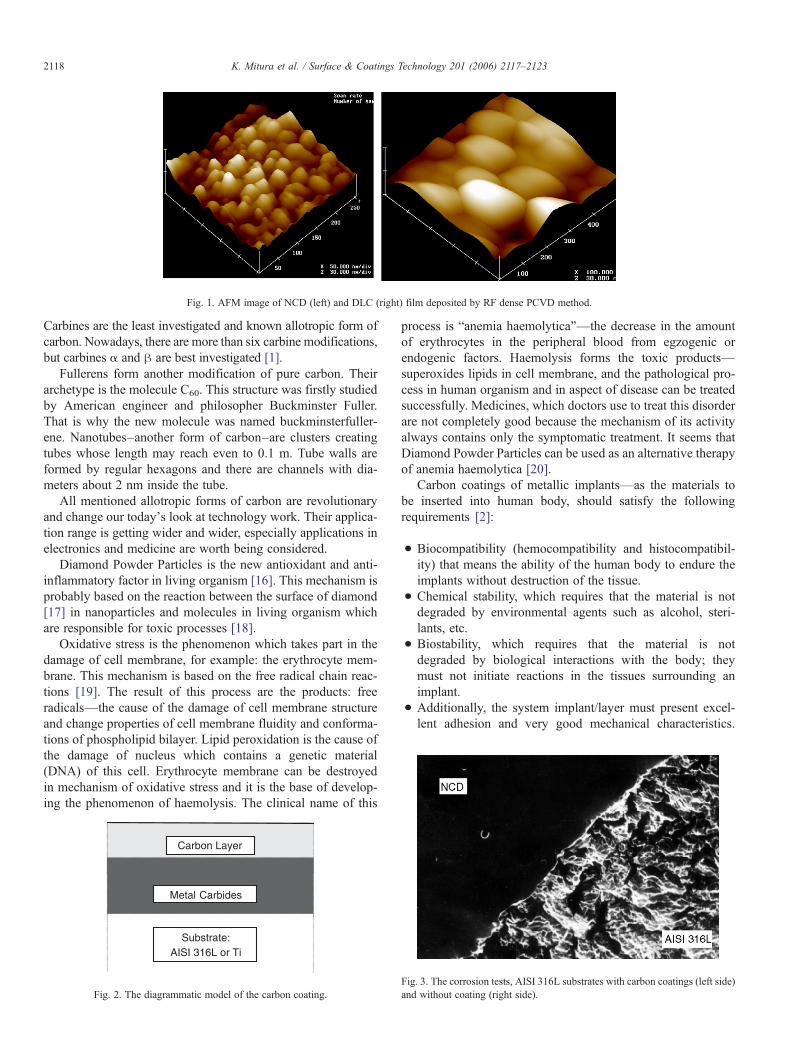
Fig. 1. AFM image of NCD (left) and DLC (right) film deposited by RF dense PCVD method.
2118 K. Mitura et al. / Surface & Coatings Technology 201 (2006) 2117–2123
Carbines are the least investigated and known allotropic form ofcarbon. Nowadays, there are more than six carbine modifications,but carbines α and β are best investigated [1].
Fullerens form another modification of pure carbon. Theirarchetype is the molecule C60. This structure was firstly studiedby American engineer and philosopher Buckminster Fuller.That is why the new molecule was named buckminsterfuller-ene. Nanotubes–another form of carbon–are clusters creatingtubes whose length may reach even to 0.1 m. Tube walls areformed by regular hexagons and there are channels with dia-meters about 2 nm inside the tube.
All mentioned allotropic forms of carbon are revolutionaryand change our today's look at technology work. Their applica-tion range is getting wider and wider, especially applications inelectronics and medicine are worth being considered.
Diamond Powder Particles is the new antioxidant and anti-inflammatory factor in living organism [16]. This mechanism isprobably based on the reaction between the surface of diamond[17] in nanoparticles and molecules in living organism whichare responsible for toxic processes [18].
Oxidative stress is the phenomenon which takes part in thedamage of cell membrane, for example: the erythrocyte mem-brane. This mechanism is based on the free radical chain reac-tions [19]. The result of this process are the products: freeradicals—the cause of the damage of cell membrane structureand change properties of cell membrane fluidity and conforma-tions of phospholipid bilayer. Lipid peroxidation is the cause ofthe damage of nucleus which contains a genetic material(DNA) of this cell. Erythrocyte membrane can be destroyedin mechanism of oxidative stress and it is the base of develop-ing the phenomenon of haemolysis. The clinical name of this
Carbon Layer
Metal Carbides
Substrate:AISI 316L or Ti
Fig. 2. The diagrammatic model of the carbon coating.
process is “anemia haemolytica”—the decrease in the amountof erythrocytes in the peripheral blood from egzogenic orendogenic factors. Haemolysis forms the toxic products—superoxides lipids in cell membrane, and the pathological pro-cess in human organism and in aspect of disease can be treatedsuccessfully. Medicines, which doctors use to treat this disorderare not completely good because the mechanism of its activityalways contains only the symptomatic treatment. It seems thatDiamond Powder Particles can be used as an alternative therapyof anemia haemolytica [20].
Carbon coatings of metallic implants—as the materials tobe inserted into human body, should satisfy the followingrequirements [2]:
• Biocompatibility (hemocompatibility and histocompatibil-ity) that means the ability of the human body to endure theimplants without destruction of the tissue.
• Chemical stability, which requires that the material is notdegraded by environmental agents such as alcohol, steri-lants, etc.
• Biostability, which requires that the material is notdegraded by biological interactions with the body; theymust not initiate reactions in the tissues surrounding animplant.
• Additionally, the system implant/layer must present excel-lent adhesion and very good mechanical characteristics.
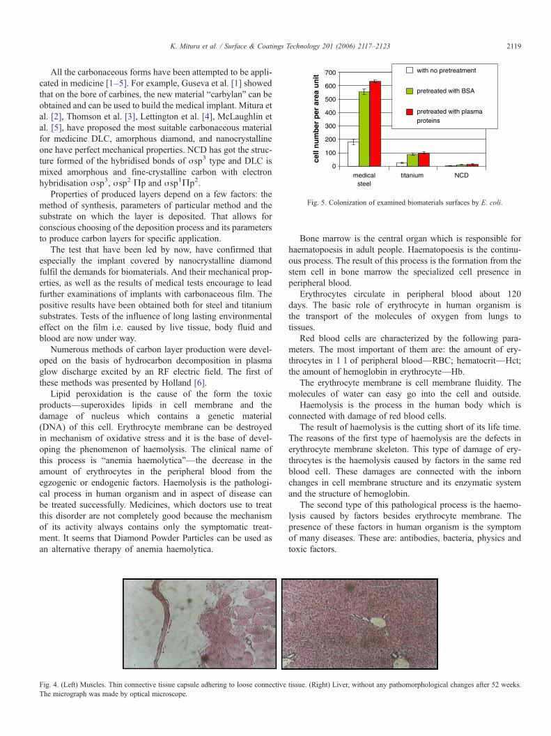
Fig. 3. The corrosion tests, AISI 316L substrates with carbon coatings (left side)and without coating (right side).
0
100
200
300
400
500
600
700
medicalsteel
titanium NCD
cell
nu
mb
er p
er a
rea
un
it
with no pretreatment
pretreated with BSA
pretreated with plasmaproteins
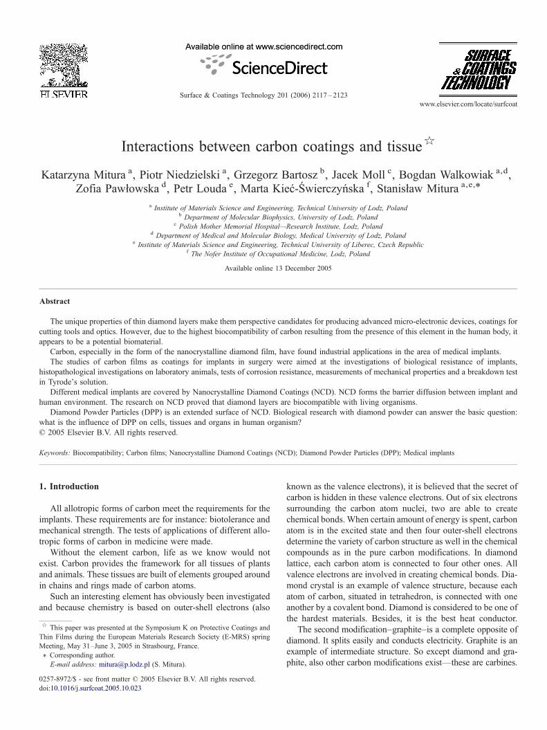
Fig. 5. Colonization of examined biomaterials surfaces by E. coli.
2119K. Mitura et al. / Surface & Coatings Technology 201 (2006) 2117–2123
All the carbonaceous forms have been attempted to be appli-cated in medicine [1–5]. For example, Guseva et al. [1] showedthat on the bore of carbines, the new material “carbylan” can beobtained and can be used to build the medical implant. Mitura etal. [2], Thomson et al. [3], Lettington et al. [4], McLaughlin etal. [5], have proposed the most suitable carbonaceous materialfor medicine DLC, amorphous diamond, and nanocrystallineone have perfect mechanical properties. NCD has got the struc-ture formed of the hybridised bonds of σsp3 type and DLC ismixed amorphous and fine-crystalline carbon with electronhybridisation σsp3, σsp2 Πp and σsp1Πp2.
Properties of produced layers depend on a few factors: themethod of synthesis, parameters of particular method and thesubstrate on which the layer is deposited. That allows forconscious choosing of the deposition process and its parametersto produce carbon layers for specific application.
The test that have been led by now, have confirmed thatespecially the implant covered by nanocrystalline diamondfulfil the demands for biomaterials. And their mechanical prop-erties, as well as the results of medical tests encourage to leadfurther examinations of implants with carbonaceous film. Thepositive results have been obtained both for steel and titaniumsubstrates. Tests of the influence of long lasting environmentaleffect on the film i.e. caused by live tissue, body fluid andblood are now under way.
Numerous methods of carbon layer production were devel-oped on the basis of hydrocarbon decomposition in plasmaglow discharge excited by an RF electric field. The first ofthese methods was presented by Holland [6].
Lipid peroxidation is the cause of the form the toxicproducts—superoxides lipids in cell membrane and thedamage of nucleus which contains a genetic material(DNA) of this cell. Erythrocyte membrane can be destroyedin mechanism of oxidative stress and it is the base of devel-oping the phenomenon of haemolysis. The clinical name ofthis process is “anemia haemolytica”—the decrease in theamount of erythrocytes in the peripheral blood from theegzogenic or endogenic factors. Haemolysis is the pathologi-cal process in human organism and in aspect of disease canbe treated successfully. Medicines, which doctors use to treatthis disorder are not completely good because the mechanismof its activity always contains only the symptomatic treat-ment. It seems that Diamond Powder Particles can be used asan alternative therapy of anemia haemolytica.
Fig. 4. (Left) Muscles. Thin connective tissue capsule adhering to loose connectiveThe micrograph was made by optical microscope.
Bone marrow is the central organ which is responsible forhaematopoesis in adult people. Haematopoesis is the continu-ous process. The result of this process is the formation from thestem cell in bone marrow the specialized cell presence inperipheral blood.
Erythrocytes circulate in peripheral blood about 120days. The basic role of erythrocyte in human organism isthe transport of the molecules of oxygen from lungs totissues.
Red blood cells are characterized by the following para-meters. The most important of them are: the amount of ery-throcytes in 1 l of peripheral blood—RBC; hematocrit—Hct;the amount of hemoglobin in erythrocyte—Hb.
The erythrocyte membrane is cell membrane fluidity. Themolecules of water can easy go into the cell and outside.
Haemolysis is the process in the human body which isconnected with damage of red blood cells.
The result of haemolysis is the cutting short of its life time.The reasons of the first type of haemolysis are the defects inerythrocyte membrane skeleton. This type of damage of ery-throcytes is the haemolysis caused by factors in the same redblood cell. These damages are connected with the inbornchanges in cell membrane structure and its enzymatic systemand the structure of hemoglobin.
The second type of this pathological process is the haemo-lysis caused by factors besides erythrocyte membrane. Thepresence of these factors in human organism is the symptomof many diseases. These are: antibodies, bacteria, physics andtoxic factors.
tissue. (Right) Liver, without any pathomorphological changes after 52 weeks.
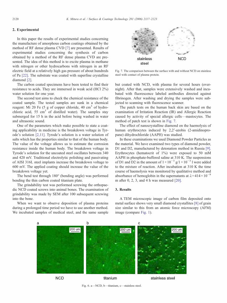
Fig. 7. The comparison between the surface with and without NCD on stainlesssteel with contact of plasma protein.
2120 K. Mitura et al. / Surface & Coatings Technology 201 (2006) 2117–2123
2. Experimental
In this paper the results of experimental studies concerningthe manufacture of amorphous carbon coatings obtained by themethod of RF dense plasma CVD [7] are presented. Results ofexperimental studies concerning the synthesis of carbonobtained by a method of the RF dense plasma CVD are pre-sented. The idea of this method is to excite plasma in methanewith nitrogen or other hydrocarbons with nitrogen in an RFelectric field at a relatively high gas pressure of about hundredsof Pa [22]. The substrate was coated with superfine crystallinediamond [2].
The carbon coated specimens have been tested to find theirresistance to acids. They are immersed in weak acid (HCl 2%)water solution for one year.
The second test aims to check the chemical resistance of thecoated sample. The tested samples are sunk in a chemicalreagent: Mi 20 Fe (5 g of copper chloride, 40 cm3 of hydro-chloric acid, 55 cm3 of distilled water). The samples staysubmerged for 15 h in the acid before being washed in waterand ultrasonic sound.
One of the parameters which make possible to state a coat-ing applicability in medicine is the breakdown voltage in Tyr-ode's solution [2,11]. Tyrode's solution is a water solution ofsalts which has the properties similar to that of the human body.The value of the voltage allows us to estimate the corrosionresistance inside the human body. The breakdown voltage inTyrode's solution for the uncoated steel oscillates between 340and 420 mV. Traditional electrolytic polishing and passivatingof AISI 316L steel implants increase the breakdown voltage to600 mV. The applied coating should increase the value of thebreakdown voltage yet.
The bend test through 180° (bending angle) was performedbending the thin carbon coated titanium plate.
The grindability test was performed screwing the orthopae-dic NCD coated screws into animal bones. The examination ofgrindability was made by SEM after 100 subsequent screwinginto the bone.
When we want to observe deposition of plasma proteinsduring a prolonged time period we have to use another method.We incubated samples of medical steel, and the same sample
Fig. 6. a—NCD, b—titani
but coated with NCD, with plasma for several hours (over-night). After that, samples were extensively washed and incu-bated with fluorescence labeled antibodies directed againstfibrinogen. After washing and drying the samples were sub-jected to scanning with fluorescence scanner.
The patch tests on the human back skin are based on theexamination of Irritation Reaction (IR) and Allergic Reactioncaused by activity of special allergic cells—mastocytes. Themethod of patch test is shown in Fig. 7.
The effect of nanocrystalline diamond on the haemolysis ofhuman erythrocytes induced by 2,2′-azobis (2-amidinopro-pane) dihydrochloride (AAPH) was studied.
In these examinations we used Diamond Powder Particles asthe material. We have examined two types of diamond powder,D1 and D2, manufactured by detonation method in Russia [9].Erythrocytes (hematocrit of 1%) were exposed to 50 mMAAPH in phosphate-buffered saline at 310 K. The suspensionsof D1 and D2 in the amount of 1×10⁻³ g/1×10−3 l were addedto the mixture of reaction. After incubation at 310 K the timecourse of haemolysis was monitored by qualitative method andabsorbance of hemoglobin in the supernatants at λ=414×10−9
m after 0, 2, 3, and 4 h was measured [20].
3. Results
A TEM microscopic image of carbon film deposited ontometal surface shows very small diamond crystallites [8] of grainsize similar to this from an atomic force microscopy (AFM)image (compare Fig. 1).
um, c—stainless steel.
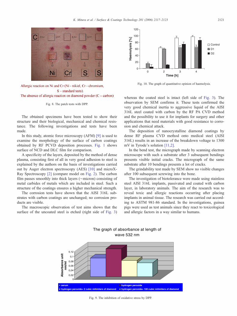
Fig. 8. The patch tests with DPP.
0
20
40
60
80
100
120
0 2 3 4Time [h]
Hae
mo
lysi
s [%
]
Control
D1
D2
Fig. 10. The graph of quantitative opinion of haemolysis.
2121K. Mitura et al. / Surface & Coatings Technology 201 (2006) 2117–2123
The obtained specimens have been tested to show theirstructure and their biological, mechanical and chemical resis-tance. The following investigations and tests have beenmade.
In this study, atomic force microscopy (AFM) [9] is used toexamine the morphology of the surface of carbon coatingsobtained by RF PCVD deposition processes. Fig. 1 showssurface of NCD and DLC film for comparison.
A specificity of the layers, deposited by the method of denseplasma, consisting first of all in very good adhesion to steel isexplained by the authors on the basis of investigations carriedout by Auger electron spectroscopy (AES) [10] and microX-Ray Spectroscopy [2] (compare model on Fig. 2). The carbonfilm passes smoothly into thick layers (∼micron) consisting ofmetal carbides of metals which are included in steel. Such astructure of the coatings ensures a higher mechanical strength.
The corrosion tests have shown that the AISI 316L sub-strates with carbon coatings are unchanged; no corrosion pro-ducts are visible.
The macroscopic observation of test aims shows that thesurface of the uncoated steel is etched (right side of Fig. 3)
The graph of abswave
0
0,02
0,04
0,06
0,08
0,1
0,12
Ab
sorb
ance
un
itn
m/m
l
serum hy
hydrogen peroxide+ 2 cubic milimiters of diamond hy
Fig. 9. The inhibition of ox
whereas the coated steel is intact (left side of Fig. 3). Theobservation by SEM confirms it. These tests confirmed thevery good chemical inertia to aggressive liquid of the AISI316L steel coated with carbon by the RF PA CVD methodand the possibility to use it for implants for surgery and otherapplications that need materials with good resistance to corro-sion and chemical attack.
The deposition of nanocrystalline diamond coatings bydense RF plasma CVD method onto medical steel (AISI316L) results in an increase of the breakdown voltage to 1300mV in Tyrode's solution [11,2].
In the bend test, the micrograph made by scanning electronmicroscope with such a substrate after 3 subsequent bendingspresents visible initial cracks. The micrograph of the samesubstrate after 10 bendings presents a lot of cracks.
The grindability test made by SEM show no visible changesafter 100 subsequent screwing into the bone.
The investigation of biotolerance were made using stainlesssteel AISI 316L implants, passivated and coated with carbonlayer, in laboratory animals. The aim of the research was toreveal toxic and allergic reactions occurring after placingimplants in animal tissue. The research was carried out accord-ing to ASTM 981-86 standard. In the investigations, guineapigs were used as test animals since they react to toxicologicaland allergic factors in a way similar to humans.
orbance at length of 532 nm
drogen peroxide
drogen peroxide+ 100 cubic milimiters of diamond
idative stress by DPP.
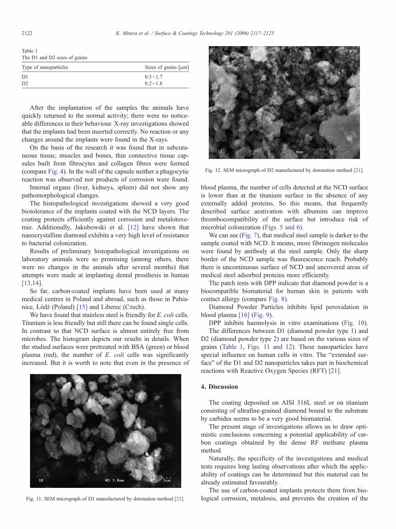
Table 1The D1 and D2 sizes of grains
Type of nanoparticles Sizes of grains [μm]
D1 0.3÷1.7D2 0.2÷1.8
Fig. 12. SEM micrograph of D2 manufactured by detonation method [21].
2122 K. Mitura et al. / Surface & Coatings Technology 201 (2006) 2117–2123
After the implantation of the samples the animals havequickly returned to the normal activity; there were no notice-able differences in their behaviour. X-ray investigations showedthat the implants had been inserted correctly. No reaction or anychanges around the implants were found in the X-rays.
On the basis of the research it was found that in subcuta-neous tissue, muscles and bones, thin connective tissue cap-sules built from fibrocytes and collagen fibres were formed(compare Fig. 4). In the wall of the capsule neither a phagocyticreaction was observed nor products of corrosion were found.
Internal organs (liver, kidneys, spleen) did not show anypathomorphological changes.
The histopathological investigations showed a very goodbiotolerance of the implants coated with the NCD layers. Thecoating protects efficiently against corrosion and metalotoxe-mie. Additionally, Jakubowski et al. [12] have shown thatnanocrystalline diamond exhibits a very high level of resistanceto bacterial colonization.
Results of preliminary histopathological investigations onlaboratory animals were so promising (among others, therewere no changes in the animals after several months) thatattempts were made at implanting dental prosthesis in human[13,14].
So far, carbon-coated implants have been used at manymedical centres in Poland and abroad, such as those in Pabia-nice, Łódź (Poland) [15] and Liberec (Czech).
We have found that stainless steel is friendly for E. coli cells.Titanium is less friendly but still there can be found single cells.In contrast to that NCD surface is almost entirely free frommicrobes. The histogram depicts our results in details. Whenthe studied surfaces were pretreated with BSA (green) or bloodplasma (red), the number of E. coli cells was significantlyincreased. But it is worth to note that even in the presence of
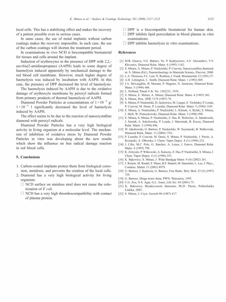
Fig. 11. SEM micrograph of D1 manufactured by detonation method [21].
blood plasma, the number of cells detected at the NCD surfaceis lower than at the titanium surface in the absence of anyexternally added proteins. So this means, that frequentlydescribed surface aestivation with albumins can improvethrombocompatibility of the surface but introduce risk ofmicrobial colonization (Figs. 5 and 6).
We can see (Fig. 7), that medical steel sample is darker to thesample coated with NCD. It means, more fibrinogen moleculeswere found by antibody at the steel sample. Only the sharpborder of the NCD sample was fluorescence reach. Probablythere is uncontinuous surface of NCD and uncovered areas ofmedical steel adsorbed proteins more efficiently.
The patch tests with DPP indicate that diamond powder is abiocompatible biomaterial for human skin in patients withcontact allergy (compare Fig. 8).
Diamond Powder Particles inhibits lipid peroxidation inblood plasma [16] (Fig. 9).
DPP inhibits haemolysis in vitro examinations (Fig. 10).The differences between D1 (diamond powder type 1) and
D2 (diamond powder type 2) are based on the various sizes ofgrains (Table 1, Figs. 11 and 12). These nanoparticles havespecial influence on human cells in vitro. The “extended sur-face” of the D1 and D2 nanoparticles takes part in biochemicalreactions with Reactive Oxygen Species (RFT) [21].
4. Discussion
The coating deposited on AISI 316L steel or on titaniumconsisting of ultrafine-grained diamond bound to the substrateby carbides seems to be a very good biomaterial.
The present stage of investigations allows us to draw opti-mistic conclusions concerning a potential applicability of car-bon coatings obtained by the dense RF methane plasmamethod.
Naturally, the specificity of the investigations and medicaltests requires long lasting observations after which the applic-ability of coatings can be determined but this material can bealready estimated favourably.
The use of carbon-coated implants protects them from bio-logical corrosion, metalosis, and prevents the creation of the
2123K. Mitura et al. / Surface & Coatings Technology 201 (2006) 2117–2123
local cells. This has a stabilizing effect and makes the recoveryof a patient possible even in serious cases.
In same cases, the use of metal implants without carboncoatings makes the recovery impossible. In each case, the useof the carbon coatings will shorten the treatment period.
In examinations in vivo NCD is biocompatible biomaterialfor tissues and cells around the implant.
Induction of erythrocytes in the presence of DPP with 2,2,-azo-bis(2-amidinopropane) (AAPH) leads to some degree ofhaemolysis induced apparently by mechanical damage to thered blood cell membrane. However, much higher degree ofhaemolysis was induced by incubation with AAPH. In thiscase, the presence of DPP decreased the level of haemolysis.
The haemolysis induced by AAPH is due to the oxidativedamage of erythrocyte membrane by peroxyl radicals formedfrom primary products of thermal haemolysis of AAPH.
Diamond Powder Particles at concentrations of 1×10⁻³ g/1×10−3 l significantly decreased the level of haemolysisinduced by AAPH.
The effect seems to be due to the reaction of nanocrystallinediamond with peroxyl radicals.
Diamond Powder Particles has a very high biologicalactivity in living organism at a molecular level. The mechan-ism of inhibition of oxidative stress by Diamond PowderParticles in vitro was developing about the new resultswhich show the influence on free radical damage reactionin red blood cells.
5. Conclusions
1. Carbon-coated implants protect them from biological corro-sion, metalosis, and prevents the creation of the local cells.
2. Diamond has a very high biological activity for livingorganism.□ NCD surface on stainless steel does not cause the colo-
nization of E coli.□ NCD has a very high thrombocompatibility with contact
of plasma protein.
□ DPP is a biocompatible biomaterial for human skin.□ DPP inhibits lipid peroxidation in blood plasma in vitro
examinations.□ DPP inhibits haemolysis in vitro examinations.
References
[1] M.B. Guseva, V.G. Babaev, Yu. P. Kudryavtsev, A.F. Alexandrov, V.V.Khvostov, Diamond Relat. Mater. 4 (1995) 1142.
[2] S. Mitura, A. Mitura, P. Niedzielski, P. Couvrat, Nanocrystalline diamond,in: S. Mitura (Ed.), Nanotechnology in Materials Science, Elsevier, 2000.
[3] L.A. Thomson, F.C. Law, N. Rushton, J. Frank, Biomaterials 12 (1991) 37.[4] A.H. Lettington, C. Smith, Diamond Relat. Mater. 1 (1992) 805.[5] J.A. McLaughlin, B. Meenan, P. Maguire, N. Jamieson, Diamond Relat.
Mater. 5 (1996) 486.[6] L. Holland, Patent U.K. No. 1582231, 1976.[7] S. Mitura, E. Mitura, A. Mitura, Diamond Relat. Mater. 4 (1995) 302.[8] S. Mitura, Proc. SPIE 3179 (1997) 79.[9] S. Mitura, P. Niedzielski, D. Jachowicz, M. Langer, E. Tochitsky, P. Louda,
P. Couvrat, M. Denis, P. Lourdin, Diamond Relat. Mater. 5 (1996) 1185.[10] E. Mitura, A. Niedzielska, P. Niedzielski, L. Klimek, A. Rylski, S. Mitura,
J. Moll, W. Pietrzykowski, Diamond Relat. Mater. 5 (1996) 998.[11] E. Mitura, S. Mitura, P. Niedzielski, Z. Has, R. Wolowiec, A. Jakubowski,
J. Szmidt, A. Sokolowska, P. Louda, J. Marciniak, B. Koczy, DiamondRelat. Mater. 3 (1994) 896.
[12] W. Jakubowski, G. Bartosz, P. Niedzielski, W. Szymanski, B. Walkowiak,Diamond Relat. Mater. 13 (2004) 1761.
[13] P. Lourdin, P. Couvrat, M. Denis, S. Mitura, P. Niedzielski, J. Perrin, A.Koziarski, A. Olborska, J. Chem. Vapor Depos. 4 (1) (1996) 232.
[14] J. Cifre, M.C. Polo, G. Sánchez, A. Lousa, J. Esteve, Diamond Relat.Mater. 4 (1995) 798.
[15] K. Zolynski, P. Witkowski, A. Kaluzny, Z. Has, P. Niedzielski, S. Mitura, J.Chem. Vapor Depos. 4 (1) (1996) 253.
[16] K. Bąkowicz, S. Mitura, J. Wide Bandgap Mater. 9 (4) (2002) 261.[17] J. Ristein, M. Riedel, F. Maier, B.F. Mantel, M. Stammler, L. Ley, J. Phys.,
Condens. Matter 13 (2001) 8979.[18] G. Bartosz, J. Kędziora, G. Bartosz, Free Radic. Biol. Med. 23 (5) (1997)
729.[19] G. Bartosz, Druga twarz tlenu, PWN, Warszawa, 1995.[20] C.G. Zou, N.S. Agar, G.L. Jones, Life Sci. 69 (2001) 75.[21] K. Bakowicz, Bioaktywność diamentu, Ph.D. Thesis, Politechnika
Łódzka, 2003.[22] S. Mitura, J. Cryst. Growth 80 (1987) 417.
Copyright © 2022 FDOKUMEN