Modeling of Tool-Tissue Interactions for Computer-Based Surgical Simulation: A Literature Review
29
Sarthak Misra K. T. Ramesh Allison M. Okamura* 125 Computational Science and Engineering Building Department of Mechanical Engineering The Johns Hopkins University 3400 North Charles Street Baltimore, MD 21218 Presence, Vol. 17, No. 5, October 2008, 463– 491 © 2008 by the Massachusetts Institute of Technology Modeling of Tool-Tissue Interactions for Computer-Based Surgical Simulation: A Literature Review Abstract Surgical simulators present a safe and potentially effective method for surgical train- ing, and can also be used in robot-assisted surgery for pre- and intra-operative plan- ning. Accurate modeling of the interaction between surgical instruments and organs has been recognized as a key requirement in the development of high-fidelity surgi- cal simulators. Researchers have attempted to model tool-tissue interactions in a wide variety of ways, which can be broadly classified as (1) linear elasticity-based, (2) nonlinear (hyperelastic) elasticity-based finite element (FE) methods, and (3) other techniques not based on FE methods or continuum mechanics. Realistic mod- eling of organ deformation requires populating the model with real tissue data (which are difficult to acquire in vivo) and simulating organ response in real time (which is computationally expensive). Further, it is challenging to account for con- nective tissue supporting the organ, friction, and topological changes resulting from tool-tissue interactions during invasive surgical procedures. Overcoming such obsta- cles will not only help us to model tool-tissue interactions in real time, but also en- able realistic force feedback to the user during surgical simulation. This review paper classifies the existing research on tool-tissue interactions for surgical simulators spe- cifically based on the modeling techniques employed and the kind of surgical opera- tion being simulated, in order to inform and motivate future research on improved tool-tissue interaction models. 1 Introduction People have always sought ways to understand and model the structure and function of the human body. The earliest known anatomical models used for surgical planning were recorded around 800 BC in India for the procedure of rhinoplasty, in which a flap of skin from the forehead is used to reconstruct a nose. These ancient practitioners used leaves to represent three-dimensional flexible tissues and plan the surgical operation (Carpue, 1981). Before the ad- vent of medical imaging a century ago, the only practical way to see inside the human body was to observe an operation or by dissection. However, cultural and religious beliefs, the difficulty of obtaining cadavers, and lack of refrigera- *Correspondence to [email protected]. Misra et al. 463
-
Upload
guadalajara -
Category
Documents
-
view
1 -
download
0
Transcript of Modeling of Tool-Tissue Interactions for Computer-Based Surgical Simulation: A Literature Review
Sarthak MisraK. T. RameshAllison M. Okamura*125 Computational Science andEngineering BuildingDepartment of MechanicalEngineeringThe Johns Hopkins University3400 North Charles StreetBaltimore, MD 21218
Presence, Vol. 17, No. 5, October 2008, 463–491
© 2008 by the Massachusetts Institute of Technology
Modeling of Tool-TissueInteractions for Computer-BasedSurgical Simulation: A LiteratureReview
Abstract
Surgical simulators present a safe and potentially effective method for surgical train-
ing, and can also be used in robot-assisted surgery for pre- and intra-operative plan-
ning. Accurate modeling of the interaction between surgical instruments and organs
has been recognized as a key requirement in the development of high-fidelity surgi-
cal simulators. Researchers have attempted to model tool-tissue interactions in a
wide variety of ways, which can be broadly classified as (1) linear elasticity-based,
(2) nonlinear (hyperelastic) elasticity-based finite element (FE) methods, and (3)
other techniques not based on FE methods or continuum mechanics. Realistic mod-
eling of organ deformation requires populating the model with real tissue data
(which are difficult to acquire in vivo) and simulating organ response in real time
(which is computationally expensive). Further, it is challenging to account for con-
nective tissue supporting the organ, friction, and topological changes resulting from
tool-tissue interactions during invasive surgical procedures. Overcoming such obsta-
cles will not only help us to model tool-tissue interactions in real time, but also en-
able realistic force feedback to the user during surgical simulation. This review paper
classifies the existing research on tool-tissue interactions for surgical simulators spe-
cifically based on the modeling techniques employed and the kind of surgical opera-
tion being simulated, in order to inform and motivate future research on improved
tool-tissue interaction models.
1 Introduction
People have always sought ways to understand and model the structureand function of the human body. The earliest known anatomical models usedfor surgical planning were recorded around 800 BC in India for the procedureof rhinoplasty, in which a flap of skin from the forehead is used to reconstructa nose. These ancient practitioners used leaves to represent three-dimensionalflexible tissues and plan the surgical operation (Carpue, 1981). Before the ad-vent of medical imaging a century ago, the only practical way to see inside thehuman body was to observe an operation or by dissection. However, culturaland religious beliefs, the difficulty of obtaining cadavers, and lack of refrigera-
*Correspondence to [email protected].
Misra et al. 463
tion imposed restrictions on the widespread use of dis-section. Frustrated by these limitations, Louis ThomasJerome Auzoux, a nineteenth century French physician,improved and popularized anatomical papier machemodels (Davis, 1975). By the early twentieth century,inexpensive and realistic plastic anatomical models thatcould be assembled and painted became popular withmedical students. Human cadavers and animals, how-ever, are still used for training and, in some cases, surgi-cal planning.
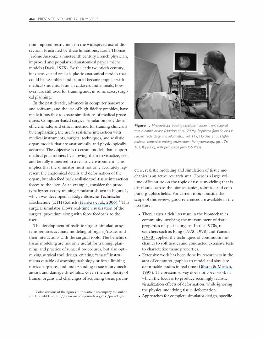
In the past decade, advances in computer hardwareand software, and the use of high-fidelity graphics, havemade it possible to create simulations of medical proce-dures. Computer-based surgical simulation provides anefficient, safe, and ethical method for training cliniciansby emphasizing the user’s real-time interaction withmedical instruments, surgical techniques, and realisticorgan models that are anatomically and physiologicallyaccurate. The objective is to create models that supportmedical practitioners by allowing them to visualize, feel,and be fully immersed in a realistic environment. Thisimplies that the simulator must not only accurately rep-resent the anatomical details and deformation of theorgan, but also feed back realistic tool-tissue interactionforces to the user. As an example, consider the proto-type hysteroscopy training simulator shown in Figure 1,which was developed at Eidgenossische TechnischeHochschule (ETH) Zurich (Harders et al., 2006).1 Thissurgical simulator allows real-time visualization of thesurgical procedure along with force feedback to theuser.
The development of realistic surgical simulation sys-tems requires accurate modeling of organs/tissues andtheir interactions with the surgical tools. The benefits oftissue modeling are not only useful for training, plan-ning, and practice of surgical procedures, but also opti-mizing surgical tool design, creating “smart” instru-ments capable of assessing pathology or force-limitingnovice surgeons, and understanding tissue injury mech-anisms and damage thresholds. Given the complexity ofhuman organs and challenges of acquiring tissue param-
eters, realistic modeling and simulation of tissue me-chanics is an active research area. There is a large vol-ume of literature on the topic of tissue modeling that isdistributed across the biomechanics, robotics, and com-puter graphics fields. For certain topics outside thescope of this review, good references are available in theliterature:
● There exists a rich literature in the biomechanicscommunity involving the measurement of tissueproperties of specific organs. In the 1970s, re-searchers such as Fung (1973, 1993) and Yamada(1970) applied the techniques of continuum me-chanics to soft tissues and conducted extensive teststo characterize tissue properties.
● Extensive work has been done by researchers in thearea of computer graphics to model and simulatedeformable bodies in real time (Gibson & Mirtich,1997). The present survey does not cover work inwhich the focus is to produce seemingly realisticvisualization effects of deformation, while ignoringthe physics underlying tissue deformation.
● Approaches for complete simulator design, specific1 Color versions of the figures in this article accompany the online
article, available at http://www.mitpressjournals.org/toc/pres/17/5.
Figure 1. Hysteroscopy training simulation environment coupled
with a haptic device (Harders et al., 2006). Reprinted from Studies in
Health Technology and Informatics, Vol. 119, Harders et al. Highly
realistic, immersive training environment for hysteroscopy, pp. 176–
181, ©(2006), with permission from IOS Press.
464 PRESENCE: VOLUME 17, NUMBER 5
medical applications, and training evaluation meth-ods have also been widely studied in the last twodecades (A. Liu, Tendick, Cleary, & Kaufmann,2003; Satava, 2001).
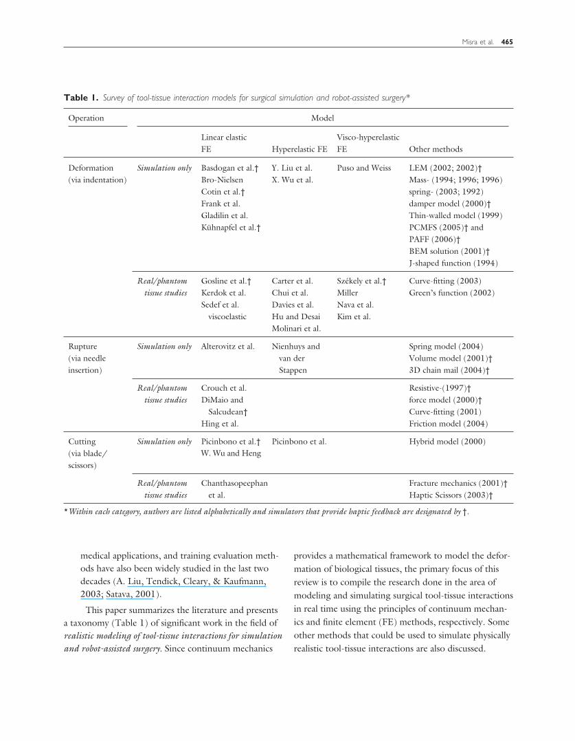
This paper summarizes the literature and presentsa taxonomy (Table 1) of significant work in the field ofrealistic modeling of tool-tissue interactions for simulationand robot-assisted surgery. Since continuum mechanics
provides a mathematical framework to model the defor-mation of biological tissues, the primary focus of thisreview is to compile the research done in the area ofmodeling and simulating surgical tool-tissue interactionsin real time using the principles of continuum mechan-ics and finite element (FE) methods, respectively. Someother methods that could be used to simulate physicallyrealistic tool-tissue interactions are also discussed.
Table 1. Survey of tool-tissue interaction models for surgical simulation and robot-assisted surgery*
Operation Model
Linear elasticFE Hyperelastic FE
Visco-hyperelasticFE Other methods
Deformation(via indentation)
Simulation only Basdogan et al.† Y. Liu et al. Puso and Weiss LEM (2002; 2002)†Bro-Nielsen X. Wu et al. Mass- (1994; 1996; 1996)Cotin et al.† spring- (2003; 1992)Frank et al. damper model (2000)†Gladilin et al. Thin-walled model (1999)Kuhnapfel et al.† PCMFS (2005)† and
PAFF (2006)†BEM solution (2001)†J-shaped function (1994)
Real/phantomtissue studies
Gosline et al.† Carter et al. Szekely et al.† Curve-fitting (2003)Kerdok et al. Chui et al. Miller Green’s function (2002)Sedef et al.
viscoelasticDavies et al. Nava et al.Hu and Desai Kim et al.Molinari et al.
Rupture(via needleinsertion)
Simulation only Alterovitz et al. Nienhuys andvan derStappen
Spring model (2004)Volume model (2001)†3D chain mail (2004)†
Real/phantomtissue studies
Crouch et al. Resistive-(1997)†DiMaio and
Salcudean†force model (2000)†Curve-fitting (2001)
Hing et al. Friction model (2004)
Cutting(via blade/scissors)
Simulation only Picinbono et al.† Picinbono et al. Hybrid model (2000)W. Wu and Heng
Real/phantomtissue studies
Chanthasopeephanet al.
Fracture mechanics (2001)†Haptic Scissors (2003)†
*Within each category, authors are listed alphabetically and simulators that provide haptic feedback are designated by †.
Misra et al. 465

This paper is organized as follows. Section 2 summa-rizes the basic concepts and theories of linear and non-linear elasticity, while Section 3 provides an overview ofthe FE modeling technique. Sections 4 and 5 classifythe prior research work that has been done in modelingnoninvasive and invasive surgical tool-tissue interac-tions, respectively. Realistic tool-tissue interaction mod-els require populating models with accurate materialproperties, and Section 6 provides a summary of someof the methods for acquiring tissue properties. Section 7lists some of the commercially available surgical simula-tors. Finally, Section 8 concludes by providing someimportant directions for research in the area of realisticmodeling of tool-tissue interactions.
2 Continuum Mechanics for TissueModeling
The study of deformation or motion of a continu-ous material under the action of forces is known as con-tinuum mechanics. The objective of this section is toprovide a brief introduction to the mechanics of softtissues using the theories of linear and nonlinear elas-ticity.
The field equations of continuum mechanics are nor-mally formulated using tensors. An overview of tensoranalysis is beyond the scope of this paper, but Ogden(1984) provides a good introduction to this subject andits application to continuum mechanics. Tensor nota-tions and manipulations consistent with the mechanicsliterature are used in the derivations in this paper; forexample, boldface characters signify tensors, matrices,and vectors, while normal characters are scalar quanti-ties.
2.1 Kinematics of Continua
Consider a body undergoing deformation so thatmaterial points initially at x are mapped to spatial loca-tions, y. Then the deformation gradient tensor, F, isdefined by
F � �y, (1)
where � is the gradient operator with respect to x. Ifthe displacement is u, then y � x � u. Thus,
F � I � �u, (2)
where I is the identity tensor. A useful measure of strainis the Green strain tensor, E, defined by
E �12�FTF � I�. (3)
Substituting Equation 2 in Equation 3 results in
E �12��u � �uT � �uT�u�. (4)
In linear elasticity theory, strains are assumed to besmall (��u� �� 1), hence,
E � � �12��u � �uT�, (5)
where � is the infinitesimal strain tensor. Thus, linearelasticity theory is valid only for small strains (1–2%).One of the fundamental drawbacks of using a linearelasticity formulation to describe soft tissues is that sur-gically relevant strains often significantly exceed thesmall strain limit, invalidating the assumption of linear-ity. However, it is used in many simulation applicationsdue to analytical simplicity and computational efficiency,so it is described next.
2.2 Linear Elasticity
Linear elastic modeling of soft tissues is the mostwidely used approach within the robotics and hapticscommunity. Materials exhibiting linear elasticity obeythe generalized Hooke’s Law, which relates the stresses,�, and infinitesimal strains, �, by the tensor of elasticmoduli, C, as
� � C:�. (6)
where : denotes double contraction, � is also known asthe Cauchy stress tensor and Equation 6 could be re-written as �ij � �m�1
3 �n�13 Cijmn�mn, where Cijmn is a
fourth-order tensor with 81 constants that are specificto the material. The subscript indices represent the
466 PRESENCE: VOLUME 17, NUMBER 5

components of the stress and strain tensor. Symmetry ofthe stress and strain tensors leads to Cijmn � Cjimn andCijmn � Cjinm, and the postulated existence of a strainenergy density leads to Cijmn � Cmnij. Thus, Cijmn has21 independent constants (called moduli) for a fullyanisotropic material, that is, a material whose propertieschange with direction.
If the assumption is made that the material is isotro-pic, then the material properties can be described by justtwo independent parameters: Young’s modulus, E, andPoisson’s ratio, �. E and � are related to the shear mod-ulus, �,
� �E
2�1 � ��. (7)
In the vast majority of surveyed literature (the researchcited under “Linear elastic FE” in Table 1), E and � arethe two parameters used to describe the soft tissueproperties. Most biological materials are, however, in-trinsically anisotropic. For example, a soft tissue con-taining fibers aligned along an axis will have differentproperties along and transverse to that axis. A summaryof such anisotropies is presented in Spencer (1972) andFigure 2 depicts the orientation of muscle fibers in theheart (Zhukov & Barr, 2003), while the nonlinear elas-tic behavior of myocardial tissue and its application tosurgical simulation is highlighted in Misra, Ramesh, andOkamura (2008).
2.2.1 Linear Viscoelasticity. Most soft tissuesare inherently viscoelastic—they have a response basedon both position and velocity. Viscoelastic materials ex-hibit properties of both elastic solids and viscous fluids.Similar to elastic materials, linear viscoelastic materialsretain the linear relationship between stress and strain,but the effective moduli depend on time. For smallstrains, the general linear viscoelastic constitutive equa-tions can be derived by separating the stresses andstrains into the hydrostatic (superscript H) and devia-toric (superscript D) components:
� � �H � �D (8)
� � �H � �D (9)
Hydrostatic stresses/strains act to change the volume ofthe material, but maintain shape, while deviatoric orshear stresses/strains are those that distort the shape,but preserve volume (in isotropic and linear elastic ma-terials). The hydrostatic stresses and strains are relatedby
�H � 3K�H, (10)
and the bulk modulus, K � E/3(1 � 2�), has smallvariations with time as compared to the shear modulusand hence, K is considered to be independent of time.However, the deviatoric stresses and strains can be re-lated by
Figure 2. Tissue fiber orientation of the heart on the inside surface, (a) and (b), and outside surface, (c) and (d), constructed using diffusion
tensor imaging (Zhukov & Barr, 2003). Images are printed with permission from ©IEEE 2003.
Misra et al. 467
�i�0
N
pi
�i�D
�ti � �j�0
M
qj
� j�D
�t j , (11)
where pi and qj are material constants. In Equation 11,the indices N and M depend on the number of materialconstants required to have good fit with the experimen-tal results.
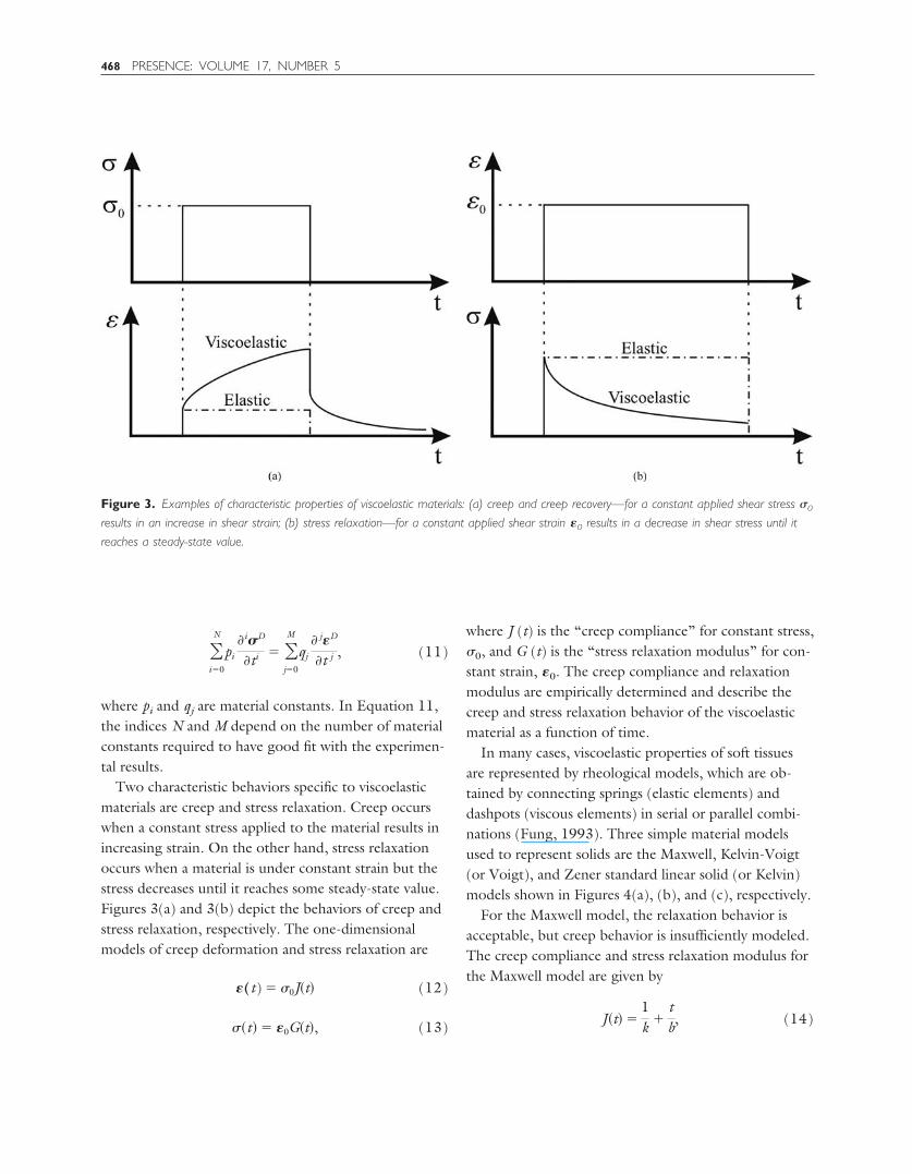
Two characteristic behaviors specific to viscoelasticmaterials are creep and stress relaxation. Creep occurswhen a constant stress applied to the material results inincreasing strain. On the other hand, stress relaxationoccurs when a material is under constant strain but thestress decreases until it reaches some steady-state value.Figures 3(a) and 3(b) depict the behaviors of creep andstress relaxation, respectively. The one-dimensionalmodels of creep deformation and stress relaxation are
�(t) � �0J�t� (12)
��t� � �0G�t�, (13)
where J (t) is the “creep compliance” for constant stress,�0, and G (t) is the “stress relaxation modulus” for con-stant strain, �0. The creep compliance and relaxationmodulus are empirically determined and describe thecreep and stress relaxation behavior of the viscoelasticmaterial as a function of time.
In many cases, viscoelastic properties of soft tissuesare represented by rheological models, which are ob-tained by connecting springs (elastic elements) anddashpots (viscous elements) in serial or parallel combi-nations (Fung, 1993). Three simple material modelsused to represent solids are the Maxwell, Kelvin-Voigt(or Voigt), and Zener standard linear solid (or Kelvin)models shown in Figures 4(a), (b), and (c), respectively.
For the Maxwell model, the relaxation behavior isacceptable, but creep behavior is insufficiently modeled.The creep compliance and stress relaxation modulus forthe Maxwell model are given by
J�t� �1k �
tb, (14)
Figure 3. Examples of characteristic properties of viscoelastic materials: (a) creep and creep recovery—for a constant applied shear stress �0
results in an increase in shear strain; (b) stress relaxation—for a constant applied shear strain �0 results in a decrease in shear stress until it
reaches a steady-state value.
468 PRESENCE: VOLUME 17, NUMBER 5

G�t� � ke�tk
b . (15)
where t is time. The Voigt model provides a satisfactoryfirst-order approximation of creep, but is an inadequatemodel for stress relaxation. For the Voigt model, thegoverning equation is that of an elastic material, sothere is no relaxation of stress, hence, the creep compli-ance is given by
J�t� �1k�1 � e
�tkb �. (16)
The Zener standard linear solid model provides a goodqualitative description of both creep and stress relax-ation. The creep compliance and relaxation modulus aregiven by
J�t� �1k1
�1k2�1 � e
�tk2
b �, (17)
G�t� �k1
k1 � k2�k2 � k1e
�t�k1�k2�
b �. (18)
2.3 Nonlinear Elasticity
Elastic materials undergoing deformations withlarge strains (�1–2%) are described by nonlinear elastic-ity theory. In order to model biological tissues, it iscommon to use hyperelasticity and visco-hyperelasticity
(Fung, 1993). A hyperelastic material is characterizedby the existence of a strain energy density function,W(F). The stress in the material as a result of deforma-tion can be obtained from
P ��W�F�
�F , (19)
where P is the first Piola-Kirchhoff stress tensor and F isthe previously defined deformation gradient tensor. TheCauchy stress tensor and first Piola-Kirchhoff stress ten-sor are related by
PFT � J�, (20)
where J � det (F). There are several formulations forthe strain energy density function, for example, the St.Venant-Kirchhoff, Blatz-Ko, Ogden, Mooney-Rivlin,and Neo-Hookean models (Ogden, 1984). Ogden andMooney-Rivlin strain energy density formulations pro-vide a fairly accurate representation of the constitutivelaws for many biological tissues (Holzapfel, 2000). Inan Ogden model, the strain energy density function foran isotropic material is given in terms of the principalstretches, �i, as
W � �k�1
N�k
k��1
k � �2k � �3
k � 3�, (21)
where �1�2�3 � 1, i.e. thermal incompressibility, and �k
and k are material parameters determined from experi-ments. The Mooney-Rivlin model commonly used torepresent rubber-like materials, is widely used for softtissues and is given in terms of the principal invariants,Ii, for isotropic and incompressible materials as
W � C1�I1 � 3� � C2�I2 � 3�, (22)
where C1 and C2 are material constants. The principalinvariants are defined in terms of right Cauchy-Greentensor, C � FTF, as
I1 � C:I, (23)
I2 �12 ��C:I�2 � �C:C��, (24)
Figure 4. Standard viscoelastic models commonly used to represent
soft tissues (a) Maxwell (b) Kelvin-Voigt (or Voigt) (c) Zener standard
linear solid (or Kelvin) (Fung, 1993).
Misra et al. 469

I3 � det C. (25)
Some researchers have used the Neo-Hookean model torepresent soft tissues. The Neo-Hookean strain energydensity function is a special case of the Mooney-Rivlinmodel and is given by
W � C1�I1 � 3�. (26)
If the material parameter constants for Ogden orMooney-Rivlin models are defined in terms of creep andstress relaxation functions, then the material can bemodeled as visco-hyperelastic, which may represent therealistic behavior of many soft tissues (Fung, 1993).
3 Finite Element Modeling
The FE method is a numerical technique for solv-ing field equations, typically partial differential equa-tions, and has been used in the last decade to simulatesoft tissue deformation by solving the equations of con-tinuum mechanics. The FE method originated from theneed to find approximate solutions to complex prob-lems in elasticity and vibration analysis (Cook, Malkus,& Plesha, 1989). Its development can be traced back tothe work by Hrennikoff (1941) and Courant (1942)(Fung & Tong, 2001). Over the years, the FE methodhas spread to applications in many different areas of en-gineering, including structural analysis in civil and aero-nautical engineering, thermal analysis, and biomechan-ics. Numerous FE computer programs are commerciallyavailable for general or specific applications. These in-clude ABAQUS (Simulia), ADINA (ADINA R & DInc.), ANSYS (ANSYS Inc.), DYNA3D (Lawrence Liv-ermore National Laboratory), FEMLAB (COMSOLInc.), GT STRUDL (Georgia Tech-CASE Center), NXI-deas (Siemens PLM Software), and NASTRAN (MSCSoftware Corp.). This section provides a very high-leveloverview of the FE method; there are numerous textbooksthat deal with this subject, e.g., Cook et al. (1989), Desaiand Abel (1972), and Zienkiewicz, Taylor, and Zhu (2005).
In the FE method, the continuum is divided ormeshed into a finite number of subregions called ele-ments, such as tetrahedrons, quadrilaterals, and so on.
Two adjacent elements are connected via nodes. Theelastic behavior of each element is categorized usingmatrices in terms of the element’s material and geomet-ric properties, and distribution of loading within theelement and at the nodes of the element. Linear or qua-dratic shape or interpolation functions are used to ap-proximate the behavior of the field variables at thenode. The element behavior is characterized by partialdifferential equations governing the motion of materialpoints of a continuum, resulting in the following dis-crete system of differential equations:
Mu � Cu � Ku � F � R, (27)
where M, C, and K are the element mass, damping, andstiffness matrices, respectively, u is the vector of nodaldisplacements, and F and R are the external and internalnode force vectors, respectively. All these matrices andvectors may be time dependent. One approach to solveEquation 27 in a quasi-static manner is by setting u �
u � 0. Thus, with every simulation iteration, a largenumber of element-stiffness and element-force vectorsare assembled, which leads to a system of algebraicequations, called the global system. The accuracy andnumerical efficiency of the FE method lies largely in thedevelopment of effective pre- and post-processors, andalgorithms for efficiently solving large systems of equa-tions. On the other hand, Equation 27 can be solvedwith a dynamic approach, using implicit or explicit inte-gration schemes. For explicit integration methods, thestate at a given instant is a function of the previous timeinstants, while for the implicit scheme, the state at a cer-tain instant cannot be explicitly expressed as a functionof the state at the previous time step. The implicitscheme involves inversion of the stiffness matrix at eachtime step, typically a computationally expensive process.The explicit scheme can be easily implemented by avoidingthe matrix inversion process but suffers from numericalinstability under an inappropriately chosen integrationtime step. Thus, explicit time integration is only condition-ally stable, potentially requiring very small time steps toprovide a suitably accurate and stable solution. Properlydesigned implicit methods can be numerically stable over awide range of integration time step values. Hence, they are
470 PRESENCE: VOLUME 17, NUMBER 5
preferable for simulation of systems described with stiff andnonlinear differential equations.
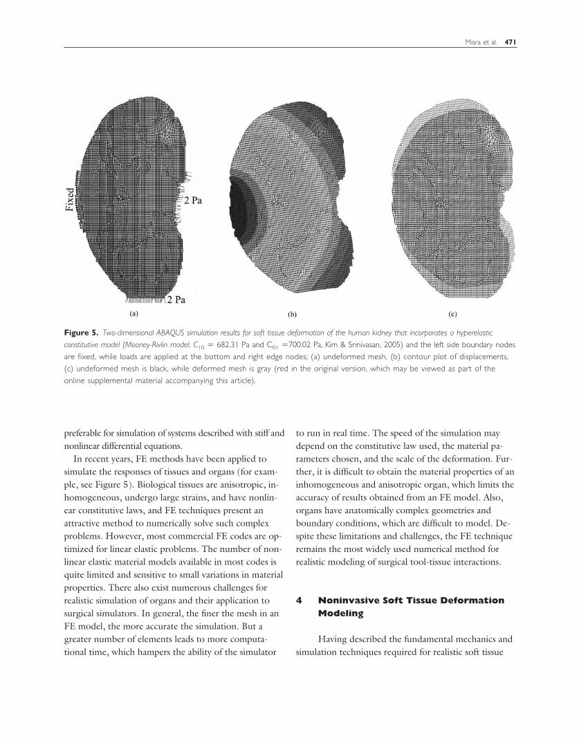
In recent years, FE methods have been applied tosimulate the responses of tissues and organs (for exam-ple, see Figure 5). Biological tissues are anisotropic, in-homogeneous, undergo large strains, and have nonlin-ear constitutive laws, and FE techniques present anattractive method to numerically solve such complexproblems. However, most commercial FE codes are op-timized for linear elastic problems. The number of non-linear elastic material models available in most codes isquite limited and sensitive to small variations in materialproperties. There also exist numerous challenges forrealistic simulation of organs and their application tosurgical simulators. In general, the finer the mesh in anFE model, the more accurate the simulation. But agreater number of elements leads to more computa-tional time, which hampers the ability of the simulator
to run in real time. The speed of the simulation maydepend on the constitutive law used, the material pa-rameters chosen, and the scale of the deformation. Fur-ther, it is difficult to obtain the material properties of aninhomogeneous and anisotropic organ, which limits theaccuracy of results obtained from an FE model. Also,organs have anatomically complex geometries andboundary conditions, which are difficult to model. De-spite these limitations and challenges, the FE techniqueremains the most widely used numerical method forrealistic modeling of surgical tool-tissue interactions.
4 Noninvasive Soft Tissue DeformationModeling
Having described the fundamental mechanics andsimulation techniques required for realistic soft tissue
Figure 5. Two-dimensional ABAQUS simulation results for soft tissue deformation of the human kidney that incorporates a hyperelastic
constitutive model (Mooney-Rivlin model: C10 � 682.31 Pa and C01 �700.02 Pa, Kim & Srinivasan, 2005) and the left side boundary nodes
are fixed, while loads are applied at the bottom and right edge nodes; (a) undeformed mesh, (b) contour plot of displacements,
(c) undeformed mesh is black, while deformed mesh is gray (red in the original version, which may be viewed as part of the
online supplemental material accompanying this article).
Misra et al. 471
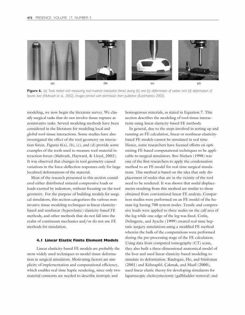
modeling, we now begin the literature survey. We clas-sify surgical tasks that do not involve tissue rupture asnoninvasive tasks. Several modeling methods have beenconsidered in the literature for modeling local andglobal tool-tissue interactions. Some studies have alsoinvestigated the effect of the tool geometry on interac-tion forces. Figures 6(a), (b), (c), and (d) provide someexamples of the tools used to measure tool-material in-teraction forces (Mahvash, Hayward, & Lloyd, 2002).It was observed that changes in tool geometry causedvariations in the force-deflection responses only for largelocalized deformations of the material.
Most of the research presented in this section consid-ered either distributed uniaxial compressive loads orloads exerted by indentors, without focusing on the toolgeometry. For the purpose of building models for surgi-cal simulators, this section categorizes the various non-invasive tissue modeling techniques as linear elasticity-based and nonlinear (hyperelastic) elasticity-based FEmethods, and other methods that do not fall into therealm of continuum mechanics and/or do not use FEmethods for simulation.
4.1 Linear Elastic Finite Element Models
Linear elasticity-based FE models are probably themost widely used techniques to model tissue deforma-tion in surgical simulators. Motivating factors are sim-plicity of implementation and computational efficiency,which enables real-time haptic rendering, since only twomaterial constants are needed to describe isotropic and
homogenous materials, as stated in Equation 7. Thissection describes the modeling of tool-tissue interac-tions using linear elasticity-based FE methods.
In general, due to the steps involved in setting up andrunning an FE calculation, linear or nonlinear elasticity-based FE models cannot be simulated in real time.Hence, some researchers have focused efforts on opti-mizing FE-based computational techniques to be appli-cable to surgical simulators. Bro-Nielsen (1998) wasone of the first researchers to apply the condensationmethod to an FE model for real-time surgical simula-tions. This method is based on the idea that only dis-placement of nodes that are in the vicinity of the toolneed to be rendered. It was shown that nodal displace-ments resulting from this method are similar to thoseobtained from conventional linear FE analysis. Compar-ison studies were performed on an FE model of the hu-man leg having 700 system nodes. Tensile and compres-sive loads were applied to three nodes on the calf area ofthe leg while one edge of the leg was fixed. Cotin,Delingette, and Ayache (1999) created real-time hep-tatic surgery simulations using a modified FE methodwherein the bulk of the computations were performedduring the pre-processing stage of the FE calculation.Using data from computed tomography (CT) scans,they also built a three-dimensional anatomical model ofthe liver and used linear elasticity-based modeling tosimulate its deformation. Basdogan, Ho, and Srinivasan(2001) and Kuhnapfel, Cakmak, and Maaß (2000),used linear elastic theory for developing simulators forlaparoscopic cholecystectomy (gallbladder removal) and
Figure 6. (a) Tools tested and measuring tool-material interaction forces during (b) and (c) deformation of rubber and (d) deformation of
bovine liver (Mahvash et al., 2002). Images printed with permission from publisher (EuroHaptics 2002).
472 PRESENCE: VOLUME 17, NUMBER 5

endoscopic surgical training, respectively. In order toenable real-time visual and haptic simulation, Basdoganet al. (2001) only considered the significant vibrationmodes to compute tissue deformation, while Kuhnapfelet al. (2000) implemented the previously mentionedcondensation method. An example of a non-real-timesurgical simulation system using linear FE modeling isthat of Gladilin, Zachow, Deuflhard, and Hege (2001),who used a conventional linear FE model to simulatetissue deformations for craniofacial surgery.
The models discussed above typically used assumedmaterial properties, and were not validated by compar-ing them with experimental results. Some researchers,however, have attempted to develop linear elastic FEmodels based on experimental studies conducted onphantom or real tissue. Since noninvasive tool-tissueinteraction modeling is a subset of invasive surgical pro-cedures, researchers such as DiMaio and Salcudean(2003a), and a few others presented in Table 1 (underinvasive surgical procedures) first performed indentationor other noninvasive measurements to characterize thetissue properties. Gosline, Salcudean, and Yan (2004)developed an FE simulation model and coupled it to thehaptic device used by DiMaio and Salcudean (2002). InGosline et al. (2004), the authors used a linear elasticity-based FE model to simulate organs filled with fluid.Simulations were compared with experimental studies
done on phantom tissue with a fluid pocket. The phan-tom tissue was deformed with a known load, while thefluid pocket was imaged using ultrasound and the sur-face of the tissue was tracked using a digitizing pen.Kerdok et al. (2003) devised a method to measure theaccuracy of soft tissue models, by comparing experimen-tal studies against FE models. They built the “TruthCube” [Figure 7(a)], which was a silicone cube embed-ded with fiducials having Young’s modulus of 15 kPa.They found good agreement between the experimentaland simulation results for small strains (1–2%), wherelinear elasticity theory is valid. As expected, the linearelasticity-based FE method did not compare well againstthe experimental results for large strains. Figure 7(b)depicts the CT image for the large strain indentationcase. The results from the imaging studies were com-pared to the FE model, shown in Figure 7(c). Sedef,Samur, and Basdogan (2006) used a linear viscoelasticmodel where the material properties, time constants forthe relaxation function, and normalized values of shearmoduli were derived from indentation experiments per-formed in vivo on porcine liver.
4.2 Hyperelastic Finite Element Models
Soft tissues undergo large deformations duringsurgical procedures and the study of nonlinear solid me-
Figure 7. Indentation test on the “Truth Cube” embedded with fiducials; (a) experimental test setup, (b) CT of center vertical slice under
22% strain, (c) FE model under 22% strain (Kerdok et al., 2003). Images printed with permission from ©2003 Elsevier B.V.
Misra et al. 473

chanics, specifically hyperelasticity, provides a frame-work for analyzing such problems. The key to studyinglarge deformation problems is the identification of anappropriate strain energy function. Once the strain en-ergy function is known, the constitutive stress-strainrelationships can be derived. A vast majority of thestrain energy functions used for biological soft tissuesare adapted from those used to model polymers andrubber-like materials. The Arruda-Boyce model (Arruda& Boyce, 1993), which is based on statistical mechanicsand normally used to model rubber, has been used tosimulate palpation of breast tissues in Y. Liu, Kerdok,and Howe (2004). X. Wu, Downes, Goktekin, andTendick (2001) implemented the widely used Mooney-Rivlin model to simulate tissue deformation in the train-ing simulator developed by Tendick et al. (2000). Theyintroduced the concept of dynamic progressive meshingto enable real-time computation of deformation.
In order to develop the best possible constitutivemodel and add greater realism to the tissue model, sev-eral researchers have used experimental data and elabo-rate setups to populate the coefficients of the strain en-ergy function. Carter, Frank, Davies, McLean, andCuschieri (2001) conducted several indentation tests onsheep and pig liver, pig spleen ex vivo, and human liverin vivo for the intended development of a laparoscopicsurgical simulator. An exponential relationship that re-lates the stress to the stretch ratio, developed by Fung(1973), was used to fit the experimental data. Davies,Carter, and Cuschieri (2002) conducted large and smallprobe indentation experiments on unperfused and per-fused pig spleen for potential use in surgical simulators.The experimental data were fitted to a hyperelasticmodel of Neo-Hookean, Mooney-Rivlin, and exponen-tial forms. The goal of their study was to underscore thefact that experimental studies are required to build real-istic tool-tissue interaction models, and the hyperelasticmodel of exponential form is suitable for modeling pigspleen. Hu and Desai (2004) and Chui, Kobayashi,Chen, Hisada, and Sakuma (2004) based their modelon results obtained from pig liver. In Hu and Desai, theauthors compared results obtained from Mooney-Rivlinand Ogden models, while Chui et al. considered severalstrain energy functions that were combinations of poly-
nomial, exponential, and logarithmic forms. Chui et al.concluded that both the Mooney-Rivlin model withnine material constants and the combined strain energyof polynomial and logarithmic form with three materialconstants were able to fit the experimental data. A low-est root mean square error of 29.78 17.67 Pa wasobserved between analytical and experimental results forthe tension experiments, where maximum stresses wereon the order of 3.5 kPa. Molinari, Fato, Leo, Riccardo,and Beltrame (2005) present a model of the scalp skinto be used by plastic surgeons for pre-operative plan-ning. The authors assumed a strain energy function ofpolynomial form with four parameters dependent on theskin tissue. The maximum nodal displacement betweenthe simulated and experimental results was observed tobe 0.45 mm for load cases ranging from 5 N to 50 N.
4.3 Visco-Hyperelastic Finite ElementModels
Real soft tissues exhibit both viscoelastic and non-linear properties. Thus, the coupling of viscoelastic andhyperelastic modeling techniques results in a more real-istic representation of soft tissues. Puso and Weiss(1998) were the first to implement an anisotropic visco-hyperelastic FE model for soft tissue simulations andapplied this technique to model the femur-medial col-lateral ligament-tibia complex. In order to model thequasi-linear viscoelastic behavior, the authors used anexponential relaxation function. This was coupled withthe Mooney-Rivlin model to represent hyperelasticity ofthe tissue. Though simulation data were not comparedwith real tissue data and this work does not represent asurgical tool-tissue interaction model, it provides an ele-gant FE modeling framework for modeling soft tissues.
The endoscopic surgical simulator developed at ETHis one of the few complete systems that incorporatescontinuum mechanics-based tool-tissue interactionmodeling techniques, and provides realistic visualizationand haptic feedback in real time (Szekely et al., 2000).The simulator development at ETH is the culminationof many years of work and taps into the expertise of sev-eral engineering disciplines (Hutter, Schmitt, & Nie-derer, 2000; Szekely et al., 2000; Vuskovic, Kauer,
474 PRESENCE: VOLUME 17, NUMBER 5

Szekely, & Reidy, 2000). They built a very detailed ana-tomical model of the uterus to be simulated, followedby the development of a three-dimensional homoge-nous isotropic FE model of the organ and populated itwith real tissue material properties. Further, they de-signed parallel computing capability for the simulator tofunction in real time and integrated a custom-built forcefeedback device that would enable simulation of hyster-oscopy. The authors used a novel tissue aspirationmethod to capture the force-displacement relationshipof the uterine tissue in vivo. A hyperelastic model(Szekely et al., 2000) with five material constants and avisco-hyperelastic model (Vuskovic et al., 2000) withtwo material constants and two constants due to thestress relaxation function, were considered by the au-thors. Nava, Mazza, Haefner, and Bajka (2004) usedthe tissue aspiration method of Vuskovic et al. (2000)on bovine liver and focused on modeling the precondi-tioning phase of soft tissue. The authors believe thatcharacterizing this phenomenon can provide informa-tion on the capability of the tissue to adapt to load andrecover to its original configuration when unloaded dur-ing surgical tool and tissue interactions. A reduced poly-nomial form of the strain energy function was used to
model hyperelasticity. Thirteen material constants relat-ing to the visco-hyperelastic model were deemed suffi-cient to match the experimental data. Figure 8(a) de-picts the FE model and Figure 8(b) depicts tool-tissueinteraction presented in Szekely et al. (2000).
A few other researchers have also used visco-hyper-elastic models to simulate soft tissue behavior, thoughtheir studies are not as detailed as the work presented inSzekely et al. (2000). Work in accurate fitting of hyper-viscoelastic constitutive models to real tissue data is pre-sented below. Kim, Tay, Stylopoulos, Rattner, andSrinivasan (2003) and Kim and Srinivasan (2005) useddata from indentation experiments on porcine esopha-gus, liver, and kidney. They fit a Blatz-form strain en-ergy function to force-displacement data obtained fromquasi-static experiments, while both linear (Kelvin) andnonlinear viscoelastic models were used to fit force-timedata from dynamic experiments (Kim et al., 2003). AMooney-Rivlin model was used by Kim and Srinivasan.The nonlinear viscoelastic model consisted of severalsprings in parallel with nonlinear stiffness and was ableto match the stress relaxation curves derived from dy-namic experiments. Miller (2000) and Miller and Chin-zei (2002) presented a visco-hyperelastic model to sim-
Figure 8. Formulation and results of the endoscopic simulator: (a) FE model of the human uterus containing 2000 elements. (b) Tool-tissue
interaction model used in the surgical simulator (Szekely et al., 2000). Images printed with permission from MIT Press Journals ©2000 by the
Massachusetts Institute of Technology.
Misra et al. 475

ulate the tissue response of pig brain to external loads.In Miller (2000), biphasic (tissue is assumed to be amixture of a solid deformable porous matrix and a pene-trating fluid) and single phase models were evaluated,and the single phase model showed good correlationwith the experimental data for up to 30% strains. Thevisco-hyperelastic models considered were in terms ofstrain invariants and fractional powers of principalstretches in Miller (2000) and Miller and Chinzei(2002), respectively. Both models had two independentmaterial parameters and one parameter relating to thestress relaxation function. In Miller (2000) theoreticalresults were also compared with published in vivo stress-strain data for Rhesus monkey liver and kidney. Real-time implementation of the simulation models has notbeen shown.
4.4 Other Modeling Methods
The primary motivation for choosing a tissuemodeling technique that is not based on linear elasticityor hyperelasticity-based FE methods is to generate acomputationally efficient simulation model. These spe-cialized models are designed for straightforward imple-mentation and could be used for static and dynamiccomputation, as described in Section 3. The realism oftissue deformation can be compromised as a result ofsuch modeling simplicities, since it is difficult to relatefundamental tissue properties to these models.
Mass-spring-damper models are the most commonnoncontinuum mechanics-based technique used formodeling soft tissues. Organs have been modeled bycombining the spring-damper models, described in Sec-tion 2.2.1, in series or parallel combination. In this case,a set of points are linked by springs and dampers, andthe masses are lumped at the nodal points. Delingette,Subsol, Cotin, and Pignon (1994), Keeve, Girod, andGirod (1996), Koch et al. (1996), Castaneda and Cosıo(2003), and Waters (1992) are some of the studies thathave used mass-spring-damper models to simulate tissuedeformation, but they do not provide any informationon the tissue properties required for the simulation. Onthe other hand, d’Aulignac, Balaniuk, and Laugier(2000) used a sophisticated apparatus for data acquisi-
tion to enable virtual ultrasound display of the humanthigh while providing force feedback to the user. Themodel for the human thigh was composed of a mass-spring system whose physical parameters were based onan earlier study conducted by d’Aulignac, Laugier, andCavusoglu (1999). The two-layer model was composedof a mesh of masses and linear springs, and set of non-linear springs orthogonal to the surface mesh to modelvolumetric effects. The novelty of this research was that,in order to provide real-time haptic feedback to theuser, the authors incorporated a buffer model betweenthe physical model and haptic device. This computa-tionally simple model locally simulates the physicalmodel and can estimate contact forces at haptic updaterates. The buffer model was defined by a set of parame-ters and was continuously adapted in order to fit thevalues provided by the physical model.
In addition to continuum mechanics and FE meth-ods, other innovative approaches have been developedto achieve real-time performance. In order to ease thecomputation burden caused by using FE-based model-ing techniques, without resorting to nonphysical meth-ods such as mass-spring-damper models, researchershave tried to implement models with two-dimensionaldistributed elements filled with an incompressible fluid.Such models are known as the Long Element Models(LEM) and the advantage of this method is that thenumber of the elements is one order of magnitude lessthan in an FE method based on tetrahedral or cubicelements. Balaniuk and Salisbury (2002) presented theconcept of LEM to simulate deformable bodies. Theirapproach implements a static solution for linear elasticglobal deformation of objects based on Pascal’s princi-ple and volume conservation. Using this method, it ispossible to incorporate physically based simulation ofcomplex deformable bodes, multi-modal interactivity,stable haptic interface, changes in topology, and in-creased graphic rendering, all done in real time. The useof static equations instead of partial differential equa-tions avoids problems concerning numerical integration,ensuring stability during simulation. Sundaraj, Men-doza, and Laugier (2002) used the concept of LEM tosimulate palpation of the human thigh with a probe,where the average linear elastic material constant was
476 PRESENCE: VOLUME 17, NUMBER 5
derived from experimental studies. Sagar, Bullivant,Mallinson, Hunter, and Hunter (1994) presented a de-tailed and complete training system for ophthalmologi-cal applications. Their micro-surgical training systemincluded a teleoperated device for the user to interactwith the virtual model eye, a high-fidelity three-dimen-sional anatomical model of the eye, and an FE model ofthe cornea. Modeling of the collagen fibers in the cor-nea was done using nonlinear elastic J-shaped uniaxialconstitutive laws. Simulation tests concluded that thevirtual environment was able to provide graphics in realtime. Similar to all the studies mentioned in this section,no comparisons have been made between simulationresults and actual tissue deformations during micro-surgery in the eye. In essence, Sagar et al. (1994) usedan FE technique for simulation, but the soft tissues werenot modeled using linear or hyperelastic models, andhence this work is classified in this section.
FE modeling methods can be extremely sensitive tomesh resolution, so in the last decade research has beendone to avoid using meshes altogether. Such meshless,particle, or finite point methods share the characteristicthat there is no need to explicitly provide the connectiv-ity information between the nodes. De, Kim, Lim, andSrinivasan (2005) described a meshless technique formodeling tool-tissue interactions during minimally invasivesurgery. They call this method the Point Collocation-based Method of Finite Spheres (PCMFS), whereincomputational particles are scattered on a domain linkedto a node. Approximation functions are defined on eachparticle and are used to solve the differential equationsbased on linear elasticity. The PCMFS proved to becomputationally superior to commercially available FEpackages, and performed simulations in real time. Theauthors are currently extending PCMFS to include non-linear elastic properties of tissues and future work wouldenable users to simulate tissue cutting. The work pre-sented in De et al. (2005) is based on continuum me-chanics but does not use FE techniques for simula-tion; hence, it is grouped in this section. In De, Lim,Manivannan, and Srinivasan (2006), the authors ex-tended the concept of PCMFS to Point-Associated Fi-nite Field Approach (PAFF), where points are used ascomputational primitives and are connected by elastic
force fields. PAFF also assumes linear elasticity for mod-eling soft tissues. De and Srinivasan (1999) presentedan innovative method to model soft tissue by modelingorgans as thin-walled membrane structures filled withfluid. Using this technique, it was possible to model exper-imental data obtained in vivo, though the authors did notprovide information on the simulation input parameters.
In order to add realism to their simulation models,some researchers have performed experimental studiesto populate their models with material parameters. Huand Desai (2003) described a hybrid viscoelastic modelto fit the experimental results obtained during indenta-tion experiments on pig liver. The hybrid model useslinear and quadratic expressions to relate the measuredforce-displacement values, which are valid for smallstrains (up to 16% compression) and large strains (from16–50% compression), respectively. The model used bythe authors represents the local surface deformation ofliver. James and Pai (2001) have achieved real-time in-teraction by using boundary element models. If the ge-ometry, homogeneous material properties, and bound-ary conditions of the model are known, then reasonablegraphical update rates are achievable by precomputingthe discrete Green’s functions of the boundary valueproblem. A force interpolation scheme was used to ap-proximate forces in between time steps which allowedfor higher haptic update rate than the visual update rate.Inhomogeneous materials cannot be supported by theboundary element analysis technique, which is a disad-vantage in applications for surgical simulation. Lang,Pai, and Woodham (2002) used the concept of Green’sfunctions matrix for linear elastic deformation. The esti-mation of the Green’s function matrix was based onlocal deformations while probing an anatomical soft-wrist model and a plush toy. The global deformationswere based on the range-flow on the object’s surface. Sim-ulation and experimental results have not been compared.
5 Invasive Soft Tissue DeformationModeling
Almost all surgical procedures involve tissue rup-ture and damage, either by cutting using scissors, a
Misra et al. 477
blade, or procedures such as electro-cautery, or duringoperations involving needle insertion. Hence, realisticmodeling and simulation of these two operations isprobably the most important requirement for a surgicalsimulator. Further, complex but common procedureslike suturing could be extrapolated from the techniquesdeveloped for modeling cutting and needle insertion.Modeling and simulation of invasive procedures involvesconstantly changing boundary constraints and accuratemodeling of friction, which are difficult to measure. Ac-curate models of friction become especially importantwhen simulating minimally invasive surgical procedures,in which the surgeon has no direct contact with the tis-sue, but manipulates the tissue via laparoscopic instru-ments. In this case, not only must sliding friction be-tween the instruments and the organs be accounted for,but friction in the trocar and hinges must be modeled.Organs are connected to bones, muscles, and/or otherorgans via connective tissue. Hence, modeling of theseconnective tissues is also essential to simulate accurateresponse of the organ for both noninvasive and invasiveprocedures. Similar to modeling of noninvasive surgicalprocedures, linear elasticity-based FE models have beenthe most prevalent technique for simulating invasiveoperations. Very few studies have invoked nonlinearelasticity-based FE methods. Some modeling techniquesthat are not based on continuum mechanics are alsodescribed in this section.
5.1 Finite Element Methods
As mentioned earlier, the ability to model the re-sponse of soft tissue during needle insertion and/or cut-ting is of primary importance in the development ofrealistic surgical simulators. Modeling and simulation ofinvasive tissue deformation in an FE framework is signif-icantly more challenging than noninvasive modelingprimarily due to two factors. First, it is difficult to mea-sure the fracture toughness of inhomogeneous soft tis-sues to accurately model the rupture process. Second,invasive surgical simulation involves breaking andremeshing of nodes, which is computationally expensivefor reliable simulation. Nonetheless, simulation of nee-dle insertion through soft tissue is an active research
area because of applications in minimally invasive percu-taneous procedures such as biopsies and brachytherapy.Research in needle insertion has examined the followingtopics: modeling and simulation of needle-tissue inter-action forces, tissue deformation, deflection of the nee-dle during insertion, path-planning of needle trajec-tories based on tissue deformation, and devisingexperimental setups for robot-assisted needle insertion.Also, modeling of surgical cutting has focused on usingsingle blade scalpels or surgical scissors to model theresulting soft tissue deformation. In this section we fo-cus on recent studies of modeling tool-tissue interactionforces and tissue response during invasive proceduresusing FE methods, while in Section 5.2 we highlightmethods not based on continuum mechanics.
5.1.1 Linear Elastic Simulations. Tissue andneedle interactions have been studied by the roboticscommunity primarily for path planning of surgical pro-cedures. DiMaio and Salcudean (2005) were the first todevelop an interactive linear elastic FE simulation modelfor needle insertions in a planar environment. The simu-lated needle forces matched experimental data using aphantom tissue of known material properties andachieved real-time haptic refresh rates by using the con-densation technique (Bro-Nielsen, 1998) during pre-processing. Tissue modeling techniques have also beenimplemented in steering of needles by Alterovitz, Gold-berg, Pouliot, Taschereau, and Hsu (2003) and DiMaioand Salcudean (2003b). Further, Goksel, Salcudean,DiMaio, Rohling, and Morris (2005) extended thework done by DiMaio and Salcudean (2003a) to inte-grate needle insertion simulations in three-dimensionalmodels. Figure 9 depicts the experimental and simula-tion work done in DiMaio and Salcudean (2003a).
A two-dimensional linear FE model for needle inser-tion during prostate brachytherapy is presented in Al-terovitz et al. (2003). This study fine-tuned the simula-tion parameters to prostate deformation results obtainedfrom a surgical procedure, but did not independentlycompare their model to data obtained by needle inser-tion with real or phantom tissues. Their results indicatethat seed placement error depends on parameters suchas needle friction, sharpness, and velocity, rather than
478 PRESENCE: VOLUME 17, NUMBER 5

patient specific parameters (tissue stiffness and com-pressibility). Crouch, Schneider, Wainer, and Okamura(2005) used experiments and FE modeling to show thata linear elastic tissue model in conjunction with a dy-namic force function could accurately model interactionforces and tissue deformation during needle insertion.They used a phantom tissue model with known materialproperties and concluded that the accuracy of the modeldiminished during the relaxation phase, because softtissue is viscoelastic. Hing, Brooks, and Desai (2006)captured the different phases of interaction betweenneedle and pig liver. Using experimental data, the au-thors estimated the linear effective modulus of the tissuesample during puncture at various speeds.
Cutting is the most common invasive surgical proce-dure, and this operation has been modeled by some re-searchers using linear elastic FE models (Picinbono,Lombardo, Delingette, & Ayache, 2000; W. Wu &Heng, 2005). Picinbono et al. (2000) discussed thesoftware for a prototype laparoscopic surgical simulatorwhich used linear extrapolation over time and positionof the interaction forces to render haptic feedback tothe user. W. Wu and Heng (2005) presented a hybridcondensed FE model, which consisted of operationaland nonoperational regions. The authors assumed thattopological changes only occur in the operational part.The algorithm proved to be computationally efficient,but for both studies (Picinbono et al.; W. Wu & Heng),no comparison between simulated and experimental
results were presented. Chanthasopeephan, Desai, andLau (2003) computed the local effective Young’s mod-ulus of pig liver during cutting experiments. Differentvalues of the effective modulus were obtained for planestrain, plane stress, and quasi-static models, and there was adecrease in liver resistance as the cutting speed increased.
5.1.2 Hyperelastic Simulations. Due to thecomputational burden of using FE methods for model-ing invasive surgical procedures coupled with the diffi-culty in characterizing the nonlinear behavior of realtissues during rupture, very few studies have imple-mented hyperelastic models. Nienhuys and van derStappen (2004) used a compressible Neo-Hookean ma-terial model for simulating needle insertion in a three-dimensional organ model. The study was purely basedon simulations and no comparisons between real andsimulation data were provided. To date, only one studyby Picinbono, Delingette, and Ayache (2003) has im-plemented a nonlinear anisotropic model to simulatecutting of liver (hepatic resection). The anisotropicframework is similar to the study done in Picinbono etal. (2000), and this work was extended to include hy-perelasticity based on the St. Venant-Kirchhoff model.Figure 10(a) depicts the difference in deformation be-tween the linear and nonlinear elasticity-based models,while Figure 10(b) provides a screenshot simulatingelectro cautery of the liver. No validation or comparisonof the simulation model was presented.
Figure 9. Needle insertion and simulation modeling: (a) Probing for estimation of material properties of phantom tissue.
(b) 17 gauge epidural needle inserted into phantom tissue while motion of markers and insertion forces are recorded.
(c) FE simulation of needle insertion with small target embedded within elastic tissue (DiMaio & Salcudean, 2003a). Images
printed with permission from ©IEEE 2003.
Misra et al. 479

5.2 Other Methods
As discussed earlier, modeling and simulation ofinvasive procedures requires modification of organ to-pology with time. Using FE methods is generally com-putationally expensive, hence, several studies havelooked at alternative modeling methods. These are pre-sented in this section. The objective of Glozman andShoham (2004) was to formulate path planning algo-rithms for flexible needles. Virtual springs were placedorthogonal to the needle insertion axis in order tomodel the needle-tissue interaction force. They did notmention the stiffness of the springs used in the simula-tions, but the authors claim they can be determined ex-perimentally or from pre-operative images. In order tocompute soft tissue deformations while simulating pros-tate brachytherapy, Wang and Fenster (2004) used arestricted three-dimensional ChainMail method. In theChainMail formulation, each volume element is linkedto its six nearest neighboring elements in the front,back, top, bottom, left, and right. When any of the ele-ments is displaced beyond its defined limit (constraintzone), the neighboring element absorbs the movementdue to the flexibility of the structure. The authors pro-posed a restricted ChainMail method by constrainingthe angular component of the shear constraint. Thethree-dimensional prostate image was segmented basedon the restricted ChainMail method. Since soft tissue
deformation was not based on actual deflection data ofthe prostate, although the simulations could be per-formed in real time and were visually pleasing, one can-not be certain of the realism in tissue deformation.Kyung, Kwon, Kwon, Kang, and Ra (2001) developed asimulator for spine needle biopsy using the voxel-basedhaptic rendering scheme. A three-dimensional humananatomical model was generated by segmenting imagesderived from CT scans or magnetic resonance imaging(MRI). The organs modeled in the region of the lumbarvertebra were bones, lung, esophagus, arteries, skin,muscle, kidney, fat, and veins. The soft tissues weremodeled as a series of springs, which is not realistic. Thespring stiffness was determined using needle force andinsertion depth obtained from experimental results in apreviously conducted study (Popa & Singh, 1998). Theskin deformation and puncture forces were modeled as anonlinear viscoelastic model. The simulated forces werecalculated from interactions between volume image dataand the pose of the needle.
In addition to developing efficient algorithms to sim-ulate tissue rupture, the researchers presented belowalso conducted experimental studies to populate theirmodels with realistic tissue data. Brett, Parker, Harrison,Thomas, and Carr (1997) described the design of a sur-gical needle resistance force simulator for the purpose oftraining and improving skills required for epidural pro-
Figure 10. Results from work presented in Picinbono et al. (2003): (a) Comparison between linear (wireframe) versus nonlinear (solid)
elasticity-based models for same force applied to right lobe of the liver; the linear model undergoes large unrealistic deformation. (b) Simulating
hepatic resection using a nonlinear anisotropic model. Images printed with permission from ©2003 Elsevier Science (USA).
480 PRESENCE: VOLUME 17, NUMBER 5

cedures. The tissue model was composed of a Voigtmass-spring-damper model. The skin, muscular andligamental tissues, connective tissue and fascia, and fatwere modeled as nonlinear viscoelastic solid, elasticmembrane, and viscous solid, respectively. The materialparameters were based on fine tuning the results ob-tained from porcine samples and cadavers. A similarmodeling technique was used by Brett, Harrison, andThomas (2000), in combination with an elaborate laser-based spectroscopy technique for determining tissuetype and measuring tissue deformation. Brouwer et al.(2001) fitted an exponential relationship between theapplied force and the stretch ratio, which were derivedfrom experimental data on various porcine abdominalorgans. Measurements were performed both in vivo andex vivo during needle insertion and cutting tasks in or-der to develop a web database of tool-tissue interactionmodels. The objective of the work done by Okamura,Simone, and O’Leary (2004) was to model the forcesduring needle insertion into soft tissue. Experimentalstudies were conducted ex vivo on bovine liver, withintended applications for liver biopsy or ablation. Theydivided the forces during needle insertion into forcesduring initial puncture, forces due to friction, and forcesduring cutting. The forces during initial puncture weremodeled as a nonlinear spring. The spring constantswere obtained by curve fitting the experimental dataand wide variation in data was observed for these con-stants. A Karnopp friction model which includes boththe static and dynamic friction coefficients was used tomodel the friction during needle insertion. Finally, thecutting forces were obtained by subtracting the punc-ture and friction force from the total measured force.
A clever modeling technique would incorporate real-istic tool-tissue interactions from FE models and com-putational efficiency from mass-spring models. Such ahybrid model was presented by Cotin, Delingette, andAyache (2000) to simulate soft tissue deformation andcutting. The quasi-static linear elastic FE model intro-duced by the authors was computationally efficient butdid not allow topological changes to the model. On theother hand, the mass-spring model could simulate tear-ing and cutting in real time, but was not visually appeal-ing. So the authors combined the above models, such
that the small region of tool-tissue interaction was com-posed of a mass-spring model, while the major part ofthe organ underwent deformation based on the linearelastic FE model. Simulation results showed that thishybrid method was computationally efficient. Howeverit is very difficult to relate mass-spring parameters toactual material parameters.
All the previously mentioned models in this sectionhave focused on global deformation of tissue while in-teracting with a surgical tool, while the local tool-tissueinteraction is simulated as a remeshing problem ignor-ing the energetics of cutting. In the studies presentedbelow, the researchers investigated and modeled localtissue damage. Mahvash and Hayward (2001) at-tempted to model cutting of soft tissues using the frac-ture mechanics approach. They modeled cutting of softtissues as an elastic fracture undergoing plastic deforma-tion near the crack, but soft tissues in general are notlinear elastic. The process of cutting was divided intothree subtasks: deformation, cutting, and rupture,where energy exchange occurs. In the formulation,Mahvash and Hayward (2001) used fracture toughnessto describe the material property. Experimental testswere conducted on a potato sample and on bovine liver.In order to match experimental results with softwaresimulation results, the authors tweaked the material pa-rameters to get the best match. Further, for the experi-mental studies on liver, the authors were unable to pre-dict the different phases of fracture. Okamura, Webster,Nolin, Johnson, and Jafry (2003) presented the HapticScissors, a two-degree of freedom device that providesthe sensation of cutting in virtual environments by pro-viding force feedback. In this study, they discussed ananalytical framework to model tissue cutting, andshowed via experimental studies that the users could notdifferentiate between the analytical model and hapticrecordings created earlier. The analytical model was acombination of friction, assumed material properties,and user motion to determine cutting forces. This sim-plified model did not take into account the materialvariations in biological tissues. The forces felt by theuser at the handle were assumed to be a summation offorces from friction at the scissor pivot and scissorblades, and the cutting force. The data from the analyti-
Misra et al. 481

cal model did not match experimental data because theuser grip force, inhomogeneous tissue properties, andelastic forces in the tissue, were not modeled. Mahvashand Okamura (2005) and Mahvash, Voo, Kim, Jeung,and Okamura (2007) applied the framework developedin Mahvash and Hayward (2001) to the Haptic Scissors.A physically valid model would have a hyperelasticmodel describing the global tissue response while inter-acting with the tool and the local response would begoverned by fracture mechanics.
6 Methods for Model Acquisition
The importance of having accurate tissue modelshas been recognized as a key requirement for realisticand practical surgical simulators. This section presentssome of the current experimental techniques for extract-ing tissue properties both in vivo and ex vivo, and usinginvasive and noninvasive methods. Broadly, there existtwo approaches to acquire tissue properties for buildingsurgical simulators: global and local measurement. Thechoice of measurement is dictated by the intended sur-gical simulation procedure and in turn results in thetype of experimental setup developed. The design of theapparatus used is based on the organ’s structure andcomposition, boundary conditions, and how the organis to be loaded in order to extract force-displacementreadings.
The most prevalent form of measuring local materialproperties of tissues involves indentation, uniaxial com-pression/tensile, and/or shear experiments performedex vivo on a tissue sample. The applied force and tissuedisplacement are recorded and a constitutive law orforce-displacement relation that best fits the experimen-tal results is determined. Most of this research usesphantom or ex vivo tissues, although in vivo tissues mayhave significantly different dynamics due to variations intemperature, surrounding and internal blood circula-tion, and complex boundary constraints, which are al-most impossible to replicate during ex vivo testing.Hence, some researchers have used elaborate schemes toperfuse the organ ex vivo, so as not to not compromisethe inherent tissue properties that are observed in vivo
(Ottensmeyer, Kerdok, Howe, & Dawson, 2004). Onthe other hand, some researchers have developed noveldevices to measure tissue properties in vivo (Brown,Rosen, Sinanan, & Hannaford, 2003; Vuskovic et al.,2000). Brown et al. (2003) presented the “modifiedsurgical graspers,” while Vuskovic et al. (2000) pro-posed the “tissue aspiration technique,” as shown inFigure 11(a). Brouwer et al. (2001) described instru-mentation to measure the soft tissue-tool forces andtissue deflection, both in vivo and ex vivo. The followingtests were performed in a pig’s abdominal cavity: grasp-ing the pig intestine wall in the longitudinal and trans-verse directions, indentation, needle insertion duringsuturing, and cutting using scissors. Further, Ottens-meyer (2002) described the TeMPeST 1-D (1-axis Tis-sue Material Property Sampling Tool), as shown in Fig-ure 11(b), which can be used to measure linearviscoelastic properties of soft tissue in vivo. TeMPeST1-D is inserted laproscopically into the pig, and a wave-form is commanded to the instrument. Data samplingtakes approximately 20 seconds. Such localized mea-surement of tissue properties only provides informationabout a specific region of the organ, and for the pur-poses of modeling, local properties are usually assumedto describe the behavior of the complete organ. But asmentioned earlier, human organs are anisotropic andinhomogeneous, and in some cases tissue propertiesvary significantly from one location to another for thesame organ. Further, with localized measurement andmodeling techniques, it is not possible to account forthe organ geometry and complex boundary conditions.
In light of the shortcomings mentioned above, someresearchers have focused on assessing the global defor-mation of tissues to applied loads. These techniqueshave typically involved placing fiducial markers on thetop of the tissue sample (DiMaio & Salcudean, 2003a)or embedding markers (Crouch et al., 2005; Hing etal., 2006; Kerdok et al., 2003) within the tissue sample.As shown in Figures 9(a) and (b), DiMaio and Salcu-dean (2003a) first performed indentation, followed byneedle insertion experiments on the phantom tissue,and captured global tissue deformation using cameras.The displacements of the markers were tracked usingcomputer vision algorithms. Dual C-arm fluoroscopes
482 PRESENCE: VOLUME 17, NUMBER 5

and a CT scanner were used to calculate the dislocationof the fiducial markers in Hing et al. and Kerdok et al.,respectively. The main limitation of this technique isthat, placing markers on organs (either in vivo or exvivo) is not practical. This is because the use of markersin live organs might change the organ material proper-ties and could possibly damage the organ. As opposedto using markers, Lau, Ramey, Corso, Thakor, andHager (2004) implemented an algorithm to computethe surface geometry of beating pig heart in real timeusing image intensity data. Other novel techniques thatdo not use markers to visualize the dynamic response oforgans in vivo include attaching an ultrasound probe tothe end of a robotically controlled laparoscopic tool(Leven et al., 2005) and using an air pressure and strobesystem to provide an image of the deformed tissue inreal time (Kaneko, Toya, & Okajima, 2007). Leven etal. tested their system on liver, while Kaneko et al. de-signed their device to detect tumors in lungs. The tech-nologies presented in Lau et al., Leven et al., andKaneko et al. could be extended to measure tissue prop-erties of organs in vivo.
As an alternative to the aforementioned global mea-surement techniques, elastography or elasticity imagingis a quantitative technique to map internal tissue elastic-ity. This is extremely useful in the interpretation of im-age data for physical modeling processes. Several elas-tography techniques have been developed usingimaging modalities such as ultrasound, CT, MRI, andoptics, employing different tissue excitations, and ex-tracting various parameters that provide a measure oftissue displacement (Ophir et al., 2002). Depending onthe method of tissue deformation and parameters thatare imaged, different terms are used to describe the im-ages obtained, including strain, stress, velocity, ampli-tude, phase, vibration, compression, quasistatic, andfunctional images (Gao, Parker, Lerner, & Levinson,1996). The underlying method for estimation of tissueproperties is that the organ or tissue is loaded with anindentor and then, using imaging, it is possible to visu-alize the internal strain in the tissues (Tonuk & Silver-Thorn, 2003; Zhang, Zheng, & Mak, 1997). One ofthe fundamental deficiencies in using elastography formodeling tissues is that it is currently only possible to
Figure 11. Devices used to measure tissue properties in vivo: (a) Tissue aspiration technique (Vuskovic et al., 2000). Image printed with
permission from ©IEEE 2000. (b) TeMPeST 1-D with 12 mm surgical port (Ottensmeyer, 2002). Image printed with permission from Wiley-
Blackwell Publishing Ltd.
Misra et al. 483

obtain Young’s modulus and Poisson’s ratio for the tis-sue, which are characteristics of linear elasticity. Turgay,Salcudean, and Rohling (2006) presented two methodsto extract homogeneous and inhomogeneous tissueproperties while vibrating the tissue at a spectrum offrequencies and using an ultrasound probe to capturethe tissue motion. They proposed two methods: model-ing the tissues as a mass-spring-damper model and de-termining the transfer function from the tissue motionat two separate locations. The methods were able todetermine the tissue properties for the homogeneoustissue sample and only the middle layer of an inhomoge-neous (layered) tissue sample.
As a result of the difficulties in measuring tissue prop-erties in vivo with previously described methods, recentstudies have also investigated methods for online esti-mation of local tissue properties during teleoperatedsurgical procedures. Most of the research done in thisarea is a subset of the work under environment propertyestimation during telemanipulation. Duchemin, Maillet,Poignet, Dombre, and Pierrot (2005) used a hybridforce/position controller for robotic telesurgery in skinharvesting procedures. With an appropriate choice ofgains, their controller estimated skin stiffness, friction,and thickness. Alternatively, De Gersem, Van Brussel,and Vander Sloten (2005) used a Kalman filtering tech-nique to estimate the stiffness of soft materials duringtelemanipulation. Misra and Okamura (2006) proposedan indirect adaptive estimation algorithm for estimatingtissue properties while palpating the tissue. With allthese online estimation methods, the challenges associ-ated with local and global measurement of tissue prop-erties still exist.
7 Commercial Surgical Simulators
In addition to the published research on modelingtool-tissue interactions summarized in Table 1, thereexist numerous commercial surgical simulators designedfor the purpose of training clinicians. The methods em-ployed by the companies for tissue modeling are notreadily available, and there is extensive emphasis on pro-ducing models that are visually appealing. Further, in
most commercially available simulators, tissue materialparameters are tweaked based on qualitative evaluationperformed by a few surgeons, rather than actual materialtesting.
Surgical simulators with force feedback provide animmersive environment for training and most commer-cially available simulators provide haptic feedback to theuser. Reachin Technologies AB markets a simulator totrain for cholecystectomy performed via laparoscopicsurgery. In order to train surgeons for endoscopic, en-dourological, percutaneous access, laparoscopic surgery,and interventional endovascular procedures, SimbionixUSA Corp. sells GI Mentor II™, URO Mentor™,PERC Mentor™, LAP Mentor™, and ANGIO Men-tor™, respectively. Immersion Medical has developednumerous surgical simulation systems. The varioustraining simulators include, CathSim® AccuTouch®(for vascular access and phlebotomy), EndoscopyAccuTouch® (for bronchoscopy and gastro-intestinalprocedures), Endovascular AccuTouch® (for percutane-ous transluminal angioplasty and stenting procedures),Hysteroscopy AccuTouch® (for myomectomy and basichysteroscopy skills), and Laparoscopy AccuTouch® (forlaparoscopic abdominal procedures). Surgical ScienceLtd. developed the LapSim system to train for abdomi-nal keyhole surgery. ProMIS™, developed by HapticaInc., is used to train for minimally invasive surgery tech-niques, while Mimic Technologies Inc. provides customdevices and software for the surgical simulation indus-try. Mentice AB has the Procedicus VIST™ (VascularIntervention System Trainer), Procedicus VA™ (VirtualArthroscopy), and Procedicus MIST™ (Minimally Inva-sive System Trainer), which are used for training in pro-cedures such as cardiac catheterization, shoulder andknee surgery, and laparoscopic cholecystectomy, respec-tively. Of these, only the Procedicus VA™ and Procedi-cus MIST™ do not provide force feedback to the users.
There exist several other commercially available surgi-cal simulators that do not provide haptic feedback, butinstead focus on training basic surgical skills (Surgical-SIM) or provide superior visualization capabilities(EYESI®). SurgicalSIM, developed by Medical Educa-tional Technologies Inc., is used to train for generalsurgical tasks on anatomically accurate models based on
484 PRESENCE: VOLUME 17, NUMBER 5

the patient’s gender, size, and age. EYESI®, developedby VRmagic GmbH, is an ophthalmosurgical simulatorthat simulates stereo vision through the surgical micro-scope and allows tracking of tool motion. In addition tocommercially available surgical simulators, researchcenters such as the Stanford University Medical Me-dia and Information Technology develop simulatorsfor hospitals, and the Center for Integration of Medi-cine and Innovative Technology and the NationalCapital Area Medical Simulation Center develop sim-ulation technologies, primarily for defense-relatedmedical treatments.
8 Discussion
This review paper provides an introduction to softtissue modeling, with emphasis on the relevant contin-uum mechanics fundamentals and research to modelsurgical tool and tissue interactions for the purpose ofbuilding surgical simulators. We broadly split surgicaltasks into noninvasive and invasive categories, and fur-ther divided the modeling techniques based on linearelasticity theory, nonlinear elasticity theory, and othermethods. We also emphasized the use of accurate organmodels by presenting some of the current methods toacquire realistic tissue properties via experiments. Ouremphasis was on modeling methods that employedprinciples of continuum mechanics and used FE meth-ods for simulation. A synopsis of the surveyed literatureis provided in Table 1, and some of the commerciallyavailable surgical simulators are described in Section 7.
Based on our literature search, we observed that manyresearchers have done work in the area of modelingtool-tissue interactions using linear elasticity-based FEmethods. However, few studies have been publishedusing the more realistic nonlinear elasticity methods tomodel invasive tool-tissue interactions. Significant con-tributions have also been made using other nonstandardmodeling approaches. Such methods are primarily em-ployed to enable simulations to run in real time, and inmany cases accurate physics behind tissue deformation isnot deemed a priority. It is still an open research ques-tion whether continuum mechanics-based FE ap-
proaches, meshless techniques, non-physics-based meth-ods, or some combination of these is the best methodfor simulating surgical tool-tissue interactions in realtime. The constant challenge is to develop models thatcan appropriately and accurately describe the behaviorof organs, yet are computationally efficient.
A fundamental, yet unanswered, research question ishow much fidelity a surgical simulator for training needsin order to provide appropriate skill transfer to real pro-cedures. There is some controversy over the relevance ofhaptic feedback and accurate tissue modeling for skilltransfer, and rigorous, quantitative work needs to bedone in this area (Strom et al., 2006). In contrast, forsurgical planning the required accuracy is determined bythe clinical application. For example, in procedures likebiopsies of the abdominal organs, breast, and lung tis-sue, predicting the location of the tumor as the needlepenetrates the tissue is of primary importance, whilereaching the target precisely in order to place radioac-tive seeds is paramount for procedures like prostatebrachytherapy. The validation of surgical simulators isessential to motivate their application as a method fortraining and pre- and intra-operative planning. Valida-tion techniques can be subjective (e.g., face and contentvalidation) or objective (e.g., construct, concurrent, andpredictive validation) (McDougall, 2007). If we followthe assumption that model fidelity and haptic feedbackare important, then we consider an approach in whichwe model the flow of information from the real tissue toacquired data, the model, the rendering technique, thehaptic and/or visual display, and eventually the humanuser (Figure 12). We conjecture that each of thesestages acts as a “filter” in which information aboutforce-motion relationships is lost or transformed. Forexample, the filter may be a result of the resolution ofthe measurement device used for gathering experimen-tal data, the simulation model based on the constitutivelaw derived from experimental data, or simplification ofthe model required to perform real-time haptic render-ing. In addition, haptic devices have their own dynamicsand are affected by control issues such as sample-and-holdand quantization. Finally, the just noticeable differenceof force perception of the human plays a vital role inquantifying the necessary fidelity of the simulation.
Misra et al. 485

In order to accomplish surgical training and pre-oper-ative planning using simulators that incorporate realistictool-tissue interaction models along with haptic feed-back, some significant challenges need to be overcome.The three main hurdles that need to be conquered are:first, the ability to obtain global responses of tool-tissueinteractions in vivo; second, formulating a methodologyto incorporate this experimental data in a continuummechanics framework to simulate realistic deformationsof organs; and finally, making simplifications to thecomputational model such that the simulation runs inreal time but does not compromise the effective realismof the tissue response. In spite of scientific challenges,this literature survey and trends over the past decadesuggest that significant strides are being made by inter-disciplinary research teams from the robotics, mechan-ics, graphics, and medicine communities to design sur-gical simulators, and they hold great promise fortraining, practice, and patient-specific pre-operativeplanning.
Acknowledgments
The authors would like to thank Prof. Jaydev P. Desai (Uni-versity of Maryland) and Dr. Amy E. Kerdok (Harvard Uni-versity) for their helpful suggestions and comments. This workwas supported by the Link Foundation Fellowship, NationalScience Foundation Grant No. IIS-0347464, and NationalInstitutes of Health Grant Nos. R01-EB002004 and R01-EB006435.
References
Alterovitz, R., Goldberg, K., Pouliot, J., Taschereau, R., &Hsu, C. I. (2003). Needle insertion and radioactive seedimplantation in human tissues: Simulation and sensitivityanalysis. Proceedings of the IEEE International Conference onRobotics and Automation (Vol. 2, pp. 1793–1799).
Arruda, E. M., & Boyce, M. C. (1993). Three-dimensionalconstitutive model for the large stretch behavior of rubberelastic materials. Journal of the Mechanics and Physics of Sol-ids, 41(2), 389–412.
Balaniuk, R., & Salisbury, K. (2002). Dynamic simulation ofdeformable objects using the long elements method. Pro-ceedings of the 10th Symposium on Haptic Interfaces for Vir-tual Environments and Teleoperator Systems, 58–65.
Basdogan, C., Ho, C. H., & Srinivasan, M. A. (2001). Virtualenvironments for medical training: Graphical and hapticsimulation of laparoscopic common bile duct exploration.IEEE/ASME Transactions of Mechatronics, 6(3), 269–285.
Brett, P. N., Harrison, A. J., & Thomas, T. A. (2000).Schemes for the identification of tissue types and boundariesat the tool point for surgical needles. IEEE Transactions onInformation Technology in Biomedicine, 4(1), 30–36.
Brett, P. N., Parker, T. J., Harrison, A. J., Thomas, T. A., &Carr, A. (1997). Simulation of resistance forces acting onsurgical needles. Proceedings of the Institution of MechanicalEngineers, Part H: Journal of Engineering in Medicine,211(4), 335–347.
Bro-Nielsen, M. (1998). Finite element modeling in surgerysimulation. Proceedings of the IEEE, 86(3), 490–503.
Brouwer, I., Ustin, J., Bentley, L., Sherman, A., Dhruv, N., &Tendick, F. (2001). Measuring in vivo animal soft tissueproperties for haptic modeling in surgical simulation. Medi-
Figure 12. Modeling the information flow in simulator development and application. Each stage acts as a “filter” in which information about
force-motion relationships are lost or transformed. Images are obtained from Roberts et al. (2007), Eidgenossische Technische Hochschule,
Sacred Heart Medical Center, SensAble Technologies Inc., and Institut National de Recherche en Informatique et en Automatique. Image
corresponding to Roberts et al. (2007) printed with permission from ©2007 Elsevier Ltd.
486 PRESENCE: VOLUME 17, NUMBER 5

cine Meets Virtual Reality (Vol. 98, pp. 69–74). Amster-dam, The Netherlands: IOS Press.
Brown, J. D., Rosen, J., Sinanan, M. N., & Hannaford, B.(2003). In vivo and postmortem compressive properties ofporcine abdominal organs. 6th International Conference onMedical Image Computing and Computer Assisted Interven-tion (MICCAI) (Vol. 2879, pp. 238–245). Berlin:Springer.
Carpue, J. C. (1981). An account of two successful operationsfor restoring a lost nose, 1st ed. Birmingham, AL: The Clas-sics of Medicine Library.
Carter, F. J., Frank, T. G., Davies, P. J., McLean, D., & Cus-chieri, A. (2001). Measurements and modelling of the com-pliance of human and porcine organs. Medical Image Anal-ysis, 5(4), 213–236.
Castaneda, M. A. P., & Cosıo, F. A. (2003). Computer simu-lation of prostate resection for surgery training. Proceedingsof the Annual International Conference of the IEEE Engi-neering in Medicine and Biology (Vol. 1, pp. 1152–1155).
Chanthasopeephan, T., Desai, J. P., & Lau, A. C. W. (2003).Measuring forces in liver cutting: New equipment and ex-perimental results. Annals of Biomedical Engineering,31(11), 1372–1382.
Chui, C., Kobayashi, E., Chen, X., Hisada, T., & Sakuma, I.(2004). Measuring forces in liver cutting: New equipmentand experimental results. Medical and Biological Engineer-ing and Computing, 42(6), 787–798.
Cook, R. D., Malkus, D. S., & Plesha, M. E. (1989). Conceptsand applications of finite element analysis, 3rd ed. New York:Wiley.
Cotin, C., Delingette, H., & Ayache, N. (1999). Real-timeelastic deformations of soft tissues for surgery simulation.IEEE Transaction on Visualization and Computer Graphics,5(1), 62–73.
Cotin, C., Delingette, H., & Ayache, N. (2000). A hybridelastic model for real-time cutting, deformations, and forcefeedback for surgery training and simulation. The VisualComputer, 16(8), 437–452.
Crouch, J. R., Schneider, C. M., Wainer, J., & Okamura,A. M. (2005). A velocity-dependent model for needle inser-tion in soft tissue. 8th International Conference on MedicalImage Computing and Computer Assisted Intervention(MICCAI) (Vol. 3750, pp. 624–632). Berlin: Springer.
d’Aulignac, D., Balaniuk, R., & Laugier, C. (2000). A hapticinterface for a virtual exam of the human thigh. Proceedingsof the IEEE International Conference on Robotics and Auto-mation (Vol. 3, pp. 2452–2457).
d’Aulignac, D., Laugier, C., & Cavusoglu, M. C. (1999). To-wards a realistic echographic simulator with force feedback.Proceedings of the IEEE/RSJ International Conference onIntelligent Robots and Systems (Vol. 2, pp. 727–732).
Davies, P. J., Carter, F. J., & Cuschieri, A. (2002). Mathemat-ical modelling for keyhole surgery simulations: A biome-chanical model for spleen tissue. IMA Journal of AppliedMathematics, 67(1), 41–67.
Davis, A. B. (1975). Louis Thomas Jerome Auzoux and thepapier mache anatomical model. La Ceroplastica nella Sci-enza e nell’Arte: atti del I Congresso Internazionale Firenze(pp. 257–279). Florence, Italy.
De, S., Kim, J., Lim, Y.-J., & Srinivasan, M. A. (2005). Thepoint collocation-based method of finite spheres (PCMFS)for real time surgery simulation. Computers and Structures,83(17–18), 1515–1525.
De, S., Lim, Y.-J., Manivannan, M., & Srinivasan, M. A.(2006). Physically realistic virtual surgery using the point-associated finite field (PAFF) approach. Presence: Teleopera-tors and Virtual Environments, 15(3), 294–308.
De, S., & Srinivasan, M. A. (1999). Thin walled models forhaptic and graphical rendering of soft tissues in surgical sim-ulators. Medicine Meets Virtual Reality (Vol. 7, pp. 94–99).Amsterdam, The Netherlands: IOS Press.
De Gersem, G., Van Brussel, H., & Vander Sloten, J. (2005).Enhanced haptic sensitivity for soft tissues using teleopera-tion with shaped impedance reflection. World Haptics Con-ference (WHC).
Delingette, H., Subsol, G., Cotin, S., & Pignon, J. (1994). Acraniofacial surgery simulation testbed. Proceedings of the3rd International Conference on Visualization in BiomedicalComputing (VBC) (Vol. 1, pp. 607–618).
Desai, C. S., & Abel, J. F. (1972). Introduction to the finiteelement method: A numerical method for engineering analy-sis, 1st ed. Belmont, MA: Wadsworth Publishing Company.
DiMaio, S. P., & Salcudean, S. E. (2002). Simulated interac-tive needle insertion. Proceedings of the 10th Symposium onHaptic Interfaces for Virtual Environments and TeleoperatorSystems (Vol. 1, pp. 344–351). Orlando, FL.
DiMaio, S. P., & Salcudean, S. E. (2003a). Needle insertionmodeling and simulation. IEEE Transactions on Roboticsand Automation, 19(5), 864–875.
DiMaio, S. P., & Salcudean, S. E. (2003b). Needle steeringand model-based trajectory planning. The 6th InternationalConference on Medical Image Computing and ComputerAssisted Intervention (MICCAI) (Vol. 2879, pp. 33–40).Berlin: Springer.
Misra et al. 487

DiMaio, S. P., & Salcudean, S. E. (2005). Interactive simula-tion of needle insertion models. IEEE Transactions on Bio-medical Engineering, 52(7), 1167–1179.
Duchemin, G., Maillet, P., Poignet, P., Dombre, E., & Pier-rot, F. (2005). A hybrid position/force control approachfor identification of deformation models of skin and under-lying tissues. IEEE Transactions on Biomedical Engineering,52(2), 160–170.
Frank, A. O., Twombly, I. A., Barth, T. J., & Smith, J. D.(2001). Finite element methods for real-time haptic feed-back of soft-tissue models in virtual reality simulators. Pro-ceedings of the IEEE Virtual Reality Annual InternationalSymposium (VRAIS) (Vol. 1, pp. 257–263).
Fung, Y. C. (1973). Biorheology of soft tissues. Biorheology,10, 139–155.
Fung, Y. C. (1993). Biomechanics: Mechanical properties ofliving tissues, 2nd ed. Berlin: Springer.
Fung, Y. C., & Tong, P. (2001). Classical and computationalsolid mechanics, 1st ed. Singapore: World Scientific.
Gao, L., Parker, K. J., Lerner, R. M., & Levinson, S. F.(1996). Imaging of the elastic properties of tissue—A re-view. Ultrasound in Medicine and Biology, 22(8), 959–977.
Gibson, S. F., & Mirtich, B. (1997). A survey of deformablemodeling in computer graphics (Tech. Rep. No. TR-97-19).Mitsubishi Electric Information Technology Center Amer-ica.
Gladilin, E., Zachow, S., Deuflhard, P., & Hege, H. C.(2001). A biomechanical model for soft tissue simulation incraniofacial surgery. Proceedings of the International Work-shop on Medical Imaging and Augmented Reality (Vol. 1,pp. 137–141).
Glozman, D., & Shoham, M. (2004). Flexible needle steeringand optimal trajectory planning for percutaneous therapies.The 7th International Conference on Medical Image Com-puting and Computer Assisted Intervention (MICCAI) (Vol.3217, pp. 137–144). Berlin: Springer.
Goksel, O., Salcudean, S. E., DiMaio, S. P., Rohling, R., &Morris, J. (2005). 3D needle-tissue interaction simulationfor prostate brachytherapy. The 8th International Conferenceon Medical Image Computing and Computer Assisted Inter-vention (MICCAI) (Vol. 3749, pp. 827–834). Berlin:Springer.
Gosline, A. H., Salcudean, S. E., & Yan, J. (2004). Hapticsimulation of linear elastic media with fluid pockets. Pro-ceedings of the 12th Symposium on Haptic Interfaces for Vir-tual Environments and Teleoperator Systems, 266–271.
Harders, M., Bajka, M., Spaelter, U., Tuchschmid, S., Bleuler,
H., & Szekely, G. (2006). Highly-realistic, immersive train-ing environment for hysteroscopy. In J. D. Westwood et al.(Eds.), Medicine meets virtual reality (Vol. 119, pp. 176–181). Amsterdam: IOS Press.
Hing, J. T., Brooks, A. D., & Desai, J. P. (2006). Reality-based needle insertion simulation for haptic feedback inprostate brachytherapy. Proceedings of the IEEE Interna-tional Conference on Robotics and Automation (Vol. 1, pp.619–624).
Holzapfel, G. A. (2000). Nonlinear solid mechanics: A contin-uum approach for engineering, 1st ed. New York: Wiley.
Hu, T., & Desai, J. P. (2003). A biomechanical model of theliver for reality-based haptic feedback. The 6th InternationalConference on Medical Image Computing and ComputerAssisted Intervention (MICCAI) (Vol. 2878, pp. 75–82).Berlin: Springer.
Hu, T., & Desai, J. P. (2004). Characterization of soft-tissuematerial properties: Large deformation analysis. Proceedingsof the International Symposium on Medical Simulation(ISMS) (Vol. 3078, pp. 28–37). Berlin: Springer.
Hutter, R., Schmitt, K. U., & Niederer, P. (2000). Mechani-cal modeling of soft biological tissues for application in vir-tual reality based laparoscopy simulators. Technology andHealth Care, 8, 15–24.
James, D., & Pai, D. K. (2001). A unified treatment of elasto-static contact simulation for real time haptics. Haptics-e,2(1), 1–13.
Kaneko, M., Toya, C., & Okajima, M. (2007). Active strobeimager for visualizing dynamic behavior of tumors. Proceed-ings of the International Conference IEEE on Robotics andAutomation (Vol. 1, pp. 3009–3014).
Keeve, E., Girod, S., & Girod, B. (1996). Craniofacial surgerysimulation. Proceedings of the 4th International Conferenceon Visualization in Biomedical Computing (VBC) (Vol. 1,pp. 541–546).
Kerdok, A. E., Cotin, S. M., Ottensmeyer, M. P., Galea,A. M., Howe, R. D., & Dawson, S. L. (2003). Truth cube:Establishing physical standards for soft tissue simulation.Medical Image Analysis, 7(3), 283–291.
Kim, J., & Srinivasan, M. A. (2005). Characterization of vi-soelastic soft tissue properties from in vivo animal experi-ments and inverse FE parameter characterization. The 8thInternational Conference on Medical Image Computing andComputer Assisted Intervention (MICCAI) (Vol. 3750, pp.599–606). Berlin: Springer.
Kim, J., Tay, B. K., Stylopoulos, N., Rattner, D. W., & Srini-vasan, M. A. (2003). Characterization of intra-abdominal
488 PRESENCE: VOLUME 17, NUMBER 5

tissues from in vivo animal experiments for surgical simula-tion. The 6th International Conference on Medical ImageComputing and Computer Assisted Intervention (MICCAI)(Vol. 2879, pp. 206–213). Berlin: Springer.
Koch, R. M., Gross, M. H., Carls, F. R., Buren, D. F. von,Fankhauser, G., & Parish, Y. I. H. (1996). Simulating facialsurgery using finite element models. Proceedings of the ACMSIGGRAPH Conference on Computer Graphics and Interac-tive Techniques (Vol. 1, pp. 421–428). New Orleans, LA.
Kuhnapfel, U., Cakmak, H. K., & Maaß, H. (2000). Endo-scopic surgery training using virtual reality and deformabletissue simulation. Computers and Graphics, 24(5), 671–682.
Kyung, K.-K., Kwon, D.-S., Kwon, S.-M., Kang, H.-S., & Ra,J. B. (2001). Force feedback for a spine biopsy simulatorwith volume graphic model. Proceedings of the IEEE/RSJ International on Intelligent Robots and Systems (Vol. 3,pp. 1732–1737).
Lang, J., Pai, D. K., & Woodham, R. J. (2002). Acquisitionof elastic models for interactive simulation. InternationalJournal of Robotics Research, 21(8), 713–734.
Lau, W. W., Ramey, N. A., Corso, J. J., Thakor, N. V., &Hager, G. D. (2004). Stereo-based endoscopic tracking ofcardiac surface deformation. The 7th International Confer-ence on Medical Image Computing and Computer AssistedIntervention (MICCAI) (Vol. 3217, pp. 494–501). Berlin:Springer.
Leven, J., Burschka, D., Kumar, R., Zhang, G., Blumenkranz,S., Dai, X., et al. (2005). DaVinci canvas: A telerobotic sur-gical system with integrated robot-assisted, laparoscopicultrasound capability. The 8th International Conference onMedical Image Computing and Computer Assisted Interven-tion (MICCAI) (Vol. 3749, pp. 811–818). Berlin:Springer.
Liu, A., Tendick, F., Cleary, K., & Kaufmann, C. (2003). Asurvey of surgical simulation: Applications, technology, andeducation. Presence: Teleoperators and Virtual Environ-ments, 12(6), 599–614.
Liu, Y., Kerdok, A. E., & Howe, R. D. (2004). A nonlinearfinite element model of soft tissue indentation. Proceedingsof the International Symposium on Medical Simulation(ISMS) (Vol. 3078, pp. 67–76). Berlin: Springer.
Mahvash, M., & Hayward, V. (2001). Haptic rendering ofcutting: A fracture mechanics approach. Haptics-e, 2(3),1–12.
Mahvash, M., Hayward, V., & Lloyd, J. E. (2002). Hapticrendering of tool contact. Proceedings of Eurohaptics (Vol. 1,pp. 110–115).
Mahvash, M., & Okamura, A. M. (2005). A fracture mechan-ics approach to haptic synthesis of tissue cutting with scis-sors. First Joint Eurohaptics Conference and Symposium onHaptic Interfaces for Virtual Environment and TeleoperatorSystems (WHC) (Vol. 1, pp. 356–362).
Mahvash, M., Voo, L., Kim, D., Jeung, K., & Okamura,A. M. (2007). Modeling the forces of cutting with scissors.IEEE Transactions on Biomedical Engineering, 23(6),1240–1246.
McDougall, E. M. (2007). Validation of surgical simulators.Journal of Endourology, 21(3), 244–247.
Miller, K. (2000). Biomechanics of soft tissues. Medical Sci-ence Monitor, 6(1), 158–167.
Miller, K., & Chinzei, K. (2002). Mechanical properties ofbrain tissue in tension. Journal of Biomechanics, 35(4), 483–490.
Misra, S., & Okamura, A. M. (2006). Environment parameterestimation during bilateral telemanipulation. Proceedings ofthe 14th Symposium on Haptic Interfaces for Virtual Envi-ronments and Teleoperator Systems (p. 301–307). Alexandria,VA.
Misra, S., Ramesh, K. T., & Okamura, A. M. (2008). Physi-cally valid surgical simulators: Linear versus nonlinear tissuemodels. Medicine Meets Virtual Reality (Vol. 132, pp. 293–295). Amsterdam, The Netherlands: IOS Press.
Molinari, E., Fato, M., Leo, G. D., Riccardo, D., & Beltrame,F. (2005). Simulation of the biomechanical behavior of theskin in virtual surgical applications by finite elementmethod. IEEE Transactions on Biomedical Engineering,52(9), 1514–1521.
Nava, A., Mazza, E., Haefner, O., & Bajka, M. (2004). Ex-perimental observation and modelling of preconditioning insoft biological tissues. The 2nd International Symposium onMedical Simulation (pp. 1–9).
Nienhuys, H.-W., & van der Stappen, F. A. (2004). A compu-tational technique for interactive needle insertions in 3Dnonlinear material. Proceedings of the IEEE InternationalConference on Robotics and Automation (Vol. 2, pp. 2061–2067).
Ogden, R. W. (1984). Non-linear elastic deformations, 1st ed.Chichester, UK: Ellis Horwood.
Okamura, A. M., Simone, C., & O’Leary, M. D. (2004).Force modeling for needle insertion into soft tissue. IEEETransactions on Biomedical Engineering, 51(10),1707–1716.
Okamura, A. M., Webster R. J., III, Nolin, J. T., Johnson,K. W., & Jafry, H. (2003). The haptic scissors: Cutting in
Misra et al. 489

virtual environments. Proceedings of the IEEE InternationalConference on Robotics and Automation (Vol. 1, pp. 828–833).
Ophir, J., Alam, S. K., Garra, B. S., Kallel, F., Konofagou,E. E., Krousko, T., et al. (2002). Elastography: Imaging theelastic properties of soft tissues with ultrasound. Journal ofMedical Ultrasonics, 29(4), 155–171.
Ottensmeyer, M. P. (2002). Tempest i-d: An instrument formeasuring solid organ soft tissue properties. ExperimentalTechniques, 26(3), 48–50.
Ottensmeyer, M. P., Kerdok, A. E., Howe, R. D., & Dawson,S. L. (2004). The effects of testing environment on the vis-coelastic properties of soft tissues. Proceedings of MedicalSimulation: International Symposium (ISMS) (Vol. 3078,pp. 9–18). Berlin: Springer.
Picinbono, G., Delingette, H., & Ayache, N. (2003). Non-linear anisotropic elasticity for real-time surgery simulation.Graphical Models, 65(5), 305–321.
Picinbono, G., Lombardo, J.-C., Delingette, H., & Ayache,N. (2000). Anisotropic elasticity and force extrapolation toimprove realism of surgery simulation. Proceedings of theIEEE International Conference on Robotics and Automation(Vol. 1, pp. 596–602).
Popa, D. O., & Singh, S. K. (1998). Creating realistic forcesensations in a virtual environment: Experimental system,fundamental issues and results. Proceedings of the IEEE In-ternational Conference on Robotics and Automation (Vol. 1,pp. 59–64).
Puso, M. A., & Weiss, J. A. (1998). Finite element implemen-tation of anisotropic quasi-linear viscoelasticity using a dis-crete spectrum approximation. ASME Journal of Biome-chanical Engineering, 120(1), 62–70.
Roberts, J. C., Merkle, A. C., Biermann, P. J., Ward, E. E.,Carkhuff, B. G., Cain, R. P., et al. (2007). Computationaland experimental models of the human torso for non-pene-trating ballistic impact. Journal of Biomechanics, 40(1), 125–136.
Sagar, M. A., Bullivant, D., Mallinson, G. D., Hunter, P. J.,& Hunter, I. W. (1994). A virtual environment and modelof the eye for surgical simulation. Proceedings of the 21st An-nual Conference on Computer Graphics and Interactive Tech-niques (SIGGRAPH) (Vol. 1, pp. 205–212).
Satava, R. M. (2001). Accomplishments and challenges of sur-gical simulation. Surgical Endoscopy, 15(3), 232–241.
Sedef, M., Samur, E., & Basdogan, C. (2006). Visual andhaptic simulation of linear viscoelastic tissue behaviourbased on experimental data. Proceedings of the 14th Sympo-
sium on Haptic Interfaces for Virtual Environments and Te-leoperator Systems (Vol. 1, pp. 201–208).
Spencer, A. J. M. (1972). Deformations of fibre-reinforced ma-terials, 1st ed. Oxford, UK: Clarendon Press.
Strom, P., Hedman, L., Sarnå, L., Kjellin, A., Wredmark, T.,& Fellander-Tsai, L. (2006). Early exposure to haptic feed-back enhances performance in surgical simulator training: Aprospective randomized crossover study in surgical resi-dents. Surgical Endoscopy, 20(9), 1383–1388.
Sundaraj, K., Mendoza, C., & Laugier, C. (2002). A fastmethod to simulate virtual deformable objects with forcefeedback. The 7th International Conference on Automation,Robotics, Control, and Vision (ICARCV) (Vol. 1, pp. 413–418).
Szekely, G., Brechbuhler, C., Dual, J., Enzler, R., Hug, J.,Hutter, R., et al. (2000). Virtual reality-based simulation ofendoscopic surgery. Presence: Teleoperators and Virtual En-vironments, 9(3), 310–333.
Tendick, F., Downes, M., Goktekin, T., Cavusoglu, M. C.,Feygin, D., & Wu, X. (2000). A virtual environment test-bed for training laparoscopic surgical skills. Presence: Teleop-erators and Virtual Environments, 9(3), 236–255.
Tonuk, E., & Silver-Thorn, B. (2003). Nonlinear elastic mate-rial property estimation of lower extremity residual limbtissues. IEEE Transactions on Neural Systems and Rehabili-tation Engineering, 11(1), 43–53.
Turgay, E., Salcudean, S., & Rohling, R. (2006). Identifyingthe mechanical properties of tissue by ultrasound strain im-aging. Ultrasound in Medicine and Biology, 32(2), 221–235.
Vuskovic, V., Kauer, M., Szekely, G., & Reidy, M. (2000).Realistic force feedback for virtual reality based diagnosticsurgery simulators. Proceedings of the IEEE InternationalConference on Robotics and Automation (Vol. 2, pp. 1592–1598).
Wang, X., & Fenster, A. (2004). A virtual reality based 3Dreal-time interactive brachytherapy simulation of needle in-sertion and seed implantation. IEEE International Sympo-sium on Biomedical Imaging: From Nano to Macro (Vol. 1,pp. 280–283).
Waters, K. (1992). Physical model of facial tissue and musclearticulation derived from computer tomography data. Pro-ceedings of SPIE—The International Society for Optical Engi-neering (Vol. 1808, pp. 574–583).
Wu, W., & Heng, P. A. (2005). An improved scheme of aninteractive finite element model for 3D soft-tissue cuttingand deformation. The Visual Computer, 21(8–10), 707–716.
490 PRESENCE: VOLUME 17, NUMBER 5
Wu, X., Downes, M. S., Goktekin, T., & Tendick, F. (2001).Adaptive nonlinear finite elements for deformable body sim-ulation using dynamic progressive meshes. ComputerGraphics Forum, 20(3), 349–358.
Yamada, H. (1970). Strength of biological materials, 1st ed.Baltimore: Williams & Wilkins.
Zhang, M., Zheng, Y. P., & Mak, A. F. (1997). Estimatingthe effective Young’s modulus of soft tissues from indenta-tion tests—Nonlinear finite element analysis of effects of
friction and large deformation. Medical Engineering & Phys-ics, 19(6), 512–517.
Zhukov, L., & Barr, A. H. (2003). Heart-muscle fiber re-construction from diffusion tensor MRI. Proceedings ofthe IEEE Visualization Conference (Vol. 1, pp.597–602).
Zienkiewicz, O. C., Taylor, R. L., & Zhu, J. Z. (2005). Thefinite element method: Its basis and fundamentals, 6th ed.Oxford, UK: Elsevier Butterworth-Heinemann.
Misra et al. 491