Medical Surgical Nursing Part III
432
Medical Surgical Nursing Part III Dr. Nasser Abu-El-Noor 2013
-
Upload
khangminh22 -
Category
Documents
-
view
3 -
download
0
Transcript of Medical Surgical Nursing Part III

0
Medical Surgical Nursing
Part III
Dr. Nasser Abu-El-Noor
2013
1
Chapter 39
Management of Patients with Hepatic Disorders
Learning Objectives
Identify the metabolic functions of the liver and the alterations in these
functions that occur with liver disease .
Explain liver function tests and the clinical manifestations of liver dysfunction
in relation to pathophysiologic alterations of the liver .
Relate jaundice, portal hypertension, ascites, varices, nutritional deficiencies,
and hepatic coma to pathophysiologic alterations of the liver .
Describe the medical, surgical, and nursing management of patients with
esophageal varices .
Compare the various types of hepatitis and their causes, prevention, clinical
manifestations, management, prognosis, and home health care needs .
Use the nursing process as a framework for care of the patient with cirrhosis of
the liver .
Compare the nonsurgical and surgical management of patients with cancer of
the liver .
2
Describe the postoperative nursing care of the patient undergoing liver
transplantation
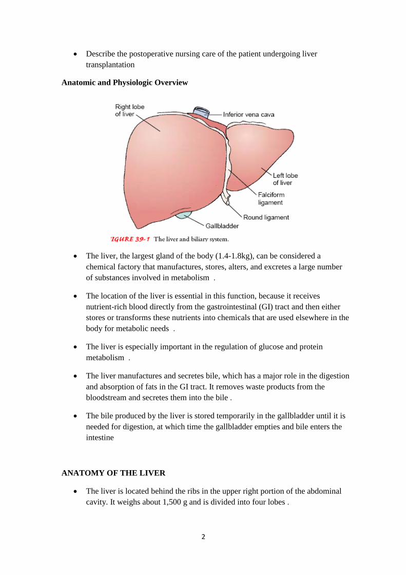
Anatomic and Physiologic Overview
The liver, the largest gland of the body (1.4-1.8kg), can be considered a
chemical factory that manufactures, stores, alters, and excretes a large number
of substances involved in metabolism .
The location of the liver is essential in this function, because it receives
nutrient-rich blood directly from the gastrointestinal (GI) tract and then either
stores or transforms these nutrients into chemicals that are used elsewhere in the
body for metabolic needs .
The liver is especially important in the regulation of glucose and protein
metabolism .
The liver manufactures and secretes bile, which has a major role in the digestion
and absorption of fats in the GI tract. It removes waste products from the
bloodstream and secretes them into the bile .
The bile produced by the liver is stored temporarily in the gallbladder until it is
needed for digestion, at which time the gallbladder empties and bile enters the
intestine
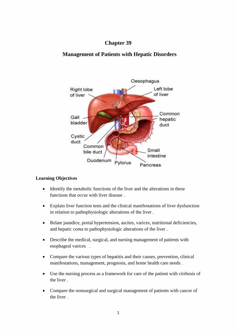
ANATOMY OF THE LIVER
The liver is located behind the ribs in the upper right portion of the abdominal
cavity. It weighs about 1,500 g and is divided into four lobes .

3
A thin layer of connective tissue surrounds each lobe, extending into the lobe
itself and dividing the liver mass into small units called lobules
The circulation of the blood into and out of the liver is of major importance in
its function. The blood that perfuses the liver comes from two sources.
Approximately 75% of the blood supply comes from the portal vein, which
drains the GI tract and is rich in nutrients. The remainder of the blood supply
enters by way of the hepatic artery and is rich in oxygen. Terminal branches of
these two blood supplies join to form common capillary beds, which constitute
the sinusoids of the liver
The sinusoids empty into a venule that occupies the center of each liver lobule
and is called the central vein. The central veins join to form the hepatic vein,
which constitutes the venous drainage from the liver and empties into the
inferior vena cava, close to the diaphragm. Thus, there are two sources of blood
flowing into the liver and only one exit pathway 10
•In addition to hepatocytes, phagocytic cells belonging to reticuloendothelial
system are present in the liver. Other organs that contain reticuloendothelial
cells are the spleen, bone marrow, lymph nodes, and lungs. In the liver, these
cells are called Kupffer cells. Their main function is to engulf particulate matter
(such as bacteria) that enters the liver through the portal blood .
The smallest bile ducts, called canaliculi, are located between the lobules of the
liver. The canaliculi receive secretions from the hepatocytes and carry them to
larger bile ducts, which eventually form the hepatic duct. The hepatic duct from
the liver and the cystic duct from the gallbladder join to form the common bile
duct, which empties into the small intestine. The sphincter of Oddi, located at
4
the junction where the common bile duct enters the duodenum, controls the
flow of bile into the intestine.
FUNCTIONS OF THE LIVER
1-Glucose Metabolism
After a meal, glucose is taken up from the portal venous blood by the liver and
converted into glycogen, which is stored in the hepatocytes .
Subsequently, the glycogen is converted back to glucose and released as needed
into the bloodstream to maintain normal levels of blood glucose .
Additional glucose can be synthesized by the liver through a process called
gluconeogenesis. For this process, the liver uses amino acids from protein
breakdown or lactate produced by exercising muscles .
2. Ammonia Conversion
Use of amino acids from protein for gluconeogenesis results in the formation of
ammonia as a byproduct. The liver converts this metabolically generated
ammonia into urea .
Ammonia produced by bacteria in the intestines is also removed from portal
blood for urea synthesis. In this way, the liver converts ammonia, a potential
toxin, into urea, a compound that can be excreted in the urine .
3. Protein Metabolism
The liver synthesizes almost all of the plasma proteins (except gamma
globulin), including albumin, alpha and beta globulins, blood clotting factors,
specific transport proteins, and most of the plasma lipoproteins .
Amino acids serve as the building blocks for protein synthesis .
Vitamin K is required by the liver for synthesis of prothrombin and some of the
other clotting factors .
4. Fat Metabolism
Fatty acids can be broken down for the production of energy and the production
of ketone bodies .
Ketone bodies are small compounds that can enter the bloodstream and provide
a source of energy for muscles and other tissues .
Breakdown of fatty acids into ketone bodies occurs primarily when the
availability of glucose for metabolism is limited, as during starvation or in
uncontrolled diabetes .
5
Fatty acids and their metabolic products are also used for the synthesis of
cholesterol, lecithin, lipoproteins, and other complex lipids .
5. Vitamin and Iron Storage
Vitamins A, B, and D and several of the B-complex vitamins are stored in large
amounts in the liver .
Iron and copper, are also stored in the liver .
Because the liver is rich in these substances, liver extracts have been used for
therapy for a wide range of nutritional disorders .
7. Drug Metabolism
The liver metabolizes many medications
Metabolism generally results in loss of activity of the medication, although in
some cases activation of the medication may occur.
One of the important pathways for medication metabolism involves conjugation
(binding) of the medication with a variety of compounds, such as glucuronic or
acetic acid, to form more soluble substances .
The conjugated products may be excreted in the feces or urine, similar to
bilirubin excretion .
8. Bile Formation
Bile is continuously formed by the hepatocytes and collected in the canaliculi
and bile ducts .
It is composed mainly of water and electrolytes such as sodium, potassium,
calcium, chloride, and bicarbonate, and significant amounts of lecithin, fatty
acids, cholesterol, bilirubin, and bile salts .
Bile is collected and stored in the gallbladder and is emptied into the intestine
when needed for digestion .
Bile also serves as an aid to digestion through the emulsification of fats by bile
salts .
Bile salts are synthesized by the hepatocytes from cholesterol. After conjugation
or binding with amino acids, they are excreted into the bile .
The bile salts, together with cholesterol and lecithin, are required for
emulsification of fats in the intestine, which is necessary for efficient digestion
and absorption .
6
Bile salts are then reabsorbed, primarily in the distal ileum, into portal blood for
return to the liver and are again excreted into the bile.
9. Bilirubin Excretion
Bilirubin is a pigment derived from the breakdown of hemoglobin by cells of
the reticuloendothelial system .
Hepatocytes remove bilirubin from the blood and modify it to be more soluble
in aqueous solutions .
The conjugated bilirubin is secreted by the hepatocytes into the adjacent bile
canaliculi and is eventually carried in the bile into the duodenum .
In the small intestine, bilirubin is converted into urobilinogen, which is in part
excreted in the feces and in part absorbed through the intestinal mucosa into the
portal blood .
Some of the urobilinogen enters the systemic circulation and is excreted by the
kidneys in the urine .
Gerontologic Considerations
The most common change in the liver in the elderly is a decrease in its size and
weight, accompanied by a decrease in total hepatic blood flow. Results of liver
function tests do not normally change in the elderly; abnormal results in an
elderly patient indicate abnormal liver function and are not the result of the
aging process itself .
The immune system is altered in the aged, and a less responsive immune system
may be responsible for the increased incidence and severity of hepatitis B in the
elderly and the increased incidence of liver abscesses secondary to decreased
phagocytosis by the Kupffer cells .
Age-Related Changes of the Hepatobiliary System
Steady decrease in size and weight of the liver, particularly in women .
Decrease in blood flow .
Decrease in replacement/repair of liver cells after injury .
Reduced drug metabolism .
Rapid progression of hepatitis C infection and lower response rate to therapy .
Decline in drug clearance capability .
7
Increased prevalence of gallstones .
Decreased gallbladder contraction after a meal .
More severe complications of biliary tract disease.
ASSESSMENT HEALTH HISTORY
If liver function test results are abnormal, the patient may need to be evaluated
for liver disease. So look if the client :
• Was exposed to hepatotoxic substances or infectious agents .
• Patient‘s occupational, recreational, and travel nhistory may assist in
identifying exposure to hepatotoxins
• Patient‘s history of alcohol and drug use
• Lifestyle behaviors (Injectable drug use, sexual practices )
• Current and past medical conditions, previous blood transfusion .
PHYSICAL EXAMINATION
Assess the patient for pallor, jaundice (skin, mucosa, and sclerae), and the
extremities are assessed for muscle atrophy, edema, and skin excoriation
secondary to scratching .
Observe the skin for petechiae or ecchymotic areas (bruises), spider angiomas,
and palmar erythema .
Assess male patient for unilateral or bilateral gynecomastia and testicular
atrophy due to endocrine changes .
Asses patient‘s cognitive status (recall, memory, abstract thinking) and
neurologic status are assessed .
Palpate abdomen to assess liver size and to detect any tenderness over the liver.
A palpable liver presents as a firm, sharp edge with a smooth surface
Tenderness of the liver implies recent acute enlargement with consequent
stretching of the liver capsule .
Enlargement of the liver is an abnormal finding requiring evaluation .
8
Diagnostic Evaluation
Liver Function Tests
More than 70% of the parenchyma of the liver may be damaged before liver
function test results become abnormal .
1. Serum enzyme activity (ie, alkaline phosphatase, lactic dehydrogenase, serum
aminotransferases )
2. Serum concentrations of proteins (albumin and globulins ,)
3. Bilirubin, ammonia, clotting factors, and lipids .
4. Serum aminotransferases (also called transaminases) are sensitive indicators of
injury to the liver cells and are useful in detecting acute liver disease such as
hepatitis .
A. Alanine aminotransferase (ALT) (formerly called serum glutamic-pyruvic
transaminase [SGPT]) (10-40 U/L )
B. Aspartate aminotransferase (AST) (formerly called serum glutamic-
oxaloacetic transaminase [SGOT]) (5-35 U/L )
C. Gamma glutamyl transferase (GGT) (also called G-glutamyl transpeptidase)
(10-48 U/L )
D. .Lactic Dehydirgenase (LDH) (100-200 U/L).
These studies measure the ability of the liver to conjugate and excrete bilirubin. Results
are abnormal in liver and biliary tract disease and are associated with jaundice
clinically.
9
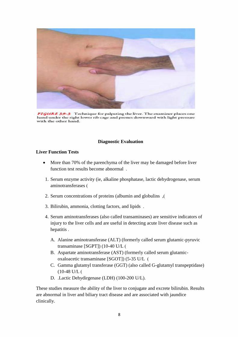
Pigment Studies Normal
1. Serum bilirubin, direct 1. 0–0.3 mg/dL (0–5.1 µmol/L)
2. Serum bilirubin,total 2. 0–0.9 mg/dL (1.7–20.5 µmol/L)
3. Urine bilirubin 3. 0(0)
4. Urine urobilinogen 4. 0.05–2.5 mg/24 h (0.09–4.23
µmol/24 h)
5. Fecal urobilinogen (infrequently used) 5. 40–200 mg/24 h (0.068–0.34
mmol/24 h)
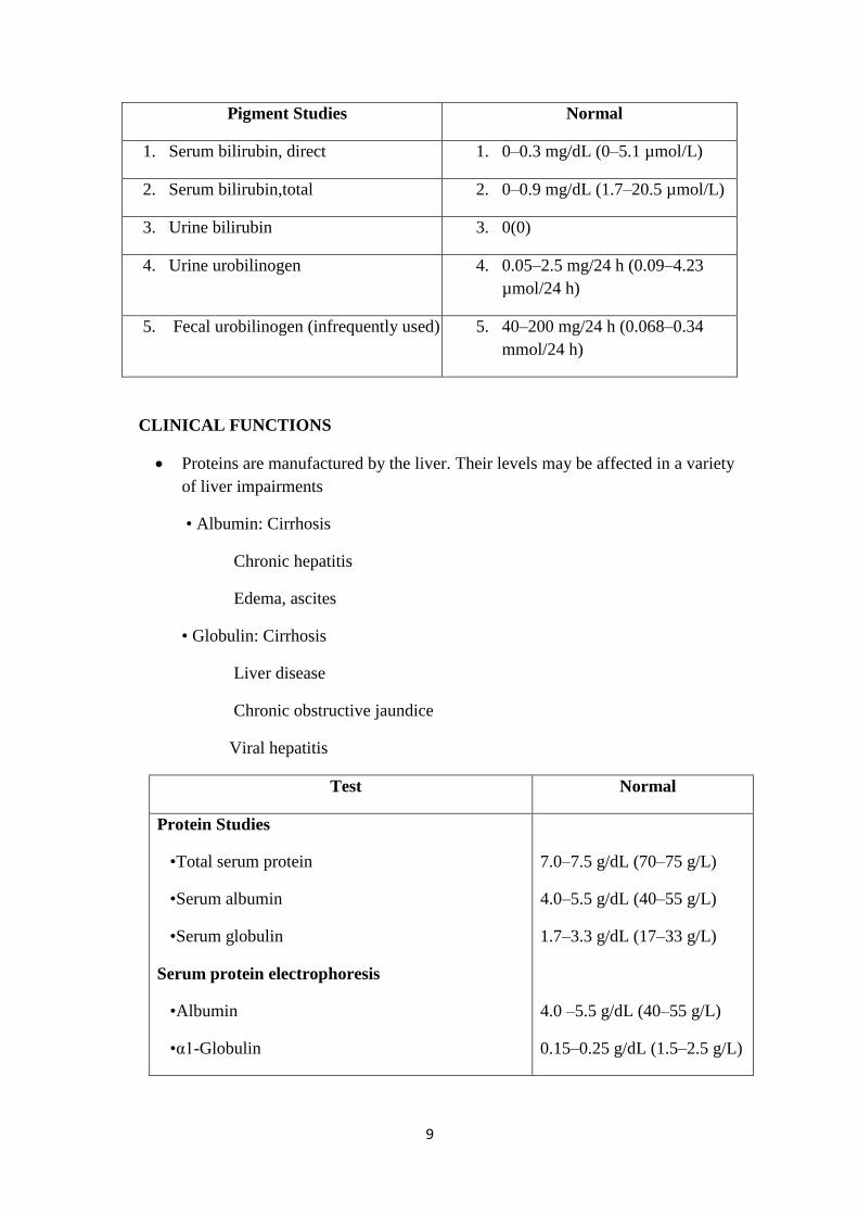
CLINICAL FUNCTIONS
Proteins are manufactured by the liver. Their levels may be affected in a variety
of liver impairments
• Albumin: Cirrhosis
Chronic hepatitis
Edema, ascites
• Globulin: Cirrhosis
Liver disease
Chronic obstructive jaundice
Viral hepatitis
Test Normal
Protein Studies
•Total serum protein
•Serum albumin
•Serum globulin
Serum protein electrophoresis
•Albumin
•α1-Globulin
7.0–7.5 g/dL (70–75 g/L)
4.0–5.5 g/dL (40–55 g/L)
1.7–3.3 g/dL (17–33 g/L)
4.0 –5.5 g/dL (40–55 g/L)
0.15–0.25 g/dL (1.5–2.5 g/L)
10
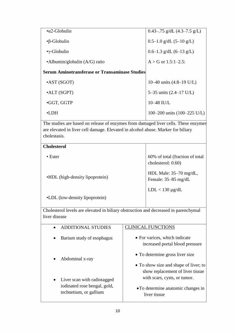
•α2-Globulin
•β-Globulin
•γ-Globulin
•Albumin/globulin (A/G) ratio
Serum Aminotransferase or Transaminase Studies
•AST (SGOT)
•ALT (SGPT)
•GGT, GGTP
•LDH
0.43–.75 g/dL (4.3–7.5 g/L)
0.5–1.0 g/dL (5–10 g/L)
0.6–1.3 g/dL (6–13 g/L)
A > G or 1.5:1–2.5:
10–40 units (4.8–19 U/L)
5–35 units (2.4–17 U/L)
10–48 IU/L
100–200 units (100–225 U/L)
The studies are based on release of enzymes from damaged liver cells. These enzymes
are elevated in liver cell damage. Elevated in alcohol abuse. Marker for biliary
cholestasis.
Cholesterol
• Ester
•HDL (high-density lipoprotein)
•LDL (low-density lipoprotein)
60% of total (fraction of total
cholesterol: 0.60)
HDL Male: 35–70 mg/dL,
Female: 35–85 mg/dL
LDL < 130 µg/dL
Cholesterol levels are elevated in biliary obstruction and decreased in parenchymal
liver disease
ADDITIONAL STUDIES
Barium study of esophagus
Abdominal x-ray
Liver scan with radiotagged
iodinated rose bengal, gold,
technetium, or gallium
CLINICAL FUNCTIONS
For varices, which indicate
increased portal blood pressure
To determine gross liver size
To show size and shape of liver; to
show replacement of liver tissue
with scars, cysts, or tumor.
To determine anatomic changes in
liver tissue

11
Liver biopsy (percutaneous or
transjugular)
Ultrasonography
Computed tomography (CT scan)
To show size of abdominal organs
and presence of masses
To detect hepatic neoplasms;
diagnose cysts, abscesses, and
hematomas; and distinguish
between obstructive and
nonobstructive jaundice. Detects
cerebral atrophy in hepatic
encephalopathy.
LIVER BIOPSY
Liver biopsy is the removal of a small amount of liver tissue, usually through
needle aspiration .
It permits examination of liver cells .
Liver biopsy is especially useful when clinical findings and laboratory tests are
not diagnostic .
Bleeding and bile peritonitis after liver biopsy are the major complications.

12
NURSING ACTIVITIES RATIONALE
Ascertain that results of coagulation
tests (prothrombin time, partial
thromboplastin time, and platelet
count) are available
a) and that compatible donor blood is
available.
Check for signed consent; confirm that
informed consent has been provided.
Measure and record the patient‘s pulse,
respirations, and blood pressure
immediately before biopsy.
Describe to the patient in advance:
steps of the procedure;
Support the patient during the
procedure.
Expose the right side of the patient‘s
upper abdomen (right hypochondriac).
Instruct the patient to inhale and exhale
deeply several times, finally to exhale,
and to hold breath at the end of
expiration. The physician promptl
introduces the biopsy needle by way of
the transthoracic (intercostal)
transabdominal (subcostal) route,
penetrates the liver, aspirates, and
withdraws.
Many patients with liver disease have
clotting defects and are at risk for
bleeding.
2. Procedure should be done with
agreement of patient
Pre-biopsy values provide a basis on
which to compare the patient‘s vital
signs and evaluate status after the
procedure.
Explanations allay fears and ensure
cooperation.
Encouragement and support of the
nurse enhance comfort and promote a
sense of security.
The skin at the site of penetration will
be cleansed and a local anesthetic will
be infiltrated.
Holding the breath immobilizes the
chest wall and the diaphragm;
penetration of the diaphragm thereby is
avoided, and the risk of lacerating the
liver is minimized.
Post-procedure
13
Immediately after the biopsy, assist the patient to turn onto the right side; place a
pillow under the costal margin, and caution the patient to remain in this position
and immobile, for several hours. Instruct the patient to avoid coughing or straining .
Measure and record the patient‘s vital sings at a15-minute intervals for the first
hour, then every 30 minutes for the next 1 to 2 hours or until the patient‘s condition
stabilizes .
Complications
–Pneumothorax
–Peritonitis
–Hemorrhage
OTHER DIAGNOSTIC TESTS
Ultrasonography, computed tomography (CT), and magnetic resonance imaging
(MRI) are used to identify normal structures and abnormalities of the liver and
biliary tree .
A radioisotope liver scan may be performed to assess liver size and hepatic blood
flow and obstruction .
Laparoscopy (insertion of a fiber-optic endoscope through a small abdominal
incision) is used to examine the liver and other pelvic structures .
Hepatic Dysfunction
Results from damage to the liver‘s parenchymal cells, either directly from primary
liver diseases or indirectly from obstruction of bile flow .
Liver dysfunction may be acute or chronic; chronic dysfunction is far more
common than acute .
The most common cause of parenchymal damage is malnutrition, especially that
related to alcoholism. The parenchymal cells respond to most noxious agents by
replacing glycogen with lipids, producing fatty infiltration with or without cell
death or necrosis .
This is commonly associated with inflammatory cell infiltration and growth of
fibrous tissue .
Among the most common and significant symptoms of liver disease are the
following :
1. Jaundice, resulting from increased bilirubin concentration in the blood
14
2. Portal hypertension, ascites, and varices, resulting from circulatory changes within
the diseased liver and producing severe GI hemorrhages and marked sodium and
fluid retention
3. Nutritional deficiencies, which result from the inability of the damaged liver cells
to metabolize certain vitamins ;
4. Hepatic encephalopathy or coma, reflecting accumulation of ammonia in the
serum due to impaired protein metabolism by the diseased liver
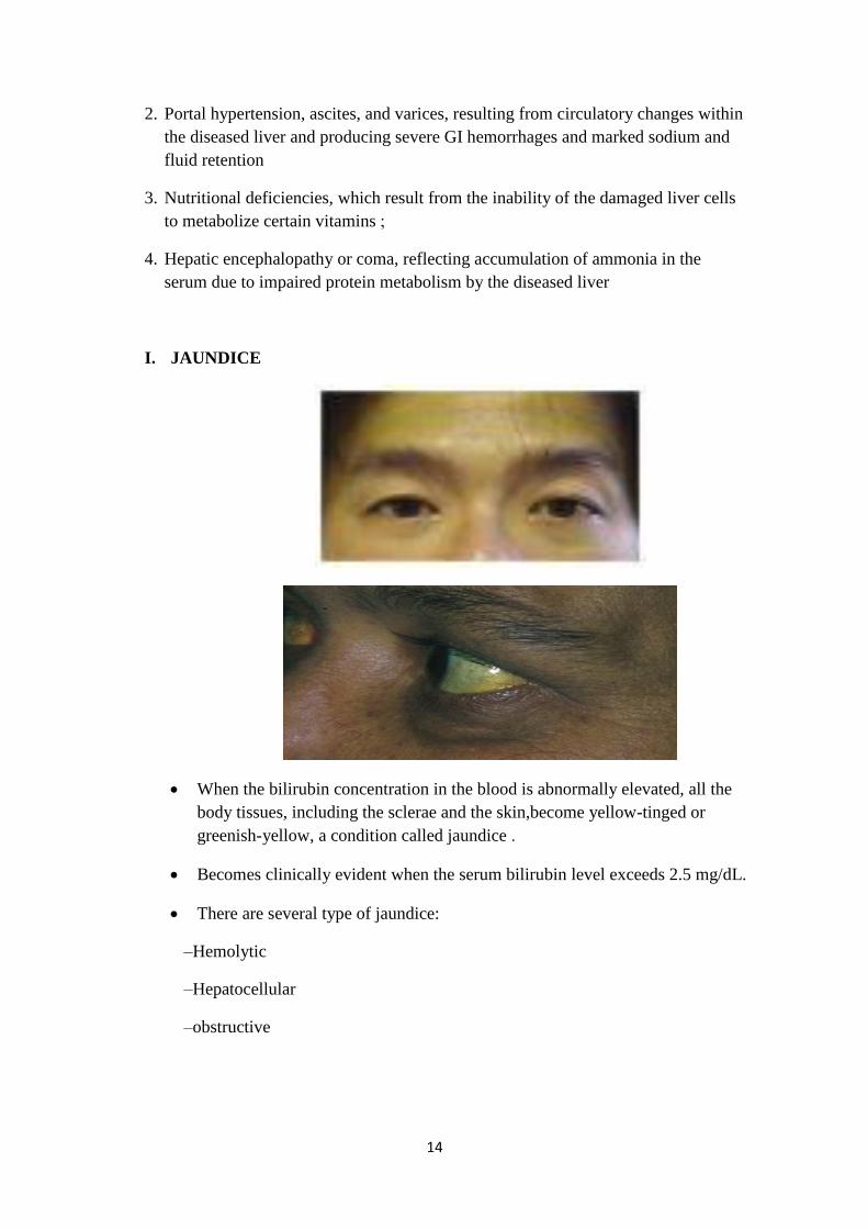
I. JAUNDICE
When the bilirubin concentration in the blood is abnormally elevated, all the
body tissues, including the sclerae and the skin,become yellow-tinged or
greenish-yellow, a condition called jaundice .
Becomes clinically evident when the serum bilirubin level exceeds 2.5 mg/dL.
There are several type of jaundice:
–Hemolytic
–Hepatocellular
–obstructive

15
1. Hemolytic Jaundice
Results from increased destruction of the red blood cells, too much bilirubin
reaches the liver, although functioning normally, cannot excrete the bilirubin as
quickly as it is formed .
Occurs with patients with hemolytic transfusion reactions and other hemolytic
disorders .
Prolonged jaundice, however, even if mild, predisposes to the formation of
pigment stones in the gallbladder, and extremely severe jaundice (levels of free
bilirubin exceeding 20 to 25 mg/dL) poses a risk for brain stem damage .
2. Hepatocellular Jaundice
•caused by the inability of damaged liver cells to clear normal amounts of
bilirubin from the blood. The cellular damage may be from infection, such as in
viral hepatitis or other viruses that affect the liver (eg, yellow fever virus,
Epstein-Barr virus), from medication or chemical toxicity (eg, carbon
tetrachloride, chloroform, phosphorus, certain medications), or from alcohol .
•Cirrhosis of the liver is a form of hepatocellular disease that may produce
jaundice. It is usually associated with excessive alcohol
3. Obstructive Jaundice
Caused by occlusion of the bile duct by a gallstone, an inflammatory process, a
tumor, or pressure from an enlarged organ .
The obstruction may also involve the small bile ducts within the liver (ie,
intrahepatic obstruction), caused, for example, by pressure on these channels
from inflammatory swelling of the liver or by an inflammatory exudate within
the ducts themselves. Intrahepatic obstruction resulting from stasis and
inspissation (thickening) of bile within the canaliculi may occur after the
ingestion of certain medications ,
These include phenothiazines, antithyroid medications, sulfonylureas, tricyclic
antidepressant agents, nitrofurantoin, androgens, and estrogens .
Whether the obstruction is intrahepatic or extrahepatic, and whatever its cause
may be, bile cannot flow normally into the intestine but is backed up into the
liver substance. It is then reabsorbed into the blood and carried throughout the
entire body, staining the skin, mucous membranes, and sclerae. It is excreted in
the urine, which becomes deep orange and foamy .
Because of the decreased amount of bile in the intestinal tract, the stools
become light or clay-colored. The skin may itch intensely, requiring repeated
soothing baths. Dyspepsia and intolerance to fatty foods may develop because
16
of impaired fat digestion in the absence of intestinal bile. AST, ALT, and GGT
levels generally rise only moderately, but bilirubin and alkaline phosphatase
levels are elevated .
4. Hereditary Hyperbilirubinemia
Results from several inherited disorders can also produce jaundice. Gilbert‘s
syndrome is a familial disorder characterized by an increased level of
unconjugated bilirubin that causes jaundice .
Other conditions that are probably caused by inborn errors of biliary
metabolism include Dubin–Johnson syndrome (chronic idiopathic jaundice,
with pigment in the liver) and Rotor‘s syndrome (chronic familial conjugated
hyperbilirubinemia without pigment in the liver.
II. PORTAL HYPERTENSION
Obstructed blood flow through the damaged liver results in increased blood
pressure (portal hypertension) throughout the portal venous system .
It is commonly associated with hepatic cirrhosis, but can also occur with
noncirrhotic liver disease .
Portal hypertnesion leads to :
–Splenomegaly (enlarged spleen )
–Ascites
–Varices .
III. ASCITES - Pathophysiology
Caused by portal hypertension and the resulting increase in capillary pressure
and obstruction of venous blood flow through the damaged liver .
The failure of the liver to metabolize aldosterone increases sodium and water
retention by the kidney. Sodium and water retention, increased intravascular
fluid volume, and decreased synthesis of albumin by the damaged liver all
contribute to fluid moving from the vascular system into the peritoneal space
Loss of fluid into the peritoneal space causes further sodium and water retention
by the kidney in an effort to maintain the vascular fluid volume, and the process
becomes self-perpetuating .
As a result of liver damage, large amounts of albumin-rich fluid, 15 L or more,
may accumulate in the peritoneal cavity as ascites .

17
With the movement of albumin from the serum to the peritoneal cavity, the
osmotic pressure of the serum decreases.
This, combined with increased portal pressure, results in movement of fluid into
the peritoneal cavity.
Clinical Manifestations
Increased abdominal girth and rapid weight gain are common presenting
symptoms of ascites .
The patient may be short of breath and uncomfortable from the enlarged
abdomen, and
Striae and distended veins may be visible over the abdominal wall .
Fluid and electrolyte imbalances are common .

18
Assessment and Diagnostic Evaluation
The presence and extent of ascites are assessed by percussion of the abdomen.
When fluid has accumulated in the peritoneal cavity, the flanks bulge when the
patient assumes a supine position. The presence of fluid can be confirmed either
by percussing for shifting dullness or by detecting a fluid wave. Daily
measurement and recording of abdominal girth and body weight are essential to
assess the progression of ascites and its response to treatment.
Medical Management A) DIETARY MODIFICATION
The goal of treatment for the patient with ascites is a negative sodium balance
to reduce fluid retention. Table salt, salty foods, salted butter and margarine,
and all ordinary canned and frozen foods should be avoided .
In the meantime, the taste of unsalted foods can be improved by using salt
substitutes such as lemon juice, oregano, and thyme .
B) DIURETICS
Use of diuretics along with sodium restriction is successful in 90% of patients
with ascites. Spironolactone (Aldactone), an aldosterone blocking agent, is most
19
often the first-line therapy. When used with other diuretics, it helps prevent
potassium loss .
Daily weight loss should not exceed 1 to 2 kg .
Possible complications of diuretic therapy include fluid and electrolyte
disturbances (including hypovolemia, hypokalemia, hyponatremia .)
C) BED REST
•In patients with ascites, an upright posture is associated with activation of the
renin-angiotensin-aldosterone system and sympathetic nervous system This
results in reduced renal glomerular filtration and sodium excretion and a
decreased response to loop diuretics. Bed rest may be a useful therapy,
especially for patients whose condition is refractory to diuretics .
D) PARACENTESIS
Paracentesis is the removal of fluid (ascites) from the peritoneal cavity through
a small surgical incision or puncture made through the abdominal wall under
sterile conditions. Ultrasound guidance may be indicated in some patients at
high risk for bleeding
Use of large-volume (5 to 6 liters) paracentesis has been shown to be a safe
method for treating patients with severe ascites. This technique, in combination
with the intravenous infusion of saltpoor albumin or other colloid, salt-poor
albumin helps reduce edema by causing the ascitic fluid to be drawn back into
the bloodstream and ultimately excretedd by the kidneys .
Preprocedure
1. Prepare the pt by providing the information and instructions about the procedure
2. Instruct the patient to void .
3. Gather appropriate sterile equipment
4. Place patient in upright position on edge of bed with feet supported on stool, or
place in chair. Fowler‘s position should be used for the patient confined to bed .
5. Monitoring of blood pressure during the procedure
6. The physician, using aseptic technique, inserts the trocar through a puncture
wound below the umbilicus. The fluid drains from the abdomen through a
drainage tube into a container .
7. Help the patient maintain position throughout procedure .

20
8. Measure and record blood pressure frequently .
9. Monitor the patient closely for signs of vascular collapse: pallor, increased
pulse rate, or decreased blood pressure .
.
Post-procedure
1. Return patient to bed or to a comfortable sitting position .
2. Measure, describe, and record the fluid collected .
3. Label samples of fluid and send to laboratory .
4. Continue to monitor vital signs every 15 minutes for 1 hour ,every 30 minutes
over 2 hours, then every hour over 2 hours and then every 4 hours. Monitor
temperature after procedure and every 4 hours .
5. Assess for hypovolemia, electrolyte loss, changes in mental status, and
encephalopathy .
6. Check puncture site when taking vital signs for bleeding and leakage .
7. Provide patient education
21
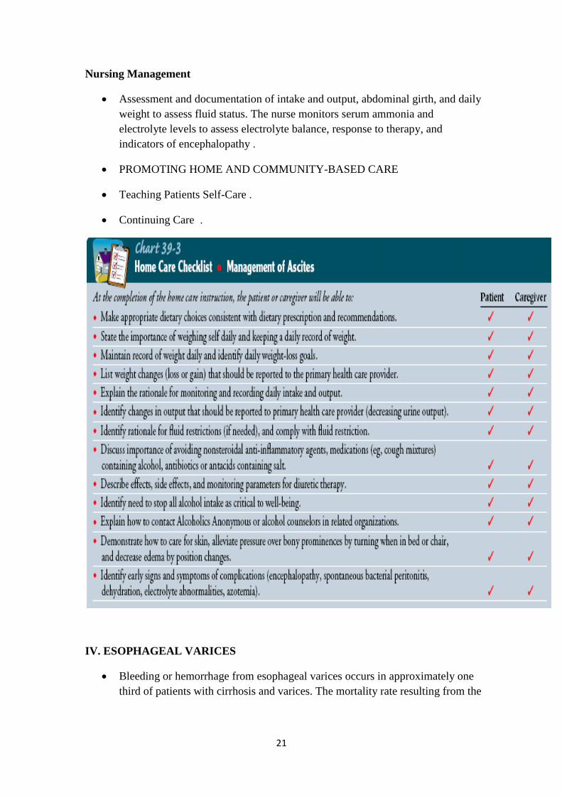
Nursing Management
Assessment and documentation of intake and output, abdominal girth, and daily
weight to assess fluid status. The nurse monitors serum ammonia and
electrolyte levels to assess electrolyte balance, response to therapy, and
indicators of encephalopathy .
PROMOTING HOME AND COMMUNITY-BASED CARE
Teaching Patients Self-Care .
Continuing Care .
IV. ESOPHAGEAL VARICES
Bleeding or hemorrhage from esophageal varices occurs in approximately one
third of patients with cirrhosis and varices. The mortality rate resulting from the
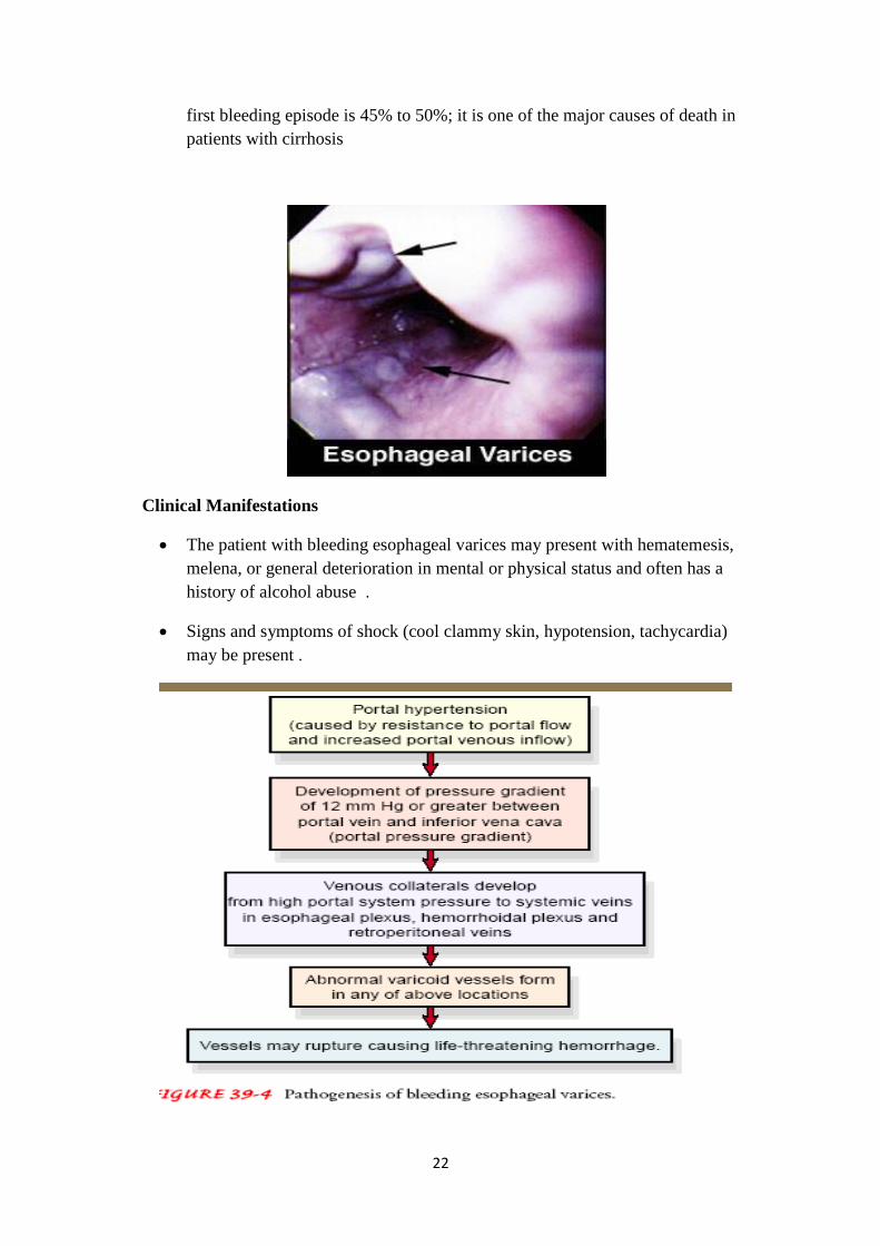
22
first bleeding episode is 45% to 50%; it is one of the major causes of death in
patients with cirrhosis
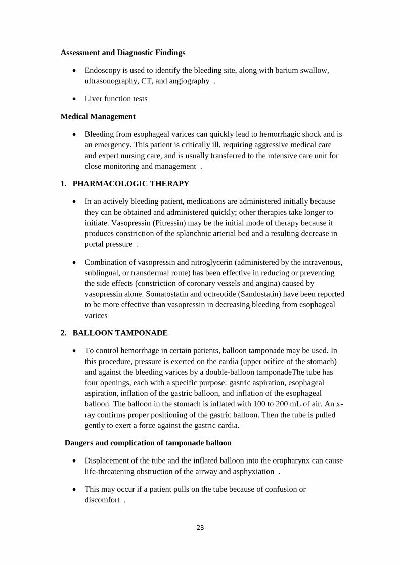
Clinical Manifestations
The patient with bleeding esophageal varices may present with hematemesis,
melena, or general deterioration in mental or physical status and often has a
history of alcohol abuse .
Signs and symptoms of shock (cool clammy skin, hypotension, tachycardia)
may be present .
71
23
Assessment and Diagnostic Findings
Endoscopy is used to identify the bleeding site, along with barium swallow,
ultrasonography, CT, and angiography .
Liver function tests
Medical Management
Bleeding from esophageal varices can quickly lead to hemorrhagic shock and is
an emergency. This patient is critically ill, requiring aggressive medical care
and expert nursing care, and is usually transferred to the intensive care unit for
close monitoring and management .
1. PHARMACOLOGIC THERAPY
In an actively bleeding patient, medications are administered initially because
they can be obtained and administered quickly; other therapies take longer to
initiate. Vasopressin (Pitressin) may be the initial mode of therapy because it
produces constriction of the splanchnic arterial bed and a resulting decrease in
portal pressure .
Combination of vasopressin and nitroglycerin (administered by the intravenous,
sublingual, or transdermal route) has been effective in reducing or preventing
the side effects (constriction of coronary vessels and angina) caused by
vasopressin alone. Somatostatin and octreotide (Sandostatin) have been reported
to be more effective than vasopressin in decreasing bleeding from esophageal
varices
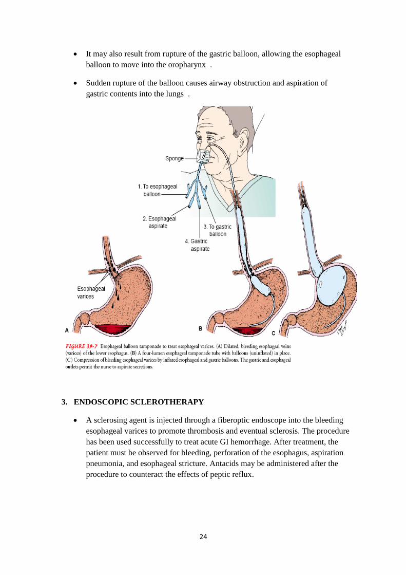
2. BALLOON TAMPONADE
To control hemorrhage in certain patients, balloon tamponade may be used. In
this procedure, pressure is exerted on the cardia (upper orifice of the stomach)
and against the bleeding varices by a double-balloon tamponadeThe tube has
four openings, each with a specific purpose: gastric aspiration, esophageal
aspiration, inflation of the gastric balloon, and inflation of the esophageal
balloon. The balloon in the stomach is inflated with 100 to 200 mL of air. An x-
ray confirms proper positioning of the gastric balloon. Then the tube is pulled
gently to exert a force against the gastric cardia.
Dangers and complication of tamponade balloon
Displacement of the tube and the inflated balloon into the oropharynx can cause
life-threatening obstruction of the airway and asphyxiation .
This may occur if a patient pulls on the tube because of confusion or
discomfort .
24
It may also result from rupture of the gastric balloon, allowing the esophageal
balloon to move into the oropharynx .
Sudden rupture of the balloon causes airway obstruction and aspiration of
gastric contents into the lungs .
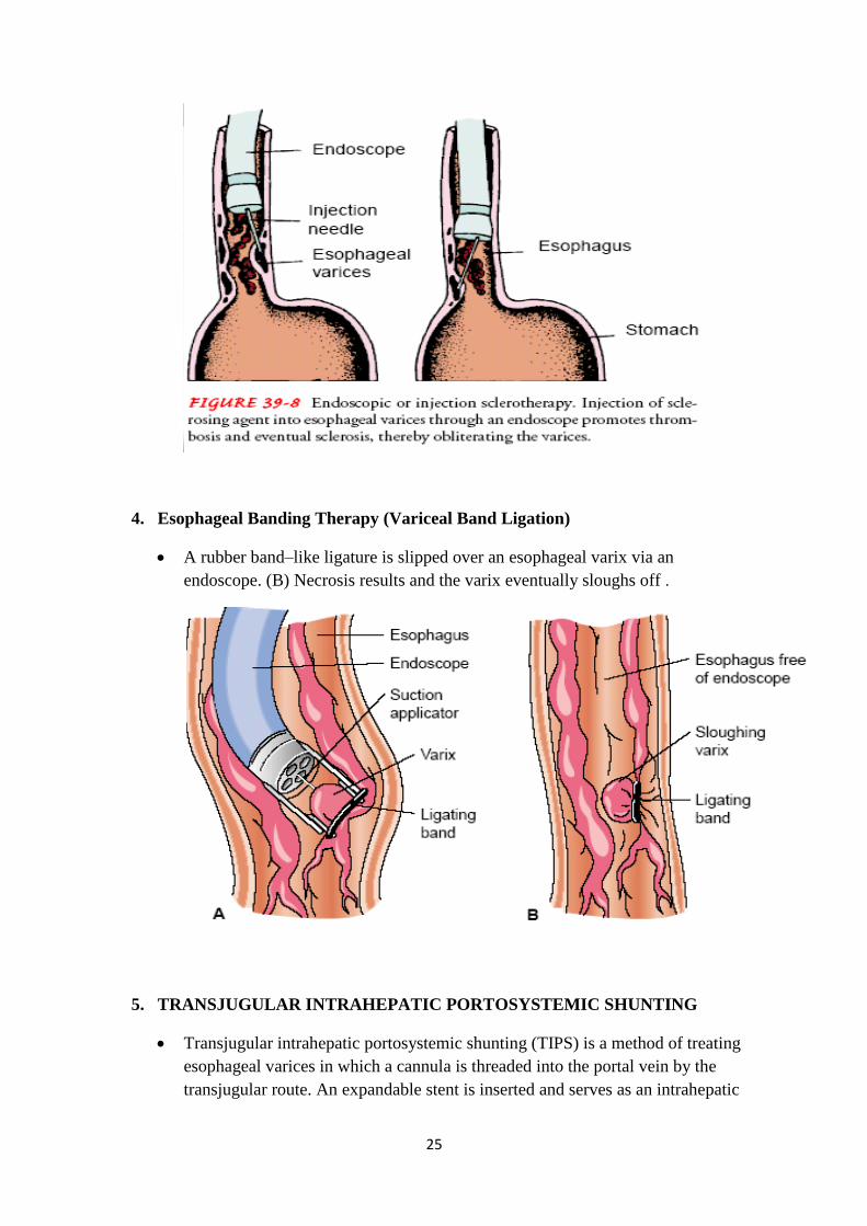
3. ENDOSCOPIC SCLEROTHERAPY
A sclerosing agent is injected through a fiberoptic endoscope into the bleeding
esophageal varices to promote thrombosis and eventual sclerosis. The procedure
has been used successfully to treat acute GI hemorrhage. After treatment, the
patient must be observed for bleeding, perforation of the esophagus, aspiration
pneumonia, and esophageal stricture. Antacids may be administered after the
procedure to counteract the effects of peptic reflux.
25
79
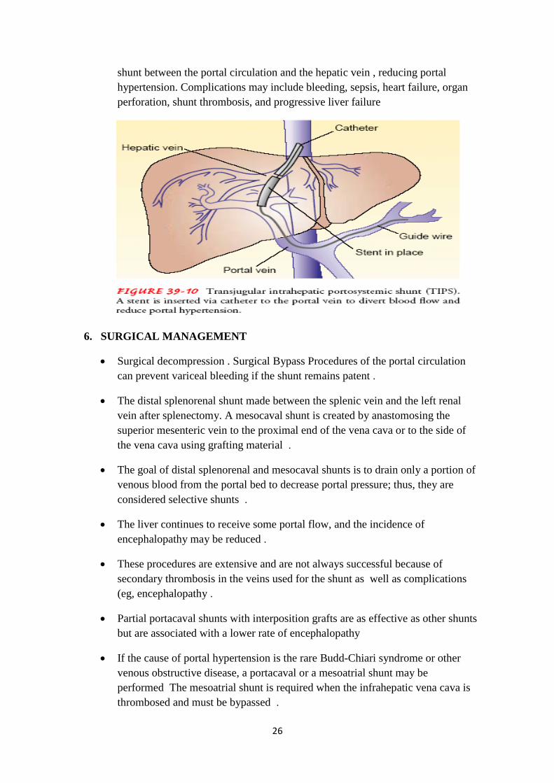
4. Esophageal Banding Therapy (Variceal Band Ligation)
A rubber band–like ligature is slipped over an esophageal varix via an
endoscope. (B) Necrosis results and the varix eventually sloughs off .
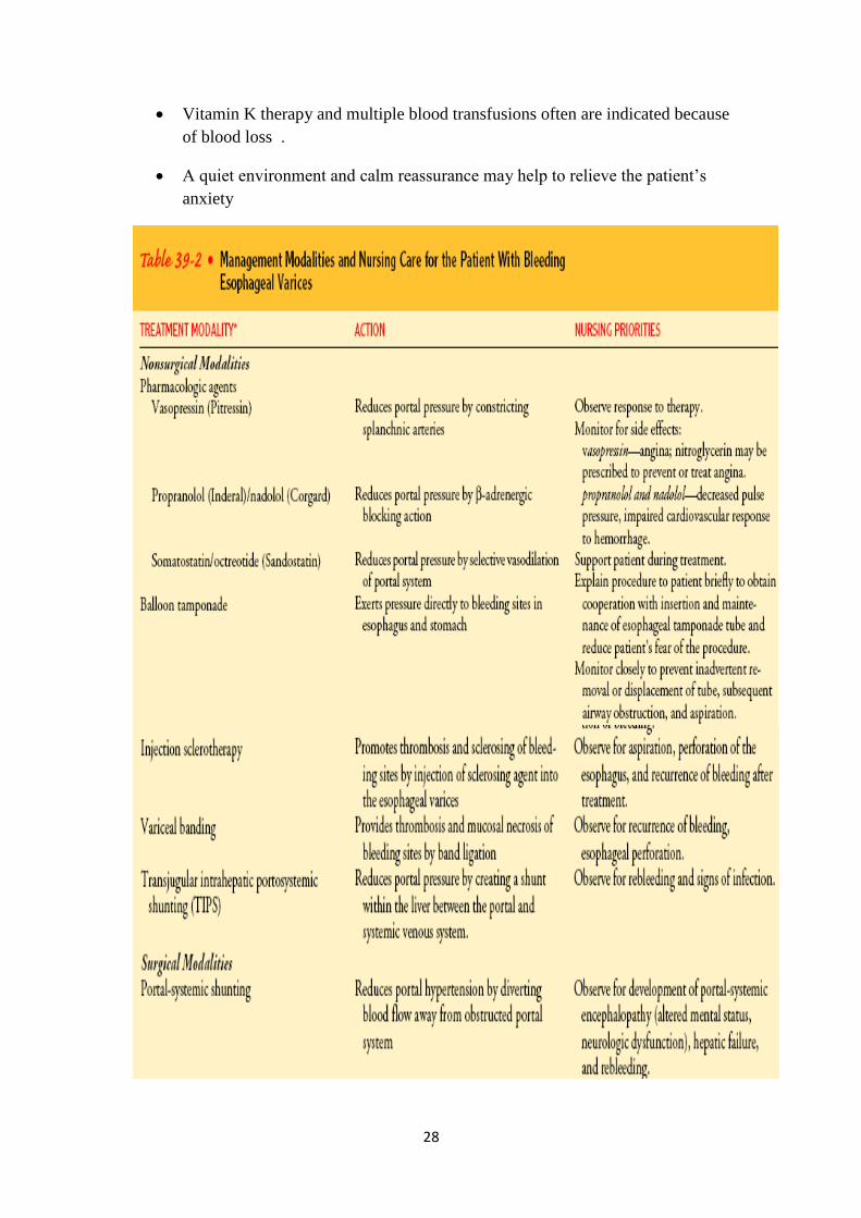
5. TRANSJUGULAR INTRAHEPATIC PORTOSYSTEMIC SHUNTING
Transjugular intrahepatic portosystemic shunting (TIPS) is a method of treating
esophageal varices in which a cannula is threaded into the portal vein by the
transjugular route. An expandable stent is inserted and serves as an intrahepatic
26
shunt between the portal circulation and the hepatic vein , reducing portal
hypertension. Complications may include bleeding, sepsis, heart failure, organ
perforation, shunt thrombosis, and progressive liver failure
6. SURGICAL MANAGEMENT
Surgical decompression . Surgical Bypass Procedures of the portal circulation
can prevent variceal bleeding if the shunt remains patent .
The distal splenorenal shunt made between the splenic vein and the left renal
vein after splenectomy. A mesocaval shunt is created by anastomosing the
superior mesenteric vein to the proximal end of the vena cava or to the side of
the vena cava using grafting material .
The goal of distal splenorenal and mesocaval shunts is to drain only a portion of
venous blood from the portal bed to decrease portal pressure; thus, they are
considered selective shunts .
The liver continues to receive some portal flow, and the incidence of
encephalopathy may be reduced .
These procedures are extensive and are not always successful because of
secondary thrombosis in the veins used for the shunt as well as complications
(eg, encephalopathy .
Partial portacaval shunts with interposition grafts are as effective as other shunts
but are associated with a lower rate of encephalopathy
If the cause of portal hypertension is the rare Budd-Chiari syndrome or other
venous obstructive disease, a portacaval or a mesoatrial shunt may be
performed The mesoatrial shunt is required when the infrahepatic vena cava is
thrombosed and must be bypassed .

27
Devascularization and Transection
Devascularization and staplegun transection procedures to separate the bleeding
site from the high-pressure portal system have been used in the emergency
management of variceal bleeding. The lower end of the esophagus is reached
through a small gastrostomy incision; a staple gun permits anastomosis of the
transected ends of the esophagus. Rebleeding is a risk, and the outcomes of
these procedures vary among patient populations .
Nursing Management
Monitoring the patient‘s physical condition and evaluating emotional responses
and cognitive status .
Monitor and record vital signs
Assess the patient‘s nutritional and neurologic status .
This assessment will assist in identifying hepatic encephalopathy resulting from
the breakdown of blood in the GI tract and a rising serum ammonia level.
Manifestations range from drowsiness to encephalopathy and coma .
Complete rest of the esophagus may be indicated with bleeding, so parenteral
nutrition is initiated. Gastric suction usually
28
Vitamin K therapy and multiple blood transfusions often are indicated because
of blood loss .
A quiet environment and calm reassurance may help to relieve the patient‘s
anxiety
29
Hepatic Encephalopathy and Coma
Is a life-threatening complication of liver disease, occurs with profound liver
failure and may result from the accumulation of ammonia and other toxic
metabolites in the blood.
Represents the most advanced stage of hepatic encephalopathy.
Pathophysiology
Ammonia accumulates because damaged liver cells fail to detoxify and convert
the ammonia that is constantly entering the bloodstream to urea.
Ammonia enters the bloodstream as a result of its absorption from the GI tract
and its liberation from kidney and muscle cells.
The increased ammonia concentration in the blood causes brain dysfunction and
damage, resulting in hepatic encephalopathy.
The largest source of ammonia is the enzymatic and bacterial digestion of
dietary and blood proteins in the GI tract. Ammonia from these sources is
increased as a result of GI bleeding (ie, bleeding esophageal varices or chronic
GI bleeding), a high-protein diet, bacterial infections, and uremia. The ingestion
of ammonium salts also increases the blood ammonia level.
Conversely, serum ammonia is decreased by elimination of protein from the
diet and by the administration of antibiotic agents, such as neomycin sulfate,
that reduce the number of intestinal bacteria capable of converting urea to
ammonia
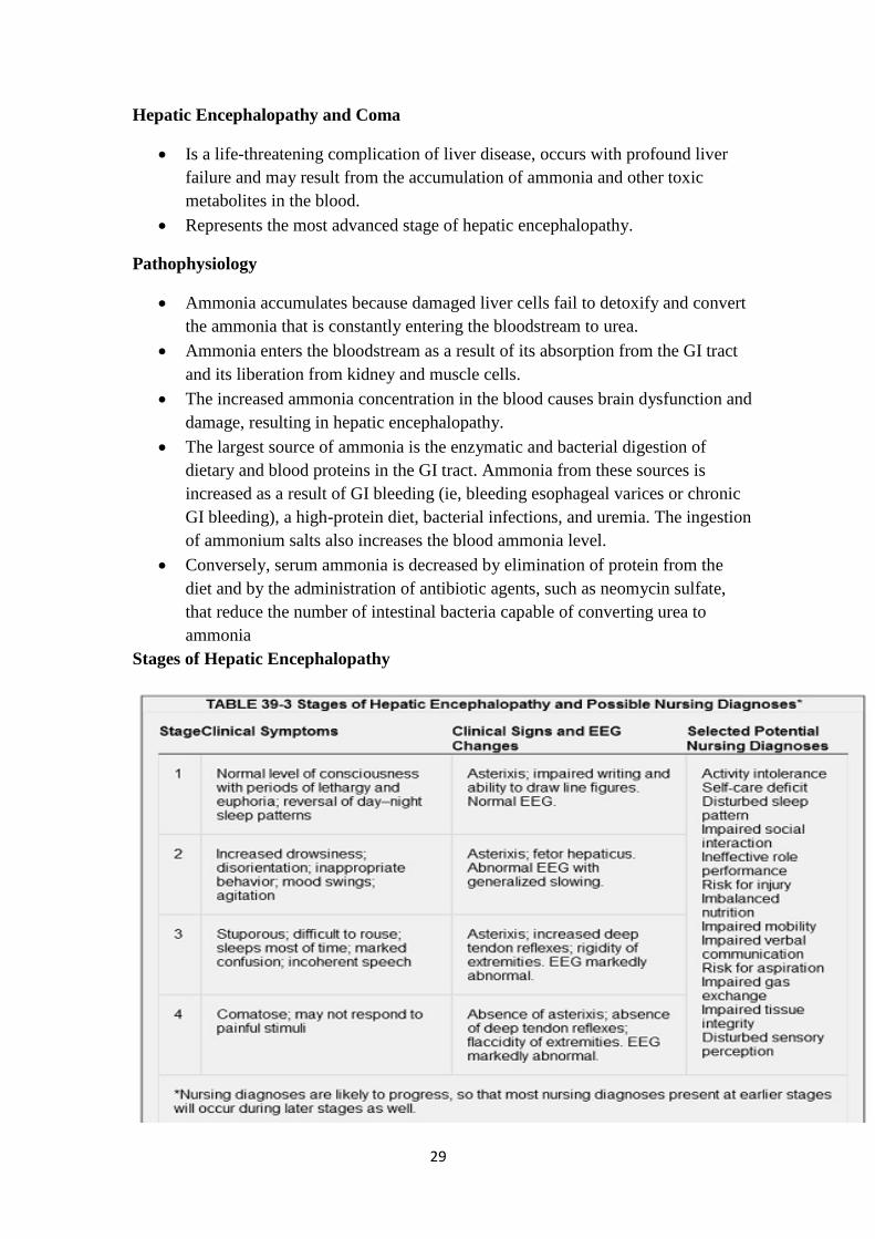
Stages of Hepatic Encephalopathy
30
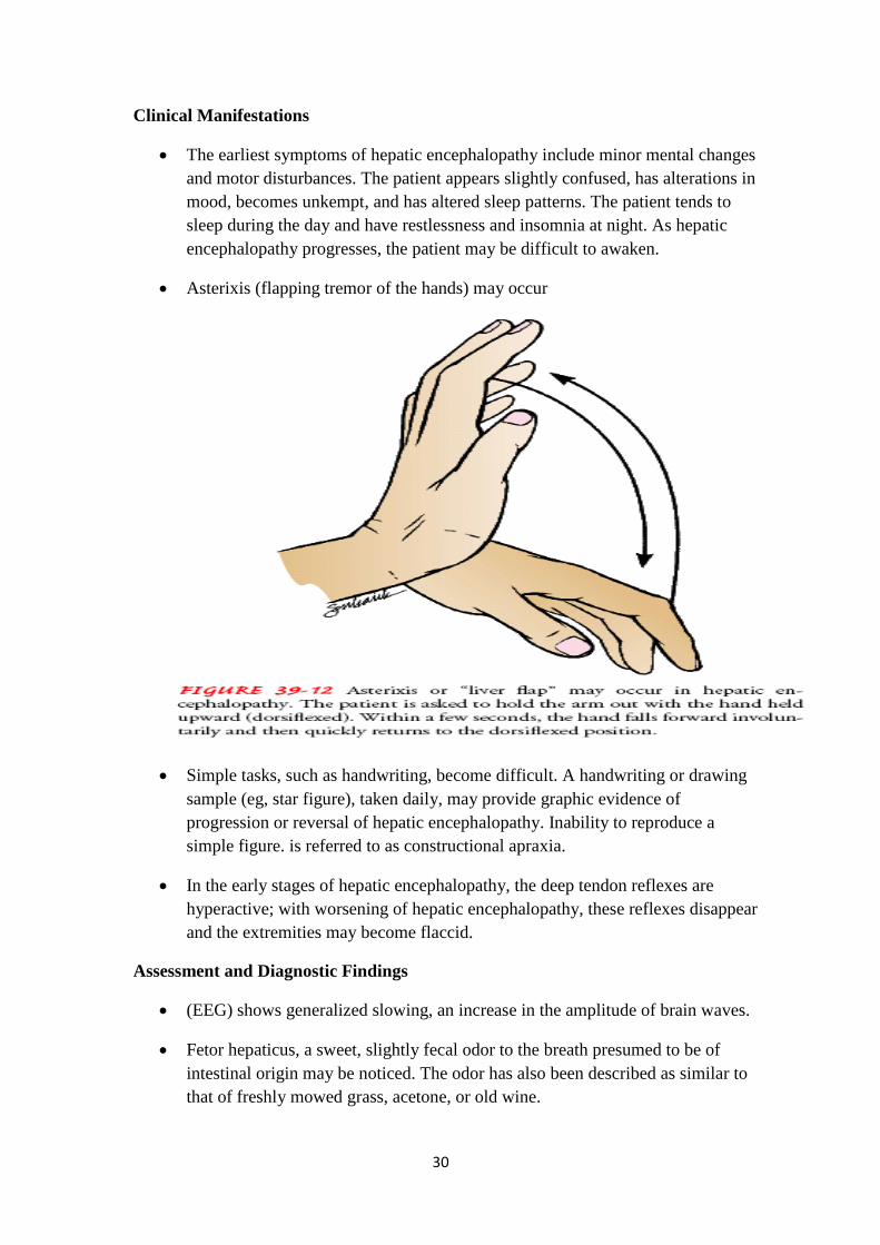
Clinical Manifestations
The earliest symptoms of hepatic encephalopathy include minor mental changes
and motor disturbances. The patient appears slightly confused, has alterations in
mood, becomes unkempt, and has altered sleep patterns. The patient tends to
sleep during the day and have restlessness and insomnia at night. As hepatic
encephalopathy progresses, the patient may be difficult to awaken.
Asterixis (flapping tremor of the hands) may occur
Simple tasks, such as handwriting, become difficult. A handwriting or drawing
sample (eg, star figure), taken daily, may provide graphic evidence of
progression or reversal of hepatic encephalopathy. Inability to reproduce a
simple figure. is referred to as constructional apraxia.
In the early stages of hepatic encephalopathy, the deep tendon reflexes are
hyperactive; with worsening of hepatic encephalopathy, these reflexes disappear
and the extremities may become flaccid.
Assessment and Diagnostic Findings
(EEG) shows generalized slowing, an increase in the amplitude of brain waves.
Fetor hepaticus, a sweet, slightly fecal odor to the breath presumed to be of
intestinal origin may be noticed. The odor has also been described as similar to
that of freshly mowed grass, acetone, or old wine.
31
With further progression of the disorder, the patient lapses into frank coma and
may have seizures. Approximately 35% of all patients with cirrhosis of the liver
die in hepatic coma.
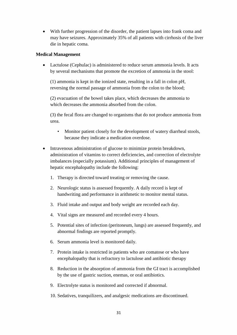
Medical Management
Lactulose (Cephulac) is administered to reduce serum ammonia levels. It acts
by several mechanisms that promote the excretion of ammonia in the stool:
(1) ammonia is kept in the ionized state, resulting in a fall in colon pH,
reversing the normal passage of ammonia from the colon to the blood;
(2) evacuation of the bowel takes place, which decreases the ammonia to
which decreases the ammonia absorbed from the colon.
(3) the fecal flora are changed to organisms that do not produce ammonia from
urea.
• Monitor patient closely for the development of watery diarrheal stools,
because they indicate a medication overdose.
Intravenous administration of glucose to minimize protein breakdown,
administration of vitamins to correct deficiencies, and correction of electrolyte
imbalances (especially potassium). Additional principles of management of
hepatic encephalopathy include the following:
1. Therapy is directed toward treating or removing the cause.
2. Neurologic status is assessed frequently. A daily record is kept of
handwriting and performance in arithmetic to monitor mental status.
3. Fluid intake and output and body weight are recorded each day.
4. Vital signs are measured and recorded every 4 hours.
5. Potential sites of infection (peritoneum, lungs) are assessed frequently, and
abnormal findings are reported promptly.
6. Serum ammonia level is monitored daily.
7. Protein intake is restricted in patients who are comatose or who have
encephalopathy that is refractory to lactulose and antibiotic therapy
8. Reduction in the absorption of ammonia from the GI tract is accomplished
by the use of gastric suction, enemas, or oral antibiotics.
9. Electrolyte status is monitored and corrected if abnormal.
10. Sedatives, tranquilizers, and analgesic medications are discontinued.
32
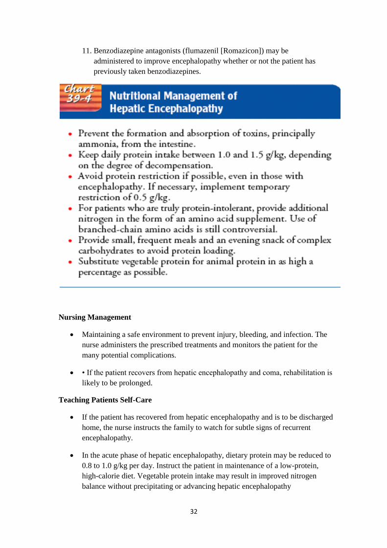
11. Benzodiazepine antagonists (flumazenil [Romazicon]) may be
administered to improve encephalopathy whether or not the patient has
previously taken benzodiazepines.
Nursing Management
Maintaining a safe environment to prevent injury, bleeding, and infection. The
nurse administers the prescribed treatments and monitors the patient for the
many potential complications.
• If the patient recovers from hepatic encephalopathy and coma, rehabilitation is
likely to be prolonged.
Teaching Patients Self-Care
If the patient has recovered from hepatic encephalopathy and is to be discharged
home, the nurse instructs the family to watch for subtle signs of recurrent
encephalopathy.
In the acute phase of hepatic encephalopathy, dietary protein may be reduced to
0.8 to 1.0 g/kg per day. Instruct the patient in maintenance of a low-protein,
high-calorie diet. Vegetable protein intake may result in improved nitrogen
balance without precipitating or advancing hepatic encephalopathy

33
Other Manifestations of Liver Dysfunction
A. Edema and Bleeding
Many patients with liver dysfunction develop generalized edema from
hypoalbuminemia that results from decreased hepatic production of albumin.
The production of blood clotting factors by the liver is also reduced, leading to
an increased incidence of bruising, epistaxis, bleeding from wounds, and, as
described above, GI bleeding.
B. Vitamin Deficiency
Decreased production of several clotting factors may be due, in part, to deficient
absorption of vitamin K from the GI tract. This probably is caused by the
inability of liver cells to use vitamin K to make prothrombin.
Absorption of the other fat-soluble vitamins (vitamins A, D, and E) as well as
dietary fats may also be impaired because of decreased secretion of bile salts
into the intestine.
The threat of these avitaminoses provides the rationale for supplementing the
diet of every patient with chronic liver disease (especially if alcohol-related)
with ample quantities of vitamins A, B complex, C, and K and folic acid
C. Metabolic Abnormalities
Abnormalities of glucose metabolism also occur; the blood glucose level may
be abnormally high shortly after a meal , but hypoglycemia may occur during
fasting because of decreased hepatic glycogen reserves and decreased
gluconeogenesis.
Because the ability to metabolize medications is decreased, medications must be
used cautiously and usual medication dosages must be reduced for the patient
with liver failure.
Many endocrine abnormalities also occur with liver dysfunction because the
liver cannot metabolize hormones normally, including androgens or sex
hormones.
Gynecomastia, amenorrhea, testicular atrophy, loss of pubic hair in the male,
and menstrual irregularities in females may occur.
D. Pruritus and Other Skin Changes
Patients with liver dysfunction resulting from biliary obstruction commonly
develop severe itching (pruritus) due to retention of bile salts. Patients may

34
develop vascular (or arterial) spider angiomas on the skin, generally above the
waistline.
Management of Patients With Viral Hepatic Disorders
Viral hepatitis is a systemic, viral infection in which necrosis and inflammation
of liver cells produce a characteristic cluster of clinical, biochemical, and
cellular changes. To date, five definitive types of viral hepatitis have been
identified: hepatitis A, B, C, D, and E .
Hepatitis A and E are similar in mode of transmission (fecal– oral route),
whereas hepatitis B, C, and D share many characteristics .
35
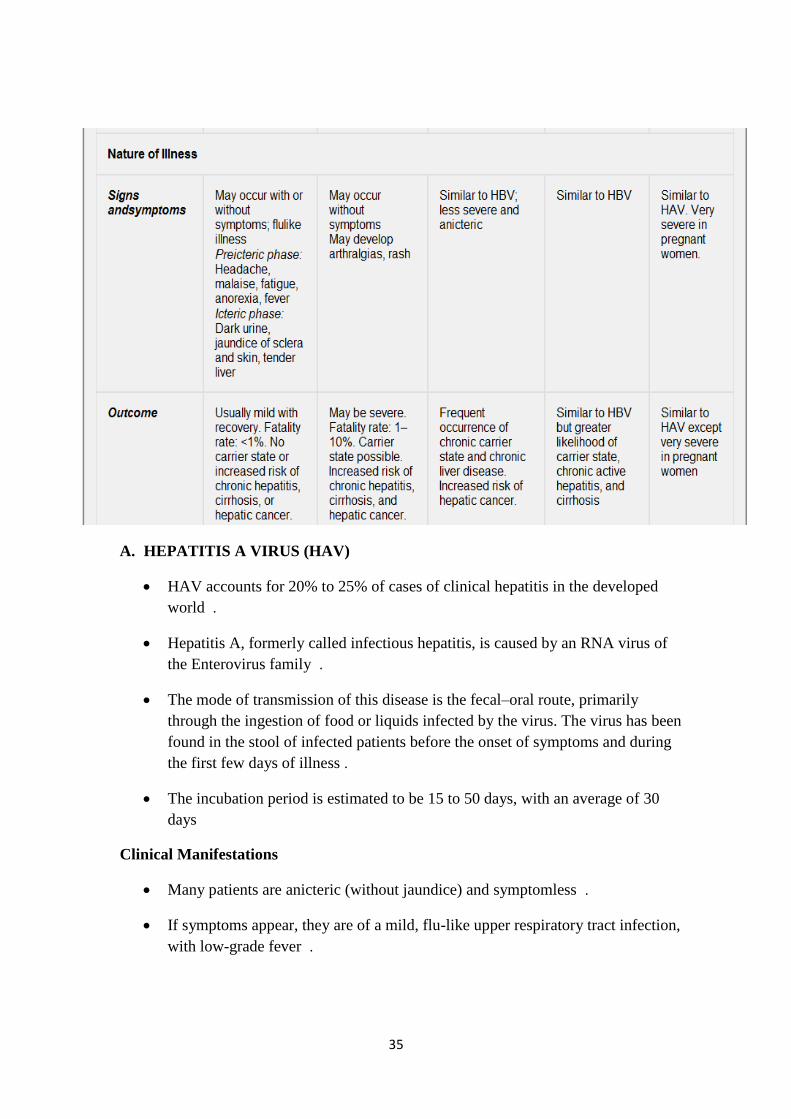
A. HEPATITIS A VIRUS (HAV)
HAV accounts for 20% to 25% of cases of clinical hepatitis in the developed
world .
Hepatitis A, formerly called infectious hepatitis, is caused by an RNA virus of
the Enterovirus family .
The mode of transmission of this disease is the fecal–oral route, primarily
through the ingestion of food or liquids infected by the virus. The virus has been
found in the stool of infected patients before the onset of symptoms and during
the first few days of illness .
The incubation period is estimated to be 15 to 50 days, with an average of 30
days
Clinical Manifestations
Many patients are anicteric (without jaundice) and symptomless .
If symptoms appear, they are of a mild, flu-like upper respiratory tract infection,
with low-grade fever .

36
Anorexia, an early symptom, is often severe. It is thought to result from release
of a toxin by the damaged liver or by failure of the damaged liver cells to
detoxify an abnormal product .
Later, jaundice and dark urine may become apparent .
Indigestion is present in varying degrees, marked by vague epigastric distress,
nausea, heartburn, & flatulence .
The liver and spleen are often moderately enlarged for a few days after onset;
otherwise, apart from jaundice, there are few physical signs .
Prevention
Medical Management
Bed rest during the acute stage and a diet that is both acceptable to the patient
and nutritious are part of the treatment and nursing care .
During the period of anorexia, the patient should receive frequent small
feedings, supplemented, if necessary, by IV fluids with glucose .

37
Because this patient often has an aversion to food, gentle persistence and
creativity may be required to stimulate the appetite .
Optimal food and fluid levels are necessary to counteract weight loss and slow
recovery .
Nursing Management
The patient is usually managed at home unless symptoms are severe. Therefore,
the nurse assists the patient and family in coping with the temporary disability
and fatigue that are common in hepatitis and instructs them to seek additional
health care if the symptoms persist or worsen .
B. HEPATITIS B VIRUS (HBV)
Transmitted primarily through blood (percutaneous and permucosal routes).
HBV has been found in blood, saliva, semen, and vaginal secretions and can be
transmitted through mucous membranes and breaks in the skin .

38
HBV is also transferred from carrier mothers to their babies, especially in areas
with a high incidence (ie, Southeast Asia). The infection is usually not via the
umbilical vein, but from the mother at the time of birth and during close contact
afterward .
HBV has a long incubation period. It replicates in the liver and remains in the
serum for relatively long periods, allowing transmission of the virus .
Those at risk for developing hepatitis B include surgeons, clinical laboratory
workers, dentists, nurses, and respiratory therapists. Staff and patients in
hemodialysis and oncology units and sexually active homosexual
Most people (>90%) who contract hepatitis B infections will develop antibodies
and recover spontaneously in 6 months. The mortality rate from hepatitis B has
been reported to be as high as 10%. Another 10% of patients who have hepatitis
B progress to a carrier state or develop chronic hepatitis with persistent HBV
infection and hepatocellular injury and inflammation .

39
Clinical Manifestations
Clinically, the disease closely resembles hepatitis A, but the incubation period is
much longer (1 to 6 months). Signs and symptoms of hepatitis B may be
insidious and variable. Fever and respiratory symptoms are rare; some patients
have arthralgias and rashes .
The patient may have loss of appetite, dyspepsia, abdominal pain, generalized
aching, malaise, and weakness. Jaundice may or may not be evident. light-
colored stools and dark urine .
The liver may be tender and enlarged. The spleen is enlarged and palpable in a
few patients; the posterior cervical lymph nodes may also be enlarged .
Assessment and Diagnostic Findings
HBV is a DNA virus composed of the following antigenic particles :
HBcAg—hepatitis B core antigen (antigenic material in an inner core
HBsAg—hepatitis B surface antigen (antigenic material on surface of HBV
HBeAg—an independent protein circulating in the blood
HBxAg—gene product of X gene of HBV/DNA
Each antigen elicits its specific antibody and is a marker for different stages of
the disease process :
Anti-HBc—antibody to core antigen or HBV; persists during the acute phase of
illness; may indicate continuing HB in the liver
Anti-HBs—antibody to surface determinants on HBV; detected during late
convalescence; usually indicates recovery and development of immunity. It
appears in the circulation in 80% to 90% of infected patients 1 to 10 weeks after
exposure, if continues for > 6 months, pt is considered a HBsAg carrier
Anti-HBe—antibody to hepatitis B e-antigen; usually signifies reduced
infectivity
•anti-HBxAg—antibody to the hepatitis B x-antigen; may indicate ongoing
replication of HBV
Prevention
The goals of prevention are to interrupt the chain of transmission, to protect
people at high risk with active immunization through the use of hepatitis B
vaccine, and to use passive immunization for unprotected people exposed to
HBV .

40
A. PREVENTING TRANSMISSION
o Continued screening of blood donors for the presence of hepatitis B
antigens
o The use of disposable syringes, needles, and lancets and the introduction
of needleless IV
o Good personal hygiene is fundamental to infection control. In the
clinical laboratory, work areas should be disinfected daily. Gloves are
worn when handling all blood and body fluids as well as HBAg positive
specimens .
o Eating and smoking are prohibited in the laboratory and in other areas
exposed to secretions, blood products .
B. Active Immunization: Hepatitis B Vaccine
o Active immunization is recommended for individuals at high risk for
hepatitis B (eg, health care personnel and hemodialysis patients). In
addition, individuals with hepatitis C and other chronic liver diseases
should receive the vaccine .
o Administered IM (in the deltoid muscle in adults) in three doses, the
second and third doses 1 & 6 months after the first dose. The third dose
is very important in producing prolonged immunity .
o Antibody response may be measured by anti-HBs levels 1 to 3 months
after completing the basic course of vaccine
o Universal vaccination of all infants .
C. PASSIVE IMMUNITY: HEPATITIS B IMMUNE GLOBULIN
o Hepatitis B immune globulin (HBIG) provides passive immunity to
hepatitis B and is indicated for people exposed to HBV who have never
had hepatitis B and have never received hepatitis B vaccine. Specific
indications for postexposure vaccine with HBIG include :
(1 ) inadvertent exposure to HBAg-positive blood through percutaneous
(needlestick) or transmucosal (splashes in contact with mucous
membrane) routes ,
( 2 ) sexual contact with people positive for HBAg, and
( 3 ) perinatal exposure (babies born to HBV-infected mothers should
receive HBIG within 12 hours of delivery .)

41
Gerontologic Considerations
The elderly patient who contracts hepatitis B has a serious risk of severe liver
cell necrosis or fulminant hepatic failure, particularly if other illnesses are
present. The patient is seriously ill and the prognosis is poor, so efforts should
be undertaken to eliminate other factors (eg, medications, alcohol) that may
affect liver function .
Medical Management
The goals of treatment are to minimize infectivity, normalize liver
inflammation, and decrease symptoms .
Alpha interferon offers the most promise. It results in remission in
approximately one third of patients
Lamivudine & adefovir are new antiviral agents .
Adequate nutrition should be maintained; proteins are restricted when the
liver‘s ability to metabolize protein byproducts is impaired, as demonstrated by
symptoms .
If vomiting persists, the patient may require hospitalization and fluid therapy .
Nursing Management
Convalescence may be prolonged, with complete symptomatic recovery
sometimes requiring 3 to 4 months or longer .
During this stage, gradual resumption of physical activity is encouraged after
the jaundice has resolved .
The nurse identifies psychosocial issues and concerns, particularly the effects of
separation from family and friends if the patient is hospitalized during the acute
and infective stages. Even if not hospitalized, the patient will be unable to work
and must avoid sexual contact .
C. HEPATITIS C VIRUS (HCV
Formerly referred to as non-A, non-B hepatitis .
Blood transfusions and sexual contact accounted for most cases of hepatitis C in
the United States, other parenteral means, such as sharing contaminated needles
by IV/injection drug users and unintentional needle-sticks and other injuries in
health care workers, now account for a significant number of cases .

42
There is no benefit from rest, diet, or vitamin supplements. Recent studies have
demonstrated that a combination of interferon (Intron-A) and ribavirin
(Rebetol), two antiviral agents, is effective in producing improvement in
patients with hepatitis C and in treating relapse.
D. HEPATITIS D VIRUS (HDV)
Hepatitis D (delta agent) occurs in some cases of hepatitis B. Because the virus
requires hepatitis B surface antigen for its replication, only individuals with
hepatitis B are at risk for hepatitis D. Anti-delta antibodies in the presence of
HBAg on testing confirm the diagnosis. It is also common among IV/injection
drug users, hemodialysis patients, and recipients of multiple blood transfusions.
Sexual contact with those with hepatitis B is considered to be an important
mode of transmission of hepatitis B and D .
E. HEPATITIS E VIRUS (HEV )
Hepatitis E is believed to be transmitted by the fecal– oral route, principally
through contaminated water in areas with poor sanitation. The incubation period
is variable, estimated to range between 15 and 65 days. In general, hepatitis E
resembles hepatitis A. It has a self-limiting course with an abrupt onset.
Jaundice is nearly always present. Chronic forms do not develop .
43
F. HEPATITIS G (HGV) AND GB VIRUS.C
It has long been believed that there is another non-A, non-B, non- C agent
causing hepatitis in humans. The incubation period for post-transfusion hepatitis
is 14 to 145 days, too long for hepatitis B or C. In the United States, about 5%
of chronic liver disease remains cryptogenic (does not appear to be autoimmune
or viral in origin), and half the patients have previously received transfusions.
Thus, a new form of hepatitis (hepatitis G or GBV-C) has been described. They
are two different isolates of the same virus. Autoantibodies are absent. The
clinical significance of this virus remains uncertain. Risk factors are similar to
those for hepatitis C .
Management of Patients With Nonviral Hepatic Disorders
Certain chemicals have toxic effects on the liver and when taken by mouth,
inhaled, or injected parenterally produce acute liver cell necrosis, or toxic
hepatitis .
The chemicals most commonly implicated in this disease are carbon
tetrachloride, phosphorus, chloroform, and gold compounds .
Drug-induced hepatitis, is similar to acute viral hepatitis, but parenchymal
destruction tends to be more extensive. Some medications that can lead to
hepatitis are isoniazide, halothane, acetaminophen, and certain antibiotics,
antimetabolites, and anesthetic agents .
TOXIC HEPATITIS
Resembles viral hepatitis in onset. Obtaining a history of exposure to
hepatotoxic chemicals, medications, or other agents assists in early treatment
and removal of the offending agent .
Anorexia, nausea, and vomiting are the usual symptoms; jaundice and
hepatomegaly are noted on physical assessment .
Recovery from acute toxic hepatitis is rapid if the hepatotoxin is identified early
and removed or if exposure to the agent has been limited .
DRUG-INDUCED HEPATITIS
Drug-induced hepatitis is responsible for 20% to 25% of cases of acute hepatic
failure in the United States .
44
Manifestations of sensitivity to a medication may occur on the first day of its
use or not until several months later, depending on the medication .
Usually the onset is abrupt, with chills, fever, rash, pruritus, arthralgia,
anorexia, and nausea. Later, there may be jaundice and dark urine & enlarged
and tender liver. When the offending medication is withdrawn, symptoms may
gradually subside .
FULMINANT HEPATIC FAILURE
Is the clinical syndrome of sudden and severely impaired liver function in a
previously healthy person. According to the original and generally accepted
definition, fulminant hepatic failure develops within 8 weeks of the first
symptoms of jaundice .
Three categories are frequently cited: hyperacute, acute, and subacute liver
failure .
FULMINANT HEPATIC FAILURE
Is the clinical syndrome of sudden and severely impaired liver function in a
previously healthy person .
In hyperacute liver failure, the duration of jaundice before the onset of
encephalopathy is 0 to 7 days; in acute liver failure, it is 8 to 28 days; and in
subacute liver failure, it is 28 to 72 days .
The prognosis for fulminant hepatic failure is much worse than for chronic liver
failure. However, in fulminant failure, the hepatic lesion is potentially
reversible, with survival rates of approximately 50% to 85% (depending on
etiology). Those who do not survive die of massive hepatocellular injury and
necrosis
FULMINANT HEPATIC FAILURE
Fulminant hepatic failure is often accompanied by coagulation defects, renal
failure and electrolyte disturbances, infection, hypoglycemia, encephalopathy,
and cerebral edema .
Viral hepatitis is a common cause of fulminant hepatic failure; other causes:
toxic medications (eg, acetaminophen) and chemicals (eg, carbon tetrachloride),
metabolic disturbances (eg, Wilson's disease, a hereditary syndrome with
deposition of copper in the liver), and structural changes (eg, Budd-Chiari
syndrome, an obstruction to outflow in major hepatic veins .)

45
Management
The key to optimizing treatment is rapid recognition of acute liver failure and
intensive interventions .
The use of antidotes for certain conditions may be indicated such as N-
acetylcysteine for acetaminophen toxicity and penicillin for mushroom
poisoning .
Treatment modalities may include plasma exchanges (plasmapheresis) to
correct coagulopathy and to stabilize the patient awaiting liver transplantation
HEPATIC CIRRHOSIS
Cirrhosis is a chronic disease characterized by replacement of normal liver
tissue with diffuse fibrosis that disrupts the structure and function of the liver.
There are three types of cirrhosis or scarring of the live:
1. Alcoholic cirrhosis, in which the scar tissue characteristically surrounds the
portal areas. This is most frequently due to chronic alcoholism and is the
most common type of cirrhosis .
2. Postnecrotic cirrhosis, in which there are broad bands of scar tissue as a late
result of a previous infection of acute viral hepatitis .
3. Biliary cirrhosis, in which scarring occurs in the liver around the bile ducts.
This type usually is the result of chronic biliary obstruction and infection
(cholangitis); it is much less common than the other two types.

46
Pathophysiology
Several factors have been implicated in the etiology of cirrhosis: alcohol
consumption (is the major causative factor), nutritional deficiency with reduced
protein intake (excessive alcohol intake is the major causative factor in fatty
liver and its consequences .)
Other factors may play a role, including exposure to certain chemicals (carbon
tetrachloride, chlorinated naphthalene, arsenic, or phosphorus) or infectious
schistosomiasis .
Twice as many men as women are affected .
Most patients are between 40 and 60 years of age .
The destroyed liver cells are replaced gradually by scar tissue; eventually the
amount of scar tissue exceeds that of the functioning liver tissue .
Islands of residual normal tissue and regenerating liver tissue may project from
the constricted areas, giving the cirrhotic liver its characteristic hobnail
appearance .
Clinical Manifestations
Signs and symptoms of cirrhosis increase in severity as the disease progresses.
The severity of the manifestations helps to categorize the disorder into two main
presentations
1. Compensated cirrhosis, with its less severe, often vague symptoms, may be
discovered secondarily at a routine physical examination .
47
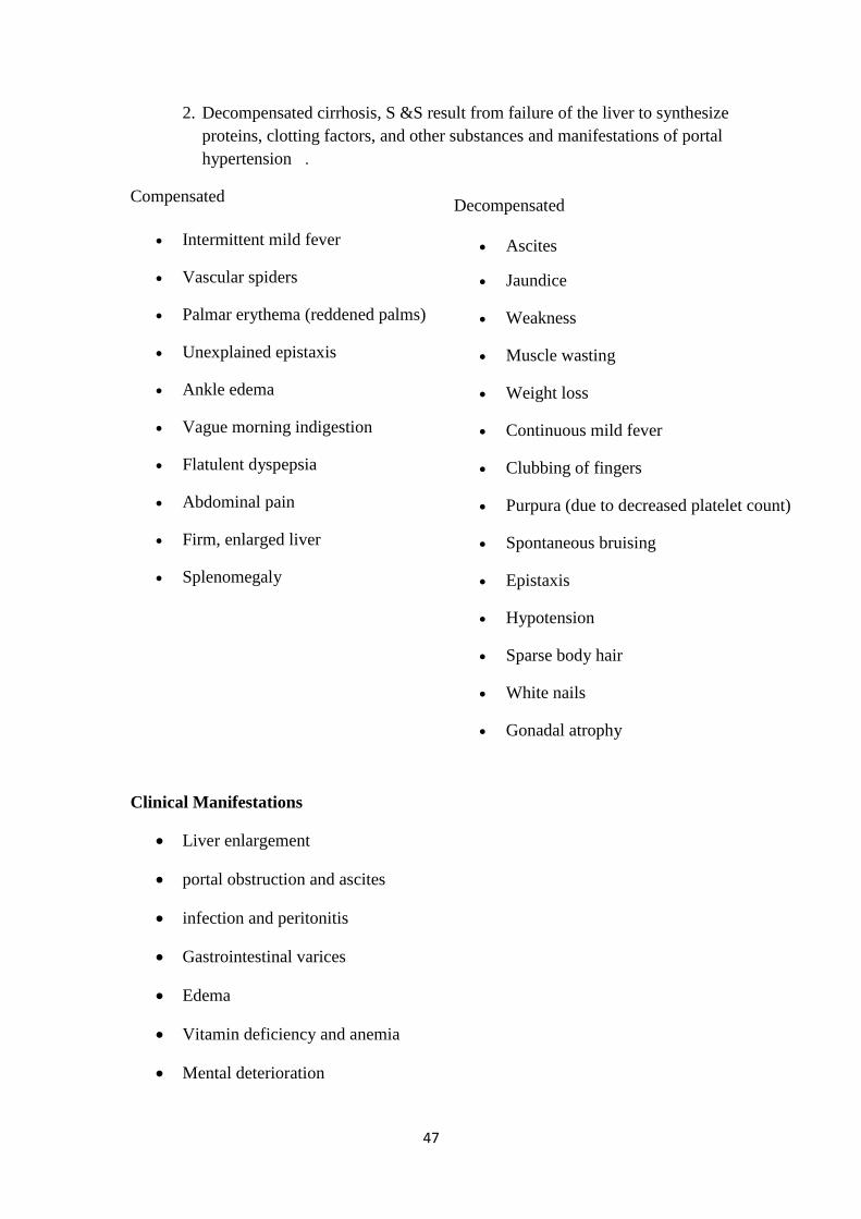
2. Decompensated cirrhosis, S &S result from failure of the liver to synthesize
proteins, clotting factors, and other substances and manifestations of portal
hypertension .
Compensated
Intermittent mild fever
Vascular spiders
Palmar erythema (reddened palms)
Unexplained epistaxis
Ankle edema
Vague morning indigestion
Flatulent dyspepsia
Abdominal pain
Firm, enlarged liver
Splenomegaly
Clinical Manifestations
Liver enlargement
portal obstruction and ascites
infection and peritonitis
Gastrointestinal varices
Edema
Vitamin deficiency and anemia
Mental deterioration
Decompensated
Ascites
Jaundice
Weakness
Muscle wasting
Weight loss
Continuous mild fever
Clubbing of fingers
Purpura (due to decreased platelet count)
Spontaneous bruising
Epistaxis
Hypotension
Sparse body hair
White nails
Gonadal atrophy

48
Assessment and Diagnostic Findings
The extent of liver disease and the type of treatment are determined after
reviewing the laboratory findings. Because the functions of the liver are
complex, there are many diagnostic tests that may provide information about
liver function .
In severe parenchymal liver dysfunction, the serum albumin level decreases.
Enzyme tests indicate liver cell damage: serum alkaline phosphatase, AST,
ALT levels increase, and the serum cholinesterase level may decrease
Bilirubin tests are performed to measure bile excretion or bile retention;
elevated levels can occur with cirrhosis and other liver disorders .
Prothrombin time is prolonged .
Ultrasound scanning is used to measure the difference in density of
parenchymal cells and scar tissue .
CT, MRI, and radioisotope liver scans give information about liver size and
hepatic blood flow and obstruction .
Diagnosis is confirmed by liver biopsy .
Medical Management
The management of the patient with cirrhosis is usually based on the presenting
symptoms. For example, antacids are prescribed to decrease gastric distress and
minimize the possibility of GI bleeding .
Vitamins and nutritional supplements promote healing of damaged liver cells
and improve the general nutritional status. Potassium-sparing diuretics
(spironolactone [Aldactone], triamterene [Dyrenium]) may be indicated to
decrease ascites, if present ;
Preliminary studies indicate that colchicine, an antiinflammatory agent used to
treat the symptoms of gout, may increase the length of survival in patients with
mild to moderate cirrhosis. Colchicine is believed to reverse the fibrotic
processes in cirrhosis, and this has improved survival
49
NURSING PROCESS:
THE PATIENT WITH HEPATIC CIRRHOSIS
Nursing diagnoses and goals
1. Activity intolerance related to fatigue, lethargy, and malaise
Goal: Patient reports decrease in fatigue and reports increased ability to
participate in activities
2. Imbalanced nutrition: less than body requirements, related to abdominal
distention and discomfort and anorexia
Goal: Positive nitrogen balance, no further loss of muscle mass; meets
nutritional requirements
3. Impaired skin integrity related to pruritus from jaundice and edema.
Goal: Decrease potential for pressure ulcer development; breaks in skin
integrity
4. High risk for injury related to altered clotting mechanisms and altered level of
consciousness
Goal: Reduced risk of injury
5. Disturbed body image related to changes in appearance, sexual dysfunction, and
role function
Goal: Patient verbalizes feelings consistent with improvement of body image
and self-esteem
6. Chronic pain and discomfort related to enlarged tender liver and ascites
Goal: Increased level of comfort
7. Fluid volume excess related to ascites and edema formation
Goal: Restoration of normal fluid volume
8. Disturbed thought processes related to deterioration of liver function and
increased serum ammonia level
Goal: Improved mental status; safety maintained
9. Risk for imbalanced body temperature: hyperthermia related to inflammatory
process of cirrhosis or hepatitis
50
Goal: Maintenance of normal body temperature, free from infection
10. Ineffective breathing pattern related to ascites and restriction of thoracic
excursion secondary to ascites, abdominal distention, and fluid in the thoracic
cavity
Goal: Improved respiratory status
Collaborative Problem
1. Gastrointestinal bleeding and hemorrhage
Goal: Absence of episodes of gastrointestinal bleeding and hemorrhage
2. Hepatic encephalopathy
Goal: Absence of changes in cognitive status and of injury
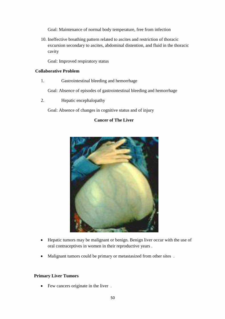
Cancer of The Liver
Hepatic tumors may be malignant or benign. Benign liver occur with the use of
oral contraceptives in women in their reproductive years .
Malignant tumors could be primary or metastasized from other sites .
Primary Liver Tumors
Few cancers originate in the liver .

51
Usually associated with chronic liver disease, hepatitis B and C infections, and
cirrhosis .
Hepatocellular carcinoma (HCC) is the most common type of primary liver
cancer. HCC is usually nonresectable because of rapid growth and metastasis .
Other types of primary liver cancer include cholangiocellular carcinoma and
combined hepatocellular and cholangiocellular carcinoma. If found early,
resection may be possible, but early detection is unlikely .
Liver Metastases
Metastases from other primary sites are found in the liver in about half of all
advanced cancer cases .
Malignant tumors are likely to reach the liver eventually, by way of the portal
system or lymphatic channels, or by direct extension from an abdominal tumor .
Moreover, the liver apparently is an ideal place for these malignant cells to
thrive .
Clinical Manifestations
The early manifestations of malignancy of the liver include pain, a continuous
dull ache in the right upper quadrant, epigastrium, or back .
Weight loss, loss of strength, anorexia, and anemia may also occur .
The liver may be enlarged and irregular on palpation .
Jaundice is present only if the larger bile ducts are occluded by the pressure of
tumor on bile ducts .
Ascites develops if tumor obstructs the portal veins or if tumor tissue is seeded
in the peritoneal cavity .
Assessment and Diagnostic Findings
Diagnosis is based on clinical signs and symptoms, the history and physical
examination, and the results of laboratory and x-ray studies .
Increased serum levels of bilirubin, alkaline phosphatase, AST, GGT, and lactic
dehydrogenase may occur .
Leukocytosis, erythrocytosis, hypercalcemia, hypoglycemia, and
hypocholesterolemia
52
The serum level of alpha-fetoprotein (AFP), which serves as a tumor marker, is
elevated in 30% to 40% of patients with primary liver cancer .
Levels of carcinoembryonic antigen (CEA), a marker of advanced cancer of the
digestive tract, may be elevated .
These two markers together are useful to distinguish between metastatic liver
disease and primary liver cancer .
Assessment and Diagnostic Findings
X-rays, liver scans, CT scans, ultrasound studies, MRI, arteriography, and
laparoscopy may be part of the diagnostic workup and may be performed to
determine the extent of the cancer .
Confirmation of a tumor‘s histology can be made by biopsy under imaging
guidance (CT scan or ultrasound .)
Medical Management
Surgical resection of the tumor is possible in some patients, but cirrhosis
(prevalent in liver cancer), increases the risks associated with surgery .
Radiation therapy and chemotherapy showed varying degrees of success .
An implantable pump has been used to deliver a high concentration of
chemotherapy to the liver through the hepatic artery. This method provides a
reliable, controlled, and continuous infusion of medication that can be carried
out in the patient‘s home .
Percutaneous Biliary Drainage
Percutaneous biliary or transhepatic drainage is used to bypass biliary ducts
obstructed by liver, pancreatic, or bile duct tumors in patients with inoperable
tumors or in those considered poor surgical risks .
A catheter is inserted through the abdominal wall and past the obstruction into
the duodenum. Such procedures are used to reestablish biliary drainage, relieve
pressure and pain from the buildup of bile behind the obstruction, and decrease
pruritus and jaundice .
Surgical Management
Surgical resection is the treatment of choice when HCC is confined to one lobe
of the liver and the function of the remaining liver is considered adequate for
postoperative recovery .
53
Capitalizing on the regenerative capacity of the liver cells, some surgeons have
successfully removed 90% of the liver .
However, the presence of cirrhosis limits the ability of the liver to regenerate .
Lobectomy
Liver Transplantation
Liver Transplantation
Liver disease for which no other form of treatment is available .
The transplantation procedure involves total removal of the diseased liver and
its replacement with a healthy liver in the same anatomic location .
The success of liver transplantation depends on successful immunosuppression
(i.e. cyclosporine corticosteroids).
Complications
The postoperative complication rate is high, primarily because of technical
complications or infection .
Immediate postoperative complication include bleeding, infection, and
rejection. Disruption, infection, or obstruction of the biliary anastomosis and
impaired biliary drainage may occur. Vascular thrombosis and stenosis are other
potential complications .
Nursing Management
1. Preoperative Nursing Interventions
Provide the patient and family with full explanations about the procedure, the
chances of success, and the risks, including the side effects of long-term
immunosuppression. The need for close follow-up and lifelong compliance with
the therapeutic regimen .
Malnutrition, massive ascites, and fluid and electrolyte disturbances are treated
before surgery to increase the chance of a successful outcome .
2. Post operative ;
The patient is maintained in an environment as free from bacteria, viruses, and
fungi as possible to prevent infection .
Cardiovascular, pulmonary, renal, neurologic, & metabolic functions are
monitored continuously .
54
Cardiac output, CVP, pulmonary capillary wedge pressure, ABG, O 2
saturation, urine output, V/S are used to evaluate the patient's hemodynamic
status and intravascular fluid volume .
Liver functions tests and coagulation profiel .
I & O including drainage from T tube .
LIVER ABSCESSES
Two categories of liver abscess have been identified: amebic and pyogenic .
1. Amebic liver abscesses are most commonly caused by Entamoeba
histolytica. Most amebic liver abscesses occur in the developing countries of
the tropics and subtropics because of poor sanitation and hygiene .
2. Pyogenic liver abscesses are much less common .
Pathophysiology
Whenever an infection develops anywhere along the biliary or GI tract, infecting
organisms may reach the liver through the biliary system, portal venous system, or
hepatic arterial or lymphatic system .
Most bacteria are destroyed promptly, but occasionally some gain a foothold .
The bacterial toxins destroy the neighboring liver cells, and the resulting necrotic
tissue serves as a protective wall for the organisms .
Meanwhile, leukocytes migrate into the infected area .
The result is an abscess cavity full of a liquid containing living and dead
leukocytes, liquefied liver cells, and bacteria .
Pyogenic abscesses of this type may be either single or multiple and small .
Examples of causes of pyogenic liver abscess include cholangitis and abdominal
trauma .
Clinical Manifestations
The clinical picture is one of sepsis with few or no localizing signs. Fever with
chills and diaphoresis, malaise, anorexia, nausea, vomiting, and weight loss may
occur .
55
The patient may complain of dull abdominal pain and tenderness in the right
upper quadrant of the abdomen .
Hepatomegaly, jaundice, anemia, and pleural effusion may develop .
Sepsis and shock may be severe and life-threatening .
Assessment and Diagnostic Findings
Blood cultures are obtained but may not identify the organism .
Aspiration of the liver abscess may be done to assist in diagnosis and to obtain
cultures of the organism .
Percutaneous drainage of pyogenic abscesses is carried out to evacuate the
abscess material and promote healing .
A catheter may be left in place for continuous drainage; the patient must be
instructed about its management .
Medical Management
Treatment includes IV antibiotic therapy; the specific antibiotic used in
treatment depends on the organism identified .
Continuous supportive care is indicated because of the serious condition of the
patient .
Open surgical drainage may be required if antibiotic therapy and percutaneous
drainage are ineffective .
Nursing Management
Depends on the patient‘s physical status and the medical management that is
indicated .
For patients who undergo evacuation and drainage of the abscess, monitoring of
the drainage and skin care are imperative .
Strategies must be implemented to contain the drainage and to protect the
patient from other sources of infection .
Vital signs are monitored to detect changes in the patient‘s physical status .
Deterioration in vital signs or the onset of new symptoms such as increasing
pain, which may indicate rupture or extension of the abscess, is reported
promptly .

56
The nurse administers IV antibiotic therapy as prescribed .
The white blood cell count and other laboratory test results are monitored
closely for changes consistent with worsening infection.

57
NURSING PROCESS FOR PATIENTS WITH HEPATIC
DYSFUNCTIONS
Nursing Diagnosis: Activity intolerance related to fatigue, lethargy, and malaise
Goal: Patient reports decrease in fatigue and reports increased ability to participate in activities
Nursing Interventions Rationale Expected Outcomes
1. Assess level of activity
tolerance and degree of fatigue,
lethargy, and malaise when
performing routine ADLs.
2. Assist with activities and
hygiene when fatigued.
3. Encourage rest when fatigued or
when abdominal pain or
discomfort occurs.
4. Assist with selection and pacing
of desired activities and
exercise.
5. Provide diet high in
carbohydrates with protein
intake consistent with liver
function.
6. Administer supplemental
vitamins (A, B complex, C, and
K).
1. Provides baseline for further
assessment and criteria for
assessment of effectiveness of
interventions
2. Promotes exercise and hygiene
within patient's level of
tolerance
3. Conserves energy and protects
the liver
4. Stimulates patient's interest in
selected activities
5. Provides calories for energy
and protein for healing
6. Provides additional nutrients
Exhibits increased interest
in activities and events
Participates in activities
and gradually increases
exercise within physical
limits
Reports increased strength
and well-being
Reports absence of
abdominal pain and
discomfort
Plans activities to allow
ample periods of rest
Takes vitamins as
prescribed
Nursing Diagnosis: Imbalanced nutrition: less than body requirements, related to abdominal distention and
discomfort and anorexia
Goal: Positive nitrogen balance, no further loss of muscle mass; meets nutritional requirements
1. Assess dietary intake and
nutritional status through diet
history and diary, daily weight
measurements and laboratory
data.
2. Provide diet high in
carbohydrates with protein
intake consistent with liver
1. Identifies deficits in nutritional
intake and adequacy of
nutritional state
2. Provides calories for energy,
sparing protein for healing
3. Reduces edema and ascites
formation
Exhibits improved nutritional
status by increased weight
(without fluid retention) and
improved laboratory data.
States rationale for dietary
modifications
Identifies foods high in
carbohydrates and within

58
function.
3. Assist patient in identifying
low-sodium foods.
4. Elevate the head of the bed
during meals.
5. Provide oral hygiene before
meals and pleasant environment
for meals at meal time.
6. Offer smaller, more frequent
meals (6 per day).
7. Encourage patient to eat meals
and supplementary feedings.
8. Provide attractive meals and an
aesthetically pleasing setting at
meal time.
9. Eliminate alcohol.
10. Apply an ice collar for nausea.
11. Administer medications
prescribed for nausea, vomiting,
diarrhea, or constipation.
12. Encourage increased fluid
intake and exercise if the patient
reports constipation.
4. Reduces discomfort from
abdominal distention and
decreases sense of fullness
produced by pressure of
abdominal contents and ascites
on the stomach
5. Promotes positive environment
and increased appetite; reduces
unpleasant taste
6. Decreases feeling of fullness,
bloating
7. Encouragement is essential for
the patient with anorexia and
gastrointestinal discomfort.
8. Promotes appetite and sense of
well-being
9. Eliminates ‗empty calories‘
and further damage from
alcohol
10. May reduce incidence of
nausea
11. Reduces gastrointestinal
symptoms and discomforts that
decrease the appetite and
interest in food
12. Promotes normal bowel
pattern and reduces abdominal
discomfort and distention
protein requirements
(moderate to high protein in
cirrhosis and hepatitis, low
protein in hepatic failure)
Reports improved appetite
Participates in oral hygiene
measures
Reports increased appetite;
identifies rationale for
smaller, frequent meals
Demonstrates intake of high-
calorie diet; adheres to protein
restriction
Identifies foods and fluids
that are nutritious and
permitted on diet
Gains weight without
increased edema or ascites
formation
Reports increased appetite
and well-being
Excludes alcohol from diet
Takes medications for
gastrointestinal disorders as
prescribed
Reports normal
gastrointestinal function with
regular bowel function
7. Nursing Diagnosis: Impaired skin integrity related to pruritus from jaundice and edema
Goal:Decrease potential for pressure ulcer development; breaks in skin integrity.
8. Nursing Interventions Rationale Expected Outcomes
9. Assess degree of discomfort
related to pruritus and edema.
10. Note and record degree of
1. Assists in determining
appropriate interventions
2. Provides baseline for detecting
Exhibits intact skin
without redness,
excoriation, or breakdown
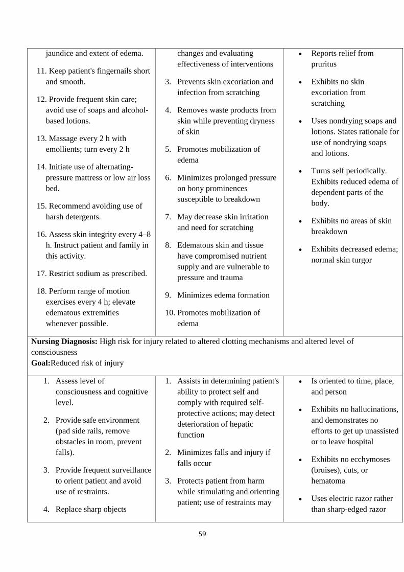
59
jaundice and extent of edema.
11. Keep patient's fingernails short
and smooth.
12. Provide frequent skin care;
avoid use of soaps and alcohol-
based lotions.
13. Massage every 2 h with
emollients; turn every 2 h
14. Initiate use of alternating-
pressure mattress or low air loss
bed.
15. Recommend avoiding use of
harsh detergents.
16. Assess skin integrity every 4–8
h. Instruct patient and family in
this activity.
17. Restrict sodium as prescribed.
18. Perform range of motion
exercises every 4 h; elevate
edematous extremities
whenever possible.
changes and evaluating
effectiveness of interventions
3. Prevents skin excoriation and
infection from scratching
4. Removes waste products from
skin while preventing dryness
of skin
5. Promotes mobilization of
edema
6. Minimizes prolonged pressure
on bony prominences
susceptible to breakdown
7. May decrease skin irritation
and need for scratching
8. Edematous skin and tissue
have compromised nutrient
supply and are vulnerable to
pressure and trauma
9. Minimizes edema formation
10. Promotes mobilization of
edema
Reports relief from
pruritus
Exhibits no skin
excoriation from
scratching
Uses nondrying soaps and
lotions. States rationale for
use of nondrying soaps
and lotions.
Turns self periodically.
Exhibits reduced edema of
dependent parts of the
body.
Exhibits no areas of skin
breakdown
Exhibits decreased edema;
normal skin turgor
Nursing Diagnosis: High risk for injury related to altered clotting mechanisms and altered level of
consciousness
Goal:Reduced risk of injury
1. Assess level of
consciousness and cognitive
level.
2. Provide safe environment
(pad side rails, remove
obstacles in room, prevent
falls).
3. Provide frequent surveillance
to orient patient and avoid
use of restraints.
4. Replace sharp objects
1. Assists in determining patient's
ability to protect self and
comply with required self-
protective actions; may detect
deterioration of hepatic
function
2. Minimizes falls and injury if
falls occur
3. Protects patient from harm
while stimulating and orienting
patient; use of restraints may
Is oriented to time, place,
and person
Exhibits no hallucinations,
and demonstrates no
efforts to get up unassisted
or to leave hospital
Exhibits no ecchymoses
(bruises), cuts, or
hematoma
Uses electric razor rather
than sharp-edged razor

60
(razors) with safer items.
5. Observe each stool for color,
consistency, and amount.
6. Be alert for symptoms of
anxiety, epigastric fullness,
weakness, and restlessness.
7. Test each stool and emesis
for occult blood.
8. Observe for hemorrhagic
manifestations: ecchymosis,
epistaxis, petechiae, and
bleeding gums.
9. Record vital signs at frequent
intervals, depending on
patient acuity (every 1–4 h).
10. Keep patient quiet and limit
activity.
11. Assist physician in passage
of tube for esophageal
balloon tamponade, if its
insertion is indicated.
12. Observe during blood
transfusions.
13. Measure and record nature,
time, and amount of vomitus.
14. Maintain patient in fasting
state, if indicated.
15. Administer vitamin K as
prescribed.
16. Remain with patient during
episodes of bleeding.
17. Offer cold liquids by mouth
when bleeding stops (if
prescribed).
18. Institute measures to prevent
disturb patient further
4. Avoids cuts and bleeding
5. Permits detection of bleeding
in gastrointestinal tract
6. May indicate early signs of
bleeding and shock
7. Detects early evidence of
bleeding
8. Indicates altered clotting
mechanisms
9. Provides baseline and evidence
of hypovolemia, and
hemorrhagic shock
10. Minimizes risk of bleeding and
straining
11. Promotes nontraumatic
insertion of tube in anxious
and combative patient for
immediate treatment of
bleeding
12. Permits detection of
transfusion reactions (risk is
increased with multiple blood
transfusions needed for active
bleeding from esophageal
varices)
13. Assists in evaluating extent of
bleeding and blood loss
14. Reduces risk of aspiration of
gastric contents and minimizes
risk of further trauma to
esophagus and stomach by
preventing vomiting
15. Promotes clotting by providing
fat-soluble vitamin necessary
Exhibits absence of frank
bleeding from
gastrointestinal tract
Exhibits absence of
restlessness, epigastric
fullness, and other
indicators of hemorrhage
and shock
Exhibits negative results
of test for occult
gastrointestinal bleeding
Is free of ecchymotic areas
or hematoma formation
Exhibits normal vital signs
Maintains rest and
remains quiet if active
bleeding occurs
Identifies rationale for
blood transfusions and
measures to treat bleeding
Uses measures to prevent
trauma (eg, uses soft
toothbrush, blows nose
gently, avoids bumps and
falls, avoids straining
during defecation)
Experiences no side
effects of medications
Takes all medications as
prescribed
Identifies rationale for
precautions with use of all
medications
Cooperates with
treatment.
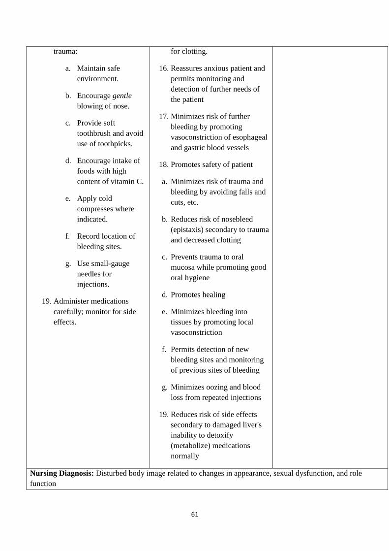
61
trauma:
a. Maintain safe
environment.
b. Encourage gentle
blowing of nose.
c. Provide soft
toothbrush and avoid
use of toothpicks.
d. Encourage intake of
foods with high
content of vitamin C.
e. Apply cold
compresses where
indicated.
f. Record location of
bleeding sites.
g. Use small-gauge
needles for
injections.
19. Administer medications
carefully; monitor for side
effects.
for clotting.
16. Reassures anxious patient and
permits monitoring and
detection of further needs of
the patient
17. Minimizes risk of further
bleeding by promoting
vasoconstriction of esophageal
and gastric blood vessels
18. Promotes safety of patient
a. Minimizes risk of trauma and
bleeding by avoiding falls and
cuts, etc.
b. Reduces risk of nosebleed
(epistaxis) secondary to trauma
and decreased clotting
c. Prevents trauma to oral
mucosa while promoting good
oral hygiene
d. Promotes healing
e. Minimizes bleeding into
tissues by promoting local
vasoconstriction
f. Permits detection of new
bleeding sites and monitoring
of previous sites of bleeding
g. Minimizes oozing and blood
loss from repeated injections
19. Reduces risk of side effects
secondary to damaged liver's
inability to detoxify
(metabolize) medications
normally
Nursing Diagnosis: Disturbed body image related to changes in appearance, sexual dysfunction, and role
function
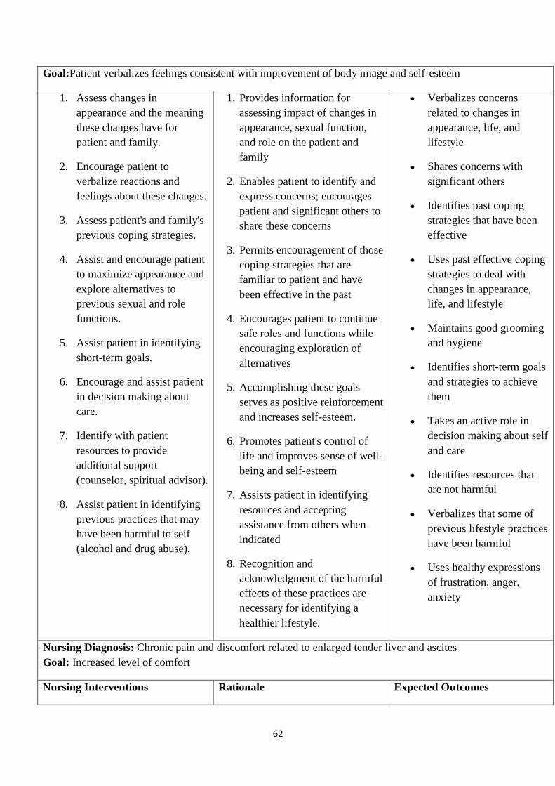
62
Goal:Patient verbalizes feelings consistent with improvement of body image and self-esteem
1. Assess changes in
appearance and the meaning
these changes have for
patient and family.
2. Encourage patient to
verbalize reactions and
feelings about these changes.
3. Assess patient's and family's
previous coping strategies.
4. Assist and encourage patient
to maximize appearance and
explore alternatives to
previous sexual and role
functions.
5. Assist patient in identifying
short-term goals.
6. Encourage and assist patient
in decision making about
care.
7. Identify with patient
resources to provide
additional support
(counselor, spiritual advisor).
8. Assist patient in identifying
previous practices that may
have been harmful to self
(alcohol and drug abuse).
1. Provides information for
assessing impact of changes in
appearance, sexual function,
and role on the patient and
family
2. Enables patient to identify and
express concerns; encourages
patient and significant others to
share these concerns
3. Permits encouragement of those
coping strategies that are
familiar to patient and have
been effective in the past
4. Encourages patient to continue
safe roles and functions while
encouraging exploration of
alternatives
5. Accomplishing these goals
serves as positive reinforcement
and increases self-esteem.
6. Promotes patient's control of
life and improves sense of well-
being and self-esteem
7. Assists patient in identifying
resources and accepting
assistance from others when
indicated
8. Recognition and
acknowledgment of the harmful
effects of these practices are
necessary for identifying a
healthier lifestyle.
Verbalizes concerns
related to changes in
appearance, life, and
lifestyle
Shares concerns with
significant others
Identifies past coping
strategies that have been
effective
Uses past effective coping
strategies to deal with
changes in appearance,
life, and lifestyle
Maintains good grooming
and hygiene
Identifies short-term goals
and strategies to achieve
them
Takes an active role in
decision making about self
and care
Identifies resources that
are not harmful
Verbalizes that some of
previous lifestyle practices
have been harmful
Uses healthy expressions
of frustration, anger,
anxiety
Nursing Diagnosis: Chronic pain and discomfort related to enlarged tender liver and ascites
Goal: Increased level of comfort
Nursing Interventions Rationale Expected Outcomes
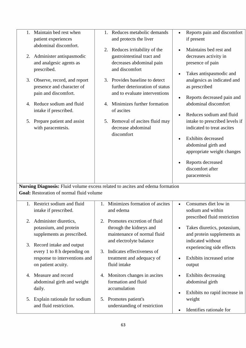
63
1. Maintain bed rest when
patient experiences
abdominal discomfort.
2. Administer antispasmodic
and analgesic agents as
prescribed.
3. Observe, record, and report
presence and character of
pain and discomfort.
4. Reduce sodium and fluid
intake if prescribed.
5. Prepare patient and assist
with paracentesis.
1. Reduces metabolic demands
and protects the liver
2. Reduces irritability of the
gastrointestinal tract and
decreases abdominal pain
and discomfort
3. Provides baseline to detect
further deterioration of status
and to evaluate interventions
4. Minimizes further formation
of ascites
5. Removal of ascites fluid may
decrease abdominal
discomfort
Reports pain and discomfort
if present
Maintains bed rest and
decreases activity in
presence of pain
Takes antispasmodic and
analgesics as indicated and
as prescribed
Reports decreased pain and
abdominal discomfort
Reduces sodium and fluid
intake to prescribed levels if
indicated to treat ascites
Exhibits decreased
abdominal girth and
appropriate weight changes
Reports decreased
discomfort after
paracentesis
Nursing Diagnosis: Fluid volume excess related to ascites and edema formation
Goal: Restoration of normal fluid volume
1. Restrict sodium and fluid
intake if prescribed.
2. Administer diuretics,
potassium, and protein
supplements as prescribed.
3. Record intake and output
every 1 to 8 h depending on
response to interventions and
on patient acuity.
4. Measure and record
abdominal girth and weight
daily.
5. Explain rationale for sodium
and fluid restriction.
1. Minimizes formation of ascites
and edema
2. Promotes excretion of fluid
through the kidneys and
maintenance of normal fluid
and electrolyte balance
3. Indicates effectiveness of
treatment and adequacy of
fluid intake
4. Monitors changes in ascites
formation and fluid
accumulation
5. Promotes patient's
understanding of restriction
Consumes diet low in
sodium and within
prescribed fluid restriction
Takes diuretics, potassium,
and protein supplements as
indicated without
experiencing side effects
Exhibits increased urine
output
Exhibits decreasing
abdominal girth
Exhibits no rapid increase in
weight
Identifies rationale for

64
6. Prepare patient and assist
with paracentesis.
and cooperation with it
6. Paracentesis will temporarily
decrease amount of ascites
present.
sodium and fluid restriction
Shows a decrease in ascites
with decreased weight
Nursing Diagnosis: Disturbed thought processes related to deterioration of liver function and increased serum
ammonia level
Goal: Improved mental status; safety maintained
1. Restrict dietary protein as
prescribed.
2. Give frequent, small
feedings of carbohydrates.
3. Protect from infection.
4. Keep environment warm and
draft-free.
5. Pad the side rails of the bed.
6. Limit visitors.
7. Provide careful nursing
surveillance to ensure
patient's safety.
8. Avoid opioids and
barbiturates.
9. Awaken at intervals (every
2–4 h) to assess cognitive
status.
1. Reduces source of ammonia
(protein foods)
2. Promotes consumption of
adequate carbohydrates for
energy requirements and
spares protein from
breakdown for energy
3. Minimizes risk for further
increase in metabolic
requirements
4. Minimizes shivering, which
would increase metabolic
requirements
5. Provides protection for the
patient should hepatic coma
and seizure activity occur
6. Minimizes patient's activity
and metabolic requirements
7. Provides close monitoring of
new symptoms and
minimizes trauma to the
confused patient
8. Prevents masking of
symptoms of hepatic coma
and prevents drug overdose
secondary to reduced ability
of the damaged liver to
metabolize opioids and
barbiturates
Adheres to protein
restriction
Demonstrates an interest
in events and activities in
environment
Demonstrates normal
attention span
Follows and participates
in conversation
appropriately
Is oriented to person,
place, and time
Remains in bed when
indicated
Reports no urinary or fecal
incontinence
Experiences no seizures
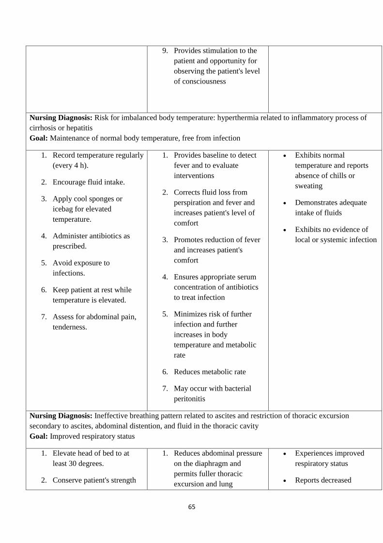
65
9. Provides stimulation to the
patient and opportunity for
observing the patient's level
of consciousness
Nursing Diagnosis: Risk for imbalanced body temperature: hyperthermia related to inflammatory process of
cirrhosis or hepatitis
Goal: Maintenance of normal body temperature, free from infection
1. Record temperature regularly
(every 4 h).
2. Encourage fluid intake.
3. Apply cool sponges or
icebag for elevated
temperature.
4. Administer antibiotics as
prescribed.
5. Avoid exposure to
infections.
6. Keep patient at rest while
temperature is elevated.
7. Assess for abdominal pain,
tenderness.
1. Provides baseline to detect
fever and to evaluate
interventions
2. Corrects fluid loss from
perspiration and fever and
increases patient's level of
comfort
3. Promotes reduction of fever
and increases patient's
comfort
4. Ensures appropriate serum
concentration of antibiotics
to treat infection
5. Minimizes risk of further
infection and further
increases in body
temperature and metabolic
rate
6. Reduces metabolic rate
7. May occur with bacterial
peritonitis
Exhibits normal
temperature and reports
absence of chills or
sweating
Demonstrates adequate
intake of fluids
Exhibits no evidence of
local or systemic infection
Nursing Diagnosis: Ineffective breathing pattern related to ascites and restriction of thoracic excursion
secondary to ascites, abdominal distention, and fluid in the thoracic cavity
Goal: Improved respiratory status
1. Elevate head of bed to at
least 30 degrees.
2. Conserve patient's strength
1. Reduces abdominal pressure
on the diaphragm and
permits fuller thoracic
excursion and lung
Experiences improved
respiratory status
Reports decreased

66
by providing rest periods and
assisting with activities.
3. Change position every 2 h.
4. Assist with paracentesis or
thoracentesis.
a. Explain procedure
and its purpose to
patient.
b. Have patient void
before paracentesis.
c. Support and maintain
position during
procedure.
d. Record both the
amount and the
character of fluid
aspirated.
e. Observe for evidence
of coughing,
increasing dyspnea,
or pulse rate.
expansion
2. Reduces metabolic and
oxygen requirements
3. Promotes expansion and
oxygenation of all areas of
the lungs
4. Paracentesis and
thoracentesis (performed to
remove fluid from the
abdominal and thoracic
cavities, respectively) may
be frightening to the patient.
a. Helps obtain patient's
cooperation with
procedures
b. Prevents inadvertent
bladder injury
c. Prevents inadvertent
organ or tissue injury
d. Provides record of
fluid removed and
indication of severity
of limitation of lung
expansion by fluid
e. Indicates irritation of
the pleural space and
evidence of
pneumothorax or
hemothorax.
shortness of breath
Reports increased strength
and sense of well-being
Exhibits normal
respiratory rate (12–
18/min) with no
adventitious sounds
Exhibits full thoracic
excursion without shallow
respirations
Exhibits normal arterial
blood gases
Exhibits adequate oxygen
saturation by pulse
oximetry
Experiences absence of
confusion or cyanosis
Collaborative Problem: Gastrointestinal bleeding and hemorrhage
Goal: Absence of episodes of gastrointestinal bleeding and hemorrhage
1. Assess patient for evidence
of gastrointestinal bleeding
or hemorrhage. If bleeding
does occur:
a. Monitor vital signs
1. Allows early detection of
signs and symptoms of
bleeding and hemorrhage
2. Minimizes increases in intra-
abdominal pressure that
10. Assess patient for
evidence of
gastrointestinal bleeding
or hemorrhage. If bleeding
does occur:

67
(blood pressure,
pulse, respiratory
rate) every 4 h or
more frequently,
depending on acuity.
b. Assess skin
temperature, level of
consciousness every
4 hours or more
frequently, depending
on acuity.
c. Monitor
gastrointestinal
secretions and output
(emesis, stool for
occult or obvious
bleeding). Test
emesis for blood
once per shift and
with any color
change. Hematest
each stool.
d. Monitor hematocrit
and hemoglobin for
trends and changes.
2. Avoid activities that increase
intra-abdominal pressure
(straining, turning).
a. Avoid
coughing/sneezing.
b. Assist patient to turn.
c. Keep all needed
items within easy
reach.
d. Use measures to
prevent constipation
such as adequate
fluid intake; stool
could lead to rupture and
bleeding of esophageal or
gastric varices
3. Equipment, medications, and
supplies will be readily
available if patient
experiences bleeding from
ruptured esophageal or
gastric varices.
4. Gastrointestinal bleeding and
hemorrhage require
emergency measures (eg,
insertion of Blakemore tube,
administration of fluids and
medications).
5. The patient is at high risk for
respiratory complications,
including asphyxiation if
gastric balloon of tamponade
tube ruptures or migrates
upward.
6. The patient who experiences
hemorrhage is very anxious
and fearful; minimizing
anxiety assists in control of
hemorrhage.
7. Risk of rebleeding is high
with all treatment modalities
used to halt gastrointestinal
bleeding.
8. Family members are likely to
be anxious about the
patient's status; providing
information will reduce their
anxiety level and promote
more effective coping.
9. Risk of rebleeding is high.
Subtle signs may be more
a. Monitor vital signs
(blood pressure,
pulse, respiratory
rate) every 4 h or
more frequently,
depending on
acuity.
b. Assess skin
temperature, level
of consciousness
every 4 hours or
more frequently,
depending on
acuity.
c. Monitor
gastrointestinal
secretions and
output (emesis,
stool for occult or
obvious bleeding).
Test emesis for
blood once per
shift and with any
color change.
Hematest each
stool.
d. Monitor
hematocrit and
hemoglobin for
trends and
changes.
11. Avoid activities that
increase intra-abdominal
pressure (straining,
turning).
a. Avoid
coughing/sneezing.
b. Assist patient to
turn.

68
softeners.
e. Ensure small meals.
3. Have equipment (Blakemore
tube, medications, IV fluids)
available if indicated.
4. Assist with procedures and
therapy needed to treat
gastrointestinal bleeding and
hemorrhage.
5. Monitor respiratory status
every hour and minimize risk
of respiratory complications
if balloon tamponade is
needed.
6. Prepare patient physically
and psychologically for other
treatment modalities if
needed.
7. Monitor patient for
recurrence of bleeding and
hemorrhage.
8. Keep family informed of
patient's status.
9. Once recovered from
bleeding episode, provide
patient and family with
information regarding signs
and symptoms of
gastrointestinal bleeding.
quickly identified c. Keep all needed
items within easy
reach.
d. Use measures to
prevent
constipation such
as adequate fluid
intake; stool
softeners.
e. Ensure small
meals.
12. Have equipment
(Blakemore tube,
medications, IV fluids)
available if indicated.
13. Assist with procedures
and therapy needed to
treat gastrointestinal
bleeding and hemorrhage.
14. Monitor respiratory status
every hour and minimize
risk of respiratory
complications if balloon
tamponade is needed.
15. Prepare patient physically
and psychologically for
other treatment modalities
if needed.
16. Monitor patient for
recurrence of bleeding and
hemorrhage.
17. Keep family informed of
patient's status.
18. Once recovered from
bleeding episode, provide
patient and family with
information regarding
signs and symptoms of
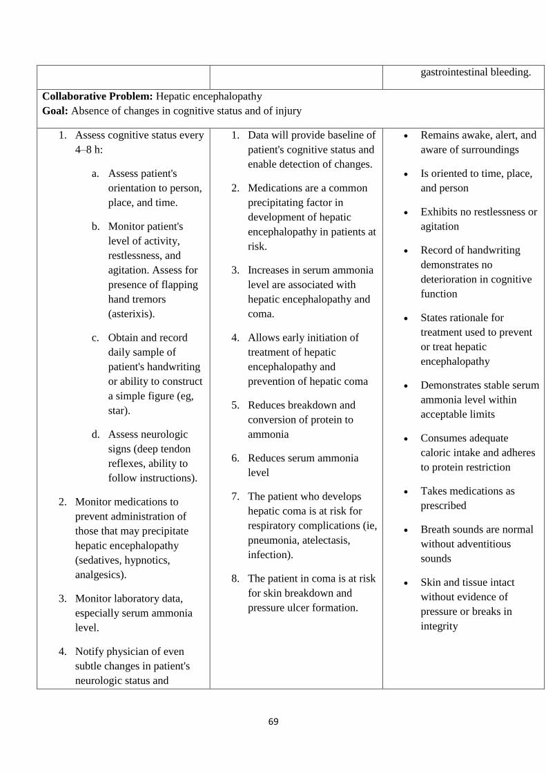
69
gastrointestinal bleeding.
Collaborative Problem: Hepatic encephalopathy
Goal: Absence of changes in cognitive status and of injury
1. Assess cognitive status every
4–8 h:
a. Assess patient's
orientation to person,
place, and time.
b. Monitor patient's
level of activity,
restlessness, and
agitation. Assess for
presence of flapping
hand tremors
(asterixis).
c. Obtain and record
daily sample of
patient's handwriting
or ability to construct
a simple figure (eg,
star).
d. Assess neurologic
signs (deep tendon
reflexes, ability to
follow instructions).
2. Monitor medications to
prevent administration of
those that may precipitate
hepatic encephalopathy
(sedatives, hypnotics,
analgesics).
3. Monitor laboratory data,
especially serum ammonia
level.
4. Notify physician of even
subtle changes in patient's
neurologic status and
1. Data will provide baseline of
patient's cognitive status and
enable detection of changes.
2. Medications are a common
precipitating factor in
development of hepatic
encephalopathy in patients at
risk.
3. Increases in serum ammonia
level are associated with
hepatic encephalopathy and
coma.
4. Allows early initiation of
treatment of hepatic
encephalopathy and
prevention of hepatic coma
5. Reduces breakdown and
conversion of protein to
ammonia
6. Reduces serum ammonia
level
7. The patient who develops
hepatic coma is at risk for
respiratory complications (ie,
pneumonia, atelectasis,
infection).
8. The patient in coma is at risk
for skin breakdown and
pressure ulcer formation.
Remains awake, alert, and
aware of surroundings
Is oriented to time, place,
and person
Exhibits no restlessness or
agitation
Record of handwriting
demonstrates no
deterioration in cognitive
function
States rationale for
treatment used to prevent
or treat hepatic
encephalopathy
Demonstrates stable serum
ammonia level within
acceptable limits
Consumes adequate
caloric intake and adheres
to protein restriction
Takes medications as
prescribed
Breath sounds are normal
without adventitious
sounds
Skin and tissue intact
without evidence of
pressure or breaks in
integrity
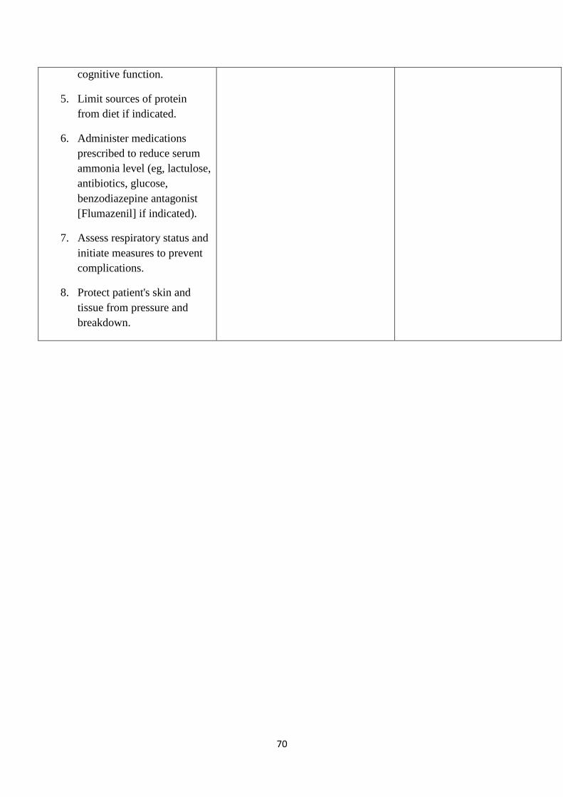
70
cognitive function.
5. Limit sources of protein
from diet if indicated.
6. Administer medications
prescribed to reduce serum
ammonia level (eg, lactulose,
antibiotics, glucose,
benzodiazepine antagonist
[Flumazenil] if indicated).
7. Assess respiratory status and
initiate measures to prevent
complications.
8. Protect patient's skin and
tissue from pressure and
breakdown.
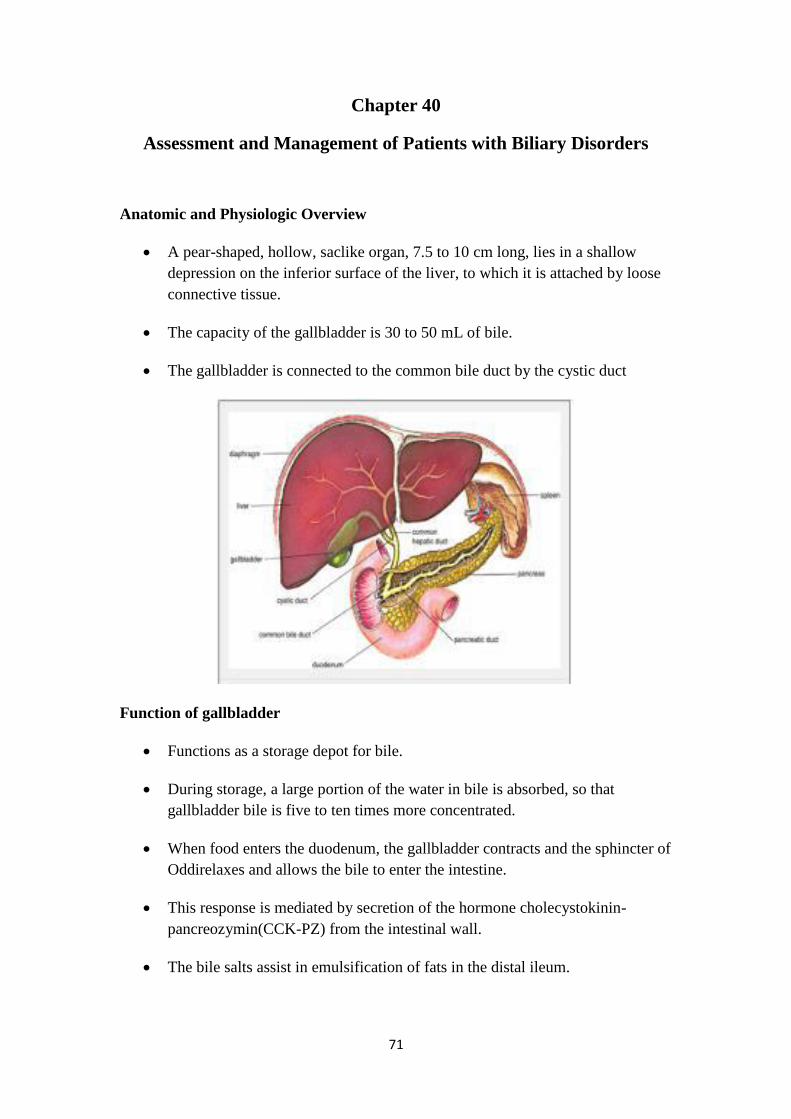
71
Chapter 40
Assessment and Management of Patients with Biliary Disorders
Anatomic and Physiologic Overview
A pear-shaped, hollow, saclike organ, 7.5 to 10 cm long, lies in a shallow
depression on the inferior surface of the liver, to which it is attached by loose
connective tissue.
The capacity of the gallbladder is 30 to 50 mL of bile.
The gallbladder is connected to the common bile duct by the cystic duct
Function of gallbladder
Functions as a storage depot for bile.
During storage, a large portion of the water in bile is absorbed, so that
gallbladder bile is five to ten times more concentrated.
When food enters the duodenum, the gallbladder contracts and the sphincter of
Oddirelaxes and allows the bile to enter the intestine.
This response is mediated by secretion of the hormone cholecystokinin-
pancreozymin(CCK-PZ) from the intestinal wall.
The bile salts assist in emulsification of fats in the distal ileum.
72
The pancreas
The pancreas, located in the upper abdomen, has endocrine as well as
exocrine functions The secretion of pancreatic enzymes into the
gastrointestinal tract through the pancreatic duct represents its exocrine
function. The secretion of insulin, glucagon, and somatostatin directly into the
bloodstream represents its endocrine function.
Exocrine Pancreas
The secretions of the exocrine portion of the pancreas are collected in the
pancreatic duct, which joins the common bile duct and enters the duodenum at
the ampulla of Vater. Surrounding the ampulla is the sphincter of Oddi, which
partially controls the rate at which secretions from the pancreas and the
gallbladder enter the duodenum.
Exocrine Pancreas
The secretions of the exocrine pancreas are digestive enzymes high in protein
content and an electrolyte-rich fluid. The secretions are very alkaline because
of their high concentration of sodium bicarbonate and are capable of
neutralizing the highly acid gastric juice that enters the duodenum.
The enzyme secretions include amylase, which aids in the digestion of
carbohydrates; trypsin, which aids in the digestion of proteins; and lipase,
which aids in the digestion of fats.
Endocrine Pancreas
The islets of Langerhans, the endocrine part of the pancreas, are collections of
cells embedded in the pancreatic tissue.
They are composed of alpha, beta, and delta cells. The hormone produced by
the beta cells is called insulin; the alpha cells secrete glucagon and the delta
cells secrete somatostatin.
1. Insulin
A major action of insulin is to lower blood glucose by permitting entry of the
glucose into the cells of the liver, muscle, and other tissues, where it is either
stored as glycogen or used for energy. Insulin also promotes the storage of fat
in adipose tissue and the synthesis of proteins in various body tissues. In the
absence of insulin, glucose cannot enter the cells and is excreted in the urine.
73
2. Glucagon
The effect of glucagon (opposite to that of insulin) is chiefly to raise the blood
glucose by converting glycogen to glucose in the liver. Glucagon is secreted
by the pancreas in response to a decrease in the level of blood glucose.
3. Somatostatin
Somatostatin exerts a hypoglycemic effect by interfering with release of
growth hormone from the pituitary and glucagon from the pancreas, both of
which tend to raise blood glucose levels.
Endocrine Control of Carbohydrate Metabolism
Glucose for body energy needs is derived by metabolism of ingested
carbohydrates and also from proteins by the process of gluconeogenesis.
Glucose can be stored temporarily in the liver, muscles, and other tissues in
the form of glycogen. The endocrine system controls the level of blood
glucose by regulating the rate at which glucose is synthesized, stored, and
moved to and from the bloodstream. Through the action of hormones, blood
glucose is normally maintained at about 100 mg/dL (5.5 mmol/L).
Insulin is the primary hormone that lowers the blood glucose level. Hormones
that raise the blood glucose level are glucagon, epinephrine,
adrenocorticosteroids, growth hormone, and thyroid hormone.
The major exocrine function is to facilitate digestion through secretion of
enzymes into the proximal duodenum. Secretin and CCK-PZ are hormones
from the gastrointestinal tract that aid in the digestion of food substances by
controlling the secretions of the pancreas. Neural factors also influence
pancreatic enzyme secretion. Considerable dysfunction of the pancreas must
occur before enzyme secretion decreases and protein and fat digestion
becomes impaired. Pancreatic enzyme secretion is normally 1,500 to 2,500
mL/day.
Definition of Biliary Terms
Cholecystitis: inflammation of the gallbladder
Cholelithiasis: the presence of calculi in the gallbladder
Cholecystectomy: removal of the gallbladder
Cholecystostomy: opening and drainage of the gallbladder
Choledochotomy: opening into the common duct
74
Choledocholithiasis: stones in the common duct
Choledocholithotomy: incision of common bile duct for removal of stones
Cholecystitis
Acute inflammation (cholecystitis) of the gallbladder causes pain, tenderness,
and rigidity of the upper right abdomen that may radiate to the midsternalarea
or right shoulder
Associated with nausea, vomiting, and the usual signs of an acute
inflammation.
An empyemaof the gallbladder develops if the gallbladder becomes filled with
purulent fluid (pus).
Calculouscholecystitisis the cause of more than 90% of cases of acute
cholecystitis.
Gallbladder stone obstructs bile outflow. Bile remaining in the gallbladder
initiates a chemical reaction; autolysis and edema occur; and the blood vessels
in the gallbladder are compressed, compromising its vascular supply.
Gangrene of the gallbladder with perforation may result.
Bacteria play a minor role in acute cholecystitis.
Acalculouscholecystitisoccurs after major surgical procedures, severe trauma,
or burns.
Cholelithiasis
Gallstones usually form in the gallbladder from the solid constituents of bile
Vary greatly in size, shape, and composition.
Common after 40 years of age, especially in women.

75
Risk Factors for Cholelithiasis
Obesity, Women, especially those who have had multiple pregnancies
Frequent changes in weight
Rapid weight loss (leads to rapid development of gallstones and high risk of
symptomatic disease)
Treatment with high-dose estrogen (ie, in prostate cancer)
Low-dose estrogen therapy—a small increase in the risk of gallstones
Ilealresection or disease
Cystic fibrosis
Diabetes mellitus
Pathophysiology
There are two major types of gallstones:
1. Composed of pigment.
o Probably form when unconjugatedpigments in the bile precipitate to
form stones.
o The risk is increased in patients with cirrhosis, hemolysis, and
infections of the biliarytract.

76
o Pigment stones cannot be dissolved and must be removed surgically.
2. Composed of cholesterol.
o Four times more women than men develop cholesterol stones and
gallbladder disease; the women are usually older than 40, multiparous,
and obese.
o The incidence rises with oral contraceptives, estrogens.
Clinical Manifestations
Gallstones may be silent with only mild GI symptoms. Such stones may be
detected incidentally.
Symptoms are due to the disease of the gallbladder itself or to obstruction of
the bile passages by a gallstone.
The symptoms may be acute or chronic.
Epigastricdistress, such as fullness, abdominal distention, and vague pain in
the right upper quadrant of the abdomen, may occur. This distress may follow
a meal rich in fried or fatty foods.
A. Pain and Biliary Colic
If a gallstone obstructs the cystic duct, the gallbladder becomes distended,
inflamed, and eventually infected (acute cholecystitis).
The patient develops fever and may have a palpable abdominal mass.
The patient may have biliarycolic with excruciating upper right abdominal
pain that radiates to the back or right shoulder,
It is usually associated with nausea and vomiting, and is noticeable several
hours after a heavy meal.
B. Jaundice
Jaundice occurs in a few patients with gallbladder disease and usually occurs
with obstruction of the common bile duct.
C. Changes in Urine and Stool Color
The excretion of the bile pigments by the kidneys gives the urine a very dark
color. The feces, no longer colored with bile pigments, are grayish, clay-
colored.
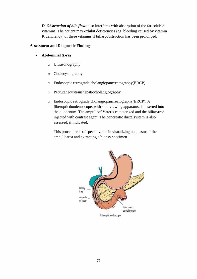
77
D. Obstruction of bile flow: also interferes with absorption of the fat-soluble
vitamins. The patient may exhibit deficiencies (eg, bleeding caused by vitamin
K deficiency) of these vitamins if biliaryobstruction has been prolonged.
Assessment and Diagnostic Findings
Abdominal X-ray
o Ultrasonography
o Cholecystography
o Endoscopic retrograde cholangiopancreatography(ERCP)
o Percutaneoustranshepaticcholangiography
o Endoscopic retrograde cholangiopancreatography(ERCP). A
fiberopticduodenoscope, with side-viewing apparatus, is inserted into
the duodenum. The ampullaof Vateris catheterized and the biliarytree
injected with contrast agent. The pancreatic ductalsystem is also
assessed, if indicated.
This procedure is of special value in visualizing neoplasmsof the
ampullaarea and extracting a biopsy specimen.
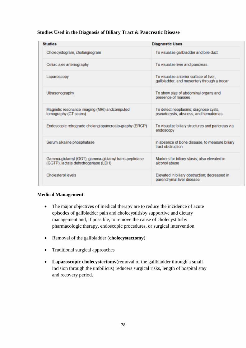
78
Studies Used in the Diagnosis of Biliary Tract & Pancreatic Disease
Medical Management
The major objectives of medical therapy are to reduce the incidence of acute
episodes of gallbladder pain and cholecystitisby supportive and dietary
management and, if possible, to remove the cause of cholecystitisby
pharmacologic therapy, endoscopic procedures, or surgical intervention.
Removal of the gallbladder (cholecystectomy)
Traditional surgical approaches
Laparoscopic cholecystectomy(removal of the gallbladder through a small
incision through the umbilicus) reducers surgical risks, length of hospital stay
and recovery period.
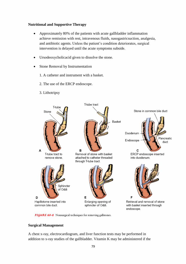
79
Nutritional and Supportive Therapy
Approximately 80% of the patients with acute gallbladder inflammation
achieve remission with rest, intravenous fluids, nasogastricsuction, analgesia,
and antibiotic agents. Unless the patient‘s condition deteriorates, surgical
intervention is delayed until the acute symptoms subside.
Ursodeoxycholicacid given to dissolve the stone.
Stone Removal by Instrumentation
1. A catheter and instrument with a basket.
2. The use of the ERCP endoscope.
3. Lithotripsy
Surgical Management
A chest x-ray, electrocardiogram, and liver function tests may be performed in
addition to x-ray studies of the gallbladder. Vitamin K may be administered if the
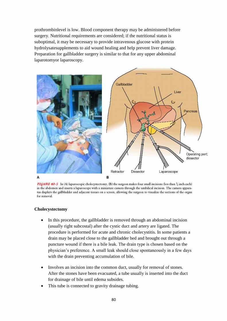
80
prothrombinlevel is low. Blood component therapy may be administered before
surgery. Nutritional requirements are considered; if the nutritional status is
suboptimal, it may be necessary to provide intravenous glucose with protein
hydrolysatesupplements to aid wound healing and help prevent liver damage.
Preparation for gallbladder surgery is similar to that for any upper abdominal
laparotomyor laparoscopy.
Cholecystectomy
In this procedure, the gallbladder is removed through an abdominal incision
(usually right subcostal) after the cystic duct and artery are ligated. The
procedure is performed for acute and chronic cholecystitis. In some patients a
drain may be placed close to the gallbladder bed and brought out through a
puncture wound if there is a bile leak. The drain type is chosen based on the
physician‘s preference. A small leak should close spontaneously in a few days
with the drain preventing accumulation of bile.
Involves an incision into the common duct, usually for removal of stones.
After the stones have been evacuated, a tube usually is inserted into the duct
for drainage of bile until edema subsides.
This tube is connected to gravity drainage tubing.
81
The gallbladder also contains stones, and as a rule a cholecystectomyis
performed at the same time.
Percutaneous cholecystostomy
has been used in the treatment and diagnosis of acute cholecystitisin patients
who are poor risks for any surgical procedure or for general anesthesia. These
may include patients with sepsis or severe cardiac, renal, pulmonary, or liver
failure.
Under local anesthesia, a fine needle is inserted through the abdominal wall
and liver edge into the gallbladder under the guidance of ultrasound or
computed tomography. Bile is aspirated to ensure adequate placement of the
needle, and a catheter is inserted into the gallbladder to decompress the
biliarytract.
Disorders of the Pancreas
Pancreatitis (inflammation of the pancreas) is a serious disorder. It can be
acute or chronic.
Acute pancreatitis can be a medical emergency associated with a high risk for
life-threatening complications and mortality.
It does not usually lead to chronic pancreatitis. However, chronic pancreatitis
can be characterized by acute episodes. Typically, patients are men 40 to 45
years of age with a history of alcoholism or women 50 to 55 years of age with
a history of biliarydisease.
Etiology is unknown, but it could be related to obstruction that causes pack up
of pancreatic enzymes, along with bile, they cause autodigestion
Acute Pancreatitis
Ranges from a mild, self-limiting to severe.
Mild acute pancreatitis is characterized by edema and inflammation of the
pancreas. Minimal organ dysfunction is present, and return to normal usually
occurs within 6 months.
patient is acutely ill and at risk for hypovolemicshock, fluid and electrolyte
disturbances, and sepsis. The tissue becomes necrotic with possible abscess
formation.
Systemic complications, such as acute respiratory distress syndrome, shock,
DIC , & pleural effusion, can increase the mortality rate to 50% or higher

82
Pathophysiology
Self-digestion of the pancreas by its own proteolyticenzymes, principally
trypsin, causes acute pancreatitis.
80% of patients with acute pancreatitis have biliarytract disease; but only 5%
of patients with gallstones develop pancreatitis. Gallstones enter the common
bile duct and lodge at the ampullaof Vater, obstructing the flow of pancreatic
juice or causing a reflux of bile from the common bile duct into the pancreatic
duct, thus activating the powerful enzymes within the pancreas which leads to
vasodilation, increased vascular permeability, necrosis, erosion, and
hemorrhage.
Other less common causes of pancreatitis include bacterial or viral infection
i.e. mumps virus.
Spasm and edema of the ampullaof Vater, resulting from duodenitis.
Blunt abdominal trauma, peptic ulcer disease, ischemic vascular disease,
hyperlipidemia, hypercalcemia, and the use of corticosteroids,
thiazidediuretics, and oral contraceptives .
Clinical Manifestations
Severe abdominal pain is the major symptom that causes the patient to seek
medical care. Typically, the pain occurs in the midepigastrium.
Pain is frequently acute in onset, occurring 24 to 48 hours after a very heavy
meal or alcohol ingestion, and it may be diffuse and difficult to localize. It is
generally more severe after meals and is unrelieved by antacids.
The patient appears acutely ill. Abdominal guarding is present. A rigid or
board-like abdomen may develop and is generally an ominous sign; the
abdomen may remain soft in the absence of peritonitis.
Ecchymosis(bruising) in the flank or around the umbilicus may indicate severe
pancreatitis.
Nausea and vomiting are common. The emesis is usually gastric in origin but
may also be bile-stained.
Fever, jaundice, mental confusion, and agitation also may occur.
Respiratory distress & hypoxia are common, the patient may develop dyspnea,
tachypnea, and abnormal blood gas values.
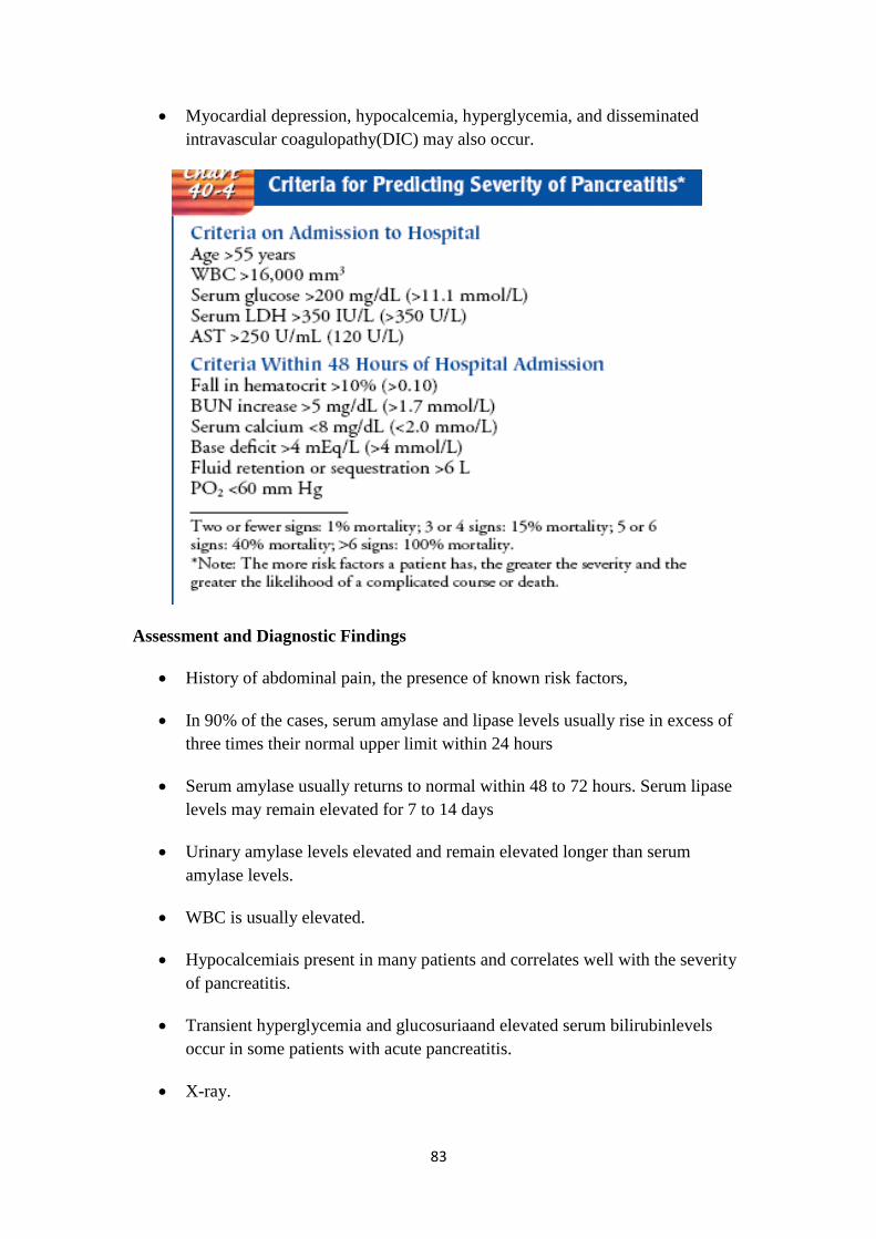
83
Myocardial depression, hypocalcemia, hyperglycemia, and disseminated
intravascular coagulopathy(DIC) may also occur.
Assessment and Diagnostic Findings
History of abdominal pain, the presence of known risk factors,
In 90% of the cases, serum amylase and lipase levels usually rise in excess of
three times their normal upper limit within 24 hours
Serum amylase usually returns to normal within 48 to 72 hours. Serum lipase
levels may remain elevated for 7 to 14 days
Urinary amylase levels elevated and remain elevated longer than serum
amylase levels.
WBC is usually elevated.
Hypocalcemiais present in many patients and correlates well with the severity
of pancreatitis.
Transient hyperglycemia and glucosuriaand elevated serum bilirubinlevels
occur in some patients with acute pancreatitis.
X-ray.

84
Ultrasound and CT scans.
Hematocritand hemoglobin levels to monitor for bleeding.
Peritoneal fluid may contain increased levels of pancreatic enzymes.
The stools are often bulky, pale, and foul-smelling. Fat content of stools varies
between 50% and 90% in pancreatic disease; normally, the fat content is 20%.
Medical Management
Management of the patient with acute pancreatitis is directed toward relieving
symptoms and preventing or treating complications. All oral intake is withheld
to inhibit pancreatic stimulation and secretion of pancreatic enzymes.
Parenteral nutrition is usually an important part of therapy, Nasogastric
suction may be used to relieve nausea and vomiting, to decrease painful
abdominal distention and paralytic ileus, and to remove hydrochloric acid so
that it does not enter the duodenum and stimulate the pancreas.
Histamine-2 (H2) antagonists to decrease pancreatic activity by inhibiting HCl
secretion.
Pain Management
Adequate pain medication is essential during the course of acute pancreatitis
to provide sufficient pain relief and minimize restlessness, which may
stimulate pancreatic secretion further. Morphine and morphine derivatives are
often avoided because it has been thought that they cause spasm of the
sphincter of Oddi; meperidine (Demerol) is often prescribed because it is less
likely to cause spasm of the sphincter
Intensive care
Correction of fluid and blood loss and low albumin levels is necessary to
maintain fluid volume and prevent renal failure. The patient is usually acutely
ill and is monitored in the intensive care unit, where hemodynamic monitoring
and arterial blood gas monitoring are initiated. Antibiotic agents may be
prescribed if infection is present; insulin may be required if significant
hyperglycemia occurs.
Respiratory Care
Aggressive respiratory care is indicated because of the high risk for elevation
of the diaphragm, pulmonary infiltrates and effusion, and atelectasis.
Hypoxemia occurs in a significant number of patients with acute pancreatitis
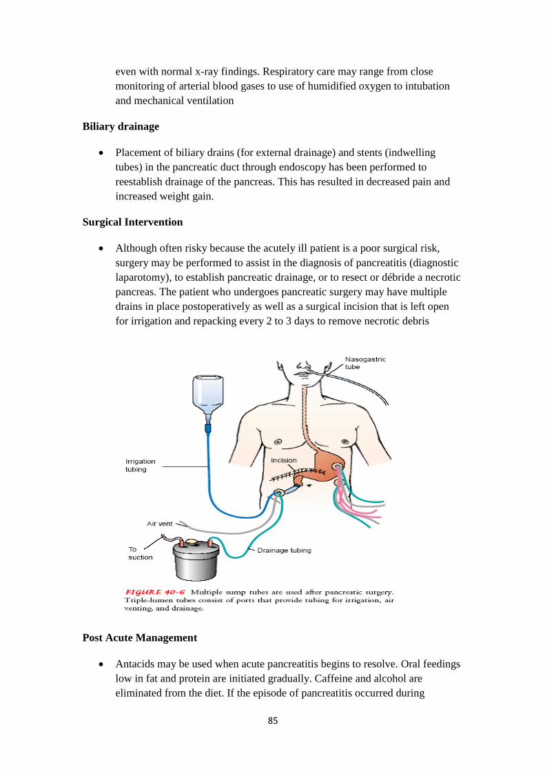
85
even with normal x-ray findings. Respiratory care may range from close
monitoring of arterial blood gases to use of humidified oxygen to intubation
and mechanical ventilation
Biliary drainage
Placement of biliary drains (for external drainage) and stents (indwelling
tubes) in the pancreatic duct through endoscopy has been performed to
reestablish drainage of the pancreas. This has resulted in decreased pain and
increased weight gain.
Surgical Intervention
Although often risky because the acutely ill patient is a poor surgical risk,
surgery may be performed to assist in the diagnosis of pancreatitis (diagnostic
laparotomy), to establish pancreatic drainage, or to resect or débride a necrotic
pancreas. The patient who undergoes pancreatic surgery may have multiple
drains in place postoperatively as well as a surgical incision that is left open
for irrigation and repacking every 2 to 3 days to remove necrotic debris
Post Acute Management
Antacids may be used when acute pancreatitis begins to resolve. Oral feedings
low in fat and protein are initiated gradually. Caffeine and alcohol are
eliminated from the diet. If the episode of pancreatitis occurred during

86
treatment with thiazide diuretics, corticosteroids, or oral contraceptives, these
medications are discontinued.
Follow-up of the patient may include ultrasound, x-ray studies, or ERCP to
determine whether the pancreatitis is resolving and to assess for abscesses and
pseudocysts.
Chronic Pancreatitis
is an inflammatory disorder characterized by progressive anatomic and
functional destruction of the pancreas.
As cells are replaced by fibrous tissue with repeated attacks of pancreatitis,
pressure within the pancreas increases. The end result is mechanical
obstruction of the pancreatic and common bile ducts and the duodenum.
Additionally, there is atrophy of the epitheliumof the ducts, inflammation, and
destruction of the secreting cells of the pancreas.
Alcohol consumption in Western societies and malnutrition worldwide are the
major causes of chronic pancreatitis. Excessive and prolonged consumption of
alcohol accounts for approximately 70% of the cases
The incidence of pancreatitis is 50 times greater in alcoholics than in the
nondrinking population. Long-term alcohol consumption causes
hypersecretion of protein in pancreatic secretions, resulting in protein plugs
and calculi within the pancreatic ducts. Alcohol also has a direct toxic effect
on the cells of the pancreas. Damage to these cells is more likely to occur and
to be more severe in patients whose diets are poor in protein content and either
very high or very low in fat.
Clinical Manifestations
Characterized by recurring attacks of severe upper abdominal and back pain,
accompanied by vomiting. Attacks are often so painful that opioids, even in
large doses, do not provide relief. As the disease progresses, recurring attacks
of pain are more severe, more frequent, and of longer duration.
Weight loss is a major problem in chronic pancreatitis: more than 75% of
patients experience significant weight loss, usually caused by decreased
dietary intake secondary to anorexia or fear that eating will precipitate another
attack. Malabsorption occurs late in the disease, when as little as 10% of
pancreatic function remains. As a result, digestion, especially of proteins and
fats, is impaired. The stools become frequent, frothy, and foul-smelling
because of impaired fat digestion,

87
Assessment and Diagnostic Findings
ERCP is the most useful study in the diagnosis of chronic pancreatitis. It
provides detail about the anatomy of the pancreas and the pancreatic and
biliary ducts. It is also helpful in obtaining tissue for analysis and
differentiating pancreatitis from other conditions, such as carcinoma.
Magnetic resonance imaging, computed tomography, and ultrasound, have
been useful in the diagnostic evaluation of patients with suspected pancreatic
disorders.
A glucose tolerance test evaluates pancreatic islet cell function, information
necessary for making decisions about surgical resection of the pancreas. An
abnormal glucose tolerance test indicative of diabetes may be present.
Medical Management
The management of chronic pancreatitis depends on its probable cause in each
patient. Treatment is directed toward preventing and managing acute attacks,
relieving pain and discomfort, and managing exocrine and endocrine
insufficiency of pancreatitis.
Nonsurgical Management
Nonsurgical approaches may be indicated for the patient who refuses surgery,
who is a poor surgical risk, or whose disease and symptoms do not warrant
surgical intervention. Endoscopy to remove pancreatic duct stones and stent
strictures may be effective in selected patients to manage pain and relieve
obstruction.
Focus is usually on the use of nonopioid methods to manage pain. Diabetes
mellitus resulting from dysfunction of the pancreatic islet cells is treated with
diet, insulin, or oral antidiabetic agents.
Surgical Management
Surgery is generally carried out to relieve abdominal pain and discomfort,
restore drainage of pancreatic secretions, and reduce the frequency of acute
attacks of pancreatitis. The surgery performed depends on the anatomic and
functional abnormalities of the pancreas, including the location of disease
within the pancreas, diabetes, exocrine insufficiency, biliary stenosis, and
pseudocysts of the pancreas.
Pancreaticojejunostomy (also referred to as Roux-en-Y) with a side-to-side
anastomosis or joining of the pancreatic duct to the jejunum allows drainage of
the pancreatic secretions into the jejunum.

88
Other surgical procedures may be performed for different degrees and types of
disease, ranging from revision of the sphincter of the ampulla of Vater, to
internal drainage of a pancreatic cyst into the stomach, to insertion of a stent,
to wide resection or removal of the pancreas. A Whipple resection
(pancreaticoduodenectomy) has been carried out to relieve the pain of chronic
pancreatitis.
Autotransplantation or implantation of the patient‘s pancreatic islet cells has
been attempted to preserve the endocrine function of the pancreas in patients
who have undergone total pancreatectomy.
Pancreatic Cysts
As a result of the local necrosis that occurs at the time of acute pancreatitis,
collections of fluid may form in the vicinity of the pancreas. These become
walled off by fibrous tissue and are calledpancreatic pseudocysts. They are the
most common type of pancreatic cysts. Less common cysts occur as a result of
congenital anomalies or are secondary to chronic pancreatitis or trauma to the
pancreas.
Diagnosis of pancreatic cysts and pseudocysts is made by ultrasound,
computed tomography, and ERCP. ERCP may be used to define the anatomy
of the pancreas and evaluate the patency of pancreatic drainage. Pancreatic
pseudocysts may be of considerable size. Because of their location behind the
posterior peritoneum, when they enlarge they impinge on and displace the
stomach or the colon, which are adjacent. Eventually, through pressure or
secondary infection, they produce symptoms and require drainage
Cancer of the Pancreas
The incidence of pancreatic cancer has decreased slightly over the past 25
years in non-Caucasian men. It is the fifth leading cause of cancer deaths in
the United States and occurs most frequently in the fifth to seventh decades of
life . Cigarette smoking, exposure to industrial chemicals or toxins in the
environment, and a diet high in fat, meat, or both are associated with
pancreatic cancer, although their role is not completely clear. The risk for
pancreatic cancer increases as the extent of cigarette smoking increases.
Diabetes mellitus, chronic pancreatitis, and hereditary pancreatitis are also
associated
Cancer may arise in any portion of the pancreas (in the head, the body, or the
tail); clinical manifestations vary depending on the location of the lesion and
whether functioning, insulinsecreting pancreatic islet cells are involved.
Approximately 75% of pancreatic cancers originate in the head of the pancreas
and give rise to a distinctive clinical picture. Functioning islet cell tumors,
89
whether benign (adenoma) or malignant (carcinoma), are responsible for the
syndrome of hyperinsulinism.
Clinical Manifestations
Pain, jaundice, or both are present in more than 90% of patients and, along
with weight loss, are considered classic signs of pancreatic carcinoma.
However, they often do not appear until the disease is far advanced. Other
signs include rapid, profound, andprogressive weight loss as well as vague
upper or midabdominal pain or discomfort that is unrelated to any
gastrointestinal function and is often difficult to describe.
An important sign, when present, is the onset of symptoms of insulin
deficiency: glucosuria, hyperglycemia, and abnormal glucose tolerance. Thus,
diabetes may be an early sign of carcinoma of the pancreas. Meals often
aggravate epigastric pain, which usually occurs before the appearance of
jaundice and pruritus.
Assessment and Diagnostic Findings
Magnetic resonance imaging and computed tomography are used to identify
the presence of pancreatic tumors. ERCP is also used in the diagnosis of
pancreatic carcinoma. Cells obtained during ERCP are sent to the laboratory
for examination. Gastrointestinal x-ray findings may demonstrate deformities
in adjacent viscera caused by the impinging pancreatic mass. Percutaneous
fine-needle aspiration biopsy of the pancreas is used to diagnose pancreatic
tumors and confirm the diagnosis
Percutaneous transhepatic cholangiography is another procedure that may be
performed to identify obstructions of the biliary tract by a pancreatic tumor.
Several tumor markers (eg, CA 19-9, CEA, DU-PAN-2) may be used
Angiography, computed tomography, and laparoscopy may be performed to
determine whether the tumor can be removed surgically. Intraoperative
ultrasonography has been used to determine if there is metastatic disease to
other organs.
Medical Management
If the tumor is resectable and localized (typically tumors in the head of the
pancreas), the surgical procedure to remove it is usually extensiveHowever,
definitive surgical treatment (ie, total excision of the lesion) is often not
possible because of the extensive growth when the tumor is finally diagnosed
and because of the probable widespread metastases (especially to the liver,
lungs, and bones). More often, treatment is limited to palliative measures.
90
The patient may be treated with radiation and chemotherapy (fluorouracil and
gemcitabine). If the patient undergoes surgery, intraoperative radiation therapy
(IORT) may be used to deliver a high dose of radiation to the tumor with
minimal injury to other tissues.
Nursing Management
Pain management and attention to nutritional requirements are important
nursing measures to improve the level of comfort. Skin care and nursing
measures are directed toward relief of pain and discomfort associated with
jaundice, anorexia, and profound weight loss. Specialty mattresses are
beneficial and protect bony prominences from pressure. Pain associated with
pancreatic cancer may be severe and may require liberal use of opioids;
Promoting Home and Community-Based Care
specific patient and family teaching indicated varies with the stage of
disease and the treatment choices made by the patient. If the patient elects to
receive chemotherapy, the nurse focuses teaching on prevention of side effects
and complications of the agents used. If surgery is performed to relieve
obstruction and establish biliary drainage, teaching addresses management of
the drainage system and monitoring for complications.
Continuing Care
A referral for home care is indicated to help the patient and family deal with
the physical problems and discomforts associated with pancreatic cancer and
the psychological impact of the disease. The home care nurse assesses the
patient‘s physical status, fluid and nutritional status, and skin integrity and the
adequacy of pain management.
Tumors Of The Headof The Pancreas
Sixty to eighty percent of pancreatic tumors occur in the head of the pancreas.
Tumors in this region of the pancreas obstruct the common bile duct where the
duct passes through the head of the pancreas to join the pancreatic duct and
empty at the ampulla of Vater into the duodenum. The tumors producing the
obstruction may arise from the pancreas, the common bile duct, or the ampulla
of Vater.
Clinical Manifestations
The obstructed flow of bile produces jaundice, clay-colored stools, and dark
urine. Malabsorption of nutrients and fat-soluble vitamins may result from
obstruction by the tumor to entry of bile in the gastrointestinal tract.
91
Abdominal discomfort or pain and pruritus may be noted, along with anorexia,
weight loss, and malaise.
Assessment and Diagnostic Findings
Diagnostic studies may include duodenography, angiography by hepatic or
celiac artery catheterization, pancreatic scanning, percutaneous transhepatic
cholangiography, ERCP, and percutaneous needle biopsy of the pancreas.
Results of a biopsy of the pancreas may aid in the diagnosis.
Medical Management
Before extensive surgery can be performed, a fairly long period of preparation
is often necessary because the patient‘s nutritional and physical condition is
often quite compromised. Various liver and pancreatic function studies are
performed. A diet high in protein along with pancreatic enzymes is often
prescribed. Preoperative preparation includes adequate hydration, correction
of prothrombin deficiency with vitamin K, and treatment of anemia to
minimize postoperative complications.
A biliary-enteric shunt may be performed to relieve the jaundice and, perhaps,
to provide time for a thorough diagnostic evaluation. Total pancreatectomy
(removal of the pancreas) may be performed if there is no evidence of direct
extension of the tumor to adjacent tissues or regional lymph nodes.
A pancreaticoduodenectomy
(Whipple‘s procedure or resection) is used for potentially resectable cancer of
the head of the pancreas79
This procedure involves removal of the gallbladder, distal portion of the
stomach, duodenum, head of the pancreas, and common bile duct and
anastomosis of the remaining pancreas and stomach to the jejunum
The result is removal of the tumor, allowing flow of bile into the jejunum.
When the tumor cannot be excised, the jaundice may be relieved by diverting
the bile flow into the jejunum by anastomosing the jejunum to the gallbladder,
a procedure known as cholecystojejunostomy.
The postoperative management of patients who have undergone a
pancreatectomy or a pancreaticoduodenectomy is similar to the management
of patients after extensive gastrointestinal and biliary surgery.

92
Nursing Management
Preoperatively and postoperatively, nursing care is directed toward promoting
patient comfort, preventing complications, and assisting the patient to return to
and maintain as normal and comfortable a life as possible.
The nurse closely monitors the patient in the intensive care unit after surgery;
the patient will have multiple intravenous and arterial lines in place for fluid
and blood replacement as well as for monitoring arterial pressures, and is on a
mechanical ventilator in the immediate postoperative period. It is important to
give careful attention to changes in vital signs, arterial blood gases and
pressures, pulse oximetry, laboratory values,and urine output. The nurse must
also consider the patient‘s compromised nutritional status and risk for
bleeding.
Promoting Home and Community-Based Care,
Teaching Patients Self-Care.
Continuing Care.
93
Nursing Process for the Patient Undergoing Surgery for Gallbladder Disease
Nursing Process for the Patient Undergoing Surgery for Gallbladder Disease
Assessment
The patient who is to undergo surgical treatment of gallbladder disease is often
admitted to the hospital or same-day surgery unit on the morning of surgery.
Preadmission testing is often completed a week or longer before admission. At that
time, the nurse instructs the patient about the need to avoid smoking, to enhance
pulmonary recovery postoperatively and to avoid respiratory complications. It also is
important to instruct the patient to avoid the use of aspirin and other agents (over-the-
counter medications and herbal remedies) that can alter coagulation and other
biochemical processes.
Assessment should focus on the patient's respiratory status. If a traditional surgical
approach is planned, the high abdominal incision required during surgery may
interfere with full respiratory excursion. The nurse notes a history of smoking,
previous respiratory problems, shallow respirations, a persistent or ineffective cough,
and the presence of adventitious breath sounds. Nutritional status is evaluated through
a dietary history and a general examination performed at the time of preadmission
testing. The nurse also reviews previously obtained laboratory results to obtain
information about the patient's nutritional status.
Diagnosis
Nursing Diagnoses
Based on all the assessment data, the major postoperative nursing diagnoses for the
patient undergoing surgery for gallbladder disease may include the following:
Acute pain and discomfort related to surgical incision
Impaired gas exchange related to the high abdominal surgical incision (if
traditional surgical cholecystectomy was performed)
Impaired skin integrity related to altered biliary drainage after surgical
intervention (if a T-tube was inserted because of retained stones in the
common bile duct or another drainage device was employed)
Imbalanced nutrition, less than body requirements, related to inadequate bile
secretion
Deficient knowledge about self-care activities related to incision care, dietary
modifications (if needed), medications, and reportable signs or symptoms (eg,
fever, bleeding, vomiting)
Collaborative Problems/Potential Complications
Based on assessment data, potential complications may include the following:
Bleeding
Gastrointestinal symptoms (may be related to biliary leak or injury to the
bowel)
Planning and Goals
The goals for the patient include relief of pain, adequate ventilation, intact skin and
improved biliary drainage, optimal nutritional intake, absence of complications, and
94
understanding of self-care routines.
Postoperative Nursing Interventions
After recovery from anesthesia, the patient is placed in the low Fowler's position.
Fluids may be administered intravenously, and nasogastric suction (a nasogastric tube
was probably inserted immediately before surgery for a nonlaparoscopic procedure)
may be instituted to relieve abdominal distention. Water and other fluids are
administered within hours after laparoscopic procedures. A soft diet is started after
bowel sounds return, which is usually the next day if the laparoscopic approach is
used.
Relieving Pain
The location of the subcostal incision in nonlaparoscopic gallbladder surgery often
causes the patient to avoid turning and moving, to splint the affected site, and to take
shallow breaths to prevent pain. Because full expansion of the lungs and gradually
increased activity are necessary to prevent postoperative complications, the nurse
administers analgesic agents as prescribed to relieve the pain and to promote well-
being in addition to helping the patient turn, cough, breathe deeply, and ambulate as
indicated. Use of a pillow or binder over the incision may reduce pain during these
maneuvers.
Improving Respiratory Status
Patients undergoing biliary tract surgery are especially prone to pulmonary
complications, as are all patients with upper abdominal incisions. Therefore, the nurse
reminds the patient to take deep breaths and cough every hour, to expand the lungs
fully and prevent atelectasis. The early and consistent use of incentive spirometry also
helps improve respiratory function. Early ambulation prevents pulmonary
complications as well as other complications, such as thrombophlebitis. Pulmonary
complications are more likely to occur in elderly patients, obese patients, and those
with preexisting pulmonary disease.
Promoting Skin Care and Biliary Drainage
In patients who have undergone a cholecystostomy or choledochostomy, the drainage
tube must be connected immediately to a drainage receptacle. The nurse should fasten
tubing to the dressings or to the patient's gown, with enough leeway for the patient to
move without dislodging or kinking it. Because a drainage system remains attached
when the patient is ambulating, the drainage bag may be placed in a bathrobe pocket
or fastened so that it is below the waist or common duct level. If a Penrose drain is
used, the nurse changes the dressings as required.
After these surgical procedures, the patient is observed for indications of infection,
leakage of bile into the peritoneal cavity, and obstruction of bile drainage. If bile is
not draining properly, an obstruction is probably causing bile to be forced back into
the liver and bloodstream. Because jaundice may result, the nurse should be
particularly observant of the color of the sclerae. The nurse should also note and
report right upper quadrant abdominal pain, nausea and vomiting, bile drainage
around any drainage tube, clay-colored stools, and a change in vital signs.
Bile may continue to drain from the drainage tract in considerable quantities for some
time, necessitating frequent changes of the outer dressings and protection of the skin
from irritation (bile is corrosive to the skin).
To prevent total loss of bile, the physician may want the drainage tube or collection
receptacle elevated above the level of the abdomen so that the bile drains externally
only if pressure develops in the duct system. Every 24 hours, the nurse measures the
bile collected and records the amount, color, and character of the drainage. After
several days of drainage, the tube may be clamped for 1 hour before and after each

95
meal to deliver bile to the duodenum to aid in digestion. Within 7 to 14 days, the
drainage tube is removed. The patient who goes home with a drainage tube in place
requires instruction and reassurance about the function and care of the tube.
In all patients with biliary drainage, the nurse (or the patient, if at home) observes the
stools daily and notes their color. Specimens of both urine and stool may be sent to
the laboratory for examination for bile pigments. In this way, it is possible to
determine whether the bile pigment is disappearing from the blood and is draining
again into the duodenum. Maintaining a careful record of fluid intake and output is
important.
Improving Nutritional Status
The nurse encourages the patient to eat a diet that is low in fats and high in
carbohydrates and proteins immediately after surgery. At the time of hospital
discharge, there are usually no special dietary instructions other than to maintain a
nutritious diet and avoid excessive fats. Fat restriction usually is lifted in 4 to 6 weeks,
when the biliary ducts dilate to accommodate the volume of bile once held by the
gallbladder and when the ampulla of Vater again functions effectively. After this time,
when the patient eats fat, adequate bile will be released into the digestive tract to
emulsify the fats and allow their digestion. This is in contrast to the condition before
surgery, when fats may not be digested completely or adequately, and flatulence may
occur. However, one purpose of gallbladder surgery is to allow a normal diet.
Monitoring and Managing Potential Complications
Bleeding may occur as a result of inadvertent puncture or nicking of a major blood
vessel. Postoperatively, the nurse closely monitors vital signs and inspects the surgical
incisions and drains, if any are in place, for evidence of bleeding. The nurse also
periodically assesses the patient for increased tenderness and rigidity of the abdomen.
If these signs and symptoms occur, they are reported to the surgeon. The nurse
instructs the patient and family to report to the surgeon any change in the color of
stools, because this may indicate complications. Gastrointestinal symptoms, although
not common, may occur with manipulation of the intestines during surgery.
After laparoscopic cholecystectomy, the nurse assesses the patient for loss of appetite,
vomiting, pain, distention of the abdomen, and temperature elevation. These may
indicate infection or disruption of the gastrointestinal tract and should be reported to
the surgeon promptly. Because the patient is discharged soon after laparoscopic
surgery, the patient and family are instructed verbally and in writing about the
importance of reporting these symptoms promptly.
Promoting Home and Community-Based Care
Teaching Patients Self-Care
The nurse instructs the patient about the medications that are prescribed (vitamins,
anticholinergics, and antispasmodics) and their actions. It also is important to inform
the patient and family about symptoms that should be reported to the physician,
including jaundice, dark urine, pale-colored stools, pruritus, and signs of
inflammation and infection, such as pain or fever.
Some patients report one to three bowel movements a day. This is the result of a
continual trickle of bile through the choledochoduodenal junction after
cholecystectomy. Usually, such frequency diminishes over a period of a few weeks to
several months.
If a patient is discharged from the hospital with a drainage tube still in place, the
patient and family need instructions about its management. The nurse instructs them
in proper care of the drainage tube and the importance of reporting to the surgeon

96
promptly any changes in the amount or characteristics of drainage. Assistance in
securing the appropriate dressings reduces the patient's anxiety about going home
with the drain or tube still in place. (See Chart 40-3 for more details.)
Patient Education
Managing Self-Care After Laparoscopic Cholecystectomy
Resuming Activity
Begin light exercise (walking) immediately.
Take a shower or bath after 1 or 2 days.
Drive a car after 3 or 4 days.
Avoid lifting objects exceeding 5 pounds after surgery, usually for 1 week.
Resume sexual activity when desired.
Caring for the Wound
Check puncture site daily for signs of infection.
Wash puncture site with mild soap and water.
Allow special adhesive strips on the puncture site to fall off. Do not pull them
off.
Resuming Eating
Resume your normal diet.
If you had fat intolerance before surgery, gradually add fat back into your diet
in small increments.
Managing Pain
You may experience pain or discomfort in your right shoulder from the gas
used to inflate your abdominal area during surgery. Sitting upright in bed or a
chair, walking, or use of a heating pad may ease the discomfort.
Take analgesics as needed and as prescribed. Report to surgeon if pain is
unrelieved even with analgesic use.
Managing Follow-Up Care
Make an appointment with your surgeon for 7 to 10 days after discharge.
Call your surgeon if you experience any signs or symptoms of infection at or
around the puncture site: redness, tenderness, swelling, heat, or drainage.
Call your surgeon if you experience a fever of 37.7°C (100°F) or more for 2
consecutive days.
Call your surgeon if you develop nausea, vomiting, or abdominal pain.
97
Chapter 41
Assessment and Management of Patients with Diabetes Mellitus
Diabetes Mellitus Definition
• Is a group of metabolic diseases characterized by increased levels of glucose in the
blood (hyperglycemia) resulting from defects in insulin secretion, insulin action, or
both
• related to:
–An endocrine disorder causes Abnormal insulin production
–Impaired insulin utilization
–Both abnormal production and impaired utilization
• Leading cause of heart disease, stroke, adult blindness, and nontraumatic lower limb
amputations
Diabetes Mellitus Etiology and Pathophysiology
–Produced by the b cells in the islets of Langherans of the pancreas
–Facilitates normal glucose range of 70 to 120 mg/dl
Functions of insulin
• Transports and metabolizes glucose for energy
• Stimulates storage of glucose in the liver and muscle (in the form of glycogen)
• Signals the liver to stop the release of glucose
• Enhances storage of dietary fat in adipose tissue
• Accelerates transport of amino acids (derived from dietary protein) into cells
• Inhibits breakdown of stored glucose, protein, and fat.
Type 1 Diabetes Mellitus
• Formerly known as ―juvenile onset‖ or ―insulin dependent‖ diabetes
• Most often occurs in people under 30 years of age
• Peak onset between ages 11 and 13

98
Etiology and Pathophysiology
• Progressive destruction of pancreatic b cells
• Autoantibodies cause a reduction of 80% to 90% of normal b cell function before
manifestations occur
• Causes:
–Genetic predisposition
• Related to human leukocyte antigens (HLAs)
–Exposure to a virus
Combined genetic, immunologic, and possibly environmental (eg, toxins, viral)
factors are thought to contribute to beta cell destruction.
There is also evidence of an autoimmune response in type 1 diabetes. This is an
abnormal response in which antibodies are directed against normal tissues of the
body, responding to these tissues as if they were foreign.
Onset of Disease
• Manifestations develop when the pancreas can no longer produce insulin
–Rapid onset of symptoms
–Present at ER with ketoacidosis
Type 1 Diabetes Mellitus Onset of Disease
• Weight loss
• Polydipsia
• Polyuria
• Polyphagia
• Diabetic ketoacidosis (DKA)
–Occurs in the absence of exogenous insulin
–Life-threatening condition
–Results in metabolic acidosis
The destruction of the beta cells results in decreased insulin production,
unchecked glucose production by the liver, and fasting hyperglycemia.
In addition, glucose cannot be stored in the liver but instead remains in the
bloodstream and contributes to postprandial (after meals) hyperglycemia.

99
If the concentration of glucose in the blood exceeds the renal threshold for
glucose, usually 180 to 200 mg/dL, the kidneys may not reabsorb all of the
filtered glucose; the glucose then appears in the urine (glycosuria). When excess
glucose is excreted in the urine, it is accompanied by excessive loss of fluids and
electrolytes. This is called osmotic diuresis.
Because insulin normally inhibits glycogenolysis (breakdown of stored glucose)
and gluconeogenesis (production of new glucose from amino acids and other
substrates), these processes occur in an unrestrained fashion in people with
insulin deficiency and contribute further to hyperglycemia. In addition, fat
breakdown occurs, resulting in an increased production of ketone bodies, which
are the byproducts of fat breakdown.
Type 2 Diabetes Mellitus
• Accounts for 90% of patients with diabetes
• Usually occurs in people over 40 years of age
• 80-90% of patients are overweight
Etiology and Pathophysiology
• Pancreas continues to produce some endogenous insulin
• Insulin produced is either insufficient or poorly utilized by the tissues
• Insulin resistance
–Body tissues do not respond to insulin
–Results in hyperglycemia
• Inappropriate glucose production by the liver
–Not considered a primary factor in the development of type 2 diabetes
Normally, insulin binds to special receptors on cell surfaces and initiates a series of
reactions involved in glucose metabolism. In type 2 diabetes, these intracellular
reactions are diminished, making insulin less effective at stimulating glucose
uptake by the tissues and at regulating glucose release by the liver.
The exact mechanisms that lead to insulin resistance and impaired insulin secretion
in type 2 diabetes are unknown, although genetic factors are thought to play a role.
To overcome insulin resistance and to prevent the buildup of glucose in the blood,
increased amounts of insulin must be secreted to maintain the glucose level at a
normal or slightly elevated level. However, if the beta cells cannot keep up with
the increased demand for insulin, the glucose level rises, and type 2 diabetes
develops.
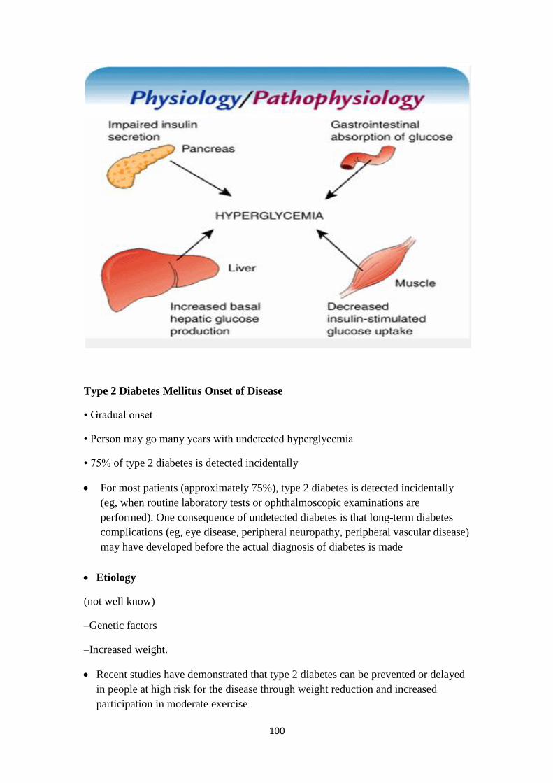
100
Type 2 Diabetes Mellitus Onset of Disease
• Gradual onset
• Person may go many years with undetected hyperglycemia
• 75% of type 2 diabetes is detected incidentally
For most patients (approximately 75%), type 2 diabetes is detected incidentally
(eg, when routine laboratory tests or ophthalmoscopic examinations are
performed). One consequence of undetected diabetes is that long-term diabetes
complications (eg, eye disease, peripheral neuropathy, peripheral vascular disease)
may have developed before the actual diagnosis of diabetes is made
Etiology
(not well know)
–Genetic factors
–Increased weight.
Recent studies have demonstrated that type 2 diabetes can be prevented or delayed
in people at high risk for the disease through weight reduction and increased
participation in moderate exercise
101
Gestational Diabetes
• Develops during pregnancy
• Detected at 24 to 28 weeks of gestation
• Risk for cesarean delivery, perinatal death, and neonatal complications
Hyperglycemia develops during pregnancy because of the secretion of placental
hormones, which causes insulin resistance.
Gestational diabetes occurs in as many as 14% of pregnant women and increases
their risk for hypertensive disorders during pregnancy
Women who are considered to be at high risk for GDM and who should be
screened by blood glucose testing at their first prenatal visit are those with marked
obesity, a personal history of GDM, glycosuria, or a strong family history of
diabetes.
Initial management includes dietary modification and blood glucose monitoring. If
hyperglycemia persists, insulin is prescribed.
Many women who have had GDM develop type 2 diabetes later in life.
Secondary Diabetes
• Results from another medical condition or due to the treatment of a medical
condition that causes abnormal blood glucose levels
–Cushing syndrome
–Hyperthyroidism
–Parenteral nutrition
Clinical Manifestations Diabetes Mellitus
• Polyuria
• Polydipsia (excessive thirst)
• Polyphagia
• In Type I
–Weight loss
–Ketoacidosis
Polyphagia (increased appetite) resulting from the catabolic state induced by
insulin deficiency and the breakdown of proteins and fats
Other symptoms include fatigue and weakness, sudden vision changes, tingling or
numbness in hands or feet, dry skin, skin lesions or wounds that are slow to heal,
and recurrent infections.

102
The onset of type 1 diabetes may also be associated with sudden weight loss or
nausea, vomiting, or abdominal pains, if DKA has developed.
Clinical Manifestations Non-specific symptoms
–Fatigue and weakness
–Sudden vision changes
–Tingling or numbness in hands or feet
–Skin lesions or recurrent infections
–Prolonged wound healing
–Visual changes
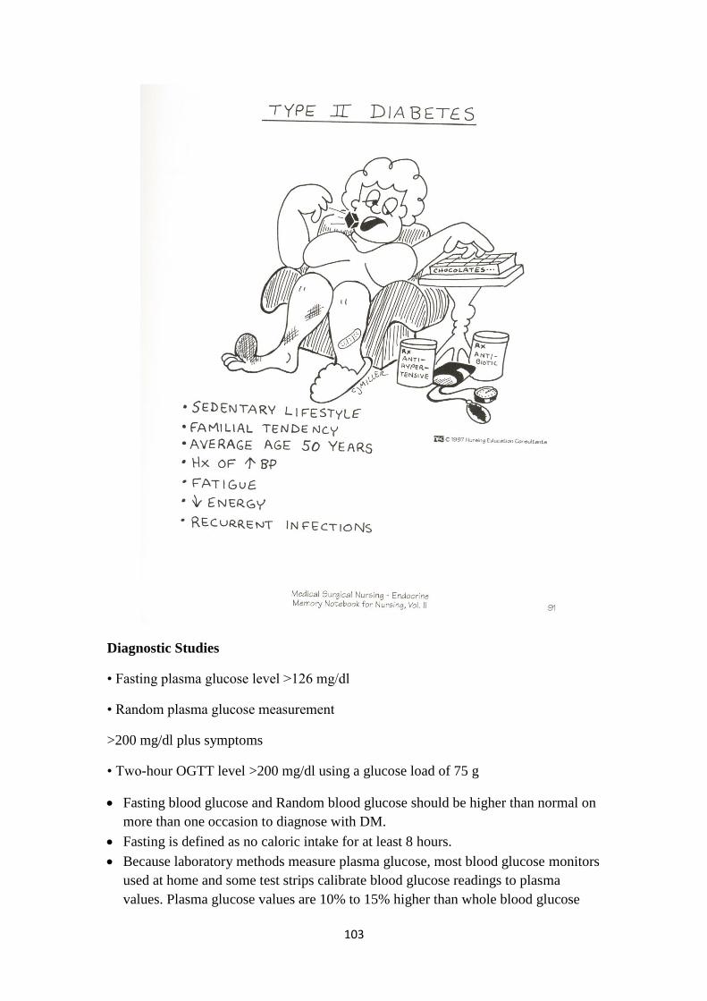
103
Diagnostic Studies
• Fasting plasma glucose level >126 mg/dl
• Random plasma glucose measurement
>200 mg/dl plus symptoms
• Two-hour OGTT level >200 mg/dl using a glucose load of 75 g
Fasting blood glucose and Random blood glucose should be higher than normal on
more than one occasion to diagnose with DM.
Fasting is defined as no caloric intake for at least 8 hours.
Because laboratory methods measure plasma glucose, most blood glucose monitors
used at home and some test strips calibrate blood glucose readings to plasma
values. Plasma glucose values are 10% to 15% higher than whole blood glucose
104
values, and it is crucial for patients with diabetes to know whether their monitor
and strips provide whole blood or plasma results
Assessing the Patient with Diabetes
• History:
Symptoms related to the diagnosis of diabetes:
Symptoms of hyperglycemia
Symptoms of hypoglycemia
Frequency, timing, severity, and resolution
Results of blood glucose monitoring
Status, symptoms, and management of chronic complications of diabetes:
Eye; kidney; nerve; genitourinary and sexual, bladder, and gastrointestinal
Cardiac; peripheral vascular; foot complications associated with diabetes
Adherence to/ability to follow prescribed dietary management plan
Adherence to prescribed exercise regimen
Adherence to/ability to follow prescribed pharmacologic treatment (insulin or oral
antidiabetic agents)
Use of tobacco, alcohol, and prescribed and over-the-counter medications/drugs
Lifestyle, cultural, psychosocial, and economic factors that may affect diabetes
treatment
Effects of diabetes or its complications on functional status (eg, mobility, vision)
Physical examination
Blood pressure (sitting and standing to detect orthostatic changes)
Body mass index (height and weight)
Fundoscopic examination and visual acuity
Foot examination (lesions, signs of infection, pulses)
Skin examination (lesions and insulin-injection sites)
Neurologic examination
Vibratory and sensory examination using monofilament
Deep tendon reflexes
Oral examination
• Laboratory Examination
• Need for Referrals
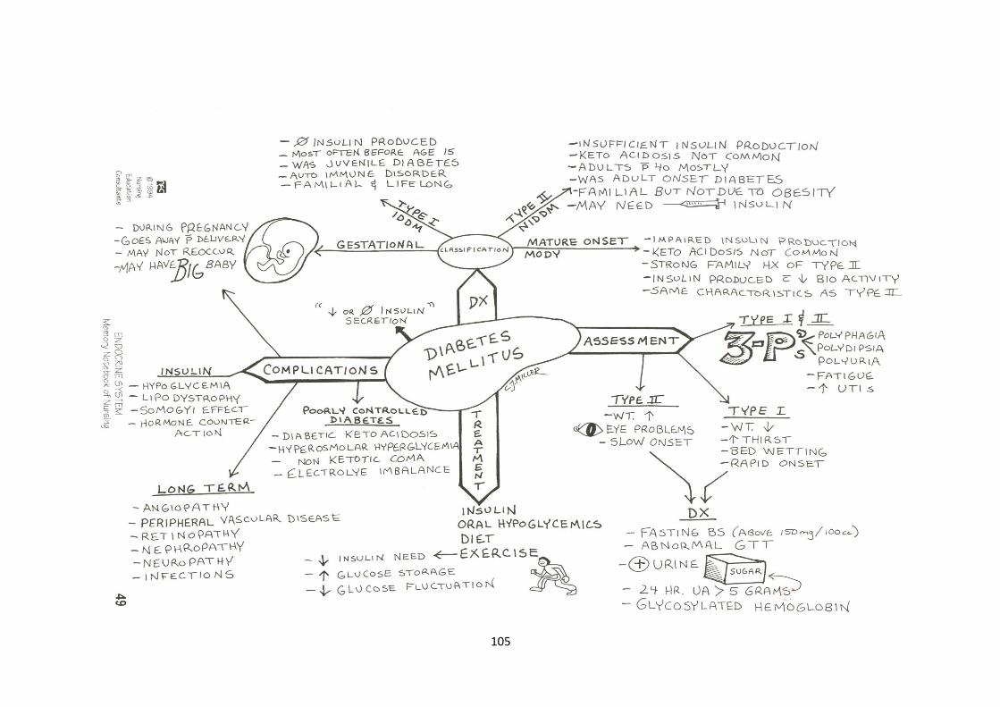
105
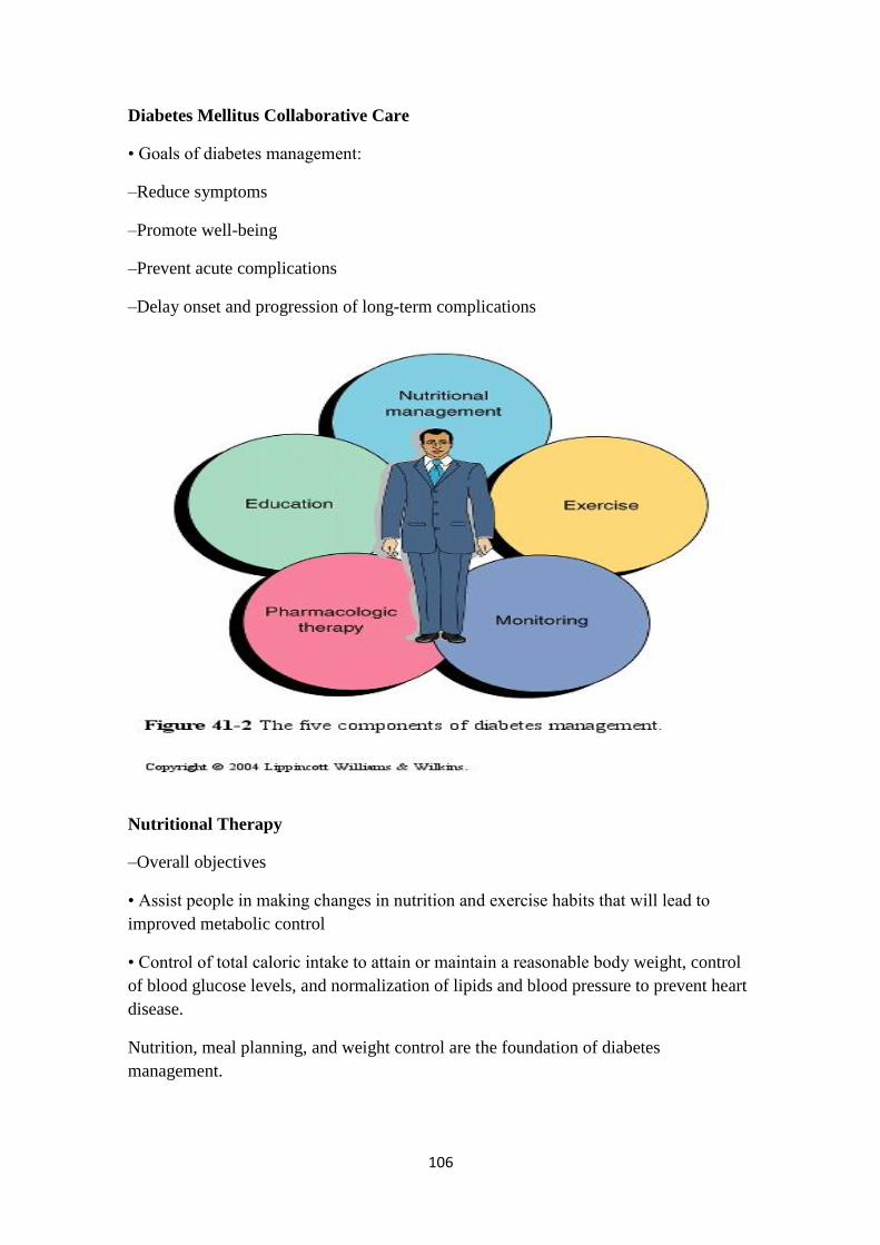
106
Diabetes Mellitus Collaborative Care
• Goals of diabetes management:
–Reduce symptoms
–Promote well-being
–Prevent acute complications
–Delay onset and progression of long-term complications
Nutritional Therapy
–Overall objectives
• Assist people in making changes in nutrition and exercise habits that will lead to
improved metabolic control
• Control of total caloric intake to attain or maintain a reasonable body weight, control
of blood glucose levels, and normalization of lipids and blood pressure to prevent heart
disease.
Nutrition, meal planning, and weight control are the foundation of diabetes
management.
107
• Obesity is associated with an increased resistance to insulin.
• Some obese patients who have type 2 diabetes and who require insulin or oral agents
to control blood glucose levels may be able to reduce or eliminate the need for
medication through weight loss.
• A weight loss as small as 10% of total weight may significantly improve blood
glucose levels
Nutritional Therapy for Type 1 DM
–Meal plan based on the individual‘s usual food intake and is balanced with insulin and
exercise patterns
Nutritional Therapy for Type 2 DM
–Emphasis placed on achieving glucose, lipid, and blood pressure goals
–Calorie reduction
• Food composition
–Individual meal plan developed with a dietitian
–Nutritionally balanced
–Does not prohibit the consumption of any one type of food
Caloric Distribution
• Calculate daily caloric requirement.
• Carbohydrates
–50% to 60% of caloric intake.
–Majority of calories should come from grains
–Foods high in carbohydrates, such as sucrose, are not eliminated from the diet but
should be eaten in moderation (up to 10% of total calories
• Fats
–20% to 30% of calories come from fat.
–Limit the amount of saturated fats to 10% of total calories
• Proteins
–10% to 20% of calories come from protein.
108
• Fiber
–Lower total cholesterol and LDL in the blood.
–Improve blood glucose levels
–Decrease the need for exogenous insulin.
–Increase satiety, which is helpful for weight loss
–Alcohol
• High in calories (lead to weigh gain)
• Promotes hypertriglyceridemia
• Can cause severe hypoglycemia
Alcohol is absorbed before other nutrients and does not require insulin for
absorption.
Large amounts can be converted to fats, increasing the risk for DKA.
A major danger of alcohol consumption by the patient with diabetes is
hypoglycemia, especially for patients who take insulin. Alcohol may decrease the
normal physiologic reactions in the body that produce glucose (gluconeogenesis).
Alcohol consumption may lead to excessive weight gain (from the high caloric
content of alcohol), hyperlipidemia, and elevated glucose levels (especially with
mixed drinks and liqueurs).
• Diet teaching
–Dietitian initially provides instruction
–Should include the patient‘s family and significant others
–Read food labels
– Sweetners

109
Exercise
–Essential part of diabetes management
–Increases insulin sensitivity
–Lowers blood glucose levels
–Decreases insulin resistance
–Decreases weight
–Reduces cardiovascular risk factors
Resistance (strength) training, such as weight lifting, can increase lean muscle mass,
thereby increasing the resting metabolic rate. These effects are useful in diabetes in
relation to losing weight, easing stress, and maintaining a feeling of well-being.
Exercise also alters blood lipid concentrations, increasing levels of high-density
lipoproteins and decreasing total cholesterol and triglyceride levels.
Exercise/ Precautions
• Don‘t exercise if blood glucose > 250 mg/dL or if there is ketone bodies in the urine.
• Don‘t exercise when the insulin at its peak
110
• Use proper footwear and.
• Avoid exercise in extreme heat or cold.
• Inspect feet daily after exercise.
Patients who have blood glucose levels exceeding 250 mg/dL and who have
ketones in their urine should not begin exercising until the urine test results are
negative for ketones and the blood glucose level is closer to normal.
Exercising with elevated blood glucose levels increases the secretion of glucagon,
growth hormone, and catecholamines. The liver then releases more glucose, and the
result is an increase in the blood glucose level
–Several small carbohydrate snacks can be taken to prevent hypoglycemia
• Before exercising
• At the end of the exercise with strenuous exercise
• At the time with strenuous exercise
• Deduce them from total daily calories
–May need to reduce inlsulin dose
The physiologic decrease in circulating insulin that normally occurs with exercise
cannot occur in patients treated with insulin. Initially, patients who require insulin
should be taught to eat a 15-g carbohydrate snack (a fruit exchange) or a snack of
complex carbohydrates with a protein before engaging in moderate exercise, to
prevent unexpected hypoglycemia.
Some patients find that they do not require a pre-exercise snack if they exercise
within 1 to 2 hours after a meal. Other patients may require extra food regardless of
when they exercise. If extra food is required, it need not be deducted from the
regular meal plan.
Diabetes Mellitus Exercise
–Best done after meals
–Exercise plans should be individualized
–Monitor blood glucose levels before, during, and after exercise
–Better to exercise at the same time daily when blood sugar at its peak
Monitoring Blood Glucose
• Self-monitoring of blood glucose (SMBG)
111
–Enables patient to make self-management decisions regarding diet, exercise, and
medication
Diabetes Mellitus Monitoring Blood Glucose
• Self-monitoring of blood glucose (SMBG)
–Important for detecting episodic hyperglycemia and hypoglycemia
–Patient training is crucial
For most patients who require insulin, SMBG is recommended two to four times
daily (usually before meals and at bedtime).
Drug Therapy: Insulin
• Exogenous insulin:
–Required for type 1 diabetes
–Prescribed for the patient with type 2 diabetes who cannot control blood glucose by
other means
• Types of insulin
–Human insulin
• Most widely used type of insulin
• Cost-effective
¯ Likelihood of allergic reaction
Human insulin preparations have a shorter duration of action than insulin from
animal sources because the presence of animal proteins triggers an immune
response that results in the binding of animal insulin, which slows its availability.
• Types of insulin
–Insulins differ in regard to onset, peak action, and duration
–Different types of insulin may be used in combination therapy
–Rapid-acting: Lispro (onset 15‘, peak 60-90‘ and last from 2-4 hours)
–Short-acting: Regular (Onset is 30-60‘, peak in 2-3h and last for 4-6 hours, and
Regular insulin is only kind for IV use.
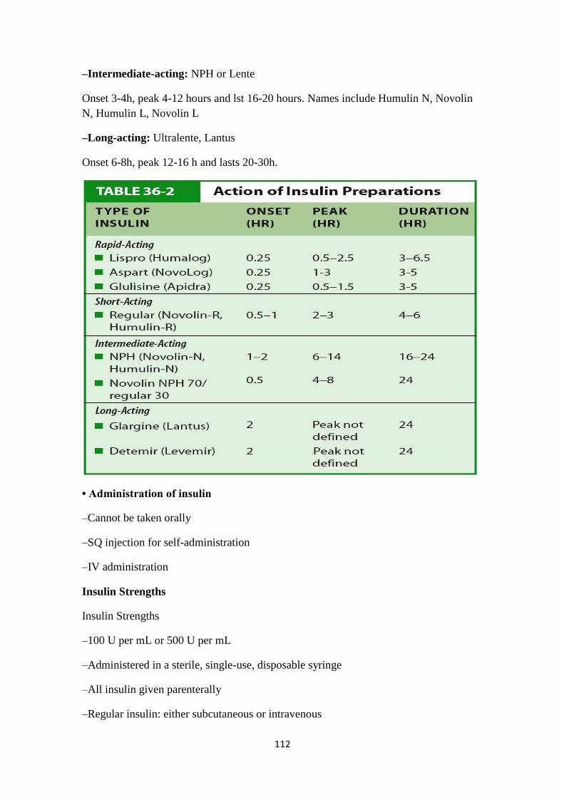
112
–Intermediate-acting: NPH or Lente
Onset 3-4h, peak 4-12 hours and lst 16-20 hours. Names include Humulin N, Novolin
N, Humulin L, Novolin L
–Long-acting: Ultralente, Lantus
Onset 6-8h, peak 12-16 h and lasts 20-30h.
• Administration of insulin
–Cannot be taken orally
–SQ injection for self-administration
–IV administration
Insulin Strengths
Insulin Strengths
–100 U per mL or 500 U per mL
–Administered in a sterile, single-use, disposable syringe
–All insulin given parenterally
–Regular insulin: either subcutaneous or intravenous
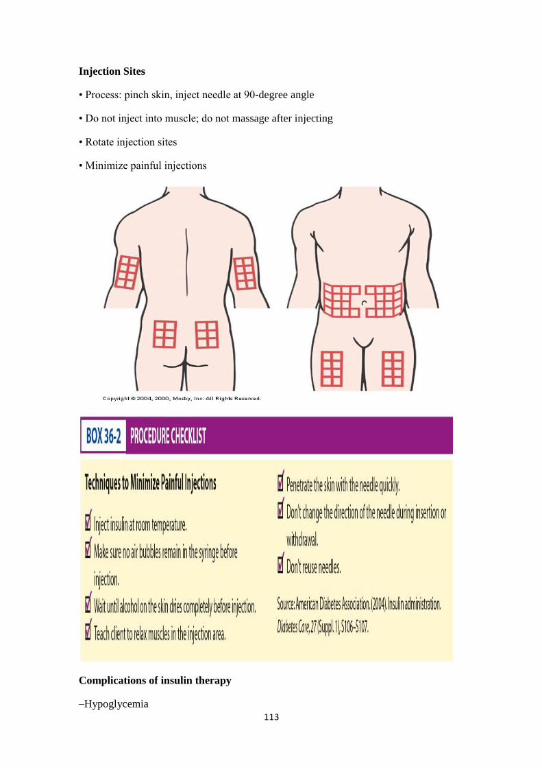
113
Injection Sites
• Process: pinch skin, inject needle at 90-degree angle
• Do not inject into muscle; do not massage after injecting
• Rotate injection sites
• Minimize painful injections
Complications of insulin therapy
–Hypoglycemia
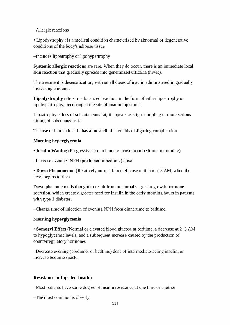
114
–Allergic reactions
• Lipodystrophy : is a medical condition characterized by abnormal or degenerative
conditions of the body's adipose tissue
–Includes lipoatrophy or lipohypertrophy
Systemic allergic reactions are rare. When they do occur, there is an immediate local
skin reaction that gradually spreads into generalized urticaria (hives).
The treatment is desensitization, with small doses of insulin administered in gradually
increasing amounts.
Lipodystrophy refers to a localized reaction, in the form of either lipoatrophy or
lipohypertrophy, occurring at the site of insulin injections.
Lipoatrophy is loss of subcutaneous fat; it appears as slight dimpling or more serious
pitting of subcutaneous fat.
The use of human insulin has almost eliminated this disfiguring complication.
Morning hyperglycemia
• Insulin Waning (Progressive rise in blood glucose from bedtime to morning)
–Increase evening‘ NPH (predinner or bedtime) dose
• Dawn Phenomenon (Relatively normal blood glucose until about 3 AM, when the
level begins to rise)
Dawn phenomenon is thought to result from nocturnal surges in growth hormone
secretion, which create a greater need for insulin in the early morning hours in patients
with type 1 diabetes.
–Change time of injection of evening NPH from dinnertime to bedtime.
Morning hyperglycemia
• Somogyi Effect (Normal or elevated blood glucose at bedtime, a decrease at 2–3 AM
to hypoglycemic levels, and a subsequent increase caused by the production of
counterregulatory hormones
–Decrease evening (predinner or bedtime) dose of intermediate-acting insulin, or
increase bedtime snack.
Resistance to Injected Insulin
–Most patients have some degree of insulin resistance at one time or another.
–The most common is obesity.
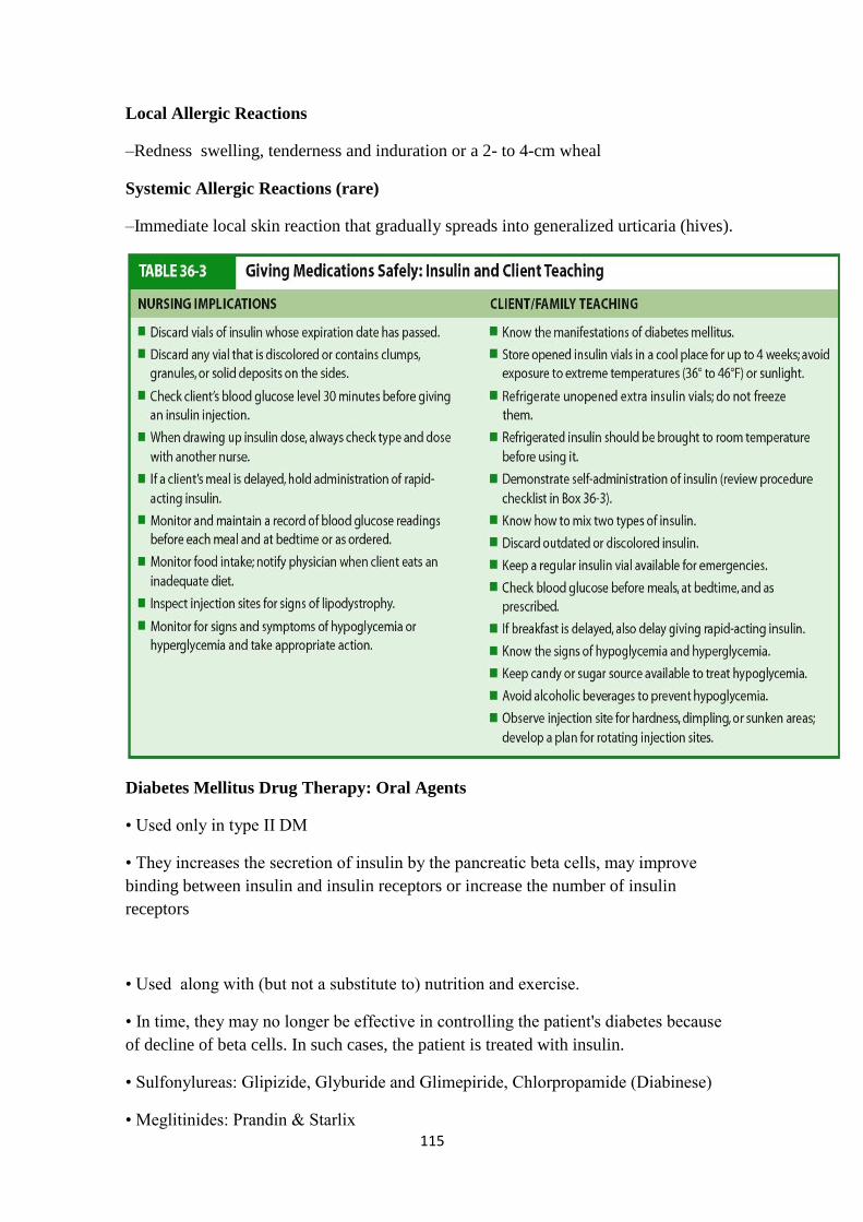
115
Local Allergic Reactions
–Redness swelling, tenderness and induration or a 2- to 4-cm wheal
Systemic Allergic Reactions (rare)
–Immediate local skin reaction that gradually spreads into generalized urticaria (hives).
Diabetes Mellitus Drug Therapy: Oral Agents
• Used only in type II DM
• They increases the secretion of insulin by the pancreatic beta cells, may improve
binding between insulin and insulin receptors or increase the number of insulin
receptors
• Used along with (but not a substitute to) nutrition and exercise.
• In time, they may no longer be effective in controlling the patient's diabetes because
of decline of beta cells. In such cases, the patient is treated with insulin.
• Sulfonylureas: Glipizide, Glyburide and Glimepiride, Chlorpropamide (Diabinese)
• Meglitinides: Prandin & Starlix
116
• Biguanides: Metformin
• a-Glucosidase inhibitors: Acarbose. Delay absorption of CHO
• Thiazolidinediones: Pioglitazone (Actos)
Other drugs affecting blood glucose levels:
– Adrenergic blockers
– Adrenaline
– Corticosteoids
Diabetes Mellitus Pancreas Transplantation
Used for patients with type 1 diabetes who have end-stage renal disease and who have
had or plan to have a kidney transplant
• Eliminates the need for exogenous insulin
• Can also eliminate hypoglycemia and hyperglycemia
Diabetes Mellitus New Developments in Diabetic Therapy
• New insulin delivery systems not yet approved by the FDA:
–Inhaled insulin
–Skin patch
–Oral spray
Diabetes Mellitus Patient education
1. Simple pathophysiology
–Basic definition of diabetes (having a high blood glucose level)
–Normal blood glucose ranges
–Effect of insulin and exercise (decrease glucose)
–Effect of food and stress, including illness and infections (increase glucose)
–Basic treatment approaches
2. Treatment modalities
–Administration of medications
–Meal planning (food groups, timing of meals)
117
–Monitoring of blood glucose and urine ketones
3. Recognition, treatment, and prevention of acute complications
–Hypoglycemia
–Hyperglycemia
4. Pragmatic information
–Where to buy and store insulin, syringes, and glucose monitoring supplies
–When and how to contact the physician
Diabetes Mellitus Patient education
• Planning In-Depth and Continuing Education
–Foot care
–Eye care
–General hygiene (eg, skin care, oral hygiene)
–Risk factor management (eg, control of blood pressure and blood lipid levels,
normalizing blood glucose levels)
Diabetes Mellitus Misconceptions Related to Insulin Treatment
1. Once insulin injections are started (for treatment of type 2 diabetes), they can never
be discontinued
2. If increasing doses of insulin are needed to control the blood glucose, the diabetes
must be getting ―worse‖
3. Insulin causes blindness (or other diabetic complications)
4. Insulin must be injected directly into the vein
5. There is extreme danger in injecting insulin if there are any air bubbles in the syringe
6. Insulin always causes people to have bad (hypoglycemic) reactions
Diabetes Mellitus Acute Complications
• Hypoglycemia
• Diabetic ketoacidosis (DKK)
• Hyperosmolar hyperglycemic nonketotic syndrome (HHNS)
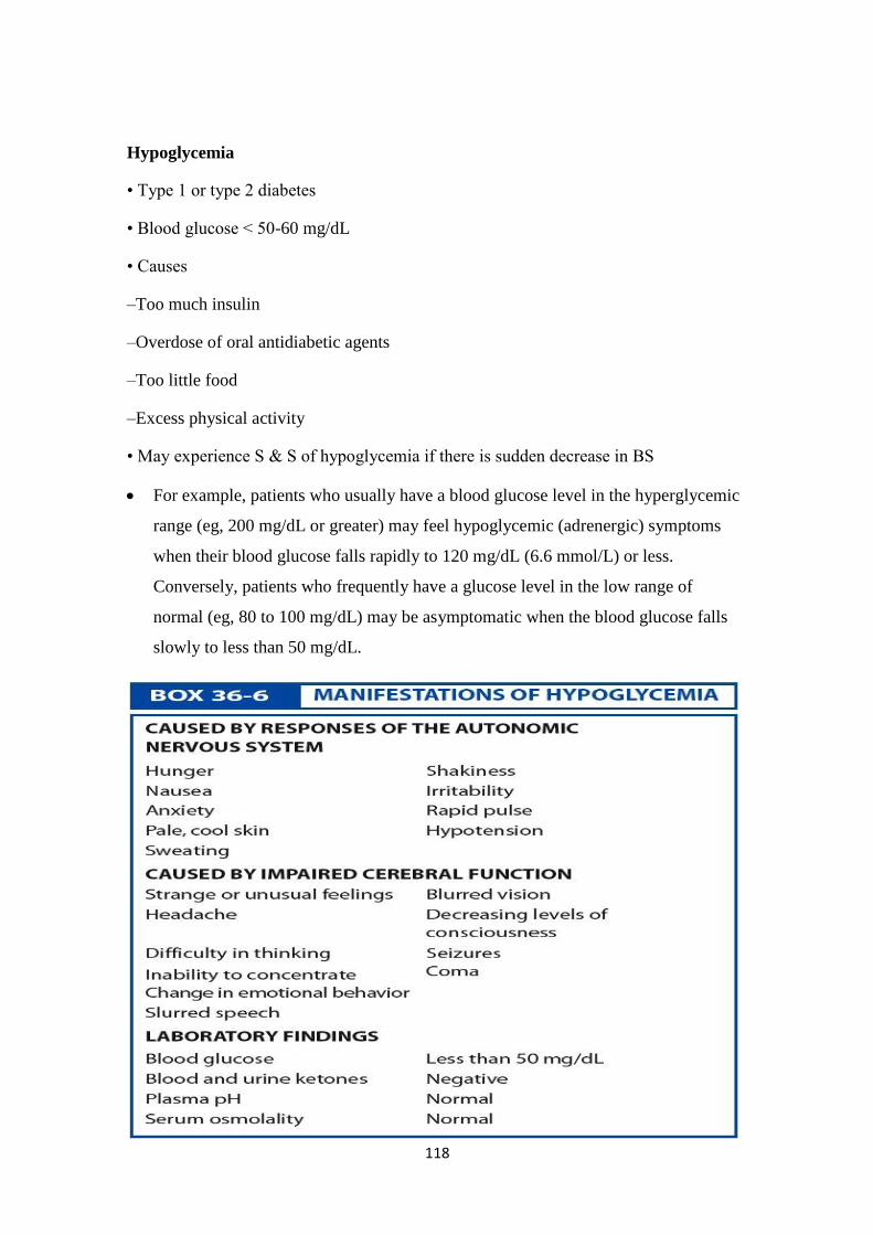
118
Hypoglycemia
• Type 1 or type 2 diabetes
• Blood glucose < 50-60 mg/dL
• Causes
–Too much insulin
–Overdose of oral antidiabetic agents
–Too little food
–Excess physical activity
• May experience S & S of hypoglycemia if there is sudden decrease in BS
For example, patients who usually have a blood glucose level in the hyperglycemic
range (eg, 200 mg/dL or greater) may feel hypoglycemic (adrenergic) symptoms
when their blood glucose falls rapidly to 120 mg/dL (6.6 mmol/L) or less.
Conversely, patients who frequently have a glucose level in the low range of
normal (eg, 80 to 100 mg/dL) may be asymptomatic when the blood glucose falls
slowly to less than 50 mg/dL.
119
• Treatment of hypoglycemia
– Mild
• Immediate treatment
• 15 g rapid-acting sugar
–Severe
• Hospitalized
• Intravenous glucose
–Teach patients to carry simple sugar with them
Diabetes Ketoacidosis (DKA)
• Life-threatening illness in type 1
–Hyperglycemia
–Dehydration and electrolyte loss
–Acidosis
• Causes of DKA
–Decreased or missed dose of insulin,
–Illness or infection,
–Undiagnosed and untreated diabetes
• Without insulin, the amount of glucose entering the cells is reduced, and production
and release of glucose by the liver is increased (lead to hyperglycemia).
• Excess glucose leads to polyuria (6.5 L/day) dehydration, sodium and potassium loss
• Burning of fat leads to ketosis
• Kidneys unable to excrete ketones, leads to ketoacidosis
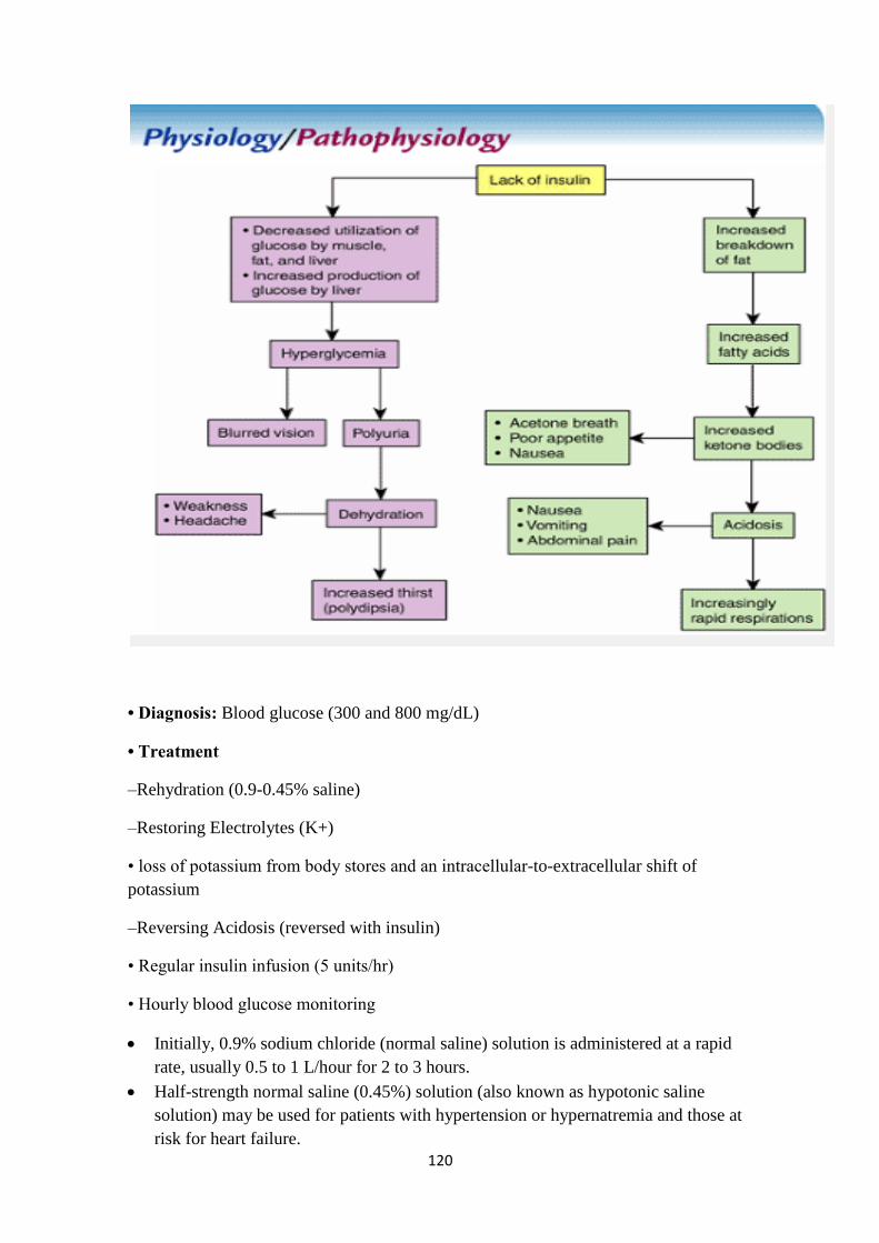
120
• Diagnosis: Blood glucose (300 and 800 mg/dL)
• Treatment
–Rehydration (0.9-0.45% saline)
–Restoring Electrolytes (K+)
• loss of potassium from body stores and an intracellular-to-extracellular shift of
potassium
–Reversing Acidosis (reversed with insulin)
• Regular insulin infusion (5 units/hr)
• Hourly blood glucose monitoring
Initially, 0.9% sodium chloride (normal saline) solution is administered at a rapid
rate, usually 0.5 to 1 L/hour for 2 to 3 hours.
Half-strength normal saline (0.45%) solution (also known as hypotonic saline
solution) may be used for patients with hypertension or hypernatremia and those at
risk for heart failure.
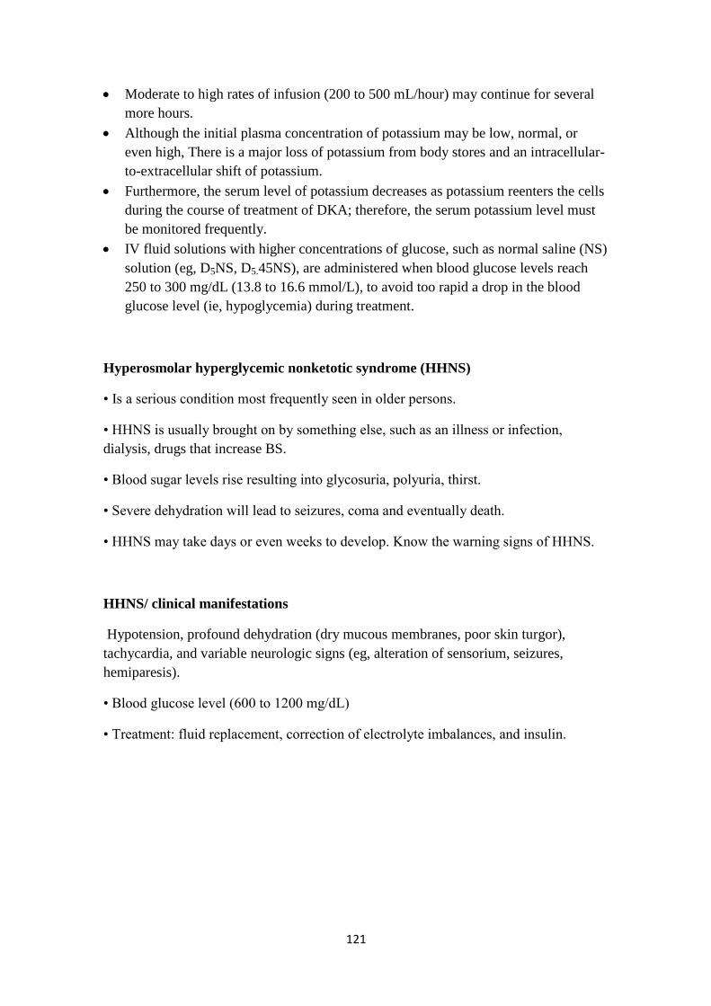
121
Moderate to high rates of infusion (200 to 500 mL/hour) may continue for several
more hours.
Although the initial plasma concentration of potassium may be low, normal, or
even high, There is a major loss of potassium from body stores and an intracellular-
to-extracellular shift of potassium.
Furthermore, the serum level of potassium decreases as potassium reenters the cells
during the course of treatment of DKA; therefore, the serum potassium level must
be monitored frequently.
IV fluid solutions with higher concentrations of glucose, such as normal saline (NS)
solution (eg, D5NS, D5.45NS), are administered when blood glucose levels reach
250 to 300 mg/dL (13.8 to 16.6 mmol/L), to avoid too rapid a drop in the blood
glucose level (ie, hypoglycemia) during treatment.
Hyperosmolar hyperglycemic nonketotic syndrome (HHNS)
• Is a serious condition most frequently seen in older persons.
• HHNS is usually brought on by something else, such as an illness or infection,
dialysis, drugs that increase BS.
• Blood sugar levels rise resulting into glycosuria, polyuria, thirst.
• Severe dehydration will lead to seizures, coma and eventually death.
• HHNS may take days or even weeks to develop. Know the warning signs of HHNS.
HHNS/ clinical manifestations
Hypotension, profound dehydration (dry mucous membranes, poor skin turgor),
tachycardia, and variable neurologic signs (eg, alteration of sensorium, seizures,
hemiparesis).
• Blood glucose level (600 to 1200 mg/dL)
• Treatment: fluid replacement, correction of electrolyte imbalances, and insulin.
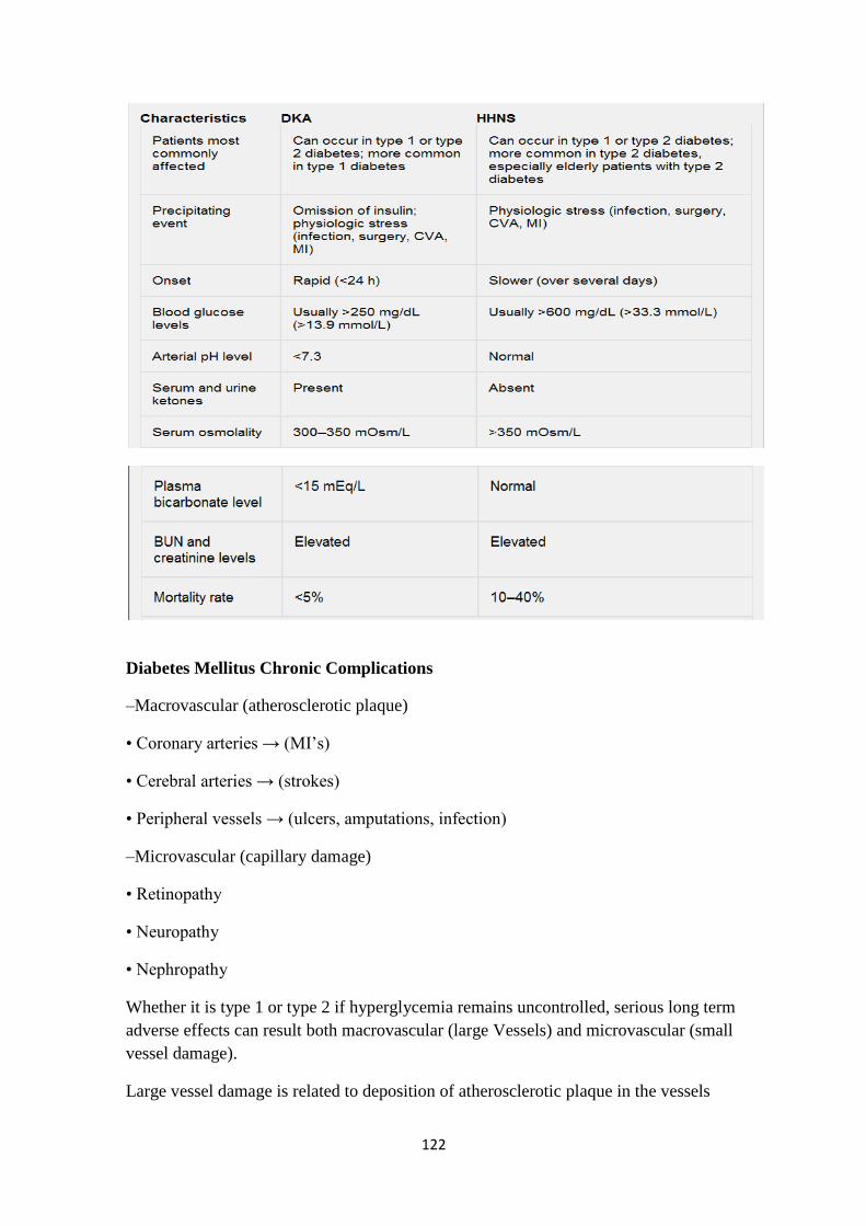
122
Diabetes Mellitus Chronic Complications
–Macrovascular (atherosclerotic plaque)
• Coronary arteries → (MI‘s)
• Cerebral arteries → (strokes)
• Peripheral vessels → (ulcers, amputations, infection)
–Microvascular (capillary damage)
• Retinopathy
• Neuropathy
• Nephropathy
Whether it is type 1 or type 2 if hyperglycemia remains uncontrolled, serious long term
adverse effects can result both macrovascular (large Vessels) and microvascular (small
vessel damage).
Large vessel damage is related to deposition of atherosclerotic plaque in the vessels
123
Macrovascular (atherosclerotic plaque)
Coronary arteries MI
Cerebral arteries strokes
Peripheral vessels foot ulcers, amputations
Microvascular (capillary damage)
Retinopathy partial or complete blindness
Neuropathy autonomic nerve damage: for example diabetic gastroparesis, bladder
dysfunction, unawareness of hypoglycemia, (somatic nerve damage, foot ulcers, due to
loss of sensations.)
Nephropathy proteinuria (microalbuminuria) chronic renal failure requires dialysis.
Macrovascular Complications
• Macrocirculation
–Blood vessel walls thicken, sclerose, and become occluded by plaque that adheres to
the vessel walls. finally, blood flow is blocked.
• Complications
–Coronary artery disease
–Stroke
–Peripheral vascular disease
Although it may be tempting to attribute the increased prevalence of macrovascular
diseases to the increased prevalence of certain risk factors (eg, obesity, increased
triglyceride levels, hypertension) in patients with diabetes, there is a higher-than-
expected rate of macrovascular diseases among patients with diabetes compared with
patients without diabetes who have the same risk factors (ADA, 2004d). Therefore,
diabetes itself is seen as an independent risk factor for accelerated atherosclerosis.
Other potential factors that may play a role in diabetes-related atherosclerosis include
platelet and clotting factor abnormalities, decreased flexibility of red blood cells,
decreased oxygen release, changes in the arterial wall related to hyperglycemia, and
possibly hyperinsulinemia.
Complication: CAD
• CAD account for 50% to 60% of all deaths among patients with diabetes.
• High cholesterol and high triglycerides

124
• MI is twice as common in men and three times in women with diabetes, compared to
people without diabetes.
• Silent MI
• Higher risk for a second infarction
Complication: Stroke
• People with diabetes have twice the risk of developing cerebrovascular disease.
• There is a greater likelihood of death from cerebrovascular disease.
• Recovery is slower with high BS.
• Hypertension plays a role
Complication: Peripheral Vascular Disease
• Diabetes-induced arteriosclerosis
• 2-3 times higher than in nondiabetic people
• S & S: diminished peripheral pulses and intermittent claudication (pain in the buttock,
thigh, or calf during walking)
• Can lead to leg ulcers and gangrene and amputation.
Management of Macrovascualr changes
• Prevention and treatment of risk factors for atherosclerosis.
–obesity, hypertension, and hyperlipidemia (exercise, stop smoking).
–Control of blood glucose levels may reduce triglyceride concentrations and can
significantly reduce the incidence of complications.
Microvascular Complications
• Microcirculation
–Eyes
–Kidneys
–Nerves
Complication: Diabetic Retinopathy
• Leading cause of blindness in people ages 20 to 74 in US
• Almost all patients with type 1 diabetes and more than 60% of patients with type 2
diabetes have some degree of retinopathy after 20 years
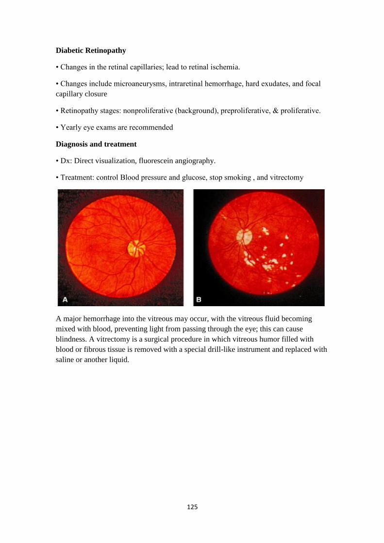
125
Diabetic Retinopathy
• Changes in the retinal capillaries; lead to retinal ischemia.
• Changes include microaneurysms, intraretinal hemorrhage, hard exudates, and focal
capillary closure
• Retinopathy stages: nonproliferative (background), preproliferative, & proliferative.
• Yearly eye exams are recommended
Diagnosis and treatment
• Dx: Direct visualization, fluorescein angiography.
• Treatment: control Blood pressure and glucose, stop smoking , and vitrectomy
A major hemorrhage into the vitreous may occur, with the vitreous fluid becoming
mixed with blood, preventing light from passing through the eye; this can cause
blindness. A vitrectomy is a surgical procedure in which vitreous humor filled with
blood or fibrous tissue is removed with a special drill-like instrument and replaced with
saline or another liquid.

126
Complication: Diabetic Nephropathy
• Disease of the kidneys (50% of RF due to DM)
• Characterized by albumin in the urine, hypertension, edema, renal insufficiency
• DM is the most common cause of renal failure
• First indication: microalbuminuria
• Treatment: ACE inhibitors, control BP and BS, prevent & treat UTI, low Na &
protein diet.
About 20% to 30% of people with type 1 or type 2 diabetes develop nephropathy, but
fewer of those with type 2 diabetes progress to ESRD.
As renal failure progresses, the catabolism (breakdown) of both exogenous and
endogenous insulin decreases, and frequent hypoglycemic episodes may result.
Complication: Diabetic Neuropathy
• Disorder of the peripheral nerves, spinal cord, and autonomic nervous system

127
• Results: sensory and motor impairments, postural hypotension, delayed gastric
emptying, diarrhea, impaired GU function
• Result from the thickening of the capillary membrane and destruction of myelin
sheath which disrupt nerve conductions.
The prevalence increases with the age of the patient and the duration of the disease and
may be as high as 50% in patients who have had diabetes for 25 years.
The two most common types of diabetic neuropathy are sensorimotor polyneuropathy
and autonomic neuropathy.
• Bilateral sensory disorders
–Appear first in toes, feet, and progress upward to fingers and hands
–Tingling, decrease in proprioception , and a decreased sensation of light touch
• Treatment
–Controlling BS delay the onset.
–Analgesics to control pain
Proprioception (awareness of posture and movement of the body and of position and
weight of objects in relation to the body
Complication: Autonomic Neuropathy
• Involves numerous body systems such as:
–Cardiovascular (slight tachycardia, orthostatic hypotension & silent MI).
–Gastrointestinal (Delayed gastric emptying , N&V, early satiety, variation of BS
absorption)
–Genitourinary (urinary symptoms of neurogenic bladder, UTI, erectile dysfunction)
–Hypoglycemic unawareness (DM diminish function of adrenal medulla)
Delayed gastric emptying may occur with the typical gastrointestinal symptoms of
early satiety, bloating, nausea, and vomiting. ―Diabetic‖ constipation or diarrhea
(especially nocturnal diarrhea) may occur as a result. In addition, there may be
unexplained wide swings in blood glucose levels related to inconsistent absorption of
the glucose from ingested foods secondary to the inconsistent gastric emptying.
Foot and Leg Problems
• 50% -75% of lower extremity amputations are performed on people with diabetes.
• More than 50% of these amputations are thought to be preventable.
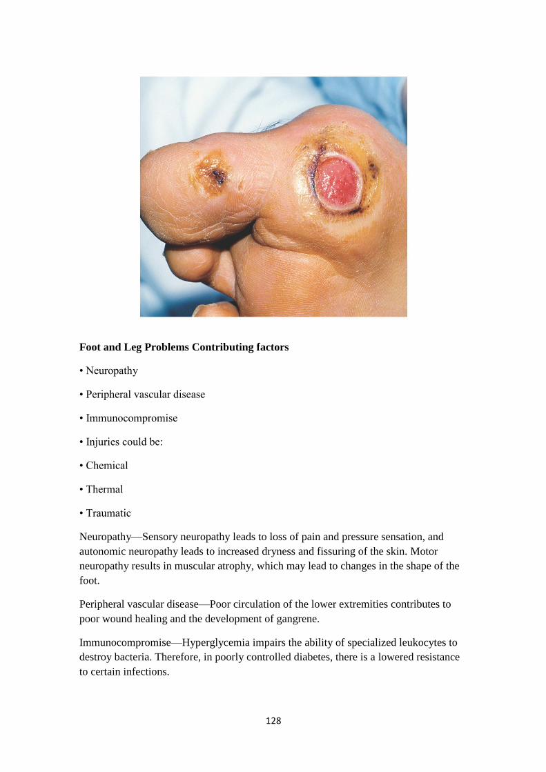
128
Foot and Leg Problems Contributing factors
• Neuropathy
• Peripheral vascular disease
• Immunocompromise
• Injuries could be:
• Chemical
• Thermal
• Traumatic
Neuropathy—Sensory neuropathy leads to loss of pain and pressure sensation, and
autonomic neuropathy leads to increased dryness and fissuring of the skin. Motor
neuropathy results in muscular atrophy, which may lead to changes in the shape of the
foot.
Peripheral vascular disease—Poor circulation of the lower extremities contributes to
poor wound healing and the development of gangrene.
Immunocompromise—Hyperglycemia impairs the ability of specialized leukocytes to
destroy bacteria. Therefore, in poorly controlled diabetes, there is a lowered resistance
to certain infections.
129
Foot Care
1. Take care of your diabetes.
2. Inspect your feet every day.
3. Wash your feet every day (dry between toes well).
4. Keep the skin soft and smooth.
5. Smooth corns and calluses gently.
6. Trim your toenails each week or when needed.
7. Wear shoes and socks at all times.
8. Protect your feet from hot and cold.
9. Keep the blood flowing to your feet.
10. Check with your health care provider.
Special Issues in Diabetes Care
• Patients with diabetes who are undergoing surgery
–Hyperglycemia (due to stress hormones)
–Hypoglycemia (being NPO)
• Hold morning insulin unless it is > 200 mg/dL.
–Diuresis leads to fluid and electrolytes imbalance
If hyperglycemia is not controlled during surgery, the resulting osmotic diuresis may
lead to excessive loss of fluids and electrolytes. Patients with type 1 diabetes also risk
developing ketoacidosis during periods of stress.
The blood glucose level is controlled during surgery with the IV infusion of regular
insulin, which is balanced by an infusion of dextrose. The insulin and dextrose infusion
rates are adjusted according to frequent (hourly) capillary glucose determinations.
Diabetes Mellitus Nursing Process Patient newly diagnosed with DM
• Assessment:
–Signs and Symptoms of DM
–Infections.
–Complications.
–Blood glucose

130
–S &S of DKA/ HHNS
Nursing Diagnoses
• Risk for fluid volume deficit related to polyuria and dehydration
• Imbalanced nutrition related to imbalance of insulin, food, and physical activity
• Deficient knowledge about diabetes self-care skills/information
Nursing Diagnoses
• Deficient knowledge about diabetes self-care skills/information
• Potential self-care deficit related to physical impairments or social factors
• Anxiety related to loss of control, fear of inability to manage diabetes, misinformation
related to diabetes, fear of diabetes complications
Collaborative Problems/ Potential Complications
• Fluid overload, pulmonary edema, and heart failure
• Hypokalemia
• Hyperglycemia and ketoacidosis
• Hypoglycemia
• Cerebral edema
Diabetes Mellitus Nursing Management Planning
• Overall goals:
–Active patient participation
–No episodes of acute hyperglycemic emergencies or hypoglycemia
–Maintain normal blood glucose levels
–Prevent chronic complications
–Lifestyle adjustment with minimal stress
Nursing Interventions
• Maintaining Fluid and Electrolyte Balance
• Improving Nutritional Intake
• Reducing Anxiety
• Improving Self-Care

131
Monitoring and Managing Potential Complications
• Fluid Overload
• Hypokalemia
• Hyperglycemia and Ketoacidosis
• Hypoglycemia
• Cerebral Edema
Diabetes Mellitus Nursing Management Nursing Implementation
• Health Promotion
–Identify those at risk
–Routine screening for overweight adults over age 45
• Ambulatory and Home Care
–Overall goal:
• Enable the patient or caregiver to reach an optimal level of independence
• Ambulatory and Home Care
–Insulin therapy and oral agents
–Personal hygiene
–Medical identification and travel
–Patient and family teaching
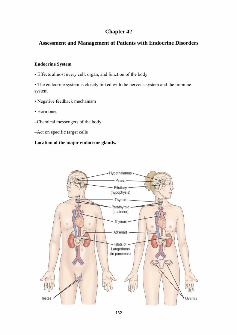
132
Chapter 42
Assessment and Management of Patients with Endocrine Disorders
Endocrine System
• Effects almost every cell, organ, and function of the body
• The endocrine system is closely linked with the nervous system and the immune
system
• Negative feedback mechanism
• Hormones
–Chemical messengers of the body
–Act on specific target cells
Location of the major endocrine glands.

133
Hypothalamus
• Sits between the cerebrum and brainstem
• Houses the pituitary gland and hypothalamus
• Regulates:
–Temperature
–Fluid volume
–Growth
–Pain and pleasure response
–Hunger and thirst
Hypothalamus Hormones
• Releasing and inhibiting hormones
• Corticotropin-releasing hormone
• Thyrotropin-releasing hormone
• Growth hormone-releasing hormone
• Gonadotropin-releasing hormone
• Somatostatin-=-inhibits GH and TSH
Pituitary Gland
• Sits beneath the hypothalamus
• Termed the ―master gland‖
• Divided into:
–Anterior Pituitary Gland
–Posterior Pituitary Gland

134
Actions of the major hormones of the pituitary gland.
Adrenal Glands
• Pyramid-shaped organs that sit on top of the kidneys
• Each has two parts:
–Outer Cortex
–Inner Medulla
Adrenal Cortex
• Mineralocorticoid—aldosterone. Affects sodium absorption, loss of potassium by
kidney
• Glucocorticoids—cortisol. Affects metabolism, regulates blood sugar levels, affects
growth, anti-inflammatory action, decreases effects of stress
• Adrenal androgens— dehydroepiandrosterone and
Adrenal Medulla
• Secretion of two hormones
–Epinephrine

135
–Norepinephrine
• Serve as neurotransmitters for sympathetic system
• Involved with the stress response
Thyroid Gland
• Butterfly shaped
• Sits on either side of the trachea
• Has two lobes connected with an isthmus
• Functions in the presence of iodine
• Stimulates the secretion of three hormones
• Involved with metabolic rate management and serum calcium levels

136
Hypothalamic-Pituitary-Thyroid Axis
Thyroid
• Follicular cells—excretion of triiodothyronine (T3) and thyroxine (T4)—Increase
BMR, increase bone and protien turnover, increase response to catecholamines, need
for infant G&D
• Thyroid C cells—calcitonin. Lowers blood calcium and phosphate levels
• BMR: Basal Metabolic Rate
Parathyroid Glands
• Embedded within the posterior lobes of the thyroid gland
• Secretion of one hormone
• Maintenance of serum calcium levels
• Parathyroid hormone—regulates serum calcium
Pancreas
• Located behind the stomach between the spleen and duodenum
• Has two major functions
–Digestive enzymes
–Releases two hormones: insulin and glucagon
Kidney
• 1, 25 dihydroxyvitamin D—stimulates calcium absorption from the intestine
• Renin—activates the Renin-Angiotensin System (RAS)
137
• Erythropoietin—Increases red blood cell production
Ovaries
• Estrogen
• Progesterone—important in menstrual cycle, maintains pregnancy,
Testes
• Androgens, testosterone—secondary sexual characteristics, sperm production
Thymus
• Releases thymosin and thymopoietin
• Affects maturation of T lymphocetes
Pineal
• Melatonin
• Affects sleep, fertility and aging
Assessment of Endocrine system:
Past Medical History
• Hormone replacement therapy
• Surgeries, chemotherapy, radiation
• Family history: diabetes mellitus, diabetes insipidus, goiter, obesity, Addison‘s
disease, infertility
• Sexual history: changes, characteristics, menstruation, menopause
Physical Assessment
• General appearance
–Vital signs, height, weight
• Integumentary
–Skin color, temperature, texture, moisture
–Bruising, lesions, wound healing
–Hair and nail texture, hair growth
• Face
–Shape, symmetry
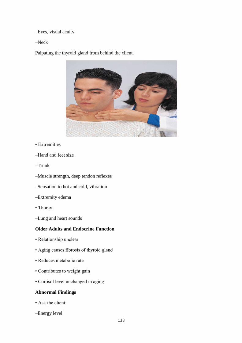
138
–Eyes, visual acuity
–Neck
Palpating the thyroid gland from behind the client.
• Extremities
–Hand and feet size
–Trunk
–Muscle strength, deep tendon reflexes
–Sensation to hot and cold, vibration
–Extremity edema
• Thorax
–Lung and heart sounds
Older Adults and Endocrine Function
• Relationship unclear
• Aging causes fibrosis of thyroid gland
• Reduces metabolic rate
• Contributes to weight gain
• Cortisol level unchanged in aging
Abnormal Findings
• Ask the client:
–Energy level
139
–Fatigue
–Maintenance of ADL
–Sensitivity to heat or cold
–Weight level
–Bowel habits
–Level of appetite
–Urination, thirst, salt craving
• Ask the client:
–Cardiovascular status: blood pressure, heart rate, palpitations, SOB
–Vision: changes, tearing, eye edema
–Neurologic: numbness/tingling lips or extremities, nervousness, hand tremors, mood
changes, memory changes, sleep patterns
–Integumentary: hair changes, skin changes, nails, bruising, wound healing
Most Common Endocrine Disorders
• Thyroid abnormalities
• Diabetes mellitus
Diagnostic Tests
• GH: fasting, well rested, not physically stressed
• T3/T4, TSH: no specific preparation
• Serum calcium/phosphate: fasting may or may not be required
• Cortisol/aldosterone level
• 24 urine collection to measure the level of catacholamines (epinephrine,
norepinephrine, dopamine).
Thyroid Disorders
• Cretinism
• Hypothyroidism
• Hyperthyroidism
• Thyroiditis

140
• Goiter
• Thyroid cancer
HYPOTHYRODISM
Hypothyroidism is the disease state caused by insufficient production of thyroid
hormone by the thyroid gland.
INCEDENCE
• 30-60 yrs of age
• Mostly women (5 times more than men)
• Causes
–Autoimmune disease (Hashimoto's
thyroiditis, post–Graves' disease)
–Atrophy of thyroid gland with aging
–Therapy for hyperthyroidism
• Radioactive iodine (131I)
• Thyroidectomy

141
• Medications
• Radiation to head and neck
Clinical Manifestations:
1. Fatigue.
2. Constipation.
3. Apathy
4. Weight gain.
5. Memory and mental impairment and decreased concentration.
6. Masklike face.
7. Menstrual irregularities and loss of libido.
8. Coarseness or loss of hair.
9. Dry skin and cold intolerance.
10. Menstrual disturbances
11. N umbness and tingling of fingers.
12. Tongue, hands, and feet may enlarge
13. Slurred speach
14. Hyperlipidemia.
15. Reflex delay.
16. Bradycardia.
17. Hypothermia.
18. C ardiac and respiratory complications .
Laboratory Assessment
• Decreased T3
• Decreased T4
• Increased TSH
142
Treatment
Lifelong Thyroid Hormone Replacement
• levothyroxine sodium (Synthroid, T4, Eltroxin)
• IMPORTANT: start at low does, to avoid hypertension, heart failure and MI
• Teach about S&S of hyperthyroidism with replacement therapy
MYXEDEMA DEVELOPS
• Rare serious complication of untreated hypothyroidism
• Decreased metabolism causes the heart muscle to become flabby
• Leads to decreased cardiac output
• Leads to decreased perfusion to brain and other vital organs
• Leads to tissue and organ failure
• LIFE THREATENING EMERGENCY WITH HIGH MORTALITY RATE
• Edema changes client‘s appearance
• Nonpitting edema appears everywhere especially around the eyes, hands, feet,
between shoulder blades
• Tongue thickens, edema forms in larynx, voice husky
Problems Seen With Myxedema Coma
• Coma
• Respiratory failure
• Hypotension
• Hyponatremia
• Hypothermia
• Hypoglycemia
Treatment Of Myexedema Coma
• Patent airway
• Replace fluids with IV.
• Give levothyroxine sodium IV
143
• Give glucose IV
• Give corticosteroids
• Check temp, BP hourly
• Monitor changes LOC hourly
• Aspiration precautions, keep warm
Hyperthyroidism
• Hyperthyroidism is the second most prevalent endocrine disorder, after diabetes
mellitus.
• Graves' disease: the most common type of hyperthyroidism, results from an excessive
output of thyroid hormones.
• May appear after an emotional shock, stress, or an infection
• Other causes: thyroiditis and excessive ingestion of thyroid hormone
• Affects women 8X more frequently than men (appears between second and fourth
decade)
Clinical Manifestations ( thyrotoxicosis):
1. Heat intolerance.
2. Palpitations, tachycardia, elevated systolic BP.
3. Increased appetite but with weight loss.
4. Menstrual irregularities and decreased libido.
5. Increased serum T4, T3.
6. Exophthalmos (bulging eyes)
7. Perspiration, skin moist and flushed ; however, elders‘ skin may be dry and pruritic
8. Insomnia.
9. Fatigue and muscle weakness
10. Nervousness, irritability, can‘t sit quietly.
11. Diarrhea.
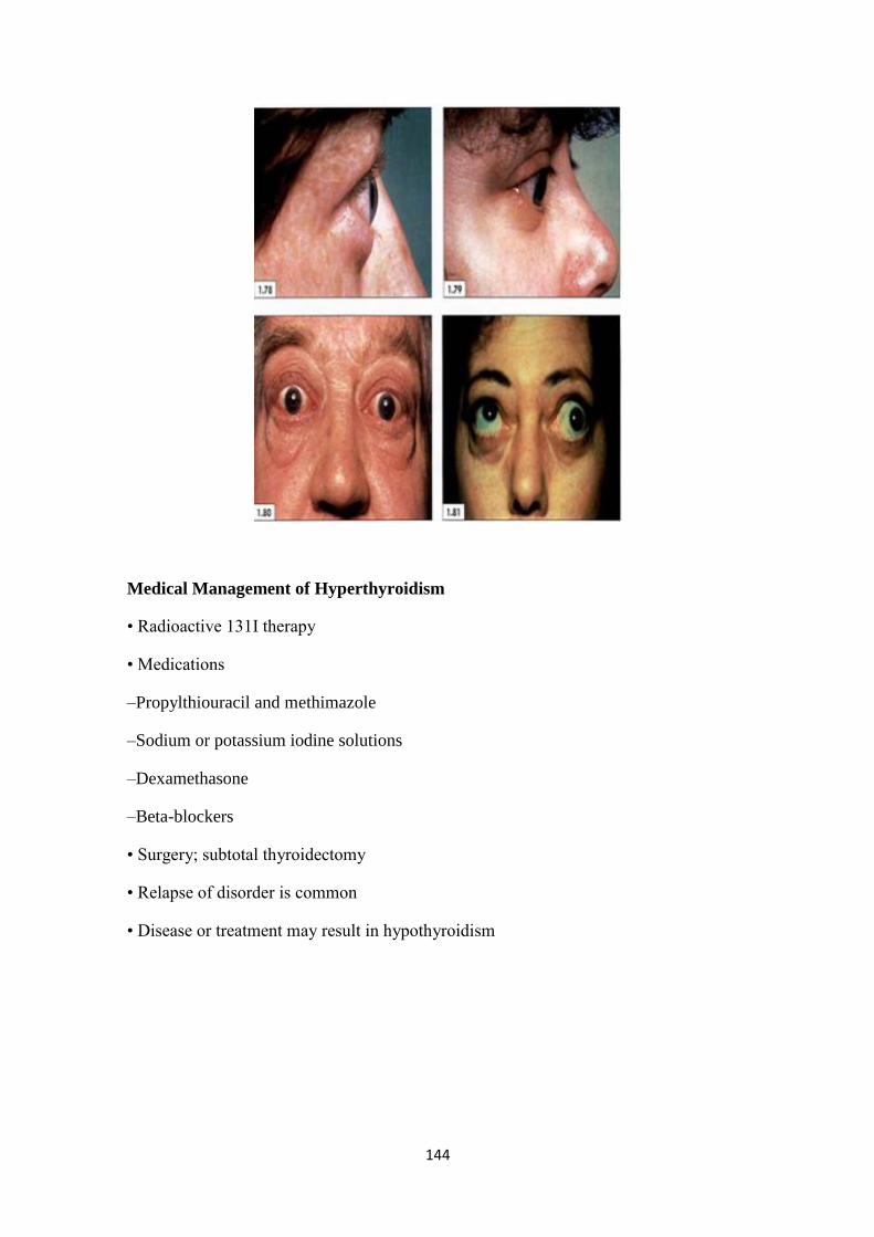
144
Medical Management of Hyperthyroidism
• Radioactive 131I therapy
• Medications
–Propylthiouracil and methimazole
–Sodium or potassium iodine solutions
–Dexamethasone
–Beta-blockers
• Surgery; subtotal thyroidectomy
• Relapse of disorder is common
• Disease or treatment may result in hypothyroidism
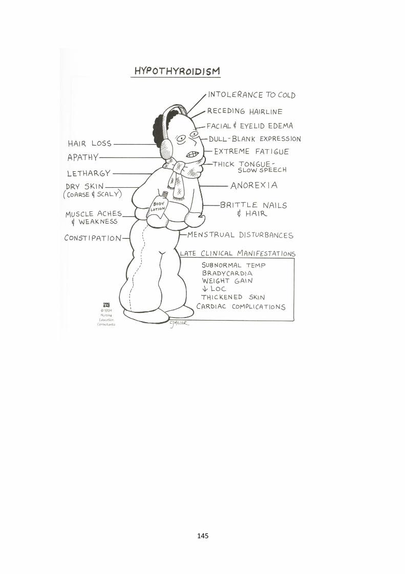
145
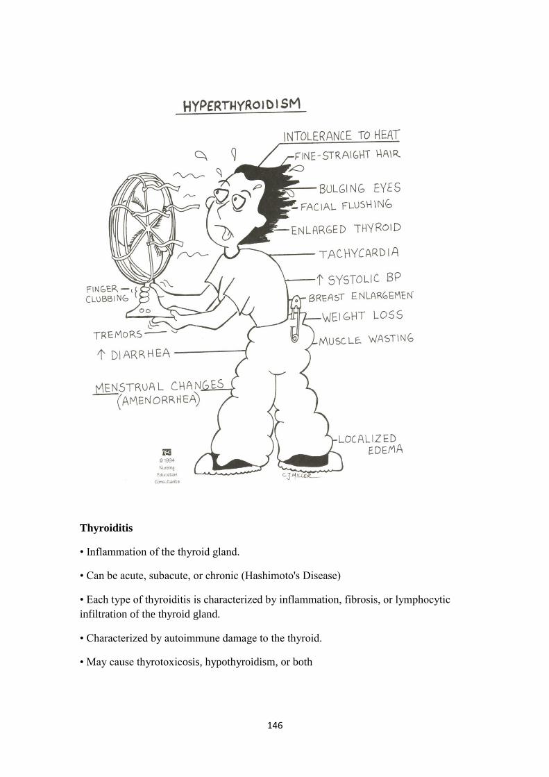
146
Thyroiditis
• Inflammation of the thyroid gland.
• Can be acute, subacute, or chronic (Hashimoto's Disease)
• Each type of thyroiditis is characterized by inflammation, fibrosis, or lymphocytic
infiltration of the thyroid gland.
• Characterized by autoimmune damage to the thyroid.
• May cause thyrotoxicosis, hypothyroidism, or both
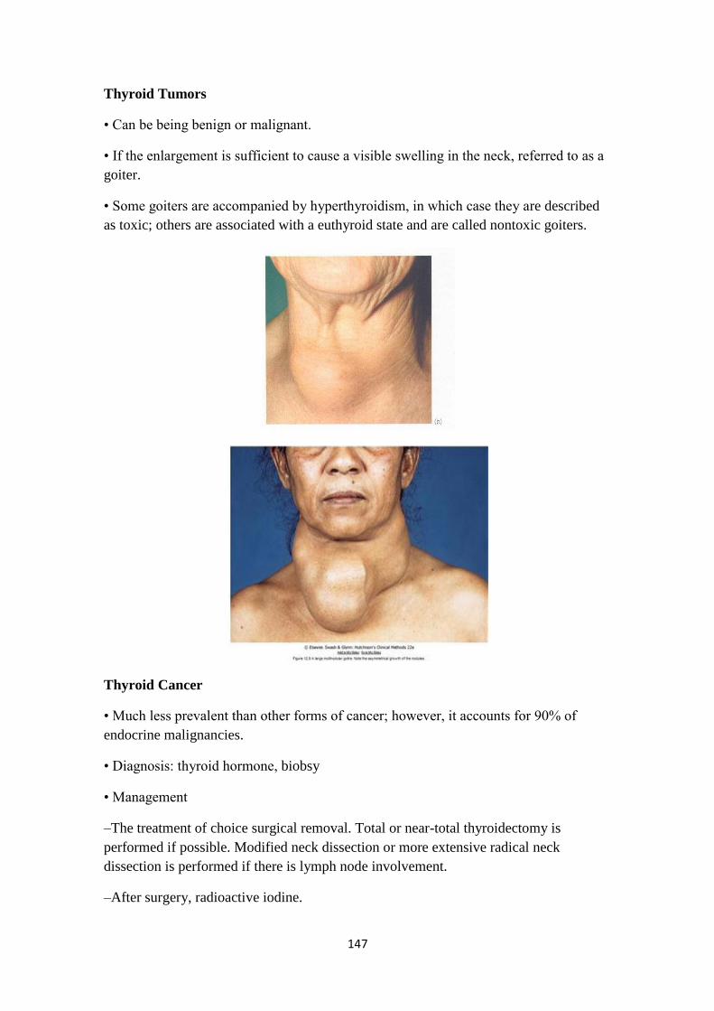
147
Thyroid Tumors
• Can be being benign or malignant.
• If the enlargement is sufficient to cause a visible swelling in the neck, referred to as a
goiter.
• Some goiters are accompanied by hyperthyroidism, in which case they are described
as toxic; others are associated with a euthyroid state and are called nontoxic goiters.
Thyroid Cancer
• Much less prevalent than other forms of cancer; however, it accounts for 90% of
endocrine malignancies.
• Diagnosis: thyroid hormone, biobsy
• Management
–The treatment of choice surgical removal. Total or near-total thyroidectomy is
performed if possible. Modified neck dissection or more extensive radical neck
dissection is performed if there is lymph node involvement.
–After surgery, radioactive iodine.

148
Thyroidectomy
• Treatment of choice for thyroid cancer
• Preoperative goals include the reduction of stress and anxiety to avoid precipitation of
thyroid storm (euothyroid)
• Iodine prep (Lugols or K iodide solution) to decrease size and vascularity of gland to
minimize risk of hemorrhage, reduces risk of thyroid storm during surgery
• Preoperative teaching includes dietary guidance to meet patient metabolic needs and
avoidance of caffeinated beverages and other stimulants,
Postoperative Care
• Monitor dressing for potential bleeding and hematoma formation; check posterior
dressing
• Monitor respirations; potential airway impairment
• Assess pain and provide pain relief measures
• Semi-Fowler‘s position, support head
• Assess voice but discourage talking
• Potential hypocalcaemia related to injury or removal of parathyroid glands; monitor
for hypocalcaemia
Post-Op Thyroidectomy Nursing Care
1. VS, I&O, IV
2. Semifowlers
3. Support head
4. Avoid tension on sutures
5. Pain meds, analgesic lozengers
6. Humidified oxygen, suction
7. First fluids: cold/ice, tolerated best, then soft diet
8. Limited talking , hoarseness common
9 Assess for voice changes: injury to the recurrent laryngeal nerve
149
Postop Thyroidectomy Nursing Care
• CHECK FOR HEMORRHAGE 1st 24 hrs:
• Look behind neck and sides of neck
• Check for c/o pressure or fullness at incision site
• Check drain
• REPORT TO MD
• CHECK FOR RESPIRATORY DISTRESS
• Laryngeal stridor (harsh hi pitched resp sounds)
• Result of edema of glottis, hematoma,or tetany
• Tracheostomy set/airway/ O2, suction
• CALL MD for extreme hoarseness (Check every 30-60 min for extreme hoarseness
(S&S) of respiratory distress
Complication of operation:
Hemorrhage
Laryngeal nerve damage.
Hypoparathyrodism
Hypothyroidism
Septesis
Postoperative infection
Parathyroid
• Four glands on the posterior thyroid gland
• Parathormone regulates calcium and phosphorus balance
–Increased parathormone elevates blood calcium by increasing calcium absorption
from the kidney, intestine, and bone.
–Parathormone lowers phosphorus level.
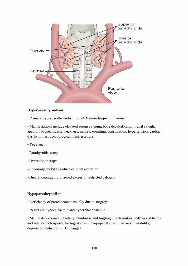
150
Hyperparathyroidism
• Primary hyperparathyroidism is 2–4 X more frequent in women.
• Manifestations include elevated serum calcium, bone decalcification, renal calculi,
apathy, fatigue, muscle weakness, nausea, vomiting, constipation, hypertension, cardiac
dysrhythmias, psychological manifestations
• Treatment
–Parathyroidectomy
–Hydration therapy
–Encourage mobility reduce calcium excretion
–Diet: encourage fluid, avoid excess or restricted calcium
Hypoparathryoidism
• Deficiency of parathormone usually due to surgery
• Results in hypocalcaemia and hyperphosphatemia
• Manifestations include tetany, numbness and tingling in extremities, stiffness of hands
and feet, bronchospasm, laryngeal spasm, carpopedal spasm, anxiety, irritability,
depression, delirium, ECG changes
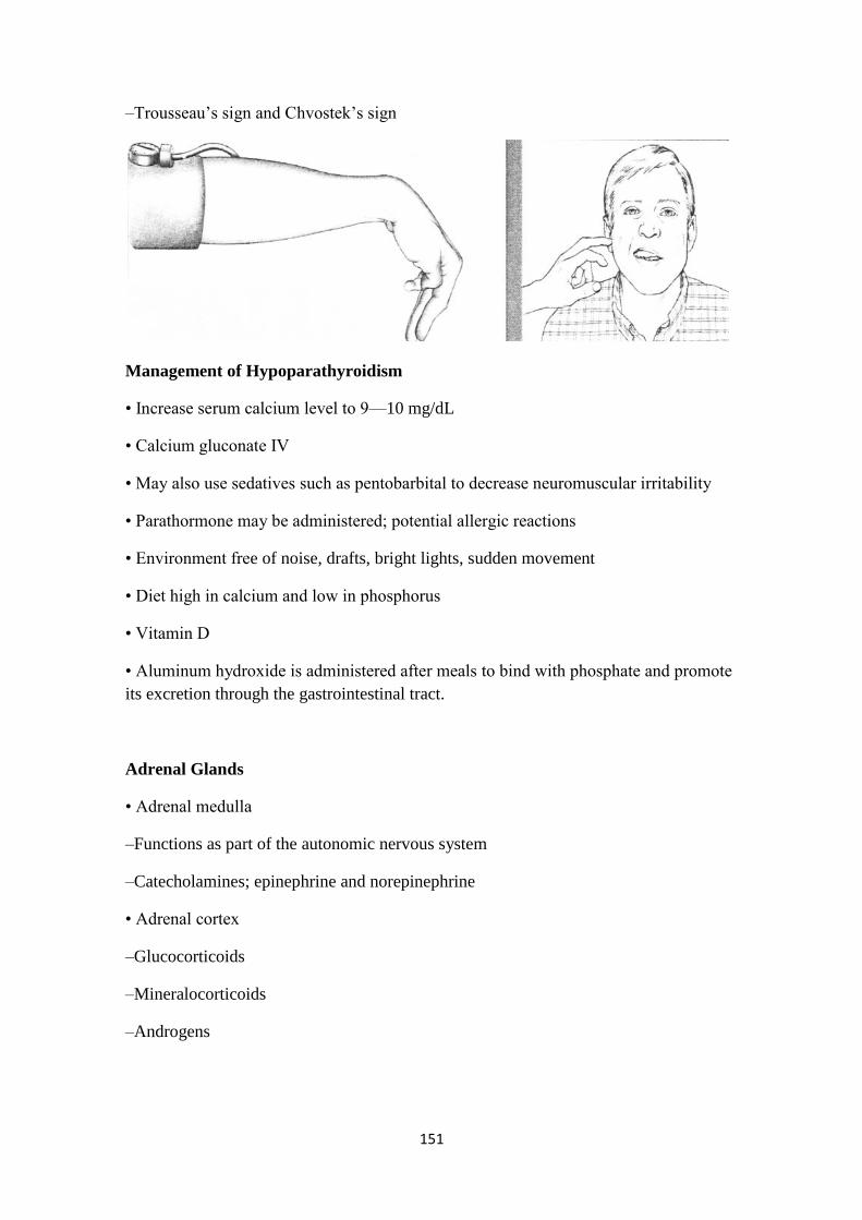
151
–Trousseau‘s sign and Chvostek‘s sign
Management of Hypoparathyroidism
• Increase serum calcium level to 9—10 mg/dL
• Calcium gluconate IV
• May also use sedatives such as pentobarbital to decrease neuromuscular irritability
• Parathormone may be administered; potential allergic reactions
• Environment free of noise, drafts, bright lights, sudden movement
• Diet high in calcium and low in phosphorus
• Vitamin D
• Aluminum hydroxide is administered after meals to bind with phosphate and promote
its excretion through the gastrointestinal tract.
Adrenal Glands
• Adrenal medulla
–Functions as part of the autonomic nervous system
–Catecholamines; epinephrine and norepinephrine
• Adrenal cortex
–Glucocorticoids
–Mineralocorticoids
–Androgens
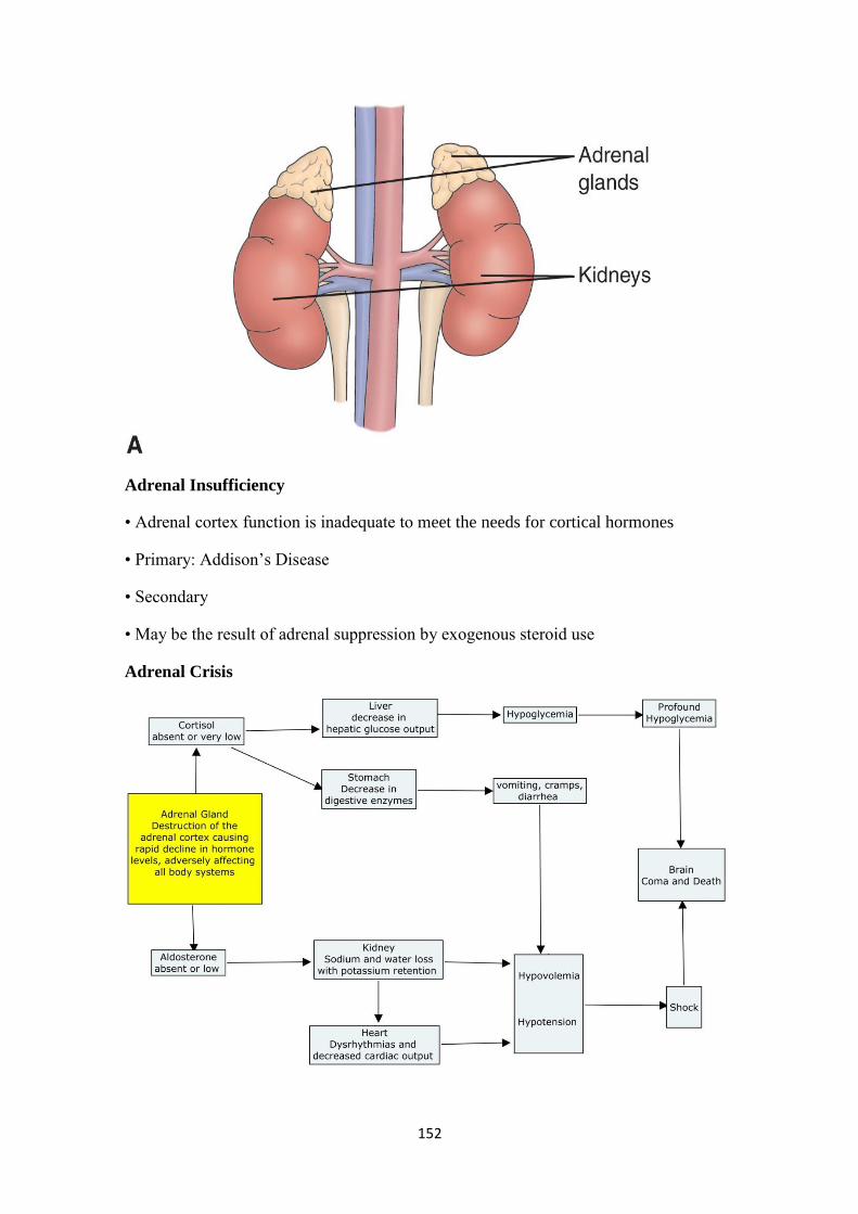
152
Adrenal Insufficiency
• Adrenal cortex function is inadequate to meet the needs for cortical hormones
• Primary: Addison‘s Disease
• Secondary
• May be the result of adrenal suppression by exogenous steroid use
Adrenal Crisis
153
Manifestations
• Muscle weakness, anorexia, GI symptoms, fatigue, dark pigmentation of skin and
mucosa, hypotension, low blood glucose, low serum sodium, high serum potassium,
mental changes, apathy, emotional lability, confusion
• Addisonian crisis: circulatory collapse
• Diagnostic tests; adrenocortical hormone levels, ACTH levels, ACTH stimulation test
Medical Management
• Immediate
–Reverse shock
–Restore blood circulation
• Antibiotics if infection
• Identify cause
• Supplement glucocorticoids during stressful procedures or significant illness
Nursing Management
• Assess fluid balance
• Monitor VS closely
• Good skin assessment
• Limit activity
• Provide quiet, non-stressful environment
Nursing Process: The Care of the Patient with Adrenocortical Insufficiency
Assessment
• Level of stress; note any illness or stressors that may precipitate problems
• Fluid and electrolyte status
• VS and postural blood pressures
• Note signs and symptoms related to adrenocortical insufficiency such as weight
changes, muscle weakness, and fatigue
• Medications
• Monitor for signs and symptoms of Addisonian
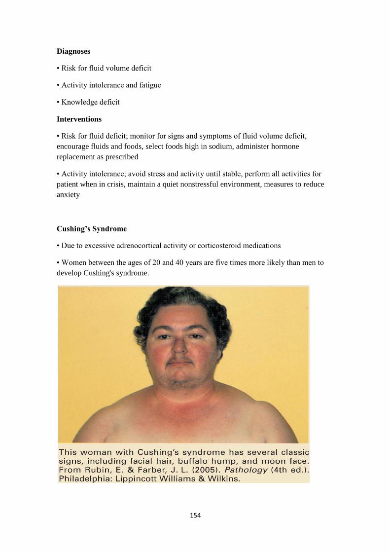
154
Diagnoses
• Risk for fluid volume deficit
• Activity intolerance and fatigue
• Knowledge deficit
Interventions
• Risk for fluid deficit; monitor for signs and symptoms of fluid volume deficit,
encourage fluids and foods, select foods high in sodium, administer hormone
replacement as prescribed
• Activity intolerance; avoid stress and activity until stable, perform all activities for
patient when in crisis, maintain a quiet nonstressful environment, measures to reduce
anxiety
Cushing’s Syndrome
• Due to excessive adrenocortical activity or corticosteroid medications
• Women between the ages of 20 and 40 years are five times more likely than men to
develop Cushing's syndrome.
155
Clinical Manifestations
• Hyperglycemia which may develop into diabetes, weight gain, central type obesity
with ―buffalo hump,‖ heavy trunk and thin extremities, fragile thin skin, ecchymosis,
striae, weakness, lassitude, sleep disturbances, osteoporosis, muscle wasting,
hypertension, ―moon-face‖, acne, increased susceptibility to infection, slow healing,
virilization in women, loss of libido, mood changes, increased serum sodium, decreased
serum potassium
• Diagnosis: Dexamethasone suppression test, ↑ Na+ ↑ glucose, ↓ K+, metabolic
alkalosis
Excessive protein catabolism occurs, producing muscle wasting and osteoporosis.
Kyphosis, backache, and compression fractures of the vertebrae may result. Retention
of sodium and water occurs as a result of increased mineralocorticoid activity,
producing hypertension and heart failure.
Virilization is characterized by the appearance of masculine traits and the recession of
feminine traits.
There is an excessive growth of hair on the face (hirsutism), the breasts atrophy,
menses cease, the clitoris enlarges, and the voice deepens.
Dexamethasone (1 mg) is administered orally at 11 PM, and a plasma cortisol level is
obtained at 8 AM the next morning. Suppression of cortisol to less than 5 mg/dL
indicates that the hypothalamic–pituitary–adrenal axis is functioning properly.
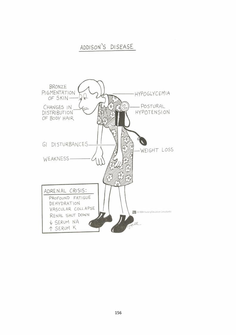
156

157
Medical Management
• Pituitary tumor
–Surgical removal
–radiation
• Adrenalectomy
• Adrenal enzyme inhibitors
• Attempt to reduce or taper corticosteroid dose

158
Nursing Management
• Prevent injury
• Increased protein, calcium and vitamin D in diet
• Medical asepsis
• Monitor blood glucose
• Moderate activity with rest periods
• Provide restful environment
Nursing Process: The Care of the Patient with Cushing’s Syndrome
Assessment
• Activity level and ability to carry out self-care
• Skin assessment
• Changes in physical appearance and patient responses to these changes
• Mental function
• Emotional status
• Medications
Diagnoses
• Risk for injury
• Risk for infection
• Self-care deficit
• Impaired skin integrity
• Disturbed body image
• Disturbed thought processes
Collaborative Problems/Potential Complications
• Addisonian crisis
• Adverse effects of adrenocortical activity
• Planning: Goals may include
159
1. Decreased risk of injury,
2. Decreased risk of infection,
3. Increased ability to carry out self-care activities,
4. Improved skin integrity,
5. Improved body image,
6. Improved mental function, and
7. Absence of complications
Interventions
• Decrease risk of injury; establish a protective environment; assist as needed;
encourage diet high in protein, calcium, and vitamin D.
• Decrease risk of infection; avoid exposure to infections, assess patient carefully as
corticosteroids mask signs of infection.
• Plan and space rest and activity.
• Meticulous skin care and frequent, careful skin assessment.
• Explanation to the patient and family about causes of emotional instability.
• Patient teaching.
Diabetes Insipidus
• A disorder of the posterior lobe of the pituitary gland that is characterized by a
deficiency of ADH (vasopressin). Excessive thirst (polydipsia) and large volumes of
dilute urine.
• It may occur secondary to head trauma, brain tumor, or surgical ablation or irradiation
of the pituitary gland, infections of the central nervous system or with tumors
• Another cause of diabetes insipidus is failure of the renal tubules to respond to ADH
Medical Management
• The objectives of therapy are .
1. to replace ADH (which is usually a long-term therapeutic program),
2. to ensure adequate fluid replacement, and
3. to identify and correct the underlying intracranial pathology.
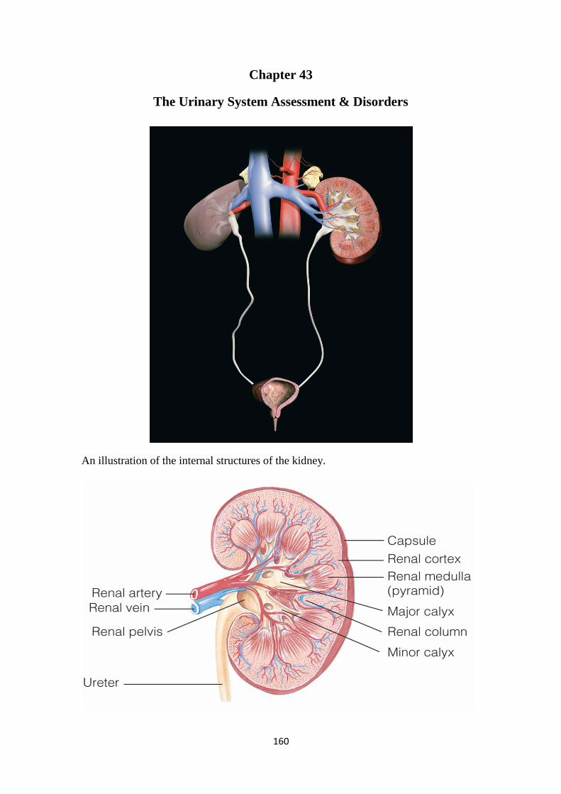
160
Chapter 43
The Urinary System Assessment & Disorders
An illustration of the internal structures of the kidney.
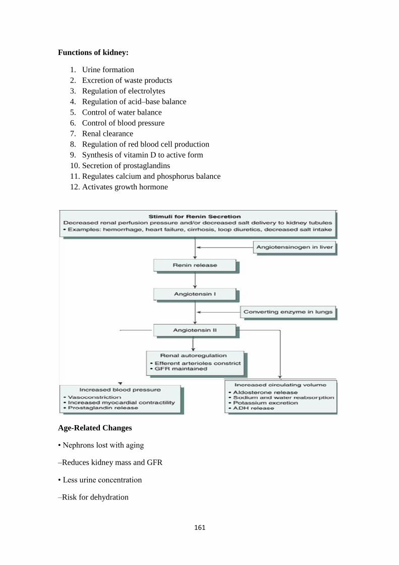
161
Functions of kidney:
1. Urine formation
2. Excretion of waste products
3. Regulation of electrolytes
4. Regulation of acid–base balance
5. Control of water balance
6. Control of blood pressure
7. Renal clearance
8. Regulation of red blood cell production
9. Synthesis of vitamin D to active form
10. Secretion of prostaglandins
11. Regulates calcium and phosphorus balance
12. Activates growth hormone
Age-Related Changes
• Nephrons lost with aging
–Reduces kidney mass and GFR
• Less urine concentration
–Risk for dehydration

162
Assessment
• Use simple language
• Assess for incontinence (esp. muliparous)
• Family history
• Chief concern
• Location and character of pain
• Previous UTI, stones, urinary problems
• Pattern or urination
• Color, clarity, amount of urine
• Difficulty initiating urination or changes in stream
• Changes in urinary pattern
• Dysuria, nocturia, hematuria, pyuria
Obtaining a urologic health history requires excellent communication skills, because
many patients are embarrassed or uncomfortable discussing genitourinary function or
symptoms
• History of urinary problems
• Urinary or abdominal surgeries
• Smoking, alcohol use
• Chance of pregnancy
• History of diabetes or other endocrine disorders
• Unexplained anemia
Diagnostic Tests
• Clean-catch urine
• 24-hour urine collection
• Culture and sensitivity
• BUN, creatinine and creatinine clearance = {Vol. of urine (ML/hr) x urine
creatine}/serum creatinine
BUN values are affected by protein intake, tissue breakdown, and fluid volume
changes.
163
• IVP, Retrograde Pyelography
• Cystography, voiding cystogram
• CT scan, MRI
• Renal scan
• Ultrasound
• X-ray (KUB)
• Cystoscopy
• Renal Angiography
• Kidney biopsy (by needle or open procedure)
Glomerulonephritis
• Inflammatory condition of glomerulus
• Antigen–antibody complexes form in the blood and become trapped in the glomerular
capillaries, inducing an inflammatory response.
• Damages capillary membrane
–Blood cells and proteins escape into filtrate
–Hematuria, proteinuria, azotemia (increase BUN & Creatinin)
• Acute or chronic
Acute Glomerulonephritis -Manifestations
• Usually follows infection of group A beta-hemolytic Streptococcus
• Affect children > 2 years
• Manifestations develop abruptly
–Hematuria (? Microscopic, or frank, urine is cola color), proteinuria, edema, azotemia
(High BUN and Creatineine) hypertension, fatigue, hypoalbuminemia, hyperlipidemia
–? headache, malaise and flank pain

164
165
Glomerulonephritis –Diagnostic Tests
• ASO titer ( anti streptolysine O)
• BUN
• Serum creatinine
• Serum electrolytes
• Urinalysis
• KUB x-ray
• Kidney scan or biopsy
Complications of acute glomerulonephritis
• Hypertensive encephalopathy,
• Heart failure,
• Pulmonary edema,
• Without treatment, end-stage renal disease (ESRD) develops in a matter of weeks or
months.
Glomerulonephritis –Treatment
• Focus is on identifying and treating underlying disease process and preserving kidney
function
• If residual streptococcal infection is suspected, penicillin.
• Corticosteroids and immunosuppressant medications may be prescribed for patients
with rapidly progressive acute glomerulonephritis.
• Dietary protein is restricted when renal insufficiency (elevated BUN) develop.
• Sodium is restricted when the patient has hypertension, edema, and heart failure.
• Loop diuretic and antihypertensive medications may be prescribed to control
hypertension.
• Bed rest during acute phase.
Glomerulonephritis –Nursing Care
• Decrease protein and increase CHO to prevent protein breakdown.
• Accurate I & O (consider insensible loss)
166
Chronic Glomerulonephritis
• Could be due to repeated episodes of acute glomerulonephritis, hypertensive
nephrosclerosis, hyperlipidemia, glomerular sclerosis
• Other causes include SLE, DM
• Kidney size reduce to 1/5th of original size and many scar tissue formed leading to
ESRF.
Chronic Glomerulonephritis/ S&S
• Many are asymptomatic
• ? Discovered when patient diagnosed with Hypertension.
• ? severe nosebleed, a stroke, or a seizure, swollen feet at night.
• Heneral symptoms, such as loss of weight and strength, increasing irritability,
nocturia, Headaches, dizziness, and digestive disturbances.
• Finally, S&S of renal failure.
Medical Management
• Control BP: Na & water restriction, antihypertensive drug
• Monitor weight.
• Diuretics.
• Adequate CHO diet to spare protien
• Treat UTI
• ? Dialysis.
Nephrotic Syndrome
• Is a cluster of clinical findings, including:
1. Marked increase in protein (particularly albumin) in the urine (proteinuria)
2. Decrease in albumin in the blood (hypoalbuminemia)
3. Edema (periorbital, ascites, and dependent edema)
4. High serum cholesterol and low-density lipoproteins (hyperlipidemia)
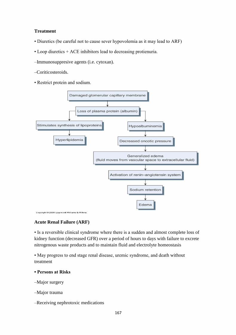
167
Treatment
• Diuretics (be careful not to cause sever hypovolemia as it may lead to ARF)
• Loop diuretics + ACE inhibitors lead to decreasing protienuria.
–Immunosuppresive agents (i.e. cytoxan).
–Coriticosteroids.
• Restrict protein and sodium.
Acute Renal Failure (ARF)
• Is a reversible clinical syndrome where there is a sudden and almost complete loss of
kidney function (decreased GFR) over a period of hours to days with failure to excrete
nitrogenous waste products and to maintain fluid and electrolyte homeostasis
• May progress to end stage renal disease, uremic syndrome, and death without
treatment
• Persons at Risks
–Major surgery
–Major trauma
–Receiving nephrotoxic medications

168
–Elderly
• ARF mostly occur within hospital settings
Causes of ARF
1. Prerenal Failure
2. Intrarenal Failure
3. Postrenal Failure
Prerenal Failure
• Volume depletion resulting from:
–Hemorrhage
–Renal losses (diuretics, osmotic diuresis)
–Gastrointestinal losses (vomiting, diarrhea, nasogastric suction)
• Impaired cardiac efficiency resulting from:
–Myocardial infarction
–Heart failure
–Dysrhythmias
–Cardiogenic shock
169
• Vasodilation resulting from:
–Sepsis
–Anaphylaxis
–Antihypertensive medications or other medications that cause vasodilation
Intrarenal Failure
• Prolonged renal ischemia resulting from:
–Pigment nephropathy (associated with the break-down of blood cells containing
pigments that in turn occlude kidney structures)
–Myoglobinuria (trauma, crush injuries, burns)
–Hemoglobinuria (transfusion reaction, hemolytic anemia)
• Nephrotoxic agents such as:
–Aminoglycoside antibiotics (gentamicin, tobramycin, amicacin)
–Radiopaque contrast agents
–Heavy metals (lead, mercury)
–Solvents and chemicals (ethylene glycol, carbon tetrachloride, arsenic)
–Nonsteroidal anti-inflammatory drugs (NSAIDs)
–Angiotensin-converting enzyme inhibitors (ACE inhibitors)
• Infectious processes such as:
–Acute pyelonephritis
–Acute glomerulonephritis
Postrenal failure
• Urinary tract obstruction, including:
–Calculi (stones)
–Tumors
–Benign prostatic hyperplasia
–Strictures
–Blood clots
170
Phases of Acute Renal Failure
• Initiation period: begins with the initial insult and ends when oliguria develops.
• Oliguria period: UOP < 400 ml/day, increase in urea, creatinine, uric acid, K &
magnesium. Some people have normal urine output (2 L/d)
Diuretic – UOP ^ to as much as 4000 mL/d but BUN & Cretinine still high, at end of
this stage may begin to see improvement
Recovery – things go back to normal. It may take up to 3-12 months
The oliguria period is accompanied by an increase in the serum concentration of
substances usually excreted by the kidneys (urea, creatinine, uric acid, organic acids,
and the intracellular cations [potassium and magnesium]). The minimum amount of
urine needed to rid the body of normal metabolic waste products is 400 mL. In this
phase uremic symptoms first appear and life-threatening conditions such as
hyperkalemia develop.
Some patients have decreased renal function with increasing nitrogen retention, yet
actually excrete normal amounts of urine (2 L/day or more). This is the nonoliguric
form of renal failure and occurs predominantly after exposure of the patient to
nephrotoxic agents; it may also occur with burns, traumatic injury, and the use of
halogenated anesthetic agents.
The diuresis period is marked by a gradual increase in urine output, which signals that
glomerular filtration has started to recover. Laboratory values stop increasing and
eventually decrease. Although the volume of urinary output may reach normal or
elevated levels, renal function may still be markedly abnormal. Because uremic
symptoms may still be present, the need for expert medical and nursing management
continues. The patient must be observed closely for dehydration during this phase; if
dehydration occurs, the uremic symptoms are likely to increase.
The recovery period signals the improvement of renal function and may take 3 to 12
months. Laboratory values return to the patient's normal level. Although a permanent
1% to 3% reduction in the GFR is common, it is not clinically significant.
Acute Renal Failure S & S
–The patient may appear critically ill and lethargic.
–The skin and mucous membranes are dry from dehydration.
–Central nervous system signs and symptoms include drowsiness, headache, muscle
twitching, and seizures.
–Urine output varies (scanty to normal volume), ? hematuria & urine has a low specific
gravity
171
• Diagnostic tests
–BUN, creatinine, potassium increase.
–pH
–Hgb and Hct
–Urine studies
–US of kidneys
–High phosphorus and low calcium.
With a decline in the GFR, the patient cannot excrete potassium normally. Patients with
oliguria and anuria are at high risk for hyperkalemia. Protein catabolism results in the
release of cellular potassium into the body fluids, causing severe hyperkalemia (high
serum potassium levels).
Prevention of ARF
• Provide adequate hydration
• Prevent and treat shock promptly
• Hourly urine output for critical patients
• Continuosally assess renal function
• Prevent and treat infections promptly
• Monitor for effects of toxic drugs
Medical treatment of ARF
• Objectives of treatment are to restore normal chemical balance and prevent
complications until repair of renal tissue and restoration of renal function can occur.
• Management includes
–maintaining fluid balance,
–avoiding fluid excesses, or
–possibly performing dialysis.
• Medical treatment
–Treat the cause
–Fluid and replacement or restrictions
–Monitor for fluid overload
172
–Diuretics
–Maintain E-lytes
–May need dialysis (especially with high K)
–May need to stimulate production of urine with IV fluids, Dopomine, diuretics, etc.
If caused by meds, must stop meds
If caused by obstruction, must remove obstruction
If caused by blockage of artery, must open artery
Dietary restrictions may include : low K+, adequate carbs, also may give TPN
Fluids : calculate closley I/O
Hyperkalemia is life threatening
Lower K+ with Kayexalate, glucose, insulin, NaBicarb, caalcium carbonate
–Hemodialysis
• Subclavian approach
• Femoral approach
–Peritoneal dialysis
–Nutritional Therapy
• ? Decrease Protein (according to BUN level)
• Increase CHO
• Decrease potassium and phosphrous
• Nursing Diagnosis-
–imbalanced fluid volume= excess
–Altered electrolyte balance
–Impaired tissue perfusion: renal
–Anxiety
–Imbalanced nutrition
–Risk for infection
–Fatigue
173
–Knowledge deficit
• Plan-
–Promote recovery of optimal kidney function.
–Maintain normal fluid and electrolyte balance.
–Decrease anxiety.
–Increase knowledge.
Nursing interventions
• Monitoring Fluid and Electrolyte Balance
• Reducing Metabolic Rate
• Promoting Pulmonary Function
• Preventing Infection
• Providing Skin Care
Chronic Renal Failure
• Chronic renal failure, or ESRD, is a progressive, irreversible deterioration in renal
function in which the body's ability to maintain metabolic and fluid and electrolyte
balance fails, resulting in uremia or azotemia.
• Results from gradual, progressive loss of renal function
• Occasionally results from rapid progression of acute renal failure
• Conditions that cause ESRD include systemic diseases, such as diabetes mellitus
(leading cause); hypertension; chronic glomerulonephritis; pyelonephritis; obstruction
of the urinary tract; hereditary lesions, as in polycystic kidney disease; vascular
disorders; infections; medications; or toxic agents.
• Symptoms occur when 75% of function is lost but considered chronic if 90-95% loss
of function
• Dialysis is necessary D/T accumulation of uremic toxins, which produce changes in
major organs
Chronic renal failure/ S&S Cardiovascular: the most common cause of death
–Hypertension
–Pitting edema (feet, hands, sacrum)
174
–Periorbital edema
–Pericardial friction rub
–Acidosis (kidney can‘t excrete amonia, reabsorb bicarb, high phosphate)
–Engorged neck veins
–Pericarditis
–Pericardial effusion
–Pericardial tamponade
–Hyperkalemia
–Hyperlipidemia
Chronic renal failure affects almost all body systems
Hypertension occurs due to sodium and water retention and due to the activation of
renin- angiotesnin-aldosteron axis.
Decreased acid secretion results from the inability of the kidney tubules to excrete
ammonia (NH3-) and to reabsorb sodium bicarbonate (HCO3
-). There is also decreased
excretion of phosphates and other organic acids.
Chronic renal failure/ S&S Neurologic
• Weakness and fatigue
• Confusion
• Inability to concentrate
• Disorientation
• Tremors
• Seizures
• Asterixis
• Restlessness of legs
• Burning of soles of feet
• Behavior changes
Chronic renal failure/ S&S Pulmonary Integumentary
• Crackles
175
• Thick, tenacious sputum
• Depressed cough reflex
• Pleuritic pain
• Shortness of breath
• Tachypnea
• Kussmaul-type respirations
• Uremic pneumonitis
Chronic renal failure/ S&S Integumentary
• Gray-bronze skin color
• Dry, flaky ( ُمقّشر ) skin
• Pruritus
• Ecchymosis
• Purpura
• Thin, brittle nails
• Coarse, thinning hair
Chronic renal failure/ S&S Gastrointestinal
• Ammonia odor to breath (―uremic fetor‖)
• Metallic taste
• Mouth ulcerations and bleeding
• Anorexia, nausea, vomiting
• Hiccups
• Constipation or diarrhea
Chronic renal failure/ S&S Hematological
• Bleeding from GI tract
• Anemia
• Thrombocytopenia
Chronic renal failure/ S&S Reproductive

176
• Amenorrhea
• Testicular atrophy
• Infertility
• Decreased libido
• Muscle cramps
Chronic renal failure/ S&S Musculoskeletal
• Loss of muscle strength
• Renal osteodystrophy
• Bone pain
• Bone fractures
• Foot drop

177
• Lab findings
–BUN – indicator of glomerular filtration rate and is affected by the breakdown of
protein.
–Serum creatinine – waste product of skeletal muscle breakdown and is a better
indicator of kidney function.
–Creatinine clearance is best determent of kidney function (GFR). Must be a 24 hour
urine collection.
• K+
–The kidneys are means which K+ is excreted. Normal is 3.5-5.0 ,mEq/L. maintains
muscle contraction and is essential for cardiac function.
–Both elevated and decreased can cause problems with cardiac rhythm
–Hyperkalemia is treated with IV glucose and Na Bicarb which pushes K+ back into
the cell
–Kayexalate ( Sodium polystyrene sulfonate ) is also used to promote the exchange of
sodium and potassium in the body.
• Ca++
–With disease in the kidney, the enzyme for utilization of Vit D is absent
–Ca absorption depends upon Vit D
–Also, phosphate level increase leading to decreasing level of Ca
–Parathyroid hormone level increase in attempt to increase calcium, but because of the
high phosphorus level, there is limited response.
–Body moves Ca out of the bone to compensate.
–Renal osteodystrophy is the end result
Hypocalcemia = tetany
Parathyroid hormone level increase in attempt to increase calcium, but because of the
high phosphorus level, there is limited response.
• Other abnormal findings
–Metabolic acidosis
–Fluid imbalance
–Insulin resistance
178
–Anemia
–Immunoligical problems
Kidneys cannot excrete wastes or change bicarb levels if needed
Retain Na and water which can elevate B/P
Insulin resistance cells cause rise in glucose which produces more lipids in the liver
Anemia = kidneys produce erythropioetin which is needed to make RBC‘s
Patients have less resistance to illness
Complications
• Hyperkalemia
• Pericarditis, pericardial effusion, and pericardial tamponade
• Hypertension
• Anemia
• Bone disease and metastatic and vascular calcifications
Hyperkalemia due to decreased excretion, metabolic acidosis, catabolism, and
excessive intake (diet, medications, fluids)
Pericarditis, pericardial effusion, and pericardial tamponade due to retention of uremic
waste products and inadequate dialysis
Hypertension due to sodium and water retention and malfunction of the renin–
angiotensin–aldosterone system
Anemia due to decreased erythropoietin production, decreased RBC life span, bleeding
in the GI tract from irritating toxins and ulcer formation, and blood loss during
hemodialysis
Bone disease and metastatic and vascular calcifications due to retention of phosphorus,
low serum calcium levels, abnormal vitamin D metabolism, and elevated aluminum
levels
Medical management
• Calcium carbonate, or calcium acetate: bind to phosphours and decrease its level.
• Antihypertensive and Cardiovascular Agents
• Antiseizure Agents
• Erythropoietin
179
Nutritional Therapy
• Protein is restricted (allowed protein should be of high biologic value)
• Restrict fluid (500-600 ml/day more than previous day‘s urine output).
• Restrict K, Na, Phosphorus
• Increase CHO to meat caloric needs
• Vitamin suplements
Protein is restricted because urea, uric acid, and organic acids–the breakdown products
of dietary and tissue proteins–accumulate rapidly in the blood when there is impaired
renal clearance.
Treatment
• Dialysis
• Transplantation
• Nursing diagnosis
–Excess fluid volume related to decreased urine output, dietary excesses, and retention
of sodium and water
–Imbalanced nutrition: less than body requirements related to anorexia, nausea and
vomiting, dietary restrictions, and altered oral mucous membranes
Nursing diagnosis
–Deficient knowledge regarding condition and treatment regimen
–Activity intolerance related to fatigue, anemia, retention of waste products, and
dialysis procedure
–Risk for situational low self-esteem related to dependency, role changes, changes in
body image, and sexual dysfunction
Chronic Renal Failure
• Nursing care: see nursing process below
Dialysis
• Dialysis is used to remove fluid and uremic waste products from the body when the
kidneys are unable to do so.
• Chronic: in ESRF when the kidney can‘t remove waste products.
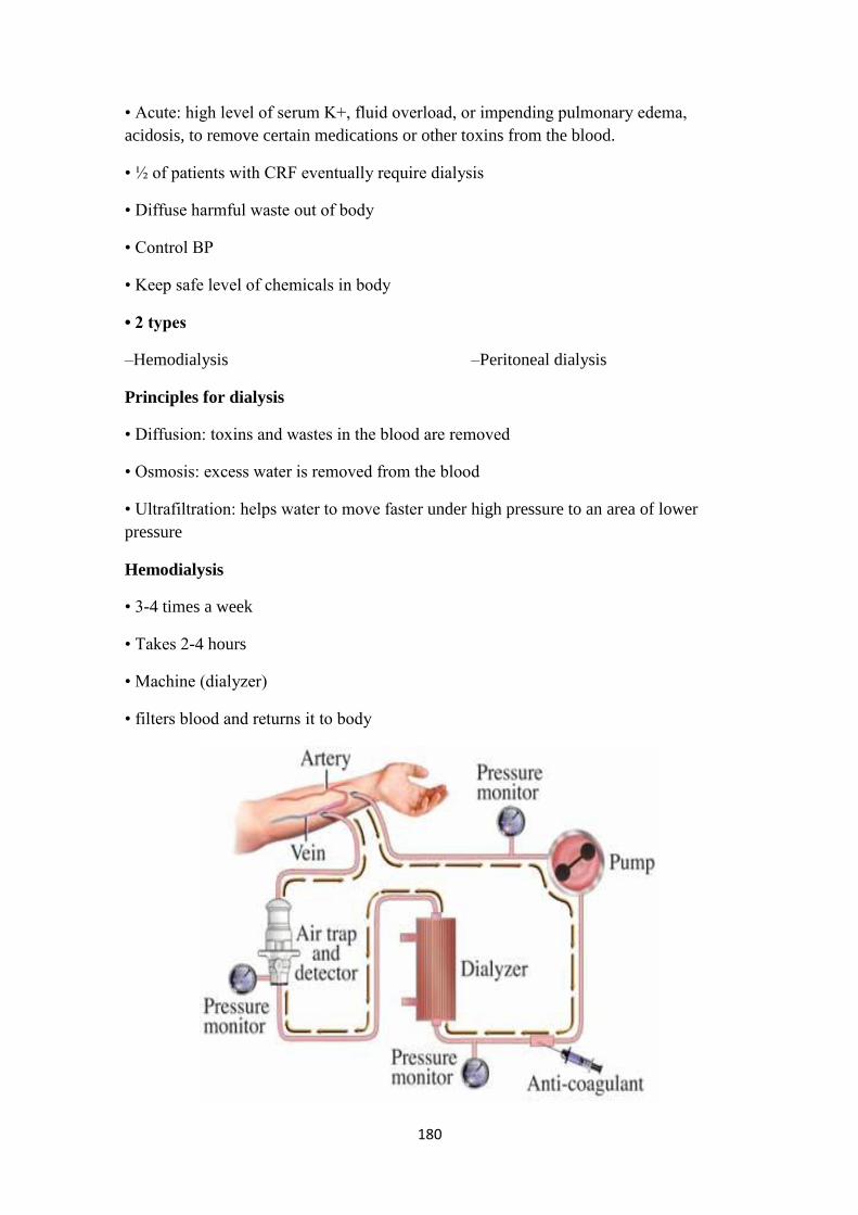
180
• Acute: high level of serum K+, fluid overload, or impending pulmonary edema,
acidosis, to remove certain medications or other toxins from the blood.
• ½ of patients with CRF eventually require dialysis
• Diffuse harmful waste out of body
• Control BP
• Keep safe level of chemicals in body
• 2 types
–Hemodialysis –Peritoneal dialysis
Principles for dialysis
• Diffusion: toxins and wastes in the blood are removed
• Osmosis: excess water is removed from the blood
• Ultrafiltration: helps water to move faster under high pressure to an area of lower
pressure
Hemodialysis
• 3-4 times a week
• Takes 2-4 hours
• Machine (dialyzer)
• filters blood and returns it to body
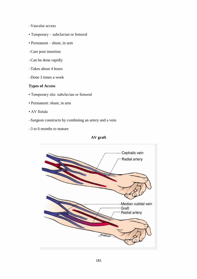
181
–Vascular access
• Temporary – subclavian or femoral
• Permanent – shunt, in arm
–Care post insertion
–Can be done rapidly
–Takes about 4 hours
–Done 3 times a week
Types of Access
• Temporary site: subclavian or femoral
• Permanent: shunt, in arm
• AV fistula
–Surgeon constructs by combining an artery and a vein
–3 to 6 months to mature
AV graft

182
What This Means for You
• No BP on same arm as fistula
• Protect arm from injury
• Never inject anything into catheter
• Control obvious hemorrhage
–Bleeding will be arterial
–Maintain direct pressure
• No IV on same arm as fistula
• A thrill will be felt – this is normal
Complication of dialysis
• Hypotension
• Painful muscle cramping (due to rapid alterations in electrolyte balance)
• Dysrhythmias may result from electrolyte and pH changes
• Air embolism
• Dialysis disequilibrium results from cerebral fluid shifts.
Peritoneal Dialysis
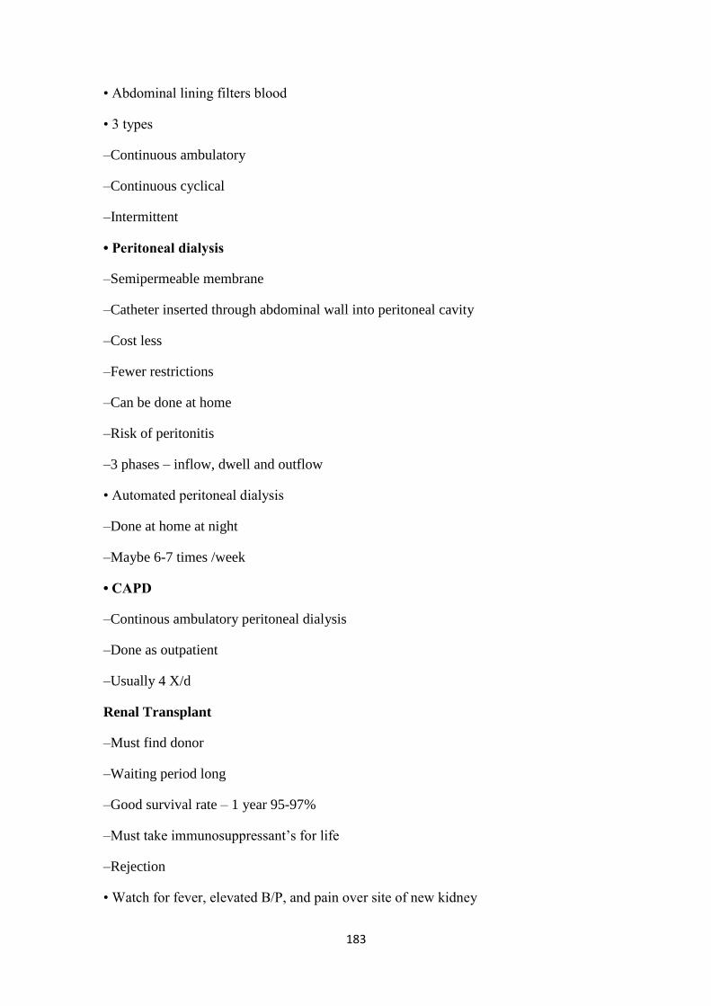
183
• Abdominal lining filters blood
• 3 types
–Continuous ambulatory
–Continuous cyclical
–Intermittent
• Peritoneal dialysis
–Semipermeable membrane
–Catheter inserted through abdominal wall into peritoneal cavity
–Cost less
–Fewer restrictions
–Can be done at home
–Risk of peritonitis
–3 phases – inflow, dwell and outflow
• Automated peritoneal dialysis
–Done at home at night
–Maybe 6-7 times /week
• CAPD
–Continous ambulatory peritoneal dialysis
–Done as outpatient
–Usually 4 X/d
Renal Transplant
–Must find donor
–Waiting period long
–Good survival rate – 1 year 95-97%
–Must take immunosuppressant‘s for life
–Rejection
• Watch for fever, elevated B/P, and pain over site of new kidney
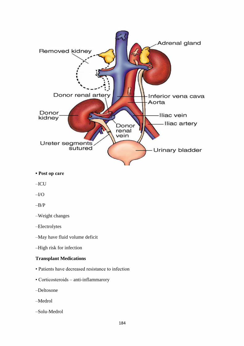
184
• Post op care
–ICU
–I/O
–B/P
–Weight changes
–Electrolytes
–May have fluid volume deficit
–High risk for infection
Transplant Medications
• Patients have decreased resistance to infection
• Corticosteroids – anti-inflammarory
–Deltosone
–Medrol
–Solu-Medrol
185
• Cytotoxic – inhibit T and B lymphocytes
–Imuran
–Cytoxan
–Cellcept
• T-cell depressors - Cyclosporin
Renal Trauma
• ? due to rib fractures or fractures vertebrae.
• 80% to 90% of all renal injuries are blunt injuries
• S & S: Pain, hematouria, S & S of shock
• Rx: Bed rest, antibiotics. In sever cases, need surgical repair or ? Nephrectomy.
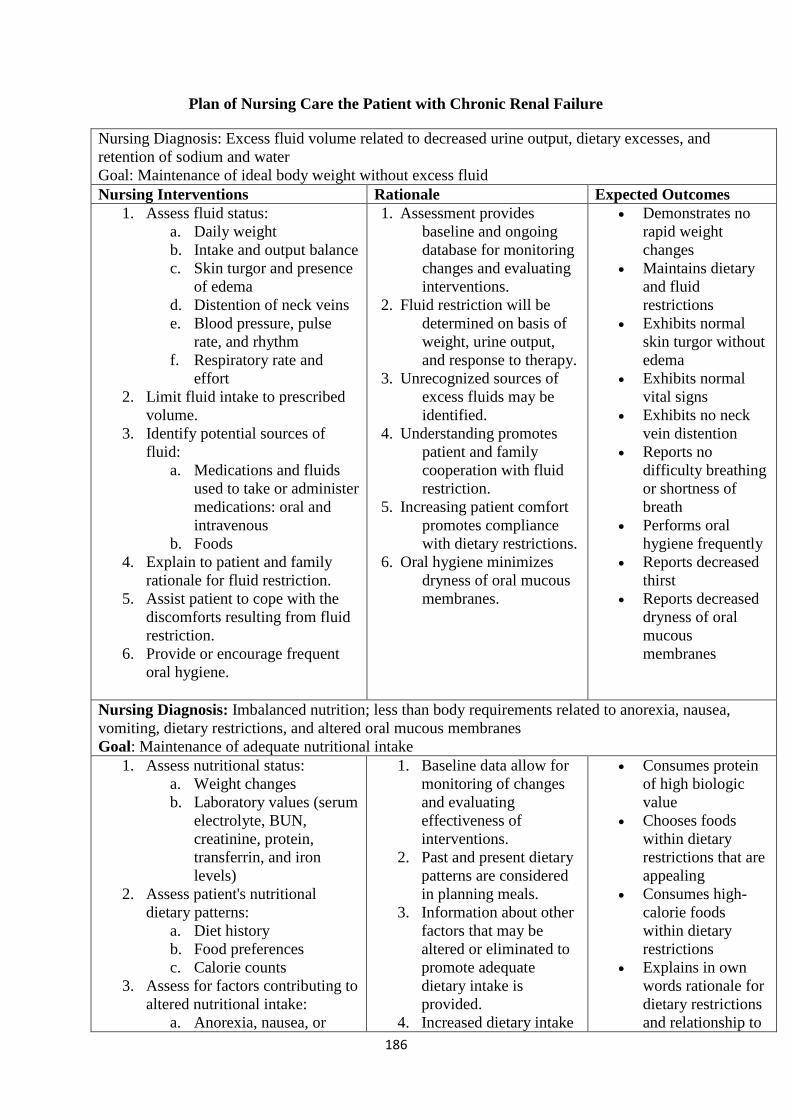
186
Plan of Nursing Care the Patient with Chronic Renal Failure
Nursing Diagnosis: Excess fluid volume related to decreased urine output, dietary excesses, and
retention of sodium and water
Goal: Maintenance of ideal body weight without excess fluid
Nursing Interventions Rationale Expected Outcomes
1. Assess fluid status:
a. Daily weight
b. Intake and output balance
c. Skin turgor and presence
of edema
d. Distention of neck veins
e. Blood pressure, pulse
rate, and rhythm
f. Respiratory rate and
effort
2. Limit fluid intake to prescribed
volume.
3. Identify potential sources of
fluid:
a. Medications and fluids
used to take or administer
medications: oral and
intravenous
b. Foods
4. Explain to patient and family
rationale for fluid restriction.
5. Assist patient to cope with the
discomforts resulting from fluid
restriction.
6. Provide or encourage frequent
oral hygiene.
1. Assessment provides
baseline and ongoing
database for monitoring
changes and evaluating
interventions.
2. Fluid restriction will be
determined on basis of
weight, urine output,
and response to therapy.
3. Unrecognized sources of
excess fluids may be
identified.
4. Understanding promotes
patient and family
cooperation with fluid
restriction.
5. Increasing patient comfort
promotes compliance
with dietary restrictions.
6. Oral hygiene minimizes
dryness of oral mucous
membranes.
Demonstrates no
rapid weight
changes
Maintains dietary
and fluid
restrictions
Exhibits normal
skin turgor without
edema
Exhibits normal
vital signs
Exhibits no neck
vein distention
Reports no
difficulty breathing
or shortness of
breath
Performs oral
hygiene frequently
Reports decreased
thirst
Reports decreased
dryness of oral
mucous
membranes
Nursing Diagnosis: Imbalanced nutrition; less than body requirements related to anorexia, nausea,
vomiting, dietary restrictions, and altered oral mucous membranes
Goal: Maintenance of adequate nutritional intake
1. Assess nutritional status:
a. Weight changes
b. Laboratory values (serum
electrolyte, BUN,
creatinine, protein,
transferrin, and iron
levels)
2. Assess patient's nutritional
dietary patterns:
a. Diet history
b. Food preferences
c. Calorie counts
3. Assess for factors contributing to
altered nutritional intake:
a. Anorexia, nausea, or
1. Baseline data allow for
monitoring of changes
and evaluating
effectiveness of
interventions.
2. Past and present dietary
patterns are considered
in planning meals.
3. Information about other
factors that may be
altered or eliminated to
promote adequate
dietary intake is
provided.
4. Increased dietary intake
Consumes protein
of high biologic
value
Chooses foods
within dietary
restrictions that are
appealing
Consumes high-
calorie foods
within dietary
restrictions
Explains in own
words rationale for
dietary restrictions
and relationship to

187
vomiting
b. Diet unpalatable to
patient
c. Depression
d. Lack of understanding of
dietary restrictions
e. Stomatitis
4. Provide patient's food
preferences within dietary
restrictions.
5. Promote intake of high biologic
value protein foods: eggs, dairy
products, meats.
6. Encourage high-calorie, low-
protein, low-sodium, and low-
potassium snacks between
meals.
7. Alter schedule of medications so
that they are not given
immediately before meals.
8. Explain rationale for dietary
restrictions and relationship to
kidney disease and increased
urea and creatinine levels.
9. Provide written lists of foods
allowed and suggestions for
improving their taste without use
of sodium or potassium.
10. Provide pleasant surroundings at
meal-times.
11. Weigh patient daily.
12. Assess for evidence of
inadequate protein intake:
a. Edema formation
b. Delayed wound healing
c. Decreased serum
albumin levels
is encouraged.
5. Complete proteins are
provided for positive
nitrogen balance needed
for growth and healing.
6. Reduces source of
restricted foods and
proteins and provides
calories for energy,
sparing protein for tissue
growth and healing.
7. Ingestion of medications
just before meals may
produce anorexia and
feeling of fullness.
8. Promotes patient
understanding of
relationships between
diet and urea and
creatinine levels to renal
disease.
9. Lists provide a positive
approach to dietary
restrictions and a
reference for patient and
family to use when at
home.
10. Unpleasant factors that
contribute to patient's
anorexia are eliminated.
11. Allows monitoring of
fluid and nutritional
status.
12. Inadequate protein
intake can lead to
decreased albumin and
other proteins, edema
formation, and delay in
wound healing.
urea and creatinine
levels
Takes medications
on schedule that
does not produce
anorexia or feeling
of fullness
Consults written
lists of acceptable
foods
Reports increased
appetite at meals
Exhibits no rapid
increases or
decreases in weight
Demonstrates
normal skin turgor
without edema;
wound healing and
acceptable plasma
albumin levels
Nursing Diagnosis: Deficient knowledge regarding condition and treatment
Goal: Increased knowledge about condition and related treatment
1. Assess understanding of cause of
renal failure, consequences of
renal failure, and its treatment:
a. Cause of patient's renal
failure
b. Meaning of renal failure
c. Understanding of renal
function
d. Relationship of fluid and
1. Provides baseline for
further explanations and
teaching.
2. Patient can learn about
renal failure and
treatment as he or she
becomes ready to
understand and accept
the diagnosis and
Verbalizes
relationship of
cause of renal
failure to
consequences
Explains fluid and
dietary restrictions
as they relate to
failure of kidney's

188
dietary restrictions to
renal failure
e. Rationale for treatment
(hemodialysis, peritoneal
dialysis, transplantation)
2. Provide explanation of renal
function and consequences of
renal failure at patient's level of
understanding and guided by
patient's readiness to learn.
3. Assist patient to identify ways to
incorporate changes related to
illness and its treatment into
lifestyle.
4. Provide oral and written
information as appropriate
about:
a. Renal function and
failure
b. Fluid and dietary
restrictions
c. Medications
d. Reportable problems,
signs, and symptoms
e. Follow-up schedule
f. Community resources
g. Treatment options
consequences.
3. Patient can see that his
or her life does not have
to revolve around the
disease.
4. Provides patient with
information that can be
used for further
clarification at home.
regulatory
functions
States in own
words relationship
of renal failure and
need for treatment
Asks questions
about treatment
options, indicating
readiness to learn
Verbalizes plans to
continue as normal
a life as possible
Uses written
information and
instructions to
clarify questions
and seek additional
information
Nursing Diagnosis: Activity intolerance related to fatigue, anemia, retention of waste products, and
dialysis procedure
Goal: Participation in activity within tolerance
1. Assess factors contributing to
activity intolerance:
a. Fatigue
b. Anemia
c. Fluid and electrolyte
imbalances
d. Retention of waste
products
e. Depression
2. Promote independence in self-
care activities as tolerated; assist
if fatigued.
3. Encourage alternating activity
with rest.
4. Encourage patient to rest after
dialysis treatments.
1. Indicates factors
contributing to severity
of fatigue.
2. Promotes improved self-
esteem
3. Promotes activity and
exercise within limits
and adequate rest.
4. Adequate rest is
encouraged after
dialysis treatments,
which are exhausting to
many patients.
Participates in
increasing levels of
activity and
exercise
Reports increased
sense of well-being
Alternates rest and
activity
Participates in
selected self-care
activities
Nursing Diagnosis: Risk for situational low self-esteem related to dependency, role changes, change in
body image, and change in sexual function
Goal: Improved self-esteem
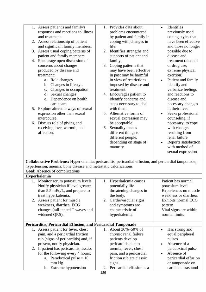
189
1. Assess patient's and family's
responses and reactions to illness
and treatment.
2. Assess relationship of patient
and significant family members.
3. Assess usual coping patterns of
patient and family members.
4. Encourage open discussion of
concerns about changes
produced by disease and
treatment:
a. Role changes
b. Changes in lifestyle
c. Changes in occupation
d. Sexual changes
e. Dependence on health
care team
5. Explore alternate ways of sexual
expression other than sexual
intercourse.
6. Discuss role of giving and
receiving love, warmth, and
affection.
1. Provides data about
problems encountered
by patient and family in
coping with changes in
life.
2. Identifies strengths and
supports of patient and
family.
3. Coping patterns that
may have been effective
in past may be harmful
in view of restrictions
imposed by disease and
treatment.
4. Encourages patient to
identify concerns and
steps necessary to deal
with them.
5. Alternative forms of
sexual expression may
be acceptable.
6. Sexuality means
different things to
different people,
depending on stage of
maturity.
Identifies
previously used
coping styles that
have been effective
and those no longer
possible due to
disease and
treatment (alcohol
or drug use;
extreme physical
exertion)
Patient and family
identify and
verbalize feelings
and reactions to
disease and
necessary changes
in their lives
Seeks professional
counseling, if
necessary, to cope
with changes
resulting from
renal failure
Reports satisfaction
with method of
sexual expression
Collaborative Problems: Hyperkalemia; pericarditis, pericardial effusion, and pericardial tamponade;
hypertension; anemia; bone disease and metastatic calcifications
Goal: Absence of complications
Hyperkalemia
1. Monitor serum potassium levels.
Notify physician if level greater
than 5.5 mEq/L, and prepare to
treat hyperkalemia.
2. Assess patient for muscle
weakness, diarrhea, ECG
changes (tall-tented T waves and
widened QRS).
1. Hyperkalemia causes
potentially life-
threatening changes in
the body.
2. Cardiovascular signs
and symptoms are
characteristic of
hyperkalemia.
Patient has normal
potassium level
Experiences no muscle
weakness or diarrhea.
Exhibits normal ECG
pattern
Vital signs are within
normal limits
Pericarditis, Pericardial Effusion, and Pericardial Tamponade
1. Assess patient for fever, chest
pain, and a pericardial friction
rub (signs of pericarditis) and, if
present, notify physician.
2. If patient has pericarditis, assess
for the following every 4 hours:
a. Paradoxical pulse > 10
mm Hg
b. Extreme hypotension
1. About 30%–50% of
chronic renal failure
patients develop
pericarditis due to
uremia; fever, chest
pain, and a pericardial
friction rub are classic
signs.
2. Pericardial effusion is a
Has strong and
equal peripheral
pulses
Absence of a
paradoxical pulse
Absence of
pericardial effusion
or tamponade on
cardiac ultrasound
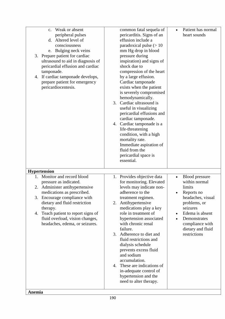
190
c. Weak or absent
peripheral pulses
d. Altered level of
consciousness
e. Bulging neck veins
3. Prepare patient for cardiac
ultrasound to aid in diagnosis of
pericardial effusion and cardiac
tamponade.
4. If cardiac tamponade develops,
prepare patient for emergency
pericardiocentesis.
common fatal sequela of
pericarditis. Signs of an
effusion include a
paradoxical pulse (> 10
mm Hg drop in blood
pressure during
inspiration) and signs of
shock due to
compression of the heart
by a large effusion.
Cardiac tamponade
exists when the patient
is severely compromised
hemodynamically.
3. Cardiac ultrasound is
useful in visualizing
pericardial effusions and
cardiac tamponade.
4. Cardiac tamponade is a
life-threatening
condition, with a high
mortality rate.
Immediate aspiration of
fluid from the
pericardial space is
essential.
Patient has normal
heart sounds
Hypertension
1. Monitor and record blood
pressure as indicated.
2. Administer antihypertensive
medications as prescribed.
3. Encourage compliance with
dietary and fluid restriction
therapy.
4. Teach patient to report signs of
fluid overload, vision changes,
headaches, edema, or seizures.
1. Provides objective data
for monitoring. Elevated
levels may indicate non-
adherence to the
treatment regimen.
2. Antihypertensive
medications play a key
role in treatment of
hypertension associated
with chronic renal
failure.
3. Adherence to diet and
fluid restrictions and
dialysis schedule
prevents excess fluid
and sodium
accumulation.
4. These are indications of
in-adequate control of
hypertension and the
need to alter therapy.
Blood pressure
within normal
limits
Reports no
headaches, visual
problems, or
seizures
Edema is absent
Demonstrates
compliance with
dietary and fluid
restrictions
Anemia
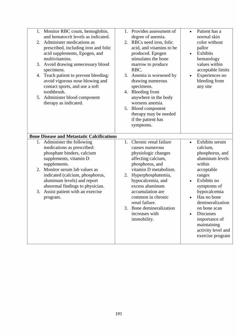
191
1. Monitor RBC count, hemoglobin,
and hematocrit levels as indicated.
2. Administer medications as
prescribed, including iron and folic
acid supplements, Epogen, and
multivitamins.
3. Avoid drawing unnecessary blood
specimens.
4. Teach patient to prevent bleeding:
avoid vigorous nose blowing and
contact sports, and use a soft
toothbrush.
5. Administer blood component
therapy as indicated.
1. Provides assessment of
degree of anemia.
2. RBCs need iron, folic
acid, and vitamins to be
produced. Epogen
stimulates the bone
marrow to produce
RBC.
3. Anemia is worsened by
drawing numerous
specimens.
4. Bleeding from
anywhere in the body
worsens anemia.
5. Blood component
therapy may be needed
if the patient has
symptoms.
Patient has a
normal skin
color without
pallor
Exhibits
hematology
values within
acceptable limits
Experiences no
bleeding from
any site
Bone Disease and Metastatic Calcifications
1. Administer the following
medications as prescribed:
phosphate binders, calcium
supplements, vitamin D
supplements.
2. Monitor serum lab values as
indicated (calcium, phosphorus,
aluminum levels) and report
abnormal findings to physician.
3. Assist patient with an exercise
program.
1. Chronic renal failure
causes numerous
physiologic changes
affecting calcium,
phosphorus, and
vitamin D metabolism.
2. Hyperphosphatemia,
hypocalcemia, and
excess aluminum
accumulation are
common in chronic
renal failure.
3. Bone demineralization
increases with
immobility.
Exhibits serum
calcium,
phosphorus, and
aluminum levels
within
acceptable
ranges
Exhibits no
symptoms of
hypocalcemia
Has no bone
demineralization
on bone scan
Discusses
importance of
maintaining
activity level and
exercise program
192
Chapter 45
Management of Patients with Urinary Disorders
Lower Urinary Tract Infections
• Cystitis (inflammation of the urinary bladder),
• Prostatitis (inflammation of the prostate gland), and
• Urethritis (inflammation of the urethra).
• Upper UTI; Pylonephritis (inflammation of the renal pelvis), interstitial nephritis
(inflammation of the kidney), and renal abscesses
Lower Urinary Tract Infections
• Mechanisms maintain the sterility of the bladder:
–the physical barrier of the urethra,
–urine flow,
–ureterovesical junction competence,
–various antibacterial enzymes and antibodies, and antiadherent effects mediated by the
mucosal cells of the bladder.
Risk Factors for UTI
• Inability or failure to empty the bladder completely
• Obstructed urinary flow
• Decreased natural host defenses or immunosuppression
• Instrumentation of the urinary tract (eg, catheterization, cystoscopic procedures)
• Inflammation or abrasion of the urethral mucosa
–Contributing conditions: DM, Pregnancy, neurogenic disorders, Gout, and altered
states caused by incomplete emptying of the bladder and urinary stasis
Pathophysiology
• Bacteria gain access to the bladder, attach to and colonize the epithelium of the
urinary tract to avoid being washed out with voiding, evade host defense mechanisms,
and initiate inflammation.

193
• Many UTIs result from fecal organisms that ascend from the perineum to the urethra
and the bladder and then adhere to the mucosal surfaces.
• Escherichia coli is the most common agent
• Urethrovesical reflux: With coughing and straining, bladder pressure rises, which may
force urine from the bladder into the urethra. (A) When bladder pressure returns to
normal, the urine flows back to the bladder (B), which introduces bacteria from the
urethra to the bladder. Ureterovesical reflux: With failure of the ureterovesical valve,
urine moves up the ureters during voiding (C) and flows into the bladder when voiding
stops (D). This prevents complete emptying of the bladder. It also leads to urinary
stasis and contamination of the ureters with bacteria-laden urine.
Routes of Infection
• Transurethral route (ascending infection),
• Through the bloodstream (hematogenous spread), or
• By means of a fistula from the intestine (direct extension)
Clinical Manifestations
• About half of all patients with bacteriuria have no symptoms.
194
• dysuria (painful or difficult urination), burning on urination, frequency (voiding more
than every 3 hours), urgency, nocturia, incontinence, and suprapubic or pelvic pain.
Hematuria and back pain may also be present
Gerontologic Considerations
• High incidence of chronic illness
• Frequent use of antimicrobial agents
• Presence of infected pressure ulcers
• Immunocompromise
• Cognitive impairment
• Immobility and incomplete emptying of bladder
• Use of a bedpan rather than a commode or toilet
Diagnosis
• UA: puss cells > 4, ? hematouria
• C&S.
• WBCs
Medical Management
• Pharmacological agents according to C&S
• Patient should be instructed to complete the antibiotic course
UTIs – Nursing Care
• Assessment
• Impaired Urinary Elimination
• Readiness for Enhanced Self Health Management
• Teaching
Acute Pyelonephritis
• Clinical Manifestations
–Chills, fever, leukocytosis, bacteriuria and pyuria.
195
–Low back pain, flank pain, nausea and vomiting, headache, malaise, and painful
urination are common findings.
–Pain and tenderness in the area of the costovertebral angle
–Symptoms of lower UTI
Medical managmenet
• On outpatient basis: AB for 2 weeks
• Good oral hydration
• If there is a relapse, AB for 6 weeks
• If there is N&V > admission, IV Fluids and IV AB
Chronic Pyelonephritis
• Clinical Manifestations
• Usually asymptomatic unless an acute exacerbation occurs.
• Noticeable S&S may include fatigue, headache, poor appetite, polyuria, excessive
thirst, and weight loss.
• Persistent and recurring infection may produce progressive scarring of the kidney
resulting in renal failure
Urinary Incontinence
• Involuntary urination
• Causes of Transient Incontinence: DIAPPERS
–Delirium
–Infection of urinary tract
–Atrophic vaginitis, urethritis
–Pharmacologic agents (anticholinergics, sedatives, analgesics, diuretics, muscle
relaxants, adrenergic
–Psychological factors (depression, regression)
–Excessive urine production (increased intake, diabetes insipidus, diabetic
ketoacidosis)
196
–Restricted activity
–Stool impaction
Urinary Incontinence
• Types
1. Stress incontinence is the involuntary loss of urine through an intact urethra as
a result of sneezing, coughing, or changing position
2. Urge incontinence is the involuntary loss of urine associated with a strong urge
to void that cannot be suppressed. The patient is aware of the need to void but is
unable to reach a toilet in time
3. Reflex incontinence is the involuntary loss of urine due to hyperreflexia in the
absence of normal sensations usually associated with voiding. This commonly
occurs in patients with spinal cord injury because they have neither
neurologically mediated motor control of the detrusor nor sensory awareness of
the need to void
4. Overflow incontinence is the involuntary loss of urine associated with
overdistention of the bladder. Such overdistention results from the bladder's
inability to empty normally, despite frequent urine loss. Both neurologic
abnormalities (eg, spinal cord lesions) and factors that obstruct the outflow of
urine (eg, tumors, strictures, and prostatic hyperplasia) can cause overflow
incontinence
5. Functional incontinence refers to those instances in which lower urinary tract
function is intact but other factors, such as severe cognitive impairment (eg,
Alzheimer's dementia), make it difficult for the patient to identify the need to
void or physical impairments make it difficult or impossible
6. Iatrogenic incontinence refers to the involuntary loss of urine due to extrinsic
medical factors, predominantly medications. One such example is the use of
alpha-adrenergic agents to decrease blood pressure.
7. Mixed
Urinary Incontinence - Treatment
• Medications
–Anticholinergic agents
–alpha-adrenergic
–Estrogen therapy

197
• Surgery
–Bladder neck suspension
–Prostatectomy
• Behavioral modification
–Kegal exercise
–Fluid management
–Timed voiding (? Every 2 hours)
Urinary Retention
• Occurs when bladder cannot empty
• May be caused by obstructive or functional problem
–Benign prostatic hypertrophy
–Surgery
–Drugs
–Neurologic diseases
–Trauma
Urinary retention may result from diabetes, prostatic enlargement, urethral pathology
(infection, tumor, calculus), trauma (pelvic injuries), pregnancy, or neurologic
disorders such as stroke, spinal cord injury, multiple sclerosis, or Parkinson's disease.
Urinary Retention -Manifestations
–Overflow voiding (dribbling, frequency)
–Incontinence
–S & S of UTI
• hematuria, urgency, frequency, nocturia, and dysuria
–Firm, distended bladder
• May be displaced
Urinary Retention/ Complications
–Hydronephrosis
–Acute renal failure

198
–Urinary tract infection which may lead to urolithiasis or nephrolithiasis
Suprapubic Catheters
• Is a temporary measure to divert the flow of urine from the urethra when the urethral
route is impassable
• Inserting a catheter into the bladder through a suprapubic incision or puncture.
Hydronephrosis, Hydroureter, and Urethral Stricture
• Outflow obstruction
–Urethral stricture
• Causes bladder distention and progresses to the ureters and the kidneys
–Hydronephrosis –
• Kidney enlarges as urine collects in the pelvis and kidney tissue due to obstruction in
the outflow tract
• Over a few hours this enlargement can damage the blood vessels and the tubules
–Hydroureter
• Effects are similar, but occurs lower in the ureter
Causes of Obstruction
• Tumor
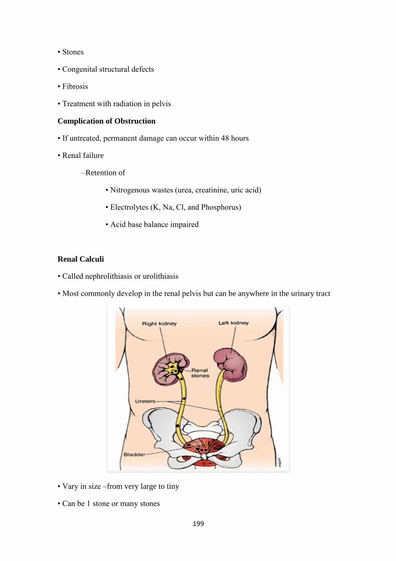
199
• Stones
• Congenital structural defects
• Fibrosis
• Treatment with radiation in pelvis
Complication of Obstruction
• If untreated, permanent damage can occur within 48 hours
• Renal failure
–Retention of
• Nitrogenous wastes (urea, creatinine, uric acid)
• Electrolytes (K, Na, Cl, and Phosphorus)
• Acid base balance impaired
Renal Calculi
• Called nephrolithiasis or urolithiasis
• Most commonly develop in the renal pelvis but can be anywhere in the urinary tract
• Vary in size –from very large to tiny
• Can be 1 stone or many stones
200
• May stay in kidney or travel into the ureter
• Can damage the urinary tract
• May cause hydronephrosis
• More common in white males 30-50 years of age
• Predisposing factors
–Dehydration
–Prolonged immobilization
–Infection
–Obstruction
–Anything which causes the urine to be alkaline
–Metabolic factors
• Excessive intake of calcium, calcium based antacids or Vit D
• Hyperthyroidism
• Elevated uric acid
Dehydration and immobilization causes urinary concentration and pooling of calculus
forming substances
Urine should be acidic
Alkaline urine- bacteria (proteus, klebsiella, and pseudmonas
• Subjective symptoms
–Sever pain in the flank area, suprapubic area, pelvis or external genitalia
–May radiate anteriorly and downward toward the bladder in females and toward the
testis in males.
–If in ureter, may have spasms called ―renal colic‖
–Urgency, frequency of urination
–N/V
–Chills
• Objective symptoms
–Increased temperature
201
–Pallor
–Hematuria
–Abdominal distention
–Pyuria
–Anuria
–May have UTI on urinalysis
Renal Calculi- Manifestations
• Kidney/Pelvis
–May be asymptomatic
–Dull, aching flank pain
• Ureter
–Acute severe flank pain, may radiate
–Nausea/vomiting
–Pallor
–Hematuria
• Bladder
–May be asymptomatic
–Dull suprapubic pain
–Hematuria
• Diagnostic procedures
–Urinalysis with C & S
–KUB
–IVP
–Renal CT
–Kidney ultrasound
–Cystoscopy with retrograde pyleogram
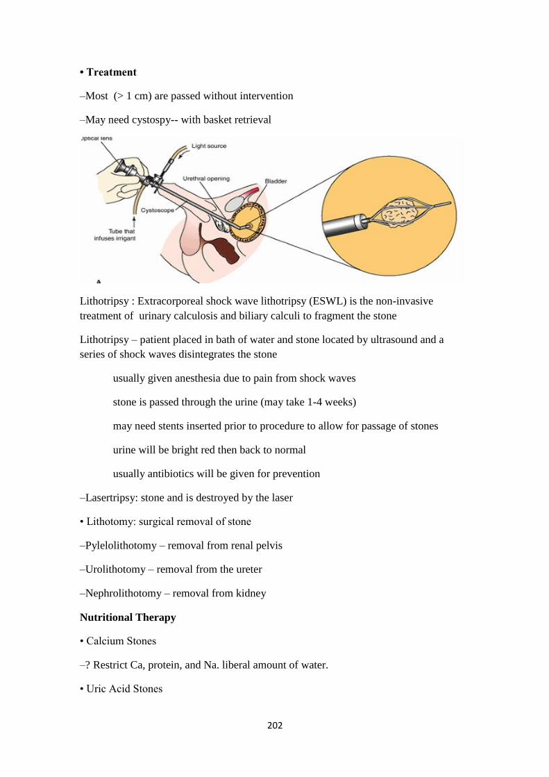
202
• Treatment
–Most (> 1 cm) are passed without intervention
–May need cystospy-- with basket retrieval
Lithotripsy : Extracorporeal shock wave lithotripsy (ESWL) is the non-invasive
treatment of urinary calculosis and biliary calculi to fragment the stone
Lithotripsy – patient placed in bath of water and stone located by ultrasound and a
series of shock waves disintegrates the stone
usually given anesthesia due to pain from shock waves
stone is passed through the urine (may take 1-4 weeks)
may need stents inserted prior to procedure to allow for passage of stones
urine will be bright red then back to normal
usually antibiotics will be given for prevention
–Lasertripsy: stone and is destroyed by the laser
• Lithotomy: surgical removal of stone
–Pylelolithotomy – removal from renal pelvis
–Urolithotomy – removal from the ureter
–Nephrolithotomy – removal from kidney
Nutritional Therapy
• Calcium Stones
–? Restrict Ca, protein, and Na. liberal amount of water.
• Uric Acid Stones

203
–low-purine diet to reduce urinary excretion of uric acid (shellfish, mushrooms, and
organ meats), limit protein, Allopurinol.
• Avoid food contain oxylate: spinach, strawberries, chocolate, tea, peanuts, and wheat
bran
Historically, patients with calcium-based renal stones were advised to restrict calcium
in their diet. However, recent evidence has questioned the advisability of this practice,
except for patients with type II absorptive hypercalciuria (half of all patients with
calcium stones), in whom stones are clearly due to excess dietary calcium.
It is thought that a high-protein diet is associated with increased urinary excretion of
calcium and uric acid, thereby causing a supersaturation of these substances in the
urine. Similarly, a high sodium intake has been shown in some studies to increase the
amount of calcium in the urine.
Foods high in purine (shellfish, anchovies, asparagus, mushrooms, and organ meats)
are avoided
Renal Calculi/ Assessment
–History and physical exam
–Location, severity, and nature of pain
–I/O
–Vital signs, looking for fever
–Palpation of flank area, and abdomen
–? N/V
• Nursing interventions
–Primary is to treat pain – usually with opioids
–Ambulate
–Force fluids, may have IV
• Watch for fluid overload
–Strain urine – send stone to lab if passed
–Accurate I/O
–Medicate N/V
Renal Calculi/Surgical removal
–Routine pre and post op care

204
–May return with catheter, drains, nephrostomy tube and ureteral stent – must maintain
patency and may need to irrigate as ordered
–Measure drainage from all tubes – need at least 30 cc/hr
–Watch site for bleeding
–May need frequent dressing changes due to fluid leakage, or may have collection bag
• Discharge and prevention
–Continue to force fluids post discharge
–May need special diet
• Stones are analyzed for calcium or other minerals
• May need to watch products with calcium
Cancer of the Urinary Tract
• Bladder cancer
• Kidney tumors
Bladder Cancer
• Bladder cancer is 4th leading cause of cancer deaths.
• More common in men than women
• Cancers arising from the prostate, colon, and rectum in males and from the lower
gynecologic tract in females may metastasize to the bladder
Risk factor for bladder cancer
• Cigarette smoking: risk increase with number of years and packs smoked
• Exposure to environmental carcinogens: dyes, rubber, leather, ink, or paint
• Recurrent or chronic bacterial infection of the urinary tract
• Bladder stones
• High urinary pH
• High cholesterol intake
• Pelvic radiation therapy

205
Bladder Cancer -Manifestations
• Painless hematuria • Frequency
• Urgency • Dysuria
Diagnostic tests
–Bladder ultrasound –Urinalysis
–Urine cytology –Cystoscopy
–Biopsy
Treatment
–Medications
–Surgery: remove tumor or bladder, Urinary Diversions
Cutaneous Urinary Diversions
A. conventional ileal conduit, B. cutaneous ureterostomy
C. vesicostomy D. nephrostomy
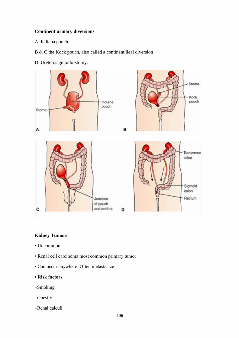
206
Continent urinary diversions
A. Indiana pouch
B & C the Kock pouch, also called a continent ileal diversion
D. Ureterosigmoido-stomy.
Kidney Tumors
• Uncommon
• Renal cell carcinoma most common primary tumor
• Can occur anywhere, Often metastasize
• Risk factors
–Smoking
–Obesity
–Renal calculi
207
Kidney Tumors -Manifestations
• May be silent
• Flank pain
• Palpable mass
• Fever, fatigue
• Weight loss, anemia, polycythemia
• Hypercalcemia, hypertension, or hyperglycemia
Kidney Tumors –Interdisciplinary Care
• Diagnostic tests
–Renal ultrasound
–CT scan
–Kidney biopsy
• Treatment: Radical nephrectomy
Bladder and Kidney Cancer – Nursing Care
• Assessment
• Diagnosing, Planning, and Implementing
–Impaired Urinary Elimination
–Risk for Impaired Skin Integrity
–Disturbed Body Image
• Diagnosing, Planning, and Implementing
–Acute Pain
–Ineffective Breathing Pattern
–Disturbed Body Image

208
Chapter 48
Assessment and Management of Patients with Breast Disorders
Assessment
ᴥ Health History
Physical Assessment/ Inspection
ᴥ Retraction Signs
ᴥ Signs include skin dimpling, creasing, or changes in the contour of the breast or
nipple
ᴥ Secondary to fibrosis or scar tissue formation in the breast
ᴥ Retraction signs may appear only with position changes or with breast palpation.

209
ᴥ Breast Cancer Mass (Malignant Tumor)
Usually occurs as a single mass (lump) in one breast
Firm, hard, embedded in surrounding tissue
Referral and biopsy indicated for definitive diagnosis
Usually nontender
Irregular shape
ᴥ Breast Cyst (Benign Mass)
Occur as single or multiple lumps in one or both breasts
Usually tender (omitting caffeine reduces tenderness); tenderness increases during
premenstrual period
Round shape
Soft or firm, mobile
Referral and biopsy indicated for definitive diagnosis, especially for first mass.
ᴥ Fibroadenoma (Benign Breast Lump)
Usually occurs as a single mass in women aged 15–35 years
Usually nontender
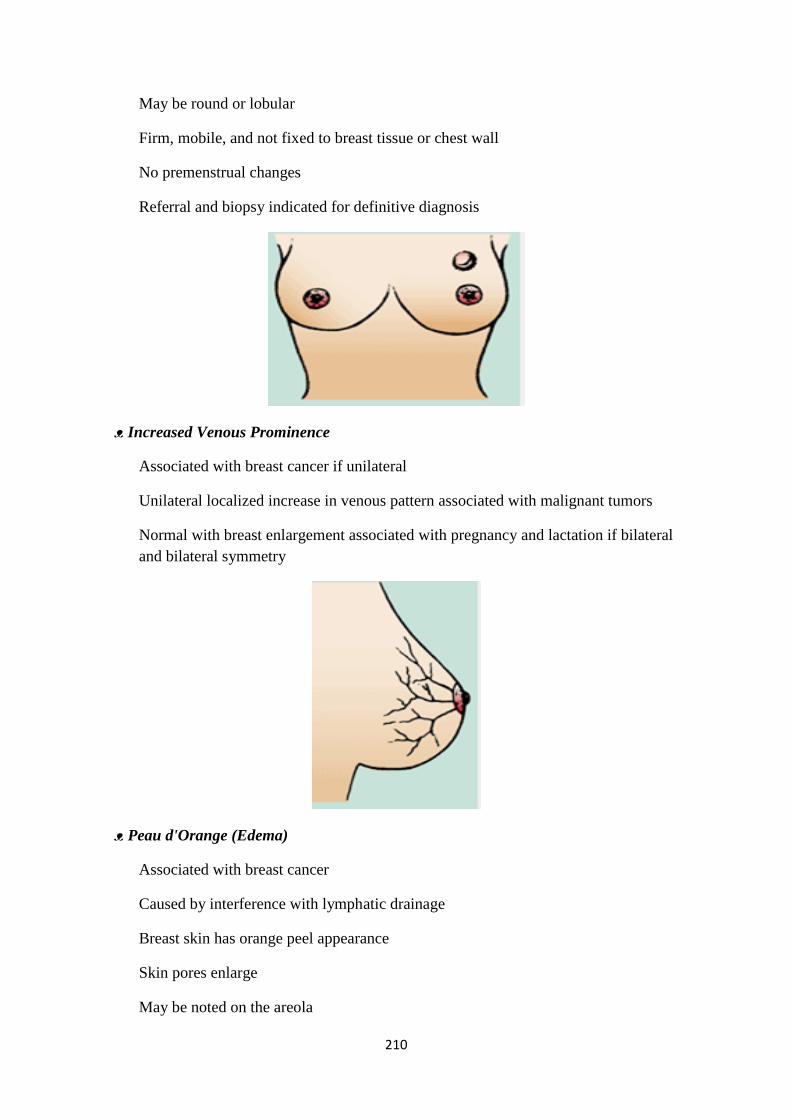
210
May be round or lobular
Firm, mobile, and not fixed to breast tissue or chest wall
No premenstrual changes
Referral and biopsy indicated for definitive diagnosis
ᴥ Increased Venous Prominence
Associated with breast cancer if unilateral
Unilateral localized increase in venous pattern associated with malignant tumors
Normal with breast enlargement associated with pregnancy and lactation if bilateral
and bilateral symmetry
ᴥ Peau d'Orange (Edema)
Associated with breast cancer
Caused by interference with lymphatic drainage
Breast skin has orange peel appearance
Skin pores enlarge
May be noted on the areola

211
Skin becomes thick, hard, immobile
Skin discoloration may occur
ᴥ Nipple Inversion
Considered normal if long-standing
Associated with fibrosis and malignancy if recent development
ᴥ Acute Mastitis (Inflammation of the Breasts)
Associated with lactation but may occur at any age
Nipple cracks or abrasions noted
Breast skin reddened and warm to touch
Tenderness
Systemic signs include fever and increased pulse
ᴥ Paget Disease (Malignancy of Mammary Ducts)
Early signs: erythema of nipple and areola
Late signs: thickening, scaling, and erosion of the nipple and areola
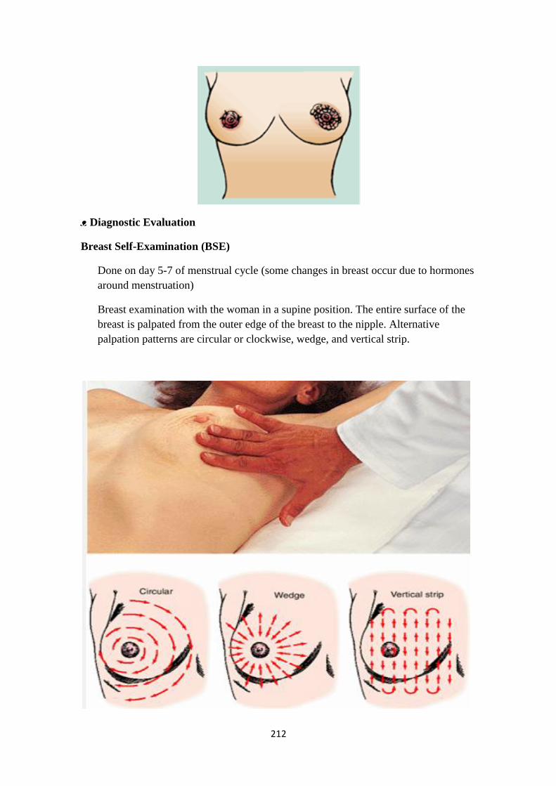
212
ᴥ Diagnostic Evaluation
Breast Self-Examination (BSE)
Done on day 5-7 of menstrual cycle (some changes in breast occur due to hormones
around menstruation)
Breast examination with the woman in a supine position. The entire surface of the
breast is palpated from the outer edge of the breast to the nipple. Alternative
palpation patterns are circular or clockwise, wedge, and vertical strip.

213
Breast-Self Examination
Diagnostic Evaluation Mammography
Detect nonpalpable lesions (< 1 cm) and assist in diagnosing palpable masses
Recommended to be done every year beginning at 40 years of age (women with
family history, start at younger age)

214
Diagnostic Evaluation
Magnetic Resonance Imaging
Procedures for Tissue Analysis
Percutaneous Biopsy
Fine-Needle Aspiration
Surgical Biopsy
• Excisional Biopsy
• Incisional Biopsy
Comparison of Various Breast Masses
Benign Neoplasms: Fibrocystic Breast Disease (FBD)
Also called chronic mastitis or lumpy breast syndrome, it is the most common breast
lesion in females and usually occurs between ages 35 and 50. Many cases will subside
after menopause.
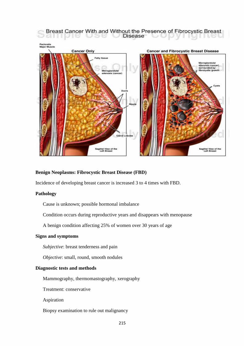
215
Benign Neoplasms: Fibrocystic Breast Disease (FBD)
Incidence of developing breast cancer is increased 3 to 4 times with FBD.
Pathology
Cause is unknown; possible hormonal imbalance
Condition occurs during reproductive years and disappears with menopause
A benign condition affecting 25% of women over 30 years of age
Signs and symptoms
Subjective: breast tenderness and pain
Objective: small, round, smooth nodules
Diagnostic tests and methods
Mammography, thermomastography, xerography
Treatment: conservative
Aspiration
Biopsy examination to rule out malignancy
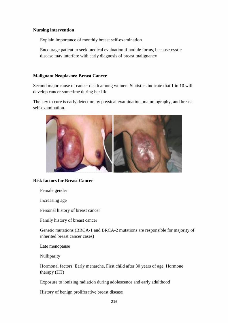
216
Nursing intervention
Explain importance of monthly breast self-examination
Encourage patient to seek medical evaluation if nodule forms, because cystic
disease may interfere with early diagnosis of breast malignancy
Malignant Neoplasms: Breast Cancer
Second major cause of cancer death among women. Statistics indicate that 1 in 10 will
develop cancer sometime during her life.
The key to cure is early detection by physical examination, mammography, and breast
self-examination.
Risk factors for Breast Cancer
Female gender
Increasing age
Personal history of breast cancer
Family history of breast cancer
Genetic mutations (BRCA-1 and BRCA-2 mutations are responsible for majority of
inherited breast cancer cases)
Late menopause
Nulliparity
Hormonal factors: Early menarche, First child after 30 years of age, Hormone
therapy (HT)
Exposure to ionizing radiation during adolescence and early adulthood
History of benign proliferative breast disease

217
Obesity
High-fat diet (controversial)
Alcohol intake
Protective Factors
Regular exercise (after age of 35, strenuous excursive ˅ chance for BC by 14%.
Breastfeeding
Having completed a full-term pregnancy before 30 years of age
Types of Breast Cancer
1. Ductal Carcinoma in Situ
Characterized by the proliferation of malignant cells inside the milk ducts without
invasion into the surrounding tissue.
It is a noninvasive form of cancer (also called intraductal carcinoma).
If not treated, ? become invasive cancer
2. Infiltrating Ductal Carcinoma
Is the most common histologic type of breast cancer.
Accounts for 75% of all cases.
The tumors arise from the duct system and invade the surrounding tissues.
They often form a solid irregular mass in the breast.
Other types of breast cancer
3. Infiltrating Lobular Carcinoma (5-10%)
Infiltrating lobular carcinoma accounts for 5% to 10% of breast cancers. The
tumors arise from the lobular epithelium and typically occur as an area of ill-
defined thickening in the breast. They are often multicentric and can be
bilateral.
4. Medullary Carcinoma (5%)
Medullary carcinoma accounts for about 5% of breast cancers, and it tends to be
diagnosed more often in women younger than 50 years. The tumors grow in a
capsule inside a duct. They can become large and may be mistaken for a
fibroadenoma. The prognosis is often favorable.
218
5. Mucinous Carcinoma (3%)
Mucinous carcinoma accounts for about 3% of breast cancers and often presents
in postmenopausal women 75 years and older. A mucin producer, the tumor is
also slow-growing and thus the prognosis is more favorable than in many other
types.
6. Tubular Ductal Carcinoma (2%)
Tubular ductal carcinoma accounts for about 2% of breast cancers. Because
axillary metastases are uncommon with this histology, prognosis is usually
excellent.
7. Inflammatory Carcinoma (2%)
Inflammatory carcinoma is a rare (1% to 2%) and aggressive type of breast
cancer that has unique symptoms. The cancer is characterized by diffuse edema
and brawny erythema of the skin, often referred to as peau d'orange (resembling
an orange peel). This is due to malignant cells blocking the lymph channels in
the skin. An associated mass may or may not be present; if there is, it is often a
large area of indiscrete thickening. Inflammatory carcinoma can be confused
with an infection because of its presentation. The disease can spread to other
parts of the body rapidly. Chemotherapy often plays an initial role in controlling
disease progression, but radiation and surgery may also be useful.
8. Paget Disease (1%)
Paget disease of the breast accounts for 1% of diagnosed breast cancer cases.
Symptoms typically include a scaly, erythematous, pruritic lesion of the nipple.
Paget disease often represents ductal carcinoma in situ of the nipple but may
have an invasive component. Mammography should be performed followed by
a biopsy of the involved skin area.
Signs and symptoms
Subjective: nontender nodule
Objective:
• Enlarged axillary nodes • Nipple retraction or elevation
• Skin dimpling • Nipple discharge
• Usually appeal in the upper outer quadrant
Diagnostic tests and methods: Mammography, breast biopsy examination

219
Treatment
1. Lumpectomy: removal of the lump and partial breast tissue; indicated for early
detection
2. Mastectomy
• Simple mastectomy: removal of breast
• Modified radical mastectomy: removal of breast, pectoralis minor muscle, and
some of an adjacent lymph nodes
• Radical mastectomy: removal of the breast, pectoral muscles, pectoral fascia,
and axillary lymph node dissection
3. Other Surgical procedures
• Oophorectomy, adrenalectomy, hypophysectomy to remove source of estrogen
and the hormones that stimulate the breast tissue
4. Radiation therapy to destroy malignant residual malegnant cells
5. Chemotherapeutic agents to shrink, retard, and destroy cancer growth
6. Corticosteroids, antigens, and anti-estrogens to alter cancer that is dependent on
hormonal environment
Collaborative Problems/Potential Complications
Based on the assessment data, potential complications may include the following:
Lymphedema
Hematoma/seroma formation
Infection
Preoperative Nursing Diagnosis
Deficient knowledge about the planned surgical treatments
Anxiety related to the diagnosis of cancer
Fear related to specific treatments and body image changes
Risk for ineffective coping (individual or family) related to the diagnosis of breast
cancer and related treatment options
Decisional conflict related to treatment options
Preoperative Nursing Interventions
Providing Education and Preparation About Surgical Treatments

220
Reducing Fear and Anxiety and Improving Coping Ability
Promoting Decision-Making Ability
Breast Cancer/ Postoperative Nursing Diagnosis
Pain and discomfort related to surgical procedure
Disturbed sensory perception related to nerve irritation in affected arm, breast, or
chest wall
Disturbed body image related to loss or alteration of breast
Risk for impaired adjustment related to the diagnosis of cancer and surgical
treatment
Self-care deficit related to partial immobility of arm
Risk for sexual dysfunction related to loss of body part, change in self-image, and
fear of partner's responses
Deficient knowledge: drain management after breast surgery
Deficient knowledge: arm exercises to regain mobility of affected extremity
Deficient knowledge: hand and arm care after an axillary lymph node dissection
(ALND)
Postoperative Nursing Interventions
Relieving Pain and Discomfort
Managing Postoperative Sensations
Promoting Positive Body Image
Promoting Positive Adjustment and Coping
Monitoring and Managing Potential Complications
Lymphedema
Occurs in about 10% to 30% of patients who undergo ALND and in about 0% to
7% of patients who have SLNB
lymphatic channels become inadequate to ensure a return flow of lymph fluid to the
general circulation.
Risk factors: age, obesity, extensive axillary disease, radiation treatment, and injury
or infection to the extremity
Edema resolved after development of collateral circulation.

221
Postoperative transient edema develops until collateral circulation has completely
taken over this function, which generally occurs within a month. Performing
prescribed exercises, elevating the arm above the heart several times a day, and
gentle muscle pumping (making a fist and releasing) can help reduce the transient
edema. The patient needs reassurance that this transient swelling is not
lymphedema
Hand and Arm Care After Axillary Lymph Node Dissection
Avoid BP, injections, and blood draws in affected arm.
Use sunscreen for extended exposure to sun.
Apply insect repellent to avoid insect bites.
Wear gloves for gardening.
Use cooking mitt for removing objects from oven.
Avoid cutting cuticles; push them back during manicures.
Use electric razor for shaving armpit.
Avoid lifting objects greater than 2-4 kg.
If a trauma or break in the skin occurs, wash the area with soap and water, and
apply an antibacterial ointment.
Observe the area and extremity for 24 hours; if redness, swelling, or a fever occurs,
call the surgeon or nurse.
Post-op arm exercise

222
Nursing intervention
Provide atmosphere of acceptance, frequent patient contact, and encouragement in
illness adjustment
Encourage grooming activities
Arrange attractive environment
If the patient is receiving radiation or chemotherapy, explain and assist with
potential side effects
If the patient has undergone surgical intervention, follow post operative nursing actions
• Elevate affected arm above level of right atrium to prevent edema
• Drawing blood or administering parenteral fluids or taking blood pressure on
affected arm is contraindicated
• Monitor dressing for hemorrhage, observed back for pooling of blood
• Empty Hemovac and measure drainage every 8 hours
• Assess circulatory status of affected limb
• Measure upper arm and forearm, twice daily, to monitor edema
• Encourage exercises of the affected arm when approved by a physician; avoid
abduction

223
Patient teaching on discharge
• Exercise to tolerance
• Sleep with arm elevated
• Elevated arm several times daily
• Avoid injections, vaccinations, IV, and taking blood pressure, in affected arm
Malignant Neoplasms: Breast Cancer
Five year survival rate is 97% for localized cancer, 76% for cancer that has spread
regionally, and 21% for cancers having distant metastases.
Metastatic breast cancer to the back
Male Breast Cancer
Accounts for less than 1% of all cases of breast cancer
Average age at diagnosis is 67 years
Risk factors:
A history of mumps orchitis,
Radiation exposure,
Decreased testosterone levels
Liver disease (compromises estrogen metabolism).

224
Nursing Process
The Patient Undergoing Surgery for Breast Cancer
Assessment
The health history is a valuable tool to assess the patient's reaction to the diagnosis and
her ability to cope with it. Pertinent questions include the following:
How is the patient responding to the diagnosis?
What coping mechanisms does she find most helpful?
What psychological or emotional supports does she have and use?
Is there a partner, family member, or friend available to assist her in making
treatment choices?
What are her educational needs?
Is she experiencing any discomfort?
Diagnosis
Preoperative Nursing Diagnoses
Based on the health history and other assessment data, major preoperative nursing
diagnoses may include the following:
Deficient knowledge about the planned surgical treatments
Anxiety related to the diagnosis of cancer
Fear related to specific treatments and body image changes
Risk for ineffective coping (individual or family) related to the diagnosis of
breast cancer and related treatment options
Decisional conflict related to treatment options
Postoperative Nursing Diagnoses
Based on the health history and other assessment data, major postoperative nursing
diagnoses may include the following:
Pain and discomfort related to surgical procedure
Disturbed sensory perception related to nerve irritation in affected arm, breast,
or chest wall
Disturbed body image related to loss or alteration of the breast
Risk for impaired adjustment related to the diagnosis of cancer and surgical
treatment
Self-care deficit related to partial immobility of upper extremity on operative
side
Risk for sexual dysfunction related to loss of body part, change in self-image,
and fear of partner's responses
Deficient knowledge: drain management after breast surgery
Deficient knowledge: arm exercises to regain mobility of affected extremity
Deficient knowledge: hand and arm care after an axillary lymph node dissection
(ALND)
Collaborative Problems/Potential Complications
Based on the assessment data, potential complications may include the following:
Lymphedema
Hematoma/seroma formation
Infection
225
Planning and Goals
The major goals may include increased knowledge about the disease and its treatment;
reduction of preoperative and postoperative fear, anxiety, and emotional stress;
improvement of decision-making ability; pain management; improvement in coping
abilities; improvement in sexual function; and the absence of complications.
Preoperative Nursing Interventions
Providing Education and Preparation About Surgical Treatments
Patients with newly diagnosed breast cancer are expected to absorb an abundance of
new information during a very emotionally difficult time. The nurse plays a key role in
reviewing treatment options by reinforcing information provided to the patient and
answering any questions. The nurse fully prepares the patient for what to expect before,
during, and after surgery. Patients undergoing breast conservation with ALND, or a
total or modified radical mastectomy, generally remain in the hospital overnight (or
longer if they have immediate reconstruction). Surgical drains will be inserted in the
mastectomy incision and in the axilla if the patient undergoes an ALND. A surgical
drain is generally not needed after a SLNB. The patient should be informed that she
will go home with the drain(s) and that complete instructions about drain care will be
provided prior to discharge. In addition, the patient should be informed that she will
often have decreased arm and shoulder mobility after an ALND and that she will be
shown range-of-motion exercises prior to discharge. The patient should also be
reassured that appropriate analgesia and comfort measures will be provided to alleviate
any postoperative discomfort.
Reducing Fear and Anxiety and Improving Coping Ability
The nurse must help the patient cope with the physical as well as the emotional effects
of surgery. Many fears may emerge during the preoperative phase. These can include
fear of pain, mutilation (after mastectomy), and loss of sexual attractiveness; concern
about inability to care for oneself and one's family; concern about taking time off from
work; and coping with an uncertain future. Providing the patient with realistic
expectations about the healing process and expected recovery can help alleviate fears.
Maintaining open communication and assuring the patient that she can contact the
nurse at any time with questions or concerns can be a source of comfort. The patient
should also be made aware of available resources at the treatment facility as well as in
the breast cancer community such as social workers, psychiatrists, and support groups.
Some women find it helpful and reassuring to talk to a breast cancer survivor who has
undergone similar treatments.
Promoting Decision-Making Ability
The patient may be eligible for more than one therapeutic approach; she may be
presented with treatment options and then asked to make a choice. This can be very
frightening for some patients, and they may prefer to have someone else make the
decision for them (e.g., surgeon, family member). The nurse can be instrumental in
ensuring that the patient and family members truly understand their options. The nurse
can then help the patient weigh the risks and benefits of each option. The patient may
be presented with the option of having breast conservation treatment followed by
radiation or a mastectomy. The nurse can explore the issues with the individual patient
by asking questions such as the following:
How would you feel about losing your breast?
Are you considering breast reconstruction?
If you choose to retain your breast, would you consider undergoing radiation
treatments 5 days a week for 5 to 6 weeks?
226
Questions such as these can help the patient focus. Once the patient's decision is made,
it is very important to support it.
Postoperative Nursing Interventions
Relieving Pain and Discomfort
Many patients tolerate the breast surgery quite well and have minimal pain during the
postoperative period. This is particularly true of the less invasive procedures such as
breast conservation treatment with SLNB. However, all patients must be carefully
assessed, because individual patients can have varying degrees of pain. Patients who
have had more invasive procedures such as a modified radical mastectomy with
immediate reconstruction may have considerably more pain. All patients are discharged
home with analgesic medication (e.g., oxycodone and acetaminophen [Percocet] or
propoxyphene and acetaminophen [Darvocet]) and are encouraged to take it if needed.
An over-the-counter analgesic such as acetaminophen may provide sufficient relief.
Sometimes patients complain of a slight increase in pain after the first few days of
surgery; this may occur as patients regain sensation around the surgical site and become
more active. However, patients who report excruciating pain must be evaluated to rule
out any potential complications such as infection or a hematoma. Alternative methods
of pain management such as taking warm showers and using distraction methods (eg,
guided imagery) may also be helpful.
Managing Postoperative Sensations
Because nerves in the skin and axilla are often cut or injured during breast surgery,
patients experience a variety of sensations. Common sensations include tenderness,
soreness, numbness, tightness, pulling, and twinges. These sensations may occur along
the chest wall, in the axilla, and along the inside aspect of the upper arm. After
mastectomy, some patients experience phantom sensations and report a feeling that the
breast and/or nipple are still present. Overall, patients do not find these sensations
severe or distressing (Baron et al., 2004). Sensations usually persist for several months
and then begin to diminish, although some may persist for as long as 2 years and
possibly longer. Patients should be reassured that this is a normal part of healing and
that these sensations are not indicative of a problem.
Promoting Positive Body Image
Patients who have undergone mastectomy often find it very difficult to view the
surgical site for the first time. No matter how prepared the patient may think she is, the
appearance of an absent breast can be very emotionally distressing. Some patients who
have undergone breast conservation treatment may find it difficult to view their
surgical incisions, although this is rare. Ideally, the patient sees the incision for the first
time when she is with the nurse or another health care provider who is available for
support.
The nurse first assesses the patient's readiness and provides gentle encouragement. It is
important to maintain a patient's privacy while assisting her as she views the incision;
this allows her to express feelings safely to the nurse. Asking the patient what she
perceives, acknowledging her feelings, and allowing her to express her emotions are
important nursing actions. Reassuring the patient that her feelings are a normal
response to breast cancer surgery may be comforting. If the patient has not had
immediate reconstruction, providing her with a temporary breast form to place in her
bra on discharge can help alleviate feelings of embarrassment or self-consciousness.
Promoting Positive Adjustment and Coping
Providing ongoing assessment of how the patient is coping with her diagnosis of breast
cancer and her surgical treatment is important in determining her overall adjustment.
Assisting the patient in identifying and mobilizing her support systems can be
beneficial to her well-being. The patient's spouse or partner may need guidance,
227
support, and education as well. The patient and partner may benefit from a wide
network of available community resources, including the Reach to Recovery program
of the ACS, advocacy groups, or a spiritual advisor. Encouraging the patient to discuss
issues and concerns with other patients who have had breast cancer may help her to
understand that her feelings are normal and that other women who have had breast
cancer can provide invaluable support and understanding.
The patient may also have considerable anxiety about the treatments that will follow
surgery (i.e., chemotherapy and radiation) and their implications. Providing her with
information about the plan of care and referring her to the appropriate members of the
health care team also promote coping during recovery. Some women require additional
support to adjust to their diagnosis and the changes that it brings. If a woman displays
ineffective coping, consultation with a mental health practitioner may be indicated.
Table 48-5 summarizes the needs and nursing interventions for patients and their
partners at various stages of the breast cancer experience. Chart 48-6 provides
strategies for initiating conversations with patients and their partners during different
phases of therapy.
Improving Sexual Function
Once discharged from the hospital, most patients are physically allowed to engage in
sexual activity. However, any change in the patient's body image, selfesteem, or the
response of her partner may increase her anxiety level and affect sexual function. Some
partners may have difficulty looking at the incision, whereas others may be completely
unaffected. Encouraging the patient to openly discuss how she feels about herself and
about possible reasons for a decrease in libido (e.g., fatigue, anxiety, self-
consciousness) may help clarify issues for her. Helpful suggestions for the patient may
include varying the time of day for sexual activity (when the patient is less tired),
assuming positions that are more comfortable, and expressing affection using
alternative measures (e.g., hugging, kissing, manual stimulation).
Most patients and their partners adjust with minimal difficulty if they openly discuss
their concerns. However, if issues cannot be resolved, a referral for counseling (e.g.,
psychologist, psychiatrist, psychiatric clinical nurse specialist, social worker, sex
therapist) may be helpful. The ambulatory care nurse in the outpatient clinic or hospital
should inquire whether the patient is having difficulty with sexuality issues, because
many patients are reluctant or embarrassed to bring it up themselves.
Monitoring and Managing Potential Complications
Lymphedema
Lymphedema occurs in about 10% to 30% of patients who undergo ALND and in
about 0% to 7% of patients who have SLNB (Leidenius, Leivonen, Vironen, et al.,
2005; Wilke, McCall, Posther, et al., 2006). Risk factors for lymphedema include
increasing age, obesity, presence of extensive axillary disease, radiation treatment, and
injury or infection to the extremity (Golshan, Martin & Dowlatshahi, 2003).
Lymphedema results if functioning lymphatic channels are inadequate to ensure a
return flow of lymph fluid to the general circulation. After axillary lymph nodes are
removed, collateral circulation must assume this function. Transient edema in the
postoperative period occurs until collateral circulation has completely taken over this
function, which generally occurs within a month. Performing prescribed exercises,
elevating the arm above the heart several times a day, and gentle muscle pumping
(making a fist and releasing) can help reduce the transient edema. The patient needs
reassurance that this transient swelling is not lymphedema.
Once lymphedema develops, it tends to be chronic, so preventive strategies are vital.
After ALND, the patient is taught hand and arm care to prevent injury or trauma to the
affected extremity, thus decreasing the likelihood for lymphedema development (Chart
228
48-7). The patient is instructed to follow these guidelines for the rest of her life. She is
also instructed to contact the physician or a nurse immediately if she suspects that she
has lymphedema, because early intervention provides the best chance for control. If
allowed to progress without treatment, the swelling can become more difficult to
manage. Treatment may consist of a course of antibiotics if an infection is present. A
referral to a rehabilitation specialist (e.g., occupational or physical therapist) may be
necessary for a compression sleeve and/or glove, exercises, manual lymph drainage,
and a discussion of ways to modify daily activities to avoid worsening lymphedema.
Hematoma or Seroma Formation
Hematoma formation (collection of blood inside the cavity) may occur after either
mastectomy or breast conservation and usually develops within the first 12 hours after
surgery. The nurse assesses for signs and symptoms of a hematoma at the surgical site,
which may include swelling, tightness, pain, and bruising of the skin. The surgeon
should be notified immediately for gross swelling or increased bloody output from the
drain. Depending on the surgeon's assessment, a compression wrap may be applied to
the incision for approximately 12 hours, or the patient may be returned to the operating
room so that the incision may be reopened to identify the source of bleeding. Some
hematomas are small, and the body absorbs the blood naturally. The patient may take
warm showers or apply warm compresses to help increase the absorption. A hematoma
usually resolves in 4 to 5 weeks.
A seroma, a collection of serous fluid, may accumulate under the breast incision after
mastectomy or breast conservation or in the axilla. Signs and symptoms may include
swelling, heaviness, discomfort, and a sloshing of fluid. Seromas may develop
temporarily after the drain is removed or if the drain is in place and becomes
obstructed. Seromas rarely pose a threat and may be treated by unclogging the drain or
manually aspirating the fluid with a needle and syringe. Large, long-standing seromas
that have not been aspirated could lead to infection. Small seromas that are not
bothersome to the patient usually resolve on their own.
Hand and Arm Care After Axillary Lymph Node Dissection
Avoid blood pressures, injections, and blood draws in affected extremity.
Use sunscreen (higher than 15 SPF) for extended exposure to sun.
Apply insect repellent to avoid insect bites.
Wear gloves for gardening.
Use cooking mitt for removing objects from oven.
Avoid cutting cuticles; push them back during manicures.
Use electric razor for shaving armpit.
Avoid lifting objects greater than 5–10 pounds.
If a trauma or break in the skin occurs, wash the area with soap and water, and
apply an over-the-counter antibacterial ointment (Bacitracin or Neosporin).
Observe the area and extremity for 24 hours; if redness, swelling, or a fever
occurs, call the surgeon or nurse.
Infection
Although infection is rare, it is a risk after any surgical procedure. This risk may be
higher in patients with accompanying conditions such as diabetes, immune disorders,
and advanced age, as well as in those with poor hygiene. Patients are taught to monitor
for signs and symptoms of infection (redness, warmth around incision, tenderness, foul-
smelling drainage, temperature greater than 100.4°F, chills) and to contact the surgeon
229
or nurse for evaluation. Treatment consists of oral or IV antibiotics (for more severe
infections) for 1 or 2 weeks. Cultures are taken of any foul-smelling discharge.
Promoting Home and Community-Based Care
Teaching Patients Self-Care
Patients who undergo breast cancer surgery receive a tremendous amount of
information preoperatively and postoperatively. It is often difficult for the patient to
absorb all of the information, partly because of the emotional distress that often
accompanies the diagnosis and treatment. Prior to discharge, the nurse must assess the
patient's readiness to assume self-care responsibilities, and any gaps in knowledge must
be identified. Teaching may need to be reviewed and reinforced to ensure that the
patient and family are prepared to manage the necessary home care. The nurse
reiterates symptoms the patient should report, such as infection, seroma, hematoma, or
arm swelling. All teaching should be reinforced during office visits and by telephone.
Most patients are discharged 1 or 2 days after ALND and/or mastectomy (possibly later
if they have had immediate reconstruction) with surgical drains in place. Initially, the
drainage fluid appears bloody, but it gradually changes to a serosanguineous and then a
serous fluid over the next several days. The patient is given instructions about drainage
management at home (Chart 48-8). If the patient lives alone and drainage management
is difficult for her, a referral for a home care nurse should be made. The drains are
usually removed when the output is less than 30 mL in a 24-hour period (approximately
7 to 10 days). The home care nurse also reviews pain management and incision care.
Generally, the patient may shower on the second postoperative day and wash the
incision and drain site with soap and water to prevent infection. If immediate
reconstruction has been performed, showering may be contraindicated until the drain is
removed. A dry dressing may be applied to the incision each day for 7 days. The
patient should know that sensation may be decreased in the operative area because the
nerves were disrupted during surgery, and gentle care is needed to avoid injury. After
the incision has completely healed (usually after 4 to 6 weeks), lotions or creams may
be applied to the area to increase skin elasticity. The patient can begin to use deodorant
on the affected side, although many women note that they no longer perspire as much
as before the surgery.
After ALND, patients are taught arm exercises on the affected side to restore range of
motion (Chart 48-9). After SLNB, patients may also benefit from these exercises,
although they are less likely to have decreased range of motion than those who have
undergone ALND. Range of motion exercises are initiated on the second postoperative
day, although instruction often occurs on the first postoperative day. The goals of the
exercise regimen are to increase circulation and muscle strength, prevent joint stiffness
and contractures, and restore full range of motion. The patient is instructed to perform
range of motion exercises at home 3 times a day for 20 minutes at a time until full
range of motion is restored (generally 4 to 6 weeks). Most patients find that after the
drain is removed, range of motion returns quickly if they have adhered to their exercise
program.
Patient Education
Exercise After Breast Surgery
Wall handclimbing. Stand facing the wall with feet apart and toes as close to the
wall as possible. With elbows slightly bent, place the palms of the hand on the
wall at shoulder level. By flexing the fingers, work the hands up the wall until
arms are fully extended. Then reverse the process, working the hands down to
the starting point.

230
Rope turning. Tie a light rope to a doorknob. Stand facing the door. Take the
free end of the rope in the hand on the side of surgery. Place the other hand on
the hip. With the rope-holding arm extended and held away from the body
(nearly parallel with the floor), turn the rope, making as wide swings as
possible. Begin slowly at first; speed up later.
Rod or broomstick lifting. Grasp a rod with both hands, held about 2 feet apart.
Keeping the arms straight, raise the rod over the head. Bend elbows to lower the
rod behind the head. Reverse maneuver, raising the rod above the head, then
return to the starting position.

231
Pulley tugging. Toss a light rope over a shower curtain rod or doorway curtain
rod. Stand as nearly under the rope as possible. Grasp an end in each hand.
Extend the arms straight and away from the body. Pull the left arm up by
tugging down with the right arm, then the right arm up and the left down in a
see-sawing motion.
If the patient is having any discomfort, taking an analgesic 30 minutes before beginning
the exercises can be helpful. Taking a warm shower before exercising can also loosen
stiff muscles and provide comfort. When exercising, the patient is encouraged to use
the muscles in both arms and to maintain proper posture. Specific exercises may need
to be prescribed and introduced gradually if the patient has had skin grafts; has a tense,
tight surgical incision; or has had immediate reconstruction. Self-care activities, such as
brushing the teeth, washing the face, and brushing the hair, are physically and
232
emotionally therapeutic because they aid in restoring arm function and provide a sense
of normalcy for the patient.
The patient is instructed about postoperative activity limitation. Generally, heavy lifting
(more than 5 to 10 lbs) is avoided for about 4 to 6 weeks, although normal household
and work-related activities are promoted to maintain muscle tone. Brisk walking, use of
stationary bikes and stepping machines, and stretching exercises may begin as soon as
the patient feels comfortable. Once the drain is removed, the patient may begin to drive
if she has full arm range of motion and is no longer taking opioid analgesics. General
guidelines for activity focus on the gradual introduction of previous activities (e.g.,
bowling, weight-training) once fully healed, although checking with the physician or
nurse beforehand is recommended.
Continuing Care
Patients who have difficulty managing their postoperative care at home may benefit
from a home health care referral. The home care nurse assesses the patient's incision
and surgical drain(s), adequacy of pain management, adherence to the exercise plan,
and overall physical and psychological functioning. In addition, the home care nurse
reinforces previous teaching and communicates important physiologic findings and
psychosocial issues to the patient's primary care provider, nurse, or surgeon.
The frequency of follow-up visits after surgery may vary but generally should occur
every 3 to 6 months for the first several years. The patient may alternate visits with the
surgeon, medical oncologist, or radiation oncologist, depending on the treatment
regimen. The ambulatory care nurse can also be a great source of comfort and security
for the patient and family and should encourage them to telephone if they have any
questions or concerns. It is common for people to ignore routine health care when a
major health issue arises, so women who have been treated for breast cancer should be
reminded of the importance of participating in routine health screening.
Evaluation
Expected Preoperative Patient Outcomes
Expected preoperative patient outcomes may include:
Exhibits knowledge about diagnosis and surgical treatment options
o Asks relevant questions about diagnosis and available surgical
treatments
o States rationale for surgery
o Describes advantages and disadvantages of treatment options
Verbalizes willingness to deal with anxiety and fears related to the diagnosis
and the effects of surgery on self-image and sexual functioning
Demonstrates ability to cope with diagnosis and treatment
o Verbalizes feelings appropriately and recognizes normalcy of mood
lability
o Proceeds with treatment in timely fashion
o Discusses impact of diagnosis and treatment on family and work
Demonstrates ability to make decisions regarding treatment options in timely
fashion
Expected Postoperative Patient Outcomes
Expected postoperative patient outcomes may include:
Reports that pain has decreased and states pain and discomfort management
strategies are effective
233
Identifies postoperative sensations and recognizes that they are a normal part of
healing
Exhibits clean, dry, and intact surgical incisions without signs of inflammation
or infection
Lists the signs and symptoms of infection to be reported to the nurse or surgeon
Verbalizes feelings regarding change in body image
Discusses meaning of the diagnosis, surgical treatment, and fears appropriately
Participates actively in self-care activities
o Performs exercises as prescribed
o Participates in self-care activities as prescribed
Discusses issues of sexuality and resumption of sexual relations
Demonstrates knowledge of postdischarge recommendations and restrictions
o Describes follow-up care and activities
o Demonstrates appropriate care of incisions and drainage system
o Demonstrates arm exercises and describes exercise regimen and activity
limitations during postoperative period
o Describes care of affected arm and hand and lists indications to contact
the surgeon or nurse
Experiences no complications
o Identifies signs and symptoms of reportable complications (eg, redness,
heat, pain, edema)
o Explains how to contact appropriate health care providers in case of
complications
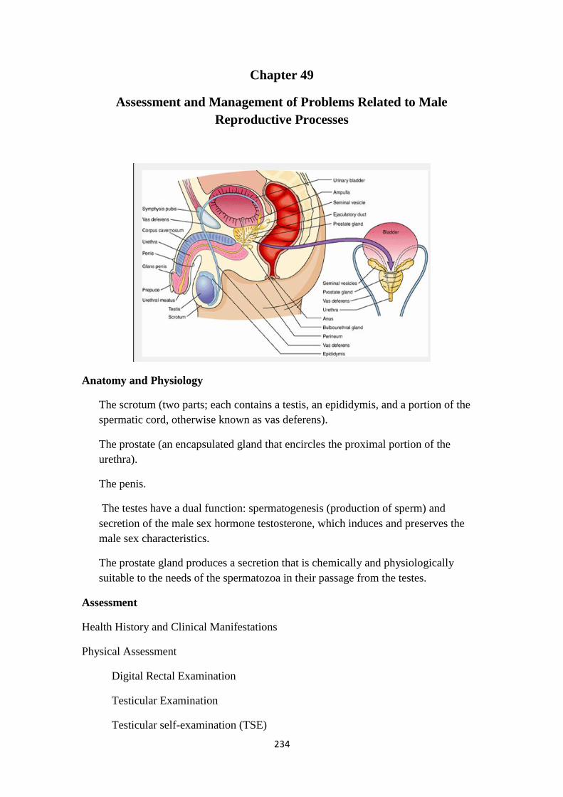
234
Chapter 49
Assessment and Management of Problems Related to Male
Reproductive Processes
Anatomy and Physiology
The scrotum (two parts; each contains a testis, an epididymis, and a portion of the
spermatic cord, otherwise known as vas deferens).
The prostate (an encapsulated gland that encircles the proximal portion of the
urethra).
The penis.
The testes have a dual function: spermatogenesis (production of sperm) and
secretion of the male sex hormone testosterone, which induces and preserves the
male sex characteristics.
The prostate gland produces a secretion that is chemically and physiologically
suitable to the needs of the spermatozoa in their passage from the testes.
Assessment
Health History and Clinical Manifestations
Physical Assessment
Digital Rectal Examination
Testicular Examination
Testicular self-examination (TSE)

235
Diagnostic Evaluation
Prostate-Specific Antigen Test
Prostate Fluid or Tissue Analysis
Inflammatory Diseases: Epididymitis
An infection of the epididymis, which usually descends from an infected prostate or
urinary tract.
Symptoms include unilateral pain and soreness in the inguinal canal, sudden, severe
pain in the scrotum, scrotal swelling, fever, pyuria, bacteriuria, dysuria, and pyuria.
Epididymitis/ treatment
If it is caused by a chlamydial infection, the patient and his wife must be treated
with antibiotics.
Observe for abscess formation
Epididymectomy (excision of the epididymis from the testis) may be performed for
patients who have chronic, painful conditions
Nursing Management
Bed rest and scrotal support to prevent traction on the spermatic cord and to relieve
pain.
Intermittent cold compresses to ease the pain.
Sitz baths may help resolve the inflammation.
Analgesic for pain relief as prescribed.
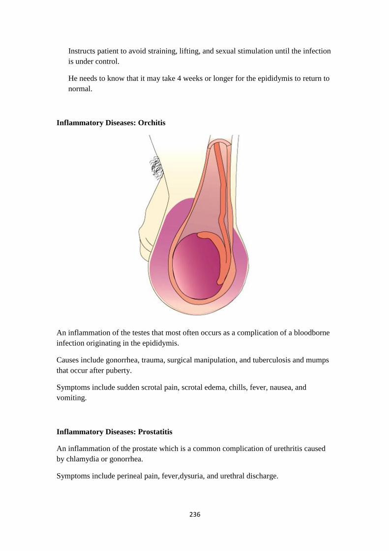
236
Instructs patient to avoid straining, lifting, and sexual stimulation until the infection
is under control.
He needs to know that it may take 4 weeks or longer for the epididymis to return to
normal.
Inflammatory Diseases: Orchitis
An inflammation of the testes that most often occurs as a complication of a bloodborne
infection originating in the epididymis.
Causes include gonorrhea, trauma, surgical manipulation, and tuberculosis and mumps
that occur after puberty.
Symptoms include sudden scrotal pain, scrotal edema, chills, fever, nausea, and
vomiting.
Inflammatory Diseases: Prostatitis
An inflammation of the prostate which is a common complication of urethritis caused
by chlamydia or gonorrhea.
Symptoms include perineal pain, fever,dysuria, and urethral discharge.

237
Nursing Interventions
Encourage bed rest
Monitor VS, esp. temp for fever
Monitor I & O
Assess pain
Sitz bath – provide comfort = PROSTATITIS
Provide ice pack to scrotum to decrease swelling
Elevate or provide scrotal support
Interventions
Analgesic
Antibiotic
Procaine = anesthetic
Stool softeners
Digital massage – rectally – to release infected fluid
Benign Prostatic Hyperplasia
BPH is a progressive adenomatous enlargement of the prostate gland that occurs
with aging.
More than 50% of men over the age of 50 and 80% of men ≥ 80 demonstrate some
increase in the size of the prostate gland.
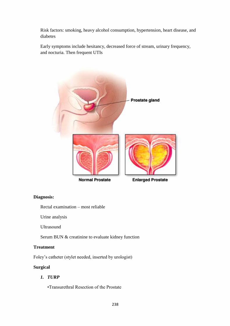
238
Risk factors: smoking, heavy alcohol consumption, hypertension, heart disease, and
diabetes
Early symptoms include hesitancy, decreased force of stream, urinary frequency,
and nocturia. Then frequent UTIs
Diagnosis:
Rectal examination – most reliable
Urine analysis
Ultrasound
Serum BUN & creatinine to evaluate kidney function
Treatment
Foley‘s catheter (stylet needed, inserted by urologist)
Surgical
1. TURP
•Transurethral Resection of the Prostate

239
•Continuous irrigation = reduce or prevent clot formation = clogs urethra =
urinary retention = kidney damage
•Monitor I & O
•3-way f/c
•Monitor for distention -bladder
2. PROSTATECTOMY
A. Perineal prostectomy – incision through perineum
B. Suprapubic resection – lower abdomen – incision through the bladder –
urethrotomy
C. Retropubic – lower abdomen – does not go through the bladder
Pharmacological Treatment
Alpha blockers – relax the smooth muscles along urinary tract
Narcotic analgesic – relieve post-op pain –Morphine, Codeine
Nursing Interventions
Increased fluids – monitor I & O
Maintain gravity drainage of F/C
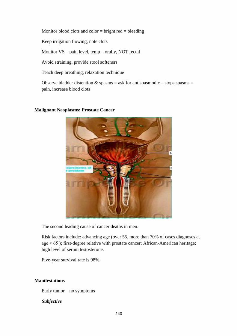
240
Monitor blood clots and color = bright red = bleeding
Keep irrigation flowing, note clots
Monitor VS – pain level, temp – orally, NOT rectal
Avoid straining, provide stool softeners
Teach deep breathing, relaxation technique
Observe bladder distention & spasms = ask for antispasmodic – stops spasms =
pain, increase blood clots
Malignant Neoplasms: Prostate Cancer
The second leading cause of cancer deaths in men.
Risk factors include: advancing age (over 55, more than 70% of cases diagnoses at
age ≥ 65 ); first-degree relative with prostate cancer; African-American heritage;
high level of serum testosterone.
Five-year survival rate is 98%.
Manifestations
Early tumor – no symptoms
Subjective
241
• Back pain, same symptoms as BPH – hesitancy, decrease pressure, frequency,
dysuria, urinary retention, painful ejaculation.
Objective
• Symptoms from metastasis
• ? Blood in urine or semen.
• Lumps – inguinal
• Enlarged lymph nodes
• Blockage of urethra, and rectal dysfunction
Diagnostic Test – Prostate CA
Digital Rectal Examination
PSA- prostate specific antigen – elevated
Bone scan to detect metastasis
MRI, CT scan
Complications
Sexual dysfunction (gets worse with treatment)
Treatment – Prostate CA
Radiation, chemo, surgical removal
Complete surgical removal of the prostate, seminal vesicles, tips of the vas
deferens, and often the surrounding fat, nerves, and blood vessels
Bilateral orchiectomy (removal of testes)
TURP
Estrogen therapy – inhibits serum testosterone = contradicts
Agonists of LH – estrogen
Radioactive seed implant – rectally
Nursing process
Anxiety related to concern and lack of knowledge about the diagnosis, treatment
plan, and prognosis

242
Reduced stress and improved ability to cope
Urinary retention related to urethral obstruction secondary to prostatic enlargement
or tumor and loss of bladder tone due to prolonged distention/retention
Improved pattern of urinary elimination
Deficient knowledge related to the diagnosis of: cancer, urinary difficulties, and
treatment modalities
Understanding of the diagnosis and ability to care for self
Imbalanced nutrition: less than body requirements related to decreased oral intake
because of anorexia, nausea, and vomiting caused by cancer or its treatment
Maintain optimal nutritional status
Sexual dysfunction related to effects of therapy: chemotherapy, hormonal therapy,
radiation therapy, surgery
Ability to resume/enjoy modified sexual functioning
Pain related to progression of disease and treatment modalities
Relief of pain
Impaired physical mobility and activity intolerance related to tissue hypoxia,
malnutrition, and exhaustion and to spinal cord or nerve compression from
metastases
Improved physical mobility
Collaborative Problems: Hemorrhage, infection, bladder neck obstruction
Goal: Absence of complications
Nursing interventions – Prostate CA
BPH interventions
Be supportive – expect feminization, more emotional, educate
Gynecomastia – enlargement of the breast
Control pain – terminally ill = hospice, palliative care
Malignant Neoplasms: Testicular Cancer
Although it accounts for only 1% of all cancer in men, it is the most common
cancer in young men between the ages of 15 and 40.
Essential for clients to learn TSE (testicular self-examination). – monthly
During shower
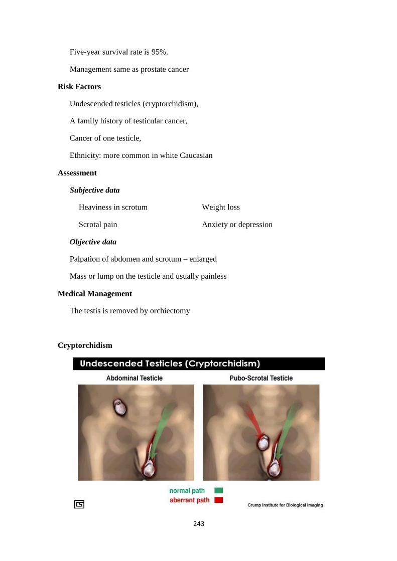
243
Five-year survival rate is 95%.
Management same as prostate cancer
Risk Factors
Undescended testicles (cryptorchidism),
A family history of testicular cancer,
Cancer of one testicle,
Ethnicity: more common in white Caucasian
Assessment
Subjective data
Heaviness in scrotum Weight loss
Scrotal pain Anxiety or depression
Objective data
Palpation of abdomen and scrotum – enlarged
Mass or lump on the testicle and usually painless
Medical Management
The testis is removed by orchiectomy
Cryptorchidism

244
Hydrocele
•Collection of amber fluid within the testes, tunica vaginalis, and spermatic cord
•Painful
•Swelling
•Discomfort in sitting and walking
•Treatment: aspiration (usually in children)
•Hydrocelectomy – removal of the sac
Nursing Interventions:
•Preoperative and postoperative management
•Scrotal support (elevation)
•Supportive to parents/patient
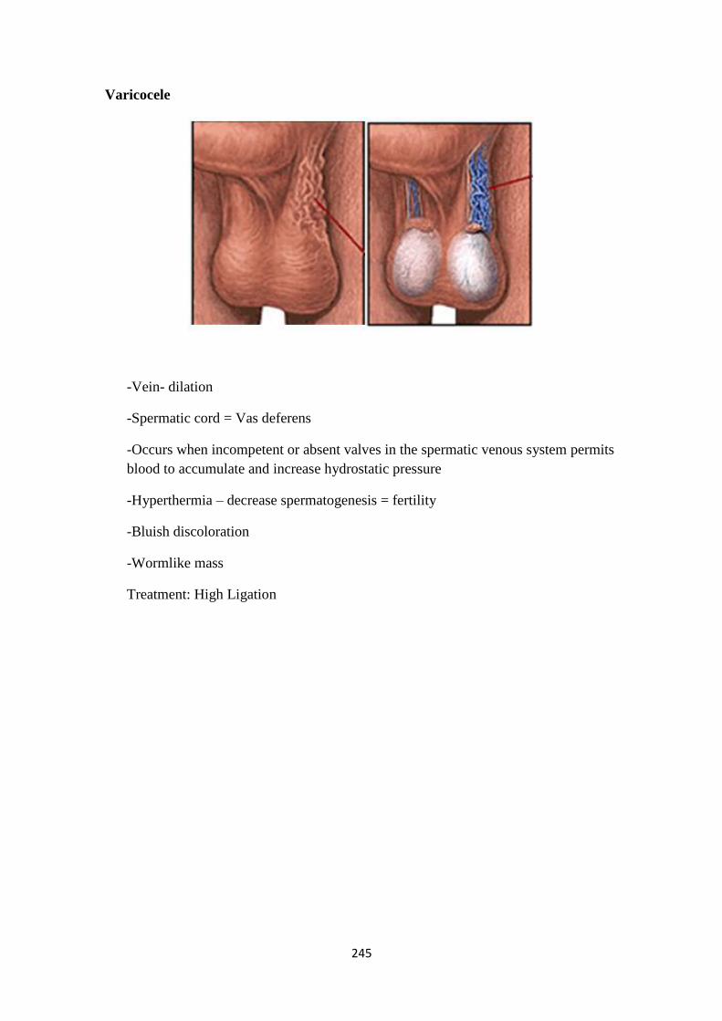
245
Varicocele
-Vein- dilation
-Spermatic cord = Vas deferens
-Occurs when incompetent or absent valves in the spermatic venous system permits
blood to accumulate and increase hydrostatic pressure
-Hyperthermia – decrease spermatogenesis = fertility
-Bluish discoloration
-Wormlike mass
Treatment: High Ligation
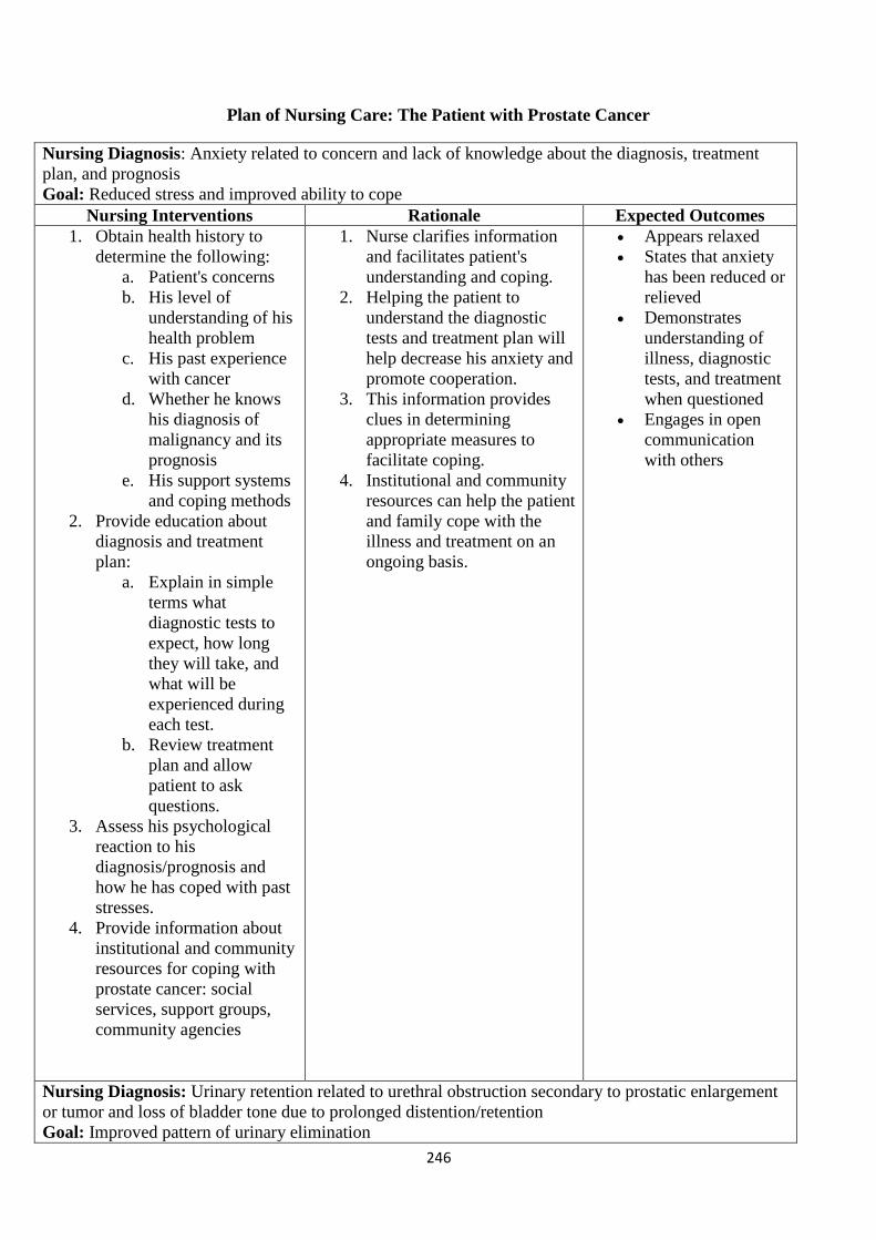
246
Plan of Nursing Care: The Patient with Prostate Cancer
Nursing Diagnosis: Anxiety related to concern and lack of knowledge about the diagnosis, treatment
plan, and prognosis
Goal: Reduced stress and improved ability to cope
Expected Outcomes Rationale Nursing Interventions
Appears relaxed
States that anxiety
has been reduced or
relieved
Demonstrates
understanding of
illness, diagnostic
tests, and treatment
when questioned
Engages in open
communication
with others
1. Nurse clarifies information
and facilitates patient's
understanding and coping.
2. Helping the patient to
understand the diagnostic
tests and treatment plan will
help decrease his anxiety and
promote cooperation.
3. This information provides
clues in determining
appropriate measures to
facilitate coping.
4. Institutional and community
resources can help the patient
and family cope with the
illness and treatment on an
ongoing basis.
1. Obtain health history to
determine the following:
a. Patient's concerns
b. His level of
understanding of his
health problem
c. His past experience
with cancer
d. Whether he knows
his diagnosis of
malignancy and its
prognosis
e. His support systems
and coping methods
2. Provide education about
diagnosis and treatment
plan:
a. Explain in simple
terms what
diagnostic tests to
expect, how long
they will take, and
what will be
experienced during
each test.
b. Review treatment
plan and allow
patient to ask
questions.
3. Assess his psychological
reaction to his
diagnosis/prognosis and
how he has coped with past
stresses.
4. Provide information about
institutional and community
resources for coping with
prostate cancer: social
services, support groups,
community agencies
Nursing Diagnosis: Urinary retention related to urethral obstruction secondary to prostatic enlargement
or tumor and loss of bladder tone due to prolonged distention/retention
Goal: Improved pattern of urinary elimination
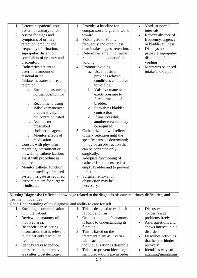
247
Voids at normal
intervals
Reports absence of
frequency, urgency,
or bladder fullness
Displays no
palpable suprapubic
distention after
voiding
Maintains balanced
intake and output
1. Provides a baseline for
comparison and goal to work
toward
2. Voiding 20 to 30 mL
frequently and output less
than intake suggest retention.
3. Determines amount of urine
remaining in bladder after
voiding
4. Promotes voiding
a. Usual position
provides relaxed
conditions conducive
to voiding.
b. Valsalva maneuver
exerts pressure to
force urine out of
bladder.
c. Stimulates bladder
contraction
d. If unsuccessful,
another measure may
be required.
5. Catheterization will relieve
urinary retention until the
specific cause is determined;
it may be an obstruction that
can be corrected only
surgically.
6. Adequate functioning of
catheter is to be ensured to
empty bladder and to prevent
infection.
7. Surgical removal of
obstruction may be
necessary.
1. Determine patient's usual
pattern of urinary function.
2. Assess for signs and
symptoms of urinary
retention: amount and
frequency of urination,
suprapubic distention,
complaints of urgency and
discomfort.
3. Catheterize patient to
determine amount of
residual urine.
4. Initiate measures to treat
retention:
a. Encourage assuming
normal position for
voiding.
b. Recommend using
Valsalva maneuver
preoperatively, if
not contraindicated.
c. Administer
prescribed
cholinergic agent.
d. Monitor effects of
medication.
5. Consult with physician
regarding intermittent or
indwelling catheterization;
assist with procedure as
required.
6. Monitor catheter function;
maintain sterility of closed
system; irrigate as required.
7. Prepare patient for surgery
if indicated.
Nursing Diagnosis: Deficient knowledge related to the diagnosis of: cancer, urinary difficulties, and
treatment modalities
Goal: Understanding of the diagnosis and ability to care for self
Discusses his
concerns and
problems freely
Asks questions and
shows interest in his
disorder
Describes activities
that help or hinder
recovery
Identifies ways of
attaining/maintainin
1. This is designed to establish
rapport and trust.
2. Orientation to one's anatomy
is basic to understanding its
function.
3. This is based on the
treatment plan; as it varies
with each patient,
individualization is desirable.
4. This is to prevent bleeding;
such precautions are in order
1. Encourage communication
with the patient.
2. Review the anatomy of the
involved area.
3. Be specific in selecting
information that is relevant
to the patient's particular
treatment plan.
4. Identify ways to reduce
pressure on the operative
area after prostatectomy:
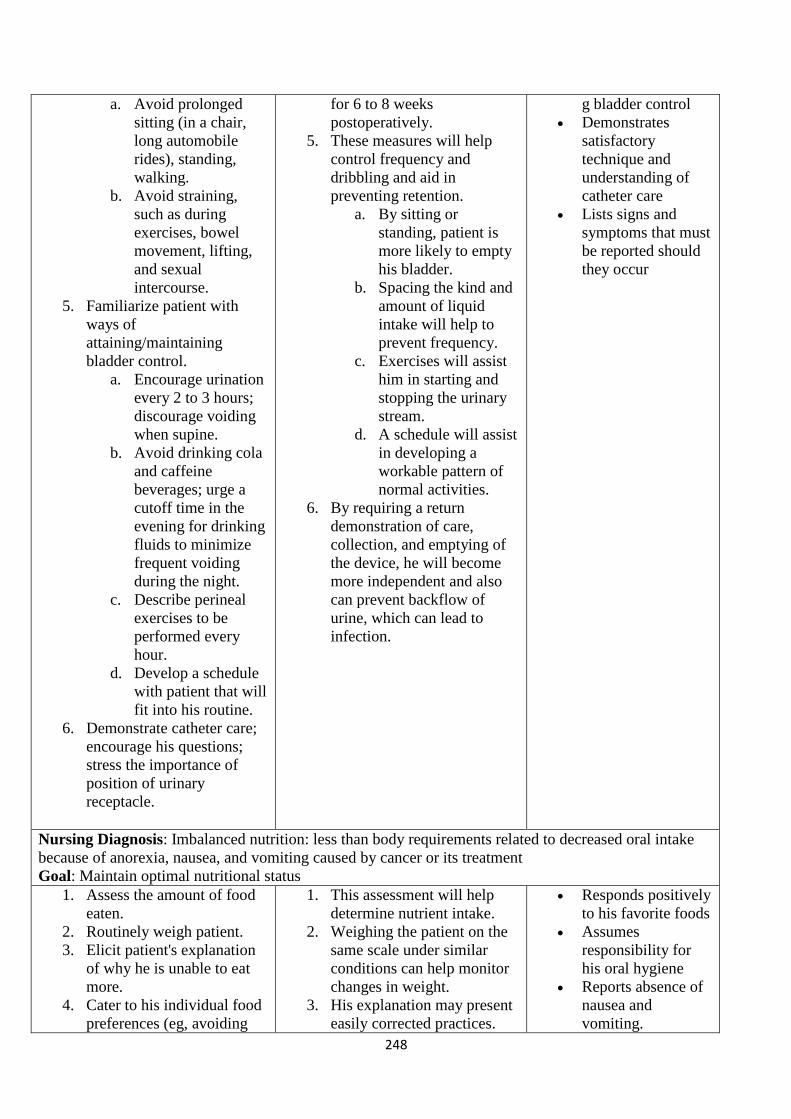
248
g bladder control
Demonstrates
satisfactory
technique and
understanding of
catheter care
Lists signs and
symptoms that must
be reported should
they occur
for 6 to 8 weeks
postoperatively.
5. These measures will help
control frequency and
dribbling and aid in
preventing retention.
a. By sitting or
standing, patient is
more likely to empty
his bladder.
b. Spacing the kind and
amount of liquid
intake will help to
prevent frequency.
c. Exercises will assist
him in starting and
stopping the urinary
stream.
d. A schedule will assist
in developing a
workable pattern of
normal activities.
6. By requiring a return
demonstration of care,
collection, and emptying of
the device, he will become
more independent and also
can prevent backflow of
urine, which can lead to
infection.
a. Avoid prolonged
sitting (in a chair,
long automobile
rides), standing,
walking.
b. Avoid straining,
such as during
exercises, bowel
movement, lifting,
and sexual
intercourse.
5. Familiarize patient with
ways of
attaining/maintaining
bladder control.
a. Encourage urination
every 2 to 3 hours;
discourage voiding
when supine.
b. Avoid drinking cola
and caffeine
beverages; urge a
cutoff time in the
evening for drinking
fluids to minimize
frequent voiding
during the night.
c. Describe perineal
exercises to be
performed every
hour.
d. Develop a schedule
with patient that will
fit into his routine.
6. Demonstrate catheter care;
encourage his questions;
stress the importance of
position of urinary
receptacle.
Nursing Diagnosis: Imbalanced nutrition: less than body requirements related to decreased oral intake
because of anorexia, nausea, and vomiting caused by cancer or its treatment
Goal: Maintain optimal nutritional status
Responds positively
to his favorite foods
Assumes
responsibility for
his oral hygiene
Reports absence of
nausea and
vomiting.
1. This assessment will help
determine nutrient intake.
2. Weighing the patient on the
same scale under similar
conditions can help monitor
changes in weight.
3. His explanation may present
easily corrected practices.
1. Assess the amount of food
eaten.
2. Routinely weigh patient.
3. Elicit patient's explanation
of why he is unable to eat
more.
4. Cater to his individual food
preferences (eg, avoiding
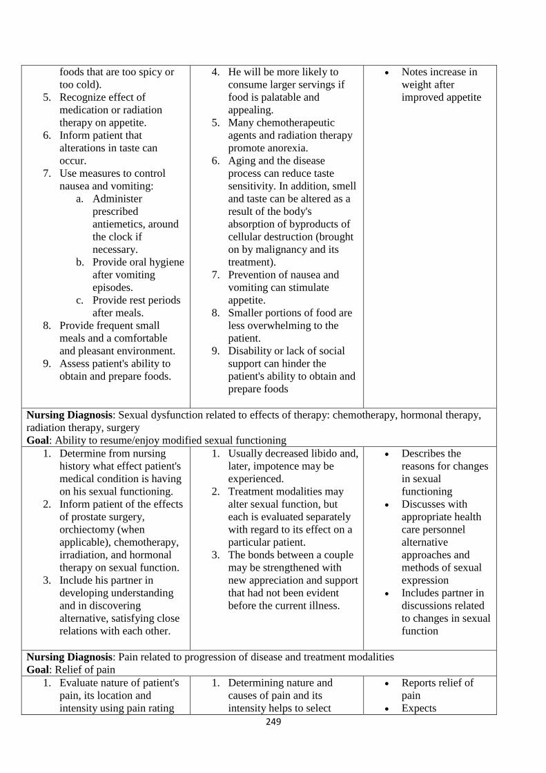
249
Notes increase in
weight after
improved appetite
4. He will be more likely to
consume larger servings if
food is palatable and
appealing.
5. Many chemotherapeutic
agents and radiation therapy
promote anorexia.
6. Aging and the disease
process can reduce taste
sensitivity. In addition, smell
and taste can be altered as a
result of the body's
absorption of byproducts of
cellular destruction (brought
on by malignancy and its
treatment).
7. Prevention of nausea and
vomiting can stimulate
appetite.
8. Smaller portions of food are
less overwhelming to the
patient.
9. Disability or lack of social
support can hinder the
patient's ability to obtain and
prepare foods
foods that are too spicy or
too cold).
5. Recognize effect of
medication or radiation
therapy on appetite.
6. Inform patient that
alterations in taste can
occur.
7. Use measures to control
nausea and vomiting:
a. Administer
prescribed
antiemetics, around
the clock if
necessary.
b. Provide oral hygiene
after vomiting
episodes.
c. Provide rest periods
after meals.
8. Provide frequent small
meals and a comfortable
and pleasant environment.
9. Assess patient's ability to
obtain and prepare foods.
Nursing Diagnosis: Sexual dysfunction related to effects of therapy: chemotherapy, hormonal therapy,
radiation therapy, surgery
Goal: Ability to resume/enjoy modified sexual functioning
Describes the
reasons for changes
in sexual
functioning
Discusses with
appropriate health
care personnel
alternative
approaches and
methods of sexual
expression
Includes partner in
discussions related
to changes in sexual
function
1. Usually decreased libido and,
later, impotence may be
experienced.
2. Treatment modalities may
alter sexual function, but
each is evaluated separately
with regard to its effect on a
particular patient.
3. The bonds between a couple
may be strengthened with
new appreciation and support
that had not been evident
before the current illness.
1. Determine from nursing
history what effect patient's
medical condition is having
on his sexual functioning.
2. Inform patient of the effects
of prostate surgery,
orchiectomy (when
applicable), chemotherapy,
irradiation, and hormonal
therapy on sexual function.
3. Include his partner in
developing understanding
and in discovering
alternative, satisfying close
relations with each other.
Nursing Diagnosis: Pain related to progression of disease and treatment modalities
Goal: Relief of pain
Reports relief of
pain
Expects
1. Determining nature and
causes of pain and its
intensity helps to select
1. Evaluate nature of patient's
pain, its location and
intensity using pain rating
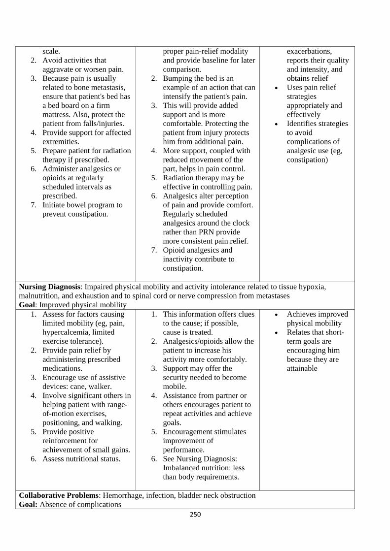
250
exacerbations,
reports their quality
and intensity, and
obtains relief
Uses pain relief
strategies
appropriately and
effectively
Identifies strategies
to avoid
complications of
analgesic use (eg,
constipation)
proper pain-relief modality
and provide baseline for later
comparison.
2. Bumping the bed is an
example of an action that can
intensify the patient's pain.
3. This will provide added
support and is more
comfortable. Protecting the
patient from injury protects
him from additional pain.
4. More support, coupled with
reduced movement of the
part, helps in pain control.
5. Radiation therapy may be
effective in controlling pain.
6. Analgesics alter perception
of pain and provide comfort.
Regularly scheduled
analgesics around the clock
rather than PRN provide
more consistent pain relief.
7. Opioid analgesics and
inactivity contribute to
constipation.
scale.
2. Avoid activities that
aggravate or worsen pain.
3. Because pain is usually
related to bone metastasis,
ensure that patient's bed has
a bed board on a firm
mattress. Also, protect the
patient from falls/injuries.
4. Provide support for affected
extremities.
5. Prepare patient for radiation
therapy if prescribed.
6. Administer analgesics or
opioids at regularly
scheduled intervals as
prescribed.
7. Initiate bowel program to
prevent constipation.
Nursing Diagnosis: Impaired physical mobility and activity intolerance related to tissue hypoxia,
malnutrition, and exhaustion and to spinal cord or nerve compression from metastases
Goal: Improved physical mobility
Achieves improved
physical mobility
Relates that short-
term goals are
encouraging him
because they are
attainable
1. This information offers clues
to the cause; if possible,
cause is treated.
2. Analgesics/opioids allow the
patient to increase his
activity more comfortably.
3. Support may offer the
security needed to become
mobile.
4. Assistance from partner or
others encourages patient to
repeat activities and achieve
goals.
5. Encouragement stimulates
improvement of
performance.
6. See Nursing Diagnosis:
Imbalanced nutrition: less
than body requirements.
1. Assess for factors causing
limited mobility (eg, pain,
hypercalcemia, limited
exercise tolerance).
2. Provide pain relief by
administering prescribed
medications.
3. Encourage use of assistive
devices: cane, walker.
4. Involve significant others in
helping patient with range-
of-motion exercises,
positioning, and walking.
5. Provide positive
reinforcement for
achievement of small gains.
6. Assess nutritional status.
Collaborative Problems: Hemorrhage, infection, bladder neck obstruction
Goal: Absence of complications
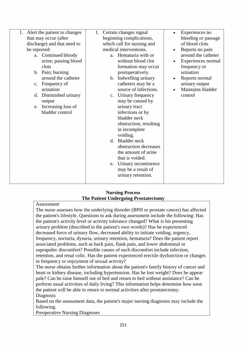
251
Experiences no
bleeding or passage
of blood clots
Reports no pain
around the catheter
Experiences normal
frequency or
urination
Reports normal
urinary output
Maintains bladder
control
1. Certain changes signal
beginning complications,
which call for nursing and
medical interventions.
a. Hematuria with or
without blood clot
formation may occur
postoperatively.
b. Indwelling urinary
catheters may be a
source of infections.
c. Urinary frequency
may be caused by
urinary tract
infections or by
bladder neck
obstruction, resulting
in incomplete
voiding.
d. Bladder neck
obstruction decreases
the amount of urine
that is voided.
e. Urinary incontinence
may be a result of
urinary retention.
1. Alert the patient to changes
that may occur (after
discharge) and that need to
be reported:
a. Continued bloody
urine; passing blood
clots
b. Pain; burning
around the catheter
c. Frequency of
urination
d. Diminished urinary
output
e. Increasing loss of
bladder control
Nursing Process
The Patient Undergoing Prostatectomy
Assessment
The nurse assesses how the underlying disorder (BPH or prostate cancer) has affected
the patient's lifestyle. Questions to ask during assessment include the following: Has
the patient's activity level or activity tolerance changed? What is his presenting
urinary problem (described in the patient's own words)? Has he experienced
decreased force of urinary flow, decreased ability to initiate voiding, urgency,
frequency, nocturia, dysuria, urinary retention, hematuria? Does the patient report
associated problems, such as back pain, flank pain, and lower abdominal or
suprapubic discomfort? Possible causes of such discomfort include infection,
retention, and renal colic. Has the patient experienced erectile dysfunction or changes
in frequency or enjoyment of sexual activity?
The nurse obtains further information about the patient's family history of cancer and
heart or kidney disease, including hypertension. Has he lost weight? Does he appear
pale? Can he raise himself out of bed and return to bed without assistance? Can he
perform usual activities of daily living? This information helps determine how soon
the patient will be able to return to normal activities after prostatectomy.
Diagnosis
Based on the assessment data, the patient's major nursing diagnoses may include the
following.
Preoperative Nursing Diagnoses

252
Anxiety about surgery and its outcome
Acute pain related to bladder distention
Deficient knowledge about factors related to the disorder and the treatment
protocol
Postoperative Nursing Diagnoses
Acute pain related to the surgical incision, catheter placement, and bladder
spasms
Deficient knowledge about postoperative care and management
Collaborative Problems/Potential Complications
Based on the assessment data, the potential complications may include the following:
Hemorrhage and shock
Infection
Deep vein thrombosis
Catheter obstruction
Sexual dysfunction
Planning and Goals
The major preoperative goals for the patient may include reduced anxiety and learning
about his prostate disorder and the perioperative experience. The major postoperative
goals may include maintenance of fluid volume balance, relief of pain and discomfort,
ability to perform self-care activities, and absence of complications.
Preoperative Nursing Interventions
Reducing Anxiety
The patient is frequently admitted to the hospital on the morning of surgery. Because
contact with the patient may be limited before surgery, the nurse must establish
communication with the patient to assess his understanding of the diagnosis and of the
planned surgical procedure. The nurse clarifies the nature of the surgery and expected
postoperative outcomes. In addition, the nurse familiarizes the patient with the
preoperative and postoperative routines and initiates measures to reduce anxiety.
Because the patient may be sensitive and embarrassed discussing problems related to
the genitalia and sexuality, the nurse provides privacy and establishes a trusting and
professional relationship. Guilt feelings often surface if the patient falsely assumes a
cause-and-effect relationship between sexual practices and his current problems. He is
encouraged to verbalize his feelings and concerns.
Relieving Discomfort
If the patient experiences discomfort before surgery, he is prescribed bed rest,
analgesic agents are administered, and measures are initiated to relieve anxiety. If he
is hospitalized, the nurse monitors his voiding patterns, watches for bladder
distention, and assists with catheterization if indicated. An indwelling catheter is
inserted if the patient has continuing urinary retention or if laboratory test results
indicate azotemia (accumulation of nitrogenous waste products in the blood). The
catheter can help decompress the bladder gradually over several days, especially if the
patient is elderly and hypertensive and has diminished renal function or urinary
retention that has existed for many weeks. For a few days after the bladder begins
draining, the blood pressure may fluctuate and renal function may decline. If the
patient cannot tolerate a urinary catheter, he is prepared for a cystostomy (see
253
Chapters 44 and 45).
Providing Instruction
Before surgery, the nurse reviews with the patient the anatomy of the affected
structures and their function in relation to the urinary and reproductive systems, using
diagrams and other teaching aids if indicated. This instruction often takes place during
the preadmission testing visit or in the urologist's office. The nurse explains what will
take place as the patient is prepared for diagnostic tests and then for surgery
(depending on the type of prostatectomy planned). The nurse also describes the type
of incision, which varies with the surgical approach (directly over the bladder, low on
the abdomen, or in the perineal area; in the case of a transurethral procedure, no
incision will be made), and informs the patient about the likely type of urinary
drainage system, the type of anesthesia, and the recovery room procedure. The
amount of information given is based on the patient's needs and questions. The nurse
explains procedures expected to occur during the immediate perioperative period,
answers questions the patient or family may have, and provides emotional support. In
addition, the nurse provides the patient with information about postoperative pain
management.
Preparing the Patient
If the patient is scheduled for a prostatectomy, the preoperative preparation described
in Chapter 18 is provided. Elastic compression stockings are applied before surgery
and are particularly important for prevention of deep vein thrombosis (DVT) if the
patient is placed in a lithotomy position during surgery. An enema is usually
administered at home on the evening before surgery or on the morning of surgery to
prevent postoperative straining, which can induce bleeding.
Postoperative Nursing Interventions
Maintaining Fluid Balance
During the postoperative period, the patient is at risk for imbalanced fluid volume
because of the irrigation of the surgical site during and after surgery. With irrigation
of the urinary catheter to prevent its obstruction by blood clots, fluid may be absorbed
through the open surgical site and retained, increasing the risk of excessive fluid
retention, fluid imbalance, and water intoxication. The urine output and the amount of
fluid used for irrigation must be closely monitored to determine whether irrigation
fluid is being retained and to ensure an adequate urine output. An intake and output
record, including the amount of fluid used for irrigation, must be maintained. The
patient also is monitored for electrolyte imbalances (eg, hyponatremia), increasing
blood pressure, confusion, and respiratory distress. These signs and symptoms are
documented and reported to the surgeon. The risk of fluid and electrolyte imbalance is
greater in elderly patients with preexisting cardiovascular or respiratory disease.
Relieving Pain
After a prostatectomy, the patient is assisted to sit and dangle his legs over the side of
the bed on the day of surgery. The next morning, he is assisted to ambulate. If pain is
present, the cause and location are determined and the severity of pain and discomfort
is assessed. The pain may be related to the incision or may be the result of excoriation
of the skin at the catheter site. It may be in the flank area, indicating a kidney
problem, or it may be caused by bladder spasms. Bladder irritability can initiate
bleeding and result in clot formation, leading to urinary retention.
Patients experiencing bladder spasms may note an urgency to void, a feeling of
pressure or fullness in the bladder, and bleeding from the urethra around the catheter.
Medications that relax the smooth muscles can help ease the spasms, which can be
intermittent and severe; these medications include flavoxate (Urispas) and oxybutynin
(Ditropan). Warm compresses to the pubis or sitz baths may also relieve the spasms.
254
The nurse monitors the drainage tubing and irrigates the system as prescribed to
relieve any obstruction that may cause discomfort. Usually, the catheter is irrigated
with 50 mL of irrigating fluid at a time. It is important to make sure that the same
amount is recovered in the drainage receptacle. Securing the catheter drainage tubing
to the leg or abdomen can help decrease tension on the catheter and prevent bladder
irritation. Discomfort may be caused by dressings that are too snug, saturated with
drainage, or improperly placed. Analgesic agents are administered as prescribed.
After the patient is ambulatory, he is encouraged to walk but not to sit for prolonged
periods, because this increases intra-abdominal pressure and the possibility of
discomfort and bleeding. Prune juice and stool softeners are provided to ease bowel
movements and to prevent excessive straining. An enema, if prescribed, is
administered with caution to avoid rectal perforation.
Monitoring and Managing Potential Complications
After prostatectomy, the patient is monitored for major complications such as
hemorrhage, infection, DVT, catheter obstruction, and sexual dysfunction.
Hemorrhage
The immediate dangers after a prostatectomy are bleeding and hemorrhagic shock.
This risk is increased with BPH, because a hyperplastic prostate gland is very
vascular. Bleeding may occur from the prostatic bed. Bleeding may also result in the
formation of clots, which then obstruct urine flow. The drainage normally begins as
reddish-pink and then clears to a light pink within 24 hours after surgery. Bright red
bleeding with increased viscosity and numerous clots usually indicates arterial
bleeding. Venous blood appears darker and less viscous. Arterial hemorrhage usually
requires surgical intervention (eg, suturing or transurethral coagulation of bleeding
vessels), whereas venous bleeding may be controlled by applying prescribed traction
to the catheter so that the balloon holding the catheter in place applies pressure to the
prostatic fossa. The surgeon applies traction by securely taping the catheter to the
patient's thigh if hemorrhage occurs.
Nursing management includes assistance in implementing strategies to stop the
bleeding and to prevent or reverse hemorrhagic shock. If blood loss is extensive,
fluids and blood component therapy may be administered. If hemorrhagic shock
occurs, treatments described in Chapter 15 are initiated.
Nursing interventions include closely monitoring vital signs; administering
medications, IV fluids, and blood component therapy as prescribed; maintaining an
accurate record of intake and output; and carefully monitoring drainage to ensure
adequate urine flow and patency of the drainage system. The patient who experiences
hemorrhage and his family are often anxious and benefit from explanations and
reassurance about the event and the procedures that are performed.
Infection
After perineal prostatectomy, the surgeon usually changes the dressing on the first
postoperative day. Further dressing changes may become the responsibility of the
nurse or home care nurse. Careful aseptic technique is used, because the potential for
infection is great. Dressings can be held in place by a double-tailed, T-binder bandage
or a padded athletic supporter. The tails cross over the incision to give double
thickness, and then each tail is drawn up on either side of the scrotum to the waistline
and fastened.
Rectal thermometers, rectal tubes, and enemas are avoided because of the risk of
injury and bleeding in the prostatic fossa. After the perineal sutures are removed, the
perineum is cleansed as indicated. A heat lamp may be directed to the perineal area to
promote healing. The scrotum is protected with a towel while the heat lamp is in use.
Sitz baths are also used to promote healing.
255
Urinary tract infections and epididymitis are possible complications after
prostatectomy. The patient is assessed for their occurrence; if they occur, the nurse
administers antibiotics as prescribed.
Because the risk for infection continues after discharge from the hospital, the patient
and family need to be instructed to monitor for signs and symptoms of infection
(fever, chills, sweating, myalgia, dysuria, urinary frequency, and urgency). The
patient and family are instructed to contact the urologist if these symptoms occur.
Deep Vein Thrombosis
Because patients undergoing prostatectomy have a high incidence of DVT and
pulmonary embolism, the physician may prescribe prophylactic (preventive) low-dose
heparin therapy. The nurse assesses the patient frequently after surgery for
manifestations of DVT and applies elastic compression stockings to reduce the risk
for DVT and pulmonary embolism. Nursing and medical management of DVT and
pulmonary embolism are described in Chapters 31 and 23, respectively. The patient
who is receiving heparin must be closely monitored for excessive bleeding.
Obstructed Catheter
After a TUR, the catheter must drain well; an obstructed catheter produces distention
of the prostatic capsule and resultant hemorrhage. Furosemide (Lasix) may be
prescribed to promote urination and initiate postoperative diuresis, thereby helping to
keep the catheter patent.
The nurse observes the lower abdomen to ensure that the catheter has not become
blocked. An overdistended bladder manifests a distinct, rounded swelling above the
pubis.
The drainage bag, dressings, and incisional site are examined for bleeding. The color
of the urine is noted and documented; a change in color from pink to amber indicates
reduced bleeding. Blood pressure, pulse, and respirations are monitored and compared
with baseline preoperative vital signs to detect hypotension. The nurse also observes
the patient for restlessness, diaphoresis, pallor, any drop in blood pressure, and an
increasing pulse rate.
Drainage of the bladder may be accomplished by gravity through a closed sterile
drainage system. A three-way drainage system is useful in irrigating the bladder and
preventing clot formation (Fig. 49-5). Continuous irrigation may be used with TUR.
Some urologists leave an indwelling catheter attached to a dependent drainage system.
Gentle irrigation of the catheter may be prescribed to remove any obstructing clots.
If the patient complains of pain, the tubing is examined. The drainage system is
irrigated, if indicated and prescribed, to clear any obstruction. Usually, the catheter is
irrigated with 50 mL of irrigating fluid at a time. The amount of fluid recovered in the
drainage bag must equal the amount of fluid injected. Overdistention of the bladder is
avoided, because it can induce secondary hemorrhage by stretching the coagulated
blood vessels in the prostatic capsule.
To prevent traction on the bladder, the drainage tube (not the catheter) is taped to the
shaved inner thigh. If a cystostomy catheter is in place, it is taped to the abdomen.
The nurse explains the purpose of the catheter to the patient and assures him that the
urge to void results from the presence of the catheter and from bladder spasms. He is
cautioned not to pull on the catheter, because this causes bleeding and subsequent
catheter blockage, which leads to urinary retention.
Complications With Catheter Removal
After the catheter is removed (usually when the urine appears clear), urine may leak
around the wound for several days in the patient who has undergone perineal,
suprapubic, or retropubic surgery. The cystostomy tube may be removed before or
after the urethral catheter is removed. Some urinary incontinence may occur after
256
catheter removal, and the patient is informed that this is likely to subside over time.
Sexual Dysfunction
Depending on the type of surgery, the patient may experience sexual dysfunction
related to erectile dysfunction, decreased libido, and fatigue. These issues may
become a concern to the patient soon after surgery or in the weeks to months of
rehabilitation. Several options to restore erectile function are discussed with the
patient by the surgeon or urologist. These options may include medications, surgically
placed implants, or negative-pressure devices. A decrease in libido is usually related
to the impact of the surgery on the man's body. Reassurance that the usual level of
libido will return after recuperation from surgery is often helpful for the patient and
his partner. The patient should be aware that he may experience fatigue during
rehabilitation from surgery. This fatigue may also decrease his libido and alter his
enjoyment of usual activities.
Nursing interventions include assessing for the presence of sexual dysfunction after
surgery. Providing a private and confidential environment to discuss issues of
sexuality is important. The emotional challenges of prostate surgery and its
consequences need to be carefully explored with the patient and his partner. Providing
the opportunity to discuss these issues can be very beneficial to the patient. For
patients who demonstrate significant problems adjusting to their sexual dysfunction, a
referral to a sex therapist may be indicated.
Promoting Home and Community-Based Care
Teaching Patients Self-Care
The patient undergoing prostatectomy may be discharged within several days. The
length of the hospital stay depends on the type of prostatectomy performed. Patients
undergoing a perineal prostatectomy are hospitalized for 3 to 5 days. If a retropubic or
suprapubic prostatectomy is performed, the hospital stay may extend to 5 to 7 days.
The patient and family require instructions about how to manage the drainage system,
how to assess for complications, and how to promote recovery. The nurse provides
verbal and written instructions about the need to maintain the drainage system and to
monitor urinary output, about wound care, and about strategies to prevent
complications, such as infection, bleeding, and thrombosis. In addition, the patient
and family need to know about signs and symptoms that should be reported to the
physician (eg, blood in urine, decreased urine output, fever, change in wound
drainage, calf tenderness).
As the patient recovers and drainage tubes are removed, he may become discouraged
and depressed because he cannot regain bladder control immediately. Furthermore,
urinary frequency and burning may occur after the catheter is removed. Teaching the
patient the following exercises may help him regain urinary control:
Tense the perineal muscles by pressing the buttocks together; hold this
position; relax. This exercise can be performed 10 to 20 times each hour while
sitting or standing.
Try to interrupt the urinary stream after starting to void; wait a few seconds
and then continue to void.
Perineal exercises should continue until the patient gains full urinary control. The
patient is instructed to urinate as soon as he feels the first urge to do so. It is important
that the patient know that regaining urinary control is a gradual process; he may
continue to ―dribble‖ after being discharged from the hospital, but this should
gradually diminish (within 1 year). Lining underwear with absorbent pads can help
minimize embarrassing stains on clothing. The urine may be cloudy for several weeks
257
after surgery but should clear as the prostate area heals.
While the prostatic fossa heals (6 to 8 weeks), the patient should avoid activities that
produce Valsalva effects (straining, heavy lifting), because this may increase venous
pressure and produce hematuria. He should avoid long motor trips and strenuous
exercise, which increase the tendency to bleed. He should also know that spicy foods,
alcohol, and coffee may cause bladder discomfort. The patient should be cautioned to
drink enough fluids to avoid dehydration, which increases the tendency for a blood
clot to form and obstruct the flow of urine. Signs of complications, such as bleeding,
passage of blood clots, a decrease in the urinary stream, urinary retention, or
symptoms of urinary tract infection symptoms, should be reported to the physician
(Chart 49-5).
Continuing Care
Referral for home care may be indicated if the patient is elderly or has other health
problems, if the patient and family cannot provide care in the home, or if the patient
lives alone without available supports. The home care nurse assesses the patient's
physical status (cardiovascular and respiratory status, fluid and nutritional status,
patency of the urinary drainage system, wound and nutritional status) and provides
catheter and wound care, if indicated. The nurse reinforces previous teaching and
assesses the ability of the patient and family to manage required care. The home care
nurse encourages the patient to ambulate and to carry out perineal exercises as
prescribed. The patient may need to be reminded that return of bladder control may
take time.
The patient is reminded about the importance of participating in routine health
screening and other health promotion activities. If the prostatectomy was performed to
treat prostate cancer, the patient and family are also instructed about the importance of
follow-up and monitoring with the physician.
Evaluation
Expected Preoperative Patient Outcomes
Expected preoperative patient outcomes may include the following:
Demonstrates reduced anxiety
States that pain and discomfort are decreased
Relates understanding of the surgical procedure and postoperative course and
practices perineal muscle exercises and other techniques useful in facilitating
bladder contrlo
Expected Postoperative Patient Outcomes
Expected postoperative patient outcomes may include the following:
Reports relief of discomfort
Exhibits fluid and electrolyte balance
o Irrigation fluid and urinary output are within parameters determined by
surgeon
o Experiences no signs or symptoms of fluid retention
Participates in self-care measures
o Increases activity and ambulation daily
o Produces urine output within normal ranges and consistent with intake
o Performs perineal exercises and interrupts urinary stream to promote
bladder control
o Avoids straining and lifting heavy objects
Is free of complications
258
o Maintains vital signs within normal limits
o Exhibits wound healing, without signs of inflammation or hemorrhage
o Maintains acceptable level of urinary elimination
o Maintains optimal drainage of catheter and other drainage tubes
o Reports understanding of changes in sexual function
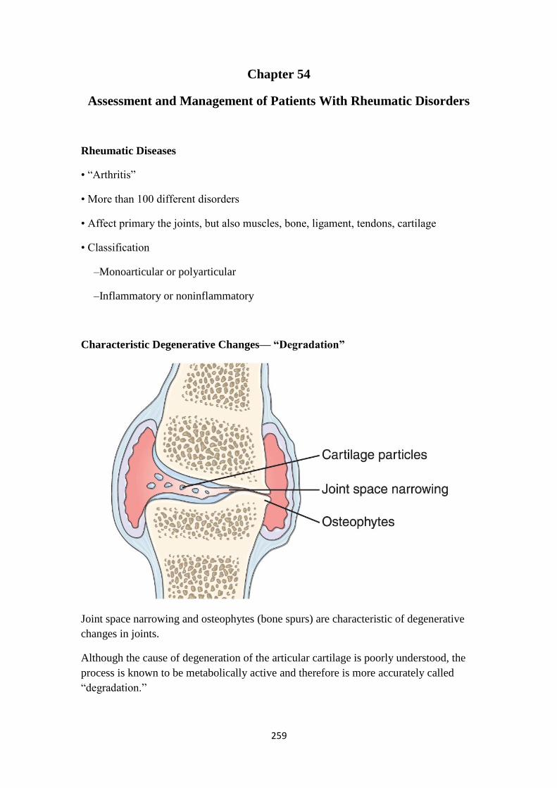
259
Chapter 54
Assessment and Management of Patients With Rheumatic Disorders
Rheumatic Diseases
• ―Arthritis‖
• More than 100 different disorders
• Affect primary the joints, but also muscles, bone, ligament, tendons, cartilage
• Classification
–Monoarticular or polyarticular
–Inflammatory or noninflammatory
Characteristic Degenerative Changes— ―Degradation‖
Joint space narrowing and osteophytes (bone spurs) are characteristic of degenerative
changes in joints.
Although the cause of degeneration of the articular cartilage is poorly understood, the
process is known to be metabolically active and therefore is more accurately called
―degradation.‖

260
Factors associated with degenerative joint changes
• Mechanical Stress (The articular plate (subchondral bone) thins, and its ability to
absorb shock decreases)
• Altered Lubrication
• Immobility
Clinical Manifestations
• Pain
• Joint swelling
• Limited movement
• Stiffness
• Weakness
• Fatigue
• Warmth,
• Erythema
• Joint involvement begins in the small joints of the hands, wrists, and feet. As the
disease progresses, the knees, shoulders, hips, elbows, ankles, and cervical spine joints
are affected.
• The onset of symptoms is usually acute. Symptoms are usually bilateral and
symmetric. In addition to joint pain and swelling, another classic sign of RA is joint
stiffness, especially in the morning, lasting at least 30 to 45 minutes

261
Pathophysiology and Associated Physical Signs of Rheumatoid Arthritis
Pannus (newly formed synovial tissue infiltrated with inflammatory cells)
Patient Assessment and Diagnostic Findings
• Health history: include onset of and evolution of symptoms, family history, past
health history, and contributing factors
• Functional assessment
• Arthrocentesis (to take fluid for sampling and to relieve pain caused by pressure of
increased fluid volume. After procedure, observe the patient for signs of infection and
hemarthrosis)
• X-rays, bone scans, CTs, and MRIs
• Tissue biopsy
• Blood studies
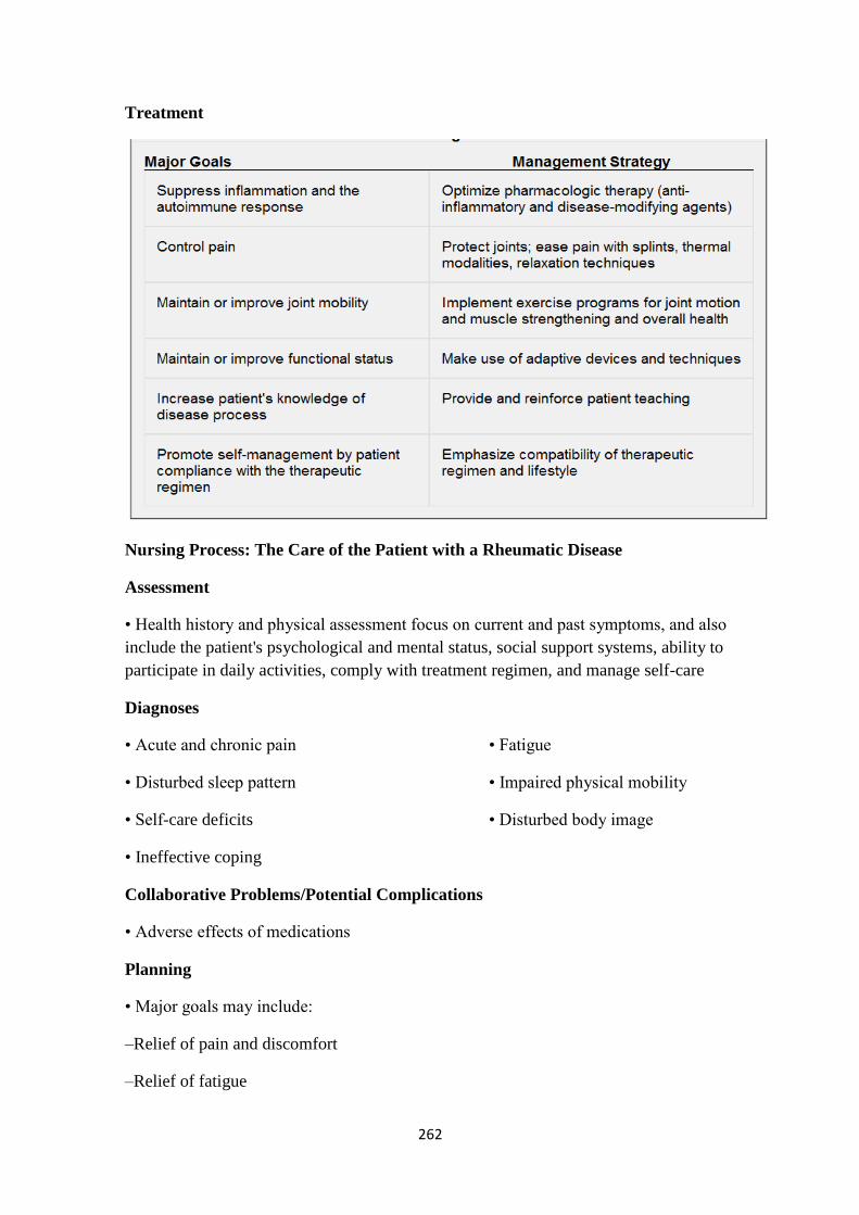
262
Treatment
Nursing Process: The Care of the Patient with a Rheumatic Disease
Assessment
• Health history and physical assessment focus on current and past symptoms, and also
include the patient's psychological and mental status, social support systems, ability to
participate in daily activities, comply with treatment regimen, and manage self-care
Diagnoses
• Acute and chronic pain • Fatigue
• Disturbed sleep pattern • Impaired physical mobility
• Self-care deficits • Disturbed body image
• Ineffective coping
Collaborative Problems/Potential Complications
• Adverse effects of medications
Planning
• Major goals may include:
–Relief of pain and discomfort
–Relief of fatigue

263
–Promotion of restorative sleep
–Increased mobility
–Maintenance of self-care
–Improved body image
–Effective coping
–Absence of complications
Systemic Lupus Erythematosus
• More common in women than men
• An autoimmune disease. The immunoregulatory disturbance is brought about by some
combination of genetic factors, hormonal factors (as evidenced by the usual onset
during the childbearing years), environmental factors (eg, sunlight, thermal burns), and
some medications (hydralazine (Apresoline), isoniazid (INH), chlorpromazine, and
some antiseizure medications).
Clinical Manifestations
• The onset of SLE may be insidious or acute. For this reason, SLE may remain
undiagnosed for many years.
• It can affect any body system (musculoskeletal system, with arthralgias and arthritis
(synovitis), skin (butter fly rash), Oral ulcers, Pericarditis, renal involvement (increased
creatinine) which leads to hypertension, CNS (subtle changes in behavior patterns or

264
Gout
• Is a heterogeneous group of conditions related to a genetic defect of purine
metabolism that results in hyperuricemia.
• Occur as a result of uversecretion of uric acid or a renal defect to excrete uric acid, or
a combination of both.
• Primary hyperuricemia may be caused by severe dieting or starvation, excessive
intake of foods that are high in purines (shellfish, organ meats), or heredity.
• Secondary hyperuricemia is due to conditions
Pathophysiology
• With hyperuricemia, accumulations of sodium urate crystals, called tophi, are
deposited in peripheral areas of the body, such as the great toe, the hands, and the ear.
• Renal urate lithiasis (kidney stones), with chronic renal disease secondary to urate
deposition, may develop.
Clinical Manifestations
• Acute gouty arthritis (recurrent attacks of severe articular and periarticular
inflammation), tophi (crystalline deposits accumulating in articular tissue, osseous
tissue, soft tissue, and cartilage)
• The metatarsophalangeal joint of the big toe is the most commonly affected joint
(90%). The ankle or knee may also be affected. Less commonly, the wrists, fingers, and
elbows.
• Severe pain, redness, swelling, & warmth of the affected joint
Medical Management
• Pharmacology
• Diet
265
Nursing Process
The Patient with a Rheumatic Disease
Assessment
The depth and focus of the nursing assessment depend on several factors: the health
care setting (clinic or office, home, extended care facility, or hospital), the role of the
nurse (home care nurse; nurse practitioner; hospital, clinic, or office nurse), and the
needs of the patient. The nurse is often the first health care team member to come in
contact with the patient. This enables the nurse to assess the patient's perceptions of the
disorder and situation, actions taken to relieve symptoms, plans for treatment, and
expectations. The nurse's assessment may lead to identifying issues and concerns that
can be addressed by nursing interventions and, through collaboration with other team
members, to achieving the expected patient outcomes.
The health history and physical assessment focus on current and past symptoms, such
as fatigue, weakness, pain, stiffness, fever, or anorexia, and the effects of these
symptoms on the patient's lifestyle and self-image. Because the rheumatic diseases
affect many body systems, the history and physical assessment include a review and
examination of all systems, with particular attention given to those areas most
commonly affected, including the musculoskeletal system (see Chart 54-1).
The patient's psychological and mental status and social support systems are also
assessed, as well as his or her ability to participate in daily activities, comply with the
treatment regimen, and manage self-care. The information obtained can give insight
into the patient's understanding of the medication regimen and may reveal misuse of
medications, failure to follow the treatment regimen, or use of potentially harmful
unproven remedies. Additional areas assessed include the patient's understanding,
motivation, knowledge, coping abilities, past experiences, preconceptions, and fears.
The effects of the disease on the patient's self-concept and coping abilities are also
assessed. The patient's perception of the condition and its impact influences the
decisions, choices, and actions associated with treatment recommendations.
In addition, the patient's response to having a chronic disorder or a disability is
assessed. Patients' and families' responses to illness are discussed in detail in Chapter 7,
and chronic illness and disability are addressed in detail in Chapter 10.
Diagnosis
Nursing Diagnoses
Although many nursing diagnoses are appropriate for the patient with a rheumatic
disease, the following are a few of the most common ones:
Acute and chronic pain related to inflammation and increased disease activity,
tissue damage, fatigue, or lowered tolerance level
Fatigue related to increased disease activity, pain, inadequate sleep/rest,
deconditioning, inadequate nutrition, and emotional stress/depression
Disturbed sleep pattern related to pain, depression, and medications
Impaired physical mobility related to decreased range of motion, muscle
weakness, pain on movement, limited endurance, lack of or improper use of
ambulatory devices
Self-care deficits related to contractures, fatigue, or loss of motion
Disturbed body image related to physical and psychological changes and
dependency imposed by chronic illness
Ineffective coping related to actual or perceived lifestyle or role changes

266
Collaborative Problems/Potential Complications
Based on assessment data, potential complications may include the following:
Adverse effects of medications
Planning and Goals
The major goals for the patient may include relief of pain and discomfort, relief of
fatigue, promotion of restorative sleep, increased mobility, maintenance of self-care,
improved body image, effective coping, and absence of complications.
Nursing Interventions
An understanding of the underlying disease process (ie, degeneration or inflammation,
including degeneration resulting from inflammation or vice versa) guides the nurse's
critical thinking processes. In addition, knowledge about whether the condition is
localized or more widely systemic influences the scope of the nursing activity.
Some rheumatic diseases (eg, OA) are more localized alterations in which control of
symptoms such as pain or stiffness is possible. Others (eg, gout) have a known cause
and specific treatment to control the symptoms. The diseases that usually present the
greatest challenge are those with systemic manifestations, such as the diffuse
connective tissue diseases. The plan of nursing care in Chart 54-2 details the nursing
interventions to be considered for each nursing diagnosis.
Relieving Pain and Discomfort
Medications are used on a short-term basis to relieve acute pain. Because the pain may
be persistent, nonopioid analgesics such as acetaminophen are often used. After
administering medications, the nurse needs to reassess pain levels at intervals. With
persistent pain, assessment findings should be compared with baseline measurements
and evaluations. Additional measures include exploring coping skills and strategies that
have worked in the past.
The patient needs to understand the importance of taking medications, such as NSAIDs
and DMARDs, exactly as prescribed to achieve maximum benefits. These benefits
include relief of pain and anti-inflammatory action as the disease is brought under
control. Because disease control and pain relief are delayed, the patient may mistakenly
believe the medication is ineffective or may think of the medication as merely ―pain
pills,‖ taking them only sporadically and failing to achieve control over the disease
activity. Alternatively, the patient may not understand the need to continue the
medication for its anti-inflammatory actions once pain control has been achieved.
A weight reduction program may be recommended to relieve stress on painful joints.
Heat applications are also helpful in relieving pain, stiffness, and muscle spasm.
Superficial heat may be applied in the form of warm tub baths or showers and warm
moist compresses. Paraffin baths (dips), which offer concentrated heat, are helpful to
patients with wrist and small-joint involvement. Maximum benefit is achieved within
20 minutes after application. More frequent use for shorter lengths of time is most
beneficial. Therapeutic exercises can be carried out more comfortably and effectively
after heat has been applied.
However, in some patients, heat may actually increase pain, muscle spasm, and
synovial fluid volume. If the inflammatory process is acute, cold applications in the
form of moist packs or an ice bag may be tried. Both heat and cold are analgesic to
nerve pain receptors and can relax muscle spasms. Safe use of heat and cold must be
evaluated and taught, particularly to patients with impaired sensation. Further study of
the effectiveness of these modalities is needed.
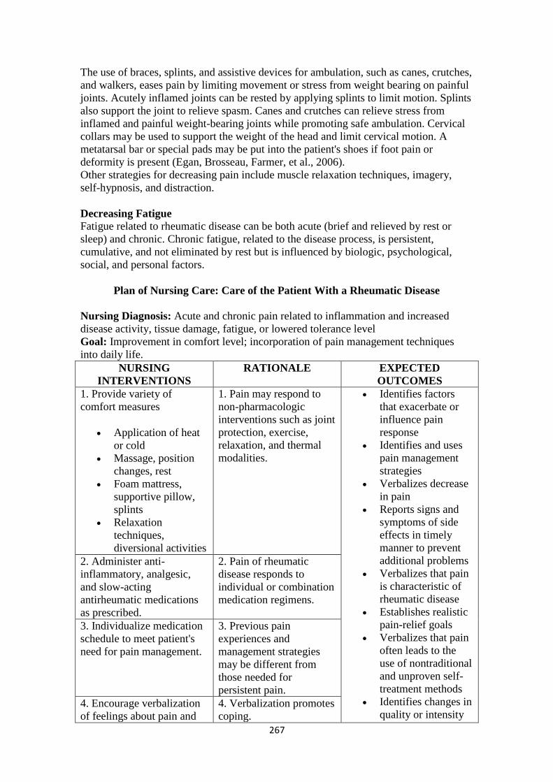
267
The use of braces, splints, and assistive devices for ambulation, such as canes, crutches,
and walkers, eases pain by limiting movement or stress from weight bearing on painful
joints. Acutely inflamed joints can be rested by applying splints to limit motion. Splints
also support the joint to relieve spasm. Canes and crutches can relieve stress from
inflamed and painful weight-bearing joints while promoting safe ambulation. Cervical
collars may be used to support the weight of the head and limit cervical motion. A
metatarsal bar or special pads may be put into the patient's shoes if foot pain or
deformity is present (Egan, Brosseau, Farmer, et al., 2006).
Other strategies for decreasing pain include muscle relaxation techniques, imagery,
self-hypnosis, and distraction.
Decreasing Fatigue
Fatigue related to rheumatic disease can be both acute (brief and relieved by rest or
sleep) and chronic. Chronic fatigue, related to the disease process, is persistent,
cumulative, and not eliminated by rest but is influenced by biologic, psychological,
social, and personal factors.
Plan of Nursing Care: Care of the Patient With a Rheumatic Disease
Nursing Diagnosis: Acute and chronic pain related to inflammation and increased
disease activity, tissue damage, fatigue, or lowered tolerance level
Goal: Improvement in comfort level; incorporation of pain management techniques
into daily life.
NURSING
INTERVENTIONS
RATIONALE EXPECTED
OUTCOMES
1. Provide variety of
comfort measures
Application of heat
or cold
Massage, position
changes, rest
Foam mattress,
supportive pillow,
splints
Relaxation
techniques,
diversional activities
1. Pain may respond to
non-pharmacologic
interventions such as joint
protection, exercise,
relaxation, and thermal
modalities.
Identifies factors
that exacerbate or
influence pain
response
Identifies and uses
pain management
strategies
Verbalizes decrease
in pain
Reports signs and
symptoms of side
effects in timely
manner to prevent
additional problems
Verbalizes that pain
is characteristic of
rheumatic disease
Establishes realistic
pain-relief goals
Verbalizes that pain
often leads to the
use of nontraditional
and unproven self-
treatment methods
Identifies changes in
quality or intensity
2. Administer anti-
inflammatory, analgesic,
and slow-acting
antirheumatic medications
as prescribed.
2. Pain of rheumatic
disease responds to
individual or combination
medication regimens.
3. Individualize medication
schedule to meet patient's
need for pain management.
3. Previous pain
experiences and
management strategies
may be different from
those needed for
persistent pain.
4. Encourage verbalization
of feelings about pain and
4. Verbalization promotes
coping.

268
chronicity of disease. of pain
5. Teach pathophysiology of
pain and rheumatic disease,
and assist patient to
recognize that pain often
leads to unproven treatment
methods.
5. Knowledge of
rheumatic pain and
appropriate treatment may
help patient avoid unsafe,
ineffective therapies.
6. Assist in identification of
pain that leads to use of
unproven methods of
treatment.
6. The impact of pain on
an individual's life often
leads to misconceptions
about pain and pain
management techniques.
7. Assess for subjective
changes in pain.
7. The individual's
description of pain is a
more reliable indicator
than objective
measurements such as
change in vital signs,
body movement, and
facial expression.
Nursing Diagnosis: Fatigue related to increased disease activity, pain, inadequate
sleep/rest, deconditioning, inadequate nutrition, and emotional stress/depression
Goal: Incorporates as part of daily activities strategies necessary to modify fatigue
NURSING
INTERVENTIONS
RATIONALE EXPECTED
OUTCOMES
1. Provide instruction
about fatigue
Describe
relationship of
disease activity to
fatigue.
Describe comfort
measures while
providing them.
Develop and
encourage a sleep
routine (warm bath
and relaxation
techniques that
promote sleep).
Explain importance
of rest for relieving
systematic,
articular, and
emotional stress.
Explain how to use
energy
1. The patient's
understanding of fatigue
will affect his or her
actions.
The amount of
fatigue is directly
related to the
activity of the
disease.
Relief of discomfort
can relieve fatigue.
Effective bedtime
routine promotes
restorative sleep.
Different kinds of
rest are needed to
relieve fatigue and
are based on patient
need and response.
A variety of
measures can be
used to conserve
Self-evaluates and
monitors fatigue
pattern
Verbalizes the
relationship of
fatigue to disease
activity
Uses comfort
measures as
appropriate
Practices effective
sleep hygiene and
routine
Makes use of
various assistive
devices (splints,
canes) and strategies
(bed rest, relaxation
techniques) to ease
different kinds of
fatigue
Incorporates time
management
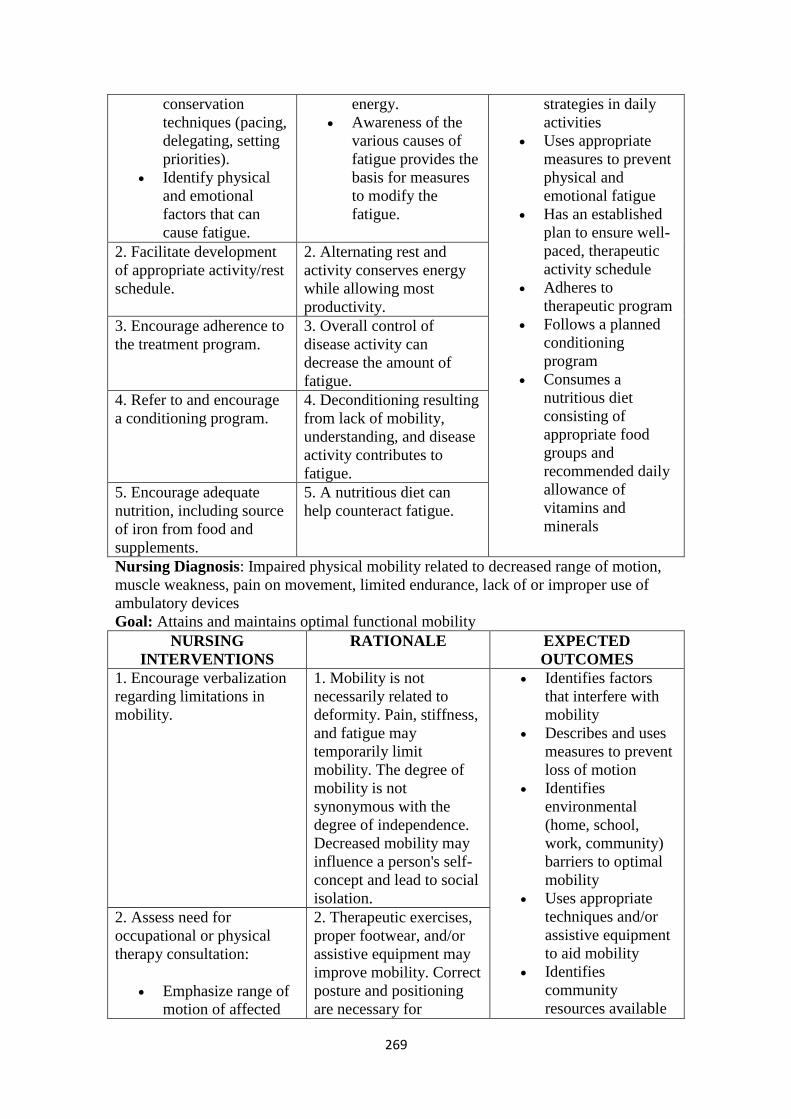
269
conservation
techniques (pacing,
delegating, setting
priorities).
Identify physical
and emotional
factors that can
cause fatigue.
energy.
Awareness of the
various causes of
fatigue provides the
basis for measures
to modify the
fatigue.
strategies in daily
activities
Uses appropriate
measures to prevent
physical and
emotional fatigue
Has an established
plan to ensure well-
paced, therapeutic
activity schedule
Adheres to
therapeutic program
Follows a planned
conditioning
program
Consumes a
nutritious diet
consisting of
appropriate food
groups and
recommended daily
allowance of
vitamins and
minerals
2. Facilitate development
of appropriate activity/rest
schedule.
2. Alternating rest and
activity conserves energy
while allowing most
productivity.
3. Encourage adherence to
the treatment program.
3. Overall control of
disease activity can
decrease the amount of
fatigue.
4. Refer to and encourage
a conditioning program.
4. Deconditioning resulting
from lack of mobility,
understanding, and disease
activity contributes to
fatigue.
5. Encourage adequate
nutrition, including source
of iron from food and
supplements.
5. A nutritious diet can
help counteract fatigue.
Nursing Diagnosis: Impaired physical mobility related to decreased range of motion,
muscle weakness, pain on movement, limited endurance, lack of or improper use of
ambulatory devices
Goal: Attains and maintains optimal functional mobility
NURSING
INTERVENTIONS
RATIONALE EXPECTED
OUTCOMES
1. Encourage verbalization
regarding limitations in
mobility.
1. Mobility is not
necessarily related to
deformity. Pain, stiffness,
and fatigue may
temporarily limit
mobility. The degree of
mobility is not
synonymous with the
degree of independence.
Decreased mobility may
influence a person's self-
concept and lead to social
isolation.
Identifies factors
that interfere with
mobility
Describes and uses
measures to prevent
loss of motion
Identifies
environmental
(home, school,
work, community)
barriers to optimal
mobility
Uses appropriate
techniques and/or
assistive equipment
to aid mobility
Identifies
community
resources available
2. Assess need for
occupational or physical
therapy consultation:
Emphasize range of
motion of affected
2. Therapeutic exercises,
proper footwear, and/or
assistive equipment may
improve mobility. Correct
posture and positioning
are necessary for

270
joints.
Promote use of
assistive ambulatory
devices.
Explain use of safe
footwear.
Use individual
appropriate
positioning/posture.
maintaining optimal
mobility.
to assist in
managing decreased
mobility
3. Assist to identify
environmental barriers.
3. Furniture and
architectural adaptations
may enhance mobility.
4. Encourage independence
in mobility and assist as
needed.
Allow ample time
for activity
Provide rest period
after activity.
Reinforce principles
of joint protection
and work
simplification.
4. Changes in mobility
may lead to a decrease in
personal safety.
5. Initiate referral to
community health agency.
5. The degree of mobility
may be slow to improve
or may not improve with
intervention.
Nursing Diagnosis: Self-care deficits related to contractures, fatigue, or loss of motion
Goal: Achieves self-care independently or with the use of resources
NURSING
INTERVENTIONS
RATIONALE EXPECTED
OUTCOMES
1. Assist patient to identify
self-care deficits and factors
that interfere with ability to
perform self-care activities.
1. The ability to perform
self-care activities is
influenced by the disease
activity and the
accompanying pain,
stiffness, fatigue, muscle
weakness, loss of motion,
and depression.
Identifies factors
that interfere with
the ability to
perform self-care
activities
Identifies alternative
methods for meeting
self-care needs
Uses alternative
methods for meeting
self-care needs
Identifies and uses
other health care
resources for
2. Develop a plan based on
the patient's perceptions and
priorities on how to
establish and achieve goals
to meet self-care needs,
incorporating joint
2. Assistive devices may
enhance self-care abilities.
Effective planning for
changes must include the
patient who must accept
and adopt the plan.

271
protection, energy
conservation, and work
simplification concepts.
Provide appropriate
assistive devices.
Reinforce correct
and safe use of
assistive devices.
Allow patient to
control timing of
self-care activities.
Explore with the
patient different
ways to perform
difficult tasks or
ways to enlist the
help of someone
else.
meeting self-care
needs
3. Consult with community
health care agencies when
individuals have attained a
maximum level of self-care
yet still have some deficits,
especially regarding safety.
3. Individuals differ in
ability and willingness to
perform self-care
activities. Changes in
ability to care for self may
lead to a decrease in
personal safety.
Nursing Diagnosis: Disturbed body image related to physical and psychological
changes and dependency imposed by chronic illness
Goal: Adapts to physical and psychological changes imposed by the rheumatic disease
NURSING
INTERVENTIONS
RATIONALE EXPECTED
OUTCOMES
1. Help patient identify
elements of control over
disease symptoms and
treatment.
1. The individual's self-
concept may be altered by
the disease or its
treatment.
Verbalizes an
awareness that
changes taking place
in self-concept are
normal responses to
rheumatic disease
and other chronic
illnesses
Identifies strategies
to cope with altered
self-concept
2. Encourage patient's
verbalization of feelings,
perceptions, and fears.
Help to assess
present situation and
identify problems.
Assist to identify
past coping
mechanisms.
Assist to identify
effective coping
mechanisms.
2. The individual's coping
strategies reflect the
strength of his or her self-
concept.
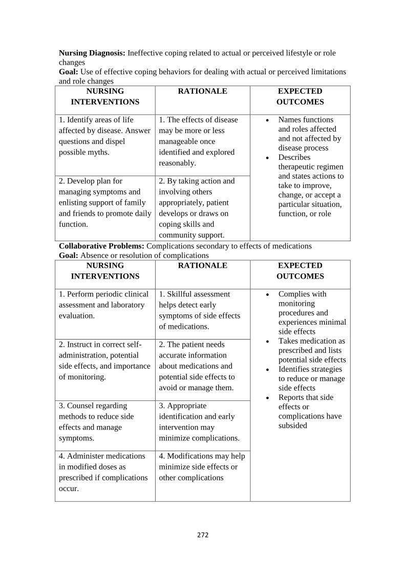
272
Nursing Diagnosis: Ineffective coping related to actual or perceived lifestyle or role
changes
Goal: Use of effective coping behaviors for dealing with actual or perceived limitations
and role changes
NURSING
INTERVENTIONS
RATIONALE EXPECTED
OUTCOMES
1. Identify areas of life
affected by disease. Answer
questions and dispel
possible myths.
1. The effects of disease
may be more or less
manageable once
identified and explored
reasonably.
Names functions
and roles affected
and not affected by
disease process
Describes
therapeutic regimen
and states actions to
take to improve,
change, or accept a
particular situation,
function, or role
2. Develop plan for
managing symptoms and
enlisting support of family
and friends to promote daily
function.
2. By taking action and
involving others
appropriately, patient
develops or draws on
coping skills and
community support.
Collaborative Problems: Complications secondary to effects of medications
Goal: Absence or resolution of complications
NURSING
INTERVENTIONS
RATIONALE EXPECTED
OUTCOMES
1. Perform periodic clinical
assessment and laboratory
evaluation.
1. Skillful assessment
helps detect early
symptoms of side effects
of medications.
Complies with
monitoring
procedures and
experiences minimal
side effects
Takes medication as
prescribed and lists
potential side effects
Identifies strategies
to reduce or manage
side effects
Reports that side
effects or
complications have
subsided
2. Instruct in correct self-
administration, potential
side effects, and importance
of monitoring.
2. The patient needs
accurate information
about medications and
potential side effects to
avoid or manage them.
3. Counsel regarding
methods to reduce side
effects and manage
symptoms.
3. Appropriate
identification and early
intervention may
minimize complications.
4. Administer medications
in modified doses as
prescribed if complications
occur.
4. Modifications may help
minimize side effects or
other complications
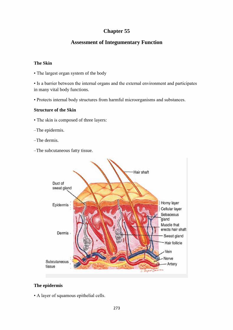
273
Chapter 55
Assessment of Integumentary Function
The Skin
• The largest organ system of the body
• Is a barrier between the internal organs and the external environment and participates
in many vital body functions.
• Protects internal body structures from harmful microorganisms and substances.
Structure of the Skin
• The skin is composed of three layers:
–The epidermis.
–The dermis.
–The subcutaneous fatty tissue.
The epidermis
• A layer of squamous epithelial cells.
274
• Most of these cells produce a tough, fibrous protein called keratin.
• The epidermis also produced specialized cells called melanocytes. These produce
melanin (skin pigment). Aggregations of melanocytes are nevi (moles and birthmarks).
–melanin can absorb ultraviolet light in sunlight
keratin, an insoluble, fibrous protein that forms the outer barrier of the skin and has the
capacity to repel pathogens and prevent excessive fluid loss from the body. Keratin is
the principal hardening ingredient of the hair and nails.
Dermis
• Dense, irregular connective tissue composed of collagen and elastic fibers, blood and
lymph vessels, nerves, sweat, and sebaceous glands and hair roots.
• The sebaceous glands produce sebum, an oily substance that lubricates the skin.
Subcutaneous Tissue
• Primarily connective and adipose (fatty) tissue. Here the skin is anchored to muscle
and bone.
• Provides a cushion between the skin layers, muscles, and bones.
• It promotes skin mobility
Functions of the Skin
• Protection.
• Temperature regulation.
• Sensory perception.
• Fluid and electrolyte balance.
• Vitamin Production
• Immune Response Function
Hair: Structure
• Hair is composed of dead epidermal cells that begin to grow and divide in the base of
the hair follicle.
• As the cells are pushed toward the skin surface, they become keratinized and die.
• Hair color is genetically determined.
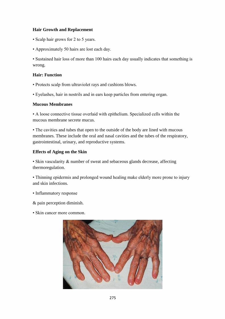
275
Hair Growth and Replacement
• Scalp hair grows for 2 to 5 years.
• Approximately 50 hairs are lost each day.
• Sustained hair loss of more than 100 hairs each day usually indicates that something is
wrong.
Hair: Function
• Protects scalp from ultraviolet rays and cushions blows.
• Eyelashes, hair in nostrils and in ears keep particles from entering organ.
Mucous Membranes
• A loose connective tissue overlaid with epithelium. Specialized cells within the
mucous membrane secrete mucus.
• The cavities and tubes that open to the outside of the body are lined with mucous
membranes. These include the oral and nasal cavities and the tubes of the respiratory,
gastrointestinal, urinary, and reproductive systems.
Effects of Aging on the Skin
• Skin vascularity & number of sweat and sebaceous glands decrease, affecting
thermoregulation.
• Thinning epidermis and prolonged wound healing make elderly more prone to injury
and skin infections.
• Inflammatory response
& pain perception diminish.
• Skin cancer more common.
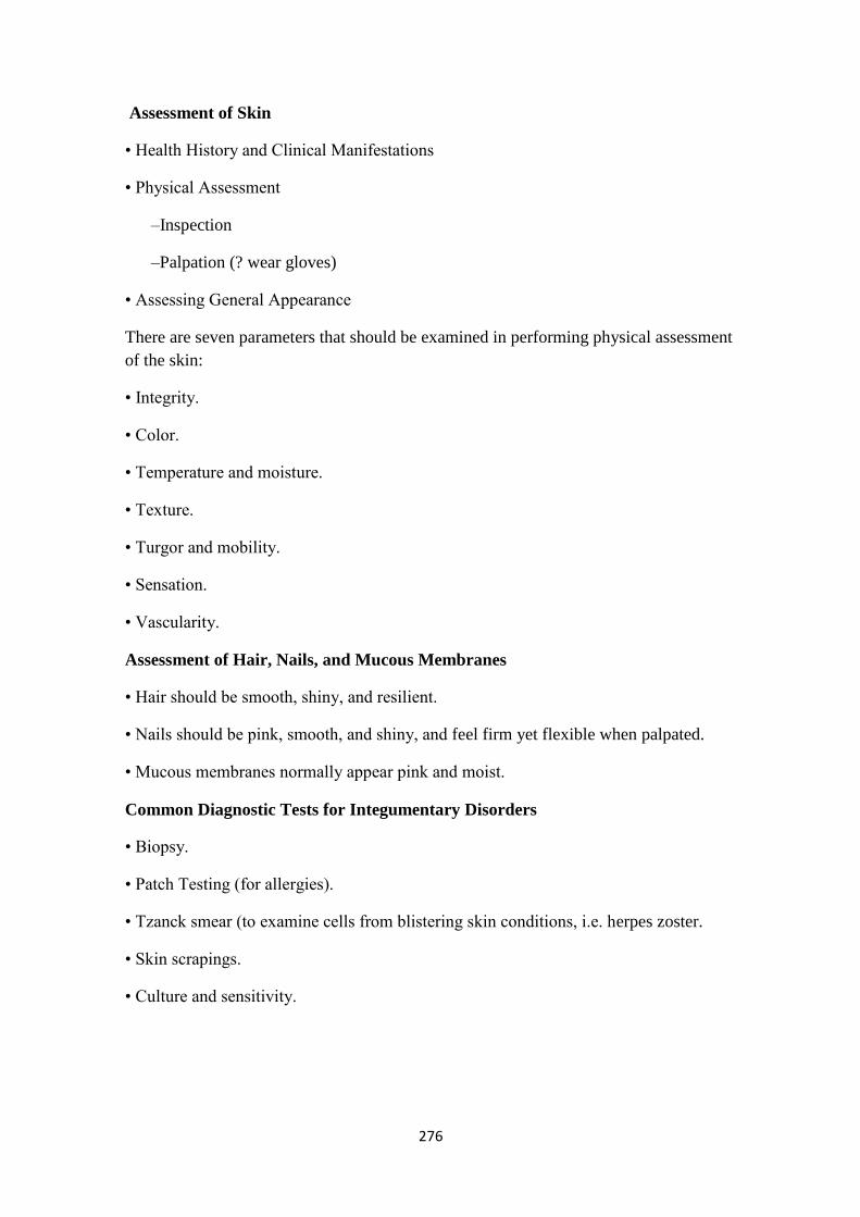
276
Assessment of Skin
• Health History and Clinical Manifestations
• Physical Assessment
–Inspection
–Palpation (? wear gloves)
• Assessing General Appearance
There are seven parameters that should be examined in performing physical assessment
of the skin:
• Integrity.
• Color.
• Temperature and moisture.
• Texture.
• Turgor and mobility.
• Sensation.
• Vascularity.
Assessment of Hair, Nails, and Mucous Membranes
• Hair should be smooth, shiny, and resilient.
• Nails should be pink, smooth, and shiny, and feel firm yet flexible when palpated.
• Mucous membranes normally appear pink and moist.
Common Diagnostic Tests for Integumentary Disorders
• Biopsy.
• Patch Testing (for allergies).
• Tzanck smear (to examine cells from blistering skin conditions, i.e. herpes zoster.
• Skin scrapings.
• Culture and sensitivity.
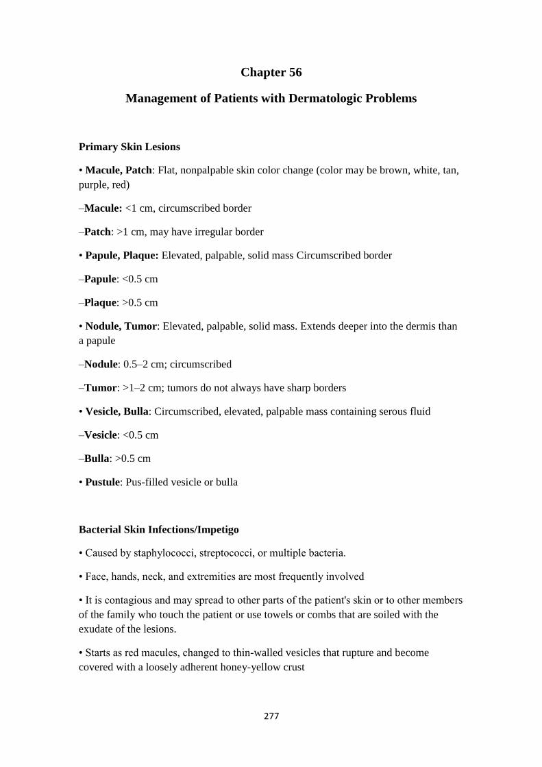
277
Chapter 56
Management of Patients with Dermatologic Problems
Primary Skin Lesions
• Macule, Patch: Flat, nonpalpable skin color change (color may be brown, white, tan,
purple, red)
–Macule: <1 cm, circumscribed border
–Patch: >1 cm, may have irregular border
• Papule, Plaque: Elevated, palpable, solid mass Circumscribed border
–Papule: <0.5 cm
–Plaque: >0.5 cm
• Nodule, Tumor: Elevated, palpable, solid mass. Extends deeper into the dermis than
a papule
–Nodule: 0.5–2 cm; circumscribed
–Tumor: >1–2 cm; tumors do not always have sharp borders
• Vesicle, Bulla: Circumscribed, elevated, palpable mass containing serous fluid
–Vesicle: <0.5 cm
–Bulla: >0.5 cm
• Pustule: Pus-filled vesicle or bulla
Bacterial Skin Infections/Impetigo
• Caused by staphylococci, streptococci, or multiple bacteria.
• Face, hands, neck, and extremities are most frequently involved
• It is contagious and may spread to other parts of the patient's skin or to other members
of the family who touch the patient or use towels or combs that are soiled with the
exudate of the lesions.
• Starts as red macules, changed to thin-walled vesicles that rupture and become
covered with a loosely adherent honey-yellow crust
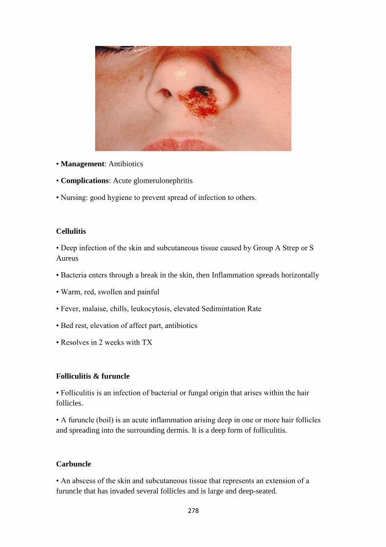
278
• Management: Antibiotics
• Complications: Acute glomerulonephritis
• Nursing: good hygiene to prevent spread of infection to others.
Cellulitis
• Deep infection of the skin and subcutaneous tissue caused by Group A Strep or S
Aureus
• Bacteria enters through a break in the skin, then Inflammation spreads horizontally
• Warm, red, swollen and painful
• Fever, malaise, chills, leukocytosis, elevated Sedimintation Rate
• Bed rest, elevation of affect part, antibiotics
• Resolves in 2 weeks with TX
Folliculitis & furuncle
• Folliculitis is an infection of bacterial or fungal origin that arises within the hair
follicles.
• A furuncle (boil) is an acute inflammation arising deep in one or more hair follicles
and spreading into the surrounding dermis. It is a deep form of folliculitis.
Carbuncle
• An abscess of the skin and subcutaneous tissue that represents an extension of a
furuncle that has invaded several follicles and is large and deep-seated.
279
• Caused by a staphylococcal infection.
• Appear in areas where the skin is thick and inelastic; the back of the neck and the
buttocks are common sites.
• Purulent secretions, high fever, pain, leukocytosis, and even extension of the infection
to the bloodstream.
Herpes Zoster
• Caused by the varicella-zoster virus
• Usually, patients have a history of chickenpox. The virus becomes dormant and lies
inside nerve cells near the brain and spinal cord
• The viruses become latent, until immunity decrease, then it will become active. They
travel by way of the peripheral nerves to the skin.
• S & S: painful vesicular eruption (usually on face, neck & thorax). Vesicles contain
serum, then become purulent.
• Treatment:
–Antiviral agents such as acyclovir (Zovirax)
–Pain is controlled with analgesics
–? Corticosteroids
Herpes Simplex
• Orolabial Herpes (fever blisters or cold sores)
–clusters of grouped vesicles on the lips.
–The onset is often accompanied by high fever, regional lymphadenopathy, and
generalized malaise.
• Genital Herpes
–Minor infections may produce no symptoms
–Severe infections can cause systemic flulike illness. Lesions appear as grouped
vesicles on the vagina, rectum, or penis.

280
Fungal (Mycotic) Infections Tinea Capitis
–Person to person transmission
–Commonly known as ringworm
–Affect the Scalp
• Scaling area with broken hair shafts, plaques, pustules
• Possible permanent hair loss
• Treatment
–Oral Griseofulvin
–Selenium Sulfide shampoos
–Topical antifungal agents
Tinea Corporis (Body)
• Appears on face, neck, trunk, and extremities.
• From infected pets
• Elevated scalilng, erythematous, very pruritic
• May spread to the hair, scalp, or nails.
• Oral Griseofulvin
• Cool compresses
• Antifungal cream applied 1 inch beyond lesion for 1-2 weeks until resolved

281
Tenia Cruris (Groin)
• Begins with small, red scaling patches, which spread to form circular elevated
plaques, very pruritic
• Clusters of pustules may be seen around borders.
• Local application of Tolnaftate liquid
• Wet compresses or sitz baths may be soothing
Tenia Pedis (athlete's foot)
• Between toes and soles of the feet
• Maceration and fissuring between toes
• May have pinhead vesicles
• Oral Griseofulvin
• Antifungal powder
• Eliminate heat and perspiration by clean light socks, well ventilated shoes, avoidance
of occlusive shoes
• Soak feet in vinegar and water solution.
Nondermatophyte Infections Candidiasis
• Yeast fungal infection
• Glistening, fiery red or moist pink, beefy red with satellite pustules, severe
itching/burning
• Sites: skin folds/groin area, oral-thrush, diaper rash
• Teach prevention and management
–Disposable diapers or cloth diapers without rubber pants, Change as soon as soiled
–Exposure to open air, apply ointments (zinc oxide), avoid over washing, caution with
perfumed soaps
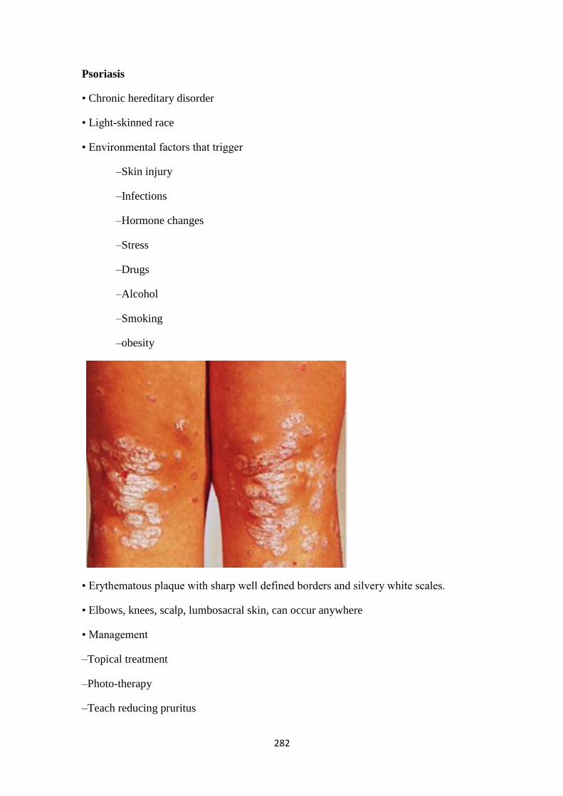
282
Psoriasis
• Chronic hereditary disorder
• Light-skinned race
• Environmental factors that trigger
–Skin injury
–Infections
–Hormone changes
–Stress
–Drugs
–Alcohol
–Smoking
–obesity
• Erythematous plaque with sharp well defined borders and silvery white scales.
• Elbows, knees, scalp, lumbosacral skin, can occur anywhere
• Management
–Topical treatment
–Photo-therapy
–Teach reducing pruritus
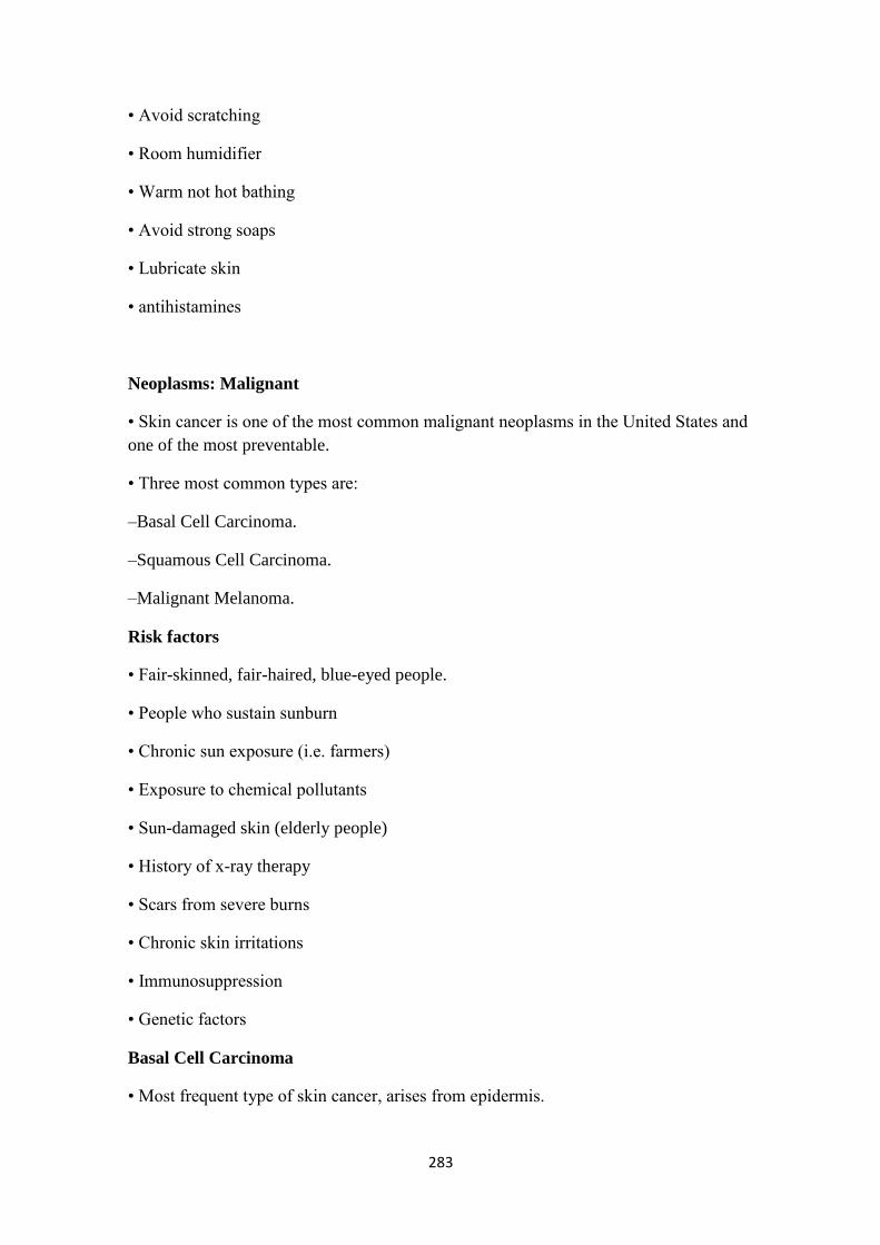
283
• Avoid scratching
• Room humidifier
• Warm not hot bathing
• Avoid strong soaps
• Lubricate skin
• antihistamines
Neoplasms: Malignant
• Skin cancer is one of the most common malignant neoplasms in the United States and
one of the most preventable.
• Three most common types are:
–Basal Cell Carcinoma.
–Squamous Cell Carcinoma.
–Malignant Melanoma.
Risk factors
• Fair-skinned, fair-haired, blue-eyed people.
• People who sustain sunburn
• Chronic sun exposure (i.e. farmers)
• Exposure to chemical pollutants
• Sun-damaged skin (elderly people)
• History of x-ray therapy
• Scars from severe burns
• Chronic skin irritations
• Immunosuppression
• Genetic factors
Basal Cell Carcinoma
• Most frequent type of skin cancer, arises from epidermis.
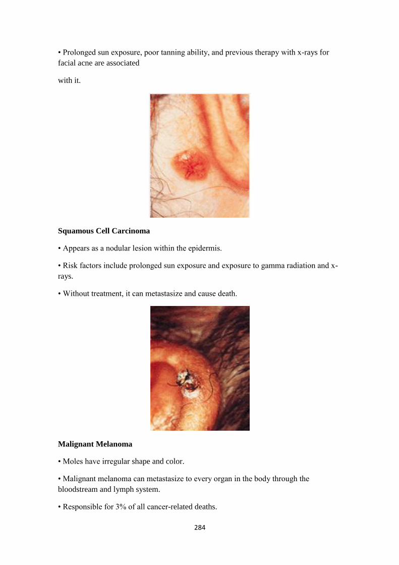
284
• Prolonged sun exposure, poor tanning ability, and previous therapy with x-rays for
facial acne are associated
with it.
Squamous Cell Carcinoma
• Appears as a nodular lesion within the epidermis.
• Risk factors include prolonged sun exposure and exposure to gamma radiation and x-
rays.
• Without treatment, it can metastasize and cause death.
Malignant Melanoma
• Moles have irregular shape and color.
• Malignant melanoma can metastasize to every organ in the body through the
bloodstream and lymph system.
• Responsible for 3% of all cancer-related deaths.

285
Preventing Skin Cancer
• Avoid sun between 10 AM and 3 PM
• Sunscreen cream
• Reapply after swimming, strenuous exercises, or prolonged sun bathing
• Lip balm
• Protective clothing
• Inspect skin regularly
Nonmalignant skin tumor
• Benign tumor of the skin include:
–Warts
–Lipomas (benign fatty tumors).
–Keloids (abnormal growth of scar tissue).
–Sebaceous cysts.
–Nevi (moles).
–Angiomas (birthmarks).
Inflammatory Disorders of the Skin: Dermatitis/Eczema
• In current usage, eczema has almost become synonymous with dermatitis, although
eczema tends to be used most often to refer to chronic forms of dermatitis.
286
• Eczema is an atopic dermatitis often associated with rhinitis and asthma. It is a
chronic superficial inflammation that evolves into pruritic, red, weeping, crusted
lesions.
Contact Dermatitis
• Skin reacts to external irritants like:
–allergens (e.g. poison ivy or cosmetics).
–harsh chemical substances (detergents, insecticides).
–metals such as nickel.
–mechanical irritations from wool or glass fibers.
–body substances like urine or feces.
Strategies for Avoiding Contact Dermatitis
• Avoid contact with causative materials.
• Avoid heat, soap, and rubbing.
• Choose bath soaps, detergents, and cosmetics that do not contain fragrance.
• Avoid topical medications, lotions, or ointments, except those specifically prescribed
for your condition.
• Wash your skin thoroughly immediately after exposure to possible irritants.
287
Chapter 57
Management of Patients with Burn Injury
Burn Prevention
• Keep matches and lighters out of the reach of children.
• Emphasize the importance of never leaving children unattended around fire.
• Develop and practice a home exit fire drill.
• Set the water heater temperature no higher than 70°C.
• Don‘t smoke in bed, and caution against falling asleep while smoking.
• Caution against throwing flammable liquids onto an already burning fire.
• Caution against using flammable liquids to start fires.
• Caution against removing the radiator cap from a hot engine.
• Watch for overhead electrical wires and underground wires when working outside.
• Keep hot irons out of reach of children.
• Caution against running electric cords under carpets or rugs.
• Avoid storing flammable liquids near a fire source.
• Caution when cooking.
• Keep a working fire extinguisher in the home, and to know how to use it.
BURNS
• Results in ??? deaths annually
• Survival best at ages 15-45
• Children, elderly, and diabetics are at higher risk
• Survival best when burns cover less than 20% of TBA
Types of burns
1. Thermal
Exposure to flame or a hot object (i.e. hot water)
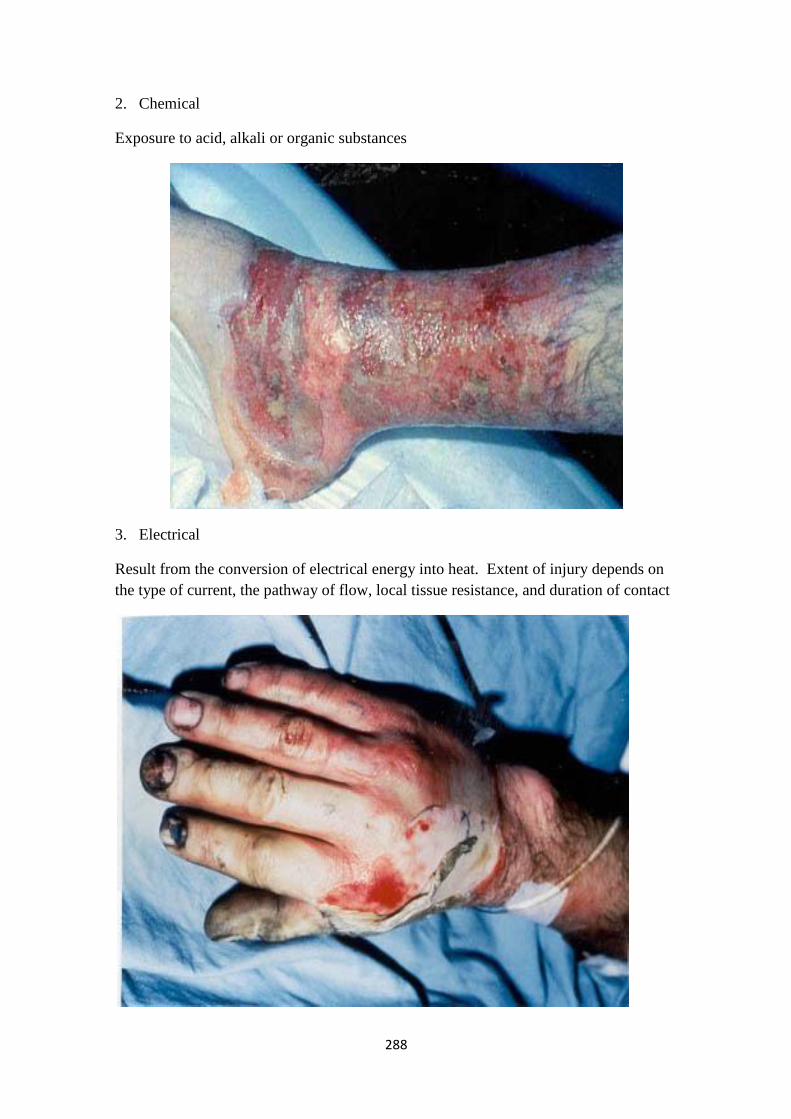
288
2. Chemical
Exposure to acid, alkali or organic substances
3. Electrical
Result from the conversion of electrical energy into heat. Extent of injury depends on
the type of current, the pathway of flow, local tissue resistance, and duration of contact

289
4. Radiation
Result from radiant energy being transferred to the body resulting in production of
cellular toxins
Burn Wound Assessment
• Classified according to depth of injury and extent of body surface area involved
• Burn wounds differentiated depending on the level of dermis and subcutaneous tissue
involved
1. Superficial (first-degree)
2. Deep (second-degree)
3. Full thickness (third and fourth degree)
The depth of the injury depends on the temperature of the burning agent and the
duration of contact with the agent. For example, in the case of scald burns in adults, 1
second of contact with hot tap water at 68.9°C (156°F) may result in a burn that
destroys both the epidermis and the dermis, causing a full-thickness (third-degree)
injury. Fifteen seconds of exposure to hot water at 56.1°C (133°F) results in a similar
full-thickness injury. Temperatures less than 111°F can be tolerated for long periods
without injury.
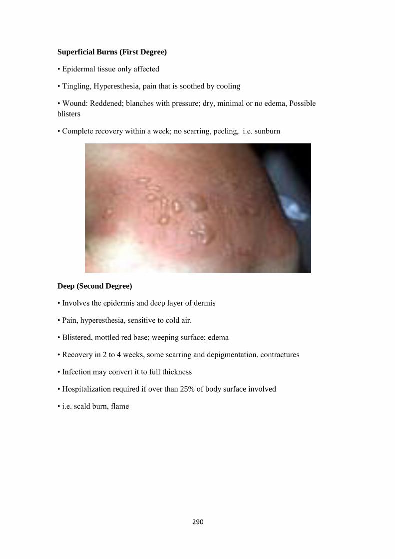
290
Superficial Burns (First Degree)
• Epidermal tissue only affected
• Tingling, Hyperesthesia, pain that is soothed by cooling
• Wound: Reddened; blanches with pressure; dry, minimal or no edema, Possible
blisters
• Complete recovery within a week; no scarring, peeling, i.e. sunburn
Deep (Second Degree)
• Involves the epidermis and deep layer of dermis
• Pain, hyperesthesia, sensitive to cold air.
• Blistered, mottled red base; weeping surface; edema
• Recovery in 2 to 4 weeks, some scarring and depigmentation, contractures
• Infection may convert it to full thickness
• Hospitalization required if over than 25% of body surface involved
• i.e. scald burn, flame

291
Full Thickness (Third/Fourth Degree)
• Destruction of all skin layers
• Requires immediate hospitalization
• Dry, waxy white (or charred), leathery, or hard skin, no pain, edema
• Exposure to flames, electricity or chemicals can cause 3rd degree burns
• Grafting necessary, scarring and loss of contour and function; contractures, loss of
digits or extremity possible

292
Calculation of Burned Body Surface Area
Total Body Surface Area (TBSA)
• Superficial burns are not involved in the calculation
• Lund and Browder Chart is the most accurate because it adjusts for age
• Rule of nines divides the body – adequate for initial assessment for adult burns
• Palm rule
Rule of Nines
• Head & Neck = 9%
• Each upper extremity (Arms) = 9%
• Each lower extremity (Legs) = 18%
• Anterior trunk= 18%
• Posterior trunk = 18%
• Genitalia (perineum) = 1%
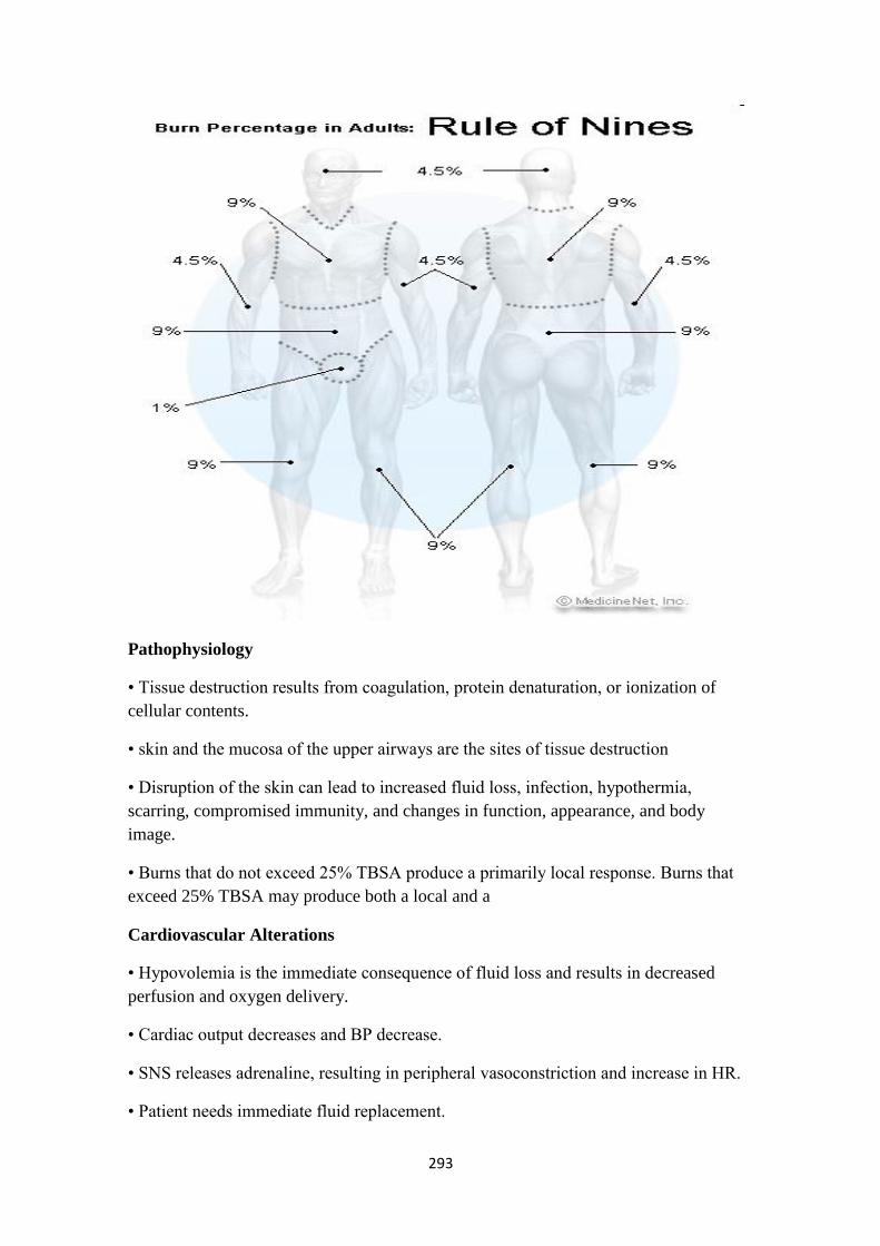
293
Pathophysiology
• Tissue destruction results from coagulation, protein denaturation, or ionization of
cellular contents.
• skin and the mucosa of the upper airways are the sites of tissue destruction
• Disruption of the skin can lead to increased fluid loss, infection, hypothermia,
scarring, compromised immunity, and changes in function, appearance, and body
image.
• Burns that do not exceed 25% TBSA produce a primarily local response. Burns that
exceed 25% TBSA may produce both a local and a
Cardiovascular Alterations
• Hypovolemia is the immediate consequence of fluid loss and results in decreased
perfusion and oxygen delivery.
• Cardiac output decreases and BP decrease.
• SNS releases adrenaline, resulting in peripheral vasoconstriction and increase in HR.
• Patient needs immediate fluid replacement.
294
• Most of fluid (in first 24-36 hr) leaks out of blood vessels.
• As the capillaries begin to regain their integrity, burn shock resolves and fluid returns
to the vascular compartment.
• As fluid is reabsorbed from the interstitial tissue into the vascular compartment, blood
volume increases.
• If renal and cardiac function is adequate, urinary output increases.
• Diuresis continues for several days to 2 weeks.
Fluid shift
Local edema occurs if burn area is less that 25% TBSA
Generalized edema if burn area is more than 25% TBSA
Maximal edema is after 24 hours.
Begins to resolve 1 to 2 days after the burn and usually is completely resolved
within 7 to 10 days.
As edema increases, pressure on small blood vessels and nerves in the distal
extremities causes an obstruction of blood flow and consequent ischemia.
This complication is similar to a compartment syndrome.
The physician may need to perform an escharotomy , a surgical incision into the
eschar (devitalized tissue resulting from a burn), to relieve the constricting
effect of the burned tissue
• Circulating blood volume decreases during burn shock.
• Evaporative fluid loss through the burn wound may reach 3 to 5 L or more over a 24-
hour period until the burn surfaces are covered.
• Usually, hyponatremia is present.
• Immediately after burn injury, hyperkalemia results from massive cell destruction.
Hypokalemia (potassium depletion) may occur later with fluid shifts and inadequate
potassium replacement.
Fluid Remobilization
• Occurs after 24 hours
• Capillary leak stops
• See diuretic stage where edema fluid shifts from the interstitial spaces into the
vascular space
295
• Blood volume increases leading to increased renal blood flow and diuresis
• Body weight returns to normal
• ? Hypokalemia if no potassium replacement
Effects on blood volume
• At the time of burn injury, some red blood cells may be destroyed and other damaged,
resulting in anemia
• Despite this the hematocrit may be elevated due to plasma loss
• Blood loss during surgical procedures, wound care, diagnostic studies and ongoing
hemolysis further contribute to anemia
• Blood transfusions are required periodically to maintain adequate hemoglobin levels
for oxygen delivery
• Abnormalities in coagulation, including a decrease in platelets (thrombocytopenia)
and prolonged clotting and prothrombin times occur with burn injury
Pulmonary Alterations
• Inhalation injury is the leading cause of death in fire victims
• Half of these deaths could have been prevented with use of a smoke detector
• Burn victims make it out of a burning home safely
• Once they are outside they may realize that loved ones or valuable items are still
inside the burning home
• They then reenter the burning home and are overcome with toxic smoke and fumes
and become disoriented or unconcious
Pulmonary response
• Pulmonary imjuries fall into several categories, upper airway injury, inhalational
injury, including carbon monoxide poisining, and restrictive defects
• Upper air way injury results from direct heat or edema
• It is manifested by mechanical obstruction of the upper airway, including the pharynx
and larynx
• Upper airway injury is treated by early nasotracheal or endotracheal intubation
• Inhalation injury below the glottis results from inhaling products of incomplete
combustion (Burning) or noxious gases: carbon monoxide, sulfer oxide, nitrogen oxide,
& benzene
296
• The injury results directly from chemical irritation of the pulmonary tisues at the
alveolar level
• Inhalation injuries below glottis cause loss of ciliary action, hypersecretion, severe
mucosal edema and bronchospasm
• The pulmonary surfactant is reduced, resulting in atelectasis (collapse of alveoli)
• Expectoration of carbon particles in the sputum is the cardinal sign of this injury
• The pathophysiology effects are due to tissue hypoxia a result of carbon monoxide
combining with hemoglobin to form carboxyhemoglobin which competes with oxygen
for available hemoglobin sites
• The effinity of hemoglobin for carbon monoxide is 200 times greater than that for
oxygen
• Treatment usually consists of ealy intubation and mechnical ventilation with 100%
oxygen
• Administering 100% O2 is essential to accelerate the removal of carbon monoxide
from the hemoglobin molecule
• restrictive defects arise when edema develops under full-thickness burns encircling
the neck and thorax
• Chest expansion may be greatly restricted resulting in decreased tidal volume
• In such situation escharotomy is necessary
• More than half of all burn victims with pulmonary involvement do not initially
demonstrate pulmonary signs and symptoms
• Any pat with possible inhalation injury must be observed for at least 24 h for
respiratory complications
Pulmonary response
• Airway obstruction may occur very rapidly in hours
• Decreased lung compliance, decreased arterial oxygen levels and respiratory acidosis
may occur gradually over the first 5 days after a burn
Indication of possible pulmonary damages include
• History indicating that the burn occured in an enclosed area
• Burns of the face and neck
• Hoarseness , voice change, dry cough, stridor.
• Bloody sputum
297
• Labored breathing or tachypnea (rapid breathing) and other signs of reduced oxygen
levels
• Erythema and blistering of the oral or pharyngeal mucosa
• The immediate intervention is intubation and mechanical ventilation
• If ventilation is impaired by restricted chest excursion, immediate chest escharotomy
is needed
• ARDS may develop in the first few days after the burn injury secondary to systemic
and pulmonary responses to the burn and inhalation injury
Renal alteration
• Renal function may be altered as a result of decreased blood volume
• Destruction of red blood cells at the injury site frees hemoglobin in the urine
• If muscle damage occurs (from electric burns e..) myoglobin is released from the
muscle cells and excreted by the kidney
• Adequate fluid volume replacement restores renal blood flow, increasing the
glomerular filtration rate and urine volume
• If there is inadequate blood flow through the kidneys, the hemoglobin and myoglobin
occlude the renal tubules, resultinh in acute tubular necrosis and renal failure
Immunologic Alterations
• The immunologic defences of the body are greatly altered by burn injury
• Serious burn injury diminishes resistance to the infection
• As a result, sepsis remains the leading cause of death in thermally injured patients
• The loss of skin integrity is compounded by the release of abnormal inflammatory
factors, altered levels of immunglobulins, impared neutrophil function and a reduction
in lymphocytes (lymphocytopeni)
• Research suggests that burn injury results in loss of T -helper.cell lymphocytes
Thermoregulatory Alterations
• Loss of skin results in an inability to regulate body temperature
• Burn patients may therefore exhibit low body temperature in the early hours after
injury
• As of hypermetabolism, core temperature increase.

298
• Burn patients become hyperthermic for much of the postburn period, even in the
absence of infection
GI responses
• Paralytic ileus (absence of intestinal peristalsis):
–decreased peristalsis and bowel sounds are manifestations of paralytic ileus resulting
from burn trauma and
–Gastric distention and nausea may lead to vomiting unless gastric decompression is
initiated
• Curling´s ulcer (gastric or deuodenal erosion)
–Gastric bleeding secondary to massive physiologic stress may be signaled by occult
blood in the stool, regurgitation of coffee ground material from the stomach or bloody
vomitus
Phases of Burn Injuries
• Emergent Phase: From onset of injury to completion of fluid resuscitation
• Acute Phase: From beginning of diuresis to near completion of wound closure
• Rehabilitative Phase: From major wound closure to return to individual's optimal
level of physical and psychosocial adjustment
Emergent Phase
• Immediate problem is fluid loss, edema, reduced blood flow (fluid and electrolyte
shifts)
• Goals:
1. First aid (A, B, C)
2. Intubation if needed
3. Prevention of shock
4. Prevention of respiratory distress
5. Detection and treatment of concomitant injuries
6. Wound assessment and initial care
Emergent phase/ at fire sceine
• Extinguish the flames , switch off power source, remove chemical soaked clothes,
etc…

299
• Assess and maintain an open airway
• Cool the burn
• Remove restrictive objects.
• Cover the wound
• Irrigate chemical burns
First Aid Scene
• Cool burn wound:
- reduce direct thermal trauma & stabilize mast cells, reducing release of
histamine and other inflammatory mediators, and will reduce edema
- pain relief
- running water (15 degree C.)
- worth considering for up to 2 hours
Intubation:
• Suspicion of inhalational injury
• Severe facial & neck burn
― easy early intubation will become difficult and impossible later on‖
Inhalational injury
• Fires
• Closed space
• Carbonaceous material in nose & mouth
• Direct thermal injury (upper airway)
• Chemical injury ; products of combustion (lower airway)
• CO poisoning ; systemic effect
CO poisoning
• Toxic symptoms > 20%, death rate is 60%
• CO-Hb > 5% is indicative of inhalational injury but not severity
• 200 – 250 × greater affinity for Hb than oxygen
• Treatment: 100% O2, hyperbaric oxygen
300
EMERGENT PHASE
• Transfer to the nearest hospital
• Knowledge of circumstances surrounding the burn injury
• Obtain client‘s pre-burn weight to calculate fluid rates
Clinical Manifestations in the Emergent Phase
• Clients with major burn injuries and with inhalation injury are at risk for respiratory
problems
• Inhalation injuries are present in 20% to 50% of the clients admitted to burn centers
• Assess the respiratory system by inspecting the mouth, nose, and pharynx
• Burns of the lips, face, ears, neck, eyelids, eyebrows, and eyelashes are strong
indicators that an inhalation injury may be present
• Change in respiratory pattern may indicate a pulmonary injury.
• The client may: become progressively hoarse, develop a brassy cough, drool or have
difficulty swallowing, produce expiratory sounds that include audible wheezes and
strider
• Upper airway edema and inhalation injury are most common in the trachea and
mainstem bronchi
• Auscultate these areas for wheezes
• If wheezes disappear, this indicates impending airway obstruction and demands
immediate intubation
• Cardiovascular symptoms: will begin immediately which can include shock (Shock is
a common cause of death in the emergent phase in clients with serious injuries)
• Obtain a baseline EKG
• Monitor for edema, measure central and peripheral pulses, blood pressure, capillary
refill and pulse oximetry
• Changes in renal function are related to decreased renal blood flow
• Urine is usually highly concentrated and has a high specific gravity
• Urine output is decreased during the first 24 hours of the emergent phase
• Fluid resuscitation is provided at the rate needed to maintain adult urine output at 30
to 50-mL/hr.
• Measure BUN, creatinine and elesctrolyte levels
301
• Sympathetic stimulation during the emergent phase causes reduced GI motility and
paralytic ileus
• Auscultate the abdomen to assess bowel sounds which may be reduced
• Monitor for n/v and abdominal distention
• Clients with burns of 25% TBSA or who are intubated generally require a NG tube
inserted to prevent aspiration and removal of gastric secretions
Skin Assessment
• Assess the skin to determine the size and depth of burn injury
• The size of the injury is first estimated in comparison to the total body surface area
(TBSA). For example, a burn that involves 40% of the TBSA is a 40% burn
• Use the rule of nines for clients whose weights are in normal proportion to their
heights
Classification of Extent of Burn Injury
1. Minor Burn Injury
–Second-degree burn of <15% total body surface area (TBSA) in adults or <10%
TBSA in children
–Third-degree burn of <2% TBSA not involving special care areas (eyes, ears, face,
hands, feet, perineum, joints)
–Excludes all patients with electrical injury, inhalation injury, or concurrent trauma and
all poor-risk patients (eg, extremes of age, intercurrent disease)
2. Moderate, Uncomplicated Burn Injury
–Second-degree burns of 15–25% TBSA in adults or 10–20% in children
–Third-degree burns of <10% TBSA not involving special care areas
–Excludes electrical injury, inhalation injury, or concurrent trauma and all poor-risk
patients (eg, extremes of age, intercurrent disease)
3. Major Burn Injury
–Second-degree burns >25% TBSA in adults or >20% in children
–All third-degree burns >10% TBSA
–All burns involving eyes, ears, face, hands, feet, perineum, joints
–All inhalation injury, electrical injury, or concurrent trauma, and all poor-risk patients
302
Management of Major Burns
Initial patient management
1. Intravenous access established & I.V. fluids
2. N/G tube; enteral feeding
3. Folley‘s catheter
4. Analgesia
5. Tetanus vaccine
6. Patient must be kept warm
7. Dressing
IV Fluid Therapy
• Infusion of IV fluids is needed to maintain sufficient blood volume for normal
Cardiac Output
• Clients with burns involving 15% to 20% of the TBSA require IV fluid
• Purpose is to prevent shock by maintaining adequate circulating blood fluid volume
• Severe burn requires large fluid loads in a short time to maintain blood flow to vital
organs
• Fluid replacement formulas are calculated from the time of injury and not from the
time of arrival at the hospital
• Diuretics should not be given to increase urine output.
• Change the amount and rate of fluid administration to improve urine output.
• Diuretics do not increase CO; they actually decrease circulating volume and CO by
pulling fluid from the circulating blood volume to enhance diuresis
Guidelines and Formulas for Fluid Replacement in Burn Patients
• Consensus Formula
• Lactated Ringer's solution (or other balanced saline solution): 2–4 mL × kg body
weight × % total body surface area (TBSA) burned.
• Half to be given in first 8 hours; remaining half to be given over next 16 h.
• Evans Formula
–Colloids: 1 mL × kg body weight × % TBSA burned
303
–Electrolytes (saline): 1 mL × body weight × % TBSA burned
–Glucose (5% in water): 2000 mL for insensible loss
–Day 1: Half to be given in first 8 hours; remaining half over next 16 h.
–Day 2: Half of previous day's colloids and electrolytes; all of insensible fluid
replacement.
–Maximum of 10,000 ML over 24 hr. Second- and
Nursing Diagnosis in the Emergent Phase
• Impaired gas exchange related to carbon monoxide poisoning, smoke inhalation, and
upper airway obstruction
–Goal: Maintenance of adequate tissue oxygenation
• Ineffective airway clearance related to edema and effects of smoke inhalation
–Goal: Maintain patent airway and adequate airway clearance
• Fluid volume deficit related to increased capillary permeability and evaporative losses
from the burn wound
–Goal: Restoration of optimal fluid and electrolyte balance and perfusion of vital
organs
• Hypothermia related to loss of skin microcirculation and open wounds
–Goal: Maintenance of adequate body temperature
• Pain related to tissue and nerve injury and emotional impact of injury
–Goal: Control of pain
• Anxiety related to fear and the emotional impact of burn injury
–Goal: Minimization of patient's and family's anxiety
• Collaborative Problems:
• Acute respiratory failure, distributive shock, acute renal failure, compartment
syndrome, paralytic ileus, Curling's ulcer
–Goal: Absence of complications
ACUTE PHASE OF BURN INJURY
• Lasts until wound closure is complete
• Care is directed toward continued assessment and maintenance of the CV and
respiratory systems

304
• Pneumonia is a concern which can result in respiratory failure requiring mechanical
ventilation
• Infection (Topical antibiotics – Silvadene)
• Tetanus toxoid
• Weight daily without dressings or splints and compare to pre-burn weight
• A 2% loss of body weight indicates a mild deficit
• A 10% or greater weight loss requires modification of calorie intake
• Monitor for signs of infection
Fluid and electrolytes changes in the acute phase
Observation Explanation
Hemodilution (decreased
hematocrit)
Blood cell concentration is diluted as fluid enters the intravascular compartment; loss of
red blood cells destroyed at burn site
Increased urinary output Fluid shift into intravascular compartment increases renal blood flow and causes
increased urine formation.
Sodium (Na+) deficit With diuresis, sodium is lost with water; existing serum sodium is diluted by water
influx.
Potassium (K+) deficit (occurs
occasionally in this phase)
Beginning on the fourth or fifth postburn day, K+ shifts from extracellular fluid into
cells.
Metabolic acidosis Loss of sodium depletes fixed base; relative carbon dioxide content increases.
Local and Systemic Signs of Infection- Gram Negative Bacteria
• Pseudomonas, Proteus
• May led to septic shock
• Conversion of a partial-thickness injury to a full-thickness injury
• Ulceration of health skin at the burn site
• Erythematous, nodular lesions in uninvolved skin
• Excessive burn wound drainage
305
• Odor
• Sloughing of grafts
• Altered level of consciousness
• Changes in vital signs
• Oliguria
• GI dysfunction such as diarrhea, vomiting
• Metabolic acidosis
Lab Values
• Na – hyponatremia or Hypernatremia
• K – Hyperkalemia or Hypokalemia
• WBC – 10,000-20,000
Nursing Diagnosis In The Acute Phase
• Excessive fluid volume related to resumption of capillary integrity and fluid shift
from the interstitial to the intravascular compartment
• Risk for infection related to loss of skin barrier and impaired immune response
• Imbalanced nutrition, less than body requirements, related to hypermetabolism and
wound healing needs
• Impaired skin integrity related to open burn wounds
• Acute pain related to exposed nerves, wound healing, and treatments
• Impaired physical mobility related to burn wound edema, pain, and joint contractures
• Ineffective coping related to fear and anxiety, grieving, and forced dependence on
health care providers
• Interrupted family processes related to burn injury
• Deficient knowledge about the course of burn treatment
Planning and Implementation
• Nonsurgical management: removal of exudates and necrotic tissue, cleaning the area,
stimulating granulation and revascularization and applying dressings. Debridement
may be needed
306
Prevent infection
• Major sources of pathogens:
–Intestine: Mucosa of intestine normally prevent bacteria in the GI to pass into the
blood stream. But when blood vessels loose its permeability (due to burn), bacteria
pass from the GI into circulation and causes infection.
–Environment
• Eschar (a nonviable crust with no blood supply) vent leukocytes & antibodies to reach
the area.
• Wound infection may lead to septicemia.
Wound cleaning
• Hydrotherapy (whirl pool)
–Antiseptics added to the water
–Temp kept between 37-38º C
–Warmth allow for easier movement of joints which will prevent contracture
–Movement of water will remove dead tissue.
• Topical Antibacterial Therapy
–They does not sterilize the burn wound; but they reduce the number of bacteria. The
overall microbial population can be controlled by the body's host defense mechanisms.
• Wound Dressing
–Remove old dressing gently, ? Give analgesia to control pain.
–Remove old ointment and clean thoroughly.
–Allow for movement at joint areas.
–The fingers and toes should be wrapped individually to promote adequate healing
–Burns to the face may be left open to air once they have been cleaned and the topical
agent has been applied.
Dressing the Burn Wound
• After burn wounds are cleaned and debrided, topical antibiotics are reapplied to
prevent infection
307
• Standard wound dressings are multiple layers of gauze applied over the topical agents
on the burn wound
Wound Débridement
1. Natural Débridement
2. Mechanical Débridement
3. Surgical Débridement
Wound Grafting
• coverage of the burn wound is necessary until coverage with a graft is possible.
• The purposes of wound coverage are to:
–Decrease the risk of infection;
–Prevent further loss of protein, fluid, and electrolytes through the wound; and
–Minimize heat loss through evaporation.
• Wound coverage may consist of biologic:
1. Biosynthetic,
2. Synthetic, and
3. Autologous methods or
4. A combination of these approaches
• The main areas for skin grafting include the face (for cosmetic and psychological
reasons); functional areas, such as the hands and feet; and areas that involve joints.
• Grafting permits earlier functional ability and reduces wound contractures.
• It should have a bacterial count of less than 100,000 per gram of tissue to optimize
graft success.
• Biologic, Biosynthetic and Synthetic Dressings
Biologic Dressings (Homografts and Heterografts)
• In extensive burns, biologic dressing save lives by providing temporary wound
coverage and protecting the granulation tissue until autografting is possible.
• used as temporary coverings, eventually rejected because of regiction.
• Two types of biologic dressing
1. Homografts (or allografts): skin obtained from living or recently deceased
humans or amniotic membrane.
308
2. Heterografts (or xenografts): skin taken from
Autografts
• Graft taken from the same person
• Graft care (goal: not to dislodge the graft)
–Immobilize graft area (use splint)
–Occlusive dressing, first dressing removed by Dr (usually after 2-5 days in there
is no infection.
–Graft could be left open and stabilized with staples.
–Turn and position patient carefully
–If graft get dislodged, immerse in in normal saline.
–Elevate grafted extremity, and bed rest for 5-7 days.
• Donor site care:
–Wet saline gauze dressing.
Other care during acute phase
• Pain management
• Nutrition
–Metabolic rate is very high because of anabolism
–High caloric high protein diet.
Disorders of Wound Healing
• hypertrophic scars: characterized by an overabundant formation of matrix, especially
collagen, in wounds that heal by granulation
• Keloids: A large, heaped-up mass of scar tissue, a keloid, may develop and extend
beyond the wound surface.
• Failure to Heal: due to infection, other disease process, pressure, or inadequate
nutrition
• Contractures
Rehabilitative Phase of Burn Injury
• Starts at the time of admission

309
• Technically begins with wound closure and ends when the client returns to the highest
possible level of functioning
• Provide psychosocial support
• Assess home environment, financial resources, medical equipment, prosthetic rehab
Prevention of Hypertrophic Scarring
• The wound is in a dynamic state for 1.5 to 2 years after the burn occurs.
• Wear pressure garments , massage, lubrication, exercise, splints, manual lymphatic
drainage and injectable steroids
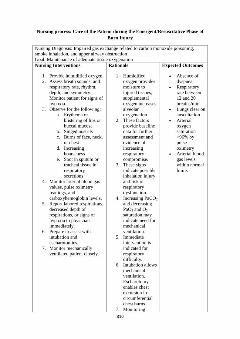
310
Nursing process: Care of the Patient during the Emergent/Resuscitative Phase of
Burn Injury
Nursing Diagnosis: Impaired gas exchange related to carbon monoxide poisoning,
smoke inhalation, and upper airway obstruction
Goal: Maintenance of adequate tissue oxygenation
Nursing Interventions Rationale Expected Outcomes
1. Provide humidified oxygen.
2. Assess breath sounds, and
respiratory rate, rhythm,
depth, and symmetry.
Monitor patient for signs of
hypoxia.
3. Observe for the following:
a. Erythema or
blistering of lips or
buccal mucosa
b. Singed nostrils
c. Burns of face, neck,
or chest
d. Increasing
hoarseness
e. Soot in sputum or
tracheal tissue in
respiratory
secretions
4. Monitor arterial blood gas
values, pulse oximetry
readings, and
carboxyhemoglobin levels.
5. Report labored respirations,
decreased depth of
respirations, or signs of
hypoxia to physician
immediately.
6. Prepare to assist with
intubation and
escharotomies.
7. Monitor mechanically
ventilated patient closely.
1. Humidified
oxygen provides
moisture to
injured tissues;
supplemental
oxygen increases
alveolar
oxygenation.
2. These factors
provide baseline
data for further
assessment and
evidence of
increasing
respiratory
compromise.
3. These signs
indicate possible
inhalation injury
and risk of
respiratory
dysfunction.
4. Increasing PaCO2
and decreasing
PaO2 and O2
saturation may
indicate need for
mechanical
ventilation.
5. Immediate
intervention is
indicated for
respiratory
difficulty.
6. Intubation allows
mechanical
ventilation.
Escharotomy
enables chest
excursion in
circumferential
chest burns.
7. Monitoring
Absence of
dyspnea
Respiratory
rate between
12 and 20
breaths/min
Lungs clear on
auscultation
Arterial
oxygen
saturation
>96% by
pulse
oximetry
Arterial blood
gas levels
within normal
limits
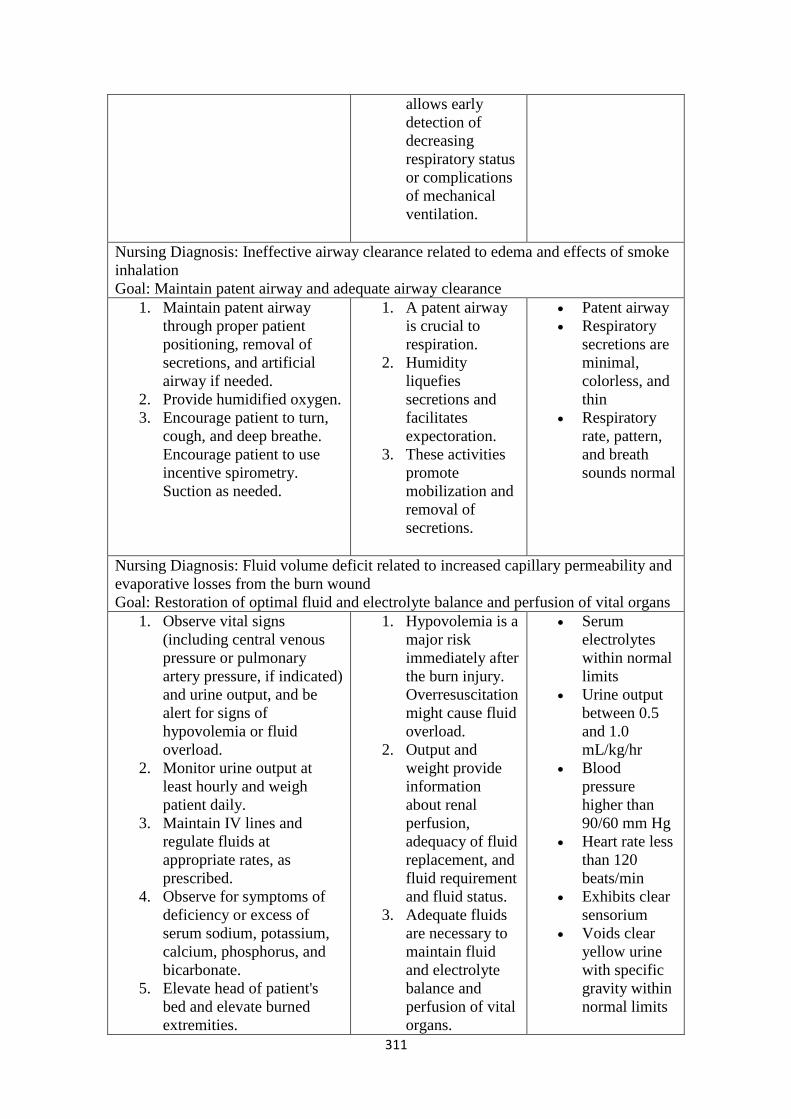
311
allows early
detection of
decreasing
respiratory status
or complications
of mechanical
ventilation.
Nursing Diagnosis: Ineffective airway clearance related to edema and effects of smoke
inhalation
Goal: Maintain patent airway and adequate airway clearance
1. Maintain patent airway
through proper patient
positioning, removal of
secretions, and artificial
airway if needed.
2. Provide humidified oxygen.
3. Encourage patient to turn,
cough, and deep breathe.
Encourage patient to use
incentive spirometry.
Suction as needed.
1. A patent airway
is crucial to
respiration.
2. Humidity
liquefies
secretions and
facilitates
expectoration.
3. These activities
promote
mobilization and
removal of
secretions.
Patent airway
Respiratory
secretions are
minimal,
colorless, and
thin
Respiratory
rate, pattern,
and breath
sounds normal
Nursing Diagnosis: Fluid volume deficit related to increased capillary permeability and
evaporative losses from the burn wound
Goal: Restoration of optimal fluid and electrolyte balance and perfusion of vital organs
1. Observe vital signs
(including central venous
pressure or pulmonary
artery pressure, if indicated)
and urine output, and be
alert for signs of
hypovolemia or fluid
overload.
2. Monitor urine output at
least hourly and weigh
patient daily.
3. Maintain IV lines and
regulate fluids at
appropriate rates, as
prescribed.
4. Observe for symptoms of
deficiency or excess of
serum sodium, potassium,
calcium, phosphorus, and
bicarbonate.
5. Elevate head of patient's
bed and elevate burned
extremities.
1. Hypovolemia is a
major risk
immediately after
the burn injury.
Overresuscitation
might cause fluid
overload.
2. Output and
weight provide
information
about renal
perfusion,
adequacy of fluid
replacement, and
fluid requirement
and fluid status.
3. Adequate fluids
are necessary to
maintain fluid
and electrolyte
balance and
perfusion of vital
organs.
Serum
electrolytes
within normal
limits
Urine output
between 0.5
and 1.0
mL/kg/hr
Blood
pressure
higher than
90/60 mm Hg
Heart rate less
than 120
beats/min
Exhibits clear
sensorium
Voids clear
yellow urine
with specific
gravity within
normal limits
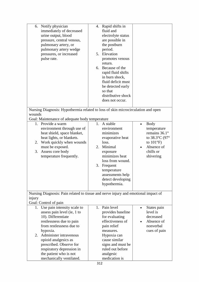
312
6. Notify physician
immediately of decreased
urine output, blood
pressure, central venous,
pulmonary artery, or
pulmonary artery wedge
pressures, or increased
pulse rate.
4. Rapid shifts in
fluid and
electrolyte status
are possible in
the postburn
period.
5. Elevation
promotes venous
return.
6. Because of the
rapid fluid shifts
in burn shock,
fluid deficit must
be detected early
so that
distributive shock
does not occur.
Nursing Diagnosis: Hypothermia related to loss of skin microcirculation and open
wounds
Goal: Maintenance of adequate body temperature
1. Provide a warm
environment through use of
heat shield, space blanket,
heat lights, or blankets.
2. Work quickly when wounds
must be exposed.
3. Assess core body
temperature frequently.
1. A stable
environment
minimizes
evaporative heat
loss.
2. Minimal
exposure
minimizes heat
loss from wound.
3. Frequent
temperature
assessments help
detect developing
hypothermia.
Body
temperature
remains 36.1°
to 38.3°C (97°
to 101°F)
Absence of
chills or
shivering
Nursing Diagnosis: Pain related to tissue and nerve injury and emotional impact of
injury
Goal: Control of pain
1. Use pain intensity scale to
assess pain level (ie, 1 to
10). Differentiate
restlessness due to pain
from restlessness due to
hypoxia.
2. Administer intravenous
opioid analgesics as
prescribed. Observe for
respiratory depression in
the patient who is not
mechanically ventilated.
1. Pain level
provides baseline
for evaluating
effectiveness of
pain relief
measures.
Hypoxia can
cause similar
signs and must be
ruled out before
analgesic
medication is
States pain
level is
decreased
Absence of
nonverbal
cues of pain

313
Assess response to
analgesic.
3. Provide emotional support
and reassurance.
administered.
2. Intravenous
administration is
necessary
because of
altered tissue
perfusion from
burn injury.
3. Emotional
support is
essential to
reduce fear and
anxiety resulting
from burn injury.
Fear and anxiety
increase the
perception of
pain.
Nursing Diagnosis: Anxiety related to fear and the emotional impact of burn injury
Goal: Minimization of patient's and family's anxiety
1. Assess patient's and
family's understanding of
burn injury, coping skills,
and family dynamics.
2. Individualize responses to
the patient's and family's
coping level.
3. Explain all procedures to
the patient and the family in
clear, simple terms.
4. Maintain adequate pain
relief.
5. Consider administering
prescribed anti-anxiety
medications if the patient
remains extremely anxious
despite nonpharmacologic
interventions.
1. Previous
successful coping
strategies can be
fostered for use
in the present
crisis.
Assessment
allows planning
of individualized
interventions.
2. Reactions to burn
injury are
extremely
variable.
Interventions
must be
appropriate to the
patient's and
family's present
level of coping.
3. Increased
understanding
alleviates fear of
the unknown.
High levels of
anxiety may
interfere with
understanding of
complex
explanations.
Patient and
family
verbalize
understanding
of emergent
burn care
Able to
answer simple
questions
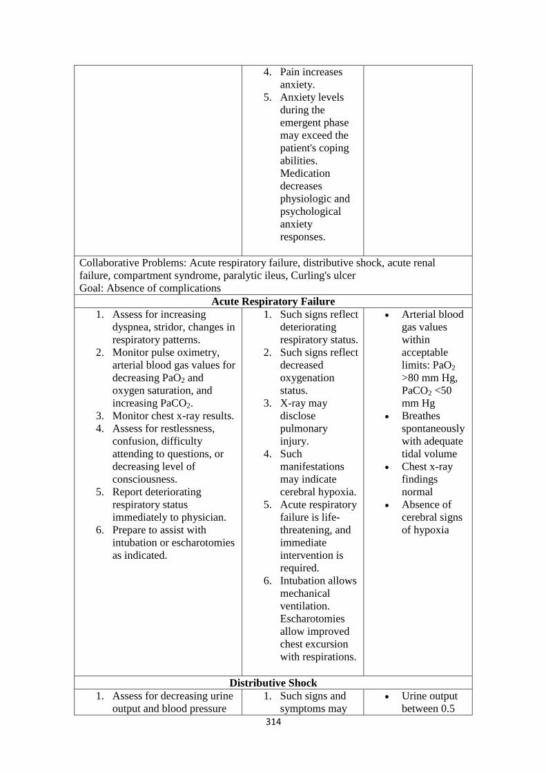
314
4. Pain increases
anxiety.
5. Anxiety levels
during the
emergent phase
may exceed the
patient's coping
abilities.
Medication
decreases
physiologic and
psychological
anxiety
responses.
Collaborative Problems: Acute respiratory failure, distributive shock, acute renal
failure, compartment syndrome, paralytic ileus, Curling's ulcer
Goal: Absence of complications
Acute Respiratory Failure
1. Assess for increasing
dyspnea, stridor, changes in
respiratory patterns.
2. Monitor pulse oximetry,
arterial blood gas values for
decreasing PaO2 and
oxygen saturation, and
increasing PaCO2.
3. Monitor chest x-ray results.
4. Assess for restlessness,
confusion, difficulty
attending to questions, or
decreasing level of
consciousness.
5. Report deteriorating
respiratory status
immediately to physician.
6. Prepare to assist with
intubation or escharotomies
as indicated.
1. Such signs reflect
deteriorating
respiratory status.
2. Such signs reflect
decreased
oxygenation
status.
3. X-ray may
disclose
pulmonary
injury.
4. Such
manifestations
may indicate
cerebral hypoxia.
5. Acute respiratory
failure is life-
threatening, and
immediate
intervention is
required.
6. Intubation allows
mechanical
ventilation.
Escharotomies
allow improved
chest excursion
with respirations.
Arterial blood
gas values
within
acceptable
limits: PaO2
>80 mm Hg,
PaCO2 <50
mm Hg
Breathes
spontaneously
with adequate
tidal volume
Chest x-ray
findings
normal
Absence of
cerebral signs
of hypoxia
Distributive Shock
1. Assess for decreasing urine
output and blood pressure
1. Such signs and
symptoms may
Urine output
between 0.5
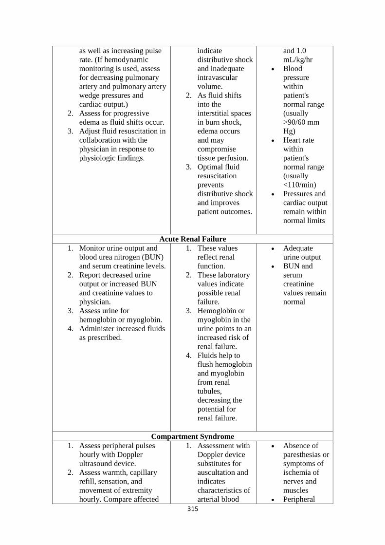
315
as well as increasing pulse
rate. (If hemodynamic
monitoring is used, assess
for decreasing pulmonary
artery and pulmonary artery
wedge pressures and
cardiac output.)
2. Assess for progressive
edema as fluid shifts occur.
3. Adjust fluid resuscitation in
collaboration with the
physician in response to
physiologic findings.
indicate
distributive shock
and inadequate
intravascular
volume.
2. As fluid shifts
into the
interstitial spaces
in burn shock,
edema occurs
and may
compromise
tissue perfusion.
3. Optimal fluid
resuscitation
prevents
distributive shock
and improves
patient outcomes.
and 1.0
mL/kg/hr
Blood
pressure
within
patient's
normal range
(usually
>90/60 mm
Hg)
Heart rate
within
patient's
normal range
(usually
<110/min)
Pressures and
cardiac output
remain within
normal limits
Acute Renal Failure
1. Monitor urine output and
blood urea nitrogen (BUN)
and serum creatinine levels.
2. Report decreased urine
output or increased BUN
and creatinine values to
physician.
3. Assess urine for
hemoglobin or myoglobin.
4. Administer increased fluids
as prescribed.
1. These values
reflect renal
function.
2. These laboratory
values indicate
possible renal
failure.
3. Hemoglobin or
myoglobin in the
urine points to an
increased risk of
renal failure.
4. Fluids help to
flush hemoglobin
and myoglobin
from renal
tubules,
decreasing the
potential for
renal failure.
Adequate
urine output
BUN and
serum
creatinine
values remain
normal
Compartment Syndrome
1. Assess peripheral pulses
hourly with Doppler
ultrasound device.
2. Assess warmth, capillary
refill, sensation, and
movement of extremity
hourly. Compare affected
1. Assessment with
Doppler device
substitutes for
auscultation and
indicates
characteristics of
arterial blood
Absence of
paresthesias or
symptoms of
ischemia of
nerves and
muscles
Peripheral
316
with unaffected extremity.
3. Remove blood pressure cuff
after each reading.
4. Elevate burned extremities.
5. Report loss of pulse or
sensation or presence of
pain to physician
immediately.
6. Prepare to assist with
escharotomies.
flow.
2. These
assessments
indicate
characteristics of
peripheral
perfusion.
3. Cuff may act as a
tourniquet as
extremities swell.
4. Elevation reduces
edema formation.
5. These signs and
symptoms may
indicate
inadequate tissue
perfusion.
6. Escharotomies
relieve the
constriction
caused by
swelling under
circumferential
burns and
improve tissue
perfusion.
pulses
detectable by
Doppler
Curling's Ulcer
1. Assess gastric aspirate for
pH and blood.
2. Assess stools for occult
blood.
3. Administer histamine
blockers and antacids as
prescribed.
1. Acidic pH
indicates need for
antacids or
histamine
blockers. Blood
indicates possible
gastric bleeding.
2. Blood in stools
may indicate
gastric or
duodenal ulcer.
3. Such medications
reduce gastric
acidity and risk
of ulceration.
Absence of
abdominal
distention
Normal bowel
sounds within
48 hours
Gastric
aspirate and
stools do not
contain blood
317
Nursing Process
Care of the Patient during the Acute Phase
Assessment
Continued assessment of the patient during the early weeks after the burn injury
focuses on hemodynamic alterations, wound healing, pain and psychosocial responses,
and early detection of complications. Assessment of respiratory and fluid status
remains the highest priority for detection of potential complications.
The nurse assesses vital signs frequently. Continued assessment of peripheral pulses is
essential for the first few postburn days while edema continues to increase, potentially
damaging peripheral nerves and restricting blood flow. Observation of the
electrocardiogram may give clues to cardiac dysrhythmias resulting from potassium
imbalance, preexisting cardiac disease, or the effects of electrical injury or burn shock.
Assessment of residual gastric volumes and pH in the patient with a nasogastric tube is
also important. Blood in the gastric fluid or in the stools must also be noted and
reported.
Assessment of the burn wound requires an experienced eye, hand, and sense of smell.
Important wound assessment features include size, color, odor, eschar, exudate, abscess
formation under the eschar, epithelial buds (small pearl-like clusters of cells on the
wound surface), bleeding, granulation tissue appearance, status of grafts and donor
sites, and quality of surrounding skin. Any significant changes in the wound are
reported to the physician, because they usually indicate burn wound or systemic sepsis
and require immediate intervention.
Other significant and ongoing assessments focus on pain and psychosocial responses,
daily body weights, caloric intake, general hydration, and serum electrolyte,
hemoglobin, and hematocrit levels. Assessment for excessive bleeding from blood
vessels adjacent to areas of surgical exploration and débridement is necessary as well.
Gerontologic Considerations
In elderly patients, a careful history of preburn medications and preexisting illnesses is
essential. Nursing assessment of the elderly patient with burns should include particular
attention to pulmonary function, response to fluid resuscitation, and signs of mental
confusion or disorientation. Because of lowered resistance, burn wound sepsis and
lethal systemic septicemia are more likely in elderly patients. Furthermore, fever may
not be present in the elderly to signal such events. Therefore, surveillance for other
signs of infection becomes even more important. Nursing care of the elderly patient
with burn injuries promotes early mobilization, aggressive pulmonary care, and
attention to preventing complications.
Diagnosis
Nursing Diagnoses
Based on the assessment data, priority nursing diagnoses in the acute phase of burn care
may include the following:
Excessive fluid volume related to resumption of capillary integrity and fluid
shift from the interstitial to the intravascular compartment
Risk for infection related to loss of skin barrier and impaired immune response
Imbalanced nutrition, less than body requirements, related to hypermetabolism
and wound healing needs
Impaired skin integrity related to open burn wounds
Acute pain related to exposed nerves, wound healing, and treatments
Impaired physical mobility related to burn wound edema, pain, and joint
318
contractures
Ineffective coping related to fear and anxiety, grieving, and forced dependence
on health care providers
Interrupted family processes related to burn injury
Deficient knowledge about the course of burn treatment
Collaborative Problems/Potential Complications
Based on the assessment data, potential complications that may develop in the acute
phase of burn care may include:
Heart failure and pulmonary edema
Sepsis
Acute respiratory failure
Acute respiratory distress syndrome
Visceral damage (electrical burns)
Planning and Goals
The major goals for the patient may include restoration of normal fluid balance,
absence of infection, attainment of anabolic state and normal weight, improved skin
integrity, reduction of pain and discomfort, optimal physical mobility, adequate patient
and family coping, adequate patient and family knowledge of burn treatment, and
absence of complications. Achieving these goals requires a collaborative,
interdisciplinary approach to patient management.
Nursing Interventions
Restoring Normal Fluid Balance
To reduce the risk of fluid overload and consequent heart failure and pulmonary edema,
the nurse closely monitors IV and oral fluid intake, using IV infusion pumps to
minimize the risk of rapid fluid infusion. To monitor changes in fluid status, careful
intake and output and daily weights are obtained. Changes, including those of blood
pressure and pulse rate, are reported to the physician (invasive hemodynamic
monitoring is avoided because of the high risk of infection). Low-dose dopamine to
increase renal perfusion and diuretics may be prescribed to promote increased urine
output. The nurse's role is to administer these medications as prescribed and to monitor
the patient's response.
Preventing Infection
A major part of the nurse's role during the acute phase of burn care is detecting and
preventing infection. The nurse is responsible for providing a clean and safe
environment and for closely scrutinizing the burn wound to detect early signs of
infection. Culture results and white blood cell counts are monitored.
Clean technique is used for wound care procedures. Aseptic technique is used for any
invasive procedures, such as insertion of IV lines and urinary catheters or tracheal
suctioning. Meticulous hand hygiene before and after each patient contact is also an
essential component of preventing infection, even though gloves are worn to provide
care.
The nurse protects the patient from sources of contamination, including other patients,
staff members, visitors, and equipment. Invasive lines and tubing must be routinely
changed according to recommendations of the CDC. Tube feeding reservoirs, ventilator
circuits, and drainage containers are replaced regularly. Fresh flowers, plants, and fresh
fruit baskets are not permitted in the patient's room because of the risk of
319
microorganism growth. Visitors are screened to avoid exposure of the
immunocompromised patient to pathogens.
Patients can inadvertently promote migration of microorganisms from one burned area
to another by touching their wounds or dressings. Bed linens also can spread infection
through either colonization with wound microorganisms or fecal contamination.
Regular bathing of unburned areas and changing of linens can help prevent infection.
Maintaining Adequate Nutrition
Oral fluids should be initiated slowly after bowel sounds resume. The patient's
tolerance is recorded. If vomiting and distention do not occur, fluids may be increased
gradually and the patient may be advanced to a normal diet or to tube feedings.
The nurse collaborates with the dietitian or nutrition support team to plan a protein- and
calorie-rich diet that is acceptable to the patient. Family members may be encouraged
to bring nutritious and favorite foods to the hospital. Milkshakes and sandwiches made
with meat, peanut butter, and cheese may be offered as snacks between meals and late
in the evening. High-calorie nutritional supplements such as Ensure and Resource may
be provided. Caloric intake must be documented. Vitamin and mineral supplements
may be prescribed.
If caloric goals cannot be met by oral feeding, a feeding tube is inserted and used for
continuous or bolus feedings of specific formulas. The volume of residual gastric
secretions should be checked to ensure absorption. Parenteral nutrition may also be
required but should be used only if gastrointestinal function is compromised (see
Chapter 36).
The patient should be weighed each day and his or her weights graphed. The patient
can use this information to set goals for nutritional intake and to monitor weight loss
and gain. Ideally, the patient will lose no more than 5% of preburn weight if aggressive
nutritional management is implemented.
The patient with anorexia requires encouragement and support from the nurse to
increase food intake. The patient's surroundings should be as pleasant as possible at
mealtime. Catering to food preferences and offering high-protein, high-vitamin snacks
are ways of encouraging the patient to increase intake.
Promoting Skin Integrity
Wound care is usually the single most time-consuming element of burn care after the
emergent phase. The physician prescribes the desired topical antibacterial agents and
specific biologic, biosynthetic, or synthetic wound coverings and plans for surgical
excision and grafting. The nurse needs to make astute assessments of wound status, use
creative approaches to wound dressing, and support the patient during the emotionally
distressing and very painful experience of wound care.
The nurse serves as the coordinator of the complex aspects of wound care and dressing
changes for the patient. The nurse must be aware of the rationale and nursing
implications for the various wound management approaches. Nursing functions include
assessing and recording any changes or progress in wound healing and keeping all
members of the health care team informed of changes in the wound or in treatment. A
diagram, updated daily by the nurse responsible for the patient's care, helps inform all
those concerned about the latest wound care procedures in use for the patient.
The nurse also assists the patient and family by providing instruction, support, and
encouragement to take an active part in dressing changes and wound care when
appropriate. Discharge planning needs for wound care are anticipated early in the
course of burn management, and the strengths of the patient and family are assessed
and used in preparing for the patient's eventual discharge and home care.

320
Relieving Pain and Discomfort
Pain measures, discussed earlier, are continued during the acute phase of burn recovery.
Analgesic agents and anxiolytic medications are administered as prescribed. Frequent
assessment of pain and discomfort is essential. To increase its effectiveness, analgesic
medication is provided before the pain becomes severe. Nursing interventions such as
teaching the patient relaxation techniques, giving the patient some control over wound
care and analgesia, and providing frequent reassurance are helpful. Guided imagery
may be effective in altering the patient's perceptions of and responses to pain. Other
pain-relieving approaches include distraction through video programs or video games,
hypnosis, biofeedback, and behavioral modification.
The nurse assesses the patient's sleep patterns daily. Lack of sleep and rest interferes
with healing, comfort, and restoration of energy. If necessary, sedatives are prescribed
on a regular basis in addition to analgesics and anxiolytics.
The nurse works quickly to complete treatments and dressing changes to reduce pain
and discomfort. The patient is encouraged to take analgesic medications before painful
procedures. The patient's response to the medication and other interventions is assessed
and documented.
Healing burn wounds are typically described by patients as itchy and tight. Oral
antipruritic agents, a cool environment, frequent lubrication of the skin with water or a
silica-based lotion, exercise and splinting to prevent skin contracture, and diversional
activities all help promote comfort in this phase.
Promoting Physical Mobility
An early priority is to prevent complications of immobility. Deep breathing, turning,
and proper positioning are essential nursing practices that prevent atelectasis and
pneumonia, control edema, and prevent pressure ulcers and contractures. These
interventions are modified to meet the patient's needs. Low-air-loss and rotation beds
may be useful, and early sitting and ambulation are encouraged. If the lower extremities
are burned, elastic pressure bandages should be applied before the patient is placed in
an upright position. These bandages promote venous return and minimize edema
formation.
The burn wound is in a dynamic state for at least 1 year after wound closure. During
this time, aggressive efforts must be made to prevent contracture and hypertrophic
scarring. Both passive and active range-of-motion exercises are initiated from the day
of admission and are continued after grafting, within prescribed limitations. Splints or
functional devices may be applied to extremities for contracture control. The nurse
monitors the splinted areas for signs of vascular insufficiency and nerve compression.
Strengthening Coping Strategies
In the acute phase of burn care, the patient is facing the reality of the burn trauma and is
grieving over obvious losses. Depression, regression, and manipulative behavior are
common responses of patients who have burn injuries. Withdrawal from participation
in required treatments and regression must be viewed with an understanding that such
behavior may help the patient cope with an enormously stressful event. Although most
patients recover emotionally from a burn injury, some have more difficult
psychological reactions to the injury and its outcomes (Morton, Willebrand, Gerhard, et
al. 2005).
Personality characteristics, rather than the size or severity of the injury, determine the
ability of the patient to cope after burn injury (Kidal, Willebrand, Andersson, et al.,
2004). Difficulty coping along with other psychological stressors often limits the

321
patient's physical and psychological recovery (Fauerbach, Lezotte, & Hills, 2005).
Patients who experience a burn injury tend to have high rates of involvement in risky
behaviors (eg, alcohol and substance abuse, depression) before the injury (Morton et
al., 2005). They may also have poor coping skills. Coping styles and perceived threat of
death at the time of the burn injury are strong predictors of how well the patient
recovers psychologically in the postburn period (Willebrand, Anderson & Ekselius,
2004). Intrusive thoughts of the burn event and reliving it over and over may also occur
and can indicate posttraumatic stress disorder.
Much of the patient's energy goes into maintaining vital physical functions and wound
healing in the early postburn weeks, leaving little emotional energy for coping in a
more effective manner. The nurse can assist the patient to develop effective coping
strategies by setting specific expectations for behavior, promoting truthful
communication to build trust, helping the patient practice appropriate strategies, and
giving positive reinforcement when appropriate. Most importantly, the nurse and all
members of the health care team must demonstrate acceptance of the patient.
The patient frequently vents feelings of anger. At times the anger may be directed
inward because of a sense of guilt, perhaps for causing the fire or even for surviving
when loved ones perished. The anger may be directed outward toward those who
escaped unharmed or toward those who are now providing care. One way to help the
patient handle these emotions is to enlist someone to whom the patient can vent
feelings without fear of retaliation. A nurse, social worker, psychiatric liaison nurse, or
spiritual advisor who is not involved in direct care activities may fill this role
successfully.
Patients with burn injuries are very dependent on health care team members during the
long period of treatment and recovery. However, even when physically unable to
contribute much to self-care, they should be included in decisions regarding care and
encouraged to assert their individuality in terms of preferences and recognition of their
unique identities. As the patient improves in mobility and strength, the nurse works
with the patient to set realistic expectations for self-care, including self-feeding,
assistance with wound care procedures, exercise, and planning for the future. Many
patients respond positively to the use of contractual agreements and other strategies that
recognize their independence and their specific role as part of the health care team
moving toward the goal of self-care. Consultation with psychiatric/mental health care
providers may be helpful to assist the patient in developing effective coping strategies.
Supporting Patient and Family Processes
Family functioning is disrupted with burn injury. One of the nurse's responsibilities is
to support the patient and family and to address their spoken and unspoken concerns.
Family members need to be instructed about ways that they can support the patient as
adaptation to burn trauma occurs. The family also needs support from the health care
team. The burn injury has tremendous psychological, economic, and practical impact
on the patient and family. Referrals for social services or psychological counseling
should be made as appropriate. This support continues into the rehabilitation phase.
Patients who experience major burns are commonly sent to burn centers far from home.
Because burn injuries are sudden and unexpected, family roles are disrupted. Therefore,
both the patient and the family need thorough information about the patient's burn care
and expected course of treatment. Patient and family education begins at the initiation
of burn management. Barriers to learning are assessed and considered in teaching. The
preferred learning styles of both the patient and family are assessed. This information is
used to tailor teaching activities. The nurse assesses the ability of the patient and family
to grasp and cope with the information. Verbal information is supplemented by videos,
322
models, or printed materials if available. Patient and family education is a priority in
the acute and rehabilitation phases.
Nurses must remain sensitive to the possibility of changing family dynamics. It is not
unusual for the provider in the family to be the one who is injured. Roles begin to
change, which adds more stress to the family. In addition, families are often relocated
due to loss of property from the fire. Social services play an integral part in providing
support at this time.
Monitoring and Managing Potential Complications
Heart Failure and Pulmonary Edema
The patient is assessed for fluid overload, which may occur as fluid is mobilized from
the interstitial compartment back into the intravascular compartment. If the cardiac and
renal systems cannot compensate for the excess vascular volume, heart failure and
pulmonary edema may result. The patient is assessed for signs of heart failure,
including decreased cardiac output, oliguria, jugular vein distention, edema, and the
onset of an S3 or S4 heart sound. If invasive hemodynamic monitoring is used,
increasing central venous, pulmonary artery, and wedge pressures indicate increased
fluid volume.
Crackles in the lungs and increased difficulty with respiration may indicate a fluid
buildup in the lungs, which is reported promptly to the physician. In the meantime, the
patient is positioned comfortably, with the head of the bed raised (if not contraindicated
because of other treatments or injuries) to promote lung expansion and gas exchange.
Management of this complication includes providing supplemental oxygen,
administering IV diuretic agents, carefully assessing the patient's response, and
providing vasoactive medications, if indicated.
Sepsis
The signs of early systemic sepsis are subtle and require a high index of suspicion and
very close monitoring of changes in the patient's status. Early signs of sepsis may
include increased temperature, increased pulse rate, widened pulse pressure, and
flushed dry skin in unburned areas. As with many observations of the patient with a
burn injury, one needs to look for patterns or trends in the data. (See Chapter 15 for a
more detailed discussion of septic shock.)
Wound and blood cultures are performed as prescribed, and results are reported to the
physician immediately. The nurse also observes for and reports early signs of sepsis
and promptly intervenes, administering prescribed IV fluids and antibiotics to prevent
septic shock, a complication with a high mortality rate. Antibiotics must be
administered as scheduled to maintain proper blood concentrations. Serum antibiotic
levels are monitored for evidence of maximal effectiveness, and the patient is
monitored for toxic side effects.
Acute Respiratory Failure and Acute Respiratory Distress Syndrome
The patient's respiratory status is monitored closely for increased difficulty in
breathing, change in respiratory pattern, or onset of adventitious (abnormal) sounds.
Typically at this stage, signs and symptoms of injury to the respiratory tract become
apparent. Respiratory failure may follow. As described previously, signs of hypoxia
(decreased oxygen to the tissues), decreased breath sounds, wheezing, tachypnea,
stridor, and sputum tinged with soot (or in some cases containing sloughed tracheal
tissue) are among the many possible findings. Patients receiving mechanical ventilation
must be assessed for a decrease in tidal volume and lung compliance. The key sign of
the onset of ARDS is hypoxemia while receiving 100% oxygen, with decreased lung
323
compliance and significant shunting. The physician should be notified immediately of
deteriorating respiratory status.
Medical management of the patient with acute respiratory failure requires intubation
and mechanical ventilation (if not already in use). If ARDS has developed, higher
oxygen levels, positive end-expiratory pressure, and pressure support are used with
mechanical ventilation to promote gas exchange across the alveolar–capillary
membrane (see Chapter 25).
Visceral Damage
The nurse must be alert to signs of necrosis of visceral organs due to electrical injury.
Tissues affected are usually located between the entrance and exit wounds of the
electrical burn. All patients with electrical burns should undergo cardiac monitoring,
with dysrhythmias being reported to the physician. Careful attention must also be paid
to signs or reports of pain related to deep muscle ischemia. To minimize the severity of
complications, visceral ischemia must be detected as early as possible. In the operating
room, the physician may perform fasciotomies to relieve the swelling and ischemia in
the muscles and fascia and to promote oxygenation of the injured tissues. Because of
the deep incisions involved with fasciotomies, the patient must be monitored carefully
for signs of excessive blood loss and hypovolemia.
Evaluation
Expected Patient Outcomes
Expected patient outcomes may include the following:
Achieves optimal fluid balance
o Maintains intake and output and body weight that correlate with
expected pattern
o Exhibits vital signs and hemodynamic values within designated limits
o Demonstrates increased urine output in response to diuretic and
vasoactive medications
o Has heart rate less than 110 beats/min in normal sinus rhythm
Has no localized or systemic infection
o Has wound culture results showing minimal bacteria
o Has normal urine and sputum culture results
Demonstrates anabolic nutritional status
o Gains weight daily after initial loss secondary to fluid diuresis and no
oral intake of food or fluid
o Shows no signs of protein, vitamin, or mineral deficiencies
o Meets required nutritional needs entirely by oral intake
o Participates in selecting diet containing prescribed nutrients
o Exhibits normal serum protein levels
Demonstrates improved skin integrity
o Exhibits generally intact skin that remains free of infection, pressure,
and injury
o Demonstrates remaining open wound areas that are pink, re-
epithelializing, and free of infection
o Demonstrates donor graft sites that are clean and healing
o Has healed wounds that are soft and smooth
o Demonstrates skin that is lubricated and elastic
Has minimal pain
o Requests analgesic agents before specific wound care procedures or
physical therapy activities

324
o Reports minimal pain
o Gives no physiologic, verbal, or nonverbal cues that pain is moderate or
severe
o Uses pain control measures such as nitrous oxide, relaxation, imagery,
and distraction techniques to cope with and alleviate pain and
discomfort
o Can sleep without being disturbed by pain
o Reports that skin is comfortable, with no pruritus or tightness
Demonstrates optimal physical mobility and function
o Improves range of motion of joints daily
o Demonstrates preinjury range of motion of all joints
o Has no signs of calcification around the joints
o Participates in activities of daily living
Uses appropriate coping strategies to deal with postburn problems
o Verbalizes reactions to burns, therapeutic procedures, losses
o Identifies coping strategies used effectively in previous stressful
situations
o Accepts dependency on health care providers during acute phase
o Verbalizes realistic view of problems resulting from burn injury and
plans for future
o Cooperates with health care providers in required therapy
o Participates in decision making regarding care
o Begins to manage grief over losses resulting from burn injury and
circumstances surrounding injury (eg, death of others, damage to home
or other property)
o States realistic objectives for plastic surgery, further medical
intervention, and results
o Verbalizes realistic abilities and goals
o Displays hopeful attitude toward future
Relates appropriately in patient/family processes
o Patient and family verbalize feelings regarding change in family
interactions
o Family emotionally supports the patient during the hospitalization
o Family states that their own needs are being met
Patient and family verbalize understanding of the treatment course
o State rationales for the various aspects of treatment
o State realistic time period for recovery
Absence of complications
o Lungs clear on auscultation
o Exhibits no dyspnea or orthopnea and can breathe easily when standing,
sitting, and lying down
o Exhibits no S3 or S4 heart sounds or jugular venous distention
o Exhibits adequate urine output
o Exhibits normal blood, sputum, and urine culture results
o Maintains arterial blood gas values or O2 saturation within normal or
acceptable limits
o Has normal lung compliance
o Has no visceral organ damage
o Has stable cardiac rhythm
325
Nursing Process
Care of the Patient during the Rehabilitation Phase
Assessment
Information about the patient's educational level, occupation, leisure activities, cultural
background, religion, and family interactions is obtained early. The patient's self-
concept, mental status, emotional response to the injury and hospitalization, level of
intellectual functioning, previous hospitalizations, response to pain and pain relief
measures, and sleep pattern are also essential components of a comprehensive
assessment. Information about the patient's general selfconcept, self-esteem, and coping
strategies in the past are valuable in addressing emotional needs. Ongoing physical
assessments related to rehabilitation goals include range of motion of affected joints,
functional abilities in activities of daily living, early signs of skin breakdown from
splints or positioning devices, evidence of neuropathies (neurologic damage), activity
tolerance, and quality or condition of healing skin. The patient's participation in care
and ability to demonstrate self-care in such areas as ambulation, eating, wound
cleaning, and applying pressure wraps are documented on a regular basis. In addition to
these assessment parameters, specific complications and treatments require additional
specific assessments; for example, the patient undergoing primary excision requires
postoperative assessment. Recovery from burn injury involves every system of the
body. Therefore, assessment of the patient with a burn injury must be comprehensive
and continuous. Priorities vary at different points during the rehabilitation phase.
Understanding the pathophysiologic responses to burn injury forms the framework for
detecting early progress or signs and symptoms of complications. Early detection leads
to early intervention and enhances the potential for successful rehabilitation.
Diagnosis
Nursing Diagnoses
Based on the assessment data, priority nursing diagnoses in the long-term rehabilitation
phase of burn care may include the following:
Activity intolerance related to pain on exercise, limited joint mobility, muscle
wasting, and limited endurance
Disturbed body image related to altered physical appearance and self-concept
Deficient knowledge about postdischarge home care and follow-up needs
Collaborative Problems/Potential Complications
Based on the assessment data, potential complications that may develop in the
rehabilitation phase include:
Contractures
Inadequate psychological adaptation to burn injury
Planning and Goals
The major goals for the patient include increased participation in activities of daily
living; increased understanding of the injury, treatment, and planned followup care;
adaptation and adjustment to alterations in body image, self-concept, and lifestyle; and
absence of complications.
Nursing Interventions
326
Promoting Activity Tolerance
Nursing interventions that must be carried out according to a strict regimen and the
pain that accompanies movement take their toll on the patient. The patient may become
confused and disoriented and lack the energy to participate optimally in care. The nurse
must schedule care in such a way that the patient has periods of uninterrupted sleep. A
good time for planned patient rest is after the stress of dressing changes and exercise,
while pain interventions and sedatives are still effective. This plan must be
communicated to family members and other care providers.
The patient may have insomnia related to frequent nightmares about the burn injury or
to other fears and anxieties about the outcome of the injury. The nurse listens to and
reassures the patient and administers hypnotic agents, as prescribed, to promote sleep.
Reducing metabolic stress by relieving pain, preventing chilling or fever, and
promoting the physical integrity of all body systems help the patient conserve energy
for therapeutic activities and wound healing.
The nurse incorporates physical therapy exercises in the patient's care to prevent
muscle atrophy and to maintain the mobility required for daily activities. The patient's
activity tolerance, strength, and endurance gradually increase if activity occurs over
increasingly longer periods. Fatigue, fever, and pain tolerance are monitored and used
to determine the amount of activity to be encouraged on a daily basis. Activities such as
family visits and recreational or play therapy (eg, video games, radio, TV) can provide
diversion, improve the patient's outlook, and increase tolerance for physical activity. In
elderly patients and those with chronic illnesses and disabilities, rehabilitation must
take into account preexisting functional abilities and limitations.
Improving Body Image and Self-Concept
Patients who have survived burn injuries frequently suffer profound losses. These
include not only a loss of body image due to disfigurement but also losses of personal
property, homes, loved ones, and ability to work. They lack the benefit of anticipatory
grief often seen in a patient who is approaching surgery or dealing with the terminal
illness of a loved one.
As care progresses, the patient who is recovering from burns becomes aware of daily
improvement and begins to exhibit basic concerns: Will I be disfigured or be disabled?
How long will I be in the hospital? What about my job and family? Will I ever be
independent again? How can I pay for my care? Was my burn the result of my
carelessness? As the patient expresses such concerns, the nurse must take time to listen
and to provide realistic support. The nurse can refer the patient to a support group, such
as those usually available at regional burn centers or through organizations such as the
Phoenix Society. Through participation in such groups, the patient will meet others
with similar experiences and learn coping strategies to help him or her deal with losses.
Interaction with other burn survivors allows the patient to see that adaptation to the
burn injury is possible. If a support group is not available, visits from other survivors of
burn injuries can be helpful to the patient coping with such a traumatic injury.
A major responsibility of the nurse is to constantly assess the patient's psychosocial
reactions. Questions to consider include the following: What are the patient's fears and
concerns? Does the patient fear loss of control of care, independence, or sanity itself? Is
the patient afraid of rejection by family and loved ones? Does he or she fear being
unable to cope with pain or physical appearance? Does the patient have concerns about
sexuality, including sexual function? Being aware of these anxieties and understanding
the basis of the patient's fears enable the nurse to provide support and to cooperate with
other members of the health care team in developing a plan to help the patient deal with
these feelings.

327
When caring for a patient with a burn injury, the nurse needs to be aware that there are
prejudices and misunderstandings in society about those who are viewed as different.
Opportunities and accommodations available to others are often denied those who are
disfigured. Such amenities include social participation, employment, prestige, various
roles, and status. The health care team must actively promote a healthy body image and
self-concept in patients with burn injuries so that they can accept or challenge others'
perceptions of those who are disfigured or disabled. Survivors themselves must show
others who they are, how they function, and how they want to be treated.
The nurse can help patients practice their responses to people who may stare or inquire
about their injury once they are discharged from the hospital. The nurse can help
patients build self-esteem by recognizing their uniqueness—for example, with small
gestures such as providing a birthday cake, combing the patient's hair before visiting
hours, giving information about the availability of a cosmetician to enhance
appearance, and teaching the patient ways to direct attention away from a disfigured
body to the self within. Consultants such as psychologists, social workers, vocational
counselors, and teachers are valuable participants in assisting burn patients to regain
their self-esteem.
Monitoring and Managing Potential Complications
Contractures
With early and aggressive physical and occupational therapy, contractures are rarely a
long-term complication. However, surgical intervention is indicated if a full range of
motion in the burn patient is not achieved. (See Chapter 11 for a discussion of
prevention of contractures.)
Impaired Psychological Adaptation to the Burn Injury
Some patients, particularly those with limited coping skills or psychological function or
a history of psychiatric problems before the burn injury, may not achieve adequate
psychological adaptation to the burn injury. Psychological counseling or psychiatric
referral may be made to assess the patient's emotional status, to help the patient develop
coping skills, and to intervene if major psychological issues or ineffective coping is
identified.
Promoting Home and Community-Based Care
Teaching Patients Self-Care
As the inpatient phase of recovery becomes shorter, the focus of rehabilitative
interventions is directed toward outpatient care or care in a rehabilitation center. In the
long term, much of the care of healing burns will be performed by the patient and
others at home. Throughout the phases of burn care, efforts are made to prepare the
patient and family for the care that will continue at home. They are instructed about the
measures and procedures that they will need to perform. For example, patients
commonly have small areas of clean, open wounds that are healing slowly. They are
instructed to wash these areas daily with mild soap and water and to apply the
prescribed topical agent or dressing.
In addition to instructions about wound care, patients and families require careful
written and verbal instructions about pain management, nutrition, and prevention of
complications. Information about specific exercises and use of pressure garments and
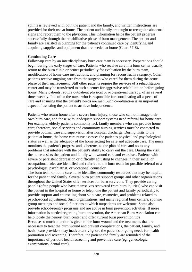
328
splints is reviewed with both the patient and the family, and written instructions are
provided for their use at home. The patient and family are taught to recognize abnormal
signs and report them to the physician. This information helps the patient progress
successfully through the rehabilitative phase of burn management. The patient and
family are assisted in planning for the patient's continued care by identifying and
acquiring supplies and equipment that are needed at home (Chart 57-8).
Continuing Care
Follow-up care by an interdisciplinary burn care team is necessary. Preparations should
begin during the early stages of care. Patients who receive care in a burn center usually
return to the burn clinic or center periodically for evaluation by the burn team,
modification of home care instructions, and planning for reconstructive surgery. Other
patients receive ongoing care from the surgeon who cared for them during the acute
phase of their management. Still other patients require the services of a rehabilitation
center and may be transferred to such a center for aggressive rehabilitation before going
home. Many patients require outpatient physical or occupational therapy, often several
times weekly. It is often the nurse who is responsible for coordinating all aspects of
care and ensuring that the patient's needs are met. Such coordination is an important
aspect of assisting the patient to achieve independence.
Patients who return home after a severe burn injury, those who cannot manage their
own burn care, and those with inadequate support systems need referral for home care.
For example, elderly patients commonly lack family members who can provide home
care; therefore, social services and community nursing services must be contacted to
provide optimal care and supervision after hospital discharge. During visits to the
patient at home, the home care nurse assesses the patient's physical and psychological
status as well as the adequacy of the home setting for safe and adequate care. The nurse
monitors the patient's progress and adherence to the plan of care and notes any
problems that interfere with the patient's ability to carry out the care. During the visit,
the nurse assists the patient and family with wound care and exercises. Patients with
severe or persistent depression or difficulty adjusting to changes in their social or
occupational roles are identified and referred to the burn team for possible referral to a
psychologist, psychiatrist, or vocational counselor.
The burn team or home care nurse identifies community resources that may be helpful
for the patient and family. Several burn patient support groups and other organizations
throughout the United States offer services for burn survivors. They provide caring
people (often people who have themselves recovered from burn injuries) who can visit
the patient in the hospital or home or telephone the patient and family periodically to
provide support and counseling about skin care, cosmetics, and problems related to
psychosocial adjustment. Such organizations, and many regional burn centers, sponsor
group meetings and social functions at which outpatients are welcome. Some also
provide school-reentry programs and are active in burn prevention activities. If more
information is needed regarding burn prevention, the American Burn Association can
help locate the nearest burn center and offer current burn prevention tips
Because so much attention is given to the burn wound and the treatments that are
necessary to treat the burn wound and prevent complications, the patient, family, and
health care providers may inadvertently ignore the patient's ongoing needs for health
promotion and screening. Therefore, the patient and family are reminded of the
importance of periodic health screening and preventive care (eg, gynecologic
examinations, dental care).

329
Evaluation
Expected Patient Outcomes
Expected patient outcomes may include the following:
Demonstrates activity tolerance required for desired daily activities
o Obtains adequate sleep daily
o Reports absence of nightmares or sleep disturbances
o Shows gradually increasing tolerance and endurance in physical
activities
o Can concentrate during conversations
o Has energy available to sustain desired daily activities
Adapts to altered body image
o Verbalizes accurate description of alterations in body image and accepts
physical appearance
o Demonstrates interest in resources that may improve body appearance
and function
o Uses cosmetics, wigs, and prostheses as desired to achieve acceptable
appearance
o Socializes with significant others, peers, and usual social group
o Seeks and achieves return to role in family, school, and community as a
contributing member
Demonstrates knowledge of required self-care and follow-up care
o Describes surgical procedures and treatments accurately
o Verbalizes detailed plan for follow-up care
o Demonstrates ability to perform wound care and prescribed exercises
o Returns for follow-up appointments as scheduled
o Identifies resource people and agencies to con tact for specific problems
Exhibits no complications
o Demonstrates full range of motion
o Shows no signs of withdrawal or depression
o Displays no psychotic behaviors

330
Chapter 58
Assessment and Management of Patients with Eye and Vision
Disorders
External Structures of the Eye
Cross-Section of the Eye

331
Assessment and Evaluation of Vision
• Ocular history
• Visual acuity
–Snellen chart
• Record each eye
• 20/20 means the patient can read the ―20‖ line at a distance of 20 feet
• Finger count or hand motion
Diagnostic Evaluation
• Ophthalmoscopy
–Direct and indirect
–Examines the cornea, lens and retina
• Tonometry
–Measures intraocular pressure
Impaired Vision
• Refractive errors
–Can be corrected by lenses which focus light rays on the retina
• Myopia: nearsighted
• Hyperopia: farsighted
• Astigmatism: distortion due to irregularity of the cornea. Due to refractive error in
which light rays are spread over a diffuse area rather than sharply focused on the retina,
a condition caused by differences in the curvature of the cornea and lens
Glaucoma
— A group of ocular conditions in which damage to the optic nerve is related to
increased intraocular pressure (IOP) caused by congestion of the aqueous humor
— The leading cause of blindness in adults in the U.S.
— Incidence increases with age

332
Risk factors
— Family history of glaucoma
— Older age
— Diabetes mellitus
— Cardiovascular disease
— Nearsightedness (myopia)
— Eye trauma
— Prolonged use of topical or systemic corticosteroids
Pathophysiology of Glaucoma
— In glaucoma, aqueous production and drainage are not in balance.
— When aqueous outflow is blocked, pressure builds up in the eye.
— Increased IOP causes irreversible mechanical and/or ischemic damage to the optic
nerve.
Types of glaucoma:
1. Open-angle
2. Angle-closure (pupillary block) glaucoma
3. Congenital glaucomas
4. Glaucoma secondary to other conditions
Clinical Manifestations
• ―Silent thief‖ of vision; unaware of the condition until there is significant vision loss;
peripheral vision loss, blurring, halos, difficulty focusing, difficulty adjusting eyes to
low lighting
• May also have aching or discomfort around eyes or headache
• Diagnosis: Tonometry to assess IOP
Treatment
• Goal is to prevent further optic nerve damage
• Maintain IOP within a range unlikely to cause damage
• Pharmacologic therapy
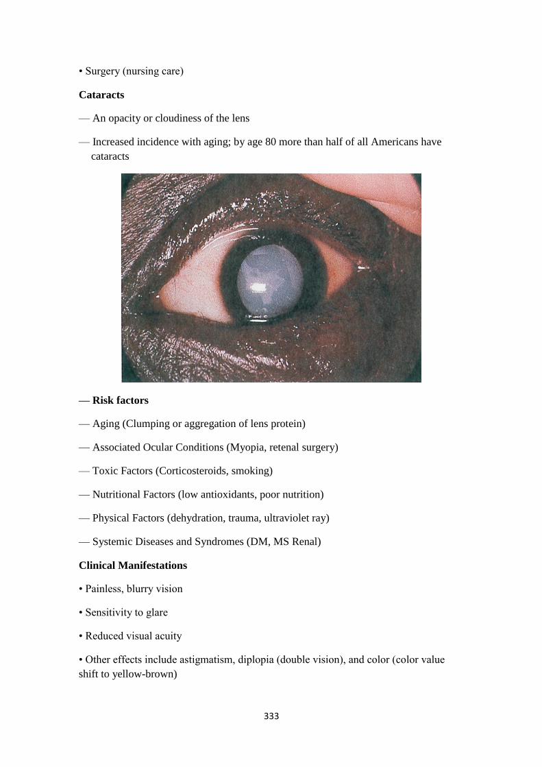
333
• Surgery (nursing care)
Cataracts
— An opacity or cloudiness of the lens
— Increased incidence with aging; by age 80 more than half of all Americans have
cataracts
— Risk factors
— Aging (Clumping or aggregation of lens protein)
— Associated Ocular Conditions (Myopia, retenal surgery)
— Toxic Factors (Corticosteroids, smoking)
— Nutritional Factors (low antioxidants, poor nutrition)
— Physical Factors (dehydration, trauma, ultraviolet ray)
— Systemic Diseases and Syndromes (DM, MS Renal)
Clinical Manifestations
• Painless, blurry vision
• Sensitivity to glare
• Reduced visual acuity
• Other effects include astigmatism, diplopia (double vision), and color (color value
shift to yellow-brown)
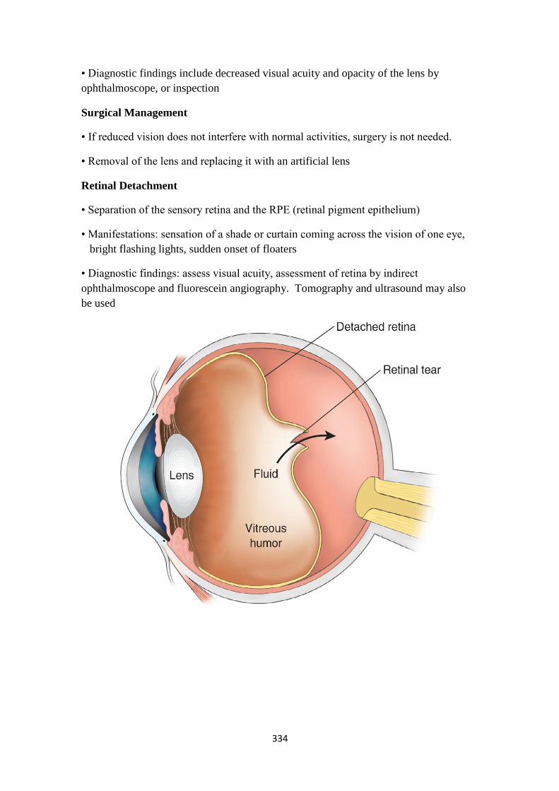
334
• Diagnostic findings include decreased visual acuity and opacity of the lens by
ophthalmoscope, or inspection
Surgical Management
• If reduced vision does not interfere with normal activities, surgery is not needed.
• Removal of the lens and replacing it with an artificial lens
Retinal Detachment
• Separation of the sensory retina and the RPE (retinal pigment epithelium)
• Manifestations: sensation of a shade or curtain coming across the vision of one eye,
bright flashing lights, sudden onset of floaters
• Diagnostic findings: assess visual acuity, assessment of retina by indirect
ophthalmoscope and fluorescein angiography. Tomography and ultrasound may also
be used
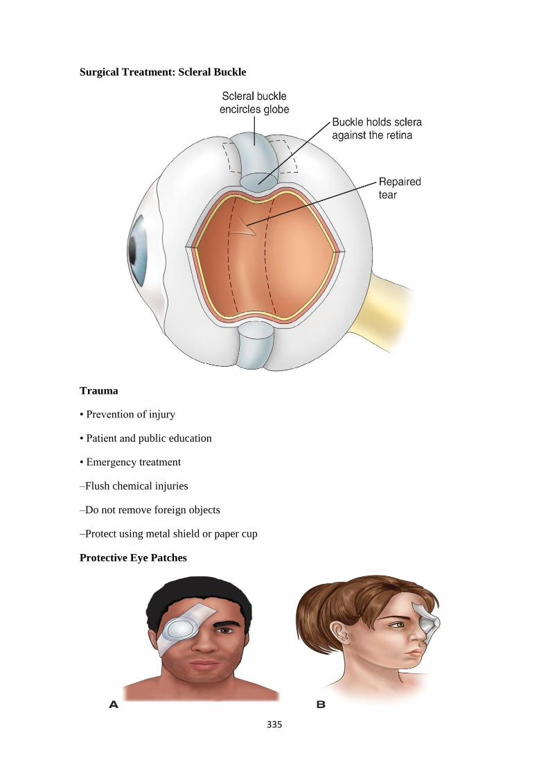
335
Surgical Treatment: Scleral Buckle
Trauma
• Prevention of injury
• Patient and public education
• Emergency treatment
–Flush chemical injuries
–Do not remove foreign objects
–Protect using metal shield or paper cup
Protective Eye Patches
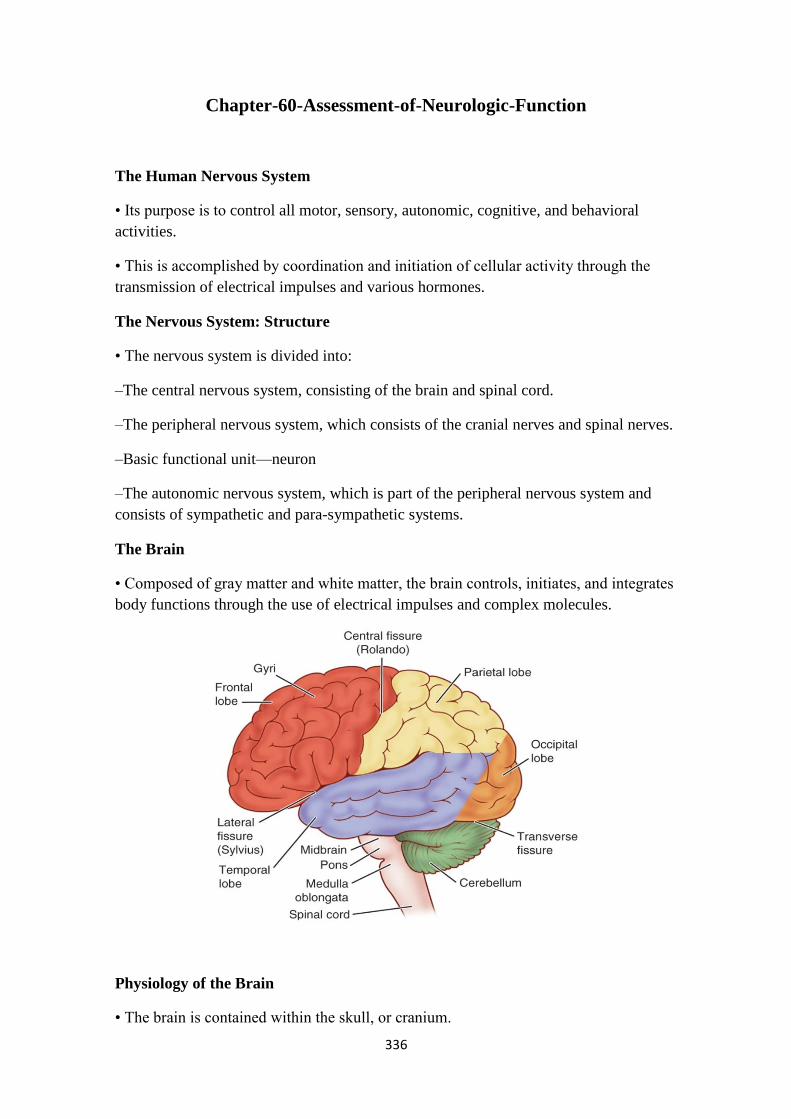
336
Chapter-60-Assessment-of-Neurologic-Function
The Human Nervous System
• Its purpose is to control all motor, sensory, autonomic, cognitive, and behavioral
activities.
• This is accomplished by coordination and initiation of cellular activity through the
transmission of electrical impulses and various hormones.
The Nervous System: Structure
• The nervous system is divided into:
–The central nervous system, consisting of the brain and spinal cord.
–The peripheral nervous system, which consists of the cranial nerves and spinal nerves.
–Basic functional unit—neuron
–The autonomic nervous system, which is part of the peripheral nervous system and
consists of sympathetic and para-sympathetic systems.
The Brain
• Composed of gray matter and white matter, the brain controls, initiates, and integrates
body functions through the use of electrical impulses and complex molecules.
Physiology of the Brain
• The brain is contained within the skull, or cranium.

337
• Three coverings of the brain, called the meninges. They are the dura mater, arachnoid
mater, and pia mater.
The Brain Hemispheres
• The right side receives information from and controls the left side of the body.
Specializes in perception of physical environment, art, music, nonverbal
communication, spiritual aspects.
• The left receives information from and controls the right side of the body. Specializes
in analysis, calculation, problem solving, verbal communication, interpretation,
language, reading, & writing.
The Spinal Cord
• A continuation of the brain stem.
• Exits the skull through the foramen magnum, an opening in the base of the skull.
Cerebrospinal Fluid
• Provides for shock absorption and bathes the brain and spinal cord.
Peripheral Nervous System: Cranial Nerves
• Twelve pairs of cranial nerves have sensory, motor, or mixed functions.
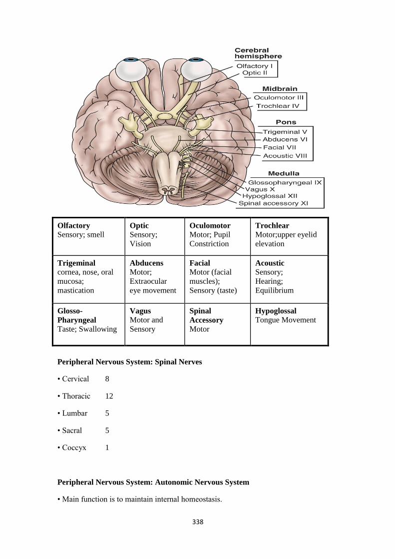
338
Olfactory Sensory; smell
Optic Sensory;
Vision
Oculomotor Motor; Pupil
Constriction
Trochlear Motor;upper eyelid
elevation
Trigeminal cornea, nose, oral
mucosa;
mastication
Abducens Motor;
Extraocular
eye movement
Facial Motor (facial
muscles);
Sensory (taste)
Acoustic Sensory;
Hearing;
Equilibrium
Glosso-
Pharyngeal Taste; Swallowing
Vagus Motor and
Sensory
Spinal
Accessory Motor
Hypoglossal Tongue Movement
Peripheral Nervous System: Spinal Nerves
• Cervical 8
• Thoracic 12
• Lumbar 5
• Sacral 5
• Coccyx 1
Peripheral Nervous System: Autonomic Nervous System
• Main function is to maintain internal homeostasis.
339
• Two subdivisions of ANS:
–The sympathetic system (activated by stress, prepares body for ―fight or flight‖
response).
–The parasympathetic system (conserves, restores, and maintains vital body
functions, slowing heart rate, increasing gastrointestinal activity, and activating
bowel and bladder evacuation).
Neurologic Assessment: Health History
• Pain
• Seizures
• Dizziness (abnormal sensation of imbalance or movement) and vertigo (illusion of
movement, usually rotation)
• Visual disturbances
• Weakness
• Abnormal sensations
Neurologic Assessment
• Cerebral function; mental status, intellectual function thought content, emotional
status, perception, motor ability, and language ability
–Note the impact of any neurologic impairment on lifestyle and patient abilities and
limitations
–Agnosia is the inability to interpret or recognize objects seen through the special
senses.
• Motor system; posture, gait, muscle tone and strength, coordination and balance,
Romberg test
• Sensory system; tactile sensation, superficial pain, vibration and position sense
• Reflexes; DTRs, abdominal, and plantar (Babinski)
The Romberg test is a screening test for balance. The patient stands with feet
together and arms at the side, first with eyes open and then with both eyes closed for
20 to 30 seconds. The examiner stands close to reassure the patient of support if he
or she begins to fall. Slight swaying is normal, but a loss of balance is abnormal and
is considered a positive Romberg test.

340
Techniques Eliciting Major Reflexes
(A) Biceps reflex. (B) Triceps reflex. (C) Patellar reflex. (D) Ankle or Achilles reflex.
(E) Babinski response.
Figure Used to Record Muscle Strength
• 5, full range of motion against gravity and resistance; 4, full range of motion against
gravity and a moderate amount of resistance; 3, full range of motion against gravity
only; 2, full range of motion when gravity is eliminated; 1, a weak muscle contraction
when muscle is palpated, but no movement; and 0, complete paralysis.
341
Gerontological Considerations
• Important to distinguish normal aging changes from abnormal changes
• Determine previous mental status for comparison. Assess mental status carefully to
distinguish delirium from dementia.
• Normal changes may include:
–Losses in strength and agility; changes in gait, posture and balance; slowed reaction
times and decreased reflexes; visual and hearing alterations; deceased sense of taste and
smell; dulling of tactile sensations; changes in the perception of pain; and decreased
thermoregulatory ability
Pupil Size
• Normal range: 2 - 6 mm
–Drugs: pinpoint pupils
–Increased intracranial pressure: pupils begin to dilate
• Dilated and fixed, poor prognosis
Glascow Coma Scale
• Score BEST response in each category
• Highest score = 15 (normal)
• Lowest score = 3 (deep coma)

342
Eye Opening Spontaneous 4
To Voice 3
To Pain 2
None 1
Best Verbal Oriented 5
Confused 4
Inappropriate Words 3
Incomprehensible Sounds 2
None 1
Best Motor Obeys Commands 6
Localizes Pain 5
Withdraws to Pain 4
Flexion to Pain (decorticate) 3
Extension to Pain (decerebrate) 2
None 1
Diagnostic Tests
• Computed tomography (CT)
• Magnetic resonance imaging (MRI)
• Cerebral angiography
• Myelography
• Noninvasive carotid flow studies
• Transcranial doppler
• Electroencephalography (EEG)
• Electromyography (EMG)
• Lumbar puncture and analysis of cerebrospinal fluid
343
Chapter 61
Management of Patients with Neurologic Dysfunction
Altered Level of Consciousness (LOC)
• LOC: is apparent in the patient who is not oriented, does not follow commands, or
needs persistent stimuli to achieve a state of alertness.
• Level of responsiveness and consciousness is the most important indicator of the
patient's condition
• LOC is a continuum from normal alertness and full cognition (consciousness) to coma
• Altered LOC is not the disorder but the result of a pathology
• Coma: unconsciousness, unarousable unresponsiveness
• Akinetic mutism: unresponsiveness to the environment, makes no movement or sound
but sometimes opens eyes
• Persistent vegetative state: devoid of cognitive function but has sleep-wake cycles
• Locked-in syndrome: inability to move or respond except for eye movements due to a
lesion affecting the pons
Coma is a clinical state of unarousable unresponsiveness in which there are no
purposeful responses to internal or external stimuli, although nonpurposeful
responses to painful stimuli and brain stem reflexes may be present (Hickey, 2003).
The duration of coma is usually limited to 2 to 4 weeks.
Altered LOC is not a disorder itself; rather, it is a result of multiple pathophysiologic
phenomena. The cause may be neurologic (head injury, stroke), toxicologic (drug
overdose, alcohol intoxication), or metabolic (hepatic or renal failure, diabetic
ketoacidosis).
Nursing Process: The Care of the Patient with Altered Level of Consciousness—
Assessment
• Assess verbal response and orientation
• Alertness
• Motor responses
• Respiratory status
• Eye signs
• Reflexes
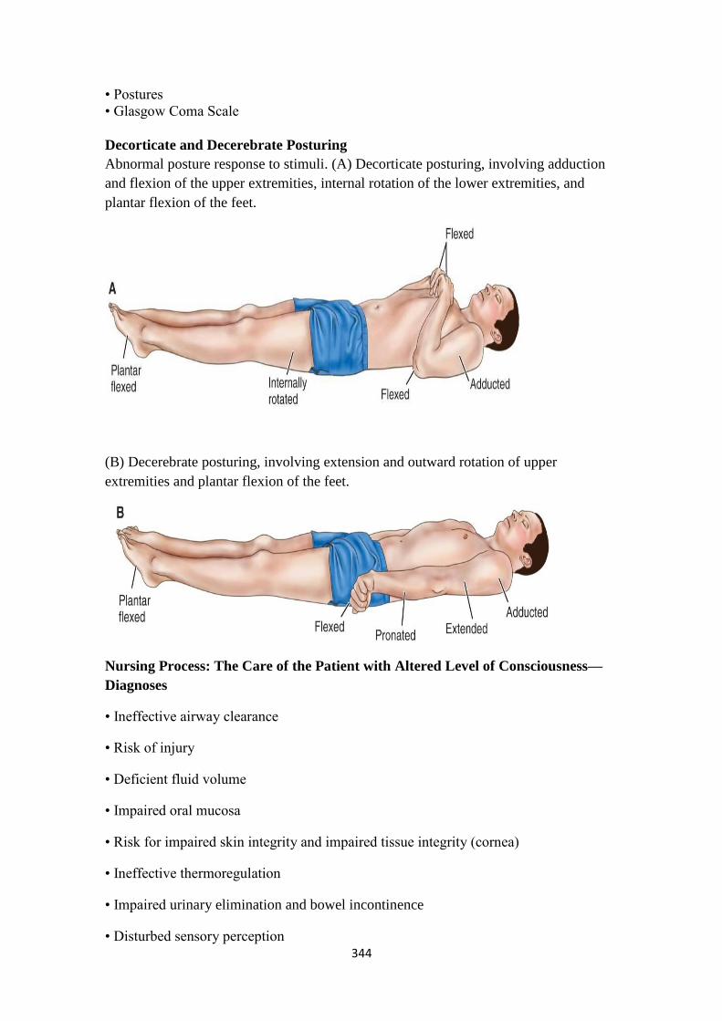
344
• Postures
• Glasgow Coma Scale
Decorticate and Decerebrate Posturing
Abnormal posture response to stimuli. (A) Decorticate posturing, involving adduction
and flexion of the upper extremities, internal rotation of the lower extremities, and
plantar flexion of the feet.
(B) Decerebrate posturing, involving extension and outward rotation of upper
extremities and plantar flexion of the feet.
Nursing Process: The Care of the Patient with Altered Level of Consciousness—
Diagnoses
• Ineffective airway clearance
• Risk of injury
• Deficient fluid volume
• Impaired oral mucosa
• Risk for impaired skin integrity and impaired tissue integrity (cornea)
• Ineffective thermoregulation
• Impaired urinary elimination and bowel incontinence
• Disturbed sensory perception
345
• Interrupted family processes
Collaborative Problems/Potential Complications
• Respiratory distress or failure
• Pneumonia
• Aspiration
• Pressure ulcer
• Deep vein thrombosis (DVT)
• Contractures
Nursing Process: The Care of the Patient with Altered Level of Consciousness—
Planning
• Goals may include:
–Maintenance of clear airway
–Protection from injury
–Attainment of fluid volume balance
–Maintenance of skin integrity
–Absence of corneal irritation
–Effective thermoregulation
–Accurate perception of environmental stimuli
–Maintenance of intact family or support system
–Absence of complications
Interventions
• A major nursing goal is to compensate for the patient's loss of protective reflexes and
to assume responsibility for total patient care. Protection also includes maintaining the
patient‘s dignity and privacy.
1. Maintaining an airway
–Frequent monitoring of respiratory status including auscultation of lung sounds
–Positioning to promote accumulation of secretions and prevent obstruction of upper
airway—HOB elevated 30°, lateral or semiprone position
–Suctioning, oral hygiene, and Chest Physiotherapy

346
2. Maintaining Tissue Integrity
• Assess skin frequently, especially areas with high potential for breakdown
• Frequent turning; use turning schedule
• Careful positioning in correct body alignment
• Passive ROM
• Use of splints, foam boots, trochanter rolls, and specialty beds as needed
• Clean eyes with cotton balls moistened with saline
• Use artificial tears as prescribed
• Measures to protect eyes; use eye patches cautiously as the cornea may contact patch
• Frequent oral care
3. Maintaining fluid status
–Assess fluid status by examining tissue turgor and mucosa, lab data, and I&O.
–Administer IVs, tube feedings, and fluids via feeding tube as required— monitor
ordered rate of IV fluids carefully.
4. Maintaining body temperature
–Adjust environment and cover patient appropriately.
–If temperature is elevated, use minimum amount of bedding, administer
acetaminophen, use hypothermia blanket, give a cooling sponge bath, and allow fan to
blow over patient to increase cooling.
–Monitor temperature frequently and use measures to prevent shivering.
5. Promoting Bowel and Bladder Function
• Assess for urinary retention and urinary incontinence
• May require indwelling or intermittent catherization
• Bladder-training program
• Assess for abdominal distention, potential constipation, and bowel incontinence
• Monitor bowel movements
• Promote elimination with stool softeners, glycerin suppositories, or enemas as
indicated
• Diarrhea may result from infection, medications, or hyperosmolar fluids
347
6. Sensory Stimulation and Communication
• Talk to and touch patient and encourage family to talk to and touch the patient
• Maintain normal day night pattern of activity
• Orient the patient frequently
• Note: When arousing from coma, a patient may experience a period of agitation;
minimize stimulation at this time
• Programs for sensory stimulation
• Allow family to ventilate and provide support
• Reinforce and provide and consistent information to family
• Referral to support groups and services for family
Increased Intracranial Pressure
• Monro-Kellie hypothesis: because of limited space in the skull, an increase in any one
of components of the skull—brain tissue, blood, and CSF—will cause a change in the
volume of the others
• Compensation to maintain a normal ICP of 10–20 mm Hg is normally accomplished
by shifting or displacing CSF
• Elevated ICP is most commonly associated with head injury, it also may be seen as a
secondary effect in other conditions, such as brain tumors, subarachnoid hemorrhage,
and toxic and viral encephalopathies
• Increased ICP decreases cerebral perfusion and causes ischemia, cell death, and
(further) edema
• Brain tissues may shift through the dura and result in herniation
• CO2 plays a role; decreased CO2 results in vasoconstriction, increased CO2 results in
vasodilatation
Manifestations of Increased ICP: Early
• Changes in LOC
• Any change in condition
–Restlessness, confusion, increasing drowsiness, increased respiratory effort,
purposeless movements
348
• Pupillary changes and impaired ocular movements
• Weakness in one extremity or one side
• Headache—constant, increasing in intensity or aggravated by movement or straining
Manifestations of Increased ICP: Late
• Respiratory and vasomotor changes
• VS: Increase in systolic blood pressure, widening of pulse pressure, and slowing of
the heart rate; pulse may fluctuate rapidly from tachycardia to bradycardia; temperature
increase
–Cushing‘s triad: bradycardia, hypertension, bradypnea
• Projectile vomiting
• Further deterioration of LOC; stupor (reacting only to loud or painful stimuli) to coma
• Hemiplegia, decortication, decerebration, or flaccidity
• Respiratory pattern alterations including Cheyne-Stokes breathing and respiratory
arrest
• Loss of brainstem reflexes—pupil, gag, corneal, and swallowing
Nursing Process: The Care of the Patient with Increased Intracranial Pressure—
Assessment
• Frequent and ongoing neurologic assessment
• Evaluate neurologic status as completely as possible
• Glasgow Coma Scale
• Pupil checks
• Assessment of selected cranial nerves
• Frequent vital signs
• Assessment of intracranial pressure

349
ICP Monitoring
Intracranial Pressure Waves
Nursing Process: The Care of the Patient with Increased Intracranial Pressure—
Diagnoses
• Ineffective airway clearance
• Ineffective breathing pattern
• Ineffective cerebral perfusion

350
• Deficient fluid volume related to fluid restriction
• Risk for infection related to ICP monitoring
Collaborative Problems/Potential Complications
• Brainstem herniation
• Diabetes insipidus
• SIADH
• Infection
Nursing Process: The Care of the Patient with Increased Intracranial Pressure—
Planning
• Major goals may include:
–Maintaining a Patent Airway
–Achieving an Adequate Breathing Pattern
–Preventing Infection
–Optimizing Cerebral Tissue Perfusion
–Maintaining Negative Fluid Balance
–Absence of complications
Interventions
• Frequent monitoring of respiratory status and lung sounds and measures to maintain a
patent airway (suction is contraindicated)
• Position with head in neutral position and elevation of HOB 0–60° to promote venous
drainage
• Avoid hip flexion, Valsalva maneuver, abdominal distention, or other stimuli that may
increase ICP
• Maintain a calm, quiet atmosphere and protect patient from stress
• Monitor fluid status carefully; every hour I&O during acute phase
• Use strict aseptic technique for management of ICP monitoring system
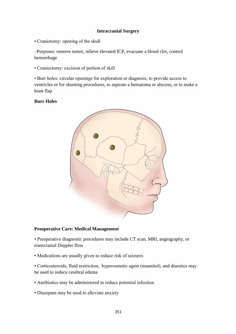
351
Intracranial Surgery
• Craniotomy: opening of the skull
–Purposes: remove tumor, relieve elevated ICP, evacuate a blood clot, control
hemorrhage
• Craniectomy: excision of portion of skill
• Burr holes: circular openings for exploration or diagnosis, to provide access to
ventricles or for shunting procedures, to aspirate a hematoma or abscess, or to make a
bone flap
Burr Holes
Preoperative Care: Medical Management
• Preoperative diagnostic procedures may include CT scan, MRI, angiography, or
transcranial Doppler flow
• Medications are usually given to reduce risk of seizures
• Corticosteroids, fluid restriction, hyperosmotic agent (mannitol), and diuretics may
be used to reduce cerebral edema
• Antibiotics may be administered to reduce potential infection
• Diazepam may be used to alleviate anxiety
352
Preoperative Care: Nursing Management
• Obtain baseline neurologic assessment
• Assess patient and family understanding of and preparation for surgery.
• Provide information, reassurance, and support
Postoperative Care
• Postoperative care is aimed at detecting and reducing cerebral edema, relieving pain,
preventing seizures, monitoring ICP, and neurologic status.
• The patient may be intubated and have arterial and central venous lines.
Nursing Process: The Care of the Patient Undergoing Intracranial Surgery:
Assessment
• Careful, frequent monitoring of respiratory function including ABGs
• Monitor VS and LOC frequently; note any potential signs of increasing ICP
• Assess dressing and for evidence of bleeding or CSF drainage
• Monitor for potential seizures; if seizures occur, carefully record and report these
• Monitor for signs and symptoms of complications
• Monitor fluid status and laboratory data
Diagnoses
• Ineffective cerebral tissue perfusion
• Risk for imbalanced body temperature
• Potential for impaired gas exchange
• Disturbed sensory perception
• Body image disturbance
• Impaired communication (aphasia)
• Risk for impaired skin integrity
• Impaired physical mobility
Collaborative Problems/Potential Complications
• Increased ICP
• Bleeding and hypovolemic shock
353
• Fluid and electrolyte disturbances
• Infection
• Seizures
Planning
• Major goals may include:
–Improved tissue perfusion
–Adequate thermoregulation
–Normal ventilation and gas exchange
–Ability to cope with sensory deprivation
–Adaptation to changes in body image
–Absence of complications
Maintaining Cerebral Perfusion
• Monitor respiratory status; even slight hypoxia or hypercapnia can effect cerebral
perfusion
• Assess VS and neurologic status every 15 minutes to every hour
• Strategies to reduce cerebral edema; cerebral edema peaks 24–36 hours
• Strategies to control factors that increase ICP
• Avoid extreme head rotation
• Head of bed may be flat or elevated 30° according to needs related to the surgery and
surgeon preference
Interventions
1. Regulating temperature
–Cover patient appropriately.
–Treat high temperature elevations vigorously; apply ice bags, use hypothermia
blanket, administer prescribed acetaminophen.
2. Improving gas exchange
–Turn and reposition every 2 hours.
–Encourage deep breathing and incentive spirometry.
354
–Suction or encourage coughing cautiously as needed (suctioning and coughing
increase ICP).
–Humidification of oxygen may help loosen secretions.
3. Sensory deprivation
– Periorbital edema may impair vision, announce presence to avoid startling the patient;
cool compresses over eyes and elevation of HOB may be used to reduce edema if not
contraindicated.
4. Enhancing self-image
–Encourage verbalization.
–Encourage social interaction and social support.
–Attention to grooming.
–Cover head with turban and, later, a wig.
5. Monitor I&O, weight, blood glucose, serum and urine electrolyte levels, and
osmolality and urine specific gravity.
6. Preventing infections
–Assess incision for signs of hematoma or infection.
–Assess for potential CSF leak.
–Instruct patient to avoid coughing, sneezing, or nose blowing, which may increase the
risk of CSF leakage.
–Use strict aseptic technique.
• Patient teaching for self-care
Seizures
• Abnormal episodes of motor, sensory, autonomic, or psychic activity (or a
combination of these) resulting from a sudden, abnormal, uncontrolled electrical
discharge from cerebral neurons
• Classification of seizures Partial seizures: begin in one part of the brain
• Simple partial: consciousness remains intact
• Complex partial: impairment of consciousness
– Generalized seizures: involve the whole brain
355
Specific Causes of Seizures
• Cerebrovascular disease
• Hypoxemia
• Fever (childhood)
• Head injury
• Hypertension
• Central nervous system infections
• Metabolic and toxic conditions
• Brain tumor
• Drug and alcohol withdrawal
• Allergies
Plan of Care for a Patient Experiencing a Seizure
• Observation and documentation of patient signs and symptoms before, during, and
after seizure
• Nursing actions during seizure for patient safety and protection
• After seizure care to prevent complications
Nursing Care after the Seizure
• Keep the patient on one side to prevent aspiration. Make sure the airway is patent.
• There is usually a period of confusion after a grand mal seizure.
• A short apneic period may occur during or immediately after a generalized seizure.
• The patient, on awakening, should be reoriented to the environment.
• If the patient becomes agitated after a seizure, use calm persuasion and gentle
restraint.

356
Nursing Care during a Seizure
• Provide privacy and protect the patient from curious onlookers. (The patient who has
an aura [warning of an impending seizure] may have time to seek a safe, private place.)
• Ease the patient to the floor, if possible.
• Protect the head with a pad to prevent injury (from striking
a hard surface).
• Loosen constrictive clothing.
• Push aside any furniture that may injure the patient during the seizure.
• If the patient is in bed, remove pillows and raise side rails.
• If an aura precedes the seizure, insert an oral airway to reduce the possibility of the
patient's biting the tongue or cheek.
• Do not attempt to open jaws that are clenched in a spasm or to insert anything. Broken
teeth and injury to the lips and tongue may result from such an action.
• No attempt should be made to restrain the patient during the seizure, because
muscular contractions are strong and restraint can produce injury.
• If possible, place the patient on one side with head flexed forward, which allows the
tongue to fall forward and facilitates drainage of saliva and mucus. If suction is
available, use it if necessary to clear secretions.
Status Epilepticus
• Status epilepticus (acute prolonged seizure activity) is a series of generalized seizures
that occur without full recovery of consciousness between attacks
357
Chapter 61
Nursing Process
The Patient with an Altered Level of Consciousness Assessment
Assessment of the patient with an altered LOC depends on the patient's circumstances,
but clinicians often start by assessing the verbal response. Determining the patient's
orientation to time, person, and place assesses verbal response. The patient is asked to
identify the day, date, or season of the year and to identify where he or she is or to
identify the clinicians, family members, or visitors present. Other questions such as,
―Who is the president?‖ or ―What is the next holiday?‖ may be helpful in determining
the patient's processing of information in the environment. (Verbal response cannot be
evaluated if the patient is intubated or has a tracheostomy, and this should be clearly
documented.)
Alertness is measured by the patient's ability to open the eyes spontaneously or in
response to a vocal or noxious stimulus (pressure or pain). Patients with severe
neurologic dysfunction cannot do this. The nurse assesses for periorbital edema
(swelling around the eyes) or trauma, which may prevent the patient from opening the
eyes, and documents any such condition that interferes with eye opening.
Motor response includes spontaneous, purposeful movement (eg, the awake patient can
move all four extremities with equal strength on command), movement only in
response to painful stimuli, or abnormal posturing (Hickey, 2003; Seidel, Ball, Dains,
et al., 2003). If the patient is not responding to commands, the motor response is tested
by applying a painful stimulus (firm but gentle pressure) to the nailbed or by squeezing
a muscle. If the patient attempts to push away or withdraw, the response is recorded as
purposeful or appropriate (―patient withdraws to painful stimuli‖). This response is
considered purposeful if the patient can cross the midline from one side of the body to
the other in response to painful stimuli. An inappropriate or nonpurposeful response is
random and aimless. Posturing may be decorticate or decerebrate (Fig. 61-1; see also
Chapter 60). The most severe neurologic impairment results in flaccidity. The motor
response cannot be elicited if the patient has been administered pharmacologic
paralyzing agents.
In addition to LOC, the nurse monitors parameters such as respiratory status, eye signs,
and reflexes on an ongoing basis. Table 61-1 summarizes the assessment and the
clinical significance of the findings. Body functions (circulation, respiration,
elimination, fluid and electrolyte balance) are examined in a systematic and ongoing
manner.
Diagnosis
Nursing Diagnoses
Based on the assessment data, the major nursing diagnoses may include the following:
Ineffective airway clearance related to altered LOC
Risk of injury related to decreased LOC
Deficient fluid volume related to inability to take fluids by mouth
Impaired oral mucous membrane related to mouth-breathing, absence of
pharyngeal reflex, and altered fluid intake
Risk for impaired skin integrity related to immobility
Impaired tissue integrity of cornea related to diminished or absent corneal reflex
Ineffective thermoregulation related to damage to hypothalamic center
Impaired urinary elimination (incontinence or retention) related to impairment
in neurologic sensing and control

358
Bowel incontinence related to impairment in neurologic sensing and control and
also related to changes in nutritional delivery methods
Disturbed sensory perception related to neurologic impairment
Interrupted family processes related to health crisis
Collaborative Problems/Potential Complications
Based on the assessment data, potential complications may include:
Respiratory distress or failure
Pneumonia
Aspiration
Pressure ulcer
Deep vein thrombosis (DVT)
Contractures
Planning and Goals
The goals of care for the patient with altered LOC include maintenance of a clear
airway, protection from injury, attainment of fluid volume balance, achievement of
intact oral mucous membranes, maintenance of normal skin integrity, absence of
corneal irritation, attainment of effective thermoregulation, and effective urinary
elimination. Additional goals include bowel continence, accurate perception of
environmental stimuli, maintenance of intact family or support system, and absence of
complications (Rice et al., 2005).
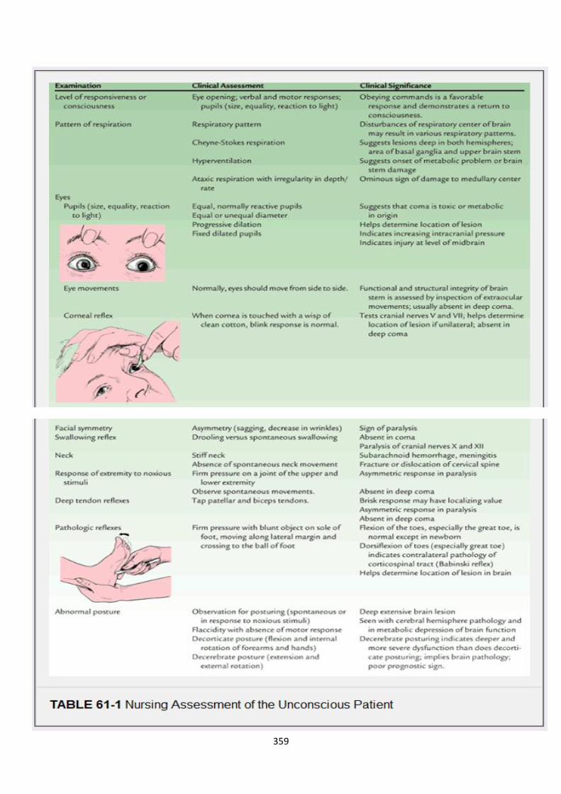
359
360
Because the unconscious patient's protective reflexes are impaired, the quality of
nursing care provided literally may mean the difference between life and death. The
nurse must assume responsibility for the patient until the basic reflexes (coughing,
blinking, and swallowing) return and the patient becomes conscious and oriented.
Therefore, the major nursing goal is to compensate for the absence of these protective
reflexes.
Nursing Interventions
Maintaining the Airway
The most important consideration in managing the patient with altered LOC is to
establish an adequate airway and ensure ventilation. Obstruction of the airway is a risk
because the epiglottis and tongue may relax, occluding the oropharynx, or the patient
may aspirate vomitus or nasopharyngeal secretions.
The accumulation of secretions in the pharynx presents a serious problem. Because the
patient cannot swallow and lacks pharyngeal reflexes, these secretions must be
removed to eliminate the danger of aspiration. Elevating the head of the bed to 30
degrees helps prevent aspiration. Positioning the patient in a lateral or semiprone
position also helps, because it permits the jaw and tongue to fall forward, thus
promoting drainage of secretions.
Positioning alone is not always adequate, however. Suctioning and oral hygiene may be
required. Suctioning is performed to remove secretions from the posterior pharynx and
upper trachea. Before and after suctioning, the patient is hyperoxygenated and
adequately ventilated to prevent hypoxia (Hickey, 2003). In addition to these
interventions, chest physiotherapy and postural drainage may be initiated to promote
pulmonary hygiene, unless contraindicated by the patient's underlying condition. The
chest should be auscultated at least every 8 hours to detect adventitious breath sounds
or absence of breath sounds.
Despite these measures, or because of the severity of impairment, the patient with
altered LOC often requires intubation and mechanical ventilation. Nursing actions for
the mechanically ventilated patient include maintaining the patency of the endotracheal
tube or tracheostomy, providing frequent oral care, monitoring arterial blood gas
measurements, and maintaining ventilator settings (see Chapter 25).
Protecting the Patient
For the protection of the patient, side rails are padded. Two rails are kept in the raised
position during the day and three at night; however, raising all four side rails is
considered a restraint by the Joint Commission on Accreditation of Healthcare
Organizations. Care should be taken to prevent injury from invasive lines and
equipment, and other potential sources of injury should be identified, such as restraints,
tight dressings, environmental irritants, damp bedding or dressings, and tubes and
drains.
Protection also includes protecting the patient's dignity during altered LOC. Simple
measures such as providing privacy and speaking to the patient during nursing care
activities preserve the patient's dignity. Not speaking negatively about the patient's
condition or prognosis is also important, because patients in a light coma may be able
to hear. The comatose patient has an increased need for advocacy, and the nurse is
responsible for seeing that these advocacy needs are met (Hickey, 2003).

361
Maintaining Fluid Balance and Managing Nutritional Needs
Hydration status is assessed by examining tissue turgor and mucous membranes,
assessing intake and output trends, and analyzing laboratory data. Fluid needs are met
initially by administering the required IV fluids. However, IV solutions (and blood
component therapy) for patients with intracranial conditions must be administered
slowly. If they are administered
too rapidly, they can increase ICP. The quantity of fluids administered may be
restricted to minimize the possibility of cerebral edema.
If the patient does not recover quickly and sufficiently enough to take adequate fluids
and calories by mouth, a feeding or gastrostomy tube will be inserted for the
administration of fluids and enteral feedings (Dudek, 2006; Worthington, 2004).
Providing Mouth Care
The mouth is inspected for dryness, inflammation, and crusting. The unconscious
patient requires conscientious oral care, because there is a risk of parotitis if the mouth
is not kept scrupulously clean. The mouth is cleansed and rinsed carefully to remove
secretions and crusts and to keep the mucous membranes moist. A thin coating of
petrolatum on the lips prevents drying, cracking, and encrustations. If the patient has an
endotracheal tube, the tube should be moved to the opposite side of the mouth daily to
prevent ulceration of the mouth and lips.
Maintaining Skin and Joint Integrity
Preventing skin breakdown requires continuing nursing assessment and intervention.
Special attention is given to unconscious patients, because they cannot respond to
external stimuli. Assessment includes a regular schedule of turning to avoid pressure,
which can cause breakdown and necrosis of the skin. Turning also provides kinesthetic
(sensation of movement), proprioceptive (awareness of position), and vestibular
(equilibrium) stimulation. After turning, the patient is carefully repositioned to prevent
ischemic necrosis over pressure areas. Dragging or pulling the patient up in bed must
be avoided, because this creates a shearing force and friction on the skin surface (see
Chapter 11).
Maintaining correct body position is important; equally important is passive exercise of
the extremities to prevent contractures. The use of splints or foam boots aids in the
prevention of foot drop and eliminates the pressure of bedding on the toes. The use of
trochanter rolls to support the hip joints keeps the legs in proper alignment. The arms
are in abduction, the fingers lightly flexed, and the hands in slight supination. The heels
of the feet are assessed for pressure areas. Specialty beds, such as fluidized or low-air-
loss beds, may be used to decrease pressure on bony prominences (Hickey, 2003).
Preserving Corneal Integrity
Some unconscious patients have their eyes open and have inadequate or absent corneal
reflexes. The cornea may become irritated, dried out, or scratched, leading to
ulceration. The eyes may be cleansed with cotton balls moistened with sterile normal
saline to remove debris and discharge (Hickey, 2003). If artificial tears are prescribed,
they may be instilled every 2 hours. Periorbital edema (swelling around the eyes) often
occurs after cranial surgery. Cold compresses may be prescribed, and care must be
exerted to avoid contact with the cornea. Eye patches should be used cautiously
because of the potential for corneal abrasion from contact with the patch.

362
Maintaining Body Temperature
High fever in the unconscious patient may be caused by infection of the respiratory or
urinary tract, drug reactions, or damage to the hypothalamic temperature-regulating
center. A slight elevation of temperature may be caused by dehydration. The
environment can be adjusted, depending on the patient's condition, to promote a normal
body temperature. If body temperature is elevated, a minimum amount of bedding—a
sheet, small drape, or towel—is used. The room may be cooled to 18.3°C (65°F).
However, if the patient is elderly and does not have an elevated temperature, a warmer
environment is needed.
Because of damage to the temperature center in the brain or severe intracranial
infection, unconscious patients often develop very high temperatures. Such temperature
elevations must be controlled, because the increased metabolic demands of the brain
can exceed cerebral circulation and oxygenation, resulting in cerebral deterioration
(Diringer, 2004; Hickey, 2003). Persistent hyperthermia with no identified clinical
source of infection indicates brain stem damage and a poor prognosis.
Strategies for reducing fever include:
Removing all bedding over the patient (with the possible exception of a light
sheet or small drape)
Administering acetaminophen as prescribed
Giving cool sponge baths and allowing an electric fan to blow over the patient
to increase surface cooling
Using a hypothermia blanket
Frequent temperature monitoring is indicated to assess the patient's response to
the therapy and to prevent an excessive decrease in temperature and shivering.
Preventing Urinary Retention
The patient with an altered LOC is often incontinent or has urinary retention. The
bladder is palpated or scanned at intervals to determine whether urinary retention is
present, because a full bladder may be an overlooked cause of overflow incontinence.
A portable bladder ultrasound instrument is a useful tool in bladder management and
retraining programs (O'Farrell, Vandervoort, Bisnaire, et al., 2001).
If the patient is not voiding, an indwelling urinary catheter is inserted and connected to
a closed drainage system. A catheter may also be inserted during the acute phase of
illness to monitor urinary output. Because catheters are a major factor in causing
urinary tract infection, the patient is observed for fever and cloudy urine. The area
around the urethral orifice is inspected for drainage. The urinary catheter is usually
removed if the patient has a stable cardiovascular system and if no diuresis, sepsis, or
voiding dysfunction existed before the onset of coma. Although many unconscious
patients urinate spontaneously after catheter removal, the bladder should be palpated or
scanned with a portable ultrasound device periodically for urinary retention (O'Farrell
et al., 2001). An intermittent catheterization program may be initiated to ensure
complete emptying of the bladder at intervals, if indicated.
An external catheter (condom catheter) for the male patient and absorbent pads for the
female patient can be used for unconscious patients who can urinate spontaneously
although involuntarily. As soon as consciousness is regained, a bladder-training
program is initiated (Hickey, 2003). The incontinent patient is monitored frequently for
skin irritation and skin breakdown. Appropriate skin care is implemented to prevent
these complications.
363
Promoting Bowel Function
The abdomen is assessed for distention by listening for bowel sounds and measuring
the girth of the abdomen with a tape measure. There is a risk for diarrhea from
infection, antibiotics, and hyperosmolar fluids. Frequent loose stools may also occur
with fecal impaction. Commercial fecal collection bags are available for patients with
fecal incontinence.
Immobility and lack of dietary fiber can cause constipation. The nurse monitors the
number and consistency of bowel movements and performs a rectal examination for
signs of fecal impaction. Stool softeners may be prescribed and can be administered
with tube feedings. To facilitate bowel emptying, a glycerin suppository may be
indicated. The patient may require an enema every other day to empty the lower colon.
Providing Sensory Stimulation
Once increased ICP is not a problem, sensory stimulation can help overcome the
profound sensory deprivation of the unconscious patient. Efforts are made to restore the
sense of daily rhythm by maintaining usual day and night patterns for activity and
sleep. The nurse touches and talks to the patient and encourages family members and
friends to do so. Communication is extremely important and includes touching the
patient and spending enough time with the patient to become sensitive to his or her
needs. It is also important to avoid making any negative comments about the patient's
status or prognosis in the patient's presence.
The nurse orients the patient to time and place at least once every 8 hours. Sounds from
the patient's usual environment may be introduced using a tape recorder. Family
members can read to the patient from a favorite book and may suggest radio and
television programs that the patient previously enjoyed as a means of enriching the
environment and providing familiar input (Hickey, 2003).
When arousing from coma, many patients experience a period of agitation, indicating
that they are becoming more aware of their surroundings but still cannot react or
communicate in an appropriate fashion. Although this is disturbing for many family
members, it is actually a positive clinical sign. At this time, it is necessary to minimize
stimulation by limiting background noises, having only one person speak to the patient
at a time, giving the patient a longer period of time to respond, and allowing for
frequent rest or quiet times. After the patient has regained consciousness, videotaped
family or social events may assist the patient in recognizing family and friends and
allow him or her to experience missed events.
Various programs of structured sensory stimulation for patients with brain injury have
been developed to improve outcomes. Although these are controversial programs with
inconsistent results, some research supports the concept of providing structured
stimulation (Davis & Gimenez, 2003).
Meeting the Family's Needs
The family of the patient with altered LOC may be thrown into a sudden state of crisis
and go through the process of severe anxiety, denial, anger, remorse, grief, and
reconciliation. Depending on the disorder that caused the altered LOC and the extent of
the patient's recovery, the family may be unprepared for the changes in the cognitive
and physical status of their loved one. If the patient has significant residual deficits, the
family may require considerable time, assistance, and support to come to terms with
these changes. To help family members mobilize resources and coping skills, the nurse
reinforces and clarifies information about the patient's condition, permits the family to
be involved in care, and listens to and encourages ventilation of feelings and concerns
while supporting decision making about posthospitalization management and
364
placement (Bond, Draeger, Mandleco, et al., 2003). Families may benefit from
participation in support groups offered through the hospital, rehabilitation facility, or
community organizations.
In some circumstances, the family may need to face the death of their loved one. The
patient with a neurologic disorder is often pronounced brain dead before the heart stops
beating. The term brain death describes irreversible loss of all functions of the entire
brain, including the brain stem (Booth, Boone, Tomlinson, et al., 2004). The term may
be misleading to the family because, although brain function has ceased, the patient
appears to be alive, with the heart rate and blood pressure sustained by vasoactive
medications and breathing continued by mechanical ventilation. When discussing a
patient who is brain dead with family members, it is important to provide accurate,
timely, understandable, and consistent information (Henneman & Karras, 2004). End-
of-life care is discussed in Chapter 17.
Monitoring and Managing Potential Complications
Pneumonia, aspiration, and respiratory failure are potential complications in any patient
who has a depressed LOC and who cannot protect the airway or turn, cough, and take
deep breaths. The longer the period of unconsciousness, the greater the risk for
pulmonary complications.
Vital signs and respiratory function are monitored closely to detect any signs of
respiratory failure or distress. Total blood count and arterial blood gas measurements
are assessed to determine whether there are adequate red blood cells to carry oxygen
and whether ventilation is effective. Chest physiotherapy and suctioning are initiated to
prevent respiratory complications such as pneumonia. If pneumonia develops, cultures
are obtained to identify the organism so that appropriate antibiotics can be
administered.
The patient with altered LOC is monitored closely for evidence of impaired skin
integrity, and strategies to prevent skin breakdown and pressure ulcers are continued
through all phases of care, including hospitalization, rehabilitation, and home care.
Factors that contribute to impaired skin integrity (eg, incontinence, inadequate dietary
intake, pressure on bony prominences, edema) are addressed. If pressure ulcers
develop, strategies to promote healing are undertaken. Care is taken to prevent bacterial
contamination of pressure ulcers, which may lead to sepsis and septic shock.
Assessment and management of pressure ulcers are discussed in Chapter 11.
The patient should also be monitored for signs and symptoms of deep vein thrombosis
(DVT). Patients who develop DVT are at risk for pulmonary embolism. Prophylaxis
such as subcutaneous heparin or low-molecular-weight heparin (Fragmin, Orgaran)
should be prescribed if not contraindicated (Kurtoglu, Yanar, Bilsel, et al., 2004).
Thigh-high elastic compression stockings or pneumatic compression devices should
also be prescribed to reduce the risk for clot formation. The nurse observes for signs
and symptoms of DVT.
Evaluation
Expected Patient Outcomes
Expected patient outcomes may include the following:
Maintains clear airway and demonstrates appropriate breath sounds
Experiences no injuries
Attains or maintains adequate fluid balance
o Has no clinical signs or symptoms of dehydration
o Demonstrates normal range of serum electrolytes
o Has no clinical signs or symptoms of overhydration

365
Achieves healthy oral mucous membranes
Maintains normal skin integrity
Has no corneal irritation
Attains or maintains thermoregulation
Has no urinary retention
Has no diarrhea or fecal impaction
Receives appropriate sensory stimulation
Family members cope with crisis
o Verbalize fears and concerns
o Participate in patient's care and provide sensory stimulation by talking
and touching
Patient is free of complications
o Has arterial blood gas values or O2 saturation levels within normal range
o Displays no signs or symptoms of pneumonia
o Exhibits intact skin over pressure areas
o Does not develop DVT or pulmonary embolism (PE)
Nursing Process
The Patient with Increased Intracranial Pressure
Assessment
Initial assessment of the patient with increased ICP includes obtaining a history of
events leading to the present illness and the pertinent past medical history. It is usually
necessary to obtain this information from family or friends. The neurologic
examination should be as complete as the patient's condition allows. It includes an
evaluation of mental status, LOC, cranial nerve function, cerebellar function (balance
and coordination), reflexes, and motor and sensory function. Because the patient is
critically ill, ongoing assessment is more focused, including pupil checks, assessment
of selected cranial nerves, frequent measurements of vital signs and ICP, and use of the
Glasgow Coma Scale. Assessment of the patient with altered LOC is summarized in
Table 61-1.
Diagnosis
Nursing Diagnoses
Based on the assessment data, the major nursing diagnoses for patients with increased
ICP include the following:
Ineffective airway clearance related to diminished protective reflexes (cough,
gag)
Ineffective breathing patterns related to neurologic dysfunction (brain stem
compression, structural displacement)
Ineffective cerebral tissue perfusion related to the effects of increased ICP
Deficient fluid volume related to fluid restriction
Risk for infection related to ICP monitoring system (fiberoptic or
intraventricular catheter)
Other relevant nursing diagnoses are included in the section on altered LOC.
Collaborative Problems/Potential Complications
Based on the assessment data, potential complications include:

366
Brain stem herniation
Diabetes insipidus
SIADH
Planning and Goals
The goals for the patient include maintenance of a patent airway, normalization of
respiration, adequate cerebral tissue perfusion through reduction in ICP, restoration of
fluid balance, absence of infection, and absence of complications.
Nursing Interventions
Maintaining a Patent Airway
The patency of the airway is assessed. Secretions that are obstructing the airway must
be suctioned with care, because transient elevations of ICP occur with suctioning
(Hickey, 2003). The patient is hyperoxygenated before and after suctioning to maintain
adequate oxygenation. Hypoxia caused by poor oxygenation leads to cerebral ischemia
and edema. Coughing is discouraged, because coughing and straining increase ICP.
The lung fields are auscultated at least every 8 hours to determine the presence of
adventitious sounds or any areas of congestion. Elevating the head of the bed may aid
in clearing secretions and improve venous drainage of the brain.
Achieving an Adequate Breathing Pattern
The patient must be monitored constantly for respiratory irregularities. Increased
pressure on the frontal lobes or deep midline structures may result in Cheyne-Stokes
respirations, whereas pressure in the midbrain can cause hyperventilation. If the lower
portion of the brain stem (the pons and medulla) is involved, respirations become
irregular and eventually cease.
If hyperventilation therapy is deemed appropriate to reduce ICP (by causing cerebral
vasoconstriction and a decrease in cerebral blood volume), the nurse collaborates with
the respiratory therapist in monitoring the PaCO2, which is usually maintained at 30 to
35 mm Hg (Hickey, 2003).
A neurologic observation record (Fig. 61-6) is maintained, and all observations are
made in relation to the patient's baseline condition. Repeated assessments of the patient
are made (sometimes minute by minute) so that improvement or deterioration may be
noted immediately. If the patient's condition deteriorates, preparations are made for
surgical intervention.
Optimizing Cerebral Tissue Perfusion
In addition to ongoing nursing assessment, strategies are initiated to reduce factors
contributing to the elevation of ICP (Table 61-2).
Proper positioning helps to reduce ICP. The head is kept in a neutral (midline) position,
maintained with the use of a cervical collar if necessary, to promote venous drainage.
Elevation of the head is maintained at 0 to 60 degrees to aid in venous drainage unless
otherwise prescribed (Fan, 2004). Extreme rotation of the neck and flexion of the neck
are avoided, because compression or distortion of the jugular veins increases ICP.
Extreme hip flexion is also avoided, because this position causes an increase in intra-
abdominal and intrathoracic pressures, which can produce an increase in ICP.
Relatively minor changes in position can significantly affect ICP (Fan, 2004). If
monitoring reveals that turning the patient raises ICP, rotating beds, turning sheets, and
holding the patient's head during turning may minimize the stimuli that increase ICP.

367
The Valsalva maneuver, which can be produced by straining at defecation or even
moving in bed, raises ICP and is to be avoided. Stool softeners may be prescribed. If
the patient is alert and able to eat, a diet high in fiber may be indicated. Abdominal
distention, which increases intra-abdominal and intrathoracic pressure and ICP, should
be noted. Enemas and cathartics are avoided if possible. When moving or being turned
in bed, the patient can be instructed to exhale (which opens the glottis) to avoid the
Valsalva maneuver.
Mechanical ventilation presents unique problems for the patient with increased ICP.
Before suctioning, the patient should be preoxygenated and briefly hyperventilated
using 100% oxygen on the ventilator (Hickey, 2003). Suctioning should not last longer
than 15 seconds. High levels of positive end-expiratory pressure (PEEP) are avoided,
because they may decrease venous return to the heart and decrease venous drainage
from the brain through increased intrathoracic pressure (Hickey, 2003).
Activities that increase ICP, as indicated by changes in waveforms, should be avoided
if possible. Spacing of nursing interventions may prevent transient increases in ICP.
During nursing interventions, the ICP should not increase more than 25 mm Hg, and it
should return to baseline levels within 5 minutes. Patients with increased ICP should
not demonstrate a significant increase in pressure or change in the ICP waveform.
Patients with the potential for a significant increase in ICP may need sedation and a
paralytic agent before initiation of nursing activities (Hickey, 2003).
Emotional stress and frequent arousal from sleep are avoided. A calm atmosphere is
maintained. Environmental stimuli (eg, noise, conversation) should be minimal.
Maintaining Negative Fluid Balance
The administration of various osmotic and loop diuretics is part of the treatment
protocol to reduce ICP. Corticosteroids may be used to reduce cerebral edema (except
when it results from trauma), and fluids may be restricted (Brain Trauma Foundation,
2003). All of these treatment modalities promote dehydration.
Skin turgor, mucous membranes, urine output, and serum and urine osmolality are
monitored to assess fluid status. If IV fluids are prescribed, the nurse ensures that they
are administered at a slow to moderate rate with an IV infusion pump, to prevent too-
rapid administration and avoid overhydration. For the patient receiving mannitol, the
nurse observes for the possible development of heart failure and pulmonary edema,
because the intent of treatment is to promote a shift of fluid from the intracellular
compartment to the intravascular system, thus controlling cerebral edema.
For patients undergoing dehydrating procedures, vital signs, including blood pressure,
must be monitored to assess fluid volume status. An indwelling urinary catheter is
inserted to permit assessment of renal function and fluid status. During the acute phase,
urine output is monitored hourly. An output greater than 250 mL/hour for 2 consecutive
hours may indicate the onset of diabetes insipidus (Suarez, 2004). These patients also
need careful oral hygiene, because mouth dryness occurs with dehydration. Frequently
rinsing the mouth with nondrying solutions, lubricating the lips, and removing
encrustations relieve dryness and promote comfort.
Preventing Infection
Risk for infection is greatest when ICP is monitored with an intraventricular catheter,
and the risk of infection increases with the duration of the monitoring (Park, Garton,
Kocan, et al., 2004). Most health care facilities have written protocols for managing
these systems and maintaining their sterility; strict adherence to the protocols is
essential.

368
Aseptic technique must be used when managing the system and changing the
ventricular drainage bag. The drainage system is also checked for loose connections,
because they can cause leakage and contamination of the CSF as well as inaccurate
readings of ICP. The nurse observes the character of the CSF drainage and reports
observations of increasing cloudiness or blood. The patient is monitored for signs and
symptoms of meningitis: fever, chills, nuchal (neck) rigidity, and increasing or
persistent headache. (See Chapter 64 for a discussion of meningitis.)
Monitoring and Managing Potential Complications
The primary complication of increased ICP is brain herniation resulting in death (see
Fig. 61-2). Nursing management focuses on detecting early signs of increasing ICP,
because medical interventions are usually ineffective once later signs develop. Frequent
neurologic assessments and documentation and analysis of trends will reveal the subtle
changes that may indicate increasing ICP.
Detecting Early Indications of Increasing Intracranial Pressure
The nurse assesses for and immediately reports any of the following early signs or
symptoms of increasing ICP:
Disorientation, restlessness, increased respiratory effort, purposeless
movements, and mental confusion; these are early clinical indications of
increasing ICP because the brain cells responsible for cognition are extremely
sensitive to decreased oxygenation
Pupillary changes and impaired extraocular movements; these occur as the
increasing pressure displaces the brain against the oculomotor and optic nerves
(cranial nerves II, III, IV, and VI), which arise from the midbrain and brain
stem (see Chapter 60)
Weakness in one extremity or on one side of the body; this occurs as increasing
ICP compresses the pyramidal tracts
Headache that is constant, increasing in intensity, and aggravated by movement
or straining; this occurs as increasing ICP causes pressure and stretching of
venous and arterial vessels in the base of the brain
Detecting Later Indications of Increasing ICP
As ICP increases, the patient's condition worsens, as manifested by the following signs
and symptoms:
The LOC continues to deteriorate until the patient is comatose.
The pulse rate and respiratory rate decrease or become erratic, and the blood
pressure and temperature increase. The pulse pressure (the difference between
the systolic and the diastolic pressures) widens. The pulse fluctuates rapidly,
varying from bradycardia to tachycardia.
Altered respiratory patterns develop, including Cheyne-Stokes breathing
(rhythmic waxing and waning of rate and depth of respirations alternating with
brief periods of apnea) and ataxic breathing (irregular breathing with a random
sequence of deep and shallow breaths).
Projectile vomiting may occur with increased pressure on the reflex center in
the medulla.
Hemiplegia or decorticate or decerebrate posturing may develop as pressure on
the brain stem increases; bilateral flaccidity occurs before death.

369
Loss of brain stem reflexes, including pupillary, corneal, gag, and swallowing
reflexes, is an ominous sign of approaching death.
Monitoring Intracranial Pressure
Because clinical assessment is not always a reliable guide in recognizing increased ICP,
especially in comatose patients, monitoring of ICP and cerebral oxygenation is an
essential part of management (Hickey, 2003). ICP is monitored closely for continuous
elevation or significant increase over baseline. The trend of ICP measurements over
time is an important indication of the patient's underlying status. Vital signs are
assessed when an increase in ICP is noted.
Strict aseptic technique is used when handling any part of the monitoring system. The
insertion site is inspected for signs of infection. Temperature, pulse, and respirations
are closely monitored for systemic signs of infection. All connections and stopcocks are
checked for leaks, because even small leaks can distort pressure readings and lead to
infection.
When ICP is monitored with a fluid system, the transducer is calibrated at a particular
reference point, usually 2.5 cm (1 inch) above the ear with the patient in the supine
position; this point corresponds to the level of the foramen of Monro (Fig. 61-7). CSF
pressure readings depend on the patient's position. For subsequent pressure readings,
the head should be in the same position relative to the transducer. Fiberoptic catheters
are calibrated before insertion and do not require further referencing; they do not
require the head of the bed to be at a specific position to obtain an accurate reading.
Whenever technology is associated with patient management, the nurse must be certain
that the technological equipment is functioning properly. The most important concern,
however, must be the patient who is attached to the equipment. The patient and family
must be informed about the technology and the goals of its use. The patient's response
is monitored, and appropriate comfort measures are implemented to ensure that the
patient's stress is minimized.
ICP measurement is only one parameter; repeated neurologic checks and clinical
examinations remain important measures. Astute observation, comparison of findings
with previous observations, and interventions can assist in preventing life-threatening
ICP elevations.
Monitoring for Secondary Complications
The nurse also assesses for complications of increased ICP, including diabetes
insipidus and SIADH (see Chapters 14 and 42). Urine output should be monitored
closely. Diabetes insipidus requires fluid and electrolyte replacement, along with the
administration of vasopressin, to replace and slow the urine output. Serum electrolyte
levels are monitored for imbalances. SIADH requires fluid restriction and monitoring
of serum electrolyte levels.
Evaluation
Expected Patient Outcomes
Expected patient outcomes may include the following:
Maintains patent airway
Attains optimal breathing pattern
o Breathes in a regular pattern
o Attains or maintains arterial blood gas values within acceptable range
Demonstrates optimal cerebral tissue perfusion
o Increasingly oriented to time, place, and person
370
o Follows verbal commands; answers questions correctly
Attains desired fluid balance
o Maintains fluid restriction
o Demonstrates serum and urine osmolality values within acceptable
range
Has no signs or symptoms of infection
o Has no fever
o Shows no signs of infection at arterial, IV, and urinary catheter sites
o Has no redness, swelling, or purulent drainage from invasive intracranial
monitoring device
Absence of complications
o Has ICP values that remain within normal limits
o Demonstrates urine output and serum electrolyte levels within
acceptable limits
Nursing Process
The Patient Undergoing Intracranial Surgery
Assessment
After surgery, the frequency of postoperative monitoring is based on the patient's
clinical status. Assessing respiratory function is essential, because even a small degree
of hypoxia can increase cerebral ischemia. The respiratory rate and pattern are
monitored, and arterial blood gas values are assessed frequently. Fluctuations in vital
signs are carefully monitored and documented, because they may indicate increased
ICP. The patient's temperature is measured to assess for hyperthermia secondary to
infection or damage to the hypothalamus. Neurologic checks are made frequently to
detect increased ICP resulting from cerebral edema or bleeding. A change in LOC or
response to stimuli may be the first sign of increasing ICP.
The surgical dressing is inspected for evidence of bleeding and CSF drainage. The
nurse must be alert to the development of complications; all assessments are carried out
with these problems in mind. Chart 61-2 provides an overview of the nursing
management of the patient who has undergone intracranial surgery. Seizures are a
potential complication, and any seizure activity is carefully recorded and reported.
Restlessness may occur as the patient becomes more responsive, or restlessness may be
caused by pain, confusion, hypoxia, or other stimuli.
Nursing Diagnoses
Based on the assessment data, the patient's major nursing diagnoses after intracranial
surgery may include the following:
Ineffective cerebral tissue perfusion related to cerebral edema
Risk for imbalanced body temperature related to damage to the hypothalamus,
dehydration, and infection
Potential for impaired gas exchange related to hypoventilation, aspiration, and
immobility
Disturbed sensory perception related to periorbital edema, head dressing,
endotracheal tube, and effects of ICP
Body image disturbance related to change in appearance or physical disabilities

371
Other nursing diagnoses may include impaired communication (aphasia) related to
insult to brain tissue and high risk for impaired skin integrity related to immobility,
pressure, and incontinence; impaired physical mobility related to a neurologic deficit
secondary to the neurosurgical procedure or to the underlying disorder may also occur.
Collaborative Problems/Potential Complications
Potential complications include the following:
Increased ICP
Bleeding and hypovolemic shock
Fluid and electrolyte disturbances
Infection
Seizures
Planning and Goals
The major goals for the patient include neurologic homeostasis to improve cerebral
tissue perfusion, adequate thermoregulation, normal ventilation and gas exchange,
ability to cope with sensory deprivation, adaptation to changes in body image, and
absence of complications.
Nursing Interventions
Maintaining Cerebral Tissue Perfusion
Attention to the patient's respiratory status is essential, because even slight decreases in
the oxygen level (hypoxia) or slight increases in the carbon dioxide level (hypercarbia)
can affect cerebral perfusion, the clinical course, and the patient's outcome. The
endotracheal tube is left in place until the patient shows signs of awakening and has
adequate spontaneous ventilation, as evaluated clinically and by arterial blood gas
analysis. Secondary brain damage can result from impaired cerebral oxygenation.
Some degree of cerebral edema occurs after brain surgery; it tends to peak 24 to 36
hours after surgery, producing decreased responsiveness on the second postoperative
day. The control of cerebral edema was discussed earlier. Nursing strategies used to
control factors that may raise ICP were presented in the previous Nursing Process
section on increased ICP.
Intraventricular drainage is carefully monitored, using strict asepsis when any part of
the system is handled.
Overview of Nursing Management for the Patient after Intracranial Surgery
Postoperative Interventions
Nursing Diagnosis: Risk for ineffective breathing pattern related to postoperative
cerebral edema
Goal: Achievement of adequate respiratory function
Establish proper respiratory exchange to eliminate systemic hypercapnia and
hypoxia, which increase cerebral edema.
o Unless contraindicated, place the patient in a lateral or a semiprone
position to facilitate respiratory gas exchange until consciousness
returns.
o Suction trachea and pharynx cautiously to remove secretions; suctioning
can raise ICP.
372
o Maintain patient on controlled ventilation if prescribed to maintain
normal ventilatory status; monitor arterial blood gas results to determine
respiratory status.
o Elevate the head of the bed as prescribed.
o Administer nothing by mouth until active coughing and swallowing
reflexes are demonstrated, to prevent aspiration.
Nursing Diagnosis: Risk for imbalanced fluid volume related to intracranial
pressure or diuretics
Goal: Attainment of fluid and electrolyte balance
Monitor for polyuria, especially during first postoperative week; diabetes
insipidus may develop in patients with lesions around the pituitary or
hypothalamus.
o Monitor urinary specific gravity.
o Monitor serum and urinary electrolyte levels.
Evaluate patient's electrolyte status; patients may retain water and sodium.
o Early postoperative weight gain indicates fluid retention; a greater-than-
estimated weight loss indicates negative water balance.
o Loss of sodium and chloride can produce weakness, lethargy, and coma.
o Low potassium levels can cause confusion, decreased level of
responsiveness, and cardiac dysrhythmias.
Weigh patient daily; keep intake and output record.
Administer prescribed IV fluids cautiously—rate and composition depend on
fluid deficit, urine output, and blood loss. Fluid intake and fluid losses should
remain relatively equal.
Nursing Diagnosis: Disturbed sensory perception (visual/auditory) related to
periorbital edema and head dressings
Goal: Compensate for sensory deprivation; prevent injury
Perform supportive measures until the patient can care for self.
o Change position as indicated; position changes can increase ICP.
o Administer prescribed analgesics (eg, codeine) that do not mask the
level of responsiveness.
Use measures prescribed to relieve signs of periocular edema.
o Lubricate eyelids and around eyes with petrolatum.
o Apply light, cold compresses over eyes at specified intervals.
o Observe for signs of keratitis if cornea has no sensation.
Put extremities through range-of-motion exercises.
Evaluate and support patient during episodes of restlessness.
o Evaluate for airway obstruction, distended bladder, meningeal irritation
from bloody CSF.
o Pad patient's hands and bed rails to prevent injury.
Reinforce blood-stained dressings with sterile dressing; blood-soaked dressings
act as a culture medium for bacteria.
Orient patient frequently to time, place, and person.
Monitor and Manage Complications
Cerebral edema
373
o Assess patient's level of responsiveness/consciousness; decreased level
of consciousness may be the first sign of increased ICP.
Eye opening (spontaneous, to sound, to pain); pupillary reactions
to light
Response to commands
Assessment of spinal motor reflexes (pinch Achilles tendon, arm,
or other body site)
Observation of patient's spontaneous activity
o Maintain a neurologic flow sheet to assess and document neurologic
status, fluid administration, laboratory data, medications, and treatments.
o Evaluate for signs and symptoms of increasing ICP, which can lead to
ischemia and further impairment of brain function.
Assess patient minute by minute, hour by hour, for
Diminished response to stimuli
Fluctuations of vital signs
Restlessness
Weakness and paralysis of extremities
Increasing headache
Changes or disturbances of vision; pupillary changes
Modify nursing management to prevent further increases in ICP.
o Control postoperative cerebral edema as prescribed.
Administer corticosteroids and osmotic diuretics as prescribed to
reduce brain swelling.
Monitor fluid intake; avoid overhydration.
Maintain a normal temperature. Temperature control may be
impaired in certain neurologic states, and fever increases the
metabolic demands of the brain.
Monitor rectal temperature at specified intervals. Assess
temperature of extremities, which may be cold and dry
due to impaired heat-losing mechanisms (vasodilation
and sweating).
Employ measures as prescribed to reduce fever: ice bags
to axillae and groin; hypothermia blanket. Use ECG
monitoring to detect dysrhythmias during hypothermia
procedures.
Employ hyperventilation when prescribed and indicated (results
in respiratory alkalosis, which causes cerebral vasoconstriction
and reduces intracranial pressure).
Elevate head of bed to reduce ICP and facilitate respirations.
Avoid excessive stimuli.
Use ICP monitoring if patient is at risk for intracranial
hypertension.
Intracranial hemorrhage
o Postoperative bleeding may be intraventricular, intracerebellar, subdural,
or extradural.
o Observe for progressive impairment of state of consciousness and other
signs of increasing ICP.
o Prepare deteriorating patient for return to surgery for evacuation of
hematoma.
Seizures (greater risk with supratentorial operations)
374
o Administer prescribed antiseizure agents; monitor antiseizure
medication blood levels.
o Observe for status epilepticus, which may occur after any intracranial
surgery.
Infections
o Urinary tract infections
o Pulmonary infections related to aspiration secondary to depressed level
of responsiveness; may result in atelectasis and aspiration pneumonia
o CNS infections (postoperative meningitis, CSF shunt infection)
o Surgical site infections/septicemia
Venous thrombosis
o Assess for pain, redness, warmth, and edema.
o Apply sequential compression device.
o Administer anticoagulant therapy as prescribed.
Leakage of CSF
o Differentiate between CSF and mucus.
Collect fluid on Dextrostix; if CSF is present, the indicator will
have a positive reaction, as CSF contains glucose.
Assess for moderate elevation of temperature and mild neck
rigidity.
o Caution patient against nose blowing or sniffing.
o Elevate head of bed as prescribed.
o Assist with insertion of lumbar CSF drainage system if inserted to
reduce CSF pressure.
Ventricular catheters may be inserted in the patient undergoing
surgery of the posterior fossa (ventriculostomy); the catheter is
connected to a closed drainage system.
Administer antibiotics as prescribed.
o Gastrointestinal ulceration (probably caused by stress response); monitor
for signs and symptoms of hemorrhage, perforation, or both.
Evaluation
Expected patient outcomes
Demonstrates normal breathing pattern
o Absence of crackles
o Demonstrates active swallowing and coughing reflexes
Attains/maintains fluid balance
o Takes fluids orally
o Maintains weight within expected range
Compensates for sensory deprivation
o Makes needs known
o Demonstrates improvement of vision
Exhibits absence of complications
o No evidence of increased ICP
o Opens eyes on request
o Obeys commands
o Has appropriate motor responses
o Shows increasing alertness
o No evidence of rhinorrhea, otorrhea, or CSF leakage
o Absence of fever
375
o No evidence of inflammation or infection at surgical site
o Absence of seizures
o No evidence of DVT or GI bleeding
Nursing Process
The Patient with Epilepsy
Assessment
The nurse elicits information about the patient's seizure history. The patient is asked
about the factors or events that may precipitate the seizures. Alcohol intake is
documented. The nurse determines whether the patient has an aura before an epileptic
seizure, which may indicate the origin of the seizure (eg, seeing a flashing light may
indicate that the seizure originated in the occipital lobe). Observation and assessment
during and after a seizure assist in identifying the type of seizure and its management.
The effects of epilepsy on the patient's lifestyle are assessed (Stafstrom & Rho, 2004).
What limitations are imposed by the seizure disorder? Does the patient have a
recreational program? Social contacts? Is the patient working, and is it a positive or
stressful experience? What coping mechanisms are used?
Diagnosis
Nursing Diagnoses
Based on the assessment data, the patient's major nursing diagnoses may include the
following:
Risk for injury related to seizure activity
Fear related to the possibility of seizures
Ineffective individual coping related to stresses imposed by epilepsy
Deficient knowledge related to epilepsy and its control
Collaborative Problems/Potential Complications
The major potential complications for patients with epilepsy are status epilepticus and
medication side effects (toxicity).
Planning and Goals
The major goals for the patient may include prevention of injury, control of seizures,
achievement of a satisfactory psychosocial adjustment, acquisition of knowledge and
understanding about the condition, and absence of complications.
Nursing Interventions
Preventing Injury
Injury prevention for the patient with seizures is a priority. If the type of seizure the
patient is having places him or her at risk for injury, the patient should be lowered
gently to the floor (if not in bed), and any potentially harmful items nearby (eg,
furniture) should be removed. The patient should never be restrained or forced into a
position, nor should anyone attempt to insert anything into the patient's mouth once a
seizure has begun. Patients for whom seizure precautions are instituted should have
pads applied to the side rails while in bed.

376
Reducing Fear of Seizures
Fear that a seizure may occur unexpectedly can be reduced by the patient's adherence to
the prescribed treatment regimen. Cooperation of the patient and family and their trust
in the prescribed regimen are essential for control of seizures. The nurse emphasizes
that the prescribed antiseizure medication must be taken on a continuing basis and that
drug dependence or addiction does not occur. Periodic monitoring is necessary to
ensure the adequacy of the treatment regimen, to prevent side effects, and to monitor
for drug resistance (Rho et al., 2004).
In an effort to control seizures, factors that may precipitate them are identified, such as
emotional disturbances, new environmental stressors, onset of menstruation in female
patients, or fever (Rho et al., 2004). The patient is encouraged to follow a regular and
moderate routine in lifestyle, diet (avoiding excessive stimulants), exercise, and rest
(sleep deprivation may lower the seizure threshold). Moderate activity is therapeutic,
but excessive exercise should be avoided. An additional dietary intervention, referred to
as the ketogenic diet, may be helpful for control of seizures in some patients (Stafstrom
& Rho, 2004). This high-protein, low-carbohydrate diet is most effective in children
whose seizures have not been controlled with two antiepileptic medications, but it is
sometimes used for adults who have had poor seizure control (Stafstrom & Rho, 2004).
Photic stimulation (bright flickering lights, television viewing) may precipitate
seizures; wearing dark glasses or covering one eye may be preventive. Tension states
(anxiety, frustration) induce seizures in some patients. Classes in stress management
may be of value. Because seizures are known to occur with alcohol intake, alcoholic
beverages should be avoided.
Improving Coping Mechanisms
The social, psychological, and behavioral problems that frequently accompany epilepsy
can be more of a disability than the actual seizures. Epilepsy may be accompanied by
feelings of stigmatization, alienation, depression, and uncertainty. The patient must
cope with the constant fear of a seizure and the psychological consequences (Rho et al.,
2004). Children with epilepsy may be ostracized and excluded from school and peer
activities. These problems are compounded during adolescence and add to the
challenges of dating, not being able to drive, and feeling different from other people.
Adults face these problems in addition to the burden of finding employment, concerns
about relationships and childbearing, insurance problems, and legal barriers. Alcohol
abuse may complicate matters. Family reactions may vary from outright rejection of the
person with epilepsy to overprotection. As a result, many people with epilepsy have
psychological and behavioral problems.
Counseling assists the patient and family to understand the condition and the
limitations it imposes. Social and recreational opportunities are necessary for good
mental health. Nurses can improve the quality of life for patients with epilepsy by
teaching them and their families about symptoms and their management (Bader &
Littlejohns, 2004).
Providing Patient and Family Education
Perhaps the most valuable facets of care contributed by the nurse to the person with
epilepsy are education and efforts to modify the attitudes of the patient and family
toward the disorder. The person who experiences seizures may consider every seizure a
potential source of humiliation and shame. This may result in anxiety, depression,
hostility, and secrecy on the part of the patient and family. Ongoing education and
encouragement should be given to patients to enable them to overcome these reactions.
The patient with epilepsy should carry an emergency medical identification card or
377
wear a medical information bracelet. The patient and family need to be educated about
medications as well as care during a seizure.
Monitoring and Managing Potential Complications
Status epilepticus, the major complication, is described later in this chapter. Another
complication is the toxicity of medications. The patient and family are instructed about
side effects and are given specific guidelines to assess and report signs and symptoms
that indicate medication overdose. Many antiseizure medications require careful
monitoring for therapeutic levels. The patient should plan to have serum drug levels
assessed at regular intervals. Many known drug interactions occur with antiseizure
medications. A complete pharmacologic profile should be reviewed with the patient to
avoid interactions that either potentiate or inhibit the effectiveness of the medications.
Promoting Home and Community-Based Care
Teaching Patients Self-Care
Thorough oral hygiene after each meal, gum massage, daily flossing, and regular dental
care are essential to prevent or control gingival hyperplasia in patients receiving
phenytoin (Dilantin). The patient is also instructed to inform all health care providers of
the medication being taken, because of the possibility of drug interactions. An
individualized comprehensive teaching plan is needed to assist the patient and family to
adjust to this chronic disorder. Written patient education materials must be appropriate
for the patient's reading level and must be provided in alternative formats if warranted
(Murphy, Chesson, Berman et al., 2001). See Chart 61-5 for home care instruction
points.
Continuing Care
Because epilepsy is a long-term disorder, the use of costly medications can create a
significant financial burden. The Epilepsy Foundation of America (EFA) offers a mail-
order program to provide medications at minimal cost and access to life insurance. This
organization also serves as a referral source for special services for people with
epilepsy.
For many, overcoming employment problems is a challenge. State vocational
rehabilitation agencies can provide information about job training. The EFA has a
training and placement service. If seizures are not well controlled, information about
sheltered workshops or home employment programs may be obtained. Federal and state
agencies and federal legislation may be of assistance to people with epilepsy who
experience job discrimination. As a result of the Americans with Disabilities Act, the
number of employers who knowingly hire people with epilepsy is increasing, but
barriers to employment still exist (Bader & Littlejohns, 2004).
People who have uncontrollable seizures accompanied by psychological and social
difficulties can be referred to comprehensive epilepsy centers where continuous audio-
video and EEG monitoring, specialized treatment, and rehabilitation services are
available (Bader & Littlejohns, 2004). Patients and their families need to be reminded
of the importance of following the prescribed treatment regimen and of keeping follow-
up appointments. In addition, they are reminded of the importance of participating in
health promotion activities and recommended health screenings to promote a healthy
lifestyle. Genetic and preconception counseling is advised.
Evaluation
Expected Patient Outcomes
Expected patient outcomes may include the following:
378
Sustains no injury during seizure activity
o Complies with treatment regimen and identifies the hazards of stopping
the medication
o Patient and family can identify appropriate care during seizure
Indicates a decrease in fear
Displays effective individual coping
Exhibits knowledge and understanding of epilepsy
o Identifies the side effects of medications
o Avoids factors or situations that may precipitate seizures (eg, flickering
lights, hyperventilation, alcohol)
o Follows a healthy lifestyle by getting adequate sleep and eating meals at
regular times to avoid hypoglycemia
Absence of complications

379
Chapter 62
Management of Patients with Cerebrovascular Disorders
Cerebrovascular Disorders
• Functional abnormality of the CNS that occurs when the blood supply is disrupted
• Stroke is the primary cerebrovascular disorder and the third leading cause of death in
the U.S.
• Stroke is the leading cause of serious long-term disability in the U.S.
Prevention
• Nonmodifiable risk factors
–Age (over 55), male gender, African-American race
• Modifiable risk factors
–Hypertension is the primary risk factor
–Cardiovascular disease
–Elevated cholesterol or elevated hematocrit
–Obesity
–Diabetes
–Oral contraceptive use
–Smoking and drug and alcohol abuse
Stroke
• ―Brain attack‖
• Sudden loss of function resulting from a disruption of the blood supply to a part of
the brain
• Types of stroke
–Ischemic (80–85%)
–Hemorrhagic (15–20%)

380
Ischemic Stroke
• Disruption of the blood supply due to an obstruction, usually a thrombus or embolism,
that causes infarction of brain tissue
• Types
–Large artery thrombosis
–Small artery thrombosis
–Cardiogenic embolism
–Other
Pathophysiology
Manifestations of Ischemic Stroke
— Symptoms depend upon the location and size of the affected area
— Numbness or weakness of face, arm, or leg, especially on one side
— Confusion or change in mental status
— Trouble speaking or understanding speech
— Difficulty in walking, dizziness, or loss of balance or coordination
— Sudden, severe headache
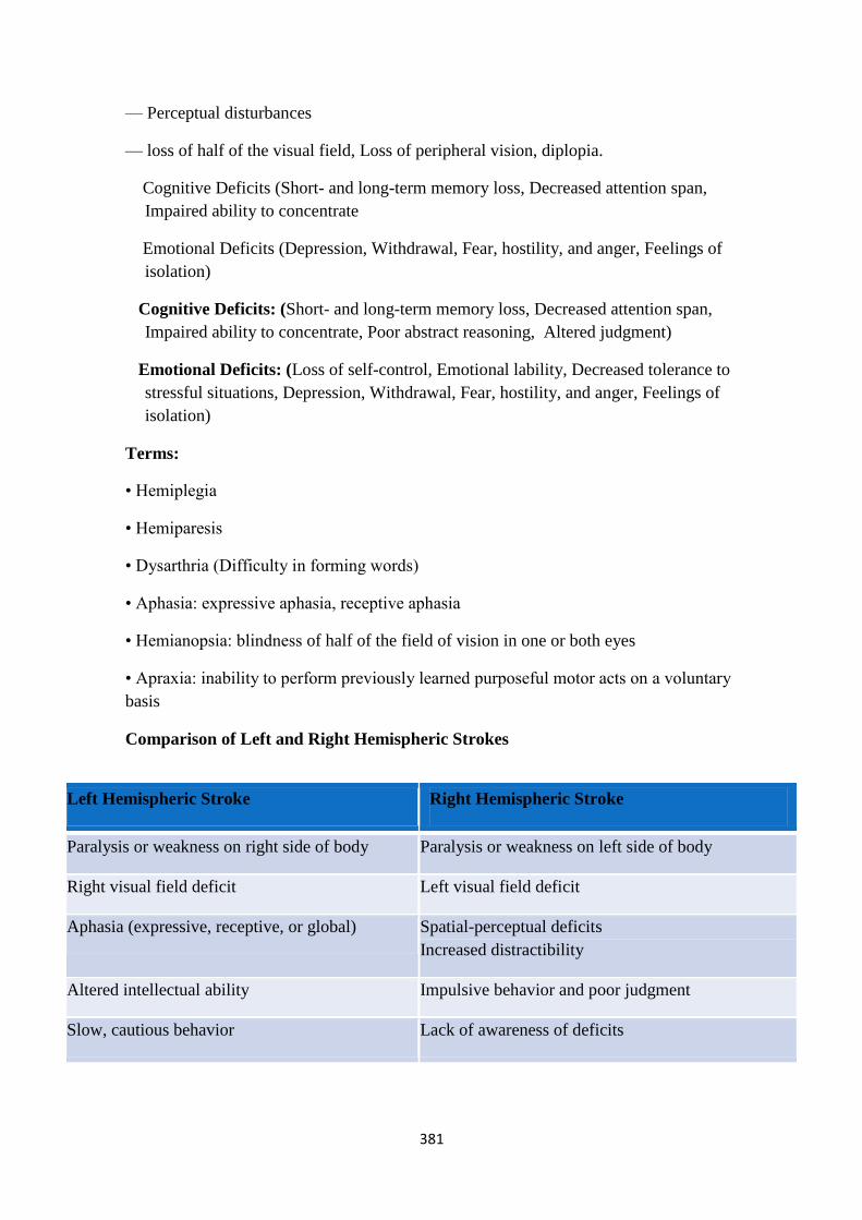
381
— Perceptual disturbances
— loss of half of the visual field, Loss of peripheral vision, diplopia.
Cognitive Deficits (Short- and long-term memory loss, Decreased attention span,
Impaired ability to concentrate
Emotional Deficits (Depression, Withdrawal, Fear, hostility, and anger, Feelings of
isolation)
Cognitive Deficits: (Short- and long-term memory loss, Decreased attention span,
Impaired ability to concentrate, Poor abstract reasoning, Altered judgment)
Emotional Deficits: (Loss of self-control, Emotional lability, Decreased tolerance to
stressful situations, Depression, Withdrawal, Fear, hostility, and anger, Feelings of
isolation)
Terms:
• Hemiplegia
• Hemiparesis
• Dysarthria (Difficulty in forming words)
• Aphasia: expressive aphasia, receptive aphasia
• Hemianopsia: blindness of half of the field of vision in one or both eyes
• Apraxia: inability to perform previously learned purposeful motor acts on a voluntary
basis
Comparison of Left and Right Hemispheric Strokes
Left Hemispheric Stroke Right Hemispheric Stroke
Paralysis or weakness on right side of body Paralysis or weakness on left side of body
Right visual field deficit Left visual field deficit
Aphasia (expressive, receptive, or global) Spatial-perceptual deficits
Increased distractibility
Altered intellectual ability Impulsive behavior and poor judgment
Slow, cautious behavior Lack of awareness of deficits

382
Transient Ischemic Attack (TIA)
• Temporary neurologic deficit resulting from a temporary impairment of blood flow
• ―Warning of an impending stroke‖
• Classic symptom is fleeting blindness in one eye.
• Diagnostic workup is required to treat and prevent irreversible deficits
Carotid Endarterectomy
Preventive Treatment and Secondary Prevention
• Health maintenance measures including a healthy diet, exercise, and the prevention
and treatment of periodontal disease
• Carotid endarterectomy
• Anticoagulant therapy
• Antiplatelet therapy: aspirin,
• Antihypertensive medications
Medical Management—Acute Phase of Stroke
• Prompt diagnosis and treatment
• Assessment of stroke
• Thrombolytic therapy
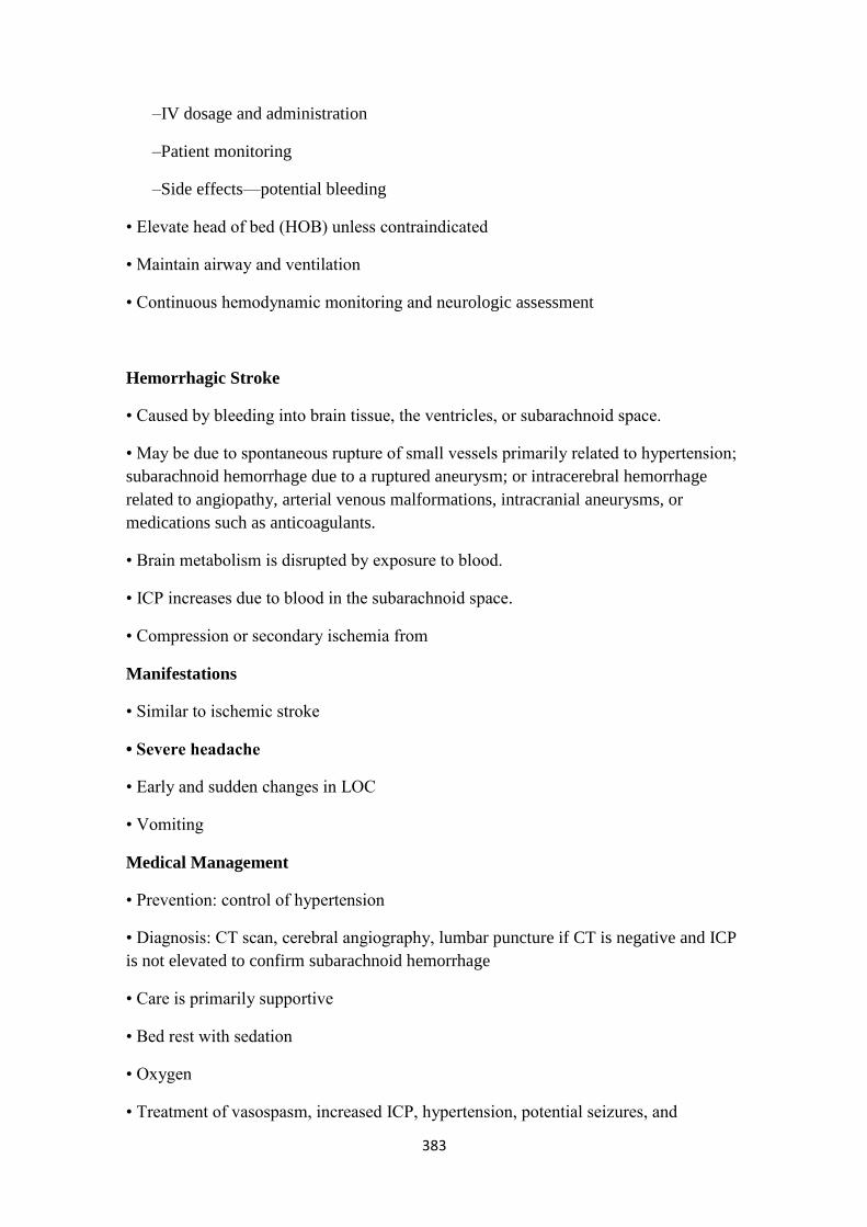
383
–IV dosage and administration
–Patient monitoring
–Side effects—potential bleeding
• Elevate head of bed (HOB) unless contraindicated
• Maintain airway and ventilation
• Continuous hemodynamic monitoring and neurologic assessment
Hemorrhagic Stroke
• Caused by bleeding into brain tissue, the ventricles, or subarachnoid space.
• May be due to spontaneous rupture of small vessels primarily related to hypertension;
subarachnoid hemorrhage due to a ruptured aneurysm; or intracerebral hemorrhage
related to angiopathy, arterial venous malformations, intracranial aneurysms, or
medications such as anticoagulants.
• Brain metabolism is disrupted by exposure to blood.
• ICP increases due to blood in the subarachnoid space.
• Compression or secondary ischemia from
Manifestations
• Similar to ischemic stroke
• Severe headache
• Early and sudden changes in LOC
• Vomiting
Medical Management
• Prevention: control of hypertension
• Diagnosis: CT scan, cerebral angiography, lumbar puncture if CT is negative and ICP
is not elevated to confirm subarachnoid hemorrhage
• Care is primarily supportive
• Bed rest with sedation
• Oxygen
• Treatment of vasospasm, increased ICP, hypertension, potential seizures, and
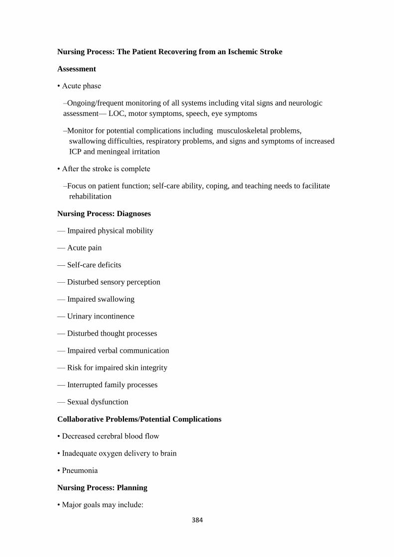
384
Nursing Process: The Patient Recovering from an Ischemic Stroke
Assessment
• Acute phase
–Ongoing/frequent monitoring of all systems including vital signs and neurologic
assessment— LOC, motor symptoms, speech, eye symptoms
–Monitor for potential complications including musculoskeletal problems,
swallowing difficulties, respiratory problems, and signs and symptoms of increased
ICP and meningeal irritation
• After the stroke is complete
–Focus on patient function; self-care ability, coping, and teaching needs to facilitate
rehabilitation
Nursing Process: Diagnoses
— Impaired physical mobility
— Acute pain
— Self-care deficits
— Disturbed sensory perception
— Impaired swallowing
— Urinary incontinence
— Disturbed thought processes
— Impaired verbal communication
— Risk for impaired skin integrity
— Interrupted family processes
— Sexual dysfunction
Collaborative Problems/Potential Complications
• Decreased cerebral blood flow
• Inadequate oxygen delivery to brain
• Pneumonia
Nursing Process: Planning
• Major goals may include:
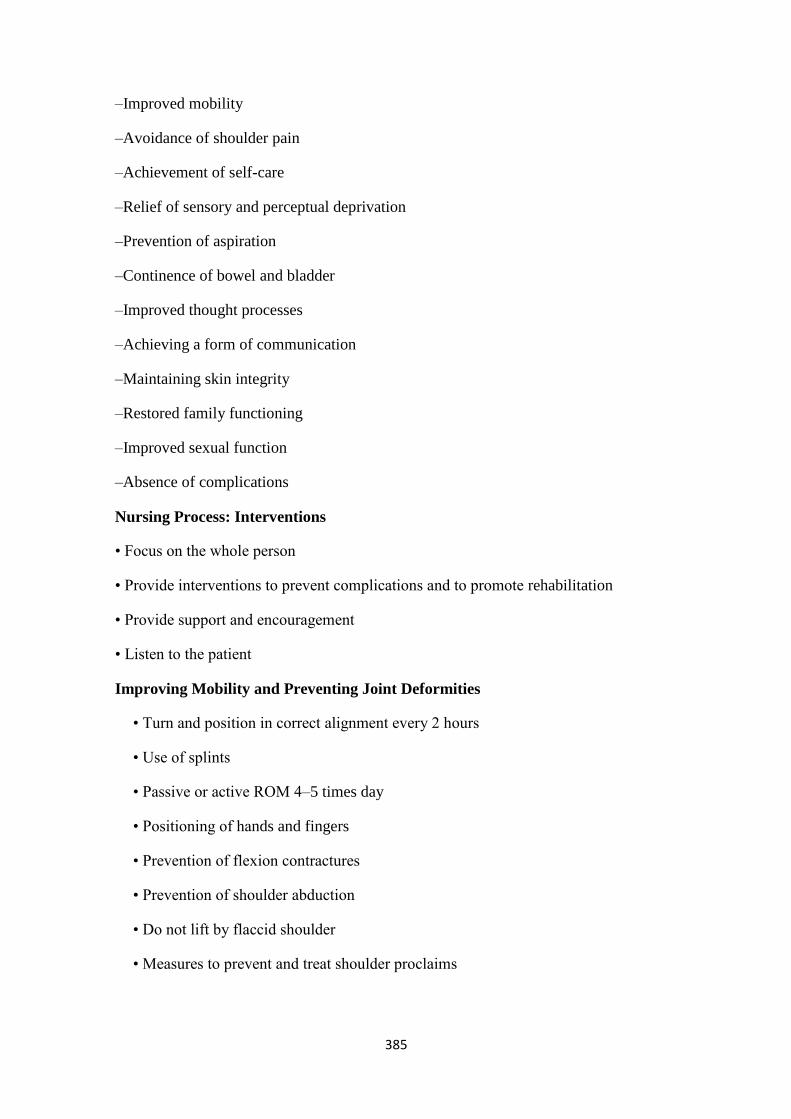
385
–Improved mobility
–Avoidance of shoulder pain
–Achievement of self-care
–Relief of sensory and perceptual deprivation
–Prevention of aspiration
–Continence of bowel and bladder
–Improved thought processes
–Achieving a form of communication
–Maintaining skin integrity
–Restored family functioning
–Improved sexual function
–Absence of complications
Nursing Process: Interventions
• Focus on the whole person
• Provide interventions to prevent complications and to promote rehabilitation
• Provide support and encouragement
• Listen to the patient
Improving Mobility and Preventing Joint Deformities
• Turn and position in correct alignment every 2 hours
• Use of splints
• Passive or active ROM 4–5 times day
• Positioning of hands and fingers
• Prevention of flexion contractures
• Prevention of shoulder abduction
• Do not lift by flaccid shoulder
• Measures to prevent and treat shoulder proclaims
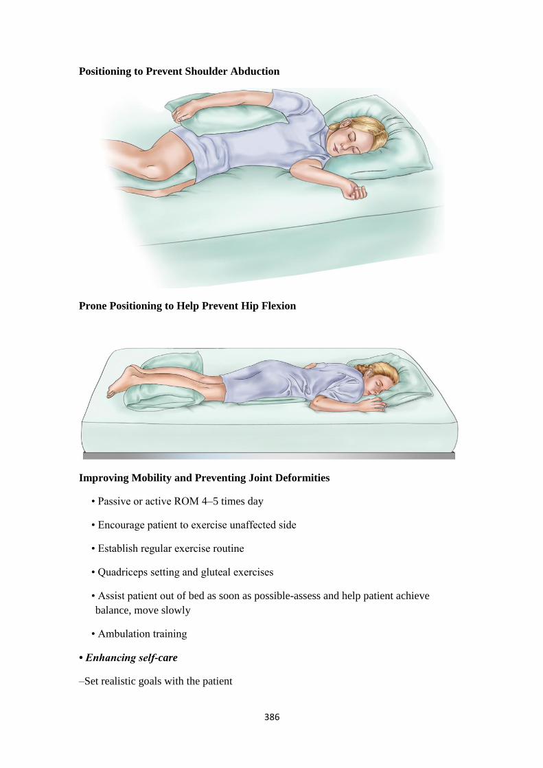
386
Positioning to Prevent Shoulder Abduction
Prone Positioning to Help Prevent Hip Flexion
Improving Mobility and Preventing Joint Deformities
• Passive or active ROM 4–5 times day
• Encourage patient to exercise unaffected side
• Establish regular exercise routine
• Quadriceps setting and gluteal exercises
• Assist patient out of bed as soon as possible-assess and help patient achieve
balance, move slowly
• Ambulation training
• Enhancing self-care
–Set realistic goals with the patient

387
–Encourage personal hygiene
–Assure that patient does not neglect the affected side
–Use of assistive devices and modification of clothing
• Support and encouragement
• Strategies to enhance communication
• Encourage patient to turn head, look to side with visual field loss
• Nutrition
–Consult with speech therapy or nutritional services
–Have patient sit upright, preferably OOB, to eat
–Chin tuck or swallowing method
–Use of thickened liquids or pureed ( مهروس ) diet
• Bowel and bladder control
–Assessment of voiding and scheduled voiding
–Measures to prevent constipation—fiber, fluid, toileting schedule
–Bowel and bladder retraining
Nursing Process: The Patient with a Hemorrhagic Stroke
Assessment
• Complete and ongoing neurologic assessment—use neurologic flow chart
• Monitor respiratory status and oxygenation
• Monitoring of ICP
• Patients with intracerebral or subarachnoid hemorrhage should be monitored in the
ICU
• Monitor for potential complications
• Monitor fluid balance and laboratory data
• All changes must be reported immediately
Nursing Process: Diagnoses
• Ineffective tissue perfusion (cerebral)
388
• Disturbed sensory perception
• Anxiety
Collaborative Problems/Potential Complications
• Vasospasm
• Seizures
• Hydrocephalus
• Rebleeding
• Hyponatremia
Nursing Process: Planning
• Goals may include:
–Improved cerebral tissue perfusion
–Relief of sensory and perceptual deprivation
–Relief of anxiety
–The absence of complications
Aneurysm Precautions
• Absolute bed rest
• Elevate HOB 30° to promote venous drainage or flat to increase cerebral perfusion
• Avoid all activity that may increase ICP or BP; Valsalva maneuver, acute flexion or
rotation of neck or head
• Exhale through mouth when voiding or defecating to decrease strain
• Nurse provides all personal care and hygiene
• Nonstimulating, nonstressful environment; dim lighting, no reading, no TV, no radio
• Prevent constipation
Interventions
• Relieving sensory deprivation and anxiety
• Keep sensory stimulation to a minimum for aneurysm precautions
• Realty orientation
389
• Patient and family teaching
• Support and reassurance
• Seizure precautions
• Strategies to regain and promote self-care and rehabilitation
Home Care and Teaching for the Patient Recovering from a Stroke
• Prevention of subsequent strokes, health promotion, and follow-up care
• Prevention of and signs and symptoms of complications
• Medication teaching
• Safety measures
• Adaptive strategies and use of assistive devices for ADLs
• Nutrition—diet, swallowing techniques, tube feeding administration
• Elimination—bowel and bladder programs, catheter use
390
Nursing Process
The Patient Recovering From an Ischemic Stroke
The acute phase of an ischemic stroke may last 1 to 3 days, but ongoing monitoring of
all body systems is essential as long as the patient requires care. The patient who has
had a stroke is at risk for multiple complications, including deconditioning and other
musculoskeletal problems, swallowing difficulties, bowel and bladder dysfunction,
inability to perform self-care, and skin breakdown. After the stroke is complete,
management focuses on the prompt initiation of rehabilitation for any deficits.
Assessment
During the acute phase, a neurologic flow sheet is maintained to provide data about the
following important measures of the patient's clinical status:
Change in level of consciousness or responsiveness as evidenced by movement,
resistance to changes of position, and response to stimulation; orientation to
time, place, and person
Presence or absence of voluntary or involuntary movements of the extremities;
muscle tone; body posture; and position of the head
Stiffness or flaccidity of the neck
Eye opening, comparative size of pupils and pupillary reactions to light, and
ocular position
Color of the face and extremities; temperature and moisture of the skin
Quality and rates of pulse and respiration; arterial blood gas values as indicated,
body temperature, and arterial pressure
Ability to speak
Volume of fluids ingested or administered; volume of urine excreted each 24
hours
Presence of bleeding
Maintenance of blood pressure within the desired parameters
After the acute phase, the nurse assesses mental status (memory, attention span,
perception, orientation, affect, speech/language), sensation/perception (usually the
patient has decreased awareness of pain and temperature), motor control (upper and
lower extremity movement), swallowing ability, nutritional and hydration status, skin
integrity, activity tolerance, and bowel and bladder function. Ongoing nursing
assessment continues to focus on any impairment of function in the patient's daily
activities, because the quality of life after stroke is closely related to the patient's
functional status.
Diagnosis
Nursing Diagnoses
Based on the assessment data, the major nursing diagnoses for a patient with a stroke
may include the following:
Impaired physical mobility related to hemiparesis, loss of balance and
coordination, spasticity, and brain injury
Acute pain (painful shoulder) related to hemiplegia and disuse
Self-care deficits (bathing, hygiene, toileting, dressing, grooming, and feeding)
related to stroke sequelae
391
Disturbed sensory perception related to altered sensory reception, transmission,
and/or integration
Impaired swallowing
Total urinary incontinence related to flaccid bladder, detrusor instability,
confusion, or difficulty in communicating
Disturbed thought processes related to brain damage, confusion, or inability to
follow instructions
Impaired verbal communication related to brain damage
Risk for impaired skin integrity related to hemiparesis, hemiplegia, or decreased
mobility
Interrupted family processes related to catastrophic illness and caregiving burdens
Sexual dysfunction related to neurologic deficits or fear of failure
Collaborative Problems/Potential Complications
Potential complications include:
Decreased cerebral blood flow due to increased ICP
Inadequate oxygen delivery to the brain
Pneumonia
Planning and Goals
Although rehabilitation begins on the day the patient has the stroke, the process is
intensified during convalescence and requires a coordinated team effort. It is helpful for
the team to know what the patient was like before the stroke: his or her illnesses,
abilities, mental and emotional state, behavioral characteristics, and activities of daily
living (ADLs). It is also helpful for clinicians to be knowledgeable about the relative
importance of predictors of stroke outcome (age, NIHSS score, and level of
consciousness at time of admission) in order to provide stroke survivors and their
families with realistic goals.
The major goals for the patient (and family) may include improved mobility, avoidance
of shoulder pain, achievement of self-care, relief of sensory and perceptual deprivation,
prevention of aspiration, continence of bowel and bladder, improved thought processes,
achieving a form of communication, maintaining skin integrity, restored family
functioning, improved sexual function, and absence of complications.
Nursing Interventions
Nursing care has a significant impact on the patient's recovery. Often, many body
systems are impaired as a result of the stroke, and conscientious care and timely
interventions can prevent debilitating complications. During and after the acute phase,
nursing interventions focus on the whole person. In addition to providing physical care,
the nurse encourages and fosters recovery by listening to the patient and asking
questions to elicit the meaning of the stroke experience.
Improving Mobility and Preventing Joint Deformities
A patient with hemiplegia has unilateral paralysis (paralysis on one side). When control
of the voluntary muscles is lost, the strong flexor muscles exert control over the
extensors. The arm tends to adduct (adductor muscles are stronger than abductors) and
to rotate internally. The elbow and the wrist tend to flex, the affected leg tends to rotate
externally at the hip joint and flex at the knee, and the foot at the ankle joint supinates
and tends toward plantar flexion.

392
Correct positioning is important to prevent contractures; measures are used to relieve
pressure, assist in maintaining good body alignment, and prevent compressive
neuropathies, especially of the ulnar and peroneal nerves. Because flexor muscles are
stronger than extensor muscles, a posterior splint applied at night to the affected
extremity may prevent flexion and maintain correct positioning during sleep. (See
Chapter 11 for additional information.)
Preventing Shoulder Adduction
To prevent adduction of the affected shoulder while the patient is in bed, a pillow is
placed in the axilla when there is limited external rotation; this keeps the arm away
from the chest. A pillow is placed under the arm, and the arm is placed in a neutral
(slightly flexed) position, with distal joints positioned higher than the more proximal
joints (ie, the elbow is positioned higher than the shoulder and the wrist higher than the
elbow). This helps to prevent edema and the resultant joint fibrosis that will limit range
of motion if the patient regains control of the arm (Fig. 62-3).
Positioning the Hand and Fingers
The fingers are positioned so that they are barely flexed. The hand is placed in slight
supination (palm faces upward), which is its most functional position. If the upper
extremity is flaccid, a volar resting splint can be used to support the wrist and hand in a
functional position. If the upper extremity is spastic, a hand roll is not used, because it
stimulates the grasp reflex. In this instance a dorsal wrist splint is useful in allowing the
palm to be free of pressure. Every effort is made to prevent hand edema.
Spasticity, particularly in the hand, can be a disabling complication after stroke.
Researchers reported that repeated intramuscular injections of botulinum toxin A into
wrist and finger muscles reduced upper limb spasticity after stroke, resulting in
significant and sustained improvements in muscle tone.
Changing Positions
The patient's position should be changed every 2 hours. To place a patient in a lateral
(side-lying) position, a pillow is placed between the legs before the patient is turned. To
promote venous return and prevent edema, the upper thigh should not be acutely flexed.
The patient may be turned from side to side, but if sensation is impaired, the amount of
time spent on the affected side should be limited.

393
If possible, the patient is placed in a prone position for 15 to 30 minutes several times a
day. A small pillow or a support is placed under the pelvis, extending from the level of
the umbilicus to the upper third of the thigh (Fig. 62-4). This position helps to promote
hyperextension of the hip joints, which is essential for normal gait and helps prevent
knee and hip flexion contractures. The prone position also helps to drain bronchial
secretions and prevents contractural deformities of the shoulders and knees. During
positioning, it is important to reduce pressure and change position frequently to prevent
pressure ulcers.
Establishing an Exercise Program
The affected extremities are exercised passively and put through a full range of motion
four or five times a day to maintain joint mobility, regain motor control, prevent
contractures in the paralyzed extremity, prevent further deterioration of the
neuromuscular system, and enhance circulation. Exercise is helpful in preventing
venous stasis, which may predispose the patient to thrombosis and pulmonary embolus.
Repetition of an activity forms new pathways in the CNS and therefore encourages new
patterns of motion. At first, the extremities are usually flaccid. If tightness occurs in
any area, the range-of-motion exercises should be performed more frequently (see
Chapter 11).
The patient is observed for signs and symptoms that may indicate pulmonary embolus
or excessive cardiac workload during exercise; these include shortness of breath, chest
pain, cyanosis, and increasing pulse rate with exercise. Frequent short periods of
exercise always are preferable to longer periods at infrequent intervals. Regularity in
exercise is most important. Improvement in muscle strength and maintenance of range
of motion can be achieved only through daily exercise.
The patient is encouraged and reminded to exercise the unaffected side at intervals
throughout the day. It is helpful to develop a written schedule to remind the patient of
the exercise activities. The nurse supervises and supports the patient during these
activities. The patient can be taught to put the unaffected leg under the affected one to
assist in moving it when turning and exercising. Flexibility, strengthening,
coordination, endurance, and balancing exercises prepare the patient for ambulation.

394
Quadriceps muscle setting and gluteal setting exercises are started early to improve the
muscle strength needed for walking; these are performed at least five times daily for 10
minutes at a time.
Preparing for Ambulation
As soon as possible, the patient is assisted out of bed. Usually an active rehabilitation
program is started as soon as the patient regains consciousness. The patient is first
taught to maintain balance while sitting and then to learn to balance while standing. If
the patient has difficulty in achieving standing balance, a tilt table, which slowly brings
the patient to an upright position, can be used. Tilt tables are especially helpful for
patients who have been on bed rest for prolonged periods and have orthostatic blood
pressure changes.
If the patient needs a wheelchair, the folding type with hand brakes is the most practical
because it allows the patient to manipulate the chair. The chair should be low enough to
allow the patient to propel it with the uninvolved foot and narrow enough to permit it to
be used in the home. When the patient is transferred from the wheelchair, the brakes
must be applied and locked on both sides of the chair.
The patient is usually ready to walk as soon as standing balance is achieved. Parallel
bars are useful in these first efforts. A chair or wheelchair should be readily available in
case the patient suddenly becomes fatigued or feels dizzy.
The training periods for ambulation should be short and frequent. As the patient gains
strength and confidence, an adjustable cane can be used for support. Generally, a three-
or four-pronged cane provides a stable support in the early phases of rehabilitation.
Preventing Shoulder Pain
As many as 70% of stroke patients suffer severe pain in the shoulder that prevents them
from learning new skills. Shoulder function is essential in achieving balance and
performing transfers and self-care activities. Three problems can occur: painful
shoulder, subluxation of the shoulder, and shoulder–hand syndrome.
A flaccid shoulder joint may be overstretched by the use of excessive force in turning
the patient or from overstrenuous arm and shoulder movement. To prevent shoulder
pain, the nurse should never lift the patient by the flaccid shoulder or pull on the

395
affected arm or shoulder. If the arm is paralyzed, subluxation (incomplete dislocation)
at the shoulder can occur as a result of overstretching of the joint capsule and
musculature by the force of gravity when the patient sits or stands in the early stages
after a stroke. This results in severe pain. Shoulder–hand syndrome (painful shoulder
and generalized swelling of the hand) can cause a frozen shoulder and ultimately
atrophy of subcutaneous tissues. When a shoulder becomes stiff, it is usually painful.
Many shoulder problems can be prevented by proper patient movement and
positioning. The flaccid arm is positioned on a table or with pillows while the patient is
seated. Some clinicians advocate the use of a properly worn sling when the patient first
becomes ambulatory, to prevent the paralyzed upper extremity from dangling without
support. Range-of-motion exercises are important in preventing painful shoulder.
Overstrenuous arm movements are avoided. The patient is instructed to interlace the
fingers, place the palms together, and push the clasped hands slowly forward to bring
the scapulae forward; he or she then raises both hands above the head. This is repeated
throughout the day. The patient is instructed to flex the affected wrist at intervals and
move all the joints of the affected fingers. He or she is encouraged to touch, stroke, rub,
and look at both hands. Pushing the heel of the hand firmly down on a surface is useful.
Elevation of the arm and hand is also important in preventing dependent edema of the
hand. Patients with continuing pain after attempted movement and positioning may
require the addition of analgesia to their treatment program.
Medications are helpful in the management of poststroke pain. Amitriptyline
hydrochloride (Elavil) has been used, but it can cause cognitive problems, has a
sedating effect, and is not effective in all patients. The antiseizure medication
lamotrigine (Lamictal) has been found to be effective for poststroke pain, and it may
serve as an alternative for patients who cannot tolerate amitriptyline (Nicholson, 2004).
Enhancing Self-Care
As soon as the patient can sit up, personal hygiene activities are encouraged. The
patient is helped to set realistic goals; if feasible, a new task is added daily. The first
step is to carry out all self-care activities on the unaffected side. Such activities as
combing the hair, brushing the teeth, shaving with an electric razor, bathing, and eating
can be carried out with one hand and are suitable for self-care. Although the patient
may feel awkward at first, the various motor skills can be learned by repetition, and the
unaffected side will become stronger with use. The nurse must be sure that the patient
does not neglect the affected side. Assistive devices will help make up for some of the
patient's deficits (Chart 62-3). A small towel is easier to control while drying after
bathing, and boxed paper tissues are easier to use than a roll of toilet tissue.
Return of functional ability is important to the patient recovering after a stroke. An
early baseline assessment of functional ability with an instrument such as the
Functional Independence Measure (FIMTM
) is important in team planning and goal
setting for the patient. The FIMTM
is a widely used instrument in stroke rehabilitation
and provides valuable information about motor, social, and cognitive function (Kelly-
Hayes, 2004). The patient's morale will improve if ambulatory activities are carried out
in street clothes. The family is instructed to bring in clothing that is preferably a size
larger than that normally worn. Clothing fitted with front or side fasteners or Velcro
closures is the most suitable. The patient has better balance if most of the dressing
activities are carried out while seated.
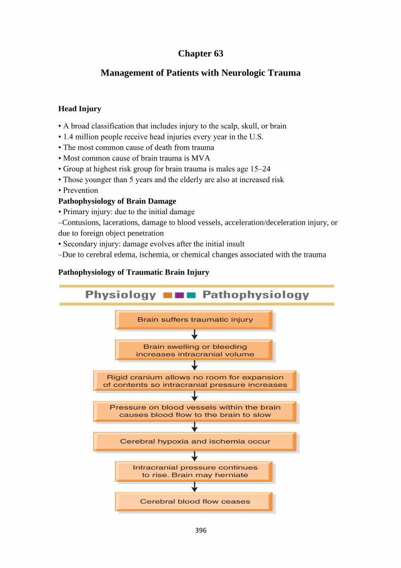
396
Chapter 63
Management of Patients with Neurologic Trauma
Head Injury
• A broad classification that includes injury to the scalp, skull, or brain
• 1.4 million people receive head injuries every year in the U.S.
• The most common cause of death from trauma
• Most common cause of brain trauma is MVA
• Group at highest risk group for brain trauma is males age 15–24
• Those younger than 5 years and the elderly are also at increased risk
• Prevention
Pathophysiology of Brain Damage
• Primary injury: due to the initial damage
–Contusions, lacerations, damage to blood vessels, acceleration/deceleration injury, or
due to foreign object penetration
• Secondary injury: damage evolves after the initial insult
–Due to cerebral edema, ischemia, or chemical changes associated with the trauma
Pathophysiology of Traumatic Brain Injury
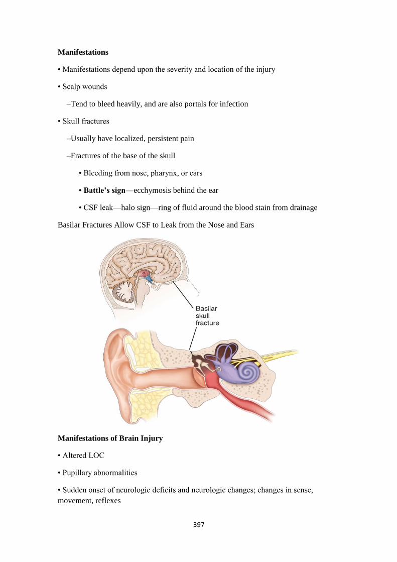
397
Manifestations
• Manifestations depend upon the severity and location of the injury
• Scalp wounds
–Tend to bleed heavily, and are also portals for infection
• Skull fractures
–Usually have localized, persistent pain
–Fractures of the base of the skull
• Bleeding from nose, pharynx, or ears
• Battle’s sign—ecchymosis behind the ear
• CSF leak—halo sign—ring of fluid around the blood stain from drainage
Basilar Fractures Allow CSF to Leak from the Nose and Ears
Manifestations of Brain Injury
• Altered LOC
• Pupillary abnormalities
• Sudden onset of neurologic deficits and neurologic changes; changes in sense,
movement, reflexes
398
• Changes in vital signs
• Headache
• Seizures
Brain Injury
• Closed brain injury (blunt trauma): acceleration/deceleration injury occurs when the
head accelerates and then rapidly decelerates, damaging brain tissue
• Open brain injury: object penetrates the brain or trauma is so severe that the scalp and
skull are opened
• Concussion: a temporary loss of consciousness with no apparent structural damage
• Contusion: more severe injury with possible surface hemorrhage
–Symptoms and recovery depend upon the amount of damage and associated
cerebral edema
–Longer period of unconsciousness with more symptoms of neurologic deficits and
changes in vital signs
• Diffuse axonal injury: involves widespread damage to axons in the cerebral
hemispheres, corpus callosum, and brain stem. It can be seen with mild, moderate, or
severe head trauma. Patient develops immediate coma.
• Intracranial bleeding
1. Epidural hematoma
2. Subdural hematoma
3. Acute and subacute
4. Chronic
5. Intracerebral hemorrhage and hematoma
Concussion
• Patient may be admitted for observation or sent home
• Observation of patients after head trauma; report immediately
–Observe for any changes in LOC
–Difficulty in awakening, lethargy, dizziness, confusion, irritability, anxiety
–Difficulty in speaking or movement
–Severe headache
–Vomiting

399
• Patient should be aroused and assessed frequently
Location of Subdural, Intracerebral and Epidural Hemorrhages
Epidural Hematoma
• Blood collection in the space between the skull and the dura.
• Patient may have a brief loss of consciousness with return of lucid state then as
hematoma expands increased ICP will often suddenly reduce LOC.
• An emergency situation!
• Treatment include measures to reduce ICP, remove the clot and stop bleeding—burr
holes or craniotomy.
• Patient will need monitoring and support of
Subdural Hematoma
• Collection of blood between the dura and the brain
• Acute/Subacute
–Acute: symptoms develop over 24–48 hours
–Subacute: symptoms develop over 48 hours to 2 weeks
–Requires immediate craniotomy and control of ICP
• Chronic

400
–Develops over weeks to months
–Causative injury may be minor and forgotten
–Clinical signs and symptoms may fluctuate
–Treatment is evacuation of the clot
Intracerebral Hemorrhage
• Hemorrhage occurs into the substance of the brain
• May be due to trauma or a nontraumatic cause
• Treatment
–Supportive care
–Control of ICP
–Administration of fluids, electrolytes, and antihypertensive medications
–Craniotomy or craniectomy to remove clot and control hemorrhage; this may not
be possible due the location or lack of circumscribed area of hemorrhage
Diagnostic Evaluation
• Physical and neurologic exam
• Skull and spinal x-rays
• CT scan
• MRI
• PET (Positron emission tomography)
Management of the Patient with a Head Injury
• Assume cervical spine injury until this is ruled out
• Therapy to preserve brain homeostasis and prevent secondary damage
–Treat cerebral edema
–Maintain cerebral perfusion; treat hypotension, hypovolemia and bleeding, monitor
and manage ICP
–Maintain oxygenation; cardiovascular and respiratory function
–Manage fluid and electrolyte balance

401
Supportive Measures
• Respiratory support; intubation and mechanical ventilation
• Seizure precautions and prevention
• NG to manage reduced gastric motility and prevent aspiration
• Fluid and electrolyte maintenance
• Pain and anxiety management
• Nutrition
Nursing Process: The Care of the Patient with Brain Injury
Assessment
• Health history with focus upon the immediate injury, time, cause, and the direction
and force of the blow
• Baseline assessment
• LOC—Glasgow Coma Scale
• Frequent and ongoing neurologic assessment
• Multisystem assessment
Nursing Process: Diagnoses
• Ineffective airway clearance and impaired gas exchange
• Ineffective cerebral perfusion
• Deficient fluid volume
• Imbalanced nutrition
• Risk for injury
• Risk for imbalanced body temperature
• Risk for impaired skin integrity
• Disturbed thought patterns
• Disturbed sleep pattern
• Interrupted family process
• Deficient knowledge

402
Collaborative Problems/Potential Complications
• Decreased cerebral perfusion
• Cerebral edema and herniation
• Impaired oxygenation and ventilation
• Impaired fluid, electrolyte, and nutritional balance
• Risk of posttraumatic seizures
Nursing Process: Planning
— Major goals may include
— Maintenance of patent airway,
— Adequate cerebral perfusion pressure (CPP),
— Fluid and electrolyte balance,
— Adequate nutritional status,
— Prevention of secondary injury,
— Maintenance of normal temperature,
— Maintenance of skin integrity,
— Improvement of cognitive function,
— Prevention of sleep deprivation,
— Effective family coping,
— Increased knowledge about rehabilitation process, and
Nursing Process: Interventions
• Ongoing assessment and monitoring is vital
• Maintenance of airway
–Positioning to facilitate drainage of oral secretions with HOB usually elevated
30° to decrease venous pressure
–Suctioning with caution
–Prevention of aspiration and respiratory insufficiency
–Monitor ABGs, ventilation, and mechanical ventilation
–Monitor for pulmonary complications, potential
403
• I&O and daily weights
• Monitor blood and urine electrolytes and osmolality and blood glucose
• Measures to promote adequate nutrition
• Strategies to prevent injury
–Assessment of oxygenation
–Assessment of bladder and urinary output
–Assessment for constriction due to dressings and casts
–Pad side-rails
–Mittens to prevent self-injury; avoid restraints
• Strategies to prevent injury
–Reduce environmental stimuli
–Adequate lighting to reduce visual hallucinations
–Measures to minimize disruption of sleep-wake cycles
–Skin care
–Measures to prevent infection
• Maintaining body temperature
–Maintain appropriate environmental temperature
–Use of coverings—sheets, blankets to patient needs
–Administration of acetaminophen for fever
–Cooling blankets or cool baths; avoid shivering
• Support of cognitive function
• Support of family
–Provide and reinforce information
–Measures to promote effective coping
–Setting of realistic, well-defined, short-term goals
–Referral for counseling
–Support groups

404
• Patient and family teaching
Spinal Cord Injury (SCI)
Definition:
• Fracture or displacement of one or more vertebrae causing damae to spinal cord and
nerve roots with resulting neurological deficit and altered sensory perception or
paralysis or both. There will be a total or partial absence of motor and/or sensory
function below the level of injury. (Ignatavious and Workman, 2006)
Spinal Cord Injury (SCI)
• A major health problem
• 200,000 persons in the U.S. live with disability from SCI
• Causes include MVAs (35%), violence (24%), falls (22%), and sports injuries (8%)
• Males account for 82% of SCIs
• Young people ages 16–30 account for more than half of all new SCIs
• African–Americans are at higher risk
• Risk factors include alcohol and drug use
• Prevention
Spinal Cord Injury
— The result of concussion, contusion, laceration or compression of spinal cord.
— Primary injury is the result of the initial trauma.
— Secondary injury is usually the result of ischemia, hypoxia, and hemorrhage that
destroys the nerve tissues.
— Secondary injuries are thought to be reversible/preventable during the first 4–6
hours after injury.
— Treatment is needed to prevent partial injury from developing into more extensive,
permanent
Clinical Manifestations
— Manifestations depend on the type and level of injury
— Incomplete spinal cord lesions (the sensory or motor fibers, or both, are preserved
below the lesion): below the injury; total sensory and motor paralysis, loss of bladder
405
and bowel control (usually with urinary retention and bladder distention), loss of
sweating and vasomotor tone, & marked reduction of blood pressure.
— Complete spinal cord lesion (total loss of sensation and voluntary muscle control
below the lesion): paraplegia or tetraplegia.
— If conscious, the patient usually complains of acute pain in the back or neck
Spinal and Neurogenic Shock
• Spinal shock
–A sudden depression of reflex activity below the level of spinal injury
–develops due to the loss of autonomic nervous system function below the level of the
lesion
–Muscular flaccidity, lack of sensation and reflexes
• Neurogenic shock
–Due to the loss of function of the autonomic nervous system
–Blood pressure, heart rate, and cardiac output decrease
–Venous pooling occurs due to peripheral vasodilation
–Paralyzed portions of the body do not perspire
Medical Management: Acute Phase
• Goals:
–Prevent further SCI and observe for signs of neurological deficit
• High dose corticosteroids (controversial)
• Research is continuing
Medical management:
• Pharmacologic therapy
• Respiratory therapy
• Skeletal fracture reduction and traction
• Surgical management
406
Nursing Process: The Care of the Patient with SCI
Assessment
• Monitor respirations and breathing pattern
• Lung sounds and cough
• Monitor for changes in motor or sensory function; report immediately
• Assess for spinal shock
• Monitor for bladder retention or distention, gastric dilation, and ilieus
• Temperature; potential hyperthermia
Nursing Process: Diagnoses
• Ineffective breathing pattern
• Ineffective airway clearance
• Impaired physical mobility
• Disturbed sensory perception
• Risk for impaired skin integrity
• Impaired urinary elimination
• Constipation
• Acute pain
Collaborative Problems/Potential Complications
• DVT
• Orthostatic hypotension
• Autonomic dysreflexia
Nursing Process: Planning
• Major goals may include improved breathing pattern and airway clearance, improved
mobility, improved sensory and perceptual awareness, maintenance of skin integrity,
promotion of comfort, and absence of complications.
Promotion of Effective Breathing and Airway Clearance
• Monitor carefully to detect potential respiratory failure
–Pulse oximetry and ABGs

407
–Lung sounds
• Early and vigorous pulmonary care to prevent and remove secretions
• Suctioning with caution
• Breathing exercises
• Assisted coughing
• Humidification and hydration
Improving Mobility
• Maintain proper body alignment
• Turn only if spine is stable and as indicated by physician
• Monitor blood pressure with position changes
• PROM at least four times a day
• Use neck brace or collar, as prescribed, when patient is mobilized
• Move gradually to erect position
408
Nursing Process
The Patient with a Traumatic Brain Injury
Assessment
Depending on the patient's neurologic status, the nurse may elicit information from the
patient, from the family, or from witnesses or emergency rescue personnel. Although
all usual baseline data may not be collected initially, the immediate health history
should include the following questions:
When did the injury occur?
What caused the injury? A high-velocity missile? An object striking the head?
A fall?
What was the direction and force of the blow?
A history of unconsciousness or amnesia after a head injury indicates a significant
degree of brain damage, and changes that occur minutes to hours after the initial injury
can reflect recovery or indicate the development of secondary brain damage. Therefore,
the nurse also should try to determine if there was a loss of consciousness, what the
duration of the unconscious period was, and whether the patient could be aroused.
In addition to asking questions that establish the nature of the injury and the patient's
condition immediately after the injury, the nurse examines the patient thoroughly. This
assessment includes determining the patient's LOC using the Glasgow Coma Scale and
assessing the patient's response to tactile stimuli (if unconscious), pupillary response to
light, corneal and gag reflexes, and motor function. The Glasgow Coma Scale (Chart
63-4) is based on the three criteria of eye opening, verbal responses, and motor
responses to verbal commands or painful stimuli. It is particularly useful for monitoring
changes during the acute phase, the first few days after a head injury. It does not take
the place of an in-depth neurologic assessment. Additional detailed assessments are
made initially and at frequent intervals throughout the acute phase of care (Hickey,
2003). The baseline and ongoing assessments are critical nursing interventions for the
patient with brain injury, whose condition can worsen dramatically and irrevocably if
subtle signs are overlooked. More information on assessment is provided in the
following sections and in Figure 63-5 and Table 63-1.
Diagnosis
Nursing Diagnoses
Based on the assessment data, the patient's major nursing diagnoses may include the
following:
Ineffective airway clearance and impaired gas exchange related to brain injury
Ineffective cerebral tissue perfusion related to increased ICP, decreased CPP,
and possible seizures
Deficient fluid volume related to decreased LOC and hormonal dysfunction
Imbalanced nutrition, less than body requirements, related to increased
metabolic demands, fluid restriction, and inadequate intake
Risk for injury (self-directed and directed at others) related to seizures,
disorientation, restlessness, or brain damage
Risk for imbalanced body temperature related to damaged temperature-
regulating mechanisms in the brain
409
Risk for impaired skin integrity related to bed rest, hemiparesis, hemiplegia,
immobility, or restlessness
Disturbed thought processes (deficits in intellectual function, communication,
memory, information processing) related to brain injury
Disturbed sleep pattern related to brain injury and frequent neurologic checks
Interrupted family processes related to unresponsiveness of patient,
unpredictability of outcome, prolonged recovery period, and the patient's
residual physical disability and emotional deficit
Deficient knowledge about brain injury, recovery, and the rehabilitation process
The nursing diagnoses for the unconscious patient and the patient with increased ICP
also apply (see Chapter 61).
Collaborative Problems/Potential Complications
Based on all the assessment data, the major complications include the following:
Decreased cerebral perfusion
Cerebral edema and herniation
Impaired oxygenation and ventilation
Impaired fluid, electrolyte, and nutritional balance
Risk of posttraumatic seizures
Planning and Goals
The goals for the patient may include maintenance of a patent airway, adequate CPP,
fluid and electrolyte balance, adequate nutritional status, prevention of secondary
injury, maintenance of normal body temperature, maintenance of skin integrity,
improvement of cognitive function, prevention of sleep deprivation, effective family
coping, increased knowledge about the rehabilitation process, and absence of
complications.
Nursing Interventions
The nursing interventions for the patient with a head injury are extensive and diverse;
they include making nursing assessments, setting priorities for nursing interventions,
anticipating needs and complications, and initiating rehabilitation.
Monitoring for Declining Neurologic Function
The importance of ongoing assessment and monitoring of the patient with brain injury
cannot be overstated. The following parameters are assessed initially and as frequently
as the patient's condition requires. As soon as the initial assessment is made, the use of
a neurologic flow chart is started and maintained.
Level of Consciousness
The Glasgow Coma Scale is used to assess LOC at regular intervals, because changes
in the LOC precede all other changes in vital and neurologic signs. The patient's best
responses to predetermined stimuli are recorded (see Chart 63-4). Each response is
scored (the greater the number, the better the functioning), and the sum of these scores
gives an indication of the severity of coma and a prediction of possible outcome. The
lowest score is 3 (least responsive); the highest is 15 (most responsive). A score of 8 or
less is generally accepted as indicating a severe head injury (Haymore, 2004).
410
Vital Signs
Although a change in LOC is the most sensitive neurologic indication of deterioration
of the patient's condition, vital signs also are monitored at frequent intervals to assess
the intracranial status. Table 63-1 depicts the general assessment parameters for the
patient with a head injury.
Signs of increasing ICP include slowing of the heart rate (bradycardia), increasing
systolic blood pressure, and widening pulse pressure. As brain compression increases,
respirations become rapid, the blood pressure may decrease, and the pulse slows
further. This is an ominous development, as is a rapid fluctuation of vital signs (Hickey,
2003). A rapid increase in body temperature is regarded as unfavorable because
hyperthermia increases the metabolic demands of the brain and may indicate brain stem
damage, a poor prognostic sign. The temperature is maintained at less than 38°C
(100.4°F). Tachycardia and arterial hypotension may indicate that bleeding is occurring
elsewhere in the body.
Motor Function
Motor function is assessed frequently by observing spontaneous movements, asking the
patient to raise and lower the extremities, and comparing the strength and equality of
the upper and lower extremities at periodic intervals. To assess upper extremity
strength, the nurse instructs the patient to squeeze the examiner's fingers tightly. The
nurse assesses lower extremity motor strength by placing the hands on the soles of the
patient's feet and asking the patient to push down against the examiner's hands.
Examination of the motor system is discussed in Chapter 60 in more detail. The
presence or absence of spontaneous movement of each extremity is also noted, and
speech and eye signs are assessed.
If the patient does not demonstrate spontaneous movement, responses to painful stimuli
are assessed (Haymore, 2004). Motor response to pain is assessed by applying a central
stimulus, such as pinching the pectoralis major muscle, to determine the patient's best
response. Peripheral stimulation may provide inaccurate assessment data because it
may result in a reflex movement rather than a voluntary motor response. Abnormal
responses (lack of motor response; extension responses) are associated with a poorer
prognosis.
Other Neurologic Signs
In addition to the patient's spontaneous eye opening, evaluated with the Glasgow Coma
Scale, the size and equality of the pupils and their reaction to light are assessed. A
unilaterally dilated and poorly responding pupil may indicate a developing hematoma,
with subsequent pressure on the third cranial nerve due to shifting of the brain. If both
pupils become fixed and dilated, this indicates overwhelming injury and intrinsic
damage to the upper brain stem and is a poor prognostic sign (Arbour, 2004; Hickey,
2003).
The patient with a head injury may develop deficits such as anosmia (lack of sense of
smell), eye movement abnormalities, aphasia, memory deficits, and posttraumatic
seizures or epilepsy. Patients may be left with residual psychological deficits
(impulsiveness, emotional lability, or uninhibited, aggressive behaviors) and, as a
consequence of the impairment, may lack insight into their emotional responses.
Maintaining the Airway
One of the most important nursing goals in the management of head injury is to
establish and maintain an adequate airway. The brain is extremely sensitive to hypoxia,
and a neurologic deficit can worsen if the patient is hypoxic. Therapy is directed toward

411
maintaining optimal oxygenation to preserve cerebral function. An obstructed airway
causes carbon dioxide retention and hypoventilation, which can produce cerebral vessel
dilation and increased ICP.
Interventions to ensure an adequate exchange of air are discussed in Chapter 61 and
include the following:
Maintain the unconscious patient in a position that facilitates drainage of oral
secretions, with the head of the bed elevated about 30 degrees to decrease
intracranial venous pressure (Fan, 2004).
Establish effective suctioning procedures (pulmonary secretions produce
coughing and straining, which increase ICP).
Guard against aspiration and respiratory insufficiency.
Closely monitor arterial blood gas values to assess the adequacy of ventilation.
The goal is to keep blood gas values within the normal range to ensure adequate
cerebral blood flow.
Monitor the patient who is receiving mechanical ventilation.
Monitor for pulmonary complications such as acute respiratory distress
syndrome (ARDS) and pneumonia (Bader & Littlejohns, 2004).
Monitoring Fluid and Electrolyte Balance
Brain damage can produce metabolic and hormonal dysfunctions. The monitoring of
serum electrolyte levels is important, especially in patients receiving osmotic diuretics,
those with syndrome of inappropriate antidiuretic hormone (SIADH), and those with
posttraumatic diabetes insipidus.
Serial studies of blood and urine electrolytes and osmolality are carried out, because
head injuries may be accompanied by disorders of sodium regulation. Hyponatremia is
common after head injury due to shifts in extracellular fluid, electrolytes, and volume.
Hyperglycemia, for example, can cause an increase in extracellular fluid that lowers
sodium (Hickey, 2003). Hypernatremia may also occur as a result of sodium retention
that may last several days, followed by sodium diuresis. Increasing lethargy, confusion,
and seizures may be due to electrolyte imbalance.
Endocrine function is evaluated by monitoring serum electrolytes, blood glucose
values, and intake and output. Urine is tested regularly for acetone. A record of daily
weights is maintained, especially if the patient has hypothalamic involvement and is at
risk for the development of diabetes insipidus.
Promoting Adequate Nutrition
Head injury results in metabolic changes that increase calorie consumption and
nitrogen excretion. Protein demand increases. Early initiation of nutritional therapy has
been shown to improve outcomes in patients with head injury. Parenteral nutrition via a
central line or enteral feedings administered via a nasogastric or nasojejunal feeding
tube should be started within 48 hours after admission (Bader, Littlejohns & March,
2003). If CSF rhinorrhea occurs, an oral feeding tube should be inserted instead of a
nasal tube.
Laboratory values should be monitored closely in patients receiving parenteral
nutrition. Elevating the head of the bed and aspirating the enteral tube for evidence of
residual feeding before administering additional feedings can help prevent distention,
regurgitation, and aspiration. A continuous-drip infusion or pump may be used to
regulate the feeding. The principles and technique of enteral feedings are discussed in
Chapter 36. Enteral or parenteral feedings are usually continued until the swallowing
reflex returns and the patient can meet caloric requirements orally.

412
Preventing Injury
Often, as the patient emerges from coma, a period of lethargy and stupor is followed by
a period of agitation. Each phase is variable and depends on the individual, the location
of the injury, the depth and duration of coma, and the patient's age. The patient
emerging from a coma may become increasingly agitated toward the end of the day.
Restlessness may be caused by hypoxia, fever, pain, or a full bladder. It may indicate
injury to the brain but may also be a sign that the patient is regaining consciousness.
(Some restlessness may be beneficial because the lungs and extremities are exercised.)
Agitation may also be due to discomfort from catheters, IV lines, restraints, and
repeated neurologic checks. Alternatives to restraints must be used whenever possible.
Strategies to prevent injury include the following:
The patient is assessed to ensure that oxygenation is adequate and the bladder is
not distended. Dressings and casts are checked for constriction.
Padded side rails are used or the patient's hands are wrapped in mitts to protect
the patient from self-injury and dislodging of tubes (Fig. 63-6). Restraints are
avoided, because straining against them can increase ICP or cause other injury.
Enclosed or floor-level specialty beds may be indicated.
Opioids are avoided as a means of controlling restlessness, because these
medications depress respiration, constrict the pupils, and alter responsiveness.
Environmental stimuli are reduced by keeping the room quiet, limiting visitors,
speaking calmly, and providing frequent orientation information (eg, explaining
where the patient is and what is being done).
Adequate lighting is provided to prevent visual hallucinations.
Efforts are made to minimize disruption of the patient's sleep/wake cycles.
The patient's skin is lubricated with oil or emollient lotion to prevent irritation
due to rubbing against the sheet.
If incontinence occurs, an external sheath catheter may be used on a male
patient. Because prolonged use of an indwelling catheter inevitably produces
infection, the patient may be placed on an intermittent catheterization schedule.
Maintaining Body Temperature
An increase in body temperature in the patient with a head injury can be the result of
damage to the hypothalamus, cerebral irritation from hemorrhage, or infection. The
nurse monitors the patient's temperature every 2 to 4 hours. If the temperature
increases, efforts are made to identify the cause and to control it using acetaminophen
and cooling blankets to maintain normothermia (Bader & Littlejohns, 2004; Diringer,
2004). Cooling blankets should be used with caution so as not to induce shivering,
which increases ICP. If infection is suspected, potential sites of infection are cultured
and antibiotics are prescribed and administered.
Use of mild hypothermia to 34° to 35° C (94° to 96° F) has been tested in small
randomized controlled trials for at least 12 hours versus normothermia (control) in
patients with closed head injury (Alderson, Gadkary & Signorini, 2005). The clinical
trials with small samples showed improvement in patient outcomes but need to be
repeated in larger trials. Because hypothermia increases the risk of pneumonia and has
other side effects, this treatment is not currently recommended outside of controlled
clinical trials.
Maintaining Skin Integrity
Patients with traumatic head injury often require assistance in turning and positioning
because of immobility or unconsciousness. Prolonged pressure on the tissues decreases
413
circulation and leads to tissue necrosis. Potential areas of breakdown need to be
identified early to avoid the development of pressure ulcers. Specific nursing measures
include the following:
Assessing all body surfaces and documenting skin integrity every 8 hours
Turning and repositioning the patient every 2 hours
Providing skin care every 4 hours
Assisting the patient to get out of bed to a chair three times a day
Improving Cognitive Functioning
Although many patients with head injury survive because of resuscitative and
supportive technology, they frequently have significant cognitive sequelae that may not
be detected during the acute phase of injury. Cognitive impairment includes memory
deficits, decreased ability to focus and sustain attention to a task (distractibility),
reduced ability to process information, and slowness in thinking, perceiving,
communicating, reading, and writing. Psychiatric, emotional, and relationship problems
develop in many patients after head injury (Hsueh-Fen & Stuifbergen, 2004). Resulting
psychosocial, behavioral, emotional, and cognitive impairments are devastating to the
family as well as to the patient.
These problems require collaboration among many disciplines. A neuropsychologist
(specialist in evaluating and treating cognitive problems) plans a program and initiates
therapy or counseling to help the patient reach maximal potential (Eslinger, 2002).
Cognitive rehabilitation activities help the patient to devise new problem-solving
strategies. The retraining is carried out over an extended period and may include the
use of sensory stimulation and reinforcement, behavior modification, reality
orientation, computer-training programs, and video games. Assistance from many
disciplines is necessary during this phase of recovery. Even if intellectual ability does
not improve, social and behavioral abilities may.
The patient recovering from a traumatic brain injury may experience fluctuations in the
level of cognitive function, with orientation, attention, and memory frequently affected.
Many types of sensory stimulation programs have been tried, and research on these
programs is ongoing (Davis & Gimeniz, 2004). When pushed to a level greater than the
impaired cortical functioning allows, the patient may show symptoms of fatigue, anger,
and stress (headache, dizziness). The Rancho Los Amigos Level of Cognitive Function
is a scale frequently used to assess cognitive function and evaluate ongoing recovery
from head injury. Nursing management and a description of each level are included in
Table 63-2. Progress through the levels of cognitive function can vary widely for
individual patients.
Preventing Sleep Pattern Disturbance
Patients who require frequent monitoring of neurologic status may experience sleep
deprivation as they are awakened hourly for assessment of LOC. To allow the patient
longer times of uninterrupted sleep and rest, the nurse can group nursing care activities
so that the patient is disturbed less frequently. Environmental noise is decreased, and
the room lights are dimmed. Back rubs and other measures to increase comfort can
assist in promoting sleep and rest.
Supporting Family Coping
Having a loved one sustain a serious head injury can produce a great deal of prolonged
stress in the family. This stress can result from the patient's physical and emotional
deficits, the unpredictable outcome, and altered family relationships. Families report
414
difficulties in coping with changes in the patient's temperament, behavior, and
personality. Such changes are associated with disruption in family cohesion, loss of
leisure pursuits, and loss of work capacity, as well as social isolation of the caretaker.
The family may experience marital disruptions, anger, grief, guilt, and denial in
recurring cycles (Hsueh-Fen & Stuifbergen, 2004).
To promote effective coping, the nurse can ask the family how the patient is different
now, what has been lost, and what is most difficult about coping with this situation.
Helpful interventions include providing family members with accurate and honest
information and encouraging them to continue to set well-defined short-term goals.
Family counseling helps address the family members' overwhelming feelings of loss
and helplessness and gives them guidance for the management of inappropriate
behaviors. Support groups help the family members share problems, develop insight,
gain information, network, and gain assistance in maintaining realistic expectations and
hope.
The Brain Injury Association (see Resources) serves as a clearinghouse for information
and resources for patients with head injuries and their families, including specific
information on coma, rehabilitation, behavioral consequences of head injury, and
family issues. This organization can provide names of facilities and professionals who
work with patients with head injuries and can assist families in organizing local support
groups.
Many patients with severe head injury die of their injuries, and many of those who
survive experience long-term disabilities that prevent them from resuming their
previous roles and functions. During the most acute phase of injury, family members
need support and facts from the health care team.
Many patients with severe head injuries that result in brain death are young and
otherwise healthy and are therefore considered for organ donation. Family members of
patients with such injuries need support during this extremely stressful time and
assistance in making decisions to end life support and permit donation of organs. They
need to know that the patient who is brain dead and whose respiratory and
cardiovascular systems are maintained through life support is not going to survive and
that the severe head injury, not the removal of the patient's organs or the removal of life
support, is the cause of the patient's death. Bereavement counselors and members of the
organ procurement team are often very helpful to family members in making decisions
about organ donation and in helping them cope with stress.
Monitoring and Managing Potential Complications
Decreased Cerebral Perfusion Pressure
Maintenance of adequate CPP is important to prevent serious complications of head
injury due to decreased cerebral perfusion (Bader et al., 2003; Littlejohns et al., 2003).
Adequate CPP is greater than 70 mm Hg. Any decrease in this pressure can impair
cerebral perfusion and cause brain hypoxia and ischemia, leading to permanent
damage. Therapy (eg, elevation of the head of the bed and increased IV fluids) is
directed toward decreasing cerebral edema and increasing venous outflow from the
brain. Systemic hypotension, which causes vasoconstriction and a significant decrease
in CPP, is treated with increased IV fluids.
Cerebral Edema and Herniation
The patient with a head injury is at risk for additional complications such as increased
ICP and brain stem herniation. Cerebral edema is the most common cause of increased
ICP in the patient with a head injury, with the swelling peaking approximately 48 to 72

415
hours after injury. Bleeding also may increase the volume of contents within the rigid,
closed compartment of the skull, causing increased ICP and herniation of the brain
stem and resulting in irreversible brain anoxia and brain death (Arbour, 2004; Censullo
& Sebastian, 2004). Measures to control ICP are discussed in Chapter 61 and listed in
Chart 63-5.
Impaired Oxygenation and Ventilation
Impaired oxygen and ventilation may require mechanical ventilatory support. The
patient must be monitored for a patent airway, altered breathing patterns, and
hypoxemia and pneumonia. Interventions may include endotracheal intubation,
mechanical ventilation, and positive end-expiratory pressure. These topics are
discussed in further detail in Chapters 25 and 61.
Impaired Fluid, Electrolyte, and Nutritional Balance
Fluid, electrolyte, and nutritional imbalances are common in the patient with a head
injury. Common imbalances include hyponatremia, which is often associated with
SIADH (see Chapters 14 and 42), hypokalemia, and hyperglycemia (Hickey, 2003).
Modifications in fluid intake with tube feedings or IV fluids, including hypertonic
saline, may be necessary to treat these imbalances (Johnson & Criddle, 2004). Insulin
administration may be prescribed to treat hyperglycemia.
Undernutrition is also a common problem in response to the increased metabolic needs
associated with severe head injury. If the patient cannot eat, enteral feedings or
parenteral nutrition may be initiated within 48 hours after the injury to provide
adequate calories and nutrients (Bader et al., 2003). Nutritional support in the form of
early feeding after head injury is associated with better survival outcomes and
decreased disability (Yanagawa, Bunn, Roberts, et al., 2002).
Post-traumatic Seizures
Patients with head injury are at an increased risk for post-traumatic seizures. Post-
traumatic seizures are classified as immediate (within 24 hours after injury), early
(within 1 to 7 days after injury), or late (more than 7 days after injury) (Somjen, 2004).
Seizure prophylaxis is the practice of administering antiseizure medications to patients
with head injury to prevent seizures. It is important to prevent post-traumatic seizures,
especially in the immediate and early phase of recovery, because seizures may increase
ICP and decrease oxygenation. However, many antiseizure medications impair
cognitive performance and can prolong the duration of rehabilitation. Therefore, it is
important to weigh the overall benefit of these medications against their side effects.
Research evidence supports the use of prophylactic antiseizure agents to prevent
immediate and early seizures after head injury, but not for prevention of late seizures
(Somjen, 2004).
The nurse must assess the patient carefully for the development of post-traumatic
seizures. Risk factors that increase the likelihood of seizures are brain contusion with
subdural hematoma, skull fracture, loss of consciousness or amnesia of 1 day or more,
and age older than 65 years (Somjen, 2004). The nursing management of seizures is
addressed in Chapter 61.
Other complications after traumatic head injury include systemic infections
(pneumonia, urinary tract infection [UTI], septicemia), neurosurgical infections (wound
infection, osteomyelitis, meningitis, ventriculitis, brain abscess), and heterotrophic
ossification (painful bone overgrowth in weight-bearing joints).

416
Promoting Home and Community-Based Care
Teaching Patients Self-Care
Teaching early in the course of head injury often focuses on reinforcing information
given to the family about the patient's condition and prognosis. As the patient's status
and expected outcome change over time, family teaching may focus on interpretation
and explanation of changes in the patient's physical and psychological responses.
If the patient's physical status allows discharge to home, the patient and family are
instructed about limitations that can be expected and complications that may occur. The
nurse explains to the patient and family, verbally and in writing, how to monitor for
complications that merit contacting the neurosurgeon. Depending on the patient's
prognosis and physical and cognitive status, the patient may be included in teaching
about self-care management strategies.
If the patient is at risk for late posttraumatic seizures, antiseizure medications may be
prescribed at discharge. The patient and family require instruction about the side effects
of these medications and the importance of continuing to take them as prescribed.
Continuing Care
Rehabilitation of the patient with a head injury begins at the time of injury and
continues into the home and community. Depending on the degree of brain damage, the
patient may be referred to a rehabilitation setting that specializes in cognitive
restructuring after brain injury (Ashley, 2004). The patient is encouraged to continue
the rehabilitation program after discharge, because improvement in status may continue
3 or more years after injury. Changes in the patient with a head injury and the effects of
long-term rehabilitation on the family and their coping abilities need frequent
assessment. Continued teaching and support of the patient and family are essential as
their needs and the patient's status change. Teaching points to address with the family
of the patient who is about to return home are described in Chart 63-6.
Depending on his or her status, the patient is encouraged to return to normal activities
gradually. Referral to support groups and to the Brain Injury Association may be
warranted.
During the acute and rehabilitation phases of care, the focus of teaching is on obvious
needs, issues, and deficits. The nurse needs to remind the patient and family of the need
for continuing health promotion and screening practices after these initial phases.
Patients who have not been involved in these practices in the past are educated about
their importance and are referred to appropriate health care providers.
Evaluation
Expected Patient Outcomes
Expected patient outcomes may include the following:
Attains or maintains effective airway clearance, ventilation, and brain
oxygenation
o Achieves normal blood gas values and has normal breath sounds on
auscultation
o Mobilizes and clears secretions
Achieves satisfactory fluid and electrolyte balance
o Demonstrates serum electrolytes within normal range
o Has no clinical signs of dehydration or overhydration
Attains adequate nutritional status
o Has less than 50 mL of aspirate in stomach before each tube feeding
o Is free of gastric distention and vomiting
o Shows minimal weight loss

417
Avoids injury
o Shows lessening agitation and restlessness
o Is oriented to time, place, and person
Maintains normal body temperature
o Absence of fever
o Absence of hypothermia
Demonstrates intact skin integrity
o Exhibits no redness or breaks in skin integrity
o Exhibits no pressure ulcers
Shows improvement in cognitive function and improved memory
Demonstrates normal sleep/wake cycle
Demonstrates absence of complications
o Exhibits normal vital signs and body temperature, and increasing
orientation to time, place, and person
o Demonstrates normal or reduced ICP
Experiences no posttraumatic seizures
o Takes antiseizure medications as prescribed
o Identifies side effects/adverse effects of antiseizure medications
Family demonstrates adaptive family processes
o Joins support group
o Shares feelings with appropriate health care personnel
o Makes end-of-life decisions, if needed
Participates in rehabilitation process as indicated for patient and family
members
o Takes active role in identifying rehabilitation goals and participating in
recommended patient care activities
o Prepares for discharge
Nursing Process
The Patient with Acute Spinal Cord Injury
Assessment
The breathing pattern is observed, the strength of the cough is assessed, and the lungs
are auscultated, because paralysis of abdominal and respiratory muscles diminishes
coughing and makes clearing of bronchial and pharyngeal secretions difficult. Reduced
excursion of the chest also results.
The patient is monitored closely for any changes in motor or sensory function and for
symptoms of progressive neurologic damage. In the early stages of SCI, determining
whether the cord has been severed may be impossible, because signs and symptoms of
cord edema are indistinguishable from those of cord transection. Edema of the spinal
cord may occur with any severe cord injury and may further compromise spinal cord
function.
Motor and sensory functions are assessed through careful neurologic examination.
These findings usually are recorded on a flow sheet so that changes in the baseline
neurologic status can be monitored closely and accurately. The ASIA classification is
commonly used to describe level of function for SCI patients. Chart 63-7 gives
examples of the effects of altered spinal cord function. At the minimum:

418
Motor ability is tested by asking the patient to spread the fingers, squeeze the
examiner's hand, and move the toes or turn the feet.
Sensation is evaluated by gently pinching the skin or touching it lightly with an
object such as a tongue blade, starting at shoulder level and working down both
sides of the extremities. The patient should have both eyes closed so that the
examination reveals true findings, not what the patient hopes to feel. The patient
is asked where the sensation is felt.
Any decrease in neurologic function is reported immediately.
The patient is also assessed for spinal shock, a complete loss of all reflex, motor,
sensory, and autonomic activity below the level of the lesion that causes bladder
paralysis and distention. The lower abdomen is palpated for signs of urinary retention
and overdistention of the bladder. Further assessment is made for gastric dilation and
ileus caused by an atonic bowel, a result of autonomic disruption.
Temperature is monitored, because the patient may have periods of hyperthermia as a
result of alteration in temperature control due to autonomic disruption.
Diagnosis
Nursing Diagnoses
Based on the assessment data, the patient's major nursing diagnoses may include the
following:
Ineffective breathing patterns related to weakness or paralysis of abdominal and
intercostal muscles and inability to clear secretions
Ineffective airway clearance related to weakness of intercostal muscles
Impaired bed and physical mobility related to motor and sensory impairments
Disturbed sensory perception related to motor and sensory impairment
Risk for impaired skin integrity related to immobility and sensory loss
Impaired urinary elimination related to inability to void spontaneously
Constipation related to presence of atonic bowel as a result of autonomic
disruption
Acute pain and discomfort related to treatment and prolonged immobility
Collaborative Problems/Potential Complications
Based on the assessment data, potential complications that may develop include:
DVT
Orthostatic hypotension
Autonomic dysreflexia
Planning and Goals
The goals for the patient may include improved breathing pattern and airway clearance,
improved mobility, improved sensory and perceptual awareness, maintenance of skin
integrity, relief of urinary retention, improved bowel function, promotion of comfort,
and absence of complications.
Nursing Interventions
Promoting Adequate Breathing and Airway Clearance
Possible impending respiratory failure is detected by observing the patient, measuring
vital capacity, monitoring oxygen saturation through pulse oximetry, and monitoring
419
arterial blood gas values. Early and vigorous attention to clearing bronchial and
pharyngeal secretions can prevent retention of secretions and atelectasis. Suctioning
may be indicated, but caution must be used, because this procedure can stimulate the
vagus nerve, producing bradycardia, which can result in cardiac arrest.
If the patient cannot cough effectively because of decreased inspiratory volume and
inability to generate sufficient expiratory pressure, chest physical therapy and assisted
coughing may be indicated. Specific breathing exercises are supervised by the nurse to
increase the strength and endurance of the inspiratory muscles, particularly the
diaphragm. Assisted coughing promotes clearing of secretions from the upper
respiratory tract and is similar to the use of abdominal thrusts to clear an airway (see
Chapter 25). Ensuring proper humidification and hydration is important to prevent
secretions from becoming thick and difficult to remove even with coughing. The
patient is assessed for signs of respiratory infection (cough, fever, dyspnea). Smoking is
discouraged, because it increases bronchial and pulmonary secretions and impairs
ciliary action.
Ascending edema of the spinal cord in the acute phase may cause respiratory difficulty
that requires immediate intervention. Therefore, the patient's respiratory status must be
monitored frequently.
Improving Mobility
Proper body alignment is maintained at all times. The patient is repositioned frequently
and is assisted out of bed as soon as the spinal column is stabilized. The feet are prone
to footdrop; therefore, various types of splints are used to prevent footdrop. When used,
the splints are removed and reapplied every 2 hours. Trochanter rolls, applied from the
crest of the ilium to the midthigh of both legs, help prevent external rotation of the hip
joints.
Patients with lesions above the midthoracic level have loss of sympathetic control of
peripheral vasoconstrictor activity, leading to hypotension. These patients may tolerate
changes in position poorly and require monitoring of blood pressure when positions are
changed. Usually, the patient is turned every 2 hours. If not on a rotating bed, the
patient should not be turned unless the spine is stable and the physician has indicated
that it is safe to do so.
Contractures develop rapidly with immobility and muscle paralysis. A joint that is
immobilized too long becomes fixed as a result of contractures of the tendon and joint
capsule. Atrophy of the extremities results from disuse. Contractures and other
complications may be prevented by range-of-motion exercises that help preserve joint
motion and stimulate circulation. Passive range-of-motion exercises should be
implemented as soon as possible after injury. Toes, metatarsals, ankles, knees, and hips
should be put through a full range of motion at least four, and ideally five, times daily.
For most patients who have a cervical fracture without neurologic deficit, reduction in
traction followed by rigid immobilization for 6 to 8 weeks restores skeletal integrity.
These patients are allowed to move gradually to an erect position. A four-poster neck
brace or molded collar is applied when the patient is mobilized after traction is
removed (see Fig. 63-8).
Promoting Adaptation to Sensory and Perceptual Alterations
The nurse assists the patient to compensate for sensory and perceptual alterations that
occur with SCI. The intact senses above the level of the injury are stimulated through
touch, aromas, flavorful food and beverages, conversation, and music. Additional
strategies include the following:
420
Providing prism glasses to enable the patient to see from the supine position
Encouraging use of hearing aids, if indicated, to enable the patient to hear
conversations and environmental sounds
Providing emotional support to the patient
Teaching the patient strategies to compensate for or cope with these deficits
Maintaining Skin Integrity
Because patients with SCI are immobilized and have loss of sensation below the level
of the lesion, they have the highest prevalence of pressure ulcers in the United States
(Phillips, 2003). Pressure ulcers have developed within 6 hours in areas of local tissue
ischemia, where there is continuous pressure and where the peripheral circulation is
inadequate as a result of spinal shock and a recumbent position. Prolonged
immobilization of the patient on a transfer board also increases the risk for pressure
ulcers. The most common sites are over the ischial tuberosity, the greater trochanter,
the sacrum, and the occiput (back of head). Patients who wear cervical collars for
prolonged periods may develop breakdown from the pressure of the collar under the
chin, on the shoulders, and at the occiput.
The patient's position is changed at least every 2 hours. Turning not only assists in the
prevention of pressure ulcers but also prevents pooling of blood and edema in the
dependent areas.
Careful inspection of the skin is made each time the patient is turned. The skin over the
pressure points is assessed for redness or breaks; the perineum is checked for soilage,
and the catheter is observed for adequate drainage. The patient's general body
alignment and comfort are assessed. Special attention should be given to pressure areas
in contact with the transfer board.
The patient's skin should be kept clean by washing with a mild soap, rinsing well, and
blotting dry. Pressure-sensitive areas should be kept well lubricated and soft with hand
cream or lotion. To increase understanding of the reasons for preventive measures, the
patient is educated about the danger of pressure ulcers and is encouraged to take control
and make decisions about appropriate skin care (Kinder, 2005). See Chapter 11 for
other aspects of the prevention of pressure ulcers.
Maintaining Urinary Elimination
Immediately after SCI, the urinary bladder becomes atonic and cannot contract by
reflex activity. Urinary retention is the immediate result. Because the patient has no
sensation of bladder distention, overstretching of the bladder and detrusor muscle may
occur, delaying the return of bladder function.
Intermittent catheterization is carried out to avoid overdistention of the bladder and
UTI. If this is not feasible, an indwelling catheter is inserted temporarily. At an early
stage, family members are shown how to carry out intermittent catheterization and are
encouraged to participate in this facet of care, because they will be involved in long-
term follow-up and must be able to recognize complications so that treatment can be
instituted.
The patient is taught to record fluid intake, voiding pattern, amounts of residual urine
after catheterization, characteristics of urine, and any unusual sensations that may
occur. The management of a neurogenic bladder (bladder dysfunction that results from
a disorder or dysfunction of the nervous system) is discussed in detail in Chapter 11.
Improving Bowel Function
Immediately after SCI, a paralytic ileus usually develops due to neurogenic paralysis of
the bowel; therefore, a nasogastric tube is often required to relieve distention and to
prevent vomiting and aspiration.
421
Bowel activity usually returns within the first week. As soon as bowel sounds are heard
on auscultation, the patient is given a high-calorie, high-protein, high-fiber diet, with
the amount of food gradually increased. The nurse administers prescribed stool
softeners to counteract the effects of immobility and analgesic agents. A bowel
program is instituted as early as possible.
Providing Comfort Measures
A patient who has had pins, tongs, or calipers placed for cervical stabilization may have
a slight headache or discomfort for several days after the pins are inserted. Patients
initially may be bothered by the rather startling appearance of these devices, but usually
they readily adapt to it because the device provides comfort for the unstable neck. The
patient may complain of being caged in and of noise created by any object coming in
contact with the steel frame of a halo device, but he or she can be reassured that
adaptation to such annoyances will occur.
The Patient in Halo Traction
The areas around the four pin sites of a halo device are cleaned daily and observed for
redness, drainage, and pain. The pins are observed for loosening, which may contribute
to infection. If one of the pins becomes detached, the head is stabilized in a neutral
position by one person while another notifies the neurosurgeon. A torque screwdriver
should be readily available in case the screws on the frame need tightening.
The skin under the halo vest is inspected for excessive perspiration, redness, and skin
blistering, especially on the bony prominences. The vest is opened at the sides to allow
the torso to be washed. The liner of the vest should not become wet, because dampness
causes skin excoriation. Powder is not used inside the vest, because it may contribute to
the development of pressure ulcers. The liner should be changed periodically to
promote hygiene and good skin care. If the patient is to be discharged with the vest,
detailed instructions must be given to the family, with time allowed for them to return
demonstrate the necessary skills of halo vest care (Chart 63-9).
Monitoring and Managing Potential Complications
Thrombophlebitis
Thrombophlebitis is a relatively common complication in patients after SCI. DVT
occurs in a high percentage of SCI patients, placing them at risk for PE. The patient
must be assessed for symptoms of thrombophlebitis and PE: chest pain, shortness of
breath, and changes in arterial blood gas values must be reported promptly to the
physician. The circumferences of the thighs and calves are measured and recorded
daily; further diagnostic studies are performed if a significant increase is noted. Patients
remain at high risk for thrombophlebitis for several months after the initial injury.
Patients with paraplegia or tetraplegia are at increased risk for the rest of their lives.
Immobilization and the associated venous stasis, as well as varying degrees of
autonomic disruption, contribute to the high risk and susceptibility for DVT (Farray,
Carman & Fernandez, 2004).
Anticoagulation is initiated once head injury and other systemic injuries have been
ruled out. Low-dose fractionated or unfractionated heparin may be followed by long-
term oral anticoagulation (ie, warfarin) or subcutaneous fractionated heparin injections.
Additional measures such as range-of-motion exercises, thigh-high elastic compression
stockings, and adequate hydration are important preventive measures. Pneumatic
compression devices may also be used to reduce venous pooling and promote venous

422
return. It is also important to avoid exter-nal pressure on the lower extremities that may
result from flexion of the knees while the patient is in bed.
Orthostatic Hypotension
For the first 2 weeks after SCI, the blood pressure tends to be unstable and quite low. It
gradually returns to preinjury levels, but periodic episodes of severe orthostatic
hypotension frequently interfere with efforts to mobilize the patient. Interruption in the
reflex arcs that normally produce vasoconstriction in the upright position, coupled with
vasodilation and pooling in abdominal and lower extremity vessels, can result in blood
pressure readings of 40 mm Hg systolic and 0 mm Hg diastolic. Orthostatic
hypotension is a particularly common problem for patients with lesions above T7. In
some patients with tetraplegia, even slight elevations of the head can result in dramatic
changes in blood pressure.
A number of techniques can be used to reduce the frequency of hypotensive episodes.
Close monitoring of vital signs before and during position changes is essential.
Vasopressor medication can be used to treat the profound vasodilation. Thigh-high
elastic compression stockings should be applied to improve venous return from the
lower extremities. Abdominal binders may also be used to encourage venous return and
provide diaphragmatic support when the patient is upright. Activity should be planned
in advance, and adequate time should be allowed for a slow progression of position
changes from recumbent to sitting and upright. Tilt tables frequently are helpful in
assisting patients to make this transition.
Autonomic Dysreflexia
Autonomic dysreflexia (autonomic hyperreflexia) is an acute emergency that occurs as
a result of exaggerated autonomic responses to stimuli that are harmless in normal
people. It occurs only after spinal shock has resolved. This syndrome is characterized
by a severe, pounding headache with paroxysmal hypertension, profuse diaphoresis
(most often of the forehead), nausea, nasal congestion, and bradycardia. It occurs
among patients with cord lesions above T6 (the sympathetic visceral outflow level)
after spinal shock has subsided. The sudden increase in blood pressure may cause a
rupture of one or more cerebral blood vessels or lead to increased ICP. A number of
stimuli may trigger this reflex: distended bladder (the most common cause); distention
or contraction of the visceral organs, especially the bowel (from constipation,
impaction); or stimulation of the skin (tactile, pain, thermal stimuli, pressure ulcer).
Because this is an emergency situation, the objectives are to remove the triggering
stimulus and to avoid the possibly serious complications.
The following measures are carried out:
The patient is placed immediately in a sitting position to lower blood pressure.
Rapid assessment is performed to identify and alleviate the cause.
The bladder is emptied immediately via a urinary catheter. If an indwelling
catheter is not patent, it is irrigated or replaced with another catheter.
The rectum is examined for a fecal mass. If one is present, a topical anesthetic is
inserted 10 to 15 minutes before the mass is removed, because visceral
distention or contraction can cause autonomic dysreflexia.
The skin is examined for any areas of pressure, irritation, or broken skin.
Any other stimulus that could be the triggering event, such as an object next to
the skin or a draft of cold air, must be removed.
423
If these measures do not relieve the hypertension and excruciating headache, a
ganglionic blocking agent (hydralazine hydrochloride [Apresoline]) is
prescribed and administered slowly by the IV route.
The medical record or chart is labeled with a clearly visible note about the risk
for autonomic dysreflexia.
The patient is instructed about prevention and management measures.
Any patient with a lesion above the T6 segment is informed that such an
episode is possible and may occur even many years after the initial injury.
Promoting Home and Community-Based Care
Teaching Patients Self-Care
In most cases, patients with SCI (ie, patients with tetraplegia or paraplegia) need long-
term rehabilitation. The process begins during hospitalization, as acute symptoms begin
to subside or come under better control and the overall deficits and long-term effects of
the injury become clear. The goals begin to shift from merely surviving the injury to
learning strategies necessary to cope with the alterations that the injury imposes on
activities of daily living (ADLs). The emphasis shifts from ensuring that the patient is
stable and free of complications to specific assessment and planning designed to meet
the patient's rehabilitation needs. Patient teaching may initially focus on the injury and
its effects on mobility, dressing, and bowel, bladder, and sexual function. As the patient
and family acknowledge the consequences of the injury and the resulting disability, the
focus of teaching broadens to address issues necessary for carrying out the tasks of
daily living and taking charge of their lives (Kinder, 2005). Teaching begins in the
acute phase and continues throughout rehabilitation and throughout the patient's life as
changes occur, the patient ages, and problems arise (Capoor & Stein, 2005).
Caring for the patient with SCI at home may at first seem a daunting task to the family.
They will require dedicated nursing support to gradually assume full care of the patient.
Although maintaining function and preventing complications will remain important,
goals regarding self-care and preparation for discharge will assist in a smooth transition
to rehabilitation and eventually to the community.
Continuing Care
The ultimate goal of the rehabilitation process is independence. The nurse becomes a
support to both the patient and the family, assisting them to assume responsibility for
increasing aspects of patient care and management. Care for the patient with SCI
involves members of all the health care disciplines, which may include nursing,
medicine, rehabilitation, respiratory therapy, physical and occupational therapy, case
management, and social services. The nurse often serves as coordinator of the
management team and as a liaison with rehabilitation centers and home care agencies.
The patient and family often require assistance in dealing with the psychological
impact of the injury and its consequences; referral to a psychiatric clinical nurse
specialist or other mental health care professional often is helpful.
The nurse should reassure female patients with SCI that pregnancy is not
contraindicated, but that pregnant women with acute or chronic SCI pose unique
management challenges (Jackson, Lindsey, Klebine, et al., 2004). The normal
physiologic changes of pregnancy may predispose women with SCI to many potentially
life-threatening complications, including autonomic dysreflexia, pyelonephritis,
respiratory insufficiency, thrombophlebitis, PE, and unattended delivery. Preconception
assessment and counseling are strongly recommended to ensure that the woman is in
optimal health and to increase the likelihood of an uneventful pregnancy and healthy
outcomes.

424
As more patients survive acute SCI, they face the changes associated with aging with a
disability. Therefore, teaching in the home and community focuses on health promotion
and addresses the need to minimize risk factors (eg, smoking, alcohol and drug abuse,
obesity) (Mastrogiovanni, Phillips & Fine, 2003). Home care nurses and others who
have contact with patients with SCI are in a position to teach patients about healthy
lifestyles, remind them of the need for health screenings, and make referrals as
appropriate. Assisting patients to identify accessible health care providers, clinical
facilities, and imaging centers may increase the likelihood that they will participate in
health screening.
Evaluation
Expected Patient Outcomes
Expected patient outcomes may include the following:
Demonstrates improvement in gas exchange and clearance of secretions, as
evidenced by normal breath sounds on auscultation
o Breathes easily without shortness of breath
o Performs hourly deep-breathing exercises, coughs effectively, and clears
pulmonary secretions
o Is free of respiratory infection (ie, has normal temperature, respiratory
rate, and pulse, normal breath sounds, absence of purulent sputum)
Moves within limits of the dysfunction and demonstrates completion of
exercises within functional limitations
Demonstrates adaptation to sensory and perceptual alterations
o Uses assistive devices (eg, prism glasses, hearing aids, computers) as
indicated
o Describes sensory and perceptual alterations as a consequence of injury
Demonstrates optimal skin integrity
o Exhibits normal skin turgor; skin is free of reddened areas or breaks
o Participates in skin care and monitoring procedures within functional
limitations
Regains urinary bladder function
o Exhibits no signs of UTI (ie, has normal temperature; voids clear, dilute
urine)
o Has adequate fluid intake
o Participates in bladder training program within functional limitations
Regains bowel function
o Reports regular pattern of bowel movement
o Consumes adequate dietary fiber and oral fluids
o Participates in bowel training program within functional limitations
Reports absence of pain and discomfort
Is free of complications
o Demonstrates no signs of thrombophlebitis or DVT
o Exhibits no manifestations of pulmonary embolism (eg, no chest pain or
shortness of breath; arterial blood gas values are normal)
o Maintains blood pressure within normal limits
o Reports no lightheadedness with position changes
o Exhibits no manifestations of autonomic dysreflexia (ie, absence of
headache, diaphoresis, nasal congestion, bradycardia, or diaphoresis)
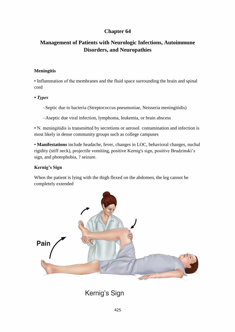
425
Chapter 64
Management of Patients with Neurologic Infections, Autoimmune
Disorders, and Neuropathies
Meningitis
• Inflammation of the membranes and the fluid space surrounding the brain and spinal
cord
• Types
–Septic due to bacteria (Streptococcus pneumoniae, Neisseria meningitidis)
–Aseptic due viral infection, lymphoma, leukemia, or brain abscess
• N. meningitidis is transmitted by secretions or aerosol contamination and infection is
most likely in dense community groups such as college campuses
• Manifestations include headache, fever, changes in LOC, behavioral changes, nuchal
rigidity (stiff neck), projectile vomiting, positive Kernig's sign, positive Brudzinski‘s
sign, and photophobia, ? seizure.
Kernig’s Sign
When the patient is lying with the thigh flexed on the abdomen, the leg cannot be
completely extended

426
Brudzinski’s Sign
When the patient's neck is flexed (after ruling out cervical trauma or injury), flexion of
the knees and hips is produced; when the lower extremity of one side is passively
flexed, a similar movement is seen in the opposite extremity
Assessment and Diagnostic Findings
• Bacterial culture and Gram staining of CSF and blood are key diagnostic tests
(Fischbach, 2002).
• The presence of polysaccharide antigen in CSF further supports the diagnosis of
bacterial meningitis
Medical Management
• Prevention by vaccination against Haemophilus influenzae and S. pneumoniae for all
children and all at-risk adults
• Early administration of high doses of appropriate IV antibiotics (should cross BBB)
for bacterial meningitis
• Dexamethasone
• Treatment dehydration, shock, and seizures
Nursing Management
• Frequent/continual assessment including VS and LOC
• Protect patient form injury related to seizure activity or altered LOC

427
• Monitor daily weight, serum electrolytes, urine volume, specific gravity, and
osmolality
• Prevent complications associated with immobility
• Infection control precautions
• Supportive care
• Measures to facilitate coping of patient and family
Brain Abscess
— Collection of infectious material within brain tissue
— Risk is increased in immunocompromised patient
— Prevent by treating otitis media, mastoiditis, sinusitis, dental infections, and
systemic infections promptly
— Manifestations may include headache that is usually worse in the morning, fever,
vomiting, neurologic deficits, signs and symptoms of increased ICP
— Diagnosis by MRI or CT
— CT-guided aspiration is used to identify the causative organisms
• Medical management
–Control ICP –Drain abscess
–Administer appropriate antibiotic therapy. Corticosteroids may be used to treat
cerebral edema
• Nursing management
–Frequent and ongoing neurologic assessment and of responses to treatment
–Assure patient safety and protect from injury
–Provide supportive care
Encephalitis
• Acute, inflammatory process of the brain tissue
• Causes include viral infections (herpes simplex [HSV]), vector-borne viral infections
(West Nile, St. Louis), and fungal infections

428
• Manifestations may include headache, fever, confusion, changes in LOC; rash, flaccid
paralysis, Parkinson-like movements
• Medical management
–Acyclovir for HSV infection, amphotericin and/or other antifungal agents for fungal
infection
• Nursing management
–Frequent and ongoing assessment
–Supportive care
Multiple Sclerosis (MS)
• A progressive immune-related demyelination disease of the CNS
• Clinical manifestations vary and have different patterns
• Frequently, the disease is relapsing and remitting, has exacerbations and recurrences
of symptoms including fatigue, weakness, numbness, difficulty in coordination, loss of
balance, pain, and visual disturbances
• Medical management
–Disease-modifying therapies; interferon b-1a and interferon b-1b, glatiramer acetate
(Copaxone), and IV methylprednisolone
Process of Demyelination
429
Clinical Manifestations
• Has a relapsing remitting (RR) course. With each relapse, recovery is usually
complete.
• Quadriparesis, cognitive dysfunction, visual loss.
• Fatigue, depression, weakness, numbness, difficulty in coordination, loss of balance,
and pain. Visual disturbances due to lesions in the optic nerves or their connections
may include blurring of vision, diplopia (double vision), patchy blindness (scotoma),
and total blindness.
Multiple Sclerosis treatment
• No cure exists for MS
• Treatment directed toward to relieving the patient's symptoms and provide continuing
support.
• Immunosuppressive agents
Myasthenia Gravis
Autoimmune disorder affecting the myoneural junction
Antibodies directed at acetylcholine at the myoneural junction impair transmission of
impulses
Manifestations
Myasthenia gravis, a motor disorder
Initially symptoms involve ocular muscles; diplopia and ptosis
Weakness of facial muscles, swallowing and voice impairment (dysphonia),
generalized weakness, weakness affects all the extremities and the intercostal
Medical Management
• Pharmacologic therapy
–Cholinesterase inhibitor: pyrostigmine bromide (Mestinon)
–Immunomodulating therapy
• Plasmapheresis
• Thymectomy

430
Normal ACh receptor site ACh receptor site in myasthenia gravis
Guillain-Barré Syndrome
• Autoimmune disorder with acute attack of peripheral nerve myelin
• Rapid demyelination may produce respiratory failure and autonomic nervous system
dysfunction with CV instability
• Most often follows a viral infection
• Manifestations are variable and may include weakness, paralysis, paresthesias, pain,
and diminished or absent reflexes starting with the lower extremities and progressing
upward; tachycardia; bradycardia; hypertension; or
• Medical management
–Requires intensive care management with continuous monitoring and respiratory
support
–Plasmapheresis used to reduce circulating antibodies
• Recovery rates vary, but most patients recover completely
Bell’s Palsy
• Facial paralysis due to unilateral inflammation of the 7th cranial nerve
• Manifestations—unilateral facial muscle weakness or paralysis with facial distortion,
increased lacrimation, and painful sensations in the face, may have difficulty with
speech and eating
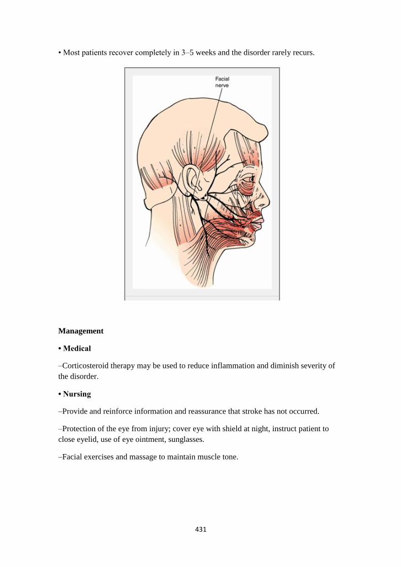
431
• Most patients recover completely in 3–5 weeks and the disorder rarely recurs.
Management
• Medical
–Corticosteroid therapy may be used to reduce inflammation and diminish severity of
the disorder.
• Nursing
–Provide and reinforce information and reassurance that stroke has not occurred.
–Protection of the eye from injury; cover eye with shield at night, instruct patient to
close eyelid, use of eye ointment, sunglasses.
–Facial exercises and massage to maintain muscle tone.