Surgical Management of Movement Disorders
351
-
Upload
khangminh22 -
Category
Documents
-
view
0 -
download
0
Transcript of Surgical Management of Movement Disorders


SurgicalManagement of Movement
Disorders
NDT_half-series-title 6/24/05 12:31 PM Page A

NEUROLOGICAL DISEASE AND THERAPY
Advisory Board
Louis R. Caplan, M.D.Professor of Neurology
Harvard University School of MedicineBeth Israel Deaconess Medical Center
Boston, Massachusetts
William C. Koller, M.D.Mount Sinai School of Medicine
New York, New York
John C. Morris, M.D.Friedman Professor of Neurology
Co-Director, Alzheimer’s Disease Research CenterWashington University School of Medicine
St. Louis, Missouri
Bruce Ransom, M.D., Ph.D.Warren Magnuson Professor
Chair, Department of NeurologyUniversity of Washington School of Medicine
Seattle, Washington
Kapil Sethi, M.D.Professor of Neurology
Director, Movement Disorders ProgramMedical College of Georgia
Augusta, Georgia
Mark Tuszynski, M.D., Ph.D.Associate Professor of Neurosciences
Director, Center for Neural RepairUniversity of California–San Diego
La Jolla, California
NDT_half-series-title 6/24/05 12:31 PM Page B

1. Handbook of Parkinson’s Disease, edited by William C. Koller
2. Medical Therapy of Acute Stroke, edited by Mark Fisher3. Familial Alzheimer’s Disease: Molecular Genetics
and Clinical Perspectives, edited by Gary D. Miner, Ralph W. Richter, John P. Blass, Jimmie L. Valentine, and Linda A. Winters-Miner
4. Alzheimer’s Disease: Treatment and Long-TermManagement, edited by Jeffrey L. Cummings and Bruce L. Miller
5. Therapy of Parkinson’s Disease, edited by William C. Koller and George Paulson
6. Handbook of Sleep Disorders, edited by Michael J. Thorpy7. Epilepsy and Sudden Death, edited by Claire M. Lathers
and Paul L. Schraeder8. Handbook of Multiple Sclerosis, edited by Stuart D. Cook9. Memory Disorders: Research and Clinical Practice,
edited by Takehiko Yanagihara and Ronald C. Petersen10. The Medical Treatment of Epilepsy, edited by
Stanley R. Resor, Jr., and Henn Kutt11. Cognitive Disorders: Pathophysiology and Treatment,
edited by Leon J. Thal, Walter H. Moos, and Elkan R. Gamzu
12. Handbook of Amyotrophic Lateral Sclerosis, edited byRichard Alan Smith
13. Handbook of Parkinson’s Disease: Second Edition, Revisedand Expanded, edited by William C. Koller
14. Handbook of Pediatric Epilepsy, edited by Jerome V. Murphy and Fereydoun Dehkharghani
15. Handbook of Tourette’s Syndrome and Related Tic and Behavioral Disorders, edited by Roger Kurlan
16. Handbook of Cerebellar Diseases, edited by Richard Lechtenberg
17. Handbook of Cerebrovascular Diseases, edited by Harold P. Adams, Jr.
18. Parkinsonian Syndromes, edited by Matthew B. Stern and William C. Koller
19. Handbook of Head and Spine Trauma, edited by Jonathan Greenberg
20. Brain Tumors: A Comprehensive Text, edited by Robert A. Morantz and John W. Walsh
21. Monoamine Oxidase Inhibitors in Neurological Diseases,edited by Abraham Lieberman, C. Warren Olanow,Moussa B. H. Youdim, and Keith Tipton
NDT_half-series-title 6/24/05 12:31 PM Page C

22. Handbook of Dementing Illnesses, edited by John C. Morris
23. Handbook of Myasthenia Gravis and MyasthenicSyndromes, edited by Robert P. Lisak
24. Handbook of Neurorehabilitation, edited by David C. Good and James R. Couch, Jr.
25. Therapy with Botulinum Toxin, edited by Joseph Jankovicand Mark Hallett
26. Principles of Neurotoxicology, edited by Louis W. Chang27. Handbook of Neurovirology, edited by
Robert R. McKendall and William G. Stroop28. Handbook of Neuro-Urology, edited by David N. Rushton29. Handbook of Neuroepidemiology, edited by
Philip B. Gorelick and Milton Alter30. Handbook of Tremor Disorders, edited by Leslie J. Findley
and William C. Koller31. Neuro-Ophthalmological Disorders: Diagnostic Work-Up
and Management, edited by Ronald J. Tusa and Steven A. Newman
32. Handbook of Olfaction and Gustation, edited by Richard L. Doty
33. Handbook of Neurological Speech and LanguageDisorders, edited by Howard S. Kirshner
34. Therapy of Parkinson’s Disease: Second Edition, Revised and Expanded, edited by William C. Koller and George Paulson
35. Evaluation and Management of Gait Disorders, edited by Barney S. Spivack
36. Handbook of Neurotoxicology, edited by Louis W. Changand Robert S. Dyer
37. Neurological Complications of Cancer, edited by Ronald G. Wiley
38. Handbook of Autonomic Nervous System Dysfunction,edited by Amos D. Korczyn
39. Handbook of Dystonia, edited by Joseph King Ching Tsuiand Donald B. Calne
40. Etiology of Parkinson’s Disease, edited by Jonas H. Ellenberg, William C. Koller, and J. William Langston
41. Practical Neurology of the Elderly, edited by Jacob I. Sageand Margery H. Mark
42. Handbook of Muscle Disease, edited by Russell J. M. Lane43. Handbook of Multiple Sclerosis: Second Edition,
Revised and Expanded, edited by Stuart D. Cook
NDT_half-series-title 6/24/05 12:31 PM Page D

44. Central Nervous System Infectious Diseases and Therapy,edited by Karen L. Roos
45. Subarachnoid Hemorrhage: Clinical Management, edited by Takehiko Yanagihara, David G. Piepgras, and John L. D. Atkinson
46. Neurology Practice Guidelines, edited by Richard Lechtenberg and Henry S. Schutta
47. Spinal Cord Diseases: Diagnosis and Treatment, edited byGordon L. Engler, Jonathan Cole, and W. Louis Merton
48. Management of Acute Stroke, edited by Ashfaq Shuaiband Larry B. Goldstein
49. Sleep Disorders and Neurological Disease, edited byAntonio Culebras
50. Handbook of Ataxia Disorders, edited by Thomas Klockgether
51. The Autonomic Nervous System in Health and Disease,David S. Goldstein
52. Axonal Regeneration in the Central Nervous System, edited by Nicholas A. Ingoglia and Marion Murray
53. Handbook of Multiple Sclerosis: Third Edition,edited by Stuart D. Cook
54. Long-Term Effects of Stroke, edited by Julien Bogousslavsky
55. Handbook of the Autonomic Nervous System in Healthand Disease, edited by C. Liana Bolis, Julio Licinio, and Stefano Govoni
56. Dopamine Receptors and Transporters: Function, Imaging, and Clinical Implication, Second Edition, edited by Anita Sidhu, Marc Laruelle, and Philippe Vernier
57. Handbook of Olfaction and Gustation: Second Edition,Revised and Expanded, edited by Richard L. Doty
58. Handbook of Stereotactic and Functional Neurosurgery,edited by Michael Schulder
59. Handbook of Parkinson’s Disease: Third Edition, edited byRajesh Pahwa, Kelly E. Lyons, and William C. Koller
60. Clinical Neurovirology, edited by Avindra Nath and Joseph R. Berger
61. Neuromuscular Junction Disorders: Diagnosis andTreatment, Matthew N. Meriggioli, James F. Howard, Jr.,and C. Michel Harper
62. Drug-Induced Movement Disorders, edited by Kapil D. Sethi
NDT_half-series-title 6/24/05 12:31 PM Page E

63. Therapy of Parkinson’s Disease: Third Edition, Revised andExpanded, edited by Rajesh Pahwa, Kelly E. Lyons, andWilliam C. Koller
64. Epilepsy: Scientific Foundations of Clinical Practice, edited by Jong M. Rho, Raman Sankar, and José E. Cavazos
65. Handbook of Tourette’s Syndrome and Related Tic and Behavioral Disorders: Second Edition, edited by Roger Kurlan
66. Handbook of Cerebrovascular Diseases: Second Edition,Revised and Expanded, edited by Harold P. Adams, Jr.
67. Emerging Neurological Infections, edited by Christopher Power and Richard T. Johnson
68. Treatment of Pediatric Neurologic Disorders,Harvey S. Singer, Eric H. Kossoff, Adam L. Hartman,and Thomas O. Crawford
69. Synaptic Plasticity: Basic Mechanisms to ClinicalApplications, edited by Michel Baudry, Xiaoning Bi, and Steven S. Schreiber
70. Handbook of Essential Tremor and Other TremorDisorders, edited by Kelly E. Lyons and Rajesh Pahwa
71. Handbook of Peripheral Neuropathy, edited by Mark B. Bromberg and A. Gordon Smith
72. Carotid Artery Stenosis: Current and Emerging Treatments,edited by Seemant Chaturvedi and Peter M. Rothwell
73. Gait Disorders: Evaluation and Management, edited byJeffrey M. Hausdorff and Neil B. Alexander
74. Surgical Management of Movement Disorders, edited byGordon H. Baltuch and Matthew B. Stern
NDT_half-series-title 6/24/05 12:31 PM Page F

Surgical Management of Movement
Disorders
edited by
Gordon H. BaltuchUniversity of Pennsylvania
Philadelphia, Pennsylvania, U.S.A.
Matthew B. SternUniversity of Pennsylvania
Philadelphia, Pennsylvania, U.S.A.
Boca Raton London New York Singapore
NDT_half-series-title 6/24/05 12:31 PM Page i

Published in 2005 byTaylor & Francis Group 6000 Broken Sound Parkway NW, Suite 300Boca Raton, FL 33487-2742
© 2005 by Taylor & Francis Group, LLC
No claim to original U.S. Government worksPrinted in the United States of America on acid-free paper10 9 8 7 6 5 4 3 2 1
International Standard Book Number-10: 0-8247-2950-1 (Hardcover) International Standard Book Number-13: 978-0-8247-2950-9 (Hardcover)
This book contains information obtained from authentic and highly regarded sources. Reprinted material isquoted with permission, and sources are indicated. A wide variety of references are listed. Reasonable effortshave been made to publish reliable data and information, but the author and the publisher cannot assumeresponsibility for the validity of all materials or for the consequences of their use.
No part of this book may be reprinted, reproduced, transmitted, or utilized in any form by any electronic,mechanical, or other means, now known or hereafter invented, including photocopying, microfilming, andrecording, or in any information storage or retrieval system, without written permission from the publishers.
For permission to photocopy or use material electronically from this work, please access www.copyright.com(http://www.copyright.com/) or contact the Copyright Clearance Center, Inc. (CCC) 222 Rosewood Drive,Danvers, MA 01923, 978-750-8400. CCC is a not-for-profit organization that provides licenses and registrationfor a variety of users. For organizations that have been granted a photocopy license by the CCC, a separatesystem of payment has been arranged.
Trademark Notice: Product or corporate names may be trademarks or registered trademarks, and are used onlyfor identification and explanation without intent to infringe.
Library of Congress Cataloging-in-Publication Data
Catalog record is available from the Library of Congress
Visit the Taylor & Francis Web site at http://www.taylorandfrancis.com
Taylor & Francis Group is the Academic Division of T&F Informa plc.
DK3798_Discl.fm Page 1 Monday, July 11, 2005 4:15 PM

To our families, for their support


Preface
Movement disorders represent major causes of neurological disability andeventual mortality affecting millions of people across the globe. FromParkinson’s disease to spasticity, these neurological disorders devastateyoung and old worldwide. While progress continues to be made towardeffective treatment, many limitations remain.
The combination of the limitation of medical therapy and surgicaltechnological advances have, however, led to an exponential growth in func-tional neurosurgery in the last 5 years. Surgery represents an alternativewhere there existed only finite treatment options before. This field is devel-oping rapidly with emerging novel therapies as well as evolving indicationsfor existing procedures.
We intended this book to be a thorough review of the surgical treat-ments currently available for various movement disorders, with an emphasison surgical indications and results of surgery. It should be of utmost interestto practitioners and trainees in the clinical neurosciences (neurology/neuro-surgery) who want a better understanding of candidates for movement dis-order surgery, the current surgical procedures, the effected results of surgery,and the complication rate of these procedures. Our goal was to summarizethe current status of the field as well as make projections for the next fewyears.
Gordon H. BaltuchMatthew B. Stern
v

Contents
Preface . . . . vContributors . . . . xiii
1. Overview . . . . . . . . . . . . . . . . . . . . . . . . . . . . . . . . . . . . 1Kelvin L. Chou, Gordon H. Baltuch, and Matthew B. Stern1. Introduction . . . . 12. Pathophysiology of Movement Disorders . . . . 23. Surgical Treatment of Movement Disorders . . . . 54. Conclusions . . . . 16
References . . . . 16
SECTION I: PARKINSON’S DISEASE
2. Patient Selection and Indications for Surgery . . . . . . . . . . 27Jill L. Ostrem, Philip A. Starr, and William J. Marks, Jr.1. Introduction . . . . 272. Importance of Patient Selection . . . . 283. Indications for Surgery . . . . 284. Ablative Procedures vs. Deep Brain Stimulation . . . . 355. Unilateral vs. Bilateral Treatment . . . . 366. Previous Surgery for Parkinson’s Disease . . . . 377. Areas of Controversy . . . . 378. Future of Parkinson’s Disease Surgery and Patient
Selection . . . . 39References . . . . 40
vii

3. Surgical Technique and Complication Avoidance . . . . . . . . 45Joshua M. Rosenow and Ali R. Rezai1. Introduction . . . . 452. Patient Selection and Preparation . . . . 463. Frame Placement . . . . 464. Target Planning . . . . 475. Surgical Technique . . . . 516. Mapping the STN: Confirming the Optimal Target . . . . 537. Securing the Electrode . . . . 558. Adverse Events . . . . 579. Postoperative Care . . . . 5810. IPG Implantation . . . . 59
References . . . . 61
4. Surgical Results . . . . . . . . . . . . . . . . . . . . . . . . . . . . . . 63Jean-Guy Villemure, Joseph Ghika, and Francois Vingerhoets1. Introduction . . . . 632. Thalamus . . . . 643. Pallidum . . . . 654. Subthalamic Nucleus . . . . 675. Preoperative Physiological Definition of the
Target . . . . 706. Conclusions . . . . 72
References . . . . 73
5. Novel Surgical Strategies: Motor Cortex Stimulation,Transplantation, Gene Therapy, Stem Cells, andCNS Drug Delivery . . . . . . . . . . . . . . . . . . . . . . . . . . . . 83Jason M. Schwalb and Andres M. Lozano1. Introduction . . . . 832. Limitations of Current Therapy . . . . 833. Motor Cortex Stimulation . . . . 844. History of Transplantation . . . . 855. Ethics of Sham Surgery . . . . 896. Future Directions in Implanting Dopaminergic
Neurons . . . . 907. Induction of Endogenous Stem Cells . . . . 928. Trophic Factors Rather than Dopaminergic Neurons—
GDNF Therapy . . . . 929. Gene Therapy for Parkinson’s Disease . . . . 93
viii Contents

10. Genetic Manipulation of Stem Cells . . . . 9611. Conclusions . . . . 9612. Additional Resources . . . . 97
References . . . . 97
SECTION II: ESSENTIAL TREMOR
6. Essential Tremor: Patient Selection, Technique,and Surgical Results . . . . . . . . . . . . . . . . . . . . . . . . . . 111Eun-Kyung Won, Habib E. Ellamushi, Uzma Samadani, andGordon H. Baltuch1. Introduction . . . . 1112. Clinical Characteristics and Epidemiology . . . . 1113. Genetics and Etiology . . . . 1134. Pathophysiology . . . . 1135. Pharmacological Treatment for Essential Tremor . . . . 1156. Surgical Therapy for ET . . . . 116
References . . . . 117
SECTION III: MS, POST-STROKE ANDPOST-TRAUMATIC TREMOR
7. Tremor: Patient Selection and Surgical Results . . . . . . . . 123Eun-Kyung Won, Uzma Samadani, and Gordon H. Baltuch1. Introduction . . . . 1232. Tremor in Multiple Sclerosis . . . . 1243. Post-Traumatic Tremor . . . . 1274. Tremor After Cerebral Infarct . . . . 129
References . . . . 130
SECTION IV: DYSTONIA
8. Dystonia: Classification, Etiology, and TherapeuticOptions . . . . . . . . . . . . . . . . . . . . . . . . . . . . . . . . . . . 135Galit Kleiner-Fisman and Santiago Figuereo1. Introduction . . . . 1352. Classification . . . . 1363. Etiology and Pathophysiology . . . . 1394. Best Medical Therapy . . . . 1425. Surgical Therapy . . . . 1486. Anatomical Targets in DBS Surgery . . . . 149
Contents ix

7. Surgical Technique . . . . 1538. Results of Surgery in Dystonia . . . . 1549. Conclusions . . . . 157
References . . . . 157
SECTION V: CERVICAL DYSTONIA
9. Chemodenervation: Botulinum Toxin . . . . . . . . . . . . . . . 169Tanya Simuni1. Introduction . . . . 1692. Clinical Efficacy of BTX-A for CD . . . . 1723. BTX-B . . . . 1754. Other BTX Strains . . . . 1765. Conclusions . . . . 176
References . . . . 177
10. Ablative Denervation . . . . . . . . . . . . . . . . . . . . . . . . . . 183Jeff D. Golan and Line Jacques1. Introduction . . . . 1832. Clinical Presentation . . . . 1843. Etiology . . . . 1854. Clinical Forms of Torticollis . . . . 1865. Clinical Evaluation . . . . 1866. Treatment . . . . 1887. Selective Peripheral Denervation . . . . 1908. Results . . . . 1959. Summary . . . . 196
References . . . . 197
11. Deep Brain Stimulation . . . . . . . . . . . . . . . . . . . . . . . . 203Atsushi Umemura1. Introduction . . . . 2032. Early Attempts in Ablative Procedure for ST . . . . 2043. DBS for ST . . . . 2054. Rationale of DBS for ST . . . . 2075. Technical Consideration of DBS for ST . . . . 208
References . . . . 210
12. Tourette’s Syndrome Surgical Therapy . . . . . . . . . . . . . . 215Habib E. Ellamushi and Gordon H. Baltuch1. Introduction . . . . 215
x Contents

2. Pathophysiology of Tourette’s Syndrome . . . . 2163. Conservative Treatment of Tourette’s Syndrome . . . . 2174. Ablative Surgical Treatment for Tourette’s
Syndrome . . . . 2175. Deep Brain Stimulation for Tourette’s Syndrome . . . . 2206. Conclusions . . . . 221
References . . . . 221
SECTION VI: HEMIFACIAL SPASM
13. Botulinum Toxin/Microvascular Decompression—Indication,Technique, and Surgical Results . . . . . . . . . . . . . . . . . . 227Frederic Schils, Nicolas de Tribolet, and Michel R. Magistris1. Introduction . . . . 2272. Epidemiology . . . . 2283. Symptoms and Clinical Signs . . . . 2294. Diagnosis, Staging, Classification . . . . 2295. Physiopathology . . . . 2306. Surgical Anatomy . . . . 2327. Treatment . . . . 2348. Contraindications to Surgery . . . . 2389. Results . . . . 238
10. Complications . . . . 23911. Conclusions . . . . 240
References . . . . 241
SECTION VII: SPASTICITY
14. Cerebral Palsy . . . . . . . . . . . . . . . . . . . . . . . . . . . . . . 245Jean-Pierre Farmer and Sandeep Mittal1. Introduction . . . . 2452. Epidemiology and Demographics . . . . 2463. Pathophysiology . . . . 2464. Diagnosis and Clinical Presentation . . . . 2495. Classification . . . . 2506. Therapeutic Avenues . . . . 252
References . . . . 255
15. Baclofen . . . . . . . . . . . . . . . . . . . . . . . . . . . . . . . . . . . 257Jean-Pierre Farmer and Sandeep Mittal1. Introduction . . . . 257
Contents xi

2. Baclofen . . . . 2573. Pump Technology . . . . 2594. Selection of Patients for Intrathecal Baclofen
Infusion . . . . 2605. Preoperative Evaluation . . . . 2606. Screening Test . . . . 2617. Pump Implantation . . . . 2628. Intrathecal Baclofen Therapy . . . . 2639. Complications . . . . 26510. Outcome . . . . 27011. Conclusions . . . . 272
References . . . . 272
16. Rhizotomy . . . . . . . . . . . . . . . . . . . . . . . . . . . . . . . . . 277Jean-Pierre Farmer and Sandeep Mittal1. Introduction . . . . 2772. Historical Background . . . . 2783. Rationale Underlying Selectivity of Posterior
Rhizotomy . . . . 2784. Preoperative Evaluation . . . . 2805. Surgical Treatment . . . . 2876. Outcome . . . . 2927. Complications . . . . 3078. Conclusions . . . . 311
References . . . . 311
Index . . . . 319
xii Contents
Contributors
Gordon H. Baltuch Department of Neurosurgery, Penn NeurologicalInstitute, University of Pennsylvania School of Medicine, Philadelphia,Pennsylvania, U.S.A.
Kelvin L. Chou Department of Clinical Neurosciences, BrownUniversity Medical School, Providence, Rhode Island, U.S.A.
Nicolas de Tribolet Department of Neurosurgery, University of Geneva,Geneva, Switzerland
Habib E. Ellamushi The Royal Hospital of St. Bartholomew and TheRoyal London Hospital, London, U.K.
Jean-Pierre Farmer Division of Pediatric Neurosurgery, McGillUniversity Health Centre, Montreal, Quebec, Canada
Santiago Figuereo Department of Neurosurgery, Philadelphia VeteransAdministration Hospital, University of Pennsylvania, Philadelphia,Pennsylvania, U.S.A.
Joseph Ghika Neurology Service, Centre Hospitalier UniversitaireVaudois, Lausanne, Switzerland
Jeff D. Golan Division of Neurosurgery, McGill University, Montreal,Quebec, Canada
Line Jacques Division of Neurosurgery, McGill University, Montreal,Quebec, Canada
xiii

Galit Kleiner-Fisman Parkinson’s Disease Research Education andClinical Center (PADRECC), Philadelphia Veterans AdministrationHospital, University of Pennsylvania, Philadelphia, Pennsylvania, U.S.A.
Andres M. Lozano Division of Neurosurgery, Toronto WesternHospital, University of Toronto and University Health Network,Toronto, Ontario, Canada
Michel R. Magistris Department of Neurology, University of Geneva,Geneva, Switzerland
William J. Marks, Jr. Department of Neurology, University ofCalifornia, San Francisco and San Francisco Veterans Affairs MedicalCenter, San Francisco, California, U.S.A.
Sandeep Mittal Division of Pediatric Neurosurgery, McGill UniversityHealth Centre, Montreal, Quebec, Canada
Jill L. Ostrem Department of Neurology, University of California,San Francisco and San Francisco Veterans Affairs Medical Center,San Francisco, California, U.S.A.
Ali R. Rezai Department of Neurosurgery, Cleveland Clinic LernerCollege of Medicine, Cleveland, Ohio, U.S.A.
Joshua M. Rosenow Department of Neurosurgery, Feinberg School ofMedicine of Northwestern University, Chicago, Illinois, U.S.A.
Uzma Samadani Department of Neurosurgery, University ofPennsylvania, Philadelphia, Pennsylvania, U.S.A.
Frederic Schils Department of Neurosurgery, University of Geneva,Geneva, Switzerland
Jason M. Schwalb Division of Neurosurgery, Toronto WesternHospital, University of Toronto and University Health Network,Toronto, Ontario, Canada
Tanya Simuni Department of Neurology, Feinberg School of Medicine,Northwestern University, Chicago, Illinois, U.S.A.
Philip A. Starr Department of Neurosurgery, University of California,San Francisco and San Francisco Veterans Affairs Medical Center,San Francisco, California, U.S.A.
xiv Contributors

Matthew B. Stern Parkinson’s Disease and Movement DisordersCenter, Pennsylvania Hospital, University of Pennsylvania School ofMedicine, Philadelphia, Pennsylvania, U.S.A.
Atsushi Umemura Department of Neurosurgery, Nagoya CityUniversity Medical School, Mizuho-ku, Nagoya, Japan
Jean-Guy Villemure Neurosurgery Service, Centre HospitalierUniversitaire Vaudois, Lausanne, Switzerland
Francois Vingerhoets Neurology Service, Centre HospitalierUniversitaire Vaudois, Lausanne, Switzerland
Eun-Kyung Won Department of Neurosurgery, University ofMinnesota, Minneapolis, Minnesota, U.S.A.
Contributors xv


1
Overview
Kelvin L. Chou
Department of Clinical Neurosciences, Brown University Medical School,Providence, Rhode Island, U.S.A.
Gordon H. Baltuch
Department of Neurosurgery, Penn Neurological Institute, University ofPennsylvania School of Medicine, Philadelphia, Pennsylvania, U.S.A.
Matthew B. Stern
Parkinson’s Disease and Movement Disorders Center, Pennsylvania Hospital,University of Pennsylvania School of Medicine, Philadelphia, Pennsylvania, U.S.A.
1. INTRODUCTION
From Parkinson’s disease (PD) to dystonia, movement disorders are majorneurologic causes of disability, causing not only physical handicap, butoften social embarrassment as well. Although a plethora of pharmacologicoptions exists to treat these disorders, many limitations unfortunatelyremain. Functional neurosurgery now has the ability to reduce the severity ofsymptoms and improve the quality of life for patients with these devastatingneurological disorders.
Surgery for the field of movement disorders has evolved significantlysince Spiegel et al. first described stereotaxis in 1947 (1).Much of this progresshas been made within the last decade with the development of techniquesfor precise targeting of brain structures and the discovery of new targetsand indications for existing procedures such as deep brain stimulation(DBS). This chapter provides a general overview of the field of movement
1

disorders surgery, including a brief summary of basal ganglia structure andfunction, as well as discussions of the major surgical treatments available forPD, essential tremor (ET), other tremor disorders, dystonia, and spasticity.
2. PATHOPHYSIOLOGY OF MOVEMENT DISORDERS
All movement disorders are believed to result from abnormalities of the basalganglia.Our understanding of the organization and function of the basal gang-lia has grown significantly in the modern era, largely as a result of recentexperience with stereotactic neurosurgery in humans and animal models.Although current theories do not adequately account for the clinical findingsseen with all disorders of involuntary movement, it is still essential for thoseinterested in functional neurosurgical procedures to be familiar with the basicanatomy and functional organization of the basal ganglia.
2.1. Structure and Function of the Basal Ganglia
A detailed discussion of basal ganglia anatomy and function is beyond thescope of this chapter, but a brief summary follows. The basal ganglia areorganized into several parallel circuits (associative, limbic, motor, andoculomotor) that connect cortical regions with thalamic and basal ganglianuclei (2). Disturbances of the motor circuit are believed to manifestas movement disorders. In the classical model of basal ganglia function, abalance between two opposing pathways in the motor circuit, the directand indirect pathways, regulates normal voluntary movement (3). Both path-ways begin with neurons in cortical motor areas projecting to the putamen,which in turn sends signals that ultimately terminate in the internal segmentof the globus pallidus (GPi). Putamenal output in the indirect pathway,however, travels through the external segment of the globus pallidus (GPe)and subthalamic nucleus (STN) before it reaches the GPi, whereas the directpathway projects directly from the putamen to the GPi. The majority ofGPi output is directed toward the thalamus, which sends projections to thesupplementary motor area, the premotor cortical area, and the primarymotor cortex. Activation of the direct pathway decreases the normal inhi-bitory outflow from the GPi to the thalamus, leading to increased corticalmotor activation and the facilitation of voluntary movement. In contrast,stimulation of the indirect pathway causes inhibition of the GPe, disinhibitionof STN excitatory fibers, and an increase in the inhibitory outflow from theGPi onto the thalamus, resulting in decreased output to the motor cortexand suppression of voluntary movement.
The exact mechanism by which the basal ganglia interpret theinformation flowing through these two opposing motor circuit pathwaysto control normal movement is unclear. Two theories have been proposed:scaling and focusing (2–4). In the scaling hypothesis, movement is controlled
2 Chou et al.

by temporally changing activity in the basal ganglia. For example, putamenaloutput would first facilitate a particular movement by disinhibiting the thala-mus through the direct pathway, and subsequently stop the ongoingmovementby causing the same GPi neurons to increase inhibition of the thalamusthrough the indirect pathway. In the focusing hypothesis, movement is allowedto proceed by stimulation of the direct pathway, whereas extra, unnecessarymovements are suppressed by activity in the indirect pathway. Unfortunately,both models are likely oversimplifications of the true underlying mechanism,since they do not account for all experimental findings.
2.2. Pathophysiologic Models of Hypo- and HyperkineticMovement Disorders
Based on the model of basal ganglia function described above, an imbalancebetween the direct and indirect pathways accounts for the clinical manifes-tations of hypo- and hyperkinetic disorders. The release of dopamine fromnerve terminals in the striatum appears to stimulate the direct pathwayand inhibit the indirect pathway, thus effectively facilitating movement (5).Consequently, degeneration of the dopaminergic nigrostriatal pathway, asseen in parkinsonism, would result in decreased dopamine receptor activa-tion and disinhibition of the indirect pathway, leading to increased activityin the STN and GPi. The increased inhibitory outflow from GPi on thethalamus would decrease cortical motor activation, with the end result beingslowed movements, or bradykinesia. Conversely, hyperkinetic disorders suchas Huntington’s disease (HD) are thought to result from excess dopamine,causing overactivity of the direct pathway, less inhibitory activity fromGPi on the thalamus, increased activity of the thalamocortical projections,and excessive involuntary movement.
2.2.1. Hypokinetic Movement Disorders
There is considerable evidence supporting the classicalmodel for the parkinso-nian state. For example, in 1-methyl-4-phenyl-1,2,3,6-tetrahydropyridine(MPTP)-treated monkeys, neuronal firing frequency is increased in the STNand GPi and reduced in the GPe, consistent with the predictions of the abovemodel (6,7). Further support comes from advanced PD patients treated withSTN and GPi DBS, where stimulation clearly improves parkinsonian symp-toms (8–11) and increases activity in cortical motor areas as detected bypositron emission tomography (PET) imaging (12).
There are many inconsistencies with this model, however. For instance, alesion in the thalamus should worsen PD motor symptoms due to decreasedactivity of the thalamocortical projections, yet, thalamic lesioning and thalamicDBS have both been proven to suppress parkinsonian tremor (13,14). In addi-tion, pallidotomy should result in excessive movement (i.e., dyskinesias)
Overview 3

because of decreased inhibition on the thalamus, but in fact, the opposite istrue (15).
This has led to a rethinking of the classical model, with some investi-gators proposing that it is the overall pattern of basal ganglia activity that isaltered in PD, rather than the rate of activity of individual structures (16).Studies have suggested that in the normal brain, information flows throughindependent and parallel circuits within the basal ganglia, while in the par-kinsonian brain, these circuits break down, and interconnections betweenthese circuits become active and synchronized (17). This synchronizationis believed to contribute to the parkinsonian state. Lesions of the STNand GPi may change this pattern back to a more ‘‘normal’’ pattern ofneuronal transmission through the basal ganglia. Indirect evidence for thiscomes from human PET imaging studies that have shown a restoration ofnormal cortical activity after lesions of the STN or GPi (12,18).
2.2.2. Hyperkinetic Movement Disorders
As explained above, the classical model predicts that hyperkinetic move-ment disorders result from a reduction in GPi activity and an increase inthalamic and cortical activity. In fact, it has been shown that MPTP-treatedmonkeys given levodopa or apomorphine have a gradual reduction in GPifiring rate as the monkeys transition from an ‘‘off’’ state, to an ‘‘on’’ statewithout dyskinesias, to an ‘‘on’’ state with dyskinesias, supporting thismodel (19). GPi discharge rates are also reduced in other hyperkinetic dis-orders, such as dystonia and hemiballismus (16). However, decreased GPiactivity cannot account for hyperkinetic movements by itself; otherwise,lesions of the globus pallidus would be expected to result in extra move-ments. As with the parkinsonian model, many experts now think that itis the pattern of GPi activity, rather than the rate of GPi activity, that isimportant in determining movement (16,20). It may be that the absence ofactivity is preferable to abnormal activity, which is why surgical lesioningimproves clinical symptoms.
The pathophysiology of tremor, whether in PD, ET, or cerebellartremor, is even less clear, and remains hotly debated. There may be someperipheral afferent component to the development of tremor, but it is morelikely that a central mechanism is responsible. Evidence for this includesrecordings of neuronal rhythmic activity in the thalamus that are synchro-nized with tremor activity in the limb in patients with ET and cerebellartremor (21), and recordings of rhythmic activity in the STN that correlatewith contralateral tremor in PD patients (22,23). Other supporting evidenceincludes the fact that thalamic stimulation improves tremor, whether for PDor ET (13). Whatever the true mechanism, however, it is clear that muchwork remains to be completed before we can fully comprehend the complexmechanisms underlying abnormal movements.
4 Chou et al.

3. SURGICAL TREATMENT OF MOVEMENT DISORDERS
3.1. Parkinson’s Disease
PD is a slowly progressive neurodegenerative disorder characterized bya clinical triad of motor features—tremor, bradykinesia, and rigidity. Idealcandidates for surgery should have a clear response to levodopa therapy,but continue to have severe motor fluctuations, dyskinesias, or intractabletremor despite optimal medical therapy. Older patients tend to fare worseafter surgery, as do patients with cognitive impairment, so comprehensiveneuropsychological testing should be performed prior to surgery (10,24).It is also being increasingly recognized that psychiatric symptoms suchas depression worsen after DBS procedures (25), so more centers are incor-porating a psychiatric screen into the DBS evaluation process. Patients withatypical parkinsonism, such as progressive supranuclear palsy and multiplesystem atrophy, are not candidates for surgery (26,27).
3.1.1. Ablative Procedures
Prior to the discovery of levodopa, thalamotomies were performed routinelyfor PD. However, they were only effective for tremor, and bilateral proce-dures resulted in a high incidence of dysarthria and cognitive side effects(28,29). Given the excellent results from pallidotomy in PD and the develop-ment of DBS, which holds the advantage of reversibility and fewer sideeffects with bilateral procedures, thalamotomies are no longer recommendedfor treatment of PD.
Laitinen et al. (30) revived the posteroventral pallidotomy for thetreatment of PD in 1992. Of 38 patients followed for a mean of 28 months,92% had ‘‘complete relief or almost complete relief’’ of bradykinesia and rigid-ity, and that 81% had ‘‘complete relief or almost complete relief’’ of tremor.While many authors have reported similar results from this procedure withshort-term follow-up (15,31–34), other investigators, in contrast to Laitinenet al. (30), have reported a decline in surgical benefit from pallidotomy withlong-term follow-up (35–37). Although these studies have consistently shownthat pallidotomy’s effects on tremor and dyskinesias are maintained at 3–5years, other measures, such as the total off-period motor score, the ipsilateralmotor score, the contralateral bradykinesia subscore, and the activities of dailyliving subscore, deteriorate with time. However, it should be noted that all ofthese studies have followed unilateral pallidotomy patients only, largelybecause of reports of unacceptable speech and neuro behavioral side effectsfrom bilateral pallidotomy (38–41). As the natural history of PD suggests thatthe disease will eventually spread bilaterally, it is not surprising that the benefitof unilateral pallidotomy diminishes with time.
With regards to subthalamotomy, although animal studies hadrepeatedly shown that parkinsonian signs improved with ablative lesions ofthe STN, very few surgeons attempted to lesion the STN in humans for fear
Overview 5

of causing hemiballismus (42). However, most clinical reports have shownthat hemiballismus in subthalamotomy patients does not occur as often aspreviously thought (43–46). Unilateral subthalamotomy in patients withadvanced PD has been demonstrated to decrease the Unified Parkinson’sDisease Rating Scale (UPDRS) off-medication motor scores by about 30–50%(44–46). Persistent, lesion-induced dyskinesias have been problematic for onlya few patients overall, with two requiring additional surgical procedures tocontrol the choreic movements and another dying from aspiration pneumonia.The results of bilateral subthalamotomy have been reported for only two PDpatients in the literature (43). Both patients showed an impressive reduction inUPDRS motor scores, with one patient going from 50 to 16, and the secondimproving from 64 to 16, without adverse effects.
3.1.2. Deep Brain Stimulation
Stimulation of the thalamic ventralis intermedius (VIM) nucleus for parkin-sonian or other tremor of the limbswas the first Food andDrugAdministration(FDA) approved indication for DBS, heralding a new era in functional neuro-surgery. ThalamicDBS, inmultiple long-term studies, has been shown to be safeand effective for parkinsonian tremor (47–49). Unfortunately, as with thala-motomy, other parkinsonian symptoms are unimproved and prevent thisprocedure from being useful in the long-term treatment of PD.
With reports of pallidotomy being effective for all the motor featuresof PD, and the gaining acceptance of thalamic stimulation for tremor, manysurgical centers tried to see if stimulation of the GPi could help the parkin-sonian symptoms not addressed by VIM stimulation, and the results havebeen positive. In one of the largest prospective double blind studies pub-lished, 38 PD patients underwent implantation of DBS electrodes bilaterallyin the GPi (11). Stimulation produced a 37% median improvement in the offmedication UPDRS score at 6 months. Furthermore, the amount of ‘‘on’’time during the day without dyskinesias increased from 28% to 64%, whileamount of ‘‘off’’ time decreased from 37% to 24%. The dyskinesia scoreimproved by about 66%. Unfortunately, the mean daily levodopa dosewas not significantly changed.
An increasing number of long-term studies evaluating GPi stimulationin patients with PDhave been published in recent years, but it appears that thebenefits seen early on may not endure. Visser-Vandewalle et al. (50) followed26 patients with unilateral GPi electrodes for a mean of 32.7 months, andfound that while GPi stimulation was beneficial at 3 months for symptomsboth contralaterally and ipsilaterally, it was unsatisfactory at long-termfollow-up, leading the authors to conclude that unilateral pallidal stimulationwas not an effective treatment for advanced PD. Ghika et al. (51) followed sixPD patients treated with bilateral pallidal stimulation for 2 years, and whilethere were still significant improvements in the UPDRS motor score andin the amount of ‘‘on’’ time at 2 years compared to baseline, a slight
6 Chou et al.

worsening after 1 year was observed. In the six patients with bilateral GPielectrodes for PD followed for a mean of 3 years by Durif et al. (52), theamount of time spent in the ‘‘off’’ state returned to the preoperative valueafter 2 years, even though improvements in UPDRS motor scores andseverity of dyskinesias were sustained. Finally, Volkmann et al. (53) followed11 patients treated with chronic bilateral pallidal stimulation for up to 5 years.Although there was a sustained reduction in dyskinesias at 5 years, UPDRSmotor scores declined over time, despite continued levodopa responsivenessand changes in electrical stimulation parameters. Overall, the levodopa doseremained unchanged after surgery, but had to be increased in some patients.In all of these studies, complications were generally transient, and could berelieved by adjusting the stimulator. The hypophonia, dysarthria, and neuro-behavioral disturbances commonly reported with bilateral pallidotomy(38,40,41) were not seen with bilateral GPi stimulation.
In contrast, the benefits of DBS for PD appear to be longer lastingwith STN stimulation, a technique that was pioneered by Benabid and hiscolleagues in the mid-1990s (54,55). Initial studies of subthalamic stimula-tion in patients with advanced PD demonstrated a reduction in the UPDRSmotor score by 40–60%, as well as a decrease in dyskinesias and the dailylevodopa dose by about 50% (8,10,11). STN DBS also decreased the amountof time in the off medication state, and other studies showed improvementin gait and balance (56), and even off-period dystonia (55). In the longestlong-term follow-up study of STN stimulation in PD to date, Krack et al.reported that the UPDRS motor scores at 5 years while off medication were54% better than preoperative scores (57). Furthermore, average dailylevodopa dose and severity of dyskinesias continued to be significantlydecreased compared to baseline. Unfortunately, speech, postural stability,and freezing continued to progress, which the authors concluded wasconsistent with the natural history of PD. Two other groups found similarresults at 2 years (58,59), although the Toronto group (59) noticed thatUPDRS axial subscores, as well as the UPDRS motor score, had diminishedat the most recent follow-up. For all of these studies, most adverse eventsappeared to be transient; however, persistent events included cognitivedecline, and mood changes such as depression and anxiety. One patient inthe study by Krack et al. (57) committed suicide.
3.1.3. Comparison of Targets and Procedures
Asmentioned above, the thalamus has fallen by the wayside as a target for PDbecause of its limited effects on bradykinesia and rigidity. The GPi and STNare clearly the two structures most commonly targeted today for PD, andmost centers are using DBS rather than ablative procedures even though nodouble-blind, randomized, head-to-head comparison has been conductedbetween ablative therapy and DBS.
Overview 7
It is unclear whether GPi DBS or STN DBS is superior. Burchiel et al.(8) conducted a small randomized prospective trial comparing pallidal tosubthalamic stimulation in 10 patients with advanced PD, and found nosignificant difference in off medication UPDRS motor scores between thetwo groups, although medications were able to be decreased in the STNgroup but not the GPi group. This trial was too small, however, to drawany solid conclusions. The Deep Brain Stimulation for Parkinson’s DiseaseStudy Group performed a large multicenter trial that enrolled 96 patientswith STN DBS and 38 patients with GPi-DBS (11). Because patients werenot randomized in this trial, it is difficult to compare the two groups, butthe STN group appeared to have more favorable outcomes. Another pro-spective nonrandomized study carried out by Krause et al. (60) also sug-gested that the STN was the better target for all parkinsonian symptoms,but GPi stimulation was superior for reduction of dyskinesias.
One of the most consistent differences between the two sites is thatSTN stimulation allows patients to reduce dopaminergic medications,whereas daily levodopa equivalent doses in those undergoing pallidal stimu-lation remain largely unchanged. Many studies have also noted that STNstimulation requires less electrical energy than GPi-DBS (11,61), whichallows for a longer battery life. GPi stimulation, however, does not seemto be associated as consistently with cognitive and psychiatric side effectsas STN stimulation (51,53,57,59). Clearly, a large randomized comparisonbetween the two targets needs to be completed before recommending oneprocedure over the other. The VA and NIH are currently supporting amulticenter trial of DBS in PD to address the comparative efficacy of GPiand STN stimulation.
3.1.4. Novel Surgical Strategies
Although DBS and surgical ablation are effective for the symptoms of PD,they may not interfere with the underlying neurodegenerative process andthus do not slow down or reverse the course of the illness. Consequently,much research is being conducted to evaluate alternative therapies and stra-tegies that may alter the long-term outcome in these patients. One of thesenovel approaches is cell transplantation. Thus far, two human clinical trialsof fetal nigral transplantation have been conducted, and although bothstudies showed uptake of the embryonic dopamine neurons in the basalganglia on imaging and post-mortem examinations, both trials failed tomeet statistical significance in their outcome measures (62,63). An unantici-pated finding was the development of disabling off-medication dyskinesiasin some transplanted patients. These findings, along with ethical concernsand the scarcity of human fetal tissue, currently limit the usefulness of fetalnigral transplantation as a viable clinical option for patients with PD. Othersources of cells for transplantation into patients include xenografts (fromfetal pigs) (64) and embryonic stem cells, though these techniques still need
8 Chou et al.

to be refined and tried in small groups of patients before a large clinical trialcan even be attempted (65).
Another top prospect for therapeutic trials in PD is glial derivedneurotrophic factor (GDNF). GDNF has been extensively studied in animalmodels of PD and promotes dopamine neuron survival and growth (66). Sofar, a few clinical trials with GDNF have been undertaken in patients withPD. In the first trial, conducted by Nutt et al. (67), GDNF or placebo wasadministered through an intracerebroventricular catheter in 50 patients withadvanced PD. The patients had multiple side effects, including nausea,vomiting, and anorexia, but did not have any improvement in UPDRSscores, likely because the GDNF did not reach the appropriate brainregions. Gill et al. (68) infused GDNF directly into the putamen of fivePD patients, and reported a 39% improvement in the UPDRS off-medica-tion motor score and a reduction in dyskinesias by about 64%. In addition,fluorodopa scans showed a 28% increase in putamenal uptake. However, arecent double-blind trial of intraputaminal GDNF was stopped because asix month analysis indicated no clinical benefit, causing the manufacturer,Amgen, to withdraw GDNF from all clinical trials.
3.2. Essential Tremor
Tremor is a rhythmic oscillation of a body part that is produced by alternatingcontractions of reciprocally innervated muscles (69). Of all the tremorsyndromes, ET is the most common (70), yet fewer ET patients are referredfor surgery than PD patients. One probable reason is that most ET patientssuffer from a mild tremor which is either not severe enough to seek medicaltreatment, or can be controlled with medication (71). However, when tremorin ET is severe, it can be severely disabling, as well as cause significant socialembarrassment. ET generally presents as a bilateral, symmetric posturaland/or kinetic tremor of the hands, but it can commonly affect the head andvoice as well (72). Primidone and propranolol are considered first-line therapyand should be tried alone or in combination before considering surgery (71).
The thalamus has been considered the optimal target for tremor eversince Hassler and colleagues reported better tremor control with thalamiclesions over 40 years ago (73,74). However, because of the success of STNstimulation in controlling parkinsonian tremor (8,11,22,23), some groupshave begun to experiment with STN DBS for ET, with promising resultsin a small number of patients to date (75–78).
3.2.1. Ablative Procedures: Thalamotomy
Many investigators have reported successful suppression of tremor in ETwiththalamotomy (79–86). In the majority of these studies, greater than 90% ofpatients had improvement of tremor in the limb contralateral to the lesion.Moreover, long-term studies indicate sustained benefit (80,81,83). In the
Overview 9

largest long-term series published (81), of 65 patients with ET who were fol-lowed for a mean of 8.6 years, 80% continued to have contralateral tremorimprovement. While adverse effects of thalamotomy are usually transient,persistent postoperative complications common to most of these studiesinclude paresthesias, motor weakness, dysarthria, disequilibrium, and gaitdisturbance, occurring in approximately 10–20% of patients (80,81,83). It isimportant to note that almost all of the ET patients underwent unilateralthalamotomy, likely because of the data from the PD literature, whichdemonstrated a high occurrence of speech and cognitive disturbance frombilateral procedures (28,29).
3.2.2. Deep Brain Stimulation
Some ET patients cannot be adequately treated with unilateral thalamotomy,either because of midline tremor, such as head tremor, or severe bilateral handtremors. However, due to the adverse effects associated with bilateral thala-motomies mentioned above, DBS of the VIM nucleus of the thalamus hasbecome an increasingly popular surgical option. Benabid and colleagues werethe first to report the successful treatment of limb tremor in ET using chronicVIM stimulation in a series of patients (87). Since then, a number of investi-gators have echoed Benabid et al.’s findings of the safety and efficacy of tha-lamic DBS in patients with ET, and many have published long-term results.Koller et al. (88) followed 25 patients with ET for a mean of 40 months afterunilateral VIM DBS implantation, and found sustained tremor improvement(�50% based on tremor rating scales) with little change in electrical stimulusparameters over time. Similar results were reported by a Swedish group (89)and a multicenter European group (90) for at least 6 years of follow-up. In allof these studies, adverse effects were minimal, and typically could be correctedby adjusting the stimulator, but required battery replacement every 3–6 years.Thalamic stimulation has also been shown to improve health-related qualityof life for ET patients (91).
Fewer studies have focused on midline tremors, such as head or voicetremor, in patients with ET. Carpenter et al. (92) studied the effect ofthalamic stimulation on voice tremor in seven patients undergoing DBSfor hand tremor, and noticed mild improvement for four patients. Kolleret al. (93) evaluated unilateral thalamic DBS in 38 patients with head tremordue to ET. At one year post implantation, 75% reported improvement inhead tremor. Ondo et al. (94) studied 14 ET patients with head tremorand reported a 55% improvement at 3 months with unilateral thalamicstimulation. Obwegeser and colleagues found that although unilateral thala-mic DBS resulted in a mild improvement in head tremor, there was an addi-tional effect from bilateral stimulation (95). Furthermore, improvements invoice tremor (83%), tongue tremor (100%), and face tremor (100%) werenoted with bilateral stimulation as well. Adverse effects from stimulationincluded dysarthria, disequilibrium, and paresthesias, similar to those
10 Chou et al.

reported in the long-term studies mentioned previously (88–90), but allresolved with stimulator adjustment. In the study conducted by Tahaet al. (96), 9 of 10 patients with head tremor and 6 of 7 patients with voicetremor undergoing bilateral thalamic stimulation had greater than 50%improvement in tremor.
3.2.3. Thalamotomy vs. Thalamic DBS
A couple of studies have attempted to address whether thalamic ablative sur-gery or DBS implantation is better for the surgical treatment of ET. Taskeret al. (84) retrospectively compared 19 patients with DBS implants to26 patients who underwent thalamotomy. Although most of the patients hadPD tremor (only three patients in each group were diagnosed with ET), effi-cacy was similar in both groups. Thalamotomy had to be repeated in 15% ofpatients, while there were no revisions of DBS implants. Pahwa and collea-gues (85) studied 34 ET patients retrospectively, 17 of whom were treatedwith thalamic stimulation, and 17 with thalamotomy. Again, there were nodifferences in efficacy, but the thalamotomy group had more surgical compli-cations, including intracranial hemorrhages, cognitive abnormalities, hemipar-esis, and aphasia. Finally, Schuurman et al. (86) conducted a prospectiverandomized study comparing thalamic DBS to thalamotomy, and concludedthat both techniques were equally effective for tremor suppression, but that sti-mulation resulted in fewer adverse effects with improved function. As a result,DBS has virtually replaced thalamotomy for ET. However, there may still be arole for thalamotomy, especially if the patient is older or if there are barriersthat prevent the patient from following up consistently postimplantation (97).
3.3. Other Tremor Disorders
Patients with tremors of other etiologies, such as cerebellar tremor due tomultiple sclerosis (MS), posttraumatic tremor, or poststroke tremor tendnot to respond to pharmacologic therapy (98), and may be candidates forstereotactic neurosurgery. Thalamic stimulation has been the most commonsurgical approach applied to these disorders, but success has been variable,and consists only of case reports or small case series. Among these other tre-mor disorders, cerebellar tremor secondary to MS has been the most studied(47,99–103), and results in general have shown that the postural componentof the tremor in MS improves the most, whereas the action componentresponds variably and the cerebellar dysfunction remains unchanged.Unfortunately, it was impossible to predict which MS patients wouldrespond to stimulation, based on clinical, radiological, or intraoperativegrounds (100,103). Furthermore, those patients who responded to stimu-lation developed a ‘‘tolerance’’ to the treatment, and required frequentstimulator adjustments to maintain limb function (99,101–103). Stimulation
Overview 11
was also less effective for tremor in long-term follow-up, likely due to theprogression of MS (104).
3.4. Dystonia
Dystonia is an abnormal involuntary movement disorder characterized bysustained muscle contractions, which often result in twisting, writhing move-ments, or abnormal posturing (105). Dystonias can be classified by etiology(primary or secondary) or by body region (general, segmental or focal).Primary dystonias may be generalized, focal, or segmental. Generalizedprimary dystonia usually starts in childhood and has a genetic etiology(e.g., DYT-1) but may also be idiopathic. Focal primary dystonias, suchas cervical dystonia (CD), hemifacial spasm, and blepharospasm, tendto occur in adulthood and are usually idiopathic. The causes of secondarydystonias are varied, and include birth trauma (cerebral palsy), multiplesclerosis, structural lesions (tumor or stroke), drugs (e.g., neuroleptics),metabolic disorders, and neurodegenerative processes such as PD.
Botulinum toxin is the preferred treatment formost focal and segmentaldystonias. Too many different muscles are involved and too much botulinumtoxin is needed to make this therapy practical for the treatment of generalizeddystonia, for which anticholinergics and muscle relaxants are generally thefirst pharmacological agents prescribed. Stereotactic neurosurgery is reservedfor those patients that have failed oral medications and/or botulinum toxintreatments. Although both ablative and deep brain stimulation techniqueshave been shown to be of benefit for most dystonias, the results vary widelybetween studies. This is likely due to the fact that there is significant clinicalheterogeneity in the dystonia population, making it difficult to draw solidconclusions.
3.4.1. Chemodenervation
Botulinum toxin A (Botox�) was first shown to be effective for cervical dys-tonia by Tsui et al. in 1986 (106). Since then, Botox has been demonstratedto have long-term safety and efficacy for focal and segmental dystonias suchas CD, blepharospasm, and hemifacial spasm (107–109). Botulinum toxinis a protein produced by the anaerobic bacterium, Clostridium botulinum,and exists as several immunological types, labeled A to G. Types A and B(Myobloc) are commercially available in the United States. The toxin isinjected directly into the muscle and works by blocking the presynapticrelease of acetylcholine at the neuromuscular junction. This results indecreased contraction of the muscle, thus relieving the involuntary move-ments and oftentimes the pain as well.
A clinical response from Botox usually occurs within 1week afterinjection, and usually lasts 3–4 months. Common side effects includeinjection site pain, bruising, and focal weakness (107,110). Other side effects
12 Chou et al.

include a flu-like syndrome, and if injected into the neck muscles for CD,dysphagia (110). Some patients (5–10%) may experience diminishing effectfrom repeated treatments; this is often due to the development of antibodiesto botulinum toxin (107,109,111,112).
3.4.2. Ablative Procedures
Early on, thalamotomy seemed to be the preferred procedure for dystonia,largely due to the results published by Cooper from the late 1950s to the1970s (113). He performed thalamotomies on over 200 patients withprimary generalized dystonia, and after an average follow-up of 7.9 years,approximately 70% had mild to marked improvement in their symptoms.Subsequent case series were also positive, although less favorable, withimprovement seen in only 25–60% of all cases (114–117), and no significantoverall difference in response between dystonias of primary or secondaryetiologies. Though Cardoso et al. (117) noted better results for generalizeddystonia (80% of patients improved) than other subgroups, and Andrewet al. (115) found that hemidystonia patients improved dramatically(100% showing fair to excellent improvement), the overall results of thesestudies suggest that there is no enhanced benefit from thalamotomy forany particular subgroup of dystonia patients. Furthermore, thalamotomyis associated with significant complications, notably dysarthria, especiallyif performed bilaterally (56–73% of patients) (115,116).
Pallidotomy was not widely performed for dystonia until studies ofpatients with PD undergoing pallidotomy reported a dramatic reductionin levodopa induced dyskinesias, both choreic and dystonic, and off-perioddystonia (35,118,119). Seeing these encouraging results, many surgical centersthen tried applying pallidotomy to patients with dystonia, and the GPi is nowconsidered the target of choice for the treatment of dystonia (120). Overall,primary generalized dystonia patients seem to benefit more from pallidotomy(120–126), with mean reductions in the Burke–Marsden–Fahn dystonia ratingscale (127) by 59–80% (122,124–126). Typically, the improvement is mildto moderate in the immediate postoperative period, but continues to showgradual improvement over the next few months (120–122). Most patients inthe studies cited tolerated pallidotomy well, with minimal complications. Inthe study reported by Yoshor et al. (120), 6 of 18 patients undergoing thala-motomy had postoperative complications compared to 2 of 14 patients under-going pallidotomy.
3.4.3. Deep Brain Stimulation
Though results from thalamic DBS were published as early as the late 1970s(128), most of the literature has focused instead on the globus pallidus as astimulation target, likely because the data from ablative proceduressuggested that GPi was a more effective site, but also because the fewreports of thalamic DBS in dystonia have had generally poor results. The
Overview 13

Grenoble group, in reviewing their experience with thalamic stimulation formovement disorders, noted that their five patients with dystonia were‘‘inconsistently, less significantly, or not improved’’ (129).
Vercueil et al. (130) implanted DBS leads into the thalamus of12 patients, and although six of the patients rated their global functional out-come as satisfactory, dystonia rating scales and disability scales were notimproved. Finally, Kupsch et al. (131) reported eight patients with heteroge-neous causes of dystonia who underwent simultaneous bilateral GPi and VIMelectrode implantation, with only two patients benefiting from stimulation ofthe VIM.
As with pallidotomy, the results from the pallidal DBS literature fordystonia are more encouraging. Indeed, in the few series where patientsunderwent both thalamic and GPi stimulation, the latter site seemed toresult in greater improvement (130,131). Pallidal stimulation has beendemonstrated to be effective for both primary (130,132–136) and secondarydystonia (130,134–136) although it appears to be most successful for gener-alized DYT-1 dystonia (132,136,137). The seven DYT-1 dystonia patients(six children, one adult) reported by Coubes et al. (132), had a meanimprovement of 90.3% as measured by the Burke–Marsden–Fahn dystoniarating scale. This improvement appeared to be sustained after 2 years offollow-up (136). Primary DYT-1 negative dystonias, including idiopathicgeneralized dystonia as well as cervical dystonia, generally see between 20%and 80% improvement in their symptoms (133–136), while those with sec-ondary dystonias, unfortunately, have even more variable results (134–136).This wide range in the amount clinical benefit is likely due to the heterogeneityof the dystonia population. Nevertheless, this provides intractable dystoniapatients, whether primary or secondary, with another treatment option wherebefore there was no palatable alternative. In contrast to DBS for PD, theeffects from stimulation in dystonia tend to be more gradual, and the voltagesand pulse widths used tend to be higher (131).
STN DBS is just starting to receive attention as a possible treatmentfor dystonia of either primary or secondary etiologies. The first clinicalevidence to support studying the STN as a target for dystonia came fromLimousin et al. (55), who found that off-period dystonia in PD patientsdisappeared with high-frequency stimulation of the STN. The same investi-gators then placed STN stimulators in four generalized dystonia patients inaddition to 22 patients with PD who exhibited severe off-period dystonia(138). Although off-period dystonia was reduced by 70% in the PD patients,the generalized dystonia patients unfortunately had no benefit. This is in con-trast to the case series reported by Sun et al. (139), in which four generalizeddystonia patients all improved with STN stimulation, and the cervical dysto-nia patient reported by Chou et al. (75), whose pain and dystonic neck pos-ture improved with stimulation of the STN. These results are encouraging,
14 Chou et al.

but further investigations are necessary before recommending the subthala-mic nucleus as a target for the treatment of dystonia.
3.4.4. Comparison of Targets and Procedures
As mentioned earlier, most centers for movement disorders appear to betargeting the globus pallidus for dystonia, likely because the results frompallidotomy and pallidal DBS are more dramatic with fewer complications.Yet, there have been no prospective, head-to head trials comparing the glo-bus pallidus to the thalamus, and thus, neither site has been proven to besuperior to the other. Further, the STN needs to be further evaluated as apotential target for dystonia surgery. The retrospective study by Yoshoret al. (120) comparing thalamotomy versus pallidotomy suggests significantlybetter long-term improvement from pallidotomy, but comparisons betweenthe two groups are limited because some had bilateral surgeries while othershad unilateral. Further investigations comparing the two sites need to beperformed in order to determine the optimal site. It also remains to be seenwhether or not the subthalamic nucleus plays a role in the treatment ofdystonia. Moreover, although the pendulum is swinging towards DBS overablative surgery, no comparative study has been performed between DBSand ablation for dystonia. It is essential that such a study be conducted, asses-sing relative efficacy, complications, and cost-effectiveness so that optimalprocedure can be recommended.
3.5. Spasticity
Spasticity is defined as an increase in muscle tone that is velocity dependent(140), and when severe, can manifest as painful muscle spasms and impairfunction. As with other movement disorders, surgery is indicated for severespasticity onlywhen noninvasive treatment with oralmuscle relaxants, benzo-diazepines, and physical therapy has failed. Historically, many differentsurgical approaches to spasticity have been attempted, including stereotacticbrain surgery (141), cerebellar stimulation (142), andmyelotomy (143). Theseprocedures, however, ultimately proved unsuccessful and no longer play arole in the surgical treatment of spasticity. Instead, intrathecal baclofen andselective posterior rhizotomy have emerged as the most commonly recom-mended neurosurgical therapies. Intrathecal baclofen, because it is a reversi-ble procedure that has been proven to be effective, is often considered beforeselective posterior rhizotomy, an irreversible neuroablative technique bettersuited for patients with focal spasticity.
Baclofen is a gamma-aminobutyric acid (GABA) agonist whose neteffect is to inhibit spinal synaptic reflexes, and intrathecal infusion allowshigher levels of the drug to be delivered in the central nervous system whencompared to oral administration (144). The clinical effectiveness of intrathecalbaclofen was first demonstrated by Penn and Kroin (144,145), and has since
Overview 15

been substantiated by others (140,146–148). Potential candidates for intrathe-cal baclofen are first given a trial through a percutaneous catheter or accessport. If candidates respond, an electronic pump is then implanted subcuta-neously. Hardware complications often occur, including catheter occlusion,migration, disconnection, or infection (140,146). Baclofen itself is a centralnervous system depressant, and can cause sedation and drowsiness, and inhigh doses can induce respiratory depression and coma (148).
Dorsal rhizotomy is one of the oldest surgical procedures for spasti-city, but the technique has been refined over the years to what is now knownas selective posterior rhizotomy (SPR). This procedure relieves spasticity by
interrupting the peripheral stretch reflex, and can be performed in thecervical or lumbar areas. In the peripheral stretch reflex arc, impulses frommuscle spindle fibers detecting a passive stretch are delivered through theposterior nerve roots and synapse on the alpha motor neuron, which thenfires to contract the stretched muscle. In SPR, usually one quarter to onehalf of these posterior nerve roots are severed. SPR has been evaluated inthree different prospective randomized trials for spasticity in cerebral palsy(149–151). A meta-analysis of these three trials concluded that SPR, alongwith physical therapy, reduces spasticity in children with cerebral palsy(152). Side effects of the procedure include sensory loss, weakness, andbowel or bladder dysfunction.
4. CONCLUSIONS
Functional neurosurgery for movement disorders has evolved considerablyin recent years. Many patients with a variety of symptoms enjoy significantlyimproved quality of life with minimal side effects. There are now more treat-ment options for patients suffering with advanced PD, ET, cerebellar tre-mor, dystonia, and spasticity, and even more promising therapies are onthe horizon. Nevertheless, surgical therapies for movement disorders as awhole still do not yet meet evidence-based standards. Further studies areessential in order to clarify the surgical targets and techniques that are opti-mally suited to treat patients with each of these devastating conditions.
REFERENCES
1. Spiegel EA, Wycis HT, Marks M, Lee AJ. Stereotaxic apparatus for opera-tions on the human brain. Science 1947; 106:349–350.
2. Alexander GE, DeLong MR, Strick PL. Parallel organization of functionallysegregated circuits linking basal ganglia and cortex. Annu Rev Neurosci 1986;9:357–381.
3. Wichmann T, DeLong MR. Functional neuroanatomy of the basal ganglia inParkinson’s disease. Adv Neurol 2003; 91:9–18.
16 Chou et al.

4. Albin RL, Young AB, Penney JB. The functional anatomy of basal gangliadisorders. Trends Neurosci 1989; 12(10):366–375.
5. Gerfen CR, Engber TM, Mahan LC, Susel Z, Chase TN, Monsma FJ, Jr.,Sibley DR. D1 and D2 dopamine receptor-regulated gene expression of stria-tonigral and striatopallidal neurons. Science 1990; 250(4986):1429–1432.
6. Bergman H, Wichmann T, Karmon B, DeLong MR. The primate subthalamicnucleus. II. Neuronal activity in the MPTP model of parkinsonism. J Neuro-physiol 1994; 72(2):507–520.
7. Filion M, Tremblay L. Abnormal spontaneous activity of globus pallidusneurons in monkeys withMPTP-induced parkinsonism. Brain Res 1991; 547(1):142–151.
8. Burchiel KJ, Anderson VC, Favre J, Hammerstad JP. Comparison of pallidaland subthalamic nucleus deep brain stimulation for advanced Parkinson’sdisease: results of a randomized, blinded pilot study. Neurosurgery 1999;45(6):1375–1382; discussion 1382–1384.
9. Kumar R, Lang AE, Rodriguez-Oroz MC, Lozano AM, Limousin P, Pollak P,Benabid AL, Guridi J, Ramos E, van der Linden C, Vandewalle A, Caemaert J,Lannoo E, van den Abbeele D, Vingerhoets G, Wolters M, Obeso JA. Deepbrain stimulation of the globus pallidus pars interna in advanced Parkinson’sdisease. Neurology 2000; 55(12 suppl 6):S34–S39.
10. Limousin P, Krack P, Pollak P, Benazzouz A, Ardouin C, Hoffmann D,Benabid AL. Electrical stimulation of the subthalamic nucleus in advancedParkinson’s disease. N Engl J Med 1998; 339(16):1105–1111.
11. Deep Brain Stimulation for Parkinson’s Disease Study Group. Deep-brainstimulation of the subthalamic nucleus or the pars interna of the globus pallidusin Parkinson’s disease. N Engl J Med 2001; 345(13):956–963.
12. Limousin P, Greene J, Pollak P, Rothwell J, Benabid AL, Frackowiak R.Changes in cerebral activity pattern due to subthalamic nucleus or internalpallidum stimulation in Parkinson’s disease. Ann Neurol 1997; 42(3):283–291.
13. Koller W, Pahwa R, Busenbark K, Hubble J, Wilkinson S, Lang A, Tuite P,Sime E, Lazano A, Hauser R, Malapira T, Smith D, Tarsy D, Miyawaki E,Norregaard T, Kormos T, Olanow CW. High-frequency unilateral thalamicstimulation in the treatment of essential and parkinsonian tremor. Ann Neurol1997; 42(3):292–299.
14. Marsden CD, Obeso JA. The functions of the basal ganglia and the paradox ofstereotaxic surgery in Parkinson’s disease. Brain 1994; 117(Pt 4):877–897.
15. LozanoAM,LangAE,Galvez-JimenezN,Miyasaki J,Duff J,HutchinsonWD,Dostrovsky JO. Effect of GPi pallidotomy on motor function in Parkinson’sdisease. Lancet 1995; 346(8987):1383–1387.
16. Vitek JL, Giroux M. Physiology of hypokinetic and hyperkinetic movementdisorders: model for dyskinesia. Ann Neurol 2000; 47(4 suppl 1):S131–S140.
17. Bergman H, Feingold A, Nini A, Raz A, Slovin H, Abeles M, Vaadia E.Physiological aspects of information processing in the basal ganglia of normaland parkinsonian primates. Trends Neurosci 1998; 21(1):32–38.
18. DavisKD,TaubE,Houle S, LangAE,Dostrovsky JO,TaskerRR,LozanoAM.Globuspallidus stimulationactivates the corticalmotor systemduringalleviationof parkinsonian symptoms. Nat Med 1997; 3(6):671–674.
Overview 17

19. Papa SM, Desimone R, Fiorani M, Oldfield EH. Internal globus pallidusdischarge is nearly suppressed during levodopa-induced dyskinesias. Ann Neurol1999; 46(5):732–738.
20. Obeso JA, Rodriguez-Oroz MC, Rodriguez M, Lanciego JL, Artieda J,Gonzalo N, Olanow CW. Pathophysiology of the basal ganglia in Parkinson’sdisease. Trends Neurosci 2000; 23(10 suppl):S8–S19.
21. Kobayashi K, Katayama Y, Kasai M, Oshima H, Fukaya C, Yamamoto T.Localization of thalamic cells with tremor-frequency activity in Parkinson’sdisease and essential tremor. Acta Neurochir Suppl 2003; 87:137–139.
22. RodriguezMC,Guridi OJ,Alvarez L,MewesK,MaciasR, Vitek J,DeLongMR,Obeso JA. The subthalamic nucleus and tremor in Parkinson’s disease. MovDisord 1998; 13(suppl 3):111–118.
23. Krack P, Benazzouz A, Pollak P, Limousin P, Piallat B, Hoffmann D, Xie J,Benabid AL. Treatment of tremor in Parkinson’s disease by subthalamicnucleus stimulation. Mov Disord 1998; 13(6):907–914.
24. Lang AE, Widner H. Deep brain stimulation for Parkinson’s disease: patientselection and evaluation. Mov Disord 2002; 17(suppl 3):S94–S101.
25. Berney A, Vingerhoets F, Perrin A, Guex P, Villemure JG, Burkhard PR,Benkelfat C, Ghika J. Effect on mood of subthalamic DBS for Parkinson’sdisease: a consecutive series of 24 patients. Neurology 2002; 59(9):1427–1429.
26. Chou KL, FormanMS, Trojanowski JQ, Hurtig HI, Baltuch GH. Subthalamicnucleus deep brain stimulation in a patient with levodopa-responsive multiplesystem atrophy. Case report. J Neurosurg 2004; 100(3):553–556.
27. Tarsy D, Apetauerova D, Ryan P, Norregaard T. Adverse effects of subthala-mic nucleus DBS in a patient with multiple system atrophy. Neurology 2003;61(2):247–249.
28. Selby G. Stereotactic surgery for the relief of Parkinson’s disease. 2. An analysisof the results in a series of 303 patients (413 operations). J Neurol Sci 1967;5(2):343–375.
29. Kelly PJ, Gillingham FJ. The long-term results of stereotaxic surgery andL-dopa therapy in patients with Parkinson’s disease. A 10-year follow-upstudy. J Neurosurg 1980; 53(3):332–337.
30. Laitinen LV, Bergenheim AT, Hariz MI. Leksell’s posteroventral pallidotomyin the treatment of Parkinson’s disease. J Neurosurg 1992; 76(1):53–61.
31. Baron MS, Vitek JL, Bakay RA, Green J, Kaneoke Y, Hashimoto T, TurnerRS, Woodard JL, Cole SA, McDonald WM, DeLong MR. Treatment ofadvanced Parkinson’s disease by posterior GPi pallidotomy: 1-year resultsof a pilot study. Ann Neurol 1996; 40(3):355–366.
32. Kishore A, Turnbull IM, Snow BJ, de la Fuente-Fernandez R, Schulzer M,Mak E, Yardley S, Calne DB. Efficacy, stability and predictors of outcomeof pallidotomy for Parkinson’s disease. Six-month follow-up with additional1-year observations. Brain 1997; 120(Pt 5):729–737.
33. Krauss JK, Desaloms JM, Lai EC, King DE, Jankovic J, Grossman RG.Microelectrode-guided posteroventral pallidotomy for treatment of Parkinson’sdisease: postoperative magnetic resonance imaging analysis. J Neurosurg 1997;87(3):358–367.
18 Chou et al.

34. Uitti RJ, Wharen RE Jr, Turk MF, Lucas JA, Finton MJ, Graff-Radford NR,Boylan KB, Goerss SJ, Kall BA, Adler CH, Caviness JN, Atkinson EJ.Unilateral pallidotomy for Parkinson’s disease: comparison of outcome inyounger versus elderly patients. Neurology 1997; 49(4):1072–1077.
35. Fine J, Duff J, Chen R, Chir B, Hutchison W, Lozano AM, Lang AE. Long-term follow-up of unilateral pallidotomy in advanced Parkinson’s disease.N Engl J Med 2000; 342(23):1708–1714.
36. BaronMS,Vitek JL,BakayRA,Green J,McDonaldWM,Cole SA,DeLongMR.Treatment of advanced Parkinson’s disease by unilateral posterior GPi palli-dotomy: 4-year results of a pilot study. Mov Disord 2000; 15(2):230–237.
37. Pal PK, Samii A, Kishore A, Schulzer M, Mak E, Yardley S, Turnbull IM,Calne DB. Long term outcome of unilateral pallidotomy: follow up of15 patients for 3 years. J Neurol Neurosurg Psychiatry 2000; 69(3):337–344.
38. Ghika J, Ghika-Schmid F, Fankhauser H, Assal G, Vingerhoets F, Albanese A,Bogousslavsky J, Favre J. Bilateral contemporaneous posteroventral palli-dotomy for the treatment of Parkinson’s disease: neuropsychological andneurological side effects. Report of four cases and review of the literature.J Neurosurg 1999; 91(2):313–321.
39. Lang AE, Duff J, Saint-Cyr JA, Trepanier L, Gross RE, Lombardi W,Montgomery E, Hutchinson W, Lozano AM. Posteroventral medial palli-dotomy in Parkinson’s disease. J Neurol 1999; 246(suppl 2):II28–II41.
40. Favre J, Burchiel KJ, Taha JM,Hammerstad J. Outcome of unilateral and bilat-eral pallidotomy for Parkinson’s disease: patient assessment. Neurosurgery2000; 46(2):344–353; discussion 353–355.
41. De Bie RM, Schuurman PR, Esselink RA, Bosch DA, Speelman JD. Bilateralpallidotomy in Parkinson’s disease: a retrospective study. Mov Disord 2002;17(3):533–538.
42. Bergman H, Wichmann T, DeLong MR. Reversal of experimental parkin-sonism by lesions of the subthalamic nucleus. Science 1990; 249(4975):1436–1438.
43. Gill SS, Heywood P. Bilateral dorsolateral subthalamotomy for advancedParkinson’s disease. Lancet 1997; 350(9086):1224.
44. Alvarez L, Macias R, Guridi J, Lopez G, Alvarez E, Maragoto C, Teijeiro J,Torres A, Pavon N, Rodriguez-Oroz MC, Ochoa L, Hetherington H, Juncos J,DeLong MR, Obeso JA. Dorsal subthalamotomy for Parkinson’s disease.Mov Disord 2001; 16(1):72–78.
45. Patel NK, Heywood P, O’Sullivan K, McCarter R, Love S, Gill SS. Unilateralsubthalamotomy in the treatment of Parkinson’s disease. Brain 2003; 126(Pt 5):1136–1145.
46. Su PC, Tseng HM, Liu HM, Yen RF, Liou HH. Treatment of advancedParkinson’s disease by subthalamotomy: one-year results. Mov Disord 2003;18(5):531–538.
47. Benabid AL, Benazzouz A, Hoffmann D, Limousin P, Krack P, Pollak P.Long-term electrical inhibition of deep brain targets in movement disorders.Mov Disord 1998; 13(suppl 3):119–125.
Overview 19

48. Lyons KE, Koller WC, Wilkinson SB, Pahwa R. Long term safety and efficacyof unilateral deep brain stimulation of the thalamus for parkinsonian tremor.J Neurol Neurosurg Psychiatry 2001; 71(5):682–684.
49. Kumar R, Lozano AM, Sime E, Lang AE. Long-term follow-up of thalamicdeep brain stimulation for essential and parkinsonian tremor. Neurology2003; 61(11):1601–1604.
50. Visser-Vandewalle V, van der Linden C, Temel Y, Nieman F, Celik H, Beuls E.Long-term motor effect of unilateral pallidal stimulation in 26 patients withadvanced Parkinson disease. J Neurosurg 2003; 99(4):701–707.
51. Ghika J, Villemure JG, Fankhauser H, Favre J, Assal G, Ghika-Schmid F.Efficiency and safety of bilateral contemporaneous pallidal stimulation (deepbrain stimulation) in levodopa-responsive patients with Parkinson’s diseasewith severe motor fluctuations: a 2-year follow-up review. J Neurosurg 1998;89(5):713–718.
52. Durif F, Lemaire JJ, Debilly B, Dordain G. Long-term follow-up of globuspallidus chronic stimulation in advanced Parkinson’s disease. Mov Disord2002; 17(4):803–807.
53. Volkmann J, Allert N, Voges J, Sturm V, Schnitzler A, Freund HJ. Long-termresults of bilateral pallidal stimulation in Parkinson’s disease. Ann Neurol2004; 55(6):871–875.
54. Benabid AL, Pollak P, Gross C, Hoffmann D, Benazzouz A, Gao DM,Laurent A, Gentil M, Perret J. Acute and long-term effects of subthalamicnucleus stimulation in Parkinson’s disease. Stereotact Funct Neurosurg 1994;62(1–4):76–84.
55. Limousin P, Pollak P, Benazzouz A, Hoffmann D, Le Bas JF, Broussolle E,Perret JE, Benabid AL. Effect of parkinsonian signs and symptoms of bilateralsubthalamic nucleus stimulation. Lancet 1995; 345(8942):91–95.
56. Allert N, Volkmann J, Dotse S, Hefter H, Sturm V, Freund HJ. Effects ofbilateral pallidal or subthalamic stimulation on gait in advanced Parkinson’sdisease. Mov Disord 2001; 16(6):1076–1085.
57. Krack P, Batir A, Van Blercom N, Chabardes S, Fraix V, Ardouin C, KoudsieA, Limousin PD, Benazzouz A, LeBas JF, Benabid AL, Pollak P. Five-yearfollow-up of bilateral stimulation of the subthalamic nucleus in advancedParkinson’s disease. N Engl J Med 2003; 349(20):1925–1934.
58. Herzog J, Volkmann J, Krack P, Kopper F, Potter M, Lorenz D, Steinbach M,Klebe S, Hamel W, Schrader B, Weinert D, Muller D, Mehdorn HM, DeuschlG. Two-year follow-up of subthalamic deep brain stimulation in Parkinson’sdisease. Mov Disord 2003; 18(11):1332–1337.
59. Kleiner-Fisman G, Fisman DN, Sime E, Saint-Cyr JA, Lozano AM, Lang AE.Long-term follow up of bilateral deep brain stimulation of the subthalamicnucleus in patients with advanced Parkinson disease. J Neurosurg 2003;99(3):489–495.
60. KrauseM,FogelW,HeckA,HackeW,BonsantoM,TrenkwalderC,TronnierV.Deep brain stimulation for the treatment of Parkinson’s disease: subthalamicnucleus versus globus pallidus internus. J Neurol Neurosurg Psychiatry 2001;70(4):464–470.
20 Chou et al.

61. Volkmann J, Allert N, Voges J, Weiss PH, Freund HJ, Sturm V. Safety andefficacy of pallidal or subthalamic nucleus stimulation in advanced PD.Neurology 2001; 56(4):548–551.
62. Freed CR, Greene PE, Breeze RE, Tsai WY, DuMouchel W, Kao R, Dillon S,Winfield H, Culver S, Trojanowski JQ, Eidelberg D, Fahn S. Transplantationof embryonic dopamine neurons for severe Parkinson’s disease. N Engl J Med2001; 344(10):710–719.
63. Olanow CW, Goetz CG, Kordower JH, Stoessl AJ, Sossi V, Brin MF,Shannon KM, Nauert GM, Perl DP, Godbold J, Freeman TB. A double-blindcontrolled trial of bilateral fetal nigral transplantation in Parkinson’s disease.Ann Neurol 2003; 54(3):403–414.
64. Schumacher JM, Ellias SA, Palmer EP, Kott HS, Dinsmore J, Dempsey PK,Fischman AJ, Thomas C, Feldman RG, Kassissieh S, Raineri R, Manhart C,Penney D, Fink JS, Isacson O. Transplantation of embryonic porcine mesen-cephalic tissue in patients with PD. Neurology 2000; 54(5):1042–1050.
65. Drucker-Colin R, Verdugo-Diaz L. Cell transplantation for Parkinson’sdisease: present status. Cell Mol Neurobiol 2004; 24(3):301–316.
66. Hurelbrink CB, Barker RA. The potential of GDNF as a treatment forParkinson’s disease. Exp Neurol 2004; 185(1):1–6.
67. Nutt JG, Burchiel KJ, Comella CL, Jankovic J, Lang AE, Laws ER, Jr.,Lozano AM, Penn RD, Simpson RK, Jr., Stacy M, Wooten GF. Randomized,double-blind trial of glial cell line-derived neurotrophic factor (GDNF) in PD.Neurology 2003; 60(1):69–73.
68. Gill SS, Patel NK, Hotton GR, O’Sullivan K, McCarter R, Bunnage M,Brooks DJ, Svendsen CN, Heywood P. Direct brain infusion of glial cellline-derived neurotrophic factor in Parkinson disease. Nat Med 2003; 9(5):589–595.
69. Wasielewski PG, Burns JM, Koller WC. Pharmacologic treatment of tremor.Mov Disord 1998; 13(suppl 3):90–100.
70. Zesiewicz TA, Hauser RA. Phenomenology and treatment of tremor disorders.Neurol Clin 2001; 19(3):651–680.
71. Deuschl G, Bain P. Deep brain stimulation for tremor: patient selection andevaluation. Mov Disord 2002; 17(suppl 3):S102–S111.
72. Elble RJ. Diagnostic criteria for essential tremor and differential diagnosis.Neurology 2000; 54(11 suppl 4):S2–S6.
73. Hassler R, Riechert T. [Indications localization of stereotactic brain opera-tions]. Der Nervenarzt 1954; 25(11):441–447.
74. Hassler R, Riechert T, Mundinger F, Umbach W, Ganglberger JA. Physio-logical observations in stereotaxic operations in extrapyramidal motor distur-bances. Brain 1960; 83:337–350.
75. Chou KL, Hurtig HI, Jaggi JL, Baltuch GH. Bilateral subthalamic nucleusdeep brain stimulation in a patient with cervical dystonia and essential tremor.Mov Disord 2005; 20(3):377–380.
76. Plaha P, Patel NK, Gill SS. Stimulation of the subthalamic region for essentialtremor. J Neurosurg 2004; 101(1):48–54.
Overview 21

77. Murata J, Kitagawa M, Uesugi H, Saito H, Iwasaki Y, Kikuchi S, Tashiro K,Sawamura Y. Electrical stimulation of the posterior subthalamic area for thetreatment of intractable proximal tremor. J Neurosurg 2003; 99(4):708–715.
78. KitagawaM,Murata J, Kikuchi S, Sawamura Y, Saito H, Sasaki H, Tashiro K.Deepbrain stimulationof subthalamic area for severeproximal tremor.Neurology2000; 55(1):114–116.
79. Ohye C, Hirai T, Miyazaki M, Shibazaki T, Nakajima H. VIM thalamotomyfor the treatment of various kinds of tremor. Appl Neurophysiol 1982;45(3):275–280.
80. Nagaseki Y, Shibazaki T, Hirai T, Kawashima Y, Hirato M, Wada H,Miyazaki M, Ohye C. Long-term follow-up results of selective VIM-thalamot-omy. J Neurosurg 1986; 65(3):296–302.
81. Mohadjer M, Goerke H, Milios E, Etou A, Mundinger F. Long-term resultsof stereotaxy in the treatment of essential tremor. Stereotact Funct Neurosurg1990; 54–55:125–129.
82. Goldman MS, Ahlskog JE, Kelly PJ. The symptomatic and functional out-come of stereotactic thalamotomy for medically intractable essential tremor.J Neurosurg 1992; 76(6):924–928.
83. Jankovic J, Cardoso F, Grossman RG, Hamilton WJ. Outcome after stereo-tactic thalamotomy for parkinsonian, essential, and other types of tremor.Neurosurgery 1995; 37(4):680–686; discussion 686–687.
84. Tasker RR. Deep brain stimulation is preferable to thalamotomy for tremorsuppression. Surg Neurol 1998; 49(2):145–153; discussion 153–154.
85. Pahwa R, Lyons KE, Wilkinson SB, Troster AI, Overman J, Kieltyka J,Koller WC. Comparison of thalamotomy to deep brain stimulation of thethalamus in essential tremor. Mov Disord 2001; 16(1):140–143.
86. Schuurman PR, Bosch DA, Bossuyt PM, Bonsel GJ, van Someren EJ,de Bie RM, Merkus MP, Speelman JD. A comparison of continuous thalamicstimulation and thalamotomy for suppression of severe tremor. N Engl J Med2000; 342(7):461–468.
87. Benabid AL, Pollak P, Gervason C, Hoffmann D, Gao DM, Hommel M,Perret JE, de Rougemont J. Long-term suppression of tremor by chronic stimu-lation of the ventral intermediate thalamic nucleus. Lancet 1991; 337(8738):403–406.
88. Koller WC, Lyons KE, Wilkinson SB, Troster AI, Pahwa R. Long-term safetyand efficacy of unilateral deep brain stimulation of the thalamus in essentialtremor. Mov Disord 2001; 16(3):464–468.
89. Rehncrona S, Johnels B, Widner H, Tornqvist AL, Hariz M, Sydow O. Long-term efficacy of thalamic deep brain stimulation for tremor: double-blindassessments. Mov Disord 2003; 18(2):163–170.
90. Sydow O, Thobois S, Alesch F, Speelman JD. Multicentre European studyof thalamic stimulation in essential tremor: a six year follow up. J NeurolNeurosurg Psychiatry 2003; 74(10):1387–1391.
91. Hariz GM, Lindberg M, Bergenheim AT. Impact of thalamic deep brainstimulation on disability and health-related quality of life in patients withessential tremor. J Neurol Neurosurg Psychiatry 2002; 72(1):47–52.
22 Chou et al.

92. Carpenter MA, Pahwa R, Miyawaki KL, Wilkinson SB, Searl JP, Koller WC.Reduction in voice tremor under thalamic stimulation. Neurology 1998;50(3):796–798.
93. Koller WC, Lyons KE, Wilkinson SB, Pahwa R. Efficacy of unilateral deepbrain stimulation of the VIM nucleus of the thalamus for essential head tremor.Mov Disord 1999; 14(5):847–850.
94. OndoW, Jankovic J, SchwartzK,AlmaguerM, SimpsonRK.Unilateral thalamicdeep brain stimulation for refractory essential tremor and Parkinson’s diseasetremor. Neurology 1998; 51(4):1063–1069.
95. Obwegeser AA, Uitti RJ, Turk MF, Strongosky AJ, Wharen RE. Thalamicstimulation for the treatment of midline tremors in essential tremor patients.Neurology 2000; 54(12):2342–2344.
96. Taha JM, Janszen MA, Favre J. Thalamic deep brain stimulation for the treat-ment of head, voice, and bilateral limb tremor. J Neurosurg 1999; 91(1):68–72.
97. Speelman JD, Schuurman R, de Bie RM, Esselink RA, Bosch DA. Stereotacticneurosurgery for tremor. Mov Disord 2002; 17(suppl 3):S84–S88.
98. Alusi SH, Worthington J, Glickman S, Bain PG. A study of tremor in multiplesclerosis. Brain 2001; 124(Pt 4):720–730.
99. Geny C, Nguyen JP, Pollin B, Feve A, Ricolfi F, Cesaro P, Degos JD.Improvement of severe postural cerebellar tremor in multiple sclerosis bychronic thalamic stimulation. Mov Disord 1996; 11(5):489–494.
100. Whittle IR, Hooper J, Pentland B. Thalamic deep-brain stimulation for move-ment disorders due to multiple sclerosis. Lancet 1998; 351(9096):109–110.
101. Montgomery EB, Jr., Baker KB, Kinkel RP, Barnett G. Chronic thalamic sti-mulation for the tremor of multiple sclerosis. Neurology 1999; 53(3):625–628.
102. Alusi SH, Aziz TZ, Glickman S, Jahanshahi M, Stein JF, Bain PG. Stereo-tactic lesional surgery for the treatment of tremor in multiple sclerosis: a pros-pective case-controlled study. Brain 2001; 124(Pt 8):1576–1589.
103. Hooper J, Taylor R, Pentland B, Whittle IR. A prospective study of thalamicdeep brain stimulation for the treatment of movement disorders in multiplesclerosis. Br J Neurosurg 2002; 16(2):102–109.
104. SchulderM, SernasTJ,KarimiR.Thalamic stimulation in patientswithmultiplesclerosis: long-term follow-up. Stereotact Funct Neurosurg 2003; 80(1–4):48–55.
105. Fahn S, Bressman SB, Marsden CD. Classification of dystonia. Adv Neurol1998; 78:1–10.
106. Tsui JK, Eisen A, Stoessl AJ, Calne S, Calne DB. Double-blind study ofbotulinum toxin in spasmodic torticollis. Lancet 1986; 2(8501):245–247.
107. Hsiung GY, Das SK, Ranawaya R, Lafontaine AL, Suchowersky O. Long-term efficacy of botulinum toxin A in treatment of various movement disor-ders over a 10-year period. Mov Disord 2002; 17(6):1288–1293.
108. Lew MF, Brashear A, Factor S. The safety and efficacy of botulinum toxintype B in the treatment of patients with cervical dystonia: summary of threecontrolled clinical trials. Neurology 2000; 55(12 suppl 5):S29–S35.
109. Kessler KR, Skutta M, Benecke R. Long-term treatment of cervical dystoniawith botulinum toxin A: efficacy, safety, and antibody frequency. GermanDystonia Study Group. J Neurol 1999; 246(4):265–274.
Overview 23

110. Dauer WT, Burke RE, Greene P, Fahn S. Current concepts on the clinicalfeatures, aetiology and management of idiopathic cervical dystonia. Brain1998; 121(Pt 4):547–560.
111. Rollnik JD, Wohlfarth K, Dengler R, Bigalke H. Neutralizing botulinumtoxin type a antibodies: clinical observations in patients with cervical dystonia.Neurol Clin Neurophysiol 2001; 2001(3):2–4.
112. Jankovic J, Schwartz K. Botulinum toxin injections for cervical dystonia.Neurology 1990; 40(2):277–280.
113. Cooper IS. 20-year followup study of the neurosurgical treatment of dystoniamusculorum deformans. Adv Neurol 1976; 14:423–452.
114. Gros C, Frerebeau P, Perez-Dominguez E, Bazin M, Privat JM. Long termresults of stereotaxic surgery for infantile dystonia and dyskinesia. Neuro-chirurgia 1976; 19(4):171–178.
115. Andrew J, Fowler CJ, Harrison MJ. Stereotaxic thalamotomy in 55 cases ofdystonia. Brain 1983; 106(Pt 4):981–1000.
116. Tasker RR, Doorly T, Yamashiro K. Thalamotomy in generalized dystonia.Adv Neurol 1988; 50:615–631.
117. CardosoF, Jankovic J,GrossmanRG,HamiltonWJ.Outcomeafter stereotacticthalamotomy for dystonia and hemiballismus. Neurosurgery 1995; 36(3):501–507; discussion 507–508.
118. Kondziolka D, Bonaroti E, Baser S, Brandt F, Kim YS, Lunsford LD.Outcomes after stereotactically guided pallidotomy for advanced Parkinson’sdisease. J Neurosurg 1999; 90(2):197–202.
119. de Bie RM, Schuurman PR, de Haan PS, Bosch DA, Speelman JD. Unilateralpallidotomy in advanced Parkinson’s disease: a retrospective study of 26patients. Mov Disord 1999; 14(6):951–957.
120. Yoshor D, Hamilton WJ, Ondo W, Jankovic J, Grossman RG. Comparison ofthalamotomy and pallidotomy for the treatment of dystonia. Neurosurgery2001; 48(4):818–824; discussion 824–826.
121. Eltahawy HA, Saint-Cyr J, Giladi N, Lang AE, Lozano AM. Primary dystoniais more responsive than secondary dystonia to pallidal interventions: outcomeafter pallidotomy or pallidal deep brain stimulation. Neurosurgery 2004;54(3):613–619; discussion 619–621.
122. Ondo WG, Desaloms JM, Jankovic J, Grossman RG. Pallidotomy for gener-alized dystonia. Mov Disord 1998; 13(4):693–698.
123. Iacono RP, Kuniyoshi SM, Lonser RR, Maeda G, Inae AM, Ashwal S. Simul-taneous bilateral pallidoansotomy for idiopathic dystonia musculorum defor-mans. Pediatr Neurol 1996; 14(2):145–148.
124. Lozano AM, Kumar R, Gross RE, Giladi N, Hutchison WD, Dostrovsky JO,Lang AE. Globus pallidus internus pallidotomy for generalized dystonia. MovDisord 1997; 12(6):865–870.
125. Teive HA, Sa DS, Grande CV, Antoniuk A, Werneck LC. Bilateral palli-dotomy for generalized dystonia. Arq Neuropsiquiatr 2001; 59(2-B):353–357.
126. Lin JJ, Lin SZ, LinGY,ChangDC, Lee CC. Treatment of intractable generalizeddystonia by bilateral posteroventral pallidotomy—one-year results. Zhonghua YiXue Za Zhi (Taipei) 2001; 64(4):231–238.
24 Chou et al.

127. Burke RE, Fahn S, Marsden CD, Bressman SB, Moskowitz C, Friedman J.Validity and reliability of a rating scale for the primary torsion dystonias.Neurology 1985; 35(1):73–77.
128. Mundinger F. [New stereotactic treatment of spasmodic torticollis with a brainstimulation system (author’s transl.)]. Med Klin 1977; 72(46):1982–1986.
129. Benabid AL, Pollak P, Gao D, Hoffmann D, Limousin P, Gay E, Payen I,Benazzouz A. Chronic electrical stimulation of the ventralis intermediusnucleus of the thalamus as a treatment of movement disorders. J Neurosurg1996; 84(2):203–214.
130. VercueilL,PollakP,FraixV,CaputoE,MoroE,BenazzouzA,Xie J,KoudsieA,Benabid AL. Deep brain stimulation in the treatment of severe dystonia.J Neurol 2001; 248(8):695–700.
131. KupschA,KuehnA,Klaffke S,MeissnerW,HarnackD,Winter C,Haelbig TD,Kivi A, Arnold G, Einhaupl KM, Schneider GH, Trottenberg T. Deep brainstimulation in dystonia. J Neurol 2003; 250(suppl 1):I47–I52.
132. Coubes P, Roubertie A, Vayssiere N, Hemm S, Echenne B. Treatment ofDYT1-generalised dystonia by stimulation of the internal globus pallidus.Lancet 2000; 355(9222):2220–2221.
133. Bereznai B, Steude U, Seelos K, Botzel K. Chronic high-frequency globuspallidus internus stimulation in different types of dystonia: a clinical, video,and MRI report of six patients presenting with segmental, cervical, and gener-alized dystonia. Mov Disord 2002; 17(1):138–144.
134. Yianni J, Bain P, Giladi N, Auca M, Gregory R, Joint C, Nandi D, Stein J,Scott R, Aziz T. Globus pallidus internus deep brain stimulation for dystonicconditions: a prospective audit. Mov Disord 2003; 18(4):436–442.
135. Krauss JK, Pohle T, Weber S, Ozdoba C, Burgunder JM. Bilateral stimulationof globus pallidus internus for treatment of cervical dystonia. Lancet 1999;354(9181):837–838.
136. Cif L, El Fertit H, Vayssiere N, Hemm S, Hardouin E, Gannau A, Tuffery S,Coubes P. Treatment of dystonic syndromes by chronic electrical stimulationof the internal globus pallidus. J Neurosurg Sci 2003; 47(1):52–55.
137. Tronnier VM, Fogel W. Pallidal stimulation for generalized dystonia. Reportof three cases. J Neurosurg 2000; 92(3):453–456.
138. Detante O, Vercueil L, Krack P, Chabardes S, Benabid AL, Pollak P. Off-perioddystonia in Parkinson’s disease but not generalized dystonia is improved byhigh-frequency stimulation of the subthalamic nucleus. Adv Neurol 2004; 94:309–314.
139. Sun B, Li D, Sun C, Liu D, Zhao Y, Shen J, Chen S. Target selection for pri-mary dystonia deep brain stimulation: GPi or STN [abstr]. Proceedings fromthe American Society for Stereotactic and Functional Neurosurgery 2003Quadrennial Meeting. New York City, NY, 2003, pp 91.
140. Ordia JI, Fischer E, Adamski E, Spatz EL. Chronic intrathecal delivery ofbaclofen by a programmable pump for the treatment of severe spasticity.J Neurosurg 1996; 85(3):452–457.
141. Speelman D, van Manen J. Cerebral palsy and stereotactic neurosurgery: longterm results. J Neurol Neurosurg Psychiatry 1989; 52(1):23–30.
Overview 25

142. Gahm NH, Russman BS, Cerciello RL, Fiorentino MR, McGrath DM.Chronic cerebellar stimulation for cerebral palsy: a double-blind study. Neuro-logy 1981; 31(1):87–90.
143. Laitinen L, Singounas E. Longitudinal myelotomy in the treatment of spasti-city of the legs. J Neurosurg 1971; 35(5):536–540.
144. Penn RD, Kroin JS. Continuous intrathecal baclofen for severe spasticity.Lancet 1985; 2(8447):125–127.
145. Penn RD, Kroin JS. Intrathecal baclofen alleviates spinal cord spasticity.Lancet 1984; 1(8385):1078.
146. Van Schaeybroeck P, Nuttin B, Lagae L, Schrijvers E, Borghgraef C, Feys P.Intrathecal baclofen for intractable cerebral spasticity: a prospective placebo-controlled, double-blind study. Neurosurgery 2000; 46(3):603–609; discussion609–612.
147. Penn RD. Intrathecal baclofen for spasticity of spinal origin: seven years ofexperience. J Neurosurg 1992; 77(2):236–240.
148. Lazorthes Y, Sallerin-Caute B, Verdie JC, Bastide R, Carillo JP. Chronicintrathecal baclofen administration for control of severe spasticity. J Neuro-surg 1990; 72(3):393–402.
149. Wright FV, Sheil EM, Drake JM, Wedge JH, Naumann S. Evaluation ofselective dorsal rhizotomy for the reduction of spasticity in cerebral palsy: arandomized controlled trial. Dev Med Child Neurol 1998; 40(4):239–247.
150. Steinbok P, Reiner AM, BeauchampR, Armstrong RW, Cochrane DD, Kestle J.A randomized clinical trial to compare selective posterior rhizotomy plus phy-siotherapy with physiotherapy alone in children with spastic diplegic cerebralpalsy. Dev Med Child Neurol 1997; 39(3):178–184.
151. McLaughlin JF, Bjornson KF, Astley SJ, Graubert C, Hays RM, Roberts TS,Price R, Temkin N. Selective dorsal rhizotomy: efficacy and safety in aninvestigator-masked randomized clinical trial. Dev Med Child Neurol 1998;40(4):220–232.
152. McLaughlin J, Bjornson K, Temkin N, Steinbok P, Wright V, Reiner A,Roberts T, Drake J, O’Donnell M, Rosenbaum P, Barber J, Ferrel A. Selectivedorsal rhizotomy: meta-analysis of three randomized controlled trials. DevMed Child Neurol 2002; 44(1):17–25.
26 Chou et al.

2Patient Selection and Indications
for Surgery
Jill L. Ostrem
Department of Neurology, University of California, San Francisco andSan Francisco Veterans Affairs Medical Center, San Francisco, California, U.S.A.
Philip A. Starr
Department of Neurosurgery, University of California, San Francisco andSan Francisco Veterans Affairs Medical Center, San Francisco, California, U.S.A.
William J. Marks, Jr.
Department of Neurology, University of California, San Francisco andSan Francisco Veterans Affairs Medical Center, San Francisco, California, U.S.A.
1. INTRODUCTION
Parkinson’s disease is a complex neurological disorder, painting a variety ofclinical pictures. Age of onset, specific features and distribution of motorsymptomatology, rate of disease progression, and presence of nonmotorsigns and symptoms span a diverse spectrum among patients with the dis-order. An array of modern therapeutic options enables clinicians to controlmany parkinsonian symptoms effectively for years. The challenge facingclinicians with regard to surgical treatment is determining which patientsare likely to realize meaningful benefit from surgery and when along thecourse of each patient’s disease to intervene. Since all surgical proceduresare associated with the potential, though relatively small, for serious compli-cation, anticipated benefit from surgical intervention must always bebalanced against possible risks.
Though surgical procedures have been employed for decades to treatParkinson’s disease, practices in the modern era of surgical treatment arecertainly not standardized, and they continue to evolve. Despite the absence
SECTION I: PARKINSON’S DISEASE
27
of rigorously established guidelines informing the indications for surgeryand selection of patients for such treatment, a general consensus is emergingthat is useful in guiding clinical practice. Particularly with the availabilityof the nondestructive technique of deep brain stimulation, the role of surgeryis now viewed as a means of maintaining motor function before significantdisability ensues, rather than a last-resort intervention for end-stageparkinsonian patients with no other treatment options.
2. IMPORTANCE OF PATIENT SELECTION
Appropriate patient selection is a major determinant of successful postopera-tive outcome. The following sections of this chapter detail the factors that needto be considered when evaluating each patient’s candidacy for deep brainstimulation or ablative surgery. Evaluation of patient suitability for surgeryis probably best accomplished using a multidisciplinary approach in whichneurological, medical, cognitive/psychiatric, and social issues are assessedby a team of clinicians. This team is typically comprised of neurologists,neurosurgeons, neuropsychologists, nurses, and others who work togetherto evaluate and educate patients and their families.
3. INDICATIONS FOR SURGERY
When patients with advanced Parkinson’s disease develop moderate to severemotor fluctuation, medication-induced dyskinesia, medication refractory tre-mor, or significant intolerance to medications, surgery should be considered.A number of factors need to be assessed in a systematic manner to determineeach patient’s candidacy for surgical treatment.
3.1. Certainty of Diagnosis
Surgical treatment is most effective and appropriate for patients with idio-pathic Parkinson’s disease, and it is generally not helpful for patients withother parkinsonian syndromes. Thus, verification of the diagnosis of idio-pathic Parkinson’s disease is the first step in assessing a patient for possiblesurgery. Table 1 summarizes the clinical features of idiopathic Parkinson’sdisease.
Not uncommonly, patients initially diagnosed with Parkinson’s diseaseare later found to have another type of parkinsonism, such as diffuse Lewybody disease, vascular parkinsonism, or a Parkinson’s plus syndrome. A thor-ough neurological history and examination focused on the patient’s initial andpresent symptoms and signs, rate of disease progression, response to dopami-nergic therapy, and presence or absence of atypical symptoms or signs allowssolidification or refutation of the diagnosis of idiopathic Parkinson’s disease.
28 Ostrem et al.

3.2. Identification of Motor Symptoms and Their Disability
Once the diagnosis of Parkinson’s disease is clinically certain, clinicians shouldascertain which symptoms and signs are most troublesome or disabling foreach patient to determine whether those particular problems are likely to beimproved by surgery. Many surgical trials have now demonstrated the abilityof surgical treatments, particularly deep brain stimulation of the subthalamicnucleus or globus pallidus, to improve the cardinal motor features of Parkin-son’s disease, including rigidity, tremor, bradykinesia, and disturbances of gait.In addition, a reduction in motor fluctuation is commonly seen followingsurgery.Patients experiencehighqualitymotor function (‘‘on’’ time) consistentlythroughout their day, with fewer episodes of troubling motor symptomatology(‘‘off ’’ periods) and less hyperkinetic movement (dyskinesia) (1–13).
Features of Parkinson’s disease that appear to be less responsive tosurgical intervention include speech dysfunction and swallowing difficulty.In addition, patients who experience severe postural instability or freezingof gait deserve special mention. These symptoms can be two of the most dis-abling problems, and they often become more difficult to treat pharmacolo-gically as Parkinson’s disease progresses. Thus, patients with these symptomsmay be referred for surgery when medications have failed. Severe disturbanceof balance or gait unimproved by medication or that occurs when the patientis otherwise in a good state of medication-responsiveness (during the ‘‘on’’period) appears to be resistant to improvement by surgery. Though not wellstudied, many nonmotor manifestations of Parkinson’s disease (e.g., cognitivedysfunction, dysautonomia) also do not appear to benefit from surgical inter-vention. Recognizing the factors that contribute to each patient’s disability,and the extent to which the patient’s troubling symptoms are those that tend
Table 1 Features Characteristic of Idiopathic Parkinson’s Disease
� Presence of at least two of the three cardinal features of parkinsonism (resttremor, rigidity, bradykinesia)
� Asymmetric onset of signs/symptoms� Substantial response to levodopa or dopamine agonist� Absence of features suggesting alternative diagnoses
– Prominent postural instability in the first 3 years after symptom onset– Freezing phenomena in the first 3 years– Hallucinations unrelated to medication in the first 3 years of disease– Dementia preceding motor symptoms or in the first year– Supranuclear gaze palsy– Severe, symptomatic dysautonomia unrelated to medications– Documentation of a condition known to produce parkinsonism and
plausibly connected to the patient’s symptoms
Source: From Ref. 51.
Patient Selection and Indications for Surgery 29

to respond to surgical intervention, will help identify those patients who willexperience the greatest benefit from surgery. Table 2 lists helpful questionsto be used in assessing the presence and extent of various motor symptomspreoperatively.
From the patient history and examination (particularly with thepatient examined in their off-medication and on-medication states), as wellas from the responses to the focused questions outlined in Table 2, one candevelop an appreciation for the level of disability experienced by eachpatient. In order to justify the potential risk of surgical intervention, patientsshould be at a stage in their Parkinson’s disease in which they are experien-cing significant disability. Determining what constitutes significant disabilityis subjective and should be individualized for each patient. The goal is tointervene, if possible, just when the patient reaches a stage where the dailyburden of parkinsonian motor symptomatology begins to cause significantinterference with daily function, occupational activities, important leisuretime pursuits, and/or basic activities of daily living.
A useful tool in gauging daily function is the motor fluctuation diary, achart in which patients record their level of motor function periodicallythroughout their day. Typically patients are asked to rate their motor func-tion every 30–60min during wakefulness as to whether they are ‘‘off’’ (slowand/or experiencing other troublesome symptoms), ‘‘on’’ (functioning rea-sonably well), or ‘‘on with troublesome dyskinesia’’ (experiencing excessive,involuntary movements that impede function). Patients are also asked to
Table 2 Questions for Patients to Assess Their Motor Symptomatology andDisability
1. What are the symptoms from your Parkinson’s disease that bother you themost, beginning with the most troublesome problem, in order of severity?
2. During what percent of your waking day are you ‘‘off,’’ that is, experiencingsignificant symptoms from your Parkinson’s disease, despite taking yourmedication?
3. How would you describe the most severe ‘‘off’’ state on a typical day?4. During what percent of your waking day do you experience troublesome
dyskinesia, that is excessive and uncontrolled movements that bother you?5. Do you have rigidity or muscle stiffness, and how much of a problem is it for
you?6. Do you have tremor or shaking, and how much of a problem is it for you?7. Do you have slowness of movement or difficulty initiating movement, and how
much of a problem is it for you?8. Do you have trouble with walking, and how much of a problem is it for you?9. Do you have problems with balance, and how much of a problem is it for you?10. Do you fall? If so, why and how often?11. What activities previously performed are now difficult or impossible due to
your Parkinson’s disease?
30 Ostrem et al.

indicate when medications were taken, allowing interpretation by the clini-cian of relationships between motor function and medication timing. Such‘‘real time’’ ratings by patients and their family members provide extremelyhelpful information to clinicians in understanding the cumulative quantityand patterns of motor disability throughout each patient’s day. Some surgicalresearch protocols require that patients experience a minimum amount (e.g., 3or more hours) of cumulative ‘‘off’’ and/or dyskinetic time each day to justifysurgery. Other useful tools to document the extent of dyskinesia include theUnified Parkinson’s Disease Rating Scale (UPDRS) Part IVa (dyskinesia)score, the Abnormal Involuntary Movement Scale (AIMS), or the RushDyskinesia Scale (14). To assess health-related quality of life, the Parkinson’sDisease Questionnaire (PDQ-39) is most commonly used (15). To date, qualityof life scores typically do not play a major role in the clinical assessment of sur-gical candidacy, though they are commonly incorporated into clinical studies.
3.3. Status of Pharmacological Treatment
Since currently available surgical treatments suppress symptoms but do notclearly alter disease progression, they are used to control symptoms whenpharmacotherapy fails to provide adequate and consistent symptom relief.To deem medication treatment sufficiently ineffective before proceeding tosurgery, one needs to ensure that the patients’ medication regimen has beenoptimized for their particular symptoms. The patients’ current and pastmedications and their dosing schemes should be carefully reviewed, as thereare several strategies that can be used to improve control of symptoms,motor fluctuation, and dyskinesia. If not already undertaken, the strategiesin Table 3 can be considered in an attempt to optimize pharmacological
Table 3 Strategies for Optimizing Pharmacological Treatment of Motor Symptomsin Advanced Parkinson’s Disease
� Administer immediate-release levodopa at the appropriate dose and frequencytailored to the patient’s wake/sleep, meal, and activity schedule (note that inadvanced patients, controlled-release preparations of levodopa are often lessconsistent in their effect)
� Use a dopamine agonist at appropriate doses in conjunction with levodopa astolerated; if one agonist is ineffective or poorly tolerated, consider a trial ofanother agonist
� Use a catechol-O-methyltransferase (COMT) inhibitor to maximize the durationof effect from levodopa
� Use an anticholinergic medication if the patient has severe tremor or dystonicdyskinesia
� Use amantadine to treat troublesome dyskinesia� Use injectable apomorphine to rescue patients from severe ‘‘off’’ periods
Patient Selection and Indications for Surgery 31

treatment. Note that most of these strategies can be employed relativelyquickly to determine whether symptoms can be brought under adequatecontrol and surgery can be deferred. In some patients, medications arepoorly tolerated and it may be best to proceed to surgery without exhaustingtrials of all medication options.
3.4. Extent of Dopaminergic Responsiveness
The degree to which a patient is responsive to dopaminergic medication,particularly levodopa, generally predicts how responsive motor symptomswill be to surgical treatment, especially treatment with deep brain stimula-tion (16). Levodopa responsiveness can sometimes be inferred from a carefulhistory, but having objective confirmation of levodopa responsiveness and itsextent can be helpful when evaluating a patient for surgery. The most widelyused scale to assess motor signs and function in patients with Parkinson’sdisease is the motor subscale (part III) of the UPDRS (17). Many cliniciansassess the UPDRS III score with the patients in their most symptomatic(‘‘off’’) state and then again once the patient has responded to their antiparkin-sonian medication and has achieved their best motor function (‘‘on’’ state).This is practically achieved by assessing the patients in the morning,following cessation of antiparkinsonian medication for about 12hr overnight,and then again after the patient has ingested their usual morning dose of med-ication (with or without extra levodopa) and derived a good response. Evalua-tion of the patients in the ‘‘off’’ state provides an instructive glimpse into theirmotor symptoms and associated disability at its most severe (not often appre-ciated in a routine office visit), and comparison of the ‘‘off’’ and ‘‘on’’ UPDRSIII scores indicates the degree of responsiveness to dopaminergic medication.In order for patients to be considered for surgical treatment, some cliniciansrequire patients to have a minimum UPDRS III score of 30/108 in the‘‘off’’ state (18). The minimal degree of improvement required after a dopami-nergic challenge to be considered a candidate for surgery is not well established.The Core Assessment Program for the Surgical Interventional Therapies inParkinson’s Disease (CAPSIT-PD) recommends a 33% improvement or greaterbefore recommending surgery (19). We rarely offer surgery to patients who donot demonstrate at least a 30% improvement in the UPDRS III score. By per-forming these measures, one also derives information helpful in educatingpatients and their families about which symptoms are most likely to respondto surgery. Symptoms and signs resistant to levodopa will likely be resistantto surgical intervention, as well—with the notable exception of medication-resistant tremor.We also find that videotaping the patient’s preoperative exam-ination during the ‘‘off’’ and ‘‘on’’ UPDRS III assessments provides a usefulvisual record of baseline motor dysfunction, which can later be reviewed post-operatively to gauge treatment response.
32 Ostrem et al.

3.5. Cognitive Status
A clear understanding of the patient’s cognitive function is important whenconsidering surgical candidacy. Dementia is common in patients withParkinson’s disease, with the prevalence increasing with advanced age anddisease progression (20). Most clinicians do not offer surgery to patients withbona fide dementia, as this finding suggests more widespread disease that maybe a marker for less robust motor response to surgery and because the presenceof dementia produces practical obstacles to achieving optimal outcomes.Patients with dementia have difficulty tolerating and cooperating with theawake surgical procedures typically employed. Patients with dementia alsohave difficulty accurately observing and articulating their symptoms, makingadjustment of deep brain stimulation parameters and medications more diffi-cult. Finally, patients with preexisting dementia may experience a worseningof their cognitive status following surgery (21–23).
To screen for dementia, a Mini Mental Status Exam (MMSE) can beperformed. It is generally accepted that a MMSE score of �24 is an indica-tor of poor surgical candidacy (18). Certainly, if a question exists about apatient’s cognitive status, formal neuropsychological testing should be per-formed. In fact, many clinicians at experienced surgical centers evaluate allpatients with a battery of neurocognitive tests preoperatively. Patients withParkinson’s disease can develop cognitive deficits in areas of executive func-tioning, visuospatial processing, attention and set shifting, and memoryfunction. In the neurocognitive testing battery, it is important to includemeasures of general cognitive functioning, such as the Mattis DementiaRating Scale (MDRS) ; measures of executive functioning and attention, suchas verbal fluency tests, paced auditory serial addition tests, and the WisconsinCard Sorting Test; measures of short- and long-term memory function; mea-sures of visuospatial function; and measures of language function. In someinstances, results from neuropsychological testing may reveal a pattern ofdementia that is more compatible with Alzheimer’s disease, diffuse Lewybody disease, or progressive supranuclear palsy, offering evidence againstrecommending surgery. Some clinicians exclude patients based on a MDRStotal score of �120–130/144 (18,19). We generally use a rejection criterionof a MDRS total score 2 or more standard deviations below the age-adjustedmean normal score or the criterion of 2 or more (out of 5) subtest scores thatlie beyond 2 standard deviations.
3.6. Psychiatric Symptoms
Patients with Parkinson’s disease are prone to depression, anxiety, andpsychotic symptoms, including hallucinations and delusions. These symptomscan be a direct result of the disease process or exacerbated by medicationsused to control the symptoms of Parkinson’s disease. Approximately 40%of Parkinson’s disease patients suffer from depression. The literature is
Patient Selection and Indications for Surgery 33

conflicting on the effect of surgical treatment on mood. Some studies suggestimprovement in mood after surgery (24,25); however, a growing body of lit-erature suggests, in some individuals, that depression and anxiety can worsenafter surgery (26,27). Although there is no clear evidence that the presence ofpreexisting mood disorder increases the risk of postoperative disturbance inmood, it seems reasonable to assume that before proceeding with surgery,mood disorders should be effectively treated with medication. Furthermore,offering surgery to patients with severe preexisting depression or anxiety thatdoes not adequately respond to pharmacological treatment may not be advi-sable. The Beck Depression Inventory (BDI) or theMontgomery and AsbergDepression Rating Scale (MADRS) can be used to assess depression. In ourcenter, if a patient has a score of�18 on the BDI, then surgery is generally notrecommended. The CAPSIT-PD recommends a score ranging from 7 to 19 onthe MADRS as an exclusion criterion (19).
Parkinson’s disease patients referred for surgery are also at greater riskfor psychiatric symptoms, as they usually have relatively advanced diseaseand are being treated with moderate to high doses of medications that havethe potential to cause psychiatric adverse effects. Patients with active hallu-cinations or delusions may be at increased risk for psychiatric complicationsafter surgery. A wide range of psychiatric symptoms has been describedfollowing subthalamic nucleus deep brain stimulation surgery, includinghallucinations, severe psychosis, mania, and impulsivity (25). Many timesthese symptoms occur in the immediate postoperative period, when patientsare hospitalized, and are transient. Cases of persistent postoperative beha-vioral disturbance have been reported, though, and these may be more likelyto occur in patients who are prone to these problems preoperatively (21).Thus, patients with active psychotic symptoms should not undergo surgicaltreatment (21,28). In many instances, reduction of antiparkinsonian medica-tion or addition of an atypical antipsychotic agent will improve these symp-toms, with the patient then able to proceed with surgery. Certainly, if apatient’s psychotic symptoms are mild and clearly medication-induced, thensurgical treatment may be beneficial, since following surgery, reduction inmedication, and its associated adverse effects, is often possible.
3.7. Surgical Risk
Surgery is contraindicated by the presence of any medical condition that sub-stantially increases its risk. The two most important risks of surgery arehemorrhagic stroke and, especially for deep brain stimulation surgery,device-related infection (29,30). In large surgical series, the risk of sympto-matic hemorrhage complicating deep brain stimulation lead insertion is1.5–3% per lead implant. The risk of a hemorrhage resulting in permanentlyincreased morbidity is 0.5–1% per lead. This risk is increased in the setting ofuntreated hypertension, coagulopathy, or evidence on magnetic resonance
34 Ostrem et al.

imaging (MRI) of significant small vessel ischemic disease or extensive cere-bral atrophy. Most clinicians require that a screening MRI of the brain beobtained, prior to making a final determination about surgical candidacy.The risk of serious infection of a newly implanted device, defined as an infec-tion requiring reoperation to remove all or part of the implanted hardware, is2–5% per device. Conditions that increase this risk, such as long-standingsevere diabetes or need for chronic immunosuppression, are relative contrain-dications for device insertion. Such patients may be more appropriatecandidates for unilateral pallidotomy.
3.8. Patient Expectations and Social Support
Candidates for surgery should demonstrate a clear understanding of theprocedures entailed in their surgical treatment, potential risks of surgery,and realistic expectations about what can be achieved with surgery. Patientsneed to understand that currently available surgical treatments will not‘‘cure’’ their disease or likely alter disease progression and that the goal ofsurgery is suppression of motor symptoms and optimization of motor func-tion. Patients need to understand that the benefits of surgery will take timeto accrue, particularly for treatment with deep brain stimulation, in which anumber of visits may be required to optimize stimulator settings and conco-mitant medication. Patients and their families should be committed to work-ing closely with the medical team in the postoperative management of theirdeep brain stimulation therapy, both in the early postoperative period andover time.
3.9. Multidisciplinary Consensus
An effective method for arriving at decisions regarding the surgical candi-dacy of patients is to collect the required data for each patient and thenconvene a conference inwhich thesedetails arediscussedby themultidisciplinaryteam. We find it extremely helpful for all team members to review together themedical history, motor testing scores, neurocognitive and psychiatric data, neu-roimaging findings, and general clinical impressions (summarized in Table 4).This process allows a consensus decision to be reached on the suitability of eachparticular patient for surgical treatment and fosters a consistent and cohesivetreatment approach.
4. ABLATIVE PROCEDURES vs. DEEP BRAIN STIMULATION
Surgical approaches to treat Parkinson’s disease include destructive proce-dures, such as pallidotomy and thalamotomy, and the nonablative techni-que of deep brain stimulation. Deep brain stimulation is a nondestructiveand reversible means of disrupting the abnormal function of neurons inbrain structures affected by Parkinson’s disease. It mimics to some extent
Patient Selection and Indications for Surgery 35

the effect of a lesion in the target structure that is stimulated, although theexact mechanism by which this occurs remains unclear. The deep brain sti-mulation system consists of a four-contact lead placed into the brain that isconnected to a programmable generator, implanted in the subclavicularregion, via a wire tunneled under the scalp down to the chest. The stimula-tion parameters are programmed noninvasively to deliver the appropriatelevel of stimulation to the optimal anatomic region to maximize sympto-matic benefit and minimize adverse effects. The benefits of deep brain stimu-lation compared to ablative surgery are its nondestructive nature, reversibility,and adjustability. In addition, when used bilaterally the technique does notgenerally produce the speech, swallowing, or cognitive complications com-monly seen with ablative procedures (thalamotomy and pallidotomy); thus,deep brain stimulation is preferred for the provision of bilateral treatment(31–33). For patients in whom deep brain stimulation is not an appropriateoption (e.g., patients who will be unable to adhere to follow-up requirementsfor stimulator programming), consideration can be given to treatment with aunilateral pallidotomy.
5. UNILATERAL vs. BILATERAL TREATMENT
The ability to safely provide bilateral treatment with deep brain stimulationhas lead to this treatment being used bilaterally in the majority of Parkinson’sdisease patients who experience bilateral appendicular and/or axial motorsymptoms. In moderately advanced patients with Parkinson’s disease, bilat-
Table 4 Summary of Generally Accepted Criteria for Surgical Candidacy
Inclusion criteria Exclusion criteria
Diagnosis of idiopathic Parkinson’sdisease
Serious surgical comorbidities
Disabling motor symptoms, includingmotor fluctuation or dyskinesia,despite optimized pharmacologicaltreatment
Uncontrolled psychiatric illness,including anxiety and mood disorder(BDI>18)
Robust motor response (other thantremor) to levodopa (>30%improvement of UPDRS III score)
Dementia (MMSE � 24, MDRS � 120)
Clear understanding of risks and realisticexpectations from surgery
Preoperative MRI with extensive whitematter changes or severe cerebralatrophy
UPDRS III, Unified Parkinson’s Disease Rating Scale, Part III (motor subscale); BDI, Beck
Depression Inventory; MMSE, Mini Mental Status Examination; MDRS, Mattis Dementia
Rating Scale; MRI, Magnetic Resonance Imaging.
36 Ostrem et al.

eral treatment with subthalamic or pallidal deep brain stimulation providesincremental benefit over unilateral treatment and allows for a greater reduc-tion in post-operative medication requirements (34,35). Unilateral deep brainstimulation used in patients with bilateral symptoms generally providesincomplete benefit and can result in more challenging postoperative manage-ment. Certainly, in patients with unilateral or strongly asymmetric motorsymptoms, contralateral unilateral surgical intervention with deep brain sti-mulation is appropriate.
6. PREVIOUS SURGERY FOR PARKINSON’S DISEASE
In general, previous surgery does not exclude the possibility of additionalsurgical intervention. In a patient with a previous unilateral pallidotomywho continues to experience contralateral benefit but now requires treat-ment on the opposite side, pallidal deep brain stimulation on the oppositeside of the brain can be used. Alternatively, if the benefits from the previouspallidotomy have waned, bilateral subthalamic nucleus deep brain stimula-tion can be employed with excellent benefit (36). Patients with a previousthalamotomy can also be successfully treated with bilateral subthalamicnucleus or globus pallidus deep brain stimulation.
7. AREAS OF CONTROVERSY
7.1. Earlier Intervention
The possibility of intervening earlier in the course of disease with surgicaltreatments has been proposed. There is speculation that subthalamic nucleusdeep brain stimulation could potentially exert a neuroprotective effect. Thishypothesis is based on indirect evidence implicating glutamate toxicity inParkinson’s disease and the possibility that deep brain stimulation of thesubthalamic nucleus, by reducing glutaminergic outflow, could produce aneuroprotective effect (37). It remains to be proven, however, whether thishypothesis has merit. Others have suggested that reduction of medicationor potential avoidance of medication exposure, often possible following sur-gery, could result in less neuronal toxicity, leading to a secondary neuropro-tective effect. Today, there is no clear evidence that levodopa is neurotoxicin vivo (38). In fact, some studies suggest dopamine agonists may have aneuroprotective effect (39,40). These issues will need further study. At thistime, there is no convincing evidence to recommend surgical interventionwith deep brain stimulation based on its bestowing a neuroprotective effect.One of the most convincing arguments for earlier surgical intervention withdeep brain stimulation therapy, though, is the promise of allowing patientswith Parkinson’s disease to enjoy a better quality of life by improved controlof symptoms (41). A large clinical trial will be required to determine if treat-
Patient Selection and Indications for Surgery 37

ing milder patients with deep brain stimulation will have this desired effectin the context of an acceptable risk/benefit analysis.
7.2. Upper Age Limit
Whether there is an upper age limit above which surgery should no longer beoffered is the topic of debate. Several previous studies have shown thatpatients older than 70 years benefit less from pallidotomy compared toyounger patients (42). Similar findings of only modest motor improvementin older patients after subthalamic nucleus deep brain stimulation surgeryhave also been reported (16,43). Others have found an increased incidenceof cognitive dysfunction in older patients after surgery (21). Older patientsmay tolerate surgery less well and may be more susceptible to transient post-operative confusion, especially after bilateral subthalamic nucleus deep brainstimulation surgery. We and others, however, have found no difference inpostoperative outcomes in patients with advanced age, providing thesepatients are carefully selected and exhibit a robust response to a dopaminergicchallenge (10,44,45). We typically do not exclude patients from surgery basedon age alone. If older patients experience severe motor fluctuation, dyskinesia,a good response to levodopa, no signs of dementia or major psychiatricdisturbance, and are in good general health, we will offer surgical treatment.
7.3. Surgical Brain Target Selection
The surgical treatment of choice today for most patients with Parkinson’sdisease is deep brain stimulation. The best location for stimulation, how-ever, remains controversial. Both the subthalamic nucleus and globus palli-dus internus have been studied extensively as target locations for deep brainstimulation, and both have been shown to treat the cardinal motor featuresof Parkinson’s disease. To date, few rigorously conducted comparison stu-dies have been completed to determine the relative merits and disadvantagesof stimulation at each target and whether treatment at one target might,indeed, be superior to the other. Comparative studies to date and a meta-analysis of reports concerning deep brain stimulation of the subthalamicnucleus or of the globus pallidus have shown no statistically significantdifference in the motor improvement provided by these treatments (46).A large-scale, multicenter, prospective randomized study sponsored by theDepartment of Veterans Affairs Cooperative Studies Program, the NationalInstitute of Neurological Disease and Stroke, and Medtronic Neurologicalcomparing bilateral subthalamic nucleus and globus pallidus deep brainstimulation is currently underway. The findings from this landmark studyare expected to clarify numerous issues concerning the use of deep brainstimulation at the two targets.
In many centers, deep brain stimulation of the subthalamic nucleus isthe default surgical treatment. Subthalamic nucleus stimulation may
38 Ostrem et al.

currently offer several advantages over pallidal stimulation. These includethe larger scope of published experience with deep brain stimulation ofthe subthalamic nucleus, the greater familiarity that many neurosurgeonshave in mapping and operating upon this target, and the suggestion fromclinical practice that postoperative medication reduction is greater inpatients treated at this target (47). Conversely, pallidal stimulation mayoffer some advantages over subthalamic stimulation. Some have suggestedthat globus pallidus deep brain stimulation provides a direct antidyskineticeffect on levodopa-induced dyskinesia, allowing medication levels to bemaintained to help treat symptoms in a synergistic manner (7). Additionally,cognitive, mood, and behavior abnormalities may be less prevalent inpatients treated with globus pallidus deep brain stimulation compared withthose receiving subthalamic nucleus stimulation, though this remains to beproven (48).
Interventions at the thalamic target (specifically, the ventral intermedi-ate nucleus of the thalamus), whether with deep brain stimulation or ablation,are effective in suppressing contralateral parkinsonian tremor but do notaddress other motor symptoms (49,50). In contrast, deep brain stimulationof either the subthalamic nucleus or of the globus pallidus can provide effec-tive tremor control while simultaneously improving other parkinsonianmotor symptoms, diminishing motor fluctuation, and suppressing dyskinesia.Thus, even in those patients with Parkinson’s disease who predominantlymanifest tremor as their main source of disability, basal ganglia targets seemto be preferable to the thalamic target in many of these instances.
8. FUTURE OF PARKINSON’S DISEASE SURGERY ANDPATIENT SELECTION
The value of currently available surgical treatments for Parkinson’s disease—particularly deep brain stimulation—is their ability to often dramatically sup-press motor symptoms and provide patients with more consistent, high qualitymotor function. Patient selection for these procedures therefore heavilyemphasizes patients with significant motor disability and focuses on thosepatients with relatively advanced disease in whom the risks of surgery canbe justified by the benefits typically achieved. There is vital need to providepatients with therapies that domore thanmerely provide symptomatic benefit,however. Disease-modifying treatments that decelerate or halt disease progres-sion, repair and restore degenerated neural circuits, and protect against thesymptomatic declaration of disease in the first place are desperately needed.As these restorative and protective therapies, likely delivered using neuro-surgical approaches, become available, selection criteria for their use willundoubtedly evolve. In the future, our therapies will be directed at thosepatients with new-onset or newly diagnosed Parkinson’s disease and a mildlevel of symptomatology.
Patient Selection and Indications for Surgery 39

Thus, it seems likely that the field of surgical movement disorders will
arrive at the point of being able to provide patients a spectrum of interven-
tional therapies along the continuum of Parkinson’s disease progression,
with patient candidacy algorithms designed according to the efficacy and
risk/benefit calculus at each stage of the disease.
REFERENCES
1. Volkmann J, Allert N, Voges J, Sturm V, Schnitzler A, Freund HJ. Long-termresults of bilateral pallidal stimulation in Parkinson’s disease. Ann Neurol 2004;55(6):871–875.
2. Krause M, Fogel W, Mayer P, Kloss M, Tronnier V. Chronic inhibition ofthe subthalamic nucleus in Parkinson’s disease. J Neurol Sci 2004; 219(1–2):119–124.
3. Thobois S, Mertens P, Guenot M, Hermier M, Mollion H, Bouvard M,Chazot G, Broussolle E, Sindou M. Subthalamic nucleus stimulation in Parkin-son’s disease: clinical evaluation of 18 patients. J Neurol 2002; 249(5):529–534.
4. Loher TJ, Burgunder JM, Pohle T, Weber S, Sommerhalder R, Krauss JK.Long-term pallidal deep brain stimulation in patients with advanced Parkinsondisease: 1-year follow-up study. J Neurosurg 2002; 96(5):844–853.
5. Simuni T, Jaggi JL, Mulholland H, Hurtig HI, Colcher A, Siderowf AD,Ravina B, Skolnick BE, Goldstein R, Stern MB, Baltuch GH. Bilateral stimula-tion of the subthalamic nucleus in patients with Parkinson disease: a study ofefficacy and safety. J Neurosurg 2002; 96(4):666–672.
6. Pinter MM, Alesch F, Murg M, Seiwald M, Helscher RJ, Binder H. Deep brainstimulation of the subthalamic nucleus for control of extrapyramidal features inadvanced idiopathic Parkinson’s disease: one year follow-up. J Neural Transm1999; 106(7/8):693–709.
7. Burchiel KJ, Anderson VC, Favre J, Hammerstad JP. Comparison of pallidaland subthalamic nucleus deep brain stimulation for advanced Parkinson’sdisease: results of a randomized, blinded pilot study. Neurosurgery 1999;45(6):1375–1382; discussion 1382–1374.
8. Ghika J, Villemure JG, Fankhauser H, Favre J, Assal G, Ghika-Schmid F.Efficiency and safety of bilateral contemporaneous pallidal stimulation (deepbrain stimulation) in levodopa-responsive patients with Parkinson’s diseasewith severe motor fluctuations: a 2-year follow-up review. J Neurosurg 1998;89(5):713–718.
9. Kumar R, Lozano AM, Kim YJ, Hutchison WD, Sime E, Halket E, Lang AE.Double-blind evaluation of subthalamic nucleus deep brain stimulation inadvanced Parkinson’s disease. Neurology 1998; 51(3):850–855.
10. Kleiner-Fisman G, Fisman DN, Sime E, Saint-Cyr JA, Lozano AM, Lang AE.Long-term follow up of bilateral deep brain stimulation of the subthalamicnucleus in patients with advanced Parkinson disease. J Neurosurg 2003;99(3):489–495.
11. Tavella A, Bergamasco B, Bosticco E, LanotteM, Perozzo P, RizzoneM, Torre E,Lopiano L. Deep brain stimulation of the subthalamic nucleus in Parkinson’sdisease: long-term follow-up. Neurol Sci 2002; 23(suppl 2):S111–S112.
40 Ostrem et al.

12. Krack P, Batir A, Van Blercom N, Chabardes S, Fraix V, Ardouin C, Koudsie A,Limousin PD, Benazzouz A, LeBas JF, Benabid AL, Pollak P. Five-year follow-upof bilateral stimulation of the subthalamic nucleus in advanced Parkinson’sdisease. N Engl J Med 2003; 349(20):1925–1934.
13. Deep-Brain Stimulation for Parkinson’s Disease Study Group. Deep-brainstimulation of the subthalamic nucleus or the pars interna of the globus pallidusin Parkinson’s disease. N Engl J Med 2001; 345(13):956–963.
14. Goetz CG. Rating scale for dyskinesia in Parkinson’s disease. Mov Disord1999; 14(suppl 1):48–53.
15. Jenkinson C, Fitzpatrick R, Peto V. The Parkinson’s Disease Questionnaire.User Manual for the PDQ-39, PDQ-8, and PDQ Summary Index. Oxford:Oxford Health Services Research Unit, 1998.
16. Charles PD, Van Blercom N, Krack P, Lee SL, Xie J, Besson G, Benabid AL,Pollak P. Predictors of effective bilateral subthalamic nucleus stimulation forPD. Neurology 2002; 59(6):932–934.
17. Fahn S, EltonRL.Members of the UPDRS development committee. Unified Par-kinson’s disease rating scale. In: Fahn S, Marsden CD, Calne CB, Goldstien M,eds. Recent Developments in Parkinson’s Disease. Florham Park, NJ: MacMillanHealthcare Information, 1987:153–163.
18. Pollak P. Deep brain stimulation. Annual Course of the American Academy ofNeurology, San Diego, CA, 2000.
19. Defer GL,Widner H,Marie RM, Remy P, LevivierM. Core assessment programfor surgical interventional therapies in Parkinson’s disease (CAPSIT-PD). MovDisord 1999; 14(4):572–584.
20. Aarsland D, Andersen K, Larsen JP, Lolk A, Kragh-Sorensen P. Prevalenceand characteristics of dementia in Parkinson disease: an 8-year prospectivestudy. Arch Neurol 2003; 60(3):387–392.
21. Saint-Cyr JA, Trepanier LL, Kumar R, Lozano AM, Lang AE. Neuropsycho-logical consequences of chronic bilateral stimulation of the subthalamic nucleusin Parkinson’s disease. Brain 2000; 123(Pt 10):2091–2108.
22. Trepanier LL, Saint-Cyr JA, Lozano AM, Lang AE. Neuropsychological con-sequences of posteroventral pallidotomy for the treatment of Parkinson’sdisease. Neurology 1998; 51(1):207–215.
23. Scott R, GregoryR,HinesN, Carroll C,HymanN, Papanasstasiou V, Leather C,Rowe J, Silburn P, Aziz T. Neuropsychological, neurological, and functional out-come following pallidotomy for Parkinson’s disease. A consecutive series ofeight simultaneous bilateral and twelve unilateral procedures. Brain 1998;121(Pt 4):659–675.
24. Troster A, Fields J, Wilkinson S, Pahwa R, Koller W, Lyons K. Effect of motorimprovement on quality of life following subthalamic stimulation is mediatedby changes in depressive symptomatology. Stereotact Funct Neurosurg 2003;80(1–4):43–47.
25. Funkiewiez A, Ardouin C, Caputo E, Krack P, Fraix V, Klinger H, Chabardes S,Foote K, Benabid AL, Pollak P. Long term effects of bilateral subthalamicnucleus stimulation on cognitive function, mood, and behaviour in Parkinson’sdisease. J Neurol Neurosurg Psychiatry 2004; 75(6):834–839.
Patient Selection and Indications for Surgery 41

26. Saint-Cyr JA, Trepanier LL. Neuropsychologic assessment of patients formovement disorder surgery. Mov Disord 2000; 15(5):771–783.
27. Doshi P, Chhaya N, Bhatt M. Depression leading to attempted suicide afterbilateral subthalamic nucleus stimulation for Parkinson’s disease. Mov Disord2002; 17(5):1084–1085.
28. Lang AE, Widner H. Deep brain stimulation for Parkinson’s disease: patientselection and evaluation. Mov Disord 2002; 17(suppl 3):S94–S101.
29. Hariz MI. Complications of deep brain stimulation surgery. Mov Disord 2002;17(suppl 3):S162–S166.
30. Umemura A, Jaggi JL, Hurtig HI, Siderowf AD, Colcher A, Stern MB,Baltuch GH. Deep brain stimulation for movement disorders: morbidity andmortality in 109 patients. J Neurosurg 2003; 98:779–784.
31. Hallett M, Litvan I. Evaluation of surgery for Parkinson’s disease: a report ofthe Therapeutics and Technology Assessment Subcommittee of the AmericanAcademy of Neurology. Neurology 1999; 53:1910–1921.
32. Hallett M, Litvan I. Scientific position paper of the Movement Disorder Societyevaluation of surgery for Parkinson’s disease. Mov Disord 2000; 15:436–438.
33. Starr P, Vitek J, Bakay R. Ablative surgery and deep brain stimulation forParkinson’s disease. Neurosurgery 1998; 43:989–1015.
34. Marks WJ Jr, Christine C, Clay H, Heath S, Aminoff M, Starr P. Unilateralchronic deep brain stimulation of the globus pallidus or subthalamic nucleusin patients with medically refractory Parkinson’s disease: short-term resultsfrom a randomized trial. Neurology 2000; 54(suppl 3):A188–A189.
35. Marks WJ Jr, Christine C, Ostrem J, Starr P. A prospective, randomized trial ofglobus pallidus vs. subthalamic nucleus deep brain stimulation for Parkinson’sdisease. Mov Disord 2004; 19(suppl 9):S318–S319.
36. Mogilner AY, Sterio D, Rezai AR, Zonenshayn M, Kelly PJ, Beric A. Subtha-lamic nucleus stimulation in patients with a prior pallidotomy. J Neurosurg2002; 96(4):660–665.
37. Benazzouz A, Piallat B, Pollak P, Benabid AL. Responses of substantia nigrapars reticulata and globus pallidus complex to high frequency stimulation ofthe subthalamic nucleus in rats: electrophysiological data. Neurosci Lett1995; 189(2):77–80.
38. Fahn S, Parkinson’s Study Group. Results of the ELLDOPA (earlier vs. laterlevodopa) study. Mov Disord 2002; 17(suppl 5):S13–S14.
39. Lida M, Miyazaki L, Tanaka K, Kabuto H, Iwata-Ichikawae, Ogawa N. Dopa-mine D2 receptor-mediated antioxidant and neuroprotective effect of ropini-role, a dopamine agonist. Brain Res 1999; 838:51–59.
40. Sawada H, Ibi M, Kihara T, Urushitani M, Akaike A, Kimura J, Shimohama S.Dopamine D2-type agonists protect mesencephalic neurons from glutamate neu-rotoxicity: mechanisms of neuroprotective treatment against oxidative stress.Ann Neurol 1998; 44(1):110–119.
41. Mesnage V, Houeto JL, Welter ML, Agid Y, Pidoux B, Dormont D, Cornu P.Parkinson’s disease: neurosurgery at an earlier stage? J Neurol NeurosurgPsychiatry 2002; 73(6):778–779.
42 Ostrem et al.

42. Van Horn G, Hassenbusch SJ, Zouridakis G, Mullani NA, Wilde MC,Papanicolaou AC. Pallidotomy: a comparison of responders and nonresponders.Neurosurgery 2001; 48(2):263–271; discussion 271–273.
43. WelterML,Houeto JL, Tezenas duMontcel S,MesnageV, BonnetAM, Pillon B,Arnulf I, Pidoux B, Dormont D, Cornu P, Agid Y. Clinical predictive factors ofsubthalamic stimulation in Parkinson’s disease. Brain 2002; 125:575–583.
44. Tavella A, Bergamasco B, Bosticco E, LanotteM, Perozzo P, RizzoneM, Torre E,Lopiano L. Deep brain stimulation of the subthalamic nucleus in Parkinson’sdisease: long-term follow-up. Neurol Sci 2002; 23:S111–S112.
45. Ostrem J, Christine C, Starr P, Heath S, Marks WJ Jr. Effect of patient age onresponse to subthalamic nucleus or globus pallidus deep brain stimulation forParkinson’s disease: results from a prospective, randomized study. Neurology2004; 62(suppl 5):A396.
46. Weaver FM, Follett K, Hur K, Stern M. A meta-analysis of studies of deepbrain stimulation for persons with Parkinson’s disease. VA HSR&D 2003National Meeting, Washington, DC, Feb 13–15, 2003.
47. Follett KA. Comparision of pallidal and subthalamic deep brain stimulation forthe treatment of levodopa-induced dyskinesias. Neurosurg Focus 2004;17(1):14–19.
48. Vitek J. Deep brain stimulation for Parkinsons’ disease. A critical re-evaluationof STN versus GPi DBS. Stereotact Funct Neurosurg 2002; 78:119–131.
49. Putzke J, Wharen RJ, Wszolek Z, Turk M, Strongosky A, Uitti R. Thalamicdeep brain stimulation for tremor-predominant Parkinson’s disease. Parkinson-ism Relat Disord 2003; 10(2):81–88.
50. Kumar R, Lozano AM, Sime E, Lang AE. Long-term follow-up of thalamicdeep brain stimulation for essential and parkinsonian tremor. Neurology2003; 61(11):1601–1604.
51. Gelb DJ, Oliver E, Gilman S. Diagnostic criteria for Parkinson’s disease. ArchNeurol 1999; 56(1):33–39.
Patient Selection and Indications for Surgery 43


3
Surgical Technique and ComplicationAvoidance
Joshua M. Rosenow
Department of Neurosurgery, Feinberg School of Medicine of NorthwesternUniversity, Chicago, Illinois, U.S.A.
Ali R. Rezai
Department of Neurosurgery, Cleveland Clinic Lerner Collegeof Medicine, Cleveland, Ohio, U.S.A.
1. INTRODUCTION
The rapid rise in the number of patients undergoing subthalamic nucleus(STN) stimulation for Parkinson’s disease is remarkable, given the tradi-tional view that STN lesions cause disabling hemiballismus and the hypo-thesis that deep brain stimulation (DBS) produces a ‘‘functional lesion.’’However, human trials followed the demonstration that STN lesions couldalleviate many of the symptoms observed in primate models of Parkinson’sdisease (1–3). The clinical benefits of chronic electrical stimulation of theSTN were first reported by the Grenoble group in 1994 (4) and a lengthierpaper in 1995 (5) describing the first three patients to have electrodes imp-lanted chronically in the STN for the treatment of Parkinson’s disease.
In this chapter, we present a practical methodology for implantingdeep brain stimulating electrodes in the STN. While the techniques and pre-ferences described here are ones our group has found work best (6,7), alter-native successful styles do exist (8–11). Moreover, the advent of framelesstechniques has added an extra dimension to the procedure. In general, the
45

anatomic and physiologic considerations for the frame-based and framelesstechniques are essentially the same. However, each frameless system has itsown nuances regarding setup and alignment. This chapter will focus on thetraditional frame-based procedure and will not delve into the multipleframeless systems available.
2. PATIENT SELECTION AND PREPARATION
Patients considered for surgery must not have any medical contraindicationsto anesthesia and surgery in general. Moreover, they should not have ahistory of a bleeding diathesis. Any anticoagulant medications, includingaspirin, ticlopidine, clopidogrel, and all nonsteroidal anti-inflammatorydrugs, should be discontinued at least 7–10 days preoperatively to ensurethe return of normal blood clotting function. Other medications that mayaffect platelet function, such as valproic acid and high dose vitamin E,should also be discontinued.
In addition, patients should be willing and able, both physically andcognitively, to undergo an awake surgical procedure requiring several hoursof immobilization in a stereotactic frame. Moreover, surgical candidatesneed to be able to remain attentive and cooperative under stressful condi-tions. Lastly, the patient and family must be willing and able to bear theresponsibilities for maintaining a chronically implanted hardware system.
3. FRAME PLACEMENT
Patients are admitted the evening before surgery so that theymay be optimizedmedically and obtain a portion of their preoperative imaging studies. By com-pleting the magnetic resonance imaging (MRI) sequence preoperatively, pati-ents do not need to spend time in the claustrophobic space of the MRI magnetwhile in the stereotactic frame. This also significantly improves efficiency bydecreasing the amount of imaging time on the morning of surgery.
Most stereotactic head frames are compatible with the currentlyavailable surgical planning stations. We use the Leksell model G stereotactichead frame (Elekta, Stockholm, Sweden). It is easier to apply the frame withthe patient sitting upright rather than supine. The frame should be posi-tioned with the base parallel to the line between the anterior and posteriorcommissures (the AC–PC line), which may be approximated by the lineextending from the lateral canthus to the tragus. Ear bars may be used tostabilize the frame during fixation, with padded ear plugs used to minimizediscomfort. Alternatively, an assistant may be used to position the framewhile the lead individual applies the pins.
The pins must be placed correctly to avoid damage to subcutaneous andintracranial structures. In patients with prior craniotomies, pin placementwithin the bone flap may cause the flap to fracture inwards and penetrate
46 Rosenow and Rezai

the brain. The anterior pins should be placed two fingerbreadths above theorbital rim, taking care to avoid the supraorbital nerve. Damaging the nervemay cause persistent pain and potentially even neuroma formation. Poster-iorly, pins should be located so as to avoid penetration of the cerebral venoussinuses. The height of the base of the frame should allow prospective targetsto lie comfortably within the frame’s coordinate system, avoiding theextremes, which will mechanically hamper positioning the arc for targeting.In addition, since the pins create significant artifactual distortion of computedtomography (CT) imaging, they should be placed such that they do not dis-tort the anterior commissure, the posterior commissure, or the STN.
Pins should have firm, direct purchase on the skull without scything atan extreme angle. Moreover, pin lengths should be chosen so that the frameand its posts do not contact the patient’s nose, scalp, neck, or shoulders toavoid the risk of developing a pressure sore. Moreover, the blunt endsshould not extend more than 5mm from the external face of the stereotacticframe post so as not to interfere with placement of fiducial boxes for imag-ing. A spinal needle used to inject lidocaine may be inserted through the pinhole of the post to approximate the necessary pin length.
A generous amount of local anesthetic is infiltrated into the scalp anddown to the periostium prior to fixing the pins. Given the length of the pro-cedure, it is advantageous to use a long-acting mixture such as a 1:1 combi-nation of 0.5% lidocaine with 1:200,000 epinephrine and 0.75% marcaine.Beginning the infiltration slowly, or adding one part sodium bicarbonateto nine parts anesthetic, ameliorates some of the burning sensation associa-ted with these drugs. Sometimes administering a small amount of a short-acting benzodiazepine, such as midazolam, aids in reducing the patient’sanxiety about frame placement and improves cooperation. The tips of thepins may be coated in antibiotic ointment prior to fixation.
Once applied, the pins should be tightened in opposing pairs to evenlydistribute pressure on the skull. Pins should be hand tight and should not beover tightened. Over-tightening can result in frame distortion, which cancreate a source of targeting error. Intraoperative alteration in pin positionwill invalidate all stereotactic planning already completed. If the pins areset appropriately, it should be possible to gently shake the patient’s head,gripping the frame, without causing pain. Pain results from pin slippage.If performed carefully, this maneuver can provide added assurance againstintraoperative displacement.
4. TARGET PLANNING
4.1. Image Acquisition
While MRI has superior soft-tissue resolution, CT is less susceptible to thedistortional artifacts produced by the inhomogeneities in the magnetic field
Surgical Technique and Complication Avoidance 47

and thus more accurately represents the actual position of intracerebral struc-tures in space. Consequently, we rely on image fusion to allow us to capitalizeon the respective advantages of each methodology. Our surgical planningutilizes several MRI sequences (1.5T Siemens Magnetom Vision, Symphony,or Sonata) and one CT sequence (Siemens Emotion or Plus4). As previouslystated, a volumetric T1-weighted axial MRI (MPRAGE) (TR 20, TE 6, Flipangle 30�, 256 field of view, 1mm thick slices 256� 256 matrix) is obtained onthe day of surgery or up to several days preoperatively without the stereotac-tic frame in place. A coronal T2 sequence (TR 5000, TE 96, Flip angle 180�,300 field of view, 2mm thick slices 154 � 256 matrix) is performed from infront of the anterior commissure to behind the posterior commissure. TheSTNmay be directly visualized on this sequence and it serves as an additionalmethod for determining our initial target, as will be described below. A CTscan is then obtained with the patient, frame, and CT fiducial box securedto the table to ensure that the frame is parallel to the axis of scanning. Thegantry angle must be kept at 0� in order to allow most planning software toconstruct a volumetric cranial model. The field of view is enlarged so thatall of the fiducial rods are visible. Images are taken at 1–2mm intervals fromthe base of the frame through the top of the fiducial set. While stereotacticsurgery initially relied on contrast ventriculography to visualize the anteriorand posterior commissures, the superb visualization of these structures onmodern MRI machines renders this technique unnecessary.
4.2. Target and Trajectory
Targeting is performed using several methods. The T2-weighted MRI maybe used for direct anatomical targeting of the STN. A defined stereotacticformula in combination with a stereotactic atlas indirectly targets thestructure. Lastly, microelectrode recording (MER) locates the nucleus vianeurophysiologic mapping.
After CT scanning the patient is transported to the operating room.The acquired images are uploaded into a planning station via the hospitalintranet or from an optical disk. We utilize the Stealth Station Treon Plus(Medtronic Surgical Navigation Technologies, Colorado, U.S.A.). The CTscan serves as the reference examination against the multiple MRI exams.The CT and the MRI series are fused using automated image fusion soft-ware. The fused images are inspected to confirm that structures on each scanoverlie each other. If this fusion is found to be unsatisfactory, correspondingpoints on each exam may be chosen for a manual point merge. Alterna-tively, the MRI may be fused to the CT scan using the point merge system.
The stereotactic coordinate system is then established by registeringthe fiducial rods. Ideally, this should be done on the CT images set to bonewindows so that artifacts are reduced. The CT scan is utilized due to its
48 Rosenow and Rezai

more accurate spatial resolution. Once the rods are identified, the computercan then assign a triplanar set of Cartesian coordinates to any intracranialstructure.
The commissures and at least one midline point are then identified andstored. Because stereotactic targeting formulas were all determined with theuse of ventriculography, the posterior portion of the AC and the anteriorportion of the PC should be selected to correspond with those regions visua-lized on a ventriculogram (Fig. 1). The computer then aligns the images toparallel the intercommissural line. If the Leksell frame is placed perfectly,both the AC and PC will have identical X- and Z-coordinates.
Figure 1 T1-weighted MRI on the sur-gical planning station with the AC andPC marked.
Surgical Technique and Complication Avoidance 49

If redundancy in planning to minimize the chance of error is desired,we advocate a separate manual calculation of the location of the mid-commissural point (MCP). Most CT and MRI scanners provide enoughimage analysis functions to derive the displacement of AC and PC fromframe center. These numbers can easily be transformed into stereotacticcoordinates by adding or subtracting them from the x¼ 100, y¼ 100, andz¼ 100 (frame center). By convention, point displacement to the left, ante-rior, or inferior of frame center are added to 100, while displacement tothe right, posterior, or superior of frame center are subtracted from 100.If no planning station is available, we recommend obtaining all imagingwith the frame and performing separate AC/PC calculations on the CTand MRI consoles to provide redundancy.
Standard stereotactic formulas are used to locate the STN in relationto the MCP. For those patients with standard size skulls and no ventri-culomegaly, we target 12.5–13mm from the midline, 4mm posterior to theMCP, and 5mm below the intercommissural line. In older patients withmore generous ventricles, the lateral coordinate may need to be shiftedseveral tenths of a millimeter laterally. Some stereotactic systems providea digitized version of the Schaltenbrand and Wahren Atlas that is morphedto conform to the patient’s caudate head, thalamic height, and brainstem.This is then overlaid on the formulaic targets to confirm that the target lieswithin the STN.
Figure 2 shows a T1-weighted MRI with the overlayed atlas andelectrode trajectories. A typical trajectory will pass through the anteriorthalamus, the zona incerta/fields of Forel, the STN, and the substantialnigra pars reticulata (SNpr). The distinct electrical signatures of each willbe used to determine the final target. Figure 3 demonstrates the ability todirectly target the STN using T2-weighted MRI.
Entry points are then chosen to provide the ring and arc measurementsfor targeting. The trajectory taken to the target can be as important as thetarget itself. This should be approximately 1 cm anterior to the coronal
Figure 2 T1-weighted MRI with the overlayed reformatted Schaltenbrand andWahren Atlas and bilateral electrode tracks.
50 Rosenow and Rezai

suture so the sagittal angle of approach will result in the microelectrodetraversing appropriate superficial structures. Similarly, the entry pointshould be between 2 and 3 cm from the midline in order to avoid the medialbridging veins and avoid a lateral tract in the internal capsule. In addition tothe risk of damaging the internal capsule, lateral trajectories that do not passthrough the thalamus and zona incerta provide limited MER data. The pre-cise entry point may be refined on the planning console such that the trajec-tory passes through the crown of a gyrus rather than into a sulcus. Thisavoids inadvertently damaging sulcal or pial vessels which lie on the corticalsurface. After selecting preliminary points, the images are then reformattedto the trajectory view, which provides three orthogonal planes positionedwith respect to the trajectory rather than the patient’s anatomy. The app-roach then may be traced at millimeter intervals to ensure that no deep sulciare transgressed and that the ventricular ependyma is not scythed. Whilemany groups use a standard entry point for all patients and do not makethe effort to intensively refine the trajectory in this manner (Table 1), we feelthat this step attempts to minimize the risk of deep hemorrhage.
5. SURGICAL TECHNIQUE
A Foley catheter is placed. The patient’s head in the frame is rigidly fixedto the operating table in a comfortable position anticipating a 4–6 hr proce-dure. The frame is set to the target’s Cartesian coordinates and the Leksellarc is placed on the patient with the arc and ring settings for the entry point,which is marked on the skin. The head is then prepped and draped usingsterile procedure. Prophylactic intravenous antibiotics are administered.Preliminary review of our infection data suggests that antibiotics should beadministered approximately 45min prior to the skin incision. Steroids andanticonvulsants are not routinely given. The patient is sedated with a short-acting agent such as propofol for this portion of the operation. Long-acting
Figure 3 T2-weighted MRI with the overlayed reformatted Schaltenbrand andWahren Atlas and bilateral electrode tracks. Direct targeting serves to confirmformula-derived coordinates.
Surgical Technique and Complication Avoidance 51

Tab
le1
Clues
asto
whether
theTrajectory
isTooPosterior,Anterior,Medial,orLateralwhen
TargetingtheSubthalamic
Nucleus
Tooposterior
Tooanterior
Toomedial
Toolateral
Thalamus
Bottom
ofthalamus
lower
thananticipated;
VOPencountered
Bottom
ofthalamus
higher
than
anticipated
Bottom
ofthalamus
lower
than
anticipated
Bottom
ofthalamus
higher
than
anticipated
Zonaincerta
Smallwidth
Largewidth
Largewidth
Smallwidth
STN
neuronalactivity
Sensori-m
otordriven
neuronsencountered
Sensori-m
otordriven
neuronsencountered
Few
sensori-m
otor
driven
discharges
Sensori-m
otordriven
neuronsencountered
STN—
heightandwidth
Encounteredlower
than
anticipated;sm
allwidth
Encounteredhigher
thananticipated;
smallwidth
Encounteredlower
thananticipated;
smallwidth
Encounteredhigher
thananticipated;
smallwidth
STN—
heightandwidth
after
movinganteriorly
Encounteredhigher;
larger
width
Encounteredlower;
smaller
width
Nodifference
Nodifference
STN—
heightandwidth
after
movingposteriorly
Encounteredlower;
smaller
width
Encounteredlower;
larger
width
Nodifference
Nodifference
STN—
heightandwidth
after
movingmedially
Nodifference
Nodifference
Encounteredlower
Encounteredhigher
STN—
heightandwidth
after
movinglaterally
Nodifference
Nodifference
Encounteredhigher
Encounteredlower
Distance
between
bottom
ofSTN
and
topofSNR
Small
Large
Small
LargeorSNR
maynotbe
encountered
Microstim
ulationeffects
Paresthesias(except
over
thetopof
STN—
seetext)
Muscle
contraction
Diplopia,
eyelid
closure
Muscle
contraction
52 Rosenow and Rezai

benzodiazepines are avoided due to their effect on the neurophysiology.Because the patient must be quickly returned to an awake and cooperativestate when needed, the use of narcotics is limited as well. We usuallyperform bilateral electrode implantations in a single setting. Alternatively,staged procedures may be used.
The patient is draped such that the neurophysiology and anesthesiateams may observe the patient and interact with him or her but are isolatedfrom the surgical team. After generous infiltration with local anesthetic, twoincisions are made. Scalp incisions can be curvilinear to accommodate theburr hole cap, or may be made as parasagittal linear incisions that passdirectly over the burr hole. For bilateral procedures, the preliminary steps(scalp opening and burr hole creation) are completed on the left side beforemoving the frame coordinates to the right side target and performing theentire procedure on that side. Once the first electrode is implanted, the frameis once again set to the left side target and microelectrode recording and leadplacement is completed there. This serves the dual purpose of rapidly com-pleting all the skull drilling (eliminating the need for heavy sedation) andreducing the number of frame movements. Burr holes are made with anair drill and self-stopping perforator exactly 14mm in diameter in orderto allow the use of the Medtronic silastic burr hole ring and cover, if desired.The dura is coagulated and then opened in a cruciate manner. The duralleaves are thoroughly coagulated to prevent oozing of blood into the burrhole. If the silastic ring is not used, and alternative burr hole ring such asthe Navigus Stim-Loc (Image-Guided Neurologics, Inc., Melbourne, FL,U.S.A.) is then secured to the scalp with self-drilling screws.
The pia arachnoids are bipolar cauterized to obtain absolute hemo-stasis. A generous opening in the pia is then created with a #11 blade toallow passage of the cannula. It is important not to make this openingtoo small or the brain will be deformed as the cannula is passed, riskinghemorrhage from cortical vessels and bridging veins. The cannula is insertedinto the brain with a constant gentle rotating motion to gently push aside,rather than tear, any small vessels it encounters. We offset the cannula holderon the Leksell frame such that we provide for 15mm of MER distanceabove our intended target. For example, the radius of the frame is 190mm.We typically use a cannula 170mm in length, which would typically stop20mm above our target. Therefore, we use a þ5mm offset. Fibrin glue isused to seal the hole during each track to prevent the egress of the cerebro-spinal fluid and the entry of air. At this time, physiologic mapping isperformed using MER.
6. MAPPING THE STN: CONFIRMING THE OPTIMAL TARGET
The strategy we use in attempting to locate the optimal target site varies basedon the structure targeted, reflecting differences in the type of anatomical/
Surgical Technique and Complication Avoidance 53

physiological information needed (12). In targeting STN, the goal of the
initial microelectrode penetration, and that of any subsequent trajectories,
is to find the optimal trajectory through STN. This is in contrast to targeting
other regions such as the thalamus or globus pallidus, where the purpose of
the initial penetration is to identify the anatomical boundaries of structures
to be avoided. The criteria we use to define an optimal tract are:
1. At least 5mm of sensori-motor STN (region of the STN whose
neurons can be activated by passive movement).2. No adverse effects during microstimulation, up to 90 mA.3. Some improvement in symptoms, whether as a result of a ‘‘micro-
subthalamotomy’’ effect or in response to microstimulation,
although this is inconstant.
If the frame is well placed and the imaging adequate, this trajectory
can be often be located with a single microelectrode penetration. When
using the single-electrode approach, the entire trajectory must be studied
in order to accurately anatomically localize the tract as well as to provide
enough information to facilitate any potential correction of the target/
trajectory on subsequent tracts. Clues are provided by the depth and width
of the various structures traversed by the microelectrode. Our trajectory
begins 15mm above the image-guided target, which typically places the
tip of our cannula in the reticular or anterior thalamus. From here our tra-
jectory takes us through the zona incerta (ZI) , fields of Forel, the STN, and
into the substantia nigra pars reticulata (SNpr). Landmarks to be noted
along the tract include the bottom of thalamus, the width of the ZI/fields
of Forel, the height at which the STN is encountered, the amount of the
STN traversed and the distance between the bottom of STN and the top
of SNpr. Sensori-motor testing usually begins when neuronal activities char-
acteristic of STN are encountered, and is performed at each location where a
neuron is encountered provided it is at least 0.4mm past the last neuronal
recording site. The 0.4mm criterion serves two purposes. First, it would
be impractical to record at shorter intervals considering the time involved.
Second, it helps to ensure that unique units are tested.Structures are identified by their neuronal firing characteristics.
Parameters include discharge frequency, discharge regularity, and neuronal
density (as indicated in the number of different extracellular action poten-
tials recognized in the recording at a single site or the distance traversed
before an active site is encountered). Figure 4 shows a graphical representa-
tion of the firing characteristics of the different structures encountered along
with a view of the operative setup. Note that distances are relative because
there are many technical factors and individual idiosyncrasies influencing
the lengths and depths of anatomical/physiological structures.
54 Rosenow and Rezai

7. SECURING THE ELECTRODE
After identifying an appropriate track and target, the fluoroscopy machineis draped and bought into the field. Crosshair targets are placed in theLeksell frame to serve as reference markers for both depth confirmation andparallax avoidance. The fluoroscope and/or operating table are adjustedsuch that the targets on the frame are aligned with one another. The pictureshould be rotated so that the cannula approaches the center of the targetsfrom the upper left corner of the screen. The cannula should be clearly visi-ble and not obstructed by the crosses on the target markers. The electrode(Model 3387, Medtronic Neurologic, Minneapolis, MN, U.S.A.) is thenmeasured such that the distal edge of the distal contact (contact 0) will beat the ventral edge of the physiologically determined target (bottom of theSTN). The electrode is either advanced via the microdrive or threaded byhand. If the electrode is advanced by hand, it is recommended that themicrodrive be set so that the distance the electrode must travel freely beyondthe cannula to the target is minimized. For some drive systems, this meansretracting the drive only to the ventral target depth before exchanging the
Figure 4 The microelectrode recording (MER) characteristics of the structuresencountered during STN targeting. The small picture shows an intraoperative viewof the MER setup.
Surgical Technique and Complication Avoidance 55

microelectrode for the macroelectrode. Figure 5 depicts the fluoroscopic
view of the electrode at target.Macrostimulation is performed with the hand-held stimulator set at
130Hz and 90 msec pulse width. Bipolar stimulation is performed, first with
contact 0 as negative and contact 3 as positive though gradually escalating
voltages. At each level, the patient is assessed for both therapeutic effects
and adverse side effects. Finger taps, fist opening/closing, and speech are
assessed at each voltage setting. Worrisome effects include sustained par-
esthesias, dysarthria, forced gaze deviation, and muscle contractions. Most
side effects are attributable to stimulation of the internal capsule. However,
an electrode that is too deep in the midbrain may produce disconjugate eye
deviation and some sensory changes from stimulation of the oculomotor
nerve fibers or the medial lemniscus, respectively. The contacts may be var-
ied to determine if a satisfactory combination exists. The results of macro-
stimulation are not intended to predict a patient’s final outcome, but only
to verify that an acceptable therapeutic window exists between effective
stimulation and unacceptable side effects.
Figure 5 Fluoroscopic view of the macroelectrode at the target visualized with thetargeting disks in place on the stereotactic frame.
56 Rosenow and Rezai

The Navigus Stim-Loc burr hole ring includes a disc that seats in thering and rotates to grasp and secure the electrode. This device maintains aconstant electrode position during microdrive removal. The electrode is thensecured in the notch in the burr hole ring and the cover is placed. All stepsare performed with fluoroscopic visualization. The position of the electrodeat the termination of macrostimulation (final position) is saved on one of thescreens of the dual screen fluoroscopy unit. The tip of the electrode is thenmonitored on fluoroscopy, shooting a new x-ray after each piece of themicrodrive is removed to ensure that the electrode remains stable.
Other devices to secure the electrode are available. At present, theserings lack a fixation device that can be employed prior to removal of themicrodrive and stylet. With these devices, the surgeon must vigilantly moni-tor the fluoroscopy as individual pieces of the microdrive are removed toensure that the electrode does not stray from the correct position. Compen-sation is often required (e.g., pulling the electrode back by approximately1mm). Some surgeons prefer to use microplate systems or methyl metha-crylate to secure electrodes. While these approaches avoid electrode displa-cement, they have been anecdotally associated with lead fracture.
Finally, the electrodes are connected to the extension wires, which areeither externalized or buried. The use of the extender protects the exposedconnection leads from moisture and allows the end of the lead to be easilypalpated at the time of connection to the distal connector and the implantablepulse generator (IPG). It is critical not to overtighten the screws in the exten-der. This subtle error can induce curvature in the lead, which can increase thedifficulty of removal at the time of IPG implantation. Finally, the wounds arecopiously irrigated and the galea is reapproximated followed by closure of theskin. Attention to sterile technique throughout the procedure is critical, andthe number of individuals passing in and out of the operating suite shouldbe minimized to reduce the risk of hardware contamination.
8. ADVERSE EVENTS
If there is any intraoperative evidence of hemorrhage, such as bleeding fromthe cannula, gentle irrigation down the cannula is performed until the efflu-ent is clear. We have had cases of active intraoperative hemorrhage wherethis has taken over 90min, but it is crucial to continue as long as the patientis neurologically stable. Clinical hypervigilance is imperative. Any neurolo-gical deterioration such as the onset of lethargy or a new focal deficit iscause for immediately aborting the procedure and proceeding immediatelyto the CT scanner. If the patient cannot protect his or her airway, theyshould be intubated prior to leaving the operating room. Equipment toperform an emergent craniotomy should be readily available at all times.
We have generally left the stereotactic frame in place for emergentpostoperative scans. In theory, the frame can facilitate the aspiration of
Surgical Technique and Complication Avoidance 57

deep hemorrhages that are inducing mass effect. However, we have neverbeen forced to utilize the frame for such an aspiration.
Several events may provide clues to the presence of an occult intra-operative hemorrhage. As there are no active neurons within a hemorrhagecavity, a microelectrode that encounters or produces a hemorrhage along itstrack will record only silence. Therefore, unexpected electrical silence notattributable to problems with the electrode itself or its position (such as inthe internal capsule) should raise the suspicion of an intraoperative hemor-rhage. In addition, deviation of the electrode tip on fluoroscopy after place-ment not only signals the presence of the development of an intracranialhemorrhage, but also provides some information as to the magnitude ofthe problem. Reports quote the risk of intraoperative hemorrhage duringDBS electrode implantation as ranging between 0.3% and 3.6% (9,13–15).
As with all implanted hardware systems, there is a certain risk ofcomplications related to the hardware itself. In Koller et al.’s (16) seriesof 53 patients, the incidence of infection was 3.8% with a 1.9% malfunctionrate. The Grenoble group’s (9) large series of 197 patients contained threepatients who experienced infections and five with scalp erosions leading toexposed hardware, for a total rate of 2.5%. The European Multicentre Study(13) yielded a rate of 2.7% and the North American trial reported 2.9%.Among the 143 patients in the multicenter prospective series of patientsundergoing GPi or STN DBS (17), there were five leads that migrated, fourinfected leads, two broken leads, one scalp erosion, and one incidence ofequipment malfunction. Of note, the manufacturer of the DBS equipmentclaims that the rate of lead migration is approximately 1%, lead fracture0.5%, short circuit 1%, equipment failure 1.5%, infection 1.5%, and scalperosion 2.5% (18). Overall, 25.3% of patients and 18.5% of electrodesincluded in the Toronto series (14) developed a complication of any type.Half of these involved infected leads or scalp erosions. Importantly, fiveof six infected electrodes that the group attempted to save with debridementand intravenous antibiotics eventually needed to be explanted anyway.
9. POSTOPERATIVE CARE
A CT scan is obtained soon after the operation to both assess electrodeposition and to rule out intracranial hemorrhage. AP and lateral skull x-raysare also performed (Fig. 6). Patients are observed in a monitored setting(either ICU or step-down unit) overnight and are mobilized on the morningof postoperative day one. The Foley catheter is usually removed in the rec-overy room or the next day. Antibiotics are given for 24 hr postoperatively.Most patients are discharged on postoperative day two. Patients undergopostoperative MRI scanning for the purposes of electrode position confir-mation and functional imaging studies (Fig. 7). There have been recent
58 Rosenow and Rezai

guidelines published regarding the safety of implanted neurostimulationsystems in the MRI environment (19).
10. IPG IMPLANTATION
We prefer to perform implantation of the programmable pulse generatorsbetween 7 and 14 days after electrode placement. Alternatively, this maybe done the same day as the electrode placement. The procedure is perfor-med under general anesthesia. Once again, prophylactic antibiotics are
Figure 7 Postoperative axial and sagittal T1-weighted MRI scans showing theimplanted STN DBS electrodes.
Figure 6 Postoperative AP and lateral skull x-rays demonstrating the implantedSTN DB electrodes.
Surgical Technique and Complication Avoidance 59

administered 45min prior to skin incision. The head is thoroughly cleanedand prepped along with the neck and upper chest. One side is draped andimplanted at a time, so two separate instrument tables are required to assuresterility and prevent cross-contamination.
The connector is palpated and a short linear incision is made adjacentto it. The connector is externalized and the boot removed. The connector isdiscarded. Extreme care must be taken during this step to prevent distrac-tion of the lead, which could result in lead fracture, contact breakage, orinsulation tears. All of these complications render the electrode useless.
A subcutaneous pocket is then created. The most common location forthe IPG placement is infraclavicular. However, certain patients may requireplacement in other locations due to body habitus (very thin patients), age(pediatric patients), a history of prior surgery in the region, or vanity. Inaddition, certain activities such as hunting require the use of the chest to sta-bilize equipment. The IPG should be placed in a location that minimizespressure and trauma to the unit. Anecdotal reports cite local trauma as aninfection risk with other implanted hardware systems. Other locationsinclude the subcostal area and the flank as well as the lumbar region and but-tocks. All of these alternate sites require the use of a longer extension lead.The standard extension is 51 cm in length, but a 66 cm version is available.
The subcutaneous tunneling device is passed from the cranial incisionto the pocket incision. Passing incisions are used as needed, but is should beremembered that the extension is passed from the generator pocket to thecranial incision. Once the tunnel is created, the proximal extension connec-tor is placed in its carrier and brought cranially through the tunnel until itexits the cranial incision. The slim-line boot is placed on the electrode andthe lead and connector are attached. Once again, excessive force on thescrews should be avoided. The current equipment includes a torque wrenchthat is designed to prevent this. The boot is slid over the connector andsecured with multiple ties. Some surgeons will smear a small amount of bonewax over the connector to prevent moisture from entering the connector andcausing a short circuit. The other end of the extension is placed into the gen-erator and the screws are tightened.
In the infraclavicular region, the subcutaneous pocket is developed overthe pectoralis fascia. This should be sized so as to hold the IPGwithout leavingsignificant excess space that could become filled with seroma. Thin individualsmay require the IPG placed below the fascia to ensure enough tissue coverageto prevent erosion. The generator is tacked down to the pectoralis fascia withtwo silk sutures to prevent migration and rotation. Any extra extension lead iscoiled beneath the IPG.Gentle traction on the extension is used to remove anysignificant kinks or bends in the lead. The connector should be seated overthe occipital region rather than in the neck, where the connector couldbecome subjected to excessive torque, risking lead fracture. All incisions are
60 Rosenow and Rezai

thoroughly irrigated with dilute betadine solution and antibiotic-impregnated
saline. A multilayer closure is performed.
REFERENCES
1. Aziz TZ, Peggs D, Agarwal E, et al. Subthalamic nucleotomy alleviates parkin-sonism in the 1-methyl-4-phenyl-1,2,3,6-tetrahydropyridine (MPTP)-exposedprimate. Br J Neurosurg 1992; 6:575–582.
2. Aziz TZ, Peggs D, Sambrook MA, et al. Lesion of the subthalamic nucleusfor the alleviation of 1-methyl-4-phenyl-1,2,3,6-tetrahydropyridine (MPTP)-induced parkinsonism in the primate. Mov Disord 1991; 6:288–292.
3. Bergman H, Wichmann T, DeLong MR. Reversal of experimental parkinson-ism by lesions of the subthalamic nucleus. Science 1990; 249:1436–1438.
4. Benabid AL, Pollak P, Gross C, et al. Acute and long-term effects of subthala-mic nucleus stimulation in Parkinson’s disease. Stereotact Funct Neurosurg1994; 62:76–84.
5. Limousin P, Pollak P, Benazzouz A, et al. Effect of parkinsonian signs andsymptoms of bilateral subthalamic nucleus stimulation. Lancet 1995; 345:91–95.
6. Rezai A, Hutchison W, Lozano A. Chronic subthalamic nucleus stimulation forParkinson’s disease. Neurosurg Oper Atlas 1999; 8:196–207.
7. Sterio D, Zonenshayn M, Mogilner AY, et al. Neurophysiological refinement ofsubthalamic nucleus targeting. Neurosurgery 2002; 50:58–67; discussion 67–69.
8. Ashby P, Kim YJ, Kumar R, et al. Neurophysiological effects of stimulationthrough electrodes in the human subthalamic nucleus. Brain 1999; 122:1919–1931.
9. Benabid AL, Benazzouz A, Hoffmann D, et al. Long-term electrical inhibitionof deep brain targets in movement disorders. Mov Disord 1998; 13:119–125.
10. Pollak P, Krack P, Fraix V, et al. Intraoperative micro- and macrostimulationof the subthalamic nucleus in Parkinson’s disease. Mov Disord 2002; 17:S155–S161.
11. Zonenshayn M, Rezai AR, Mogilner AY, et al. Comparison of anatomic andneurophysiological methods for subthalamic nucleus targeting. Neurosurgery2000; 47:282–292; discussion 292–294.
12. Loddenkemper T, Pan A, Neme S, et al. Deep brain stimulation in epilepsy.J Clin Neurophysiol 2001; 18:514–532.
13. Limousin P, Speelman JD, Gielen F, et al. Multicentre European study ofthalamic stimulation in parkinsonian and essential tremor. J Neurol NeurosurgPsychiatry 1999; 66:289–296.
14. Oh MY, Abosch A, Kim SH, et al. Long-term hardware-related complicationsof deep brain stimulation. Neurosurgery 2002; 50:1268–1274.
15. Schuurman PR, Bosch DA, Bossuyt PM, et al. A comparison of continuousthalamic stimulation and thalamotomy for suppression of severe tremor.N Engl J Med 2000; 342:461–468.
16. Koller W, Pahwa R, Busenbark K, et al. High-frequency unilateral thalamicstimulation in the treatment of essential and parkinsonian tremor. Ann Neurol1997; 42:292–299.
Surgical Technique and Complication Avoidance 61

17. Bilateral deep brain stimulation (DBS) of the subthalamic nucleus (STN) or theglobus pallidus interna (GPi) for treatment of advanced Parkinson’s disease.Tecnologica MAP Suppl 2001; 1–8.
18. Medtronic I. Deep Brain Stimulation 3387/89 Lead Kit: Implant Manual.Minneapolis, MN: Medtronic, Inc., 2002.
19. Rezai AR, Finelli D, Nyenhuis JA, et al. Neurostimulation systems for deepbrain stimulation: in vitro evaluation of magnetic resonance imaging-relatedheating at 1.5 tesla. J Magn Reson Imaging 2002; 15:241–250.
62 Rosenow and Rezai

4
Surgical Results
Jean-Guy Villemure
Neurosurgery Service, Centre Hospitalier Universitaire Vaudois,Lausanne, Switzerland
Joseph Ghika and Francois Vingerhoets
Neurology Service, Centre Hospitalier Universitaire Vaudois,Lausanne, Switzerland
1. INTRODUCTION
The expected results following the surgical treatment of Parkinson’s disease(PD) must take into account many variables: selection criteria, anatomical tar-get, targeting methodology, surgical methodology, and methods of evaluatingresults.
Someof these issues are addressed in other chapters of this book. Selectionof patients and indications are the crucial steps in ascertaining a correct diagno-sis, confirming the likelihood of clinical benefit, and estimating potential compli-cationsor side effects.Therehavebeenmanyanatomical structures targetedoverthe past five decades for the treatment of PD, and results have varied accordingto these targets. Furthermore, the surgical methodology utilized ( i.e., lesioningor stimulating ) has also led to different results. This latter issue, namely surgicaltechnique, is addressed in other sections of this book. However, for the sametarget, the surgical methodology employed will not only influence the resultson PD symptoms or signs, but also those on the type, severity, and frequencyof adverse events.
63

In this chapter, we will focus on the results obtained with respect to spe-cific targets (thalamus, pallidum, subthalamus) and the methodology (lesion,stimulation) used.
2. THALAMUS
2.1. Thalamotomy
The ventralis intermedius (VIM) nucleus of the thalamus has been one ofthe earliest stereotactic targets for the treatment of PD, with results beingpredominantly, not to say exclusively, on tremor. Many series report initialcontrol of tremor in up to 90% of patients, with the effect fading graduallyin some 10–20%. The results at 3 months postoperation are usually perma-nent and thus very satisfactory in most patients (70–80%) (1–4). Rigidityappears to be improved as well, with lesions located slightly more anteriorly(ventralis oralis posterior, or VOP), and few reports outline some benefits onL-dopa-induced dyskinesia obtained with such a target (4,5). Bilaterallesions have been associated with adverse events in over 30% of patients,a reason why contralateral surgery was carried out at varying intervals fromthe first surgery to decrease the complications (6–11).
2.1.1. Complications/Side Effects
The anatomical location of the lesion within the thalamus, unilaterality, orbilaterality, as well as its size are responsible for most of the side effectsencountered with thalamotomy. These manifest as dysarthria, paresthesia,ataxia of gait, dystonia, limb weakness, and cognitive decline (2,4,8,10,11).Dysarthria, ataxia, and cognitive decline occur more frequently in bilaterallesions, at a frequency reaching 50% in some series (12). Hemorrhagic compli-cations are relatively rare, occurring in less than 2% of cases.
2.2. VIM-Deep Brain Stimulation
The introduction of chronic thalamic stimulation by Benabid et al. (13) hasconfirmed the efficacy of the methodology. This allows to obtain the benefitsof lesional surgery with a decrease in side effects of bilateral surgeries. How-ever, the lack of efficacy of the thalamic target on the other symptoms of PD,namely rigidity, bradykinesia, and dyskinesia, has been clearly demonstrated(4,13–24). A randomized prospective study comparing thalamotomy andVIM-deep brain stimulation (DBS) confirmed the superiority of stimulationover thalamotomy for control of tremor, in addition to greater improvementin activities of daily living (ADL) in theDBS group (4). Other studies compar-ing thalamotomy and VIM-DBS have also been reported (24,25). The longlasting effect of VIM-DBS for tremor-dominant PD has been demonstrated(17,26–28). Similar to the results obtained with thalamotomy, VIM-DBS is
64 Villemure et al.

effective only against tremor and does not influence the other clinical featuresof PD to any significant degree.
2.2.1. Complications/Side Effects
The same side effects that are encountered with thalamotomy are also seen withunilateral and bilateral VIM-DBS, but they can bemodulated, to a great extent,with adjustments of the stimulation parameters (29). The literature reportsdysarthria (10%) (18,19), ataxia (5%) (14), limbweakness (5%) (24), paresthesiae(10–20%) (18,24), and dystonia (5%) (22,24) as the side effects. Complicationsrelated to hardware are inherent to this type of surgery and have been reportedto occur in 4% to as high as 20–25% of patients (14,30,31). There appears to be ahigher number of occurrences of hardware and functional complicationsreported in the literature. However, such complications have not been encoun-tered at such a high rate in our experience. They are to some extent operatordependent. Infection occurs in 2–3.5%, while hemorrhage has been rare at thissite (<1%) (4,31,32).
2.3. Summary—Thalamic Target
� VIM is an excellent target to control tremor.� Thalamotomy and thalamic stimulation are both effective in
controlling tremor.� Other symptoms of PD are not really modified.� There are no significant benefits on bradykinesia and axial signs.� Anteriorly (VOP) placed target may moderately improve rigidity
as well.� Anteriorly (VOP) placed target may moderately improve drug-
induced dyskinesia.� Functional complications are in part due to size and location of
target (motor, sensory).� Dysarthria is frequent (10%), and occurs in 30–50% of patients
after bilateral surgeries.� Cognitive deficits are more frequent with bilateral surgeries, and
represent a contraindication to bilateral lesioning.� Functional complications are less frequent, and can be modulated
with stimulation techniques.� DBS hardware complications vary in incidence between 4% and 25%.
3. PALLIDUM
The pars interna of the globus pallidus (GPi), being a major relay in the basalganglia circuit, became a surgical target for the treatment of PD 50 years ago.Following Hassler’s introduction of the thalamic target and the excellentresults obtained on tremor, the pallidum target (33) faced competition due
Surgical Results 65

to the advent of effective antiparkinsonian drugs, and was reintroduced onlyin the early 1990s (34), when motor complications of these treatmentsbecame a major burden. Surgical methodologies consisting of either lesionor stimulation, either unilateral or bilateral, have been applied to the GPi.
3.1. Pallidotomy
Between the mid- and late 1990s, many clinical reports confirmed the bene-fits of pallidotomy for PD. The early results obtained with pallidotomy,either unilateral (34–38) or bilateral (40–43), confirmed that GPi was mainlyeffective on dyskinesia with improvements in PD signs being insufficient toallow drug reduction. One study concerning 40 patients—with follow-uplonger than 52 months in 20 patients—confirmed the longest benefit inpatients presenting a very asymmetric involvement and who were treatedfor their worse side (44–46). Few other long-term follow-up reports confirmbenefits for all cardinal signs, with a fading effect starting within 2 years fol-lowing surgery (44,47–51). Some long-term effects remain in less than 50%of the population. No clear predictive factors responsible for long-termeffects have been identified.
Staged bilateral pallidotomy, done in a high proportion of patientswho have undergone unilateral surgery, confirms the lack of efficacy ofunilateral surgery. This strategy, most frequently done at a point later in timefrom the original surgery (34,51,52) to decrease complications, or contem-poraneous pallidotomies (51,53,54), has not introduced any convincing argu-ments in support of its superiority on the duration of efficacy, but as expected,the benefits have been bilateral.
3.1.1. Complications/Side Effects
Most series of pallidotomy report complications when the surgery is bilateral.These are characterized by transient paresis (35), corticobulbar syndrome(12,54,55), visual field defect (34,56), cognitive deficit of various amplitude(12,40,54,57–59), delayed ischemic stroke (60), hemorrhage (53), abscess(53) and mood changes (38,54). These have been either transient or perma-nent. They relate to the proximity of the target to the internal capsule, tothe optic tract, and to the cortico-bulbar fibers. The site and size of the lesionsare correlated with the type and severity of the complications. Hemorrhagiccomplications have occurred in 1–3% (61).
3.2. Pallidal Stimulation
Bilateral chronic stimulation of the GPi for PD was first reported by Siegfriedand Lippitz (62). Most reports concern bilateral stimulation, and few reportsaddress the results obtained in unilateral stimulation (63). The resultsexpected were similar to those obtained with pallidotomy, with the addedinherent flexibility provided by the stimulation technique, thus decreasing
66 Villemure et al.

the rate of permanent complications. Reports with short- and long-termfollow-up of patients with bilateral pallidal stimulation, most frequentlycarried out contemporaneously, confirmed the results obtained with lesionalmethodology with regards to the benefits and duration. Benefits on the motorfunction fade gradually during the first and second postoperative years whilethe improvement in dyskinesias is maintained (54,65–77).
3.2.1. Complications/Side Effects
Complications, in the motor or cognitive sphere, encountered with GPi-DBSare much less frequent than with pallidotomy, and are reversible to somedegree with adjustments of stimulation parameters (59,66,68,77,78). Compli-cations related to stimulation hardware are inherent to this methodology(12,30,31). In the DBS for PD study group (DBSPDSG) report, the incidenceof hardware complications within the first 6 months is of the order of 9% (74).Hemorrhagic complications appear to be higher at that target for DBS andwere found to be as high as 6.7% per lead in one series (32) and at 10% inthe DBSPDSG report (74).
3.3. Summary—Pallidal Target
� Pallidotomy and pallidal stimulation are effective in improving allfeatures of PD but mainly contralateral drug-induced dyskinesias.
� Improvement is observed contralateral to surgery, with no signi-ficant sustained effect on ipsilateral motor function or dyskinesia.
� There is improvement in the Unified Parkinson’s Disease RatingScale (UPDRS)motor scores in the ‘‘off ’’ period. This improvementis seen preferentially in the tremor, rather than in the rigidity, andbradykinesia.
� This improvement is insufficient to allow drug reduction.� Medication cannot be reduced significantly, if at all.� There is no significant effect on axial signs, i.e., on postural
instability and gait.� Functional complications are related to size and site of lesion, or site
and intensity of stimulation, as well as to the bilaterality.� Functional complications are less frequent with DBS, and their
severity is modifiable with adjustment of stimulation parameters.� Occurrences of hemorrhagic complications with DBS are high, and
the highest of all targets for PD.
4. SUBTHALAMIC NUCLEUS
4.1. Subthalamotomy
Lesions of the subthalamic nucleus (STN) have been associated withhemiballism, and surgeons have been reluctant to lesion this nucleus to treat
Surgical Results 67

advanced PD (79). There are, however, many reports, some dating as faras 1963, of STN lesioning in PD (80). Few reports of subthalamotomydescribe hemiballism as a complication, even when present transiently (81)or permanently (82). Benefits resulting from subthalamotomy have beennoted on motor function, with follow-up as long as 24 months, with unilateralsubthalamotomy for unilateral predominating symptoms (80,81,84,85), butlong-term motor and cognitive assessments in bilateral cases are lacking.Although hemiballism is a proven complication of subthalamotomy, itsincidence and long-lasting presence have not been clearly demonstrated(86,87).
4.2. STN-DBS
Chronic stimulation of the STN, either unilaterally but most frequently bilater-ally, has been carried out for more than 10 years (88). Its introduction and effi-cacy have been such that it has become the target and technique of choice forthe surgical treatment of PD in most centers. Many reports have demonstratedthat motor functions improve significantly (50% UPDRS-motor) apart fromimprovements in ADL (50%), and the possibility of reducing medication allowsfor a major improvement in dyskinesia scores (89–104). While most series referto short-term follow-up, Krack et al.’s group reported on 5-year long-term out-comes in 49 consecutive patients treated with bilateral STN-DBS (105). All par-kinsonian signs except speech improved with treatment, and this improvementhas been sustained over the baseline data for the whole period of 5 years.Worsening of akinesia, speech, and gait were observed over the years, and inter-preted as possibly reflecting the progression of the disease. Similar interpreta-tion was given for some patients who developed progressive dementia,considering that stimulation had no documented effect of cognitive functions(106–109).Mood changes characterized by hypomania or depression have beenobserved in the early post-operative phase. No direct effect of stimulation onmood could be clearly documented (105).
In our series of 50 consecutive patients treated with bilateral STN-DBSwith a median follow-up of 18 months, we observed improvement of all featuresof PD, i.e., UPDRS-motor, 58%; fluctuations, 78%; and dyskinesia, 95%.Twenty-two patients remained off drugs after surgery, while medication wasreduced by 55% in the others.
Bilateral STN-DBS has also proved effective following failure ofpallidotomy and bilateral pallidal stimulation (78,110–112).
4.2.1. Complications/Side Effects
In a consecutive series of 50 bilateral STN-DBS, we encountered the followingadverse events. Frame displacement was noted in a patient with severe tremor,leading to the misplacement of electrodes, which required replacement, withthe patient being drug-free since then. Two patients required repositioning of
68 Villemure et al.

one electrode, based on lack of positive response during trial stimulationperiod, and magnetic resonance imaging (MRI) confirmation of medialplacement. One patient was clearly documented as having suffered a per-operative air embolism without consequences. One patient presented a panicattack characterized by hyperventilation and lack of cooperation at the timeof skin incision, which led to abortion of surgery. The patient was successfullyimplanted 7 days later under general anesthesia, and has remained drug-free. One patient presented a seizure as the dura was being opened by electro-coagulation, which prompted us to stop surgery and reschedule it. There weretwo connector wounds dehiscence treated by local revision without having toremove the electrodes. One patient presented bilateral infection at the stimula-tor site, manifesting 3 months after placement, and requiring removal ofgenerators and extension cables, which were reimplanted 3 and 5months later.Six patients presented transient or prolonged cognitive problems or confu-sional states.
The rate of hemorrhage has been shown to be of the order of 2–2.5% perlead (32,105), and is 3% in the DBSPDSG (74), for the STN target. We havedocumented on routine post-operative MRI a small thalamic hemorrhage in1 of 80 consecutive patients who underwent bilateral STN-DBS; in most ofour patients, one-track microelectrode recording was obtained.
Stimulation hardware complications in STN-DBS have been the subjectof some reports and are observed to occur in as low as 4% to as high as20–25% of patients (12,30,31). We did not encounter more than 6% incidenceof hardware complications in our initial series.
Many authors report cognitive (114), behavioral (113,115,116), and psy-chiatric (105,108) mood changes associated with STN-DBS. (115,117–120).Axial signs, advanced age, and cognitive deficits are clearly identified asunfavorable factors (88,102,121).
4.3. Summary—Subthalamic Nucleus
� Subthalamotomy and chronic bilateral STN-DBS improve all motorfeatures of PD.
� STN-DBS allows marked reduction, and even cessation, ofantiparkinsonian medication.
� Dyskinesia is markedly improved.� The beneficial effects of STN-DBS are sustained for at least
5 years.� Worsening of motor and cognitive functions may occur with time,
and seems to reflect the evolution of the disease.� STN-DBS has proven to be effective, following failure of bilateral
pallidotomy.� The current status of subthalamotomy does not allow comparison
with STN-DBS as to long-term outcome and complications.
Surgical Results 69

� With STN-DBS, age and cognitive functions are predictive factorsof outcome.
� STN-DBS may be complicated with serious transient or permanentcognitive problems.
� More mood changes are observed with STN-DBS than at othertargets.
� According to reports, stimulation hardware complications occur in4–25% of cases.
5. PREOPERATIVE PHYSIOLOGICAL DEFINITIONOF THE TARGET
There is continuous debate concerning the indications for recordings in move-ment disorder surgery. There are three possible scenarios: microelectrode‘‘guided’’ target definition, microelectrode ‘‘assisted’’ target definition, or nouse of recording. In the first instance, there is a need for multiple microelec-trode trajectories that determine, on the basis of neural noise and single cellrecordings, the best anatomical definition of the target. In the ‘‘assisted’’ useof recordings, the anatomical definition of the target is determined, in its ros-tro-caudal extent, without looking for further refinement within the nucleus.Finally, microrecording may not be used at all. However, most teams useeither micro- or macrostimulation for determining the positive and negativeeffects of a specific target’s stimulation. Some have reported the superiorityof the information obtained from the stimulation as compared to the microre-cordings (123). The usefulness of a preoperative physiological definition ofthe target is confirmed by the results obtained, and by taking into account,the benefits and side effects of the technique. There are no randomized studieson this topic; however, there are many reports that comment on the benefitsand complications (89–104,124–126,128).
Discrepancies between anatomical, radiological localization, and stereo-tactic targeting using microelectrodes have been reported (100,122,127,128).Some explanations may account for these minimal, but important differences,including inherent mechanical aspects such as frame fixation, stereotacticdevice rigidity, and isocentric accuracy, cerebrospinal fluid (CSF) drainagerelated to the size of dural opening, and length of surgery, which may allowbrain shift, rigidity, and perfect axis of instrumentation reaching the target,etc. All these may contribute to the discrepancies reported, and in theseinstances microrecording may allow preoperative correction to the physio-logical target. However, it is assumed that the physiological microrecording-based STN target identification would provide the best clinical response,but this has never clearly been proven. Taking into account some reports thatdescribe the beneficial effect of DBS, aimed at the STN, but where the effectiveplot is outside the nucleus, the necessity of operating within the STN nucleusis being questioned. This has raised the importance of the role played by
70 Villemure et al.

adjacent anatomical structures in the benefits thus obtained (128,129). It hasalso been our experience that the effective stimulation contact proved to beoutside the STN in some 20% of cases; in slightly more than half of these,the clinical outcome has been very favorable. However, most patients whosestimulated contacts are within the STN boundaries have a good clinical out-come. Nevertheless, the technique of microrecording provides an additionalphysiological indicator to place the permanent lesion or electrode at a target,which may be considered ideal. When combined with per-operative stimula-tion, it appears to increase the likelihood for a good outcome. We have usedmicrorecording ‘‘assisted’’ STN targeting (126), combined with macrostimula-tion to determine electrode placement within the STN (92,102).
Possible benefits of microrecording are, however, to be weighted againstthe adverse events related to the use of such a technique. Microrecordings addsome time to surgery, and this will vary according to the number of trajectoriesand extent of recordings done—from 20 to 30minutes per trajectory. We willnot discuss microrecording hardware problems that may interfere with inter-pretation of data and are inherent to the technique. Excluding the time neces-sary to carry microrecording, the only other issues related to recording are therisks of hemorrhage and infection. The latter is difficult to assess and itsincidence must be extremely low.
The potential hemorrhagic complications related to additional brainpenetration should be compared with the benefits of microrecordings, andthis issue represents the core of the debate. Incidence of hemorrhage reportedin the literature has to take into account targets, techniques, and whether thehemorrhage is symptomatic or not. Not all reports are explicit about thelatter. The analysis by Starr, based on 357 implants (32), of the incidence ofhemorrhage in DBS surgery for PD, essential tremor, or dystonia is illustra-tive. All patients underwent imaging within 24 hours of surgery. There was anoverall incidence of 3.1% hemorrhage per lead, with 1.4% that were sympto-matic. The following two observations were made: the target had a significanteffect on the rate of hemorrhage (2.5% STN, 6.7% pallidum, 0% VIM), andthere was no correlation with the number of microelectrode recording trajec-tories (average 2.6). This latter observation is not in accordance with theobservations made by the DBSPDSG (74), who reported that the ‘‘increasednumbers of microelectrode passes were associated with an increased risk ofintracranial bleeding.’’ That study, however, confirmed the higher hemorrha-gic rate at the pallidum (10%) as compared with the STN (3%). In our experi-ence with STN-DBS and one track microrecording, the incidence ofhemorrhage per lead, as documented by MRI routinely done the day aftersurgery, has been less than 1%.
The technique of microelectrode guided or assisted STN targeting isbeing challenged by the emerging evidence of MRI accuracy for such target-ing (130–132) with reports of comparable clinical outcome (133).
Surgical Results 71

6. CONCLUSIONS
The main surgical targets have historically been, and remain, the VIM
nucleus of the thalamus, the postero-infero-lateral portion of the globus
pallidus internus (GPi), and subthalamic nucleus. While the thalamic target
has been successful in controlling parkinsonian tremor, it has had little, if
any, impact on bradykinesia, rigidity, and dyskinesia. This had been the tar-
get of choice over a 40-year period, until Laitinen re-emphasized the benefits
obtained with the GPi target in improving the motor fluctuations encoun-
tered in akineto-rigid patients suffering from drug-induced dyskinesias.
The pallidal target has not, however, allowed patients to benefit from a
lower medication dosage, and many series have confirmed its non-lasting
efficacy in controlling symptoms. The definition of the role of the STN in
the pathophysiology of PD and the surgical results obtained with surgery
at this target have led most teams to consider at this time, the STN as the
ideal target for the surgical treatment of PD.While similar benefits onmotor functions are obtained with lesion or sti-
mulation at these targets, results obtained with these methodologies have con-
firmed the superiority of the stimulation technique in terms of overall benefits
and reduction of complications or side effects. In that respect, comparative
data concerning lesion and stimulation of the STN are too scarce to allow a
statement, but STN-DBS provides excellent results with low morbidity.At this point in time, it is obvious that, on the one hand, the stimulation
technique has replaced the lesional approach, and on the other hand, the
STN has turned out to be the best target for the treatment of PD. There
remain considerations for targeting the thalamus in long-standing, stable
dominant parkinsonian tremor, but there are really no reasons not to target
the STN. Additionally, one could also find a reason to perform a thalamic
lesion in a unilateral long-standing non–dominant parkinsonian tremor, in
order to avoid the cost of the stimulation equipment. In our opinion, there
are no more indications for pallidal surgery in PD.The microrecordings during STN-DBS surgery, combined with clinical
observations resulting from stimulation, are excellent tools for determining
the final electrode position. It does not seem that an effective stimulation
requires refined positioning of the electrode within the STN. There are indi-
cations that some structures in the neighborhood of the STN may also be
responsible for clinical improvement with DBS. However, stimulating
within the STN has been demonstrated to be effective. While it is assumed
that the greater number of brain penetrations with electrodes should be asso-
ciated with a greater risk of hemorrhage, there are conflicting reports related
to the issue. There is emerging evidence that further refinement in MRI-based
targeting provides an accuracy comparable to the preoperative physiological
localization techniques.More clinical studies are necessary beforeMRI-based
72 Villemure et al.

targeting and surgery under anesthesia can be recommended as standardpractice.
In our opinion, STN-DBS represents, at present, the surgical treat-ment of choice for advanced PD.
REFERENCES
1. Brophy BP,Kimber TJ, Thompson PD. Thalamotomy for Parkinsonian tremor.Stereotact Funct Neurosurg 1997; 69:1–4.
2. Schuurman PR, Bosch A, Bossuyt PMM, Bonsel GJ, van Someren EJW,de Bie RMA,MerkusMP, Speelman JD. A comparison of continuous thalamicstimulation and thalamotomy for suppression of severe tremor. N Engl J Med2000; 342:461–468.
3. Linhares MN, Tasker RR. Microelectrode-guided thalamotomy for Parkinson’sdisease. Neurosurgery 2000; 46(2):390–395.
4. Siegfried J, Blond S. The Neurosurgical Treatment of Parkinson’s Disease.Baltimore, MD: Williams and Wilkins, 1997:66–72.
5. Narabayashi H, Yokosch F, Nakajima Y. Levodopa induced dyskinesia andthalamotomy. J Neurol Neurosurg Psychiatry 1984; 47:831–839.
6. Kelly PJ, Gillingham FJ. The long-term results of stereotaxic surgery andL-dopa therapy in patients with Parkinson’s disease: a 10-year follow-up study.J Neurosurg 1980; 53:332–337.
7. Nagaseki Y, Shibazaki T, Hirai T, Kawashima Y, Hirato M, Wada H,Miyazaki M, Ohye C. Long-term follow-up results of selective VIM-thalamot-omy. J Neurosurg 1986; 65:296–302.
8. Wester K, Hauglie-Hanssen E. Stereotaxic thalamotomy—experiences fromthe levodopa era. J Neurol Neurosurg Psychiatry 1990; 53:427–430.
9. Fox MW, Ahlskog JE, Kelly PJ. Stereotactic ventrolateralis thalamotomy formedically refractory tremor in post-levodopa era Parkinson’s disease patients.J Neurosurg 1991; 75:723–730.
10. Jankovic J, Cardoso F, Grossman RG, Hamilton WJ. Outcome after stereo-tactic thalamotomy for parkinsonian, essential, and other types of tremor.Neurosurgery 1995; 37:680–686.
11. Tasker RR. Thalamotomy for Parkinson’s disease and other types of tremor. II.The outcome of thalamotomy for tremor. Gildenberg PL, Tasker RR, eds. Text-book of Stereotactic and Functional Neurosurgery. New York: McGraw-Hill,1998:1179–1198.
12. Hariz MI. Complications of movement disorder surgery and how to avoidthem. Prog Neurol Surg 2000; 15:246–265.
13. Benabid AL, Pollak P, Louveau A, Henry S, de Rougemont J. Combined(thalamotomy and stimulation) stereotactic surgery of theVIM thalamic nucleusfor bilateral Parkinson’s disease. Appl Neurophysiol 1987; 50:344–346.
14. Benabid AL, Pollak P, Gervason C, Hoffmann D, Gao DM, Hommel M,Perret JE, de Rougemont J. Long-term suppression of tremor by chronicstimulation of the ventral intermediate thalamic nucleus. Lancet 1991; 337:403–406.
Surgical Results 73

15. Blond S, Caparros-Lefebvre D, Parker F, Assaker R, Petit H, Guieu JD,Christiaens JL. Control of tremor and involuntary movement disorders bychronic stereotactic stimulation of the ventral intermediate thalamic nucleus.J Neurosurg 1992; 77:62–68.
16. Benabid AL, Pollak P, Seigneuret E, Hoffmann D, Gay E, Perret J. ChronicVIM thalamic stimulation in Parkinson’s disease, essential tremor and extra-pyramidal dyskinesias. Acta Neurochir Suppl (Wien) 1993; 58:39–44.
17. Benabid AL, Pollak P, Gao D. Chronic electrical stimulation of the ventralisintermedius nucleus of the thalamus as a treatment of movement disorders.J Neurosurg 1996; 84:203–214.
18. Alesch F, PinterMM,Helscher RJ, Fertl L, Benabid AL,KoosWT. Stimulationof the ventral intermediate thalamic nucleus in tremor dominated Parkinson’sdisease and essential tremor. Acta Neurochir (Wien) 1995; 136:75–81.
19. Ondo W, Jankovic J, Schwartz K, Almaguer M, Simpson RK. Unilateralthalamic deep brain stimulation for refractory essential tremor and Parkinson’sdisease tremor. Neurology 1998; 51:1063–1069.
20. Hariz GM, Bergenheim AT, Hariz MI, Lindberg M. Assessment of ability/disability in patients treated with chronic thalamic stimulation for tremor. MovDisord 1998; 13:78–83.
21. Limousin P, Speelman JD, Gielen F, Janssens M. Multicentre European studyof thalamic stimulation in parkinsonian and essential tremor. J Neurol Neuro-surg Psychiatry 1999; 66:289–296.
22. Pollak P, Benabid AL, Gervason CL, Hoffmann D, Seigneuret E, Perret J.Long-term effects of chronic stimulation of the ventral intermediate thalamicnucleus in different types of tremor. Adv Neurol 1993; 60:408–413.
23. Speelman JD, Schuurman PR, de Bie RMA, Bosch DA. Thalamic surgery andtremor. Mov Disord 1998; 113(suppl 3):103–106.
24. Koller W, Pahwa R, Busenbark K, et al. High-frequency unilateral thalamicstimulation in the treatment of essential and parkinsonian tremor. Ann Neurol1997; 42:292–299.
25. Tasker RR, Munz M, Junn FS, et al. Deep brain stimulation and thala-motomy for tremor compared. Acta Neurochir Suppl (Wien) 1997; 68:49–53.
26. Krauss JK, Simpson RK Jr, Ondo WG, Pohle T, Burgunder JM, Jankovic J.Concepts and methods in chronic thalamic stimulation for treatment of tremor:technique and application. Neurosurgery 2001; 48(3):535–541.
27. Putzke JD, Wharen RE, Wszolek ZK, Turk MF, Strongosky AJ, Uitti RJ.Thalamic depp brain stimulation for tremor-predominant Parkinson’s disease.Parkinsonism Relat Disord 2003; 10:81–88.
28. Rehncrona S, Johnels B, Widner H, Tomqvist AL, Hariz M, Sydow O. Long-term efficacy of thalamic deep brain stimulation for tremor: double-blindassessments. Mov Disord 2003; 18:163–170.
29. Hariz MI. Complications of deep brain stimulation surgery. Mov Disord 17,S3, 202, S162–S166.
30. Joint C, Nardi D, Parkin S, Gregory R, Aziz T. Hardware related problems ofdeep brain stimulation. Mov Disord 2002; 17:S175–S180.
74 Villemure et al.

31. Lyons KE, Wilkinson SB, Overman J, Pahwa R. Surgical and hardware com-plications of subthalamic stimulation: a series of 160 procedures. Neurology2004; 63(4):612–616.
32. Binder DK, Rau G, Starr PA. Hemorrhagic complications of microelectrode-guided deep brain stimulation. Stereotact Funct Neurosurg 2003; 80:28–31.
33. Svennilson E, Torvik A, Lowe R, Leksell L. Treatment of parkinsonism bystereotactic thermolesions in the pallidal region. A clinical evaluation of 81cases. Acta Psychiatr Neurol Scand 1960; 35:358–377.
34. Laitinen LV, Bergenheim AT, Hariz MI. Leksell’s posteroventral pallidotomyin the treatment of parkinson’s disease. J Neurosurg 1992; 76:53–61.
35. LozanoAM, LangAE, Galvez-Jimenez N,Miyasaki J, Duff J, HutchinsonWD,Dostrovsky JO. Effect of GPi pallidotomy on motor function in Parkinson’sdisease. Lancet 1995; 346:1383–1387.
36. BaronMS, Vitek JL, Bakay RA, Green J, KaneokeY, Hashimoto T, Turner RS,Woodard JL, Cole SA, McDonald WM, DeLong MR. Treatment of advancedParkinson’s disease by posterior GPi pallidotomy: 1-year results of a pilot study.Ann Neurol 1996; 40:355–366.
37. Uitti RJ, Wharen RE Jr, Turk MF, Lucas JA, Finton MJ, Graff-Radford NR,Boylan KB, Goerss SJ, Kall BA, Adler CH, Caviness JN, Atkinson EJ.Unilateral pallidotomy for Parkinson’s disease: comparison of outcome inyounger versus elderly patients. Neurology 1997; 49:1072–1077.
38. Lang AE, Lozano AM, Montgomery E, Duff J, Tasker R, Hutchinson W.Posteroventral medial pallidotomy in advanced Parkinson’s disease. N EnglJ Med 1997; 337:1036–1042.
39. Samii A, Turnbull IM, Kishore A, Schulzer M, Mak E, Yardley S, Calne DB.Reassessment of unilateral pallidotomy in Parkinson’s disease: a 2-year follow-up study. Brain 1999; 122:417–425.
40. Galvez-Jimenez N, Lozano AM, Duff J, Trepanier L, Saint-Cyr JA, Lang AE.Bilateral pallidotomy: pronounced amelioration of incapacitating levodopa-induced dyskinesias but accompanying cognitive decline. Mov Disord 1996;11(suppl 1):242.
41. Kishore A, Turnbull IM, Snow BJ, de la Fuente-Fernandez R, Schulzer M,Mak E, Yardley S, Calne DB. Efficacy, stability and predictors of outcomeof pallidotomy for Parkinson’s disease: six-month follow-up with additional1-year observations. Brain 1997; 120:729–737.
42. Shannon KM, Penn RD, Kroin JS, Adler CH, Janko KA, York M, Cox SJ.Stereotactic pallidotomy for the treatment of Parkinson’s disease: efficacy andadverse effects at 6 months in 26 patients. Neurology 1998; 50:434–438.
43. Samuel M, Caputo E, Brooks DJ, et al. A study of medial pallidotomy forParkinson’s disease: clinical outcome, MRI location and complications. Brain1998; 121:59–75.
44. Fazzini E, Dogali M, Sterio D, Eidelberg D, Beric A. Stereotactic pallidotomyfor Parkinson’s disease: a long-term follow-up of unilateral pallidotomy.Neurology 1997; 48:1273–1277.
45. Gross RE, Lombardi WJ, Lang AE, Duff J, Hutchison WD, Saint-Cyr JA,Tasker RR, Lozano AM. Relationship of lesion location to clinical outcome
Surgical Results 75

following microelectrode-guided pallidotomy for Parkinson’s disease. Brain1999; 122:405–416.
46. Fine J, Duff J, Chen R, HutchisonW, Lozano A, Lang AE. Long-term follow-upof unilateral pallidotomy in advanced Parkinson’s disease. N Engl J Med 2000;342:1708–1714.
47. Lai EC, Jankovic J, Krauss JK, Ondo WG, Grossman RG. Long-term efficacyof posteroventral pallidotomy in the treatment of Parkinson’s disease. Neurology2000; 55(8):1218–1222.
48. Alkhani A, Lozano AM. Pallidotomy for Parkinson disease: a review ofcontemporary literature. J Neurosurg 2001; 94:43–49.
49. Valldeoriola F,Martinez-Rodriguez J, Tolosa E, Rumia J, Alegret M, Pilleri M,Ferrer E. Four year follow-up study after unilateral pallidotomy in advancedParkinson’s disease. J Neurol 2002; 249:1671–1677.
50. de Bie RM, Schuurman PR, Esselink RA, Bosch DA, Speelman JD. Bilateralpallidotomy in Parkinson’s disease: a retrospective study. Mov Disord 2002;17:533–538.
51. Hariz MI, Bergenheim AT. A 10-year follow-up review of patients who under-went Leksell’s posteroventral pallidotomy for Parkinson disease. J Neurosurg2001; 94:552–558.
52. Kim R, Alterman R, Kelly PJ, Fazzini E, Eidelberg D, Beric A, Sterio D.Efficacy of bilateral pallidotomy. Neurosurg Focus 1997; 2(3):e8.
53. Iacono RP, Shima F, Lonser RR, Kuniyoshi S, Maeda G, Yamada S. Theresults, indications and physiology of posteroventral pallidotomy for patientswith Parkinson’s disease. Neurosurgery 1995; 36:1118–1127.
54. Ghika J, Ghika-Schmid F, Fankhauser H, Assal G, Vingerhoets F, Albanese A,Bogousslavsky J, Favre J. Bilateral contemporaneous posteroventral pallidotomyfor the treatment of Parkinson’s disease: neuropsychological and neurological sideeffects: report of four cases and review of the literature. J Neurosurg 1999; 91:313–321.
55. Merello M, Starkstein S, Nouzeilles MI, Kuzis G, Leiguarda R. Bilateral pal-lidotomy for treatment of Parkinson’s disease induced corticobulbar syndromeand psychic akinesia avoidable by globus pallidus lesion with contralateral sti-mulation. J Neurol Neurosurg Psychiatry 2001; 71:611–614.
56. Laitinen LV, Hariz MI. Movement Disorders. Youmans JR, ed. NeurologicalSurgery. 4th ed. Philadelphia, PA: W.B. Saunders, 1996:3575–3609.
57. Hua Z, Guodong G, Qinchuan L, Yaqun Z, Qinfen W, Xuelian W. Analysisof complications of radiofrequency pallidotomy. Neurosurgery 2003; 52:89–101.
58. Trepanier LL, Saint-Cyr JA, Lozano AM, Lang AI. Neuropsychologicalconsequences of posteroventral pallidotomy for the treatment of Parkinson’sdisease. Neurology 1998; 51:207–215.
59. Ardouin C, Pillon B, Peiffer E, Bejjani P, Limousin P, Damier P, Arnulf I,Benabid AL, Agid Y, Pollak P. Bilateral subthalamic or pallidal stimulationfor Parkinson’s disease affects neither memory nor executive functions: a con-secutive series of 62 patients. Ann Neurol 1999; 46(2):217–223.
60. Hariz MI, DeSalles AAF. The side effects and complications of posteroventralpallidotomy. Acta Neurochir Suppl 1997; 68:42–48.
76 Villemure et al.

61. Higouchi Y, Iacono RP. Surgical complications in patients with Parkinson’sdisease after posteroventral pallidotomy. Neurosurgery 2003; 52:558–571.
62. Siegfried J, Lippitz B. Chronic electrical stimulation of the VL-VPL complexand of the pallidum in the treatment of movement disorders: personal experi-ence since 1982. Stereotact Funct Neurosurg 1994; 62:71–75.
63. Visser-Vandewalle V, van der Linden C, Temel Y, Nieman F, Celik H,Beuls E. Long-term motor effect of unilateral pallidal stimulation in 26patients with advanced Parkinson disease. J Neurosurg 2003; 99(4):701–707.
64. Iacono RP, Lonser RR, Mandybur G, Yamada S. Stimulation of the globuspallidus in Parkinson’s disease. Br J Neurosurg 1995; 9(4):505–510.
65. Gross C, Rougier A, Guehl D, Boraud T, Julien J, Bioulac B. High-frequencystimulation of the globus pallidus internalis in Parkinson’s disease: a study ofseven cases. J Neurosurg 1997; 87(4):491–498.
66. Pahwa R, Wilkinson S, Smith D, Lyons K, Miyawaki E, Koller WC. High-frequency stimulation of the globus pallidus for the treatment of Parkinson’sdisease. Neurology 1997; 49:249–253.
67. Bejjani B, Damier P, Arnulf I, Bonnet AM, VidailhetM, Dormont D, Pidoux B,Cornu P, Marsault C, Agid Y. Pallidal stimulation for Parkinson’s disease. Twotargets? Neurology 1997; 49(6):1564–1569.
68. Ghika J, Villemure JG, Fankhauser H, Favre J, Assal G, Ghika-Schmid F.Efficiency and safety of bilateral contemporaneous pallidal stimualtion inlevo-dopa responsive patients with parkinson’s disease with severe motorfluctuations: a 2-year follow-up review. J Neurosurg 1998; 89:713–718.
69. Kumar R, Lozano AM,Montgomery E, Lang AE. Pallidotomy and deep brainstimulation of the pallidum and subthalamic nucleus in advanced Parkinson’sdisease. Mov Disord 1998; 13(suppl 1):73–82.
70. Krack P, Pollak P, Limousin P, Hoffmann D, Xie J, Benazzouz A, Benabid AL.Opposite motor effects of pallidal stimulation in Parkinson’s disease. Ann Neurol1998; 43(2):180–192.
71. Galvez-Jimenez N, Lozano A, Tasker R, Duff J, Hutchison W, Lang AE.Pallidal stimulation in Parkinson’s disease patients with a prior unilateralpallidotomy. Can J Neurol Sci 1998; 25:300–305.
72. Burchiel KJ, Anderson VC, Favre J, Hammerstad JP. Comparison of pallidaland subthalamic nucleus deep brain stimulation for advanced Parkinson’sdisease: results of a randomized, blinded pilot study. Neurosurgery 1999; 45:1375–1382.
73. Volkmann J, Sturm V, Weiss P, Kappler J, Voges J, Koulousakis A, Lehrke R,Hefter H, Freund HJ. Bilateral high-frequency stimulation of the internalglobus pallidus in advanced Parkinson’s disease. AnnNeurol 1998; 44:953–961.
74. The Deep-brain stimulation of the subthalamic nucleus or pars interna of theglobus pallidus in Parkinson’s disease. N Engl J Med 2001; 345:956–993.
75. Durif F, Lemaire JJ, Debilly B, Dordain G. Long-term follow-up of globuspallidus chronic stimulation in advanced Parkinson’s disease. Mov Disord2002; 17:803–807.
76. Loher TJ, Burgunder JM, Pohle T, Weber S, Sommerhalder R, Krauss JK.Long-term pallidal deep brain stimulation in patients with advanced Parkin-son disease: 1-year follow-up study. J Neurosurg 2002; 96(5):844–853.
Surgical Results 77

77. Volkmann J, Albert N, Voges J, Sturm V, Schnitzler A, Freund HJ. Long-termresults of bilateral pallidal stimulation in Parkinson’s disease. Ann Neurol2004; 55:871–875.
78. Volkmann J, Allert N, Voges J, Weiss PH, Freund HJ, Sturm V. Safety andefficacy of pallidal or subthalamic nucleus stimulation in advanced PD.Neurology 2001; 56(4):548–551.
79. Guridi J, Obeso JA. The subthalamic nucleus, hemiballismus and Parkinson’sdisease: reappraisal of a neurosurgical dogma. Brain 2001; 124(Pt 1):5–19.
80. Andy OJ, Jurko MF, Sias FR. Subthalamotomy in treatment of parkinsoniantremor. J Neurosurg 1963; 20:860–870.
81. Su PC, Tseng HM, Liu HM, Yen RF, Liou HH. Treatment of advancedParkinson’s disease by subthalamotomy: one-year results. Mov Disord 2003;18(5):531–538.
82. Chen CC, Lee ST, Wu T, Chen CJ, Huang CC, Lu CS. Hemiballism aftersubthalamotomy in patients with Parkinson’s disease: report of 2 cases. MovDisord 2002; 17(6):1367–1371.
83. Gill SS, Heywood P. Bilateral dorsolateral subthalamotomy for advancedParkinson’s disease. Lancet 1997; 350(9086):1224.
84. Alvarez L, Macias R, Guridi J, Lopez G, Alvarez E, Maragoto C, Teijeiro J,Torres A, Pavon N, Rodriguez-Oroz MC, Ochoa L, Hetherington H, Juncos J,DeLong MR, Obeso JA. Dorsal subthalamotomy for Parkinson’s disease. MovDisord 2001; 16(1):72–78.
85. Patel NK, Heywood P, O’Sullivan K, McCarter R, Love S, Gill SS. Unilateralsubthalamotomy in the treatment of Parkinson’s disease. Brain 2003; 126(Pt 5):1136–1145.
86. Klostermann W, Vieregge P, Kompf D. Apraxia of eyelid opening afterbilateral stereotaxic subthalamotomy. J Neuroophthalmol 1997; 17(2):122–123.
87. Tseng HM, Su PC, Liu HM. Persistent hemiballism after subthalamotomy: thesize of the lesion matters more than the location. Mov Disord 2003; 18(10):1209–1211.
88. Benabid AL, Krack PP, Benazzouz A, Limousin P, Koudsie A, Pollak P.Deep brain stimulation of the subthalamic nucleus for Parkinson’s disease:methodologic aspects and clinical criteria. Neurology 2000; 55(12 suppl 6):S40–S44.
89. Krack P, Limousin P, Benabid AL, Pollak P. Chronic stimulation of subtha-lamic nucleus improves levodopa-induced dyskinesias in Parkinson’s disease.Lancet 1997; 350:1676(letter).
90. Kumar R, Lozano AM, Kim YJ, Hutchison WD, Sime E, Halket E, Lang AE.Double-blind evaluation of subthalamic nucleus deep brain stimulation inadvanced Parkinson’s disease. Neurology 1998; 51:850–855.
91. Limousin P, Krack P, Pollak P, Benazzouz A, Ardouin C, Hoffmann D,Benabid AL. Electrical stimulation of the subthalamic nucleus in advancedParkinson’s disease. N Engl J Med 1998; 339:1105–1111.
92. Vingerhoets FJ, Villemure JG, Temperli P, Pollo C, Pralong E, Ghika J.Subthalamic DBS replaces levodopa in Parkinson’s disease: two-year follow-up. Neurology 2002; 58(3):396–401.
78 Villemure et al.

93. Simuni T, Jaggi JL, Mulholland H, Hurtig HI, Colcher A, Siderowf AD,Ravina B, Skolnick BE, Goldstein R, Stern MB, Baltuch GH. Bilateral stimu-lation of the subthalamic nucleus in patients with Parkinson disease: a study ofefficacy and safety. J Neurosurg 2002; 96(4):666–672.
94. Herzog J, Volkmann J, Krack P, Kopper F, Potter M, Lorenz D, Steinbach M,Klebe S, HamelW, Schrader B,WeinertD,Muller D,MehdornHM,Deuschl G.Two-year follow-up of subthalamic deep brain stimulation in Parkinson’sdisease. Mov Disord 2003; 18(11):1332–1337.
95. Jarraya B, Bonnet AM, Duyckaerts C, Houeto JL, Cornu P, Hauw JJ, Agid Y.Parkinson’s disease, subthalamic stimulation, and selection of candidates: apathological study. Mov Disord 2003; 18(12):1517–1520.
96. Patel NK, Plaha P, O’Sullivan K, McCarter R, Heywood P, Gill SS. MRIdirected bilateral stimulation of the subthalamic nucleus in patients withParkinson’s disease. J Neurol Neurosurg Psychiatry 2003; 74(12):1631–1637.
97. Kleiner-Fisman G, Fisman DN, Sime E, Saint-Cyr JA, Lozano AM, Lang AE.Long-term follow up of bilateral deep brain stimulation of the subthalamicnucleus in patients with advanced Parkinson disease. J Neurosurg 2003; 99(3):489–495.
98. Rousseaux M, Krystkowiak P, Kozlowski O, Ozsancak C, Blond S, Destee A.Effects of subthalamic nucleus stimulation on parkinsonian dysarthria andspeech intelligibility. J Neurol 2004; 251(3):327–334.
99. Krause M, Fogel W, Mayer P, Kloss M, Tronnier V. Chronic inhibition of thesubthalamic nucleus in Parkinson’s disease. J Neurol Sci 2004; 219(1–2):119–124.
100. Jaggi JL, Umemura A, Hurtig HI, Siderowf AD, Colcher A, Stern MB,Baltuch GH. Bilateral stimulation of the subthalamic nucleus in Parkinson’sdisease: surgical efficacy and prediction of outcome. Stereotact Funct Neuro-surg 2004; 82(2–3):104–114.
101. Rodriguez-Oroz MC, Zamarbide I, Guridi J, Palmero MR, Obeso JA.Efficacy of deep brain stimulation of the subthalamic nucleus in Parkinson’sdisease 4 years after surgery: double blind and open label evaluation. J NeurolNeurosurg Psychiatry 2004; 75(10):1382–1385.
102. RussmannH, Ghika J, Villemure JG, Robert B, Bogousslavsky J, Burkhard PR,Vingerhoets FJ. Subthalamic nucleus deep brain stimulation in Parkinsondisease patients over age 70 years. Neurology 2004; 63(10):1952–1954.
103. Thobois S, Mertens P, Guenot M, Hermier M,Mollion H, BouvardM, ChazotG, Broussolle E, SindouM. Subthalamic nucleus stimulation in Parkinson’s dis-ease: clinical evaluation of 18 patients. J Neurol 2002; 249(5):529–534.
104. Tavella A, Bergamasco B, Bosticco E, LanotteM, Perozzo P, RizzoneM, TorreE, Lopiano L.Deep brain stimulation of the subthalamic nucleus in Parkinson’sdisease: long-term follow-up. Neurol Sci 2002; 23(suppl 2):S111–S112.
105. Krack P, Batir A, Van BlercomN, Chabardes S, Fraix V, ArdouinC,Koudsie A,Limousin PD, Benazzouz A, LeBas JF, Benabid AL, Pollak P. Five-year follow-up of bilateral stimulation of the subthalamic nucleus in advanced Parkinson’sdisease. N Engl J Med 2003; 349(20):1925–1934.
106. Pillon B, Ardouin C, Damier P, Krack P, Houeto JL, Klinger H, Bonnet AM,Pollak P, Benabid AL, Agid Y. Neuropsychological changes between ‘‘off’’
Surgical Results 79

and ‘‘on’’ STN or GPi stimulation in Parkinson’s disease. Neurology 2000;55:411–418.
107. Jahanshahi M, Ardouin CM, Brown RG, Rothwell JC, Obeso J, Albanese A,Rodriguez-Oroz MC, Moro E, Benabid AL, Pollak P, Limousin-Dowsey P.The impact of deep brain stimulation on executive function in Parkinson’sdisease. Brain 2000; 123:1142–1154.
108. Houeto JL, Mesnage V, Mallet L, Pillon B, Gargiulo M, du Moncel ST, BonnetAM, Pidoux B, Dormont D, Cornu P, Agid Y. Behavioural disorders, Parkin-son’s disease and subthalamic stimulation. J Neurol Neurosurg Psychiatry2002; 72:701–707.
109. Funkiewiez A,Ardouin C, Caputo E, Krack P, Fraix V,KlingerH, Chabardes S,Foote K, Benabid AL, Pollak P. Long term effects of bilateral subthalamicnucleus stimulation on cognitive function, mood, and behaviour in Parkinson’sdisease. J Neurol Neurosurg Psychiatry 2004; 75(6):834–839.
110. Villemure JG, Vingerhoets F, Temperli P, Pollo C, Ghika J. Effect of bilateralsubthalamic deep brain stimulation (STN-DBS) after bilateral contempora-neous pallidal DBS (Gpi-DBS) in Parkinson’s disease (PD). Neurology 2000;54(suppl 3):A186.
111. Houeto JL, Bejjani PB, Damier P, Staedler C, Bonnet AM, Pidoux B, DormontD, Cornu P, Agid Y. Failure of long-term pallidal stimulation corrected by sub-thalamic stimulation in PD. Neurology 2000; 55(5):728–730.
112. Mogilner AY, Sterio D, Rezai AR, Zonenshayn M, Kelly PJ, Beric A. Subtha-lamic nucleus stimulation in patients with a prior pallidotomy. J Neurosurg2002; 96(4):660–665.
113. Morrison CE, Borod JC, Perrine K, Beric A, Brin MF, Rezai A, Kelly P,Sterio D, Germano I, Weisz D, Olanow CW. Neuropsychological functioningfollowing bilateral subthalamic nucleus stimulation in Parkinson’s disease.Arch Clin Neuropsychol 2004; 19(2):165–181.
114. Pahwa R, Wilkinson SB, Overman J, Lyons KE. Bilateral subthalamic stimu-lation in patients with Parkinson disease: long-term follow up. J Neurosurg2003; 99:71–77.
115. Romito LM, Raja M, Daniele A, Contarino MF, Bentivoglio AR, Barbier A,Scerrati M, Albanese A. Transient mania with hypersexuality after surgery forhigh frequency stimulation of the subthalamic nucleus in Parkinson’s disease.Mov Disord 2002; 17:1371–1374.
116. Woods SP, Fields JA, Troster AI. Neuropsychological sequelae of subtha-lamic nucleus deep brain stimulation in Parkinson’s disease: a critical review.Neuropsychol Rev 2002; 12(2):111–126.
117. Berney A, Vingerhoets F, Perrin A, Guex P, Villemure JG, Burkhard PR,Benkelfat C, Ghika J. Effect on mood of subthalamic DBS for Parkinson’sdisease: a consecutive series of 24 patients. Neurology 2002; 59:1427–1429.
118. Doshi PK, Chhaya N, Bhatt MH. Depression leading to attempted suicideafter bilateral subthalamic nucleus stimulation for Parkinson’s disease. MovDisord 2002; 17:1084–1085.
119. Funkiewiez A, Ardouin C, Krack P, Fraix V, Van Blercom N, Xie J, Moro E,Benabid AL, Pollak P. Acute psychotropic effects of bilateral subthalamic
80 Villemure et al.

nucleus stimulation and levodopa in Parkinson’s disease. Mov Disord 2003;18:524–530.
120. Herzog J, Reiff J, Krack P, Witt K, Schrader B, Muller D, Deuschl G. Manicepisode with psychotic symptoms induced by subthalamic nucleus stimulationin a patient with Parkinson’s disease. Mov Disord 2003; 18(11):1382–1384.
121. Welter ML, Houeto JL, Tezenas du Montcel S, Mesnage V, Bonnet AM, PillonB, Arnulf I, Pidoux B, Dormont D, Cornu P, Agid Y. Clinical predictive factorsof subthalamic stimulation in Parkinson’s disease. Brain 2002; 125(3):575–583.
122. Zonenshayn M, Rezai AR, Mogilner AY, Beric A, Sterio D, Kelly PJ. Com-parison of anatomic and neurophysiological methods for subthalamic nucleustargeting. Neurosurgery 2000; 47:282–292.
123. Houeto JL, Welter ML, Bejjani PB, Tezenas du Montcel S, Bonnet AM,Mesnage V, Navarro S, Pidoux B, Dormont D, Cornu P, Agid Y. Subthalamicstimulation in Parkinson disease: intraoperative predictive factors. Arch Neurol2003; 60(5):690–694.
124. Benazzouz A, Breit S, Koudsie A, Pollack P, Krack P, Benabid AL. Intra-operative microrecordings of the subthalamic nucleus in Parkinson’s disease.Mov Disord 2002; 17(suppl3):S145–S149.
125. Molinuevo JL, Valldeoriola F, Valls-Sole J. Usefulness of neurophysiologic tech-niques in stereotactic subthalamic nucleus stimulation in advanced Parkinson’sdisease. Clin Neurophysiol 2003; 114:1793–1799.
126. Pralong E, Ghika J, Temperli P, Pollo C, Vingerhoets F, Villemure JG. Electro-physiological localization of the subthalamic nucleus in parkinsonian patients.Neurosci Lett 2002; 325:144–146.
127. Guridi J, Rodriguez-Oroz MC, Ramos E, Linazasoro G, Obeso JA. Discre-pancy between imaging and neurophysiology in deep brain stimulation ofmedial pallidum and subthalamic nucleus in Parkinson’s disease. Neurologia2002; 17:183–192.
128. Lanotte MM, Rizzone M, Bergamasco B, Faccani G, Melcarne A, Lopiano L.Deep brain stimulation of the subthalamic nucleus: anatomical, neurophysio-logical, and outcome correlations with the effects of stimulation. J NeurolNeurosurg Psychiatry 2002; 72(1):53–58.
129. Caire F, Derost P, Coste J, Durif F, Frenoux E, Lemaire JJ. Deep brainstimulation for Parkinson’s disease: anatomical location of effective contactsin the subthalamic area. Acta Neurochir 2004; 146(8):869.
130. Andrade-SouzaYM, Schwalb JM,Hamani C, EltahawyH, Saint-Cyr J, LozanoAM. Comparison of three methods of targeting the subthalamic nucleus forchronic stimulation in Parkinson disease. Acta Neurochir 2004; 146(8):869.
131. Vayssiere N, Fertit HE, Cif L, Hemm S, Coubes P. Quality control procedurefor stereotactic magnetic resonance imaging during neurosurgery of movementdisorders. Acta Neurochir 2004; 146(8):881.
132. Vayssiere N, Charif M, Garrigues G, Cif L, Touchon J, Coubes P. Treatmentof Parkinson’s disease by deep brain stimulation: evaluation of an MRI-basedsurgery under general anesthesia. Acta Neurochir 2004; 146(8):905.
133. Zrinzo L, Blomstedt P, Dowsey-Limousin P, Hariz M. Accuracy of stereotactictargeting in deep brain stimulation. Acta Neurochir 2004; 146(8):909.
Surgical Results 81


5
Novel Surgical Strategies: Motor CortexStimulation, Transplantation,Gene Therapy, Stem Cells, and
CNS Drug Delivery
Jason M. Schwalb and Andres M. Lozano
Division of Neurosurgery, Toronto Western Hospital, University of Toronto andUniversity Health Network, Toronto, Ontario, Canada
1. INTRODUCTION
Deep brain stimulation (DBS) and lesional stereotactic surgery arewell-established treatments for Parkinson’s disease (PD). This chapter willdiscuss the limitations of current strategies and potential novel therapiesin the treatment of PD. These include attempts to replace dopaminergicneurons, prevention of ongoing loss of dopaminergic neurons, and directinfusion of pharmacologic agents. However, before discussing novel thera-pies, some of the lessons learned in previous clinical trials for adrenalchromaffin and human fetal mesencephalic transplants will be examined.Ethical issues raised by these studies will also be examined.
2. LIMITATIONS OF CURRENT THERAPY
As discussed in the previous chapter by Villemure, Ghika, and Vingerhoets,DBS is a highly effective treatment for medically refractory PD. DBSreduces dopamine-related dyskinesias and improves motor function whennot taking dopaminergic drugs (1–3). When DBS of the subthalamic nucleus
83

(STN) is performed, dopamine dosage can be significantly decreased(1,2,4,5). In short, DBS reduces fluctuations in functions related to levodopadosing and bioavailability.
Performing lesions and placing DBS systems seem to be equally effec-tive, at least when using the nucleus ventralis intermedialis (Vim) thalami asa target (6). However, there is a much higher risk of serious complicationswith lesional techniques (6–8), especially if performed bilaterally (9–12).
There are several limitations to DBS and lesional techniques that havespurred researchers to investigate alternative surgical therapies. DBS doesnot confer benefits above a patient’s best condition ‘‘on’’ medication. Thereis little or no effect on autonomic dysfunction or depression associated withPD. Akinesia, speech, postural stability, freezing of gait, and cognitive func-tion worsen between the first and the fifth year after surgery (13). It is alsounknown whether DBS alters the natural history of PD.
In addition, there are significant risks and costs related to DBS andlong-term hardware implantation. There is a risk of intraparenchymalbleeding associated with targeting deep structures. Late hardware complica-tions include DBS electrode fracture, extension wire failure, lead migration,erosion, infection, foreign body reaction, pulse generator malfunction, andpain over the pulse generator (14–21). Rates of these complications varyfrom center to center, from 2.7% to 50% of patients.
3. MOTOR CORTEX STIMULATION
As early as 1979, it was reported that electrical stimulation of the primarymotor cortex can reduce rigidity and tremor in patients with PD (22).Some neurosurgeons who placed chronic motor cortex stimulators inpatients with poststroke pain also noted reductions in rigidity and tremor(23–25). On the basis of these observations, a group from Turin, Italy,performed chronic motor cortex stimulation (MCS) in five patients withPD (26–28). Although the electrode was placed over the area of the motorcortex involved in movement of the face and arm of the opposite side,these patients were reported to have significant improvement in theirambulation and other bilateral symptoms. Although the core assessmentprogram for surgical interventional therapies (CAPSIT) was not usedfor evaluation (29), the Turin group reported improvements in drug-induced dyskinesia, focal dystonia, tremor, gait, and speech. One of theirpatients had a significant reduction in levodopa medication needed tocontrol her symptoms.
There are several other reasons to believe that MCS might be effectivein the treatment of PD. Patients with tremor from PD have electrical oscilla-tions in their motor cortices that correlate with their tremors, as detected byelectroencephalography (EEG) or magnetoencephalography (MEG) andelectromyography (EMG) (30,31). These oscillations precede the physical
84 Schwalb and Lozano

tremor in PD, indicating that the primary motor cortex is involved in thegeneration of the tremor (31). Positron emission tomography (PET) andfunctional magnetic resonance imaging have demonstrated disturbances inthe activity of premotor and motor cortices in PD that improve in responseto effective medical and surgical therapy (32–36). However, some of thechanges associated with PD may be due to pronounced disruption of con-nections between the basal ganglia and the motor cortex, so that therapiesdirected at the basal ganglia may not have an effect (37). Experiments onmonkeys made parkinsonian with the drug N-methyl-4-phenyl-1,2,3,6-tetrahydropyridine (MPTP) suggest that some of their rigidity and akinesiamay be caused by excessive synchrony of the neurons in the motor cortexwhich causes persistent contraction of muscles with opposing actions (37).In addition, unilateral MCS improved motor performance in three baboonsmade parkinsonian with MPTP (38).
Techniques of directly altering the function of these cortical areas havebeen applied in a limited fashion with some promising results. Transcranialmagnetic stimulation (TMS), wherein a magnetic stimulus is given throughthe skull to induce a current and transient activation and/or inactivation ofthe cortical brain area underneath, can reset the tremors of patients with PD(39). There have also been transient improvements in bradykinesia and gaitdysfunction in patients with PD [reviewed in (40,41)]. TMS causes an acuterelease of dopamine in the striatum in humans and macaques, which may beresponsible for some of the benefits seen with TMS in patients with PD(42,43). There is some evidence that TMS may induce effects in a similarway to chronic electrical stimulation (44).
MCS has several possible advantages over lesional and DBS techni-ques. It is a less complex operation, involving techniques that are more com-monly practiced by neurosurgeons. Electrophysiologic localization of themotor cortex does not require the same degree of complexity as localizationof the Vim, globus pallidus pars interna (GPi), or STN, and is therefore lessexpensive and time consuming. There may be bilateral effects from a unilat-eral electrode (26–28). Placing the electrode on the surface of the brain doesnot violate the brain, so there should be a lower risk of intraparechymalhemorrhage. It may be less likely that DBS of deep structures will resultin unwanted side effects from current spread to adjacent structures, suchas paresthesias, dizziness, dysarthria, depression, gait disorders, and visualdisturbances (1,6,7,45–49). Therefore, this technique may be useful inpatients at risk for DBS, e.g., those with multiple vascular lesions in thebrain, cognitive or psychiatric problems, or advanced age.
4. HISTORY OF TRANSPLANTATION
The hallmark of PD is the loss of dopaminergic neurons in the substantianigra pars compacta (SNc) (50,51). The hope has been that replacement
Novel Surgical Strategies 85

of lost dopaminergic neurons will reverse the symptoms of the disease. Thisavenue of research has been aided by the use of animal models of PD,namely the use of the neurotoxins MPTP in mice and nonhuman primatesand 6-hydroxydopamine (6-OHDA) in rats, which cause an acute loss ofdopaminergic neurons.
The first attempt at replacement of dopaminergic neurons in humanswith PD was with adrenal chromaffin cells, which are derived embryolo-gically from the neural crest. Dopaminergic cells were harvested from theadrenal glands of patients and then placed into their postcommissural puta-mina, the site enervated by the SNc that is involved in appendicular motorfunction. This method avoided any issues of immunosuppression, sinceautotransplants were used. Unfortunately, very few of the cells survivedthe transplantation process and there was little clear, sustained benefit inthe patients treated (52–54). Part of the problem may have been that thesecells were also affected by the patients’ disease process.
After this experience, it was hoped that cells derived from the substan-tia nigra of 6–9-week-old human fetuses could be used. On the basis ofmultiple open label studies suggesting benefit [reviewed in (55)], the NationalInstitutes of Health (NIH) funded two randomized trials of fetal nigraltransplantation (56,57). In both studies, the patients and evaluating neurol-ogists were blinded to whether the patient was given the transplant orunderwent trepanation alone.
There are many lessons for subsequent trial design to be learned byevaluating these trials closely (Table 1). In the study by Freed and colleagues(56), tissue from two embryos per side was transplanted into the post-commissural putamina of the patients in the therapy arm of the trial via atransfrontal approach. Tissue was transplanted up to 4weeks after it hadbeen obtained. No immunosuppression was given. There were no changesin global rating scales. There were no improvements of Unified PD RatingScale (UPDRS) scores when ‘‘on’’ medication. UPDRS motor subscores‘‘off’’ medication decreased 18% at 12 months in the transplantation groupand remained stable in the sham-surgery group (p¼ 0.04 using the generalestimating equation). Preoperative response to levodopa challenge was pre-dictive of a positive response (58,59). In comparison, in open label studies,bilateral STN DBS improved ‘‘off’’ medication UPDRS motor subscores by56% at 12months (3).
Olanow and colleagues transplanted tissue from either one or fourembryos per side into the postcommissural putamina of patients in the ther-apy arms of the trial via an approach from Kocher’s point (57). Tissue wastransplanted within 2 days of being obtained. All patients, including those inthe sham-surgery group, were immunosuppressed with cyclosporine from2 weeks prior to the procedure to 6 months afterwards. There were signifi-cant improvements in UPDRS motor subscores at 6 and 9 months aftersurgery, but not afterwards. The only significant treatment effect at 24
86 Schwalb and Lozano

Tab
le1
Differencesin
StudyDesignoftheRandomized
TrialsofFetalNigralTransplantation
Trial
Number
of
embryos
Tim
efrom
harvestto
transplantation
Approach
Immunosuppression
Length
of
follow-up
Freed
etal.(2001)
2per
side
Within
4weeks
Transfrontalthrough
forehead
No
12months
Olanow
etal.(2003)
1or4per
side
Within
2days
Via
Kocher’spoint
Yes,2weeks
preopto
6months
postop
24months
Novel Surgical Strategies 87

months was in patients with less severe motor scores (UPDRS<49) whoreceived transplants from four donors per side. These patients improvedby 1.5� 4.2 points, compared to a 21.4� 4.3 decrement in the group thatunderwent trepanation alone. These findings suggest that therapies maybe more likely to be helpful in less advanced disease and that 12 monthfollow-up may be insufficient for subsequent trials.
18F-fluorodopa (FDOPA) PET scanning and postmortem examina-tion revealed viable dopaminergic neurons in both trials (Fig. 1). However,this did not correlate with clinical benefit.
Of concern, patients who received transplants in both trials hadincreased risk of postoperative confusion, hallucinations, or increases inpsychosis compared to the sham-surgery groups. In addition, in bothstudies, 15–56% of those who received transplants developed dystonia anddyskinesia more than a year after surgery that could not be controlledby eliminating levodopa therapy. Some of these patients required STN orpallidal DBS to control their dyskinesias (58). In the trial by Freed andcolleagues, but not the trial by Olanow et al., all of these subjects had initialclinical improvement as measured by UPDRS. Their average UPDRS ‘‘on’’and ‘‘off’’ motor subscores were better preoperatively than patients who did
Figure 1 Human fetal dopamine neuron in putamen of Parkinson’s patient dying8 years after cell transplantation. The cell is immunostained for tyrosine hydroxylaseand has multiple neuronal processes growing out from the cell body. Other stainingmethods used onother transplanted cells in the samepatient revealedneuromelaninpig-ment particles typical of mature substantia nigra dopamine neurons (magnification�400). Source: From Ref. 188. Copyright 2002 National Academy of Sciences, U.S.A.
88 Schwalb and Lozano

not develop dyskinesias (60). This complication was probably not the resultof absolute overproduction of dopamine, since PET scans did not demon-strate FDOPA uptake that was higher than in normal patients withoutPD (60). Regional imbalance and partial, but inadequate, graft survivalhave been proposed as possible causes of this complication (57,60).
All in all, these trials indicate that fetal nigral transplantation is not aviable clinical therapy at the present time. There was no benefit over bestcondition ‘‘on’’ medication. Apart from the ethical and political issuesinherent in using fetal tissue, the therapy, under the different protocols usedin these two randomized trials, was not as effective as bilateral STN DBS.However, valuable lessons can be learned from these trials: (a) immuno-suppression may not be necessary for the survival of some types of grafts;(b) improvement in dopamine delivery into the postcommissural putamen,as measured by PET, does not necessarily correlate with clinical improve-ment; (c) the patients who are most likely to gain benefit from transplanta-tion may be those who are younger with less advanced disease. It may be‘‘too late’’ to transplant patients with more advanced disease.
5. ETHICS OF SHAM SURGERY
There was much controversy before the fetal transplant trials regarding theuse of sham surgery as a control (61–63), which has continued (64–67).Much of the justification for using sham surgery was based upon drug trialson patients with early stage PD, which demonstrated a significant placeboeffect (68,69). There is also increased endogenous dopamine release anddecreased activity of subthalamic nucleus neurons in response to placebo,in patients with advanced PD who are usually considered eligible for DBSor transplantation (70–73).
Of concern, there were some ‘‘nonserious’’ adverse effects reported inthe sham surgery arm of the trial by Freed and colleagues, includingincreased ‘‘off’’ time, freezing, dyskinesias, depression, constipation, head-aches, and worsened ‘‘on’’ states (56). It is unclear whether these changeswere transient or not, or whether this reflects variablity of the disease overtime that would have been seen if the control group had not undergonesham surgery.
The controlled, randomized trials demonstrate that the natural historyof advanced PD is quite variable. There are few data about the naturalhistory of advanced PD [reviewed in (74)]. There is significant fluctuationin UPDRS motor ‘‘off’’ scores if evaluating patients more frequently thanevery 4 or 6months. Patients who enter trials with fairly good scores(<49) have significant worsening at 24months, while those with higherscores, and therefore worse disease, tend to remain fairly stable (57).Patients in the control arm of the trial by Freed and colleagues had fairly
Novel Surgical Strategies 89

stable disease over time, while those in the study by Olanow et al. hadsignificant deterioration over the 2 years of the study.
Freed and colleagues (58), among others, have maintained that double-blind designs with sham surgery are necessary, since previous open labelstudies of fetal transplantation gave positive results. However, except forone study that included 22 patients (75), the open label studies cited weretoo small, containing 10 patients or less (76–87). It is unclear why previoustrials did not reveal off-period dyskinesias, even when the patients werefollowed for 2 years (75).
At this point, it is unclear how significant and long-lasting the placeboeffect is in patients receiving sham surgery, and therefore how necessarysham surgery controls are. The side effects reported in the sham surgerygroups are concerning. It is doubtful that this gap in our knowledge willchange unless a trial is performed with three groups: a treatment group,a sham-surgery group, and a third group that receives medical therapy alonebut would have met inclusion criteria with similarly advanced PD.
6. FUTURE DIRECTIONS IN IMPLANTINGDOPAMINERGIC NEURONS
In spite of the fact that delivering dopamine into the postcommisuralputamen does not necessarily improve PD symptoms, several groups areworking on cell-based strategies to do just this, several of which wereinitiated before the results of the fetal transplantation trials were known.Autotransplants of carotid body cell aggregates (88), porcine fetal tissuexenografts (89), and human retinal pigment epithelial cell transplants withgelatin microcarriers (90) have been performed in open label studies. Thesepatients had modest improvements in ‘‘off’’ UPDRS scores. ‘‘On’’ stateUPDRS III subscore changes have not been reported.
Human retinal pigment epithelial cell transplantation has proceeded toa Phase II, NIH-funded trial which started in December 2002 (91). These cellsproduce levodopa and may, therefore, act as constant levodopa infusionminipumps. They may also produce dopamine (90). This trial is expected torandomize 68 patients to either postcommissural placement of these cells withgelatin microcarriers (Spheramine�) or sham surgery. The investigators planto follow the patients for 5 years.
Additional experimental work has focused on developing stem celllines that can be induced to differentiate into neurons. This technologycould supply large amounts of homogenous material for transplantationthat could be more stably transported than primary fetal cultures. Murineneuronal stem cells, isolated before the embryonic stage of dopaminergicdifferentiation, can be propagated in culture, differentiate into dopami-nergic neurons, survive intrastriatal transplantation, and induce functionalrecovery in animal models (92–97). Cells from earlier stages of embryonic
90 Schwalb and Lozano

rodent development, i.e., embryonic stem (ES) cells, can also differentiateinto dopaminergic neurons in vitro (98–100) and in vivo (101). Humanand nonhuman primate ES cells can be induced to differentiate into dopa-minergic neurons as well (102–104). Unfortunately, some of the humanembryonic stem cell lines that are eligible for NIH funding may be contami-nated with murine cells (105).
Clonal cell lines, such as the C17.2 line, which is immortalized with theoncogene v-myc, can also express dopaminergic markers after transplan-tation (106). The ability to form dopaminergic neurons is significantlyaffected by host conditions and location. For example, C17.2 cells are morelikely to form dopaminergic neurons when transplanted into damaged brain(107). This is also true of endogenous stem cells (see following text) (108).
Adult bone marrow stromal cells are being explored as an alternativesource of dopaminergic neurons that avoid ethical and political concernsabout using human embryonic tissue. It is possible to transplant thesecells autologously, without a theoretical need for immunosuppression. Thesecells can be greatly expanded in culture without losing their ability to differ-entiate into different cell types (109). Although these cells are thought to beof mesodermal origin, they are also capable of differentiating into astrocytes,neurons, and oligodendrocytes, both in vitro and in vivo (110–117). They canbe induced to differentiate in vitro into neurons with dopaminergic markers(118). In addition, marrow stromal cells have powerful effects on adjacentcells, promoting the outgrowth of neurites from hippocampal slices andreducing cell damage in an ischemia model (119).
However, many of the in vivo experiments demonstrating stem cellplasticity have been called into question because of fusion with host cells,especially in the case of bone marrow stromal cells (120–124). Subsequentcharacterization of the grafted cells can be misinterpreted due to markersexpressed by the host tissue. Prior experiments that did not check for poly-ploidy have been called into question. There are no clear data on the stab-ility of these chimeras and whether or not they may be oncogenic. However,there is good evidence, when polyploidy was looked for, that bone marrowstromal cells can differentiate into neurons in vivo and can be detected up to6 years after transplantation (125). It is clear that stem cells can be differen-tiated into neurons in vitro and can cause functional recovery in animalmodels of PD.
It should be noted that some stem cells can enter the central nervoussystem and differentiate into neurons when administered intravenously(126–129). To date, these experimental models have been performed forconditions where there is breakdown of the blood–brain barrier, such asglioblastomas, trauma, and multiple sclerosis. One study has shown thatmarrow stromal cells can enter into the brains of MPTP-lesioned mice,but it is unknown whether these cells differentiated into neurons (130). It
Novel Surgical Strategies 91

has been shown that cells used in bone marrow transplants can differentiateinto hippocampal and cortical neurons in humans (125,131).
Other strategies include cografting with human Sertoli cells to preventrejection. Sertoli cells provide local immunoprotection to cografted cells,including those from xenogeneic sources. Sertoli cells, which are found inthe testes, normally provide local immunologic protection to developinggerm cells (132,133). They may also directly encourage cell survival of dopa-minergic neurons (134,135).
7. INDUCTION OF ENDOGENOUS STEM CELLS
In 1998, Eriksson and colleagues demonstrated that ongoing neurogenesistakes place in specific areas of the adult human brain, especially the hippo-campus (136). Since then, some groups have worked on inducing endogen-ous stem cells in adult animals to differentiate into functional dopaminergicneurons. Although most neuronal stem cells reside in the subventricularzone, there are some indications that stem cells in the adult substantia nigracan be induced to differentiate into neurons under the appropriate condi-tions (108). Infusion of TGF-a into the striata of 6-OHDA lesioned ratsinduces proliferation, migration, and differentiation of endogenous stemcells, along with functional recovery (137).
8. TROPHIC FACTORS RATHER THAN DOPAMINERGICNEURONS—GDNF THERAPY
While many groups have been working on replacing dopaminergic neurons,others have been pursuing strategies of preventing further loss of dopami-nergic neurons with trophic factors. Glial cell line–derived neurotrophicfactor (GDNF) is a member of the TGF-b superfamily that was isolateddue to its ability to promote the survival and morphological differentiationof dopaminergic neurons in embryonic midbrain cultures and increase high-affinity dopamine uptake (138). Since its description in 1993, 1479 articleshave been published in English on GDNF, of which 170 are reviews, asascertained by searching PubMed (139). GDNF not only protects dopami-nergic neurons from injury, but also induces axonal sprouting and regenera-tion when delivered into the cerebral ventricles, striatum, or substantia nigraof both rodents and nonhuman primates that have had neurotoxins admi-nistered prior to GDNF infusion [reviewed in (140)]. GDNF also causesincreased dopamine turnover and release in the striatum and leads tofunctional recovery in animal models.
On this basis, a multicenter, randomized, double-blinded trial wasinitiated in which recombinant GDNFwas injected monthly into the cerebralventricles of PD patients via a subcutaneous filtered access port (141).‘‘On’’ and ‘‘off’’ total and motor UPDRS scores were not improved by
92 Schwalb and Lozano

GDNF at any of the doses tested. In addition, intrathecal GDNFcaused adverse effects, such as nausea, anorexia, vomiting, weight loss, andparesthesias, often described as electric shocks (Lhermitte’s sign). Anautopsy of one of the study patients, who hadmany of these adverse side effects,revealed increased tyrosine hydroxylase immunoreactivity in the nucleus basalisand substantia innominata, but not in the striatum or substantia nigra (142).
It was hypothesized that GDNF was not getting to the target struc-tures with intrathecal administration, whereas it had in studies involvinganimals with smaller brains. Therefore, Gill and colleagues continuouslyinfused recombinant GDNF into the postcommissural putamina of fivepatients with PD (143). Their results were remarkable. This was the firststudy that showed improvement in UPDRS scores in the ‘‘on’’ state,suggesting functional regeneration. In addition, there were improvementsin autonomic and sexual symptoms, as well as olfaction, which had not beenseen in the fetal transplant trials or with DBS.
On the basis of this open label trial, a Phase 2 multicenter, double-blinded, randomized crossover trial was initiated in which 34 patientsreceived either recombinant GDNF or saline. Unfortunately, there was noclinical difference between the GDNF group and the placebo group at6 months [Amgen press release on June 28, 2004 (144)], in spite of improve-ments on F-DOPA PET studies (145). In addition, four of the patientsdeveloped antibodies to GDNF and GDNF at high doses was found tocause cerebellar degeneration in two nonhuman primates (145,146). As aresult, the therapy was discontinued in the study patients and the trial washalted.
It is unclear why the results of the open label pilot study and the multi-center trial are different. Could the placebo effect in the open label studyhave been that strong? Other issues to consider are the different dosagesand different catheter sizes used in these studies (146).
At this point, the fate of further GDNF trials is unclear. However,there are other trophic factors, which support dopaminergic survival andregeneration in animal models, that are candidates for human therapy, theGDNF homologs neurturin (147) and artemin (148), basic fibroblast growthfactor (bFGF) (149), and brain derived neurotrophic factor (BDNF)(150,151).
9. GENE THERAPY FOR PARKINSON’S DISEASE
Another possible strategy is to modulate the ability of human cells to producetherapeutic molecules they do not normally produce. By inducing cells to pro-duce neurotrophins, e.g., the complications associated with pumps and thecost of recombinant proteins could be avoided. Similarly, host cells couldbe induced to express dopamine without the need to transplant dopaminergicneurons. This strategy has been used in animal studies by introducingRNAor
Novel Surgical Strategies 93

DNA into cells with a variety of techniques, direct transfer of naked nucleicacids, liposomal transfer, and packaging in replication-deficient viruses.These methods differ in their stability of expression over time, the sizeof the construct that can be packaged, and immunogenicity. The mostlong-lasting transduction method is with lentiviruses, which use reversetranscriptase to integrate the gene of interest into the host DNA. Theother main virus that has been investigated for gene therapy for PD isadeno-associated virus (AAV) (152). AAV can induce gene expressionfor at least 15 months in nonhuman primates and is not as immunogenicas adenovirus (153).
Several of these strategies are moving toward clinical trials. Initial ani-mal studies showed improvement in parkinsonian symptoms by transducingneurons to express tyrosine hydroxylase (TH), the rate-limiting enzymaticstep in dopamine synthesis (154). A subsequent study has shown that intro-ducing genes forGTP-cyclohydrolase I, which synthesizes theTHcofactor tet-rahydrobiopterin, and aromatic acid decarboxylase (AADC) along with THcan improve dopamine production and behavioral outcome (155). In thisstudy, confocal microscopy has been used to show that striatal neurons canchange their phenotypes and produce dopamine. Oxford Biomedica has pro-posed a clinical trial using this vector (156). Other studies have used differentcombinations of these genes (157–160). However, such trials may encountersimilar problems to those seenwith fetal transplantation, in that there is no reg-ulation of gene expression and dopamine production other than the number ofvirions injected. Patients receiving this therapy could similarly develop ‘‘off’’dyskinesias. Due to this concern, Avigen has proposed a trial of AADC genetherapy in the human striatum with an AAV vector (161). This would poten-tially increase responsiveness to levodopa therapy with effective regulationof dopamine production, since dopamine could not be synthesized withoutexogenous levodopa (157). In addition, Ceregene has proposed a trial withthe GDNF homolog neurturin (162).
There is one active gene therapy trial in humans for PD, which takesadvantage of the ability to change neuronal phenotype. Single injections oflidocaine or muscimol, a GABAA receptor agonist, into the thalamus arresttremor (163–165). Single injections into the STN or GPi also reverse parkin-sonian symptoms (166,167). On this basis, Luo and colleagues transfectedSTN neurons with two isoforms of the enzyme glutamic acid decarboxylase(GAD-65 and GAD-67), which synthesize GABA, using AAV vectors(168). This construct changed the phenotype of these glutamatergic projectionneurons into GABAergic projection neurons, leading to increased GABA inthe substantia nigra pars reticulata (Fig. 2). In addition, dopaminergicneurons were protected from injury with 6-OHDA. This group has initiateda open label trial in humans of stereotactic gene therapy using this constructat Weill Cornell Medical Center (169,170).
94 Schwalb and Lozano

As with all medical therapies, there are risks associated with gene ther-apy. There is concern about recombination with wild-type virus, leading toa replication-competent virus that could cause disease. An immune response
in one child to an adenoviral vector at the University of Pennsylvania led tohis death (171,172). In a successful trial of gene therapy for children withX-linked severe combined immunodeficiency (SCID), 2 of the 11 childrendeveloped leukemia due to integration of the therapeutic gene into the hostgenome near a proto-oncogene (173,174).
Safety can be improved by inserting controls into the constructs.
Overactive gene expression can be controlled by inserting a tetracycline-responsive element (175,176). When doxycycline, a tetracycline homolog
Figure 2 Increase in GABA, but not glutamate, concentrations in the SNr afterelectrical stimulation of the STN in transduced animals. Surprisingly, there was noincrease in glutamate concentrations in the nontransduced animals. Sequential mean(A) GABA and (B) glutamate concentrations after electrical stimulation, as deter-mined by microdialysis in 15minute epochs, were pooled (n¼ 4 rats for each ofthe five groups). Groups were unlesioned control rats and parkinsonian rats trans-duced with saline, GFP, GAD65, or GAD67. Error bars represent SEMs. Asterisk,p� 0.05, repeated measures ANOVA. Source: From Ref. 168.
Novel Surgical Strategies 95

that crosses the blood–brain barrier, is given orally, gene transcription isshut down or turned on. Similar gene switches using steroids, rapamycin,or ecdysone can also be used (175,177).
10. GENETIC MANIPULATION OF STEM CELLS
Stem cell therapy can be combined with gene therapy ex vivo to improvesafety and efficacy and to produce large numbers of dopaminergic neurons.Neuronal stem cells can be transfected to produce GDNF (178). Bone mar-row stromal cells transfected with GDNF provide a greater functionalrecovery than untransfected cells when injected intravenously into MPTP-lesioned mice (130). Neuronal stem cells can be immortalized and made tooverexpress the nuclear receptor Nurr1, an important protein in the devel-opment of dopaminergic neurons (179). When exposed to fibroblast growthfactor-8 (FGF-8) and sonic hedgehog signals, this cell line generates largenumbers of dopaminergic cells. Murine ES cells that constitutively expressBcl-XL, an antiapoptotic protein of Bcl-2 family, form dopaminergic neu-rons, are more resistant to MPTP injury, and induce greater behavioralrecovery in animal models than nontransfected cells (180).
Dezawa and colleagues recently showed that marrow stromal cellstransduced with the Notch intracellular domain via lipofection can beinduced to form almost pure neuronal cultures when exposed to a specificcocktail of trophic factors (181). Forty-one percent of these near-pureneuronal cultures were dopaminergic, whereas previous efforts producedcultures with 30% dopaminergic neurons with admixed glial cells (118).Dezawa and colleagues transplanted these neurons into the striata of6-OHDA lesioned rats and demonstrated behavioral improvement ofparkinsonian symptoms (181).
When patients in the randomized fetal mesencephalic transplantationtrials developed ‘‘off’’ period dyskinesias, nothing could be done to modu-late the transplanted cells. Many of these patients underwent DBS to controltheir dyskinesias (58). However, future cell transplantation protocols couldhave better built-in safety characteristics. Suicide genes like the herpes sim-plex virus thymidine kinase can be inserted into cells to be used in transplan-tation (182). If the cells become oncogenic, gancyclovir can be administeredto the patient to selectively kill those cells that are dividing. Proliferationand differentiation of cells can also be controlled by using a tetracycline-responsive element to control the expression of inserted genes (175,176,183,184).
11. CONCLUSIONS
There are many promising therapies for PD that are in various stages ofdevelopment, which may supplement or replace DBS. These include cellular
96 Schwalb and Lozano

transplants, gene therapy, neurotrophic factors, motor cortex stimulation,and combined therapies. However, there are lessons to be learned fromprevious trials about the pitfalls likely to be encountered in clinical trials,as well as in our thinking about PD. For example, from the clinical trialsof fetal mesencephalic tissue and GDNF, it is now clear that increased dopa-mine delivery to the putamen does not necessarily produce clinical improve-ment. This fact forces us to reassess our assumption that PD is primarily adisease of the substantia nigra, especially in the patients with advanceddisease that is refractory to levodopa therapy. Autopsy data reveal involve-ment of the dorsal motor nucleus of the vagus nerve and the interme-diate reticular zone in earliest stages of PD, before there is involvement ofthe substantia nigra [reviewed in (185)]. Patients with late-stage PD alsohave widespread disease in the temporal mesocortex and neocortex (185).We may need to develop animal models that resemble advanced, idiopathicPD more closely in order to develop effective therapies (186,187).
ADDITIONAL RESOURCES
The NIH publishes information on the Internet on clinical trials that it isfunding at http://clinicaltrials.gov/ct/gui/ and http://www.ninds.nih.gov/parkinsonsweb/.
REFERENCES
1. Deep-brain stimulation of the subthalamic nucleus or the pars interna of theglobus pallidus in Parkinson’s disease. N Engl J Med 2001; 345:956–963.
2. Limousin P, Krack P, Pollak P, Benazzouz A, Ardouin C, Hoffmann D,Benabid AL. Electrical stimulation of the subthalamic nucleus in advancedParkinson’s disease. N Engl J Med 1998; 339:1105–1111.
3. Hamani C, Richter E, Schwalb JM, Lozano AM. Bilateral subthalamicnucleus stimulation for Parkinson’s disease: a systematic review of the clinicalliterature. Neurosurgery, 2005; 56(6): 1313–1324.
4. Moro E, Scerrati M, Romito LM, Roselli R, Tonali P, Albanese A. Chronicsubthalamic nucleus stimulation reduces medication requirements in Parkin-son’s disease. Neurology 1999; 53:85–90.
5. Katayama Y, Kasai M, Oshima H, Fukaya C, Yamamoto T, Ogawa K,Mizutani T. Subthalamic nucleus stimulation for Parkinson disease: benefitsobserved in levodopa-intolerant patients. J Neurosurg 2001; 95:213–221.
6. Schuurman PR,BoschDA,Bossuyt PM, Bonsel GJ, van Someren EJ, de Bie RM,MerkusMP, Speelman JD.A comparison of continuous thalamic stimulation andthalamotomy for suppression of severe tremor. N Engl J Med 2000; 342:461–468.
7. Benabid AL, Pollak P, Gao D, Hoffmann D, Limousin P, Gay E, Payen I,Benazzouz A. Chronic electrical stimulation of the ventralis intermediusnucleus of the thalamus as a treatment of movement disorders. J Neurosurg1996; 84:203–214.
Novel Surgical Strategies 97

8. Tasker RR. Deep brain stimulation is perferable to thalamotomy for tremorsuppression. Surg Neurol 1998; 49:145–153; discussion 153–154.
9. Kelly PJ, Gillingham FJ. The long-term results of stereotaxic surgery andL-dopa therapy in patients with Parkinson’s disease. A 10-year follow-upstudy. J Neurosurg 1980; 53:332–337.
10. Mohadjer M, Goerke H, Milios E, Etou A, Mundinger F. Long-term resultsof stereotaxy in the treatment of essential tremor. Stereotact Funct Neurosurg1990; 54–55:125–129.
11. Burchiel KJ. Thalamotomy for movement disorders. Neurosurg Clin N Am1995; 6:55–71.
12. Matsumoto K, Shichijo F, Fukami T. Long-term follow-up review of cases ofParkinson’s disease after unilateral or bilateral thalamotomy. J Neurosurg1984; 60:1033–1044.
13. Krack P, Batir A,VanBlercomN,Chabardes S, FraixV,ArdouinC,KoudsieA,Limousin PD, Benazzouz A, LeBas JF, Benabid AL, Pollak P. Five-year follow-up of bilateral stimulation of the subthalamic nucleus in advanced Parkinson’sdisease. N Engl J Med 2003; 349:1925–1934.
14. Oh MY, Abosch A, Kim SH, Lang AE, Lozano AM. Long-term hardware-related complications of deep brain stimulation. Neurosurgery 2002; 50:1268–1274; discussion 1274–1276.
15. Merello M, Cammarota A, Leiguarda R, Pikielny R. Delayed intracerebralelectrode infection after bilateral STN implantation for Parkinson’s disease.Case report. Mov Disord 2001; 16:168–170.
16. Umemura A, Jaggi JL, Hurtig HI, Siderowf AD, Colcher A, Stern MB,Baltuch GH. Deep brain stimulation for movement disorders: morbidity andmortality in 109 patients. J Neurosurg 2003; 98:779–784.
17. HamelW, Schrader B,Weinert D,Herzog J,Muller D,Deuschl G, Volkmann J,Mehdorn HM. Technical complication in deep brain stimulation. ZentralblNeurochir 2002; 63:124–127.
18. Schwalb JM, Riina HA, Skolnick B, Jaggi JL, Simuni T, Baltuch GH.Revision of deep brain stimulator for tremor. Technical note. J Neurosurg2001; 94:1010–1012.
19. Hariz MI. Complications of deep brain stimulation surgery. Mov Disord 2002;17(suppl 3):S162–S166.
20. Beric A, Kelly PJ, Rezai A, Sterio D, Mogilner A, Zonenshayn M, Kopell B.Complications of deep brain stimulation surgery. Stereotact Funct Neurosurg2001; 77:73–78.
21. Joint C, Nandi D, Parkin S, Gregory R, Aziz T. Hardware-related problems ofdeep brain stimulation. Mov Disord 2002; 17(suppl 3):S175–S180.
22. Woolsey CN, Erickson TC, Gilson WE. Localization in somatic sensory andmotor areas of human cerebral cortex as determined by direct recording ofevoked potentials and electrical stimulation. J Neurosurg 1979; 51:476–506.
23. Garcia-Larrea L, Peyron R, Mertens P, Gregoire MC, Lavenne F, Le Bars D,Convers P, Mauguiere F, Sindou M, Laurent B. Electrical stimulation ofmotor cortex for pain control: a combined PET-scan and electrophysiologicalstudy. Pain 1999; 83:259–273.
98 Schwalb and Lozano

24. Katayama Y, Fukaya C, Yamamoto T. Control of poststroke involuntary andvoluntary movement disorders with deep brain or epidural cortical stimula-tion. Stereotact Funct Neurosurg 1997; 69:73–79.
25. Nguyen JP, Pollin B, Feve A, Geny C, Cesaro P. Improvement of actiontremor by chronic cortical stimulation. Mov Disord 1998; 13:84–88.
26. Canavero S, Bonicalzi V, Paolotti R, Castellano G, Greco-Crasto S, Rizzo L,Davini O, Maina R. Therapeutic extradural cortical stimulation for movementdisorders: a review. Neurol Res 2003; 25:118–122.
27. Canavero S, Paolotti R, Bonicalzi V, Castellano G, Greco-Crasto S, Rizzo L,Davini O, Zenga F, Ragazzi P. Extradural motor cortex stimulation foradvanced Parkinson disease. Report of two cases. J Neurosurg 2002; 97:1208–1211.
28. Pagni CA, Zeme S, Zenga F. Further experience with extradural motor cortexstimulation for treatment of advanced Parkinson’s disease. Report of 3 newcases. J Neurosurg Sci 2003; 47:189–193.
29. DeferGL,WidnerH,MarieRM,RemyP,LevivierM.Core assessment programfor surgical interventional therapies in Parkinson’s disease (CAPSIT-PD).Mov Disord 1999; 14:572–584.
30. Hellwig B, Haussler S, Schelter B, Lauk M, Guschlbauer B, Timmer J,Lucking CH. Tremor-correlated cortical activity in essential tremor. Lancet2001; 357:519–523.
31. Timmermann L, Gross J, Dirks M, Volkmann J, Freund HJ, Schnitzler A. Thecerebral oscillatory network of parkinsonian resting tremor. Brain 2003; 126:199–212.
32. DavisKD,TaubE,Houle S, LangAE,Dostrovsky JO,TaskerRR,LozanoAM.Globuspallidus stimulationactivates the corticalmotor systemduringalleviationof parkinsonian symptoms. Nat Med 1997; 3:671–674.
33. Ceballos-Baumann AO, Boecker H, Bartenstein P, von Falkenhayn I,Riescher H, Conrad B, Moringlane JR, Alesch F. A positron emission tomo-graphic study of subthalamic nucleus stimulation in Parkinson disease: enhancedmovement-related activity of motor-association cortex and decreased motorcortex resting activity. Arch Neurol 1999; 56:997–1003.
34. Thobois S, Dominey P, Fraix V, Mertens P, Guenot M, Zimmer L, Pollak P,Benabid AL, Broussolle E. Effects of subthalamic nucleus stimulation onactual and imagined movement in Parkinson’s disease: a PET study. J Neurol2002; 249:1689–1698.
35. Hesselmann V, Sorger B, Girnus R, Lasek K, Maarouf M, Wedekind C,Bunke J, Schulte O, Krug B, Lackner K, Sturm V. Intraoperative functionalMRI as a new approach to monitor deep brain stimulation in Parkinson’sdisease. Eur Radiol 2003.
36. Brooks DJ, Samuel M. The effects of surgical treatment of Parkinson’s diseaseon brain function: PET findings. Neurology 2000; 55:S52–59.
37. Goldberg JA, Boraud T, Maraton S, Haber SN, Vaadia E, Bergman H.Enhanced synchrony among primary motor cortex neurons in the 1-methyl-4-phenyl-1,2,3,6-tetrahydropyridine primate model of Parkinson’s disease.J Neurosci 2002; 22:4639–4653.
Novel Surgical Strategies 99

38. Drouot X, Oshino S, Lefaucheur J-P, L. B, Conde F, Keravel Y, Peschanski M,Hantraye P, Palfi S. Electrical neuromodulation of motor cortex facilitateslocomotor activity in a primate model of Parkinson disease. Society forNeuroscience, Orlando, FL, November 2–7, 2002.
39. Britton TC, Thompson PD, Day BL, Rothwell JC, Findley LJ, Marsden CD.Modulation of postural wrist tremors by magnetic stimulation of the motorcortex in patients with Parkinson’s disease or essential tremor and in normalsubjects mimicking tremor. Ann Neurol 1993; 33:473–479.
40. Wassermann EM, Lisanby SH. Therapeutic application of repetitive transcra-nial magnetic stimulation: a review. Clin Neurophysiol 2001; 112:1367–1377.
41. Cantello R, Tarletti R, Civardi C. Transcranial magnetic stimulation andParkinson’s disease. Brain Res Rev 2002; 38:309–327.
42. Strafella AP, Paus T, Fraraccio M, Dagher A. Striatal dopamine releaseinduced by repetitive transcranial magnetic stimulation of the human motorcortex. Brain 2003; 126:2609–2615.
43. Ohnishi T, Hayashi T, Okabe S, Nonaka I, Matsuda H, Iida H, Imabayashi E,Watabe H, Miyake Y, Ogawa M, Teramoto N, Ohta Y, Ejima N, Sawada T,Ugawa Y. Endogenous dopamine release induced by repetitive transcranialmagnetic stimulation over the primary motor cortex: an [11C]raclopridepositron emission tomography study in anesthetized macaque monkeys. BiolPsychiatry 2004; 55:484–489.
44. Canavero S, Bonicalzi V, Dotta M, Vighetti S, Asteggiano G, Cocito D. Tran-scranial magnetic cortical stimulation relieves central pain. Stereotact FunctNeurosurg 2002; 78:192–196.
45. Siegfried J, Lippitz B. Bilateral chronic electrostimulation of ventropostero-lateral pallidum: a new therapeutic approach for alleviating all parkinsoniansymptoms. Neurosurgery 1994; 35:1126–1129; discussion 1129–1130.
46. Pahwa R, Wilkinson S, Smith D, Lyons K, Miyawaki E, Koller WC. High-frequency stimulation of the globus pallidus for the treatment of Parkinson’sdisease. Neurology 1997; 49:249–253.
47. Bryant JA, De Salles A, Cabatan C, Frysinger R, Behnke E, Bronstein J. Theimpact of thalamic stimulation on activities of daily living for essential tremor.Surg Neurol 2003; 59:479–484; discussion 484–475.
48. Limousin P, Speelman JD, Gielen F, Janssens M. Multicentre European studyof thalamic stimulation in parkinsonian and essential tremor. J NeurolNeurosurg Psychiatry 1999; 66:289–296.
49. Koller WC, Lyons KE, Wilkinson SB, Troster AI, Pahwa R. Long-term safetyand efficacy of unilateral deep brain stimulation of the thalamus in essentialtremor. Mov Disord 2001; 16:464–468.
50. Lang AE, Lozano AM. Parkinson’s disease. Second of two parts. N Engl JMed 1998; 339:1130–1143.
51. Lang AE, Lozano AM. Parkinson’s disease. First of two parts. N Engl J Med1998; 339:1044–1053.
52. Velasco F, Velasco M, Rodriguez Cuevas H, Jurado J, Olvera J, Jimenez F.Autologous adrenal medullary transplants in advanced Parkinson’s diseasewith particular attention to the selective improvement in symptoms. StereotactFunct Neurosurg 1991; 57:195–212.
100 Schwalb and Lozano

53. Ahlskog JE,Kelly PJ, vanHeerden JA, Stoddard SL, TyceGM,WindebankAJ,Bailey PA, Bell GN, Blexrud MD, Carmichael SW. Adrenal medullary trans-plantation into the brain for treatment of Parkinson’s disease: clinical outcomeand neurochemical studies. Mayo Clin Proc 1990; 65:305–328.
54. Goetz CG, Stebbins GT, Klawans HL, Koller WC, Grossman RG,Bakay RA, Penn RD. United Parkinson Foundation NeurotransplantationRegistry on adrenal medullary transplants: presurgical, and 1- and 2-yearfollow-up. Neurology 1991; 41:1719–1722.
55. Hallett M, Litvan I. Scientific position paper of the Movement DisorderSociety evaluation of surgery for Parkinson’s disease. Task Force on Surgeryfor Parkinson’s Disease of the American Academy of Neurology Therapeuticand Technology Assessment Committee. Mov Disord 2000; 15:436–438.
56. Freed CR, Greene PE, Breeze RE, Tsai WY, DuMouchel W, Kao R, Dillon S,Winfield H, Culver S, Trojanowski JQ, Eidelberg D, Fahn S. Transplantationof embryonic dopamine neurons for severe Parkinson’s disease. N Engl J Med2001; 344:710–719.
57. Olanow CW, Goetz CG, Kordower JH, Stoessl AJ, Sossi V, Brin MF,Shannon KM, Nauert GM, Perl DP, Godbold J, Freeman TB. A double-blindcontrolled trial of bilateral fetal nigral transplantation in Parkinson’s disease.Ann Neurol 2003; 54:403–414.
58. Freed CR, Leehey MA, Zawada M, Bjugstad K, Thompson L, Breeze RE. Dopatients with Parkinson’s disease benefit from embryonic dopamine cell trans-plantation? J Neurol 2003; 250(suppl 3):III44–III46.
59. Freed CR, Breeze RE, Fahn S, Eidelberg D. Preoperative response to levo-dopa is the best predictor of transplant outcome. Ann Neurol 2004; 55:896;author reply 896–897.
60. Ma Y, Feigin A, Dhawan V, Fukuda M, Shi Q, Greene P, Breeze R, Fahn S,Freed C, Eidelberg D. Dyskinesia after fetal cell transplantation for parkin-sonism: a PET study. Ann Neurol 2002; 52:628–634.
61. Cohen J. New fight over fetal tissue grafts. Science 1994; 263:600–601.62. Cohen P. Focus: is it ethical for surgeons to cut patients open and sew them up
without doing anything? New Sci 1999; 163:18–19.63. Freeman TB, Vawter DE, Leaverton PE, Godbold JH, Hauser RA, Goetz CG,
Olanow CW. Use of placebo surgery in controlled trials of a cellular-basedtherapy for Parkinson’s disease. N Engl J Med 1999; 341:988–992.
64. London AJ, Kadane JB. Placebos that harm: sham surgery controls in clinicaltrials. Stat Methods Med Res 2002; 11:413–427.
65. Weijer C. I need a placebo like I need a hole in the head. J Law Med Ethics2002; 30:69–72.
66. Miller FG. Sham surgery: an ethical analysis. Sci Eng Ethics 2004; 10:157–166.67. Boer GJ, Widner H. Clinical neurotransplantation: core assessment protocol
rather than sham surgery as control. Brain Res Bull 2002; 58:547–553.68. Goetz CG, Janko K, Blasucci L, Jaglin JA. Impact of placebo assignment in
clinical trials of Parkinson’s disease. Mov Disord 2003; 18:1146–1149.69. Shetty N, Friedman JH, Kieburtz K, Marshall FJ, Oakes D. The placebo
response in Parkinson’s disease. Parkinson Study Group. Clin Neuropharmacol1999; 22:207–212.
Novel Surgical Strategies 101

70. de la Fuente-Fernandez R, Ruth TJ, Sossi V, SchulzerM, CalneDB, Stoessl AJ.Expectation and dopamine release: mechanism of the placebo effect in Parkin-son’s disease. Science 2001; 293:1164–1166.
71. de la Fuente-Fernandez R, Schulzer M, Stoessl AJ. The placebo effect inneurological disorders. Lancet Neurol 2002; 1:85–91.
72. de la Fuente-Fernandez R. Uncovering the hidden placebo effect in deep-brainstimulation for Parkinson’s disease. Parkinsonism Relat Disord 2004; 10:125–127.
73. Benedetti F,CollocaL,TorreE,LanotteM,MelcarneA,PesareM,BergamascoB,LopianoL. Placebo-responsive Parkinsonpatients showdecreased activity in singleneurons of subthalamic nucleus. Nat Neurosci 2004; 7:587–588.
74. Poewe WH, Wenning GK. The natural history of Parkinson’s disease. AnnNeurol 1998; 44:S1–S9.
75. Kopyov OV, Jacques D, Lieberman A, Duma CM, Rogers RL. Clinical studyof fetal mesencephalic intracerebral transplants for the treatment of Parkin-son’s disease. Cell Transplant 1996; 5:327–337.
76. Freed CR, Breeze RE, Rosenberg NL, Schneck SA, Wells TH, Barrett JN,Grafton ST, Mazziotta JC, Eidelberg D, Rottenberg DA. Therapeutic effectsof human fetal dopamine cells transplanted in a patient with Parkinson’s dis-ease. Prog Brain Res 1990; 82:715–721.
77. Lindvall O, Brundin P, Widner H, Rehncrona S, Gustavii B, Frackowiak R,Leenders KL, Sawle G, Rothwell JC, Marsden CD, et al. Grafts of fetal dopa-mine neurons survive and improve motor function in Parkinson’s disease.Science 1990; 247:574–577.
78. Freed CR, Breeze RE, Rosenberg NL, Schneck SA, Kriek E, Qi JX, Lone T,Zhang YB, Snyder JA, Wells TH, et al. Survival of implanted fetal dopaminecells and neurologic improvement 12 to 46 months after transplantation forParkinson’s disease. N Engl J Med 1992; 327:1549–1555.
79. Spencer DD, Robbins RJ, Naftolin F, Marek KL, Vollmer T, Leranth C,Roth RH, Price LH, Gjedde A, Bunney BS, et al. Unilateral transplantationof human fetal mesencephalic tissue into the caudate nucleus of patients withParkinson’s disease. N Engl J Med 1992; 327:1541–1548.
80. WidnerH,Tetrud J, Rehncrona S, SnowB, Brundin P,Gustavii B, BjorklundA,Lindvall O, Langston JW. Bilateral fetal mesencephalic grafting in two patientswith parkinsonism induced by 1-methyl-4-phenyl-1,2,3,6-tetrahydropyridine(MPTP). N Engl J Med 1992; 327:1556–1563.
81. FreemanTB,OlanowCW,HauserRA,NauertGM, SmithDA, BorlonganCV,Sanberg PR,HoltDA,Kordower JH, Vingerhoets FJ, et al. Bilateral fetal nigraltransplantation into the postcommissural putamen in Parkinson’s disease. AnnNeurol 1995; 38:379–388.
82. Peschanski M, Defer G, N’Guyen JP, Ricolfi F, Monfort JC, Remy P, Geny C,Samson Y, Hantraye P, Jeny R, et al. Bilateral motor improvement and altera-tion of L-dopa effect in two patients with Parkinson’s disease following intras-triatal transplantation of foetal ventral mesencephalon. Brain 1994; 117(Pt3):487–499.
83. Defer GL, Geny C, Ricolfi F, Fenelon G, Monfort JC, Remy P, Villafane G,Jeny R, Samson Y, Keravel Y, Gaston A, Degos JD, Peschanski M, Cesaro P,
102 Schwalb and Lozano

Nguyen JP. Long-term outcome of unilaterally transplanted parkinsonianpatients. I. Clinical approach. Brain 1996; 119(Pt1):41–50.
84. Wenning GK, Odin P, Morrish P, Rehncrona S, Widner H, Brundin P,Rothwell JC, Brown R, Gustavii B, Hagell P, Jahanshahi M, Sawle G,Bjorklund A, Brooks DJ, Marsden CD, Quinn NP, Lindvall O. Short- andlong-term survival and function of unilateral intrastriatal dopaminergic graftsin Parkinson’s disease. Ann Neurol 1997; 42:95–107.
85. Hauser RA, Freeman TB, Snow BJ, Nauert M, Gauger L, Kordower JH,Olanow CW. Long-term evaluation of bilateral fetal nigral transplantationin Parkinson disease. Arch Neurol 1999; 56:179–187.
86. Levivier M, Dethy S, Rodesch F, Peschanski M, Vandesteene A, David P,Wikler D, Goldman S, Claes T, Biver F, Liesnard C, Goldman M, Hildeb-rand J, Brotchi J. Intracerebral transplantation of fetal ventral mesence-phalon for patients with advanced Parkinson’s disease. Methodology and6-month to 1-year follow-up in 3 patients. Stereotact Funct Neurosurg1997; 69:99–111.
87. Lopez-Lozano JJ, Bravo G, Brera B, Millan I, Dargallo J, Salmean J, Uria J,Insausti J. Long-term improvement in patients with severe Parkinson’s diseaseafter implantation of fetal ventral mesencephalic tissue in a cavity of the cau-date nucleus: 5-year follow up in 10 patients. Clinica Puerta de Hierro NeuralTransplantation Group. J Neurosurg 1997; 86:931–942.
88. Arjona V, Minguez-Castellanos A, Montoro RJ, Ortega A, Escamilla F,Toledo-Aral JJ, Pardal R, Mendez-Ferrer S, Martin JM, Perez M, Katati MJ,Valencia E, Garcia T, Lopez-Barneo J. Autotransplantation of human carotidbody cell aggregates for treatment of Parkinson’s disease. Neurosurgery 2003;53:321–328; discussion 328–330.
89. Fink JS, Schumacher JM, Ellias SL, Palmer EP, Saint-Hilaire M, Shannon K,Penn R, Starr P, VanHorne C, Kott HS, Dempsey PK, Fischman AJ, Raineri R,Manhart C, Dinsmore J, Isacson O. Porcine xenografts in Parkinson’s diseaseand Huntington’s disease patients: preliminary results. Cell Transplant 2000;9:273–278.
90. Bakay RA, Raiser CD, Stover NP, Subramanian T, Cornfeldt ML,Schweikert AW, Allen RC, Watts R. Implantation of spheramine in advancedParkinson’s disease (PD). Front Biosci 2004; 9:592–602.
91. http://clinicaltrials.gov/ct/gui/show/NCT00059007?order¼27 (accessedNovember4, 2004).
92. Studer L, Tabar V, McKay RD. Transplantation of expanded mesencephalicprecursors leads to recovery in parkinsonian rats. Nat Neurosci 1998; 1:290–295.
93. Studer L, Csete M, Lee SH, Kabbani N, Walikonis J, Wold B, McKay R.Enhanced proliferation, survival, and dopaminergic differentiation of CNSprecursors in lowered oxygen. J Neurosci 2000; 20:7377–7383.
94. Storch A, Paul G, Csete M, Boehm BO, Carvey PM, Kupsch A, Schwarz J.Long-term proliferation and dopaminergic differentiation of human mesence-phalic neural precursor cells. Exp Neurol 2001; 170:317–325.
95. Stull ND, Iacovitti L. Sonic hedgehog and FGF8: inadequate signals for thedifferentiation of a dopamine phenotype in mouse and human neurons inculture. Exp Neurol 2001; 169:36–43.
Novel Surgical Strategies 103

96. Yan J, Studer L, McKay RD. Ascorbic acid increases the yield of dopaminer-gic neurons derived from basic fibroblast growth factor expanded mesencepha-lic precursors. J Neurochem 2001; 76:307–311.
97. Kim JH,Auerbach JM,Rodriguez-Gomez JA,Velasco I,GavinD,LumelskyN,Lee SH, Nguyen J, Sanchez-Pernaute R, Bankiewicz K, McKay R. Dopamineneurons derived from embryonic stem cells function in an animal model ofParkinson’s disease. Nature 2002; 418:50–56.
98. Barberi T, Klivenyi P, Calingasan NY, Lee H, Kawamata H, Loonam K,Perrier AL, Bruses J, Rubio ME, Topf N, Tabar V, Harrison NL, Beal MF,Moore MA, Studer L. Neural subtype specification of fertilization and nucleartransfer embryonic stem cells and application in parkinsonian mice. Nat Bio-technol 2003; 21:1200–1207.
99. Kawasaki H, Mizuseki K, Nishikawa S, Kaneko S, Kuwana Y, Nakanishi S,Nishikawa SI, Sasai Y. Induction of midbrain dopaminergic neurons from EScells by stromal cell-derived inducing activity. Neuron 2000; 28:31–40.
100. Lee SH, Lumelsky N, Studer L, Auerbach JM, McKay RD. Efficient genera-tion of midbrain and hindbrain neurons from mouse embryonic stem cells.Nat Biotechnol 2000; 18:675–679.
101. Bjorklund LM, Sanchez-Pernaute R, Chung S, Andersson T, Chen IY,McNaught KS, Brownell AL, Jenkins BG, Wahlestedt C, Kim KS, IsacsonO. Embryonic stem cells develop into functional dopaminergic neurons aftertransplantation in a Parkinson rat model. Proc Natl Acad Sci USA 2002;99:2344–2349.
102. Park S, Lee KS, Lee YJ, Shin HA, Cho HY, Wang KC, Kim YS, Lee HT,Chung KS, Kim EY, Lim J. Generation of dopaminergic neurons in vitrofrom human embryonic stem cells treated with neurotrophic factors. NeurosciLett 2004; 359:99–103.
103. Perrier AL, Tabar V, Barberi T, Rubio ME, Bruses J, Topf N, Harrison NL,Studer L. Derivation of midbrain dopamine neurons from human embryonicstem cells. Proc Natl Acad Sci USA 2004; 101:12543–12548.
104. Kawasaki H, Suemori H, Mizuseki K, Watanabe K, Urano F, Ichinose H,Haruta M, Takahashi M, Yoshikawa K, Nishikawa S, Nakatsuji N, Sasai Y.Generation of dopaminergic neurons and pigmented epithelia from primateES cells by stromal cell-derived inducing activity. Proc Natl Acad Sci USA2002; 99:1580–1585.
105. Bjorklund A, Dunnett SB, Brundin P, Stoessl AJ, Freed CR, Breeze RE,Levivier M, Peschanski M, Studer L, Barker R. Neural transplantation forthe treatment of Parkinson’s disease. Lancet Neurol 2003; 2:437–445.
106. Yang M, Stull ND, Berk MA, Snyder EY, Iacovitti L. Neural stem cellsspontaneously express dopaminergic traits after transplantation into the intactor 6-hydroxydopamine-lesioned rat. Exp Neurol 2002; 177:50–60.
107. Yang M, Donaldson AE, Jiang Y, Iacovitti L. Factors influencing the differ-entiation of dopaminergic traits in transplanted neural stem cells. Cell MolNeurobiol 2003; 23:851–864.
108. Lie DC, Dziewczapolski G, Willhoite AR, Kaspar BK, Shults CW, Gage FH.The adult substantia nigra contains progenitor cells with neurogenic potential.J Neurosci 2002; 22:6639–6649.
104 Schwalb and Lozano

109. Prockop DJ, Azizi SA, Colter D, Digirolamo C, Kopen G, Phinney DG.Potential use of stem cells from bone marrow to repair the extracellular matrixand the central nervous system. Biochem Soc Trans 2000; 28:341–345.
110. Kopen GC, Prockop DJ, Phinney DG. Marrow stromal cells migrate through-out forebrain and cerebellum, and they differentiate into astrocytes afterinjection into neonatal mouse brains. Proc Natl Acad Sci USA 1999; 96:10711–10716.
111. Mezey E, Chandross KJ, Harta G, Maki RA, McKercher SR. Turning bloodinto brain: cells bearing neuronal antigens generated in vivo from bonemarrow. Science 2000; 290:1779–1782.
112. Sanchez-Ramos J, Song S, Cardozo-Pelaez F, Hazzi C, Stedeford T, Willing A,Freeman TB, Saporta S, Janssen W, Patel N, Cooper DR, Sanberg PR. Adultbone marrow stromal cells differentiate into neural cells in vitro. Exp Neurol2000; 164:247–256.
113. Deng W, Obrocka M, Fischer I, Prockop DJ. In vitro differentiation of humanmarrow stromal cells into early progenitors of neural cells by conditions thatincrease intracellular cyclic AMP. Biochem Biophys Res Commun 2001;282:148–152.
114. Kohyama J, Abe H, Shimazaki T, Koizumi A, Nakashima K, Gojo S, Taga T,Okano H, Hata J, Umezawa A. Brain from bone: efficient ‘‘meta-differen-tiation’’ of marrow stroma-derived mature osteoblasts to neurons with Nogginor a demethylating agent. Differentiation 2001; 68:235–244.
115. Song S, Sanchez-Ramos J. Preparation of neural progenitors from bonemarrow and umbilical cord blood. Methods Mol Biol 2002; 198:79–88.
116. Suzuki H, Taguchi T, Tanaka H, Kataoka H, Li Z, Muramatsu K, Gondo T,Kawai S. Neurospheres induced from bone marrow stromal cells are multi-potent for differentiation into neuron, astrocyte, and oligodendrocyte pheno-types. Biochem Biophys Res Commun 2004; 322:918–922.
117. Jin K, Mao XO, Batteur S, Sun Y, Greenberg DA. Induction of neuronalmarkers in bone marrow cells: differential effects of growth factors and pat-terns of intracellular expression. Exp Neurol 2003; 184:78–89.
118. Jiang Y, Jahagirdar BN, Reinhardt RL, Schwartz RE, Keene CD, Ortiz-Gonzalez XR, Reyes M, Lenvik T, Lund T, Blackstad M, Du J, Aldrich S,Lisberg A, Low WC, Largaespada DA, Verfaillie CM. Pluripotency of mesen-chymal stem cells derived from adult marrow. Nature 2002; 418:41–49.
119. Zhong C, Qin Z, Zhong CJ, Wang Y, Shen XY. Neuroprotective effects ofbone marrow stromal cells on rat organotypic hippocampal slice culture modelof cerebral ischemia. Neurosci Lett 2003; 342:93–96.
120. Ying QL, Nichols J, Evans EP, Smith AG. Changing potency by spontaneousfusion. Nature 2002; 416:545–548.
121. TeradaN,HamazakiT,OkaM,HokiM,MastalerzDM,NakanoY,MeyerEM,MorelL,PetersenBE,ScottEW.Bonemarrowcells adopt thephenotypeofothercells by spontaneous cell fusion. Nature 2002; 416:542–545.
122. Spees JL,OlsonSD,Ylostalo J,LynchPJ,Smith J,PerryA,PeisterA,WangMY,ProckopDJ.Differentiation, cell fusion, and nuclear fusion during ex vivo repairof epithelium by human adult stem cells from bone marrow stroma. Proc NatlAcad Sci USA 2003; 100:2397–2402.
Novel Surgical Strategies 105

123. Wang X, Willenbring H, Akkari Y, Torimaru Y, Foster M, Al-Dhalimy M,Lagasse E, Finegold M, Olson S, Grompe M. Cell fusion is the principalsource of bone-marrow-derived hepatocytes. Nature 2003; 422:897–901.
124. Vassilopoulos G, Wang PR, Russell DW. Transplanted bone marrow regener-ates liver by cell fusion. Nature 2003; 422:901–904.
125. Cogle CR, Yachnis AT, Laywell ED, Zander DS, Wingard JR, Steindler DA,Scott EW. Bone marrow transdifferentiation in brain after transplantation: aretrospective study. Lancet 2004; 363:1432–1437.
126. Pluchino S, Quattrini A, Brambilla E, Gritti A, Salani G, Dina G, Galli R, DelCarro U, Amadio S, Bergami A, Furlan R, Comi G, Vescovi AL, Martino G.Injection of adult neurospheres induces recovery in a chronic model of multi-ple sclerosis. Nature 2003; 422:688–694.
127. Fujiwara Y, Tanaka N, Ishida O, Fujimoto Y, Murakami T, Kajihara H,Yasunaga Y, Ochi M. Intravenously injected neural progenitor cells of trans-genic rats can migrate to the injured spinal cord and differentiate into neurons,astrocytes and oligodendrocytes. Neurosci Lett 2004; 366:287–291.
128. Tang Y, Shah K, Messerli SM, Snyder E, Breakefield X, Weissleder R. In vivotracking of neural progenitor cell migration to glioblastomas. Hum Gene Ther2003; 14:1247–1254.
129. Chu K, Kim M, Park KI, Jeong SW, Park HK, Jung KH, Lee ST, Kang L,Lee K, Park DK, Kim SU, Roh JK. Human neural stem cells improve sensor-imotor deficits in the adult rat brain with experimental focal ischemia. BrainRes 2004; 1016:145–153.
130. Park KW, Eglitis MA, Mouradian MM. Protection of nigral neurons byGDNF-engineered marrow cell transplantation. Neurosci Res 2001; 40:315–323.
131. Mezey E, Key S, Vogelsang G, Szalayova I, Lange GD, Crain B. Transplantedbone marrow generates new neurons in human brains. Proc Natl Acad SciUSA 2003; 100:1364–1369.
132. Emerich DF, Hemendinger R, Halberstadt CR. The testicular-derived Sertolicell: cellular immunoscience to enable transplantation. Cell Transplant 2003;12:335–349.
133. Sanberg PR, Borlongan CV, Saporta S, Cameron DF. Testis-derived Sertolicells survive and provide localized immunoprotection for xenografts in ratbrain. Nat Biotechnol 1996; 14:1692–1695.
134. Othberg AI, Willing AE, Cameron DF, Anton A, Saporta S, Freeman TB,Sanberg PR. Trophic effect of porcine Sertoli cells on rat and human ventralmesencephalic cells and hNT neurons in vitro. Cell Transplant 1998; 7:157–164.
135. Sanberg PR, Borlongan CV, Othberg AI, Saporta S, Freeman TB, CameronDF. Testis-derived Sertoli cells have a trophic effect on dopamine neuronsand alleviate hemiparkinsonism in rats. Nat Med 1997; 3:1129–1132.
136. Eriksson PS, Perfilieva E, Bjork-Eriksson T, Alborn AM, Nordborg C,Peterson DA, Gage FH. Neurogenesis in the adult human hippocampus.Nat Med 1998; 4:1313–1317.
137. Fallon J, Reid S, Kinyamu R, Opole I, Opole R, Baratta J, Korc M, Endo TL,Duong A, Nguyen G, Karkehabadhi M, Twardzik D, Patel S, Loughlin S. Invivo induction of massive proliferation, directed migration, and differentiation
106 Schwalb and Lozano

of neural cells in the adult mammalian brain. Proc Natl Acad Sci USA 2000;97:14686–14691.
138. Lin LF, Doherty DH, Lile JD, Bektesh S, Collins F. GDNF: a glial cell line-derived neurotrophic factor for midbrain dopaminergic neurons. Science 1993;260:1130–1132.
139. http://www.ncbi.nlm.nih.gov/entrez/query.fcgi (accessed October 7, 2004).140. Kirik D, Georgievska B, Bjorklund A. Localized striatal delivery of GDNF as
a treatment for Parkinson disease. Nat Neurosci 2004; 7:105–110.141. Nutt JG, Burchiel KJ, Comella CL, Jankovic J, Lang AE, Laws ER Jr.
Lozano AM, Penn RD, Simpson RK Jr. Stacy M, Wooten GF. Randomized,double-blind trial of glial cell line-derived neurotrophic factor (GDNF) in PD.Neurology 2003; 60:69–73.
142. Kordower JH, Palfi S, Chen EY, Ma SY, Sendera T, Cochran EJ, Mufson EJ,Penn R, Goetz CG, Comella CD. Clinicopathological findings following intra-ventricular glial-derived neurotrophic factor treatment in a patient with Par-kinson’s disease. Ann Neurol 1999; 46:419–424.
143. Gill SS, Patel NK, Hotton GR, O’Sullivan K, McCarter R, Bunnage M,Brooks DJ, Svendsen CN, Heywood P. Direct brain infusion of glial cell line-derived neurotrophic factor in Parkinson disease. Nat Med 2003; 9:589–595.
144. http://www.amgen.com/news/viewPR.jsp?id¼585632 (accessed November 4,2004).
145. Gill SS, Heywood P, Lozano AM, Lang AE, Moro E, Penn R, Dalvi A,Burchiel KJ, Nutt J, Kelly PJ, Hutchinson M, Elias WJ, Wooten F, TurnerDA, Scott BL, Patel N, Laws ER Jr. Matcham J, Coffey R, Traub M. Bilateralintraputaminal infusion of liatermin (glial cell line-derived neurotrophic factor;r-metHuGDNF) in subjects with advanced Parkinson’s disease. Congress ofNeurological Surgeons, San Francisco, CA, October 16–21, 2004.
146. http://www.nature.com/news/2004/041004/pf/041004-6_pf.html (accessedOctober 7, 2004).
147. Horger BA,NishimuraMC,ArmaniniMP,WangLC, PoulsenKT,RosenbladC,Kirik D, Moffat B, Simmons L, Johnson E Jr. Milbrandt J, Rosenthal A, Bjork-lund A, Vandlen RA, Hynes MA, Phillips HS. Neurturin exerts potent actionson survival and function of midbrain dopaminergic neurons. J Neurosci 1998;18:4929–4937.
148. Rosenblad C, GronborgM, Hansen C, BlomN,MeyerM, Johansen J, Dago L,Kirik D, Patel UA, Lundberg C, Trono D, Bjorklund A, Johansen TE. In vivoprotection of nigral dopamine neurons by lentiviral gene transfer of the novelGDNF-family member neublastin/artemin. Mol Cell Neurosci 2000; 15:199–214.
149. Fontan A, Rojo A, Sanchez Pernaute R, Hernandez I, Lopez I, Castilla C,Sanchez Albisua J, Perez Higueras A, Al-Rashid I, Rabano A, Gonzalo I,Angeles Mena M, Cools A, Eshuis S, Maguire P, Pruim J, Leenders K, Garciade Yebenes J. Effects of fibroblast growth factor and glial-derived neuro-trophic factor on akinesia, F-DOPA uptake and dopamine cells in parkinso-nian primates. Parkinsonism Relat Disord 2002; 8:311–323.
150. Takeda M. [Intrathecal infusion of brain-derived neurotrophic factor protectsnigral dopaminergic neurons from degenerative changes in 1-methyl-4-phenyl-
Novel Surgical Strategies 107

1,2,3,6-tetrahydropyridine-induced monkey parkinsonian model]. HokkaidoIgaku Zasshi 1995; 70:829–838.
151. Klein RL, Lewis MH, Muzyczka N, Meyer EM. Prevention of 6-hydroxydo-pamine-induced rotational behavior by BDNF somatic gene transfer. BrainRes 1999; 847:314–320.
152. Gao GP, Alvira MR, Wang L, Calcedo R, Johnston J, Wilson JM. Noveladeno-associated viruses from rhesus monkeys as vectors for human genetherapy. Proc Natl Acad Sci USA 2002; 99:11854–11859.
153. Schimmenti S, Boesen J, Claassen EA, Valerio D, Einerhand MP. Long-termgenetic modification of rhesus monkey hematopoietic cells following trans-plantation of adenoassociated virus vector-transduced CD34þ cells. HumGene Ther 1998; 9:2727–2734.
154. KaplittMG,LeoneP, SamulskiRJ,XiaoX, PfaffDW,O’MalleyKL,DuringMJ.Long-term gene expression and phenotypic correction using adeno-associatedvirus vectors in the mammalian brain. Nat Genet 1994; 8:148–154.
155. Azzouz M, Martin-Rendon E, Barber RD, Mitrophanous KA, Carter EE,Rohll JB, Kingsman SM, Kingsman AJ, Mazarakis ND. Multicistroniclentiviral vector-mediated striatal gene transfer of aromatic L-amino aciddecarboxylase, tyrosine hydroxylase, and GTP cyclohydrolase I inducessustained transgene expression, dopamine production, and functionalimprovement in a rat model of Parkinson’s disease. J Neurosci 2002; 22:10302–10312.
156. http://www.biomedica.co.uk/prosavin.htm (accessed November 4, 2004).157. Leff SE, Spratt SK, Snyder RO, Mandel RJ. Long-term restoration of striatal
L-aromatic amino acid decarboxylase activity using recombinant adeno-associated viral vector gene transfer in a rodent model of Parkinson’s disease.Neuroscience 1999; 92:185–196.
158. Mandel RJ, Rendahl KG, Spratt SK, Snyder RO, Cohen LK, Leff SE. Char-acterization of intrastriatal recombinant adeno-associated virus-mediated genetransfer of human tyrosine hydroxylase and human GTP-cyclohydrolase I in arat model of Parkinson’s disease. J Neurosci 1998; 18:4271–4284.
159. During MJ, Samulski RJ, Elsworth JD, Kaplitt MG, Leone P, Xiao X, Li J,Freese A, Taylor JR, Roth RH, Sladek JR Jr. O’Malley KL, Redmond DE Jr.In vivo expression of therapeutic human genes for dopamine production in thecaudates of MPTP-treated monkeys using an AAV vector. Gene Ther 1998;5:820–827.
160. Ozawa K, Fan DS, Shen Y, Muramatsu S, Fujimoto K, Ikeguchi K, OgawaM,Urabe M, Kume A, Nakano I. Gene therapy of Parkinson’s disease usingadeno-associated virus (AAV) vectors. J Neural Transm Suppl 2000:181–191.
161. http://www.avigen.com/product/clinical_trials.htm (accessed November 4,2004).
162. http://www.ceregene.com/f-sci-park.html (accessed November 4, 2004).163. Pahapill PA, Levy R, Dostrovsky JO, Davis KD, Rezai AR, Tasker RR,
Lozano AM. Tremor arrest with thalamic microinjections of muscimol inpatients with essential tremor. Ann Neurol 1999; 46:249–252.
108 Schwalb and Lozano

164. Parrent AG, Tasker RR, Dostrovsky JO. Tremor reduction by microinjectionof lidocaine during stereotactic surgery. Acta Neurochir Suppl (Wien) 1993;58:45–47.
165. Dostrovsky JO, Sher GD, Davis KD, Parrent AG, HutchisonWD, Tasker RR.Microinjection of lidocaine into human thalamus: a useful tool in stereotacticsurgery. Stereotact Funct Neurosurg 1993; 60:168–174.
166. Levy R, Lang AE, Dostrovsky JO, Pahapill P, Romas J, Saint-Cyr J,Hutchison WD, Lozano AM. Lidocaine and muscimol microinjections in sub-thalamic nucleus reverse Parkinsonian symptoms. Brain 2001; 124:2105–2118.
167. Penn RD, Kroin JS, Reinkensmeyer A, Corcos DM. Injection of GABA-agonist into globus pallidus in patient with Parkinson’s disease. Lancet 1998;351:340–341.
168. LuoJ,KaplittMG,FitzsimonsHL,ZuzgaDS,LiuY,OshinskyML,DuringMJ.SubthalamicGADgene therapy in aParkinson’s disease ratmodel. Science 2002;298:425–429.
169. Oransky I. Gene therapy trial for Parkinson’s disease begins. Lancet 2003;362:712.
170. During MJ, Kaplitt MG, Stern MB, Eidelberg D. Subthalamic GAD genetransfer in Parkinson disease patients who are candidates for deep brain stimu-lation. Hum Gene Ther 2001; 12:1589–1591.
171. Smith L, Byers JF. Gene therapy in the post-Gelsinger era. JONAS HealthcLaw Ethics Regul 2002; 4:104–110.
172. Somia N, Verma IM. Gene therapy: trials and tribulations. Nat Rev Genet2000; 1:91–99.
173. Hacein-Bey-Abina S, von Kalle C, Schmidt M, Le Deist F, Wulffraat N,McIntyre E, Radford I, Villeval JL, Fraser CC, Cavazzana-Calvo M,Fischer A. A serious adverse event after successful gene therapy for X-linkedsevere combined immunodeficiency. N Engl J Med 2003; 348:255–256.
174. Hacein-Bey-Abina S, Von Kalle C, Schmidt M, McCormack MP, WulffraatN, Leboulch P, Lim A, Osborne CS, Pawliuk R, Morillon E, Sorensen R,Forster A, Fraser P, Cohen JI, de Saint Basile G, Alexander I, Wintergerst U,Frebourg T, Aurias A, Stoppa-Lyonnet D, Romana S, Radford-Weiss I, GrossF, Valensi F, Delabesse E, Macintyre E, Sigaux F, Soulier J, Leiva LE, WisslerM, Prinz C, Rabbitts TH, Le Deist F, Fischer A, Cavazzana-Calvo M. LMO2-associated clonal T cell proliferation in two patients after gene therapy for SCID-X1. Science 2003; 302:415–419.
175. Meyer-Ficca ML, Meyer RG, Kaiser H, Brack AR, Kandolf R, Kupper JH.Comparative analysis of inducible expression systems in transient transfectionstudies. Anal Biochem 2004; 334:9–19.
176. Harding TC, Geddes BJ, Noel JD, Murphy D, Uney JB. Tetracycline-regulated transgene expression in hippocampal neurones following transfec-tion with adenoviral vectors. J Neurochem 1997; 69:2620–2623.
177. Fitzsimons HL, Bland RJ, During MJ. Promoters and regulatory elements thatimprove adeno-associated virus transgene expression in the brain. Methods2002; 28:227–236.
Novel Surgical Strategies 109

178. Akerud P, Canals JM, Snyder EY, Arenas E. Neuroprotection through deliv-ery of glial cell line-derived neurotrophic factor by neural stem cells in a mousemodel of Parkinson’s disease. J Neurosci 2001; 21:8108–8118.
179. Kim TE, Lee HS, Lee YB, Hong SH, Lee YS, Ichinose H, Kim SU, Lee MA.Sonic hedgehog and FGF8 collaborate to induce dopaminergic phenotypes inthe Nurr1-overexpressing neural stem cell. Biochem Biophys Res Commun2003; 305:1040–1048.
180. Shim JW, Koh HC, Chang MY, Roh E, Choi CY, Oh YJ, Son H, Lee YS,Studer L, Lee SH. Enhanced in vitro midbrain dopamine neuron differen-tiation, dopaminergic function, neurite outgrowth, and 1-methyl-4-phenyl-pyridium resistance in mouse embryonic stem cells overexpressing Bcl-XL.J Neurosci 2004; 24:843–852.
181. DezawaM,KannoH,HoshinoM,ChoH,MatsumotoN, ItokazuY,TajimaN,Yamada H, Sawada H, Ishikawa H, Mimura T, Kitada M, Suzuki Y, Ide C.Specific induction of neuronal cells frombonemarrow stromal cells and applica-tion for autologous transplantation. J Clin Invest 2004; 113:1701–1710.
182. Burger SR, Kadidlo DM, Basso L, Bostrom N, Orchard PJ. Cellular engineer-ing of HSV-tk transduced, expanded T lymphocytes for graft-versus-hostdisease management. Acta Haematol 2003; 110:121–131.
183. Schwalb J, Han S, Obrocka M, Tessler A, Murray M, Fischer I. Intraspinalgrafting of conditionally immortalized human spinal cord stem cells into adultrats. National Neurotrauma Society, San Diego, CA, November 9–10, 2001.
184. Mohan RR, Possin DE, Sinha S, Wilson SE. Development of genetically engi-neered tet HPV16-E6/E7 transduced human corneal epithelial clones havingtight regulation of proliferation and normal differentiation. Exp Eye Res2003; 77:395–407.
185. Braak H, Ghebremedhin E, Rub U, Bratzke H, Del Tredici K. Stages in thedevelopment of Parkinson’s disease-related pathology. Cell Tissue Res 2004;318:121–134.
186. Lang AE, Obeso JA. Time to move beyond nigrostriatal dopamine deficiencyin Parkinson’s disease. Ann Neurol 2004; 55:761–765.
187. Beal MF. Experimental models of Parkinson’s disease. Nat Rev Neurosci2001; 2:325–334.
188. Freed CR. Will embryonic stem cells be a useful source of dopamine neuronsfor transplant into patients with Parkinson’s disease? Proc Natl Acad Sci USA2002; 99:1755–1757.
110 Schwalb and Lozano

6
Essential Tremor: Patient Selection,Technique, and Surgical Results
Eun-Kyung Won
Department of Neurosurgery, University of Minnesota, Minneapolis, Minnesota, U.S.A.
Habib E. Ellamushi
The Royal Hospital of St. Bartholomew and The Royal London Hospital,London, U.K.
Uzma Samadani
Department of Neurosurgery, University of Pennsylvania, Philadelphia,Pennsylvania, U.S.A.
Gordon H. Baltuch
Department of Neurosurgery, Penn Neurological Institute, University ofPennsylvania School of Medicine, Philadelphia, Pennsylvania, U.S.A.
1. INTRODUCTION
Essential tremor (ET) is a common movement disorder affecting from 1% to 6%of the population. In this chapter we review its clinical characteristics, epidemiol-ogy, genetics, and etiology. We discuss how lesion or stimulation of nuclei withinthe thalamus may aid with treatment of ET in light of its pathophysiology.Finally, we review both the pharmacologic and surgical treatment options for ET.
2. CLINICAL CHARACTERISTICS AND EPIDEMIOLOGY
ET is more prevalent than Parkinson’s or Alzheimer’s disease, and is themost common movement disorder of adulthood (1–7). It is characterized by
SECTION II: ESSENTIAL TREMOR
111

a postural and kinetic tremor typically involving the hands, forearms, andhead with an oscillation frequency ranging from 4 to12Hz. Though tremoroscillation frequency decreases with age, amplitude increases, resulting in moresevere disability (8–10). Thus, persons who are affected by ET for a longer per-iod of time tend to be more severely affected, and the most severe forms areobserved in older adults who acquired ET early in life (8). People with ETmay experience depression or anxiety due to impaired ability to work, commu-nicate, or perform activities of daily living and recreation requiring manualdexterity (11–13). One-fifth of individuals with ET are forced to leave their jobsor change job responsibilities because of tremor-related disability (13).
Mild to moderate asymmetry of tremor between the upper extremities ischaracteristic of ET. Community-based studies suggest tremor occurs moreoften in the nondominant arm, while clinic-based studies have reportedgreater tremor in the dominant arm, suggesting that patients with dominantarm ET are more likely to seek medical attention (14,15).
Head tremor is one of the major expressions of ET; for unknown rea-sons it is more frequently observed in women than men with ET, indepen-dent of disease duration (16–19). Such gender predilections are speculatedto reflect differences in the distribution of disease pathology within the brain(17). Tandem gait dysfunction, or ataxia, is also seen in about half of all ETpatients (20).
The differential diagnosis of ET includes medication-induced tremor.Tricyclic antidepressants, valproate, serotonin reuptake inhibitors, steroids,lithium, cyclosporine, beta-adrenergic agonists, ephedrine, theophyline, andantipsychotics can all cause a kinetic tremor. ET is distinguished fromParkinsonian tremor because the latter occurs at rest, has a lower frequency(3–5Hz) and is often accompanied by rigidity, bradykinesia, or posturalinstability.
ET affects both young and old; however, there is a substantial rise inprevalence in older age groups. The prevalence estimates in different studiesworldwide range from 0.4% to 3.9% for the general population to 1.3% to5.1% for the age group above 60 years (4,7,21–23). In the United States,the prevalence of ET has been reported to differ considerably in individualsliving in various regions ranging from urban New York to rural Mississippiwith estimates for ages �60 years from 1.3% to 20.5% (23,24). These widelydisparate reports could reflect methodological differences between studiessuch as differences in screening questions and/or differences in study popu-lations in terms of age, environmental exposures, and genetic susceptibilities(4,25). The wide range of reported prevalence may also be due to the factthat some people with ET may be only mildly affected and not seek medicalattention, or their disorder may be incorrectly ascribed to other medical con-ditions, such as Parkinson’s disease (PD). Furthermore, mild forms of ETmay be dismissed as incidental, particularly in the elderly. For these reasons,surveys that ascertain their study subjects from treatment settings or use
112 Won et al.

medical records of patients evaluated at a clinic or hospital may underesti-mate the prevalence of ET (4,26).
A trend for higher prevalence was observed in Caucasians than inAfrican Americans (3,23,24). In a recent study, Louis et al. reported afour-fold greater incidence of ET among Caucasians vs. African Americans(1.7% vs. 0.4) (27). In this study, physician-diagnosed ET was associatedwith Caucasian ethnicity but not with age, gender, education, mental statusor depression scores, income, smoking status, or alcohol consumption. Thesedifferences in prevalence could reflect ethnically related differences in geneticor environmental risk factors.
3. GENETICS AND ETIOLOGY
Estimates of the proportion of ET patients with an afflicted first degree relativevary from 17% to as high as 96% depending on how the study was conducted(28–30). Family studies frequently show a pattern of aggregation correspond-ing to an autosomal dominant mode of inheritance of ET (5,6,10). Linkagestudies have identified two susceptibility genes for ET. Familial ET geneFET1 or ETM1 maps to chromosome 3q13 (31) and ETM2maps to chromo-some 2p24.1 (32–34). Analyses of large families with autosomal dominant ETsuggest that at least one other autosomal dominant susceptibility gene mustexist (34,35).
Specific environmental factors predisposing patients to ET have notbeen identified, although exposure to pesticides, lead, and other toxins hasbeen found in association with action tremor (36,37). Though some agricul-tural chemicals may play a role in development of ET, statistically significantdata have not been reported (38). The serotonin antagonist harmaline is usedto mimic ET in animal models (39–42).
Clinically, ET is exacerbated by stress, fatigue, or conditions wherevigilance is required (43–46). The development of phobic-like behavior insome persons with ET has been reported (47). In a personality assessmentstudy, patients with ET scored higher on the harm avoidance subscale thancontrol subjects (48). Harm avoidance subscale scores did not correlate withthe severity of tremor or with subjective and objective scales of disability,suggesting that the personality profile observed was not entirely related tofunctional disability caused by the tremor. These observations suggest thatET is under biobehavioral control (47). Patients with ET also have increasedhearing disability compared to patients with PD and normal controls, whichcorrelates with tremor severity (49).
4. PATHOPHYSIOLOGY
The etiology and pathogenesis of ET are as yet unknown. One hypoth-esis proposes a disturbance of olivocerebellar pathways with enhanced
Essential Tremor: Patient Selection, Technique, and Surgical Results 113

olivocerebellar oscillation (39). Abnormal olivary oscillations may be trans-mitted or amplified through the cerebellum, resulting in an entrainment ofthe thalamus, motor cortex and the brain stem nuclei (50–52). In support ofthis hypothesis, neuroimaging studies showed higher resting blood flow valuesin the cerebellar hemispheres of ET patients compared to normal subjects (53).In ET, there is corticomuscular coherence at the tremor frequencies in simul-taneous EEG-EMG recordings, thus demonstrating that the sensorimotorcortex is involved in ET generation (52). Neurophysiological properties ofET are consistent with a central oscillator with only a weak influence fromsomatosensory pathways. Central oscillators are defined as autonomous neu-ronal networks with frequencies independent of sensory feedback, limbmechanics, and reflex arc length (54). In ET, the tremor frequency is not afunction of the reflex arc length (54). The rhythmic activity of a single groupof neurons within the inferior olive or the thalamus may be candidates for theoscillator.
A second possible mechanism proposed for ET is that oscillationsmight be generated within loops consisting of different nuclei and theirconnections (55).
Microelectrode recordings during sterotactic surgery have shown thattremor disorders are characterized by the presence of neurons in the ventralthalamus that have a spontaneous rate of discharge known as TRA cells ortremor cells (56). These TRA cells are thalamic neurons firing at frequenciessynchronous with tremor, suggesting a role in the generation and mainte-nance of tremor. Indeed, the aim of surgical interventions for the treatmentof tremor disorders is to disrupt the activity of TRA cells in the region of theventralis intermedius (VIM). Even though the overall distribution of TRAcells was widespread through the ventral lateral thalamus, most of these cellsare concentrated in the lower half of the VIM. There were no large differ-ences in the locations of TRA cells among the different diagnostic classesof tremor found in PD, ET, and multiple sclerosis (MS), although therewas a difference in the incidence of TRA cells in patients with PD, whohad greater than 3.8 times more cells per thalamic trajectory than patientswith ET and approximately five times more cells than patients with MSor cerebellar disorders (56).
Lee et al. investigated the characteristics of the neuronal activitiesof the motor thalamus [VIM and ventralis oralis posterior (Vop)] in ETpatients, and compared the results with those of PD patients (57). Thekinetic (Ki) neurons were found mainly in the VIM, whereas the voluntary(Vo) neurons were found principally in the Vop of ET patients. The meanfiring rates of the ET patients were higher than those of the PD patients.In addition, the mean firing rates of the Ki neurons of the ET patients werehigher than those of the PD patients in the VIM nuclei. However, the meanfiring rates of the ventralis caudalis (Vc) neurons, which respond to sensorystimulation, were similar in each group. An analysis of the incidence of
114 Won et al.

bursting neurons revealed that the Vop nucleus of the ET patients had lessbursting neurons than in the PD patients. However, in the VIM nucleus,both groups possessed bursting neurons even though the incidence wasslightly different. Tremor cells were observed less frequently in the VIMnucleus of ET patients than in the PD patients. The full meaning of thesecharacteristic features of the neuronal activities of ET patients comparedto those of PD patients is not known. Continued research will give us cluesto understanding the impact on society, identifying genetic and environmen-tal contributors to the disease, understanding the significance of a sporadiccase, the phenotypic spectrum and timing of presentation, and the relation-ship with other neurologic disorders. Because the condition is both clinicallyand genetically heterogeneous and there is overlap with these other disorders,such as dystonia, parkinsonism, peripheral neuropathy, and migraine, thedefinition of phenotype plagues research in this area (58).
5. PHARMACOLOGICAL TREATMENTFOR ESSENTIAL TREMOR
Alcohol ingestion reduces tremor intensity in 50–70% of patients (59). Thebeneficial response to ethanol as a secondary criterion for the diagnosis ofET has been suggested (60,61). It has been shown that ethanol specificallyreduces ET at low blood levels with a reduction in tremor up to 67%,whereas in Parkinson’s tremor and cerebellar tremor there was no signifi-cant reduction in amplitude (61). In contrast, diazepam has little, if any,effect on the tremor (62).
Kirsten et al. suggested that the improvement in tremor after ethanolingestion was due, at least in part, to an effect on the central oscillator (63).Decreased tremor amplitude in the central component after ethanol inges-tion suggests that ethanol specifically affects the central oscillator, whoselocation remains unknown. Physiological and neuroimaging studies wereconducted to determine the origin of the central oscillator and the effectof ethanol in patients with ET (51). After ethanol was given, both normalsubjects and ET patients showed reduced cerebellar regional cerebral bloodflow (rCBF), though the ET patients demonstrated less of a reduction. Inaddition, only ET patients showed a marked bilateral increase in the medul-lary rCBF in the region of the inferior olivary nucleus after alcohol (53). Theauthors concluded that ethanol might cause a decreased inhibitory inputfrom the cerebellar cortex to the deep nuclei, which could in turn lead toincreased cerebellar nuclear inhibitory output to the inferior olive via directpathways. This might be the mechanism for tremor suppression, and wouldbe manifest in the accelerometry as a specific effect on central oscillation.
Pharmacological treatment for ET is initiated when the tremor becomessufficiently symptomatic that the patient cannot perform activities of dailyliving or develops anxiety or psychological distress. Two treatment options
Essential Tremor: Patient Selection, Technique, and Surgical Results 115

are the anticonvulsant primidone, which reduces tremor by an unknownmechanism, and b-adrenergic antagonists such as propranolol hydrochloride(64,65). These agents have demonstrated efficacy in many patients, but certainpopulations such as the elderly, do not benefit from these drugs, because ofcontraindications, adverse effects, or a failure to achieve adequate tremorcontrol (65–69).Topiramate, another anticonvulsant that functions by blockingsodium channels and potentiating GABA, has also been used in the treatmentof ET (70). The short-acting benzodiazepine alprazolam has shown efficacyin treating ET (71), but its long-term efficacy may be limited by its abusepotential. The atypical neuroleptic clozapine (72,73) is efficacious againstET, but its use is balanced by its risk of causing agranulocytosis. Clonidine,barbiturates, methazolamide, 1-octanol, and arotinolol hydrochloride (a non-selective and hydrophilic b-blocker that has difficulty crossing the blood–brainbarrier) have shown limited efficacy (74–77).
Some authors propose that the impact of pharmacologic treatmenteffects on ET is overestimated because the measures used to evaluate tremorhave questionable validity and reliability (i.e., clinical ratings, spectral analysis,and accelerometry) (78). Moreover, head tremor and hand tremor do notnecessarily respond in the same way to treatments, such as b-blockade (79).
In view of limitations regarding pharmacotherapy and the strongrelationship between environmental stressors and tremor severity, biobeha-vioral (i.e., relaxation training and EMG biofeed-back) intervention hasbeen suggested. Progressive muscle relaxation and behavioral techniqueshave been helpful in reducing ET (47,80,81).
6. SURGICAL THERAPY FOR ET
In patients with medically refractory tremor, alternative therapies, includingsurgery may be considered. Thalamotomy using chemical or electric ablationof nuclei within the ventrolateral thalamus has been performed for ET treat-ment for 30 years (82,83). More recently, deep brain stimulation (DBS) ofthalamic nuclei has become the procedure of choice for surgical managementof ET based on reversibility of its effects and reduced complication profile(84–86). Most surgeries have targeted the VIM thalamus (ventral intermediatenucleus) which can only be unilaterally lesioned due to the risk of dysphagiafrom bilateral lesions. Efficacy has also been demonstrated with subthalamicwhite matter stimulation, which has the advantage of being bilaterally lesion-able, andmay bemore effective for treatment of head tremor (87,88).Numerousoutcomemeasures demonstrate about 80% efficacy for VIMDBS (89–92), withresults demonstrable at up to 7 years after lead placement (93). Recently, somehave proposed that as many as half of all patients requiring treatment for ETshould receive DBS rather than pharmacologic therapy alone (94).
Reported complications of DBS include the risk of hematoma and itssequelae, seizures, and device-related complications (90,95,96).
116 Won et al.

REFERENCES
1. Deuschl G, Koller WC. Introduction: Essential tremor. Neurology 2000; 54:S1.2. Koller WC, Huber SJ. Tremor disorders of aging: diagnosis and management.
Geriatrics 1989; 44:33–36, 41.3. Louis ED, Ford B, Lee H, et al. Diagnostic criteria for essential tremor: a
population perspective. Arch Neurol 1998; 55:823–828.4. Louis ED, Ottman R, Hauser WA. How common is the most common adult
movement disorder? Estimates of the prevalence of essential tremor throughoutthe world. Mov Disord 1998; 13:5–10.
5. Rajput AH, Offord KP, Beard CM, et al. Essential tremor in Rochester,Minnesota: a 45-year study. J Neurol Neurosurg Psychiatry 1984; 47:466–470.
6. Rautakorpi I, Takala J, Marttila RJ, et al. Essential tremor in a Finnish popu-lation. Acta Neurol Scand 1982; 66:58–67.
7. Salemi G, Savettieri G, Rocca WA, et al. Prevalence of essential tremor: a door-to-door survey in Terrasini, Sicily. Sicilian Neuro-Epidemiologic Study Group.Neurology 1994; 44:61–64.
8. Elble RJ. Essential tremor frequency decreases with time. Neurology 2000;55:1547–1551.
9. Koller WC, Busenbark K, Gray C, et al. Classification of essential tremor. ClinNeuropharmacol 1992; 15:81–87.
10. Larsson T, Sjogren T. Essential tremor: a clinical and genetic population study.Acta Psychiatr Scand 1960; 36(suppl 144):1–176.
11. Auff E, Doppelbauer A, Fertl E. Essential tremor: functional disability vs.subjective impairment. J Neural Transm Suppl 1991; 33:105–110.
12. Busenbark KL, Nash J, Nash S, et al. Is essential tremor benign? Neurology1991; 41:1982–1983.
13. Metzer WS. Severe essential tremor compared with Parkinson’s disease in maleveterans: diagnostic characteristics, treatment, and psychosocial complications.South Med J 1992; 85:825–828.
14. Biary N, Koller W. Handedness and essential tremor. Arch Neurol 1985; 42:1082–1083.
15. Louis ED, Wendt KJ, Pullman SL, et al. Is essential tremor symmetric? Obser-vational data from a community-based study of essential tremor. Arch Neurol1998; 55:1553–1559.
16. Ashenhurst EM. The nature of essential tremor. Can Med Assoc J 1973; 109:876–878.
17. Hardesty DE, Maraganore DM, Matsumoto JY, et al. Increased risk of headtremor in women with essential tremor: longitudinal data from the RochesterEpidemiology Project. Mov Disord 2004; 19:529–533.
18. Hubble JP, Busenbark KL, Pahwa R, et al. Clinical expression of essentialtremor: effects of gender and age. Mov Disord 1997; 12:969–972.
19. Lou JS, Jankovic J. Essential tremor: clinical correlates in 350 patients. Neurology1991; 41:234–238.
20. Singer C, Sanchez-Ramos J, Weiner WJ. Gait abnormality in essential tremor.Mov Disord 1994; 9:193–196.
Essential Tremor: Patient Selection, Technique, and Surgical Results 117

21. Benito-Leon J, Bermejo-Pareja F, Morales JM, et al. Prevalence of essentialtremor in three elderly populations of central Spain. Mov Disord 2003; 18:389–394.
22. Bharucha NE, Bharucha EP, Bharucha AE, et al. Prevalence of essential tremorin the Parsi community of Bombay, India. Arch Neurol 1988; 45:907–908.
23. Louis ED, Marder K, Cote L, et al. Differences in the prevalence of essentialtremor among elderly African Americans, whites, and Hispanics in northernManhattan, NY. Arch Neurol 1995; 52:1201–1205.
24. Haerer AF, Anderson DW, Schoenberg BS. Prevalence of essential tremor.Results from the Copiah County study. Arch Neurol 1982; 39:750–751.
25. Louis ED, Ford B, Lee H, et al. Does a screening questionnaire for essentialtremor agree with the physician’s examination? Neurology 1998; 50:1351–1357.
26. Findley LJ. Epidemiology and genetics of essential tremor. Neurology 2000;54:S8–S13.
27. Louis ED, Fried LP, Fitzpatrick AL, et al. Regional and racial differences inthe prevalence of physician-diagnosed essential tremor in the United States.Mov Disord 2003; 18:1035–1040.
28. Busenbark K, Barnes P, Lyons K, et al. Accuracy of reported family histories ofessential tremor. Neurology 1996; 47:264–265.
29. Louis ED, Ford B, Wendt KJ, et al. Validity of family history data on essentialtremor. Mov Disord 1999; 14:456–461.
30. Louis ED, Ottman R. How familial is familial tremor? The genetic epidemiologyof essential tremor. Neurology 1996; 46:1200–1205.
31. Gulcher JR, Jonsson P, Kong A, et al. Mapping of a familial essential tremorgene, FET1, to chromosome 3q13. Nat Genet 1997; 17:84–87.
32. Higgins JJ, Jankovic J, Lombardi RQ, et al. Haplotype analysis of the ETM2locus in familial essential tremor. Neurogenetics 2003; 4:185–189.
33. Higgins JJ, Pho LT, Nee LE. A gene (ETM) for essential tremor maps tochromosome 2p22–p25. Mov Disord 1997; 12:859–864.
34. Illarioshkin SN, Rakhmonov RA, Ivanova-Smolenskaia IA, et al. [Moleculargenetic analysis of essential tremor]. Genetika 2002; 38:1704–1709.
35. Abbruzzese G, Pigullo S, Di Maria E, et al. Clinical and genetic study of essen-tial tremor in the Italian population. Neurol Sci 2001; 22:39–40.
36. Louis ED. Etiology of essential tremor: should we be searching for environmentalcauses? Mov Disord 2001; 16:822–829.
37. SoderlundDM,Clark JM, Sheets LP, et al.Mechanisms of pyrethroid neurotoxi-city: implications for cumulative risk assessment. Toxicology 2002; 171:3–59.
38. Salemi G, Aridon P, Calagna G, et al. Population-based case-control study ofessential tremor. Ital J Neurol Sci 1998; 19:301–305.
39. Deuschl G, Elble RJ. The pathophysiology of essential tremor. Neurology 2000;54:S14–S20.
40. Elble RJ. Animal models of action tremor. Mov Disord 1998; 13(suppl 3):35–39.
41. Martin FC, Thu Le A, Handforth A. Harmaline-induced tremor as a potentialpreclinical screening method for essential tremor medications. Mov Disord2005; 3:298–305.
118 Won et al.

42. Wilms H, Sievers J, Deuschl G. Animal models of tremor. Mov Disord 1999;14:557–571.
43. Gengo FM, Kalonaros GC, McHugh WB. Attenuation of response to mentalstress in patients with essential tremor treated with metoprolol. Arch Neurol1986; 43:687–689.
44. Koller WC. Propranolol therapy for essential tremor of the head. Neurology1984; 34:1077–1079.
45. KollerWC, BiaryNM. Volitional control of involuntarymovements.MovDisord1989; 4:153–156.
46. Pohorecky LA. Stress and alcohol interaction: an update of human research.Alcohol Clin Exp Res 1991; 15:438–459.
47. Lundervold DA, Poppen R. Biobehavioral intervention for older adults copingwith essential tremor. Appl Psychophysiol Biofeedback 2004; 29:63–73.
48. Chatterjee A, Jurewicz EC, Applegate LM, et al. Personality in essential tremor:further evidence of non-motor manifestations of the disease. J Neurol NeurosurgPsychiatry 2004; 75:958–961.
49. Ondo WG, Sutton L, Dat Vuong K, et al. Hearing impairment in essentialtremor. Neurology 2003; 61:1093–1097.
50. Elble RJ, Higgins C, Hughes L. Essential tremor entrains rapid voluntarymovements. Exp Neurol 1994; 126:138–143.
51. Growdon JH, Shahani BT, Young RR. The effect of alcohol on essentialtremor. Neurology 1975; 25:259–262.
52. Hellwig B, Haussler S, Schelter B, et al. Tremor-correlated cortical activity inessential tremor. Lancet 2001; 357:519–523.
53. BoeckerH,Wills AJ, Ceballos-BaumannA, et al. The effect of ethanol on alcohol-responsive essential tremor: a positron emission tomography study. Ann Neurol1996; 39:650–658.
54. Elble RJ. Central mechanisms of tremor. J Clin Neurophysiol 1996; 13:133–144.55. Deuschl G, Raethjen J, Lindemann M, et al. The pathophysiology of tremor.
Muscle Nerve 2001; 24:716–735.56. Brodkey JA, Tasker RR, Hamani C, et al. Tremor cells in the human thalamus:
differences among neurological disorders. J Neurosurg 2004; 101:43–47.57. Lee BH, Lee KH, Chung SS, et al. Neurophysiological identification and char-
acterization of thalamic neurons with single unit recording in essential tremorpatients. Acta Neurochir Suppl 2003; 87:133–136.
58. Brin MF, Koller W. Epidemiology and genetics of essential tremor. Mov Disord1998; 13(suppl 3):55–63.
59. Deuschl G. Differential diagnosis of tremor. J Neural Transm Suppl 1999; 56:211–220.
60. Bain P, Brin M, Deuschl G, et al. Criteria for the diagnosis of essential tremor.Neurology 2000; 54:S7.
61. Koller WC, Biary N. Effect of alcohol on tremors: comparison with propranolol.Neurology 1984; 34:221–222.
62. Koller WC. A new drug for treatment of essential tremor? Time will tell. MayoClin Proc 1991; 66:1085–1087.
63. Zeuner KE, Molloy FM, Shoge RO, et al. Effect of ethanol on the centraloscillator in essential tremor. Mov Disord 2003; 18:1280–1285.
Essential Tremor: Patient Selection, Technique, and Surgical Results 119

64. Hubble JP, Busenbark KL, Koller WC. Essential tremor. Clin Neuropharmacol1989; 12:453–482.
65. Winkler GF, Young RR. Efficacy of chronic propranolol therapy in actiontremors of the familial, senile or essential varieties. N Engl J Med 1974; 290:984–988.
66. Calzetti S, Findley LJ, Perucca E, et al. The response of essential tremor topropranolol: evaluation of clinical variables governing its efficacy on prolongedadministration. J Neurol Neurosurg Psychiatry 1983; 46:393–398.
67. Findley LJ, Cleeves L, Calzetti S. Primidone in essential tremor of the hands andhead: a double blind controlled clinical study. J Neurol Neurosurg Psychiatry1985; 48:911–915.
68. Gorman WP, Cooper R, Pocock P, et al. A comparison of primidone, pro-pranolol, and placebo in essential tremor, using quantitative analysis. J NeurolNeurosurg Psychiatry 1986; 49:64–68.
69. Koller WC, Royse VL. Efficacy of primidone in essential tremor. Neurology1986; 36:121–124.
70. Connor GS. A double-blind placebo-controlled trial of topiramate treatmentfor essential tremor. Neurology 2002; 59:132–134.
71. Gunal DI, Afsar N, Bekiroglu N, et al. New alternative agents in essentialtremor therapy: double-blind placebo-controlled study of alprazolam andacetazolamide. Neurol Sci 2000; 21:315–317.
72. Ceravolo R, Salvetti S, Piccini P, et al. Acute and chronic effects of clozapine inessential tremor. Mov Disord 1999; 14:468–472.
73. Pakkenberg H, Pakkenberg B. Clozapine in the treatment of tremor. ActaNeurolScand 1986; 73:295–297.
74. Lee KS, Kim JS, Kim JW, et al. A multicenter randomized crossover multiple-dose comparison study of arotinolol and propranolol in essential tremor.Parkinsonism Relat Disord 2003; 9:341–347.
75. Muenter MD, Daube JR, Caviness JN, et al. Treatment of essential tremor withmethazolamide. Mayo Clin Proc 1991; 66:991–997.
76. Sasso E, Perucca E, Fava R, et al. Quantitative comparison of barbiturates inessential hand and head tremor. Mov Disord 1991; 6:65–68.
77. Serrano-Duenas M. [Clonidine versus propranolol in the treatment of essentialtremor. A double-blind trial with a one-year follow-up]. Neurologia 2003; 18:248–254.
78. Bain PG. Tremor assessment and quality of life measurements. Neurology2000; 54:S26–S29.
79. Calzetti S, Sasso E, Negrotti A, et al. Effect of propranolol in head tremor:quantitative study following single-dose and sustained drug administration.Clin Neuropharmacol 1992; 15:470–476.
80. Lundervold DA, Belwood MF, Craney JL, et al. Reduction of tremor sever-ity and disability following behavioral relaxation training. J Behav Ther ExpPsychiatry 1999; 30:119–135.
81. WakeA, Takahashi Y, Onishi T, et al. [Treatment of essential tremor by behaviortherapy. Use of Jacobson’s progressive relaxation method (author’s transl.)].Seishin Shinkeigaku Zasshi 1974; 76:509–517.
120 Won et al.

82. Lakie M, Arblaster LA, Roberts RC, et al. Effect of stereotactic thalamic lesionon essential tremor. Lancet 1992; 340:206–207.
83. Nagaseki Y, Shibazaki T, Hirai T, et al. Long-term follow-up results of selectiveVIM-thalamotomy. J Neurosurg 1986; 65:296–302.
84. Pahwa R, Lyons KE, Wilkinson SB, et al. Comparison of thalamotomy todeep brain stimulation of the thalamus in essential tremor. Mov Disord 2001;16:140–143.
85. Schuurman PR, Bosch DA, Bossuyt PM, et al. A comparison of continuousthalamic stimulation and thalamotomy for suppression of severe tremor.N Engl J Med 2000; 342:461–468.
86. Tasker RR. Deep brain stimulation is preferable to thalamotomy for tremorsuppression. Surg Neurol 1998; 49:145–153; discussion 153–144.
87. Murata J, Kitagawa M, Uesugi H, et al. Electrical stimulation of the posteriorsubthalamic area for the treatment of intractable proximal tremor. J Neurosurg2003; 99:708–715.
88. Plaha P, Patel NK, Gill SS. Stimulation of the subthalamic region for essentialtremor. J Neurosurg 2004; 101:48–54.
89. Fields JA, Troster AI, Woods SP, et al. Neuropsychological and quality of lifeoutcomes 12 months after unilateral thalamic stimulation for essential tremor.J Neurol Neurosurg Psychiatry 2003; 74:305–311.
90. Koller WC, Lyons KE, Wilkinson SB, et al. Long-term safety and efficacy ofunilateral deep brain stimulation of the thalamus in essential tremor. Mov Disord2001; 16:464–468.
91. Ondo W, Jankovic J, Schwartz K, et al. Unilateral thalamic deep brain stimula-tion for refractory essential tremor and Parkinson’s disease tremor. Neurology1998; 51:1063–1069.
92. Troster AI, Fields JA, Pahwa R, et al. Neuropsychological and quality oflife outcome after thalamic stimulation for essential tremor. Neurology 1999;53:1774–1780.
93. Rehncrona S, Johnels B, Widner H, et al. Long-term efficacy of thalamic deepbrain stimulation for tremor: double-blind assessments. Mov Disord 2003;18:163–170.
94. Lyons KE, Pahwa R. Deep brain stimulation and essential tremor. J Clin Neu-rophysiol 2004; 21:2–5.
95. Beric A, Kelly PJ, Rezai A, et al. Complications of deep brain stimulation surgery.Stereotact Funct Neurosurg 2001; 77:73–78.
96. Duff J, Sime E. Surgical interventions in the treatment of Parkinson’s disease(PD) and essential tremor (ET): medial pallidotomy in PD and chronic deepbrain stimulation (DBS) in PD and ET. Axone 1997; 18:85–89.
Essential Tremor: Patient Selection, Technique, and Surgical Results 121


7
Tremor: Patient Selection and SurgicalResults
Eun-Kyung Won
Department of Neurosurgery, University of Minnesota, Minneapolis, Minnesota, U.S.A.
Uzma Samadani
Department of Neurosurgery, University of Pennsylvania, Philadelphia,Pennsylvania, U.S.A.
Gordon H. Baltuch
Department of Neurosurgery, Penn Neurological Institute, University ofPennsylvania School of Medicine, Philadelphia, Pennsylvania, U.S.A.
1. INTRODUCTION
Tremor is an involuntary, rhythmic, periodic, mechanical oscillation of abody part. Action, or kinetic, tremor appears during voluntary movements,as opposed to resting tremor, which is present in a body part not voluntarilyactivated and completely supported against gravity. Surgical management ofParkinson’s disease (PD) tremor, which classically is a resting tremor, and ofessential tremor (ET), which is kinetic, is addressed elsewhere in this book.
Gordon Morgan Holmes, an Irish neurologist at Queens Square inLondon, described his eponymous tremor in patients with cerebellar gun-shot wounds and tumors in 1922 (1). Since then, this action tremor asso-ciated with lesions of the cerebellar outflow system, or midbrain, has beengiven numerous names including rubral tremor, midbrain tremor, thalamictremor, myorrythmia, and Benedikt’s syndrome. Criteria for Holmes’ tremorinclude: a rest and intention tremor with irregular presentation; a frequency
SECTION III: MS, POST-STROKE AND POST-TRAUMATIC TREMOR
123

usually less than 4.5Hz; and a variable delay, typically ranging from 4 weeksto 2 years, between presentation of the tremor and insult to the brain. Inmany patients with Holmes’ tremor, postural tremor is also present, butmay not be as rhythmic as other tremors (2).
Pure cerebellar tremors are similar to Holmes’ tremor in frequency andthe existence of a possible postural component. A cerebellar tremor, how-ever, does not have a resting tremor component (2). Additionally, someHolmes’ tremors have been noted to respond to dopaminergic therapy (3)while pure cerebellar tremors do not.
Among the numerous causes of Holmes’ and cerebellar tremors aremultiple sclerosis (MS), traumatic brain injury, and infarct. In this chapterwe review the incidence, characteristics, and pathophysiology of tremor dueto these causes as background to a discussion of treatment with functionalneurosurgery.
2. TREMOR IN MULTIPLE SCLEROSIS
2.1. Incidence, Characteristics, and Pathophysiology ofTremor in Multiple Sclerosis
Tremor is estimated to occur in 32–75% of MS patients (4–7). The widerange of observed tremor incidence reflects difficulties in studying tremorin this patient population (4,6,7). Relapse and remission of neurologicsymptoms are a hallmark of MS, and along with underreporting (8), cancontribute to sampling error. Most studies are conducted at secondaryand tertiary MS centers, which have a higher percentage of more disabledpatients. Differentiating tremor from other complex movement disorderscommonly seen in MS patients, such as dysmetria and ataxia, can also bechallenging (9).
The most common tremor found in MS patients is a bilateral upperextremity postural or kinetic tremor. Postural tremor of the legs, head,and trunk, in order of decreasing prevalence, can also occur. Upper extre-mity tremor in MS patients can be subclassified into 32% distal postural tre-mor, 36% distal postural and kinetic tremor, 16% proximal postural andkinetic tremor; 4% proximal and distal postural and kinetic tremor, and12% isolated intention tremor (4). Approximately 16% of MS patients havea distal postural tremor resembling physiologic tremor but with frequenciesmore typical of ET (10).
Exact classification of the tremor afflicting patients with MS remainscontroversial. Some neurologists consider the primary tremor a severe Holmes’tremor (11–13), while others state that the MS tremor lacks the resting tremorcomponent of Holmes’ tremor and is a pure cerebellar tremor (4). The diversenature of MS suggests that both Holmes’ and cerebellar tremors could occur.
Tremor severity ranges from minimal (27%), to mild (16%), moderate/severe (15%), and disabling (13% of patients) (4,6,7,14). Patients with tremor
124 Won et al.

were more likely than those without tremor to be wheelchair dependent, andhad worse Expanded Disability Systems Scores (4). Degree of disability cor-related with upper limb tremor severity, but not duration of tremor or dis-ease. Proximal tremor was found to be more disabling than distal postural/kinetic tremor, isolated intention tremor, or distal postural tremor (4). Thepresence of severe cerebellar signs was associated with a poorer prognosisand a high risk of respiratory impairment (6,7).
The action tremor of MS is thought to result from demyelination in thecerebellum or its connections. Coexistence with cerebellar signs, includ-ing dysarthria, dysmetria and dysdiadochokinesia, and intact supratentorialfunctions including normal cognitive function, Barthel disability profile, gripstrength, andwalking speed, are consistentwith a cerebellar rather than supra-tentorial tremor etiology (4). Tremor affects thalamic neurons differently inPD, ET, andMS. TRA neurons in the vental lateral thalamus andmost parti-cularly lower VIM (nucleus ventralis intermedius) have a discharge ratesynchronous with the tremor frequency. Brodkey et al. found an increasedincidence of TRA neurons along the thalamic trajectory in patients with PDrelative to ET (3.8-fold) and MS (five-fold) or other Holmes’ tremors (15).
2.2. Medical Treatment of Tremor in MS Patients
Medical management of MS tremor is an ongoing challenge. Sabra et al.proposed isoniazid in 1982 (16), which was also found efficacious by Halletet al. (17), Morrow et al. (18), and Francis et al. (19); however, further stu-dies showed improvement was insignificant (20,21). Clifford in 1983 showedimprovement in two of eight MS patients ingesting tetrahydrocannabinol(22). Sechi et al. showed tremor improvement in 10 patients taking carbama-zepine (23). Glutethimide, which was known to be effective for ET, was usedfor MS tremor by Aisen et al. (24). Henkin and Herishanu presented twocase reports using primidone to treat MS tremor in 1991 (25). More recently,intravenous ondansetron was tried in 20 patients with cerebellar tremormostly caused by MS (26), and the efficacy of cannabis was disproven (27).Weiss et al. demonstrated improvement of bilateral upper extremity tremorin an MS patient being treated with intrathecal baclofen for spasticity (28).No controlled trial has demonstrated the efficacy of any pharmacologicalagent for the treatment of MS tremor.
2.3. Surgical Treatment of Tremor in MS Patients
Stereotactic thalamotomy for tremor in MS patients has been performedsince 1960 (29,30). Thalamotomy has been found to alleviate contralaterallimb tremor in 65–100% of MS patients; however, functional arm improve-ment is seen in a smaller fraction (14,29–35). Improvement is seen in severeaxial and proximal arm tremor as well as truncal ataxia (14,35); however,patients with severe arm clumsiness, marked weakness, and/or significant
Tremor: Patient Selection and Surgical Results 125

sensory deficits are unlikely to regain functional capacity after thalamotomy(10,34,35). Although tremor suppression via thalamotomy did not improvesome specific arm functions such as writing, it was associated with improve-ment in tremor-related disability (34–37).
Tremor can recur between 1 and 5 years after thalamotomy (32,37), andthe procedural morbidity ranges from 0% to 45%. The most common compli-cations are gait deterioration, hemiparesis, and dysarthria; less frequently,epilepsy, sensory disturbances, dysphagia, transient bladder disturbances,confusion, depression, lethargy, and somnolence have been described (12,29,30,34–41). Dysarthria occurs at higher frequency in patients undergoing bilat-eral thalamotomy (33,39,42,43).
Initial concerns that thalamotomy could potentially contribute to MSdisease progression (31,34) were subsequently found to be without merit (36).
Thalamotomy has also been performed via gamma-knife irradiationin three patients with MS tremor (44). The risks and complications of radio-surgical thalamotomy are discussed in the section on surgical treatment ofposttraumatic tremor.
Since 1980 (45), deep brain stimulation (DBS), which is better toleratedbilaterally than thalamotomy, and has been shown in a prospective ran-domized trial to be less morbid and equally effective for PD and ET (46),has been performed as an alternative to lesional surgery for MS tremor(45–51). The results of a randomized controlled trial of VIM thalamotomyversus stimulation with 10 MS patients demonstrated that stimulation wasno better than thalamotomy at suppressing tremor; stimulation also improveddisability less than thalamotomy, although these differences were not signifi-cant (46). Larger trials are necessary to compare DBS versus thalamotomy forMS patients.
Determining the optimum lesion orDBS target forMS tremor reductionis an area of ongoing research. Lesion of the nucleus ventralis lateralis of thethalamus, a known target for PD and ET tremor suppression, failed to relievetremor adequately (35). Hirai and colleagues noted that lesions centered inthe lower part of nucleus VIM needed to be large to alleviate kinetic tremor,particularly if the tremor was low frequency and high amplitude, or involvedproximal or widely distributed muscle groups, which is characteristic of sometypes of MS sclerosis tremor (52).
The hypothesis that cerebellar tremors, unlike those of PD and ET, aregenerated by diminished inhibition of the basal ganglia (53–55) has generatedinterest in eliminating basal ganglia output to the motor cortex via target-ing of the ventralis oralis posterior (Vop) (35,52,56). Vop is the basal gangliaoutput nucleus, while VIM is the cerebellar input nucleus of the thalamus.Liu and colleagues have shown that Vop thalamotomy in MS patients withdistal tremor and minimal ataxia resulted in greater than 50% tremor reduc-tion while patients with complex ataxic movement disorders benefited lessthan 50% (57).
126 Won et al.

Stimulation of the zona incerta and subthalamic nuclei resulted inmarked improvement in contralateral upper limb tremor and manual func-tion in selected cases (36). Medial globus pallidus and the cerebellar nucleiproject not only to the thalamus, but also caudally to the upper brainstem,particularly to the fields of Forel, the zona incerta, the pedunculo-pontinenuclei, and the reticular pontine nuclei. Neuronal discharge in these areascorrelates with activity of the proximal spinal and limb girdle muscles dur-ing reaching and locomotion, and their axons decussate to project directlyor indirectly to their corresponding motor neurons in the ventromedial graymatter of the spinal cord. Proximal tremor is mediated not by distal musclesthat are controlled predominantly by thalamocortical circuits, but mainly byparaspinal and limb girdle muscles which, as Lawrence and Kuypers firstshowed in monkeys, are controlled mainly from areas in the medial partof the upper brainstem (58). Hence zona incerta lesions should alleviateproximal MS tremor by interrupting aberrant oscillatory activity in thecerebellar and basal ganglia outputs.
Patient selection and tailoring of the DBS target based on symptomsposes an ongoing challenge for the treatment of MS tremor. Broager andFog’s 1962 observations that the patients most likely to improve are thosewith symptoms dominated by tremor and hyperkinetic movements, and thatthose with terminal disease benefit less than those with a longstandingbenign course (31,59), still hold true today.
3. POST-TRAUMATIC TREMOR
3.1. Incidence, Characteristics, and Pathophysiology ofTremor After Traumatic Brain Injury
Cerebellar and Holmes’ tremors can occur as a late complication after severehead injury (60–63). A study of 221 brain injury survivors who had pre-sented with an initial Glasgow Coma Score (GCS) of 8 or less, revealed thatapproximately 23% (54 patients) developed movement disorders at a latencyfrom 2weeks to 6months after the trauma (62).Nineteen percent (42 patients)had tremors, half of which were transient. For 5% (12 patients), the post-traumatic movement disorder was disabling and consisted of low-frequencykinetic tremor (2.5–4Hz,) and/or dystonia. Patients with chronic disablingtremors were more likely than nontremulous survivors to have an initialadmission GCS less than 6, or have generalized brain edema on admissioncomputed tomograph.
In addition to diffuse cerebral swelling, other risk factors for post-traumatic kinetic tremor include young age, hemorrhagic lesion in the midbrainor its surroundings, intraventricular hemorrhage, or multiple hemorrhagiclesions consistent with diffuse or shearing injury (64). Moderate and mild
Tremor: Patient Selection and Surgical Results 127

head injury result in a lower incidence of movement disorders that frequentlyresolve and rarely require treatment (65).
Post-traumatic kinetic tremor is believed to result from disruptionof the cerebellar outflow tracts. Dentate nucleus lesions were noted onmagnetic resonance imaging of 18 patients with severe kinetic post-traumatictremor (66). Lesions were classified into four types: Type I—ipsilateral to thetremor (4%); Types II and III—involving the ipsilateral predecussationaldentatothalamic pathway (56%); and Type IV—involving the contralateralpost-decussational course (28%). One patient with a mild head traumahad a lesion of the contralateral thalamus. Two of three patients with aparkinsonian-like rest tremor had Type IV lesions involving the substantianigra. Midbrain damage is consistent with the hypothesis that damage tothe dentatothalamic system accounts for the occurrence of intention tremor.Other studies have implicated damage to the red nucleus of the midbrain ingeneration of post-traumatic tremor (63).
3.2. Medical Treatment of Post-Traumatic Tremor
Most post-traumatic tremors resolve gradually and spontaneously. Persistentand disabling Holmes’ tremors may be responsive to clonazepem (67) orpropranolol (60). Intention tremors occurring in patients with midbraininjuries have been successfully treated with nadolol (68) and anticholinergicor dopaminergic therapy (63).
3.3. Surgical Treatment of Post-Traumatic Tremor
Stereotactic thalamotomy has been reported to reduce tremor dramatically,improve limb function, and increase independence in patients after trauma(69–72). Procedural morbidity ranged from 50% to 70%. Alternatively, VIMDBS has been performed in a few patients with post-traumatic tremors(56,72–75). We have successfully treated Holmes’ tremor via VIM stimula-tion in one patient after trauma (75), and in a separate patient after resectionof a midbrain cavernous angioma (76). Nguyen and Degos implanted elec-trodes with four stimulation sites in the length of the VIM to demonstratethat stimulation of the lower VIM suppresses distal tremor, while stimula-tion of the upper VIM reduces proximal symptoms (56). The advantagesof DBS over ablative surgery are its reversibility, adjustability, and reducedside effects allowing bilateral procedures, while disadvantages includedevice-related complications. Further studies, including a randomized pro-spective trial, are needed to establish the efficacy of DBS versus thalamotomyin the treatment of trauma-induced tremor.
Gamma-knife VIM thalamotomy has been performed for PD, ET, andfor at least five patients with post-traumatic tremor (77,78) with half ofpatients showing improvement when follow-up was obtained. Complicationsof gamma-knife thalamotomy for PD and ET include dysphagia and
128 Won et al.

aspiration pneumonia, resulting in death, hemiplegia, homonymous hemia-nopsia, hand weakness, dysarthria, hypophonia, aphasia, arm and facenumbness, pseudobulbar laughter (79), and induction of a more disablingcomplex movement disorder (80).
4. TREMOR AFTER CEREBRAL INFARCT
4.1. Incidence, Characteristics, and Pathophysiology ofTremor After Stroke
Stroke is the second leading cause of death in the world, and affects 700,000people in the United States annually (81). Movement disorders are a rarecomplication of acute stroke affecting only 29 of 2500 patients experiencinga first stroke (82). Only 9 of these 29 patients had tremors, asterixis, or limb-shaking disorders, and all but three of 29 experienced resolution of the dystonicsymptoms within 6 months (82).
Thalamic infarction can cause disruption of the dentatorubrothalamictract, resulting in Holmes’ tremor in children (83) and adults (84–87). It ismore likely to occur after hemorrhagic strokes resulting in dense hemiplegiaand sensory loss, and may be part of a more complex movement disorderor delayed-onset cerebellar syndrome occurring at a latency of 3 weeks to2 years after ischemic insult (88,89).
Infarct in the anterior cerebral artery distribution can also causesupplementary motor area lesions resulting in a resting tremor similar tothat of idiopathic PD. Such tremors are levodopa-unresponsive, and resolvespontaneously (90). There are also case reports of anterior thalamic strokeresulting in hand tremor (91), and focal tremor following striatal infarct (92).
4.2. Nonsurgical Treatment of Post-Stroke Tremor
After thalamic stroke, somatotopic reorganization can occur as evidenced bynuclear mapping during stimulation for lead placement (93). Such reorgani-zation may be part of the mechanism underlying spontaneous tremor resolu-tion. Additionally, physical therapy, in conjunction with electromyographicbiofeedback, has been shown to be of benefit for post-thalamic stroke move-ment disorders and their related pain (94). Haloperidol and dopamine therapyhave been used to treat post-stroke tremor in a few patients (95).
4.3. Surgical Treatment of Post-Stroke Tremor
The rarity of stroke-related tremor, and likelihood of symptoms resolvingspontaneously, contribute to an overall low number of patients with intract-able post-stroke tremor. Gamma-knife thalamotomy has been performed inat least one post-stroke patient for tremor (78). DBS for post-stroke tremor
Tremor: Patient Selection and Surgical Results 129

with cathodal Vop and anodal VIM revealed that while tremor improvedin all six patients, two required dual-lead stimulation and one required astimulation intensity high enough to provoke unpleasant parasthesias (96).Other studies suggest that while tremor may be improved by Vop or VIMDBS, movement disorder related pain after stroke may require motor cortexstimulation (97).
REFERENCES
1. Holmes G. On the clinical symptoms of cerebellar disease. Lancet 1922; 1:1177–1182.
2. Deuschl G, Bain P, Brin M. Consensus statement of the Movement DisorderSociety on Tremor. Ad Hoc Scientific Committee. Mov Disord 1998; 13(suppl 3):2–23.
3. Findley LJ, Gresty MA. Suppression of ‘‘rubral’’ tremor with levodopa.Br Med J 1980; 281:1043.
4. Alusi SH, Worthington J, Glickman S, et al. A study of tremor in multiplesclerosis. Brain 2001; 124:720–730.
5. Weinshenker BG. Epidemiology of multiple sclerosis. Neurol Clin 1996; 14:291–308.
6. Weinshenker BG, Bass B, Rice GP, et al. The natural history of multiplesclerosis: a geographically based study. 2. Predictive value of the early clinicalcourse. Brain 1989; 112(Pt 6):1419–1428.
7. Weinshenker BG, Bass B, Rice GP, et al. The natural history of multiple sclerosis:a geographically based study. I.Clinical course and disability. Brain 1989; 112(Pt 1):133–146.
8. Alusi SH, Worthington J, Glickman S, et al. Evaluation of three different waysof assessing tremor in multiple sclerosis. J Neurol Neurosurg Psychiatry 2000;68:756–760.
9. Sabra AF, Hallett M. Action tremor with alternating activity in antagonistmuscles. Neurology 1984; 34:151–156.
10. Alusi SH, Glickman S, Aziz TZ, et al. Tremor in multiple sclerosis. J NeurolNeurosurg Psychiatry 1999; 66:131–134.
11. Bain P. A combined clinical and neurophysiological approach to the study ofpatients with tremor. J Neurol Neurosurg Psychiatry 1993; 56:839–844.
12. Hooper J, Taylor R, Pentland B, et al. Rater reliability of Fahn’s tremor ratingscale in patients with multiple sclerosis. Arch Phys Med Rehabil 1998; 79:1076–1079.
13. Hopfensperger K, Koller WC. Non-Parkinsonian tremor. Curr Opin NeurolNeurosurg 1992; 5:321–323.
14. Kandel EI, Hondcarian OA. Surgical treatment of the hyperkinetic form ofmultiple sclerosis. Acta Neurol (Napoli) 1985; 7:345–347.
15. Brodkey JA, Tasker RR, Hamani C, et al. Tremor cells in the human thalamus:differences among neurological disorders. J Neurosurg 2004; 101:43–47.
16. Sabra AF, Hallett M, Sudarsky L, et al. Treatment of action tremor in multiplesclerosis with isoniazid. Neurology 1982; 32:912–913.
130 Won et al.

17. Hallett M, Lindsey JW, Adelstein BD, et al. Controlled trial of isoniazid therapyfor severe postural cerebellar tremor in multiple sclerosis. Neurology 1985; 35:1374–1377.
18. Morrow J, McDowell H, Ritchie C, et al. Isoniazid and action tremor inmultiple sclerosis. J Neurol Neurosurg Psychiatry 1985; 48:282–283.
19. Francis DA, Grundy D, Heron JR. The response to isoniazid of action tremor inmultiple sclerosis and its assessment using polarised light goniometry. J NeurolNeurosurg Psychiatry 1986; 49:87–89.
20. Bozek CB, Kastrukoff LF, Wright JM, et al. A controlled trial of isoniazidtherapy for action tremor in multiple sclerosis. J Neurol 1987; 234:36–39.
21. Koller WC. Pharmacologic trials in the treatment of cerebellar tremor. ArchNeurol 1984; 41:280–281.
22. Clifford DB. Tetrahydrocannabinol for tremor in multiple sclerosis. Ann Neurol1983; 13:669–671.
23. Sechi GP, Zuddas M, Piredda M, et al. Treatment of cerebellar tremors withcarbamazepine: a controlled trial with long-term follow-up. Neurology 1989;39:1113–1115.
24. Aisen ML, Holzer M, Rosen M, et al. Glutethimide treatment of disablingaction tremor in patients with multiple sclerosis and traumatic brain injury.Arch Neurol 1991; 48:513–515.
25. Henkin Y, Herishanu YO. Primidone as a treatment for cerebellar tremor inmultiple sclerosis—two case reports. Ir J Med Sci 1989; 25:720–721.
26. Rice GP, Lesaux J, Vandervoort P, et al. Ondansetron, a 5-HT3 antagonist,improves cerebellar tremor. J Neurol Neurosurg Psychiatry 1997; 62:282–284.
27. Fox P, Bain PG, Glickman S, et al. The effect of cannabis on tremor in patientswith multiple sclerosis. Neurology 2004; 62:1105–1109.
28. Weiss N, North RB, Ohara S, et al. Attenuation of cerebellar tremor withimplantation of an intrathecal baclofen pump: the role of gamma-aminobutyricacidergic pathways. Case report. J Neurosurg 2003; 99:768–771.
29. Cooper IS. Neurosurgical alleviation of intention tremor of multiple sclerosisand cerebellar disease. N Engl J Med 1960; 263:441–444.
30. Cooper IS. Neurosurgical relief of intention tremor due to cerebellar diseaseand multiple sclerosis. Arch Phys Med Rehabil 1960; 41:1–4.
31. Broager B, Fog T. Thalamotomy for the relief of intention tremor in multiplesclerosis. Acta Neurol Scand 1962; 38(suppl 3):153–156.
32. Critchley GR, Richardson PL. VIM thalamotomy for the relief of the intentiontremor of multiple sclerosis. Br J Neurosurg 1998; 12:559–562.
33. Krayenbuehl H, Yasargil MG. Relief of intention tremor due to multiplesclerosis by stereotaxic thalamotomy. Confin Neurol 1962; 22:368–374.
34. Speelman JD, Van Manen J. Stereotactic thalamotomy for the relief of intentiontremor of multiple sclerosis. J Neurol Neurosurg Psychiatry 1984; 47:596–599.
35. Whittle IR, Haddow LJ. CT guided thalamotomy for movement disordersin multiple sclerosis: problems and paradoxes. Acta Neurochir Suppl 1995; 64:13–16.
36. Alusi SH, Aziz TZ, Glickman S, et al. Stereotactic lesional surgery for thetreatment of tremor in multiple sclerosis: a prospective case-controlled study.Brain 2001; 124:1576–1589.
Tremor: Patient Selection and Surgical Results 131

37. Shahzadi S, Tasker RR, Lozano A. Thalamotomy for essential and cerebellartremor. Stereotact Funct Neurosurg 1995; 65:11–17.
38. Hariz GM, Bergenheim AT, Hariz MI, et al. Assessment of ability/disabilityin patients treated with chronic thalamic stimulation for tremor. Mov Disord1998; 13:78–83.
39. Samra K, Waltz JM, Riklan M, et al. Relief of intention tremor by thalamicsurgery. J Neurol Neurosurg Psychiatry 1970; 33:7–15.
40. Siegfried J. Therapeutic stereotactic procedures on the thalamus for motormovement disorders. Acta Neurochir (Wien) 1993; 124:14–18.
41. vanManen J. Stereotaxic operations in cases of hereditary and intention tremor.Acta Neurochir Suppl 1974; 21:49–55.
42. Andrew J, Fowler CJ, Harrison MJ. Stereotaxic thalamotomy in 55 cases ofdystonia. Brain 1983; 106(Pt 4):981–1000.
43. Loher TJ, Pohle T, Krauss JK. Functional stereotactic surgery for treatment ofcervical dystonia: review of the experience from the lesional era. StereotactFunct Neurosurg 2004; 82:1–13.
44. NiranjanA,KondziolkaD,Baser S, et al. Functional outcomes after gammaknifethalamotomy for essential tremor and MS-related tremor. Neurology 2000;55:443–446.
45. Brice J, McLellan L. Suppression of intention tremor by contingent deep-brainstimulation. Lancet 1980; 1:1221–1222.
46. Schuurman PR, Bosch DA, Bossuyt PM, et al. A comparison of continuousthalamic stimulation and thalamotomy for suppression of severe tremor. NEngl J Med 2000; 342:461–468.
47. Geny C, Nguyen JP, Pollin B, et al. Improvement of severe postural cerebellartremor in multiple sclerosis by chronic thalamic stimulation. Mov Disord 1996;11:489–494.
48. Nandi D, Aziz TZ. Deep brain stimulation in the management of neuropathicpain and multiple sclerosis tremor. J Clin Neurophysiol 2004; 21:31–39.
49. Schulder M, Sernas TJ, Karimi R. Thalamic stimulation in patients with multiplesclerosis: long-term follow-up. Stereotact Funct Neurosurg 2003; 80:48–55.
50. Whittle IR, Hooper J, Pentland B. Thalamic deep-brain stimulation formovement disorders due to multiple sclerosis. Lancet 1998; 351:109–110.
51. Wishart HA, Roberts DW, Roth RM, et al. Chronic deep brain stimulation forthe treatment of tremor in multiple sclerosis: review and case reports. J NeurolNeurosurg Psychiatry 2003; 74:1392–1397.
52. Hirai T, Miyazaki M, Nakajima H, et al. The correlation between tremorcharacteristics and the predicted volume of effective lesions in stereotaxicnucleus ventralis intermedius thalamotomy. Brain 1983; 106(Pt 4):1001–1018.
53. Cooper IS. Neurosurgical treatment of the dyskinesias. Clin Neurosurg 1977;24:367–390.
54. Deuschl G, Raethjen J, Lindemann M, et al. The pathophysiology of tremor.Muscle Nerve 2001; 24:716–735.
55. Stein JF, Aziz TZ. Does imbalance between basal ganglia and cerebellaroutputs cause movement disorders? Curr Opin Neurol 1999; 12:667–669.
56. Nguyen JP, Degos JD. Thalamic stimulation and proximal tremor. A specifictarget in the nucleus ventrointermedius thalami. Arch Neurol 1993; 50:498–500.
132 Won et al.

57. Liu X, Aziz TZ, Miall RC, et al. Frequency analysis of involuntary movementsduring wrist tracking: a way to identify ms patients with tremor who benefitfrom thalamotomy. Stereotact Funct Neurosurg 2000; 74:53–62.
58. Lawrence DG, Kuypers HG. The functional organization of the motor systemin the monkey. I. The effects of bilateral pyramidal lesions. Brain 1968; 91:1–14.
59. Hauptvogel H, Poser S, Orthner H, et al. [Indications for stereotaxic neurosurgeryof patients with multiple sclerosis (author’s transl.)]. J Neurol 1975; 210:239–251.
60. Biary N, Cleeves L, Findley L, et al. Post-traumatic tremor. Neurology 1989;39:103–106.
61. Koller WC, Wong GF, Lang A. Posttraumatic movement disorders: a review.Mov Disord 1989; 4:20–36.
62. Krauss JK, Trankle R, Kopp KH. Post-traumatic movement disorders insurvivors of severe head injury. Neurology 1996; 47:1488–1492.
63. SamieMR, Selhorst JB, KollerWC. Post-traumatic midbrain tremors. Neurology1990; 40:62–66.
64. Iwadate Y, Saeki N, Namba H, et al. Post-traumatic intention tremor—clinicalfeatures and CT findings. Neurosurg Rev 1989; 12(suppl 1):500–507.
65. Krauss JK, Trankle R, Kopp KH. Posttraumatic movement disorders aftermoderate or mild head injury. Mov Disord 1997; 12:428–431.
66. Krauss JK, Wakhloo AK, Nobbe F, et al. Lesion of dentatothalamic pathwaysin severe post-traumatic tremor. Neurol Res 1995; 17:409–416.
67. Jacob PC, Pratap Chand R. Posttraumatic rubral tremor responsive to clona-zepam. Mov Disord 1998; 13:977–978.
68. Sandyk R. Nadolol in posttraumatic intention tremor. J Neurosurg 1986;64:162.
69. Andrew J, Fowler CJ, Harrison MJ. Tremor after head injury and its treatmentby stereotaxic surgery. J Neurol Neurosurg Psychiatry 1982; 45:815–819.
70. Bullard DE, Nashold BS Jr. Stereotaxic thalamotomy for treatment of post-traumatic movement disorders. J Neurosurg 1984; 61:316–321.
71. Goldman MS, Kelly PJ. Symptomatic and functional outcome of stereotacticventralis lateralis thalamotomy for intention tremor. J Neurosurg 1992; 77:223–229.
72. Krauss JK, Jankovic J. Head injury and posttraumatic movement disorders.Neurosurgery 2002; 50:927–939; discussion 939–940.
73. Broggi G, Brock S, Franzini A, et al. A case of posttraumatic tremor treated bychronic stimulation of the thalamus. Mov Disord 1993; 8:206–208.
74. Krauss JK, Simpson RK Jr, Ondo WG, et al. Concepts and methods in chronicthalamic stimulation for treatment of tremor: technique and application. Neuro-surgery 2001; 48:535–541; discussion 541–543.
75. Umemura A, Samadani U, Jaggi JL, et al. Thalamic deep brain stimulation forposttraumatic action tremor. Clin Neurol Neurosurg 2004; 106:280–283.
76. Samadani U, Umemura A, Jaggi JL, et al. Thalamic deep brain stimulation fordisabling tremor after excision of a midbrain cavernous angioma. Case report.J Neurosurg 2003; 98:888–890.
77. Ohye C, Shibazaki T, Zhang J, et al. Thalamic lesions produced by gammathalamotomy for movement disorders. J Neurosurg 2002; 97:600–606.
Tremor: Patient Selection and Surgical Results 133

78. Young RF, Jacques S, Mark R, et al. Gamma knife thalamotomy for treatmentof tremor: long-term results. J Neurosurg 2000; 93(suppl 3):128–135.
79. Okun MS, Stover NP, Subramanian T, et al. Complications of gamma knifesurgery for Parkinson disease. Arch Neurol 2001; 58:1995–2002.
80. Siderowf A, Gollump SM, Stern MB, et al. Emergence of complex, involuntarymovements after gamma knife radiosurgery for essential tremor. Mov Disord2001; 16:965–967.
81. Ingall T. Stroke—incidence, mortality, morbidity and risk. J Insur Med 2004;36:143–152.
82. Ghika-Schmid F, Ghika J, Regli F, et al. Hyperkinetic movement disordersduring and after acute stroke: the Lausanne Stroke Registry. J Neurol Sci1997; 146:109–116.
83. Tan H, Turanli G, Ay H, et al. Rubral tremor after thalamic infarction inchildhood. Pediatr Neurol 2001; 25:409–412.
84. Ferbert A, Gerwig M. Tremor due to stroke. Mov Disord 1993; 8:179–182.85. Kim JS. Delayed onset hand tremor caused by cerebral infarction. Stroke 1992;
23:292–294.86. Lehericy S, Grand S, Pollak P, et al. Clinical characteristics and topography
of lesions in movement disorders due to thalamic lesions. Neurology 2001; 57:1055–1066.
87. Miwa H, Hatori K, Kondo T, et al. Thalamic tremor: case reports and implica-tions of the tremor-generating mechanism. Neurology 1996; 46:75–79.
88. Kim JS. Delayed onset mixed involuntary movements after thalamic stroke:clinical, radiological and pathophysiological findings. Brain 2001; 124:299–309.
89. Louis ED, Lynch T, Ford B, et al. Delayed-onset cerebellar syndrome. ArchNeurol 1996; 53:450–454.
90. Kim JS. Involuntary movements after anterior cerebral artery territory infarc-tion. Stroke 2001; 32:258–261.
91. Cho C, Samkoff LM. A lesion of the anterior thalamus producing dystonictremor of the hand. Arch Neurol 2000; 57:1353–1355.
92. Brannan T, Yahr MD. Focal tremor following striatal infarct—a case report.Mov Disord 1999; 14:368–370.
93. Ohara S, Lenz FA. Reorganization of somatic sensory function in the humanthalamus after stroke. Ann Neurol 2001; 50:800–803.
94. Edwards CL, Sudhakar S, Scales MT, et al. Electromyographic (EMG) biofeed-back in the comprehensive treatment of central pain and ataxic tremor followingthalamic stroke. Appl Psychophysiol Biofeedback 2000; 25:229–240.
95. Frates EP, Burke DT, Chae H, et al. Post-stroke violent adventitial movementresponsive to levo-dopa/carbi-dopa therapy. Brain Inj 2001; 15:911–916.
96. Yamamoto T, Katayama Y, Kano T, et al. Deep brain stimulation for thetreatment of parkinsonian, essential, and poststroke tremor: a suitable stimula-tion method and changes in effective stimulation intensity. J Neurosurg 2004;101:201–209.
97. Katayama Y, Yamamoto T, Kobayashi K, et al. Deep brain and motorcortex stimulation for post-stroke movement disorders and post-stroke pain.Acta Neurochir Suppl 2003; 87:121–123.
134 Won et al.

8
Dystonia: Classification, Etiology, andTherapeutic Options
Galit Kleiner-Fisman
Parkinson’s Disease Research Education and Clinical Center (PADRECC),Philadelphia Veterans Administration Hospital, University of Pennsylvania,
Philadelphia, Pennsylvania, U.S.A.
Santiago Figuereo
Department of Neurosurgery, Philadelphia Veterans Administration Hospital,University of Pennsylvania, Philadelphia, Pennsylvania, U.S.A.
1. INTRODUCTION
Dystonia is characterized by sustained involuntary postures and excessivemovements of muscles. The movements may be fixed or mobile and oftenare increased with voluntary action. In some cases dystonia, especially if axial,is severely disabling. Severe dystonia may compromise individuals’ self-careabilities and may be life-threatening. The term ‘‘dystonia’’ can be used todescribe a symptom, a sign, a diagnosis, or a syndrome.
This chapter will provide an approach to the classification of dystonia,discuss pathophysiologic and etiological considerations, and outline the bestmedical therapy for dystonia and its limitations. We will then review sur-gical indications, anatomical targets, and surgical techniques, followed bya summary of available results of deep brain stimulation (DBS) and ablativesurgeries to date. This chapter will exclude cervical dystonia (CD), which isaddressed separately in chapter 9.
SECTION IV: DYSTONIA
135

2. CLASSIFICATION
Dystonia may be classified according to age at onset, anatomical areas
affected, or etiology (Table 1). Age of onset and anatomical distribution are
independent classifications but are closely linked to disease severity. The earlier
the onset, the more likely the dystonia will generalize and lead to disability (2).
Age at onset has been found to have a bimodal distribution in a defined and
clinically ascertained population. Peaks occur in childhood (age 9) and adult-
hood (age 45), with a lower incidence in between (median age 27) (3). As such,
dystonia has been divided into early- and late-onset disease (4–6).Anatomic distribution of muscle involvement is another method of
approaching classification of dystonias. Categories include focal, segmental,
multifocal, and generalized. In some cases, dystonia may present in one distri-
bution (focal), and progress to other distributions (generalized), as in dystonia
caused by DYT1 gene mutation (7). This commonly presents in one leg and
then progresses to involve multiple limbs and trunk.Another approach to dystonia classification is according to etiology.
Dystonia may be primary or secondary (Table 2). Primary (idiopathic)
Table 1 Classification of Dystonia According to Age of Onset, Distribution, andEtiology
Age at onsetEarly (<26): Usually starts in a limb (leg> arm) and frequently progresses toinvolve other regions
Late (>26): Usually starts in the neck, cranial muscles, or arm and tends to remainlocalized
DistributionFocal (single body part): Includes writer’s cramp, blepharospasm, torticollis,spasmodic dysphonia, and oromandibular dystonia
Segmental (contiguous body regions): Includes face and jaw or, torticollis andwriter’s cramp
Multifocal (noncontiguous body regions):Hemi: Ipsilateral arm and legGeneralized: Leg, trunk, and one other region
EtiologyPrimary: Dystonia is the only sign except tremor with no other cause to explain thedystonia
Secondary: Signs other than dystonia and/or neurodegeneration distinguished fromprimary dystonia
Inherited degenerativeInherited nondegenerativeSymptomatic
Source: Adapted from Ref. 6.
136 Kleiner-Fisman and Figuereo

classification is defined as dystonia being the sole clinical manifestation attri-
butable to the condition with no evidence of imaging, laboratory, or patholo-
gical abnormalities that would suggest an inherited or other underlying cause.
Exceptions to this exist as in the case of tremor-accompanying idiopathic
(primary torsion) dystonia (8). Individuals who do not meet these criteria
(have symptoms other than dystonia) are classified as having secondary dys-
tonia. Secondary causes of dystonia can also be subdivided into inherited
(nondegenerative), inherited (degenerative), and acquired or symptomatic
causes (Table 2) (6).As novel gene mutations are recognized, dystonias of previously
unknown causes have been demonstrated to have a genetic basis (Table 3).
This suggests that the definition of primary dystonia may require modification
and re-classification periodically as genetic factors become better understood.
Table 2 Causes of Dystonia
PrimaryAutosomal dominant
Early onset (DYT1)Mixed (DYT6, DYT13)Late onset (DYT7)Other genes to be determinedGenetic causes due to other inheritance patterns
SecondaryInherited
NondegenerativeDRD (DYT5-GHC1, DYT14, tyrosine hydroxylase deficiency, other biopterindeficiencies)
Myoclonus-dystonia (DYT11-epsilon sarcoglycan, DYT15–18p locus)Rapid-onset dystonia parkinsonism (DYT12)DegenerativeAutosomal dominant (HD, SCAs)Autosomal recessive (Wilson’s disease, NB1A1, Parkin GM1 and GM2gangliosidoses)
X-linked (X-linked dystonia-parkinsonism/Lubag, deafness-dystonia/DDP)MitochondrialComplex (PD, MSA, PSP, CBD)SymptomaticTardive, perinatal/hypoxic injury, head trauma, infectious and postinfectious,tumor, vascular malformation, stroke, MS, central pontine myelinolysis, others
Abbreviations: DRD, dopa-responsive dystonia; GCH1, guanosine triphosphate (GTP) cyclohy-
drolase 1; HD, Huntington’s disease; SCA, spinocerebellar ataxia; NBIA1, neurodegeneration
with brain iron accumulation; DDP, deafness dystonia peptide.
Source: Adapted from Ref. 6.
Dystonia: Classification, Etiology, and Therapeutic Options 137
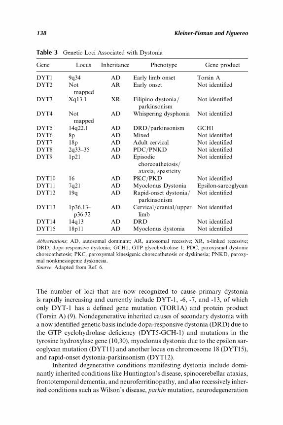
The number of loci that are now recognized to cause primary dystonia
is rapidly increasing and currently include DYT-1, -6, -7, and -13, of which
only DYT-1 has a defined gene mutation (TOR1A) and protein product
(Torsin A) (9). Nondegenerative inherited causes of secondary dystonia with
a now identified genetic basis include dopa-responsive dystonia (DRD) due to
the GTP cyclohydrolase deficiency (DYT5-GCH-1) and mutations in the
tyrosine hydroxylase gene (10,30), myoclonus dystonia due to the epsilon sar-
coglycan mutation (DYT11) and another locus on chromosome 18 (DYT15),
and rapid-onset dystonia-parkinsonism (DYT12).Inherited degenerative conditions manifesting dystonia include domi-
nantly inherited conditions like Huntington’s disease, spinocerebellar ataxias,
frontotemporal dementia, and neuroferritinopathy, and also recessively inher-
ited conditions such as Wilson’s disease, parkin mutation, neurodegeneration
Table 3 Genetic Loci Associated with Dystonia
Gene Locus Inheritance Phenotype Gene product
DYT1 9q34 AD Early limb onset Torsin ADYT2 Not
mappedAR Early onset Not identified
DYT3 Xq13.1 XR Filipino dystonia/parkinsonism
Not identified
DYT4 Notmapped
AD Whispering dysphonia Not identified
DYT5 14q22.1 AD DRD/parkinsonism GCH1DYT6 8p AD Mixed Not identifiedDYT7 18p AD Adult cervical Not identifiedDYT8 2q33–35 AD PDC/PNKD Not identifiedDYT9 1p21 AD Episodic
choreoathetosis/ataxia, spasticity
Not identified
DYT10 16 AD PKC/PKD Not identifiedDYT11 7q21 AD Myoclonus Dystonia Epsilon-sarcoglycanDYT12 19q AD Rapid-onset dystonia/
parkinsonismNot identified
DYT13 1p36.13–p36.32
AD Cervical/cranial/upperlimb
Not identified
DYT14 14q13 AD DRD Not identifiedDYT15 18p11 AD Myoclonus dystonia Not identified
Abbreviations: AD, autosomal dominant; AR, autosomal recessive; XR, x-linked recessive;
DRD, dopa-responsive dystonia; GCH1, GTP glycohydrolase 1; PDC, paroxysmal dystonic
choreoathetosis; PKC, paroxysmal kinesigenic choreoathetosis or dyskinesia; PNKD, paroxy-
mal nonkinesiogenic dyskinesia.
Source: Adapted from Ref. 6.
138 Kleiner-Fisman and Figuereo

with brain iron accumulation (NBIA1 or PKAN), GM1 and 2 gangliosidoses,glutaric acidemia, neuroacanthocytosis, and others.
Atypical parkinsonian syndromes including multiple system atrophy,progressive supranuclear palsy, and corticobasal degeneration are also de-generative conditions that manifest dystonia, but whether a definable pat-tern of inheritance is involved in its etiology, and what the pattern of theinheritance may be, remain complex and unknown.
There are many causes of symptomatic dystonia, including those thatresult from birth trauma, kernicterus, hypoxia, head injury, tumor, infectiousand postinfectious conditions, multiple sclerosis, structural lesions, drugs,toxins, and stroke, amongst others.
Objective, standardized measures for rating disease symptoms andseverity have been developed to assess generalized dystonia (GD), and includethe Burke–Fahn–Marsden Dystonia Rating Scale (BFM-DRS), UnifiedDystonia Rating Scale (UDRS), and the Global Dystonia Scale (GDS).The Toronto Western Spasmodic Torticollis Scale (TWSTRS) is the mostcommon scale used to evaluate CD.
3. ETIOLOGY AND PATHOPHYSIOLOGY
3.1. Electrophysiology
Clinically, dystonia is characterized by involuntary distorted postures andmovements. Electromyographic (EMG) studies confirm prolonged burstswith phasic muscle contractions. Co-contraction of agonist and antagonistmuscles occur, with overflow activation of other uninvolved muscles duringspecific actions (11).
Some evidence suggests that dystonia is generated by a loss of inhibitionin basal ganglia circuitry (12–17). One model proposes lower globus pallidusinternus (GPi) output (18) (Fig. 1) with decreased pallidal inhibition to thethalamus resulting in excessive release of thalamocortical activity. Evidenceof a change in the firing pattern in contrast to the actual firing rate also existswith reported changes in the extent of synchronization and sensory respon-siveness of neurons in the pallidum (19). In contrast to the tonic pattern ofdischarge present under normal circumstances, patients with dystonia haveirregularly grouped discharges with intermittent pauses (20). The effectivenessof pallidal ablation in improving dystonia suggests that an abnormal patternof neuronal firing, rather than the absolute firing rate, may be responsible forthe dysfunction that causes dystonia (21–24).
Further data suggest that dystonia may be related to defects in‘‘surround inhibition’’ (25,26). According to this model, when a specific move-ment is generated, there is simultaneous suppression of unwantedmovements.If surround inhibition is impaired, then dystonia may emerge. This is consis-tent with evidence of decreased dopamine D2 receptor striatal binding in
Dystonia: Classification, Etiology, and Therapeutic Options 139

primates and in people with dystonia (27–29). Dysfunction of the ‘‘indirectstriato-pallidal pathway’’ in dystonia, lowering the discharge of subthalamicnucleus (STN)-GPi projections, may explain the findings of reduced Gpiactivity and decreased activity in the inhibitory surround (26).
3.2. Pathology and Biochemistry
Aside from electrophysiological considerations, clues from examination ofpostmortem brains of dystonic patients also suggest an abnormality in thedopaminergic system (30–32). A comprehensive study of markers of dopa-mine transmission in three specimens of postmortem DYT1 striatum foundincreased dopamine turnover compared to controls (33). However, no patho-logical or biochemical abnormalities have consistently been found in DYT1
Figure 1 (A) Schematic illustration of the basal ganglia under normal conditions.(B) A schematic of basal ganglia function in dystonia. Striatal overactivity in boththe direct and indirect pathways is postulated to be responsible for the changes in neu-ronal activity in dystonia. Width of lines represents the relative rate in mean dischargerates compared to normal.Wider lines represent an increase and thinner lines representa decrease in mean discharge rates. Dotted lines represent the altered patterns andincreased synchronization of neuronal activity in the BG thalamocortical circuit,resulting in unregulated cortical output. Black arrows represent inhibitory projections,gray arrows represent excitatory projections. D1, D2, dopamine 1 and 2 receptor sub-types; Snc, substantia nigra compacta; Gpe, globus pallidus externa; Snr, substantianigra pars reticulata; VL, ventrolateral thalamus. Source: Adapted from Vitek JL,Giroux M. Physiology of hypokinetic and hyperkinetic and hypokinetic movementdisorders: model for dystonia. Ann Neurol 2000:47:S131–S140.
140 Kleiner-Fisman and Figuereo

dystonia (34,35). This is in keeping with a functional, rather than a degenera-tive or structural, abnormality as the etiology of DYT1 dystonia.
3.3. Imaging
Metabolic studies measuring brain glucose metabolism using fluorodeoxy-glucose positron emission tomography (FDG-PET) demonstrate an abnormalmetabolic brain network characterized by hypermetabolism of the basalganglia, supplementary motor area, and the cerebellum in both patients mani-festing and those not clinically manifesting DYT1 dystonia. In addition, thosethat manifest dystonia also show another pattern characterized by increasedactivity of the midbrain, cerebellum, and pons (36–38). It has been suggestedthat a clinical correlate of this abnormal metabolic pattern, found even innonmanifestingDYT1 carriers, is impairedmotor sequence learning (39), withan alternate brain network being activated when the task is performed.
3.4. Torsin A
Finally, further insights into the pathophysiology of dystonia may be gainedby the examination of the biology of Torsin A, the protein product of theimplicated gene (TOR1A) in DYT1 dystonia. A study using in situ hybridi-zation with a Torsin A probe in nondiseased postmortem brain revealedubiquitous distribution, with greatest protein expression found in the basalganglia, brain stem, and cerebellum (40,41). The function of this protein,which shows similarity to the AAAþ family of adenosine triphosphatases(ATPases), includes involvement in cellular metabolism, particularly inATP binding and hydrolysis, but the exact sequence and details of thisprocess are unclear (42–45). The relationship between the ATPase catalyticcycle and the conformational changes leading to biological activity remainto be defined.
In addition to a lack of conclusive knowledge about the function of thenormally expressed protein, the consequence of the D302/3 mutation (foundin DYT1 dystonia), a deleted glutamic acid residue in the C-terminus ofTorsin A, is unknown (46–48). Efforts to create a mouse model of dystoniaby modifying allelic expression of Torsin A such that there is only 2.5–5% ofwild-type protein expression (loss-of-function mutant) have failed to createa dystonic phenotype in the rodent (49), possibly suggesting that this isa necessary but insufficient abnormality to cause dystonia. As such, insightsgained from this animal model in its current state are limited.
Recently, preliminary work has been done creating another transgenicmouse model of mutant Torsin A that resulted in a phenotype of abnormalmotor function manifesting as fore- and hind-limb clasping, circling, post-uring, and head shaking (50). These features were apparent in � 40% ofthe transgenic mice, similar to the penetrance in DYT-1 dystonia in humans.Furthermore, in examining the striatum of these transgenic mice, there was
Dystonia: Classification, Etiology, and Therapeutic Options 141

evidence of increased dopamine content and turnover, as has been reportedin humans with the disease (33). Implications of this work remain to bedetermined.
3.5. Dopaminergic Transmission
Examination of abnormalities thought to play a role in the pathophysiologyof some forms of dystonia, like mutations in the tyrosine hydroxylase gene(30) and the guanosine triphosphate cyclohydrolase gene (31), further supportdopamine-mediated dysfunction. Individuals affected with these disordershave limited dopamine synthesis that results in a dystonic phenotype. Despitethis, there is currently no evidence pathologically in humans to suggest a rela-tionship between abnormal Torsin A production and dopaminergic functionin DYT1 dystonia (49).
4. BEST MEDICAL THERAPY
Treatment options for dystonia are based on etiology and anatomical distri-bution, each of which influences the effectiveness of therapy. In most cases,dystonia therapy is offered to alleviate symptoms that interfere in activitiesof daily living and quality of life and is not curative. In rare cases of ‘‘dystonicstorm,’’ a medical emergency that can manifest in hyperthermia, rhabdo-myolysis, and myoglobinuria, medical treatment is crucial and potentiallylife-saving (51).
The effective treatment of patients with dystonia requires a multi-disciplinary and comprehensive approach that includes pharmacologic, non-pharmacologic, and surgical options (for a comprehensive review see Ref. 1).However, there are few randomized, placebo-controlled trials of adequatepower to permit definitive recommendations of effective treatment for anindividual dystonic patient. An algorithm for the treatment of dystonia isproposed based on the literature available and on clinical experience (Fig. 2).
4.1. Nonpharmacologic Considerations
Emphasis on patient and caregiver education, support, and expectations areimportant for successful therapy. Physical and occupational therapy mayaugment the benefit of medical therapy (52,53). In some cases, assistiveequipment, such as a mechanical device, have proven useful in alleviatingopisthotonus and retrocollis that typically occur in tardive dystonia (54).The introduction of a hand orthosis in writers’ cramp has also shown somebenefit (55). Recently, there has been some evidence to suggest that immo-bilization of an affected limb for a brief period of time may result inimprovement in dystonia, though whether this effect is sustained is notknown (56–58). It is also unknown how immobilization results in sympto-matic improvement of dystonia. One possible explanation is that some
142 Kleiner-Fisman and Figuereo

patients with dystonia have impaired sensory perception and sensory-motorintegration (59,60), and the removal of sensory and motor input to the limbmay result in ‘‘resetting’’ and normalizing of the cortical map (61,62).Furthermore, sensory training and mental exercises to restore hand sensoryrepresentation have shown benefit in focal limb dystonia (63,64). Finally, insome patients with dystonia, muscle relaxation and sensory feedback ther-apy can enhance medical or surgical treatments (1,65,66).
4.2. Pharmacologic Interventions
4.2.1. Dopaminergic Therapy
In DRD, supplementation with small doses of levodopa or dopamine agonistsprovides dramatic improvement in symptoms (53,67). A trial of dopaminergicreplacement is appropriate in all childhood-onset dystonia, regardless ofwhether the clinical presentation is typical of DRD (1,53). A trial of levodopain adults with non-DRD is also indicated as benefit from dopaminergictherapy exists in 25% of patients with adult-onset primary and secondarydystonias (1).
Figure 2 Treatment algorithm for dystonia. Source: Adapted from Ref. 1.
Dystonia: Classification, Etiology, and Therapeutic Options 143

4.2.2. Antidopaminergic Therapy
While some patients benefit from treatment with dopaminergic therapy,others improve with antidopaminergic treatments (68,69). However, use ofthese agents that include neuroleptics is often limited by unwanted side effectslike parkinsonism, sedation, and tardive dyskinesia or tardive dystonia.
The more recent atypical neuroleptic agents may also prove to beuseful in the treatment of dystonia. Clozapine has high D4 receptor affinitywith combined serotonergic and dopaminergic effects. It has shown benefitin a small number of patients with segmental and generalized dystonia in anopen-label trial (70) using 300–400mg/day as the optimal dose. However, insome patients treatment was withdrawn due to intolerable side effects suchas orthostatic hypotension. Its use is also pragmatically complicated becauseof a risk of agranulocytosis, requiring weekly blood draws to monitor thewhite blood cell count.
Risperidone, also considered an atypical neuroleptic despite D2-receptor blocking properties, has also been reported to be efficacious indoses of up to 8 mg/day in the treatment of segmental dystonia in a smallnumber of patients (68,71) and in idiopathic and symptomatic dystonia (69).Tetrabenazine, a presynaptic monoamine-depleting agent and dopaminereceptor blocker not available in the United States, is used preferentiallyin Canada and the United Kingdom for hyperkinetic movement disordersand does not induce tardive dyskinesia (72).
4.2.3. Anticholinergic Therapy
The mainstay of medication treatment for dystonia has been anticholinergicagents, particularly trihexyphenidyl (Artane) (73,74).
Generally, it is necessary to proceed with a slow titration, starting with1–2mg at bedtime and usually progressing to 12 mg/day divided over threedoses over the course of 4–6weeks (1). This is to prevent or decrease side effectssuch as drowsiness, cognitive impairment, and autonomic symptoms such asurinary retention, dry mouth, and blurred vision. In some cases, high doses(60–100mg) may be needed for the desired effect, though use at the higherdoses may not be tolerated due to the aforementioned side effects, especiallyin the elderly. Pyridostigmine (an anticholinesterase agent), pilocarpineeye-drops, and synthetic salivamay be introduced in order to ameliorate theseunwanted anticholinergic effects (1).
4.2.4. Muscle Relaxants
Muscle relaxing agents have proven efficacy in patients with spasticity andmay have a role to play in dystonic patients as well. Though there are no trialsto attest to their efficacy, drugs such as cyclobenzaprine, metaxalone, cariso-prodol, and orphenadrine have been used anecdotally as part of the therapeu-tic armementarium to treat dystonia. Tizanidine hydrochloride, a centrally
144 Kleiner-Fisman and Figuereo

acting alpha-adrenergic agonist, is an agent with proven efficacy in spasticity(75) and has sometimes been included as part of a treatment strategy, thoughits effects in dystonia have not been encouraging. One open-label study ofadministration of tizanidine did not significantly improve cranial dystoniain 10 patients (76), and another small randomized double-blind cross-overtrial of tizanidine treatment in patients with spasmodic torticollis did notshow much benefit over a 6-week course with doses up to 12mg/day (77).
Under certain circumstances, oral baclofen may provide benefit inreducing dystonia by putatively increasing presynaptic inhibition of motorneurons (78,79). An intrathecal baclofen (ITB) pump may also play a lim-ited role in the treatment of medically refractory axial or generalized dysto-nia (80–84). Evidence from studies conducted in patients with spastic anddystonic cerebral palsy provide support for this practice (85). However,response to an ITB pump is unpredictable. In one series of 14 patients withprimary and secondary causes of dystonia, only five patients showed objectivebenefit as measured by the BFM-DRS. Doses averaged 590mg/day with amean length of therapy of 29 months. Tolerance to the drug, device-relatedcomplications, and side effects such as constipation, drowsiness, anddecreased trunk control (86,87) were identified. The cause of the dystoniadid not predict whether or not a patient would respond to an ITB pump.
4.2.5. Other
4.2.5.1. Morphine sulfate: In a series of nine patients (88), slowrelease morphine sulfate appeared to improve dystonic movements in indi-viduals with tardive and generalized dystonia over and above the improve-ment due to pain relief. However, the effect seen was transient and is notcurrently a recommended therapy.
4.2.5.2. Gamma-hydroxy butyric acid (GHB): Subjective improve-ment of both myoclonus and dystonia in a patient with inherited myoclonicdystonia treated with GHB has been reported (89). Rationale for the use ofGHB is based on its therapeutic effects in the treatment of alcohol abuse asit mimics the effects of alcohol. No trials currently support the use of thisagent.
4.2.5.3. Mexilitene: In one open-label trial of six patients with spas-modic torticollis, treatment with Mexilitene up to 800mg/day for a 6-weekperiod resulted in significant improvement in dystonia as measured by theTWSTRS and the BFM-DRS with no significant adverse effects (90). Furtherexperience with this agent in the treatment of dystonia is needed to confirm orrefute this finding.
4.2.5.4. Antihistamines: In three of five patients with idiopathic trun-cal dystonia, treatment with up to 500mg/kg/day of oral diphenhydraminewas found to be beneficial during 20 months of follow-up (91). This result
Dystonia: Classification, Etiology, and Therapeutic Options 145

was re-enforced in another small, open-label trial (92). This is in contrast to adouble-blind, placebo-controlled study of intravenous chlorpheniraminemaleate in 20 patients with adult onset focal dystonia that saw no consistentbenefit from this intervention (93). However, given the wide availability ofthese medications, relative safety, paucity of side effects, and the extent ofdisability caused by dystonia, a trial of an antihistamine (such as diphen-hydramine) is warranted when other treatments have failed (94).
4.2.5.5. Benzodiazepines: Benzodiazepines such as clonazepam, dia-zepam, and lorazepam may be indicated as alternative or additional agentswhen other medications such as anticholinergics have failed (95).
4.2.5.6. Summary: Medications in combination are often necessaryto achieve satisfactory results. Each patient may require a different combina-tion of medications in varying dosages and the optimal combination for eachindividual patient is often found by trial and error. The benefits obtained bypharmacologic therapy must always be weighed against the potential for sys-temic side effects that increase with use of multiple agents. A therapy such asbotulinum toxin that acts locally and eliminates the risk of unwanted systemiceffects is an attractive alternative under the appropriate circumstances.
4.2.6. Botulinum Toxin
Botulinum toxin has revolutionized the treatment of dystonia and hasbecome the first line agent in some forms of focal dystonias. There areseven isoforms of botulinum neurotoxins identified by the letters A–G thatvary with respect to biochemical activity and antigenicity (96,97). Botulinumtoxin A (Botox�, Dysport�) received approval from the Food and DrugAdministration (FDA) in 1989 for the indications of blepharospasm, hemi-facial spasm, and cervical dystonia. Another preparation using a differentsubunit of the toxin, botulinum toxin B (Myobloc) was FDA approved inthe United States in 2000 as treatment for cervical dystonia. Currently, theseare the only two isoforms of botulinum toxin that are commercially avail-able and approved by the FDA, though experimental uses of some of theother subunits may show therapeutic promise in the future (98,99).
The therapeutic effect of either botulinum toxin formulation resultsfrom denervation in injected muscle. The toxin irreversibly binds to the pre-synaptic membrane at the neuromuscular junction to prevent acetylcholinerelease and consequent flaccid weakness (100). Though the binding of theneurotoxin to the terminal is irreversible, the clinical effect of paralysis isnot permanent due to synaptic plasticity and nerve sprout regeneration(101,102). Repeated injections are necessary approximately every 3 monthsto maintain the effect, though a complete return to baseline muscle activitydoes not usually occur within this time period.
Botulinum toxins A and B are generally believed to be of comparablesafety, efficacy, and duration of benefit, though these have not been directly
146 Kleiner-Fisman and Figuereo

compared in dystonia. A study in healthy volunteers looking at magnitudeand duration of paralysis, as measured by M-wave amplitude on EMG atperiodic intervals following injection of either botulinum toxin A or B,found that the magnitude and duration of effect was greater in individualshaving received botulinum toxin A as compared to botulinum toxin B (103).Whether M-wave amplitude is a valid marker for clinical response tobotulinum toxin is unclear (1).
Though initially effective in most patients with focal dystonia, somepatients receiving repeated injections of botulinum toxin A develop neutrali-zing antibodies that confer resistance to the effects of future injections. Thisphenomenon may be of historical interest, as it appears to be uncommon withthe use of the current formulation (104). This more recent formulationcontains 5 ng of neurotoxin complex protein/100 units in contrast to the olderpreparation containing 25ng/100units and may therefore be less antigenic.
The risk of development of neutralizing antibodies was found to beincreased when larger, more frequent doses were administered and foundto occur at a higher incidence in younger individuals (105). Further discussionof botulinum toxins and when to use each is presented in chapter 9 in thecontext of cervical dystonia.
Safety and therapeutic benefit of botulinum toxin in dystonia has beendemonstrated in multiple clinical studies with efficacy and optimal doses vary-ing according to dystonia subtype (106). Experience with botulinum toxin inblepharospasm has been generally excellent with sustained efficacy followingrepeated use (107) with an average duration of maximum benefit of 16 weeks(1). However, it is not uncommon for patients to experience blurred vision,ptosis, and tearing, especially in the first 2–3 weeks following injections. Theseusually subside and are dose dependent.
Botulinum toxin can be an effective treatment option in oromandibulardystonia (OMD) with sustained improvement in chewing and speech in�70% of patients (108) and improved quality of life (109) whether tardiveor idiopathic.
OMDmanifests as jaw-clenching, bruxism, jaw-opening, or jaw-closingdystonia. In cases of jaw-opening dystonia, EMG-guidance may be requiredto isolate the lateral pterygoid or the submental muscle complex. Comparisonof tardive and idiopathic OMD suggests equivalent benefit from botulinumtoxin injections (110). A long-term study (108) of outcomes in OMD withan average follow-up time of 4.4 years used a mean dose of 54.2� 15.2Uof botulinum toxin A injected into the masseter muscle and 28.6� 16.7Uwere administered to the submentalis complex. The average duration ofresponse to injections was 16.4 weeks. Of note, 11% of patients in this studyexperienced dysphagia and dysarthria following at least one injection duringthe course of follow-up.
Botulinum toxin has emerged as the treatment of choice in patients withspasmodic dysphonia (laryngeal dystonia). Excellent outcomes with a low risk
Dystonia: Classification, Etiology, and Therapeutic Options 147

of adverse events are reported in the majority of patients treated (111), withconcomitant improvement in voice-related quality of life (112). These injec-tions need to be performed by experienced practitioners and usually requirethe use of laryngoscopy.
The first line treatment strategy in action-specific limb dystonia such aswriters’ cramp is also botulinum toxin (113). In one study, 167 patientspresenting with writers’ cramp underwent EMG-guided botulinum toxin Ainjections. The results showed good efficacy and tolerance of this treatment
in the long term (up to 10 years) with recovery of normal writing in 46%,partial benefit in 10%, failure in 21%, and loss to follow-up after the firstinjection in 23% of patients. Amongst those that benefited, EMG-guidedinjections were repeated approximately every 9 months (114). ThoughEMG-guidance is often used to isolate the specific muscles involved in thedystonia, evidence suggests that localization with EMG-guidance does notnecessarily imply better clinical results than can be achieved using clinicalassessment of affected muscle groups (115).
5. SURGICAL THERAPY
Surgical treatment alternatives for patients with dystonia have evolved as
a result of mixed or inadequate results of medical therapy. The evolution ofsurgical approaches has improved along with further understanding of thepathophysiology of the involuntary movements seen in dystonia. Segmentalfusions (116) and selective myectomy (117) have been performed or proposedas rare but alternative palliative approaches to refractory dystonia. Mostsurgical interventions, however, attempt to obtain relief by interrupting thepathways of abnormal motor excitation.
In order to avoid the potential adverse effects of central nervous systeminterventions, peripheral nerve denervations have been performed for focaldystonia (118,119). Other strategies such as dorsal column stimulation havealso given moderate relief to patients with focal (cervical) dystonia (120).Mixed results and frequent complications have discouraged the generalizeduse of these strategies.
For the past 50 years (121), ablation or DBS of basal ganglia motornuclei have provided the best outcomes in the surgical treatment of dystonia.Pallidotomy (122–124) and thalamotomy (125,126) have been associated withadequate but inconsistent results as well as complications, particularly dys-arthria and dysphagia when performed bilaterally. Better understanding ofthe functional organization of the basal ganglia, flexibility in adjusting the
parameters of stimulation, reversibility of stimulation, and the feasibility ofbilateral electrode implantation without unwanted adverse effects makeDBS an attractive surgical alternative for the treatment of dystonia.
148 Kleiner-Fisman and Figuereo

5.1. Patient Selection
The selection of patients for dystonia surgery is based on identifying thosein whom the anticipated benefit of symptomatic improvement outweighsthe risks of undergoing brain surgery. Though experience with functionalstereotactic brain surgery has increased and the risk of serious surgicalmorbidity has been reduced to approximately 5% (127), the risk of surgeryis only justified in cases of severe dystonia causing intractable pain or disabil-ity and when other conservative treatment options have failed.
The main considerations when evaluating a dystonic patient for aneurosurgical intervention are:
1. Determining whether the target symptom is responsible for the dis-ability. For example, if fixed contractures have already occurred,alleviating the dystonia may not provide added benefit.
2. Empirical estimation of the likelihood of improvement of symp-toms and quality of life following surgery.
3. Estimation of the surgical risk factors in each individual patientand the likelihood of an adverse event.
4. Assessment of whether the patient’s expectations of the surgicaloutcome are realistic and achievable (128).
6. ANATOMICAL TARGETS IN DBS SURGERY
6.1. GPi
The rationale for the alleviation of dystonia by stimulating the GPi relatesto the observed decrease in the firing rate of GPi neurons and abnormalgrouping of discharges in a burst–pause pattern in patients with dystonia(18,129). The observation that pallidotomy alleviates levodopa-induceddystonic dyskinesias and OFF period dystonia in Parkinson’s disease (PD)patients, combined with reports that pallidotomy may be beneficial in pri-mary generalized dystonia (GD), has lent further support to the role of theGPi as a target in dystonia surgery (22,130–132). Though preliminary studiessuggest that the GPi is a promising target especially in GD (Table 4), conclu-sive data is lacking due to the paucity of large well-designed trials.
6.2. Subthalamic Nucleus
There is minimal experience with STN DBS in dystonia. The theoretical basisfor choosing the STN as a target is rooted in the classical model of basalganglia function (Fig. 1) (159). According to this model, hyperkinetic move-ment disorders such as dystonia result from overactivity of both the direct andindirect pathways, leading to increased inhibitory activity of the GPi. It ispostulated that there is decreased neuronal activity in the globus pallidus
Dystonia: Classification, Etiology, and Therapeutic Options 149

Tab
le4
Summary
ofDystonia
DBSStudies
Study
number
and
citation
Studydesign
Dx
No.
Target
U/Lvs.
B/L
Follow-uptime
%im
provem
ent
BFM-D
RS
Other
ratingscales
1.(133)
14-G
D;6-C
D20
GPi
GD-8.6
mos
(range3–12);
CD-18.9mos
(range12–24)
GD:47%
(SS);
38%
(DS);
46%
(TS)
TWSTRS;CD:64%
(SS);60%
(DS);
60%
(PS);
60%
(TS)
2.(134)
15-D
YT1;
17-notknown;
21-secondary
53
GPi
B/L
1yr
71%
(CS);
63%
(FS);
74%
(CS);
49%
(FS);
31%
(CS);
7%
(FS)
3.(135)
Case
series
12-primary
GD;
7-C
D;4-M
D;
1-TD;
1-posttraumaHD
25
B/L
GD-9.2
mos
(range4–18);
CD-19(range
12–24);Misc11
(range4–24)
GD:47.6%
(SS);
37.6%
(DS);
45.8%
(TS)
TWSTRS;CD:
63.8%
(SS);
60%
(DA);60.3%
(PS);59.5%
(TS);
AIM
Smisc37.1%
4.(136)
9-Primary
GD
(2DYT1);
10-secondary
trauma,
stroke,
anoxia
(NBIA
-1)
19
9-V
im;
6-V
im;
then
GPi;
7-G
Pi
3U/L;Vim
4.5
yrs
(4mosto
11yrs)
4–36mos
14mos
6–24mos
GFVim
50%
score
of
2or3GPi;70%
score
of2or3
6B/L
Vim
6B/L
Gpi
1B/L
GPi
5.(137)
Case
series
5CD,3CD
withmyelopathy
85GPi;3GPi
andlamin
B/L
20mos
63%
SS;
69%
DS;
50%
PS
150 Kleiner-Fisman and Figuereo

6.(138)
Case
series
2GD;4chor
dueto
CP
6Gpi
B/L
2yrs
2GD:74%
SS;
68%
DS;4chor;
23%
DS,13%
DS(2
pts)
7.(139)
Case
series
4GD
(1DYT-1,
1IG
D,1HuntDis;
1TD);3CD;1HD
8GPi
7B/L;
1U/L
6mos
GD:12–35%
TWSTRS;CD:
43–82%
8.(140)
Case
series
1DYT-1
GD;
3segmentaldyst,
2CD
6GPi
B/L
1yr
76%
9.(141)
Case
series
5GPi
B/L
6mos
72%
10.(142)
Case
report
1DYT-1
GD;
1IG
D
2GPi
B/L
2yrs
95%
DYT-1;
80%
IGD
11.(143)
Case
report;
double-
blind
assessm
ent
IGD
1GPi
B/L
1yr
67%
12.(144)
Case
report
PosttraumaHD
1GPi
U/L
4yrs
Improved-no
objectivemeasure
13.(145)
Case
report
PosttraumaCD
1GPi
U/L
1yr
Improved-no
objectivemeasure
14.(68)
Case
report
Segmentaldystonia
1GPi
B/L
9mos
86%
15.(146)
Case
report
CD
1GPi
U/L
Improved-no
objectivemeasure
16.(147)
Case
report
Postanoxic
GD
1VOA
B/L
4mos
UDRS50%
17.(148)
Case
report
ICD,truncal
dystonia
1GPi
B/L
7wks
TWSTRS50%
18.(149)
Case
report
CD
2GPi
B/L
24and17mos
64%
VAS,21%
Tsui;
55%
VAS,7%
Tsui
19.(150)
Case
report
IGD
1GPi
Palland
C/LGPi
DBS
1yr
Worsened-no
objectivemeasure
(Continued)
Dystonia: Classification, Etiology, and Therapeutic Options 151

Tab
le4
Summary
ofDystonia
DBSStudies(C
ontinued
)
Study
number
and
citation
Studydesign
Dx
No.
Target
U/L
vs.
B/L
Follow-uptime
%im
provem
ent
BFM-D
RS
Other
ratingscales
20.(151)
Case
series
7DYT-1
GD
7GPi
B/L
3mos
90.3%
21.(152)
Case
report
ICD
1GPi
B/L
1yr
Improved-no
objectivemeasure
22.(153)
Case
series
ICD
3GPi
B/L
3mos
TWSTRS4;1%
(SS);
43%
(PS);
52%
(DS)
23.(154)
Case
report
Double-
blind
TD
1Vim andGPi
B/LB/L
6mos
NochangewithVim
,
73%
GPi
Gpi(A
IMS)54%
24.(155)
Case
series
1IG
D,1,
DYT-1
GD,
1postanoxic
at
birth
3GPi
B/L
6–18mos
DYT-1
34%,
IGD
59%;anoxia
14%
25.(156)
Case
series
ICD
3GPi
B/L
3mos
Improved-no
objectivemeasure
26.(157)
Case
report
FMD
1GPi
B/L
20mos
Improved-no
objectivemeasure
27.(158)
Case
series
3NB1A1;1IG
D4
STN
B/L
3mos
Noim
provem
ent
Abbreviations:SS,severityscore;DS,disabilityscore;PS,pain
score;TS,totalscore;CS,clinicalscore;FS,functionalscore;Chor,choreoathetosis;
GF,globalfunctionoutcome;
IGD,idiopathic
generalizeddystonia;IC
D,idiopathic
cervicaldystonia;HD,hem
idystonia;TD,tardivedystonia;Hunt
Dis,Huntington’sdisease;NB1A1,neurodegenerationwithbrain
irondeposition;mos,months;yrs,years;wks,weeks;Gpi,globuspallidusinternus;
VLp,ventrallateralposteriorthalamic
nucleus;VOA,ventrooralisanteriorthalamicnucleus;Vim
,ventralisinterm
ediusthalamicnucleus;UDRS,Uni-
fied
Dystonia
RatingScale;AIM
S,Abnorm
alInvo
luntary
MovementScale;VAS,visualanalogu
escale;TWSTRS,Toronto
Western
TorticollisRating
Scale;BFM-D
RS,Burke–Fahn–Marsden
Dystonia
RatingScale;FMD,familialmyoclonic
dystonia;NBIA
-1,abnorm
albrain
ironaccumulation;pall,
pallidotomy;lamin,laminectomy;U/L,unilateral;B/L,bilateral;C/L,contralateral;N/A,notavailable.
152 Kleiner-Fisman and Figuereo

externus (GPe), which increases the excitatory drive to the STN. This in turnfurther increases GPi inhibitory activity (Fig. 1).
One study in support of this hypothesis shows decreased D2-likebinding in the striatum of patients with idiopathic focal dystonia (160). Thiscould result in reduced dopaminergic inhibition and an increase in the activityof striatal neurons in the indirect pathway (19). Other studies, performedusing neuronal recording, PET, and TMS, suggest that overactivity of theSTN results from both increased cortical activity and increased GPe activityin some forms of dystonia (19,161–166). Again, this would support the use ofthe STN as a target location for alleviation of dystonic symptoms. Indirectclinical evidence in favor of the STN as a target is provided by the observationthat OFF period dystonia resolves in PD patients that have undergonebilateral STN stimulation (158,167–169).
7. SURGICAL TECHNIQUE
The surgical technique for the treatment of dystonia by either ablative orstimulation procedures does not differ significantly from the proceduresdescribed elsewhere in the treatment of PD (170–172). Whether the ultimategoal is to create a lesion or to implant a stimulating lead, the adjuvant tech-nology is used with the intention of precisely localizing a target point. Mostsurgeons will rely on a combination of methods rather than on one particu-lar localization technique. These techniques include imaging, electrophysio-logical recording, or macrostimulation. Imaging approaches may includefluoroscopy, pneumoencephalography, magnetic resonance imaging (MRI),computed tomography (CT), or a combination of two or more of these. Astereotactic frame is fixed to the patient’s head before the imaging study isobtained in order to localize the target’s anatomical point in reference tothe scaled device. A Leksell Model G stereotactic head frame is most com-monly used; however, new navigation technology that integrates imagingstudies with computer software may soon popularize frameless localizingdevices (173–176). Once a target has been determined, a trajectory devised,and an entry point identified, general surgical technique is used to performan access Burr hole, traditionally at the level of or just in front of the coronalsuture bilaterally. An insertion cannula is placed along the planned trajectoryin the brain to guide the microelectrode used for electrophysiologic record-ings. Electropysiologic recordings are used to further define the optimallocation for placement of the macroelectrode.
Several techniques have been debated in terms of electrophysiologicalrecording. Some proclaim the benefits of accurate delineation of the targetorgan by multiple electrode recording (either simultaneously or staggered),while others praise the decreased risk of intracerebral hemorrhage with asingle electrode pass (177,178). The published data suggest both techniquesto be adequately safe and effective. Regardless of the recording technique
Dystonia: Classification, Etiology, and Therapeutic Options 153

chosen, the goal is to confirm the coordinates obtained from the imagingstudies by identifying the neuronal discharge pattern of the target organ.
Another approach to localization includes stimulation through theimplanted macroelectrode to verify an appropriate clinical response in anawake patient. This commonly occurs in PD patients undergoing DBS,but occurs less often in cases of dystonia as the patient is often operatedunder general anesthesia such that no dystonia is present. Clinical responsesused to confirm electrode placement include reduction in muscle tone or adecrease of involuntary repetitive muscle contractions. Conversely, macro-electrode stimulation intraoperatively may be performed in order to detectchanges in the neuronal discharge pattern recorded from the electrodes.An example of the latter is the use of flashing lights to elicit a visual evokedresponse to verify proximity of the optic tract to the final electrode locationin the GPi.
Once the target site has been identified by any combination of thesemethods, the electrodes are used to create the lesion in the case of ablativesurgery, or are replaced by permanent electrodes used in DBS. The electro-des are subsequently tunneled under the skin and connected to a generatorimplanted in the subcutaneous tissue, usually in the chest. The programmingof the electrodes occurs days to weeks following electrode implantation andis achieved using a computer-controlled remote device.
8. RESULTS OF SURGERY IN DYSTONIA
8.1. Thalamotomy and Pallidotomy
The thalamus preceded the GPi as a target for dystonia. Thalamotomyoutcome studies have shown that dystonia was alleviated in only 1/3 to1/2 of patients studied and there was an unacceptable rate of intolerableside effects, especially with bilateral procedures (179). Studies comparingthe efficacy of surgery on the thalamus versus the GPi have not been per-formed. Which site is optimal for the indication of dystonia has not beenunequivocally determined; however, the current trend, based on previousexperience, is toward selecting the GPi as the preferred target.
Since the early 1990s, improvement of dystonia by pallidotomy in theposteroventralmedial pallidumhasbeen reported in approximately 75patientsin multiple case reports and case series, with follow-ups ranging from 3 to 42months (180). Results in these patients have shown that pallidotomy canreduce functional disability that is caused by dystonia with best results demon-strated in patients with primary and tardive causes of dystonia. Both unilateraland bilateral procedures have been performed safely without the cognitiveadverse effects experienced by PD patients who underwent bilateral pallidot-omy (181). Also, in contrast to the immediate effects of surgery in PDpatients, improvement seen following pallidotomy in dystonia was, in some
154 Kleiner-Fisman and Figuereo

instances, delayed and occurred in a gradual fashion several months afterthe procedure. However, long-term outcomes have not been reliably docu-mented, and even though there may be benefit that is seen early after thesurgical procedure, there are cases of only temporary improvement withrecurrent debilitating symptoms (23,182). As such, the results of pallidot-omy for dystonia are difficult to predict.
8.2. Deep Brain Stimulation
In dystonia as well as other conditions such as PD, DBS has largely replacedablative surgery as the surgical treatment of choice because it is both rever-sible and modifiable.
However, large, blinded, controlled clinical trials are lacking in thefield of dystonia DBS surgery. This may be because there are a relativelysmall number of individuals affected by dystonia severe enough to warrantfunctional neurosurgery and the fact that DBS is still considered investi-gational for dystonia. The lack of uniformity among studies in reportingsurgical outcomes makes comparison of outcomes between studies difficult.Furthermore, as symptoms of dystonia may be modified by psychologicalfactors, outcomes are likely susceptible to placebo effects (183). In additionto inadequate objective assessment of motor benefit, assessment of qualityof life measures following dystonia surgery are also lacking, so the trueimpact of the procedure is unknown. Notwithstanding these limitations,review of the mostly single case reports or case series published suggests thatDBS surgery in carefully selected patients may be a viable treatment optionfor patients with intractable dystonia.
8.3. Thalamic DBS
In a retrospective study of patients with variable causes of dystonia who hadeither GPi stimulation or thalamic stimulation, or both (GPi followingunsuccessful thalamic stimulation), the patients with pallidal stimulationclearly had a better outcome compared to those implanted in the thalamus(136). Another series of eight patients with heterogeneous causes of dystoniaand both ventral intermedius nucleus of the thalamus (Vim) and GPi stimu-lation reported little benefit in the Vim group, compared to >50% improve-ment as a result of GPi stimulation (184). In light of the demonstrated betteroutcomes of GPi DBS compared to Vim DBS, the thalamus as a target sitefor dystonia has largely been abandoned.
8.3. GPi DBS
Table 4 provides a complete review of the literature with respect to pallidalstimulation.
Dystonia: Classification, Etiology, and Therapeutic Options 155

Unlike PD, where there are clinical tests (levodopa challenge) that maypredict response to surgery, no such tests exist for dystonia. In addition, thepaucity of conclusive published studies of outcomes in dystonia stereotacticsurgery limits the ability to determine which patient is likely to benefit. Fromthe limited reports that do exist, however, there are some clinical factors thatsuggest a more favorable outcome. As in the pallidotomy experience, reviewof GPi DBS results suggests that those with primary causes of dystonia tendto have greatest benefit from surgery. Those with DYT-1 genetic mutationshave been reported to have up to a 95% reduction in BFM-DRS scores(136,151,155,185).
This is not always the case, however; there have been at least three casesof DYT-1 positive dystonia with poor response to bilateral GPi DBS(186,187). This underscores the point that the mutation itself does not neces-sarily predict a good outcome from surgery, and that other as yet unidentifiedfactors are likely important as well.
Review of the literature suggests that those with secondary causes ofdystonia tend to have a less robust and predictable response to DBS thanthose with a primary cause (136,155,185,188,189). Nonetheless, even in thesecondary dystonia population, which is heterogeneous, there have beenreports of dramatic responses to DBS surgery (190–192).
Regardless of the cause, optimal benefit may take several months toachieve. This has been postulated to be due to the gradual resolution ofdisturbances downstream from the pallidum or due to nervous systemplasticity (193). This hypothesis is re-enforced by a study that used PETin a patient with primary dystonia and bilateral GPi DBS placement withsignificant improvement in the dystonia after the stimulators were turnedon. In a blinded fashion the stimulators were turned on and off and PETimaging was performed while performing a motor task. GPi stimulationreversed the abnormal excess activation of multiple brain regions, includingprimary motor, lateral motor, supplementary motor, anterior cingulate, andprefrontal cortices, leading the authors to propose that GPi stimulation maydirectly suppress excessive motor area activation (143). Clinically, it hasbeen observed that the mobile component of dystonia tends to resolve ear-lier than fixed dystonia following DBS (128,136,184). In a study looking atthe pattern of recurrence of dystonia following the discontinuation of stimu-lation, mobile dystonia re-emerged within a few minutes while the toniccomponent recurred gradually over several hours (194).
8.4. STN DBS
Despite a good theoretical basis for considering STN as a target in dystonia,the only published experience reported disappointing results. PD patients withOFF-period dystonia and four patients with generalized dystonia underwentbilateral STN DBS. Though there was definite relief of OFF-period dystonia
156 Kleiner-Fisman and Figuereo

in those patients with PD, there was no difference in severity of dystonia inthose patients with generalized dystonia that underwent STNDBS. However,three of the four patients suffered from neurodegeneration with brain ironaccumulation [also known as pantothenate kinase associated neurodege-neration (PKAN) and formerly as Hallevorden-Spatz disease] (158). It isdifficult to deduce from this limited experience and highly select populationwhether STN DBS is effective in other populations.
9. CONCLUSIONS
Dystonia is a clinically heterogeneous condition with many etiologies.A standard classification and rating system is crucial both for optimal clin-ical care of patients and for conducting rigorous studies that would furtheradvance therapeutic options. Though medical therapies relieve symptoms toa variable extent, the occurrence of intractable dystonia, causing significantdisability and reduction in quality of life, makes development of a safe andeffective surgical therapy attractive.
Identification of clinical and pathophysiological factors that predict agood response to surgery is an important research priority. Studies determin-ing the appropriate target site need to be conducted, as the optimal site mayvary according to the cause of dystonia, patterns of inheritance, and distribu-tion. Finally, large, prospective, long-term controlled trials using standar-dized rating and quality of life scales will be an important addition to thecurrent body of knowledge to determine the true efficacy of this procedure.
REFERENCES
1. Jankovic J. Dystonia: medical therapy and botulinum toxin. Adv Neurol 2004;94:275–285 (Fahn S, Hallett M, DeLong MR Dystonia 4).
2. Greene P, Kang UJ, Fahn S. Spread of symptoms in idiopathic torsion dysto-nia. Mov Disord 1995; 10(2):143–152.
3. Bressman SB, de Leon D, Brin MF, et al. Idiopathic dystonia amongAshkenazi Jews: evidence for autosomal dominant inheritance. Ann Neurol1989; 26(5):612–620.
4. Bressman SB. Dystonia. Curr Opin Neurol 1998; 11(4):363–372.5. Jarman PR, Warner TT. The dystonias. J Med Genet 1998; 35(4):314–318.6. Bressman SB. Dystonia genotypes, phenotypes, and classification. Adv Neurol
2004; 94:101–107.7. Bressman SB, Sabatti C, Raymond D, et al. The DYT1 phenotype and guide-
lines for diagnostic testing. Neurology 2000; 54(9):1746–1752.8. Dauer WT, Burke RE, Greene P, Fahn S. Current concepts on the clinical fea-
tures, aetiology and management of idiopathic cervical dystonia. Brain 1998;121(Pt 4):547–560.
9. Ozelius LJ, Hewett JW, Page CE, et al. The early-onset torsion dystonia gene(DYT1) encodes an ATP-binding protein. Nat Genet 1997; 17(1):40–48.
Dystonia: Classification, Etiology, and Therapeutic Options 157

10. Ludecke B, Dworniczak B, Bartholome K. A point mutation in the tyrosinehydroxylase gene associated with Segawa’s syndrome. Hum Genet 1995;95(1):123–125.
11. Cohen LG, Hallett M. Hand cramps: clinical features and electromyographicpatterns in a focal dystonia. Neurology 1988; 38(7):1005–1012.
12. Rothwell JC, Obeso JA, Day BL, Marsden CD. Pathophysiology of dystonias.Adv Neurol 1983; 39:851–863.
13. Nakashima K, Rothwell JC, Day BL, et al. Reciprocal inhibition between fore-arm muscles in patients with writer’s cramp and other occupational cramps,symptomatic hemidystonia and hemiparesis due to stroke. Brain 1989; 112(Pt 3):681–697.
14. Panizza ME, Hallett M, Nilsson J. Reciprocal inhibition in patients with handcramps. Neurology 1989; 39(1):85–89.
15. Chen RS, Tsai CH, Lu CS. Reciprocal inhibition in writer’s cramp. MovDisord 1995; 10(5):556–561.
16. Deuschl G, Seifert C, Heinen F, et al. Reciprocal inhibition of forearm flexormuscles in spasmodic torticollis. J Neurol Sci 1992; 113(1):85–90.
17. Valls-Sole J, Hallett M. Modulation of electromyographic activity of wristflexor and extensor muscles in patients with writer’s cramp. Mov Disord1995; 10(6):741–748.
18. Vitek JL, Chockkan V, Zhang JY, et al. Neuronal activity in the basal gangliain patients with generalized dystonia and hemiballismus. Ann Neurol 1999;46(1):22–35.
19. Vitek JL. Pathophysiology of dystonia: a neuronal model. Mov Disord 2002;17(suppl 3):S49–S62.
20. Vitek JL, Giroux M. Physiology of hypokinetic and hyperkinetic movementdisorders: model for dyskinesia. Ann Neurol 2000; 47(4 suppl 1):S131–S140.
21. Gernert M, Bennay M, Fedrowitz M, et al. Altered discharge pattern of basalganglia output neurons in an animal model of idiopathic dystonia. J Neurosci2002; 22(16):7244–7253.
22. Lozano AM, Kumar R, Gross RE, et al. Globus pallidus internus pallidotomyfor generalized dystonia. Mov Disord 1997; 12(6):865–870.
23. Ondo WG, Desaloms JM, Jankovic J, Grossman RG. Pallidotomy for gener-alized dystonia. Mov Disord 1998; 13(4):693–698.
24. Vitek JL, Zhang J, Evatt M, et al. GPi pallidotomy for dystonia: clinical out-come and neuronal activity. Adv Neurol 1998; 78:211–219.
25. Hallett M. Dystonia: abnormal movements result from loss of inhibition. AdvNeurol 2004; 94:1–9.
26. Mink JW. The basal banglia and involuntary movements: impaired inhibitionof competing motor patterns. Arch Neurol 2003; 60(10):1365–1368.
27. Perlmutter JS, Stambuk MK, Markham J, et al. Decreased [18F]spiperonebinding in putamen in idiopathic focal dystonia. JNeurosci 1997; 17(2):843–850.
28. Perlmutter JS, Stambuk MK, Markham J, et al. Decreased [18F]spiperonebinding in putamen in dystonia. Adv Neurol 1998; 78:161–168.
29. Naumann M, Pirker W, Reiners K, et al. Imaging the pre- and postsynapticside of striatal dopaminergic synapses in idiopathic cervical dystonia: a SPECT
158 Kleiner-Fisman and Figuereo

study using [123I] epidepride and [123I] beta-CIT. Mov Disord 1998; 13(2):319–323.
30. Knappskog PM, Flatmark T, Mallet J, et al. Recessively inherited L-DOPA-responsive dystonia caused by a point mutation (Q381K) in the tyrosinehydroxylase gene. Hum Mol Genet 1995; 4(7):1209–1212.
31. Ichinose H, Ohye T, Takahashi E, et al. Hereditary progressive dystonia withmarked diurnal fluctuation caused by mutations in the GTP cyclohydrolase Igene. Nat Genet 1994; 8(3):236–242.
32. Burke RE, Fahn S, Jankovic J, et al. Tardive dystonia: late-onset and persistentdystonia caused by antipsychotic drugs. Neurology 1982; 32(12):1335–1346.
33. Augood SJ, Hollingsworth Z, Albers DS, et al. Dopamine transmission inDYT1 dystonia. Adv Neurol 2004; 94:53–60.
34. Walker RH, Brin MF, Sandu D, et al. Torsin A immunoreactivity in brains ofpatients with DYT1 and non-DYT1 dystonia. Neurology 2002; 58(1):120–124.
35. Rostasy K, Augood SJ, Hewett JW, et al. Torsin A protein and neuropatho-logy in early onset generalized dystonia with GAG deletion. Neurobiol Dis2003; 12(1):11–24.
36. Eidelberg D, Moeller JR, Ishikawa T, et al. The metabolic topography of idio-pathic torsion dystonia. Brain 1995; 118(Pt 6):1473–1484.
37. Eidelberg D, Moeller JR, Antonini A, et al. Functional brain networks inDYT1 dystonia. Ann Neurol 1998; 44(3):303–312.
38. Carbon M, Su S, Dhawan V, et al. Regional metabolism in primary torsiondystonia: effects of penetrance and genotype. Neurology 2004; 62(8):1384–1390.
39. Ghilardi MF, Carbon M, Silvestri G, et al. Sequence learning is impaired inclinically unaffected carriers of DYT1 mutation. Mov Disord 2002; 17(5):S300.
40. Augood SJ, Penney JB Jr, Friberg IK, et al. Expression of the early-onset torsiondystonia gene (DYT1) in human brain. Ann Neurol 1998; 43(5):669–673.
41. Augood SJ, Martin DM, Ozelius LJ, et al. Distribution of the mRNAs encod-ing Torsin A and Torsin B in the normal adult human brain. Ann Neurol1999; 46(5):761–769.
42. Rouiller I, Butel VM, Latterich M, et al. A major conformational change inp97 AAA ATPase upon ATP binding. Mol Cell 2000; 6:1485–1490.
43. Zhang X, Shaw A, Bates PA, et al. Structure of the AAA ATPase p97. MolCell 2000; 6:1473–1484.
44. Rouiller I, DeLaBarre B, May AP, et al. Conformational changes of the multi-function p97 AAA ATPase during its ATPase cycle. Nat Struct Biol 2002;9(12):950–957.
45. Dalal S, Hanson PI. Membrane traffic: what drives the AAA motor? Cell2001; 104(1):5–8.
46. Kustedjo K, Bracey MH, Cravatt BF. Torsin A and its torsion dystonia-associated mutant forms are lumenal glycoproteins that exhibit distinct subcel-lular localizations. J Biol Chem 2000; 275(36):27933–27939.
47. Hewett J, Gonzalez-Agosti C, Slater D, et al. Mutant Torsin A, responsible forearly-onset torsion dystonia, forms membrane inclusions in cultured neuralcells. Hum Mol Genet 2000; 9(9):1403–1413.
Dystonia: Classification, Etiology, and Therapeutic Options 159

48. O’Farrell C, Hernandez DG, Evey C, et al. Normal localization of deltaF323-Y328 mutant Torsin A in transfected human cells. Neurosci Lett 2002;327(2):75–78.
49. Dauer W, Goodchild R. Mouse models of Torsin A dysfunction. Adv Neurol2004; 94:67–72.
50. Shashidharan P, Walker RH, McNaught KS, Brin MF, Olanow CW. Trans-genic mouse model of childhood-onset dystonia [abstract]. Mov Disord 2004;19(suppl 9):S86–S87.
51. Opal P, Tintner R, Jankovic J, et al. Intrafamilial phenotypic variability ofthe DYT1 dystonia: from asymptomatic TOR1A gene carrier status to dystonicstorm. Mov Disord 2002; 17(2):339–345.
52. Chen R, Hallett M. Focal dystonia and repetitive motion disorders. ClinOrthop 1998; 351:102–106.
53. Hinson VK, Goetz CG. Torsion Dystonia in Children. Curr Treat OptionsNeurol 2003; 5(4):291–297.
54. Krack P, Schneider S, Deuschl G. Geste device in tardive dystonia with retro-collis and opisthotonic posturing. Mov Disord 1998; 13(1):155–157.
55. Tas N, Karatas GK, Sepici V. Hand orthosis as a writing aid in writer’s cramp.Mov Disord 2001; 16(6):1185–1189.
56. Pesenti A, Barbieri S, Priori A. Limb immobilization for occupational dysto-nia: a possible alternative treatment for selected patients. Adv Neurol 2004;94:247–254.
57. Priori A, Pesenti A, Cappellari A, et al. Limb immobilization for the treatmentof focal occupational dystonia. Neurology 2001; 57(3):405–409.
58. Candia V, Elbert T, Altenmuller E, et al. Constraint-induced movementtherapy for focal hand dystonia in musicians. Lancet 1999; 353(9146):42.
59. Tinazzi M, Rosso T, Fiaschi A. Role of the somatosensory system in primarydystonia. Mov Disord 2003; 18(6):605–622.
60. Abbruzzese G, Berardelli A. Sensorimotor integration in movement disorders.Mov Disord 2003; 18(3):231–240.
61. Levy CE, Nichols DS, Schmalbrock PM, et al. Functional MRI evidenceof cortical reorganization in upper-limb stroke hemiplegia treated withconstraint-induced movement therapy. Am J Phys Med Rehabil 2001; 80(1):4–12.
62. Taub E, Uswatte G, Morris DM. Improved motor recovery after stroke andmassive cortical reorganization following constraint-induced movementtherapy. Phys Med Rehabil Clin N Am 2003; 14(1 suppl):S77–S91.
63. Byl NN, McKenzie A. Treatment effectiveness for patients with a history ofrepetitive hand use and focal hand dystonia: a planned, prospective follow-up study. J Hand Ther 2000; 13(4):289–301.
64. Byl NN, Nagajaran S, McKenzie AL. Effect of sensory discrimination trainingon structure and function in patients with focal hand dystonia: a case series.Arch Phys Med Rehabil 2003; 84(10):1505–1514.
65. Jahanshahi M, Sartory G, Marsden CD. EMG biofeedback treatment oftorticollis: a controlled outcome study. Biofeedback Self Regul 1991; 16(4):413–448.
160 Kleiner-Fisman and Figuereo

66. Korein J, Brudny J, Grynbaum B, et al. Sensory feedback therapy of spasmo-dic torticollis and dystonia: results in treatment of 55 patients. Adv Neurol1976; 14:375–402.
67. Nygaard TG, Marsden CD, Fahn S. Dopa-responsive dystonia: long-termtreatment response and prognosis. Neurology 1991; 41(2 Pt 1):174–181.
68. Wohrle JC, Weigel R, Grips E, et al. Risperidone-responsive segmental dysto-nia and pallidal deep brain stimulation. Neurology 2003; 61(4):546–548.
69. Grassi E, Latorraca S, Piacentini S, et al. Risperidone in idiopathic and symp-tomatic dystonia: preliminary experience. Neurol Sci 2000; 21(2):121–123.
70. Karp BI, Goldstein SR, Chen R, et al. An open trial of clozapine for dystonia.Mov Disord 1999; 14(4):652–657.
71. Zuddas A, Cianchetti C. Efficacy of risperidone in idiopathic segmental dysto-nia. Lancet 1996; 347(8994):127–128.
72. Jankovic J, Beach J. Long-term effects of tetrabenazine in hyperkinetic move-ment disorders. Neurology 1997; 48(2):358–362.
73. Hoon AH Jr, Freese PO, Reinhardt EM, et al. Age-dependent effects of trihex-yphenidyl in extrapyramidal cerebral palsy. Pediatr Neurol 2001; 25(1):55–58.
74. Greene P, Shale H, Fahn S. Analysis of open-label trials in torsion dystoniausing high dosages of anticholinergics and other drugs. Mov Disord 1988;3(1):46–60.
75. Nance PW, Bugaresti J, Shellenberger K, et al. Efficacy and safety of tizani-dine in the treatment of spasticity in patients with spinal cord injury. NorthAmerican Tizanidine Study Group. Neurology 1994; 44(11 suppl 9):S44–S51.
76. Lang AE, Riley DE. Tizanidine in cranial dystonia. Clin Neuropharmacol1992; 15(2):142–147.
77. ten Houten R, Lakke JP, de Jong P, et al. Spasmodic torticollis: treatmentwith Tizanidine. Acta Neurol Scand 1984; 70(5):373–376.
78. Greene P. Baclofen in the treatment of dystonia. Clin Neuropharmacol 1992;15(4):276–288.
79. Greene PE, Fahn S. Baclofen in the treatment of idiopathic dystonia in chil-dren. Mov Disord 1992; 7(1):48–52.
80. Hou JG, Ondo W, Jankovic J. Intrathecal baclofen for dystonia. Mov Disord2001; 16(6):1201–1202.
81. van Hilten JJ, Hoff JI, Thang MC, et al. Clinimetric issues of screening forresponsiveness to intrathecal baclofen in dystonia. J Neural Transm 1999;106(9–10):931–941.
82. Ford B, Greene PE, Louis ED, et al. Intrathecal baclofen in the treatment ofdystonia. Adv Neurol 1998; 78:199–210.
83. Albright AL, Barry MJ, Fasick P, et al. Continuous intrathecal baclofeninfusion for symptomatic generalized dystonia. Neurosurgery 1996; 38(5):934–938.
84. Narayan RK, Loubser PG, Jankovic J, et al. Intrathecal baclofen for intract-able axial dystonia. Neurology 1991; 41(7):1141–1142.
85. Butler C, Campbell S. Evidence of the effects of intrathecal baclofen for spas-tic and dystonic cerebral palsy. AACPDM Treatment Outcomes CommitteeReview Panel. Dev Med Child Neurol 2000; 42(9):634–645.
Dystonia: Classification, Etiology, and Therapeutic Options 161

86. Walker RH, Danisi FO, Swope DM, et al. Intrathecal baclofen for dystonia:benefits and complications during six years of experience. Mov Disord 2000;15(6):1242–1247.
87. Albright AL, Barry MJ, Shafton DH, Ferson SS. Intrathecal baclofen forgeneralized dystonia. Dev Med Child Neurol 2001; 43(10):652–657.
88. Berg D, Becker G, Naumann M, Reiners K. Morphine in tardive and idio-pathic dystonia (short communication). J Neural Transm 2001; 108(8–9):1035–1041.
89. Priori A, Bertolasi L, Pesenti A, et al. Gamma-hydroxybutyric acid foralcohol-sensitive myoclonus with dystonia. Neurology 2000; 54(8):1706.
90. Lucetti C, Nuti A, Gambaccini G, et al. Mexiletine in the treatment of torti-collis and generalized dystonia. Clin Neuropharmacol 2000; 23(4):186–189.
91. Truong DD, Sandroni P, van den NS, Matsumoto RR. Diphenhydramine iseffective in the treatment of idiopathic dystonia. Arch Neurol 1995; 52(4):405–407.
92. Granana N, Ferrea M, Scorticati MC, et al. Beneficial effects of diphenhydra-mine in dystonia. Medicina (B Aires) 1999; 59(1):38–42.
93. Lang AE, Sheehy MP, Marsden CD. Anticholinergics in adult-onset focaldystonia. Can J Neurol Sci 1982; 9(3):313–319.
94. Lang AE. Antihistaminics in idiopathic dystonia. Arch Neurol 1996;53(5):405.
95. Boghen DR, Lesser RL. Blepharospasm and hemifacial spasm. Curr TreatOptions Neurol 2000; 2(5):393–400.
96. Hatheway CL. Botulism: the present status of the disease. Curr Top MicrobiolImmunol 1995; 195:55–75.
97. Smith LD, Sugiyama H. Botulism: the organism, its toxins, the disease.Springfield, IL: C.C. Thomas Publisher, 1988.
98. Eleopra R, Tugnoli V, Quatrale R, et al. Different types of botulinum toxin inhumans. Mov Disord 2004; 19(suppl 8):S53–S59.
99. Foran PG, Mohammed N, Lisk GO, et al. Evaluation of the therapeuticusefulness of botulinum neurotoxin B, C1, E, and F compared with the longlasting type A. Basis for distinct durations of inhibition of exocytosis in centralneurons. J Biol Chem 2003; 278(2):1363–1371.
100. Rossetto O, Caccin P, Rigoni M, et al. The metalloprotease activity of tetanusand botulinum neurotoxins. In: Brin MF, Jankovic J, Hallett M, eds. Scientificand Therapeutic Aspects of Botulinum Toxin. Philadelphia: LippincottWilliams and Wilkins, 2002:3–10.
101. Angaut-Petit D, Molgo J, Comella JX, et al. Terminal sprouting in mouseneuromuscular junctions poisoned with botulinum type A toxin: mor-phological and electrophysiological features. Neuroscience 1990; 37(3):799–808.
102. Meunier FA, Lisk G, Sesardic D, Dolly JO. Dynamics of motor nerve terminalremodeling unveiled using SNARE-cleaving botulinum toxins: the extent andduration are dictated by the sites of SNAP-25 truncation. Mol Cell Neurosci2003; 22(4):454–466.
103. Sloop RR, Cole BA, Escutin RO. Human response to botulinum toxin injec-tion: type B compared with type A. Neurology 1997; 49(1):189–194.
162 Kleiner-Fisman and Figuereo

104. Jankovic J, Vuong KD, Ahsan J. Comparison of efficacy and immunogenicityof original versus current botulinum toxin in cervical dystonia. Neurology2003; 60(7):1186–1188.
105. Rollnik JD, Wohlfarth K, Dengler R, Bigalke H. Neutralizing botulinumtoxin type a antibodies: clinical observations in patients with cervical dystonia.Neurol Clin Neurophysiol 2001; 2001(3):2–4.
106. Hsiung GY, Das SK, Ranawaya R, et al. Long-term efficacy of botulinumtoxin A in treatment of various movement disorders over a 10-year period.Mov Disord 2002; 17(6):1288–1293.
107. Calace P, Cortese G, Piscopo R, et al. Treatment of blepharospasm withbotulinum neurotoxin type A: long-term results. Eur J Ophthalmol 2003; 13(4):331–336.
108. Tan EK, Jankovic J. Botulinum toxin A in patients with oromandibular dys-tonia: long-term follow-up. Neurology 1999; 53(9):2102–2107.
109. Bhattacharyya N, Tarsy D. Impact on quality of life of botulinum toxin treat-ments for spasmodic dysphonia and oromandibular dystonia. Arch Otolaryn-gol Head Neck Surg 2001; 127(4):389–392.
110. Tan EK, Jankovic J. Tardive and idiopathic oromandibular dystonia: a clin-ical comparison. J Neurol Neurosurg Psychiatry 2000; 68(2):186–190.
111. Tisch SH, Brake HM, Law M, et al. Spasmodic dysphonia: clinical featuresand effects of botulinum toxin therapy in 169 patients—an Australian experi-ence. J Clin Neurosci 2003; 10(4):434–438.
112. Rubin AD,WodchisWP, Spak C, et al. Longitudinal effects of Botox injectionson voice-related quality of life (V-RQOL) for patients with adductory spasmo-dic dysphonia: part II. ArchOtolaryngolHeadNeck Surg 2004; 130(4):415–420.
113. Karp BI. Botulinum toxin treatment of occupational and focal hand dystonia.Mov Disord 2004; 19(suppl 8):S116–S119.
114. Marion MH, Afors K, Sheehy MP. [Problems of treating writer’s cramp withbotulinum toxin injections: results from 10 years of experience.] Rev Neurol(Paris) 2003; 159(10 Pt 1):923–927.
115. Molloy FM, Shill HA, Kaelin-Lang A, Karp BI. Accuracy of muscle localiza-tion without EMG: implications for treatment of limb dystonia. Neurology2002; 58(5):805–807.
116. Weigel R, Rittmann M, Krauss JK. Spontaneous craniocervical osseousfusion resulting from cervical dystonia. Case report. J Neurosurg 2001; 95(suppl 1):115–118.
117. Krauss JK, Koller R, Burgunder JM. Partial myotomy/myectomy of thetrapezius muscle with an asleep–awake–asleep anesthetic technique for treat-ment of cervical dystonia. Technical note. J Neurosurg 1999; 91(5):889–891.
118. Freckmann N, Hagenah R, Herrmann HD, Muller D. Bilateral microsurgicallysis of the spinal accessory nerve roots for treatment of spasmodic torticollis.Follow up of 33 cases. Acta Neurochir (Wien) 1986; 83(1–2):47–53.
119. Freckmann N, Hagenah R. Relationship between the spinal accessory nerveand the posterior root of the first cervical nerve in spasmodic torticollis andcommon autopsy cases. Zentralbl Neurochir 1986; 47(2):134–138.
120. Gildenberg PL. Treatment of spasmodic torticollis by dorsal column stimula-tion. Appl Neurophysiol 1978; 41(1–4):113–121.
Dystonia: Classification, Etiology, and Therapeutic Options 163

121. Vercueil L. Fifty years of brain surgery for dystonia: revisiting the Irving S.Cooper’s legacy, and looking forward. Acta Neurol Belg 2003; 103(3):125–128.
122. Gros C, Frerebeau P, Perez-Dominguez E, et al. Long term results of stereo-taxic surgery for infantile dystonia and dyskinesia. Neurochirurgia 1976;19(4):171–178.
123. Burzaco J. Stereotactic pallidotomy in extrapyramidal disorders. Appl Neuro-physiol 1985; 48(1–6):283–287.
124. Ondo WG, Desaloms JM, Jankovic J, Grossman RG. Pallidotomy for gener-alized dystonia. Mov Disord 1998; 13(4):693–698.
125. Hassler R, Dieckmann G. Stereotactic treatment of different kinds of spasmo-dic torticollis. Confin Neurol 1970; 32(2):135–143.
126. Andrew J, Fowler CJ, Harrison MJ. Stereotaxic thalamotomy in 55 cases ofdystonia. Brain 1983; 106(Pt 4):981–1000.
127. Umemura A, Jaggi JL, Hurtig HI, et al. Deep Brain stimulation for movementdisorders: morbidity and mortality in 109 patients. J Neurosurg 2002; 98:779–784.
128. Volkmann J, Benecke R. Deep brain stimulation for dystonia: patient selectionand evaluation. Mov Disord 2002; 17(suppl 3):S112–S115.
129. Lenz FA, Suarez JI, Metman LV, et al. Pallidal activity during dystonia:somatosensory reorganisation and changes with severity. J Neurol NeurosurgPsychiatry 1998; 65(5):767–770.
130. Lozano AM, Lang AE, Galvez-Jimenez N, et al. Effect of GPi pallido-tomy on motor function in Parkinson’s disease. Lancet 1995; 346(8987):1383–1387.
131. Iacono RP, Kuniyoshi SM, Lonser RR, et al. Simultaneous bilateral palli-doansotomy for idiopathic dystonia musculorum deformans. Pediatr Neurol1996; 14(2):145–148.
132. Jankovic J, Lai EC, Krauss J, Grossman R. Surgical treatment of levo-dopainduced dyskinesias Adv Neurol 1999; 80:603–609 (Stern GM. Parkinson’sDisease: Advances in Neurology. Stern GM. Philadelphia: Lippincott Raven).
133. Yianni J, Bain PG, Gregory RP, et al. Post-operative progress of dystoniapatients following globus pallidus internus deep brain stimulation. Eur JNeurol 2003; 10(3):239–247.
134. Cif L, El Fertit H, Vayssiere N, et al. Treatment of dystonic syndromes bychronic electrical stimulation of the internal globus pallidus. J NeurosurgSci 2003; 47(1):52–55.
135. Yianni J, Bain P, Giladi N, et al. Globus pallidus internus deep brain stimula-tion for dystonic conditions: a prospective audit. Mov Disord 2003; 18(4):436–442.
136. Vercueil L, Pollak P, Fraix V, et al. Deep brain stimulation in the treatment ofsevere dystonia. J Neurol 2001; 248(8):695–700.
137. Krauss JK, Loher TJ, Pohle T, et al. Pallidal deep brain stimulation in patientswith cervical dystonia and severe cervical dyskinesias with cervical myelopa-thy. J Neurol Neurosurg Psychiatry 2002; 72(2):249–256.
138. Krauss JK, Loher TJ, Weigel R, et al. Chronic stimulation of the globuspallidus internus for treatment of non-DYT1 generalized dystonia andchoreoathetosis: 2-year follow up. J Neurosurg 2003; 98(4):785–792.
164 Kleiner-Fisman and Figuereo

139. Eltahawy HA, Saint-Cyr J, Giladi N, et al. Primary dystonia is more respon-sive than secondary dystonia to pallidal interventions: outcome after pallidot-omy or pallidal deep brain stimulation. Neurosurgery 2004; 54(3):613–619.
140. Bereznai B, Steude U, Seelos K, Botzel K. Chronic high-frequency globuspallidus internus stimulation in different types of dystonia: a clinical, video,and MRI report of six patients presenting with segmental, cervical, and gener-alized dystonia. Mov Disord 2002; 17(1):138–144.
141. Katayama Y, Fukaya C, Kobayashi K, et al. Chronic stimulation of theglobus pallidus internus for control of primary generalized dystonia. ActaNeurochir Suppl 2003; 87:125–128.
142. Vesper J, Klostermann F, Funk T, et al. Deep brain stimulation of the globuspallidus internus (Gpi) for torsion dystonia—a report of two cases. ActaNeurochir Suppl 2002; 79:83–88.
143. Kumar R, Dagher A, Hutchison WD, et al. Globus pallidus deep brain stimu-lation for generalized dystonia: clinical and PET investigation. Neurology1999; 53(4):871–874.
144. Loher TJ, Hasdemir MG, Burgunder JM, Krauss JK. Long-term follow-upstudy of chronic globus pallidus internus stimulation for post-traumatic hemi-dystonia. J Neurosurg 2000; 92(3):457–460.
145. Chang JW, Choi JY, Lee BW, et al. Unilateral globus pallidus internusstimulation improves delayed onset post-traumatic cervical dystonia with anipsilateral focal basal ganglia lesion. J Neurol Neurosurg Psychiatry 2002;73(5):588–590.
146. Islekel S, Zileli M, Zileli B. Unilateral pallidal stimulation in cervical dystonia.Stereotact Funct Neurosurg 1999; 72(2–4):248–252.
147. Ghika J, Villemure JG, Miklossy J, et al. Postanoxic generalized dystoniaimproved by bilateral VOA thalamic deep brain stimulation. Neurology2002; 58(2):311–313.
148. Andaluz N, Taha JM, Dalvi A. Bilateral pallidal deep brain stimulation forcervical and truncal dystonia. Neurology 2001; 57(3):557–558.
149. Kulisevsky J, Lleo A, Gironell A, et al. Bilateral pallidal stimulation for cer-vical dystonia: dissociated pain and motor improvement. Neurology 2000;55(11):1754–1755.
150. Cervera A, Vallderiola F, Marti MJ, Molinuevo JL, Pilleri M, Tolosa E.Worsening of dystonia after pallidal surgery [abstr]. Mov Disord 2000;15(suppl 3):167.
151. Coubes P, Roubertie A, Vayssiere N, et al. Treatment of DYT1-generaliseddystonia by stimulation of the internal globus pallidus. Lancet 2000;355(9222):2220–2221.
152. Islekel S, Cakmur R, Zileli M, Zileli B. Pallidal stimulation: an effectivemethod in the treatment of spasmodic torticollis [abstract]. Mov Disord2000; 15(suppl 3):162.
153. Krauss JK, Pohle T, Weber S, et al. Bilateral stimulation of globus pallidusinternus for treatment of cervical dystonia. Lancet 1999; 354(9181):837–838.
154. Trottenberg T, Paul G, Meissner W, et al. Pallidal and thalamic neurostimula-tion in severe tardive dystonia. J Neurol Neurosurg Psychiatry 2001; 70(4):557–559.
Dystonia: Classification, Etiology, and Therapeutic Options 165

155. Tronnier VM, Fogel W. Pallidal stimulation for generalized dystonia. Reportof three cases. J Neurosurg 2000; 92(3):453–456.
156. Parkin S, Aziz T, Gregory R, Bain P. Bilateral internal globus pallidus stimula-tion for the treatment of spasmodic torticollis. MovDisord 2001; 16(3):489–493.
157. Liu X, Griffin IC, Parkin SG, et al. Involvement of the medial pallidum infocal myoclonic dystonia: a clinical and neurophysiological case study. MovDisord 2002; 17(2):346–353.
158. Detante O, Vercueil L, Krack P, et al. Off-period dystonia in Parkinson’s dis-ease but not generalized dystonia is improved by high-frequency stimulation ofthe subthalamic nucleus. Adv Neurol 2004; 94:309–314.
159. Albin RL, Young AB, Penney JB. The functional anatomy of basal gangliadisorders. Trends Neurosci 1989; 12(10):366–375.
160. BlackKJ,GadoMH, Perlmutter JS. PETmeasurement of dopamineD2 receptor-mediated changes in striatopallidal function. J Neurosci 1997; 17(9):3168–3177.
161. Hamada I, DeLong MR. Excitotoxic acid lesions of the primate subthalamicnucleus result in reduced pallidal neuronal activity during active holding.J Neurophysiol 1992; 68(5):1859–1866.
162. Hamada I, DeLong MR. Excitotoxic acid lesions of the primate subthalamicnucleus result in transient dyskinesias of the contralateral limbs. J Neuro-physiol 1992; 68(5):1850–1858.
163. Eidelberg D, Moeller JR, Ishikawa T, et al. The metabolic topography of idio-pathic torsion dystonia. Brain 1995; 118(Pt 6):1473–1484.
164. Ceballos-Baumann A, Brooks D. Activation positron emission tomographyscanning in dystonia. Adv Neurol 1998; 78:135–142 (In: Fahn S, Marsden CD,DeLong MR, eds. Philadelphia: Lipincott-Raven, Dystonia 3).
165. Ikoma K, Samii A, Mercuri B, et al. Abnormal cortical motor excitability indystonia. Neurology 1996; 46(5):1371–1376.
166. Ceballos-Baumann AO, Passingham RE, Warner T, et al. Overactive prefron-tal and underactive motor cortical areas in idiopathic dystonia. Ann Neurol1995; 37(3):363–372.
167. Benabid AL, Koudsie A, Benazzouz A, et al. Deep brain stimulation of thecorpus luysi (subthalamic nucleus) and other targets in Parkinson’s disease.Extension to new indications such as dystonia and epilepsy. J Neurol 2001;248(suppl 3):III37–III47.
168. Loher TJ, Burgunder JM, Weber S, et al. Effect of chronic pallidal deep brainstimulation on off period dystonia and sensory symptoms in advanced Parkin-son’s disease. J Neurol Neurosurg Psychiatry 2002; 73(4):395–399.
169. Krack P, Pollak P, Limousin P, et al. From off-period dystonia to peak-dosechorea. The clinical spectrum of varying subthalamic nucleus activity. Brain1999; 122(Pt 6):1133–1146.
170. Oh M, Abosch A, Kim S, et al. Long-term hardware-related complications ofdeep brain stimulation. Neurosurgery 2002; 50(6):1268–1274.
171. Vitek JL, Bakay RA, DeLong MR. Microelectrode-guided pallidotomy formedically intractable Parkinson’s disease. Adv Neurol 1997; 74:183–198.
172. Lozano A, Hutchison W, Kiss Z, et al. Methods for microelectrode-guidedposteroventral pallidotomy. J Neurosurg 1996; 84(2):194–202.
166 Kleiner-Fisman and Figuereo

173. Vinas FC, Zamorano L, Buciuc R, et al. Application accuracy study of a semi-permanent fiducial system for frameless stereotaxis. Comput Aided Surg 1997;2(5):257–263.
174. Lee MH, Lufkin RB, Borges A, et al. MR-guided procedures using contem-poraneous imaging frameless stereotaxis in an open-configuration system.J Comput Assist Tomogr 1998; 22(6):998–1005.
175. Varma TR, Fox SH, Eldridge PR, et al. Deep brain stimulation of thesubthalamic nucleus: effectiveness in advanced Parkinson’s disease patientspreviously reliant on apomorphine. J Neurol Neurosurg Psychiatry 2003;74(2):170–174.
176. Helm PA, Eckel TS. Accuracy of registration methods in frameless stereotaxis.Comput Aided Surg 1998; 3(2):51–56.
177. Hariz MI. Safety and risk of microelectrode recording in surgery for move-ment disorders. Stereotact Funct Neurosurg 2002; 78(3–4):146–157.
178. Hariz MI, Fodstad H. Do microelectrode techniques increase accuracy ordecrease risks in pallidotomy and deep brain stimulation? A critical reviewof the literature. Stereotact Funct Neurosurg 1999; 72(2–4):157–169.
179. Bejjani BP, Arnulf I, Vidailhet M, et al. Irregular jerky tremor, myoclonus,and thalamus: a study using low-frequency stimulation. Mov Disord 2000;15(5):919–924.
180. Ford B. Pallidotomy for generalized dystonia. Adv Neurol 2004; 94:287–299.181. Ghika J, Ghika-Schmid F, Fankhauser H, et al. Bilateral contemporaneous
posteroventral pallidotomy for the treatment of Parkinson’s disease: neuro-psychological and neurological side effects. Report of four cases and reviewof the literature. J Neurosurg 1999; 91(2):313–321.
182. Abosch A, Vitek JL, Lozano AM. Pallidotomy and pallidal deep brain stimu-lation for dystonia. Tarsy D, Vitek JL, Lozano AM, eds. Surgical Treatmentof Parkinson’s Disease and Other Movement Disorders. Totowa, NJ: HumanaPress, 2003:265–274.
183. Krack P, Vercueil L. Review of the functional surgical treatment of dystonia.Eur J Neurol 2001; 8(5):389–399.
184. Kupsch A, Kuehn A, Klaffke S, et al. Deep brain stimulation in dystonia.J Neurol 2003; 250(suppl 1):I47–I52.
185. Tagliati M, Miravite J, Shils SB, Bressman SB, Saunders-Pullman R,Alterman R. Long-term efficacy of pallidal DBS for treatment of medicallyrefractory dystonia [abstract]. Mov Disord 2004; 19(suppl 9):S321–S322.
186. Vercueil L, Krack P, Pollak P. Results of deep brain stimulation for dystonia:a critical reappraisal. Mov Disord 2002; 17(suppl 3):S89–S93.
187. Fogel W, Krause M, Tronnier V. Globus pallidus stimulation in generalizeddystonia: clinical data. Mov Disord 2000; 15(suppl 3):S144.
188. Chang JW, Choi JY, Lee BW, et al. Unilateral globus pallidus internusstimulation improves delayed onset post-traumatic cervical dystonia with anipsilateral focal basal ganglia lesion. J Neurol Neurosurg Psychiatry 2002;73(5):588–590.
189. Lin JJ, Lin SZ, Chang DC. Pallidotomy and generalized dystonia. MovDisord 1999; 14(6):1057–1059.
Dystonia: Classification, Etiology, and Therapeutic Options 167

190. Trottenberg T, Paul G, Meissner W, et al. Pallidal and thalamic neuro-stimulation in severe tardive dystonia. J Neurol Neurosurg Psychiatry 2001;70(4):557–559.
191. Loher TJ, Hasdemir MG, Burgunder JM, Krauss JK. Long-term follow-upstudy of chronic globus pallidus internus stimulation for posttraumatic hemi-dystonia. J Neurosurg 2000; 92(3):457–460.
192. Trottenberg T, Volkmann J, Deuschl G, Schrader B, Schneider GH, Kupsch A.Post-operative progress of severe tardive dystonia and dyskinesia followingglobus pallidus internus deep brain stimulation [abstract]. Mov Disord 2004;19(suppl 9):S299.
193. Lozano AM, Abosch A. Pallidal stimulation for dystonia. Adv Neurol 2004;94:301–308.
194. Grips E, Capelle HH, Blahak C, Hennerici MG, Krauss JK, Wohrle JC.Pattern of recurrence of dystonia after discontinuation of chronic deep brainstimulation [abstract]. Mov Disord 2004; 19(suppl 9):S95.
168 Kleiner-Fisman and Figuereo

9
Chemodenervation: Botulinum Toxin
Tanya Simuni
Department of Neurology, Feinberg School of Medicine,Northwestern University, Chicago, Illinois, U.S.A.
1. INTRODUCTION
Cervical dystonia (CD) is characterized by involuntary spasms of neckmuscles that cause abnormal head and neck movements and postures (1).The term spasmodic torticollis is frequently used to describe this condition,but it applies only to one type of CD manifested by jerky (spasmodic) headmovement associated with head turning (torticollis). CD is the preferredterm to describe focal dystonia that involves neck muscles. CD is the mostcommon type of adult onset focal dystonia (2). CD prevalence is estimatedat 9/100,000, but it is likely higher as that number was based on a retrospec-tive chart review (3). The etiology of CD varies. The majority of patients haveidiopathic CD, which means that there is no identifiable cause. Some pati-ents with idiopathic CD have a genetic cause; however, genetic contributionto focal dystonia has been much less studied compared to generalized dysto-nias like DYT1 (4). The most common symptomatic causes of CD are drugsand trauma (5). Drug-induced CD can occur as an acute reaction to neuro-leptics or related compounds, which is reversible with discontinuation ofthe drug, or can be a manifestation of tardive dyskinesia, in which case itis chronic with low chance for remission (6). The relationship between CDand peripheral neck injury remains debated (7,8). A number of patientsreport ‘‘significant’’ neck injury within a year prior to onset of CD. Thecausality of such injury has not been established, but the possibility of the
SECTION V: CERVICAL DYSTONIA
169

contribution of peripheral mechanisms to development of CD via involve-ment of the sensory feedback loops cannot be excluded (9,10). A number ofother neurological and non-neurological conditions can present with abnor-mal head postures and should not be misdiagnosed as CD (11). The mostimportant conditions for screening include atlanto-axial dislocation, cervicalfracture, or other bone abnormalities of the cervical spine (11). In the majorityof cases, these conditions lead to a fixed and sustained abnormal posture ofthe neck, which is atypical for CD. Appropriate imaging of the cervical spineand brain should be performed when a structural cause of CD is suspected.
Clinical manifestations of CD are variable. Apart from torticollis (headturn), the patients can have anterocollis (neck flexion), retrocollis (neckextension), laterocollis (head tilt), or a combination of above (Fig. 1). CDis frequently accompanied by dystonic head tremor, which is believed to becaused by repetitive dystonic contractures of the involved neck muscles (2).Some patients can have dystonic tremor as the major manifestation of CD.The diagnosis of CD is based on the clinical examination and observationof the type of head and neck posturing. Routine imaging is not necessaryunless a symptomatic cause of CD is suspected or the patient has atypical
Figure 1 Different manifestations of CD. (A) Torticollis; (B) retrocollis; (C) laterocol-lis; (D) anterocollis. Source: From the National Spasmodic Torticollis Association,http://torticollis.org/.
170 Simuni

findings on the exam. Patients with young onset CD should be screened forWilson’s disease (2).
CD is a chronic condition. Idiopathic CD usually has an insidiousonset with gradual progression of symptoms within 3–5 years from onset(12). The majority of patients have stabilization of symptoms at some point.It is not unusual for CD to have segmental spread (involving adjoiningmuscles of the face or upper limb) but the risk of generalization with an adultonset of CD is low (2). Spontaneous remission of CD is reported in up to20% of cases, but it is usually short lived and incomplete (13,14).
Treatment choices for CD include oral pharmacotherapy, chemo-denervation by botulinum toxin injections, and surgical intervention. Choicesof oral pharmacotherapy include anticholinergics (trihexyphenidyl), anti-spasmodic medications (baclofen or tizanidine), and muscle relaxants (15).Unfortunately, all are only partially effective and are associated with medica-tion-induced side effects, which limit their use (15). Anticholinergics are themost effective drugs for treatment of dystonia. In one study, 39% ofpatients with CD experienced benefit with trihexyphenidyl, but use of thedrug was limited by undesirable side effects of sedation, cognitive changes,urinary retention, and other undesirable anticholinergic side effects (16).Surgical treatment in the form of selective peripheral denervation and pallidalablative or stimulating procedures is reserved for patients with refractory CD,andwill be covered later in this volume. The rest of the chapter will be dedicatedto reviewing of the role of chemodenervation in the management of CD.
Chemodenervation with botulinum toxin (BTX Botox�) has becomethe first-line treatment for management of CD (17–20). It truly has revolutio-nized treatment of CD and other focal dystonias by producing significant andsustained relief of the symptoms. The first monograph on botulinum toxin,then labeled as ‘‘sausage poison,’’ was published by Justinus Kerner (21),a German medical officer, in 1820. Even then he envisioned the use of toxinfor therapeutic indications (22). However, it was not until the 1980s thatuse of BTX for chemodenervation was pioneered by Alan Scott, an ophthal-mologist, who demonstrated its efficacy for the treatment of strabismus (23).Originally, BTX-A was approved for management of strabismus and blepha-rospasm, but it is now used for a variety of focal dystonias, including CD.
BTX is a complex protein produced by Clostridium botulinum, ananaerobic bacterium. BTX is the most potent known neurotoxic substance,which has a median lethal dose of 40 ng/kg body weight of an adult rat (24).The therapeutic effect of the toxin is via blockade of the presynaptic releaseof acetylcholine at the neuromuscular junction, which leads to temporarychemodenervation of the injected muscle and results in reduction of muscu-lar contraction (24). There are seven immunologically distinct serotypes ofBTX, labeled from A to G (25). BTX A and B are commercially availablein the United States and labeled for use in CD.
Chemodenervation: Botulinum Toxin 171

2. CLINICAL EFFICACY OF BTX-A FOR CD
BTX-A was the only strain of BTX commercially available until approvalof BTX-B in 2000. The efficacy and safety of BTX-A for treatment of CDhas been demonstrated in a number of controlled and open-label trials(17,19,26–29). Efficacy generally is higher in the open-label trials, which isattributed to potential placebo effect but also to the ability to individualizethe dose and injection sites with open-label protocols (18,30). A phase IIIdouble-blind randomized placebo-controlled study was conducted byAllergan (31) and enrolled 214 subjects. In order to qualify for the study, thepatients had to receive BTX-A for CD before and demonstrated good responseand tolerable side effects. The patients were randomized to BTX-A versusplacebo and followed for at least 10 weeks postinjection. The primary studyoutcomewas theCervicalDystonia Severity Scale (CDSS), which is an objectivemeasure of CD severity based on the measurement of angle of head devia-tion in all three planes as well as change in the Global Assessment Scale (32).The impact of injection onCDpainwas alsomeasured. The study demonstratedsignificant benefit of active treatment versus placebo on all scales but the degreewas modest. Most of the patients returned to baseline within 3 months.
In an open-label study of 303 patients with medically refractory CD,Jancovic et al. (33) demonstrated that 92% of the patients had improvementin function and control of head movement, and 93% of patients had markedimprovement in pain. The average time to onset of benefit was 1 week, andaverage duration of sustained benefit 3.5 months. Fourteen percent ofpatients failed to improve after the first injection, but only 6% had noresponse with repeated attempts. The same authors have noted that optimalresponse was more likely if the dose and site for the injection were adjusted ateach subsequent visit (29).
The degree of efficacy of BTX-A injections is directly related to thecorrect selection of the muscles involved and to the appropriate dose of thetoxin (33) (Fig. 2). Generally patients with ‘‘simple’’ CD like laterocollis andtorticollis as well as those with shorter duration of the symptoms have betterresponse to the injections compared to patients with anterocollis and mixedCD (33). Selection of muscles for the injection usually is made based on theobservation of the pattern of CD and palpation of the muscles involved (18).There is a debate regarding utility of electromyography (EMG) guidance forbetter selection of the muscles (19). Some studies report a better response tothe injection when EMG was used for muscles selection (19,34). Comella etal. (19,34) demonstrated that use of EMG-guidance significantly increasedthe percentage of patients who had marked improvement with injection asdetermined by the Toronto Western Spasmodic Torticollis Rating Scale(TWSTR) (35). In another study, injection without EMG missed the targetmuscle in 17% of the 139 injections (36). While EMG is of value in some caseswhere the muscles involved are difficult to localize, the majority of clinicians
172 Simuni

perform routine CD injections based on palpation and observation of theinvolved muscles (18,37).
There are guidelines on the average injected dose of toxin for the mostcommonly involved muscles in CD (31). The median total dose of BTX-A ina study of 88 patients with good response to the injections was 236 U, with25th to 75th percentile ranges of 198–300U (31). The most commonlyinjected muscles and recommended doses of BTX-A per muscle are listedin Tables 1 and 2. BTX-A is marketed by Allergan, Inc., as Botox in theUnited States and as Dysport in the United Kingdom. The doses listed inthe following text are related to Botox preparation. Most studies support3:1 dose equivalency ratio for conversion betweenDysport andBotox (38,39).
BTX-A is generally very well toleratedwhen administered in appropriatedoses in the correct muscles. The most common injection-related side effectsinclude dysphagia (19%), neck weakness, and local pain (11%), and upperrespiratory infection (12%) (31). These are usually transitory and mild.
Table 1 Cervical Muscles Involved in Various Types of CD
Torticollis Contralateral sternocleidomastoid, ipsilateral splenius, andtrapezius
Laterocollis Ipsilateral sternocleidomastoid, splinius capitis, scalene complex,levator scapulae
Shoulderelevation
Ipsilateral trapezuis and levator scapulae
Retrocollis Bilateral splenius capitis, trapezuis, deep posterior vertebralsAnterocollis Bilateral sternocleidomastoids, scalene complex, submental
complex
Source: Adapted from Ref. 5.
Figure 2 Lateral view of the neck exposing the most commonly injected muscles inCD. Source: From Allergan, Inc., Irvine, CA, http://www.botox.com.
Chemodenervation: Botulinum Toxin 173

Jancovic et al. (33) reported their group’s long-term experience with BTX-Ainjections: dysphagia was encountered in 14% of 659 visits but only in fivecases was it severe enough to require a change of diet. Themechanism of devel-opment of dysphagia is likely related to direct versus hematogenous spreadof toxin from the injection site (40). Women with thin necks are more proneto development of that complication. Bilateral injection of sternocleidomas-toid muscle also increases the risk of development of swallowing dysfunction(33,41).Other injection-related side effects that have been reported in 2–10%ofthe patients include flu symptoms, rhinitis, soreness at the injection site, anddrowsiness (31). Most side effects resolve spontaneously within 2 weeks (18).
The average duration of benefit of BTX-A injection is 3–4 months(33,42). The beneficial effect of the injections is usually sustained with therepeated injections (43,44). One long-term study showed that 75% of patientshad persistent benefit for at least 5 years, and only 7.5% developed secondaryunresponsiveness (45). However, one of the major concerns regarding long-term use of BTX-A is the development of secondary resistance to the injections(33,46). That phenomenon is attributed to the development of antibodies thatneutralize toxin (47). The rate of development of antibodies has not been wellstudied. It is believed that antigenicity of BTX is related to the amount ofneurotoxin complex protein in the preparation (18). The original Botox� (lot79–11) contained 25ng of protein, while the new Botox preparation approvedin 1997 contains only 5 ng of protein per 100units (48). Two preparations havebeen shown to be equivalent in efficacy and tolerability (49,50). The rate ofdevelopment of antibodies with the new preparation seems to be lower: inthe group of patients who were previously treated for CD with the originalstrain (lot 79–11), 33 of 192 (17%) were antibody positive while only 2 of 96patients (2%) whowere antibody negative at the baseline developed antibodiesafter receiving two injections with the new strain (31). Jancovic (18) reported
Table 2 Cervical Muscles Frequently Injected for CD
Muscle
Number of patientstreated in thismuscle (N¼ 88)
Mean % doseper muscle
Average dose ofBotox
per muscle (U)
Splenius capitis 83 38 60–100Sternocleidomastoid 77 25 40–70Levator scapulae 52 20 25–60Trapezius 49 29 25–100Semispinalis 16 21 30–60Scalene complex 15 15 15–50Longissimus 8 29 55–90
Source: Adapted from Botox (botulinum toxin type A), purified neurotoxin complex, product
information. http://www.botox.com/prescribing_info.html, accessed 2004.
174 Simuni

that the new strain decreased the risk of antibody development by a factor of 6.There are several methods to detect antibodies (51). The mouse protectionassay (MPA) correlates best with the clinical resistance to BTX (52). However,an immunoprecipitation assay (IPA), which is the quantitative measure of thedegree of immunoresistance, has been shown to be more sensitive than MPAand can have a predictive value in determining future or impending unrespon-siveness (53). BTX resistance can also be confirmed clinically by performingthe unilateral brow injection (UBI) test or frontalis type A toxin (F-TAT)injection (18). Both tests consist of injecting a small dose of BTX-A in theselected forehead muscle on one side. Lack of facial asymmetry at the site ofthe injected muscle supports BTX-A resistance. The degree of facial asymme-try if the patient is BTX-A responsive is small and usually cosmetically accep-table. Both tests correlate well with MPA results and clinical BTX-Aresponsiveness (52). These tests can avoid the cost of obtaining the formalimmunoassays. Another major determinant of development of BTX-A resis-tance is the dose and frequency of the injections (33,54). Patients should beinjected with the smallest effective dose at an interval of at least 3 months(19). Booster injections are discouraged due to increased risk of developmentof resistance (19). Some patients who developed antibodies can revert toantibody negative status and again become BTX-A sensitive, but the time toconversion takes on average 30months (55). Besides, 50%of them lose respon-siveness to BTX-A within a relatively short period of time (55). Availability ofother strains of BTX offers patients alternative choices. Patients who devel-oped resistance to BTX-A usually respond to injections with the other BTXstrains as they are considered immunologically distinct (56–60). However,cross-reactivity between BTX strains cannot be excluded completely (61).Further studies are necessary to determine whether development of antibodiesagainst one strainmakes the patientsmore susceptible to becoming resistant toanother strain of BTX (62).
3. BTX-B
BTX-B is an alternative strain of BTX (63). BTX-B is commercially availableunder the brand name of Myobloc. BTX-B is antigenically different fromBTX-A and inhibits acetylcholine release by a different mechanism of action(64). BTX-B was studied for efficacy and safety in CD in three pivotalplacebo-controlled randomized studies (57–59). All studies used TWSTRSas the primary outcome measure. Efficacy of BTX-B was evaluated bothin BTX-A responsive and resistant patients. BTX-A resistance was deter-mined based on clinical history, presence of antibodies on MPA assay, andpositive F-TAT test (58,59). BTX-B was shown to be effective as measuredby the change of the TWSTRS score at 4 weeks after the injection, comparedto placebo in both groups of patients (57–59). Patients also demonstrated
Chemodenervation: Botulinum Toxin 175

a significant reduction in pain score (57–59). The most effective dose was10,000 U. The median duration of response was 12–16 weeks.
The most common adverse events included dysphagia and dry mouth(57–59). Dysphagia was reported in up to 28% of patients treated with10,000 U of BTX-B. The degree of dysphagia was mild to moderate. Nocases of severe dysphagia were reported. Dry mouth was reported in 16%of patients in all three studies. The severity was mild to moderate. The otheradverse events included pain at the injection site, headache, and flu likesymptoms. None of the patients withdrew from the studies due to sideeffects. The aforementioned studies did not have a direct comparisonbetween BTX-A and -B efficacy in CD. The Dystonia Study Group hasrecently completed such a study (AB-CD study), but the data have notyet been published. Based on the clinical experience, the efficacy of two pre-parations is similar when comparable doses are used. The AB-CD studyused a conversion ratio of 1 U of BTX-A (Botox) to 50 U of BTX-B (Myo-bloc). However, a fixed conversion rate should be used with caution as theindividual sensitivity of patients to different strains of BTX can vary. Gen-erally, BTX-B is reserved for the patients who developed BTX-A resistanceor have insufficient response to it. The injection of BTX-B is more painfuldue to the higher acidity of the preparation, but has the benefit of the toxinbeing supplied in the diluted form ready for the injection.
4. OTHER BTX STRAINS
BTX-F is another strain of BTX that is not commercially available but hasbeen studied for efficacy in CD (60,65,66). BTX-F was demonstrated tobe effective in a small group of BTX-A-resistant patients (60). However, asubset of patients has subsequently developed resistance to BTX-F as well(60). Besides, BTX-F has a relatively short duration of benefit, averaging7.9 weeks (65). For those reasons, it is unlikely that BTX-F will becomecommercially available.
There is very limited experience with use of other strains of BTX inhumans (25). BTX-C seems to have a mechanism and duration of actionsimilar to BTX-A (67,68). Injection of two patients with hemifacial spasmand one patient with blepharospasm resulted in sustained benefit and waswell tolerated (67). BTX-E is immunologically similar to BTX-A but theduration of effect is much shorter (69,70). Short duration of action makesBTX-E an unlikely alternative to BTX-A in clinical use.
5. CONCLUSIONS
BTX injections are effective and safe for treatment of CD. In order tomaximize the benefit and decrease the risk of injections physicians shouldfollow certain guidelines. The Therapeutic and Technology Subcommittee of
176 Simuni

the American Academy of Neurology (AAN) has proposed specific trainingguidelines for the use of BTX for treatment of neurological disorders (71).While the AAN does not require special certification for BTX administra-tion, the guidelines highlight the need for prerequisite skills and knowledgeof BTX application in clinical practice, familiarity of the physician withclinical presentation of various dystonias, anatomy of the muscles involved,and ability to modify treatment based on the response. Appropriate use ofBTX in the smallest effective dose at the longest possible interval incorrectly selected muscles increases the benefit of the injections and mini-mizes the immediate side effects as well as the risk of development of BTXresistance.
REFERENCES
1. Chan J, Brin MF, Fahn S. Idiopathic cervical dystonia: clinical characteristics.Mov Disord 1991; 6(2):119–126.
2. Dauer WT, Burke RE, Greene P, Fahn S. Current concepts on the clinicalfeatures, aetiology and management of idiopathic cervical dystonia. Brain 1998;121(Pt 4):547–560.
3. Nutt JG, Muenter MD, Aronson A, Kurland LT, Melton LJ III. Epidemiologyof focal and generalized dystonia in Rochester, Minnesota. Mov Disord 1988;3(3):188–194.
4. Stacy M. Idiopathic cervical dystonia: an overview. Neurology 2000;55(12 suppl 5):S2–S8.
5. Jankovic J. Treatment of cervical dystonia. In: Brin MF, Comella CL, JankovicJ, eds. Dystonia: Etiology, Clinical Features, and Treatment. Philadelphia, PA:Lippincott, 2004; 159–166.
6. Adityanjee, Aderibigbe YA, Jampala VC, Mathews T. The current status oftardive dystonia. Biol Psychiatry 1999; 45(6):715–730.
7. Jankovic J. Can peripheral trauma induce dystonia and other movementdisorders? Yes! Mov Disord 2001; 16(1):7–12.
8. Weiner WJ. Can peripheral trauma induce dystonia? No! Mov Disord 2001;16(1):13–22.
9. Tarsy D. Comparison of acute- and delayed-onset posttraumatic cervicaldystonia. Mov Disord 1998; 13(3):481–485.
10. Jankovic J. Post-traumatic movement disorders: central and peripheralmechanisms. Neurology 1994; 44(11):2006–2014.
11. Suchowersky O, Calne DB. Non-dystonic causes of torticollis. Adv Neurol1988; 50:501–508.
12. Lowenstein DH, Aminoff MJ. The clinical course of spasmodic torticollis.Neurology 1988; 38(4):530–532.
13. Jayne D, Lees AJ, Stern GM. Remission in spasmodic torticollis. J NeurolNeurosurg Psychiatry 1984; 47(11):1236–1237.
14. Friedman A, Fahn S. Spontaneous remissions in spasmodic torticollis. Neurol-ogy 1986; 36(3):398–400.
Chemodenervation: Botulinum Toxin 177

15. Jankovic J. Dystonia: medical therapy and botulinum toxin. Adv Neurol 2004;94:275–286.
16. Greene P, Shale H, Fahn S. Analysis of open-label trials in torsion dystonia usinghigh dosages of anticholinergics and other drugs. Mov Disord 1988; 3:46–60.
17. Poewe W, Schelosky L, Kleedorfer B, Heinen F, Wagner M, Deuschl G.Treatment of spasmodic torticollis with local injections of botulinum toxin.One-year follow-up in 37 patients. J Neurol 1992; 239(1):21–25.
18. Jankovic J. Treatment of cervical dystonia with botulinum toxin. Mov Disord2004; 19(suppl 8):S109–S115.
19. Comella CL, Jankovic J, Brin MF. Use of botulinum toxin type A in thetreatment of cervical dystonia. Neurology 2000; 55(12 suppl 5):S15–S21.
20. Ceballos-Baumann AO. Evidence-based medicine in botulinum toxin therapyfor cervical dystonia. J Neurol 2001; 248(suppl 1):14–20.
21. Kerner J. Neue Beobachtungen Uber Die In Wurttemberg so Haufigorfallenden todlichen Vergiftungen durch den Gennus geraucherter Wurste.Tubingen: Osiander, 1820.
22. Erbguth FJ. Botulinum toxin, a historical note. Lancet 1998; 351(9118):1820.23. Scott AB, Rosenbaum A, Collins CC. Pharmacologic weakening of extraocular
muscles. Invest Ophthalmol 1973; 12(12):924–927.24. Brin MF. Botulinum toxin: chemistry, pharmacology, toxicity, and immunology.
Muscle Nerve Suppl 1997; 6:S146–S168.25. Eleopra R, Tugnoli V, Quatrale R, Rossetto O, Montecucco C. Different
types of botulinum toxin in humans. Mov Disord 2004; 19(suppl 8):S53–S59.26. Blackie JD, Lees AJ. Botulinum toxin treatment in spasmodic torticollis.
J Neurol Neurosurg Psychiatry 1990; 53(8):640–643.27. Greene P, Kang U, Fahn S, Brin M, Moskowitz C, Flaster E. Double-blind,
placebo-controlled trial of botulinum toxin injections for the treatment ofspasmodic torticollis. Neurology 1990; 40(8):1213–1218.
28. Lorentz IT, Subramaniam SS, Yiannikas C. Treatment of idiopathic spasmodictorticollis with botulinum toxin A: a double-blind study on twenty-three patients.Mov Disord 1991; 6(2):145–150.
29. Jankovic J, Schwartz K. Botulinum toxin injections for cervical dystonia.Neurology 1990; 40(2):277–280.
30. Koller W, Vetere-Overfield B, Gray C, Dubinsky R. Failure of fixed-dose, fixedmuscle injection of botulinum toxin in torticollis. Clin Neuropharmacol 1990;13(4):355–358.
31. Botox (botulinum toxin type A), purified neurotoxin complex, productinformation. Online at http://www.botox.com.
32. O’Brien C, Brashear A, Cullis P, Truong D, Molho E, Jenkins S, Wojcieszek J,O’Neil T, Factor S, Seeberger L. Cervical dystonia severity scale reliabilitystudy. Mov Disord 2001; 16(6):1086–1090.
33. Jankovic J, Schwartz KS. Clinical correlates of response to botulinum toxininjections. Arch Neurol 1991; 48(12):1253–1256.
34. Comella CL, Buchman AS, Tanner CM, Brown-Toms NC, Goetz CG.Botulinum toxin injection for spasmodic torticollis: increased magnitude ofbenefit with electromyographic assistance. Neurology 1992; 42(4):878–882.
178 Simuni

35. Comella CL, Stebbins GT, Goetz CG, Chmura TA, Bressman SB, Lang AE.Teaching tape for the motor section of the Toronto Western Spasmodic Torti-collis Scale. Mov Disord 1997; 12(4):570–575.
36. Speelman JD, Brans JW. Cervical dystonia and botulinum treatment: is electro-myographic guidance necessary? Mov Disord 1995; 10(6):802.
37. Jankovic J. Needle EMG guidance for injection of botulinum toxin. NeedleEMG guidance is rarely required. Muscle Nerve 2001; 24(11):1568–1570.
38. Ranoux D, Gury C, Fondarai J, Mas JL, Zuber M. Respective potencies ofBotox and Dysport: a double blind, randomised, crossover study in cervicaldystonia. J Neurol Neurosurg Psychiatry 2002; 72(4):459–462.
39. Odergren T, Hjaltason H, Kaakkola S, Solders G, Hanko J, Fehling C, MarttilaRJ, Lundh H, Gedin S, Westergren I, Richardson A, Dott C, Cohen H. A doubleblind, randomised, parallel group study to investigate the dose equivalence ofDysport and Botox in the treatment of cervical dystonia. J Neurol NeurosurgPsychiatry 1998; 64(1):6–12.
40. Borodic GE, Ferrante R, Pearce LB, Smith K. Histologic assessment of dose-related diffusion and muscle fiber response after therapeutic botulinum A toxininjections. Mov Disord 1994; 9(1):31–39.
41. Comella CL, Tanner CM, DeFoor-Hill L, Smith C. Dysphagia after botulinumtoxin injections for spasmodic torticollis: clinical and radiologic findings.Neurology 1992; 42(7):1307–1310.
42. Brashear A, Watts MW, Marchetti A, Magar R, Lau H, Wang L. Duration ofeffect of botulinum toxin type A in adult patients with cervical dystonia: aretrospective chart review. Clin Ther 2000; 22(12):1516–1524.
43. Brashear A, Bergan K, Wojcieszek J, Siemers ER, Ambrosius W. Patients’perception of stopping or continuing treatment of cervical dystonia withbotulinum toxin type A. Mov Disord 2000; 15(1):150–153.
44. Jankovic J, Schwartz KS. Longitudinal experience with botulinum toxin injec-tions for treatment of blepharospasm and cervical dystonia. Neurology 1993;43(4):834–836.
45. Hsiung GY, Das SK, Ranawaya R, Lafontaine AL, Suchowersky O. Long-term efficacy of botulinum toxin A in treatment of various movement disordersover a 10-year period. Mov Disord 2002; 17(6):1288–1293.
46. Borodic G, Johnson E, Goodnough M, Schantz E. Botulinum toxin therapy,immunologic resistance, and problems with available materials. Neurology 1996;46(1):26–29.
47. Goschel H, Wohlfarth K, Frevert J, Dengler R, Bigalke H. Botulinum A toxintherapy: neutralizing and nonneutralizing antibodies—therapeutic consequences.Exp Neurol 1997; 147(1):96–102.
48. Aoki KR, Guyer B. Botulinum toxin type A and other botulinum toxinserotypes: a comparative review of biochemical and pharmacological actions.Eur J Neurol 2001; 8(suppl 5):21–29.
49. Jankovic J, Vuong KD, Ahsan J. Comparison of efficacy and immunogenicityof original versus current botulinum toxin in cervical dystonia. Neurology 2003;60(7):1186–1188.
Chemodenervation: Botulinum Toxin 179

50. Racette BA, McGee-Minnich L, Perlmutter JS. Efficacy and safety of a newbulk toxin of botulinum toxin in cervical dystonia: a blinded evaluation. ClinNeuropharmacol 1999; 22(6):337–339.
51. Sesardic D, Jones RG, Leung T, Alsop T, Tierney R. Detection of antibodiesagainst botulinum toxins. Mov Disord 2004; 19(suppl 8):S85–S91.
52. Hanna PA, Jankovic J. Mouse bioassay versus Western blot assay for botuli-num toxin antibodies: correlation with clinical response. Neurology 1998;50(6):1624–1629.
53. Palace J, Nairne A, Hyman N, Doherty TV, Vincent A. A radioimmuno-precipitation assay for antibodies to botulinum A. Neurology 1998; 50(5):1463–1466.
54. Zuber M, Sebald M, Bathien N, de Recondo J, Rondot P. Botulinum anti-bodies in dystonic patients treated with type A botulinum toxin: frequencyand significance. Neurology 1993; 43(9):1715–1718.
55. Sankhla C, Jankovic J, Duane D. Variability of the immunologic and clinicalresponse in dystonic patients immunoresistant to botulinum toxin injections.Mov Disord 1998; 13(1):150–154.
56. Truong DD, Cullis PA, O’Brien CF, Koller M, Villegas TP, Wallace JD. BotB(botulinum toxin type B): evaluation of safety and tolerability in botulinum toxintypeA-resistant cervical dystonia patients (preliminary study).MovDisord 1997;12(5):772–775.
57. Brashear A, Lew MF, Dykstra DD, Comella CL, Factor SA, Rodnitzky RL,Trosch R, Singer C, Brin MF, Murray JJ, Wallace JD, Willmer-Hulme A,Koller M. Safety and efficacy of NeuroBloc (botulinum toxin type B) in typeA-responsive cervical dystonia. Neurology 1999; 53(7):1439–1446.
58. Brin MF, Lew MF, Adler CH, Comella CL, Factor SA, Jankovic J, O’Brien C,Murray JJ, Wallace JD, Willmer-Hulme A, Koller M. Safety and efficacy ofNeuroBloc (botulinum toxin type B) in type A-resistant cervical dystonia.Neurology 1999; 53(7):1431–1438.
59. Lew MF, Adornato BT, Duane DD, Dykstra DD, Factor SA, Massey JM,Brin MF, Jankovic J, Rodnitzky RL, Singer C, Swenson MR, Tarsy D, MurrayJJ, Koller M, Wallace JD. Botulinum toxin type B: a double-blind, placebo-controlled, safety and efficacy study in cervical dystonia. Neurology 1997;49(3):701–707.
60. Chen R, Karp BI, Hallett M. Botulinum toxin type F for treatment of dystonia:long-term experience. Neurology 1998; 51(5):1494–1496.
61. Atassi MZ, Oshima M. Structure, activity, and immune (T and B cell) recogni-tion of botulinum neurotoxins. Crit Rev Immunol 1999; 19(3):219–260.
62. Dressler D, Bigalke H, Benecke R. Botulinum toxin type B in antibody-inducedbotulinum toxin type A therapy failure. J Neurol 2003; 250(8):967–969.
63. http://www.myobloc.com/product_information/full_prescribing_info.pdf(accessed June 2004).
64. Schiavo G, Rossetto O, Benfenati F, Poulain B, Montecucco C. Tetanus andbotulinum-B neurotoxins block neurotransmitter release by proteolytic clea-vage of synaptobrevin. Nature 1992; 359(6398):832–835.
65. Greene PE, Fahn S. Response to botulinum toxin F in seronegative botulinumtoxin A-resistant patients. Mov Disord 1996; 11(2):181–184.
180 Simuni

66. Houser MK, Sheean GL, Lees AJ. Further studies using higher doses of botu-linum toxin type F for torticollis resistant to botulinum toxin type A. J NeurolNeurosurg Psychiatry 1998; 64(5):577–580.
67. Eleopra R, Tugnoli V, Rossetto O, Montecucco C, De Grandis D. Botulinumneurotoxin serotype C: a novel effective botulinum toxin therapy in human.Neurosci Lett 1997; 224(2):91–94.
68. Eleopra R, Tugnoli V, Quatrale R, Gastaldo E, Rossetto O, De Grandis D,Montecucco C. Botulinum neurotoxin serotypes A and C do not affect motorunits survival in humans: an electrophysiological study by motor units count-ing. Clin Neurophysiol 2002; 113(8):1258–1264.
69. Eleopra R, Tugnoli V, Rossetto O, De Grandis D, Montecucco C. Differenttime courses of recovery after poisoning with botulinum neurotoxin serotypesA and E in humans. Neurosci Lett 1998; 256(3):135–138.
70. Washbourne P, Pellizzari R, Rossetto O, Bortoletto N, Tugnoli V, De GrandisD, Eleopra R, Montecucco C. On the action of botulinum neurotoxins A and Eat cholinergic terminals. J Physiol Paris 1998; 92(2):135–139.
71. Report of the Therapeutic and technology Assessment Subcommittee of theAmerican Academy of Neurology. Training guidelines for the use of botulinumtoxin for the treatment of neurological disorders. Neurology 1994; 44:2401–2403.
Chemodenervation: Botulinum Toxin 181


10
Ablative Denervation
Jeff D. Golan and Line Jacques
Division of Neurosurgery, McGill University, Montreal, Quebec, Canada
1. INTRODUCTION
Spasmodic torticollis is the most common form of adult-onset focaldystonia (1). This syndrome is characterized by deviation of the neck dueto involuntary contraction of cervical muscles. There may also be spasmodicfeatures such as head jerking or neck spasms. These, however, only manifestin approximately 62% of patients and have led some to use the more accu-rate term idiopathic cervical dystonia (2). Other forms of torticollis, as wellas the possibility of secondary dystonia, must be excluded before arrivingat this diagnosis. While its exact etiology is still unclear, there has been muchprogress in clarifying its clinical features, natural history, genetic predisposi-tion, and association with trauma and vestibular abnormalities. The goals oftreatment are to improve the quality of life and prevent secondary complica-tions. Chemodenervation of the involved musculature with botulinum toxinhas proven effective in treating this condition. Some patients do not respondwell or become resistant to botulinum toxin and require surgical interven-tion. The most commonly performed surgery is the Bertrand procedure ofselective peripheral denervation (3). Refractory cases may be amenable todeep brain stimulation (DBS) or neuroablation. The aim of this chapter isprimarily to discuss the surgical indications as well as some of the surgicaltechniques used in treating this debilitating condition.
183

2. CLINICAL PRESENTATION
The prevalence of spasmodic torticollis is approximately nine cases per100,000 population (1). Women are more commonly affected with a maleto female ratio of 1:1.5 to 1:1.9 (2,4,5). The disease usually begins betweenthe fourth and sixth decades of life (2,4–6). Symptoms usually begin insi-diously with nonspecific neck complaints and progress to a certain degreeof head rotation or deviation. Such deviations may occur in a single planeor, more commonly, in a combination of planes. The most common compo-nent is rotational torticollis. The left and right sides are equally affected(2,4–6). Other components are laterocollis, retrocollis, and anterocollis. Inaddition to head jerking or neck spasms, a postural or kinetic hand tremorcan be present. The type of tremor that affects the head is eitherdystonic or essential. The tremor is termed dystonic if there is a directionalpreponderance and increases in amplitude when the head is deviated awayfrom the direction of involuntary movement. If the tremor is rhythmical,symmetrical, and does not change considerably with head movement, it isessential (7). Dystonic movements can be reduced with sensory tricks or gesteantagoniste. These consist of tactile, proprioceptive, or other sensory modu-lators used by the patient such as touching the involved body part. Thissensory stimulation is thought to modulate presynaptic inhibition so asto improve the reciprocal inhibition and the co-contraction of agonist andantagonist muscles (8). Symptoms are commonly exacerbated by activity,fatigue, or stress.
Torticollis tends to worsen for 3–5 years, but the duration of progres-sion is highly variable (9). Partial or even complete spontaneous remissionscan occur in 10–20% of patients but are usually not sustained (10). Cervicaldystonia can spread segmentally (11) and only rarely becomes generalized.Extracervical manifestations such as oromandibular, blepaharospasm, writers’cramp, and truncal dystonia, are found in approximately 20% of cases (2,4).
Patients with spasmodic torticollis have a wide range of disability.Pain is usually present. As many as 24% of patients develop depression (12).There are also daily functional limitations, such as driving, and avoidingsocial embarrassment. Complications caused by cervical dystonia includemuscular contractures, premature degeneration of the spine, spondylosis,vertebral subluxations and osseous fusions, fractures, radiculopathies, andmyelopathies (13).
There are many causes of torticollis that must be excluded. Theseinclude lesions of the cerebellum, brainstem, cranial nerves, and cervicalspine (14–17), as well as mechanical causes such as C-2,3 dislocations. Psy-chogenic dystonia can also occur (4) and should be diagnosed with caution(18). In rare cases, a focal dystonia can be drug-induced, as in tardive dysto-nia, or secondary to a metabolic disturbance, as in Wilson’s disease. Differ-entiating these pathologic entities is crucial. Patients with intra-axial lesions
184 Golan and Jacques

frequently lack pain, the onset is often acute or subacute, and additional find-ings may be noted on examination (7). Mechanical causes are associated withthe persistence of abnormal posture during sleep, continuous pain, the absenceof typical provocative or palliative factors, and the lack of appropriate musclehypertrophy (15). Characteristics suggestive of a psychogenic etiology areabrupt onset, movements that change rapidly or begin as a fixed posture,accompanying bizarre movements, paroxysmal dystonia, and the disappear-ance of movements with distraction (7).
3. ETIOLOGY
The vast majority of cases of cervical dystonia are idiopathic (19). While theexact pathogenesis of this syndrome remains unknown, there are certaingenetic predispositions that have recently been elucidated. As many as25% of patients with focal dystonia have relatives with dystonia (20). Thegene locus on chromosome 9q32–34, has been linked to primary torsionaldystonia (21). Several other genes have been associated with dystonia(22). One of these, on chromosome 18p (23), is associated with a dystoniathat is usually limited to neck muscles with occasional facial and arm invol-vement or spasmodic dysphonia.
No consistent morphological abnormalities have been observed inthe brains of patients with primary dystonia. Several studies suggest a lossof D2 receptors in the putamen of patients with dystonia (24,25), but thesignificance of this finding is not yet clear. The mean discharge rate in the glo-bus pallidus internus (GPi) is decreased in dystonia (26–29), which suggeststhat the inhibitory output from the direct pathway is increased. Dystonia isclearly associated with several changes in neuronal activity in striatal circuits(22).
There are many reports of patients with focal dystonia secondaryto trauma (30,31). These injuries are attended by immediate pain followedby the onset of cervical dystoniawithin several days. Painmay be an importantpathogenic factor (32). The fact that peripheral injury can cause dystoniasuggests that the sensory system and subsequent alterations in the physiologyof themotor systemmaybe important in the pathogenesis of focal dystonia (7).It is possible that genetically predisposed individuals develop dystonia afterperipheral injury.
The vestibular system has also been implicated. The vestibulo-ocularreflex is asymmetrical in patients with spasmodic torticollis and fails tocorrect after treatment with botulinum toxin (33). However, these reflexabnormalities are not found in many patients with cervical dystonia andtend to occur in patients with long disease duration (34). Furthermore, otherforms of focal dystonia have not been associated with a vestibular abnor-mality. These abnormalities are most likely secondary to prolonged abnormalhead posture (7).
Ablative Denervation 185

4. CLINICAL FORMS OF TORTICOLLIS
Spasmodic torticollis is an exaggeration of normal head and neck move-ments. As such, the pathologic postures encountered are described in relationto the Cartesian axes. Rotatory movements occur along the horizontal axisand give rise to rotational torticollis. Flexion and extension movementsoccur along the sagittal axis and give rise to retrocollis and anterocollis.Movements of lateral inclination occur along the frontal axis and giverise to laterocollis. Simple movements are those occurring on only one axis.Complex movements may contain a combination of more than one move-ment, with some being predominant and others relatively secondary. Thedescription of a complex movement must clearly identify which movementis predominant. For example, rotatory retrocollis implies a dominant rota-tional component with an element of extension. The most common com-ponent in complex torticollis is rotational, followed by lateral inclination,retrocollis, and anterocollis. Pure anterocollis is primarily due to bilateralinvolvement of the sternocleidomastoid muscles, and is an exceptionally rareentity (35).
Themainmuscle responsible for a specificmovement is called an agonist.A muscle that has an action contrary to the agonist is called an antagonist.Their actions are also reinforced by one or more synergistic muscles. At timesit can be difficult to establish which muscle is the agonist, the antagonist, or thesynergist. A basic understanding of the functional anatomy of the cervicalmuscles is necessary in defining which muscles are responsible for the varioustypes of movements encountered. A simplified summary of the most com-monly involved muscles, their innervation, and their actions is provided inTable 1 (36). The anterior vertebral muscles may act as synergists in antero-collis, but do not contribute significantly to this movement, nor are they easilyamenable to surgical or pharmacologic denervation.
5. CLINICAL EVALUATION
Initial evaluation of spasmodic torticollis should focus on a good history,including drug exposure as well as a detailed family history. Some patientsmay have earlier photographs and videos that will assist in objectivelydocumenting the evolution of their disease. This is also useful for com-parison at follow-up. A physical examination may identify neurologicalsigns suggestive of another primary etiology. Cervical radiography is usedfor excluding spinal abnormalities and documenting the degree of spondylo-sis. Magnetic resonance of the brain and spine should be considered in allpatients (7). Wilson’s disease should be excluded in all patients under theage of 50 years by measurement of serum ceruloplasmin and a slit lampexamination (7). Genetic testing and counseling should be done in all those
186 Golan and Jacques

Tab
le1
FunctionalAnatomyoftheCervicalMuscles
Muscle
Superiorattachment
Inferiorattachment
Innervation
Action
Sternocleidomastoid
Mastoid
process
Clavicle
CN
XI,VR
C-2,3
Lateralandanterior
flexion
Mastoid
process,
occiput
Manubrium
streni
CN
XI,VR
C-2,3
Contralateralrotation,
anteriorflexion
Trapezius
Medialsuperiornuchal
line,
ligamentun
nuchae
SPC-7,T-1
toT-12,
lateralclavicle,
scapularspine
CN
XI,VR
C-3,4
Posterolateralflexion
(when
theshoulder
isfixed)
Levator
scapulae
TPC-1,2,posterior
tubercles
C-3,4
Medialscapularspine
VR
C-3,4,5
via
the
dorsalscapularnerve
Lateralflexion(w
hen
the
shoulder
isfixed)
Splenius
capitis
Mastoid
process,lateral
superiornuchalline
Ligamentum
nuchae,
SPC-7
andT-1
toT-6
DR
C-2
toC-6
Extension,ipsilateral
rotation
Sem
ispinalis
capitis
Medialportionbetween
superiorandinferior
nuchallines
APC-4,7,TPT-1
toT-6
DR
oflower
cervical
andupper
thoracic
spinalnerves
Extension,minim
al
contralateralrotation
Longissimus
capitis
Posteriormastoid
process
APC-4
toC-7,
TPT-1
toT-4
DR
oflower
cervical
andupper
thoracic
spinalnerves
Extension,ipsilateral
rotation
Obliquuscapitis
superior
Betweensuperiorand
inferiornuchallines,
occipitalbone
TPC-1
DR
C-1
Posterolateralflexion
Obliquuscapitis
inferior
TPC-1
SPC-2
andC-2
lamina
DR
C-1,2
Ipsilateralrotation
Rectuscapitis
posteriorminor
Medialinferiornuchal
line,
occipitalbone
PosteriorC-1
tubercle
DR
C-1
Extension
Rectuscapitis
posteriormajor
Inferiornuchalline,
occipitalbone
SPC-2
DR
C-1
Extension,ipsilateral
rotation
Abbreviations:SP,spinousprocess;AP,articulatingprocess;TP,transverse
process;DR,dorsalrami;VR,ventralrami;CN,cranialnerve.
Ablative Denervation 187

presenting before the age of 26 years or if the patient has a relative withearly-onset symptoms (37).
Several scales have been used to assess the severity of spasmodictorticollis and the response to therapeutic intervention. The Toronto WesternSpasmodic Torticollis Rating Scale (38) evaluates severity, disability, andpain in a cumulative score of 0–87. In addition, the Tsui rating scale (39),the visual analog scale, global assessment of change, and pain analog assess-ments have also been used.
The clinical evaluation is completed by electromyographic analysis ofthe neck. In order to have an accurate idea of which muscles are responsiblefor the main abnormal activity, it is mandatory to record simultaneouslyfrom at least four muscles. The goals are to determine which are the mainagonists and synergists, as well as the condition of the muscles that normallyact as antagonists (40,41). Recordings are done with the patient at rest andwhile the patient attempts voluntary movements. In order to determine therole of the synergists such as the obliqui, recti, and levator scapulae, whichare deeply situated, a long bipolar concentric electrode can be inserted torecord their activity (42). To verify the contribution of a specific muscle incomplex movements, blocks using a local anesthetic agent under electro-myographic control may be useful (43).
6. TREATMENT
6.1. Medical Therapy
Pharmacotherapy for spasmodic torticollis is not effective. The administra-tion of anticholinergic medications has been tried with little success (44–48).Dopaminomimetic drugs have an unpredictable response and may causefrequent side effects (49). Dopamine antagonists, such as haloperidol, maybe effective but can eventually worsen the dystonia or cause tardive dyskinesia(50). Clozapine has also been shown to be ineffective (51). Most tranquilizers,such as clonazepam, have no specific action in this dystonia (42). Recently,oral mexiletine, an antiarrhythmic drug, has been reported to be safe andeffective in treating spasmodic torticollis in a total of 12 patients (52,53).However, experience with this medication is very limited. Physical therapyand various relaxation techniques may be useful in preventing the devel-opment of permanent contractures, but these treatment modalities rarelyimprove function in patients with cervical dystonia (48).
Chemodenervation with botulinum toxin A (BTX-A) has become thefirst-line treatment for cervical dystonia (48). Injection of this agent intodystonically contracting muscles causes inhibition of acetylcholine releaseat the neuromuscular junction leading to paralysis. There is satisfactory reliefof symptoms in approximately 85% of cases (54,55). The effect of each treat-ment last 3–4 months (48) and patients require multiple injections per year.
188 Golan and Jacques

In a recent study (56) of long-term treatment with BTX-A in 100 consecutivepatients with cervical dystonia, this modality remained effective and safefor approximately 60% of patients for more than 10 years. Some patients,however, become unresponsive to subsequent treatments. This may be dueto the development of neutralizing or blocking antibodies to the medication(57). The use of BTX-A with lower protein loads can reduce this rate ofantibody formation (58,59). Other types of BTX have also been tried(54,60). Patients with contractures may not improve with BTX, but maypossibly benefit from surgery when denervation is combined with myectomy(61). A substantial number of patients may eventually fail chemodenervationor will choose a surgical intervention to permanently treat their symptoms.
6.2. Surgical Options
The first well-controlled and successful procedure for the treatment ofspasmodic torticollis was a unilateral rhizotomy performed by Cushing andMcKenzie in 1923 (62). Several surgical techniques have been performedsince then with varying degrees of success. Bilateral intradural upper cervi-cal rhizotomy improved symptoms in many patients, but was accompa-nied by a marked limitation in neck movements, shoulder paresis, cervicalsubluxations, increased kyphosis, transient myelopathy, and even death(63–65). Epidural cervical stimulation showed some benefit (66), but thiswas not reproducible in other centers (42,67). Another technique was micro-vascular lysis of the spinal accessory nerve roots (68). This was done in anattempt to relieve any possible areas of irritation on the nerve. As we nowknow, there are other muscles responsible for torticollis that must also beaddressed. Selective muscle resections have been attempted in various forms,but they do not result in complete denervation and involve extensive resectionof the normal muscular structure (42).
The emergence of electromyography has provided a much betterappreciation for the muscles involved and permitted the development ofselective surgical procedures. Bertrand’s technique of selective peripheraldenervation (35,69) is the current surgical procedure of choice. It allows forcomplete denervation of all muscles involved while preserving the inner-vation to those muscles that do not contribute to the abnormal movements.This is done by the extradural sectioning of the dorsal rami of C-1 to C-6,and peripheral denervation of the accessory nerve via a separate incision.Another technique (70) selectively denervates the anterior nerve roots ofC-1 and C-2 intradurally and uses a modified Bertrand approach to dener-vate C-3 toC-6 as well as the accessory nerve.We do not believe it is justifiableto enter the intradural space and continue to perform extradural sectioningof the C-1 and C-2 dorsal rami.
There has long been an interest in central treatments of cervicaldystonia. Initially, thalamic lesions were performed in order to interrupt the
Ablative Denervation 189

cerebello-rubro-thalamo-cortical pathways (71,72). Results were very incon-sistent and accompanied by a risk of dysarthria, especially with bilaterallesions. More recently, stereotactic surgery has regained much popularity.Both pallidotomy and DBS of the globus pallidus internus and thalamusare currently being investigated. Current indications for DBS are patientswho fail medical therapy and are not candidates for peripheral denervation(73). Patients who do not obtain sufficient relief from selective denervationshould also be considered.
7. SELECTIVE PERIPHERAL DENERVATION
7.1. Selection of Patients
This is the most common surgical procedure for spasmodic torticollis. It isoffered to the majority of patients who fail conservative therapy and chemode-nervation.Thedystonicmovementsmustbe stable for at least 2 years and shouldbe limited to the cervical region. Patients with generalized dystonia can still beoffered selective peripheral denervation if they have a well defined form of torti-collis with significant concomitant disability. Such patients should, however, beinformed about the possibility of a flare-up of their dystonia after surgery (42).Patients with extracervical involvement may also require special postoperativecare to prevent complications associated with exacerbations of their extracervi-cal dystonia (74). Selective denervationwill not suppress any head tremor and infact this may be exacerbated in cases where the abnormal tonic head positionserved to minimize this tremor (35).
7.2. Surgical Technique
The surgical approach depends on the patient’s type of torticollis. Dener-vation is done for the agonist and synergist muscles. Cases with rotationalcomponents require denervation of the contralateral sternocleidomastoidand ipsilateral posterior muscle groups. In complex cases of rotation andretrocollis, the contralateral posterior muscle group may also be denervated,either in the initial surgery or as a second-stage procedure. In some cases ofrotational torticollis with a prominent flexion component, the ipsilateralsternocleidomastoid may also need to be denervated in a second-stage pro-cedure. Patients with a component of lateral inclination require denervationof the levator scapulae on the side of the inclination. In pure laterocollis,denervation of the ipsilateral levator scapulae may be sufficient. In latero-collis with a component of anterior flexion, the sternocleidomastoid onthe side of the inclination is also denervated. Patients with pure retrocollisrequire bilateral denervation of the posterior muscle groups. Patients withpure anterocollis require denervation of both sternocleidomastoids.
The procedure is done under light general anesthesia without paralysis.The patient is placed in a sitting position. This allows exposure of the spinal
190 Golan and Jacques

accessory and the posterior nerves in the same surgical field (Fig. 1). The
patient’s head is fixed in a Gardner headrest with the head in slight forward
flexion and rotation to the opposite side of the ramisectomy. This enlarges
the space between the posterior arch of C-1 and the occiput. In cases of
known patent foramen ovale, a prone or park-bench position is preferred
to reduce the risk of venous air embolism (75). In addition to the usual
anesthesia monitoring devices, precordial Doppler ultrasonography is used
for the possible detection of venous air embolism. In such an event, air can
be aspirated by the anesthesiologist from the central venous catheter. Uni-
polar stimulation is essential in identifying the spinal accessory nerve and
its rootlets, as well as in differentiating branches from the ventral and dorsal
rami of all cervical roots exposed.
Figure 1 Patient positioning for peripheral denervation.
Ablative Denervation 191

After the surgery, patients may suffer from severe nausea and usuallybenefit from antiemetic therapy. Dexamethasone is used in the first 3 days tohelp control soft tissue edema. Patients get started with active physiotherapyon the third postoperative day and continue for a period of 6–12 weeks.Mirrors are commonly used as visual feedback tools in regaining headand neck proprioception. The goal is to retrain the antagonist and the non-denervated muscles in order to restore normal neck movements.
The procedures described in the following text are nearly identical tothose described by Bertrand and Molina Negro (35,42,43,69,76), who havepioneered this technique. Denervation of the trapezius muscle is not des-cribed here because it is rarely required for a successful surgical outcome.Furthermore, its denervation can result in the patient’s inability to elevatehis or her arm above the horizontal plane (74).
7.3. Denervation of the Sternocleidomastoid Muscle
A skin incision is made at the posterior edge of the sternocleidomastoidmuscle, starting at the mastoid process and extending down to the junctionof the vertical and horizontal portions of the trapezius. Care is taken to avoidinjuring the greater auricular nerve. The spinal accessory nerve is identifiedwith the use of stimulation. The branch to the trapezius is stimulated andidentified. All the other branches from the spinal accessory nerve are similarlydissected. Any stimulated branches producing isolated contraction of thesternocleidomastoid are sectioned and distally avulsed (Fig. 2). There are
Figure 2 Denervation of the sternocleidomastoid muscle.
192 Golan and Jacques

usually four to six such branches. The nerve is dissected upward to the level ofthe styloid process to assure denervation of all possible branches. At the end,only the main trunk remains with one or two branches to the trapezius.Finally, the sternocleidomastoid muscle is completely sectioned. Myectomyis required since this muscle has dual innervation with branches from thecervical descending plexus, which are not easily denervated.
7.4. Posterior Ramisectomy
A midline vertical incision is carried from the external occipital protuber-ance down to the spinous process of C-7. The dissection is continued alongthe midline to expose the posterior tubercle of C-1 and the spinous processesof C-2 to C-6 (Fig. 3). A 5-cm horizontal incision is made from the externalprotuberance laterally. This L-shaped incision allows for excellent exposurewhile minimizing retraction and possible neuropraxic injury. The occipitalaponeurosis, trapezius, splenius capitis, and semispinalis capitis muscles are
Figure 3 Dissection for posterior ramisectomy.
Ablative Denervation 193

sectioned 1-cm below their insertions on the occipital bone and are carefullyretracted laterally. These large muscles and aponeurosis will be sutured backat the end of the procedure.
There is a natural plane of cleavage along the undersurface of thesemispinalis capitis. This plane is followed laterally to expose the articularfacets of C-2,3 to C-5,6. The suboccipital triangle, bounded superomediallyby the rectus capitis posterior major, superolaterally by the obliquus capitissuperior, and inferolaterally by the obliquus capitis inferior, is then identified.The vertebral artery and dorsal ramus of C-1 underneath it are visualized inthe middle of the triangle, above the posterior arch of the atlas. Careful stimu-lation is done to ensure no response from the anterior throatmuscles, which areinnervated by the anterior ramus and should be spared to prevent swallowingdifficulties. Once stimulation of the posterior muscles is confirmed, the C-1dorsal ramus is sectioned and distally avulsed to minimize the possibility ofregeneration.
The C-2 dorsal ramus is more medial and emerges below the obliquuscapitus inferior, between the posterior arches of the atlas and the axis. Theventral ramus is deeper, within the two arches. It may be necessary to sec-tion obliquus capitus inferior in order to facilitate this exposure. Stimulationis performed to ensure no response from the anterior throat muscles andappropriate response by the posterior muscles. The C-2 dorsal ramus is thensectioned and distally avulsed.
The posterior rami of C-3 to C-6 are found between the articularfacets. The longissimus capitis and levator scapulae muscles are cut progres-sively and retracted laterally in order to expose the facet joints. Each nerve isfollowed to its point of emergence from the intervertebral foramen. Stimula-tion is used to confirm the correct ramus as described above. Stimulation ofthe ventral ramus of C-4 and C-5 can produce elevation of the diaphragm.Early dorsal collateral branches must be identified and sectioned. Once thedorsal rami are taken, stimulation of the periarticular area at each level shouldnot elicit any response in the posterior group of muscles. In cases of bilateralposterior denervation, the ramisectomy is limited to C-5, as C-6 may also con-tribute to the innervation of the paraspinous muscles.
7.5. Denervation of the Levator Scapulae
The levator scapulae can be accessed from a posterior approach when it ison the same side as the posterior ramisectomy, or from a direct lateralapproach. The posterior approach is easily performed in this situation sincethe four superior attachments (transverse processes of C-1,2 and posteriortubercles of C-3,4) are already exposed and cut. Nerve branches from theventral rami at C-3 andC-4 innervating the levator scapulae are identified withstimulation. These branches are sectioned once their stimulation producesisolated contractions of the levator scapulae.
194 Golan and Jacques

The lateral approach is performed through an incision along theposterior border of the sternocleidomastoid muscle. The levator scapulaemuscle is located between the posterior portions of the sternocleidomastoidand scalenius medius and the anterior portions of splenius capitis and trapezius.The levator scapulaemuscle is found deep to the cervical fascia. Nerve branchesfrom C-3 and C-4 are identified with stimulation. These branches are sectionedif their stimulation produces isolated contractions of the levator scapulae. Afterdenervation, the muscle itself is sectioned.
8. RESULTS
The relief from abnormal movements is apparent immediately after surgery.However, a period of 6–12 weeks of physiotherapy is usually required torestore normal neck movements and at least 6 months for the end result(35). Of 260 patients reviewed by Bertrand (35), 88% had very satisfactoryresults. Out of the last 50 patients treated in that study, 54% had no detect-able abnormal movements and 34% had slight deviation or slight residualabnormal movements. In a study by Cohen-Gadol and colleagues (74), 125(77%) of 167 patients had moderate to excellent improvement, and pain wasmarkedly improved in 131 (88%). Interestingly, in the latter study, outcomecould not be predicted based on preoperative head position, patient sex, sever-ity of abnormal posture of head, symptom duration, presence of tremor orphasic dystonic movements, or failure to respond substantially to botulinumtoxin treatment.
In the study by Bertrand, the overall percentage of patients whoremained with an appreciable amount of residual movements, whetherimmediately or delayed, was 12%. This relative failure may be due to resi-dual innervation from early collateral branches at the level of C-3 and C-4.Re-intervention was successful in three of five patients. In three other cases,fibrosis prevented a complete extension of the spine. In two patients, thepartial return of rotational torticollis was due to the unopposed action ofthe contralateral semispinalis muscle. In one of these, a contralateral dener-vation was required and suppressed the abnormal movements. One patientwith severe progressing myoclonic dystonia had a flare up of his symptoms.Two other patients had a poor outcome. One of these patients had severegeneralized dystonia and an exacerbation of his symptoms that later alsofailed thalamotomy. The other patient suffered from retrocollis withmarked extension of the entire spine and did not benefit from selectivedenervation (42). Of the eight other patients with generalized dystonia,three had an excellent result, and five had marked improvement of theirposture.
In the study by Cohen-Gadol, 17 (10%) patients underwent a secondoperation for recurrence of their symptoms. Electromyographic recordingsdemonstrated reinnervation of previously denervated muscles. Thirteen
Ablative Denervation 195

(76%) of these 17 patients experienced a marked improvement in their painand head position after the second surgery, with a mean follow up of18.3 months. Some muscles may have more than one sources of innervation,and this may only be obvious postoperatively in cases where a complete resec-tion of the dorsal rami does not result in complete denervation of a given mus-cle. A repeat operation to denervate nerve branches from the anterior rami isoften successful in these cases.
Sequelae from a unilateral selective denervation were not significantin Bertrand’s study. Some patients developed marked swelling of the neckduring the first 24hour, especially with the posterolateral approach. Mostpatients did not complain of sensory loss, which, when present, was essen-tially limited to the distribution of the greater occipital nerve. Cohen-Gadolreported no significant swallowing difficulties and no significant neck muscleweakness that compromised head control. Three of his patients suffered frompersistent C-2 distribution dysesthesias. Five patients suffered transient compli-cations that completely resolved. Three patients had nondisabling shoulderweakness that completely resolved in one patient. One patient, with idiopathicmyopathy and preexisting chest-wall muscle weakness, suffered a respiratoryarrest and eventually died following resuscitation. In a recent study by Girardet al. (75) that was specifically directed at determining the risk of venous airembolism in 342 patients who underwent selective peripheral denervation inthe sitting position, the incidence was found to be 2%. None of the patientsdied.
In assessing long-term outcome, 58 (75%) of 77 patients reviewed byMolina-Negro and Bouvier (77) were satisfied with the surgery after a follow-up period of 3–16 years. In this group, 66 (86%) patients were satisfied imme-diately after surgery. Persistence of pain was the most common reason for lackof satisfaction, followed by head and neck posture. The reduction in satis-faction over time may be due to the progressive nature of dystonia.
9. SUMMARY
The goals of treatment in spasmodic torticollis are to improve the quality oflife and prevent secondary complications. Electromyographic evaluation hasgreatly improved our ability to recognize and treat all forms of torticollis.Selective peripheral denervation offers long-lasting symptomatic relief withfew complications and remains the first-line surgical option if medical treat-ment fails. In recent years, there have been many advances in our under-standing and treatment of this condition. Research in genetics and thepathophysiology of focal dystonia will assist clinicians in the correct diagnosisof this syndrome and in better selecting patients for the most appropriatetherapeutic modality.
196 Golan and Jacques

REFERENCES
1. Nutt JG, Muenter MD, Aronson A, Kurland LT, Melton LJ 3rd. Epidemio-logy of focal and generalized dystonia in Rochester, Minnesota. Mov Disord1988; 3(3):188–194.
2. Chan J, Brin MF, Fahn S. Idiopathic cervical dystonia: clinical characteristics.Mov Disord 1991; 6(2):119–126.
3. Bertrand CM, Molina Negro P, Martinez SN. Technical aspects of selectiveperipheral denervation for spasmodic torticollis. Appl Neurophysiol 1982;45:326–330.
4. Jankovic J, Leder S, Warner D, Schwartz K. Cervical dystonia: clinical findingsand associated movement disorders. Neurology 1991; 41(7):1088–1091.
5. Rondot P, Marchand MP, Dellatolas G. Spasmodic torticollis—review of220 patients. Can J Neurol Sci 1991; 18(2):143–151.
6. Duane DD. Spasmodic torticollis: clinical and biological features and theirimplications for focal dystonia. Adv Neurol 1988; 50:473–492.
7. Dauer WT, Burke RE, Greene P, Fahn S. Current concepts on the clinicalfeatures, aetiology and management of idiopathic cervical dystonia. Brain 1998;121:547–560.
8. Madhusudanan M. Dystonia: emerging concepts in pathophysiology. NeurolIndia 1999; 47(4):263–267.
9. Lowenstein DH, Aminoff MJ. The clinical course of spasmodic torticollis.Neurology 1988; 38(4):530–532.
10. Tranchant C. [Focal dystonia: clinical, etiologic and therapeutic aspects.] RevNeurol (Paris) 2000; 156(12):1087–1094 (in French).
11. StaceyM. Idiopathic cervical dystonia: an overview. Neurology 2000; 55(suppl 5):S2–S8.
12. Jahanshahi M. Psychosocial factors and depression in torticollis. J PsychosomRes 1991; 35(4–5):493–507.
13. Konrad C, Vollmer-Haase J, Anneken K, Krecht S. Orthopedic and neurolo-gical complications of cervical dystonia—review of the literature. Acta NeurolScand 2004; 109(6):369–373.
14. Kiwak KJ, Deray MJ, Shields WD. Torticollis in three children with syringo-myelia and spinal cord tumor. Neurology 1983; 33(7):946–948.
15. Suchowersky O, Calne DB. Non-dystonic causes of torticollis. Adv Neurol1988; 50:501–508.
16. Isaac K, Cohen JA. Post-traumatic torticollis. Neurology 1989; 39(12):1642–1643.17. Cammarota A, Gershanik OS, Garcia S, Lera G. Cervical dystonia due to
spinal cord ependymoma: involvement of cervical cord segments in the patho-genesis of dystonia. Mov Disord 1995; 10(4):500–503.
18. Lesser RP, Fahn S. Dystonia: a disorder often misdiagnosed as a conversionreaction. Am J Psychiatry 1978; 135(3):349–352.
19. LeDoux MS, Brady KA. Secondary cervical dystonia associated with structurallesions of the central nervous system. Mov Disord 2003; 18(1):60–69.
20. Waddy HM, Fletcher NA, Harding AE, Marsden CD. A genetic study of idio-pathic focal dystonias. Ann Neurol 1991; 29(3):320–324.
21. Ozelius LJ, Hewett JW, Page C, Bressman SB, Kramer PL, Shalish C, de Leon D,Brin MF, Raymond D, Corey DP, Fahn S, Risch NJ, Buckler AJ, Gusella JF,
Ablative Denervation 197

Breakefield XO. The early-onset torsion dystonia gene (DYT1) encodes anATP-binding protein. Nat Genet 1997; 17(1):40–48.
22. Langlois M, Richer F, Chouinard S. New perspectives on dystonia. Can JNeurol Sci 2003; 30(suppl 1):S34–S44.
23. Leube B, Rudnicki D, Ratzlaff T, Kessler KR, Benecke R, Auburger G. Idio-pathic torsion dystonia: assignment of a gene to chromosome 18p in a Germanfamily with adult onset, autosomal dominant inheritance and purely focaldistribution. Hum Mol Genet 1996; 5:1673–1677.
24. Perlmutter JS, Stambuk MK, Markham J, Black KJ, McGee-Minnick L,Jankovic J, Moerlein SM. Decreased [18F] spiperone binding in putamen indystonia. Adv Neurol 1998; 78:161–168.
25. Naumann M, Pirker W, Reiners K, Lange KW, Becker G, Brucke T. Imagingthe pre- and postsynaptic side of striatal dopaminergic synapses in idiopathiccervical dystonia: a SPECT study using [123I] epidepride and [123I] beta-CIT. Mov Disord 1998; 13(2):319–323.
26. Lozano AM, Kumar R, Gross RE, Giladi N, Hutchison WD, Dostrovsky JO,Lang AE. Globus pallidus internus pallidotomy for generalized dystonia. MovDisord 1997; 12(6):865–870.
27. Lenz FA, Suarez JI, Metman LV, Reich SG, Karp BI, Hallett M, Rowland LH,Dougherty PM. Pallidal activity during dystonia: somatosensory reorganiza-tion and changes with severity. J Neurol Neurosurg Psychiatry 1998; 65(5):767–770.
28. Vitek JL, Chockkan V, Zhang JY, Kaneoke Y, Evatt M, DeLongMR, Triche S,Mewes K, Hashimoto T, Bakay RA. Neuronal activity in the basal ganglia inpatients with generalized dystonia and hemibalismus. Ann Neurol 1999; 46(1):22–35.
29. Delong MR. Primate models of movement disorders of basal ganglia origin.Trends Neurosci 1990; 13(7):281–285.
30. Schott GD. The relationship of peripheral trauma and pain to dystonia. J Neu-rol Neurosurg Psychiatry 1985; 48(7):698–701.
31. Jankovic J, Van der LindenC. Dystonia and tremor induced by peripheral trauma:predisposing factors. J Neurol Neurosurg Psychiatry 1988; 51(12):1512–1519.
32. Jankovic J. Post-traumatic movement disorders: central and peripheral mecha-nisms. Neurology 1994; 44(11):2006–2014.
33. Stell R, Bronstein AM, Marsden CD. Vestibulo-ocular abnormalities in spas-modic torticollis before and after botulinum toxin injections. J Neurol Neuro-surg Psychiatry 1989; 52(1):57–62.
34. Colebatch JG, Di Lazzaro V, Quartarone A, Rothwell JC, Gresty M. Click-evoked vestibulocollic reflexes in torticollis. Mov Disord 1995; 10(4):455–459.
35. Bertrand CM. Selective peripheral denervation for spasmodic torticollis: surgi-cal technique, results, and observations in 260 cases. Surg Neurol 1993; 40(2):96–103.
36. Williams PL, Warwick R, Dyson M, Bannister LH. Gray’s Anatomy. 37th edn.New York: Churchill Livingstone, 1989.
37. Bressman SB, Sabatti C, RaymondD, de LeonD, Klein C, Kramer PL, BrinMF,Fahn S, Breakefield X, Ozelius LJ, Risch NJ. The DYT1 phenotype and guide-lines for diagnostic testing. Neurology 2000; 54(9):1746–1752.
198 Golan and Jacques

38. Consky ES, Lang AE. Clinical assessments of patients with cervical dystonia.Jankovic J, Hallett M, eds. Therapy with Botulinum Toxin. New York: MarcelDekker, 1994:211–237.
39. Tsui JK, Eisen A, Stoessl AJ, Calne S, Calne DB. Double-blind study of botu-linum toxin in spasmodic torticollis. Lancet 1986; 2(8501):245–247.
40. Bertrand CM, Molina-Negro P. Selective peripheral denervation in 111 cases ofspasmodic torticollis: rationale and results. In: Fahn S, Marsden CD, Calne DB,eds. Advances in Neurology. Vol. 50. Dystonia II. New York: Raven Press,1988:637–643.
41. Bertrand C, Molina-Negro P, Bouvier G, Gorczyca W. Observations and ana-lysis of results in 131 cases of spasmodic torticollis after selective denervation.Appl Neurophysiol 1987; 50(1–6):319–323.
42. Bertrand C, Molina-Negro P, Bouvier G, Benabou R. Surgical treatmentof spasmodic torticollis. Youmans JR, ed. Neurological Surgery. 4th ed.Philadelphia, PA: WB Saunders, 1996:3701–3711.
43. Bouvier G, Molina-Negro P. Selective peripheral denervation for spasmodictorticollis. Winn RH, ed. Youmans Neurological Surgery. 5th ed. Philadelphia,PA: WB Saunders, 2004:2891–2899.
44. Cullis PA, Walker PC. The treatment of spasmodic torticollis. Quinn NP, Jen-ner PG, eds. Disorders of Movement: Clinical, Pharmacological, and Physiolo-gical Aspects. London: Academic Press, 1989:295–301.
45. Fahn S. High dosage anticholinergic therapy in dystonia. Neurology 1983;33(10):1255–1261.
46. Lee MC. Spasmodic torticollis and other idiopathic torsion dystonias. Medicalmanagement. Postgrad Med 1984; 75(7):139–141, 144–146.
47. Marsden CD, Marion MH, Quinn N. The treatment of severe dystonia in chil-dren and adults 1984; 47(11):1166–1173.
48. Jankovic J. Treatment of cervical dystonia with botulinum toxin. Mov Disord2004; 19(suppl 8):S109–S115.
49. Teravainen H, Calne S, Burton K, Beckman J, Calne DB. Efficacy of dopamineagonists in dystonia. Adv Neurol 1988; 50:571–578.
50. Adler CH. Strategies for controlling dystonia. Overview of therapies that mayalleviate symptoms. Postgrad Med 2000; 108(5):151–152, 155–156, 159–160.
51. Thiel A, Dressler D, Kistel C, Ruther E. Clozapine treatment of spasmodictorticollis. Neurology 1994; 44(5):957–958.
52. Ohara S, Hayashi R, Momoi H, Miki J, Yanagisawa N. Mexiletine in the treat-ment of spasmodic torticollis. Mov Disord 1998; 13(6):934–940.
53. Luccetti C, Nuti A, Gambaccini G, Bernadini S, Brotini S, Manca ML,Bonoccelli U. Mexiletine in the treatment of torticollis and generalized dystonia.Clin Neuropharmacol 2000; 23(4):186–189.
54. Comella CL, Jankovic J, Brin MF. Use of botulinum toxin type A in the treat-ment of cervical dystonia. Neurology 2000; 55(12 suppl 5):S15–S21.
55. Ceballos-Baumann AO. Evidence-based medicine in botulinum toxin therapyfor cervical dystonia. J Neurol 2001; 248(suppl 1):14–20.
56. Haussermann P, Marczoch S, Klinger C, Landgrebe M, Conrad B, Ceballos-Baumann A. Long-term follow-up of cervical dystonia patients treated withbotulinum toxin A. Mov Disord 2004; 19(3):303–308.
Ablative Denervation 199

57. Borodic G, Johnson E, Goodnough M, Schantz E. Botulinum toxin therapy,immunologic resistance, and problems with available materials. Neurology1996; 46(1):26–29.
58. Goeschel H, Wolhfarth K, Frevert J, Dengler R, Bigalke H. Botulinum A toxintherapy: neutralizing and nonneutralizing antibodies—therapeutic consequen-ces. Exp Neurol 1997; 147(1):96–102.
59. Jankovic J, Vuong KD, Ahsan J. Comparison of efficacy and immunogenicityof original versus current botulinum toxin in cervical dystonia. Neurology 2003;60(7):1186–1188.
60. Priori A, Berardelli A, Mercuri B, Manfredi M. Physiological effects producedby botulinum toxin treatment of upper limb dystonia. Changes in reciprocalinhibition between forearm muscles. Brain 1995; 118(3):801–807.
61. Krauss JK, Toups EG, Jankovic J, Grossman RG. Symptomatic and functionaloutcome of surgical treatment of cervical dystonia. J Neurol NeurosurgPsychiatry 1997; 63(5):642–648.
62. Rossitch E Jr, Khoshbin S, Black PM, Moore MR, Tyler HR. KennethMcKenzie, Harvey Cushing, and the early neurosurgical treatment of spasmodictorticollis. Neurosurgery 1991; 28(2):278–282.
63. Hamby WB, Schiffer S. Spasmodic torticollis: results after cervical rhizotomy in50 cases. J Neurosurg 1969; 31(3):323–326.
64. Perot PL. Upper cervical ventral rhizotomy and selective section of spinal acces-sory rootlets for spasmodic torticollis. Wilson CB, ed. Neurosurgery Proce-dures. Baltimore, MD: Williams & Wilkins, 1992:163–168.
65. Friedman AH, Nashold BS Jr, Sharp R, Caputi F, Arruda J. Treatment ofspasmodic torticollis with intradural selective rhizotomies. J Neurosurg 1993;78(1):46–53.
66. Waltz JM, Andreesen WH, Hunt DP. Spinal cord stimulation and motordisorders. Pacing Clin Electrophysiol 1987; 10:180–204.
67. Goetz CG, Penn RD, Tanner CM. Efficacy of cervical cord stimulation indystonia. Fahn S, Marsden CD, Calne DB, eds. Advances in Neurology. Vol.50. New York: Raven Press, 1988:645–649.
68. Freckmann N, Hagenah R, Herrmann HD, Muller D. Treatment of neurogenictorticollis by microvascular lysis of the accessory nerve roots—indication, tech-nique, and first results. Acta Neurochir (Wien) 1981; 59:167–175.
69. Bertrand C, Molina Negro P, Martinez SN. Technical aspects of selectiveperipheral denervation for spasmodic torticollis. Appl Neurophysiol 1982; 45(3):326–330.
70. Taira T, Kobayashi T, Takahashi K, Hori T. A new denervation procedure foridiopathic cervical dystonia. J Neurosurg 2002; 97(2 suppl):201–206.
71. Cooper PA. Effect of thalamic lesions on torticollis. N Engl J Med 1964;270:567–572.
72. Hassler R, Hess WR. [Experimental and anatomical findings in rotatorymovements and their nervous apparatus.] Arch Psychiatr Nervenkr Z GesamteNeurol Psychiatr 1954; 192(5):488–526 (in German).
73. Krauss JK, Yianni J, Loher TJ, Aziz TZ. Deep brain stimulation for dystonia.J Clin Neurophysiol 2004; 21(1):18–30.
200 Golan and Jacques

74. Cohen-Gadol AA, Ahlskog JE, Matsumoto JY, Swenson MA, McClelland RL,Davis DH. Selective peripheral denervation for the treatment of intractablespasmodic torticollis: experience with 168 patients at the Mayo Clinic. J Neuro-surg 2003; 98(6):1247–1254.
75. Girard F, Ruel M, McKenty S, Boudreault D, Chouinard P, Todorov A,Molina-Negro P, Bouvier G. Incidences of venous air embolism and patentforamen ovale among patients undergoing selective peripheral denervation inthe sitting position. Neurosurgery 2003; 53(2):316–319.
76. Bertrand CM.Operative management of spasmodic torticollis and adult-onset dys-tonia with emphasis on selective denervation. Schmidek HH, Sweet WH, eds.Operative Neurosurgical Techniques. 2nd edn. Orlando: Grune & Stratton, 1988:1261–1269.
77. Molina-Negro P, Bouvier G. Surgical treatment of spasmodic torticollis byperipheral denervation. Tarsi D, Vitek JL, Lozano AM, eds. SurgicalTreatment of Parkinson’s Disease and Other Movement Disorders. Totowa,NJ: Humana Press, 2003:275–286.
Ablative Denervation 201


11
Deep Brain Stimulation
Atsushi Umemura
Department of Neurosurgery, Nagoya City University Medical School,Mizuho-ku, Nagoya, Japan
1. INTRODUCTION
Spasmodic torticollis (ST) is the most common type of focal dystonia. Itinvolves the muscles of the neck, and is therefore called ‘‘cervical dystonia.’’As a result of abnormal involuntary contractions of the neck muscles, thehead may be rotated, tilted, flexed, extended, or any combination of thesepostures. The movements may be quick, sustained, or patterned, and there-fore may be associated with tremor.
ST usually responds poorly to oral medication; therefore, most STpatients are managed by injection of botulinum toxin into the overactivecervical muscles. The availability of botulmum toxin has dramaticallyimproved management for the majority of patients with ST. However,responses are partial and the toxin’s effects wear off. Repeat injections at3–4 monthly intervals are necessary. In addition, botulinum toxin is noteffective for 6–14% of ST patients and loses its initial efficacy with continueduse in at least 3% due to the development of immunoresistance (1,2).
Surgical treatment has been attempted for medically refractory STfor decades. Surgical alternatives include peripheral denervation and intra-cranial stereotactic surgery, which includes ablative and deep brain stimula-tion (DBS) procedures.
Recently, DBS has emerged as an alternative to ablative procedures.DBS seems to reduce the activity of the focal area in a manner similar to
203

ablative surgery by producing a functional lesion in the brain. Comparedwith ablative surgery, DBS is nondestructive and reversible. Consequently,DBS lessens permanent neurological deficits. In addition, maximal efficacywith minimal adverse events can be obtained by adjusting the stimulationparameters noninvasively. Although this procedure has been widely appliedin the treatment of Parkinson’s disease and essential tremor, few studies onthe efficacy of DBS in ST have been published.
This chapter reviews the results of stereotactic procedure for ST repor-ted in the literature. Some issues concerning methodology and rationale arealso discussed.
2. EARLY ATTEMPTS IN ABLATIVE PROCEDURE FOR ST
Stereotactic thermocoagulation procedures have been attempted for treat-ment of intractable dystonia for the past 50 years (3,4). The two major intra-cranial targets are the thalamus and globus pallidus. Initially, thalamotomywas the procedure most commonly performed. Cooper performed thermo-coagulation of the nucleus ventrointermedius (VIM) thalamus in more than200 patients with various types of dystonia. He observed sustained improve-ment in 70% of the patients (5–7). Afterwards, a variety of thalamic targetswere attempted (8,9). Pallidotomy was used less frequently than thalamo-tomy in the early decades (3).
Approximately 300 patients with ST were reported to have undergonefunctional stereotactic surgery between 1960 and the early 1980s (3). Andrewet al. performed bilateral thalamotomy in 16 patients with ST. They observedimprovement in 62% of the patients; however, the incidence of operative com-plications, in particular dysarthria, was high following bilateral lesions (10).Von Essen et al. reported improvement by unilateral VOI (nucleus ventro-oralis intemus) thalamotomy in 17 patients with horizontal torticollis (11).Goldhahn and Goldhahn reported benefits for thalamotomy in 58.4% of 24patients with ST (12). Overall, postoperative improvement was achieved inabout 50–70% of the cases in most studies (3).
The fact that ‘‘off’’ dystonia and levodopa-induced dyskinesia inParkinson’s disease were relieved by pallidotomy led to a resurgence of pal-lidotomy as a treatment for dystonia. Ondo et al. performed bilateral globuspallidus internus (GPi) thermocoagulation in eight patients with generalizeddystonia and obtained marked improvements in six patients (13). At present,GPi is much more favored as a target of choice for dystonia (4).
There has been controversy about which side of the brain should beselected for surgery in a particular type of ST. Widespread disturbance ofmotor control mechanisms may occur in patients with ST. Historically,Cooper recommended lesioning contralateral to the dystonic stemocleido-mastoid muscle (SCM) (5). However, Hassler and Dieckmann asserted thatthe ipsilateral side should be targeted (9). Kavaklis et al. suggested that
204 Umemura

VL (nucleus ventralis lateraris) thalamotomy for ST should be performed onthe side ipsilateral to the contracting SCM muscle based on results of astudy of pallidal and thalamic control of SCM muscles in cats (14). Onthe basis of a study with transcranial magnetic stimulation in patients withST, Thickbroom et al. suggested that head turning is chiefly mediated by thehemisphere ipsilateral to the direction of head rotation by means ofa corticomotor projection to the contralateral SCM (15).
3. DBS FOR ST
DBS has emerged as a significant therapeutic alternative in intractable ST.In contrast to ablative stereotactic procedures, DBS does not require destruc-tive brain lesions and therefore lessens the risk of permanent postoperativeneurological deficits. However, there are some disadvantages of DBS inclu-ding the risk of infection, the need for frequent visits to adjust stimulationparameters, mechanical problems, and significant equipment costs.
Clinical experience with DBS for ST is very limited. An early DBS targetfor treatment of ST was the thalamus. Mundinger described the benefits ofintermittent unilateral or bilateral DBS in the VOA (nucleus ventro-oralisanterior)/VOI thalamus and the subthaiamic zona incerta in seven patientswith ST (16). Andy subsequently reported on chronic unilateral thalamic sti-mulation in two other patients (17).
Recently, the GPi has been chosen as a more favorable target forST based on accumulated DBS experience for several types of dystonia(Table 1) (4,7,8,18–25). In most reports, treatment results were assessed withthe Toronto Western Spasmodic Torticollis Rating Scale (TWSTRS), whichincludes a total severity scale (TSS), a disability scale, and a pain scale(26,27).
Krauss et al. first reported on the benefits of bilateral GPi DBS in threepatients with severe complex ST (18). Since then, several studies havedescribed favorable results regarding the usefulness of bilateral GPi DBSfor treatment of ST. Kulisevsky reported on two patients with idiopathic cer-vical dystonia treated by bilateral GPi DBS who showed only mild motorimprovement but marked amelioration of pain symptoms (20). Parkin et al.reported immediate sense of ease in the neck from activating the stimulatorfor bilateral GPi DBS, and subsequent improvement in voluntary neck move-ments and natural head position, which slowly developed over 3 months(22). Goto et al. observed immediate and consistent improvement of STand associated pain (23). In their patients, the stimulating parameter of pulsewidth was extremely wide [500 msec] compared with other studies. Eltahawyet al. found that a structurally normal brain, as seen on preoperative MRIscans, is a predictor of a good response to pallidal interventions (4).It is suggested that the effect of GPi DBS for ST may depend on etiology.
Deep Brain Stimulation 205

Tab
le1
PallidalDeepBrain
StimulationforSpasm
odic
Torticollis
Series
Patients
Procedure
Stimulationparameters
Follow
up
Result(improvem
entrate)
Krauss
etal.(18)
3BilGPiDBS
3.1–5.0V,130–160Hz,
210msec
6–15months
41–53%
inSS,49–80%
inDS,
25–57%
inPSofmodified
TWSTRS
Islekel
etal.(19)
1UniGPiDBS
2.5V,160Hz,
90msec
3weeks
Improvem
ent
Kulisevskyet
al.(20)
2BilGPiDBS
N/A
17–24months
Mildim
provem
entoftorticollis
scale,marked
improvem
entofVAS
Andaluzet
al.(21)
1BilGPiDBS
N/A
8months
50%
improvem
entin
TWSTRS
Parkin
etal.(22)
3BilGPiDBS
3.0–4.0V,130Hz,
90msec,mono-or
bipolar
2–6months
Improvem
ent
Goto
etal.(23)
3BilGPiDBS
4.5–8.0V,50–60Hz,
500msec,bipolar
12months
73–79%
inSS,63–76in
DS,
100%
inPSofTWSTRS
Bereznaiet
al.(7)
5BilGPiDBS
1.8–2.5V,130Hz,
120–180msec,monopolar
3–12months
45–92%
improvem
ent
inTsuiscore
Changet
al.(24)
1UniGPiDBS
2.0V,160Hz,
180msec
12months
Improvem
ent
Krauss
etal.(8)
5BilGPiDBS
3.0–4.5
V,130–145Hz,
210msec,bipolar
20months
63%
inSS69%,in
DS,
50%
inPSofTWSTRS
Yianniet
al.(25)
6BilGPiDBS
5.8
V,144Hz,
169msec,
bipolar(m
ean)
18.9
months
64%
inSS60%,in
DS,
60%
inPSofTWSTRS
Eltahawyet
al.(4)
3BilGPiDBS
N/A
6months
58(43–82)%
improvem
entin
TWSTRS
Abbreviations:
TWSTRS,Toronto
Western
Spasm
odic
Torticollis
RatingScale
(SS,severityscale;DS,disabilityscale;PS,pain
scale);
VAS,visual
analogu
escale
forpain.
206 Umemura

Good results are achieved in patients with primary ST, and poor results areachieved in patients with secondary ST and structural lesions.
Islekel et al. (19) and Chang et al. (24) reported benefits of unilateralGPi DBS in patients with ST. Islekel et al. argued that the target should bethe GPi contralateral to the contracted SCM (19). Chang et al. reported on apatient with delayed onset post-traumatic ST (24). The DBS was implantedon the same side as the initial traumatic lesion. However, bilateral DBSseems to be more effective for ST, since transcranial magnetic stimulationstudies have confirmed that the corticomotor projection to the SCM followsboth contralateral monosynaptic pathways and ipsilateral disynaptic path-ways (28). In addition, studies of stemocleidomastoid function during Wadatesting have shown that this muscle receives bilateral hemispheric innerva-tion and that maximal input comes from the ipsilateral hemisphere (29).
Considering these small series of studies overall, significant pain reduc-tion and more than 50% improvement in the TWSTRS score can be achievedin patients with ST after pallidal DBS. This procedure seems to be a promis-ing treatment for primary and secondary ST, which are refractory to oralmedication or botulinum toxin, but a poor candidate for peripheral denerva-tion. In most series, it takes a few months to obtain the full range of clinicalimprovement throughDBS. This delayed improvement is also seen in patientswith other types of dystonia (8).
4. RATIONALE OF DBS FOR ST
The anatomical substrate and pathophysiology of ST remain unknown andthe pathophysiological mechanism of GPi DBS is not well understood. It isgenerally thought that dysfunction of the basal ganglia, which causes therelay of erroneous information from the thalamus to the premotor, supple-mentary motor, and primary motor cortices, is responsible for the devel-opment of dystonia. Patients with primary dystonias have a functionaldisturbance in basal ganglia outflow that disrupts its targets. Removal ofthis disturbance, by either pallidotomy or pallidal DBS, allows the motorsystem to revert to a more normal level of function (3,8).
The primary projection from GPi is inhibitory to the motor thalamus.According to a model of movement disorder, hypokinetic disorders such asParkinson’s disease appear to result from increased mean discharge rates ofneurons in the GPi, whereas hyperkinetic disorders such as dystonia areassociated with decreased mean discharge rates in the GPi (30). In fact, elec-trophysiological data has demonstrated that the firing rate of GPi neuronsin patients with generalized dystonia was lower than that in patients withParkinson’s disease or in normal primates (31,32). Therefore, it is reason-able to conclude that pallidal DBS, which reduces GPi activity, is effectivefor Parkinson’s disease. However, pallidal DBS is also effective for dystonia.
Deep Brain Stimulation 207

Regarding this contradiction, Vitek et al. additionally demonstrated changesin the pattern and degree of synchronization of pallidal neurons in dystonia(33). Excessive reductions in mean discharge rates of GPi neurons could leadto changes in the pattern and degree of synchronization of thalamic neuro-nal activity, which may in turn contribute to the development of dystonia.Therefore, the effectiveness of GPi DBS seems to lie in the fact that itremoves the source of inhibitory input to the thalamus and normalizesthalamic neuronal activity (33).
Dysfunction of the basal ganglia is also shown in functional neuroima-ging studies. According to a recent PET study, ST patients showed a signifi-cantly higher glucose metabolism bilaterally in the lentiform nucleus (34).A recent SPECT study showed that Striatal D2 receptor binding was signi-ficantly reduced bilaterally in patients with ST. Altered striatal activity mayinduce secondary changes in the GPi via direct and indirect striatopallidalpathways (35). It was shown that in patients with idiopathic torsion dysto-nia, metabolic overactivity of the lentiform nucleus and premotor corticesresulted in dissociation of lentiform and thalamic metabolism (36).
5. TECHNICAL CONSIDERATION OF DBS FOR ST
5.1. Surgical Procedure
The surgical procedure is performed under local or general anesthesia.General anesthesia is chosen when the patient shows continuous hyper-kinetic dystonic movement. A DBS electrode is implanted under stereotacticguidance. The target localization is based solely on MR imaging, withoutphysiological refinement by intraoperative microelectrode recording. Thecoordinates of the GPi (posteroventral pallidum) target, where the lowercontact of the electrode is placed, will be 18–22mm lateral, 2–3mm anteriorto the midcommisural point, and 3–5 mm inferior to AC–PC line. However,there is known to be significant interindividual variability in the position ofpallidal structures. Therefore, targeting of the GPi should rely on the directvisualization of the structure by MRI. Visualization of the pallidocapsularborder, medial putaminal border, and optic tract are especially importantto determine the anatomical target point (37). Surgical planning softwareis useful in determining the coordinates of the target and the entry point(Fig. 1). Burr holes are made over the coronal suture at the mid-pupillaryline in the frontal area, and the DBS leads (Activa 3387; Medtronic,Minneapolis, MN) are implanted at the target point bilaterally. When thepatient is under local anesthesia, microelectrode recording is used to refinethe target. Microelectrode recording is used to characterize the patterns ofactivity and receptive field properties of single pallidal neurons and to iden-tify important bordering structures, including the optic tract and internal
208 Umemura

capsule (38). Generally, the firing rate of pallidal neurons in patients withdystonia (20–50Hz) is lower than that in patients with Parkinson’s disease(80–85Hz) (32). In the final stage, internal pulse generators (Soletra; Med-tronic, Minneapolis, MN) are placed in infraclavicular pockets bilaterallyand subcutaneously connected to the DBS leads.
5.2. Postoperative Care
After the operation, MRI is performed to confirm appropriate placement ofDBS electrodes in the GPi. Then, the stimulator is activated. The stimulatorcan deliver pulses with three variable parameters: frequency (0–185Hz),width (60–420 msec), and amplitude (0–10.5V). In most series, stimulationparameters (amplitude, frequency, and pulse width) in patients with ST have
Figure 1 MRI based GPi targeting with surgical planning software (Flame Link;Medtronic, Inc.). This software can easily calculate the coordinates for the targetpoint based on the AC–PC line in 3-D space. The tentative target of GPi is deter-mined as 19mm lateral, 2mm anterior to the midcommisural point, and 4mm infer-ior to AC–PC line in this case. The anatomical localization of the target point andtrajectory are confirmed in the superimposed Schaltenbrand and Wahren Atlas,which is sized to the patient’s own MR images.
Deep Brain Stimulation 209

been much higher than those in patients with Parkinson’s disease (Table 1).This may be due to the severity of the disease or to a different mechanismfor the inhibition of GPi activity. Generally, it seems that a much longerpulse width (>210 msec) is required for dystonia (8,23,32). DBS providesthe capacity for increasing the intensity of stimulation to yield a better resultthrough the adjustment of parameters.
Regarding the site of stimulation, Goto et al. reported that stimulationof the posteroventral portion of the GPi led to pronounced alleviation ofdystonia, while stimulation of the anterodorsal portion or at the dorsalborder of the GPi resulted in significant worsening of symptoms (23). Theyspeculated on the existence of at least two different functional zones withinthe GPi.
In patient with Parkinson’s disease, the effect of DBS on tremor,rigidity, or aktnesia is almost immediate. By contrast, some authors havereported that the improvement of dystonia was delayed, taking a fewmonths for the full effect after pallidotomy or DBS to be achieved. There-fore, when a quick response cannot be obtained in patients with ST, it isgenerally necessary to allow at least 24 hr of continuous stimulation onany one setting before assessing the efficacy of stimulation (39). These differ-ential results suggest that other mechanism of DBS are involved in dystonia.One can speculate that the improvement of dystonia is a phenomenon invol-ving plasticity rather than the simple inhibition of a signal. That is, molecu-lar processes, i.e., protein synthesis, may also play a role in the relief ofdystonia during DBS (7,40).
5.3. Surgical Complications
In general, DBS seems to be a relatively safe procedure (41,42). The rate ofpermanent severe surgical morbidity is less than 2%, and the rate of device-related complications such as infection or skin erosion is less than 5% incenters with extensive experience. Some characteristic symptoms such ascervical movement or head rubbing may expose patients with ST to skinerosions with subsequent infection more frequently than patients withParkinson’s disease (43,44).
REFERENCES
1. Krauss JK, Toups EG, Jankovic J, Grossman RG. Symptomatic and functionaloutcome of surgical treatment of cervical dystonia. J Neurol NeurosurgPsychiatry 1997; 63:642–648.
2. Jankovic J, Schwartz K. Response and immunoresistance to botulinum toxininjections. Neurology 1995; 45:1743–1746.
3. Krauss JK, Pohle T, Stibal A, Loher T, Burgunder JM. Functional stereotacticsurgery for treatment of cervical dystonia. Krauss JK, Jankovic J, Grossman
210 Umemura

RG, eds. Surgery for Parkinson’s Disease andMovement Disorder. Philadelphia:Lippincott Williams & Wilkins, 2001:343–349.
4. Eltahawy HA, Saint-Cyr J, Giladi N, Lang AE, Lozano AM. Primary dystoniais more responsive than secondary dystonia to pallidal interventions: outcomeafter pallidotomy or pallidal deep brain stimulation. Neurosurgery 2004; 54:613–621.
5. Cooper IS. Effect of thalamic lesions upon torticollis. N Engl J Med 1964;270:567–572.
6. Cooper IS. 20-year follow up study of the neurosurgical treatment of dystoniamusculorum deformans. Adv Neurol 1976; 14:423–452.
7. Bereznai B, Steude U, Seelos K, Botzel K. Chronic high-frequency globuspallidus internus stimulation in different types of dystonia: a clinical, video,and MRI report of six patients presenting with segmental, cervical, and general-ized dystonia. Mov Disord 2002; 17:138–144.
8. Krauss JK, Loher TJ, Pohle T, Weber S, Taub E, Barlocher CB, Burgunder JM.Pallidal deep brain stimulation in patients with cervical dystonia and severecervical dyskinesias with cervical myelopathy. J Neurol Neurosurg Psychiatry2002; 72:249–256.
9. Hassler R, Dieckmann G. Stereotactic treatment of different kinds of spasmo-dic torticollis. Confin Neurol 1970; 32:135–143.
10. Andrew J, Fowler CJ, Harrison MJG. Stereotactic thalamotomy in 55 cases ofdystonia. Brain 1983; 106:981–1000.
11. Von Essen C, Augustinsson LE, Lindqvist G. VOI thalamotomy in spasmodictorticollis. Appl Neurophysiol 1980; 43:159–163.
12. Goldhahn G, Goldhahn WE. Experience with stereotactic brain surgery forspasmodic torticollis. Zentralbl Neurochir 1977; 38:87–96.
13. Ondo WG, Desaloms JM, Jankovic J, Grossman RG. Pallidotomy for general-ized dystonia. Mov Disord 1998; 13:693–698.
14. Kavaklis O, Shima F, Kato M, Fukui M. Ipsilateral pallidal control on thesternocleidomastoid muscle in cats: relationship to the side of thalamotomyfor torticollis. Neurosurgery 1992; 30:724–731.
15. Thickbroom GW, Byrnes ML, Stell R, Mastaglia FL. Reversible reorganisationof the motor cortical representation of the head in cervical dystonia. MovDisord 2003; 18:395–402.
16. Mundinger F. New stereotactic treatment of spasmodic torticollis with a brainstimulation system. Med Klin 1977; 72:1982–1986.
17. Andy OJ. Thalamic stimulation for control of movement disorders. ApplNeurophysiol 1983; 46:107–111.
18. Krauss JK, Pohle T, Weber S, Ozdoba C, Burgunder JM. Bilateral stimulationof globus pallidus internus for treatment of cervical dystonia. Lancet 1999;354:837–838.
19. Islekel S, Zileli M, Zileli B. Unilateral pallidal stimulation in cervical dystonia.Stereotact Funct Neurosurg 1999; 72:248–252.
20. Kulisevsky J, Lleo A, Gironell A, Molet J, Pascual-Sedano B, Pares P. Bilateralpallidal stimulation for cervical dystonia: dissociated pain and motor improve-ment. Neurology 2000; 55:1754–1755.
Deep Brain Stimulation 211

21. Andaluz N, Taha JM, Dalvi A. Bilateral pallidal deep brain stimulation forcervical and truncal dystonia. Neurology 2001; 57:557–558.
22. Parkin S, Aziz T, Gregory R, Bain P. Bilateral internal globus pallidus stimula-tion for the treatment of spasmodic torticollis. Mov Disord 2001; 16:489–493.
23. Goto S, Mita S, Ushio Y. Bilateral pallidal stimulation for cervical dystonia.Stereotact Funct Neurosurg 2002; 79:221–227.
24. Chang JW, Choi JY, Lee BW, Kang UJ, Chung SS. Unilateral globus pallidusinternus stimulation improves delayed onset post-traumatic cervical dystoniawith an ipsilateral focal basal ganglia lesion. J Neurol Neurosurg Psychiatry2002; 73:588–590.
25. Yianni J, Bain P, Giladi N, Auca M, Gregory R, Joint C, Nandi D, Stein J,Scott R, Aziz T. Globus pallidus internus deep brain stimulation for dystonicconditions: a prospective audit. Mov Disord 2003; 18:436–442.
26. Consky ES, Lang AE. Clinical assessments of patients with cervical dystonia.Jankovic J, Hallet M, eds. Therapy with Botulinum Toxin. New York:Marcel Dekker, 1994:211–237.
27. Cornelia CL, Stebbins GT, Goetz CG, Chmura TA, Bressman SB, Lang AE.Teaching tape for the motor section of the Tronto Western Spasmodic Torticol-lis Scale. Mov Disord 1997; 12:570–575.
28. Thompson ML, Thickbroom GW, Mastaglia FL. Corticomotor representationof the sternocleidomastoid muscle. Brain 1997; 120:245–255.
29. DeToledo JC, Dow R. Sternomastoid function during hemispheric suppressionby amytal: insights into the inputs to the spinal accessory nerve nucleus. MovDisord 1998; 13:809–812.
30. Vitek JL. Pathophysiology of dystonia. A neuronal model. Mov Disord 2002;17(suppl 3):S49–S62.
31. Lozano AM, Kumar R, Gross RE, Giladi N, Hutchinson WD, Dostrovsky JO,Lang AE. Globus pallidus internus pallidotomy for generalized dystonia. MovDisord 1997; 12:865–870.
32. Lozano AM, Abosch A. Pallidal stimulation for dystonia. Adv Neurol 2004;94:301–308.
33. Vitek JL, Chockkan V, Zhang JY, Kaneoke Y, Evatt M, DeLong MR, Triche S,Mewes K, Hashimoto T, Bakay RA. Neuronal activity in the basal ganglia inpatients with generalized dystonia and hemiballismus. Ann Neurol 1999; 46:22–35.
34. Magyar-Lehmann S, Antonini A, Roelcke U, Maguire RP, Missimer J, MeyerM, Leenders KL. Cerebral glucose metabolism in patients with spasmodic torti-collis. Mov Disord 1997; 12:704–708.
35. Naumann M, Pirker W, Reiners K, Lange KW, Becker G, Brucke T. Imagingthe pre- and postsynaptic side of striatal dopaminergic synapses in idiopathiccervical dystonia: a SPECT study using [1231] epidepride and [1231] betaCIT. Mov Disord 1998; 13:319–323.
36. Eidelberg D, Moeller JR, Ishikawa T, Dhawan V, Spetsieris P, Przedborski S,Fahn S. The metabolic topography of idiopathic torsion dystonia. Brain 1995;118:1473–1484.
37. Hirabayashi H, Tengvar M, Hariz MI. Stereotactic imaging of the pallidaltarget. Mov Disord 2002; 17(suppl 3):S130–S134.
212 Umemura

38. Lozano AM, Hutchinson WD. Microelectrode recordings in the pallidum. MovDisord 2002; 17(suppl 3):S150–S154.
39. Kumar R. Methods for programming and patient management with deep brainstimulation of the globus pallidus for the treatment of advanced Parkinson’sdisease and dystonia. Mov Disord 2002; 17(suppl 3):S198–S207.
40. Benabid AL, Koudsie, A, Benazzouz A, Vercueil L, Fraix V, Chabardes S,Lebas JF, Pollak P. Deep brain stimulation of the corpus luysi (subthalamicnucleus) and other targets in Parkinson’s disease. Extension to new indicationssuch as dystonia and epilepsy. J Neurol 2001; 248(suppl 3):HI37–H47.
41. Pollak P, Fraix V, Krack P, Moro E, Mendes A, Chabardes S, Koudsie A,Benabid AL. Treatment result: Parkinson’s disease. Mov Disord 2002;17(suppl 3):S75–S83.
42. Umemura A, Jaggi XL, Hurtig H, Siderowf AD, Colcher A, Stern MB, BaltuchGH. Deep brain stimulation for movement disorders: morbidity and mortalityin 109 patients. J Neurosurg 2003; 98:779–784.
43. Vercueil L, Krack P, PoUak P. Results of deep brain stimulation for dystonia: acritical reappraisal. Mov Disord 2002; 17(suppl 3):S89–S93.
44. Oh MY, Abosch A, Kim SH, Lang AE, Lozano AM. Long-term hardware-related complications of deep brain stimulation. Neurosurgery 2002; 50:1268–1276.
Deep Brain Stimulation 213


12
Tourette’s Syndrome Surgical Therapy
Habib E. Ellamushi
The Royal Hospital of St. Bartholomew and The Royal London Hospital,London, U.K.
Gordon H. Baltuch
Department of Neurosurgery, Penn Neurological Institute, University ofPennsylvania School of Medicine, Philadelphia, Pennsylvania, U.S.A.
1. INTRODUCTION
Tourette’s syndrome (TS) is a neuropsychiatric disorder with onset in earlychildhood. TS is named after Georges Albert Edouard Brutus Gilles de laTourette, a French neuropsychiatrist (1857–1904) (1,2). The syndrome ischaracterized by motor and vocal tics, which are sudden, brief, intermittent,involuntary or semivoluntary movements (motor tics) or sounds (phonic orvocal tics). Tics usually wax and wane in frequency and intensity during thenatural course of the disease. Motor and vocal tics in patients with TS areusually associated with behavioral symptoms such as obsessive–compulsivedisorder (OCD) and attention deficit–hyperactivity disorder (AD/HD) (3–5).
It is well known that TS is usually self-limited; however, some patientsremain symptomatic and require chronic treatment. The general consensusis that the mean age of onset of tics is 7 years (6). Facial simple motor ticssuch as eye blinking are thought to be the most common initial symptom.Simple vocal tics occur, on the average, at the age of 11 years. Complexmotor tics appear between the ages of 11 and 13 years, and complex vocaltics usually appear a little later, between the ages of 11 and 15 years. The fullpicture of TS usually develops at about 10 years of age. The severity of tics is
215

thought to peak during the early teens, and thereafter the overall symptomsoften go into remission (6,7).
Behavioral disorders such as OCD and AD/HD frequently occur in TSpatients. Some studies reported an incidence of more than 50% of AD/HD inTS patients. Other behavioral symptoms such as impulsivity and aggressivityare high in TS patients. Some have reported that 20–30% of TS patients haveproblems such as physical aggression toward other persons and/or proper-ties (8,9). Anxiety and depression might be more common in TS patients thanin healthy controls. Generally, like tics, these associated behavioral symp-toms tend to remit with age, possibly by the time the patient reaches 20 yearsof age or over (8–11).
2. PATHOPHYSIOLOGY OF TOURETTE’S SYNDROME
TS is a heterogeneous disorder with multiple factors possibly involved in itspathogenesis. A number of theories have been proposed concerning thecauses of TS including psychogenic, developmental, inflammatory, immune,and genetic factors.
While the pathogenesis at a molecular and cellular level remains largelyunknown, several clinical neurophysiological and neuroimaging studies havebeen performed in an attempt to clarify the pathophysiology of TS (5,12–34).Quantitative analyses of scalp electroencephalography (EEG) (5,12–15),premovement EEG potentials (12), contingent negative variation (16,17),transcranial magnetic stimulation (27–30), and neuroimaging studies (31,32)including echo-planar images and positron emission tomography scans haverevealed the likely involvement of the subcortical and the cortical structures,particularly of the basal ganglia and related cortico-striato-thalamo-corticalcircuits, in the pathophysiology of TS (35–42). Surface electromyography,evoked potentials, saccadic eye movements (22,23), and polysomnographies(18–20,24,25) have also suggested a dysfunction of the basal ganglia andthe brainstem neurons in TS patients. The dysfunction of both motor andnonmotor basal ganglia, thalamus, and cortex circuits were hypothesizedto be caused by hypofunction of the dopamine (DA) neurons associated withDA receptor supersensitivity, as well as hypofunction of the serotonergicneurons of the brainstem. It is assumed that the basal ganglia play a majorrole in the timing and sequencing of motor and behavioral programs byselecting desired and suppressing unwanted programs to be executed(43–46). Uncontrolled movements and vocalizations in TS might be theresult of defective inhibitory mechanisms at the level of the basal ganglia,leading to expression of simple or more complex motor or behavioral action.The primary pathology that leads to defective selection mechanisms remainsunknown, however intrinsic striatal or extrinsic factors (e.g., excitatory ordopaminergic afferents at the level of the striatum) might play a role (47).
216 Ellamushi and Baltuch

Studies suggested that the activity of DA and 5HT neurons, which havetheir critical ages of development from late infancy to early childhood, arekey elements in pathophysiology of TS. Moreover, TS has a strong geneticcomponent, and considerable progress has been made in understanding themode of transmission and in identifying potential genomic loci.
3. CONSERVATIVE TREATMENT OF TOURETTE’S SYNDROME
The standard treatment of TS is pharmacologic, using mainly neuroleptics,2-adrenergic agonists, and sometimes benzodiazepines.
Treatment begins with educational and supportive interventions.However, for severe cases, anti-tic drugs may be necessary. Although therehave been only a limited number of double-blind, placebo-controlled trials,neuroleptics are considered to be the most effective remedies. However, limi-ted effectiveness and side effects are major problems of anti-tic treatment(48,49). Two classes of drugs are used most widely to control TS-associatedtics: 2-adrenergic agonists such as clonidine and guafacine, and neurolepticssuch as tiapride, haloperidol, sulpiride, and pimozide (typical neurolepticdrugs), and olanzapine and risperidone (atypical neuroleptics). 2-adrenergicagonists are generally not as potent as neuroleptics in the suppression oftics; however, their side effects tend to be less severe. The main side effect of2-adrenergic agonists such as clonidine and guafacine is sedation. The com-monly used neuroleptics are tiapride, haloperidol, sulpiride, and pimozide(typical neuroleptics drugs) and olanzapine, risperidone, and ziprasidone(atypical neuroleptics). Long-term treatment with typical neuroleptics is oftenlimited because of the side effects, such as sedation, weight gain, and dys-phoria as well as extrapyramidal symptoms. Atypical neuroleptics are lesslikely to cause extrapyramidal symptoms but they can cause weight gainand sedation (48,49). Ziprasidone is an atypical neuroleptic drug which hasbeen shown to diminish the severity of tics (50), is well tolerated, and doesnot tend to cause significant weight gain or sedation (51,52).
4. ABLATIVE SURGICAL TREATMENT FORTOURETTE’S SYNDROME
It is estimated that 1/3 of the patients with TS had persistent disabilitysymptoms in spite of medical therapy. Consequently, there has been aninterest in the surgical treatment of intractable symptoms in TS. Manyinvestigators have reported successful suppression of the tics and behavioralsymptoms in TS using ablative procedures (53–62), however, the safety andefficacy of such procedures in treating TS are not well documented. Severalbrain areas have been targeted in lesioning in an attempt to treat intractableTS. These areas include the frontal lobe, the cingulate cortex, and variousparts of the thalamic nuclei, particularly the midline intralaminar and
Tourette’s Syndrome Surgical Therapy 217

ventrolateral nuclei infrathalamic at the level of the H fields of Forel and thezona incerta (ZI), the dentate nucleus, corpus callosum, and caudate–putamencomplex. Several ablative procedures have been performed including prefron-tal lobotomy and leucotomy, anterior cingulotomy, thalamotomy, and lesion-ing of the dentate nucleus. Table 1 lists the reports on ablative surgery for TSthat have been published in the literature.
In 1962, Baker (63) reported the first leucotomy for intractable TS. Thepatient was a man with severe vocal and motor tics associated with severeobsessive–compulsive symptoms (OCS). The patient’s symptoms improvedand after 1 year follow-up the patient had only minimal tics and behavioralsymptoms. The patient’s surgery was complicated with a brain abscess, whichrequired aspiration.
Cooper (64) reported a case of a 16-year-old girl with intractable tics. Thepatient underwent right chemothalamectomy followed by a left chemothala-mectomy 1 year later. There was significant improvement of the tics followingthe surgery.
In 1964, Stevens and colleagues (65) reported a case with severe motorand vocal tics and OCS that underwent first prefrontal lobotomy, carried outby James Watts in 1955. The patient had marked improvement of the tics.
In 1969, Cooper et al. reported their experience in the treatment of sixpatients with severe tics using bilateral thalamic lesioning (66).
Sweet et al. (67) reported the results of treating three patients withintractable TS using bilateral thalamic lesioning of the intralaminar andmedial thalamic nuclei, as well as the ventro-oralis internus (VOI). The ticssymptoms improved by 100% in Patient 1, 90% in Patient 2, and 70% inPatient 3.
Sawle and colleagues (68) reported a case of intractable TS with severevocal and motor tics, as well as OCS. The patient had bilateral limbic leuco-tomy with no effect on the tics initially; however, 19 months after surgery, thetics resolved completely with significant improvement of the OCS.
In 1994, Baer and colleagues (69) reported a patient with TS and OCDwho had undergone cingulotomy. The surgery improved the OCS with noeffect on the tics. The authors concluded that cingulotomy alone was notan effective treatment for TS.
Babel et al. (70) reported the largest published series of severe intractableTS in 16 patients, with long-term follow-up in 11 patients. Eleven patientsunderwent lesioning in both the ZI and thalamus, four underwent lesioningof ZI region, and one patient underwent unilateral thalamotomy. There wasa significant reduction in vocal and motor tics immediately postoperativelyand at long-term follow-up. However, a majority of the patients remained onmedical treatment. Several patients developed complications such as hemipar-esis and hemiballism.
Temel and Visser-Vandewalle (71) reported a complete review ofthe literature on the surgical treatment of TS. They found 21 reports and
218 Ellamushi and Baltuch

Table 1 List of the Published Reports on Ablative Surgical Therapy for TS
AuthorNo. ofpatients
Surgicalprocedure
Follow-up(months) Outcome
Babel et al. 16 Zona incertaand/orthalamotomy
3.5–9.5 45–52% ticsreduction
Hassler et al. 15 Intralaminar/medial/VOIthalamic
Not specified 50–100% ticsreduction
Cooper 6 Thalamotomy/prefrontallobotomy
22–108 Marked ticsreduction
Cappabiancaet al.
4 Thalamotomy Not specified Temporaryimprovement
Korzenec et al. 4 Not specified Not specified Not specifiedBeckers et al. 3 Campotomy/
prefrontalleucotomy
24 Slight improvement
Rauch et al. 3 Anteriorcingulotomy
Not specified 90% tics reduction
Asam et al. 2 Zona incertalesioning
60 Temporary ticsrelief
Kurlan et al. 2 Cingulotomy 24 No effect on ticsBaker 1 Bilateral frontal
leucotomy12 Marked reduction
of ticsStevens 1 Prefrontal
lobotomy108 Marked tics
reductionNadvorniket al.
1 Bilateraldentotomy
Not specified Tics reduction
Moldofskyet al.
1 Leucotomy/thalamotomy
Not specified No long-termbenefit
Wassman et al. 1 Prefrontallobotomy
Not specified Not specified
Robertson et al. 1 Bilateral limbicleucotomy
24 75% tics reduction
Korzen et al. 1 Bilateralthalamotomy
12 Symptomsdisappeared
Sawle et al. 1 Bilateral limbicleucotomy
21 Tics disappeared
Leckman et al. 1 Cingulotomy 8 35% ticsimprovement
Baer et al. 1 Cingulotomy 9 Tics minimallyworse
Tourette’s Syndrome Surgical Therapy 219
3 descriptions, including about 65 patients in total, who had undergoneablative procedures for intractable TS (71). However, one cannot draw anyconclusions on the safety and efficacy of these ablative procedures in thesereports for several reasons: (a) lack of information on certainty and confirma-tion of the target localization in these reports; (b) lack of rationale for choos-ing specific targets for these ablative procedures; (c) lack of criteria for thediagnosis of TS and lack of evaluation criteria in assessing the tic reduction;(d) lack of blinding and independence of the assessors; (e) many of thepatients remained on medical treatment following the surgery, which couldinfluence the outcome of the surgery.
5. DEEP BRAIN STIMULATION FOR TOURETTE’S SYNDROME
Since the introduction of deep brain stimulation (DBS) using permanentlyimplanted brain electrodes by Benabid et al. in 1987 (72), DBS has gainedin popularity and has revolutionized the surgical treatment of movementdisorders. It is no surprise that DBS is now being used for the treatmentof refractory psychiatric disorders. DBS has inherent advantages over theablative procedures as it is less invasive, reversible, adjustable to the indivi-dual, and can be used bilaterally with fewer side effects. Many studiesdocument the safety and efficacy of DBS for treatment of PD, essential tre-mor, and dystonia. However, reports on using DBS for neuropsychiatricdisorders are minimal, comprising several case reports mostly of treatingintractable OCD (73–75). To date, there have been only four published casesof bilateral DBS therapy for intractable TS (76–78). The thalamic nucleiwere the target in three patients and globus pallidus internus (GPi) inone case.
In 1999, Vandewalle and colleagues (76) reported the first case ofintractable TS who underwent DBS therapy. They selected the centro-median nucleus, the intralaminar nucleus, and the ventro-oralis internus(VOI) nucleus as the targets, which are the same group of thalamic nucleiselected by Hassler and Dieckmann for ablative surgery in 1970 (79).
The same authors reported three cases of TS, including the casereported in 1999, all of whom were treated with bilateral thalamic stimula-tion (77). All three patients were male and suffering from intractable tics.The target selected for stimulation in these cases was at the level of thecentromedian nucleus, substantia periventricularis, and the nucleus VOI.At a follow-up of 5 years for Patient 1, 1 year for Patient 2, and 8 monthsfor Patient 3, there was a tic reduction of 90.1% in Patient 1, 72.2% in Patient2, and 82.6% in Patient 3. Postoperative complications included three revi-sions of the pulse generator and extension cable. Two patients developed sti-mulation related side effects which, manifested as increased sexual drive inone patient and reduced sexual potency and ejaculation in another.
220 Ellamushi and Baltuch

In 2002, van der Linden and associates reported a case of TS withmotor and vocal tics successfully treated with bilateral internal pallidum(GPi) stimulation (78). The patient underwent bilateral GPi stimulatorimplantation and, at the same time, bilateral thalamic electrodes wereimplanted. The patient had test stimulation for 7 days comparing the resultof stimulating the thalamus andGPi. The tics improved by 80% with thalamicstimulation and 95% with GPi stimulation. At 6 months follow-up, the GPistimulation effect remained the same with no postoperative complicationsor side effects.
6. CONCLUSIONS
For several reasons one cannot draw a conclusion about the safety andefficacy of surgical ablative therapy for intractable TS based on the reportsin the literature.
DBS therapy for intractable TS is a new approach with promisinginitial results in treating tics and associated behavioral symptoms. However,efficacy data are still uncertain, as only a few cases have been reported.
Thus, ablative and DBS surgery for TS continue to be investigationaland experimental at this stage. It is clear that more studies with a strictprotocol, including well-defined diagnosis criteria, well-defined targets forsurgery, and clear specifications of evaluation methods for tics and othersymptoms are required.
The increased understanding of the physiology of the basal gangliaand the basal ganglia–thalamocortical circuits, as well as advances in gene-tics and brain imaging, have provided insight into the possible patho-physiological basis underlying the development of TS. Furthermore, theadvances in imaging technology and electrophy-physiological techniquesused for localization of brain structures, such as microelectrode mapping,have improved the ability to accurately identify and lesion or stimulate tar-get structures deep in the brain. These developments and data will helpincrease the degree and consistency of clinical benefit of surgical therapyfor TS, as well as contribute to the development of superior methods fortreating intractable TS.
REFERENCES
1. Jankovic J. Tourette syndrome. Phenomenology and classification of tics.Neurol Clin 1997; 15:267–275.
2. Lees AJ. Georges Gilles de la Tourette. The man and his times. Rev Neurol(Paris) 1986; 142:808–816.
3. Cummings JL, Frankel M. Gilles de la Tourette syndrome and the neurologicalbasis of obsessions and compulsions. Biol Psychiatry 1985; 20:1117–1126.
Tourette’s Syndrome Surgical Therapy 221

4. Jankovic J. Tourette’s syndrome. N Engl J Med 2001; 345:1184–1192.5. Shapiro AK, Shapiro ES, Bruun RD, Sweet RD. Gilles de la Tourette
Syndrome. New York, NY: Raven Press, 1978.6. Bruun RD. The natural history of Tourette’s syndrome. Cohen DJ, Bruun RD,
Leckman JF, eds. Tourette’s Syndrome and Tic Disorders: Clinical Understandingand Treatment. New York, NY: John Wiley & Sons, 1988:22–39.
7. Leckman JF, Zhang H, Vitale A, Lahnin F, Lynch K, Bondi C, Kim YS,Peterson BS. Course of tic severity in Tourette syndrome: the first two decades.Pediatrics 1998; 102:14–19.
8. ComingsDE,ComingsBG.Tourette syndrome: clinical andpsychological aspects.Am J Hum Genet 1985; 37:435–450.
9. Freeman RD, Fast DK, Burd L, Kerbeshian J, Robertson MM, Sandor L. Aninternational perspective on Tourette syndrome: selected findings from 3,500individuals in 22 countries. Dev Med Child Neurol 2000; 42:436–447.
10. Coffey BJ, Biederman J, Geller DA, Spencer T, Park KS, Shapiro SJ, Garfield SB.The corn-se of Tourette’s disorder: a literature review. Harv Rev Psychiatry 2000;8:192–198.
11. Caine ED, Mcbride MC, Chiverton P, Bamford KA, Redress S, Shiao J. Tourettesyndrome in Monroe County school children. Neurology 1988; 38:472–475.
12. Obese JA, Rothwell JC, Marsden CD. The neurophysiology of Tourettesyndrome. Adv Neurol 1982; 35:105–114.
13. Stevens A, Gunther W, Lutzenberger W, Bartels M, Muller N. Abnormal topo-graphyofEEGmicrostates inGilles de laTourette syndrome.EurArchPsychiatryClin Neurosci 1996; 246:310–316.
14. Gunther W,Muller N, TrappW, Haag C, Putz A, Straube A. Quantitative EEGanalysis during motor function and music perception in Tourette’s syndrome.Eur Arch Psychiatry Clin Neurosci 1996; 246:197–202.
15. Hyde TM, Emsellem HA, Randolph C, Rickler KC, Weinberger DR. Electroen-cephalographic abnormalities in monozygotic twins with Tourette’s syndrome.Br J Psychiatry 1994; 164:811–817.
16. Weate SJ, Newell SA, Bongner JE, Andrews JM, Drake ME. Contingent nega-tive variation in Giles de la Tourette syndrome. Clin Electroencephal 1993;24:188–191.
17. Castellanos FX, Fine EJ, Kaysen D, Marsh WL, Rapoport JL, Hallett M.Sensorimotor gating in boys with Tourette’s syndrome and ADHD: preliminaryresults. Biol Psychiatry 1996; 39:33–41.
18. Straube A,Mennicken JB, Riedel M, Eggert T, Muller N. Saccades in Gilles de laTourette’s syndrome. Mov Disord 1997; 12:536–546.
19. Farber RH, Swerdlow NR, Clementz BA. Saccadic performance characteristicsand the behavioural neurology of Tourette’s syndrome. J Neurol NeurosurgPsychiatry 1999; 66:305–312.
20. Darsun SM, Burke JG, Reveley MA. Antisaccade eye movement abnormali-ties in Tourette’s syndrome: evidence for cortico-stiriatal network dysfunction.J Psychopharmacol 2000; 14:37–39.
21. Segawa M. Neurophysiology of Tourette’s Syndrome: pathophysiological con-sideration. Brain Dev 2003; 25(suppl I):S62–S69.
222 Ellamushi and Baltuch

22. Hikosaka O, Wurtz RH. The basal ganglia. Wurtz RH, Goldberg ME, eds. TheNeurobiology of Saccadic Eye Movements. Amsterdam: Elsevier, 1989:257–281.
23. Hikosaka O, Sakamoto M, Usui S. Functional properties of monkey caudateneurons. Activities related to saccadic eye movements. I Neurophysiol 1989;61:780–798.
24. Nomura Y. Tics: Gilled de la Tourette syndrome. Sinkeikenkyu No Sinpo 1985;29:265–275.
25. FukudaH,SegawaM,NomuraY,NishiharaK,OnoY.Phasic activityduringREMsleep inmovement disorders. Age-RelatedDopamine-DependentDisorders,Mono-graphs in Neural Sciences Vol. 14. Basel: Karger, 1995:69–76.
26. Segawa M. Autism, school refusal and circadian rhythm. Jiritsu shinkei 1998;35:280–286.
27. Ziemann U, Paulus W, Rothenberger A. Decreased motor inhibition in Tourette’sdisorder: evidence from transcranial magnetic stimulation. Am J Psychiatry 1997;154:1277–1284.
28. Ozonoff S, Stayer DL, McMahonWM, Filloux F. Inhibitory deficits in Tourettesyndrome: a function of comorbidity and symptom severity. J Child PsycholPsychiatry 1998; 39:1109–1118.
29. Sandyk R. Improvement of right hemispheric functions in a child with Gilles dela Tourette’s syndrome by weak electromagnetic fields. Int J Neurosci 1995;81:199–213.
30. Sandyk R. Reversal of a visuoconstructional disorder by weak electromagneticfields in a child with Tourette’s syndrome. Int J Neurosci 1997; 90:159–167.
31. BraunAR,RandolphC,StoetterB,MohrE,CoxC,VladarK, et al. The functionalneuroanatomy of Tourette’s syndrome: an FDG-PET study. II: relationshipsbetween regional cerebral metabolism and associated behavioral and cognitivefeatures of the illness. Neuropsychopharmacology 1995; 13:151–168.
32. Biswal B, Ulmer JL, Krippendorf RL, Harsch HH, Daniels DL, Hyde JS, et al.Abnormal cerebral activation associated with amotor task in Tourette syndrome.Am J Neuroradiol 1998; 19:1509–1515.
33. Shapiro AK, Shapiro E, Wayne H. Treatment of Tourette’s syndrome withhaloperidol, review of 34 cases. Arch Gen Psychiatry 1973; 28:92–97.
34. Cohen DJ, Shaywitz BA, Young JG, et al. Central biogenic amine metabolismin children with the syndrome of chronic multiple tics of Gilles de la Tourette:norepinephrine, serotonin, and dopamine. J Am Acad Child Psychiatry 1979;18:320–341.
35. Braun AR, Stoetter B, Randolph C. The functional neuroanatomy of Tourette’ssyndrome: an FDG-PET study. Regional changes in cerebral glucose metabolismdifferentiating patients and controls. Neuropsychopharmacology 1993; 9:277–291.
36. Graybiel AM, Canales JJ. The neurobiology of repetitive behaviors: clues to theneurobiology of Tourette syndrome. Adv Neurol 2001; 85:123–131.
37. Graybiel AM, Rauch SL. Toward a neurobiology of obsessive–compulsivedisorder. Neuron 2000; 28:343–347.
38. Leckman JF, Pauls DL, Peterson BS. Pathogenesis of Tourette syndrome: cluesfrom the clinical phenotype and natural history. Adv Neurol 1992; 58:15–24.
Tourette’s Syndrome Surgical Therapy 223

39. Peterson B, RiddleMA, Cohen DJ. Reduced basal ganglia volumes in Tourette’ssyndrome using three-dimensional reconstruction techniques from magneticresonance images. Neurology 1993; 43:941–949.
40. Rauch SL, Whalen PJ, Curran T. Probing striato-thalamic function inobsessive–compulsive disorder and Tourette syndrome using neuroimagingmethods. Adv Neurol 2001; 85:207–224.
41. Singer HS, Butler IJ, Tune LE.Dopaminergic dysfunction in Tourette syndrome.Ann Neurol 1982; 12:361–366.
42. Right CI, Peterson BS, Rauch SL. Neuroimaging studies in Tourette syndrome.CNS Spectr 1999; 4:54–61.
43. Alexander GE, Delong MR, Strick PL. Parallel organization of functionallysegregated circuits linking basal ganglia and cortex. Ann Rev Neurosci 1986;9:357–381.
44. Marsden CD. The mysterious motor function of the basal ganglia: the RobertWartenberg Lecture. Neurology 1982; 32:514–539.
45. Mink JW. The basal ganglia: focused selection and inhibition of competingmotor programs. Prog Neurobiol 1996; 50:381–425.
46. Redgrave P, Prescott TJ, Gurney K. The basal ganglia: a vertebrate solution tothe selection problem? Neuroscience 1999; 89:1009–1023.
47. Mink JW. Basal ganglia dysfunction in Tourette’s syndrome: a new hypothesis.Pediatr Neurol 2001; 25:190–198.
48. Jankovic J. Tourette’s syndrome. N Engl J Med 2001; 345:1184–1192.49. Leckman JF. Tourette’s syndrome. Lancet 2002; 360:1577–1586.50. Sallee FR, Kurlan R, Goetz CG, Singer H, Scahill L, Law G, Dittman VM,
Chappell PB. Ziprasidone treatment of children and adolescents with Tourette’ssyndrome: a pilot study. J Am Acad Child Adolesc Psychiatry 2000; 39:292–299.
51. Gunasekara NS, Spencer CM, Keating GM. Ziprasidone: a review of its use inschizophrenia and schizoaffective disorder. Drugs 2002; 62:1217–1251.
52. Stimmel GL, Gutierrez MA, Lee V. Ziprasidone: an atypical antipsychotic drugfor the treatment of schizophrenia. Clin Ther 2002; 24:21–37.
53. Nadvornik P, Sramka M, Lisy L, Svicka I. Experiences with dentatotomy.Confin Neurol 1972; 34:320–324.
54. Beckers W. [Gilles de la Tourette’s disease based on five own observations.]Arch Psychiatr Nervenkr 1973; 217:169–186.
55. Hassler R, Dieckmann G. Relief of obsessive–compulsive disorders, phobiasand tics by stereotactic coagulations of the rostral intralaminar and medial-thalamic nuclei. Laitinen LV, Livingston K, eds. Surgical Approaches in Psy-chiatry. Proceedings of the Third International Congress of Psychosurgery.Cambridge, UK: Garden City Press, 1973:206–212.
56. Hassler R. Stereotaxic surgery for psychiatric disturbances. Schaltenbrand G,Walker AE, eds. Stereotaxy of the Human Brain. New York: Thieme-StrattonInc., 1982:570–590.
57. Cappabianca P, Spaziante R, Carrabs G, de Divitiis E. Surgical stereotactictreatment for Gilles de la Tourette’s syndrome. Acta Neurol (Napoli) 1987;9:273–280.
58. de Divitiis E, D’Errico A, Cerillo A. Stereotactic surgery in Gilles de la Tourettesyndrome. Acta Neurochir (Wien) 1977; 24(suppl.):73.
224 Ellamushi and Baltuch

59. Robertson M, Doran M, Trimble M, Lees AJ. The treatment of Gilles de laTourette syndrome by limbic leucotomy. J Neurol Neurosurg Psychiatry 1990;53:691–694.
60. Kurlan R, Kersun J, Ballantine HT Jr, Caine ED. Neurosurgical treatmentof severe obsessive–compulsive disorder associated with Tourette’s syndrome.Mov Disord 1990; 5:152–155.
61. Kurlan R, Caine ED, Lichter D, Ballantine HT Jr. Surgical treatment of severeobsessive–compulsive disorder associated with Tourette syndrome. Neurology1988; 38(Suppl):203.
62. Asam U, Karrass W. [Gilles de la Tourette syndrome psychosurgery.] ActaPaedopsychiatr 1981; 47:39–48.
63. Baker EFW. Tourette syndrome treated by biomedical leucotomy. Can MedAssoc J 1962; 86:746–747.
64. Cooper IS. Dystonia reversal by operation in the basal ganglia. Arch Neurol1962; 7:64–74.
65. Stevens H. The syndrome of Gilles de la Tourette and its treatment. Med AnnDistrict Columbia 1964; 36:277–279.
66. Cooper IS, ed. Involuntary Movement Disorders. New York, NY: Harper andRow, 1969:274–279.
67. Sweet RD, SolomonGE,WayneH, Shapiro E, Shapiro AK.Neurological featuresof Gilles de la Tourette’s syndrome. J Neurol Neurosurg Psychiatry 1973; 36:1–9.
68. Sawle GV, Lees AJ, Hymas NF, Brooks DJ, Frackowiak RS. The metaboliceffects of limbic leucotomy in Gilles de la Tourette syndrome. J Neurol Neuro-surg Psychiatry 1993; 56:1016–1019.
69. Baer L, Rauch SL, Ballantine HT Jr, et al. Cingulotomy for intractableobsessive–compulsive disorder. Prospective long-term follow-up of 18 patients.Arch Gen Psychiatry 1995; 52:384–392.
70. Babel TB, Warnke PC, Ostertag CB. Immediate and long term outcome afterinfrathalamic and thalamic lesioning for intractable Tourette’s syndrome.J Neurol Neurosurg Psychiatry 2001; 70:666–671.
71. Temel Y, Visser-Vandwalle V. Surgery in Tourette syndrome. Mov Disord 2004;19(1):3–14.
72. Benabid AL, Pollak P, Louveau A, Henry S, de Rougemont J. Combined(thalamotomy and stimulation) stereotactic surgery of the VIM thalamic nucleusfor bilateral Parkinson disease. Appl Neurophysiol 1987; 50(1–6):344–346.
73. Aouizerate B, Cuny E, Martin-Guehl C, Guehl D, Amieva H, Benazzouz A,Fabrigoule C, AllardM, Rougier A, Bioulac B, Tignol J, Burbaud P. Deep brainstimulation of the ventral caudate nucleus in the treatment of obsessive–compulsive disorder and major depression. Case report. J Neurosurg 2004;101(4):682–686.
74. Sturm V, Koulousakis A, Trener H, Klien JC, Klosterkotte J. The nucleusaccumbens: a target for deep brain stimulation in obsessive–compulsive andanxiety-disorders. J Chem Neuroanat 2003; 26(4):293–299.
75. Gabriels L, Cosyns P, Nuttin B, Demeulemeester H, Gybels J. Deep brainstimulation for treatment-refractory obsessive–compulsive disorder: psycho-pathological and neuropsychological outcome in three cases. Acta PsychiatrScand 2003; 107(4):275–282.
Tourette’s Syndrome Surgical Therapy 225

76. Vandewalle V, van der Linden C, Groenewegen HJ, Caemaert J. Stereotactictreatment of Gilles de la Tourette syndrome by high frequency stimulation ofthalamus. Lancet 1999; 353:724.
77. Visser-Vandewalle V, Temel Y, Boon P, et al. Chronic bilateral thalamic stimu-lation: a new therapeutic approach in intractable Tourette syndrome. A reportof three cases. J Neurosurg 2003; 99:1094–1100.
78. van der LindenC, ColleH, Vandewalle V,Alessi G,RijckaertD. Successful treat-ment of tics with bilateral internal pallidum (GPi) stimulation in a 27-year-oldmale patient with Gilles de la Tourette’s syndrome (GTS).
79. Hassler R, Dieckmann G. [Stereotaxic treatment of tics inarticulate cries orcoprolalia considered as motor obsessional phenomena in Gilles de la Tourette’sdisease.] Rev Neurol (Paris) 1970; 123:89–100.
80. Robertson MM, Trimble MR, Lees AJ. The psychopathology of the Gilles de laTourette syndrome:aphenomenological analysis.Br JPsychiatry1988; 152:383–390.
226 Ellamushi and Baltuch

13
Botulinum Toxin/MicrovascularDecompression—Indication, Technique,
and Surgical Results
Frederic Schils and Nicolas de Tribolet
Department of Neurosurgery, University of Geneva, Geneva, Switzerland
Michel R. Magistris
Department of Neurology, University of Geneva, Geneva, Switzerland
1. INTRODUCTION
Hemifacial spasm is a rare condition of intermittent, painless, involuntary,spasmodic contractions of muscles innervated by the facial nerve on one sideof the face only. As other movement disorders, it is responsible for neurolog-ical disability with potentially severe social and aesthetic impact on patients’daily quality of life. Hemifacial spasm was categorized by Janetta (1) amonghyperactive cranial nerve dysfunction syndromes caused by vascular com-pression at the root exit or entry zone of the cranial nerve with trigeminaland glossopharyngeal neuralgia. This benign condition was first describedmore than a century ago by Schultze (2) in 1875 who described a typical caseof left hemifacial spasm without other cranial nerve symptoms or signs ina patient who died from a tuberculous pneumonia. Autopsy revealed a com-pression of the seventh cranial nerve by a small aneurysm of the left vertebralartery. Gowers (3) also described in 1884 a case of hemifacial spasm dueto mechanical pressure on the facial nerve by a vertebral artery aneurysm.At that time, the cause of hemifacial spasm was presumed to be idiopathic
SECTION VI: HEMIFACIAL SPASM
227

in most cases or, rarely, to be secondary to gross organic lesions, such ascerebellopontine angle (CPA), neurinomas, cholesteatomas, or meningiomas.At the beginning of the 20th century, Cushing (4) was the first to suggest thatpalsies of the cranial nerves could be caused by vascular compression. At thesame time, many neurosurgeons were working on trigeminal neuralgia, andDandy in 1929 (5) observed that in trigeminal neuralgia, the fifth nerve wascompressed and distorted by a vascular arterial contact of the superior cerebel-lar artery with the dorsal root of the nerve.
In 1947, Campbell and Keedy (6) reported two patients with tic doulou-
reux and hemifacial spasm and demonstrated vascular arterial compression of
the seventh nerve. A year later, Laine and Nayrac (7) also discovered a vas-
cular abnormality (aneurysm of the basilar artery) in a patient with isolated
hemifacial spasm. In 1962, Gardner and Sava (8) proposed vascular decom-
pression for the treatment of hemifacial spasm inspired by the treatment of
the tic douloureux and the similarities between these two pathologies. By this
way, they achieved excellent results with 17 of 19 patients relieved of their
symptoms. Fifteen years later (9), a great contribution to our understanding
of the pathophysiological mechanism of this rhizopathy was provided by
the work of Jannetta et al.As the vascular compression of the facial nerve at its root exit zone
from the brainstem is the etiology, the most logical and curative treatment
is microsurgical vascular decompression, which is a widely accepted concept
now.
2. EPIDEMIOLOGY
Hemifacial spasm is an uncommon condition. The reported incidence is
0.8 per 100,000 per year, and the prevalences are 7.4 per 100,000 for men and
14.5 per 100,000 for women (10). Other series (11) confirmed these numbers
with amale female ratio of 35:65.Hemifacial spasm is not considered a heredi-
tary disease, even though a few familial cases have been reported (12,13).
It occurs almost exclusively in adults but may also occur in children aged 18
or younger. In Barker and Jannetta’s series, the mean patient age at onset
was 44 years (11). In the series of Mooij, the mean patient age at onset was
56 years (14), and in the latest series of Samii, the mean patient age at onset
was 54 (15) with an age range between 19 and 80 years. The pathology espe-
cially affects the middle-age population. For unknown reasons, all the authors
report that the left side is affected more frequently than the right side with a
ratio approximately 55:45 (1,16). The symptoms are usually unilateral, even
if in some cases both sides were affected (17). The mean duration of symptoms
was 6 years prior to surgical procedure. In one study, arterial hypertension
was found to be an associated condition among 66.7% of patients (18).
228 Schils et al.

3. SYMPTOMS AND CLINICAL SIGNS
Hemifacial spasm is an induced movement disorder characterized by inter-mittent, involuntary, irregular, unilateral, tonic, or clonic contractions ofmuscles innervated by the ipsilateral facial nerve. Although a benign condi-tion, it represents a distressing experience and constitutes a severe handicapfor the patient with aesthetic and social impacts on daily life. The conditionincludes synkinesis (simultaneous contraction of different muscles of theface) and spasms (involuntary sustained contractions) that vary in severityover time. The symptoms experienced by the patient are classically catego-rized into typical and atypical. The typical spasm, which is observed inthe great majority of the patients, with a frequency of 95% in Samii’s series(15), starts with involuntary twitching of the orbicularis oculi muscle andgradually progresses downward to involve the orbicularis oris, buccinator,and/or the platysma. The condition usually begins with rare contractionsof the orbicularis oculi muscle, and slowly progresses to involve the entirehalf of the face and increases in frequency. These contractions are synchro-nous in the involved muscles. After an initial period of clonic movements, atonic phase usually supervenes, producing a grinning expression called risussardonicus, better known in generalized tetanus. This phase produces aforced ipsilateral eye closure interfering with binocular vision. During theseepisodes, pain is not a significant symptom, although some patients maydescribe an aching discomfort during an attack. In atypical forms, which arequite rare, the spasm starts from the buccinator and orbicularis oris musclesand spreads upward to the orbicularis oculi muscle. Hemifacial spasm andpalatal myoclonus are the only involuntary movement disorders that persistduring sleep (19). Some trigger factors have been described among whichwe can mention emotional stress, fatigue, or voluntary contraction of anyfacial muscle. Over the years, the severity of the affliction, although fluctuat-ing, tends to increase, and a mild facial weakness can develop on the affectedside, but this finding requires a careful neurological examination becauseoften only a deficit ability to hide eyelashes on forced eye closure can be dis-covered. A clinical manifestation reported by Babinski as a specific and earlysign of the condition is the closure of the eye on the side of the spasm while thepatient is asked to raise the ipsilateral eyebrow (20). The patients have noother symptoms of cranial nerve dysfunction. Hemifacial spasm may be asso-ciated with trigeminal neuralgia, geniculate neuralgia, or vestibular and/orcochlear nerve dysfunction.
4. DIAGNOSIS, STAGING, CLASSIFICATION
Although a differential diagnosis must be made, in most cases there is noambiguity regarding the diagnosis, and hemifacial spasm is easily recognizedby those familiar with this disorder. It must be distinguished from facial
Botulinum Toxine/Microvascular Decompression 229

myokymia (continuous facial spasm), which may be a manifestation of anintrinsic brainstem glioma or of multiple sclerosis. It must also be distin-guished from blepharospasm (bilateral spasmodic closure of the orbicularisoculi muscles), which is more common in the elderly, and may be associatedwith organic brain syndrome. Blepharospasm is notorious for disappearingwhen the patient presents for medical examination (an ‘‘alerting effect’’), butmay be elicited by asking the patient to gently close the eyes and then rapidlyopen them, following which a blepharospasm may occur. The differentialdiagnosis also includes ‘‘habit’’ spasm (volitional), focal motor seizures,synkinesia after nerve injury, tardive dyskinesia, and Meige’s syndrome(oromandibular dystonia and blepharospasm) (21,22). There is no evidencethat these disorders will respond to microvascular decompression. Beforethe introduction ofmagnetic resonance (MR) imaging, computed tomographywith bone windows was the only radiological method used; since that time,both methods have been used to evaluate mass lesions. Angiography is notindicated. Magnetic resonance imaging and magnetic resonance angiographymay demonstrate the vascular features around the root exit zone and excludeother conditions. It is important to note that the absence of visible vascularcompression of the nerve on MR scans does not mean that it does not exist.Clinical classification have been developed by Palfi and Jedynak (23), andthe spasmswere classifiedasmild,moderate, or severe:mild,with almost exclu-sive involvement of the orbicularis oculi muscle; moderate, between mild andsevere, including mouth deviation; and severe, complete eye closure, deviationof mouth, and involvement of the platysma.
5. PHYSIOPATHOLOGY
It is now thought that the pathophysiological mechanism of hemifacial spasmis the result of vascular compression of the facial nerve at its root exit zonefrom the brainstem. Two pathophysiologic mechanisms have been suggestedto explain hemifacial spasm: (a) ephaptic transmission between adjacentaxons within the facial nerve (24–26) and (b) abnormal hyperexcitability ofthe facial nucleus (27–32). The ephaptic hypothesis proposed that cross-talkoccurs among axons of the facial nerve. This hypothesis has received strongsupport from several electrophysiologic studies that showed that electricalstimulation of a branch of the facial nerve was able to evoke responses inthe territory of other branches (delayed responses or lateral spread responses)(33–39) or within the territory of the same branch (28,35,36,38,40,41). Thisneurophysiologic investigation may confirm the diagnosis by demonstratingboth the typical spontaneous electromyographic pattern and the presenceof the so-called abnormal response. The complete resolution of hemifacialspasm that may be obtained by surgical microvascular decompression alsospeaks in favor of the ephaptic mechanism rather than of a sole nucleushyperexcitability. The matter nevertheless remains the subject of disputes
230 Schils et al.

(26,29–32,38). Some authors suggested that the combination of a centralfacial hyperexcitability with impaired afferent conduction in the facial nervecould be the cause of hemifacial spasm (35,36,42,43). By studying responsesof single axons of the facial nerve in patients with hemifacial spasm, Rothet al. (44) concluded that ephaptic connections, which may be bidirectionaland connect more than two axons, and hyperexcitability of the facial nucleusboth play a role in hemifacial spasm. These authors proposed that onemechanism could explain the synkinesis and the spasm observed in hemifacialspasm. Although the ephapse would be responsible for the synkinesis, thevariation of excitability of the facial nucleus would modulate the importanceof the spasm. In Roth et al.’s theory, F waves play a pivotal role (an F wave isan action potential generated sporadically by the motor neuron in response toan antidromic action potential). F waves are not observed physiologically;they supervene when a motor axon is excited artificially somewhere alongits length or when, in many pathologic conditions, including hemifacialspasm, ectopic depolarizations occur on any site along the axon. ‘‘F’’ relatesto foot because F responses were first recorded from foot muscles. In hemi-facial spasm, when an action potential travels on an axon of the facial nerve(whether it is a potential normally generated by a motor neuron or, in case ofan ectopic depolarization, occurring anywhere along the axon), it may betransmitted to an axon (or possibly to several), through the ephaptic connec-tion. The postephaptic pathway conducts the potential orthodromicallytoward the facial musculature, where it results in a synkinesis if the potentialsof the pre- and postephatic axons conduct to different muscles. The post-ephatic pathway also conducts antidromically toward the facial motorneuron. If the potential that reaches the motor neuron produces an F wave,this wave is transmitted to the whole population of motor axons inter-connected through the ephapse.
All these axons are invaded by orthodromic and antidromic waves.It is sufficient that one motor neuron gives rise to another F wave and aself-sustained involuntary activity reaches the muscle, where it results ina short spasm. A more prolonged rhythmic excitation and re-excitationof a population of neurons may result from the higher occurrence of Fresponses caused by a temporarily increased excitability of the facialnucleus. This increased excitability may be triggered by the antidromic activityitself or may relate to other influences. In hemifacial spasm patients, the occur-rence of F waves evoked by electrical stimuli of the facial nerve was observed tobe high compared with normal controls (in whom F waves of the facial nerveare uncommon). F waves are more frequent in alert states and during theJendrassik maneuver. In hemifacial spasm, it is commonly observed thatspasms are made worse by increased alertness and emotional tension. Underthese influences, the motor neurons probably become more excitable, facilitat-ing the invasion of their soma by the antidromic wave (45) and the subsequentgeneration of F waves. Spreading of the spasm may result from the excitation
Botulinum Toxine/Microvascular Decompression 231

of adjacent quiescent axons (46), the rhythmic activity (47), and the ephapticsynchronization of several populations of axons. An amplification of theephaptic response (34,48), or a mass effect, may result in spatial summation.This spatial summation may cause all axons of the facial nerve to fire together,leading to the disfiguring spasms that are occasionally observed in severehemifacial spasm. Several subsequent works by Ishikawa et al. (49–53) confirmthe finding of Roth et al. (44).
6. SURGICAL ANATOMY
The origin of the acousticofacial complex is located far laterally in the pon-tomedullar sulcus, anteriorly and slightly superiorly to the end of the lateralrecess of the rhomboid fossa of the fourth ventricule, inferolaterally to thepeduncle of the cerebellar flocculus, and above the rootlets of the glossopha-ryngeal nerve (54). This complex is composed of three nerves: the facialnerve anteriorly, the vestibulocochlear posteriorly, and the nervus interme-dius in the middle and posterosuperiorly to the facial nerve. Just after leavingthe pontomedullary sulcus, the facial nerve remains in close contact with thepons over a few millimeters in length at the subpial course of the facial nervethat can be visualized during microvascular decompression when the com-pressing artery is lifted from the nerve; the distal portion of the facial nerveis attached to the artery, while the subpial part remains closely attached tothe pons. The end of this subpial part corresponds to the point of contactbetween the nerve and the pons that usually is hidden by the cerebellar floc-culus and choroid plexus.
From the brainstem, the seventh cranial nerve enters the pontocerebellarcistern and courses anterosuperiorly into the posterior fossa. It crosses thevestibulocochlear nerve anterosuperiorly before reaching the internal auditorymeatus. The compression of the facial nerve at its root exit zone is often due tothe anterior inferior cerebellar artery (AICA) (55–58). The AICA arises at thelowest third of the basilar artery mostly as a single vessel but can be duplicatedor triplicated and rarely is absent (59). The proximal part of the AICA coursesbackward and slightly caudally around the pons, crossing the abducens nerveeither ventrally or dorsally. It then proceeds to the CPA, where it divides intotwo major trunks, rostral and caudal, before or after reaching the acousticalcomplex. One or more of these trunks course in close relationship with theseventh and eighth cranial nerves and are said to be nerve related. Distally,these two trunks anastomose with the superior cerebellar artery and theposterior inferior cerebellar artery (PICA). The nerve related trunks of theAICA are divided in different segments composed of one or two arterialtrunks. The premeatal segment starts at the basilar artery to finish in thevicinity of the internal acoustic meatus, where the meatal segment begins. Thissegment forms a convex loop directed toward or through the porus acousticus.This loop is under the acousticofacial bundle in 46% of cases, between in
232 Schils et al.

36%, and above in 6% (60). The postmeatal segment returns medially towardthe pons to supply the cerebellum and the brainstem. The distal premeatal,meatal, and proximal postmeatal segments are of clinical relevance becausethey are the portions of the AICA that potentially can compress the facialnerve in the CPA, causing hemifacial spasm.
The internal auditory artery, or the labyrinthine artery, derives fromthe nerve-related segments of the AICA, mostly from the proximal limb orthe apex of the meatal loop medially to the canal. This artery commonlycourses inferiorly or anteroinferiorly to the acoustical bundle as far as themeatus, where it terminates by giving off branches to supply the inner ear.Several other small important arteries come off these nerve-related segments,such as subarcuate or the recurrent perforating arteries. The PICA representsa frequent source of hemifacial spasm, being a compressing vessel at leastas frequently (61,62) as, or even more frequently than, the AICA (11,63).Its origin from the vertebral artery is mostly above the level of the foramenmagnum at the level of the olive, anteroinferiorly to the pyramid within thepremedulary cistern (64). Rarely, the PICA can originate from below theforamen magnum. The artery courses in various ways with the hypoglossalnerve and reaches the fila of cranial nerves 9, 10, and 11 at the posterolateralmargins of the medulla. This lateral medullary segment interacts in differentways with these nerves within the pontomedullary cistern, passing mostlybetween the rootlets of the accessory nerve. This segment is usually thesource of glossopharyngeal neuralgia but can be involved in the genesisof hemifacial spasm in compressing the root exit zone of the facial nerveas described by Janetta et al. (9). The artery continues its courses aroundthe cerebellar tonsil. Veins and the vertebral artery are also found amongcompressive vascular structures, and there are multiple cases where the com-pression is attributable to more than one vessel (AICA and vertebral artery;PICA and vertebral artery; AICA and PICA; AICA, PICA and vertebralartery; and AICA with veins).
Observation of the histology of the facial nerve shows that two differenttypes of myelin are present during its course from the brainstem to the porousacousticus. The proximal portion of the nerve root is covered by the centralmye-lin synthesized by the oligodendrocytes, whereas the Schwann cells produce athinner myelin sheet that covers the peripheral segment. The zone of transitionbetween the central segment and the peripheral segment corresponds to the rootexit zone of the facial nerve or theRedlich–Obersteiner zone. These two types ofmyelin are differentiated easily with myelin-specific staining. At the root entryzone, the central myelin forms a dome-shaped projection into the peripheralmyelin. The measurement of this projection shows a great variability amongauthors, ranging from 0.8 to 2.5mm (65,66). This zone forms a kind of funnelor ring (the so-called pial ring of Tarlov) that is reinforced by a plexus of neuro-glial and connective tissue fibers (65). It has been proposed that the passage of
Botulinum Toxine/Microvascular Decompression 233

this pial ring produces a constriction of numerous axons that could explain thediminution of myelin observed in this zone.
Similar to the other cranial nerves, the facial nerve has no perineuriumor epineurium, in contrast to the peripheral nerves, but still possesses an endo-neurium, which contributes to its sensitivity toward traction or compression.Ruby and Janetta (67) examined the ultrastructural changes of the facialnerve induced by a vascular compression and commented:
The most apparent changes were observed in the medium to largesized fibers. The most obvious pathology was a ‘‘proliferation’’ orhypermyelination of many of the myelin sheaths with a concomitantdisorganization of the myelin lamellar structure . . . the axis cylinderswere usually eccentrically placed and the denuded side was in directcontact with the extracellular collagen. Totally denuded, tortuous,hypertrophic axis cylinders exhibiting an appearance suggestive ofearly microneuroma formation were also encountered.
7. TREATMENT
7.1. Medical
The main objective in the treatment of hemifacial spasm is to decrease or endthe annoying twitches of one side of the face. The nonsurgical treatments ofhemifacial spasm vary from simple massage and the application of heatingpads to different types of medications, including carbamazepine, baclofen,clonazepam, felbamate, gabapentin, and botulin toxin injections. Before con-sidering surgical approaches for patients with hemifacial spasm, a trial ofmedical therapy should be undertaken. Many neurologists use the botu-linum toxin, which exerts its effects by a paralytic action. There are isolatedreports of spasm relief by drugs such as carbamazepine, but oral medicationis unlikely to be helpful. Botulinum toxin type A (BTX-A) is the preferredmedical treatment in hemifacial spasm patients. After endocytosis, the toxinundergoes disulfide cleavage. The remaining light chain enters the cytosoland blocks normal binding of vesicles to the axon’s presynaptic terminalmembrane. By strongly binding to presynaptic neuromuscular cholinergicreceptors, the toxin decreases the frequency of acetylcholine release. Althoughmuch work is yet to be done in this area, some general conclusions can bederived. Assuming proper technique is used, local side effects from botulinumtoxin are rare. The two most common local side effects from orofacial injec-tion of botulinum toxin are alteration in salivary consistency and inadvertentweakness of the swallowing, speech and facial muscles. These complicationsare injection-site specific (e.g., more common with lateral pterygoid injectionsand palatal and tongue muscle injections) and dose dependent problems.Dysphagia is the most common worrisome side effect, and it is caused pri-marily by spread of toxin to pharyngeal muscles. Nausea, general malaise,
234 Schils et al.

and weakness, as well as allergic reactions and the development of antibodiesto BTX-A, have also been reported. Report of successful therapy is mostlycase series based, but randomized, blinded, controlled studies are neededto establish the true efficacy of this method. For BTX-A, the duration ofthe effect is 2 or 3 months, and patients typically require injections every3–6months. It is recommended that injection be done nomore than once every12weeks to avoid the development of antibodies against the toxin. Clinicaltrials report that facial hemispasm may be treated with repeated injectionsof botulinum toxin with a high success rate and relatively low complications(68). In some patients, relief from spasm can only be obtained at the cost ofan ipsilateral upper lip droop of varying severity (69). Some clinical series havebeen published in the literature evaluating the efficacy of botulinum toxinA for the treatment of hemifacial spasm. Defazio et al. (70) presented a retro-spective review with a 95% response rate and an overall mean duration ofimprovement of 12.6 weeks with limited local adverse effects (upper lid ptosis,facial weakness, diplopia). Other series (71–73) advanced similar results witha relief response between 88% and 98% and a benefit duration superior to2months with a range between 2 and 13months. They conclude that botuli-num toxin injection is an effective and safe treatment for patients with hemi-facial spasm and provides effective, safe, and long-lasting relief of spasms.
In a minority of patients, botulinum toxin is either ineffective or poorlytolerated. In this group of patients, a trial with oral medication is warranted.A partial alcoholization sometimes helps to achieve an improvement, as doescarbamazepine.
Recently, some authors described the use of doxorubicin in hemifacialspasm (74). They proposed chemical rhizotomy of facial nerve under localanesthesia in a subgroup of patients with hemifacial spasm, especiallyelderly patients and those in the high-risk group for general anesthesiaand intracranial neurosurgery. They reported no major complications andno recurrence of spasm 3 years after the procedure in a patient with unsuc-cessful treatment with antiepileptic medicine. They did not observe regrowthof nerve fibers after this chemical rhizotomy. But again this successful ther-apy is case series based, and randomized, blinded, controlled studies areneeded to establish the true efficacy of this method.
7.2. Surgical
The patient is placed in the lateral position with the upper shoulder pulleddown and anteriorly. The head is flexed slightly and tilted toward the floorand fixed in a three point head holder. The longitudinal skin incision is 6 cmlong, 1 cm medial to the mastoid tip. The incision continues through themuscle to the bone. The occipital artery is coagulated and cut as it coursesbelow the mastoid tip. The muscle is separated from the bone, and the orificeof the emissary vein medial to the mastoid is obliterated with bone wax. While
Botulinum Toxine/Microvascular Decompression 235

drilling the bone, this emissary vein leads to the sigmoid sinus. A craniotomy orcraniectomy, 3 cm in diameter, is performed medially and slightly inferiorly tothe mastoid. The lateral edge of the craniotomymust expose the posteromedialborder of the sigmoid sinus. The transverse sinus need not be exposed. Theopened air cells are packed carefully with bone wax. Cerebrospinal fluid canbe drained through a lumbar catheter placed at the beginning of the surgicalprocedure to obtain a slack dura. At this point, the microscope is installed.A curvilinear incision of the dura that remains attached to the sigmoid sinusis performed, and the dural flap is tacked up laterally. The cerebellum is care-fully lifted rostromedially to expose the arachnoid of the cerebellomedullarycistern. Opening the arachnoid allows the escape of cerebrospinal fluid andobtains a relaxed cerebellum. No retractors are used during the procedurebecause they are more in the way than helpful. Cranial nerve 11 is identifiedand followed until cranial nerves 10 and 9 are visualized. At this point, thevertebral artery is also in the view. The arachnoid covering these nerves andthe flocculus behind them must be dissected carefully. Deep (anterior) to theflocculus, the choroids plexus of the foramen of Luschka is seen. Approximately5mm rostral to the origin of cranial nerve 9, the origin of cranial nerve 8, anddeeper to it, cranial nerve 7, can be identified.
The facial nerve is recognizable because of its slightly grayish color. Intypical cases, its origin is hidden by the offending artery.
If a large loop of the vertebral artery seemingly is compressing the originof the nerve, it is important to check that the PICA is not causing additionalcompression. Using a microdissector or the bipolar forceps, the offendingartery is lifted carefully away from the brainstem (Fig. 1). The subpial courseof cranial nerve 7 now can be seen. To keep the artery away, we use a fluffyPolytef (Teflon) pledget (Fig. 2). The artery also can be fixed to the dura witha sling and fibrin glue. Care must be taken to push it against the artery andnot the nerve. In a compression caused by a large artery such as the vertebral,it is helpful to use two or three pledgets pushed caudally as suggested byMcLaughlin et al. (75). During this delicate part of the procedure, the sur-geon must absolutely avoid tearing small vessels vascularizing the pons orthe nerves, particularly the internal auditory artery. Bipolar coagulationshould not be used.
As mentioned previously, retractors are not used because they are in theway. The suction, set at low power, is used as an intermittent retractor. Duringthis phase of the procedure, monitoring auditory evoked potentials can behelpful. When the surgeon is satisfied with separation of the artery from thenerve, the Teflon pleget is fixed in place with fibrin glue. There must be nocompression of the nerve by the pledget. If a clear arterial compression hasbeen identified proximally, which should be the case, the nerve does not needto be explored distally. At the beginning of his experience, the senior authorhad to reoperate on two patients who failed to respond and found that hehad not explored proximally enough the first time. Closure is performed with
236 Schils et al.

a running suture on the dura, the bone flap is fixed with miniplates, and theskin sutured in two layers without subcutaneous drainage. In some cases theexact site of vascular compression may be unclear. For this reason, someauthors proposed the use of intraoperative monitoring to improve the resultsof vascular decompression in hemifacial spasm. Mooij et al. presented a
Figure 2 Same case as Figure 1, the fluffy Teflon has been inserted between the originof cranial nerve 7 and the posterior inferior cerebellar artery.
Figure 1 Left cerebellopontine angle under surgical microscope. The bipolar forcepspoints to the origin of the facial nerve. The vertebral artery (arrow) is seen between cra-nial nerves 11 and 10. The offending artery (posterior inferior cerebellar artery) is liftedcarefully.
Botulinum Toxine/Microvascular Decompression 237

74-patient series (14) and demonstrated the applicability and usefulness ofintraoperative facial nerve monitoring in microvascular decompression opera-tions for hemifacial spasm. A guiding role of intraoperative monitoring wasapparent in 33.8% of patients, and a confirming role was demonstrated in52.7% of patients, which resulted in a positive contribution of approximately87% for intraoperative facial monitoring in microvascular decompression forhemifacial spasm.
8. CONTRAINDICATIONS TO SURGERY
Relative contraindications to intracranial procedures include coagulopathy,use of antiplatelet agents, and poorly controlled hypertension. These must beconsidered before surgery. All aspirin-type products are discontinued tendays before surgery. Intraoperatively, it is essential to monitor the patient’ssystolic blood pressure, because hypertension will significantly increase therisk of an intracerebral hemorrhage.
9. RESULTS
For several reasons, caution must be applied during the analysis of the clinicalresults of microvascular decompression. First, only a few important prospec-tive studies have been conducted. Second, the end points, which are most ofthe time quoted as excellent, partial relief, and failure of surgery, are difficultto compare because their definitions differ from study to study, or simply donot exist. Third, the time elapsed between the evaluations of these differentend points varies. Nevertheless, these different studies designed to evaluatethe yields of surgery not only show the superiority of microvascular decompres-sion over other forms of surgical treatment, but also show the efficacy of micro-vascular decompression in curing hemifacial spasm. Barker et al. (11) in 1995published one of the most important studies, comprising 612 patients followedfor 1 year or more (average 8 years) after microvascular decompression. Theinitial results at 1month were gratifying.
Of the patients, 86% had excellent results, 5% were partial successes, andin 9% the operation failed. Barker et al. (11) first evaluated the long-termresults (at 10 years) of the initial procedure only: 79% of patients reported excel-lent results, 5%were partial successes, and in 16% the operation failed. Then theinvestigators reported the overall treatment results, including all operations inall patients. Results were excellent in 84% of cases, partial successes in 7%, andfailures in 9%. Nearly all failures occurred within 24 months of the operation,and the percentage of reoperation was 9%. A statistically significant higherprobability of excellent results was shown in patients with typical hemifacialspasm vs. patients with atypical hemifacial spasm (88% versus 59% of excellentresults at 5 years) and for men versus women within the typical hemifacialspasm group (93% versus 83% at 10 years). Chun et al. (16) reported in 1992the results of microvascular decompression in 310 Chinese patients. Follow-up
238 Schils et al.

varied from 6 months to 8 years (average 4.3 years). The immediate results at3 days postoperatively were excellent for 273 patients (88%). They found noimprovement in 37 patients (12%). Of these 37 cases of failure, 16 (5.2%) pre-sented with spontaneous relief 4–22 months (average 21 days) after the surgeryand 5 (1.6%) improved to partial relief. All of the 16 patients (5.2%) with noimprovement after surgery were reoperated. Thirteen had complete immediaterelief. Shin et al. (76) conducted a prospective study enrolling 261 patients. Ofthese, 226 patients were followed for 6 months to 2 years. One week aftermicrovascular decompression, excellent outcomes were reported in 61.1% ofpatients. Shin et al. found that the percentage of excellent results increased to82.7% when the patients were followed for more than 6 months. The authorsconcluded that time significantly (P< 0.5) improves the clinical pictures ofhemifacial spasm after microvascular decompression. Illingworth et al. (77)published one of the longest prospective studies to evaluate the long-termbenefit of microvascular decompression in a population of 83 patients. Ofoperated patients, 78 were followed for an average of 8 years. The authorsnoted the absence of any spasm in 72 (92%) and reported minor intermittentmuscles twitches in 2 (3%); 3 (4%) had recurrent of hemifacial spasm. Onepatient’s operation was not completed.
In their study, Payner and Tew (57), in addition to reporting their ownresults of microvascular decompression, reviewed the literature concerningthe recurrence of hemifacial spasm in more than 600 operated patients. In theirown retrospective study, they enrolled 34 patients followed for 1–14 years(average 6.2 years). Complete resolution was noted in 29 patients (85%), partialrelief in 3 (9%), and failure in 2 (6%). Excellent results were obtained for 25patients immediately after operation, and for three patients, excellent resultswere obtained within 3 months to 3 years. For the patients relieved initially,some degree of spasm recurred in 10.3%, but no patient developed a recurrenceafter 24 months. The literature review by Payner and Tew (57) suggested thatthe recurrence rate is 7.0% of 751 patients. These authors also concluded thatpatients without signs of spasm after 2 years have a 0.1% risk of developinga spasm again.
Samii et al. (15) reported their long-term results for 145 cases treatedwith microvascular decompression. At discharge, 59% were spasm-free, 41%experienced further spasm. At 6 months, the number of spasm-free patientshad increased to 92.3% whereas 7.7% complained of hemifacial spasm. Thelevel of recurrence was 7.7%.
10. COMPLICATIONS
The complications reported in the literature are those that can be expected froman operation aiming to reach the cerebellopontine angle. Barker et al. (11)reported 21 cases (3%) of hearing deficit out of the 648 first operations. Seven-teen patients (2.6%)were deaf, andmild tomoderate hearing loss was present in
Botulinum Toxine/Microvascular Decompression 239

4 (0.7%). The introduction of intraoperative monitoring of brainstem evokedresponses showed a trend in diminishing this complication, but it did notreach statistical significance (P¼ 0.05). The atypical spasms were associatedsignificantly (P¼ 0.04) with an increased risk of hearing loss. On reoperation,a lesion of the acoustic nerve seems more frequent, reaching 3% of all reoper-ated patients (differences not statistically significant). Twenty-five patients(3.9%) suffered a transitory facial weakness, and 25 (3.4%) suffered a perma-nent facial weakness. Intraoperative measurement (monitoring of facial nervefunction and brainstem evoked responses) lowered the rate of facial injury sig-nificantly. If the compressing vessel was a labyrinthine artery, a facial weak-ness was more frequent (P¼ 0.002). Reoperation was associated with a five-fold increased risk of severe facial weakness. Other complications were cere-brospinal fluid leaks in 21 patients (2.7%) and aseptic meningitides affecting16% of patients. Wound infection, transient dysphagia, and hoarseness wereinfrequent. One patient (0.1%) died during the operation.
In another large study (16), deafness was present in five (1.6%) patientsand transient hearing loss in two (0.6%). Facial weakness was the mostfrequently encountered complication, being mild in 10 patients (3.2%) andmoderate in 5 (1.6%). Eight (2.6%) patients suffered from cerebrospinal fluidrhinorrhea. This last complication was no longer a problem because theexposed mastoid air cells were routinely and immediately sealed with bonewax and muscle pledgets. No surgical deaths were reported. In 1983, LoeserandChen (42) reviewed 16 series encompassing 450microvascular decompres-sion procedures done on 433 patients. They found a total of 109 (24%) com-plications: 38 (8%) were temporary and 71 were permanent. Auditory nervedysfunction in 58 (13%) patients was two times more frequent than the 26(6%) facial nerve dysfunctions reported. Loser and Chen (42) reported onedeath (0.2%).
In Samii’s series (15), complications included deafness (8.3%), reductionin auditory acuity (7.6), vertigo (9.6%), facial nerve weakness (2.7%), andcerebrospinal fluid leaks (4.8%).
11. CONCLUSIONS
The treatment of hemifacial spasm is a challenging but rewarding area forneurosurgeons. The proper selection of surgical candidates is critical toachieving positive outcomes. Both the selection of patients and the follow-upevaluation can be facilitated with the use of standardized evaluation scales.Through multidisciplinary collaboration, neurosurgeons are able to improvethe quality of life of patients who have a variety of movement disorders.
Analysis of the series in the literature demonstrates that microvasculardecompression to treat hemifacial spasm involves very low risk, is well toler-ated, is associated with a very low recurrence rate, and represents the definitivetreatment for more than 90% of cases.
240 Schils et al.

REFERENCES
1. Jannetta PJ. Cranial rhizopathies. Youmans JR, ed. Neurological Surgery. 3rded. Philadelphia, PA: W.B. Saunders, 1990:4169–4182.
2. Schultze F. Linksseitiger fatialiskrampf in folge eines aneurisma der arteriavertebralis sinistra. Virchows Arch 1875; 65:385–391.
3. Gowers R, ed. Manual of Diseases of the Nervous System. London: J&AChurchill, 1888.
4. Cushing H. Strangulation of the nervi abducentes by lateral branches of thebasilar artery in case of brain tumor. Brain 1911; 33:204–235.
5. Dandy WE. An operation for the cure of tic douloureux: partial section of thesensory root at the pons. Arch Surg 1929; 18:687–734.
6. Campbell E, Keedy C. Hemifacial spasm: a note on the aetiology in two cases.J Neurosurg 1947; 4:342–347.
7. Laine E, Nayrac P. Hemispasme facial gueri par intervention sur la fosse pos-terieure. Rev Neurol 1948; 80:38–40.
8. Gardner WJ, Sava GA. Hemifacial spasm: a reversible pathophysiological state.J Neurosurg 1962; 19:240–247.
9. Jannetta PJ, Abbasy M, Maroon JC, Ramos FM, Albin MS. Etiology anddefinitive microsurgical treatment of hemifacial spasm. Operative techniquesand results in 47 patients. J Neurosurg 1977; 47(3):321–328.
10. Auger RG, Whisnant JP. Hemifacial spasm in Rochester and Olmsted County,Minnesota, 1960 to 1984. Arch Neurol 1990; 47:1233–1234.
11. Barker FG II, Jannetta PJ, Bissonette DJ, Shields PT, Larkins MV, Jho HD.Microvascular decompression for hemifacial spasm. J Neurosurg 1995; 82:201–210.
12. Carter JB, Patrinely JR, Jankovic J, McCrary JA III, Boniuk M. Familial hemi-facial spasm. Arch Ophthalmol 1990; 108:249–250.
13. Friedman A, Jamrozik Z, Bojakowski J. Familial hemifacial spasm. Mov disord1989; 4:213–218.
14. Mooij JJ, Mustafa MK, van Weerden TW. Hemifacial spasm: intraoperativeelectromyographic monitoring as a guide for microvascular decompression.Neurosurgery 2001; 49:1365–1371.
15. Samii M, Gunther T, Iaconetta G, MuehlingM, Vorkapic P, Samii A. Microvas-cular decompression to treat hemifacial spasm: long term results for a consecutiveseries of 143 patients. Neurosurgery 2002; 50:712–718.
16. Chun IH, Ih-Hsin C, Liang-Shong L.Microvascular decompression for hemifacialspasm: analyses of operative findings and results in 310 patients. Neurosurgery1992; 30:53–57.
17. Ehni G, Woltman HW. Hemifacial spasm: a review of hundred and six cases.Arch Neurol Psychiatry 1945; 53:205–211.
18. Oliveira LD, Cardoso F, Vargas AP. Hemifacial spasm and arterial hyperten-sion. Mov Disord 1999; 14:832–835.
19. Tew JM, Yeh HS. Hemifacial spasm [abstr]. Neurosurgery 1983; 2:267–278.20. Dwoize JL. ‘‘The other’’ Babinski’s sign: paradoxal raising of the eyebrow in
hemifacial spasm. J Neurol Neurosurg Psychiatry 2001; 70:516.21. Blair RL, BerryH. Spontaneous facial movement. J Otolaryngol 1981; 10:459–462.22. Wilkins RH. Hemifacial spasm: a review. Surg Neurol 1991; 36:251–277.
Botulinum Toxine/Microvascular Decompression 241

23. Palfi S, Jedynak CP. Hemifacial spasm and other hemifacial abnormal move-ments: Clinical aspects. Sindou M, Keravel Y, Moller AR, eds. HemifacialSpasm: A Multidisciplinary Approach. Vienna: Springer-Verlag, 1997:45–49.
24. Woltman HW, Williams HL, Lambert EH. An attempt to relieve hemifacialspasm by neurolysis of the facial nerves; a report of two cases of hemifacial spasmwith reflections on the nature of the spasm, the contracture and mass movement.Mayo Clin Proc 1951; 26(13):236–240.
25. Williams HL, Lambert EH, Woltman HW. The problem of synkinesis and con-tracture in case of hemifacial spasm and Bell’s palsy. Ann Otol Rhinol Laryngol1952; 61(3):850–872.
26. Ravits J, Hallett M. Pathophysiology of hemifacial spasm. Neurology 1986;36(4):591–593.
27. Wartenberg R. Hemifacial Spasm: AClinical and Pathological Study. NewYork:Oxford University Press, 1952:86.
28. Ferguson JH. Hemifacial spasm and facial nuceus. Ann Neurol 1978; 4:97–103.29. Moller AR, Jannetta PJ. On the origin of synkinesis in hemifacial spasm: results
of intracranial recordings. J Neurosurg 1984; 61(3):569–576.30. Moller AR, Jannetta PJ. Synkinesis in hemifacial spasm: results of recording
intracranially from the facial nerve. Experienta 1985; 41(3):415–417.31. Moller AR, Jannetta PJ. Hemifacial spasm: results of electrophysiologic recording
during microvascular decompression operations. Neurology 1985; 35(7):969–974.32. Moller AR, Jannetta PJ. Microvascular decompression in hemifacial spasm: intra-
operative electrophysiological observations. Neurosurgery 1985; 16(5):612–618.33. Hopf HC, Lowitzsch K. Hemifacial spasm: location of the lesion by electrophy-
siological means. Muscle Nerve 1982; 5(9S):S84–S88.34. Nielsen VK. Electrophysiology of the facial nerve in hemifacial spasm: ectopic/
ephaptic excitation. Muscle Nerve 1985; 8(7):545–555.35. Nielsen VK. Pathophysiology of hemifacial spasm: lateral spread of the supra-
orbital nerve reflex. Neurology 1984; 34(4):427–431.36. Nielsen VK. Pathophysiology of hemifacial spasm: ephatic transmission and
ectopic excitation. Neurology 1984; 34(4):418–426.37. Nielsen VK, Jannetta PJ. Pathophysiology of hemifacial spasm: III. Effects of
facial nerve decompression. Neurology 1984; 34:891–897.38. Nielsen VK. Indirect and direct evidence of ephaptic transmission in hemifacial
spasm [letter]. Neurology 1986; 36:592.39. Sanders DB. Ephaptic transmission in hemifacial spasm: a single fiber EMG
study. Muscle Nerve 1989; 12:690–694.40. Esslen E. Der spasmus facialis: eine Parabioseerscheinung. Electrophysiologische
Untersuchungen zum Entstehungsmechanismus des Facialisspasmus. Dtsch ZNervenheilk 1957; 176:149–172.
41. Tankere F,Maisonobe T, Lamas G, Soudant J, Bouche P, Fournier E,Willer JC.Electrophysiological determination of the site involved in generating abnormalmuscle responses in hemifacial spasm. Muscle Nerve 1998; 21(8):1013–1018.
42. Loeser JD, Chen J. Hemifacial spasm: treatment by microsurgical facial nervedecompression. Neurosurgery 1983; 13(2):141–146.
43. Esteban A, Molina-Negro P. Primary hemifacial spasm: a neurophysiologicalstudy. J Neurol Neurosurg Psychiatry 1986; 49(1):58–63.
242 Schils et al.
44. Roth G, Magistris MR, Pinelli P, Rilliet B. Cryptogenic hemifacial spasm.A neurophysiological study. Electromyogr Clin Neurophysiol 1990; 6:361–370.
45. Eccles JC. The central action of antidromic impulses inmotor nerve fibers. PflugersArch 1955; 260:385–415.
46. Gasser HS. Recruitement of nerves fibers. Am J Physiol 1938; 121:193–202.47. Arvanitaki A. Effects evoked in an axon by the activity of a contiguous one.
J Neurophysiol 1942; 5:89–108.48. Nielsen VK, SosoMJ. Amplification of the ephaptic response in hemifacial spasm
by collision of impulses [abstr]. Muscle Nerve 1984; 7:S78.49. Ishikawa M, Ohira T, Namiki J, Kobayashi M, Takase M, Kawase T, Toya S.
Electrophysiological investigation of hemifacial spasm after microvasculardecompression: F waves of the facial muscles, blink reflexes, and abnormalmuscles responses. J Neurosurg 1997; 86:654–661.
50. Ishikawa M, Namiki J, Takase M, Ohira T, Nakamura A, Toya S. Effect ofrepetitive stimulation on lateral spreads and F waves in hemifacial spasm.J Neurol Sci 1996; 142:99–106.
51. Ishikawa M, Ohira T, Namiki J, Ajimi Y, Takase M, Toya S. Abnormal muscleresponse (lateral spread) and F wave in patients with hemifacial spasm. J NeurolSci 1996; 137:109–116.
52. Ishikawa M, Ohira T, Namiki J, Ishihara M, Takase M, Toya S. F-wave inpatients with hemifacial spasm: observations during microvascular decompres-sion operations. Neurol Res 1996; 18(1):2–8.
53. Ishikawa M, Ohira T, Namiki J, Gotoh K, Takase M, Toya S. Electrophysio-logical investigation of hemifacial spasm: F-waves of the facial muscles. ActaNeurochir 1996; 138:24–32.
54. Scolozzi P, Dorfl J, Tribolet N. Microsurgical anatomy of the subtentorialsupracerebellar and infracerebellar approach to the trigeminal and facial nerves.J Clin Neurosci 1999; 6:400–407.
55. Wilson CB, Yorke C, Prioleau G. Microsurgical vascular decompression fortrigeminal neuralgia and hemifacial spasm. West J Med 1980; 132:481–487.
56. Fairholm D, Wu JM, Liu KN. Hemifacial spasm: results of microvascularrelocation. Can J Neurol Sci 1983; 10:187–191.
57. Payner TD, Tew JM Jr. Recurrence of hemifacial spasm after microvasculardecompression. Neurosurgery 1996; 38:686–690.
58. Mitsuoka H, Tsunoda A, Okuda O, Sato K, Makita J. Delineation of smallnerves and blood vessels with three dimensional fast spin echo MR imaging:comparison of presurgical and surgical findings in patients with hemifacialspasm. Am J Neuroradiol 1998; 19:1823–1829.
59. Martin RG, Grant JL, Peace D, Theiss C, Rhoton AL Jr. Microsurgical relation-ships of the anterior inferior cerebellar artery and the facial-vestibulocochlearnerve complex. Neurosurgery 1980; 6:483–507.
60. Ouaknine GE. Microsurgical anatomy of the arterial loops in the pontocere-bellar angle and the internal acousticus meatus. Samii M, Jannetta PJ, eds.The Cranial Nerves. Berlin: Springer-Verlag, 1981.
61. Sindou M, Fischer C, Derraz S, Keravel Y, Palfi S. Microsurgical vasculardecompression in the treatment of hemifacial spasm. A retrospective study ofa series of 65 cases and review of the literature. Neurochirurgie 1996; 42:17–28.
Botulinum Toxine/Microvascular Decompression 243

62. Kondo A, Ishikawa J, Yamasaki Y, Konishi T. Microvascular decompressionof cranial nerves, particularly of the 7th cranial nerve. Neurol Med Chir (Tokyo)1980; 20:739–751.
63. Piatt JH Jr, Wilkins RH. Treatment of tic douloureux and hemifacial spasm byposterior fossa exploration: therapeutic implications of various neurovascularrelationships. Neurosurgery 1984; 14:462–471.
64. Lister JR, Rhoton AL Jr, Matsushima T, Peace DA. Microsurgical anatomy ofthe posterior inferior cerebellar artery. Neurosurgery 1982; 10:170–199.
65. Tarlov IM. Structure of the nerve root: 1 nature of the junction between the cen-tral and peripheral nervous system. Arch Neurol Psychiatry 1937; 37:555–583.
66. Skinner HA. Some histological features of the cranial nerves. Arch NeurolPsychiatry 1931; 25:356–372.
67. Ruby JR, Jannetta PJ. Hemifacial spasm: ultrastructural changes in the facialnerve induced by neurovascular compression. Surg Neurol 1975; 4(4):369–370.
68. ClarkGT. Themanagement of oromandibular motor disorders and facial spasmswith injections of botulinum toxin. Phys Med Rehabil Clin N Am 2003; 14:727–748.
69. BoghenDR,Lesser RL. Blepharospasm and hemifacial spasm. Curr Treat OptionsNeurol 2000; 2:393–400.
70. Defazio G, Abbruzzese G, Girlanda P, Vacca L, Curra A, De Salvia R,Marchese R, Raineri R, Roselli F, Livrea P, Berardelli A. Botulinum toxin Atreatment for primary hemifacial spasm: a 10 year multicenter study. ArchNeurol 2002; 59:418–420.
71. Thussu A, Barman CR, Prabhakar S. Botulinum toxin treatment of hemifacialspasm and blepharospasm: objective response evaluation. Neurol India 1999;47:206–209.
72. Chen RS, Lu CS, Tsai CH. Botulinum toxin A injection in the treatment ofhemifacial spasm. Acta Neurol Scand 1996; 94:207–211.
73. Cuevas C, Madrazo I, Magallon E, Zamorano C, Neri G, Reyes E. Botulinumtoxin A for the treatment of hemifacial spasm. Arch Med Res 1995; 26:405–408.
74. Ito M, Hasegawa M, Hoshida S, Miwa T, Furukawa M. Successful treatmentof hemifacial spasm with selective facial nerve block using doxorubicin (adria-mycin) under local anesthesia. Acta Otolaryngol 2004; 124(2):217–220.
75. McLaughlin MR, Jannetta PJ, Clyde BL, Subach BR, Comey CH, Resnick DK.Microvascular decompression of cranial nerves: lessons learned after 4400operations. J Neurosurg 1999; 90:1–8.
76. Shin JC, Chung UH, Kim YC, Park CI. Prospective study of microvasculardecompression in hemifacial spasm. Neurosurgery 1997; 40:730–734.
77. Illingworth RD, Porter DG, Jakubowski J. Hemifacial spasm: a prospectivelong-term follow up of 83 cases treated by microvascular decompression at twoneurosurgical centres in the United Kingdom. J Neurol Neurosurg Psychiatry1996; 60:72–77.
244 Schils et al.

14
Cerebral Palsy
Jean-Pierre Farmer and Sandeep Mittal
Division of Pediatric Neurosurgery, McGill University Health Centre,Montreal, Quebec, Canada
1. INTRODUCTION
Cerebral palsy comprises a diverse collection of clinical syndromes. Thesedisorders are characterized by abnormalities of posture and movementresulting from an insult to the immature brain. Cerebral palsy remains oneof the leading causes of childhood neurological impairment and disability.The characteristic signs are spasticity, movement disorders, muscle weakness,ataxia, and rigidity. Cerebral palsy generally is considered to be a static,nonprogressive encephalopathy, implying that the neurological injury is notan evolving process. However, the clinical expression of cerebral palsy issubject to change as children and their developing nervous system mature.The ability to have extremely immature children survive routinely has insome respect neutralized the benefits of technological advances on reducingthe incidence of the disease. Therefore, despite progress in neonatal care, cere-bral palsy remains an important clinical problem that has significant effects onfunction and health-related quality of life of patients and their caregivers. Oneshould also bear in mind that considerable cognitive, psychological, generalmedical, and social issues are also associated with the disorder. The followingchapter aims to outline the epidemiology, pathophysiology, diagnosis, andmanagement of the musculoskeletal manifestations of cerebral palsy.
SECTION VII: SPASTICITY
245

2. EPIDEMIOLOGY AND DEMOGRAPHICS
Cerebral palsy is a common problem resulting from injury to the developingcentral nervous system. The motor and postural abnormalities are thoughtto be associated with prenatal, perinatal, or postnatal events (up to 3 yearsof life) of varying etiologies that are frequently multifactorial in nature. Theoverall incidence of cerebral palsy ranges from 1.5 to 3.0 per 1000 live births(1,2). Cerebral palsy inflicts lifelong disabilities on an estimated 500,000children and adults in the United States (2,3). Worldwide, following aninitial modest decline in childhood prevalence, the rate has increased slowlyand steadily. This is probably due to not only advances in neonatal care withincreased survival of very low birth weight infants but also to improveddocumentation by national registries among other factors (4). However,causality for the increase in prevalence of cerebral palsy remains to be clearlyestablished. Cerebral palsy cannot be attributed to a single causative factor orevent. Rather, multiple etiologic factors are implicated in the development ofthe disorder that can be classified by both time frame and pathology. Prenatalcauses include multiple gestation, chorioamnionitis, maternal infection, ante-partum vaginal bleeding, untreated hyperbilirubinemia, and maternal exposureto alcohol. Perinatal factors most commonly identified are fetal anoxia, brady-cardia, hemorrhage, and infection. Neonatal causes are prematurity/low-birthweight, germinal matrix hemorrhage, and infection. Early childhood factorsinclude traumatic brain injury, near-drowning, meningitis/encephalitis.
The natural history of cerebral palsy has changed considerably over thelast few decades. When appropriate health care is provided, affected childrenwithout significant comorbidities may have a near normal life span (5).Ninety-five percent of children with diplegia and 75% of children with quad-riplegia survive until the age of 30 years (6). Ninety percent of children withmild mental retardation and 65% of children with severe mental retardationsurvive until the age of 38 years (6). Overall survival of all children with cere-bral palsy until the age of 20 years is 90% (7,8). However, mortality is higherand lifespan shorter in children with quadriparesis, hydrocephalus, lack ofbasic functional skills, refractory seizures, and profound mental retardation(8,9). Because expected lifespan is longer, particularly in diplegics, a signifi-cant emphasis has been placed on quality of life issues in the past 20 years.With a modern multidisciplinary approach, the functional status of childrenwith diplegia and mildly quadriparetic children can tend toward normality.Moreover, severely involved children and their caregivers can still benefitfrom therapeutic intervention, not only with respect to pain and comfortissues, but also to the use of their upper extremities.
3. PATHOPHYSIOLOGY
Cerebral palsy is defined as a permanent disorder caused by nonprogres-sive defects or lesions of the immature cerebral motor cortex. A clinical
246 Farmer and Mittal

presentation of cerebral palsy may result from an underlying structuralabnormality of the brain; early prenatal, perinatal, or postnatal injury dueto vascular insufficiency; toxins or infections; or the pathophysiologic risksof prematurity. Evidence suggests that prenatal factors described earlierresult in 70–80% of cases of cerebral palsy. In most cases, however, the exactcause is unknown but is most likely multifactorial. The motor skills of chil-dren with cerebral palsy improve as they mature, but the rate of improve-ment is slower compared to unaffected children. The motor impairmentsare a consequence of various neurological deficits. Central nervous systempathology associated with cerebral palsy includes: cerebral hemorrhage,mechanical spinal cord or brainstem damage, cerebral white matter andcortical hypoxia, and metabolic neuronal cell death from cerebral ischemia(10). A full understanding of the patterns of brain injury seen in children withcerebral palsy mandates a firm grasp of the embryological development ofthe nervous system. Major events in human brain development and theirpeak times of occurrence include the following: primary neurulation (week3–4 of gestation), prosencephalic development (months 2–3 of gestation),neuronal proliferation and migration (months 3–4 of gestation), neuronalcircuitry organization (month 5 of gestation to years postnatal), and axonalmyelination (birth to years postnatal). It becomes evident that given itscomplexity and vulnerability, the brain is subject to injury throughout thevarious stages of growth. Cerebral ischemia prior to the 20th week of gesta-tion may result in a neuronal migration disorder. Periventricular leukomala-cia typically occurs following cerebral damage between the 26th and 34thweeks of gestation. Between the 34th and 40th weeks, focal or multifocalcerebral injury is frequently seen. In fact, brain injury seen in prematurechildren consists of multiple lesions including: germinal matrix hemorrhage,intraventricular hemorrhage, posthemorrhagic hydrocephalus, and periven-tricular leukomalacia (11). Periventricular leukomalacia appears to be themost important determinant of neurologic compromise seen in the verylow birth weight infants (<1500 g).
Brain injury due to vascular insufficiency depends on various factorsat the time of injury, including vascular distribution to the brain, efficiencyand regulation of cerebral blood flow, and biochemical response of braintissue to decreased oxygenation. The immaturity of the cerebral vasculaturelikely predisposes the premature infant to a significant risk of developingcerebral palsy. Prior to term, the distribution of fetal circulation to the brainresults in the tendency for hypoperfusion to the periventricular white matter.Cerebral hypoperfusion at this point may result in germinal matrix hemor-rhages and periventricular leukomalacia, which is classically associated witha spastic diplegic presentation. Often, however, the clinical end result isa combination of these two pathophysiological processes. In contrast, atterm, when circulation to the brain closely resembles the mature cerebralvasculature, hypoperfusion mostly targets injury to the watershed areas of
Cerebral Palsy 247

the cortex, resulting in a predominant spastic quadriplegic phenotype. Whenthe basal ganglia are affected, an extrapyramidal presentation with promi-nent choreoathetoid or dystonic movements is seen. Cerebrovascular injuriesat term tend to occur most often in the distribution of the middle cerebralartery, resulting in a spastic hemiplegic phenotype.
Loss of autoregulation of cerebral blood flow in the human newbornoccurs with perinatal asphyxia. The extent of injury is thought to be relatedto regional vascular and metabolic factors and the regional distribution ofexcitatory synapses. Between weeks 26 and 34 of gestation, the periventri-cular white matter areas near the lateral ventricles are most susceptible tohypoxic–ischemic injury. Vacuolization and gliosis set in with secondaryenlargement of the body of the lateral ventricles. Since these areas carryfibers responsible for the motor control and muscle tone of the legs, a milderinjury can result in spastic diplegia (Fig. 1). When larger lesions extendlateral to the area of descending fibers from the motor cortex to involve thecentrum semiovale and corona radiata, both the lower and upper extremitiesmay be involved.
3.1. Hypoxic–Ischemic Injury
Periventricular leukomalacia is generally symmetric and thought to be dueto ischemic white matter injury in the premature infant. Asymmetric injuryto the periventricular white matter can result in one hemibody being moreaffected than the other. The result mimics a spastic hemiplegia but is best
Figure 1 Schematic drawing of projection fibers from the primary motor cortex inrelation to the lateral ventricle.
248 Farmer and Mittal

characterized as an asymmetric spastic diplegia and should be distinguishedfrom ‘‘double hemiplegia.’’ The germinal matrix capillaries in the periventri-cular region are particularly vulnerable to hypoxic–ischemic injury due totheir location at a vascular border zone between the end zones of the striateand thalamic arteries. In addition, since they are brain capillaries, they havea high requirement for oxidative metabolism. Therefore, another pathophy-siological process coexists with ischemic periventricular leukomalacia inthese premature children of 24–35 weeks; that is germinal matrix ischemiaand intraventricular hemorrhage.
Volpe has classified intraventricular hemorrhage into three gradesdepending on the extent of ventricular blood (12). Further emphasis isplaced on the extent of the intraparenchymal damage seen on cranial ultra-sound, which, when present, imparts the greatest prognostic ramifications.In contrast, the extent of intraventricular blood may have more of an inci-dence on raised intracranial pressure, extension of the germinal matrixdamage, and risk of late hydrocephalus. Whether alterations in flow velocityin the anterior cerebral arteries as a result of intracranial hypertension andventriculomegaly have an effect on the extent of ischemic periventricularleukomalacia is a subject of contention. Clinically, the relative impact ofthe coexistence of these two pathophysiological processes is that often sev-eral months to years later, children exhibit a mixed syndrome. The therapeu-tic procedures currently available for children have the greatest impact onspasticity and play a limited beneficial role on ataxia (other than if relatedto hydrocephalus) or dystonia.
Status marmoratus is the neuropathological result of a neonatal hypoxic–ischemic encephalopathy and is thought to affect term infants more thanpremature infants. This lesion is notable for a marbled appearance due to anabnormalmyelin pattern. It involves the basal ganglia and thalamus and resultsin a dyskinetic phenotype. The underlying brain anomaly in cerebral palsy isthought to be static, whereas the motor impairment and functional conse-quences evolve with time as the absence of cortical control and the hypotoniayield to an increased influence of the reflex arc via synaptic substitution anddominance of spasticity. By definition, cases due to underlying disorders of aprogressive or degenerative nature are excluded when diagnosing cerebralpalsy.
4. DIAGNOSIS AND CLINICAL PRESENTATION
Cerebral palsy is a descriptive term, based on clinical observation, ratherthan a diagnosis informative about etiologic factors, pathology, or prog-nosis (13). The diagnosis of cerebral palsy is based upon a history of abnor-mal motor development that is not progressive coupled with a neurologicalexamination (e.g., hypertonicity, increased reflexes, clonus) that localizesthe lesion in the brain. In order to establish that a brain abnormality exists
Cerebral Palsy 249

in children with cerebral palsy that may, in turn, suggest an etiology andprognosis, neuroimaging is recommended. Magnetic resonance imaging ofthe head is preferred to computed tomography. Metabolic and geneticstudies should be obtained if there are atypical features in the history oron the examination. Detection of a brain malformation in a child with cere-bral palsy might also suggest an underlying genetic or metabolic etiology.As cerebral infarction is high in children with hemiplegic cerebral palsy,diagnostic testing for coagulation disorders should be considered. An elec-troencephalogram is generally not recommended in the initial work-up unlessthere are features suggestive of epilepsy or a specific epileptic syndrome. Sincechildren with cerebral palsy may have associated cognitive deficits, ophthal-mologic and hearing impairments, speech and language disorders, andoral–motor dysfunction, screening for these conditions should be includedin the initial assessment (14).
5. CLASSIFICATION
Given the diversity of etiologies of cerebral palsy and themultitude of cerebralterritories which can be injured, it follows suit that the ensuing motorsyndromes may be highly variable (15). Distinguishing among subtypes ofcerebral palsy provides specificity to a broad diagnostic grouping. The topo-graphic distribution of limb involvement and the quality of the movementdisorder determine the subtypes.
5.1. Spastic Form
Statistically, � 60% of children will exhibit a predominance of spasticity,either in the form of diplegia or as symmetric or asymmetric quadriparesis.Spasticity is the clinical sign that is most amenable to various medical and sur-gical therapeutic measures (see the following text). Clinically, these childreninitially display hypotonia and delayed motor milestones. Gradually, as thespinal reflex arc’s influence increases, the predominant picture becomes thatof velocity-dependent hypertonia, hyperreflexia, and clonus. On functionalexamination, these children may have limited sitting ability, poor protectiveresponses, and very little isolation of movements in the lower extremities.These impairments lead to ambulatory difficulties with scissoring gait andequinovarus deformity. Children with the spastic form of cerebral palsy havefrequent falls even if global ataxia is not present. This is a reflection of theirpoor posture and contractures. The motor impairments far exceed the intel-lectual difficulties.
5.2. Athetoid–Dyskinetic Form
Athetosis and dystonia dominate the clinical picture in 20% of cases. Childrenthen predominantly exhibit slowwrithing involuntary movements, which may
250 Farmer and Mittal

affect the distal extremities (athetosis) or the proximal limbs and trunk (dysto-nia). In the pure form, their gait will show both hip and knee hyperextensionwith an exaggerated step. Children lean backwards and extend the trunk andshoulder girdle as the step is initiated. Involuntary, abrupt jerky movements(chorea) may also occur, particularly with periods of stress and emotions.Rarely, some childrenmay have hemiballisticmovements. Themotormanifes-tations exceed the cognitive difficulties in childrenwith the athetoid–dyskineticform of cerebral palsy.
5.3. Ataxic Form
In �10% of children, there is a predominant ataxic form of cerebral palsy.In these patients, extensive cerebellar involvement results in significant gaitand balance difficulties, which far exceed the postural problems related tospasticity (see previous text). Even short leg sitting may be arduous becauseof the imbalance. Weakness, incoordination, intention tremor, wide-based gait,and dysdiadochokinesis dominate the clinical picture. Other than ensuring thataccompanying hydrocephalus, when present, is treated, children with theataxic form of cerebral palsy have limited significant medical or surgicaloptions available to treat their condition. Occupational therapy will provideassistive devices to help these children with their activities of daily living.Physical therapy will help them develop compensatory mechanisms.
5.4. Other Forms
The other subtypes of cerebral palsy account for the remaining 10% of cases.Hemiplegia as a result of an early vascular injury with possible associatedmigration disorder or as a result of a late middle cerebral artery encephalo-clastic event should be distinguished from asymmetric quadraparesis. Theformer is less amenable to systemic antispasticity therapy than the latter.Most hemiplegics ambulate and adapt well. They are at higher risk of aseizure disorder. Hypotonia, which persists beyond age 3 years, manifestswith some spasticity but persistent poor trunk control and an inability forchildren to carry out graded repetitive motor maneuvers such as squatting.These individuals may use their spasticity positively and need it to supporttheir limited posture. A treatment aiming at eliminating the spasticity in theseindividuals could be counterproductive.
5.5. Mixed Form
It is clear that the vast majority of children with cerebral palsy present witha mixed clinical picture. The treating team has to determine which of theclinical features predominate in a given individual to be able to tailor therapy.
Cerebral Palsy 251

6. THERAPEUTIC AVENUES
The treatment of cerebral palsy is directed at repair of the injured cerebrumand at the management of the impairments and disabilities resulting fromdevelopmental brain injury. At present, there are no clinically meaningfulinterventions that can successfully repair existing damage to the brain areasthat control muscle coordination and movement. However, various inter-ventions are available to diminish the degree of impairment (e.g., musclespasticity), to increase participation in activities of daily living, and toimprove the overall functional performance of these children. Treatmentplanning requires the determination that excess tone interferes with someaspect of function, comfort, or care, and takes into consideration carefullydevised goals that meet the needs of the patient and the caregiver. Manage-ment options include physical and occupational therapy, oral medications,chemodenervation with botulinum toxin (Botox�), dorsal rhizotomy, intra-thecal baclofen, and orthopedic surgery.
6.1. Physical Therapy
Physical therapy uses several physical and behavioral approaches to diminishdisability, improve function, and maintain performance. These approachesare often incorporated within complimentary intervention strategies (see thefollowing text) and aim at lengthening contracted muscles, improving thestrength of weakened muscles, increasing the range of motion at restrictedjoints, improving movement coordination, and developing compensatorystrategies to accomplish various gross and fine motor tasks (16). The phy-siotherapist teaches new skills and, in many cases, helps the children learnto walk, balance, feed, and dress themselves. In other cases, the therapistteaches the child how to use a wheelchair, walker, or other assistive devices.A number of controlled clinical trials have demonstrated that physical therapycan serve both to improve function and tomaintain existing function. However,important questions about its use remain: for whom, when, how much, and forhow long?
6.2. Speech Therapy
The production of speech, language, and gesture for communication is oftenaffected by cerebral palsy. Communication difficulties associated with cere-bral palsy can be multifactorial, arising from motor, intellectual, and/orsensory impairments, and children with this diagnosis can experience mildto severe difficulties in expressing themselves (17). They are often referredto speech and language therapy services to maximize their communicationskills and help them to take as independent a role as possible in interaction.Various strategies have been used to treat the communication disordersassociated with cerebral palsy. This can include introducing augmentative
252 Farmer and Mittal

and alternative communication systems, such as symbol charts or speechsynthesizers. This may also include treating children’s natural forms ofcommunication. In other cases, teaching alternative ways to communicate,such as sign language or the use of a communication board or other assistivedevices, may be of considerable benefit.
6.3. Occupational Therapy
Occupational therapy for cerebral palsy focuses on the development of skillsnecessary for the performance of activities of daily living (18). Effective useof the upper limb can impact on educational outcomes, participation inactivities of daily living, and vocational options for many children withcerebral palsy. Occupational therapy teaches children easier ways to usetheir arms, hands, and upper bodies more effectively. Therapists concentrateon skills such as feeding, dressing, and hand use. They also teach childrenhow to use assistive technology to make life easier for them. Many toolsexist that may enable children with cerebral palsy to obtain significantfunctional gains.
6.4. Medication Therapy
A variety of oral medications have been used to diminish the sensitivity oflocal nerves and muscles to control their reactions to environmental stimulithat result in muscle spasticity or involuntary movements (15). The threedrugs that are used most often are diazepam, which acts as a general relaxantof the brain and body; baclofen, which blocks signals sent from the spinalcord to contract the muscles; and dantrolene, which interferes with theprocess of muscle contraction. Given by mouth, these drugs can reducespasticity for short periods, but their value for long-term control of spasticityhas not been clearly demonstrated. They may also trigger significant sideeffects, such as drowsiness, and their long-term effects on the developingnervous system are largely unknown. Dantrolene sodium (Dantrium) is amuscle relaxant that works by blocking the muscles’ ability to contract.Dantrolene exerts its action at the muscular level by inhibiting the releaseof calcium from sarcoplasmic reticulum and thereby uncoupling excitationand contraction. It can help relieve severe muscle spasms and bring pain reliefand increased movement. Baclofen (Lioresol) was introduced in 1967 as anoral medication for the treatment of muscle spasticity. It is a GABAB agonistthat acts at the spinal cord level. Diazepam (Valium) has effects both at thecentral nervous system and muscle levels. It can be used to help controlseizures and to help relieve muscle spasms and spasticity in cerebral palsysufferers. The benefits of long-term oral medication in children with spasticityare limited either by poor blood–brain barrier penetration (baclofen), bymodest long-term benefits (dantrolene), or by numerous side effects (diaze-pam). In addition, patients with a predominant athetoid form of cerebral palsy
Cerebral Palsy 253

may benefit from drugs that help reduce abnormal movements. The most oftenprescribed drugs belong to a group of anticholinergics including trihexypheni-dyl, benztropine, and procyclidine hydrochloride.
6.5. Botulinum Toxin (Botox�)
Botox is an injectable purified protein derivative that blocks the release ofacetylcholine at the neuromuscular junction. It is injected into the musclebelly through surface anatomic localization and spreads by diffusion. Thissimple, rapid, office-based procedure is generally well tolerated. The firstreport on the use of therapeutic Botox to treat spasticity in children withcerebral palsy was published in 1993 (19). Recent clinical studies have shownthat injecting Botox into the spastic muscles of cerebral palsy sufferers canbring significant relief by causing the muscles to relax. The onset of actionrelated to Botox chemodenervation typically begins within 3–10 days ofinjection. The duration of effect is usually between 1 and 6 months, withan average of 3 months. Because axonal innervation of the neuromuscularjunction is eventually reestablished, multiple injection sessions, usually sepa-rated by at least 12 weeks, are typically needed. Earlier reinjection isdiscouraged because of the increased risk of developing neutralizing antibo-dies that would render further treatments ineffective (20). The effects ofBotox are reversible and there tends to be tachyphylaxis upon repeat injec-tions. Contrary to its use in ophthalmology, the muscles involved in cerebralpalsy are bulky and the maximal dose, as determined by the weight of thesechildren, often precludes treating more than one or two muscles such thatthe effect is incomplete. Casting is required postinjection. Botox injectionsrepresent a good adjunct to other treatments when a local residual spasticityproblem interferes with progress after the global problem has been dealtwith otherwise (see the following text). In addition, it can be used to treata forearm or hand problem, and to buy time in a youngster (e.g., 2-year-old child) showing tight hip adductors and progressive hip subluxation untilmore definitive therapy can be provided. It can be used as primary treatmentmodality if the spasticity problem involves one muscle group (e.g., mildhemiparesis) or as an ‘‘outcome predictor’’ vis-a-vis a given tendon intervention.
6.6. Orthopedic Surgery Interventions
The abnormal muscle tone and imbalances characteristic of spastic cerebralpalsy can lead to progressive joint contractures, shortened muscles, andtensional deformities of the hip and foot (21). Soft tissue orthopedic proce-dures should be considered whenever an anatomic structure is at risk as aconsequence of the spasticity, with the primary goal of preventing seriousbony deformities. Tendon surgery has been a traditional method to treat dis-abling spasticity. It addresses the contractures at the tight joints. Tendonrelease needs to be done in combination (i.e., triple release) and may need
254 Farmer and Mittal

to be repeated as the pathophysiological process in response to the staticinjury evolves. Casting is required and weakness of the associated muscleis seen following the intervention. Optimal spasticity control needs to beestablished before and after soft tissue procedures to ensure favorablelong-term outcomes. We tend to employ tendon releases more locally afterthe global spasticity problem is dealt with in severely involved children on aneed basis. In addition, children with spastic cerebral palsy may benefit fromlengthening procedures of the hip flexors, adductors, hamstrings, and tricepssurae. These patients with severe spasticity are usually nonambulators andare at risk of developing scoliosis and hip migration or frank subluxation.These orthopedic problems require careful monitoring and occasional inter-vention even if antispasticity therapies tend to have a protective effect on thenatural history of these conditions.
6.7. Baclofen Pump Implantation and SelectivePosterior Rhizotomy
Intrathecal baclofen has been evaluated as a treatment for lower limb musclespasticity and for general dystonia. Selective posterior rhizotomy has beenshown to reduce lower extremity spasticity and thereby improve joint rangeof motion, gait, and gross motor and functional abilities. These neurosur-gical procedures have revolutionized the management of spasticity relatedto cerebral palsy and are discussed in detail in the ensuing two chapters.
REFERENCES
1. Russman BS, Ashwal S. Evaluation of the child with cerebral palsy. SeminPediatr Neurol 2004; 11:47–57.
2. Kuban KC, Leviton A. Cerebral palsy. N Engl J Med 1994; 330:188–195.3. Murphy KP, Molnar GE, Lankasky K. Medical and functional status of adults
with cerebral palsy. Dev Med Child Neurol 1995; 37:1075–1084.4. Rumeau-Rouquette C, Grandjean H, Cans C, duMazaubrun C, Verrier A. Preva-
lence and time trends of disabilities in school-age children. Int J Epidemiol 1997;26:137–145.
5. Strauss DJ, Shavelle RM, Anderson TW. Life expectancy of children with cere-bral palsy. Pediatr Neurol 1998; 18:143–149.
6. Crichton JU, Mackinnon M, White CP. The life expectancy of persons withcerebral palsy. Dev Med Child Neurol 1995; 37:567–576.
7. Evans PM, Evans SJ, Alberman E. Cerebral palsy: why we must plan forsurvival. Arch Dis Child 1990; 65:1329–1333.
8. Hutton JL, Pharoah PO. Effects of cognitive, motor, and sensory disabilities onsurvival in cerebral palsy. Arch Dis Child 2002; 86:84–89.
9. Strauss DJ, Shavelle RM. Life expectancy of adults with cerebral palsy. DevMed Child Neurol 1998; 40:369–375.
10. Koman LA, Smith BP, Shilt JS. Cerebral palsy. Lancet 2004; 363:1619–1631.
Cerebral Palsy 255

11. du Plessis AJ, Volpe JJ. Perinatal brain injury in the preterm and term newborn.Curr Opin Neurol 2002; 15:151–157.
12. Blair E, Stanley F. Issues in the classification and epidemiology of cerebralpalsy. Ment Retard Dev Disabil Res Rev 1997; 3:184–193.
13. Volpe JJ. Brain injury in the premature infant. Neuropathology, clinical aspects,pathogenesis, and prevention. Clin Perinatol 1997; 24:567–587.
14. Russman BS, Ashwal S. Evaluation of the child with cerebral palsy. SeminPediatr Neurol 2004; 11:47–57.
15. Murphy N, Such-Neibar T. Cerebral palsy diagnosis and management: the stateof the art. Curr Probl Pediatr Adolesc Health Care 2003; 33:146–169.
16. Stiller C, Marcoux BC, Olson RE. The effect of conductive education, intensivetherapy, and special education services on motor skills in children with cerebralpalsy. Phys Occup Ther Pediatr 2003; 23:31–50.
17. Pennington L, Goldbart J, Marshall J. Interaction training for conversationalpartners of children with cerebral palsy: a systematic review. Int J Lang Com-mun Disord 2004; 39:151–170.
18. Steultjens EM,Dekker J, Bouter LM, van deNes JC, Lambregts BL, van den EndeCH. Occupational therapy for children with cerebral palsy: a systematic review.Clin Rehabil 2004; 18:1–14.
19. Koman LA, Mooney JF, Smith BP, Goodman A, Mulvaney T. Management ofspasticity in cerebral palsy with botulinum toxin-A: preliminary investigation.J Pediatr Orthop 1993; 13:489–495.
20. Mahant N, Clouston PD, Lorentz IT. The current use of botulinum toxin.J Clin Neurosci 2000; 7:389–394.
21. Gormley M. Treatment of neuromuscular and musculoskeletal problems incerebral palsy. Pediatr Rehabil 2001; 4:5–16.
256 Farmer and Mittal

15
Baclofen
Jean-Pierre Farmer and Sandeep Mittal
Division of Pediatric Neurosurgery, McGill University Health Centre,Montreal, Quebec, Canada
1. INTRODUCTION
Spasticity is a motor disorder characterized by a velocity-dependent increasein muscle tone with an exaggerated tendon jerk resulting from hyperexcit-ability of the stretch reflex (1). Baclofen is a powerful muscle relaxant andantispasmodic agent (2) and has been widely used since the early 1970s totreat spasticity. It was originally synthesized as an antiepileptic medication,but its anticonvulsant activity was found to be negligible. In fact, baclofenhas been shown to lower seizure threshold in known epileptic patients. Pennand Kroin first introduced intrathecal baclofen in the treatment of spasticityin 1984 (3). Its first indication was for chronic, medically intractable spas-ticity of spinal origin (4,5). Indications have since been extended to include:spasticity related to cerebral palsy (6,7), stroke (8,9), generalized dystonia(10,11), traumatic brain injury (12), and chronic pain (13,14). In this chapter,we will review the treatment of spasticity related to cerebral palsy usingcontinuous intrathecal baclofen infusion.
2. BACLOFEN
Baclofen [4-amino-3-(4-chlorophenyl) butanoic acid; Lioresal] is structurallysimilar to gamma aminobutyric acid (GABA). It binds selectively to pre- andpostsynaptic GABAB receptors to reduce excitatory synaptic transmission.
257

GABAB receptors are broadly expressed in the nervous system (brainstem,dorsal horn of the spinal cord, and other central nervous system sites) andhave been implicated in a wide variety of neurological and psychiatricdisorders (15). The hyperexcitability of the alpha motoneuron and thedecrease of presynaptic inhibition of sensory Ia fibers have been well estab-lished in the pathophysiologic mechanisms of spasticity (16). The main effectof baclofen is to enhance presynaptic inhibition and thus inhibit monosynap-tic and polysynaptic reflexes (17). The precise mechanism of action of thiscentrally acting skeletal muscle relaxant and antispasmodic agent is not fullyunderstood. Baclofen is capable of inhibiting both monosynaptic and poly-synaptic reflexes at the spinal level, possibly by hyperpolarization of afferentterminals, although actions at supraspinal sites may also occur and contributeto its clinical effect. Although baclofen is a synthetic analog of the inhibitoryneurotransmitter GABA, there is no conclusive evidence that actions onGABA systems are involved in the production of its clinical effects.
2.1. Oral Baclofen
Baclofen is a white, odorless crystalline powder, which is slightly soluble inwater, very slightly soluble inmethanol, and insoluble in chloroform. Baclofentablets are available as 10 and 20mg tablets for oral administration. Oralbaclofen is rapidly and extensively absorbed by more than 80–90% in thestomach andbowel and eliminated primarily by the kidney in unchanged form.Absorption may be dose dependent, being reduced with increasing doses.When given orally, baclofen does not easily penetrate the blood–brain barrier,but is distributed equally to the brain and spinal cord (18). Experimentalstudies in rats elegantlydemonstrated that a significantly restricteddistributionof baclofen (less than 1/30 of the blood levels) is found in the brain (19). Thispoor passage of the drug across the blood–brain barrier better explains thefailure of oral medication to produce sufficient relief of spasticity. Theproblem of insufficient antispastic efficacy (in relation to the incidence of sideeffects) after systemic delivery may be overcome by direct introduction of themedication in cerebrospinal fluid.
2.2. Intrathecal Baclofen
Injectable baclofen is a sterile, pyrogen-free, isotonic solution free of antiox-idants, preservatives, or other potentially neurotoxic additives and is indi-cated only for intrathecal administration. It is not recommended forintravenous, intramuscular, subcutaneous, or epidural administration. Thedrug is stable in solution at 37�C and compatible with cerebrospinal fluid.Each milliliter of baclofen injection contains baclofen U.S.P. 500 or2000 mg. The half-life of intrathecal baclofen is approximately 4 hr (18).Intrathecally administrated baclofen acts directly and selectively at the spinallevel (20). Following lumbar administration of baclofen, the hydrophilic com-
258 Farmer and Mittal

pound migrates upwards through lumbar subarachnoid space. However,there is considerable reduction of cerebrospinal fluid drug concentrationalong the spinal canal so that the levels are approximately four times greaterin the lumbar cistern than in the cervical subarachnoid space (21). Baclofeninjection when introduced directly into the intrathecal space permits effectivecerebrospinal fluid concentrations to be achieved with resultant plasma con-centrations 100 times less than those occurring with oral administration (22).Since passage across the blood–brain barrier is obviated, intrathecal baclofencan accomplish its unique effects on GABAB receptors directly in the immedi-ate proximity of the superficial layers of the spinal cord with exponentiallylower doses compared to the orally administered drug. Hypersensitivity tooral baclofen is a contraindication for intrathecal baclofen therapy.
3. PUMP TECHNOLOGY
There are currently two programmable pumps available for continuousintrathecal delivery of injectable baclofen. The SynchroMed EL pump(Medtronic Inc., Minneapolis, MN) is available in two reservoir sizes:10mL (75mm diameter; 23mm thickness) and 18mL (75mm diameter;28mm thickness). A new model has been introduced, the SynchroMed II,which is also available in two sizes: 20mL (87.5mm diameter; 19.5mm thick-ness) and 40mL (87.5mm diameter; 26.0mm thickness). The SynchroMed IIprogrammable pump is approved for chronic intrathecal infusion of baclofenfor the management of spasticity, for epidural or intrathecal administrationof morphine for intractable pain, and for chronic intravascular infusion offloxuridine or methotrexate for the treatment of primary or metastatic cancer.The Medtronic SynchroMed II Programmable Infusion System is comprisedof the implantable pump and intrathecal catheters, an external programmerthat uses telemetry to regulate the pump, the refill kits, and the catheter accessport kit used in diagnostic procedures. The pump precisely delivers a presetprescribed rate of infusion. A Lithion thionyl-chloride battery powers thepumps. The longevity of the pump is based on battery life and device servicelife, which in turn is a function of the drug infusion rate. The average expectedduration of an implantable pump is 7 years. The main disadvantage with thesmaller sized pumps is that they have to be refilled twice as often as thelarger models. However, the smaller pump may be better suited for childrenwith a small body habitus who have limited soft-tissue mass to cover a sub-cutaneously implanted pump. Other nonprogrammable implantable deliverysystems, such as the Arrow (Arrow International Inc., Minneapolis, MN,U.S.A.), the Isomed (Medtronic Inc., Minneapolis, MN, U.S.A.), the ShileyINFUSAID (Shiley INFUSAID Inc., Boston, MA, U.S.A.), and the Archi-medes (Tricumed Medizintechnik GmbH, Kiel, Germany) are available.However, because the manufacturer presets the rate of infusion, these non-programmable pumps present a major disadvantage over the programmable
Baclofen 259

delivery systems. With the SynchroMed EL pump, intrathecal baclofen wasestablished to be stable at body temperature for 3 months. Adjustments indrug concentration are made such that the pump is refilled every 3 months.Another advantage of the SynchroMed II pump system is that intrathecalbaclofen is confirmed to be stable for a longer period of 6 months. This allowsadditional flexibility for the patient, family, and treating team.
4. SELECTION OF PATIENTS FOR INTRATHECALBACLOFEN INFUSION
The overall goal for selected patients is to identify those patients most likely tobenefit from treatment while reducing the likelihood of risks, perioperativecomplications, and long-term side effects. In general, intrathecal baclofen isrecommended in patients who have a diagnosis of severe spastic cerebral palsy.Patients chosen to undergo the intrathecal baclofen trial are typically eitherGroup II or III patients (based on NYU Classification; see Chapter 16) withlocomotor potential but a contraindication for dorsal rhizotomy (underlyinglow tone, advanced age, prior spinal fusion, or severe scoliosis) or GroupsIV and V. For the more involved children, it is important to screen familiesfor their expectations. Baclofen pumps in these patients can help ease perinealcare, sitting posture, reduce body restraints, subluxed hip pain and migration,improve upper extremity dexterity if the patient shows underlying potentialpreoperatively, as well as bulbar function (dysarthria, swallowing, drooling).Finally, in children with cerebral palsy scheduled for orthopedic spinal fusion,prior baclofen pump implantation can increase fusion efficacy by reducingtrunk spasticity and lead to better overall comfort. Baclofen at low doses haslittle action against dystonia or athetosis but can reduce tardive dyskinesia.Baclofen at high doses (1000–2000mg/day) has a beneficial effect on dystonia,but pump refills are frequent, at least prior to the availability of the secondgeneration SynchroMed II pumps. If family expectations are realistic, anintrathecal baclofen screening trial may be in order.
5. PREOPERATIVE EVALUATION
The systematic approach to patient selection would then lead the treatingteam rule out any contraindications for intrathecal baclofen pump implan-tation. A history of oral baclofen intolerance and poorly controlled seizuredisorder represent absolute and relative contraindications for intrathecalbaclofen, respectively. The presence of prior lumbosacral spinal fusion couldpreclude the trial. However, if the indications are fairly clear, one couldproceed directly to surgery via laminectomy at a level above the fusionto access the spinal canal given the high rate of positive screening tests(see following text). Prior to screening for a response to intrathecal baclofen,
260 Farmer and Mittal

an attempt should be made to slowly titrate or discontinue concomitant oralantispasmodic drugs. This is done to avoid the risk of overdose or adversedrug interactions with intrathecal baclofen. Rapid or abrupt tapering of oralbaclofen may result in seizures or hallucinations.
6. SCREENING TEST
All children are admitted for 3–4 days prior to pump implantation to bescreened for responsiveness to, and safety of, intrathecal baclofen adminis-tration. The screening test must be conducted in a fully monitored environ-ment (to include an apnea monitor and pulse oximetry). In children withdisabling spasticity, the baclofen boluses can be administered by repeatedlumbar punctures or through repeated injections into a lumbar subarach-noid catheter. We prefer to insert a lumbar catheter in the operating roomunder sedationwith eithermidazolamor triple sedation (meperidine, hydroxy-zine, and droperidol). Most children can be managed with a laryngealmask and do not require endotracheal intubation. In cases of severe scoliosis,intraoperative fluoroscopy is used for catheter insertion. The following day,an initial bolus of 25 mg of baclofen (diluted to a final concentration of 50 mg/mL) is delivered intrathecally. The onset of action of intrathecal baclofen inpatients with cerebral spasticity is seen within 30–60minutes after delivery ofthe bolus. The greatest effect of baclofen on spasticity occurs between2 and 8.5 hours, with a maximal effect at 4 hours after intrathecal baclofeninjection (23). We use a validated measure, the modified Ashworth Scale,to assess spasticity and to quantify the effect of the intrathecal test dose. Apediatric physiotherapist carries out the formal evaluation of spasticityand spasms at 2-hourly intervals after baclofen injection. A positive responseconsists of a significant decrease in muscle tone, frequency, and/or severity ofspasms. Commonly, the clinical response is suboptimal and a second bolusinjection of 50 mg (1mL of the 50 mg/mL solution) is given 24 hours afterthe first. Again, the formal evaluation is completed by the physical therapist.If the clinical response is again less than desired, a third bolus injection of75 mg in a volume of 1.5mL is administered in the thecal space. The patientis observed for an interval of 4–8 hours. If the response remains inadequate,a final bolus screening dose of 100 mg in 2mL may be administered 24 hourslater. Adverse effects related to the screening test doses were reported tobe less than 5% in one series (17). In our experience, the vast majority ofpatients will demonstrate a significant positive response to an intrathecalinjection of 50 mg. Once the patient is judged to benefit from intrathecalbaclofen infusion, the next step is pump implantation at a future date. Wevalue the baclofen screening test, even if our successful response rateapproaches 100%, because it helps us predict the starting basal rate attime of implantation. It also helps families see the potential benefits ofintrathecal baclofen prior to committing them to the frequent hospital visits
Baclofen 261

mandated by the treatment modality. In exceptional cases, we will implant apump without the screening test if the patient has a history of extensive spinalfusion rendering the test technically difficult, as long as the indications andexpectations are appropriate.
7. PUMP IMPLANTATION
Patients who successfully completed the initial screening test for intrathecalbaclofen proceed to undergo implantation of a programmable pump,usually after a minimum 2-week delay to allow the puncture site of thescreening test to heal. Following general anesthesia, the patient is placed inlateral decubitus position. The lumbosacral, flank, and anterior abdominalwall are sterilized using a chlorhexidine and 70% alcohol solution. A singleprophylactic dose of gentamycin (20mg/kg) and cefazolin (50mg/kg) isgiven intravenously prior to skin incision. A 1.5–2 cm midline incision iscentered over the L3–L4 interspace. The incision is undermined to the levelof the lumbodorsal fascia. In small children, we perform a keyhole laminect-omy to have a dural opening tailored to the size of the catheter in order toavoid cerebrospinal fluid leakage. In addition, if a cervicothoracic approachis chosen because of prior spinal fusion, then the procedure is completed intwo steps. The infusion catheter is first inserted in prone position; the patientis then turned in lateral decubitus position for the pump implantation andcatheter connection. A 14-guage Tuohy needle is inserted into the lumbarsubarachnoid space at the L3–L4 until there is good cerebrospinal fluid flow.With the needle still in position, the infusion catheter is threaded into thesubarachnoid space and guided cephalad. Catheter tip location at themid-thoracic region is confirmed by intraoperative fluoroscopy. The guidewire is removed and the infusion catheter is secured to the dorsal fascia usingthe V-wing anchor supplied in the kit. Once cerebrospinal fluid drains fromthe distal end of the tubing, we proceed with creating a subcutaneous pocketto house the pump. A 7 cm transverse abdominal incision is made just lateralto the umbilicus. Care is taken to ensure that the pocket is well below the lastrib and above the iliac crest and is large enough to accommodate the pumpwith minimal tension on the suture line. Complete hemostasis also helps toreduce the risk of postoperative seroma formation. The catheter is thenpulled through a subcutaneous tunnel into the anterior abdominal wall. Thiscatheter is then trimmed with care taken to measure the length of the excesstubing. The pump is then introduced into the subcutaneous pocket, ensuringthe proper orientation of the reservoir fill port. We prefer to use the polyeth-ylene terephthalate mesh pouch supplied by the manufacturer as it helps tostabilize the pump in the subcutaneous pocket. The distal end of the catheteris then secured onto the catheter port of the pump and the catheter strain-relief sleeve is properly anchored. Excess catheter tubing is coiled behindthe pump, which is then secured within the pocket using either the mesh
262 Farmer and Mittal

or, on alternate models, the suture loops. Once all connections are secured,we ensure meticulous hemostasis and proceed with wound closure.
7.1. Subfascial Implantation of Pump
Some authors favor a subfascial implantation of intrathecal baclofen pumpsto reduce the potential for skin breakdown and to improve the cosmeticappearance of the implantation site (24). We prefer the subcutaneousimplantation method for three main reasons. First, since gastroesophagealreflux is a common problem in this patient population, we are concernedabout further restricting the intraabdominal volume using a subfascialimplantation. Second, access to the reservoir fill port is usually more difficultwith a subfascial pump insertion. Finally, at the time of refill, needle intro-duction through the fascia is quite painful, whereas in a subcutaneousimplantation, it is generally well tolerated using topical anesthetics alone.
7.2. Preparation of Pump
Prior to implantation, careful preparation of the pump is mandatory. Thisis accomplished in three steps. First, it is necessary for the pump to be testedin vitro, under sterile conditions. This is accomplished by purging the0.36mL of sterile water already contained within the reservoir at the timeof purchase. The pump test purge occurs over approximately 15min andit is important to respect the entire time needed by the pump to accomplishthis in order to avoid overdosage (25). The next step consists of exchangingthe sterile water for the baclofen solution. The baclofen concentration useddepends on the child’s response to the screening dose of baclofen, and hencethe projected infusion rate. The final step is dead-space purging. At the endof surgery, the implanted pump is purged over 40min to prime the com-bined dead-space of both the pump and the implanted catheter. The pumphas a dead-space of 0.23 or 0.26mL depending on the model, as specified bythe manufacturer, between the reservoir and pump outlet. The implantedcatheter dead-space is calculated from its length as follows: volume¼ lengthof catheter inserted (in cm)� 2.22 mL/cm. This means that following the40min dead-space purge, once the intrathecal continuous infusion is started,the patient will actually be receiving intrathecal baclofen in the recoveryroom when emerging from anesthesia in pain and exhibiting exacerbationof the spasticity. The treating physician can then titrate the rate of infusionprior to discharge from hospital.
8. INTRATHECAL BACLOFEN THERAPY
Patients must be monitored closely in a fully equipped and staffed environ-ment during the dose-titration period immediately following implant.Resuscitative equipment should be readily available for use in case of
Baclofen 263

life-threatening or intolerable adverse effects, particularly in the 4–6 hoursfollowing the intraoperative bolus.
8.1. Postimplantation Dose Titration Period
We use the dose of baclofen that gave a positive effect during the screeningtest to help us determine the initial total daily dose of baclofen injectionfollowing implantation. We commonly use two times the screening doseand administer it over a 24-hours period. Generally, no dose increases aregiven in the first 24 hours until a steady state is achieved. After the first24 hours, the daily dose is slowly increased by 5–15% only once every24 hours, until the desired clinical effect is achieved. If there is a lack ofan important clinical response to increases in the daily dose, pump malfunc-tion or catheter-related problems must be ruled out. Plain radiographsassessing catheter position and pump connection site are obtained duringhospitalization. Patients are typically discharged from the hospital on thethird or fourth postoperative day once they achieve significant reductionin spasticity following incremental dosing during the titration period. Thefamilies subsequently return for postoperative wound check and mainte-nance dose adjustments.
8.2. Maintenance Therapy
The maintenance dose very often needs to be adjusted during the first fewmonths of therapy while patients and their families adjust to dramaticchanges resulting from elimination of spasticity. Our goal is to maintain mus-cle tone as close to normal as possible. We also aim to minimize the frequencyand severity of spasms while avoiding intolerable side effects. During periodicrefills of the pump, the daily dose may be increased 10–40%, but no more than40%, to maintain adequate symptom control. Likewise, the daily dose maybe reduced by 10–20% if patients experience adverse effects. Most patientsrequire gradual increases in dose over time to maintain optimal responseduring chronic therapy. A sudden large dose escalation characteristicallysuggests a catheter-related pump malfunction. Maintenance dosage forlong-term continuous infusion of baclofen injection has a wide range (from12 to 1500 mg/day). However, most patients obtain substantial benefits on300–800mg/day. Determination of the optimal baclofen infusion rateobviously requires individual titration. The lowest dose with an optimalresponse should be used. For those patients implanted with programmablepumps who have achieved relatively satisfactory control on continuousinfusion, further benefit may be attained using more complex schedules ofbaclofen injection delivery. For example, patients who have increased spasmsat night may require a 20% increase in their hourly infusion rate. Changes inflow rate should be programmed to start 2 hours before the time of the desiredclinical effect.
264 Farmer and Mittal

8.3. Baclofen Tolerance
Although intrathecal baclofen is safe and effective in the majority of patients,
tolerance has been reported in approximately 10% of patients during long-
term treatment (26–28). Tolerance is defined as the condition requiring
gradual dose increases of a substance to obtain the same therapeutic effect.
This physiological tolerance usually occurs in the first months of intrathecal
baclofen delivery and normally stabilizes within the first year after pump
implantation. However, in some cases of long-term baclofen infusion, a
decreasing effect is seen, which persists even with progressive dose increases
(29). After ruling out mechanical problems in the system, arachnoiditis, or
an impaired cerebrospinal fluid circulation, this phenomenon suggests the
presence of tolerance to baclofen. It has been shown that continuous, persis-
tent binding of GABAB receptors by baclofen results in a decreased response
to the medication (30). The mechanisms by which tolerance develops have not
been fully elucidated. It appears that refractoriness to the escalating doses of
intrathecal baclofen could be due to downregulation of the GABAB receptors
or intracellular changes (31). The number of receptors decreases after
repeated drug infusion, causing the loss of efficacy of baclofen (30,32). Other
authors report an interaction between GABAB receptor and opioid receptor
systems within the spinal cord (33). There is insufficient experience to estab-
lish clear guidelines for the management of baclofen tolerance. However, ‘‘a
drug holiday’’ consisting of the gradual reduction of baclofen injection over
a 2-week period and switching to alternative methods of spasticity manage-
ment should be considered. After a few weeks, sensitivity to baclofen may
return, and baclofen injection may be restarted at the initial continuous
infusion dose. Two groups (34,35) successfully employed a therapy based
on ‘‘baclofen holiday’’ for 15 days in conjunction with a bridge infusion of
intrathecal morphine to treat baclofen tolerance.
9. COMPLICATIONS
Continuous intrathecal baclofen infusion is associated with a significant num-
ber of potential complications in all patient groups (Table 1). Complication
rates following this procedure have been reported to be relatively
low. Common adverse effects may be related to the drug itself, due to local
problems with the surgical procedure or mechanical problems concerning
the pump or catheter (36). The main risks of intrathecal baclofen infusion
are symptoms related to overdose or withdrawal; the latter is more important
because of the associated severe effects on clinical status and the possibility
of death, but it is responsive to rapid intervention.
Baclofen 265

9.1. Baclofen Withdrawal
Delivery of an incorrect dose of baclofen can lead to serious sequelaeincluding drowsiness, nausea, headache, muscle weakness, light-headedness,and return of pretreatment spasticity. Intrathecal baclofen withdrawalsyndrome is a very rare, potentially life-threatening complication caused byan unexpected cessation of intrathecal baclofen. Baclofen withdrawal mostcommonly occurs as a result of a problem with the delivery system. Abruptdiscontinuation of intrathecal baclofen, regardless of the cause, results in aclinical spectrum that includes high fever, pruritus, hypotension, paresthesias,altered mental status (dysphoria, visual, auditory and tactile hallucinations,paranoia), exaggerated rebound spasticity, seizures, and muscle rigidity that
Table 1 Complications of Intrathecal Baclofen Pump
Baclofen withdrawalNauseaHeadacheWeaknessLight-headednessFeverPruritusHypotensionParesthesiasAltered mental statusRebound spasticitySeizuresRhabdomyolysisMultiorgan system failureDeath
Baclofen toxicityDizzinessSomnolenceRespiratory depressionSeizuresAltered mental statusComa
Hardware failureCatheter kinkingCatheter breakage/punctureCatheter disconnectionCatheter migrationMechanical failure of pump
InfectionHardware infectionWound infection
266 Farmer and Mittal

in rare cases may advance to rhabdomyolysis, multiple organ-system failureand death (37–39). Prevention of abrupt discontinuation of intrathecal baclo-fen requires meticulous attention to programming andmonitoring of the infu-sion system, refill scheduling and procedures, and pump alarms. Patient andfamily education remains critical in preventing serious consequences of baclo-fen withdrawal resulting from catheter-related complications. Caregiversshould be advised of the importance of keeping scheduled refill visits andshould be educated on the early symptoms of baclofen withdrawal. In orderto avoid the development of withdrawal, adequate doses of GABA agonistagents should be administered immediately prior to, and following, baclofenpump removal (40). However, oral baclofen replacement may not be an effec-tive method to treat or prevent intrathecal baclofen withdrawal syndrome.Our management algorithm includes an early recognition of syndrome,proper intensive care management, high-dose benzodiazepines, and promptanalysis of intrathecal pump with reinstitution of intrathecal baclofen.
9.2. Baclofen Toxicity
Baclofen overdose may occur during the screening test, postimplant dosetitration, and whenever intrathecal baclofen infusion has been interruptedor reintroduced. Signs and symptoms of baclofen toxicity include: drowsiness,light-headedness, dizziness, somnolence, respiratory depression, seizures, andaltered level of consciousness progressing to coma (38). Baclofen antagonistsare not available, but physostigmine may improve the lethargy and cardio-respiratory depression associated with mild overdoses (41). Overdose primar-ily arises from drug test doses or human error during refill and programmingof the pump. Overdose from pump malfunction has not been reported. Wedescribed our experience with postoperative reversible coma occurring in fivechildren related to inadvertent intrathecal baclofen bolus due to improperpump preparation technique as outlined earlier (25). Typical managementstrategies for systemic baclofen overdose are directed toward stopping furtherdrug administration, reducing drug load, and continued supportive care.Appropriate care of these patients requires awareness of the clinical patternsof toxicity and mechanics of the pump delivery system. However, in emer-gency situations, when a programmer is unavailable, emptying the reservoircan stop the pump because an empty reservoir leads to automatic stallingof the pump’s motor (42). In cases of massive intrathecal overdose of baclo-fen, removal of 30–50mL of cerebrospinal fluid by lumbar puncture hasshown beneficial effects in reducing central nervous system baclofen load(43). When available, the catheter access side port on the pump may be usedto gain direct access to cerebrospinal fluid. Admission to an intensive carefacility has been recommended for patients demonstrating clinical signs oftoxicity. Clinicians must remain aware of potential acute withdrawal symp-toms once baclofen is successfully removed and titrate judiciously.
Baclofen 267

9.3. Hardware Failure
Catheter complications, including kinking, breaking, puncturing, occluding(partially or completely), disconnecting, dislodging, and migrating werepreviously very common, occurring in up to 50% of cases (26). Current gen-eration silastic catheters are larger, stronger, and thick-walled, and havetranslated into fewer catheter-related problems (44). As mentioned earlier,with recurrence of spasticity, one needs to determine whether tolerance tobaclofen has occurred or whether there is failure of the drug delivery system.Therefore, mechanical failure of the pump and other components of thesystem must be excluded. Plain radiographs of the infusion system areobtained in anteroposterior and lateral views and compared with thoseobtained immediately after implantation. These usually easily demonstrateany obvious breaks or disconnections (Fig. 1). Catheter migration out ofthe intrathecal space is also easily seen on plain x-rays. An unusual caseof subdural catheter migration was recently documented by obtaining acomputed tomography (CT) scan following injection of a water-solubleradioopaque solution by the side port of the pump (45). Alternatively, radio-nuclide flow studies could also be used to assess catheter patency, pumpfunction, and evaluate for leaks adjacent to the pump reservoir (46).
Figure 1 Anteroposterior plain radiographs obtained immediately after baclofenpump implantation (left) and 3 months postoperatively (right). The catheter hasmigrated out of the spinal canal and has looped around the pump (arrow).
268 Farmer and Mittal

9.4. Infection
Postoperative infection is a major concern for any neurosurgical interven-
tion, but is especially cumbersome in the context of an implanted device.
A rare but potentially fatal complication is infection of the pump or the
catheter that connects it to the subarachnoid space. This may occur from
direct microbial inoculation at time of surgery, back wound contamination
in the early postoperative period, or contamination at the time of refilling of
the pump. Rarely, it is caused by hematogenous contamination of the
device. One of the main causes leading to pump infection in spasticity patients
is skin breakdown over the bulky device. Infections of central nervous
system delivering pumps are dominated by organisms of low virulence. The
microorganisms most frequently implicated in baclofen pump infections are
the coagulase-negative Staphylococcus epidermidis, Staphylococcus aureus,
gram-negative enteric bacteria, and anaerobic diphtheroids (47). Some of
these organisms demonstrate unique features that appear to facilitate the colo-
nization and subsequent infection of a pump (48). Local infection of the
wound or subcutaneous pump pocket has been reported in up to 20% of cases.
According to clinical experience, a Staphylococcal infection of an implanted
device mandates removal of the foreign material and appropriate systemic
antimicrobial therapy. Boviatsis and colleagues described their experience in
treating three patients with documented baclofen pump infection with intra-
pocket administration of antibiotics (47). Using this conservative approach,
they achieved high drug levels within the subcutaneous pocket and were able
to eradicate the deep-seated infection while maintaining the integrity of the
pump. Another group reported successful intrareservoir gentamycin injection
for treating pump infection with Pseudomonas aeruginosa without the need
for pump extraction (49). Rarely, pathogens may infect the cerebrospinal
fluid. Because success rates with intravenously administered antibiotic drugs
for the treatment of meningitis have been low, intrathecal administration of
antibiotic agents is often advocated to eradicate the pathogen. A few patients
were successfully treated by providing uninterrupted intrathecal coadministra-
tion of the baclofen and antimicrobial agent given either through the reservoir
or the side port (49–52). Finally, determining the concentration of baclofen in
the cerebrospinal fluid via lumbar puncture may also provide additional infor-
mation. We have had a single wound infection in a patient whose urine was
colonized with Pseudomonas aeruginosa and who had urinary incontinence.
The mechanism of pump infection was direct wound contamination via the
diaper. This initiall required excision of the catheter and later on the pump.
For that reason, our protocol now includes preoperative urine culture and
gentamycin treatment or prophylaxis prior to implantation, depending on
the results of the culture. This is in addition to the cephazolin prophylaxis.
Baclofen 269

9.5. Procedure-Related Adverse Effects
A further consideration is that various side effects can be related to the technicalcomplications. Occasionally, cerebrospinal fluid leakage may occur, resultingin a subcutaneous collection. This can be managed conservatively if it is notassociatedwith clinical symptoms of spinal headache, pain, or inhibiting properwound healing. With severe or symptomatic leakage, it may be necessary toreintroduce the infusion catheter at a higher spinal level. Occasionally, a pocketseroma or hematoma may develop in the postoperative period. The subcuta-neous collection usually disappears with repeated aspiration and applying apressure dressing over the pump using an elastic bandage. Pump inversionhas been documented as a cause of inability to cannulate the refill port of thepump. This typically occurs in larger patients or if the pump is not tightlyanchored to the fascia. We have seen one case of caudal migration of thepump to the groin leading to femoral vein compression in the sitting restrainedposition, which required repositioning of the pump.
10. OUTCOME
Intrathecally delivered baclofen via programmable pumps has been used asa treatment for severe spasticity resistant to oral medications since 1984 (3).Several studies have since demonstrated the effectiveness for spasticity asso-ciated with spinal spasticity and multiple sclerosis (5,53–55). Intrathecallyadministered baclofen is effective in reducing the positive manifestationsof spasticity including tone, spasms, and reflex activity. Significant reduc-tions in spasm-related pain can also be achieved. The overall reduction inspasticity leads to improvement in ability to transfer and ease of nursingcare in the majority of patients. Significant improvements are also notedin terms of mobility. Benefits are most notable in bedridden patients whoare able to sit comfortably in a wheelchair following pump implantation.The decrease in spasticity in these patients also leads to improved skin care,hygiene, reduction of pain, and improved patient transfers. Therefore,despite the high incidence of complications, patient satisfaction remainselevated in the majority of cases as a result of improved function and qualityof life (36,56).
Intrathecal baclofen appears most beneficial in severe spastic tetrapare-tics or tetraplegics. Ambulatory patients can also benefit from an improvedgait but are less often treated because they usually rely upon their spasticityfor support during ambulation. In addition, children with secondary dystoniacan be treated with intrathecal baclofen (11). However, continuous baclofenadministration does not have any positive impact in children with athetosis orataxia. In addition to improvements in locomotion, continuous intrathecalbaclofen infusion positively affects the progression of hip subluxation inchildren with cerebral palsy. A recent study noted no deterioration or
270 Farmer and Mittal

improvement of hip migration 1 year after intrathecal baclofen therapy inover 90% of cases (57).
Impaired oral motor function and speech, associated with inadequatefeeding, nutrition, growth, and communication, are prevalent in childrenwith spasticity (58). Some aspects of speech and communication seem toshow improvement following intrathecal baclofen administration (58,59).There are also considerable improvements in saliva control, appetite, andself-feeding. In some children receiving intrathecal baclofen, bowel move-ment frequency also decreases significantly. Several studies have indicatedthat intrathecal baclofen provides relief of central pain in patients with spas-ticity (60). Patients with persistent neuropathic pain of peripheral origin andwith unsatisfactory pain relief with spinal cord stimulation may also benefitfrom intrathecal baclofen infusion (61).
10.1. Our Experience
Between November 1999 and June 2004, we implanted baclofen pumps in38 children (29 boys, 9 girls). Spasticity was related to cerebral palsy in33 children and to other underlying neurological disorders in the remainingfive children. These included one child each with a Dandy–Walker malfor-mation, holoprosencephaly, spina bifida, postmeningitic hydrocephalus,and metabolic encephalopathy. Patients were categorized according toage-related severity of functional locomotive impairment using a gradingscale based on the NYU classification system (see Chapter 16 on selectiveposterior rhizotomy). None of the patients was classified in Group I (inde-pendent ambulators). Five children who required assistive mobility deviceswere in Group II and three patients were in Group III. Five nonambulatorypatients were classified in Group IV and 25 children (65.8%) had severelylimited mobility despite use of assistive devices (Group V). All patientshad a positive intrathecal baclofen screening test by day 4. The mean ageat surgery was 10.3 years (range, 3.9–19.7 years). Patients were followedfor an average of 2.1 years (range, 3 months to 6.4 years). The mean dosageof intrathecal baclofen was 284 mg/day (range, 75–2000 mg/day). When sub-grouping the children into those that had predominant spasticity (N¼ 33)and those with predominant dystonia (N¼ 5), we saw a significant differencein the daily requirements of intrathecal baclofen. Whereas children with pre-dominant spasticity required an average of 162 mg/day of intrathecal baclo-fen (range, 75–300 mg/day), patients with predominant dystonia required amaintenance infusion of 992 mg/day on average (range, 320–2000 mg/day).Similar to other series, our main complications were hardware-related andincluded catheter migration in two children, erosion of anchoring devicein one, cerebrospinal fluid leakage due to splicing of the silastic catheterin one, caudal migration of the pump in one, and shortened pump life intwo. Other side effects included truncal hypotonia in two patients, seroma
Baclofen 271

formation in six, and reversible coma in the recovery room in four. Only onepatient had an infection of the infusion system, which required removal ofthe baclofen pump. He was colonizing Pseudomonas aeroginosa in his urineand contaminated his lower lumbar wound from leakage around his diaper.Since then, we have implemented a protocol with urine culture 72 hr prior topump implantation. If the preoperative urine culture is positive, a full courseof intravenous gentamycin is administered in addition to the preoperativecefazolin prophylaxis. If the culture is negative, then gentamycin and cefa-zolin prophylaxis is given at induction only. It is too early to comment onthe benefits of this modification recommended by our institutional infectiousdiseases service.
Postoperative assessments showed no change in the functional grading.We are currently studying the beneficial effects on upper extremity functionand activities of daily living with our occupational therapy colleagues andshould be in a position to report on this matter in the near future.
11. CONCLUSIONS
In our spasticity program, dorsal rhizotomies were available as a treatmentmodality 8 years prior to baclofen pumps. Approximately one third ofpatients evaluated at our multidisciplinary clinic exhibited interfering spas-ticity but were not felt to meet rhizotomy eligibility criteria either because ofunderlying low tone in higher functioning patients or because of scoliosisand limited expected gains in the face of major surgery in more involvedchildren. The availability of the intrathecal baclofen pump system allowsus to offer a modulable treatment of spasticity for this population usinga treatment, which is less invasive than selective posterior rhizotomy.The attendant complications of infusion pumps and the cost to the familyand society in general make us insist on the development of strict selectioncriteria. A child who fulfills the selection criteria for dorsal rhizotomy should beoffered a ‘‘one time only’’ relief of his or her spasticity with selective posteriorrhizotomy first.
REFERENCES
1. Lance JW. Symposium synopsis. Feldman RG, Young RR, Koella WP, eds.Spasticity: Disordered Motor Control. Chicago, IL: Year Book MedicalPublications, 1980:485–500.
2. Krach LE. Pharmacotherapy of spasticity: oral medications and intrathecalbaclofen. J Child Neurol 2001; 16:31–36.
3. Penn RD, Kroin JS. Intrathecal baclofen alleviates spinal cord spasticity.Lancet 1984; 1:1078.
4. Ochs G, Naumann C, Dimitreejevic M, Sindou M. Intrathecal baclofen therapyfor spinal origin spasticity: spinal cord injury, spinal cord disease, and multiplesclerosis. Neuromodulation 1999; 2:108–119.
272 Farmer and Mittal

5. Plassat R, Perrouin Verbe B, Menei P, Menegalli D, Mathe JF, Richard I.Treatment of spasticity with intrathecal Baclofen administration: long-termfollow-up, review of 40 patients. Spinal Cord 2004; 42:686–693.
6. Tilton AH. Management of spasticity in children with cerebral palsy. SeminPediatr Neurol 2004; 11:58–65.
7. Hodgkinson I, Sindou M. Traitement neurochirurgical de la spasticite: indica-tions chez l’enfant. Neurochirurgie 2003; 49:408–412.
8. Francisco GE. Intrathecal baclofen therapy for stroke-related spasticity. TopStroke Rehabil 2001; 8:36–46.
9. Meythaler JM, Guin-Renfroe S, Brunner RC, Hadley MN. Intrathecal baclofenfor spastic hypertonia from stroke. Stroke 2001; 32:2099–2109.
10. Dachy B, Dan B. Electrophysiological assessment of the effect of intrathecalbaclofen in dystonic children. Clin Neurophysiol 2004; 115:774–778.
11. Albright AL, Barry MJ, Shafton DH, Ferson SS. Intrathecal baclofen for gen-eralized dystonia. Dev Med Child Neurol 2001; 43:652–657.
12. Turner MS. Early use of intrathecal baclofen in brain injury in pediatricpatients. Acta Neurochir Suppl 2003; 87:81–83.
13. Slonimski M, Abram SE, Zuniga RE. Intrathecal baclofen in pain manage-ment. Reg Anesth Pain Med 2004; 29:269–276.
14. Taira T, Hori T. Clinical application of drug pump for spasticity, pain, andrestorative neurosurgery: other clinical applications of intrathecal baclofen.Acta Neurochir Suppl 2003; 87:37–38.
15. Simeone TA, Donevan SD, Rho JM. Molecular biology and ontogeny ofgamma-aminobutyric acid (GABA) receptors in the mammalian central nervoussystem. J Child Neurol 2003; 18:39–48.
16. Katz RT. Reevaluation des mecanismes physiopathologiques qui generentle reflexe d’etirement: de nouvelles hypotheses sur la physiopathologie de laspasticite. Ann Readapt Med Phys 2001; 44:268–272.
17. Albright AL. Baclofen in the treatment of cerebral palsy. J Child Neurol 1996;11:77–83.
18. Sallerin B, Lazorthes Y. Baclofene intrathecal. Etudes experimentales et phar-macokinetiques. Neurochirurgie 2003; 49:271–275.
19. Deguchi Y, Inabe K, Tomiyasu K, Nozawa K, Yamada S, Kimura R. Study onbrain interstitial fluid distribution and blood–brain barrier transport of baclo-fen in rats by microdialysis. Pharm Res 1995; 12:1838–1844.
20. Gracies JM, Nance P, Elovic E, McGuire J, Simpson DM. Traditional pharma-cological treatments for spasticity. Part II: General and regional treatments.Muscle Nerve 1997; 6(suppl):S92–S120.
21. Kroin JS,Ali A,YorkM, PennRD.The distribution ofmedication along the spinalcanal after chronic intrathecal administration. Neurosurgery 1993; 33:226–230.
22. Albright AL, Shultz BL. Plasma baclofen levels in children receiving continuousintrathecal baclofen infusion. J Child Neurol 1999; 14:408–409.
23. Pohl M, Rockstroh G, Ruckriem S, Mehrholz J, Pause M, Koch R, Strik H.Time course of the effect of a bolus dose of intrathecal baclofen on severe cere-bral spasticity. J Neurol 2003; 250:1195–1200.
Baclofen 273

24. Kopell BH, Sala D, Doyle WK, Feldman DS, Wisoff JH, Weiner HL. Subfas-cial implantation of intrathecal baclofen pumps in children: technical note.Neurosurgery 2001; 49:753–756.
25. Anderson KJ, Farmer JP, Brown K. Reversible coma in children after improperbaclofen pump insertion. Paediatr Anaesth 2002; 12:454–460.
26. Penn RD, Kroin JS. Long-term intrathecal baclofen infusion for treatment ofspasticity. J Neurosurg 1987; 66:181–185.
27. Coffey JR, Cahill D, Steers W, Park TS, Ordia J, Meythaler J, Herman R,Shetter AG, Levy R, Gill B. Intrathecal baclofen for intractable spasticity ofspinal origin: results of a long-term multicenter study. J Neurosurg 1993; 78:226–232.
28. Meythaler JM, Steers WD, Tuel SM, Cross LL, Haworth CS. Continuousintrathecal baclofen in spinal cord spasticity. A prospective study. Am J PhysMed Rehabil 1992; 71:321–327.
29. Nielsen JF, Hansen HJ, Sunde N, Christensen JJ. Evidence of tolerance tobaclofen in treatment of severe spasticity with intrathecal baclofen. Clin NeurolNeurosurg 2002; 104:142–145.
30. Wallace M, Yaksh TL. Long-term spinal analgesic delivery: a review of thepreclinical and clinical literature. Reg Anesth Pain Med 2000; 25:117–157.
31. Abel NA, Smith RA. Intrathecal baclofen for treatment of intractable spinalspasticity. Arch Phys Med Rehabil 1994; 75:54–58.
32. Akman MN, Loubser PG, Donovan WH, O’Neill ME, Rossi CD. Intrathecalbaclofen: does tolerance occur?. Paraplegia 1993; 31:516–520.
33. Hara K, Saito Y, Kirihara Y, Yamada Y, Sakura S, Kosaka Y. The interactionof antinociceptive effects of morphine and GABA receptor agonists within therat spinal cord. Anesth Analg 1999; 89:422–427.
34. Vidal J, Gregori P, Guevara D, Portell E, Valles M. Efficacy of intrathecal mor-phine in the treatment of baclofen tolerance in a patient on intrathecal baclofentherapy (ITB). Spinal Cord 2004; 42:50–51.
35. Soni BM, Mani RM, Oo T, Vaidyanathan S. Treatment of spasticity in a spinalcord-injured patient with intrathecal morphine due to intrathecal baclofentolerance—a case report and review of literature. Spinal Cord 2003; 41:586–589.
36. Gooch JL, Oberg WA, Grams B, Ward LA, Walker ML. Complications ofintrathecal baclofen pumps in children. Pediatr Neurosurg 2003; 39:1–6.
37. Mohammed I, Hussain A. Intrathecal baclofen withdrawal syndrome—a life-threatening complication of baclofen pump: a case report. BMCClin Pharmacol2004; 4:6.
38. Dario A, Tomei G. A benefit-risk assessment of baclofen in severe spinal spas-ticity. Drug Saf 2004; 27:799–818.
39. Coffey RJ, Edgar TS, Francisco GE, Graziani V, Meythaler JM, Ridgely PM,Sadiq SA, Turner MS. Abrupt withdrawal from intrathecal baclofen: recogni-tion and management of a potentially life-threatening syndrome. Arch PhysMed Rehabil 2002; 83:735–741.
40. Greenberg MI, Hendrickson RG. Baclofen withdrawal following removal of anintrathecal baclofen pump despite oral baclofen replacement. J Toxicol ClinToxicol 2003; 41:83–85.
274 Farmer and Mittal

41. Muller-Schwefe G, Penn RD. Physostigmine in the treatment of intrathecalbaclofen overdose. Report of three cases. J Neurosurg 1989; 71:273–275.
42. Yeh RN, Nypaver MM, Deegan TJ, Ayyangar R. Baclofen toxicity in an8-year-old with an intrathecal baclofen pump. J Emerg Med 2004; 26:163–167.
43. Delhaas EM, Brouwers JR. Intrathecal baclofen overdose: report of 7 events in5 patients and review of the literature. Int J Clin Pharmacol Ther Toxicol 1991;29:274–280.
44. Penn RD, York MM, Paice JA. Catheter systems for intrathecal drug delivery.J Neurosurg 1995; 83:215–217.
45. Pasquier Y, Cahana A, Schnider A. Subdural catheter migration may lead tobaclofen pump dysfunction. Spinal Cord 2003; 41:700–702.
46. O’Connell M, Wong TZ, Forkheim KE, Jain M, Shipes SW, Fuchs HE. Com-parison of Tc99m-DTPA and Indium-111 DTPA studies of baclofen pumpfunction. Clin Nucl Med 2004; 9:578–580.
47. Boviatsis EJ, Kouyialis AT, Boutsikakis I, Korfias S, Sakas DE. Infected CNSinfusion pumps. Is there a chance for treatment without removal?Acta Neurochir2004; 146:463–467.
48. von Eiff C, Peters G, Heilmann C. Pathogenesis of infections due to coagulase-negative staphylococci. Lancet Infect Dis 2002; 2:677–685.
49. Galloway A, Falope FZ. Pseudomonas aeruginosa infection in an intrathecalbaclofen pump: successful treatment with adjunct intra-reservoir gentamicin.Spinal Cord 2000; 38:126–128.
50. Bennett MI, Tai YM, Symonds JM. Staphylococcal meningitis followingSynchromed intrathecal pump implant: a case report. Pain 1994; 56:243–244.
51. Samuel M, Finnerty GT, Rudge P. Intrathecal baclofen pump infection treatedby adjunct intrareservoir antibiotic instillation. J Neurol Neurosurg Psychiatry1994; 57:1146–1147.
52. Zed PJ, Stiver HG, Devonshire V, Jewesson PJ, Marra F. Continuous intrathe-cal pump infusion of baclofen with antibiotic drugs for treatment of pump-associated meningitis. Case report. J Neurosurg 2000; 92:347–349.
53. Albright AL, Gilmartin R, Swift D, Krach LE, Ivanhoe CB, McLaughlin JF.Long-term intrathecal baclofen therapy for severe spasticity of cerebral origin.J Neurosurg 2003; 98:291–295.
54. Campbell WM, Ferrel A, McLaughlin JF, Grant GA, Loeser JD, Graubert C,Bjornson K. Long-term safety and efficacy of continuous intrathecal baclofen.Dev Med Child Neurol 2002; 44:660–665.
55. Fitzgerald JJ, Tsegaye M, Vloeberghs MH. Treatment of childhood spasticityof cerebral origin with intrathecal baclofen: a series of 52 cases. Br J Neurosurg2004; 18:240–245.
56. Albright AL. Neurosurgical treatment of spasticity and other pediatric move-ment disorders. J Child Neurol 2003; 18(suppl 1):S67–S78.
57. Krach LE, Kriel RL, Gilmartin RC, Swift DM, Storrs BB, Abbott R, Ward JD,Bloom KK, Brooks WH, Madsen JR, McLaughlin JF, Nadell JM. Hip status incerebral palsy after one year of continuous intrathecal baclofen infusion.Pediatr Neurol 2004; 30:163–168.
58. Bjornson KF, McLaughlin JF, Loeser JD, Nowak-Cooperman KM, Russel M,Bader KA, Desmond SA. Oral motor, communication, and nutritional status of
Baclofen 275

children during intrathecal baclofen therapy: a descriptive pilot study. ArchPhys Med Rehabil 2003; 84:500–506.
59. Mason C, Gilpin P, McGowan S, Rossiter D. The effect of intrathecal baclofenon functional intelligibility of speech. Int J Lang Commun Disord 1998;33(suppl):24–25.
60. Slonimski M, Abram SE, Zuniga RE. Intrathecal baclofen in pain manage-ment. Reg Anesth Pain Med 2004; 29:269–276.
61. Lind G, Meyerson BA, Winter J, Linderoth B. Intrathecal baclofen as adjuvanttherapy to enhance the effect of spinal cord stimulation in neuropathic pain: apilot study. Eur J Pain 2004; 8:377–383.
276 Farmer and Mittal

16
Rhizotomy
Jean-Pierre Farmer and Sandeep Mittal
Division of Pediatric Neurosurgery, McGill University Health Centre,Montreal, Quebec, Canada
1. INTRODUCTION
Cerebral palsy is the term chosen to describe a heterogeneous group ofnon-progressive syndromes of posture and motor impairment that resultfrom perinatal insults to the developing central nervous system (1). Cerebralpalsy is an important neurological disorder and represents the mostcommon cause of severe physical disability in childhood (2). The predomi-nant types of cerebral palsy are classified as spastic, athetoid-dyskinetic,ataxic, hypotonic, or mixed. In recent years there have been a number ofadvances in the understanding of predisposing and protective factors in thedevelopment of cerebral palsy in infants (3). Opportunities for preventionof cerebral palsy may develop from an improved understanding of etiologicfactors and their mechanisms of operation. Similar progress has been madein the evaluation of treatments for cerebral palsy and the effects of thesetreatments on the individual’s impairment, function, and disability. Selectiveposterior rhizotomy has become a widely used neurosurgical techniquefor the treatment of lower extremity spasticity in children with cerebralpalsy. The procedure aims to relieve the velocity-dependent hypertonicity,predominantly seen in the lower limbs, and to improve motor functionin ambulatory children or younger children with emerging locomotorfunction.
277

2. HISTORICAL BACKGROUND
In 1889, two surgeons, Abbe and Bennett, independently described sensorynerve root section for the treatment of chronic pain in a limb (4,5). Foersterwas the first to systematically examine sensory rhizotomies as a treatmentfor the relief of debilitating spasticity. In 1913, he published a report on aseries of 159 patients who obtained relief from spasticity following dorsalrhizotomy; 88 of these individuals had congenital spasticity (6). Foerster alsofirst proposed intraoperative electrical stimulation to identify the segmentallevel and distinguish between ventral and dorsal roots during the lesioningprocess. Despite these impressive results, dorsal rhizotomies did not gain wideacceptance because of the severe morbidity associated with this ‘‘radical’’technique. In the mid-1960s, Gros et al. developed the concept of partialsection of each sensory root in an attempt to limit the sensorimotor complica-tions (7). He showed that significant benefits result from partial rhizotomies.Unsatisfied with their ability to control whichmuscles responded favorably tosurgery, Gros and colleagues subsequently incorporated electromyography(EMG) to the procedure to permit identification of rootlets innervating moredysfunctional muscle groups. Sindou then proposed a surgical technique thattransected the sensory fibers of nerve roots at the level of the posteriorradiculomedullary junction whose myotome contained spastic muscles(DREZotomy) (8). In 1976, Fasano et al. made the observation that a one-for-one compound motor action potential was seen in response to sensoryroot stimulation with 30–50Hz trains (9). He developed a series of criteriabased on the ‘‘abnormality’’ of evoked motor responses to electrical stimula-tion. Fasano and colleagues believed that these abnormal response patternsidentified rootlets that were abnormal and should be sectioned. In the1980s, Peacock and Arens moved the operative site to the lumbosacral canal,allowing a more secure identification of the segmental level (10). Thus themidsacral roots could be confidently identified and preserved during thelesioning, lowering the risk of urologic complications. Most centers in NorthAmerica continue to employ variations of the techniques originally describedby Fasano and Peacock.
3. RATIONALE UNDERLYING SELECTIVITYOF POSTERIOR RHIZOTOMY
Since Sherrington’s landmark experimental studies (11), the understanding ofthemechanisms of spasticity has evolved considerably. In 1967,DeCandia andcoworkers studied spinal reflexes in cats in which they showed that they couldabolish reflexive motor responses when they stimulated sensory nerves (12).Fasano and colleaguesmade the observation thatmotor responses were in factnot eliminatedby sensory root stimulation in the context of loss of uppermotorneurons (9). Previously, thework of Sindou elegantly documented the location
278 Farmer and Mittal

in humans of the 1A sensory fibers of nerve roots as they entered the dorsalhorn of the spinal cord (8). The afferent 1A action potentials are the presump-tive mediators of the hyperactive reflex arcs responsible for spasticity. Theinterneuronal pool within the spinal cord functions to modulate the patternof alpha motoneuron activation in response to afferent stimuli (13,14). In thenormal spinal cord, the 1A afferents and other joint, muscle, and cutaneousreceptor afferents normally exert inhibitory tone on the interneurons, whichexert an inhibitory influence on the alphamotoneurons, making them less sus-ceptible to activation by afferent action potentials. Innervation to the spinalcord interneuronal pool is supplied by descending fiber tracts, from the cere-brumandvarious segmental spinal afferents (15). Thesemodulatory influencesare severely disrupted following injury to the descending upper motor neurontracts as seen with brain or spinal cord injury. As a result, an imbalance occurswhereby sensory 1A fibers exert a much greater effect on the interneuronalpool. This dysregulated state results in an overall decrease in alpha moto-neuron inhibition. It is thought that the interneuronal pool becomes maxi-mally reactive to stimulatory afferent action potentials such as delivered bythe 1A fibers coming from the muscle spindles (16). The premise of currentdeafferentation techniques is that they ‘‘reset’’ the influences exerted on theinterneuronal pool back in to balance by decreasing the amount of afferentinput.
It is postulated that lumbosacral dorsal rootlets that evoke ‘‘abnormal’’reflex activity in lower extremity muscles upon direct, repetitive, high-frequency stimulation are involved in a dysfunctional spinal reflex. Abnorm-alities in these reflex circuits are believed to produce spasticity, which in turnimpairs ambulation. These ‘‘abnormal’’ rootlets should therefore be selectedfor sectioning. Similarly, dorsal rootlets with ‘‘normal’’ reflex responsesmust be spared since they presumably do not participate in the abnormalcircuitry. Targeting which 1A afferents should be cut requires identificationof those fibers that synapse at alpha motor neuron fields that have lost thedescending regulatory control. This forms the theoretical basis for selectiveposterior rhizotomy. However, there is continuing disagreement regardingthe criteria and methodology for selection of the rootlets to be lesioned.The ability of EMG-guided dorsal rhizotomy to reliably outline populationsof spinal rootlets that are maximally involved in a dysfunctional circuitryand maintenance of spasticity has become a matter of controversy. Thedebate regarding the utility of electrophysiological recording has centeredaround the lack of technical standardization (17,18), the variability ofmotor responses in response to repetitive nerve root stimulation (19,20),the effect of anesthetics on spinal reflexes (21,22), the variability of segmen-tal innervation of lower extremity muscles (23), and the observation ofsustained contractions following stimulation in patients without spasticity(24–26). Moreover, the validity of any of the criteria of ‘‘abnormality’ usedhas been challenged (27,28). The skepticism has been further heightened
Rhizotomy 279

by anecdotal reports of favorable results obtained with random partialrhizotomy (29). Those who doubt the reliability have questioned the overallimpact of intraoperative stimulation on the success of the operation. In ouropinion, spread of muscle response to the contralateral limb and/or upperextremity remains a valid criterion to define a posterior nerve rootlet thatfeeds into a disinhibited spinal circuit involved in uncontrolled spasticity.Although sustained contractions following 50Hz stimulation have beenobserved in a few nonspastic patients (24,25), none of these children demon-strated contralateral or suprasegmental propagation. We, like others (30),believe that concerns about the value of intraoperative monitoring haveresulted in part from inconsistent intraoperative conditions, from misinter-pretation of the electrophysiological information, and from the paucity ofwell-defined functional outcome measures. Using the criteria of contralat-eral and suprasegmental spread as the primary lesioning parameter, weclearly demonstrated that motor responses obtained by repetitive ortho-dromic stimulation of posterior roots are consistent and easily reproducibleby EMG (31). We showed that the absolute grade variation in response of0 or 1 occurred in 93% of roots stimulated and, further, a correlation ofgreater than 90% occurred in pattern of spread of stimulation between phy-siotherapy and EMG at our center. Our results reinforced the concept thatcontralateral and upper extremity spread is a valuable approach to delineatesensory rootlets involved in aberrant modulation of spinal cord reflexes. Wetherefore advocate the routine use of intraoperative electrophysiologicalmonitoring during selective posterior rhizotomy. By ‘‘selectively’’ lesioningthe posterior rootlets involved most in the spastic process, a significant reduc-tion of spasticity coupled with preservation of sensation, voluntary musclecontrol, and bladder function can be achieved.
4. PREOPERATIVE EVALUATION
Every child considered for neurosurgical treatment of spasticity should beevaluated using a multidisciplinary approach. Our childhood spasticity teamconsists of the following medical professionals: a neurosurgeon, a neuro-logist, an orthopedic surgeon, a physiotherapist, an occupational therapist,and a nurse clinician. The preoperative assessment begins with a thoroughreview of the patient’s prenatal, perinatal, and postnatal history. In parti-cular, evidence of hypoxic/ischemic events, gestational age, birth weight,multiple gestation, intraventricular hemorrhage, maternal/fetal infection,and seizure are elucidated. A complete developmental profile should beobtained including the patient’s fine motor and locomotor impairments, lan-guage skills, and assistance required for performing activities of daily living.Prior treatments for spasticity and orthopedic surgeries should also bereviewed in detail. A careful neurological examination is clearly essentialfor proper determination of signs of spasticity, including increased muscle
280 Farmer and Mittal

tone, hyperactive deep tendon reflexes, and ankle clonus. The child’s abilityto walk, stand, crawl, and sit should be assessed. Videotaping is helpful notonly for initial evaluation using validated measurement tools but also todocument changes in the patient’s condition over time. Abnormal posturing,such as equinus deformity of the ankles, tiptoe walking, and scissoring of thelower extremities, will help determine whether spasticity plays a significantrole on the patient’s motor performance. The absence of movement disorderssuch as athetosis and dystonia must be ascertained. Radiologic studiesinclude magnetic resonance imaging of the head and spine and plain radio-graphs of the lumbosacral spine and hip. Magnetic resonance imaging ofthe head and spine is useful to rule out progressive cerebral or spinal pathol-ogies. Lumbosacral spine x-rays will demonstrate the presence of lumbarhyperlordosis, scoliosis, spondylolysis, spondylolisthesis, and congenitalanomalies. Radiographs of the hip will reveal the degree of subluxationand dislocation and may affect the timing of neurosurgical intervention.
4.1. Functional Outcome Measurements
At our center, all patients receive a comprehensive standardized assessmentprotocol preoperatively, at 6 and 12 months postoperatively, and at yearlyintervals thereafter. Functional outcome measures consist of quantitativedeterminations of lower extremity spasticity, passive range of motion, deve-lopmental motor skills, gross motor function, finemotor skills, and evaluationof activities of daily living. A pediatric physiotherapist, who is previouslytrained in the use of the standardized evaluation tools, takes measurementsusing a consistent technique. The children are examined without braces,orthoses, or walking aids. All developmental motor skill assessments arevideotaped for analysis of the performance and scoring.
4.2. Evaluation of Spasticity and Range of Motion
The spasticity of the hip adductors, hamstrings, and ankle plantar flexors isassessed by a modification of the Ashworth scale using a 5-point gradingsystem based on the NYU Tone Scale (32). A muscle that offers normalresistance to passive limb movement is assigned grade 0. Grade 1 is givenif the limb is floppy with less than normal tone. Grades 1, 2, and 3 are desig-nated as mildly, moderately, and severely increased tone, respectively corres-ponding to the severity of resistance to passive movement and impairmentof function. Passive range of motion is measured for hip abduction frommidline with the hip and knee extended (normal, 45�). Knee extension ismeasured in supine position (normal, 0�), and ankle dorsiflexion is assessedwith the knee in extension and the subtalar joint of the foot in neutralposition (normal, 20�). The joint angle is measured at maximal range ofmovement with the use of a protractor goniometer.
Rhizotomy 281

4.3. Evaluation of Developmental Motor Skills
The method we use to assess developmental positions and transitionalmovements is taken in part from the functional assessment section of theRusk Institute of Rehabilitation/NYU Rhizotomy Evaluation Form (33).The specific developmental positions and transitional movements analyzedwere previously identified as functional skills that young ambulatory chil-dren with spastic cerebral palsy have difficulty executing. In addition,analysis of the quality of alignment in each static developmental positionis added to the assessment to quantify qualitative changes in posture afterselective posterior rhizotomy.
Five developmental positions are used to determine the effect ofselective dorsal rhizotomy on the child’s stability and the quality of align-ment in the static positions. These include long sitting, bench sitting, sidesitting (right and left), half kneeling (right and left), and standing. Duringthe assessment, each position is first demonstrated for the child by a clini-cian. The patient is then asked to assume the position and verbal cues aregiven to elicit the best performance. The ability to maintain the positionindependently is graded on a scale of 1–5. Grade 1 is given if the child can-not be maintained in position; grade 2 is assigned when the child requiresfull external support from the therapist to maintain the position. Grades 3and 4 are given if the patient requires bilateral or unilateral upper extremitysupport to maintain position, respectively. Finally, children that can main-tain the position independently are assigned grade 5. To quantify changes inthe quality of alignment, the position of relevant body segments (head,trunk, elbows, hips, knees, ankle, and feet) is assessed in each developmentalposition. An extensive grading system is used to measure quality of align-ment at each of the body segments, as appropriate, in the five developmentalpositions. The segmental scores for each static position are summed for atotal alignment score. A mean score is used for the developmental positionsin which both the right and left sides were assessed.
Five developmentally-based transitional movements are evaluated toassess the gains in the level of performance following selective posterior rhi-zotomy. These include transition from supine to long sitting, supine to sidesitting, floor to bench sitting, floor to standing, and tall kneeling to halfkneeling. During the assessment, each movement is first demonstrated forthe child by a clinician. The patient is then asked to perform the movementand verbal cues are given to elicit the best performance. All transitionalmovements are graded on a 5-point scale. Grade 1 is assigned to a childwho cannot be placed in position, and grade 2 to a child who cannot obser-vably participate in transition but in whom the therapist can complete theentire movement. Grade 3 is given to a patient who requires assistance tocomplete the limb movement and uses furniture to assist into position;and grade 4 is given when the child completes the movement but requires
282 Farmer and Mittal

assistance from the therapist or furniture. Finally, grade 5 is assigned if thepatient is able to complete the transition movement independently.
4.4. Evaluation of Motor Function
The Gross Motor Function Measure (GMFM) is a criterion-referencedobservational measure that was developed and validated to assess childrenwith cerebral palsy (34). The 88 items of the GMFM are measured by obser-vation of the child and scored on a 4-point ordinal scale. The items areweighted equally and grouped into five dimensions: A¼ lying and rolling(17 items); B¼ sitting (20 items); C¼ crawling and kneeling (14 items);D¼ standing (13 items); and E¼walking, running, and jumping (24 items).Scores for each dimension are expressed as a percentage of the maximumscore for that dimension. The total score is obtained by averaging thepercentage scores across the five dimensions. As indicated by Russell andcolleagues (34), a change of 6% in the total score or within a dimension ofthe GMFM is considered to be clinically important in children with cerebralpalsy having undergone a surgical intervention.
4.5. Measurement of Fine Motor Skills and Patterns
Functional outcome measures of fine motor skills consist of administrationof the Peabody Developmental Motor Scales (PDMS) test by pediatricoccupational therapists, who are experienced in the use of this standardizedevaluation tool.
The 1983 version of the PDMS is a criterion and norm-referencedscale (35). Normative data were established with 617 typically developingchildren aged 0–83 months. The PDMS was developed to establish thedevelopmental skill level of a child, to identify skills that are not developedor are emerging, and then to provide suggestions for program planning.It is a standardized instrument with established reliability and validity(36–38) and consists of gross motor and fine motor scales, which can beadministered and scored separately. The gross motor scale contains 170items divided into 17 age levels, with 10 items at each level. It consistsof tasks that require precise movement of the large muscles of the body.The items are classified into five skill categories: A¼ reflexes, B¼ balance,C¼ nonlocomotor, D¼ locomotor, and E¼ receipt and propulsion of objects.The fine motor scale consists of 112 items divided into 16 age levels, with six oreight items at each level. It evaluates tasks that require precise movement of thesmall muscles of the body. The items of the fine motor scale are classified intofour skill categories: A¼ grasping, B¼ hand use, C¼ eye–hand coordination,and D¼manual dexterity. Norms are provided for each skill category at eachage level as well as for total scores.
Rhizotomy 283

The fine motor scale of the PDMS is an independent scale that
measures upper extremity skills in more common daily fine motor tasks. This
scale identifies children whose fine motor skills are delayed or aber-
rant relevant to a normative group. The fine motor scale of the PDMS
also enables the examiner to measure performance across time or before
and after intervention. It provides a system that enables the examiner to mea-
sure changes that are quite small and thus not likely to be detected
using traditional measures (35).The PDMS employs a 3-point scoring system that enables identifica-
tion of emerging skills and measurement of progress in children who are slow
in acquiring new skills. A score of 0 is assigned if the child cannot or will not
attempt the item, or the attempt does not show that the skill is emerging. A
score of 1 is given if the child’s performance shows a clear resemblance to the
item criterion but does not fully meet the criterion. Finally, a score of 2 is
given if the child performs the item according to the specified item criterion.
The scores are summed for a cumulative raw score for each of the four fine
motor skills. The addition of individual skill scores provides the total raw
score. Using the PDMS conversion tables, raw scores can be converted into
age-equivalent scores, percentile ranks, and four normalized standard scores
(z-scores, T-scores, developmental motor quotients, and scaled scores). For
the present study, raw data were transformed into age-equivalent, percentile,
and z-scores.Normative scores indicate the child’s age-related fine motor skills.
Comparison of the child’s total fine-motor standard scores with those of
the norming population enables the examiner to determine whether the
child’s motor performance is like that of his or her chronological age peers.
Age-equivalents are scores that indicate the age group in which an obtained
score is average independent of the child’s actual age. They provide a useful
index for measuring the year-to-year improvement in performance. Percen-
tile ranks are scores that specify the percentage of the norming sample that
scored below a given raw score. They provide a clear measure of how a
child’s performance compares with the performance of other children in
the norming sample. Percentile ranks can also be used to describe relative
improvement in performance. If the child is improving at the same rate as
the rest of the age group, his or her percentile rank will remain the same.
If performance has not improved or has decreased, the child’s percentile
rank will go down from one age to the next. Finally, z-scores are standar-
dized scores that result from transforming sets of raw scores into distribu-
tions with a mean of 0 and a standard deviation of 1. If the child’s
z-score is þ1.0 or greater, or –1.0 or less, this suggests that the performance
is different from the mean of the norming population. If the z-score is þ1.5
or greater, or –1.5 or less, the difference is considered to be significant.
284 Farmer and Mittal

4.6. Evaluation of Activities of Daily Living
Functional performance outcome measures consist of administration of thePediatric Evaluation of Disability Inventory (PEDI) questionnaire. Pedia-tric occupational therapists that are highly experienced in managing childrenwith cerebral palsy and experienced in the use of the standardized evaluationtools take measurements using a consistent technique. The PEDI is a paren-tal report questionnaire used for chronically ill and disabled children aged0.5–7.5 years (39). The reliability and validity of the PEDI have been welldemonstrated (39–41). It is now recognized as a standardized instrumentfor evaluating functional performance. In children with cerebral palsy, itssensitivity to change in motor function following selective dorsal rhizotomyhas also been studied (42–44).
The PEDI, a judgment-based tool, is administered in the form of astructured interview with the child’s parents in conjunction with clinicalobservation by the therapist. All patients are pretested, parents are inter-viewed, and therapists complete a questionnaire. The PEDI measures bothcapability and performance of functional activities in daily life situationsin three domains (39). The functional performance capacity (197 discreteitems) explores the patient’s ability to perform various tasks. The caregiverassistance domain (20 complex functional activities) describes the amountof assistance a child requires to complete a skill. The environmental modi-fications domain (20 complex functional activities) details the adaptiveequipment a child needs to perform a given task. Each domain is furtherdivided into categories of mobility, self-care, and social skills. Individualitems of the questionnaire measure distinct aspects of self-care, bowel andbladder control, mobility and transfers, and communication and socialfunction. The self-care (81 items) and mobility (66 items) dimensions ofthe functional skills and caregiver assistance domains of the PEDI areparameters that measure function in more concrete daily activity tasks.
Two different scoring systems are used in the PEDI. For the functionalskills domain, a score of 0 is assigned if the child is unable, or limited incapability, to perform the 132 discrete items of functional skills. Conversely,a score of 1 is given if the child is capable of performing the functionaltask, or if the item had been previously mastered and that functional skillshad progressed beyond this level. For the caregiver assistance domain, a6-point ordinal scale is used to grade the 20 complex functional activities:0¼ complete dependence (total assistance); 1¼maximal assistance; 2¼moderate assistance; 3¼minimal assistance; 4¼with supervision, prompting,or monitoring; and 5¼ complete independence. The scores for each PEDIdomain are summed for a total raw score for each of the two dimensions(self-care and mobility) in both the functional skills and caregiver assistancedomains. Using the PEDI conversion tables (39), raw scores for individualitems and content areas are transformed into normative and scaled scores.
Rhizotomy 285

Normative scores indicate the child’s age-related functional skills and care-giver assistance levels, based on a mean of 50 and a standard deviation of 10.Scores outside the 10–90 range are recorded as <10 or >90. Also, as thenormative or standard scores show how close the child is tothe normal population of the same age, they are not available for patients7.5 years or older. Scaled scores indicate the child’s individual performanceand increasing levels of function along a 0–1 continuum of task difficulty.
4.7. Patient Subgrouping
All patients are categorized preoperatively according to age-related severityof functional locomotive impairment using a grading scale based on theNYU classification system as outlined in Table 1 (32). Children in Group I(independent ambulators) are thought to have the best chance of improvingappearance and efficiency of walking. Patients requiring assistive mobilitydevices (Group II) are anticipated to improve quality of locomotion anddecrease level of assistance required for ambulation. Children classified inGroup III (reciprocal and nonreciprocal quadruped crawlers) are expectedto improve their functional ability with the ultimate goal of ambulating withbraces or assistive devices. Generally, nonambulatory patients in Group IVand V (severely limited mobility despite use of assistive devices) do notobtain functional motor gains despite elimination of spasticity with SPR.Consequently, these severely disabled children are offered alternativetherapies, such as intrathecal baclofen pump implantation or tendon length-enings, in order to improve ease of care taking and facilitate positioning.
Table 1 Preoperative Locomotive Abilitiesa and Expected OutcomesFollowing SPR
Preoperative function Postoperative goals
Group I Walks without assistive devices Improve appearance and efficiencyof walking
Group II Walks with assistive mobilitydevices (canes, crutches,walkers, etc.)
Improve quality of walking anddecrease amount of assistancerequired for ambulation
Group III Quadruped crawlers; reciprocalor nonreciprocal(i.e., bunny hoppers)
Improve ability to move throughdevelopment sequence; walk withassistive devices
Group IV Commando or belly crawlers;wheelchair bound
Improve ease of care taking;facilitate function in sitting position
Group V No locomotive abilities;fully dependent
Improve ease of care taking;facilitate positioning inadaptive equipment
aBased on the NYU Classification System.
Source: From Ref. 32.
286 Farmer and Mittal

5. SURGICAL TREATMENT
The surgical procedure for selective posterior rhizotomy is straightforward ifcompleted in an ordered stepwise fashion. Since 1992, we have performedselective posterior rhizotomies on 175 patients through a narrow L1–S2laminotomy with no significant postoperative complications.
5.1. Anesthetic Conditions and Preparation
All patients are anesthetized with a mixed technique of sufentanyl(0.2–0.5 mg/kg/hr) and propofol (1–10mg/kg/hr) infusions along withnitrous oxide (70%). A short-acting nondepolarizing neuromuscular blocker,rocuronium, is used only at induction. Its effects are dissipated at the onsetof stimulation. Care is taken that all infusions achieve a steady state thatis maintained throughout the stimulation protocol. Secondary tachycardiaand hypertension is managed by delaying the stimulation sequence for 5min.Postoperative analgesia is delivered by morphine infusion (4 mg/kg/hr)containing 0.125% bupivacaine for 72 hours through an epidural catheterplaced under direct vision at the time of surgery. A bladder catheter is placedand removed only 24 hours after the epidural catheter is removed. Recordingelectrodes are placed and verified for conductance. Adhesive skin electrodesare placed bilaterally (see following text).
5.2. Laminotomy and Identification of Dorsal Roots
Patient position is very important. Surgery is performed with patients inprone position with the hips and shoulders slightly elevated on bolsters.The patient’s back is kept in neutral position, the feet are supported bypillows, and the arms are placed upwards. Care is taken to ensure freedomof the abdominal wall between widely spaced bolsters to reduce epiduralvenous engorgement. Skin preparation is done with chlorhexidine and70% alcohol solution. The spinous processes and laminae of L1 throughS2 are exposed through a midline lumbosacral skin incision. Beginning atthe caudal margin of the last lamina, the interlaminar ligament is separatedfrom the inferior border of the lamina, and the epidural space is enteredusing a small periosteal elevator or curette. Using a craniotome, the smallfootplate is inserted under the lamina into the spinal canal midway betweenthe base of the spinous process and the articular surface of the joint andadvanced through the laminar levels in one step. The same procedure isrepeated on the opposite side. In this manner, a multilevel, narrow laminot-omy (1.5 cm) may be performed safely. The laminar plate is gently retractedfrom the underlying epidural fat. Bleeding from the cut edges of the bone iscontrolled with bone wax. The high speed drill is used to make laminar andspinous process holes for subsequent reattachment, and the laminar flap ishinged at T12–L1, wrapped in bacitracin-soaked gauze. The epidural fat
Rhizotomy 287

is swept laterally with narrow Gelfoam strips (Pharmacia & Upjohn Co.,Kalamazoo, MI) and the dura is opened in the midline. Multiple tractionsutures tied to the muscular layer help to fully expose the cauda equina inits entirety. The spinal level of individual roots is determined by countingfrom the lowest root in the cul-de-sac using bony landmarks, foraminae, sizevariations of the roots, and by stimulating the motor roots of S2 and S1orthodromically. At the level of the neural foramina, the motor root liesanterior and cephalad in the foramen. The motor and sensory roots shouldseparate easily by placing a microdissector in the cleft and allowing gravityto move the motor root ventrally away from the sensory root retained by thedissector (Fig. 1). Electrical stimulation of posterior roots is performed todetermine a ‘‘threshold’’ for a motor response. Once the electrophysiologicalmonitoring protocol, as described in the following text, is completed, theroots are divided into rootlets. Based on their evoked stimulation responses,the rootlets are sectioned using the criteria defined below. At the end of theprocedure, the subarachnoid space is irrigated to remove any blood. Thedura is closed in a water-tight manner and the laminotomy flap is reattachedto the surrounding laminae and suspended using a 3-point fixation at eachlevel. Beside the two laminar sutures, a figure-of-eight suture is passedthrough a spinous process hole and incorporated in the reflected periosteumto allow the latter to overlay the fracture lines and facilitate osteogenesis. Anepidural catheter is placed for postoperative analgesia.
5.3. Electrophysiological Monitoring
In all cases, a neurophysiologist, a neurophysiology technician, and aphysical therapist are present in the operating room during stimulation.Electrical stimulation of roots and rootlets is done using two insulated unipo-lar electrodes (Aesculap Inc., Bethlehem, PA, U.S.). The cathode is placedproximally along the course of the dorsal root, and the anode is placed 1 cmdistally for orthodromic stimulation (Fig. 1). Stimulation of the dorsal rootsis obtained by elevating them from the cerebrospinal fluid pool startingon the right side from S2 to L2. The initial round of stimulations determinesthe ‘‘threshold’’ for motor response using a 1msec duration stimulus. Thestimulus strength is adjusted to give a maximal dorsal root potential, butcare is taken to keep the stimulus intensity low enough to preventcurrent spread to adjacent roots. The highest ‘‘threshold’’ stimulus intensityfor all sensory roots is used as a basis for the intensity of the tetanic sti-mulus. The entire dorsal root is then restimulated at two-to-four times thehighest ‘‘threshold’’ intensity with a 1 sec 50Hz stimulus train (Trial 1).The current intensity usually varies from 6 to 12mA (median, 8mA). Theposterior root is then separated into three to seven rootlets. The thresholdrun as well as tetanus stimulation is carried out in the same dorsal rootsequence—from S2 to L2 starting on the right.
288 Farmer and Mittal

Figure 1 Intraoperative photographs showing the steps of selective posteriorrhizotomy: exposure and identification of the L2–S2 dorsal spinal roots (upper),separation of the dorsal roots from the ventral roots (middle), and division of thespinal root into rootlet fascicles and intraoperative electromyographic testing (lower).
Rhizotomy 289

Both the neurophysiologist and the physical therapist assess the motorresponses to electrical stimulation of dorsal roots and rootlets. Recording ofthe compound muscle action potential (CMAP) is done using a multi-channel electromyograph (Viking II, Nicolet Biomedical Inc., Madison,WI, U.S.) with silver chloride surface electrodes placed on the quadriceps,hamstrings, and gastrocnemius muscle groups bilaterally and monitoredover a 2 second time frame. Upper extremity motor responses are recordedeither from the deltoid or biceps muscle. Thus, up to eight muscle groups aremonitored simultaneously (Fig. 2). Clinical pattern of muscle response isobserved and palpated by the physiotherapist with particular attentionplaced on contractions in muscle groups other than those monitored bythe neurophysiologist (e.g., hip adductors, toe flexors). Rootlets are sec-tioned based on extent of ‘‘abnormality’’ of their clinical and EMG motorresponses to electrical stimulation.
5.4. Lesioning Criteria
The response to tetanic stimulation is used to determine which rootlet to cut.The decision to transect or preserve individual rootlets is made on the basisof three pieces of information. The first is the electrophysiological responsemonitored through the eight-channel recordings. The second criterion isthe behavioral response (i.e., muscle contraction in the legs) documentedby the physiotherapist. Behavioral responses are used to confirm the EMGresponse pattern, to define contractions in unmonitored muscle groups, andto assist with trouble shooting if no electrical response is observed (e.g.,rootlet in refractory period). As well, if the physiotherapist signals rigidity,we delay further manipulation and stimulation by 5–10minute. The final setof criteria includes: (i) the root level being stimulated (less aggressive lesion-ing at L4 and S2); (ii) the number of rootlets severed at previous levels; (iii)the strength and quality of evoked responses; and (iv) the congruence of theintraoperative information with the clinical status of the patient (i.e., lowtone, massive scissoring, degree of hip subluxation). The motor responseof each root and rootlet is recorded and assigned a grade of 0, 1þ, 2þ,3þ, or 4þ, as described by Phillips and Park (45), and also employed byother authors (19,20). This scale grades the response with regards to its levelof segmental and contralateral spread and whether it is ‘‘abnormally’’ sus-tained. We use a slightly modified grading scheme such that grade 0 repre-sents an unsustained response (normal response) and grade 4þ is assignedto a sustained response with contralateral spread, as well as spread to theupper extremities as outlined in Table 2 (45). Typically, only roots assigneda grade 4þ response are divided into rootlets. The intensity and pattern ofclinical and EMG motor responses to every stimulation (of roots androotlets) contribute to the decision regarding sectioning or preservation ofindividual rootlets. In addition, some restrictions in the lesioning pattern
290 Farmer and Mittal

are carefully observed. In all patients, no more than two-thirds of dorsal
S2 rootlets are cut on one side and no more than 50% of both S2 sensory
roots combined are cut (tominimize risk of bladder dysfunction) (46), less
than half of the sensory L4 root are severed [to preserve knee stability as
Figure 2 Intraoperative EMG recordings following stimulation of individual dorsalroots with a 1-second 50-Hz constant current at the threshold level of 8.0mA in a5-year-old diplegic child. CMAP were recorded from surface electrodes placed onvarious muscle groups of the lower and upper extremity bilaterally. (A) Left: Unsus-tained CMAP following stimulation of the right S2 dorsal root. This ‘‘normal’’response was assigned a grade 0 (see Table 1 for grading scheme). Center: SustainedCMAP in the target segmental level following stimulation of the right L3 dorsal root.The grade of 1þ was assigned to this response. Right: Sustained electrophysiologicalresponse in adjacent segmental level after right S2 nerve root stimulation (Grade2þ). (B) Left: Following stimulation of the left L5 dorsal root, sustained activityspreading to multiple myotomes of the ipsilateral limb was recorded (Grade 3þ).(Right) Diffusion of the response to the contralateral lower extremity following sti-mulation train of the left S1 dorsal root. The maximally ‘‘abnormal’’ grade 4þ wasassigned to this EMG response pattern.
Rhizotomy 291

suggested by Gros and colleagues (7)], and sectioning of S3 and S4 rootsis avoided. Contrary to other authors (26,47,48), the pattern of sustainedmotor response (e.g., incremental, multiphasic, or clonic) and H-reflexrecovery curves are not used as criteria for rootlet lesioning. An identicalprocedure of stimulation followed by lesioning is performed on all sensoryrootlets in the same dorsal root sequence, from S2 to L2 starting on theright.
5.5. Postoperative Care and Follow-Up
Following dorsal rhizotomy, patients are allowed to sit in a chair on thefourth postoperative day. Children receive intensified inpatient rehabili-tation devoted to muscle reeducation and strengthening for 6 weeks. There-after, they continue with standard physiotherapy involving stretching andstrengthening exercises for lower extremities at their local center 3 hoursper week. One hour of occupational therapy per week is also recommendedfor the first year. Postoperative stability and control of lower limbs is furtherenhanced by judicious use of orthotics. All patients undergo detailedassessments pre-operatively and at 6 and 12months postoperatively, withyearly evaluations thereafter.
6. OUTCOME
Reduction of spasticity has predictable benefits, but does not automaticallylead to functional improvements. Since the fundamental aim of spasticity-relieving surgical techniques is to improve function, proper assessment oftreatment effect must include functional outcome measurements.
Table 2 Grading Scale of Motor Responsea
GradeMotor response following 1-second 50-Hz stimulus train to
dorsal root or rootlet
0 Unsustained CMAP in any muscle (‘‘normal’’ response)1+ Sustained CMAP from muscles innervated by the segmental level of the
stimulated dorsal rootlet2+ Same as grade 1+ with CMAP in muscles innervated by an adjacent
segmental level3+ Same as grade 2+ with CMAP in muscles innervated by multiple multiple
ipsilateral levels4+ Same as grade 3+ with motor response in the contralateral leg or upper
extremity
aModified from Ref. 23.
292 Farmer and Mittal

6.1. Spasticity
Decreased spasticity has been demonstrated qualitatively up to 12 years afterselective posterior rhizotomy (24,49–53). Quantitative assessments of spasti-city, using myometry, dynamometry, or the Ashworth scale or some variationof this scale, have been reported in 16 studies to date (32,54–68). This includesthree randomized controlled trials which showed significant reduction ofspasticity after selective dorsal rhizotomy and physiotherapy compared witha control group of patients having physiotherapy alone (59–61). Only twostudies performed quantitative assessments at 5 years after rhizotomy(29,54). In agreement with these earlier studies, our results demonstrate thatlower limb spasticity is significantly reduced at 1 year postrhizotomy (Fig. 3).This beneficial effect is maintained at 3 and 5 years following surgery.
Figure 3 (Upper left) Box and whisker plot showing degree of hip adductor musclespasticity, the primary outcome measure for spasticity. Each box defines the inter-quartile range, the line in each box represents the median, and the vertical bars repre-sent the 5th and 95th percentile values. The decrease in adductor spasticity frombefore rhizotomy to 1, 3, and 5 years after rhizotomy was found to be statisticallysignificant (Friedman test, p< 0.001). �Represent outliers above the 95th percentileor below the 5th percentile values. Line plots showing the degree of muscle spasticityin the hip adductors (upper right), ankle plantar flexors (lower left), and hamstrings(lower right). Assessments of muscle spasticity were made using the 5-point NYUTone Scale prior to rhizotomy, and at 1, 3, and 5 years following rhizotomy.
Rhizotomy 293

6.2. Strength
Quantitative examination of lower limb strength has been reported in only
six studies (29,54,58,61,67,69). Two long-term studies showed a tendency
for further strengthening between 1 and 5 years after selective posterior
rhizotomy (29,54). We have used five static developmental positions and five
transitional movements taken from the Rusk Institute of Rehabilitation/
NYU Rhizotomy Evaluation Form (32) to measure the postural stability,
transition, and quality of alignment. This is a nonvalidated tool for assess-
ing motor function. As noted in Figure 4, significant improvement in the
total alignment score, a primary outcome measure, is seen up to 5 years
Figure 4 (Upper left) Box and whisker plot showing the alignment score, theprimary outcome measure of motor function. Each box defines the interquartilerange, the line in each box represents the median, and the vertical bars representthe 5th and 95th percentile values. Alignment score from before rhizotomy to 1, 3,and 5 years after rhizotomy was found to be statistically significant (Friedman test,p< 0.001). �Represent outliers above the 95th percentile or below the 5th percentilevalues. Line plots showing the alignment (upper right), transition (lower left), andassistance (lower right) scores. Functional assessments of the five developmental posi-tions and five transitional movements were done using the Rusk/NYU motor func-tion evaluation scale prior to rhizotomy, and at 1, 3, and 5 years followingrhizotomy.
294 Farmer and Mittal

postsurgery. Since a large proportion of children in this study were able tomaintain the sitting positions and perform the transition movements inde-pendently prior to rhizotomy, the transition and stability scores, althoughimproved postsurgery, do not increase as much as the alignment score.Given the above results, it is believed that the children were able to effec-tively utilize the decrease in spasticity following selective dorsal rhizotomyin order to improve their performance of developmental motor skills.
6.3. Range of Motion
Range of movement in the lower limbs has been previously examinedquantitatively, using goniometry, in 10 prospective case series (29,32,54,55,64,65,67,70–72) and two randomized controlled studies (60,61). All stu-dies have indicated greater range of motion in the lower extremity jointstested from 9 months to 2 years after selective posterior rhizotomy. Onlytwo studies demonstrated an increase in range at 1 year postsurgery, whichwas maintained up to 5 years following rhizotomy (29,54). In addition toimproved range of movement, several authors have also shown increasedstride length and velocity using instrumented gait analysis in ambulatorypatients (26,56,60,72–79). Among these studies, only two had follow-uplonger than 2 years (54,74). In our group of patients, we found that passiverange of motion is significantly improved up to 5 years following rhizotomy(Fig. 5).
6.4. Gross Motor Function
Several investigators have reported beneficial effects of selective posteriorrhizotomy on quantitative parameters assessing functional limitationsinvolving sitting ability (80–82), ambulation (57,59,61,62,65,67,70,83), andmotor function (29,49,82,84). However, almost all these studies used non-validated outcome assessment measures. The validated tool most widelyused to assess motor function has been the GMFM score, which measuresindependent functional skills. It is also an indirect reflection of the abilityof spasticity to interfere with normal motor activity (34). Six studies, includ-ing two randomized controlled trials, have reported statistically significantincreases in GMFM scores following rhizotomy (54,56,60–62,66). Onlyone of these studies had follow-up data beyond 2 years (54). Figure 6demonstrates that a statistically and clinically significant improvement inmotor function occurred in all dimensions of the GMFM at 1, 3, and 5 yearspostsurgery. In our patients, this was especially evident in the two dimen-sions involving lower limb function (Fig. 7).
A milder preoperative disability predicts better outcome. As notedearlier, all patients were classified into one of five groups according to theirpreoperative locomotive abilities. Children with mild to moderate degreeof ambulatory dysfunction (Groups I, II, and III) represented 94.4%
Rhizotomy 295

(67 of 71) of the study population (54). More detailed analysis of thesethree subgroups revealed that patients with milder motor deficits (GroupsI and II, independent and dependent ambulators) weremore likely to improveambulatory function at 3 and 5 years following surgery than those who wereunable to walk (Group III, 4-point crawlers) (Fig. 7). Therefore, children whoexhibit a better baseline ambulatory functionmay potentially benefit themostfrom selective posterior rhizotomy. On the other hand, severely disabled,nonambulatory patients (Groups IV and V) are unlikely to have any worth-while improvement in motor function despite adequate elimination of lowerextremity spasticity. This is in agreement with Russell and coworkers (34)who determined that, when controlled for age, the amount of change inGMFM scores is dependent on the severity of the child’s cerebral palsy.
Figure 5 (Upper left) Box and whisker plot showing hip abduction, the primaryoutcome measure for range of motion. Each box defines the interquartile range,the line in each box represents the median, and the vertical bars represent the 5thand 95th percentile values. Hip abduction range of movement from before rhizotomyto 1, 3, and 5 years after rhizotomy was found to be statistically significant(Friedman test, p< 0.001). �Represent outliers above the 95th percentile or belowthe 5th percentile values. Line plots showing the extent of joint range of motion uponhip abduction (upper right), knee extension (lower left), and ankle dorsiflexion (lowerright). Assessments of joint range of motion were measured using a goniometer priorto rhizotomy, and at 1, 3, and 5 years following rhizotomy.
296 Farmer and Mittal

Paradoxically, children with better ambulatory function (Group I) are oftennot referred for an evaluation (or deferred referral) because of the misconcep-tion that their potential for significant gains is low. Indeed, in our own groupof patients, the total GMFM baseline score for independent ambulators was84.3%. However, we noted a mean change in dimensions D and E frompatients in Group I of 13.0% and 25.8%, respectively, at the 5 years follow-up compared to preoperative values (Fig. 7). Furthermore, we have foundthat the greatest degree of family satisfaction with respect to outcome is seenin the independent ambulating patient subgroup (unpublished data) becausethe family feels that the child is no longer stigmatized as ‘‘being different’’when introduced in a cohort of children of the same age.
6.5. Fine Motor Skills and Patterns
Whereas primary gains should be expected in lower extremity functionfollowing dorsal rhizotomy, improvements in upper extremity function havebeen noted by many authors anecdotally. Suprasegmental effects of selectivedorsal rhizotomy have been studied in both orthopedic and functional fields.In several reports, comments have been made about improvements in upperextremity function after selective posterior rhizotomy. Quantitative assess-ments have been done infrequently, using multiple different assessment mea-sures, and with variable results. As the optimal goal of spasticity-relieving
Figure 6 Line plots showing the level of motor function. Assessments of motorfunction were done using the GMFM scores prior to rhizotomy, and at 1, 3, and5 years following rhizotomy. Dimensions A, B, and C predominantly examine upperextremity function, whereas dimensions D and E of the GMFM principally reflectlower extremity motor function.
Rhizotomy 297

Figu
re7
Lineplots
comparingtotalGMFM
scores(left),dim
ensionD
(center),anddim
ensionE
(right)
betweenthestudypatients
accordingto
thepreoperativelevel
offunctioning.Patients
weresubdivided
into
fivegroupsaccordingto
theirpreoperativelocomotive
abilities.Dim
ensionsD
andEprimarily
relate
tolower
extrem
itymotorfunctionandare
theprimary
outcomemeasuresoftheGMFM.
298 Farmer and Mittal

surgical methods is primarily to improve function, assessment of treatmenteffect must include functional outcome measurements. A number of differ-ent tools have been used to examine the effects of SPR on upper extremityfunction. Folio and Fewell (35) constructed the PDMS to accomplish sev-eral purposes, including: (a) identification of children with delayed motordevelopment; (b) identification of a child’s unique strengths and needs;(c) assessment of motor development over time in response to intervention;and (d) identification of motor objectives and intervention strategies. ThePDMS was therefore designed for use as both a discriminative and an eva-luative measure. This developmental, sequential test provides a comprehen-sive assessment of a child’s gross and fine motor patterns and skills inrelation to adaptive capacities. The items tested require the child to usehis or her motor capacity to adapt to specific situations. The large numberof items provides a greater opportunity for the child to demonstrate abilitiesand for the examiner to program effectively (35). In this manner, the child’sstrengths and weaknesses can be analyzed thoroughly and accurately.
Previous studies have reported a decrease in upper extremity spasticityfollowing selective posterior rhizotomy (24,66,68,85). However, reduction ofupper limb spasticity, while having predictable benefits, does not automati-cally translate into improved fine motor function. Several investigators havereported beneficial effects of selective dorsal rhizotomy on qualitative para-meters assessing functional limitations involving upper limb range of motion(24,86) and grasp strength (64). Fifteen different prospective case series havereported suprasegmental effects of selective rhizotomy. In seven studies, thesuprasegmental benefits were described qualitatively, the most commonbeing improvement in the upper limbs (24,49,50,53,65,67,86). Quantitativeassessments using nonvalidated tools have been reported in six studies(64,87–91). These prospective studies have shown improved block stacking,with variable effects on upper limb tone and manipulation patterns. Tworecent studies, using a validated, quantitative tool to measure upper limbfunction, reported beneficial effects on upper extremity skills followingSPR (87,92). Finally, improved cognitive function was noted in one prospec-tive study using the validated Woodcock–Johnson Psychoeducationalbattery (93). However, only of these quantitative studies had follow-up databeyond 1 year after dorsal rhizotomy (87).
As noted by Buckon and colleagues (64), children with cerebral palsyfrequently have unstable force generation and inefficient sensory feedbackduring upper limb manipulation. This impairs their ability to develop thelearned motor programs necessary to execute smooth, precise hand move-ments. We therefore focused on fine motor coordination and analyzed upperextremity skills from a developmental and functional perspective in thisstudy. Standardized, validated, objective tests of fine motor skills andpatterns were used to determine if selective posterior rhizotomy results infunctional upper extremity use. We clearly demonstrated that the PDMS
Rhizotomy 299

Figure 8 Line plots showing the raw scores for each of the four categories (upperleft), the total raw score (upper right), the age-equivalence (center), the percentilerank (lower left), and the z-scores (lower right) of the fine motor skills section ofthe PDMS scores. Quantitative evaluations of upper extremity performance weremade using a comprehensive standardized assessment of the PDMS measured priorto rhizotomy, at 6 and 12 months postoperatively, and at yearly intervals thereafter.The fine motor scale evaluates tasks that require precise movement of the small mus-cles of the body and is classified into four skill categories: skill A evaluates grasping,skill B measures hand use, skill C evaluates eye-hand coordination, and skill D quan-tifies manual dexterity. The addition of the four individual skill scores provides thetotal raw score. Using the PDMS conversion tables, raw scores were converted intoage-equivalent scores. As normative scores show how close the child is to the normalpopulation of the same age, they were not available for patients 7 years or older at fol-low-up. Since none of the children was under 7 years at the 5-year assessment, the per-centile rank and z-scores were available up to the 3-year follow-up.
300 Farmer and Mittal

is capable of identifying longitudinal changes (up to 5 years postrhizotomy)in fine motor function in children with spastic cerebral palsy (Fig. 8). This isin contrast to lower extremity gains, which reach significance at the 1 yearfollow-up and are maintained subsequently but without any further signifi-cant improvements after 1 year following dorsal rhizotomy. Furthermore,eye–hand coordination and manual dexterity, where the clinically relevantimprovements occur, are skills that have a major impact on the quality oflife of affected children. Selective posterior rhizotomy allows them to reachlow-normal level of function, which even the higher functioning group ofchildren with spastic cerebral palsy does not reach preoperatively.
In our recent study (87), children with mild to moderate degree ofambulatory dysfunction (Groups I, II, and III) represented 97.8% of thestudy population (44 of 45 patients). More detailed analysis of these threesubgroups revealed that children with milder motor deficits (Group I and II,independent and dependent ambulators) were more likely to improve finemotor skills at 3 and 5 years following surgery than those who were unableto walk preoperatively (Group III, 4-point crawlers) (Fig. 9). Therefore,children who have some baseline ambulatory function may potentially ben-efit the most from selective posterior rhizotomy. On the other hand, severelydisabled, nonambulatory patients (Groups IV and V) are unlikely to haveany worthwhile improvement in functional performance (both upper andlower extremities) despite adequate elimination of spasticity. In addition,these severely affected children often have poor motor power globally.Therefore, we are in full agreement with von Koch et al. (94), who state thatthe upper extremity spasticity and blunted fine motor skills seen in quadri-plegic children may be addressed more readily with intrathecal baclofen byplacement of the catheter higher in the spinal cord. However, our studyclearly shows that for higher functioning children, excellent results in upperextremity fine motor control can be achieved with selective dorsal rhizot-omy, which remains the therapeutic gold standard for children who meetpreoperative criteria. A comparative study between selective posterior rhi-zotomy and intrathecal baclofen looking specifically at upper extremitygains would be interesting, although perhaps very difficult to organize giventhe growing body of literature demonstrating that the two operations arebetter suited for distinct subgroups of children with spastic cerebral palsy(94). Our results explicitly indicate that one should not favor baclofen pumpimplantation over SPR because of a theoretical misconception that dorsalrhizotomy only addresses lower extremity impairments.
6.6. Activities of Daily Living
Once reduction of spasticity, improved joint range of motion, and increasedmotor strength are optimized through selective posterior rhizotomy, thetherapists can focus on daily activities (such as oral-facial hygiene, groom-
Rhizotomy 301

Figu
re9
LineplotscomparingrawscoresofskillC(upper
left)andskillD
(upper
center),totalrawscores(upper
right),age-equivalence
(lower
left),percentileranks(lower
center),andz-scores(lower
right)ofthePDMSbetweenthestudypatientsaccordingto
preoperative
level
offunctioning.Patients
weresubdivided
into
fivegroupsaccordingto
theirpreoperativelocomotiveabilities.Asnorm
ativescores
showhowclose
thechildisto
thenorm
alpopulationofthesameage,they
werenotavailableforpatients
7years
orolder
atfollow-up.
Since
noneofthechildrenwereunder
7years
atthe5-yearassessm
ent,thepercentile
rankandz-scoreswereavailable
upto
the3-year
follow-up.
302 Farmer and Mittal

ing, and feeding) with particular emphasis on upper and lower extremitydressing and toileting. Mobility issues are further addressed, such as inde-pendent sitting and moving in bed; transfer onto a chair, toilet, bathtub,or car; and indoor and outdoor walking skills. These functional skills, whichserve as the foundation to all activities of daily living, ultimately translateinto a reduction in caregiver demands for patients with cerebral palsy andthus improve patient independence.
The PEDI was used as an interview tool to elicit parental assessmentsof what the child actually does achieve on average. Administration of thePEDI in this way has been suggested to be more effective than professionalassessments based on the PEDI because, owing to its administration in theclinical environment, the therapist is unable to observe the child’s perfor-mance of certain functional skills at home (41). However, comparisons ofscoring by parents and that of therapists have shown them to manifest ahigh level of agreement (39,41).
Several investigators have reported beneficial effects of selectiveposterior rhizotomy on quantitative parameters assessing functional limita-tions involving sitting ability (80–82), ambulation (57,59,61,62,65,67,70,83),and motor function (29,49,82,84). However, almost all the studies usednonvalidated outcome assessment tools. The WeeFIM has been used intwo prospective studies, which showed significant improvements in activitiesof daily living after selective posterior rhizotomy (70,92). The quantitative,validated tool most widely used to assess activities of daily living in childrenis the PEDI. Four prospective case series have used the PEDI and reportedstatistically significant improvements in the self-care and mobility domainsfollowing dorsal rhizotomy (42,44,88,95). However, only one of these stu-dies had follow-up data beyond 2 years after selective rhizotomy (44). Ourresults show that important gains in functional performance are seen asearly as 6 months after dorsal rhizotomy (Figs. 10 and 11). Furthermore,these clinically significant benefits continue to occur up to 5 years followingrhizotomy (Figs. 10 and 11).
Children with mild to moderate degree of ambulatory dysfunction(Groups I, II, and III) represented 97.6% (40 of 41) of the study population(44). More detailed analysis of these three subgroups revealed that patientswith milder motor deficits (Groups I and II, independent and dependentambulators) were more likely to improve functional performance at 3 and5 years following surgery than those who were unable to walk preoperatively(Group III, 4-point crawlers) (Fig. 12). Therefore, children that have somebaseline ambulatory function may potentially benefit the most from selectivedorsal rhizotomy. On the other hand, severely disabled, nonambulatorypatients (Groups IV and V) are likely to have more modest improvementsin activities of daily living despite adequate elimination of lower extremityspasticity. Dorsal rhizotomy in these functional groups is associated withmore complications. The modest gains are likely to be equally achievable in
Rhizotomy 303

these more involved children with a less invasive and reversible treatment—intrathecal baclofen pump implantation (see chap. 15).
6.7. Hip Deformity
We (unpublished data), and others (96,97), have noted stabilization ofprogressive hip deformity following selective posterior rhizotomy provided
Figure 10 Line plots showing the raw (upper left), normative (upper right), andscaled (lower left) scores of the self-care dimension of the functional skills domainof the PEDI. Quantitative evaluations of self-care skills were made using a compre-hensive standardized assessment of the PEDI measured prior to rhizotomy, at 6 and12 months postoperatively, and at yearly intervals thereafter. As the normativescores show how close the child is to the normal population of the same age, theywere not available for patients 7.5 years or older at follow-up. Since none of thechildren were under 7.5 years at the 5-year assessment, normative scores (upperright) were available up to the 3-year follow-up. Box and whisker plot (lower right)showing the scaled scores of the self-care dimension of the functional skills domainof the PEDI. Each box defines the interquartile range, the line in each box representsthe median, and the vertical bars represent the 5th and 95th percentile values. Theincrease in self-care skill from before rhizotomy to 1, 3, and 5 years after rhizotomywas found to be statistically significant (Friedman test, p< 0.001). �Represent outliersabove the 95th percentile or below the 5th percentile values.
304 Farmer and Mittal

that the prerhizotomy Reimer index does not exceed 50%. Therefore, if thechild meets all other criteria and the index is less than 50%, dorsal rhizotomyis recommended. If all criteria are met but the subluxation is greater than50%, orthopedic hip surgery should precede rhizotomy by 6–12 months.
6.8. Bladder Function
In an attempt to preserve sphincter control, several groups have tried tobetter identify pudendal afferents (46,98–102). In our experience, in additionto identifying pathologic reflex circuits for interruption, EMG-guidedrhizotomy also enables accurate identification of sacral nerve roots involvedin bowel and bladder control. It remains critical to limit lesioning of dorsalS2 rootlets and avoid sectioning S3 and S4 roots in selective posterior rhizot-omy. Using these lesioning criteria, all children who were toilet trained priorto dorsal rhizotomy remained so after surgery. Those who were not becametoilet trained postoperatively. Finally, detailed urodynamic studies done
Figure 11 Line plots showing the raw (upper left), normative (upper right), andscaled (lower left) scores of the mobility dimension of the functional skills domainof the PEDI. Box and whisker plot (lower right) showing the scaled scores of themobility dimension of the functional skills domain of the PEDI. The increase in func-tional mobility from before rhizotomy to 1, 3, and 5 years after rhizotomy was foundto be statistically significant (Friedman test, p< 0.001).
Rhizotomy 305

pre- and postoperatively on our patients showed that a large number ofpatients have markedly elevated bladder pressures preoperatively and lowbladder capacities and poor compliance as would be expected of a spasticbladder. The bladder pressures were in the range that would cause upperurinary tract and renal diseases in the long run. Following surgery, bladderpressure and capacity, as well as compliance, tend towardsminimally accep-table values for age (46).
6.9. Adjunctive Orthopedic Procedures
The management of spasticity in cerebral palsy is complex and best handledby a multidisciplinary team. Neurosurgical and orthopedic treatment optionsfor spastic cerebral palsy are diverse and are complementary rather thanmutually exclusive. Analysis of outcome measures does not suggest thatadjuvant orthopedic procedures during the follow-up period have a signifi-cant impact on spasticity, range of motion, or motor function. Indeed,statistically and clinically significant durable gains in all functional measuresoccurred at the 1-year assessment, prior to any orthopedic intervention (54).Also, in our patients, one could argue that the significant ongoing improve-ments seen in dimensions D and E of the GMFM at 3 and 5 years postrhi-zotomy (Fig. 6) could be partly influenced by orthopedic interventions.However, we note that only 6 of 71 patients (8.5%) underwent an orthopedicprocedure before the 3-year assessment, and that only 9 (12.7%) childrenneeded lower extremity botulinum toxin injections prior to the 3-year assess-ment. Even when these patients are excluded from analysis of dimensions Dand E, a significant gain remains at the 3 and 5 years follow-up compared tobaseline and 1-year values (54). In fact, the mean change in dimension D is
Figure 12 Line plots comparing scaled scores of self-care (left) and mobility (right)domains between the study patients according to preoperative level of functioning.Patients were subdivided into five groups according to their preoperative locomotiveabilities. Scaled scores of the self-care and mobility dimensions of the PEDI pri-marily relate to functional performance and are the primary outcome measures ofthe PEDI.
306 Farmer and Mittal

24.5% at 3-year and 31.3% at 5-year assessment compared to preoperativescores. For dimension E of the GMFM, the mean increase is 21.4% and33.9% at 3 and 5 years follow-up, respectively. Whereas there is no doubtthat orthopedic interventions can procure further gains to the children, theywill usually have reached their goals and made their significant gains prior tothe introduction of these adjuvant modalities in the armamentarium.
7. COMPLICATIONS
The long-term safety and efficacy of selective posterior rhizotomy has beenrigorously studied. However, as with any major surgical intervention, poten-tial side effects and complications can result following dorsal rhizotomy.These can occur in the intraoperative, early postoperative, and late post-operative periods (Table 3).
7.1. Intraoperative Complications
A potential hazard during selective dorsal rhizotomy is aspiration pneumo-nia. Children with advanced cerebral palsy are especially at higher risk ofaspiration pneumonia because of their increased tendency to have gastro-esophageal reflux. Euler and colleagues (103) reported that 18 of 19 childrenwith a history of gastroesophageal reflux had episodes of aspirationpneumonia. Abbott (104) reported a 12% (5 of 60 patients) incidence ofaspiration pneumonia intraoperatively, with three children requiring post-operative mechanical ventilation. Patients undergoing selective posteriorrhizotomy are also at risk of developing respiratory complications. A childborn prematurely or with a history of bronchopulmonary dysplasia has afivefold increased risk of reactive airway disease compared to the generalpediatric population (105). A threefold increased risk is seen with a childwith a history meconium aspiration (106). Also, since neuromuscular block-
Table 3 Complications of Selective Posterior Rhizotomy
Intraoperative period Early postoperative period Late postoperative period
Hypotonia Aspiration pneumonia Kyphosis/scoliosisBronchospasm Sensory loss Lumbar hyperlordosis
Dysesthesia SpondylosisNeurogenic bladder SpondylolisthesisIleus Lumbar spinal stenosisUrinary tract infection Muscle contractureCSF leakage Hip dislocationWound infection Persisting neurogenic bladderSubdural hematomaHeadache
Rhizotomy 307

ing agents cannot be used during nerve root stimulation to allow foradequate electrophysiological monitoring of the ‘‘abnormal’’ circuits, thisaggravates any inherent tendency toward bronchospasm. Abbott (104)reported that 13 of 60 patients (22%) had experienced bronchospasm duringsurgery, with five cases requiring aborting the surgery. Other groups havereported a lower incidence of pulmonary complications (107,108). Protectivemeasures such as perioperative administration of bronchodilators and H2
blockers have been suggested by some to reduce the risk of aspirationpneumonia and bronchospasm. We have not encountered any problemswith pulmonary complications, possibly because of our selection biastowards higher-functioning children where lung disease is less prevalent.Additionally, the use of propofol in our protocol has allowed us to eliminatethe need for bronchodilators which we were using routinely on early cases.
7.2. Early Postoperative Complications
Reduction of spasticity is apparent immediately following selective dorsalrhizotomy. Patients often complain of ‘‘weakness’’ in the immediate post-operative period. This is related to the unmasking of underlying hypotoniapreviously hidden by superimposed global spasticity. Postoperative weak-ness was reported by Peter and Arens (109) in 26 of 110 children who under-went dorsal rhizotomy. A transient functional decline due to weaknessfollowing surgery was experienced in 2 of 25 patients reported by Peacockand Staudt (77). Significant lower extremity weakness must be excludedprior to considering for selective dorsal rhizotomy. The postoperative hypo-tonia seen in most patients remains transient and underscores the necessityof intensified inpatient rehabilitation devoted to muscle re-education andstrengthening following dorsal rhizotomy.
Potential postoperative sensory abnormalities include sensory loss,dysesthesia, and, rarely, hyperesthesia. Most large series report persistentsensory loss in 4–10% of children after dorsal rhizotomy (108–110). The sen-sory deficits typically resolve or improve significantly in the majority ofcases. They tended to be more frequent in the late 1980s and early 1990s,when the lesioning rate was reported in certain cases to be around 70–80% of rootlets and frequently where two adjacent dorsal roots could be sec-tioned completely if the responses were abnormal. Most centers currentlylesion at 40–60% (44% in our group). Furthermore, we have taken the habitof always preserving at least the most normal one or two rootlets at eachlevel (31). Using this approach, sensory changes are limited to restless legsyndrome for the first 3 weeks at the end of a hard day of physiotherapyin approximately 30% of patients. This responds well to a small diazepamdose at bedtime and never exceeds 3 weeks following surgery. No patientshave chronic sensory changes or dysesthesias. Older children who are colla-borative will do very well with proprioceptive testing postoperatively.
308 Farmer and Mittal

An increased incidence of bladder dysfunction is seen in children withcerebral palsy (111,112). We noted that 23 of 35 patients who underwentpreoperative urodynamic testing showed abnormal bladder capacity,pressure-specific volumes, and full resting pressure (46). This patient popu-lation is at high risk for postrhizotomy neurogenic bladder and therefore,formal urodynamic testing should be performed in all children who havea history of urologic dysfunction (enuresis, stress incontinence, or drib-bling). We reported three cases of urinary retention (4.3%) requiring inter-mittent catheterization for up to 14 days following selective posteriorrhizotomy (54). This avoidable complication occurred early in the seriesand has since been avoided by cutting no more than two-thirds of dorsalS2 rootlets (and 50% combined S2) and by leaving the indwelling urinarycatheter an additional 24 hours after discontinuation of epidural morphine.Abbott and coworkers developed a technique for identifying and preservingthe afferent sacral roots containing cutaneous branches of the pudendalnerve, thereby decreasing the risk of postoperative micturition and perhapspotential sexual dysfunction (98).
Other early postoperative complications reported in children afterselective dorsal rhiztomy include cerebrospinal fluid leakage, meningitis,wound infection, ileus, emesis, constipation, subdural hematomas, andheadache (24,104,108).
7.3. Late Postoperative Complications
Spinal deformities such as spondylosis, spondylolisthesis, lumbar spinalstenosis, and lumbar hyperlordosis have been reported in children whoundergo spinal surgery including dorsal rhizotomy. The incidence of spinalabnormalities following a multilevel lumbosacral laminectomy has beendetermined by Peter and colleagues (113). They found that 19 of 99 patientsdeveloped isthmic spondylolysis or grade I spondylolisthesis followingfive-level lumbosacral laminectomies for selective posterior rhizotomy. Sixof the 19 children with grade I spondylolisthesis remained asymptomaticwithout evidence of further slipping on follow-up examination. The major-ity of these patients was ambulatory and active and the authors postulatedthat the laminectomy, associated lordosis, and increased mobility afterrhizotomy contributed to the postoperative deformity of the spine. Lumbarspinal stenosis was reported by Gooch and Walker (114) in two patientswho underwent dorsal rhizotomy for spastic diplegia 4 years after L1–S2laminectomy and 2 years after L2–S2 laminotomy, respectively. Bothpatients required decompression to relieve their symptoms. Severe lumbarlordosis was seen in two nonambulatory children with spastic quadriplegiawith significant trunk weakness following dorsal rhizotomy (115). An ana-lysis of scoliosis rates in the first 100 of our patients suggests that the rateis low (6%) and that the degree of scoliosis is very mild and not requiring
Rhizotomy 309

bracing in this group of ambulatory children. We are in the process of look-ing at the rate of hyperlordosis with lateral films of the spine. Only onepatient needed decompressive laminectomy for spinal stenosis 12 years fol-lowing dorsal rhizotomy. She was noted to have a congenitally narrow canalat the time of the rhizotomy procedure 12 years earlier. A limited two-levellaminectomy using ultrasonographic localization of the conus medullaris asdescribed by Park and colleagues (116) is postulated to reduce the long-termrisk of spinal abnormalities. The rate of complications and of spinal defor-mity complications is so low with a narrow laminoplasty that we have beenhesitant to convert to a 1–2 level technique described by Park and colleagues(116). We are not certain if the potential gains of a smaller exposure mightbe offset by the inability to stimulate all rootlets prior to deciding whichones will be lesioned (which is not possible with the conus operation giventhe limited space). We strongly believe in the benefits of intraoperativestimulation in our decision making and prefer to decide for a given rootwhen all rootlets have been stimulated and patterns of spread analyzed.Our other concern with the operation at the conus is the need for a widerexposure and a laminectomy at that level, which is the junction of the rigidthoracic and mobile lumbar segments. Long-term orthopedic consequences,if any, need to be analyzed in a controlled fashion to clarify this importantsubject.
Tendon releases, transfers, and lengthenings may be required for somepatients with progressive joint immobility due to fixed contractures, despitethe elimination of spasticity following dorsal rhizotomy. Abbott et al. (32)reported that 8 of 250 children required tendon lengthenings after selectivedorsal rhizotomy. Carroll and colleagues published a 65% incidence oforthopedic procedures in 112 patients who underwent selective dorsal rhi-zotomy. Finally, progressive hip migration requiring derotation osteoto-mies was described in seven children 1 year after rhizotomy (118). Heimand coworkers used the Reimers migration percentage as a quantitativemeasure of hip stability in 45 children before and after selective dorsal rhi-zotomy (97). They found that 80% (72 of 90 hips) remained unchanged, 9%improved, and 11% worsened radiographically. Four patients required uni-lateral derotational femoral osteotomies for persistent or worsening hip sub-luxation. We have found, comparing 88 hips preoperatively and at 1 yearpostsurgery, that the Reimer index is stable in 75%, improved in 20%, andworse in 5% (unpublished data). Those who worsened had an index exceed-ing 50% and would now be offered ‘‘prophylactic’’ hip surgery prior to dorsalrhizotomy if they met the eligibility criteria.
8. CONCLUSIONS
Lumbosacral selective posterior rhizotomy has become a standard neuro-surgical procedure for the management of carefully selected children with
310 Farmer and Mittal

the spastic form of cerebral palsy. The intent of surgery is to permanently
eliminate lower limb spasticity while improving muscle strength and range
of motion. Children with spastic diplegia also show significant functional
gains in ambulation, gross and fine motor skills, and in performing activities
of daily living as measured by validated tools. Selected children may also
show stabilization of progressive hip subluxation and augmentation of
bladder capacity. Finally, suprasegmental benefits have also been reported,
including improved upper limb coordination and function, positive changes
in oral-motor skills, and enhancement of visual attention, cognitive func-
tion, and language skills. In an effort to optimize the balance between elimi-
nation of spasticity and preservation of strength, most centers employ
intraoperative electrophysiological monitoring to reliably outline popula-
tions of spinal rootlets that are maximally involved in a dysfunctional
circuitry and maintenance of velocity-dependent hypertonicity. The proce-
dure, based upon, or modified from, the original techniques described by
Fasano and Peacock, consists of selectively cutting dorsal rootlets from
L2 to S2, thereby eliminating the aberrant myotatic reflex impulse. The chal-
lenge remains to objectively determine how these promising interventions
can alter long-term function and quality of life outcomes in children with
spastic cerebral palsy. We believe that rigorous analysis of standardized
quantitative functional outcome measurements, especially those looking at
enhancement of function rather than suppression of spasticity is critical
for defining the most relevant benefits of the EMG-guided procedure.
REFERENCES
1. Bax MCO. Terminology and classification of cerebral palsy. Dev Med ChildNeurol 1964; 6:295–296.
2. Kuban KC, Leviton A. Cerebral palsy. N Engl J Med 1994; 330:188–195.3. Peterson MC, Palmer FB. Advances in prevention and treatment of cerebral
palsy. MRDD Res Rev 2000; 7:30–37.4. Abbe R. A contribution to surgery of the spine. Med Rec NY 1889; 35:
149–152.5. Bennett WH. A case in which acute spasmodic pain in the left lower extremity
was completely relieved by subsural division of the posterior rootlets of certainspinal nerves. Med Chir Trans 1889; 72:329–348.
6. Foerster O. On the indications and results of the excision of posterior spinalnerve roots in men. Surg Gynecol Obstet 1913; 16:463–474.
7. Gros C, Ouknine G, Vlahovitch B, Frerebeau P. La radicotomie selectiveposterieure dans le traitement neurochirurgical de l’hypertonie pyramidale.Neurochirurgie 1967; 13:505–518.
8. Sindou M. Etude de la jonction radicolu-medullaire posterieure. La radicello-tomie posterieure selective dans la chirurgie de la douleur. Doctoral disserta-tion, Lyon, 1972.
Rhizotomy 311

9. Fasano VA, Barolat-Romana G, Ivaldi A, Sguazzi A. La radicotomie poster-ieure fonctionnelle dans le traitement de la spasticite cerebrale. Neurochirurgie1976; 22:23–34.
10. Peacock WJ, Arens LJ. Selective posterior rhizotomy for the relief of spasticityin cerebral palsy. S Afr Med J 1982; 62:119–124.
11. Sherrington CS. Decerebrate rigidity and reflex coordination of movements.J Physiol 1898; 22:319–337.
12. DeCandia M, Provini L, Taborikova H. Mechanisms of the reflex dischargedepression in the spinal motoneurone during repetitive orthodromic stimula-tion. Brain Res 1967; 4:284–291.
13. Lundberg A. Convergence of excitatory and inhibitory action on interneur-ones in the spinal cord. UCLA Forum Med Sci 1969; 11:231–265.
14. Yanagisawa N, Tanaka R, Ito Z. Reciprocal Ia inhibition in spastic hemiple-gia of man. Brain 1976; 99:555–574.
15. Gilbert M, Stelzner DJ. The development of descending and dorsal root con-nections in the lumbosacral spinal cord of the postnatal rat. J Comp Neurol1979; 184:821–838.
16. Katz RT. Reevaluation des mecanismes physiopathologiques qui generent lereflexe d’etirement: de nouvelles hypotheses sur la physiopathologie de la spas-ticite. Ann Readapt Med Phys 2001; 44:268–272.
17. Logigian EL, Shefner JM, Goumnerova L, Scott RM, Soriano SG, Madsen J.The critical importance of stimulus intensity in intraoperative monitoring forpartial dorsal rhizotomy. Muscle Nerve 1996; 19:415–422.
18. Steinbok P, Kestle JR. Variation between centers in electrophysiologic techni-ques used in lumbosacral selective dorsal rhizotomy for spastic cerebral palsy.Pediatr Neurosurg 1996; 25:233–239.
19. Warf BC, Nelson KR. The electromyographic responses to dorsal rootlet sti-mulation during partial dorsal rhizotomy are inconsistent. Pediatr Neurosurg1996; 25:13–19.
20. Weiss IP, Schiff SJ. Reflex variability in selective dorsal rhizotomy. J Neuro-surg 1993; 79:346–353.
21. Chabal C, Jacobson L, Little J. Effects of intrathecal fentanyl and lidocaine onsomato-sensory evoked potentials, the H reflex, and clinical responses. AnesthAnalg 1988; 67:509–513.
22. Grossi P, Arner S. Effect of epidural morphine on the Hoffman-reflex in man.Acta Anesthesiol Scand 1984; 28:152–154.
23. Phillips LH, Park TS. Electrophysiologic mapping of the segmental anatomyof the muscles of the lower extremity. Muscle Nerve 1991; 14:1213–1218.
24. Cohen AR, Webster HC. How selective is selective posterior rhizotomy? SurgNeurol 1991; 35:267–272.
25. Steinbok P, Langill L, Cochrane DD, Keyes R. Observations on electricalstimulation of lumbosacral nerve roots in children with and without lowerlimb spasticity. Childs Nerv Syst 1992; 8:376–382.
26. Storrs BB, Nishida T. Use of the ‘H’ reflex recovery curve in selective posteriorrhizotomy. Pediatr Neurosci 1988; 74:178–184.
312 Farmer and Mittal

27. Hays RM, McLaughlin JF, Bjornson KF, Stephens K, Roberts TS, Price R.Electrophysiological monitoring during selective dorsal rhizotomy, and spasti-city and GMFM performance. Dev Med Child Neurol 1998; 40:233–238.
28. Landau WM, Hunt CC. Dorsal rhizotomy, a treatment of unproven efficacy.J Child Neurol 1990; 5:174–178.
29. Gul SM, Steinbok P, McLeod K. Long-term outcome after selective posteriorrhizotomy in children with spastic cerebral palsy. Pediatr Neurosurg 1999;31:84–95.
30. Peacock WJ, Nuwer MR, Staudt LA. Dorsal rhizotomy: to monitor or not tomonitor. J Neurosurg 1994; 80:769–771.
31. Mittal S, Farmer JP, Poulin C, Silver K. Reliability of intraoperative electro-physiological monitoring in selective posterior rhizotomy. J Neurosurg 2001;95:67–75.
32. Abbott R, Johann-MurphyM, Shiminski-Maher T, Quartermain D, Forem SL,Gold JT, Epstein FJ. Selective dorsal rhizotomy: outcome and complicationsin treating spastic cerebral palsy. Neurosurgery 1993; 33:851–857.
33. Johann-Murphy M, Bier TC, Shakin R, et al. NYU rhizotomy evaluationform: reliability study. Pediatr Phys Ther 1993; 5:69–74.
34. Russell DJ, Rosenbaum PL, Cadman DT, Gowland C, Hardy S, Jarvis S. TheGross Motor Function Measure: a means to evaluate the effects of physicaltherapy. Dev Med Child Neurol 1989; 31:341–352.
35. Folio MR, Fewell RR. Peabody Developmental Motor Scales and ActivityCards. Austin, TX: PRO-ED, 1983.
36. Stokes NA, Deitz JL, Crowe TK. The Peabody Developmental Fine MotorScale: an interrater reliability study. Am J Occup Ther 1990; 44:334–340.
37. Palisano RJ, Kolobe TH, Haley SM, Lowes LP, Jones SL. Validity of thePeabody Developmental Gross Motor Scale as an evaluative measure ofinfants receiving physical therapy. Phys Ther 1995; 75:939–951.
38. Schmidt LS, Westcott SL, Crowe TK. Interrater reliability of the gross motorscale of the Peabody Developmental Motor Scales with 4- and 5-year-old chil-dren. Pediatr Phys Ther 1993; 5:169–175.
39. Haley SM, Coster J, Faas RM. A content validity study of the PediatricEvaluation of Disability Inventory. Pediatr Phys Ther 1991; 3:177–184.
40. Feldman AB, Haley SM, Coryell J. Concurrent and construct validity of thePediatric Evaluation of Disability Inventory. Phys Ther 1990; 70:602–610.
41. Nichols DS, Case-Smith J. Reliability and validity of the Pediatric Evaluationof Disability Inventory. Pediatr Phys Ther 1996; 8:15–24.
42. Bloom KK, Nazar GB. Functional assessment following selective posteriorrhizotomy in spastic cerebral palsy. Childs Nerv Syst 1994; 10:84–86.
43. Dudgeon BJ, Libby AK,McLaughlin JF, Hays RM, Bjornson KF, Roberts TS.Prospective measurement of functional changes after selective dorsal rhizotomy.Arch Phys Med Rehabil 1994; 75:46–53.
44. Mittal S, Farmer JP, Al-Atassi B, Montpetit K, Gervais N, Poulin C,Benaroch TE, Cantin MA. Functional performance following selective poster-ior rhizotomy: long-term results determined using a validated evaluativemeasure. J Neurosurg 2002; 97:510–518.
Rhizotomy 313

45. Phillips LH, Park TS. Electrophysiologic studies of selective posterior rhizot-omy patients. Park TS, Phillips LH, Peacock WJ, eds. Neurosurgery: State ofthe Art Reviews: Management of Spasticity in Cerebral Palsy and Spinal CordInjury. Philadelphia, PA: Hanley & Belfus, 1989:459–469.
46. Houle AM, Vernet O, Jednak R, Pippi Salle JL, Farmer JP. Bladder functionbefore and after selective dorsal rhizotomy in children with cerebral palsy.J Urol 1998; 160:1088–1091.
47. Peacock WJ, Staudt LA. Spasticity in cerebral palsy and the selective posteriorrhizotomy procedure. J Child Neurol 1990; 5:179–185.
48. Vaughan CL, Berman B, Peacock WJ. Cerebral palsy and rhizotomy. A 3-yearfollow-up evaluation with gait analysis. J Neurosurg 1991; 74:178–184.
49. Arens LJ, Peacock WJ, Peter J. Selective posterior rhizotomy: a long-termfollow-up study. Childs Nerv Syst 1989; 5:148–152.
50. Fasano VA, Broggi G, Zeme S. Intraoperative electrical stimulation for func-tional posterior rhizotomy. Scand J Rehabil Med Suppl 1988; 17:149–154.
51. Fraioli B, Zamponi C, Baldassarre L, et al. Selective posterior rootlet sectionin the treatment of spastic disorders of infantile cerebral palsy: immediate andlate results. Acta Neurochir Suppl 1984; 33:539–541.
52. Peter JC, Arens LJ. Selective posterior lumbosacral rhizotomy in teenagersand young adults with spastic cerebral palsy. Br J Neurosurg 1994; 8:135–139.
53. Schijman E, Erro MG, Meana NV. Selective posterior rhizotomy: experienceof 30 cases. Childs Nerv Syst 1993; 9:474–477.
54. Mittal S, Farmer JP, Al-Atassi B, Gibis J, Kennedy E, Galli C, Courchesnes G,Poulin C, Cantin MA, Benaroch TE. Long-term functional outcome after selec-tive posterior rhizotomy. J Neurosurg 2002; 97:315–325.
55. Fukuhara T, Najm IM, Levin KH, Luciano MG, Brant MSCL. Nerve rootletto be sectioned for spasticity resolution in selective dorsal rhizotomy. SurgNeurol 2000; 54:126–133.
56. Sacco DJ, Tylkowski CM, Warf BC. Nonselective partial dorsal rhizotomy: aclinical experience with 1-year follow-up. Pediatr Neurosurg 2000; 32:114–118.
57. Lazareff JA, Garcia-Mendez MA, De Rosa R, Olmstead C. Limited (L4-S1,L5-S1) selective dorsal rhizotomy for reducing spasticity in cerebral palsy.Acta Neurochir 1999; 141:743–751.
58. Engsberg JR, Olree KS, Ross SA, Park TS. Spasticity and strength changes asa function of selective dorsal rhizotomy. J Neurosurg 1998; 88:1020–1026.
59. McLaughlin JF, Bjornson KF, Astley SJ, Graubert C, Hays RM, Roberts TS,Price R, Temkin N. Selective dorsal rhizotomy: efficacy and safety in aninvestigator-masked randomized clinical trial. Dev Med Child Neurol 1998;40:220–232.
60. Wright FV, Sheil EM, Drake JM, Wedge JH, Naumann S. Evaluationof selective dorsal rhizotomy for the reduction of spasticity in cerebral palsy:a randomized controlled trial. Dev Med Child Neurol 1998; 40:239–247.
61. Steinbok P, Reiner AM, Beauchamp R, Armstrong RW, Cochrane DD,Kestle J. A randomized clinical trial to compare selective posterior rhizotomyplus physiotherapy with physiotherapy alone in children with spastic diplegiccerebral palsy. Dev Med Child Neurol 1997; 39:178–184.
314 Farmer and Mittal

62. Hodgkinson I, Berard C, Jindrich ML, Sindou M, Mertens P, Berard J. Selec-tive dorsal rhizotomy in children with cerebral palsy. Results in 18 cases at oneyear postoperatively. Stereotact Funct Neurosurg 1997; 69:259–267.
63. Steinbok P, Kestle JR. Variation between centers in electrophysiologic techni-ques used in lumbosacral selective dorsal rhizotomy for spastic cerebral palsy.Pediatr Neurosurg 1996; 25:233–239.
64. Buckon CE, Sienko Thomas S, Aiona MD, Piatt JH. Assessment of upper-extremity function in children with spastic diplegia before and after selectivedorsal rhizotomy. Dev Med Child Neurol 1996; 38:967–975.
65. Steinbok P, Gustavsson B, Kestle JR, Reiner A, Cochrane DD. Relationshipof intraoperative electrophysiological criteria to outcome after selective func-tional posterior rhizotomy. J Neurosurg 1995; 83:18–26.
66. McLaughlin JF, Bjornson KF, Astley SJ, Hays RM, Hoffinger SA,Armantrout EA, Roberts TS. The role of selective dorsal rhizotomy in cere-bral palsy: critical evaluation of a prospective clinical series. Dev Med ChildNeurol 1994; 36:755–769.
67. Steinbok P, Reiner A, Beauchamp RD, Cochrane DD, Keyes R. Selectivefunctional posterior rhizotomy for treatment of spastic cerebral palsy inchildren. Review of 50 consecutive cases. Pediatr Neurosurg 1992; 18:34–42.
68. Lazareff JA, Mata-Acosta AM, Garcia-Mendez MA. Limited selective poster-ior rhizotomy for the treatment of spasticity secondary to infantile cerebralpalsy: a preliminary report. Neurosurgery 1990; 27:535–538.
69. Engsberg JR, Ross SA, Park TS. Changes in ankle spasticity and strengthfollowing selective dorsal rhizotomy and physical therapy for spastic cerebralpalsy. J Neurosurg 1999; 91:727–732.
70. Nishida T, Thatcher SW, Marty GR. Selective posterior rhizotomy forchildren with cerebral palsy: a 7-year experience. Childs Nerv Syst 1995; 11:374–380.
71. Staudt LA, Nuwer MR, Peacock WJ. Intraoperative monitoring during selec-tive posterior rhizotomy: technique and patient outcome. ElectroencephalogrClin Neurophysiol 1995; 97:296–309.
72. Boscarino LF, Ounpuu S, Davis RB, Gage JR, DeLuca PA. Effects of selec-tive dorsal rhizotomy on gait in children with cerebral palsy. J Pediatr Orthop1993; 13:174–179.
73. Wong AM, Chen CL, Hong WH. Motor control assessment for rhizotomy incerebral palsy. Am J Phys Med Rehabil 2000; 79:441–450.
74. Subramanian N, Vaughan CL, Peter JC, Arens LJ. Gait before and 10 yearsafter rhizotomy in children with cerebral palsy spasticity. J Neurosurg 1998;88:1014–1026.
75. Thomas SS, Aiona MD, Pierce R, Piatt JH. Gait changes in children withspastic diplegia after selective dorsal rhizotomy. J Pediatr Orthop 1996;16:747–752.
76. Adams J, Cahan LD, Perry J, Beeler LM. Foot contact pattern followingselective dorsal rhizotomy. Pediatr Neurosurg 1995; 23:76–81.
77. Peacock WJ, Staudt LA. Functional outcomes following selective posteriorrhizotomy in children with cerebral palsy. J Neurosurg 1991; 74:380–385.
Rhizotomy 315

78. Cahan LD, Adams JM, Perry J, Beeler LM. Instrumented gait analysis afterselective dorsal rhizotomy. Dev Med Child Neurol 1990; 32:1037–1043.
79. Vaughan CL, Berman B, Staudt LA, Peacock WJ. Gait analysis of cerebralpalsy children before and after rhizotomy. Pediatr Neurosci 1988; 14:297–300.
80. Yang TF, Chan RC, Wong TT, Bair WN, Kao CC, Chuang TY, Hsu TC.Quantitative measurement of improvement in sitting balance in children withspastic cerebral palsy after selective posterior rhizotomy. Am J Phys MedRehabil 1996; 75:348–352.
81. Beck AJ, Gaskill SJ, Marlin AE. Improvement in upper extremity functionand trunk control after selective posterior rhizotomy. Am J Occup Ther1993; 47:704–707.
82. Berman B, Vaughan CL, Peacock WJ. The effect of rhizotomy on movementin patients with cerebral palsy. Am J Occup Ther 1990; 44:511–516.
83. Marty GR, Dias LS, Gaebler-Spira D. Selective posterior rhizotomy and soft-tissue procedures for the treatment of cerebral diplegia. J Bone Joint Surg Am1995; 77:713–718.
84. Montgomery PC. A clinical report of long term outcomes following selectiveposterior rhizotomy: implications for selection, follow-up, and research. PhysOccup Ther Pediatr 1992; 12:69–87.
85. Peacock WJ, Arens LJ, Berman B. Cerebral palsy spasticity. Selective poster-ior rhizotomy. Pediatr Neurosci 1987; 13:61–66.
86. Albright AL, Barry MJ, Fasick MP, Janosky J. Effects of continuous intrathe-cal baclofen infusion and selective posterior rhizotomy on upper extremityspasticity. Pediatr Neurosurg 1995; 23:82–85.
87. Mittal S, Farmer JP, Al-Atassi B, Montpetit K, Gervais N, Poulin C,Cantin MA, Benaroch TE. Impact of selective posterior rhizotomy on finemotor skills. Long-term results using a validated evaluative measure. PediatrNeurosurg 2002; 36:133–141.
88. Dudgeon BJ, Libby AK,McLaughlin JF, Hays RM, Bjornson KF, Roberts TS.Prospective measurement of functional changes after selective dorsal rhizotomy.Arch Phys Med Rehabil 1994; 75:46–53.
89. Beck AJ, Gaskill SJ, Marlin AE. Improvement in upper extremity functionand trunk control after selective posterior rhizotomy. Am J Occup Ther1993; 47:704–707.
90. Lewin JE, Mix CM, Gaebler-Spira D. Self-help and upper extremity changesin 36 children with cerebral palsy subsequent to selective posterior rhizotomyand intensive occupational and physical therapy. Phys Occup Ther Pediatr1993; 13:25–42.
91. Kinghorn J. Upper extremity functional changes following selective posteriorrhizotomy in children with cerebral palsy. Am J Occup Ther 1992; 46:502–507.
92. Loewen P, Steinbok P, Holsti L, MacKay M. Upper extremity performanceand self-care skill changes in children with spastic cerebral palsy followingselective posterior rhizotomy. Pediatr Neurosurg 1998; 29:191–198.
93. Craft S, Park TS, White DA, Schatz J, Noetzel M, Arnold S. Changes incognitive performance in children with spastic diplegic cerebral palsy followingselective dorsal rhizotomy. Pediatr Neurosurg 1995; 23:68–74.
316 Farmer and Mittal

94. von Koch CS, Park TS, Steinbok P, Smyth M, Peacock WJ. Selective posteriorrhizotomy and intrathecal baclofen for the treatment of spasticity. PediatrNeurosurg 2001; 35:57–65.
95. Nordmark E, Jarnlo GB, Hagglund G. Comparison of the Gross Motor Func-tion Measure and Paediatric Evaluation of Disability Inventory in assessingmotor function in children undergoing selective dorsal rhizotomy. Dev MedChild Neurol 2000; 42:245–252.
96. Heim RC, Park TS, Vogler GP, Kaufman BA, Noetzel MJ, Ortman MR.Changes in hip migration after selective dorsal rhizotomy for spastic quadri-plegia in cerebral palsy. J Neurosurg 1995; 82:567–571.
97. Park TS, Vogler GP, Phillips LH, Kaufman BA, Ortman MR, McClure SM,Gaffney PE. Effects of selective dorsal rhizotomy for spastic diplegia on hipmigration in cerebral palsy. Pediatr Neurosurg 1994; 20:43–49.
98. Deletis V, Vodusek DB, Abbott R, Epstein FJ, Turndorf H. Intraoperativemonitoring of the dorsal sacral roots:minimizing the risk of iatrogenic micturi-tion disorders. Neurosurgery 1992; 30:72–75.
99. Huang JC, Deletis V, Vodusek DB, Abbott R. Preservation of pudendal affer-ents in sacral rhizotomies. Neurosurgery 1997; 41:411–415.
100. Lang FF, Deletis V, Cohen HW, Velasquez L, Abbott R. Inclusion of the S2dorsal rootlets in functional posterior rhizotomy for spasticity in children withcerebral palsy. Neurosurgery 1994; 34:847–853.
101. Smyth MD, Peacock WJ. The surgical treatment of spasticity. Muscle Nerve2000; 23:153–163.
102. Sweetser PM, Badell A, Schneider S, Badlani GH. Effects of sacral dorsalrhizotomy on bladder function in patients with spastic cerebral palsy.Neurourol Urodyn 1995; 14:57–64.
103. Euler AR, Byrne WJ, Ament ME, Fonkalsrud EW, Strobel CT, Siegel SC,Katz RM, Rachelefsky GS. Recurrent pulmonary disease in children: a com-plication of gastroesophageal reflux. Pediatrics 1979; 63:47–51.
104. Abbott R. Complications with selective posterior rhizotomy. Pediatr Neuro-surg 1992; 18:43–47.
105. Bader D, Ramos AD, Lew CD, Platzker AC, Stabile MW, Keens TG. Child-hood sequelae of infant lung disease: exercise and pulmonary functionabnormalities after bronchopulmonary dysplasia. J Pediatr 1987; 110:693–699.
106. Swaminathan S, Quinn J, Stabile MW, Bader D, Platzker AC, Keens TG.Long-term pulmonary sequelae of meconium aspiration syndrome. J Pediatr1989; 114:356–361.
107. Van de Wiele BM, Staudt LA, Rubinstien EH, Nuwer M, Peacock WJ.Perioperative complications in children undergoing selective posterior rhizot-omy: a review of 105 cases. Paediatr Anaesth 1996; 6:479–486.
108. Steinbok P, Schrag C. Complications after selective posterior rhizotomy forspasticity in children with cerebral palsy. Pediatr Neurosurg 1998; 28:300–313.
109. Peter JC, Arens LJ. Selective posterior lumbosacral rhizotomy for the manage-ment of cerebral palsy spasticity. A 10-year experience. S Afr Med J 1993;83:745–747.
110. Fasano VA, Broggi G, Zeme S, Lo Russo G, Sguazzi A. Long-term results ofposterior functional rhizotomy. Acta Neurochir Suppl 1980; 30:435–439.
Rhizotomy 317
111. Reid CJ, Borzyskowski M. Lower urinary tract dysfunction in cerebral palsy.Arch Dis Child 1993; 68:739–742.
112. McNeal DM, Hawtrey CE, Wolraich ML, Mapel JR. Symptomatic neuro-genic bladder in a cerebral-palsied population. Dev Med Child Neurol 1983;25:612–616.
113. Peter JC, Hoffman EB, Arens LJ. Spondylolysis and spondylolisthesis afterfive-level lumbosacral laminectomy for selective posterior rhizotomy in cere-bral palsy. Childs Nerv Syst 1993; 9:285–287.
114. Gooch JL, Walker ML. Spinal stenosis after total lumbar laminectomy forselective dorsal rhizotomy. Pediatr Neurosurg 1996; 25:28–30.
115. Crawford K, Karol LA, Herring JA. Severe lumbar lordosis after dorsalrhizotomy. J Pediatr Orthop 1996; 16:336–339.
116. Park TS, Gaffney PE, Kaufman BA, Molleston MC. Selective lumbosacraldorsal rhizotomy immediately caudal to the conus medullaris for cerebralpalsy spasticity. Neurosurgery 1993; 33:929–933.
117. Carroll KL, Moore KR, Stevens PM. Orthopedic procedures after rhizotomy.J Pediatr Orthop 1998; 18:69–74.
118. Greene WB, Dietz FR, Goldberg MJ, Gross RH, Miller F, Sussman MD.Rapid progression of hip subluxation in cerebral palsy after selective posteriorrhizotomy. J Pediatr Orthop 1991; 11:494–497.
318 Farmer and Mittal

Index
18F-Fluorodopa, 88N-Methyl-4-phenyl-1,2,3,6-
tetrahydropyridine (MPTP), 85
Ablative procedures, 171, 204–205deep brain stimulation, 35–36denervation, spasmodic torticollis,
169, 183–196dystonia, 4, 12–15essential tremor, 9–10hemiballismus, 4, 6levator scapulae denervation,
194–195Parkinson’s disease, 5–6posterior ramisectomy, 193–194as surgical therapy, 217–220thalamotomy, 9–10
Abnormal involuntary movementscale (AIMS), 31
Abnormal reflex activity, 279Activities of daily living (ADL), 64Adeno-associated virus (AAV), 94Afferent stimuli, 279AIMS. See abnormal involuntary
movement scale, 31Alpha motoneurons, 279Alternative therapies and strategies
cell transplantation, 8GDNF, 8
American academy of neurology(AAN), 177
Anesthetic conditions, 287
Anterior inferior cerebellar artery(AICA), 232
Anticholinergic therapy, for dystonia,144
Antidopaminergic therapy, fordystonia, 143–144
Antihistamine therapy, for dystonia,145–146
Antitic drugs2-adrenergic agonists, 217neuroleptics, 217side effects, 217
Ataxic form, of cerebral palsy, 251Athetoid-dyskinetic form, of cerebral
palsy, 250–251Athetosis, 250Attention deficit–hyperactivity
disorder (AD/HD), 215Atypical parkinsonian syndromes, 139Autosomal dominant ET, 113Axial signs, 65, 69
Baclofen, 257–272case study, 271–272complications, 265–270hardware failure, 268infection, 269intrathecal dosage, 258–259intrathecal therapy, 263–264oral dosage, 258outcome, 270–271preoperative evaluation, 260–261
319

[Baclofen]pump technology, 259–263screening test, 261–262tolerance, 265, 267withdrawal symptoms,
266–267Baclofen infusion, patient selection
for, 260Basal ganglia, 126, 216circuit, 65, 85
Basal ganglia, movement disorders,2–16
structure and function of, 2Benzodiazepine therapy, for dystonia,
146Bilateral procedures, 53Bilateral thalamic lesioning, 218Bladder function, rhizotomy, 306Blepharospasm, 146, 230Blood–brain barrier, 258Bone marrow stromal cells, 91Botox, 169–177, 254Botox-A, 172–175Botox-B, 175–176Botox-F, 176See also Botulinum toxin
Botulinum toxin, 169–177, 254dystonia therapy, 146–148isoforms of, 146serotypes of, 171
Botulinum toxin type A (BTX-A)side effects, 234
Bradykinesia, 64, 85Braininjury to, 246, 247
Brainstem neurons, 216BTX-Aclinical efficacy of, 172resistance to, 174side effects, 172
BTX-A injectionsselection of muscles, 172side effects, 173
BTX-Badverse events, 176efficacy of, 172
BTX-F, 176
Burke-Marsden-Fahn dystonia ratingscale, 14
Burr hole, 53
Central nervous systemdeveloping, 277
Cerbellar flocculus, 232Cerebral hypoperfusion, 247Cerebral infarctnonsurgical treatment of, 129tremor after, 129
Cerebral palsy, 245–255ataxic form, 251athetoid-dyskinetic form, 250–251baclofen pump implantation, 255botulinum toxin therapy, 254classification, 250–251clinical presentation, 249–250diagnosis, 249–250hypoxic-ischemic injury, 248–249medication therapy, 253–254mixed types, 251orthopedic surgery interventions,
254–255pathophysiology, 246–249selective posterior rhizotomy, 255speech therapy, 252–253subtypes, 251therapeutic treatment, 252–255types of, 277
Cerebral tremordue to multiple selerosis (MS), 8etiology, 125
Cerebrospinal fluid, 258, 262Cervical dystonia (CD), 135,
169–177, 203Botox, 169–177Botox-B, 175–176Botox-F, 176clinical manifestations of, 170etiology of, 169prevalence, 169symptomatic causes of, 169treatment choices for, 171
Cervical dystonia severity scale(CDSS), 172
320 Index

Cervical muscles, 173, 174, 187Chemodenervation, 171, 183, 188
dystonia, 12–13Clonal cell lines, 91Clostridium botulinum, 171Comparison of targets and procedures
of, 15Compound muscle action potential
(CMAP), 290Computed tomography (CT), 47Craniotomy, 236
DBS. See deep brain stimulation.Deep brain stimulation, 1, 10–11, 83,
116, 149, 153, 155, 203–210,220–221
dystonia, 13–15, 150–152evaluation process, 5GPi, 149limitations, 83–84location for, 38Parkinson’s disease, 6–7postoperative care, 209–210rationale, 207–208subthalamic nucleus, 2, 37, 38,
149, 153surgery,ablative procedure, 35–36complications, 210surgical procedure, 208–209
Vim talamotomy, 126Dementia, Parkinson’s disease, 33Developmental motor skills, 295Diplegia, 246Disability
neurological causes of, 1Dopamine (DA), 3, 216Dopaminergic drugs, 83Dopaminergic neuron implantation,
90–92Dopaminergic neurons, 83, 85, 91Dopaminergic system, 140Dopa-responsive dystonia (DRD),
138Dorsal rhizotomy
EMG-guided, 306, 311
Dorsal roots, identification of, 287–288Drug holiday, 265Dyskinesia, 31, 83, 88abnormal involuntary movement
scale (AIMS), 31Unified Parkinson’s Disease Rating
Scale (UPDRS) Part IVascore, 31
Dysphagia, 174, 176, 234Dysport, 173Dystonia, 135–157causes, 137cervical, 169–177chemodenervation, 12–13classification, 136–139deep brain stimulation studies,
13–15, 150–152dopaminergic therapy, 143electrophysiology, 139–140etiology, 139–142gamma-hydroxy butyric acid
therapy, 145genetics, 138imaging, 141medical therapy, 142–148
anticholinergic therapy, 144antidopaminergic therapy,
143–144antihistamine therapy, 145–146benzodiazepine therapy, 146biochemistry, 140botulinum toxin therapy,
146–148mexilitene therapy, 145muscle relaxants, 144–145nonparmacologic, 142–143pharmacologic, 143–148
mexilitene therapy, 145morphine sulfate therapy, 145movement disorders, 12muscle relaxants, 144–145pathology, 140pathophysiology, 139–142torsin A, 141treatment procedures, 15surgery
ablative procedures, 13
Index 321

[Dystonia]anatomical targets, 149deep brain stimulation, 149,
153, 155GPi deep brain stimulation,
155–156pallidotomy, 154–155patient selection, 148–149results, 154–157subthalamic nucleus stimulation,
156–157technique, 153–154thalamic deep brain stimulation,
155thalamotomy, 154–155therapy, 148
Dystonic tremor, 170
Electrical stimulationmotor responses to, 278
Electrode positioning, subthalamicnucleus stimulation, 55–57
Electroencephalography (EEG), 216Electromyographic analysis, 188Electromyographic pattern, 230Electromyography (EMG), 172, 278Electrophysiological monitoring,
279, 288, 290EMG-guided injections, 148Endogenous stem cell induction, 92Ephaptic hypothesis, 230Epidural catheter, 287Essential tremor, 2, 9–11, 111–116, 123clinical characteristics, 111–113deep brain stimulation, 10–11epidemiology, 111–113etiology, 113genetics, 113pathophysiology, 113–115pharmacological treatment,
115–116surgical therapy, 116thalamontomy, 9–11thalamotomy vs. thalamic
DBS, 11Expanded disability systems scores, 124Eye–hand coordination, 301
F waves, 231Facial nervechemical rhizotomy of, 235histology of, 233
Facial nucleushyperexcitability of, 230
Fetal nigral transplantation, 89Fine motor coordination, 299Fine motor skills, rhizotomy, 297–303Focal dystonia, 171, 203spasmodic features, 183
Focusing hypothesis, 3Food and drug administration
(FDA), 146Frontalis type A toxin (F-TAT), 175Functional outcome measurements,
281
GABAB receptors, 257, 265Gamma aminobutyric acid (GABA),
257Gamma-hydroxy butyric acid therapy,
for dopamine, 145Gamma-knife thalamotomy, 128, 129GDNF. See Glial cell line-derived
neurotrophic factor, 92–93Gene therapy, 93–95Glasgow coma score (GCS), 127Glial cell line-derived neurotrophic
factor (GDNF), 92–93Global assessment scale, 172Globus pallidus, 190internal segment (GPi), 2external segment (GPe), 2
Globus pallidus internus (GPi),149, 185, 220
deep brain stimulation, 155–156GPi DBSeffectiveness of, 208
Gross motor function (GMFM), 283,295–297
Head tremor, 111, 116Hemifacial spasm, 227–240clinical signs, 229complications, 239–240
322 Index

[Hemifacial spasm]diagnosis, 229–230epidemiology, 228medical treatment, 234–235microsurgical vascular
decompression, 228physiopathology, 230–232surgical anatomy, 232–234surgical treatment, 235–239symptoms, 229
Hemiplegia, 251Hemorrhage
intraoperative evidence of, 57risk of, 58
Hemorrhagic lesion, 127Hip deformity, 305–306Holmes, gordon morgan, 123Holmes’ tremor
causes of, 124Huntington disease (HD), 3Hypo and hyperkinetic, movement
disorders, 3–5clinic manifestation of, 3pathophysiologic models of, 3–5
Hypoxic-ischemic injury, 248–249
Idiopathic Parkinson’s diseaseclinical features of, 28, 29diagnosis of, 28
Image acquisition, pre-surgical, 47–48Immunoprecipitation assay (IPA), 175Implantation of electrodes,
frame-based techniques, 46frameless techniques, 45
Implanted hardware systemsrisk of complications, 58
Injured cerebrumrepair of, 252
Innervation, 186, 279Interneuronal pool, 279Intracranial targets
globus pallidus, 204thalamus, 204
Intraoperative monitoring, 238, 240Intrathecal baclofen, 15, 288Intrathecal therapy, 263–264
[Intrathecal therapy]maintenance level, 264postimplantation dosage, 264
IPG implantation, subthalamic nucleusstimulation, 59–61
L1–S2 laminotomypatient position, 287
Laminotomy, rhizotomy, 287–288Leksell frame, 49, 53Lesional stereotactic surgery, 83–84Lesioning criteria, rhizotomy, 290–292Leucotomy, 218Levadopa, 84, 86, 88Levadopa theory, 7Levator scapulae denervation,
abalative, 194–195Lower extremity musclessegmental innervation of, 279
Lower limb strength, 294
Macrostimulation, 56Magnetic resonance imaging, 281Mapping the STN, 53–54Medical therapy, 188Medication-induced tremor, 112Meige’s syndrome, 230Mexilitene therapy, for dystonia, 145Microelectrode recording (MER),
48, 208Microrecordingadverse events related to, 71
Microsurgical vascular decompression,228
Mid-commissural point (MCP), 50Modified Ashworth Scale, 261Morphine sulfate therapy, for
dystonia, 145Motion range, rhizotomy, 295Motor circuitdirect and indirect pathways, 2
Motor cortex stimulation (MCS), 84advantages, 85
Motor cortex stimulation, Parkinson’sdisease, 84–85
Index 323

Motor disorder, 257Motor score, 6Motor symptoms, 28Mouse protection assay (MPA), 175Movement disorders,basal ganglia, 2–4dystonia, 12essential tremor, 9–11hypokinetic, 3–5Parkinson’s disease, 5–9pathophysiology of, 2–5spasticity, 15–16surgical treatement of, 5–9
Movement disorder surgeryindications for recordings in, 70
Multiple sclerosis, 124–127incidence of, 125medical treatment, 125surgical treatment, 125–127tremor, 4, 12, 124–127central mechanism, 4definition, 4
Muscleagonist, 186antagonist, 186
Muscle relaxants, for dystonia,144–145
MVD. See microsurgical vasculardecompression, 228
National Institutes of Health(NIH), 86
Neck muscles, 169, 203Neuroimaging, 250Neuronal activity, 185Neurotrophins, 93NYU tone scale, 281
Obsessive–compulsive disorder (OCD),215
Obsessive–compulsive symptoms(OCS), 218
Occupational therapy, 253Open label studies, 86, 90, 172Optimal trajectory, 54
Orbicularis oculi muscle, 229Oromandibular dystonia (OMD), 147Orthopedic procedures, rhizotomy,
306–307Orthotics, 292
Pallidal ablation, 139Pallidal DBS, 207Pallidal stimulation, 66–67Pallidotomy, 6, 66, 154–155Pallidum, pallidal stimulation, 66–67surgical results, 65–67
Parkinsonian state, 3–4classical model, 3
Parkinsonian symptomsParkinsonism, 3Parkinson’s diseaseablative procedures, 5–6cardinal motor features of, 29deep brain stimulation, 6–7dementia, 33idiopathic, characteristics, 29motor cortex stimulation, 84–85movement disorders, 5–9nonmotor manifestation of, 29novel surgery strategies, 8–9Parkindyskinesia, 31pharmacological treatment, 31psychiatric symptoms, 33–34surgeryablative procedure, 35–36bilateral treatment, 36–37brain target selection, 38–39cognitive status, 32–33comparisons, 7–8deep brain stimulation, 35–36diagnosis certainty, 28dopaminergic responsiveness, 32earlier intervention, 37importance of, 28motor symptoms, 28–31patient expectations, 35patient suitability, 28, 36pharmacological treatment, 31previous surgery, 37risk, 34–35
324 Index

[Parkinson’s disease]subthalamic nucleus stimulation,
45technique, 45–61unilateral treatment, 36–37upper age limit, 38
surgical treatment, 27Pathologic postures
in relation to the Cartesian axes, 186Pathophysiology, movement disorders,
2–5Patient history and examination, 28, 30Patient selection
for the treatment of MS tremor, 127importance of, 27NYU classification, 271
Patient subgrouping, 286Patient suitability
evaluation of, 28Peabody developmental motor scales
(PDMS)fine motor scale of, 284reliability and validity, 283normative scores, 284
Pediatric evaluation of disabilityinventory (PEDI)
scoring systems in, 285Peripheral denervation, 171, 203, 207Periventricular leukomalacia, 247, 249Physical therapy, 252Pleget, 236Polytef, 236Pontomedullary sulcus, 232Positron emission tomography
(PET), 3Postcommisural putamen, 90Postephaptic pathway, 231Posterior inferior cerebellar artery
(PICA), 232Posterior ramisectomy, 193Posterior ramisectomy, ablative
denervation, 193–194Post-traumatic tremor,
characteristics, 127–128incidence, 127–128medical treatment, 128pathophysiology, 127–128
[Post-traumatic tremor,]surgical treatment, 128
Preoperative evaluationdevelopmental profile, 280multidisciplinary approach, 280
Primary dystonia, 137, 156Primary lesioning parameter, 280Primary motor cortexstimulation of, 84
Psychiatric symptoms, Parkinson’sdisease, 33–34
Quadriparesis, 246
Range of motion, 281Reimers migration percentage, 310Rhizotomy, 189daily living activities, 285–286developmental motor skills
evaluation, 282–283fine motor skills, 283–286functional outcome measurements,
281history, 278motion range, 281motor function evaluation, 283patient subgroupings, 286preoperative evaluation, 280–281postoperative care, 292–307
adjunctive orthopedic procedures,306–307
bladder function, 306complications, 307–311daily living activities, 303–305fine motor skills, 297–303gross motor function, 295–297hip deformity, 305–306motion range, 295patterns, 297–303spasticity, 293strength, 394–295
surgery, 277–311anesthetic conditions, 287complications, immediate,
308–311
Index 325

[Rhizotomy]dorsal roots identification,
287–288electrophysiological monitoring,
288, 290laminotomy, 287–288lesioning criteria, 290motion range, 295
Risks associated with gene therapyX-linked severe combined
immunodeficiency (SCID),94–95
Rotatory retrocollis, 186Rusk institute of rehabilitation/NYU
rhizotomy evaluation form, 282
Scaling hypothesis, 2–3levator scapulae, 194–195patient selection, 190posterior ramisectomy, 193–194sternocleidomastoid muscle
denervation, 192–193surgical technique, 190–192
Schaltenbrand and Wahren Atlas, 50Schwann cells, 233Scoliosis rates, 310Securing the electrode, 55worrisome effects, 56
Selective peripheral denervation,190–195
Bertrand procedure of, 183patient selection, 190technique, 190
Selective posterior rhizotomy, 255,278–280, 287–292
Sertoli cells, 92Sham surgery, 89–90Significant disability, 30Spasmodic torticollis, 169, 203ablative denervation, 183–196ablative procedures, 204–205clinical evaluation, 186, 188clinical forms, 186clinical presentation, 184–185deep brain stimulation, 203–210etiology, 185
[Spasmodic torticollis]medical therapy, 188–189selective peripheral denervation, 190surgery, 189–190, 195–196treatment, 188–190
Spastic form, 250Spasticity 15–16, 281, 293evaluation of, 281pathophysiologic mechanisms, 258quantitative assessments of, 293selective posterior rizotomy, 14
Speech therapy, 252Spinal cord, 258dorsal horn of, 279
Status marmoratus, 249Stem cell lines, 91Stem cell therapy, 96Stemocleidomastoid muscle (SCM),
204Stereotactic formulas, 50Stereotactic framecommissures, 46pins, 46placement, 46
Stereotactic targets, 64Stereotactic thalamotomytremor suppression via, 125
Sternocleidomastoid muscledenervation, 192–193
STN. See subthalamic nucleusstimulation.
STN-DBS, 68–70Stretch reflexhyperexcitability of, 257
Stroke. See cerebral infarct.Substantia nigra, 92Subthalamic nucleus stimulation,
45, 156–157adverse effects, 57–58electrode positioning, 55–57frame placement, 46–47IPG implantation, 59–61optimal targeting, 53–54patient selection, 46postoperative care, 58–59, 190preparation, 46surgical technique, 51, 53
326 Index

[Subthalamic nucleus stimulation]target planning, 47–51STN-DBS, 68–70subthalamotomy, 67–68surgical results, 67–70
Subthalamotomy, 67–68Superior cerebellar artery, 232Surgerical intervention
benefit from, 27, 29possible risks, 27
Surgery,ablative procedure, 35–36age limit, 38brain target selection, 38–39condraidiction for, 35deep brain stimulation, 35–36diagnosis, certainty, Parkinson’s
disease, 28dopaminergic responsiveness,
Parkinson’s disease, 32earlier interventions, 37essential tremor, 111–116indications for, 28–35multiple sclerosis, 125–127Parkinson’s disease, 27–39subthalamic nucleus stimulation, 45
Surgical methodologyinfluence on results, 63
Surgical planning software, 208Surgical results,
pallidum 65–67subthalamic nucleus, 67–70target defining, 70–71thalamus, 64–65
Surgical strategies, 83–97deep brain stimulation, 83dopaminergic neuron
implantantation, 90–92endogenous stem cell induction, 92gene therapy, 93–95glial cell line-derived neurotrphic
factor, 92–93lesional stereotactic surgery, 83motor cortex stimulation, 84–85sham surgery, 89–90stem cell therapy, 96transplantation, 85–89
[Surgical strategies]trophic factors, 92–93
Symptomatic dystoniacauses of, 139
SynchroMed EL pump, 259Synkinesis, 231Systemic antispasticity therapy, 251
Tardive dyskinesia, 230Targeting methods, 48Thalamic DBS, 11Thalamic deep brain stimulation,
155Thalamotomy, 64, 154–155, 116ablative procedures, 9–10essential tremor, 11thalamic DBS, 11
Thalamus, VIM-deep brainstimulation, 64–65
Thermocoagulation, 204Tic douloureux, 228Ticsage of onset, 215motor, 215vocal, 215
Toronto western spasmodic torticollisrating scale (TWSTR), 172, 188,205
Toronto western spasmodic torticollisscale (TWSTRS), 139
Torsin A, for dystonia, 141Tourette’s syndromeablative surgical therapy, 217–220conservative treatment, 217deep brain stimulation, 220–221pathophysiology, 216–217surgical therapy, 215–221
TRA neurons, 125Transcranial magnetic stimulation
(TMS), 85Transplantation, Parkinson’s disease,
85–89Tremor, 123–129cerebral infarct, 129kinetic tremor, 123, 124, 129multiple sclerosis, 124–127
Index 327

[Tremor]post-traumatic brain injury, 127–128resting tremor, 123, 124, 129
Trophic factors, glial cell line-derivedneurotrophic factor, 92–93
Typical spasm, 229, 240Tyrosine hydroxylase (TH), 94
Unified Parkinson’s disease ratingscale (UPDRS) Part IVa score,6, 31, 86
Unilateral brow injection (UBI), 175Unilateral treatment, bilateral
treatment, surgery, 36–37UPDRS motor scoresoff medication, 6, 8
UPDRS, unified Parkinson’s diseaserating scale, 31
Velocity-dependent hypertonicity, 277Ventralis caudalis (VC), 114Ventralis intermedius (VIM)Stimulation of, 6
Ventralis oralis posterior (VOP),114, 126
Ventro-oralis internus (VOI), 218VIM-deep brain stimulation, thalamus,
64–65
Wada test, 207Wilson’s disease, 171Woodcock–Johnson
Psychoeducational battery, 299
Zona incerta (ZI), 54, 126z-scores, 284
328 Index