Pectoral Region, Axillary Walls, and Vessels - VCU eCurriculum
8
1 M1 - Anatomy Pectoral Region, Axillary Walls, and Vessels DG Simpson, Ph.D. VCU Department of Anatomy 2 Take Home Messages • Explain the gross anatomy of the female breast. Lymphatic drainage of the anterior chest wall • Draw and label the 3 parts of the axillary artery its branches. 3 “The anatomy of the female breast assumes special importance because its distortion provides the clues for diagnosing neoplastic disease.” 4 Breast Cancer • Most common cancer for women • 188,587 new cases of invasive breast cancer/year or 516/day • 55,700 new cases of in situ cancer of the breast or 152/day • 40,954 deaths/year-112 per day or about 4.6/hour (40,592 are women 362 men) CDC 2004 5 Female Breast: Mammary Gland • Modified Sweat Gland • Contours established by fat •15-20 lobes of glandular tissue: Surrounding connective tissue allows the glandular tissue to expand in pregnancy 6 Inactive Mammary Active Mammary (Non-lactating) Mammary has lots of connective tissue that functions to provide potential space
-
Upload
khangminh22 -
Category
Documents
-
view
3 -
download
0
Transcript of Pectoral Region, Axillary Walls, and Vessels - VCU eCurriculum

1
M1 - Anatomy
Pectoral Region, Axillary Walls, and Vessels
DG Simpson, Ph.D.
VCU Department of Anatomy
2
Take Home Messages
• Explain the gross anatomy of the female breast.Lymphatic drainage of the anterior chest wall
• Draw and label the 3 parts of the axillaryartery its branches.
3
“The anatomy of the female breast assumes special importance because its
distortion provides the clues for diagnosing neoplastic
disease.”
4
Breast Cancer
• Most common cancer for women
• 188,587 new cases of invasive breast cancer/year or 516/day
• 55,700 new cases of in situ cancer of the breast or 152/day
• 40,954 deaths/year-112 per day or about 4.6/hour(40,592 are women 362 men)
CDC 2004
5
Female Breast:
Mammary Gland
• Modified Sweat Gland
• Contours established by fat
•15-20 lobes of glandular tissue:Surrounding connective tissue allows the glandular tissue to
expand in pregnancy 6
Inactive Mammary
Active Mammary (Non-lactating)
Mammary has lots of connective tissue that functions to provide
potential space
7
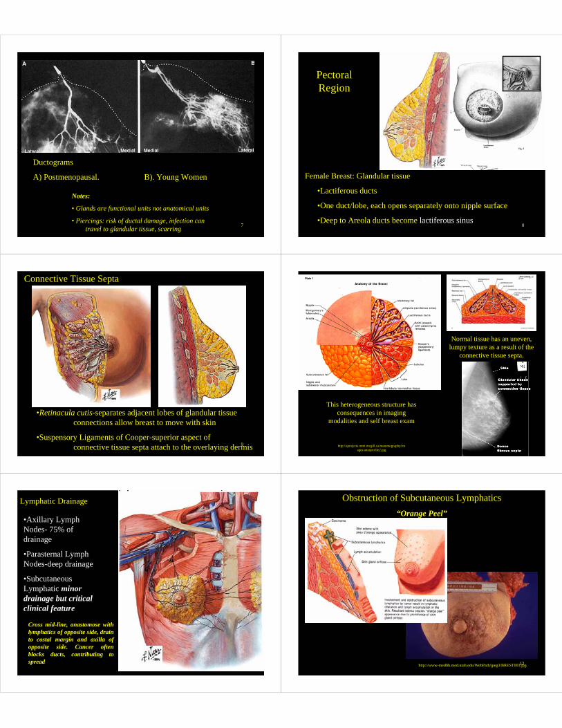
Ductograms
A) Postmenopausal. B). Young Women
Notes:
• Glands are functional units not anatomical units
• Piercings: risk of ductal damage, infection can travel to glandular tissue, scarring 8
Female Breast: Glandular tissue
•Lactiferous ducts
•One duct/lobe, each opens separately onto nipple surface
•Deep to Areola ducts become lactiferous sinus
Pectoral Region
9
Connective Tissue Septa
•Retinacula cutis-separates adjacent lobes of glandular tissueconnections allow breast to move with skin
•Suspensory Ligaments of Cooper-superior aspect ofconnective tissue septa attach to the overlaying dermis 10
Normal tissue has an uneven, lumpy texture as a result of the
connective tissue septa.
This heterogeneous structure has consequences in imaging
modalities and self breast exam
http://sprojects.mmi.mcgill.ca/mammography/images/anatprofile2.jpg
11
Lymphatic Drainage
•Axillary Lymph Nodes- 75% of drainage
•Parasternal Lymph Nodes-deep drainage
•Subcutaneous Lymphatic minor drainage but critical clinical feature
Cross mid-line, anastomose with lymphatics of opposite side, drain to costal margin and axilla of opposite side. Cancer often blocks ducts, contributing to spread 12
Obstruction of Subcutaneous Lymphatics“Orange Peel”
http://www-medlib.med.utah.edu/WebPath/jpeg3/BREST003.jpg

13
Carcinoma may grow and retract the retinacula cutis
and/or Coopers ligaments and cause dimpling of the exterior
surface of the breast.
http://www.breast-clinic.co.uk/fig9_puckeringsm.jpg
14
Male Breast
•Principally a (ornamental?) landmark
•Occasionally Subject to Neoplastic Disease362 fatal cases in men (CDC 2004)
15
Male Breast
Gynecomastia-complication of teen years, hormonal (think steroids) or drug therapy (mass typically ranges from 1-5cm in diameter).
16
•Origins from Medial 1/3 of clavicle, Sternum and upper abdominal region
•Insertion on crest of greater tubercle of humerus (lateral lip of bicipital groove)
•Innervation from Medial and Lateral Pectoral nerves
Pectoralis Major
17 18
Pectoralis Major:Three actions
•Clavicular portion flexes arm
•Sternal portion is main adductor of arm
•Medial rotation of arm
During climbing pectoralis major also can draw chest upward and prevents upward displacement of humerus.

19
•Lies between the deltoid and pectoralis major and contains the cephalic vein
Deltopectoral Triangle
20
•Origins from ribs#3 and #5
•Coracoid process of the scapula, medial surface
• Innervation from medial pectoral nerve
Note: Cannot demonstrate action or
test function of Pectoralis Minor in the living
independent of Pectoralis Major
Pectoralis Minor
*
21
•Actions: Depresses shoulder and downwardly
rotates the scapula
Pectoralis Minor
22
Lateral Pectoral Nerve(Pectoralis Major)
Passes medial to Pectoralis Minor
Medial Pectoral Nerve(Pectoralis Major & Minor)
Passes throughPectoralis Minor
This naming convention has nothing to do with the course of these nerves and everything to do with where they
come from-stay tuned.
23
Subclavius Muscle
•Actions: Depresses and rotates clavicle.
•Shunts clavicle anterior during throwing motions
•Protects the underlying subclavian artery & vein from sharp ends of fractured clavicle
•Innervation from the brachial plexus-subclavius nerve (in the neck-will not see now)
24
Axilla:
•Roughly an inverted pyramidal shaped space
•Distinct anterior, posterior, medial and lateral boarders

25
Axilla
Boundaries
•Anterior: Pectoralis Major formsthe anterior axillary fold
•Posterior
•Medial
•Lateral
26
Axilla
Boundaries
•Anterior: Pectoralis Major
•Posterior: (more than you think) Latissimus dorsi (posterior axillary fold), Teres Major, and Subscapularis
•Medial
•Lateral
27
Axilla•Boundaries
•Anterior: Pectoralis Major
•Posterior: Latissimus dorsi (posterior axillary fold), Teres Major, and Subscapularis
•Lateral
28
Axilla
Boundaries
•Anterior: Pectoralis Major
•Posterior: Latissimus dorsi, Teres Major and, Subscapularis
•Medial: Serratus anterior and chest wall
•Lateral
29
AxillaBoundaries
•Anterior: Pectoralis Major
•Posterior:Subscapularis, Teres Major (posterior axillary fold) and Latissimus dorsi
•Medial:chest wall and Serratus anterior
•Lateral: intertuberculargroove of humerus
30
Axillary Vessels
Axillary vein-From the brachial vein at teres major continues as subclavian vein
In today’s dissection, look at these vessels and remove to clarify the
underlying artery

31
Axillary artery:
Begins at 1st rib and ends at the lateral border of scapula (TeresMajor), where it becomes the Brachial artery.
Three Main Parts, each with named branches:
•Proximal (1:1)
•Deep (2:2)
•Distal (3:3)This is the hard part of
today’s lecture 32
1:1 Supreme Thoracic1st intercostal space
2:2. Thoracoacromial
2:2. Lateral Thoracic
3:3 Subscapular
3:3 Anterior humeral circumflex
3:3 Posterior humeral circumflex
Axillary Artery
33
AxillaAxillary artery
•Proximal
•Deep
•Distal
Proximal (1:1)
Deep (2:2)
Distal (3:3)
Supreme Thoracic
Thoracoacromial Lateral Thoracic
Anterior humeral circumflex
Posterior humeral circumflexSubscapular
•Pectoral
•Deltoid
•Clavicular
•Acromial
34
Thoracoacromial trunk
•Pectoral branch
With lateral nerve on inferior surface of Pectoralis major
35
Thoracoacromial trunk
•Pectoral branch
36
Thoracoacromial trunk
•Pectoral branch
•Deltoid branch,Accompanies cephalicvein in deltopectoralgroove
•Clavicular branch
•Acromial branch

37
Thoracoacromial trunk
•Pectoral branch
•Deltoid branch,Accompanies the cephalic vein in deltopectoral groove
•Clavicular branch Superomedially towardthe sternoclavicular joint
•Acromial branch
38
Thoracoacromial trunk
•Pectoral branch
•Deltoid branch,Accompanies the cephalic vein in deltopectoral groove
•Clavicular branch
•Acromial branch; may arise from deltoid branch
39
Axillary artery:•Proximal
•Deep
•Distal
Proximal
Deep
Distal
Supreme Thoracic
Thoracoacromial Lateral Thoracic
Anterior humeral circumflex
Posterior humeral circumflexSubscapular
Pectoral
Deltoid
Clavicular
Acromial
40
1. Supreme Thoracic
2. Thoracoacromial
2. Lateral Thoracic
3. Anterior humeral circumflex
3. Posterior humeral circumflex
3. Subscapular
Axillary Artery
Circumflex scapular
Thoracodorsal (latissimus dorsi)
41
Subscapular
Dorsal Scapular
Circumflex scapular
Suprascapular
42
AxillaAxillary artery
•Proximal
•Deep
•Distal
Proximal
Deep
Distal
Supreme Thoracic
Thoracoacromial Lateral Thoracic
Anterior humeral circumflex
Posterior humeral circumflexSubscapular
•Pectoral
•Deltoid
•Clavicular
•Acromial

43
AxillaAxillary artery
•Proximal
•Deep
•Distal
Proximal
Deep
Distal
Supreme Thoracic
Thoracoacromial Lateral Thoracic
Anterior humeral circumflex
Posterior humeral circumflexSubscapular
44
AxillaAxillary artery
•Proximal
•Deep
•Distal
Proximal
Deep
Distal
Supreme Thoracic
Thoracoacromial Lateral Thoracic
Anterior humeral circumflex
Posterior humeral circumflexSubscapular
•Circumflex scapular
•Thoracodorsal (usually travels with the nerve to latissimus dorsi)
45
Break 10 Minutes
46
Posterior
•Latissimus dorsi
•Teres Major
•Subscapularis