Self-recognition in chimpanzees (Pan troglodytes): Distribution, ontogeny, and patterns of emergence
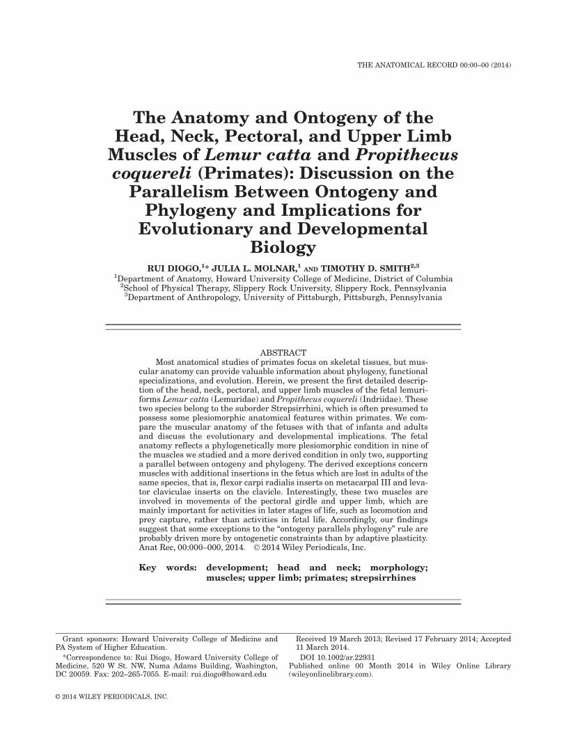
The Anatomy and Ontogeny of theHead, Neck, Pectoral, and Upper Limb
Muscles of Lemur catta and Propithecuscoquereli (Primates): Discussion on the
Parallelism Between Ontogeny andPhylogeny and Implications for
Evolutionary and DevelopmentalBiology
RUI DIOGO,1* JULIA L. MOLNAR,1 AND TIMOTHY D. SMITH2,3
1Department of Anatomy, Howard University College of Medicine, District of Columbia2School of Physical Therapy, Slippery Rock University, Slippery Rock, Pennsylvania
3Department of Anthropology, University of Pittsburgh, Pittsburgh, Pennsylvania
ABSTRACTMost anatomical studies of primates focus on skeletal tissues, but mus-
cular anatomy can provide valuable information about phylogeny, functionalspecializations, and evolution. Herein, we present the first detailed descrip-tion of the head, neck, pectoral, and upper limb muscles of the fetal lemuri-forms Lemur catta (Lemuridae) and Propithecus coquereli (Indriidae). Thesetwo species belong to the suborder Strepsirrhini, which is often presumed topossess some plesiomorphic anatomical features within primates. We com-pare the muscular anatomy of the fetuses with that of infants and adultsand discuss the evolutionary and developmental implications. The fetalanatomy reflects a phylogenetically more plesiomorphic condition in nine ofthe muscles we studied and a more derived condition in only two, supportinga parallel between ontogeny and phylogeny. The derived exceptions concernmuscles with additional insertions in the fetus which are lost in adults of thesame species, that is, flexor carpi radialis inserts on metacarpal III and leva-tor claviculae inserts on the clavicle. Interestingly, these two muscles areinvolved in movements of the pectoral girdle and upper limb, which aremainly important for activities in later stages of life, such as locomotion andprey capture, rather than activities in fetal life. Accordingly, our findingssuggest that some exceptions to the “ontogeny parallels phylogeny” rule areprobably driven more by ontogenetic constraints than by adaptive plasticity.Anat Rec, 00:000–000, 2014. VC 2014 Wiley Periodicals, Inc.
Key words: development; head and neck; morphology;muscles; upper limb; primates; strepsirrhines
Grant sponsors: Howard University College of Medicine andPA System of Higher Education.
*Correspondence to: Rui Diogo, Howard University College ofMedicine, 520 W St. NW, Numa Adams Building, Washington,DC 20059. Fax: 202–265-7055. E-mail: [email protected]
Received 19 March 2013; Revised 17 February 2014; Accepted11 March 2014.
DOI 10.1002/ar.22931Published online 00 Month 2014 in Wiley Online Library(wileyonlinelibrary.com).
THE ANATOMICAL RECORD 00:00–00 (2014)
VVC 2014 WILEY PERIODICALS, INC.

Most studies of the gross morphology of primates focuson hard tissues, and those that focus on soft tissues gener-ally refer to the adult anatomy (for a review, see, e.g.,Gibbs, 1999; Gibbs et al., 2000, 2002; Diogo and Wood,2012a). However, recent studies have shown that muscularanatomy is particularly useful for phylogenetic studies(e.g., Diogo and Wood, 2011, 2012a,b). Moreover, includingdata from non-adult specimens can add valuable informa-tion to the discussion of functional specializations (e.g.,Raichlen, 2005; Atzeva et al., 2007) and evolutionary topicssuch reversions and developmental constraints (e.g., Diogoand Wood, 2011, 2012a,b). Using postnatal samples, Diogoand Wood (2012a) presented a detailed analysis of head,neck, pectoral, and forelimb myology in each of the majorprimate higher taxa (strepsirrhines, tarsiers, new worldmonkeys, old world monkeys, and hominoids), and wehave now embarked on a second study that focuses on themuscles of the trunk, pelvis and lower limb. These publica-tions provide a broad phylogenetic and evolutionary basisfor further, more specific analyses of certain primate taxa,anatomical regions, and/or developmental stages.
In this publication, we provide the first detaileddescription of the head, neck, pectoral, and upper limbmuscles of fetal representatives of the lemuriform fami-lies Lemuridae (Lemur catta) and Indriidae (Propithecuscoquereli) of the suborder Strepsirrhini. These descrip-tions are based on dissections and histological analysisof microscope slide series. The muscle anatomy will becompared with that of older specimens (infants andadults) of these species, dissected by us and by otherauthors, as well as with that of other primates. Thiscomparison will allow us to explore and discuss the evo-lutionary and developmental implications of our anatom-ical observations, particularly their relevance to thenotion that “ontogeny parallels phylogeny.”
In Ontogeny and Phylogeny, Gould (1977) argues thatresearchers often use Haeckel’s (refuted) hypothesis thatthe ontogeny of one organism recapitulates the adultstages of its ancestors (i.e., recapitulation) as a “straw-man” to deny the existence of a parallel between otogenyand phylogeny. According to Gould, such a parallel none-theless usually exists, probably driven more by phyloge-netic/ontogenetic constraints than by adaptive plasticity.Our recent studies of human and non-human primatemuscles support Gould’s ideas (Diogo and Wood, 2012b).For instance, in karyotypically “normal” modern humanontogeny, the intermetacarpales are present as distinctmuscles, and there are multiple contrahentes muscles inaddition to the one inserting on Digit 1 (i.e., the adduc-tor pollicis), but these muscles are lost or become indis-tinct later in ontogeny. This sequence parallels theevolutionary history of primates, in which these muscleswere plesiomorphically present and then were lost inhumans (Diogo and Wood, 2011). This parallel is notrecapitulation in the Haeckelian sense: the contrahentesdigitorum and the intermetacarpales of karyotypically‘normal’ human embryos do not correspond to themuscles of adult primates such as chimpanzees, butinstead to muscles in the embryos of the latter taxa.Even after several millions of years, the developmentalpathways that produce these muscles have not beencompletely lost in modern humans, probably becausethey are related to pathways involved in the develop-ment of structures that are present and functional inmodern human adults.
As we now have detailed data about the ontogeny ofthe head and neck muscles in many tetrapod taxa andthe phylogeny and evolution of these muscles within ver-tebrates (Diogo and Abdala, 2010), we can compare theorder in which the muscles appear in ontogeny with theorder in which they evolved. For instance, our previousworks show a parallel between ontogeny and phylogenyin zebrafish head muscles, with only a few exceptions;for example, the early ontogenetic appearance of musclesthat evolved late in phylogeny but play a particularlyimportant role in the feeding mechanisms of both adultsand embryos (Diogo et al., 2008). A very similar patternwas found in axolotls (Ziermann and Diogo, 2013). How-ever, these results do not necessarily accord with thecommonly accepted view that muscles tend to differenti-ate (and not de-differentiate) during ontogeny. The orderin which muscles appear in ontogeny is usually similarto the order in which they appear in phylogeny, butmuscles are often lost/reabsorbed later in ontogeny. Forinstance, in neotenic salamander species such as axolotlsthat do not undergo full metamorphosis, some musclesbecome completely indistinct during ontogeny; for exam-ple, the pseudotemporalis profundus and the levatorhyoideus become completely integrated in the pseudo-temporalis superficialis and in the depressor mandibu-lae, respectively (Ziermann and Diogo, 2013). Parallelsbetween ontogeny and phylogeny will be further dis-cussed in the light of the results of this study.
MATERIALS AND METHODS
We dissected one side of the head, neck, pectoral, andupper limb muscles of a female Lemur catta fetus (DukeLemur Center specimen number 6888, fixed in 10% buf-fered formalin) and of a male Propithecus coquereli fetus(Duke Lemur Center specimen number P6154, fixed in10% buffered formalin). The contralateral side of thehead was also available in a microscope slide series.Each half was paraffin embedded, serially sectioned (10mm thick), and stained with hematoxylin and eosin ortrichrome procedures. Because facial skin was present inthe oronasal region of the slides series, some facialmuscles were available for analysis.
The musculature of these two fetuses was comparedwith that of an adult male Lemur catta (GWUANT LC1;fresh), an adult female P. coquereli (GWUANT PV1;fresh), and an infant female P. coquereli (GWUANT PV2;fresh). These specimens had been dissected anddescribed previously (Diogo and Wood, 2012a) and wereretrieved from fixative for direct comparison with thefetal specimens. We also compared the musculature withthat of additional specimens of these two species dis-sected by other authors (e.g., Meckel, 1820–1838; Cuvierand Laurillard, 1849; Murie and Mivart, 1872; Milne-Edwards and Grandidider, 1875; Ruge, 1878, 1885; Par-sons, 1898a,b; Tschachmachtschjan, 1912; Kollmann andPapin, 1914; Lander, 1918; Loth, 1931; Edgeworth, 1935;Straus, 1942a,b; Miller, 1943; Hill, 1953; Starck andSchneider, 1960; Jouffroy, 1960a,b, 1962, 1971; Ashtonand Oxnard, 1963; Kladetsky and Kobold, 1966; Saban,1968; Kaneff and Cihak, 1970; Seiler, 1976; Lewis, 1989;Maier, 2008) as well as numerous other primate speciesdissected by other authors (for a recent review, see Diogoand Wood, 2012a). The head, neck, pectoral, and upperlimb muscles of the Lemur and Propithecus fetuses were
2 DIOGO, MOLNAR, AND SMITH

dissected and photographed using a Nikon SMZ-1500Zoom stereo-microscope equipped with a Nikon DS Fi1 5Megapixel Color Camera Head.
The nomenclature for the head, neck, pectoral, andupper limb muscles follows that of Diogo and Abdala(2010) and Diogo and Wood (2012a). The pectoral andupper limb muscles are divided into five subgroups: pec-toral, arm, ventral forearm, dorsal forearm, and hand.We focused on four main sub-groups of head and neckmuscles: (1) mandibular muscles—generally innervatedby cranial nerve V (e.g., the muscles of mastication andone of the middle ear muscles, the tensor tympani); (2)hyoid muscles—usually innervated by cranial nerve VII(e.g., muscles of facial expression and the other middleear muscle, the stapedius); (3) branchial muscles—usu-ally innervated by cranial nerves IX and X (e.g., themajority of the intrinsic laryngeal muscles), althoughthe trapezius and sternocleidomastoideus are mainlyinnervated by cranial nerve XI; (4) hypobranchialmuscles—according to Edgeworth (1935), these musclesdevelop from the anterior myotomes of the body andmigrate into the head (e.g., infrahyoid muscles);although they retain their main innervation from spinalnerves, they may also be innervated by cranial nervesXI and XII, but they usually do not receive any branchesfrom cranial nerves V, VII, IX, or X. Head and neckmuscles not included in this study are the epibranchialmuscles sensu Edgeworth (1935), which are absent inextant osteichthyans and therefore are not present inprimates or other mammals, and the internal and exter-nal ocular muscles sensu Edgeworth (1935), which areusually innervated by the cranial nerves III, IV, and/orVI. In the sections below, we use the terms anterior, pos-terior, dorsal, and ventral as they apply to pronogradetetrapods.
RESULTS
The textual and visual (Figs. 1–10) descriptions referto our observations of both the L. catta and P. coquerelifetuses. Major differences between these two specimens(and/or specimens of the same species dissected by our-selves or by other authors) will be mentioned in the textbelow.
Mandibular Muscles
The mylohyoideus (Figs. 3A, 9A) runs from the mandi-ble to the ventral midline of the neck and to the body ofthe hyoid bone. In the L. catta fetus, the digastricusanterior (Figs. 3A, 8A) is well separated from its coun-terpart and runs from the digastric intermediate tendonand the hyoid bone to the medial margin of the mandi-ble. This configuration was also found in the adult L.catta and adult P. coquereli we dissected. A similar con-figuration was found in the P. coquereli fetus, exceptthat the digastricus anterior did not originate from thehyoid bone. We could not examine the tensor tympanibecause this region was too delicate. According to Maier(2008), this muscle is usually present in adults of bothgenera, and the chorda tympani passes below the muscle(hypotensoric configuration). The tensor veli palatiniruns from the region near the external acoustic meatusto the soft palate, surrounding the pterygoid hamulus,and its orientation is more horizontal than in modern
humans. The masseter (Figs. 1B, 8B) runs from thezygomatic arch and zygoma to the angle, lower border,and ramus of mandible. The pars superficialis and parsprofunda seem to be less differentiated than in adults ofthese two species (e.g., Hill, 1953; Diogo and Wood,2012a) (the “zygomatico-mandibularis” not present as adistinct structure). In the L. catta fetus, the main bodyof the temporalis (Figs. 1B, 8B) is undivided and origi-nates primarily from the lateral superior surface of theskull. A well-defined pars suprazygomatica (which wasalmost completely covered laterally by the zygomaticarch) originates from the inner margin of the zygomaticarch and inserts on the lateral margin of the coronoidprocess, while the main body of the temporalis insertson the medial margin of the coronoid process. This con-figuration was also found in the adult L. catta and adultP. coquereli we dissected and is similar to the one foundin the P. coquereli fetus, but in the latter specimen thepars suprazygomatica is less differentiated from themain body of the muscle. The pterygoideus lateralis orig-inates from the pterygoid lamina and adjacent regions ofthe skull. In the L. catta fetus, there is a caput superius,which is thinner and inserts on the temporomandibularjoint, and a caput inferius, which is broader and insertson the condyloid process of the mandible. The two headsare well separated anteriorly by structures including thebuccal nerve, but they blend into each other posteriorly.We could not discern whether the two heads of the pter-ygoideus lateralis are also separated in the P. coquerelifetus, and they were not described by Murie and Mivart(1872) in the adult Lemur. On one side of our adult L.catta GWUANT LC1 specimen, the two heads are notdifferentiated, but on the other side they are slightly dif-ferentiated and have a similar configuration to the L.catta fetus. Similarly, poor differentiation was presentbilaterally in our adult P. coquereli GWUANT PV1 speci-men. The pterygoideus medialis (Fig. 9A) is a single,undivided mass running from the fossa between the twowings of the pterygoid process to the inner surface of theangle of the jaw.
Hyoid Muscles
In the L. catta fetus, the stylohyoideus runs from thetympanic bulla and the most proximal (and non-ossified)portion of the hyoid apparatus and to the hyoid bone. Itpasses superficial to the digastricus tendon, withoutbeing pierced by it, and almost reaches its counterpartin the midline; that is, its insertion onto the hyoid boneis peculiarly extended in a medial direction (100%). Thestylohyoideus is also directly connected to the mylohyoi-deus and/or to the aponeurosis connecting the mylohyoi-deus to the hyoid bone. The configuration of this muscleis similar between the L. catta fetus, the P. coquerelifetus and the adults of these two species except for theconnection to the mylohyoideus, which we only observedin the L. catta fetus. The digastricus posterior (Figs. 3A,8A) runs from the mastoid region to the long, well-defined intermediate tendon. In the L. catta fetus, thejugulohyoideus is well-developed, running from the mas-toid region (deep to the digastricus posterior) to the con-nective tissue between the skull and the stylohyalligament, as it usually does in adults of this species.However, unlike in the adult specimen we dissected, thejugulohyoideus also inserts directly onto the proximal
ANATOMY AND ONTOGENY OF LEMUR AND PROPITHECUS MUSCLES 3

Fig. 1. Lemur catta (female fetus 6888). (A) Lateral view of the facialmuscles. (B) Lateral view of the mandibular muscles and the facialmuscle mandibulo-auricularis. In this and the other figures, the namesof the muscles are in italics, and SUP, INF, ANT, POS, MED, LAT, VEN,
DOR, PRO and DIS refer to superior, inferior, anterior, posterior,medial, lateral, ventral, dorsal, proximal, and distal, respectively (N.B.,in the sense that the terms are used for pronograde tetrapods: seetext for more details).

portion (i.e., closer to the neurocranium) of the hyoidapparatus. In the P. coquereli fetus, the jugulohyoideusis much more developed than it is in adult strepsirrhi-
nids. As in adults of this species, it originates from themastoid region and inserts on the connective tissuebetween the skull and the proximal portion of the hyoidapparatus, and it is deep to the digastricus posterior.Unlike in the adults, it also inserts directly onto theproximal portion of the hyoid apparatus, and it is some-what blended with the digastricus posterior. Its size,position and blending with the digastricus posteriorstrongly suggest that the jugulohyoideus is actuallyderived from the digastricus posterior and that it ismore developed in early stages than in later stages.Because the jugulohyoideus usually is not present inadult anthropoid primates, its ontogeny appears to par-allel its evolution. We could not examine the stapediusin any of the specimens we dissected.
We could not analyze the muscles of facial expressionin the P. coquereli fetus because the area was too deli-cate in this specimen; however, we were able to observesome oronasal muscles via histology. Most descriptionsin this paragraph refer to the L. catta fetus. The pla-tysma cervicale (Fig. 1A) originates from the nuchalregion (its fleshy portion reaching the dorsal midline)and inserts on the region of the mouth. The platysmamyoides (Fig. 1A) runs from the neck and chest to theregion of the mouth and the mandible. The “cervico-auriculo-occipitalis” part of the occipitalis (Fig. 1A) runsfrom the occipital region to the external ear, passing lat-erally to join the occipitalis proprius part of the muscle,which runs from the occipital region (contacting itscounterpart in the dorsal midline) to the galea aponeuro-tica. There is no distinct muscle interscutularis, but theanterior part of the occipitalis (which inserts directly onthe ear) lies somewhat anterior to the ear, thus occupy-ing the space where the interscutularis lies in othermammals (a similar condition was found in the adult L.catta shown in Fig. A2 of Diogo and Wood, 2012a). Theauricularis posterior blends with the occipitalis and runsfrom the occipital region to the posterior surface of theear. We could not examine the small muscles of the earin detail nor discern whether the sphincter colli profun-dus was present. The mandibulo-auricularis (Fig. 1B) isa fleshy muscle running from the anterior region of theear to the back of the mandible. The zygomaticus major(“auriculolabialis inferior”) is undivided and, unlike thecase in the adult L. catta, is deeply blended with thezygomaticus minor (Fig. 1A) (“auriculolabialis superior”),supporting the idea that these muscles derive from thesame anlage. The combined mass formed by the twomuscles runs from the anterior and inferoanterior mar-gin of the external ear to the angle of the mouth andblends with the platysma cervicale, which lies on thesame level. The frontalis (Fig. 1A) runs from the galeaaponeurotica to the region of the eye. The auriculo-orbitalis (Fig. 1A) runs from the anterior portion of theear to the region of the eye and blends with the fronta-lis; the auricularis anterior is not present as a distinctmuscle. The auricularis superior runs from the superiormargin of the ear to the galea aponeurotica. The orbicu-laris oculi is present and surrounds the eye, but wecould not discern whether the depressor supercilii, nasa-lis, and corrugator supercilii were present as distinctmuscles. The levator labii superioris alaeque nasi (Fig.1A) runs from the region of the glabella to the nose andupper lip. It is broader than in hominoids and blendsmore completely with the frontalis and the orbicularis
Fig. 2. Lemur catta (female fetus 6888, contralateral side to Fig. 1).(A) Serially sectioned skin of mandible, anterior to symphysis. (B)enlargement of boxed area in Fig. 2A, showing striated fibers (arrows)of mentalis projecting toward the midline. (C) The fibers originate fromthe body of the mandible near the roots of the deciduous incisors(and see Burrows and Smith, 2003, for gross description of this mus-cle in a prosimian). Scale bars: A, 0.5 mm, B, 40 mm.
ANATOMY AND ONTOGENY OF LEMUR AND PROPITHECUS MUSCLES 5

Fig. 3. Lemur catta (female fetus 6888). (A) Lateral view of the muscles lying in the auricular and neckregions and attaching to the mandible. (B) Lateral view of the laryngeal musculature.
6 DIOGO, MOLNAR, AND SMITH

Fig. 4. Lemur catta (female fetus 6888). (A) Dorsal view of the pectoral girdle and arm musculature. (B)Ventral view of the pectoral girdle and arm musculature.
ANATOMY AND ONTOGENY OF LEMUR AND PROPITHECUS MUSCLES 7

Fig. 5. Lemur catta (female fetus 6888). (A) Ventral view of the forearm and hand musculature. (B) Ven-tral view of the deeper hand musculature.
8 DIOGO, MOLNAR, AND SMITH

oculi than in adult lemurs. The procerus is not presentas a distinct muscle. The buccinatorius runs from thepterygomandibular raphe to the maxilla, mandible andorbicularis oris. The levator labii superioris (“maxillo-naso-labialis”) is an approximately horizontal musclerunning posteroanteriorly from the infraorbital region tothe region of the nose and upper lip, deep (medial) tothe levator labii superioris alaeque nasi. The levatoranguli oris facialis runs from the maxilla to the angle ofthe mouth, deep to the levator labii superioris. The orbi-cularis oris (Fig. 1A) is present and normal, but, con-trary to the case in adult lemurs, it is partially blendedposterosuperiorly with some fibers of the orbicularisoculi due to the large size of the eye and its proximity tothe mouth. The depressor labii inferioris and the depres-sor anguli oris are not present as distinct muscles.
Serial sections of the oronasal regions reveal that thementalis is present in both fetuses (Figs. 2A,B, 7A,B),with fibers attaching laterally to the mandibular bodyadjacent to the deciduous canine and the roots of the
deciduous incisors (Figs. 2C, 7C). The mentalis mergesobliquely with its counterpart near the midline, anteriorto the mandibular symphysis.
Branchial Muscles
In the P. coquereli fetus, the stylopharyngeus (Fig.9A) runs from the auditory bulla and proximal portion ofthe stylohyal ligament to the pharyngeal wall (notinserting onto the hyoid bone), passing superior to themiddle constrictor and inferior to the superior constric-tor. This configuration is found in adult lemurs andadult P. coquereli, but in the L. catta fetus the stylophar-yngeus does not originate directly from the skull. Wecould not discern whether the petropharyngeus waspresent as a distinct muscle. The ceratohyoideus con-nects the thyrohyal (greater horn of the hyoid bone),hypohyal and distal portion of the ceratohyal, lying deep(dorsal) to the insertions of the middle constrictor andthe stylopharyngeus. The trapezius runs from the
Fig. 6. Lemur catta (female fetus 6888). Dorsal view of the forearm and hand musculature.
ANATOMY AND ONTOGENY OF LEMUR AND PROPITHECUS MUSCLES 9

ligamentum nuchae and vertebrae (not from the cra-nium) to the scapular spine and acromion (not to theclavicle), inserting deep (ventral) to the insertion of thelevator claviculae. The cleido-occipitalis is not present asa distinct muscle. The sternocleidomastoideus (Fig. 3A)has a superficial caput sternomastoideum running fromthe sternum (not the sternal end of the clavicle as itoften does in adults of the two species) to the mastoidprocess in the L. catta fetus, and also to the occipitalregion in the P. coquereli fetus. The deeper caput cleido-mastoideum runs from the clavicle to the mastoidregion. The constrictor pharyngis medius (Fig. 3A) runsfrom the dorsal midline raphe to the thyrohyal (lesserhorn of the hyoid bone; pars ceratopharyngea), the hypo-hyal and, perhaps, to the ceratohyal (if it does, thiswould constitute a pars chondropharyngea). The con-strictor pharyngis inferior (Figs. 3A, 9A) runs from thedorsal midline raphe, which is associated with the cra-nium, to the thyroid (pars thyropharyngea) and cricoid(pars cricopharyngea) cartilages. The cricothyroideus(Figs. 3A, 9A,B) is only slightly differentiated into parsrecta, pars obliquua and pars interna (seemingly less sothan in adults). It runs from the cricoid cartilage to thethyroid cartilage and does not contact its counterpart inthe ventral midline or have a broad insertion onto theinferior horn of the thyroid cartilage. The constrictorpharyngis superior runs from the midline raphe to thepalate (pars pterygopharyngea), the tongue (pars glosso-pharyngea), and seemingly to the pterygomandibularraphe (pars buccopharyngea); we could not discernwhether there is a pars mylopharyngea. The palatopha-ryngeus runs from the soft palate to the pharyngealwall; we could not discern whether it reaches the thyroidcartilage or if there is a distinct muscle uvulae. Thelevator veli palatini runs from the region near the ear tothe soft palate and is more horizontal than in humans.The thyroarytenoideus (Figs. 3B, 9B) connects the thy-roid and arytenoid cartilages. It consists of a pars supe-rior (more anterior and somewhat more lateral) and apars inferior (which corresponds to the “pars intermedia”sensu Starck and Schneider, 1960). We could not findany other divisions of the muscle. The cricoarytenoideuslateralis (Figs. 3B, 9B) connects the cricoid and aryte-noid cartilages. The arytenoideus (Figs. 3B, 9B) connectsthe two arytenoid cartilages; there is no median rapheor distinct muscle arytenoideus obliquus. The cricoaryte-noideus posterior (Figs. 3B, 9B) connects the cricoid andarytenoid cartilages. It does not insert on the inferiorhorn of the thyroid cartilage but contacts its counterpartin the dorsal midline.
Hypobranchial Muscles
The geniohyoideus (Figs. 8A, 9A) originates from themandible inserts on the hyoid bone. The genioglossus(Fig. 9A) runs from the mandible to the tongue. We didnot examine the intrinsic muscles of the tongue indetail. In the P. coquereli fetus, the hyoglossus (Figs. 3A,8A) has a thinner chondroglossus part originating fromthe body of the hyoid bone (basihyal) and a broader cera-toglossus part originating from the greater horn of thehyoid bone (thyrohyal) and inserts on the inferolateralsurface of the tongue. In the L. catta fetus, the cerato-glossus part runs from the thyrohyal to the tongue, but
Fig. 7. Propithecus coquereli (male fetus P6154, contralateral sideto Fig. 9). (A–C) Serially sectioned skin of mandible, showing anteriorfibers of the mentalis muscle (arrows) passing horizontally toward themidline (B is an enlarged view of the boxed area in A). The fibers origi-nate from the body of the mandible near the roots of the deciduousincisors (C). Scale bars: A, 1 mm; B, 250 mm; C, 0.5 mm.
10 DIOGO, MOLNAR, AND SMITH

Fig. 8. Propithecus coquereli (male fetus P6154). (A) Lateral view of the head musculature. (B) Lateralview of the masticatory muscles and styloglossus.
ANATOMY AND ONTOGENY OF LEMUR AND PROPITHECUS MUSCLES 11

Fig. 9. Propithecus coquereli (male fetus P6154). (A) Ventrolateral view of the head musculature. (B)Lateral view of the laryngeal musculature.
12 DIOGO, MOLNAR, AND SMITH

Fig. 10. Propithecus coquereli (male fetus P6154). (A) Dorsal view of the muscles of the pectoral girdleand arm. (B) Ventral view of some hand muscles.
ANATOMY AND ONTOGENY OF LEMUR AND PROPITHECUS MUSCLES 13

we could not detect fibers from the basihyal; if theyexisted, the chondroglossus part is very thin. The stylo-glossus (Figs. 3A, 8A,B, 9A) runs from the proximalregion of the hyoid arch (in the L. catta fetus and seem-ingly also in the P. coquereli fetus) and from the auricu-lar region of the skull (only in the P. coquereli fetus) tothe tongue. The palatoglossus is not present as a distinctmuscle. The sternohyoideus (Figs. 8A, 9A) runs from thesternum to the hyoid bone, remains in contact with itscounterpart for most of its length and has no well-defined tendinous intersection. In the P. coquereli fetus,the omohyoideus (Fig. 3A) runs from the scapula to thehyoid bone; it is deep to the sternocleidomastoideus andhas no intermediate tendon. The configuration is similarin the L. catta fetus, except that the muscle seems tohave a tendinous intersection at about half of its length.The sternothyroideus (Figs. 3A, 8A, 9A) runs from thesternum to the thyroid cartilage, inserting posteriorly tothe origin of the thyrohyoideus with no tendinous inter-section. The thyrohyoideus (Figs. 3A, 8A) connects thethyroid cartilage to the hyoid bone and is not blendedwith the hyoglossus or the sternothyroideus.
Pectoral Muscles
The serratus anterior (Figs. 4A, 10A) runs from theribs to the medial side of the scapula and is being deeplyblended with the levator scapulae. The rhomboideus(Figs. 4A, 10A) is a continuous muscle (i.e., not subdi-vided into rhomboideus major and minor) running fromcervical and thoracic vertebrae to the medial surface ofthe scapula, as is usually the case in adults of both spe-cies. However, the rhomboideus and rhomboideus occipi-talis in the fetuses are more deeply blended with theserratus anterior and the levator scapulae than inadults. The rhomboideus occipitalis (Fig. 4A) is a verythin muscle running from the cranium to the scapula. Itis usually present in adult lemurs and may or may notbe present in adult Propithecus: Jouffroy (1962) statesthat the rhomboideus occipitalis is missing in adult Pro-pithecus verreauxi and Propithecus deckenii; this condi-tion was also found by Milne-Edwards and Grandidier(1875); however, Ashton and Oxnard (1963) state that inthe Propithecus sp. specimen they dissected there wasan attachment of the rhomboid complex to the occipitalregion and that such a condition was also described inthis genus by Milne-Edwards and Grandidier (1875),contradicting Jouffroy (1962). Because the muscle ismissing in the adult P. coquereli GWUANT PV1 speci-men, it is clear that the muscle is missing in at leastsome adult specimens of this species. In the fetuses ofboth species and in P. coquereli adults, the levator scap-ulae (Figs. 2A, 3A, 10A) runs from C2–C7 to the superiorangle of the scapula, while in adult lemurs it usuallyattaches to C1 as well. There is no distinct muscle atlan-toscapularis posticus. In the P. coquereli fetus, the leva-tor claviculae (Fig. 3A) runs from C1 to the acromionand scapular spine, passing superficial (dorsal) to thetrapezius, as it does in adults of this species and in adultlemurs (e.g., Murie and Mivart, 1872; Jouffroy, 1962;Diogo and Wood, 2012a). In the L. catta fetus, the leva-tor scapulae also inserts on the lateral extremity of theclavicle. In the L. catta fetus, the subclavius runs fromRib 1 to the clavicle, but in the P. coquereli fetus a fewfibers also run to the region of the scapula, specifically
the proximal portion of the coracoid process. This divi-sion of the muscle could be interpreted as a remnant ofthe plesiomorphic costocoracoideus muscle, which is usu-ally not present in the adults of either species.
The pars clavicularis of pectoralis major (Fig. 4B) orig-inates from the medial portion of the clavicle and fromthe sternum and inserts on the proximal humerus,superficial and distal to the insertion of the pars sterno-costalis, which originates from the sternum and ribs.The pars abdominalis is deep to the two other heads andblended distally with the pectoralis minor, originatingfrom the abdominal muscles and some ribs and insertingon the humerus, proximal to the insertion of the twoother heads. The pectoralis major contacts its counter-part in the midline. The pectoralis minor runs from thesternum and ribs to the proximal humerus. The panni-culus carnosus is present in the L. catta fetus; the parshumerodorsalis inserts on the greater tuberosity of thehumerus and blends somewhat with the distal portionsof the pars abdominalis of the pectoralis major, amongother muscles. We could not discern whether the panni-culus carnosus was present in the P. coquereli fetus. Theinfraspinatus (Figs. 4A, 10A) and the supraspinatus(Figs. 4A, 10A) originate from the infraspinatous andsupraspinatous fossae, respectively, and insert on thegreater tuberosity of the humerus. In the L. catta fetus,the deltoid complex is differentiated into a pars clavicu-laris (from the clavicle) and a pars acromialis (from theacromion), which are blended with each other to formthe deltoideus acromialis et clavicularis and well sepa-rated from the pars spinalis and deltoideus scapularis(from scapular spine and infraspinatous fascia) (Fig.4A,B), as is usually the case in adult lemurs. In the P.coquereli fetus, the deltoideus (Fig. 10A) was onlyslightly differentiated into a pars clavicularis, a parsacromialis and a pars spinalis that lies just beside thetwo other parts, as is the case in adults of this species.The teres minor runs from the lateral 1/3 (in the lemurfetus and adults) or 1/2 (in the P. coquereli fetus andadults) of the lateral border of the scapula to the greatertuberosity of the humerus. In neither fetus does theinsertion of the muscle extend distal to this tuberosity.The subscapularis (Fig. 4B) is an undivided muscle run-ning from the subscapular fossa to the lesser tuberosityof the humerus and the capsule of the shoulder joint.The teres major (Figs. 4A, 10A) runs from the lateralborder of the scapula to the proximal portion of thehumerus, blends with the subscapularis and passes dor-sally to (without blending with) the tendon of the latissi-mus dorsi. In the L. catta fetus, however, a very thintendinous slip connects the two tendons at the level ofthe axilla, just proximal to their insertion onto thehumerus. The latissimus dorsi (Figs. 4A, 10A) is some-what blended with the trapezius in the P. coquereli fetusbut not in the lemur fetus. In both fetuses, the latissi-mus dorsi does not insert directly on the scapula butruns from the dorsolumbar fascia, vertebrae, and/or ribsto the humerus.
Arm Muscles
The dorsoepitrochlearis (Figs. 4A, 10A) originatesfrom the latissimus dorsi and inserts onto the olecranonprocess and proximal portion of the shaft of the ulnaand onto the fascia superficial to the medial epicondyle
14 DIOGO, MOLNAR, AND SMITH
of the humerus (but is not directly attached to the epi-condyle). The triceps brachii (Figs. 4A, 10A) has a longhead (from the lateral border of the scapula), a lateralhead (from the surgical neck), a medial head (from thesurgical neck in the lemur fetus and adults, but fromthe humeral shaft in P. coquereli fetus and adults), anda posterior head (from the distal portion of thehumerus). The posterior head appears to be derivedfrom the medial head because its origin is noticeablyproximal to, and clearly distinct from, the proximal ori-gin of the epitrochleoanconeus. The four heads insert onthe olecranon process of the ulna. A major differencebetween the lemur fetus and all the other specimens(i.e., the lemur adults plus the P. coquereli fetus andadults) is that in the former the posterior and medialheads of the triceps brachii are profoundly blended witheach other. The brachialis is an undivided muscle run-ning from the humerus to the coronoid process of theulna. It does not appear to reach the surgical neck of thehumerus in the P. coquereli fetus, and we could not dis-cern this feature in the lemur fetus. The long head ofthe biceps brachii (Fig. 4B) originates from the infragle-noid tubercle of the scapula, while the short head origi-nates from the coracoid process of the scapula. The twoheads are completely blended distally and insert on thebicipital tubercle of the radius, where the tendon con-nects to a strong, distinct ’lacertus fibrosus’ (aponeurosisbicipitalis). The coracobrachialis (Fig. 4B) originatesfrom the coracoid process of the scapula, its caputmedium inserting on the distal 1/2 of the humerus(including the medial epicondyle) and its caput profun-dum inserting on the proximal portion of the humerus.In the P. coquereli fetus, the musculocutaneous nervepasses between these two heads, as it usually does inadults of the two species, but we could not discernwhether this was also the case in the lemur fetus.
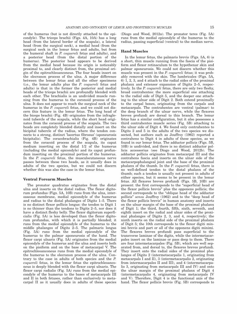
Ventral Forearm Muscles
The pronator quadratus originates from the distalulna and inserts on the distal radius. The flexor digito-rum profundus (Figs. 5A, 10B) runs from the ulna, inter-osseous membrane, medial epicondyle of the humerusand radius to the distal phalanges of Digits 1–5. Thereis no distinct flexor pollicis longus: the tendon to Digit 1is no thinner than the tendons to Digits 2–5, nor does ithave a distinct fleshy belly. The flexor digitorum superfi-cialis (Fig. 5A) is less developed than the flexor digito-rum profundus, with which it is partially blended; itruns from the medial epicondyle of the humerus to themiddle phalanges of Digits 2–5. The palmaris longus(Fig. 5A) runs from the medial epicondyle of thehumerus to the palmar aponeurosis of the hand. Theflexor carpi ulnaris (Fig. 5A) originates from the medialepicondyle of the humerus and the ulna and inserts bothon the pisiform and on the base of metacarpal V. Theepitrochleoanconeus runs from the medial epicondyle ofthe humerus to the olecranon process of the ulna. Con-trary to the case in adults of both species and the P.coquereli fetus, in the lemur fetus the epitrochleoanco-neus is deeply blended with the flexor carpi ulnaris. Theflexor carpi radialis (Fig. 5A) runs from the medial epi-condyle of the humerus to the bases of metacarpals IIIand II in both fetuses, rather than exclusively to meta-carpal II as it usually does in adults of these species
(Diogo and Wood, 2012a). The pronator teres (Fig. 5A)runs from the medial epicondyle of the humerus to theradius, passing superficial (ventral) to the median nerve.
Hand Muscles
In the lemur fetus, the palmaris brevis (Figs. 5A, 6) isa short, thin muscle running from the fascia of the pisi-form and flexor retinaculum to the hypothenar skin andpalmar aponeurosis. We could not discern whether thismuscle was present in the P. coquereli fetus; it was prob-ably removed with the skin. The lumbricales (Figs. 5A,6) 1, 2, 3, and 4 attach to the radial sides of the proximalphalanx and extensor expansion of Digits 2–5, respec-tively. In the P. coquereli fetus, there are only two fleshy,broad contrahentes: the more superficial one attachingto the radial side of Digit 5, and the deeper one attach-ing to the ulnar side of Digit 2. Both extend proximallyto the carpal bones, originating from the carpals andmetacarpals. The contrahentes are ventral (palmar) tothe deep branch of the ulnar nerve, while the flexoresbreves profundi are dorsal to this branch. The lemurfetus has a similar configuration, but it also possesses athird contrahentes muscle series (Fig. 5B) attaching tothe ulnar side of Digit 4. We found only contrahentes toDigits 2 and 5 in the adults of the two species we dis-sected, but authors such as Jouffroy (1962) reported acontrahens to Digit 4 in adults of these species, as wefound in our lemur fetus. The adductor pollicis (Figs. 5B,10B) is undivided, and there is no distinct adductor pol-licis accessorius (see Diogo and Wood, 2012b). Theadductor pollicis originates from metacarpal III and thecontrahens fascia and inserts on the ulnar side of themetacarpophalangeal joint and the base of the proximalphalanx of the thumb. In the P. coquereli fetus, there isno well-defined tendon to the distal phalanx of thethumb; such a tendon is usually not present in adults ofeither species, but it seems to be present in the lemurfetus. All flexores breves profundi (Figs. 5B, 10B) arepresent: the first corresponds to the “superficial head ofthe flexor pollicis brevis” plus the opponens pollicis; thesecond corresponds to the “oblique head of the adductorpollicis” sensu Jouffroy (1962) and to the “deep head ofthe flexor pollicis brevis” in human anatomy and insertson the ulnar margin of the base of the proximal phalanxof Digit 1; the third, fourth, fifth, sixth, seventh, andeighth insert on the radial and ulnar sides of the proxi-mal phalanges of Digits 2, 3, and 4, respectively; theninth inserts on the radial side of the proximal phalanxof Digit 5; the 10th corresponds to the flexor digiti min-imi brevis and part or all of the opponens digiti minimi.The flexores breves profundi pass superficial to thetransverse laminae of the digits, while the intermetacar-pales insert on the laminae or pass deep to them. Thereare four intermetacarpales (Fig. 5B), which are well sep-arated from, and dorsal to, the flexores breves profundi.They insert onto the radial sides of the proximal pha-langes of Digits 2 (intermetacarpalis 1, originating frommetacarpals I and II), 3 (intermetacarpalis 2, originatingfrom intermacarpales II and III), and 4 (intermetacarpa-lis 3, originating from metacarpals III and IV) and ontothe ulnar margin of the proximal phalanx of Digit 4(intermetacarpalis 4, originating from metacarpals IVand V). Therefore, Digit 4 is the functional axis of thehand. The flexor pollicis brevis (Fig. 5B) corresponds to
ANATOMY AND ONTOGENY OF LEMUR AND PROPITHECUS MUSCLES 15

the “superficial head of the flexor pollicis brevis” inhuman anatomy and is well separated from the flexorbrevis profundus 2 (which corresponds to the “deep headof the flexor pollicis brevis” in human anatomy). It origi-nates from the trapezium and flexor retinaculum andinserts on the radial sesamoid of the interphalangealjoint and the base of the proximal phalanx of the thumb,together with the abductor pollicis brevis, radial to thetendon of the flexor digitorum profundus to Digit 1 (theinsertion of the flexor brevis profundus 2 is ulnar to thetendon of the flexor digitorum profundus to Digit 1).More so than in adults of these two species, the configu-ration in the fetuses indicates that both the opponenspollicis and the flexor pollicis brevis (“superficial head”in human anatomy) derive from the flexor brevis profun-dus 1, because the two former muscles are clearlyblended with each other. The opponens pollicis (Fig. 5B)is a thin muscle that runs from the trapezius to meta-carpal I and also to the radial sesamoid of the interpha-langeal joint. The flexor digiti minimi brevis (Figs. 5A,B,6) originates from the hamate and flexor retinaculumand inserts on the ulnar side of the proximal phalanx ofDigit 5, together with the abductor digiti minimi. Theopponens digiti minimi (Fig. 5B) originates from thehamate and flexor retinaculum and inserts along theentire length of metacarpal V, completely dorsal (deep)to the deep branch of the ulnar nerve. The adductor pol-licis brevis (Fig. 5A,B) originates from the sesamoidbone associated with the trapezium and from the flexorretinaculum and inserts on the radial side of the base ofthe proximal phalanx of Digit 1. The adductor digitiminimi (Figs. 5A,B, 6) originates from the pisiform andinserts on the ulnar side of the base of the proximal pha-lanx of Digit 5. The flexor digiti minimi brevis, opponensdigiti minimi, adductor pollicis brevis and adductor digitiminimi are all undivided muscles.
Dorsal Forearm Muscles
The extensor carpi radialis longus (Fig. 6) runs fromthe lateral supracondylar ridge of the humerus, just dis-tal to the origin of the brachioradialis, to the base ofmetacarpal II. The extensor carpi radialis brevis (Fig. 6)runs from the lateral ridge and epicondyle of thehumerus to the base of metacarpal III. The brachioradia-lis runs from the distal portion of the humerus to theradius, reaching the styloid process distally. The supina-tor originates from the lateral epicondyle of thehumerus, annular ligament and proximal portion of theradius and inserts on the shaft of the radius. The exten-sor carpi ulnaris (Fig. 6) runs from the lateral epicon-dyle of the humerus (caput humerale) and ulna (caputulnare) to the base of metacarpal V. In the P. coquerelifetus, the anconeus runs from the lateral epicondyle ofthe humerus to the olecranon process of the ulna and ispartially blended with the triceps brachii, as is usuallythe case in adults of the two species. We could not dis-cern whether the anconeus was present as a distinctmuscle in the lemur fetus. The extensor digitorum (Fig.6) runs from the lateral epicondyle of the humerus toDigits 2–5. In the P. coquereli fetus, the equivalent ofthe extensor digiti minimi runs from the lateral epicon-dyle of the humerus to Digits 4–5, as is usually the casein adults of the two species. However, in the lemur fetusit also sends a very thin tendon to Digit 3, supporting
the idea that some tendons of the extensor indicis and/orextensor digiti minimi are lost during ontogeny (e.g.,Kaneff and Cihak, 1970; see Discussion below). In thelemur fetus, the extensor indicis (Fig. 6) runs from theulna and interosseous membrane to Digits 2 and 3,while in the P. coquereli fetus it also inserts on Digit 4.The adult configuration of this muscle is ambiguous: aninsertion onto Digits 2–4 in Lemur was described byBarnard (1875), Kaneff and Cihak (1970) and Murie andMivart (1872), while an insertion onto Digits 2–3 wasdescribed by Jouffroy (1962) and Kaneff (1980) andobserved in the GWUANT LC1 specimen we dissected.Likewise, an insertion onto Digits 2–4 in Propithecuswas reported in two cases by Jouffroy (1962), while aninsertion onto Digits 2–3 was reported in two cases byJouffroy (1962) and observed in our GWUANT PV 1specimen. In the P. coquereli fetus, the extensor pollicislongus runs from the ulna and interosseous membraneto the distal phalanx of Digit 1, as is usually the case inadults of this species. In the lemur fetus, the extensorpollicis longus (Fig. 6) inserts on both Digits 1 and 2, asreported in some (e.g., Kaneff and Cihak, 1970) but notall (e.g., Murie and Mivart, 1872; our GWUANT LC1specimen) lemur adults. In the lemur fetus, the abductorpollicis longus (Fig. 6) originates from the radius, inter-osseous membrane and ulna and sends an undividedtendon to the base of metacarpal I and to the trapeziumand/or associated sesamoid bone. In the P. coquerelifetus, the origin is similar but the muscle has two ten-dons, one inserting onto the base of metacarpal I andthe other inserting onto the trapezium and/or sesamoidbone.
DISCUSSION
As explained in the Introduction, our recent studies ofthe muscles of humans, non-human primates, zebrafish,and salamanders support Gould’s idea of a parallelbetween ontogeny and phylogeny. The evolutionary anddevelopmental implications of our data on fetal primatesare best understood in comparison with other anatomicalstudies. In particular, we will examine whether intraspe-cific differences in muscular anatomy between fetuses,infants and adults reveal a more plesiomorphic phyloge-netic condition in the fetus (as expected from the“ontogeny parallels phylogeny” hypothesis), or a derivedcondition already present in the earliest developmentalstages. This part of the text is divided into two main sec-tions, the first concerning characters specificallydescribed and used by Diogo and Wood’s (2011) myologi-cal phylogenetic analysis of primates, and the secondconcerning characters that were not used specifically bythese authors but can easily be polarized. Each featuresummarized below is described in detail in the Resultssection. The abbreviations PLESIO and DERIVED referto phylogenetically more plesiomorphic/derived condi-tions, respectively, of the fetus compared with older onto-genetic stages in members of the same species.
Finally, we would like to address two points that bearon the results of this paper: sample size and anatomicalvariation. We performed dissections and histologicalanalyses of two fetuses, one specimen of L. catta and onespecimen of P. coquereli. As explained above, this studyis the first to describe the muscles of fetuses of thesespecies in detail. With small sample sizes, the extent of
16 DIOGO, MOLNAR, AND SMITH

intraspecific variation can be a concern. However, wehave found in our previous research on the musculoskel-etal structure of various non-human primates, includingdata from dissection and from the literature for numer-ous adult specimens (in some cases, more than 50) (e.g.,Diogo and Wood, 2012a,b), that the features discussedhere (e.g., attachments of muscles) vary little betweenmembers of the same species at the same developmentalstage. Therefore, we assume that the fetuses of P.coquereli and L. catta we dissected are reasonable repre-sentatives of the normal fetal configuration for each spe-cies. We plan to undertake future works to test thisprediction and to further study myological intraspecificvariation within primates.
Characters from Diogo and Wood (2011)
� PLESIO: In the P. coquereli fetus, the pars suprazygo-matica appears to be less differentiated from the mainbody of the muscle than in adults of this species. Thisconfiguration resembles the plesiomorphic adult condi-tion reported by Diogo and Wood (2011; char. 8).� PLESIO: In the L. catta and P. coquereli fetuses, the
rhomboideus occipitalis was present as a distinct mus-cle, as is the case in adult lemurs, but the muscle isclearly missing in at least some adult P. coquerelispecimens. Because the muscle tends to disappear inadults of more derived primate taxa (e.g., all homi-noids except orangutans), the condition found in the P.coquereli fetus is a plesiomorphic one (Diogo andWood, 2011; char. 70).� DERIVED: In the L. catta fetus, the levator claviculae
inserts on the scapular spine and acromion, as it doesin adult lemurs, but unlike in adult lemurs it alsoinserts on the lateral extremity of the clavicle, as itdoes in adults of phylogenetically more derived taxasuch as apes (Diogo and Wood, 2011; char. 74).� PLESIO: Unlike the case in both the lemur adults and
the P. coquereli fetus and adults, the posterior andmedial heads of the triceps brachii are deeply blendedwith each other in the lemur fetus. The complexformed by these structures clearly corresponds to thetwo heads in the adult, but the posterior head is notclearly separated from the medial head in the fetus;this configuration is plesiomorphic for mammals(Diogo and Wood, 2011; char. 96).� DERIVED: In the L. catta and P. coquereli fetuses, the
flexor carpi radialis runs from the medial epicondyleof the humerus to the bases of metacarpals II and III,while in adults of both species the muscle usually goesto metacarpal II, as reported by Murie and Mivart(1872), Miller (1943) and Jouffroy (1962, 1971), and asfound in our dissections. According to the phylogeneticanalysis of Diogo and Wood (2011; char. 121), exclusiveattachment to metacarpal II represents the plesiomor-phic condition in adult primates.
Additional Characters that are Easily Polarized
� PLESIO: In the L. catta and P. coquereli fetuses, thepars superficialis and pars profunda of the masseterseem to be less differentiated than in adults of thesetwo species. The fetal condition is similar to more phy-logenetically plesiomorphic adult vertebrates in which
the adductors of the mandible are less differentiated(Diogo and Abdala, 2010).� PLESIO: In the P. coquereli fetus, the jugulohyoideus
is relatively much larger than it is in adult strepsir-rhines, and, unlike in adults, it is somewhat blendedwith the digastricus posterior. Its size, position andassociation with the digastricus posterior stronglysuggest that the jugulohyoideus is actually derivedfrom the digastricus posterior and is more developedin early stages than in later stages. This muscle isusually missing in adult anthropoids (whose morphol-ogy is presumably more derived); therefore, theontogeny of the jugulohyoideus seems to parallel itsevolution.� PLESIO: Contrary to the condition in adults of the
species, in the L. catta fetus the zygomaticus major isdeeply blended with the zygomaticus minor, support-ing the idea that these muscles derive from the sameanlage and resembling the adult plesiomorphic mam-malian configuration, in which these two muscles arenot present as differentiated structures (Diogo andAbdala, 2010).� PLESIO: In the L. catta and P. coquereli fetuses, the
rhomboideus and rhomboideus occipitalis are muchmore deeply blended with the serratus anterior andwith the levator scapulae than in the adults of the twospecies, reinforcing the idea that these musclesdevelop from the same anlage. Together with the leva-tor claviculae and subclavius, these muscles constitutethe axial pectoral muscles sensu Diogo and Abdala,2010, which are similarly poorly differentiated in phy-logenetically more plesiomorphic tetrapods such assalamanders.� PLESIO: In addition to its insertion onto the clavicle,
a few fibers of the subclavius also run to the proximalportion of the coracoid process of the scapula in the P.coquereli fetus. This portion could be interpreted as aremnant of the plesiomorphic costocoracoideus muscle,which is usually not present in the adults of eitherspecies but is present in monotremes and reptiles(Diogo and Abdala, 2010).� PLESIO: Unlike its configuration in adults of both spe-
cies and the P. coquereli fetus, the epitrochleoanconeusin the lemur fetus is deeply blended with the flexorcarpi ulnaris. These two muscles derive from the sameembryonic anlage and are profoundly blended or com-pletely undifferentiated from each other in phyloge-netically plesiomorphic tetrapods (e.g., manysalamanders) (Diogo and Abdala, 2010).
There is another clear difference between the fetaland adult members of one species, but whether it repre-sents the plesiomorphic or derived condition is ambigu-ous. As explained above, the equivalent of the extensordigiti minimi runs from the lateral epicondyle of thehumerus to Digits 4–5 in the P. coquereli fetus, as isusually the case in adults of the two species. However,in the lemur fetus, this muscle also sends a very thintendon to Digit 3, supporting the idea that some of thetendons of the extensor indicis and/or extensor digitiminimi are lost during ontogeny (e.g., Kaneff and Cihak,1970). However, this character is not easy to polarizebecause the extensor indices inserts on Digits 3–5 insome primate outgroups and Digits 4–5 in others (seeDiogo and Wood, 2011; char. 159).
ANATOMY AND ONTOGENY OF LEMUR AND PROPITHECUS MUSCLES 17

Among the 11 cases listed above that are easily polar-ized, the fetal condition reflects the more plesiomorphicphylogenetic condition in nine and the more derived con-dition in only two, supporting the argument for a paral-lel between ontogeny and phylogeny amongstrepsirrhinids. Both exceptions concern fetal musclesthat insert on additional bones, that is insertion of theflexor carpi radialis onto metacarpal III (in addition tometacarpal II, where it inserts in adults) and levatorclaviculae onto the clavicle (in addition to the scapula,where it inserts in adults). Interestingly, these twoexceptions do not appear to relate to fetal demandsbecause they involve muscles that move the pectoral gir-dle and upper limb, which are primarily important in laterstages of life (e.g., locomotion/prey capture). These exam-ples suggest that some exceptions to the “ontogeny paral-lels phylogeny” hypothesis are driven more by ontogeneticconstraints than by adaptive plasticity. On the contrary,the few exceptions to this rule we have found in, e.g.,zebrafish concern traits that play a particularly importantrole in feeding mechanisms in early developmental stages(Diogo et al., 2008). However, further developmental andfunctional studies are needed to clarify whether or not theexceptions found in strepsirrhines play a role in earlydevelopmental stages of these primates. One of the mainaims of this work is to stimulate and pave the way formore developmental studies of primate musculature—which has been neglected so far—in a broader phyloge-netic, evolutionary, and functional context.
ACKNOWLEDGEMENTS
The authors acknowledge the Duke Lemur Center forkindly providing the specimens dissected during thisproject. They also thank Z. Peng, M. Chardon, A. Bur-rows, S. Dunlap, A. Aziz, J. Adams, and V. Barriel fordiscussions and insightful comments.
LITERATURE CITED
Ashton EH, Oxnard CE. 1963. The musculature of the primateshoulder. Trans Zoo1 Soc Lond 29:553–650.
Atzeva M, Demes B, Kirkbride ML, Burrows AM, Smith TD. 2007.Comparison of hind limb muscle mass in neonate and adult prosi-mian primates. J Hum Evol 52:231–242.
Barnard WS. 1875. Observations on the membral musculation ofSimia satyrus (Orang) and the comparative myology of man andthe apes. Proc Amer Assoc Adv Sci 24:112–144.
Burrows AM, Smith TD. 2003. Muscles of facial expression in Otole-mur, with a comparison to Lemuroidea. Anat Rec 274:827–836.
Cuvier G, Laurillard L. 1849. Recueil de planches de myologie.Anatomie compar�ee, 3, Paris.
Diogo R, Abdala V (2010). Muscles of Vertebrates—comparativeanatomy, evolution, homologies and development. Enfield: SciencePublishers.
Diogo R, Wood B. 2011. Soft-tissue anatomy of the primates: phylo-genetic analyses based on the muscles of the head, neck, pectoralregion and upper limb, with notes on the evolution of thesemuscles. J Anat 219:273–359.
Diogo R, Wood B. 2012a. Comparative anatomy and phylogeny ofprimate muscles and human evolution. Oxford: Taylor andFrancis.
Diogo R, Wood B. 2012b. Violation of Dollo’s law: evidence of musclereversions in primate phylogeny and their implications for theunderstanding of the ontogeny, evolution and anatomical varia-tions of modern humans. Evolution 66:3267–3276.
Diogo R, Hinits Y, Hughes SM. 2008. Development of mandibular,hyoid and hypobranchial muscles in the zebrafish: homologiesand evolution of these muscles within bony fishes and tetrapods.BMC Dev Biol 8:24–46.
Edgeworth FH. 1935. The cranial muscles of vertebrates. Cam-bridge: Cambridge University Press.
Gibbs S. 1999. Comparative soft tissue morphology of the extantHominoidea, including Man. Unpublished PhD Thesis, The Uni-versity of Liverpool, Liverpool.
Gibbs S, Collard M, Wood BA. 2000. Soft-tissue characters inhigher primate phylogenetics. Proc Natl Acad Sci USA 97:11130–11132.
Gibbs S, Collard M, Wood BA. 2002. Soft-tissue anatomy of theextant hominoids: a review and phylogenetic analysis. J Anat200:3–49.
Gould SJ. 1977. Ontogeny and phylogeny. Cambridge: Harvard Uni-versity Press.
Hill WCO. 1953. Primates—comparative anatomy and taxonomy, I,Strepsirhini. Edinburgh: University Press.
Jouffroy FK. 1960a. Le squelette des membres et ses rapports mus-culaires dans le genre Lemur L. I. L’hum�erus. Bull Mus nat HistNat, Ser 2, 32:259–268.
Jouffroy FK. 1960b. Remarques sur la terminologie des muscles rel-eveurs de l’omoplate chez les prosimiens. Bull Mus nat Hist Nat,Ser 2, 32:371–375.
Jouffroy FK. 1962. La musculature des membres chez les l�emuriensde Madagascar—�etude descriptive et comparative. Mammalia 26:1–326.
Jouffroy FK. 1971. Musculature des membres. In: Grass�e PP, editor.Trait�e de Zoologie, XVI: 3 (Mammiferes). Paris: Masson et Cie. p1–475.
Kaneff A. 1980. �Evolution morphologique des musculi extensoresdigitorum et abductor pollicis longus chez l’Homme. III. �Evolutionmorphologique du m. extensor indicis chez l’homme, conclusiong�en�erale sur l ’�evolution morphologique des musculi extensoresdigitorum et abductor pollicis longus chez l’homme. GegenbaursMorphol Jahrb 126:774–815.
Kaneff A, Cihak R. 1970. Modifications in the musculus extensordigitorum lateralis in phylogenesis and in human ontogenesis.Acta Anat Basel 77:583–604.
Kladetsky J, Kobold H. 1966. Das teres-minor problem, der nervusauxilliaris und die Hautrumpftmuskulatur bei Tupaia glis (Diard1820). Anat Anz 119:1–29.
Kollmann M, Papin L. 1914. Etudes sur les Lemuriens I—Le Lar-ynx et le Pharynx. Anatomie comparee et Anatomie microscopi-que. Ann Sci Nat, Ser 9, 19:227–317.
Lander KF. 1918. The pectoralis minor: a morphological study. JAnat 52:292–318.
Lewis OJ. 1989. Functional morphology of the evolving hand andfoot. Oxford: Clarendon Press.
Loth E. 1931. Anthropologie des parties molles (muscles,intestins, vaisseaux, nerfs peripheriques). Paris: Mianowski–Mas-son et Cie.
Maier W. 2008. Epitensoric position of the chorda tympani inAnthropoidea: a new synapomorphic character, with remarks onthe fissura glaseri in Primates. In: Sargis EJ, Dagosto M, editors.Mammalian Evolutionary Morphology: a tribute to Frederick S.Szalay. Dordrecht: Springer. p 339–352.
Meckel JF. 1820–1838. A system of comparative anatomy. Paris:Sanson.
Milne-Edwards A, Grandidier A. 1875. Histoire physique, naturelleet politique de Madagascar, 6 and 9, Histoire naturelle des mam-miferes. Paris: Hachette.
Miller RA. 1943. Functional and morphological adaptations in theforelimbs of the slow lemurs. Am J Anat 73:153–183.
Murie J, Mivart St G. 1872. On the anatomy of the Lemuroidea.Trans Zool Soc Lond 7:1–113.
Parsons FG. 1898a. The muscles of mammals, with special relationto human myology, Lecture 1, The skin muscles and muscles ofthe head and neck. J Anat Physiol 32:428–450.
Parsons FG. 1898b. The muscles of mammals, with special relationto human myology: a course of lectures delivered at the Royal
18 DIOGO, MOLNAR, AND SMITH

College of Surgeons of England—lecture II, the muscles of theshoulder and forelimb. J Anat Physiol 32:721–752.
Raichlen DA. 2005. Ontogeny of limb mass distribution in infantbaboons (Papio cynocephalus). J Hum Evol 49:452–467.
Ruge G. 1878. Zur vergleichenden Anatomie der tiefen Muskeln derFussohle. Morph Jahrb 4:644–659.
Ruge G. 1885. €Uber die Gesichtsmuskulatur der halbaffen. GegenMorph Jahrb 11:243–315.
Saban R. 1968. Musculature de la tete. In: Grass�e PP, editor. Trait�ede Zoologie, XVI: 3 (Mammiferes). Paris: Masson et Cie. p 229–472.
Seiler R. 1976. Die Gesichtsmuskeln. In: Hofer H, Schultz AH,Starck D, editors. Primatologia, Handbuch der Primatenkunde,Bd. 4, Lieferung 6. Basel: Karger. p 1–252.
Starck D, Schneider R. 1960. Respirationsorgane. In: Hofer H,Schultz AH, Starck D, editors. Primatologia III/2. Basel: Karger.p 423–587.
Straus WL. 1942a. The homologies of the forearm flexors: urodeles,lizards, mammals. Am J Anat 70:281–316.
Straus WL. 1942b. Rudimentary digits in primates. Q Rev Biol 17:228–243.
Tschachmachtschjan H. 1912. €Uber die Pectoral- und Abdominal-musculatur und €uber die Scalenus-Gruppe bei Primataten. MorphJb 44:297–370.
Ziermann J, Diogo R. 2013. Cranial muscle development in themodel organism Ambystoma mexicanum: implications for tetrapodand vertebrate comparative and evolutionary morphology andnotes on ontogeny and phylogeny. Anat Rec 296:1031–1048.
ANATOMY AND ONTOGENY OF LEMUR AND PROPITHECUS MUSCLES 19
Copyright © 2022 FDOKUMEN