The Development of Animal Form: Ontogeny, Morphology ...
342
-
Upload
khangminh22 -
Category
Documents
-
view
1 -
download
0
Transcript of The Development of Animal Form: Ontogeny, Morphology ...

The Development of Animal Form
Ontogeny, Morphology, and Evolution
Contemporary research in the field of evolutionary deve-lopmental biology, or ‘evo-devo’, have to date been pre-dominantlydevotedtointerpretingbasicfeaturesofanimalarchitecture in molecular genetics terms. Considerably lesstime has been spent on the exploitation of the wealth offacts and concepts available from traditional disciplines,such as comparative morphology, even though these tradi-tional approaches can continue to offer a fresh insight intoevolutionary developmental questions. The Developmentof Animal Form aims to integrate traditional morphologi-cal and contemporary molecular genetic approaches andto deal with postembryonic development as well. This ap-proach leads to unconventional views on the basic featuresof animal organisation, such as body axes, symmetry, seg-ments, body regions, appendages, and related concepts.This book will be of particular interest to graduate stu-dents and researchers in evolutionary and developmentalbiology, as well as to those in related areas of cell biology,genetics, and zoology.
Alessandro Minelli is a Professor of Zoology at the Univer-sity of Padova, Italy. An honorary fellow of the Royal Ento-mological Society, he was a founding member and vice-president of the European Society for Evolutionary Biology.From 1995 to 2001, he served as president of the Interna-tional Commission on Zoological Nomenclature. He hasserved on the editorial board of multiple learned journals,including Evolution & Development.

The Developmentof Animal Form
Ontogeny, Morphology, andEvolution
ALESSANDRO MINELLIUniversity of Padova
Cambridge, New York, Melbourne, Madrid, Cape Town, Singapore, São Paulo
Cambridge University PressThe Edinburgh Building, Cambridge , United Kingdom
First published in print format
isbn-13 978-0-521-80851-4 hardback
isbn-13 978-0-511-06395-4 eBook (NetLibrary)
© Alessandro Minelli 2003
2003
Information on this title: www.cambridge.org/9780521808514
This book is in copyright. Subject to statutory exception and to the provision ofrelevant collective licensing agreements, no reproduction of any part may take placewithout the written permission of Cambridge University Press.
isbn-10 0-511-06395-4 eBook (NetLibrary)
isbn-10 0-521-80851-0 hardback
Cambridge University Press has no responsibility for the persistence or accuracy ofs for external or third-party internet websites referred to in this book, and does notguarantee that any content on such websites is, or will remain, accurate or appropriate.
Published in the United States of America by Cambridge University Press, New York
www.cambridge.org
-
-
-
-
For Pia


Contents
Preface page xiii
Acknowledgements xvii
1 The Nature of Development 1
Development for the Sake of Development 1Developmental Competition between Body Parts 7The Robustness of Morphogenesis 9
2 Everything Begun to the Service of Development:Cellular Darwinism and the Origin of Animal Form 12
Cilia, Cell Division, and Morphogenesis 12Epithelia without Cilia 13Origin of the Ecdysozoan Cuticle 14Cuticle, Body Size, and Internal Fertilisation 16Origin of Mineralised Skeletons 18Organic Matrices 19Coda 19
3 Development: Generic to Genetic 21
Developmental Genes 21Master Control Genes? 25
Self-Assembly or Cytotaxis? 27Default Morphology 30Generic Forms 32
A Bestiary of Generic Forms 35The Earthworm and the Ankylosaurus 36Deceptive Numbers 38
Genetic Assimilation 39Genes and Phenotype 41Evolutionary Dissociation between Genes and Phenotypes 42
vii
viii Contents
A Role for the ‘Developmental Genes’ 44The Hox Code 44Organic Codes 50
Universal Genetic Tools 51Genetic Networks and Morphogenesis 53
4 Periodisation 55
The Primacy of Time 56Time Schedule: Synchronous Versus Metachronous 57
Units in Time 57Homology of Developmental Stages or Events 58Comparing Stages 60
What Is a Larva? 65Metamorphosis as Metagenesis 67
Postembryonic Development 67One Life throughout the Metamorphosis 67Developmental Stages as Units of Competition? 68The Evolution of Moulting Schedules in the Ecdysozoa 71Number of Moults, Dyar’s Coefficient, and
Targeted Growth 73Lazarus Developmental Features 75Recapitulation 77
5 Body Regions: Their Boundaries and Complexity 79
Tagmosis 79Instability of Tagmatic Boundaries 83Homology of Tagmata 85Number of Tagmata and Convergence 86The Complexity of Postembryonic Development 88Williston’s Rule 92Developmental Time and Body Axes 92The Time Axis of Development and the Patterning of the
Proximo-Distal Axis of the Appendages 97Topology 99
Morphology and Developmental Topology 99Topology of Coaptations 102Topological Breakdown 104
6 Differentiation and Patterning 106
Cells as Units of Differentiation 106Cell Cycle Length 108

Contents ix
Cell Types 109Cell Autonomy, Induction, and Repatterning 109Cell Contacts and Cell Communication 110Asymmetric Cell Divisions 111
Positional Homology and the Hot Spots of Differentiation 111Positional Information or Informational Position? 111Zootype and the Patterning of the Nervous System 112Cellularity and Positional Information 115
Transpatterning 116Provisional Scaffolding 116Segments, Vertebrae, and Scales 119Guidelines to Follow 121
Phylotypic Stage and Phylotypic Period 123Morphological Assimilation in Ontogeny and Phylogeny 127Patterning in Regeneration 130
Embryonic Patterning Versus Patterning in Regeneration 130Terminal or Apical Control Versus Regeneration 131
7 Size Factors 133
Cell Size Critical for Morphogenesis 133Size and Cell Number in Embryonic Anlagen 135
Early Because Small or Small Because Early? 135Critical Number of Cells in Embryonic Anlagen 137
Miniaturisation 139Miniaturisation and Body Patterning 139Miniaturisation, Segments, and Cells 140
8 Axes and Symmetries 142
The Animal’s Main Body Axis 142Polyps, Sipunculans, and Squids 143The Dual Animal 147Sipunculans and ParaHox Genes 149Morphological Versus Functional Polarity 150
Cartesian Axes, or Not 152The Syntax of the Body 155
What Is a Tail? 156The Time Arrow of Growth and Differentiation 158
The Beginnings of Animal Polarity 159Tapeworm Polarity 160Differentiating Back to Front 162Polarity Reversal 163

x Contents
Axis Paramorphism and Origin of the Appendages 163Axis Paramorphism 164Terminal Control and Axis Paramorphism 173Gene Co-option 175Limbs and Genitalia 176
Symmetry and Asymmetry 182Directional Asymmetry 185
9 Segments 188
What Is a Segment? 188Virtual Versus Physical Segmental Boundaries 190How Many Times Did Metazoans Evolve Segmentation? 192
Reconstructing Urbilateria 193Segments in Annelids, Arthropods, and Vertebrates 195Limits of a Typological View of Segments 197
Segmentation: One Animal, More than One Mechanism 200Double Segmentation: Eosegments and Merosegments 200The Naupliar-Postnaupliar-Meromeric Model of
Arthropod Segmentation 204Reliable Patterning of Eosegments and the Variable
Schedule of Merosegmentation 209Heterogeneous Segments in Vertebrates and Annelids 212
Germ Layers and Segmentation 215Segmental Mismatches and Resegmentation 216
Dorso-Ventral Mismatches 216Resegmentation 219
10 Evo-devo Perspectives on Homology 222
Concepts and Interpretations 222Hierarchies and Beyond 222Homology: Absolute or Relative? 223Temporal Serial Homology 227Genes and Homology 228Genetic Redundancy, Network Degeneracy, and Homology 231Evolutionary Novelties 232
Units of Description and Comparison 233Modules 234Evolutionary Changes in the Discernibility of
Developmental Modules: Fusion Versus Non-disjunction 235Germ Layers and Homology 236Lesser Developmental Units 241

Contents xi
Frames of Reference: Muscles and Nerves 243Muscles and Homology 244Nerves and Homology 247
Summary and Conclusions 250
References 255
Index 313

Preface
Contemporary studies into the development and evolution ofthe head largely comprise two parallel approaches or researchstrategies: the model systems approach and the comparativeapproach. The two strategies share the same general goal –greater understanding of cranial development and evolution –but typically emphasize different problems, ask different ques-tions, and employ different methods, reflecting the contrastingbackgrounds and biases of each group of investigators; therehas been relatively little true synthesis. Each strategy is mak-ing important and valid contributions, but both have limi-tations. Resolution of many fundamental and long-standingproblems in cranial development and evolution will require acombined approach that incorporates the technical and con-ceptual strengths of each discipline.
J. Hanken 1993: 448
Until recently, evolutionary biology and developmental biology have pro-ceeded along separate pathways. Evolutionary biology is mainly a sci-ence of remote causes, investigating genotypic and phenotypic changesin species and populations, the origin of adaptations, and the diversityof life. Developmental biology, instead, is a science of proximate causes,grounded on experimental investigation of the cellular and biochemicalmechanisms responsible for organ and tissue differentiation. The evolu-tionary biologist’s interest in developmental biology was mainly limited toa little amount of descriptive embryology used to reconstruct phylogeny,but this loan has been steadily decreasing, along with growing dissatis-faction with Haeckel’s recapitulationist views. Nevertheless, if the numberof facts and concepts transferred from developmental biology to evolu-tionary biology was limited, the contribution of evolutionary biology todevelopmental biology was zero. With the exhaustion of the nineteenth
xiii
xiv Preface
century’s tradition in descriptive embryology and the deployment of anexperimental approach to the study of development, attention became in-creasingly focussed on a limited number of model species. Thus researchlost sight of the comparative method.
This sweeping historical opening is but a broad generalisation. It wouldbe impossible, for example, to ignore the insightful contributions of au-thors such as Goodrich, de Beer, and Severtzoff, whose papers and booksstill make profitable reading today (not to mention their writing style, in-comparably more enjoyable than the dull prose of most of today’s scientificliterature). Those authors, however, did not succeed immediately in estab-lishing a tradition and a research agenda in evolutionary developmentalbiology. With hindsight, we regard them as precursors, as Geoffroy Saint-Hilaire and Bateson have been before them.
Evo-devo biology – the marriage of evolutionary biology and develop-mental biology – has been met with enthusiasm from many different sec-tors of the biological community. However, the most sensible researchersin this cross-disciplinary field (e.g., Wagner 2000, Robert, Hall, and Olson2001, Arthur 2002) feel that a lot more theoretical work is still requiredbefore we can really greet evo-devo biology as an established field of en-quiry. Problems are conceptual, methodological, and factual. This bookwill provide some examples of these problems and some suggestions onhow to deal with them.
Today’s evo-devo biology is thus fostering the intertwining of two distinctthreads, respectively grown within the two research traditions as distin-guished by Mayr (1982): the biology of remote causes and the biology ofproximate causes.
During the last decade, the usual signs of academic success have markedthe advent of the new discipline, including the publication of handbooksand monographs on the subject; the launch of new, specialized journals;and the growth of a number of evo-devo meetings and symposia held inthe framework of prestigious international congresses. Some universitiesfilling the first chairs of evo-devo biology have finally crowned this trend.
One could expect evolutionary biologists to take the lead in these con-ceptual and operational efforts. The Darwinian view of life explains evo-lution as the effect of the differential fitness of different phenotypes, butit tells us little about their origin. To say that the adaptive traits of thewinner in the struggle for life are passed on to its progeny does not helpexplain the origin of those traits in the first place. This is exactly the point in

Preface xv
which evolutionary biology needs to be complemented by developmentalbiology (Arthur 1997).
Historically, however, most of the steam needed to push the new engineis coming from developmental biology rather than from evolutionary biol-ogy. Developmental biology is rapidly transferring to evolutionary biologya wealth of precious data and concepts, which are revolutionizing our cur-rent views on homology, body plans, the origin of evolutionary novelties,and many other pithy topics. This message is particularly clear not only inthe recent books of Hall (1992, 1998a), Raff (1996), Gerhart and Kirschner(1997), Carroll, Grenier, and Weatherbee (2001), Davidson (2001), but alsoin those of Gehring (1998), Wolpert et al. (1998), and Coen (1999).
Of special importance, in this process, is the role of developmental ge-netics, especially because this discipline has expanded its field of enquirybeyond the first handful of model animals: a nematode (Caenorhabditiselegans), an insect (Drosophila melanogaster), a fish (Danio rerio), and afrog (Xenopus laevis), as well as a chicken and mouse.
This largely one-sided origin of evo-devo biology explains its focus ongenes and cell–cell interactions, and its very limited attention to some ofthe key components of traditional evolutionary biology, such as populationgenetics (Gilbert et al. 1996).
There are, however, many other threads of investigation, whose contri-bution to both evolutionary biology and developmental biology has beenlarge in the past. Today, however, they are out of focus in either branch ofresearch and virtually untouched in the bridging field of evo-devo biology.This is particularly true of disciplines such as comparative morphologyand the study of postembryonic development.
In this book, I will try to inject from these traditional branches of biologyinto the lively arena of evo-devo biology a number of facts, concepts, andproblems, which have failed, until now, to find the place they deserve intoday’s debates and research agenda.
My aim is not so much to shift focus from the gene to the phenotypeor from the embryo to the larva and the metamorphosis. The choice ofthe questions to be discussed within these pages does not simply stemfrom the wish to fill some obvious gaps in the current programme of evo-devo biology. Nor does it simply mirror the light and dark facets of my ownbackground, obviously biased in favour of comparative morphology ratherthan genetics, of larvae rather than embryos and of arthropods rather thanvertebrates.

xvi Preface
The basic philosophy underlying my approach to evo-devo biology isthat we need to redress the balance between the metaphysics of evolu-tionary biology and the metaphysics of developmental biology. The latter,in my view, is still heavily biased by a finalism whose equivalent in evolu-tionary biology has been long since removed by Darwinian revolution. Ithink that a more sober approach to evo-devo biology is worth pursuing.In this book, I will try to argue why and how.
Padova, ItalyMarch 28, 2002

Acknowledgements
I have been thinking for at least two decades about writing a book on theevolutionary developmental biology of animal form. But this project firstbegun to take shape during a 1993 sabbatical leave I spent at the ZoologicalInstitute of Munich University. There I enjoyed stimulating exchangesof viewpoints with Diethard Tautz and his students, Markus Friedrichespecially.
In a proper sense, however, the first event in the long history of this bookwas a lunchtime meeting with Ward Cooper in August 1999. This happenedin Barcelona, during the seventh congress of the European Society for Evo-lutionary Biology. Ward encouraged me strongly to submit to CambridgeUniversity Press a proposal for a book on evo-devo matters. Eventually, thisproposal went into the hands of Ellen Carlin, who very sensibly cared forthe development of my book, until she left CUP–the very week my manu-script reached her office. No harm done, as my work went into the equallycompetent hands of Katrina Holliday, validly assisted by Michael Shelley.At last, during the hectic production phase I have enjoyed the very care-ful assistance of Veronica Mauro Precup (of TechBooks, Fairfax, Virginia)and the painstaking copy editing of Vivian Mason. Thanks a lot, Ward,Ellen, Katrina, Michael, Veronica, and Vivian. Without your encourage-ment and help, my book would simply not exist.
In later years, I had the opportunity of talking about many of the topicsdiscussed within these pages with various colleagues and friends. It isimpossible to give a full list of all those from whose advice I benefittedover the years. To most of them, I will give intellectual credit by citingtheir illuminating books or papers. I will only mention those people whoseassistance has been technically coupled to this book project.
I am sincerely grateful to Wallace Arthur, James Courtright, GiuseppeFusco, Nigel Hughes, Tomaso Patarnello, Ester Piccinni, and Michael
xvii

xviii Acknowledgements
Richardson for their precious comments on more or less advanced drafts ofthe text. None of them should be blamed for any of the often idiosyncraticideas I am defending in these pages. Wallace Arthur, Carlo Brena, PaoloBurighel, Paolo Fontana, Cynthia Hughes, and Chris Kettle kindly pro-vided me with unpublished information on their research results. I thankDonatella Foddai for producing all the line drawings. Cesare Dalfreddoand Monica Ronco helped me with countless literature searches. For thephotos that should remind the reader that this book deals with animalsrather than with abstract concepts, I am indebted to Wallace Arthur, CarloBrena, Paolo Burighel, Romano Dallai, Paolo Fontana, Folco Giusti, ChrisKettle, Barbara Knoflach, Birger Neuhaus, Stefano Piraino, Fredrik Pleijel,Patrizio Rigoni, Konrad Thaler, and Marco Vannini.
My final words are for Pia, who has been patiently respecting my in-tellectual adventures since the first time I confessed to her my love forevo-devo biology – some 25 years ago.

CHAPTER ONE
The Nature of Development
Ontogeny is the unfolding of coupled developmental mecha-nisms whose parameters are largely specified by the genome.We hardly understand when and whether such mechanismsgive rise to a few forms robustly or a plethora of forms eachrequiring the most delicate genetic balance among the controlparameters. If robust flow into one or a few morphologies, gov-erned by parameters easily held in vast volumes of parameterspace, is the norm when many mechanisms are coupled, thenrobust morphogenesis could be the norm as well. Robustnessmay flow from complexity itself.
B.C. Goodwin, S.A. Kauffman and J.D. Murray 1993: 143
The evolution of the cell can be regarded as the ‘big bang’ ofbiological evolution even though it took a very long time. Theorigin of embryonic development from cells can be regardedas the ‘little bang’ since the cell was already there.
L. Wolpert 1994: 79
Development for the Sake of Development
The shapes of things are temporarily stable configurations compatiblewith the underlying dynamics. This is obviously true of a flame, a riveror a water drop. But this is also true of life in all its manifestations. Theorigin of life is the origin of a peculiar set of processes rather than theorigin of peculiar things. Development is the sum of the never-endingchanges of multicellular organisms, a set of processes that transcends theconventional limits of one generation, from egg to adult.
With many examples often drawn from organisms made of a small num-ber of cells, Bonner (2000) has shown that development is the direct conse-quence of multicellularity. In other words, development is simply the sumof the changes multicellular systems undergo through time. This might
1
2 The Development of Animal Form
seem like a trivial rephrasing of the conventional notion of development,but it is not. It is the gateway to abandoning the traditional adultocentricview of development. Development, we are accustomed to saying, is theway an egg (or a seed or a spore) turns into an adult, a ‘complete’ organ-ism. Residuals of finalism are even present in Striedter’s (1998) otherwiseattractive definition of development as the trajectory of a complex phys-ical system with multiple stable states. What is at stake is the prospect ofmoving at last toward a scientific theory of development – a target, to besure, far beyond my most ambitious aims with this book.
Finalism has been largely expunged from evolutionary biology, but it isstill widely entrenched in developmental biology. Even to those like myself,who refrain from taking Gould and Lewontin’s (1979) paper too literally,the lesson of San Marco’s spandrels seems to have put an end to that naiveadaptationism which looks after purpose in anything less than the mosttrivial evolutionary change. Things are very different in developmentalbiology. Take, for example, Davidson’s (1991: 11; 1) statements that “de-velopment is the execution of the genetic program for the constructionof a given species of organism”, or “an embryo is not simply equivalentto a set of differentiating cells, even a spatially organized set. A particularfunction of embryonic cells is to interact in specific ways, in order to gen-erate morphological structure”. It is true that function is not a strong wordas is purpose (Amundson and Lauder 1994), but to say that embryoniccells are there “in order to generate morphological structure” smells offinalism nevertheless. This finality may seem more tangible, in respect tothe putative finality of evolutionary adaptations, as the ontogenetic gameis played in a much shorter time dimension than the evolutionary game.One could say: You have simply to watch a hen’s egg turning into a chick,and the latter growing into a cock or a hen, or an oak seed turning intoan oak seedling, slowly growing into a mature tree, to convince yourselfof the purposefulness of development. Consistent with this viewpoint isthe current metaphor of the developmental programme inscribed in anorganism’s genome. Programme for what? For building an adult, of course.
I admit that life would not continue were it not for the fitness of theadult animal, but the same can be said of any developmental stage. VanValen (1970) rightly remarked that a critical examination of some adultstructures would help us find restrictive boundary conditions on develop-mental processes. But this is only true in terms of an objective analysis ofthe development of a given species, not as a general prescription of howdevelopment must run to build the adult.
The Nature of Development 3
It seems more sensible to me to follow Oyama (2000a: 161), who de-scribes a developmental stage as “a kind of temporal slice through the lifecycle. It carries the evidence of past gene transcriptions, mechanical influ-ences inside and outside the organism, results of past activities, nutritionor lack of it, and so on, and it has certain prospects for change”.
Many criticisms have been levelled at the metaphor of the genetic pro-gramme (e.g., Oyama 1985, Nijhout 1990, Muller and Wagner 1991, Bolkerand Raff 1996, Neumann-Held 1999, Laubichler and Wagner 2001). Oyama(2000b: 62–63) dares to say “that whenever a program is invoked, a de-velopmental question is being ignored, or worse, being given a spuriousanswer”. More explicit is Keller (2000), who suggests that to speak in termsof genetic programme is to commit a basic error in categorisation: geneticis equated to programme at the same time as epigenetic is equated todata. But development depends not only on genetic memory, but also onthe machinery of the cellular structures, which in turn are set in place bycellular memory rather than by genetic information (see chapter 3).
Even among those who accept the metaphor of the genetic programme,indeed, there are critics of the widespread notion of development as asingle control cascade initiated by a first-moving gene. The genomic reg-ulatory system does not constitute a serial-processing algorithm, becauseat any time many genes are found to act in parallel (Kauffman 1993).
But the very concept of developmental processes initiated by a singlegene expression oversimplifies reality by ignoring the load of the system’spast history (Minelli 1971, Oyama 2000a), not to speak of the externalinfluences to which it is steadily exposed. A gene ‘initiates’ a sequence ofevents only if our investigation starts at that point (Oyama 2000a).
I believe that we can replace this finalistic view with a more sober notionof development as quasi-cyclical process, of which the egg (if any) and theadult (if any) are generally the most conspicuous and well-characterizedphases rather than the beginning and the end of a non-return way. There islittle scope for objecting that the way an egg (or a juvenile, or a larva) givesrise to an adult is quite different from the way an adult gives rise to the nextgeneration’s eggs. This is not necessarily true. Consider the different waysa cnidarian polyp may become a medusa. Cubozoan polyps metamor-phose into medusae; that is, the whole polyp is changed into a medusa,much as juveniles (but only a fraction of what we call larvae) change intothe corresponding adult. In hydrozoans and scyphozoans, however, themedusa buds off from the polyp, or detaches itself from it, much as ga-metes are released from the adult animal. These rough comparisons only

4 The Development of Animal Form
invest the hard mechanics of the processes, but this seems enough forembracing the concept of a cyclical, rather than goal-directed, nature ofdevelopment.
The reader will be ready with the next objection: where is the differencebetween this cyclical notion of development and the common notion oflife cycle? Is it not true that this cyclical notion of development simplymakes development synonymous with multicellular life?
To some extent, it does. Adopting Griesemer’s (2000) suggestion, we canregard development as the set of processes that must occur before a mul-ticellular biological system is capable of reproduction. To study develop-ment is thus to study multicellularity (Bonner 2000).
This means that the basic unit of development is the cell. This may seemanother truism, perfectly in line with the current perspectives on animaldevelopment, in which each chapter of the story begins with that uniquecell, the egg, fertilized or not. But to reduce development to the deploymentof an egg’s potentialities is, at the same time, to give too much emphasisto the egg and, more important, to underrate a basic fact in development.Every cell starts its own version of life business anew, a version differingfrom those of the other cells, egg included, only because of the constraintsprovided by local circumstances, both informational and trophic, that re-sult from a more or less long segment of history of the cell lineage to whichthis cell belongs. Sooner or later, however, fate and metabolic performanceof a given cell cluster or sheet become fixed, the only possible alternativebeing starvation or death. Some cluster of cells, however, may be savedfrom this irreversible fate, ready to start new ventures at a later stage. Suchare some clusters of set-aside cells (e.g., the imaginal discs of the insects orthe adult primordium in a sea urchin larva). Also, such are the stem cells,as well as the cells of the germ line, the only survivors, generally, from thefinal defeat of the whole multicellular company.
From this perspective, there is nothing like a developmental programme.In a sense, there is nothing special in the mechanisms of development and,in particular, nothing corresponding to final causes.
On the other hand, development is much more than a simple sum ofcellular behaviours or mechanisms. This also implies that development ismuch more than the sum of the expression patterns of an arbitrarily longlist of genes. Development, even in its simplest forms – those that give riseto the simple multicellular organism so dear to John Tyler Bonner – is thecomplex networking of cellular behaviours and mechanisms influencedby the expression of all these genes.
The Nature of Development 5
In particular, it is impossible to understand development if we do notpay enough attention to all those feedback mechanisms whose existenceis one of the main conditions explaining the predictability of course andoutcome of developmental processes. The very existence of a feedback,however, does not imply the existence of a programme.
All these behaviours, mechanisms and genes are not there to ensurethe deployment of the wonderfully complex shapes of living beings. Muchmore modestly, they are simply there and consequently affect other cellularbehaviours, mechanisms, or genes and set in place those forms of self-regulation that are the key to avoid developmental bankruptcy.
From this perspective, development is deprived of the mysterious final-istic overtones which have thus far constrained our ability to understandit. On the other hand, development becomes an even more pervasive di-mension of biology than we are accustomed to accept. Everything impor-tant in the biology of multicellular organisms belongs to development. InBonner’s (1993) words, organisms are not just adults – they are life cy-cles and life consists of a succession of life cycles. Development is thus akey aspect of the unending continuity of life. We are accustomed to cut-ting life’s thread into generations, but even this periodisation is debatable(Griesemer 1996), especially when we are dealing with haplodiplobiont oragamic organisms.
If we are ready to abandon a finalistic view of development, as the de-ployment of a programme inscribed in an egg’s nuclear genes, we shouldbe also ready to accept Berrill’s (1961) view (see also Goodwin 2000) that thesimplest and more direct type of development is to be found in the meris-tematic development of buds or in units of colonial organisms rather thanin the eggs with their highly specialised mechanisms of embryogenesis.The Hydra, in this sense, is a sort of permanent embryo (Lohmann andBosch 2000), because even adult polyps have a striking capacity to regen-erate, suggesting that molecular mechanisms underlying pattern forma-tion are permanently active and self-regulatory. In terms of phylogeny, theHydra is not basal within the Hydrozoa, or the Cnidaria generally, but thispolyp may well work as a model of a primitive metazoan condition, inwhich morphogenetic potentials were still diffuse within the multicellu-lar assembly, rather than reduced and restricted, as in modern animalsgenerally. A good indicator of this primitive condition in the Hydra is itspermanent availability to axis formation.
In so far as its cytoplasm preserves the heavy imprint of maternal genetranscription, the egg is more constrained, in terms of morphogenesis,

6 The Development of Animal Form
than a naive cell could be. But this naivety is not a consequence of being,in terms of gene expression, the equivalent of a tabula rasa. On the contrary,we should expect the transcriptome of an average hydra cell to be very richand less biased toward some transcripts than may be an egg, under thebelated effect of maternal gene transcription.
An argument in favour of this view of development is the presence oforganisms (admittedly, not metazoans) which do not have a ‘basic’, or‘default’ morphology. An example is Candida albicans, which can switchamong forms so diverse as single budding cells, multicellular threadlikehyphae and strings of yeastlike cells plus long septate filaments, known aspseudohyphae (Braun and Johnson 1997, Ishii et al. 1997, Magee 1997). Thepervasive character of plasticity and polymorphism suggested toNewman and Muller (2000) that the correspondence of a genotype toone morphological phenotype, as typically seen in higher animals, shouldbe considered exceptional. In other terms, this tight correspondence is ahighly derived condition in which an overdetermining genetic circuitryfilters out or buffers the impact of extrinsic or intrinsic variables on theorganism’s morphology. In Newman and Muller’s view, the beginning ofmulticellular era on our planet was a ‘pre-Mendelian’ world, in which theconnection between genotypes and morphological phenotypes was veryloose; that is, any given genotype would have mapped onto many phe-notypes. A closer linkage between genetic change and phenotypic changewould have emerged later, with the evolution of what may now appear asgenetic redundancy (but see page 231) and other mechanisms supportingreliability of developmental outcome.
The non-adultocentric notion of development I am advocating here isperfectly compatible with most current concepts of both developmentaland evolutionary biology – for example, with the concept of the develop-mental module (see page 234), a local cell population with its own devel-opmental dynamics, but also interacting with the other modules in a kindof metapopulation of cells (the biological individual or colony).
Moreover, it gives better sense to phenomena, such as dissogony andpaedogenesis. Dissogony is a peculiarity of some comb-jellies (Cteno-phora) that reproduce twice in their life, the first time at a very early de-velopmental stage, the second when they have reached the conventionaladult stage. Paedogenesis, known from several arthropods and flatworms,means the production of mature eggs when the animal is still in a stagecomparable with the larva, or juvenile, of its closest relatives.
The Nature of Development 7
A finalistic, adultocentric view of development requires every stage to becompatible with the following ones. The alternative view defended hereseems more sober, in that it simply requires every stage to be compati-ble with the previous one. Natural selection will then select and stabilisedevelopmental sequences compatible with the continuity of life.
Developmental Competition between Body Parts
If development is simply the network of dynamics going on in multicel-lular systems, there is no reason to regard development as a global prop-erty of an organism as such. Cells and multicellular units within it areequally involved in these dynamics and will be expected to compete withother units for access to metabolic or informational resources. Wagner’s(1996) concept of the developmental module (see page 234) comes closeto this idea, as do Buss’s (1987) theory of the evolution of individuality orEdelman’s (1987) model of neural Darwinism. The fractal geometry ofmany biological structures (so widespread among trees, inflorescences,corals and branching systems of vessels and tracheae) also speaks in favourof a multicentric view of development.
Apoptosis, in its many manifestations, is also an expression of this dif-ferential success of different cell lineages within a developing organism.During the ontogeny of the hermaphrodite individuals of Caenorhabdi-tis elegans, 131 of the 1,090 somatic cells normally die by apoptosis, andmore than 80% of the ganglion cells in the cat retina die shortly after theyare born. In the latter case, differential cell survival depends on compe-tition for limiting amounts of neurotrophic factors secreted by the targetcells these ganglion cells ‘try’ to innervate (Meier, Finch, and Evan 2000).Martin Raff suggested that cell death is the default fate of all metazoancells. (This would be the same as saying that the lemming voles of theArctic are programmed to suicide.) Survival would be obtained throughthe sustained supply of environmental survival signals, including solublecytokines and hormones, synaptic connections, and direct physical in-teractions with heterotypic cell neighbours and extracellular matrix (Raff1992, Raff et al. 1993, Raff, Durand, and Gao 1998, Meier et al. 2000). I donot underrate the importance of these data, but Raff’s interpretation is, inmy view, one more expression of an adultocentric view of development.I would describe these in more plain terms of Darwinian competition,as Moreno, Basler, and Morata (2002) also do. Every cell simply does all
8 The Development of Animal Form
it is able to do, given its history, its metabolic state, and the influencesit receives from outside. Before choosing as prototype of metazoan cellsthose that die from apoptosis, one should pay attention to the extraordi-nary potential of individual blastomeres [e.g., in frogs (Spemann 1938) andsea urchins (Driesch 1892)] that are capable of generating a fully formedembryo if isolated during an early cleavage stage.
Competition between broadly equivalent cells may be instrumental inrefining early embryonic patterns, as in the case of invertebrate synapsesknown to change during development through competition betweenaxons (Lnenicka and Murphey 1989).
Competition at the cell level may translate into visible effects of compe-tition between organs (cf. Rensch 1959). In tetrapod vertebrates, there isa fairly consistent inverse relationship between limb reduction and verte-bral elongation or, as in the Palaeozoic lepospondyls, an increased numberof vertebrae (Carroll 1999). According to Gluesenkamp (1997), limb reduc-tion in lizards is possibly determined by spatial constraints due to vertebralelongation, causing a decrease in the contribution of somites to the limbanlagen.
In scarab beetles, the production of horns reduces the size of neigh-bouring body parts: antennae, eyes, or wings, depending on the cephalicor thoracic location of the horns (Emlen 2001). Nijhout and Wheeler (1996)have remarked on the unique conditions under which adult structuresgrow in holometabolous insects. The metamorphosing insect does notfeed during the pupal stage. Therefore, at variance with the large ma-jority of growing systems, the imaginal structures grow within a virtuallyclosed system in which, by consequence, body parts are in direct and strictcompetition for metabolic resources (Roth and Mercer 2000). As noted byNijhout and Emlen (1998), this is an old notion, familiar to both Darwin andGeoffroy Saint-Hilaire, but it is difficult to demonstrate by experiments.Smith and French (1991), however, obtained relevant results experiment-ing with the flesh fly Sarcophaga. By destroying selected histoblast nests(groups of cells from which a part of an adult segment forms during meta-morphosis), they obtained the corresponding deletion of adult structuresaccompanied by enlargement of adjacent structures within the same seg-ment and in neighbouring segments (Smith and French 1991). Nijhoutand Emlen (1998) studied organ competition in two different insects. Thebutterfly Precis coenia was one of them. Nijhout and Emlen removed oneor two hind wing imaginal discs from several larvae of this species at thebeginning of the final larval instar. After metamorphosis, the relative size of

The Nature of Development 9
the adult fore wings showed a compensatory response proportional to thenumber of hind wing discs removed. Comparable results were obtained byhormonal manipulation of male scarab beetles of the genus Onthophagus,in which a reduction in the size of the cephalic horns was accompanied byan increase in the size of the eyes. A spin-off of these studies is the sugges-tion (Klingenberg and Nijhout 1998) that fluctuating asymmetry may becontrolled by competition among growing organs from a limiting resource.
Genes with specific effects on the control of cell competition are known.In Drosophila, the warts gene is required for cell proliferation to occurin the correct amount and direction, thus allowing a normal course ofmorphogenesis. Absence of its normal expression leads to the formationof fragmented and overgrown cell clones with hypertrophy of the epithelialcells in the imaginal discs (Justice et al. 1995).
Developmental biology has traditionally emphasised integration andregulation to such an extent that the ‘default’ independent activity of mul-tiple local foci of growth and differentiation has been often overlooked.This emphasis on the holistic aspects of development is a characteristicexpression of the current adultocentric views. However, even in those an-imals whose development appears to be more sophisticated and subjectto a complex network of regulatory interactions, there is still a large scopefor local autonomy, possibly culminating in competition between cells orcell lineages. Local autonomy is even compatible with syncytial organi-sation, in which one would not expect the slightest degree of compart-mentalisation to occur. Brentrup and Wolf (1993) experimented on eggsof different developmental stages of the hymenopteran Pimpla turionellafused in parabiotic tandem. The interactions between the two partnerswere limited to the exchange of a few nuclei, but each of them followedits own temporal schedule of development, although all their nuclei werestill contained in a single syncytium.
The Robustness of Morphogenesis
Goodwin, Kauffman and Murray (1993) asked: is morphogenesis an in-trinsically robust process? Robust means that it would not be disruptedby temporary disturbances of reasonably modest intensity. Goodwin et al.suggested that some dynamic principles arising from a coupling of dif-ferent developmental mechanisms (molecular synthesis, gene activation,spatial patterning of substances, cell interactions, cell sorting, and mor-phogenetic movements) result in significant reduction in the degrees of
10 The Development of Animal Form
freedom available to the whole developmental system. As a consequence,morphogenesis is intrinsically robust.
The amount of external disturbance a developing system may toler-ate is often larger than the development of Drosophila, Caenorhabditisor Xenopus would suggest. Think of what cell sorting may achieve in areaggregating mass of dissociated cells.
Robustness of development may depend on the number of develop-mental processes going on concurrently in the same system. Goodwinet al. (1993) imagined a developmental system, in which a cell sortingmechanism based on differential cohesion and surface adhesion forces(cf. Steinberg 1970), is coupled to a patterning process based on a Turingmechanism (cf. Turing 1952). In this system, two different cell types, gen-erated as a consequence of the operating Turing mechanism, would startsorting out according to their surface properties. They would thus changeposition, and in these displacements they would carry with them the mor-phogen concentrations on which the Turing process depends. Couplingof the two processes will eventually determine the production of a stableform. Generalizing from this example, Goodwin et al. (1993) stated that theplurality of developmental mechanisms acting concurrently in develop-mental systems could explain the observed robustness of the latter, despiteopposite predictions from a consideration of their structural complexity.This would be true, in particular, for the robustness of the so-called phy-lotypic stage (cf. page 123), a point also made by Galis (1999).
Azevedo and Leroi (2001) have recently criticized the current determinis-tic trend prevailing in developmental biology, in which due attention is notpaid to the considerable level of stochasticity that has been demonstratedin most cellular properties, including gene expression patterns, mitoticrates, and migration routes. It is important to realize that development ismuch more flexible, at the individual level, than textbook schemes usuallysuggest. More interestingly, this flexibility is not just a property of advancedor terminal developmental stages, but is also widespread in the earliestones. It is the sheer morphological simplicity of early developmental stagesthat limits our chances of spotting this variability. Modern technical tools,however, can provide the support we need. With the aid of a 4D-microscopesystem (multifocal, time-lapse video recording system), Schnabel et al.(1997) revealed, in the normal embryogenesis of Caenorhabditis elegans,variability in cell division timing, cell positioning, and cell–cell contactsnot seen previously with more traditional techniques. In their analysis ofthe distributions of the descendants of the early founder blastomeres at

The Nature of Development 11
the premorphogenetic stage, they demonstrated that founder blastomeresestablish discrete regions in the embryo through a considerable amountof cell movements, with different patterns in different embryos. Cell fateassignment is nevertheless conserved; This is not due to an autonomousinvariant specification of cell fates, but to cell–cell interactions occurringat very early stages when the topology of blastomeres in the embryo issufficiently precise, thus ensuing reproducible patterns of induction. Ap-parently, the role of cell lineage, despite its strict reproducibility, is notreally responsible, per se, for subsequent cell fate. If so, the embryonicdevelopment of C. elegans would follow the same basic principles seen inthe embryos of other animals, in which body regions are more obviouslyestablished by cell–cell interactions (Gurdon 1992, Schnabel et al. 1997).Comparative evidence from other nematodes, on the other hand, demon-strates that there has been exaggeration in the traditional view of a precisecell lineage as a universal attribute of nematode development (Voronovand Panchin 1998).
It has been shown recently that the robustness of a developmental sys-tem may have something to do with the peculiar topology of the networkof interactions existing between cells or other subsystems within the de-veloping organism. Interestingly, robustness is a characteristic of the so-called scale-free networks (other examples being social networks or theInternet), a class of networks with inhomogeneous distribution of wiring.These networks are very sensitive to selected attacks on a limited numberof key nodes, but otherwise robust in front of even high degrees of failureat all remaining nodes in the network (Albert, Jeong, and Barabasi 2000),which therefore demonstrate their considerable degree of autonomy fromthe rest of the network.

CHAPTER TWO
Everything Begun to the Serviceof Development: Cellular Darwinismand the Origin of Animal Form
Recent progress in developmental genetics [..] has given usremarkable insights into the molecular mechanisms of mor-phogenesis but has at the same time blurred the clear dividebetween structure and function. At the genetic or molecularlevel, it is difficult to tell where one ends and the other begins.One scientist’s ‘cause’ is another scientist’s ‘phenotype.’
S.F. Gilbert and J.A. Bolker 2001: 1
Without its robust calcified exoskeleton, a crab would be very vulnerableprey. Its pincers would be harmless; its whole locomotory apparatus wouldbe at loss. The same applies to a mammal, or a bird, without its internalskeleton. If the adult is to be endowed with a complete, functional skeleton,the business of constructing one must start early in development. But howand why were skeletons invented?
If an explanation of what happens in development is to be searched forin development itself, as argued in chapter 1, then the reasons for the firstappearance of a skeleton must be sought for in development rather thanin mechanics. I am not speaking of the heavy armour of an adult crabnor the sophisticated architecture of a vertebra. I am speaking, instead, ofthe reasons why some early multicellular organisms found it profitable toproduce a cuticle, or to adventure into the previously unexplored paths ofbiomineralisation. To place this question in a plausible context, we needto digress some.
Cilia, Cell Division, and Morphogenesis
Cell division is one of the basic prerequisites for building a multicellu-lar organism and, at the same time, one of the most dangerous threatsto its viability. In multicellular organisms, cell division cannot proceed
12
Cellular Darwinism and the Origin of Animal Form 13
uncontrolled. Buss (1987) suggested that the earliest metazoans found away out of this difficulty by exploiting what we could otherwise regard asa weak point of their protist ancestors: their inability to divide once theyhad differentiated cilia. This is still true of the cells of modern metazoans(but not of all ciliated cells; think, for example, of the ciliates, which neverlose their cilia). A metazoan ciliated cell may lose the cilia and thus re-gain the ability to divide. According to Buss (see also Gilbert 2000 for asummary), the early metazoans blocked cell proliferation by differentiat-ing into a ball of ciliated cells, something comparable to a conventionalblastula. As these ciliated cells did not divide, and could not differentiateinto other cell types, the future of these organisms remained with a fewnon-ciliated cells which stayed in (or migrated into) the ball’s inner cavity(or blastocoel), in which they could eventually proliferate and differenti-ate. In this way, a two-germ layer organism (something like a gastrula) wasproduced as a result of a compromise between the contrasting needs ofmovement, differentiation, and control of cell division within a ‘federation’of genetically identical cells.
This evolutionary scenario does not negate, of course, the obvious func-tional value of cilia in locomotion and food gathering, but it suggests anadditional developmental role of cilia. We could call this role a morpho-static one, because ciliated cells are removed from the proliferating cell lin-eage(s) and thus help maintain the little animal’s shape. This is especiallytrue because these ciliated cells represent the animal’s external cell layer.
Epithelia without Cilia
Cilia were not invented with the origin of multicellularity, but were al-ready available and thus ready to take a new role in development. Thereis a major animal lineage, however, from which true cilia have apparentlydisappeared since time immemorial. This lineage is the Ecdysozoa, thesuperphylum of the moulting animals (i.e., arthropods, nematodes, andtheir relatives). This higher taxon has been recently defined (Aguinaldoet al. 1997) based on molecular evidence, but the most obvious featureuniting such diverse animals as a nematode and a fruit fly is the presenceof a cuticle, which is periodically shed and replaced with a new one.
Vertebrates, on the other hand, have retained cilia, but their ciliatedepithelia have disappeared from their body cover. Cilia are still presentin the epidermis of a close relative of the vertebrates, the amphioxus, butonly in the very young larva. In the adult amphioxus, the outer border of

14 The Development of Animal Form
the epidermal cells is highly cuticularised (Young 1981). In vertebrates, theepidermis is multilayered and nearly always devoid of cilia; these are onlypresent in localised regions of the skin in the early developmental stagesof some amphibians.
Thus, ecdysozoans and vertebrates are two major animal groups inwhich the differentiation of cilia was not available as an option to restraincell division and to help preserve body shape. In these two groups, therequirement of a generalised control of cell division and body shape hadto be obtained by a completely new means.
Now we are in a position to return to the chapter’s opening question.My argument is that the first cuticle and the first experiments in biomin-
eralisation were useful to development per se. That is, they representedways to make development more stable, more predictable, and more ro-bust. This is so not only because of the mechanical advantages eventuallyoffered to a ‘final’ privileged stage (the adult), but also because of the ad-vantages conferred by the cuticle (or biomineralisation) to the developinganimal as such, independent of any advantage with which a cuticle (orbiomineralisation) might eventually provide the same animal in a still un-written ontogenetic and phylogenetic future.
Origin of the Ecdysozoan Cuticle
The cuticle can be soft and pliable as in spiders and earthworms, or toughbut elastic as in nematodes, or rigid and stony as in crabs. It is all too easyto point to the mechanical properties of these animals’ cuticles and thefunctional advantages with which these cuticles provide their possessorsin antagonising muscles in locomotion or opposing a valuable defence toa predator’s attack.
It is quite possible, however, that a cuticle first evolved as a means tostabilise shape and only later became a mechanical or protective device.
An argument in favour of this putative primacy of a developmental role ofthe cuticle is that arthropods develop cuticles and undergo moults whilestill in the egg. At that stage, a protective or mechanical function of thecuticle can be excluded (without mention of the very delicate nature ofthese embryonic cuticles). In many insects, three successive embryoniccuticles are shed (e.g., Louvet 1974, Dorn and Hoffmann 1981), althoughsome authors (e.g., Sbrenna Micciarelli and Sbrenna 1976) do not interpretthe first of them as a true cuticle. Three embryonic moults are also the rulein the wingless hexapod collembolans or springtails, but in Neanura thereare as many as four moults before hatching (Claypole 1898, Schaller 1970).

Cellular Darwinism and the Origin of Animal Form 15
Things are not that different in nematodes. In Caenorhabditis elegans, assoon as embryonic elongation is complete, epidermal cells make a cuticlethat stabilises their final shape (Chin-Sang and Chisholm 2000). At thatstage, any mechanical (locomotory) or protective function is obviouslyexcluded. The morphogenetic (or morphostatic) role of embryonic cuticlesin nematodes is confirmed by mutations in cuticular collagens that causegross morphological abnormalities (Kramer et al. 1990).
Therefore, it is clear that a cuticle – independent of any additional advan-tage it may provide – stabilises a little animal’s shape by providing a three-dimensional reliable frame and, possibly, by controlling cell proliferation.The morphogenetic importance of a cuticle, however, is probably not lim-ited to its citostatic and morphostatic roles. In Drosophila melanogaster,the dumpy gene seems to be a component of the process by which epi-dermal cells control the properties of the overlying cuticle and vice versa.In dumpy mutants, both growth and morphogenesis are affected, as wellas cuticle composition and function. In some larval lethal mutants of thisgene, tracheae and mouthparts grow out of proportion to the remainderof the body, showing that dumpy normally restricts growth in these tissues(Wilkin et al. 2000).
A question then arises: How far is growth compatible with the presence ofa cuticle? Detaching the epidermis from the overlying cuticle, thus allowingepidermal cells to divide before a new cuticle is formed, is obviously costly,but perhaps not much more difficult than obtaining the same result byreversibly losing the cilia.
There may be other means to escape from the size (and shape) con-straints of a cuticle. The most spectacular example is possibly that ofSphaerularia bombi, a parasitic nematode whose cuticle could not al-low the rapid increase in volume required by the enormously growingfemale genital apparatus. Consequently, the latter (which as an internalorgan is not covered by cuticle) literally explodes out of the animal’s bodywhich, in the end, appears as a tiny appendage of its genital structures (Fig-ure 2.1). There is an interesting, although less dramatic, equivalent outsidethe ecdysozoans: the larval cuticle of the acanthocephalans is shed off andnot replaced with a new one. As soon as these worms enter the host, thatis, as soon as they get in contact with a food source, they demonstrate veryrapid growth.
It must be noted, in addition, that a few ecdysozoans may be able to groweven in the absence of moults. This phenomenon is especially conspicuousin some parasitic nematodes, such as Ascaris, which attain an adult bodysize much larger than the last larval stage. In Arthropoda, growth without

16 The Development of Animal Form
Figure 2.1. In the absence of moults, a cuticle does not allow for a rapid increasein size of the internal organs. This would be too limiting for the female genital ap-paratus of the parasitic nematode Sphaerularia bombi. Following the final (adult)moult, the genital apparatus is everted from the worm’s body and grows to enor-mous size (a–c), until the worm itself – whose size does not change – appears as alittle appendage of its genitalia (d). (c′) A highly magnified view of (c).
moults seems to be much less widespread. Examples are known in pyc-nogonids (Pycnogonum littorale; Lotz and Buckmann 1968), in parasiticrepresentatives of the copepods (Snodgrass 1956, Kabata 1979), and in par-asitic mites (e.g., Vatacarus ipoides, which lives in the lung of the sea snakeLaticauda laticauda: the six-legged larva hatches from eggs 250–300 µmin diameter and grows without moulting up to 4.5 mm final length; Audy,Nadchatram, and Vercammen-Grandjean 1972). It would be very inter-esting to know how far the spectacular growth of these animals actuallydepends on cell proliferation in their epidermis.
Cuticle, Body Size, and Internal Fertilisation
Ecdysozoa seem to have adopted internal fertilisation well before they in-vaded habitats (freshwater, terrestrial) in which external fertilisation wouldbe forbidden.
Among the Ecdysozoa, external fertilisation is only found in the horse-shoe crabs and in the largest, macrobenthonic representatives of thePriapulida. Internal fertilisation is found in the smallest, meiobenthic
Cellular Darwinism and the Origin of Animal Form 17
Priapulida and in all remaining groups (Nematoda, Nematomorpha, Kinor-hyncha, Loricifera, Tardigrada, Onychophora, and nearly all Arthropoda;external fertilisation found in the tiny meiobenthic mystacocarids is cer-tainly derived within the crustacean lineage). The shift to this kind of fertili-sation may have been positively selected as an efficient resource allocationfor a free-living, active metazoan. A comparison to internal fertilisers mayhelp clarify the concept. External fertilisers are easily driven by natural se-lection towards a large size (leading to higher production of gametes) andlesser mobility (with most resources being allocated to reproduction ratherthan to somatic structures, including an efficient locomotory system). Onthe contrary, internal fertilisers are selected for a less variable, species-specific size (permitting a better fit between the two partners’ copulatorystructures) and higher motility (facilitating finding a partner). Consider-ing that a precisely controlled adult size was important, a strict controlof growth becomes much more important than in external fertilisers. Acritical step in achieving this result may be tight control of the postembry-onic increase in the epidermal body cover. An external cuticle may help,because it constrains epidermal growth, thus allowing for periodic burstsof mitosis only in coincidence with moults. In arthropods, in the time spanbetween two moults, epidermal cells are tightly attached to the overlyingcuticle by means of hemidesmosomes.
Palaeontologists continue to dispute whether early bilaterians, and earlyarthropods in particular, were very small animals (Cooper and Fortey 1998)or not (Budd and Jensen 2000). If they were, this circumstance may helpexplain the first appearance of the ecdysozoan cuticle in putatively tinymetazoans at the dawn of the Palaeozoic era. A cuticle covering the bodycould hardly be of any protective significance for a tiny animal that couldbe easily sucked in, or filter-collected, by a microphagous predator.
In my opinion, the first adaptive significance of this cuticle was in thecontrol of mitosis in the epidermis, hence in the control of growth and,by consequence, body size. Only when representatives of the Ecdysozoaacquired larger body size, later in evolution, this cuticle acquired additionalvalue in providing protection and reliable sites of muscle attachment.
Less easy to understand is the case of the nematodes. In this group, thefinal (adult) stage is always obtained after four moults, irrespective of thefinal size. However, whereas free-living nematodes, such as Caenorhabditiselegans, do not increase much in size and do not undergo mitoses afterthe final moult, things are different in some large parasitic forms. In thelatter, there may be a very conspicuous increase in body size (up to 10 times
18 The Development of Animal Form
in body length and at least 100 times in body volume in genera such asAscaris, Ascaridia and Syngamus; Malakhov 1994), sustained by mitoticactivity in the epidermis (which, in these large nematodes, is syncytial),the musculature and the intestine.
Segmentation (see chapter 9) is another way to control morphogenesis,because it brings about a uniform distribution of cell clusters and organanlagen.
Origin of Mineralised Skeletons
A parallel scenario can be suggested for the origin of the biomineralisedand especially the phosphatised skeletons. Clearly, the ability to con-trol mineralisation was acquired before minerals find their first use inskeletons. In the case of phosphates, the primary significance of mineraldeposits was probably as a form of storage of the generally scarce, butmetabolically important, phosphate anion. Among living animals, boththe calcitic mollusc shells and the phosphatic vertebrate bones may act,in addition to their skeletal function, as stores of mobilisable calciumions.
Phosphates are generally stored in amorphous rather than crystallinegranules. Interestingly, amorphous granules are also the structural phos-phatic compounds used by the large majority of the animal phyla, theexceptions (with the use of crystallised phosphates) being the skeletons ofvertebrates and certain cnidarians (the conulariids; Hughes, Gunderson,and Weedon 2000), the cuticle of some basal arachnates (aglaspidids;Briggs and Fortey 1982), and the shells of some inarticulate brachiopods(Lowenstam and Weiner 1989). At variance with its limited diffusion in thehard parts of present-day animals, calcium phosphate was a favourite ofthe earliest animals with skeletons. Calcium as a cation and phosphate asan anion were obvious choices, because animals had already evolved theability to manipulate calcium due to its importance in muscular activityand other aspects of cell functioning; phosphate was available in the formof store granules. Phosphatised skeletons developed when biomineralisa-tion became associated with the production of extracellular glycoproteinswhich, in turn, were involved since their first appearance in the control ofdevelopmental processes, such as directional cell movement.
Yet in many protist lineages, biomineralisation followed alternativeroutes, involving different materials which provided advantages of an-other type than those suggested herein for metazoans with phosphatisedskeletons.
Cellular Darwinism and the Origin of Animal Form 19
Organic Matrices
Forcing cells to multiply in monolayers (i.e., in epithelial sheets) is pos-sibly a way to avoid uncontrolled cell proliferation. The production of abasement membrane is a way to achieve this result. It is well known that ad-hesion to a substrate is a force in development (reviewed in McNeill 2000).Thus we can speculate that a cell matrix also originated for development.One might speculate that, in Precambrian times, in early multicellular or-ganisms living in contact with a solid (mostly inorganic) substrate, thelatter could influence cell proliferation and patterning. The production ofan organic extracellular matrix was a way through which cell lineages couldachieve better control of their own growth and patterning in respect to thecorresponding behaviour of their neighbours – something that provideda selective advantage that increased the chances of survival of these lin-eages and their way to development. In ‘lower’ metazoans, this control issometimes still reversible. For example, in Podocoryne carnea (a hydrome-dusa), maintenance of the differentiated state of the striated muscle de-pends on carbohydrate-mediated interactions between the cells and theextracellular matrix. If these interactions are disturbed, the striated mus-cle cells undergo DNA replication and transdifferentiate to smooth muscleand nerve cells (Reber-Muller et al. 1994).
Coda
When stepping away from an adultocentric view of development and em-phasising the high degree of local autonomy that cells and other subsys-tems within a developing organism may enjoy, despite all forms of internalcontrol, one may wonder why the specific shapes of organisms – both asadults and during earlier developmental phases – are actually importantright as shapes of developing organisms. In my opinion, these shapes are,simultaneously, the cause and effect of a control of cellular Darwinism. Ido not see any need to subscribe to the widespread belief that develop-mental processes exhibit specific adaptations to the putative function ofcreating a complete organism (Chipman 2001).
Animals have invested a lot in controlling their shape, since the earliesttimes of their evolutionary history. But why is shape so important? Thequestion is not about the adaptive significance of a well-designed wingor fin or foot. The question is whether a control of form is necessary fordevelopment to proceed. Development, I contend, has its own logic, be-sides and before being the means to produce a larva, a juvenile, or an
20 The Development of Animal Form
adult. If development is the way by which the continuity of life is guaran-teed, then it is possible to explain developmental features in terms of theirrole in producing further developmental features. Thus, shape emerges indevelopmental importance for two reasons: (1) because it helps controlcell proliferation within the growing ‘cell federation’ and (2) because shapehelps organise shape.
Cuticles and skeletons, as well as the basement membrane, may havealso been important, beyond their probable role in the control of cell pro-liferation and in the preservation of specific shapes – a role comparablewith that of the ciliary complexes in ciliates (Frankel 1989). The lack of cor-responding features is possibly the reason for the extreme morphologicalplasticity of multicellular organisms such as mushrooms and sponges.

CHAPTER THREE
Development: Generic to Genetic
Genes are not ‘determinants’ representing the one or the otherpart of the body; rather, they are modifiers of the developmen-tal processes, intervening in this or that cellular functioning,hence in these or those morphological outcomes.
E. Guyenot 1929: 40 (my transl.)
Shouldn’t we do well at this stage to be flexible, rather thansuccumb to the current tendency for each aspect or event inan organism’s life that attract our interest promptly to becomethe express responsibility of a gene.
J. Cooke 1980: 217
Developmental Genes
Are there true ‘developmental genes’? Yes and no. Yes, in the sense that pat-terns of expressions of many genes are strictly limited to and correlatedwith specific times and events in development. Yes, in so far as mutationsin these genes may critically and conspicuously alter the normal course ofdevelopment. No, however, if we take any of them as directly responsiblefor the origin of an organ or the shaping of the body. At least we need to con-sider genes in context, not just with other genes, but with the whole cellularenvironment (Maclean and Hall 1987, Keller 2000, Nijhout 2000, Hall 2001).
I subscribe fully to Gabriel Dover’s (2000: 45) text that, “There is a naivetyabout genetic determinism in both evolution and development that sig-nifies intellectual laziness at best and shameless ignorance at worst whenconfronted with issues of massive complexity.” Genes encoding transcrip-tion factors and the components of intercellular signalling pathways, suchas ligands and receptors, are very similar in animals with very differentbody plans (Davidson 2001). These genes have been probably conserved
21

22 The Development of Animal Form
by natural selection because of their generic ability to stabilise form, notbecause of any specific role in generating a particular body pattern. Let’skeep some distance from the well-entrenched habit of evaluating an an-imal’s form in terms of adult fitness only. Overall shape and detailed ar-chitecture of a developing organism are important per se, at any givenstage, as expression of a consistent and self-supporting system of cells andcell lineages involved in multifarious interactions, including competitionamong the system’s parts. Genes may help in developing or maintaininga given spatial arrangement. Genes are canalisers of life along the quasi-cyclical process of development. But, in a sense, it is form that capturedgenes more than it is genes that created form (cf. Budd 1999).
The spatially and temporally restricted patterns of expression of tran-scription factors and batteries of genes they control do not obscure thefact that a large fraction of all genes in a genome are expressed virtuallyeverywhere in the organism. Neither should we discount this fact as devel-opmentally irrelevant, by qualifying this silent majority as an indifferentbackground of housekeeping genes and the like, with no consequencefor morphogenesis. Differentiation and patterning of any given tissue orbody part require thousands of genes, whose expression is controlled byextensive regulatory networks (Davidson 2001). The number of genes in-volved in controlling the expression of other genes is impressive. It hasbeen estimated to be about 12% of the total in Arabidopsis and up to 18%in the yeast (Finnegan 2001). Without this control, gene expression wouldobviously be much more uniform across the different parts of the organ-ism, and body patterning would be very limited and less stable. Evolutionhas clearly favoured spatial repression, especially along the evolutionaryhistory of the bilaterians (Davidson 2001).
Many genes are expressed in different cell types and body parts, but – inaddition to the expression of a limited number of tissue- or organ-specificgenes – differences in the temporal expression patterns and quantitativeprofiles of the shared expressed genes will probably make the difference.If so, Wagner, Chiu, and Laubichler (2000) are correct when noting that theevidence at hand does not support the conclusion that any particular geneis instrumental in the origin of major characters, such as insect wings.
In his recent book, Davidson (2001) has convincingly argued that thereare no genes specific to a given body plan. There are no insect genes, no seaurchin genes, and no vertebrate genes. Differences between a beetle anda frog are not a matter of building stones, nor are they a matter of the kindof paper and ink used to draw a blueprint of their respective body plans.
Development: Generic to Genetic 23
To reduce developmental processes and the complex body structuresto which they give shape to the exclusive action of one or few genes canlead us astray. Observing that, in vertebrates, Brachyury (T) is requiredfor the specification of the notochord, whereas its Drosophila homologueT-related gene is required for specification of the hindgut, Kispert et al.(1994) raised the question of a common evolutionary origin of the hindgutof insects and the notochord of vertebrates. On the other hand, Technauand Bode (1999) have simply described this fact as a lack of conservation ofthe function of the Brachyury homologues in the different phyla. This dif-ference of attitude reminds me of what a comparative biologist would calla willingness to compare two objects (Eldredge and Cracraft 1980, Rieppel1988, Minelli 1993). Why are we interested in discussing the possible ho-mology between the wing of a bird and the forearm of a man, whereas wenever seriously try to compare the wing of a bird with the caudal fin of afish? In the absence of specific arguments to the contrary, shared patternsof gene expression should not lead us, per se, to homologise organs that acomparative morphologist would never try to compare.
The biological literature of the last few decades has been distinctly dom-inated by a gene-centred view of development. Here’s an example: “Recentresults indicate that for several well-studied organs there is a single gene ora small set of genes that specifies the basic form of the organ. These genesare expressed early in development in wonderfully complex patterns thatprefigure the complex form the organs will take. They are not passive mark-ers of organ structure, but rather are key morphogenetic regulators thatdrive the early developmental events that shape the organs. [..] I suggestthe name ‘genomorphen’ for this class of genes, to emphasize their keyroles in generating organic form” (Krasnov 1997: 235, 237).
It is fair to say that less extreme positions are emerging. Rather thanseeing developmental decisions as the effect of single ‘developmentalgenes’, many researchers regard them as ‘logical operations’ allowing theeffects of many signals, both short and long range, to be integrated throughthe action of multiple transcription factors. See, for example, Ghazi andVijayRaghavan’s (2000) commentary on Halfon et al.’s (2000) demonstra-tion that integration of several transcription factors (Pointer, dTCF, Mad,Twist, and Tinman) determines the specificity of the Ras inductive sig-nalling towards muscle and heart development in Drosophila.
There is much more in the genome than a simple catalogue of genes. Al-ternative splicing can create more different transcripts from a single genethan there are different genes in the animal’s genome. This mechanism

24 The Development of Animal Form
may have an extremely important role in raising the diversity in the pro-teomes. It may help understand why the number of genes in an organism’sgenome does not correlate with its morphological complexity (Graveley2001), irrespective of the metrics we use to estimate the latter.
Statements to the contrary notwithstanding, the role of genes in mor-phogenesis is likely always to be an indirect one. Thus, there is nothingdifferent from the non-specific effects observed in transgenic animals;for example, in the cohn salmon where the insertion of a gene constructcaused significant changes at once in the shape of the cranium, abdomen,and caudal peduncle (Ostenfeld, Mclean and Devlin 1998).
There are many reasons to reject a reductionist view of developmentas the mere expression of the genotype or, as it is often presented, as theoutcome of a genetic programme.
Genes, as Guyenot wrote in 1929, are not ‘determinants’ of any part of thebody, but modifiers of the developmental processes involved in specificmorphogenetic activities only as far as they control a specific aspect ofcellular functioning. The most recent advances of developmental geneticscannot but confirm the validity of Guyenot’s farsighted view.
A common reductionist approach is to distinguish between housekeep-ing genes responsible for basic metabolism and ‘luxury’ (in principle, dis-posable) genes responsible for generating patterns. In this context, thereis an interest in determining size and content of the minimum genomerequired by a living being (e.g., Hutchison et al. 1999). But the distinctionbetween housekeeping and luxury genes does not seem to be warranted.Think of a cake. You can make it square, round, or heart-shaped. Thecake’s surface may be plain or decorated; the outside may be more or lessdistinguishable from the inside. But are these differences the outcome ofpostbaking patterning or repatterning (cf. the product of luxury devel-opmental genes), or simple by-products of the very baking event (cf. theproduct of housekeeping genes)? The surface pattern is possibly the resultof your geometrical skill, but a regularly spaced pattern of bubbles maysimply result from the action of the yeast or, better still, of the inanimatebaking powder working inside the dough. Note the origin of a beautifullycracked surface whose morphogenesis would defy any deliberate attemptto do ‘by hand’ what happens ‘quite naturally’ in the oven.
This is not to say that all genes are the same and, particularly, that allgenes perform just housekeeping jobs. But we must dispose of the ideathat housekeeping genes evolved once and forever, in a remote aeon, andare now continuing to perform their job ‘at the service’ of a separate and

Development: Generic to Genetic 25
still evolving company of ‘higher level’ developmental genes. The wholesystem and all its components are evolving without rest.
According to Keller (2000), the notion of a genetic programme dependson the common mistake of identifying the distinction between ‘genetic’and ‘epigenetic’ with the distinction between ‘programme’ and ‘data’. Thegenetic information present in a zygote is usually equated to a computerprogramme, but it might be regarded otherwise as a set of data to be pro-cessed by a programme already embodied in the structure of the cell. Theprogramme, for example, might reside in the transcription and translationmachinery. Keller’s argument does not reject the programme metaphor.However, it shows clearly how arbitrary is the current identification of thegenome with a programme and how unjustified is the widespread disre-gard for the role of the cytoplasm (or, for the sake of the argument, ofanything in the cell other than the genome) in carrying and transmitting“effective traces of intergenerational memory” (p. 287).
In a single cell – but not in its genome alone – there is enough of com-plexity, morphological and metabolic, to account for the variety of be-haviours cells offer when interacting in multicellular systems. Interest-ingly, this point has been made by a microbiologist (Harold 1995), whoseproblem was not so much how multicellular systems acquire their orga-nization during development, but, more basically, how to bridge the gapbetween the genes, which specify the structure and control the synthesisof molecular components, and the spatial (I would add: and temporal)order these components enjoy in the cell. In other words, cellular formsare dependent on the collective expression of many gene products, butonly through a network of epigenetic processes by which the actual formsare generated. I ask the reader for indulgence if he or she finds here andthere, in the following pages, commonplace expressions such as ‘geneticcontrol’ and the like. It is not easy to refurbish the language systematicallyaccording to premises other than those prevailing in the literature.
Master Control Genes?The fashionable concept of master control gene was introduced by Lewis(1992) for the homeotic genes of the Bithorax complex in Drosophila, butwas mostly championed by Gehring (for an historical perspective, seeGehring 1998). A master control gene would be responsible for a majorswitch in the expression of a large number of downstream genes.
Induction of ectopic eyes by targeted expression of the eyeless (ey) gene inDrosophila has been used to support the proposition that ey is the master
26 The Development of Animal Form
control gene for eye morphogenesis. The presence of homologous genes ina variety of metazoans, such as vertebrates, ascidians, insects, cephalopodsand nemerteans, led to the suggestion that the Pax6 genes – of which eyis an homologue – may function as master control genes in the produc-tion of eyes throughout the Metazoa (Halder, Callaerts, and Gehring 1995).Corresponding experiments with Pax6 gene expression in Xenopus (Chowet al. 1999) led to the same results. The involvement of Pax6 homologuesin eye morphogenesis has been also ascertained in squids (Tomarev et al.1997), amphioxus (Glardon et al. 1998), nemerteans (Kmita-Cunisse et al.1998) and planarians (Pineda et al. 2000). The purported conclusion wasthat the origin of the eye is monophyletic throughout the animal kingdom(Gehring and Ikeo 1999, Gehring 2000). More cautiously, however, Wagner(2001) identifies the most likely ancestral role of Pax6 homologues in ini-tiating the development of light-sensitive epithelia, thus providing whatin the end was the phylogenetic precursor of both the camera eye of ver-tebrates and squids and the compound eye of arthropods.
This is an old story. As early as 1897, Minot (p. 939) wrote that, “A mor-phologist [..] must [..] feel grave hesitation in assuming that main lateraleyes have been evolved in the Articulates and Vertebrates without any ge-netic relationship. It is natural, therefore, to test the assumption that thearticulate eyes and the vertebrate eyes are phylogenetically homologous,it being, of course, understood that the comparison excludes ocelli, acces-sory eyes in Annelids and the pineal eye of Vertebrates. We note at oncethat the visual sensory apparatus in both cases is epithelial in type, andis derived immediately from the ectoderm, and further that in both eyesthe sensory apparatus is directly connected with nervous substance [..]and finally that in both cases it runs from the optic apparatus a fibre tract,which is [..] a part of the central nervous system itself.” Is modern molec-ular genetic evidence more convincing than this?
Harris (1997) and Meyer-Rochow (2000) have strongly opposed the ideathat eyes were invented only once. As poignantly argued by Harris, thereare good reasons for taking seriously the hypothesis that there has beenan evolutionary reason to conserve the role of Pax6 in eye development.But this does not necessarily imply that one subscribes to this hypothesis’bold version, [i.e., that Pax6 is a (the) master regulator of eye development].More reasonable is a weaker version of this hypothesis, namely, that Pax6is a patterning gene, expressed in the head, which has been repeatedly co-opted in the regulation of eye development. Pax6 expression in vertebratesis not limited to the eye, but extends to nasal placodes, diencephalon and
Development: Generic to Genetic 27
latero-ventral hindbrain, and spinal chord (Li et al. 1994, Amirthalingamet al. 1995). In Drosophila, the homologous gene ey is expressed in thebrain and ventral nerve cord, in addition to its expression in the eye disc.In squid, Pax6 expression in the eyes is accompanied by further expressionin the brain and in the arms (Tomarev et al. 1997). Homologues of Pax6 areknown in eyeless animals such as the nematodes, in which these genes areinvolved in differentiation of the cephalic body end (vab-3), or peripheralsense organs of the tail (mab-18; Chisholm and Horvitz 1995, Harris 1997),and in sea urchins, in which the Pax6 homologue is expressed in the tubefeet (Czerny and Busslinger 1995).
The very existence of master control genes, however, is questionable.Casci (2001) remarks that, over the past few years, seven Drosophila genes(eyeless, twin of eyeless, eyes absent, sine oculis, dachshund, eye gone, andoptix) have been assigned the master role in initiating eye cell fate. Noneof them passed the criterion of producing homeosis if not expressed at itsusual site. Davidson (2001: 27) rejects the very concept of master controlgene as a “fantasy of earlier days”. Evidence suggests that morphogeneticfunctions are controlled by complex networks of signal systems and tran-scriptional regulators, rather than by a linear hierarchical control sequencebeginning with a hypothetical master gene (Davidson 1993).
Self-Assembly or Cytotaxis?
Contrary views notwithstanding, self-assembly is not enough to builda cell. No cell will form following Lederberg’s (1966) recipe: make thepolypeptide sequences at the right time and in the right amounts, andthe organisation will take care of itself.
This is not to deny the role of self-organisation in complex systems as arebiological systems (e.g., Kaufman 1993). It is probable that, in primordialbiological systems, self-organisation played a more important role than itplays in our times.
Nevertheless, today’s cells do not form by aggregation of their con-stituent parts, but only by growth and division of pre-existing cells. Anew cell needs a pre-existing cell as a template. With development asthe self-perpetuating dynamic of multicellular organisms as suggestedin chapter 1, we expect transmission of structure and form not to beexclusively dependent on the transmission of genetic information. Thisproperty places life in that widespread category of natural phenomenathat are reproducible without being encoded by a programme. Such are
28 The Development of Animal Form
ecological successions or the daily ebb and flow of city traffic (Harold 1995).(Interestingly, as noted by Gould (2002), a similar metaphor was offeredby Darwin’s cousin Francis Galton (1884) to emphasise the recurrence inevolution of discontinuous patterns basically depending on an organism’sinternal structure, with the role of natural selection limited to pushing thesystem from a stable configuration to another within a limited number ofavailable alternatives.) All these phenomena are historical in nature. Eachevent is at once the effect of earlier events and the cause of subsequentones. It is only because like causes elicit like effects, not because the wholesequence is run by a programme that the sequence recurs time after time.I wholeheartedly subscribe to Harold’s (1995) conclusion that we urgentlyneed a conception of the organism in which our preoccupation with geneswould be placed at the service of the pivotal concept of the persistence ofstructural order.
Ciliate protists have long offered a wonderful opportunity to demon-strate the importance of cytoplasmic memory in determining the mostcomplex structural details of generation after generation of dividing cells.The extraordinarily sophisticated, species-specific architecture of theircellular cortex, with its patterned rows of cilia and its morphologically andfunctionally specialised membranelles, is built after the template offeredby the rows of cilia and the membranelles of the dividing cells, without anydirect intervention from the genome (e.g., Tartar 1961). In his importantmonograph on pattern formation in ciliates, Frankel (1989) stresses thegeneral significance, for the study of both heredity and morphogenesis,of the demonstration that inheritance is not limited to nucleotides, butextends to whole complex structures independent of either DNA or RNA.From the point of view of animal evo-devo biology, the question is whetherthese aspects of cytoplasmic information (cytotaxis sensu, Sonneborn1964) are limited to ciliates or are a general property of cells. In eukary-otes other than ciliates, a molecular marker of cell polarisation has beendescribed in diploid yeast cells, which repeatedly polarise and bud fromtheir poles. A membrane protein called Rax2 behaves as a cell polarisationmark. This protein is cytoplasmatically inherited, for several generations,in its fixed position at the cell cortex (Chen et al. 2000). Convincing com-parative evidence is not abundant, but good examples of cytotaxis areprovided by other unicellular organisms, such as Trypanosoma brucei, inwhich a specific connection develops during cell division between the oldflagellum and the new flagellum. This connective structure is only present
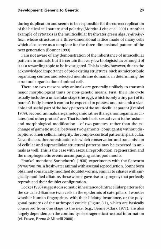
Development: Generic to Genetic 29
during duplication and seems to be responsible for the correct replicationof the helical cell pattern and polarity (Moreira-Leite et al. 2001). Anotherexample of cytotaxis is the multicellular freshwater green alga Hydrodyc-tion, whose structure is a three-dimensional lattice made of many cellswhich also serve as a template for the three-dimensional pattern of thenext generation (Bonner 1993).
I am not aware of any demonstration of the inheritance of intracellularpatterns in animals, but it is certain that very few biologists have thought ofit as a rewarding topic to be investigated. This is a pity, however, due to theacknowledged importance of pre-existing structures, such as microtubuleorganizing centres and selected membrane domains, in determining thestructural organization of animal cells.
There are two reasons why animals are generally unlikely to transmitmajor morphological traits by non-genetic means. First, their life cycleusually includes a unicellular stage (the egg), which is only a tiny part of itsparent’s body, hence it cannot be expected to possess and transmit a size-able and useful part of the body pattern of the multicellular parent (Frankel1989). Second, animals are gametogamic rather than gamontogamic as cil-iates (and other protists) are. That is, their basic sexual event is the fusion –and morphological modification – of two gametes, rather than the ex-change of gametic nuclei between two gamonts (conjugants) without dis-ruption of their cellular integrity, the complex cortical pattern in particular.Nevertheless, there are situations in which conservation and transmissionof cellular and supracellular structural patterns may be expected in ani-mals as well. This is the case with asexual reproduction, regeneration andthe morphogenetic events accompanying arthropod moults.
Frankel mentions Sonneborn’s (1930) experiments with the flatwormStenostomum, a freshwater animal with asexual reproduction. Sonnebornobtained somatically modified doublet worms. Similar to ciliates with sur-gically modified ciliature, these worms gave rise to a progeny that perfectlyreproduced their doublet configuration.
Locke (1990) suggested a somatic inheritance of intracellular patterns forthe so-called Siamese twin cells in the epidermis of caterpillars. I wonderwhether human fingerprints, with their lifelong invariance, or the poly-gonal patterns of the arthropod cuticle (Figure 3.1), which are basicallyconserved from one stage to the next (e.g., Bennet-Clark 1971), are alsolargely dependent on the continuity of extragenetic structural information(cf. Fusco, Brena & Minelli 2000).
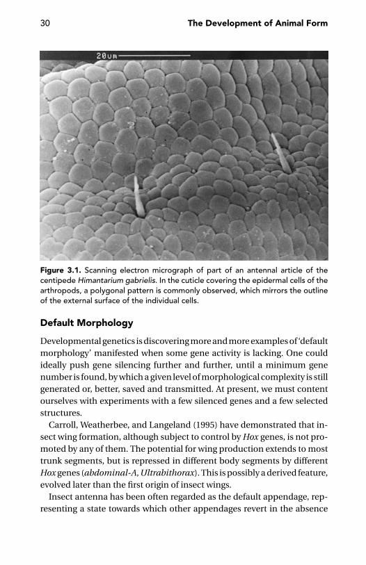
30 The Development of Animal Form
Figure 3.1. Scanning electron micrograph of part of an antennal article of thecentipede Himantarium gabrielis. In the cuticle covering the epidermal cells of thearthropods, a polygonal pattern is commonly observed, which mirrors the outlineof the external surface of the individual cells.
Default Morphology
Developmental genetics is discovering more and more examples of ‘defaultmorphology’ manifested when some gene activity is lacking. One couldideally push gene silencing further and further, until a minimum genenumber is found, by which a given level of morphological complexity is stillgenerated or, better, saved and transmitted. At present, we must contentourselves with experiments with a few silenced genes and a few selectedstructures.
Carroll, Weatherbee, and Langeland (1995) have demonstrated that in-sect wing formation, although subject to control by Hox genes, is not pro-moted by any of them. The potential for wing production extends to mosttrunk segments, but is repressed in different body segments by differentHox genes (abdominal-A, Ultrabithorax). This is possibly a derived feature,evolved later than the first origin of insect wings.
Insect antenna has been often regarded as the default appendage, rep-resenting a state towards which other appendages revert in the absence

Development: Generic to Genetic 31
of the specific input required for their determination (Stuart et al. 1991,Hughes and Kaufman 2000). This has been interpreted as proof that all thegnathal, thoracic, and abdominal segments (and their appendages, wherepresent) are built from a ground plan comparable with that of the antennalsegment (Rogers and Kaufman 1997).
In Drosophila, for example, the expression of both proboscipedia and sexcombs reduced is required for the specification of the sucking mouthparts.In the absence of these two activities, an ectopic antenna forms in placeof the ‘proboscis’ (Percival Smith et al. 1997). Other evidence points inthe opposite direction. In the absence of spineless activity, the distal partof a Drosophila antenna is transformed into leg (Duncan, Burgess, andDuncan 1998). Specification of an appendage as antenna, on the otherhand, requires specific input from extradenticle and homothorax. By re-moving the function of either of these genes, an antenna-to-leg transfor-mation is observed (Casares and Mann 1998). One reason for suggestingthat the antenna is more primitive than the leg is the presence, in the lat-ter, of a distinct intermediate domain between the proximal and the distalone, a feature without parallel in any arthropod antenna (Dong, Chu, andPanganiban 2001). The argument is not too strong, because the phyloge-netic concept of primitive feature is clearly not the same as the putative‘default morphology’, which is perhaps nothing more than pure abstrac-tion. In their recent review of this issue, Casares and Mann (2001) suggestthat the ground state of the ventral appendages in Drosophila (antennae,legs, genitalia and analia included) is a leg-like appendage consisting of aproximal segment and a distal tarsus.
One wonders in the end what ‘default morphology’ may really mean. Alltogether, from this contradictory evidence, one main conclusion seems toemerge – that is, the identification of a given body part with the specificactivity of one or a few genes is unwarranted. In Lawrence’s (2001) words,it is incorrect to conclude that a gene must be a major or pivotal one if aparticular process does not work in its absence. The gene is ignorant of itsrole in development (Dover 2000).
As early as 1990, Nijhout wrote one of the most perceptive pages on theinadequacy of a strictly gene-centred view of development. First, he re-jected the view that genes control development, stressing the endless con-tinuity of development and its manifold causality, which includes, besidesgene expression, structural, chemical, and physicochemical components.Rather than as direct controllers of developmental processes, genes actas selectors, steering development among alternative options. Moreover,
32 The Development of Animal Form
their action is far from showing the linearity of the hierarchical cascadesall too often offered as a model of gene action in development. Second,Nijhout rejected the notion that the genome contains a developmentalprogramme. He choose the example of bicoid, whose expression in theearly Drosophila embryo is currently described as essential to the estab-lishment of the antero-posterior body pattern. What is generally over-looked is the fact that the correct gradient of the bicoid product is onlyestablished if many other specific gene products are also produced and dis-tributed at the correct time, thus allowing a number of specific interactionswith structural elements in the cytoplasm to take place. The only sensiblequestion to ask about the way genes may be said to control developmentis whether a difference in a given character between two individuals is dueto genetic factors more than environmental factors.
Generic Forms
“It is possible that current evolutionary theory, with its emphasis on par-simony, may not be well equipped to deal with the numerous findings ofparallelism (and convergence) in morphological evolution” (Hodin 2000:10). Convergence, indeed, is much more widespread that was traditionallybelieved (a point recently made by Conway Morris 1998). This conclusionis now commonplace in molecular phylogenetic papers. The phenomenonis not circumscribed to organisms with very simple organisation, wheremorphology cannot help too much unraveling phylogenetic relationships.It is also rampant in animals and plants with very complex organisation,from orchids (Chase and Palmer 1988) to amphibians (Wake 1991). In thelatter case, Wake commented that the extraordinary amount of homoplasyfound in plethodontid salamanders is very likely to be the rule, rather thanan exception. This is confirmed by Moore and Willmer’s (1997) extensivesurvey of the main animal phyla. Occasionally, parallel evolution may sim-ply be the result of chance, with the use of aldehyde dehydrogenase as a lenscrystallin in squid and elephant shrews (Hodin 2000), but it is likely to owe alot to the widespread occurrence of generic mechanisms in development.
A ciliate protozoan and an insect embryo have very little in common,whatever unit of measure we might adopt in the comparison. Neither arethe two organisms closely related in terms of phylogeny, one good billionyears having elapsed since their phyletic lines separated from their mostrecent common ancestor. Nevertheless, these two biological systems haveseveral features in common that Frankel (1989) regards as the productof quasi-universal generative rules governing global pattern formation in

Development: Generic to Genetic 33
living organisms: separation between polarity pattern and differentiationpattern, global control, morphallactic reorganisation, positional continu-ity, spacing of positional values, and generative rules. How to explain theseextensively similar behaviours? It may be simply that similar generativerules are probable in biological systems of similar size (both the germband of insects and many large ciliates are in the range of 0.2–1.5 mm).
Jeong et al. (2000) analyzed the metabolic networks of 43 organisms –not just animals and plants, but also prokaryotes (eubacteria and archae-bacteria). They found that, despite significant differences in the individualconstituents and pathways in these networks, the metabolic organisationis not only identical for all living organisms, but also universally endowedwith the design principles of robust and error-tolerant, scale-free networks.
Universality of mechanisms does not imply that exactly the same mole-cules are involved. This makes sense from a mechanistic point of view, butnote the evolutionary implications. If similar but not identical moleculesactually perform the same job in different organisms, this means thatthe same result was probably obtained independently more than once.In other words, we are facing convergence rather than common descent.This may be true, for instance, for the mechanisms establishing positionalinformation in developing systems (Kauffman 1993).
Newman and Comper (1990) suggested that many morphogenetic andpatterning effects in living systems are the inevitable outcome of the phys-ical properties of cells and tissues. This does not negate the relevance ofgenetic mechanisms, but their action is simply complementary to thatof generic physical mechanisms. Newman and Comper (1990: 1) definegeneric mechanisms “as those physical processes, that are broadly appli-cable to living and non-living systems, such as adhesion, surface tensionand gravitational effects, viscosity, phase separation and phase-diffusioncoupling”, and may generate morphogenetic rearrangements of cytoplas-mic tissue and extracellular matrix components.
According to Newman (1994), two of the major types of gastrulation(epiboly and involution) and possibly a third (delamination) could haveoriginated as simple consequences of differential adhesion, even if nowa-days these processes are triggered and influenced by specific patterns ofgene expression. Observation of cell behaviour during epiboly in zebrafishseems to support a similar reduction of gastrulation to physical forces.The morphogenetic changes that accompany epiboly may result from thepassive response of cells to the reproducible forces that change the overallshape of the blastoderm prior to gastrulation (Wilson, Cretekos, and Helde1995).
34 The Development of Animal Form
Van Essen (1997) suggests that some morphogenetic mechanisms shap-ing the central nervous system of mammals may reduce to mechanicaltensions involving axons, dendrites, and glial processes. For example, ten-sion along axons in the white matter might explain the species-specificpatterns into which the cortex folds.
The morphogenetic potential of simple physicochemical processes mustnot be underrated. Jiang et al. (1999), for example, have shown how theperiodic patterning process in feather morphogenesis in birds is proba-bly dependent on the self-organising properties of the feather-producingcells. In their model, cells that become competent to form feather pri-mordia have, initially, equivalent probabilities of becoming primordia orinterprimordia. When a given threshold of cell density is reached, their ad-hesive properties lead to the random formation of many unstable micro-aggregates. The latter process leads to increased concentrations of adhe-sion molecules, so that some microaggregates increase in size or mergeby collision. Aggregates that reach a certain threshold exercise a long-range lateral inhibition that halts the neighbouring regions from becom-ing feather primordia. Because the growth of these cell microaggregates isbased on competition, this leads to their even spacing.
Drosophila mutants are known to develop symmetric patterns on suc-cessively shorter length scales (half embryo, a quarter of embryo, etc.),suggesting the existence of ‘generic’ wave-like phenomena by which theembryo might be actually subdivided into increasingly shorter longitudi-nal domains, down to single segments (Lacalli, Wilkinson, and Harrison1988, Goodwin and Kauffman 1990, Kauffman 1993). Unfortunately, inthe study of arthropod segmentation, very little attention has been fo-cused on the four-segment periodicity observed in the early expressionof some pair-rule genes in the tobacco hornworm Manduca sexta (Carrand Taghert 1989), later followed by bisegmental patterns and finally bysegmental patterns, possibly proof of the action of generic mechanisms insegmentation.
In this context, the main role for pair-rule gene expression may residein generic properties of cell adhesion, as manifested during germ-bandextension in Drosophila. After onset of gastrulation, the length of the germband increases over two-and-a-half fold. This change of shape correspondsto an increase in the number of cells along the antero-posterior axis and adecrease along the dorso-ventral axis. Mutations affecting the segmentalsubdivision of the embryo along the antero-posterior axis, such as thosein pair-rule genes, have negative effects on cell intercalation along the

Development: Generic to Genetic 35
longitudinal axis, thus reducing germ-band extension. According to Irvineand Wieschaus (1994), cell intercalation is thus dependent on the estab-lishment, through pair-rule gene expression, of adhesive differences be-tween stripes of cells.
Recently, Gordon (1999) has devoted a massive two-volume work to hisconcept of differentiation waves. When a wave of a process, such as cellcontraction or expansion, cytoskeletal changes, ionic currents, concentra-tion changes, chemical and stereochemical reactions, mechanical effects,and changes in gene transcription, traverses a tissue in an embryo, thetissue is split into two new tissues. As Papageorgiou (2001) remarks in re-viewing this book, Gordon’s theory smells of heresy from the viewpoint oftraditional embryology, but there is no reason why it should be dismissedwithout adequate testing. In the meantime, one may speculate whetherthe morphostatic role I suggested in the previous chapter for the cuticleof ecdysozoans might have something to do with blocking differentiationwaves.
A Bestiary of Generic FormsSegments In a paper provocatively entitled, Is segmentation generic?,Newman (1993) defended the thesis that the capacity of undergoing seg-mentation may be ‘generic’ to tissues, that is, an outcome of their mostgeneral physical and chemical properties, without regard for any differ-ence in their specific molecular makeup. This property, if true, would makesegmentation a generic property likely to evolve independently in severalphyla, with the possible involvement of different molecular mechanismsin different taxa. Newman’s concept of segmentation is close to that ofcompartmentalisation (for a definition, see page 242). In his view, the seg-mental organisation is frequently based, at least in part, on the inabilityof otherwise similar tissues to exchange cells at their interface. There isno shortage of evidence in support of this view. Immiscibility of cells fromneighbouring segmental units is found, for example, between Drosophilacompartments (Lawrence and Johnston 1986), vertebrate rhombomeres(Guthrie and Lumsden 1991), and half-somites in the chick embryo (Sternand Keynes 1987).
Tubes An experimental proof that generic processes may generate tubeshas been provided by Coucouvanis and Martin (1995), with the conversionof a solid primordium into a hollow tube as in the early mouse embryo, inwhich the solid embryonic ectoderm changes into a columnar epithelium

36 The Development of Animal Form
surrounding a cavity. Cavitation in this case is the result of the interplay of asignal from an outer layer of endodermal cells that induces apoptosis of theinner ectodermal cells and a rescue signal. It is mediated by contact withthe basement membrane that provides for the survival of the columnarcells lining the cavity.
Hollow Spheres Both theoretical models and experiments with reaggre-gating dissociated cells have shown that simple self-sorting mechanismsdue to differential surface adhesion properties of the cells may producehollow spheres, provided these cells are non-adhesive over portions oftheir surfaces (e.g., Keller and Segel 1970, Newman and Muller 2000).
Layers, Sheaths, Tubes, Rods, Spheres, Etc., as Aggregates of CellsMore generally, simple biomechanical forces are sufficient for mouldinggroups of cells into layers, sheaths, tubes, rods, spheres, and so forth. Asnoted by Newman and Muller (2000), the morphogenetic role of thesestructures is not limited to their importance as geometric templates formore advanced patterns or as barriers allowing the partitioning of theorganism into functional modules, but may extend to some form of controlof gene activity (see also Ingber 1993, Chen et al. 1997).
The Earthworm and the AnkylosaurusMany animals belonging to different phyla are provided with dorsal or ven-tral appendages, sensory organs or simple colour spots, which form a kindof regular, segmental checkerboard. Although the number of segments iswidely different from case to case (and the developmental origin of thesesegments may also be different; cf. chapter 9), the number of elements pertransversal row seems to follow strict, nearly universal rules.
In most earthworm species, there are four pairs of setae per segment.Their mutual spacing is different in the various genera, but distribution ishighly stereotyped, so that the whole array is simply described by speci-fying the pattern of setae in a segment and the total number of segmentsin the body. (I am disregarding minor alteration of the pattern in the ter-minal segments and in the clitellum, a short range of segments involvedin the production of the egg cocoon, which are usually marked by a dif-ferent colour and become thicker at maturity.) This set of regularly pat-terned setae repeated regularly from segment to segment gives rise to acheckerboard pattern which has its equivalent in a lot of different animals.Many insects, centipedes, millipedes, mites, etc., have one or two rows of
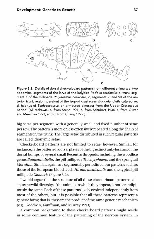
Development: Generic to Genetic 37
Figure 3.2. Details of dorsal checkerboard patterns from different animals: a, twoabdominal segments of the larva of the ladybird Rodolia cardinalis; b, trunk seg-ment X of the millipede Polydesmus coriaceus; c, segments VI and VII of the an-terior trunk region (pereion) of the isopod crustacean Buddelundiella cataractae;d, habitus of Scolecosaurus, an armoured dinosaur from the Upper Cretaceousperiod. (All redrawn– a, from Stehr 1991; b, from Schubart 1934; c, from Oliverand Meechan 1993; and d, from Charig 1979.)
big setae per segment, with a generally small and fixed number of setaeper row. The pattern is more or less extensively repeated along the chain ofsegments in the trunk. The large setae distributed in such regular patternsare called idionymic setae.
Checkerboard patterns are not limited to setae, however. Similar, forinstance, is the pattern of dorsal plates of the big extinct ankylosaurs, or thedorsal bumps of several small Recent arthropods, including the woodlicegenus Buddelundiella, the pill millipede Trachysphaera, and the springtailMorulina. Similar, again, are segmentally periodic colour patterns such asthose of the European blood leech Hirudo medicinalis and the typical pillmillipede Glomeris (Figure 3.2).
I would argue that the structure of all these checkerboard patterns, de-spite the wild diversity of the animals in which they appear, is not serendipi-tously the same. Each of these patterns likely evolved independently frommost of the others, but it is possible that all these patterns represent ageneric form; that is, they are the product of the same generic mechanism(e.g., Goodwin, Kauffman, and Murray 1993).
A common background to these checkerboard patterns might residein some common feature of the patterning of the nervous system. In

38 The Development of Animal Form
vertebrates, for example, the neural ectoderm of the embryo is subdi-vided into a checkerboard of domains by the intersection of longitudi-nal and transverse compartmentalisation (Rubenstein and Puelles 1994).Similarly, in the embryonic central nervous system of Drosophila, thecombinatorial expression of the gene products of antero-posterior andproximo-distal axis-patterning genes defines a checkerboard of equiva-lent groups (Skeath and Carroll 1994) defined by homologous cell lineages(Stent 1998). Number and spatial arrangement of these equivalence groupsare the same as the basic pattern of sensory setae in the segments of severalarthropods.
These correspondences are not limited to the nervous system and theassociated sensory organs. In Drosophila, each adult segment originates,during metamorphosis, from four pairs of histoblasts containing betweensix and thirteen cells each. These eight histoblasts are arranged in twotransverse rows (Milner and Bleasby 1985), similar to the arrangement ofthe sensory setae on the dorsal and ventral sclerites of so many arthropods.
Deceptive NumbersSimple patterns and numerical identity may be positively deceptive. Ininsects, there are eight cells in the photoreceptor of each ommatidium,the morphofunctional unit of the compound eye. One could expect theseeight cells to be the product of three successive mitoses, starting with afounder cell, but this is not the case; the eight cells of each ommatidiumare not related by lineage (Gurdon 1992, Lawrence 1992). Even more at-tractive for arithmetical speculations is the number (32) of ‘true’ segmentsin leeches. This number would be easily obtained through five binary di-visions of a founding element (25 = 32), but nothing of this kind actuallyhappens in leech development. Segments are founded by cells derivedfrom seven blast cells (m, nf, ns, o, p, qf, qs), each of which, through astereotyped series of divisions (Zackson 1984, Bissen and Weisblat 1989),generates cells which will contribute to the initial cell pool of each seg-ment. Nothing in this developmental sequence exhibits anything of theregularity expected from arithmetical considerations. First, the divisionsof the blast cells are sequential, rather than hierarchical. Their progeny isproduced in a regular antero-posterior sequence, one cell after the other,rather than by a short series of equivalent (if not also synchronous) binarydivisions leading to an exponentially growing number of progeny cells.Second, the progeny of each blast cell is not strictly limited to the 32 cellsthat would suffice, according to an hypothetical principle of economy, with

Development: Generic to Genetic 39
one offspring per blast cell allocated to each segment, without any wastecell to be discarded. Blast cells produce about 100 cells each (Weisblat andShankland 1985, Weisblat and Huang 2001). Supernumerary cells will diewithout contributing to the definitive segments (Zackson 1982). It is notyet clear how the astoundingly constant segment number of leeches isactually determined.
Another example of deceptive numerical correspondence has been dis-covered by Huys and Boxshall (1991) through their analysis of the homol-ogy of individual segments of the antennule in different genera of cope-pods. For example, the same number of antennular articles is present inMacrocyclops and in Cyclopinoides, but there is no one-to-one homologybetween the 17 segments of the former and those of the latter. Distribu-tion of sensilla along the appendage allows tracing the correspondenceof the articles to the basic segmental architecture of the copepod anten-nule, thus the first segment of Macrocyclops represents segments I–VI ofthe hypothetical ground-plan copepod antennule, followed by a free VII,whereas in Cyclopinoides I and II are free, but III–V are collapsed together,as are VI–VII.
But there are also many other numerical relationships that suggest theexistence of underlying common rules, even where our current under-standing of development fails to offer advice. Some examples are brieflydiscussed elsewhere in this book (in particular, in respect to arthropod seg-mentation, see pages 206–209). Another numerical pattern emerges fromcomparison of the vertebral position of the hindlimbs with respect to theforelimbs in three species of Palaeozoic amphibians. The two limb pairsare about sixteen vertebrae apart in Urocordylus wandersfordii (Nectridea),about twenty-four in Utaherpeton franklini (Microsauria), and about sixty-four in Brachydectes newberryi (Lysorophia; Carroll 1999). A possibilityexists that this octonary pattern has something to do with the segmen-tation clock that seems to control the pace at which vertebrate somitesare formed (Pourquie 2000, 2001). The common occurrence of octonarysegment patterns in annelids (see page 140), arthropods (see page 209),and vertebrates (this paragraph) is very likely to be serendipitous.
Genetic Assimilation
According to Wolpert (in Wolpert, Ghysen, and Garcıa-Bellido 1998), thedevelopment of the earliest animals was messy and very susceptible toenvironmental perturbations. Newman and Muller (2000) imagine a late

40 The Development of Animal Form
Precambrian ‘pre-Mendelian world’, where the earliest multicellular ag-gregates were subject to morphogenesis, as dictated by their properties ofchemically excitable, viscoelastic soft matter. Without involvement of ‘ge-netic programmes’, master genes, and developmental control cascades,those properties allowed the production of a profusion of multilayered,hollow and even segmented forms. With the subsequent transition to a‘Mendelian world’, cellular specificities and gene control acquired an in-creasingly larger role because of the presence of metabolically differenti-ated cell subpopulations produced by compartmentalisation. Biochem-ical differences between the different tissues provided components ofone another’s environment, thus eventually bringing embryonic induc-tion into existence. Early in metazoan evolution, cell–cell communicationwas perhaps mediated by small molecules such as the peptides that in‘lower’ animals such as Hydra control biological processes as diverse asmuscle contraction and neuron differentiation and establish gradients ofpositional values (Bosch and Fujisawa 2001).
The complexity and specificity of the new interactions within these de-veloping living systems did not cancel the physicochemical generic prop-erties that were responsible for the whole limited morphogenetic activityof the oldest multicellulars. Newman and Muller believe that the verte-brate skeleton may have arisen in a similar way. Connective cells andother cells arrange themselves along stress fields without requiring anyadditional (say, genetic) input (Harris, Stopak, and Wild 1980, Bard 1990).Secretion of a cartilage matrix is an autonomous property of mesenchy-mal cells, dependent on generic features, such as cell number and density(Cottrill, Archer, and Wolpert 1987) and compression (Vogel and Koob1989, Robbins, Evanko, and Vogel 1997). Therefore, any mesenchymal tis-sue mass above a certain threshold size may have begun to produce, with-out any specific input, arrays of matrix-secreting cells along stress fieldsgenerated by passive and active movements. This view of the origin ofskeletons is perfectly compatible with my suggestion that skeletogenesisoriginated as a way to stabilise development (cf. page 18).
We can suppose that in the course of evolution the role of genes in canal-ising developmental events became increasingly larger. Reading againstthe arrow of time this means that, in the earliest animals (or, at any rate, inthe earliest multicellulars organisms), this canalising effect of genes waslimited, with development being mainly driven by generic processes. Inthis sense, the similarity between some features of biological developingsystems and those of non-biological systems may represent something

Development: Generic to Genetic 41
more than a simple analogy. Meinhardt (1996) cites high sand dunes orsharply contoured rivers as examples of patterns developing from almosthomogeneous initial conditions in non-living systems. In these systems,as (putatively) in the living ones, the continuous growing of patterns iscaused by the strong feedback of initially small deviations from a homoge-neous distribution. In the case of primary embryonic pattern formation,Gierer and Meinhardt (1972) and Meinhardt (1982, 1992) suggested cou-pling of a short-range autocatalytic process with a long-range reaction thatopposes it.
Actual examples of developmental canalisation evolving through geo-logical time are difficult to obtain. For a comparatively well-studied grouplike trilobites, results are mixed. There is some evidence that early trilobiteswere more developmental plastic than later representatives of the group(Hughes 1991), but the role of external selection in promoting segment-rich, more variable morphotypes in Cambrian times, but not so muchin later times, must also be taken into account (Hughes, Chapman, andAdrain 1999).
Genes and Phenotype
The relationship between genotypic and phenotypic changes is far fromlinear (Jablonski 2000). Dramatic morphological changes are sometimesassociated with point mutations. Phenotypes can be remarkably stabledespite extensive genetic differences. Interestingly, this may be true evenwhen typical ‘developmental genes’ are involved. For example, no pheno-typic variation was found in the three-spine stickleback, despite intraspe-cific variation in the expression domains of Hox genes along the mainbody axis (Ahn and Gibson 1999a, 1999b). Differences in the developmen-tal mechanisms by which the vulva is produced in different strains of thenematode Pristionchus pacificus do not correlate with the genetic distancebetween those strains. In particular, peculiar developmental mechanismswere found in a strain from California, genetically identical to anotherstrain from Poland, which lacks them (Srinivasan et al. 2001).
A related issue is phenotypic plasticity (Schlichting and Pigliucci 1998).One speaks of phenotypic plasticity when, in response to different externalconditions, different phenotypes are produced that are adaptive, each inits own environment. If you rear the grass-feeding caterpillar of the mothPseudaletia unipuncta on hard grass, its head grows much bigger than ifyou rear it on soft wheat seedlings. This difference in the size of the head
42 The Development of Animal Form
capsule, with the correlated differences in mandibular power, has a directeffect on the insect’s ability to exploit hard food: larger heads are obvi-ously better when dealing with hard grass (Bernays 1986). Closely relatedspecies may have different, even opposite, responses to the same environ-mental cues. For example, salmon growing in fast flowing waters becomemore robust than their conspecific growing in slow waters, whereas browntrouts growing in fast flowing waters become slightly more streamlined(Pakkasmaa and Piironen 2001). A nice example of environmental regula-tion of morphogenesis is offered by the hermit crab Clibanarius vittatus,whose asymmetry, although not dependent on the initial possession ofa shell, can be lost within a few moults, if shells are no longer available(Harvey 1998).
No less interesting, however, are stories in which phenotypic plasticity isevoked by different competitive frameworks. Pfennig and Murphy (2000)compared the tadpole development of two closely related species of thespade-foot toad (Spea bombifrons and Spea multiplicata), when rearedalone or together. Depending on the diet available, individuals of bothspecies can develop into either an omnivore morph, mostly feeding on de-tritus, or a carnivore morph, mostly feeding on small crustaceans. The headin the omnivore morph is much smaller than in the carnivore morph. Whenthe two species are reared in the same tank, the presence of S. multiplicataenhances the production of the carnivore morph in S. bombifrons, andthe latter, in return, suppresses the production of the carnivore morph inS. multiplicata. In this way, competition pushes either species towards thefeeding habit for which it has advantage over the other species. As in this ex-periment the environment eliciting the response of either tadpole speciesis tadpoles of the same age of a closely related species, one is temptedto compare this system with a developing embryo, in which different celllineages are pushed towards one of their potentially open developmentalpathways by the influence from neighbouring cell lineages. Between thetwo tadpole species, there are genetic differences, whereas confronting celllineages within an embryo only differ because of the history of their geneexpression and former inductive interactions with other cells. However,the overall difference is one of degree rather than one of kind.
Evolutionary Dissociation between Genes and Phenotypes
Homology of sequence is not a guarantee of conserved function. This isclearly what we expect to find in the case of gene duplication, when twoparalogues may have functionally diverged to a more or less great extent.

Development: Generic to Genetic 43
But evolutionary dissociation is also common between vertically homolo-gous (orthologous) genes and homologous morphological features (Wray1999, Wagner 2000). Shigetani et al. (2002) have shown that the expres-sion in the chicken of ‘developmental genes’ involved in the specificationof the mandibular arch is different from the corresponding expression ofapparently orthologous genes in the lamprey, thus suggesting heterotopicshift of tissue interactions in the evolution of vertebrate jaws. In verte-brates, the ‘functions’ of Pax9 genes have sequentially expanded throughnew expression domains accompanying the emergence of more compli-cated body plans. In hemichordates and non-vertebrate chordates, thePax9 gene is only expressed in the pharyngeal slits; but, in the ‘lower’vertebrates, it is also expressed in the nasal placode and in the cranialectomesenchyme. In the amniotes, finally, its expression extends to thesomites (Ogasawara et al. 2000). In Drosophila, dorsal and snail specifythe mesoderm and the dorso-ventral polarity of the main body axis. Butin the leech Helobdella robusta, the same genes probably play a role in thediversification of cell types within segment primordia (Goldstein, Leviten,and Weisblat 2001). In insects, the Ultrabithorax protein has properties notpresent in its equivalent from onychophorans, possibly due to acquisitionof new cofactors or activity modifiers since the divergence of the two lin-eages (Grenier and Carroll 2000). In Drosophila, even-skipped has a typicalpair-rule (bisegmental) expression, but in the grasshopper Schistocerca,its homologue does not serve a pair-rule function in early development.The same gene has a conserved role in neurogenesis in both grasshopperand fruitfly (Patel, Ball, and Goodman 1992). The engrailed gene has mul-tiple expression domains and many ascertained developmental roles inarthropods and chordates, but few if any of these domains and roles arehomologous across phyla (Davis et al. 1991, Duboule 1994b, Rogers andKaufman 1996, Bely and Wray 2001).
In dipterans, the class 3 Hox gene evolved into zen, which is not expressedalong the main body axis as the Hox gene from which it derives, but spec-ifies instead extraembryonic tissue (Falciani et al. 1996). A recent duplica-tion of this Hox3/zen gave rise to bicoid, which in Drosophila is required fornormal development of the head and thorax; but in another fly, the phoridMegaselia abdita, it is also required for the development of four abdominalsegments (Stauber, Taubert and Schmidt-Ott 2000). Extensive changes inspatial and temporal patterns of expression and in developmental rolesof the homeobox genes have occurred in the different echinoderm classes(Lowe and Wray 1997). Homoplasies, or parallel changes of gene structureand function in different phyla, are also known. For example, Antennapedia
44 The Development of Animal Form
class genes are involved in the specification of trunk structures in both pro-tostomes and deuterostomes, but have duplicated independently in thetwo phyletic lines (Holland 1992, Schubert, Nieselt-Struwe and Gruss 1993,Zhang and Nei 1996).
Gene function may be maintained despite a genome structure in flux, ifcompensatory changes evolve in the genome (Dover 1992).
A Role for the ‘Developmental Genes’
By now it is clear that the specific role of most of the so-called develop-mental genes is rather mundane (Shubin and Marshall 2000). Most of themregulate the rate of cell proliferation or determine cell adhesion propertiesor act as transcription factors, binding to specific DNA sequences. Themost advanced ‘morphogenetic’ roles of these genes may lay in demarcat-ing regions or groups of cells fated to specific differentiation routes. Thedevelopmental role of all these genes is the result of their involvement inmore or less complex and pervasive genetic circuitries.
The scientific literature of the last decade is full of examples of geneswhose temporally and spatially correct expression is critical for the pro-duction of complex, specific features, but at the same time are known toencode for proteins whose mechanism of action is quite unspecific (e.g.,in regulating changes of the cytoskeleton; Schejter and Wieschaus 1993).Another important aspect is the apparent lack of specificity of action, inthat different transcription factors can apparently be substituted for oneanother. This is due to the combinatorial nature of genes, whereas whatis really relevant is the structure of promoter modules. The historical con-tinuity of function rests with these elements rather than with the wholegene (Akam in the discussion of Abouheif 1999).
The Hox CodeIn a fascinating reconstruction of the evolution of animal body plans fromthe point of view of the genetic control of development, Davidson (2001) af-firms that the secret of the bilaterians, with respect to building body parts,is their use of abstract patterning mechanisms. These mechanisms arederived from pre-existing regulatory pathways, which, instead of runningdifferentiation gene batteries, as their role was in more primitive multicel-lular organisms, acquired a new function in the regional specification oftranscription.
Current evidence suggests that some gene families were more suit-able than others in providing ‘abstract patterning mechanisms’ – none,

Development: Generic to Genetic 45
apparently, better than the Hox genes, whose organization in an orderedcluster is probably the best key to their success in body patterning (Garcıa-Fernandez and Holland 1994). The long history of association between theevolution of the Hox gene family and the evolution of patterning along themain body axis of bilaterians is apparent from the fact that the terminalgenes (labial/Hox1 and Abdominal B/Hox13), which are expressed in bodyregions close to (but not coincident with) the two body ends, are prob-ably the oldest, in that they contain the most divergent homeodomains(Gehring 1998).
Hox genes are responsible for major differences in the body architectureof different arthropod groups. One example is the difference between fliesand butterflies in the number of larval legs and adult wings: transformationof the posterior wings into a pair of halteres in the flies is correlated withdivergent regulation of Ultrabithorax (Warren et al. 1994). In the echino-derms, Lowe and Wray (1997) relate reorganisation of body architectureto extensive changes in the deployment and roles of homeobox genes, in-cluding the evolution of new developmental roles and modifications in thesymmetry of the expression domains. In this phylum, genes such as Distal-less, engrailed and orthodenticle show unique radial expression domainswhich correspond to structures that are also unique to echinoderms. Forexample, orthodenticle is expressed in the podia of brittle stars and seaurchins, and Distal-less is expressed in the podia of sea urchins and seastars, as well as in the buccal tentacles of sea cucumbers.
The role of Hox genes in determining major changes in body archi-tecture has been extensively debated. For example, Irvine and Martindale(2000) have put the distinct posterior boundaries of expression of CH-Hox1and CH-Hox2, both in the ninth body segment of Chaetopterus variopeda-tus, in relation with the unusually complex regionalisation and segmentspecialisation of this annelid, in which the ninth segment marks a majormorphological boundary (Figure 3.3).
Arthropods, with their extensive diversity in the specialisation of seg-ments and body regions, have been the focus of the debate. A key roleof changes in Hox gene regulation in the origin of major morphologi-cal changes is suggested by the good correlation found in different crus-taceans between changes in the expression patterns of Ultrabithorax andabdominal-A and modification of the anterior thoracic limbs into maxil-lipedes (Averof and Patel 1997). Similar suggestions derive from compar-isons between the expression domains of Hox genes and the morphologicalboundaries of body regions in insects and isopod crustaceans (Popadicet al. 1998, Abzhanov and Kaufman 1999, 2000a). The same applies to
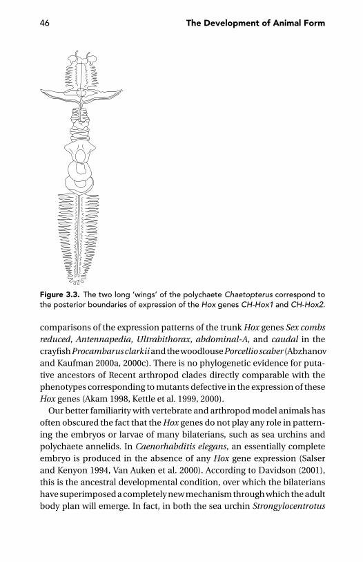
46 The Development of Animal Form
Figure 3.3. The two long ‘wings’ of the polychaete Chaetopterus correspond tothe posterior boundaries of expression of the Hox genes CH-Hox1 and CH-Hox2.
comparisons of the expression patterns of the trunk Hox genes Sex combsreduced, Antennapedia, Ultrabithorax, abdominal-A, and caudal in thecrayfish Procambarus clarkii and the woodlouse Porcellio scaber (Abzhanovand Kaufman 2000a, 2000c). There is no phylogenetic evidence for puta-tive ancestors of Recent arthropod clades directly comparable with thephenotypes corresponding to mutants defective in the expression of theseHox genes (Akam 1998, Kettle et al. 1999, 2000).
Our better familiarity with vertebrate and arthropod model animals hasoften obscured the fact that the Hox genes do not play any role in pattern-ing the embryos or larvae of many bilaterians, such as sea urchins andpolychaete annelids. In Caenorhabditis elegans, an essentially completeembryo is produced in the absence of any Hox gene expression (Salserand Kenyon 1994, Van Auken et al. 2000). According to Davidson (2001),this is the ancestral developmental condition, over which the bilaterianshave superimposed a completely new mechanism through which the adultbody plan will emerge. In fact, in both the sea urchin Strongylocentrotus
Development: Generic to Genetic 47
purpuratus (Arenas-Mena et al. 1998) and the polychaete Chaetopterus(Peterson et al. 2000), Hox genes are expressed only in the tissues that willcontribute to the adult body, not in the larval ones, which will be discardedat metamorphosis.
Some important events in animal evolution seem to be associated withmajor duplications of the Hox gene cluster. It has been proposed that oneof the conditions that permitted the Cambrian explosion of animal bodyplans was the duplication by which a protoHox gene cluster gave rise to twodistinct paralogous sets, the ParaHox and the Hox (proper) genes (Brooke,Garcıa-Fernandez and Holland 1998). More popular is the hypothesis thatthe success of the vertebrate lineage owes much to a wholesale quadru-plication of the Hox gene cluster (Bailey et al. 1997, Holland 1997), per-haps following the polyploidisation of the ancestor’s set of chromosomes(Spring 1997).
There is no need to use a lot of words to explain the concept of the Hoxcode (for an early review, see Lawrence and Morata 1994), or to discussthe colinearity between the chromosomal order of the Hox genes and theantero-posterior order, along the main body axis, of the anterior boundaryof their expression domains (e.g., Krumlauf 1992, 1994). These concepts,based on an impressive amount of experimental evidence accumulatedduring the 1980s and early 1990s, are illustrated at length in standard workssuch as Gerhart and Kirschner (1997), Wolpert et al. (1998), Hall (1999) andCarroll, Grenier, and Weatherbee (2001). Therefore, I will only mention arecent paper suggesting caution in generalising. The Hox code is proba-bly not so simple, and colinearity is not so widely conserved as generallybelieved.
It is well known that, in Drosophila, the Hox gene complex is split intotwo separate clusters, less known is that, in the different fruitfly species,the complex is split at different places. In Drosophila melanogaster, thetwo clusters are the Antennapedia complex and the Bithorax complex,respectively, whereas in Drosophila virilis, the split occurs within the lat-ter complex. In other terms, Antennapedia and Ultrabithorax are in twodifferent clusters in D. melanogaster, but in the same cluster in D. virilis,whereas Ultrabithorax, abdominal-A, and Abdominal-B are all in the samecluster in D. melanogaster, but not in D. virilis (von Allmen et al. 1996). Thetwo independent events by which these two different splits were generatedoccurred with the separation of D. melanogaster from D. virilis, possibly inthe order of 60 million years ago (Beverly and Wilson 1984). These eventssuggest that, in this group at least, the physical arrangement of these genes

48 The Development of Animal Form
is not strictly constrained. One may wonder whether this has anything todo with the near-synchronisation of morphogenesis and patterning alongthe longitudinal axis of the quickly developing Drosophila embryo.
In the mouse, for example, the Hox1 group genes do not respect the ruleof spatial colinearity; this is relevant, even if this deviation from colinearityis to be interpreted as a secondary phenomenon (Wada et al. 1998, Wada,Garcıa-Fernandez and Holland 1999, Holland 2000). Nogi and Watanabe(2001) dare to suggest that the lack of colinearity they find in the planarianDugesia japonica (the anterior expression boundary of Dj-AbdB-a beinganterior to those of two central-class genes Plox4-Dj and Plox5-Dj) couldbe ancestral. I suspect, that this suggestion derives from the traditionalview of flatworms as basal bilaterians, a concept that many recent assess-ments of animal phylogeny (e.g., Aguinaldo et al. 1997, Jenner and Schram1999, Adoutte et al. 2000, Jenner 2001, Peterson and Eernisse 2001, Nielsen2001) have largely shaken.
Two possible ways a Hox code could work were distinguished byHolland and Garcıa-Fernandez (1996). One theoretical possibility is for theHox genes to mark fixed (absolute) axial positions (in vertebrates, somitenumbers), thus controlling the development of the specific traits appropri-ate to that position in a given species. The other possibility is for Hox genesto mark presumptive phenotypic characters. In this case, the axial level oftheir expression patterns would be expected to shift between divergenttaxa. Burke et al. (1995) found strong support for the latter model, what iscomfortable for the recent widespread use of the Hox gene expression pat-terns as indicators of homology. This means that the first thoracic vertebra,as specified in both animals by the anterior limit of expression of Hoxc6,may be regarded as homologous, despite the somite affected is number 12or 13 in the mouse, but number 19 or 20 in the chick. In the comparativeanatomy of arthropods, a very strong tradition acknowledges homologyto equally numbered segments irrespective of their actual specialisation.This is probably unwarranted, as argued in chapter 9.
The temporal and spatial expression patterns of several Hox genes showsthat the classical Hox code model is unrealistic. Castelli-Gair (1998) hasbacked this objection through evidence from the expression of the Sexcombs reduced, Ultrabithorax, and Abdominal-B genes in Drosophila. Theexpression of these genes is dynamic, involving fewer segments at earlystages of development than at more advanced stages. At any instant intime, within a segment, there is a mosaic of cells expressing a given Hoxgene and cells not expressing it.
Development: Generic to Genetic 49
In the bee, the anterior expression border of Deformed changes duringgastrulation from segmental to parasegmental (see page 220 for an expla-nation of the term) in the ventral part of the maxillary segment, not as aresult of cell movement, but because of the intrinsic dynamic characterof the gene expression itself (Fleig et al. 1992). This implies that the Hoxcode identity of a cell changes during development. In this case, which isits ‘correct’ segment identity, if any?
Castelli-Gair (1998) examines the fate of a Drosophila embryo in whichthe expression of Abdominal-B is prevented in what would normally be theseventh abdominal segment (A7). The question is, whether this segmentwould assume a more anterior identity, as conventionally described for aloss-of-function Hox gene mutant. Interestingly, the answer is yes and no.The consequences of preventing Abdominal-B expression in A7 are man-ifest in the corresponding segment of the adult; but, in the embryo, manyA7 structures, such as the local differentiation of the central and peripheralnervous system, are normal (Heuer and Kaufman 1992). Only some larvalstructures of A7, such as the segment-specific denticle belt in the larva andthe cells fated to form the adult epidermis, do actually require Abdominal-B expression. Still more importantly, in the context of the question aboutthe segmental identity of a cell whose Hox code changes during devel-opmental time, is the fact that, in A7, the organisation of the peripheralnervous system is the same as in the preceding segments A2–A6 (Ghysenet al. 1986) despite the fact that A2–A4 do not express Abdominal-B, where-as A5–A7 do. This uniform patterning of the peripheral nervous system ir-respective of the expression of this gene does not mean that Abdominal-B isdispensable for the development of the peripheral nervous system. This isdemonstrated by how Abdominal-B mutants affect the peripheral nervoussystem of A8. The different responses of A7 and A8 to Abdominal-B expres-sion is likely due to a difference in the temporal expression of this Hox genein the two segments. This shows that the outcome of Hox gene expressionmay change with time; what is obviously contrary to what we would expectaccording to the conventional Hox code hypothesis (Castelli-Gair 1998).Different developmental effects following expression at different develop-mental times are not limited to Abdominal-B.
The role of Hox genes in patterning the body of diploblastic animalsis not known, and it may be unwarranted to expect it to be the same asin triploblastic animals such as insects and vertebrates. Nevertheless, theexpression of the Hox gene Cnox-2 in Hydractinia symbiolongicarpus isinteresting in the context of this discussion of the developmental changes

50 The Development of Animal Form
in a cell’s Hox gene identity. Cnox-2 is expressed in different aboral-to-oralpatterns in different kinds of polyps of this polymorphic cnidarian, andthe experimental conversion of one polyp type to another is accompaniedby concordant alteration in Cnox-2 expression (Cartwright, Bowsher, andBuss 1999).
During the 1990s, the idea of a Hox code has been also fashionable inplant developmental biology. Differentiation of the four basic kinds offloral elements (sepals, petals, stamens, and carpels) has been interpretedas deriving from the homeotic control of three classes of genes: A, B, and C.Of these, A would specify as sepals the elements of the most external whorlin the flowers; those of the following whorl would instead develop as petals,stamens and carpels, under A+B, B+C, and C function, respectively. It hasbeen soon realized, however, that things are more complex. At least twoA-function and two B-function genes have been discovered in the modelplant Arabidopsis thaliana. The ABC genes, if expressed in a part of theplant other than the flower, do not transform leaves into flower elements:expression of genes of the SEPALLATA class is additionally required forthe specification of a flower. In front of this increasingly complex picture,Theißen (2001) wonders whether the ABC combinatorial model will stillbe of any use in our future attempts to understand the linkage betweenmolecular genetic events and floral phenotypes.
A simplistic and generalised application of the Hox code concept is alsoimpossible in the case of group 11, 12, and 13 Hox genes, which controlsize and number of digits in tetrapods. In the mouse, it has been shownthat this control is dose-dependent, rather than the effect of a qualitativelydifferentiated Hox code. The same response was found in morphogenesisof the baculum (Zakany et al. 1997): digits and external genitalia are, inpart, under the same genetic control (see page 176).
Organic CodesMajor evolutionary transitions are marked by the appearance of new or-ganic codes [i.e., non-deterministic, historically frozen rules of correspon-dence between patterns (Barbieri 2003) as in the genetic code or in humanlanguage]. All this means is a kind of ‘evolution by natural conventions’.When a new code appears, the old ones are not necessarily cancelled.Therefore, with every new code appearing, there is a net increase in globalcomplexity.
We have just seen that the so-called Hox code should not be taken tooliterally. Whatever consistency this ‘code’ may have, it is quite probable
Development: Generic to Genetic 51
that in this case, too, as with the genetic code, the relationship betweenthe molecular ‘symbol’ and its phenotypic translation is little more than afrozen accident. Its lability during the short span of one embryonic de-velopment seems to confirm this suspicion. Look, for example, at therelationship between the temporal and lineage restrictions of the expres-sion of mab-5, the homologue of the Drosophila Hox gene Antennape-dia in Caenorhabditis elegans. In a given body region, the start of thisgene’s expression stimulates proliferation in a cell lineage; but shortlythereafter, a temporary suspension of its expression is required to spec-ify epidermal structures. But this is just the first half of the story. In justone branch of the epidermal cell lineage specified following the silenc-ing of the mab-5 expression, reactivation of the latter promotes neu-roblast formation, but a new silencing of this gene is required again forthe establishment of a correctly formed sense organ (Salser and Kenyon1996).
Universal Genetic Tools
A conserved genetic machinery seems to control morphogenetic eventssuch as branching and anastomosing of tubes in widely diverging ani-mal clades (e.g., vertebrates and insects). But these features, most likely,have been reinvented many times. The fact that they are now controlledby orthologous genes in different phyla is probably due to an indepen-dent (homoplasious, convergent) co-option, rather than derivation froma common ancestor with similar structures already patterned under thecontrol of those genes.
Think, for example, of the branching morphologies in the lungs, kidneysand mammary glands of many vertebrates. Common to all these structuresis contact between an epithelium and a mesenchyme, the former branch-ing more or less extensively into the latter (Hogan et al. 1997). Fleury (2001)has recently demonstrated how all these branching organs may developfollowing a simple physical process called viscous fingering. This processoccurs whenever a less viscous fluid is pushed to penetrate into a moreviscous fluid. Simple physical forces may generate branched fingers, butdo not stabilise them. Neither can they provide regularity in the spatialand temporal patternings of the sites in which similar morphologies willoriginate. This is where the generic-to-genetic transition is found again,with the intervention of genes, such as those actually involved in theseprocesses.
52 The Development of Animal Form
One could expect that the self-similarity or fractal structure of thesebranching patterns would be a bonus obtained at no extra genetic cost,but this is not necessarily the case. In Drosophila, each of the ten segmentsfrom the mesothoracic to the eight abdominal is provided, at each side,with a unit of the tracheal system, including first-, second-, and third-orderbranches. First-order tracheal branches are multicellular tubes, whereasthe second-order tubes are the product of individual cells and the termi-nal, third-order branches (tracheoles) are tiny intracellular tubes hostedwithin cytoplasmic protrusions. Different genes are involved at each levelof branching. In particular, breathless is needed for first-order branch-ing, but its expression regulates, in addition, the activation of other genesinvolved in second-order branching, such as pointed, which, in turn, isalso required for activation of the gene(s) responsible for the developmentof the tracheoles (Samakovlis et al. 1996a). The same happens in mam-mals. In a mouse lung, there may be six to eight orders of branching. Thisnumber raises to ca. 20 in a human lung, leading to the formation of ca.17 million branches. Here again, as in Drosophila, the pattern and structureof branching are specified at each branching level under specific geneticcontrol (Sutherland, Samakovlis, and Krasnow 1996, Metzger and Krasnow1999).
In the development of these respiratory structures, a complex geneticcontrol has been superimposed on the simple iterativity of a self-similarprocess that could produce per se the fractal geometry of the organ. Wecan only speculate about the adaptive significance of this genetic controlor about the tiers of frozen developmental history, of which the current de-velopmental schedules may retain the memory. It is clear that the simpleiterativity of a fingering process which adequately accounts for the beauti-ful patterning of mineral dendritic forms is also adequate to explain manypatterns found in living beings, such as vein branching in the leaves or thefractal geometry of the liver parenchyma (Ng and Iannaccone 1992). Thelesson we learn from the mouse lungs and the fruitfly tracheae is a clearwarning of the often complex and sometimes circuitous ways adoptedby nature in producing what seems to be a geometrically simple pattern.This is also the case of the regular pattern of pair-rule gene expression inDrosophila, in which seven or fourteen serial stripes of expression of genes,such as even-skipped, hairy, and runt, do not appear to mirror the peaksof a regular wave-like distribution of a signal along the longitudinal axisof the embryo, as a parsimonious hypothesis would suggest, because theindividual stripes appear to be under independent genetic control (e.g.,Howard, Ingham, and Rushlow 1988, Klinger et al. 1996).
Development: Generic to Genetic 53
Conserved mechanisms of epithelial tube fusion are suggested by re-search on the escargot homologues known from both insects and verte-brates. Networks of tubular structure are widespread throughout the wholeanimal kingdom. These include the vascular system of a great number ofanimals, vertebrates included, as well as the vertebrate kidney and the tra-cheal system of most groups of terrestrial arthropods. In the tracheal sys-tem of Drosophila, a cell located at the prospective point of fusion along atube expresses a sequence of specific markers and contacts a correspond-ing cell from another tube. An intercellular junction forms between thetwo cells, which acquire a doughnut shape, like the sponge porocytes.Samakovlis et al. (1996b) have identified one of the earliest expressed fu-sion markers as the escargot gene, whose ectopic expression suppressesbranching throughout the tracheal system, activates the fusion processand has a homologue in vertebrates (Nieto et al. 1992).
Genetic Networks and Morphogenesis
Mastick et al. (1995) estimated the number of target genes downstream ofUltrabithorax as about 170, but Liang and Biggin (1998) have later claimedthat most genes in Drosophila are regulated by homeotic proteins, whetherdirectly or indirectly, thus suggesting very extensive feedback and cross-regulation. The complexity of these interactions would be responsible forphenotypic stability (Kauffman 1993), despite the perpetual state of fluxof the underlying genetic architecture (Gibson 1999).
Focussing on the operation of genetic networks rather than on sim-ple linear control cascades means that “molecular synarchies are morerelevant than hierarchies in the governance of genetic operation” (Garcıa-Bellido 1994b). Garcıa-Bellido (1994a, 1994b) introduced the term syn-tagma to define an ensemble of genes at the service of a complex devel-opmental operation, as a conceptual tool to attack the modular nature ofembryonic development. “Organisms seem to develop as articulated, mul-ticellular modules such as polyclonal compartments or segments withinwhich patterned cell differentiation occurs” (Garcıa-Bellido 1994b: 18).Garcıa-Bellido (1994a) believes that these functional complexes are con-served in evolution since the early times of eukaryote evolution. Accord-ingly, specific cellular behaviours, such as mitosis and cell proliferation,cell recognition and selective adhesion, axonal guiding, folding of epithe-lia, segregation of cell types (e.g., epithelial from neural), and differentia-tion of the main cell type, would be brought about, in all animals, bythe same syntagmata and the same genes. With development and cell
54 The Development of Animal Form
proliferation progressing, territories in which specific syntagmata are act-ing grow to a larger size and eventually split into smaller territories gover-ned by different syntagmata. A similar concept is part of Davidson’s (2001)recent model of the origin and evolution of the bilaterian body plan.
In this perspective, it is interesting to note to which extent metabolismmay be affected by a single-locus mutation. In Arabidopsis thaliana, thelack of a single enzyme causes significant differences in 153 of 326 metabo-lites quantified using gas chromatography/mass spectrometry (Fiehnet al. 2000).
Recently, Akam (in the discussion of Wray 1999) has shifted our atten-tion from genetic networks (in the sense of complexes of genes encodingproteins that interact with a DNA site that regulates another gene) to pro-tein networks (i.e., proteins physically working together). I subscribe tohis view that protein networks are evolutionarily more stable than geneticnetwork, because DNA–protein interactions are likely to re-evolve whilethat is not so easily expected for protein–protein interactions.

CHAPTER FOUR
Periodisation
Stages exist in the mind of the biologist, not in the larva. Allthat is needed in order to recognize a stage is a fixed startingand finishing point.
C.S. Hickman 1999: 27
For centuries, philosophers have been discussing continuity versus dis-continuity. I embrace the view that these questions depend on the toolswe adopt in measuring and describing phenomena: “we need not supposethat the material world is fundamentally discontinuous, it does appear thatsome continuities involve steeper gradients than other” (Ahl and Allen1996: 166). This applies to the spatial (structural, morphological), as wellas the temporal (ontogenetic) dimension. It was previously described howarbitrary it is to cut the fundamental continuity of life into individual lifecycles. In this chapter, I will identify additional problems with the periodi-sation of a life cycle, that is articulating it into meaningful and comparabletemporal units. Corresponding problems with morphological units, suchas segments or teeth, will be discussed in the last two chapters of thisbook.
A study of the spatial aspects of development – such as segmentation,tagmosis or the positioning of the appendages – makes little sense if notcoupled with research into the temporal dimension of ontogeny. A dis-tinction between temporal and spatial aspects of the molecular controlof development is often artificial. This is true, in particular, in the con-text of the colinearity between the spatial organization of the Hox genesalong the chromosome, the temporal sequence of their activation and thespatial order of the regions along the animal’s main body axis, in whicheach of these genes is expressed (Freeman 2000). Therefore, it is wise toidentify a search for correspondence between spatial (morphological) and
55
56 The Development of Animal Form
temporal (developmental) units and patterns as a primary target of devel-opmental (and evo-devo) biology (Minelli 1996a). How to develop such aresearch programme, however, is far from obvious. A concern for homologyis granted in morphology, as exemplified by the recent debate about thesingle versus multiple origin of the eyes, heart, appendages and segmentsin bilaterians (see page 193), but similarly heated discussions have seldomarisen, if ever, about the single or multiple origin of temporal slices of de-velopment, such as the gastrula or larva. Curiously enough, developmentalevidence has been one of the major sources of insight in establishing thehomology of morphological features, but this tradition has failed thus farto generate a comparable concern for the homology of the developmentalstages.
Therefore, there are good reasons for questioning the traditional waysof establishing a periodisation of developmental time.
The Primacy of Time
Kenyon et al. (1997) asked an interesting question: whether the early meta-zoans first set up their Hox gene expression pattern by controlling cell lin-eage, or through the expression patterns of other genes that could providecells with positional information or through a tight temporal control of theHox gene expression itself. In various modern representatives of the bilate-rians, different mechanisms distinctly prevail: cell lineage in Caenorhab-ditis, positional information in Drosophila and temporal control in theleeches. It is possible that these differences only reflect secondary special-isations. Time, position and lineage are, in principle, three different facetsof the same process. In plants, movement (change in position with time)is nothing more than differential growth. The directionality of locomo-tion may also become the cue for polarising growth and differentiation,as with the cilia at Hensen’s node whose beating seems to be causallyinvolved in establishing the left-right asymmetry of the vertebrate situsviscerum (Nonaka et al. 1998). It is possible that cellular clocks providedanimals with the ability to make ‘time segments’ that later in evolutionsupplied a blueprint for organising ‘spatial segments’ (i.e., segments inthe conventional sense). At least in the case of vertebrates, this seems tobe the case (e.g., Cooke and Zeeman 1976, Cooke 1981, Palmeirim et al.1997). I wonder if animals might be able to develop spatial patterningwithout any reference to temporal patterning. One example from non-segmented structures is in the Drosophila eye; here, the planar polarity of
Periodisation 57
the epithelial cells is inverted if one induces the morphogenetic wavesweeping through the presumptive retinal epithelium to move in the re-verse direction. According to Wehrli and Tomlinson (1995), the planar po-larity of this epithelium may be determined effectively by the direction ofthe morphogenetic wave.
Time Schedule: Synchronous Versus MetachronousAccording to the theoretical models, more stable patterns are generated se-quentially rather than simultaneously (Oster et al. 1988). This is what hap-pens, in fact, in most developmental systems, with the quasi-synchronismof germ-band segmentation in dipterans (e.g., Drosophila) as a conspic-uous exception. However, as soon as a given pattern (or prepattern) isrevealed, further differentiation may proceed in parallel at many seriallyhomologous spots. Thus, even the basic antero-posterior polarity of growthand development can be safely ignored without any danger of giving riseto irregular or unstable patterns. Closely related species may differentiatetheir segmentally repeated appendages following different developmen-tal schedules. Of the four species of Euphausia (the shrimp-like krill soimportant as whale food) studied by Menshenina (1990), three (E. frigida,E. triacantha, and E. crystallorophias) show an antero-posterior progres-sion in the degree of differentiation of the thoracic appendages; but inE. superba, the differentiation of all thoracopods is synchronous.
Units in Time
In principle, two different approaches can be taken in the periodisationof development. We can select one process or feature whose progressionprovides reference points for distinguishing temporal units. Otherwise,we can take the whole developing system into account, thus dividing itsontogeny according to whatever set of features or developmental processesmay mark the beginning of a new stage. In this case, the size of the characterset to which we refer determines the temporal extension of the stages intowhich we partition the developmental time (Wheeler 1990). In any case,the basic continuity of development makes all such distinctions arbitrary.As seen in chapter 1, the very starting point of the individual life cycleis all but uncontroversial (not simply within the confines of bioethics).To avoid misunderstanding, operational definitions are thus required. Forexample: “For the purposes of this review, post-embryonic developmentin mammals is taken to cover the period between the implantation of the

58 The Development of Animal Form
embryo till just after birth. In non-mammalian vertebrates it encompassesdevelopment from the stage when the organism begins feeding until itacquires the adult phenotype upon completion of metamorphosis but nota full-grown adult” (Tata 1993: 239).
Homology of Developmental Stages or EventsRichardson (1999: 609) defines “developmental stages [as] temporal clus-ters of morphologic character states.” I like this definition, because it showsthe tight connection, historical as well as operational, between the tem-poral (causal, dynamic) and spatial (morphological, descriptive) compo-nents of developmental biology. However, two questions arise. First, arethese temporal clusters discrete enough to justify formal periodisation?Second, can homology actually be predicated of developmental stagesor events, in the same way as we predicate homology of morphologicalfeatures?
Problems with the homology of developmental stages have been of-ten discussed in the past. One difficulty derives from the diversity of de-velopmental processes by which a given stage may be attained. This isparticularly true of the earliest stages in ontogeny, the gastrula for ex-ample (Hall 1995). But homology may be less than obvious, even in thepresence of equivalent developmental processes, if we are facing a caseof heterochrony. If the temporal progression of different developmentalprocesses is not the same for two different animals, can we neverthelesssay that they develop through homologous stages? My answer is yes andno. The fact that we have identified a case of heterochrony presupposesthat we were able to see the temporal progress of a given developmen-tal process against the background of other components of the animal’sdevelopment. The process, or processes, on which we are focussing arethus reasonable candidates to become our developmental homologues,irrespective of the way they might be intertwined in an animal’s overalldevelopmental schedule. Chances that these individual components ofdevelopment actually rely on common mechanisms are obviously muchlarger than in the case of whole putatively homologous stages. As develop-ment progresses through the combined effect of a plurality of individualdevelopmental processes, developmental stages may appear as mosaics ofhomologous and non-homologous components. This may be disturbingfrom a traditional all-or-nothing view of homology, but not necessarilyso from a different perspective, such as combinatorial homology (seepage 224).
Periodisation 59
We do not need to consider heterochrony to realize that different partsof the same animals can have different ages.
Sehnal (1985) remarked that in the cockroach, metamorphosis concernswings but not legs. In the pupa of Tenebrio, legs start differentiating earlierthan the antennae (Quennedey and Quennedey 1993). I have seen twoadult male specimens of earwigs (Forficula auricularia and Apterygida al-bipennis) with asymmetrical cerci (the branches of the posterior forceps) –one in preadult form and the other in adult form. In these insects, the cerciof the preadult male are similar to those of the adult female, but the twospecimens were not gynandromorphs (half male, half female), as some-times found in insects, but ‘temporal mosaics’. Similar heterochronies arewell known from human pathology, and their equivalent is known fromother insects as well. Sometimes in the tobacco hornworm Manduca sexta,not all larval neurones degenerate during metamorphosis to be replacedby adult neurones derived from embryonic neuroblasts. A set of larvalneurones are retained and perform new functions in the adult, following amorphological and synaptic reorganisation. The problem, for the moth, isthat these larval neurons, despite the cellular rearrangement, continue todisplay pupal-like behaviour even in the adult (Levine and Truman 1982).By treating the different-aged larvae of the butterfly, Precis coenia, with ajuvenile hormone mimic, Kremen (1989) demonstrated that pupal com-mitment of the epidermis occurs in a strict temporal and spatial progres-sion from the anterior to the posterior border of each segment. In this way,there is a critical stage in which the animal is a larva and a pupa at the sametime. It is important to note that this desynchronisation does not impairthe final achievement of a fully co-ordinated adult. A correct final structurewill be obtained even if different body parts proceed at a different pace withtheir developmental schedule and this is beautifully illustrated by the fol-lowing experiment. Niemuth and Wolf (1995) applied a linear temperaturegradient of about 10◦C/mm in either direction along the main body axis ofthe egg of the hymenopteran Pimpla turionella. This treatment, appliedfor up to 5 hours, resulted in a dramatic desynchronisation of development,up to 9.3 hours between the egg poles. Within the same egg, up to sevenmitotic waves were observed at the same time, and the cellularisationprocess was extremely asynchronous. Consequently, developmental pro-cesses which in normal development occur successively, now took placesimultaneously, and vice versa. This strong disturbance notwithstanding,the developmental processes resumed normal pace after the temperaturegradient was switched off. Thus, this egg’s development demonstrated

60 The Development of Animal Form
robustness, with extreme desynchronisation during early developmentcausing no problems on the segment pattern of the resulting embryos.
Comparing StagesA comparison of the developmental stages of different animal species pre-supposes the existence of some objective, if arbitrary, criteria for establish-ing a convenient degree of equivalence between the stages to be compared.In other words, a comparison between the gastrula of a sea urchin and thegastrula of a squid is not that different from a comparison between the gutsor the nervous systems of the same pair of animals. In both cases, somedegree of homology is assumed between the two items to be compared.In the case of the developmental stages, there is an additional difficulty,besides those we met in the comparison of morphological features. Thedifficulty is that the boundaries separating developmental stages along thetemporal axis are generally less clear-cut than the boundaries between or-gans in an animal’s body architecture. This difficulty appears very clearlyupon first inspection of any of the so-called standard tables of develop-ment that have been drawn for a few dozen animal species, vertebratesespecially. Those tables make frequent reference to external parameters(e.g., standardised temperature) and quite often partition an animal’s em-bryonic development into hours or days, rather than into developmentalsegments punctuated by precise events. From a practical point of view,those tables allow standardisation of experimental protocols and compar-ison of results, as long as the same model species is used. They do not solvethe problems of comparison between more or less distantly related species.
When moving from the standardisation of the developmental scheduleof a given species to comparison of the developmental schedules of twospecies, the main problem is heterochrony. A given developmental stageof species A might be more advanced than a given developmental stage ofspecies B, if the differentiation of organ X is used as reference. The reversemay be true regarding organ Y. For example, the first postembryonic stagesof two pill millipede genera (Glomeris and Spelaeoglomeris) are equivalentwith respect to the rate of addition of dorsal plates at each moult, but notwith respect to the rate of addition of new pairs of legs. Within this per-spective, stadium II of Glomeris (8 leg-pairs) would correspond to stadiumIII in Spelaeoglomeris, and stadium IV of Glomeris (13 leg-pairs mostly) tostadium V in Spelaeoglomeris (Enghoff, Dohle, and Blower 1993).
Twenty-five years ago, Gould’s book on Ontogeny and Phylogeny (1977)spawned an interest in heterochrony that previous literature, including
Periodisation 61
de Beer’s lucid treatment on Embryos and Ancestors (1958), had failedto obtain. The enthusiasm for heterochrony, in palaeontology especially,is embodied in the already classic papers and collections of McNamara(1986, 1990, 1995), McKinney (1988) and McKinney and McNamara (1991).It also ‘infected’ developmental genetics, in which the concept of theheterochronic gene was established – first for putative examples fromCaenorhabditis elegans (Ambros 1989, Ruvkun and Giusto 1989, Ambrosand Moss 1994, Slack and Ruvkun 1997) and later for other animals, suchas sea urchins (Ferkowicz and Raff 2001). In C. elegans, for example, lossof function lin-14 causes the precocious appearance of cell lineages nor-mally observed in descendant cells one or two larval stages later (Ruvkunand Giusto 1989). Interestingly, heterochronic mutants in C. elegans affecta variety of tissues differentiating during larval development, whereas theembryonic stages are not affected (Ambros and Moss 1994). In a recentreview paper, Gould (2000) lamented that, during the last decade, termi-nology of heterochrony has become internally incoherent because of anextension of terms properly devised to describe shifts in developmentaltiming of shapes and features, and the rates and timings that cause theseshifts. It is clear that what is enough for a palaeontologist cannot easily sat-isfy a developmental biologist. But this is not the major reason for concern.
Smith (2001) is correct in lamenting that the overwhelming majority ofpapers on heterochrony have focussed, until now, on growth heterochrony,whereas too little attention has been focussed on sequence heterochrony.Growth heterochrony deals with differences between two animals in thetime a given developmental process starts or ends, or in the rate at which itproceeds. There are two problems, however, with this approach. The firstproblem is one of periodisation, that is of establishing a sound frame ofreference against which to determine when the developmental process weare studying begins or ends. The second problem is the exclusive attentionto one aspect of development. If we apply the concept of heterochrony toa wide range of developmental processes at the same time, we must stan-dardise developmental schedules independent of measures of absolutetime, either direct or indirect (e.g., by measuring size) and provide a meansto determine timing shifts of many developmental events or processessimultaneously. Smith (2001) thus stresses the importance of a ‘develop-mental sequence analysis’, a method which has been adopted, although ina less explicit and systematic way, by authors such as Richardson (1995),Cubbage and Mabee (1996), Mabee and Trendler (1996), Velhagen (1997),and Nunn and Smith (1998). A good example of sequence heterochrony
62 The Development of Animal Form
approach was provided by Smith (1997), who assessed the timing of eachof the 28 developmental events in nine species of mammals by comparingthe relative timing of every event with every other event. In the subsequentanalysis, each pair of developmental events was regarded as a character(in this example, 378 characters in all).
Interestingly, some pivotal points of the developmental schedule mayresist major changes in life-style, including the transition from indirect todirect development. Eleutherodactylus coqui is a small tree frog without aconventional tadpole (larval) phase, but it is possible to determine withaccuracy the developmental stages from which the aquatic larval stage hasbeen deleted (Callery, Fang, and Elinson 2001). A structure homologouswith the larval operculum (a skin fold covering the gills and the presump-tive limb buds) has been identified in E. coqui. In the frogs with a tadpolestage, the operculum is ruptured at the beginning of the metamorphosis,allowing the forelimbs to emerge. Formation and perforation of the op-erculum are thus convenient markers of the larval life of the anurans. InE. coqui, these events are not separated by weeks or months, but are con-densed into a short interval lasting only a few hours. This occurs at abouttwo-thirds of the way through E. coqui embryogenesis. Callery et al. believethat there has been time condensation, but not an heterochronic pre- orpostdisplacement of the operculum perforation with respect to the othermorphogenetic events. Therefore, it is possible to determine the precisepoint at which it was deleted from ancestral ontogeny.
In the case of developmental sequences punctuated by clear-cut events,such as moulting in the postembryonic life of arthropods or nematodes, itmay seem easy to identify the temporal units to be compared (e.g., the lastlarval stages of two nematode species or the larvae of two beetle speciesfreshly hatched from the egg). But things are much less straightforwardthan this would imply.
In animals with discontinuous growth punctuated by moults, especiallyarthropods, the number of developmental instars may be different be-tween the species we wish to compare, sometimes even within one species.Let’s consider a few cases.
In some arthropods, the number of postembryonic stages is constantwithin the species and often uniform across large groups. Most beetleshave three larval stages. Among arachnids, there are five moults in theRicinulei, but six in uropygids, (most) mites, schizomids, and pseudoscor-pions. Therefore, there seems to be no problem in treating as equivalent the
Periodisation 63
second larval stages of two different beetle species, or the fourth postem-bryonic stages of two different pseudoscorpions. There are problems whenthe number of stages varies, either between members of the same groupor within a species. If butterfly A has four larval instars but butterfly B hasfive, is there a meaningful way to compare a given larval instar of A with agiven larval instar of B, or are these instars individually indiscernible?
In some instances, there is no way to identify a one-to-one correspon-dence between stages, thus allowing a distinction between ‘normal’ and‘supernumerary’ or ‘missing’ stages. Apparently, the whole length of thepostembryonic development may be partitioned in a variable number ofinstars, with equivalence only existing between the whole of them, irre-spective of the number of instars into which the postembryonic develop-ment is partitioned. This is perhaps the case of anostracan (10–22 stages inEubranchipus serratus; Schram 1986) and notostracan crustaceans (Triopscancriformis), reported by Kaestner (1970) to moult up to 40 times, that is,two to three times a day). Another example is collembolans, with as muchas 50 moults reported in Orchesella villosa (Schaller 1970).
In other cases, one or more major events punctuate the postembryoniclife, so that the instars, whatever their number, are distributed among thesefew major developmental segments. Within each of these, the identifica-tion and comparison of individual instars may be meaningless. In mites,for example, the developmental schedules are generally quite conserva-tive. Ticks are an exception, in that the number of their nymphal instarsvaries among different species and sometimes even varies intraspecifi-cally. The beginning of the nymphal stage series (with a moult from thesix-legged larva) and the end of the same series (with the moult to adult)are, nevertheless, unquestionable. The lack of one-to-one correspondenceof individual instars between ticks with a different number of postembry-onic moults is restricted to the nymphal segment of life. The number ofnymphal stages may be as small as two, but it is generally three to four,although some species have as many as five to eight nymphs. In manyspecies, males reach adulthood in one less nymphal instar than do theirconspecific females. Moreover, these numbers are not necessarily constantfor a given species (Oliver 1989).
In Orthoptera, the number of postembryonic stages varies extensively,and comparisons are not always obvious, for example when the femaleshave more instars than the conspecific males (Dirsh 1967), or when com-paring locusts or crickets with a widely different number of instars. But

64 The Development of Animal Form
there are more subtle problems and less trivial terms of comparison. In twopopulations of Chorthippus brunneus from East Anglia (UK), nearly all fe-males have one instar more than usual. According to Hassall and Grayson(1987), this is due to an additional instar (IIa) between the normal instars IIand III. The IIa nymph is morphologically intermediate between nymph IIand III. Its wing buds are similar to those of nymph II, whereas genitalia aremore like those of nymph III. The fact that the additional instar fits neatlybetween nymph II and nymph III suggests that the moult between theselatter stages marks a more important transition (with a characteristic re-versal of the developing wing; Uvarov 1966) than the other moults betweennymphal stages. An equivalent ‘intercalation’ of a postembryonic stage ata fixed, ‘critical’ time along the schedule of postembryonic developmenthas been recorded in Chorthippus mollis (Thorens 1991) and in the earwigLabidura riparia (Caussanel 1966).
More intriguing, in suggesting the non-equivalence of the larval moults acaterpillar undergoes before pupation, is Franzl, Locke, and Huie’s (1984)study of lenticles – innervated sensory structures found in the larvae oflycaenid and hesperiid butterflies. Each abdominal segment is generallyprovided with a set of five lenticles on each side; but, in the first larvalinstar, the positions corresponding to the future lenticles are occupied bysetae. After a moult, lenticles are expressed at three of the five positions.After one more moult (i.e., in the third larval instar), lenticles appear atthe two positions in which no lenticle had been expressed in the previousinstar, whereas the three lenticles of the previous instar are not there. Thesechanges are repeated cyclically when the larva moults to the fourth andthen to the fifth (final) instar.
What is clear, from all these arthropod examples, is the unequal mean-ing of moults along an arthropod’s postembryonic development. Somemoults are associated with major developmental events, as in the case ofthe last instar larva of holometabolous insects turning into a pupa. Butmost moults are only associated with a substantial increase in size, withminor associated change in body surface or appendages. Sometimes nodifference is found between the animal’s conditions before and after themoult. This is especially true of arthropods with a very high and variablenumber of moults, such as the springtails and crustaceans described pre-viously. Postembryonic development seems to be punctuated by a few mo-mentous moults separating ‘hard developmental segments’ within whichthe number of ‘soft’ moults may vary. One would thus suggest that thelarva, the pupa and the adult are such ‘hard developmental segments’ of
Periodisation 65
holometabolous insects, irrespective of the number of larval instars even-tually present in a given species’ life cycle.
This way to analyze postembryonic development has been followed bysome acarologists, starting with Grandjean (1938, 1951), who introduceda complex nomenclature based on his concept of stase (see also Andre1988, 1989, 1992). A stase is defined as a major segment of an arthropod’spostembryonic life. It spans between two moults, provided that these bringabout discontinuous changes in external characters, what does not neces-sarily happen with every moult. The number of stases, according to theseacarologists, is fixed for any major taxon, with some exceptions due to sec-ondary simplification of the developmental schedule. There is abundantevidence that this periodisation of postembryonic development is ade-quate for mites and, perhaps, for some other groups, but the few effortsthus far produced to generalise this concept to all arthropods have notbeen successful.
What Is a Larva?
Terms such as larva and metamorphosis are used in different animalgroups for unrelated stages and developmental phenomena (Sehnal,Svacha and Zrzavy 1996).
I recommend restricting the meaning of the term ‘larva’ to those casesin which a real metamorphosis occurs. This means excluding the use ofthe term in the case of nematodes and many arthropods. ‘True’ larvae arethose of the holometabolous insects, those of many marine invertebratesand those of many amphibians. It is clear that the use of the term ‘larva’does not imply, per se, any degree of homology between the correspondingdevelopmental stages of different animal groups. The homology betweenlarvae is problematic even within lower taxa. For example, whether thethree amphibian orders have evolved their larvae only once or in moreindependent instances is still an unresolved question (Hanken 1999).
What is a larva? Criteria for delimiting this stage, other than habitus dif-ferences between early and more advanced postembryonic stages, includethe lack of structures in the larva strictly related to reproduction, as wellas dramatic changes in life-style, morphallaxis, and growth of subsequentstages from set-aside cells or at least the presence in early stages of partsthat will be discarded later. It is typical of the way the meaning of technicalterms evolve that this latter trait (i.e., the presence in the larva of partsthat will not be present in the adult) is totally at variance with Aristotle’s
66 The Development of Animal Form
concept of a larva. In his Generation of Animals, we read (Aristotle 1942:732a29-32) that, “The difference between an egg and a larva is this: anegg is something from part of which the new creature is formed, whilethe remainder is nourishment for it; whereas in the case of the larva, thewhole of it is used to form the whole of the offspring” (italics in A.P. Peck’stranslation).
Any definition of ‘larva’ is bound to be somehow arbitrary (Strathmann1993), and a general definition may be even impossible (McEdward andJanies 1993) for three reasons. The first reason is the continuous nature ofdevelopment; hence any starting and ending point of the larval stage willbe always determined arbitrarily. The second reason is the evolutionarydiversification of development. The third reason is the lack of a necessaryoverlap between morphological, ecological, and morphogenetic defini-tions so that a form may be a larva by one definition and not by others(Hickman 1999).
One could suggest using discontinuities in ontogeny as a help to delimitthe larval phase. This may work with the arthropods, or the Ecdysozoagenerally, whose postembryonic development is punctuated by moults.It will hardly work with the essentially continuous development of otheranimals, including those whose life history includes major changes, suchas those of a tadpole turning into a frog, or a bilaterally symmetrical bip-innaria turning into a radially symmetrical starfish. In these life histories,there is no major event by which one can unambiguously fix the end oflarval life and the start of postlarval life. Moreover, because of the hete-rochronic trends so common in many phyla, any trait we might arbitrarilyselect as typically larval in the life cycle of a given animal group may befound in the adults of a few species, as much as ‘typically adult’ traits havebeen anticipated instead to a stage that we would otherwise call larval(Jagersten 1972).
Metamorphosis offers a more or less clearly defined final term of thelarval stage (Webb 1999) – a term quite prolonged in time and not neces-sarily synchronous for all organs and features of the animal. Less obvious,perhaps, is the problem that hatching does not necessarily offer a clear-cut watershed between embryonic and larval life. This is true, at least, ofanimals such as sea stars, in which hatching may occur at different de-velopmental stages (comparable to a blastula, a gastrula, or some laterstage), according to the species. For this reason, McEdward and Janies(1993) would best restrict the term ‘embryo’ to the stages of developmentthat are of universal occurrence (from the cleavage stages to the gastrula),
Periodisation 67
irrespective of the fact that any of these occurs before or after hatching(i.e., as free-living stage or not).
Metamorphosis as MetagenesisMetamorphosis is thus another ill-defined term in which two importantaspects are associated: First, a nontrivial amount of change; second, andmore important, the presence in the premetamorphic stage (the larva)of transitory features that we cannot simply explain as a scaffold for themorphogenesis of the postmetamorphic stage. In other words, a ‘true’metamorphosis is a kind of rudimentary alternation of generations (meta-genesis), with the new ‘generation’ forming from less than the whole pre-metamorphic stage. What of the larva is discarded at metamorphosis,however, may differ enormously from trivially small parts to most of it.
In the case of marine invertebrates with free-living larvae, the abruptchange in body organisation brought about by metamorphosis does some-times separate (and associate) two very different body plans: the pre-and the postmetamorphic ones, respectively. Larvae belonging to differentphyla may be quite similar to one another. This explains why Williamson(1992, 2001) may have suggested an origin of free-living larvae throughcross-phylum hybridisation, an hypothesis experimentally refuted by Hart(1996) and simply ignored by the large majority of invertebrate zoologists.Metagenesis explains the nature and meaning of metamorphosis. Metage-nesis fits very well within the cyclical, non-adultocentric view of develop-ment suggested in chapter 1. One could ask how many different ‘lives’ ananimal may fit into its cycle. The insects with hypermetamorphosis whoselife cycle is briefly described on page 89 (see also Figure 5.4), together withsome other insects and many tapeworms, have probably reached the topof metamorphic complexity available to animals.
Postembryonic Development
One Life throughout the MetamorphosisThe metamorphoses of many animals – from frogs to barnacles, from seaurchins to butterflies – are often described as catastrophic. Such events donot simply imply profound changes in the form and habits of the meta-morphosing animal, but are often accomplished through the actual de-struction of extensive parts of the larva, with the adult forming in a senseanew, starting from a limited number of set-aside cells. The case of thefrog tadpole, whose tail and gills disappear while lungs develop (which

68 The Development of Animal Form
allow the adult animal to live its terrestrial life), is comparably much lessmomentous. More extensive changes are undergone by a sea urchin orfruit fly, whose adult body derives, in large part, from a tiny imaginal pri-mordium in the first case and from a localised set of imaginal discs in thelatter.
Closer investigations of even the most intensively studied animals arerevealing that some of these metamorphoses, conspicuous as they oftenare, are nevertheless less catastrophic than described until now. For ex-ample, the larval musculature of Drosophila does not get totally lost atmetamorphosis, as long believed, whereas the longitudinal visceral mus-culature of the midgut is perfectly preserved into the adult stage (Klapper2000). Generally, in holometabolous insects, the larva-to-adult continuityis mainly provided by the nervous system. In the beetle Tenebrio molitor,for example, a set of motor neurons and interneurons persist during meta-morphosis in the thoracic nervous system. Essentially only quantitativechanges of the neuronal shape were observed during the pupal stage.No pupa-specific degeneration of certain axodendritic structures of theseneurons was found (Breidbach 1987). In other cases, changes in the neuralarchitecture during the pupal stage may be more extensive, as in the caseof lepidopterans, in which several separate larval ganglia fuse (in patternstypical of different clades within the order) to form a lesser number ofcompound adult ganglia (Amos and Mesce 1994). No neuronal scaffoldis available for adult organs without a larval precursor. In the pupal wingof Drosophila, there are no larval nerves that act as pioneers for the laterdeveloping adult neurons. Axon bundles are first observed 15–18 hoursbefore the pupal moult, that is, before the formation of the wing veins(Anderson 1984).
Developmental Stages as Units of Competition?A useful classification introduced by Davidson (1991; see also Cameron,Peterson, and Davidson 1998) distinguishes three main types of embryos.
In type 1 embryos, blastomeres form from invariant cleavage, and theirfate is generally also invariant. But under experimental conditions, theyshow regulative capacities. Only one embryonic axis is prespecified duringoogenesis, the second being specified after fertilisation. Cell-type speci-fication occurs in situ: Founder cells arise during cleavage and precedeany large-scale embryonic cell migration. Embryogenesis is completed in10 ± 2 cell divisions and gives rise to larvae that will undergo a ‘maxi-mum metamorphosis’ postembryonic development. This type of early
Periodisation 69
development is widespread among marine invertebrates, such as seaurchins and nemerteans.
In type 2 embryos, cleavage is variable and no canonical cell fate as-signment is possible; regulative capacities are high. At most one futurebody axis is prespecified during oogenesis. Cell specification is precededor accompanied by extensive cell movements. The embryonic body planis largely specified by early regional inductions, such as in vertebrates.
In type 3 embryos, a blastoderm forms through a cleavage occurring insyncytial conditions. Cellularisation follows blastoderm formation.Antero-posterior and dorso-ventral axes of the future embryos are bothprespecified during oogenesis. Regional specification of the body plan islargely accomplished before cellularisation, as in Drosophila and otherarthropods.
With his concept of maximum metamorphosing marine invertebrates,Davidson has aptly called attention to the dramatic events occurring in thepostembryonic life of these animals. By dramatic I do not refer so much tothe obvious changes in body shape, as to the fact that a sizeable and oftenmajor part of the larval body is literally discarded at metamorphosis. At thesame time, the whole machinery of transcription and protein synthesisshifts largely from a set of embryonic or larval genes to a distinct set of‘adult genes’. This circumstance invites comments.
First, what happens to the marine invertebrates with type 1 develop-ment has much in common with the development of the holometabolousinsects. In both groups, most of the adult body derives from set-aside cells:those of a small primordium in the case of a sea urchin, those of the his-toblasts and the imaginal discs in the case of a fruit fly. From a grosslymechanistic point of view, the distance between the two systems is prob-ably not so large as the independent research traditions in insects andechinoderms with corresponding different terminologies would suggest.The comparison between the two systems provides one more example ofthe plasticity of development and the power of attraction of a few success-ful solutions, which have independently evolved in taxa without any closephylogenetic relatedness.
Extending the comparison to the holometabolous insects invites cau-tion before an otherwise attractive hypothesis formulated by Davidson(2001). In his view, type 1 larvae represent the original body plan of the bi-laterian ancestor. Differentiation of many cell types which is completed, inmodern bilaterians, after the evolutionary novel features of the adult bodyplan have been laid down, was already part of the ancestor’s developmental
70 The Development of Animal Form
schedule. It has been progressively delayed by intercalation of increasinglycomplex morphogenetic events, those by which the adult primordium setaside in the larva is finally allowed to deploy. Thus, the larva is phyloge-netically old, the adult is novel, and the final cell differentiation is largelyold. This evolutionary scenario is recapitulative in so far as the larva, onto-genetically younger than the adult, is phylogenetically more conservativethan the latter. It is not recapitulative, however, at the level of cell differ-entiation. In the case of the holometabolous insects, the scenario is muchless recapitulative. The adult is broadly conservative, not just at the level ofthe cell types forming its organs, but also in its general body architecture.What is novel, in this case, is the larva. The question then arises, whethercomparable cases may have evolved in other phyla, despite their seem-ingly primitive larvae. Detailed information from more than a few modelspecies is obviously wanted.
Davidson’s (2001) elegant model also invites a comment on the coexis-tence in the same individual of two partly independent sets (better, net-works) of developmental genes: those expressed in early life (embryonicor larval genes) and those only expressed at metamorphosis or later (adultgenes). Could competition develop between the two (or more, in the case ofmore complex metamorphoses) networks of genes? In other words, couldcompetition exist among developmental stages, as it exists, potentially atleast, among cell lineages within an individual? Competition, if any, couldproduce either a sharpening of boundaries or the disappearance of one ofthe two competitors. Abrupt metamorphoses are thus perhaps the conse-quence of a sharpening of the contrast between two temporal segments ofpostembryonic development, due to a large degree of independence of thecorresponding genetic networks. The disappearance of a developmentalstage may be, instead, a consequence of the assimilation into a larger ge-netic network of a specific set of genes under whose control that stage hasbeen until then. This scenario may help understand why the number ofmajor transitions during one life cycle is never large. The size of the genome(or the total amount of ‘developmental genes’) does not allow building andmaintaining more than a handful of networks corresponding to as manydevelopmental stages and, at the same time, to coexist in a balanced state.The degrees of freedom available to the evolution of the complexity of adevelopmental schedule may not only (or even primarily) depend on theadaptive value of the corresponding phenotype, but also on the possibilityto set up and maintain a correspondingly complex architecture of genenetworking.
Periodisation 71
The Evolution of Moulting Schedules in the EcdysozoaOne of the most conspicuous advances in metazoan phylogenetics duringthe last decade of the twentieth century has been the recognition of theEcdysozoa as a monophyletic group. As described in chapter 2, the Ecdyso-zoa are the animals, such as arthropods and nematodes, whose postembry-onic development is punctuated by moults. Moulting is required becausethe animal is covered with a cuticle that must be periodically cast awayto permit growth. Thus, these animals’ metric growth is discontinuous.There are two kinds of moults. Some moults are simple growth accidents,with the newly moulted animal strictly resembling the last premoult stage,except for increased size. Other moults, on the contrary, separate widelydifferent stages, such as caterpillar and pupa, or pupa and butterfly. Wecall these events growth moults and metamorphic moults, respectively.
In many Ecdysozoa, there are no true metamorphic moults; therefore,their preadult stages are usually called juveniles, rather than larvae. Thisis true, for example, of spiders, scorpions, or woodlice. Juvenile stages sep-arated from the adult by a more or less conspicuous metamorphosis areprimarily called larvae or nymphs (if mobile) and pupae (if motionless),but the nomenclature is far from standardised. This is due, in part, to thedifficulty in identifying reliable criteria for homologising developmentalstages of different animals. Early in the twentieth century, Berlese (1913)suggested that the larval stages of holometabolous insects, such as butter-flies, beetles, bees, and flies, are not homologous to the preadult (nymphal)stages of dragonflies, crickets, and true bugs, but rather correspond tolate embryonic stages of the latter. Evidence concerning the hormonalcontrol of development has recently suggested (Truman and Riddiford1999) that the putative three stages of the first insects (pronymph, nymph,and adult), still retained in modern heterometabolous insects, are indeedequivalent to the larva, pupa, and adult stages of holometabolous in-sects, respectively. If so, there is no meaning in comparing the numberof nymphal stages in heterometabolous insects to the number of larvalstages in holometabolous insects. But it is interesting to note that, in bothseries, a similar number of instars and similar evolutionary trends in re-ducing this number can be observed.
Homology between postembryonic stages of different arthropods is dif-ficult to assess (Svacha 1992, Sehnal, Svacha, and Zrzavy 1996), even be-tween closely related species differing in the number of preimaginal stages,sometimes also in groups in which the total number of larval or nymphalstages is not that high.
72 The Development of Animal Form
The number of larval instars in Coleoptera is mainly three to five, but itis up to nearly 30 in some Dermestidae, only two in some Histeridae andLeiodidae and only one in some ultraspecialised cavernicolous Bathysci-inae. Most lady beetles (Coccinellidae) have four larval instars, but thereare only three in Hyperaspis lateralis (McKenzie 1932).
To take an example from butterflies, there are five larval stages in Parnas-sius apollo, but only four in Parnassius mnemosyne (Ebert and Rennwald1991).
Intrageneric differences are also common in the number of nymphalstages in grasshoppers; for example, there are six stages in Conocephalusdiscolor, but only five in C. dorsalis; there are seven stages in Tettigoniaviridissima, but six in T. cantans; and there are seven stages in Metriopteraroeselii and M. bicolor, but six in M. brachyptera (Detzel 1998). Differencesare increased if we take species from different genera and families, but stillwithin the same order, as in the following examples taken from Europeanspecies of crickets and grasshoppers in which, additionally, intraspecificdifferences have been recorded: four to five stages in Chorthippus albo-marginatus, five to six stages in Ephippiger ephippiger, eight to ten stagesin Gryllotalpa gryllotalpa, and nine to sixteen stages in Acheta domesticus(Detzel 1998).
Sometimes, males undergo one moult less than their conspecific fe-males. This is a common occurrence in Orthoptera, in which such a dif-ference has been recorded, for example, in Tetrix, Oedipoda, Stethophyma,Omocestus, Stenobothrus, and Chorthippus. In the same insect order, sexualdimorphism is often compounded with individual variability. For exam-ple, whereas all males of Chorthippus brunneus and Ch. apricarius havefour nymphal stages, their females may have either four or five stages. InCh. mollis, both males and females may have either five or six nymphalstages, but the higher number is much more frequent in the females than inthe males (Detzel 1998). In the jumping spider Evarcha jucunda, most fe-males reach maturity as instar VII, most males as instar VI (Hansen 2000),whereas in the orb-web spider Argiope argentata, there are only five orsix moults in the males but as many as eleven to thirteen in the females(Kaestner 1970).
That moults do not necessarily separate obviously different stages isalso clear from the presence of adult-to-adult moults in many arthropodgroups, such as Xiphosura, Amblypygi, spiders (but only in the females ofthe Mygalomorphae, Filistatidae, and possibly Mesothelae), Diplopoda(but not in the short-lived Chordeumatida), Chilopoda, Symphyla, many

Periodisation 73
crustaceans (e.g., Isopoda and Decapoda, but not Copepoda), Collem-bola, Diplura (but not Protura), Archaeognatha, Zygaentoma, and, withinthe winged insects, in Ephemeroptera. Adults, however, do not moult inUropygi, Acari (except in some Prostigmata and, possibly, the Notostig-mata; Evans 1992), Ricinulei, Pseudoscorpionida, Scorpiones, Protura, andin the large majority of insects, nor in nematodes. In Loricifera, a moultseparates a subadult from the true adult stage, as in the mayflies.
Number of Moults, Dyar’s Coefficient, and Targeted GrowthThere is little evidence for the number of moults in arthropods to be ge-netically fixed, even in the groups in which this number is low and fixedwithin the species and the adult does not moult anymore. It is much moreprobable, instead, that the moult giving rise to the adult is determined indi-rectly, through cues that in many cases are nevertheless likely to produce afixed result. Illuminating is the following evidence provided by Nijhout andWilliams (1974) and Nijhout (1975, 1999). The larva of the tobacco horn-worm Manduca sexta undergoes metamorphosis only if it has reached aprecise threshold size. This includes the independent ‘assessment’ of twodifferent measures, the size of the hard parts of the caterpillar’s exoskele-ton and the caterpillar’s biomass. The first measure is constant during thewhole length of a larval stage, but the other measure increases continuallyas a consequence of feeding. There is a correlation between the two mea-sures, in that the biomass attained before a given moult affects the sizeof the exoskeleton of the following instar and, in particular, the size of itsmouthparts. The size of the mouthparts, in turn, affect the rate at which alarva feeds and hence grows between two moults. For example, the weightof a Manduca larva is about 1 g at the beginning of its last larval stage and7–9 g at the end of it. But this final size, which is required for the animalto moult to adult, is only attained if the mouthparts of the larva are largeenough; that is, if the width of the head capsule is at least 5 mm. Therefore,final instar larvae are those (and only those) with head capsules wider than5 mm, irrespective of the number of times they may have moulted previ-ously. Under normal conditions, this species has five larval instars, but itmay have a sixth if the larva has been growing under starving conditions.
A similar story of targeted growth is that of the cockroach Blattella ger-manica, whose adults are of the same size, irrespective of the numberof instars (either five or six) through which they have grown. Among in-dividuals attaining adulthood following the same number of moults, thecoefficient of variation in body size increases until the third instar, then it

74 The Development of Animal Form
decreases towards the adult stage, with smaller individuals compensatingwith larger growth (Tanaka 1981).
More generally, the following factors are likely to determine the num-ber of moults an arthropod (or ecdysozoan) will undergo before attainingmaturity, as well as the constancy of this number: the initial size i (itselfpossibly constrained by some size requirement of the germ-band stage),the final (adult) size f (which is likely to be under a complex selectiveregime), and the geometric mean of linear postmoult to premoult sizeratios r (when constant, known as Dyar’s coefficient, which, in additionto its environmental control, may also depend on a more or less strictlycontrolled mitotic rate per moult).
The number N of moults before adulthood is therefore
N = log( f/i)/ log r.
Within arthropods, ratios of postmoult to premoult size vary within alarge range (from less than 1 up to 2.37), but it has been contended (e.g.,Cole 1980) that there are Dyar’s coefficient values typical for some lineages,such as holometabolous and heterometabolous insects.
Little attention has been focussed on the fact that, for many rigid bodyparts (e.g., the length of the femur of the third leg in a grasshopper orthe width of the cephalic capsule in a caterpillar), f/i does not vary much,being frequently in the range of four to five. For example, this is true for thelength of several homologous parts measured from hatching to maturityin 16 species of orthopterans studied by Ingrisch (1976). Consequently, afairly strict inverse relationship exists between N and r . But r (adequatefeeding permitting) is directly related to the mitotic rate of the externalepithelium (i.e., to the cellular process which is more likely to be underdirect, and largely genetic, control). This, in turn, will determine N, withoutany need for this number to be actually ‘counted’ by the animal.
One might speculate whether the number of moults undergone by theearliest ecdysozoans was low, as in modern nematodes, or high, as in sev-eral groups of arthropods, trilobites (e.g., Chatterton and Speyer 1997), andCambrian crustaceans, such as Rehbachiella (Waloßek 1993). As in manyarthropod lineages there is a trend for the number of moults to becomesmaller and fixed, one is tempted to speculate that the earliest ecdyso-zoans also underwent many moults. But this would be probably true onlyif those animals were incapable of any sizeable intermoult growth. If, onthe contrary, they could grow even between moults or after the last moult,as nematodes do (at least those of the Ascaris type), then the alternative

Periodisation 75
possibility might also be true. I suggest that in the earliest ecdysozoans,the number of moults was not fixed and was possibly high. This number,however, became progressively stabilised, and eventually reduced, as theoriginal significance of the cuticle as a means to control proliferation ofepidermal cells became coupled to more specific morphogenetic events,such as the serial production of segments, or, more generally, to the controlof overall body shape.
Lazarus Developmental Features
Palaeontologists define Lazarus taxa (Flessa and Jablonski 1983, Smith1994) as taxa with a significant gap in the fossil record: they seem to be-come extinct, but suddenly reappear after a while. By analogy, I suggestcalling Lazarus developmental features those features that disappear froman animal’s body architecture at a given developmental stage, only to reap-pear at a later stage.
An example of embryonic features temporarily disappearing from earlypostembryonic stages, but found again in later stages, is the fourth pairof legs in the mites: present in the embryo and lacking in the larval instar,these legs are nevertheless present in the nymphal and adult stages ofmost mites. Moreover, several actinotrichid mites present a phenomenontermed hysteromorphosis by van der Hammen (1980, 1989), in which theposterior segments of the embryo temporarily ‘disappear’, but one to threesegments differentiate again in the course of postembryonic development.When as much as three Lazarus segments are involved, 13 segments arerecognisable in the newly hatched larva, 14 in the protonymph, 15 in thedeutonymph, and 16 in the tritonymph. The actual fate of the Lazarussegments during their eclipse is not known. However, on the basis of what isknown about segmentation in other arachnids (and arthropods generally),they disappear as external morphologically distinct units, but still retaintheir individuality, at least as clusters of segmentally specified cells.
Several examples of Lazarus appendages have been recorded from de-capod crustaceans (Balss et al. 1940-61, Schram 1986). In the shrimpSergestes, the appendages of the pereion (the maxillipedes as well as thelocomotory legs), which are present in the previous mysis stage, loose theirexopodites when reaching the mastigopus stage and the last two pairs of lo-comotory legs are completely regressed. All these structures will reappearduring later stages. The same was observed for the maxillae and maxilli-pedes of a larva referred to the decapod genus Petalidium. In scyllarids,
76 The Development of Animal Form
maxillae, first maxillipedes and pereiopods IV–V, already formed in theembryo, are partially reduced in late-embryonic and early postembryonicstages, but in later stages are fully redeveloped.
Examples of temporary regression of appendages are common in thepostembryonic development of several pycnogonids (Dogiel 1913). In sta-dium V–VI, the three first pairs of appendages undergo reduction and,finally, histolysis, so that in stadium VII no trace of these appendages isleft. Second and third pairs of larval appendages regress temporarily andlater grow again, thus acquiring new form and function. Dogiel comparedthis transdifferentiation with the change of abdominal to thoracic identityof a segment during regeneration in the polychaete Spirographis (currentlySabella).
Lazarus appendages are also present in some parasitic copepods as themonstrillid Cymbasoma rigidum, whose larval life is primarily spent inthe blood vessels of the serpulid worm Salmacina dysteri, whereas theadult reverts to free life. This copepod begins its postembryonic life asa free-swimming larva, a nauplius with the usual three pairs of anteriorappendages. Once the nauplius has found its host and punctured it withits mandibular hooks, it casts off its cuticle and appendages and enters thehost, becoming an oval mass of undifferentiated cells. The parasite makesits way into the ventral blood vessel of the annelid, where it secretes a newcuticle, while two arm-like processes grow out from its ventral side. Adultorgans develop within the larval cuticle, the first antennae are among thefirst features to be regenerated (Snodgrass 1956).
In some millipedes of the order Julida, the whole complex of sexual sec-ondary characters may behave as a Lazarus feature. This phenomenon,discovered by Verhoeff (1923), is called periodomorphosis. Many milli-pedes (but by no means all of them; see page 95) are long-living arthropodswhich do not stop moulting with sexual maturity. As a rule, this means thata series of two or more sexually mature stages are thus obtained. In themales of some Julidae and Blaniulidae, the first adult stage may moultinto an ‘intercalary’ male with fully regressed secondary sexual charac-ters, including one or two pairs of conspicuous specialised appendages(gonopods). After one, but sometimes two or even three intercalaries, themale moults to a new reproductive stage, in which the sexual secondarycharacters are fully fledged anew (Sahli 1990).
Completely distinct from the Lazarus developmental features are theatavisms that one would be tempted to call ‘evolutionary Lazarus features’.A nice molecular example is provided by the expression of Distal-less in

Periodisation 77
the abdominal segments, which has been suppressed at an early stage ofinsect evolution, thus determining the generalised lack of limbs on thesesegments, but has been de-repressed in butterflies, whose caterpillars arethus provided with larval legs or prolegs (Panganiban, Nagy, and Carroll1994).
Recapitulation
Cameron, Peterson and Davidson (1998) equate the cellular organisationof the marine larvae of modern indirectly developing invertebrates to theputative organisation of the earliest bilaterians, which are supposed tohave been of minuscule size. The modern adult bilaterian would thusrepresent an evolutionary innovation ‘grafted’ onto the previously existingorganisation of the earliest metazoans, an organisation now surviving inthe larval stage only. In my view, this phylogenetic scenario is unnecessarilybiased by Haeckelian recapitulationism. Why should the larva be primitiveand the adult derivate? The fact that, in the conventional (adultocentric)description of ontogeny, the larva precedes the adult is not necessarilyproof that the latter’s features have been phylogenetically added to thoseof its larva-like ancestor. Neither is the fact that the larva, in most cases,is structurally simpler than the corresponding adult. The holometabolousinsects offer a good case in point. There is little doubt as to the fact thattheir larvae are an evolutionarily innovation rather than a recapitulativeontogenetic stage.
This is also seen at the level of cells. Insect epidermis is normally com-posed of diploid dividing cells. What has deviated from the standard andrepresents an evolutionary novelty in the cyclorrhaphous Diptera or someHymenoptera is the layer of polyploid larval epidermal cells, not the disccells from which a large part of the adult will originate.
The case of holometabolous insects is also particularly illuminating withrespect to time and mechanisms of the separation between larval and adultcomponents of the developing animal, in Drosophila at least. The cells thatwill form the imaginal discs are set aside very early in development. Fol-lowing the last embryonic wave of mitosis, the larval cells polyploidise,cease to divide and differentiate into the different tissues and organs ofthe larva. The imaginal cells, to the contrary, remain diploid, continue toproliferate, and do not differentiate until metamorphosis. At which stageexactly the larval and imaginal developmental pathways become sepa-rated is not definitively clear. Harbecke et al. (1996) have demonstrated
78 The Development of Animal Form
that this does not happen very early, say at the blastoderm stage, as it waspreviously believed.
The main objection to recapitulation, from an evo-devo perspective, isthat the processes of pattern formation are phylogenetically more recentthan many or most pathways of cell differentiation, but cell differentia-tion, in ontogeny, is largely delayed until most of body patterning hadbeen accomplished (Davidson 2001). This is, in essence, the result of whatGehring and Ikeo (1999) call intercalary evolution, a process by whichgenes originally involved in differentiation pathways have been increas-ingly co-opted, by duplication and functional divergence, or by fusion withnew enhancers or promoters, to their current functions in body patterning.
Isolated examples of recapitulation can be found even at the levels ofcells, molecules and developmental mechanisms.
It has been observed, for example, that phylogenetically older cell typesare expressed earlier in development than phylogenetically younger types(Flickinger 1994). In Caenorhabditis elegans, a shift in expression over thecourse of development has been reported from evolutionarily conservedgenes to worm-specific genes (Hill et al. 2000).
In Drosophila, phylogenetically deeper and ontogenetically earlier de-velopmental events, such as formation of the pharynx, take place almostnormally in embryos deficient for reaper (rpr). This is a gene whose expres-sion is required by cells undergoing apoptosis, but later events which aremostly associated with head involution – such as retraction of the clypeo-labrum, formation of the dorsal pouch and fusion of lateral gnathal lobes –are evolutionarily more recent and fail to occur normally in rpr-deficientembryos (Nassif et al. 1998).
CHAPTER FIVE
Body Regions: Their Boundariesand Complexity
Comparative molecular genetics is nothing but comparativeanatomy by other means.
J. Deutsch 2001: 48
Tagmosis
The bodies of many animals are obviously divided into regions: the head,thorax, and abdomen in insects; the prosoma and opisthosoma in spiders;and the head, trunk, and tail in vertebrates. Comparable distinctions applyto most other bilaterians, segmented and not segmented alike.
Is this distinction just a subjective description, or does it correspond toan intrinsic pattern? If the external regionalisation corresponds to a re-gionalised ‘internal description’, how far is this pattern comparable acrosslower and higher taxa? In other terms, is a head always a head? Is the trunkof a mollusc meaningfully comparable with that of a vertebrate?
An interesting feature of body regions, or tagmata, is their number. Nozoology textbook assigns more than three to five regions to the animalswith the most extensively patterned main body axis. Does this numbercorrespond to some intrinsic constraint of body design?
When approaching the study of body tagmosis, we face the usual prob-lems of definition. What is ‘really’ a tagma? How are its boundaries defined?Things are generally simple, but the more interesting questions are foundin the exceptions.
Take, for example, the insect thorax. This is the second body region, be-tween the head and abdomen. It comprises three segments, each of whichbears a pair of legs. No ‘true’ leg is found on the head or abdomen segments.In the vast majority of cases, we do not have any difficulty recognisingthe anterior and posterior limit of the thorax, even in insects (or insect
79
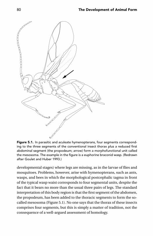
80 The Development of Animal Form
Figure 5.1. In parasitic and aculeate hymenopterans, four segments correspond-ing to the three segments of the conventional insect thorax plus a reduced firstabdominal segment (the propodeum; arrow) form a morphofunctional unit calledthe mesosoma. The example in the figure is a euphorine braconid wasp. (Redrawnafter Goulet and Huber 1993.)
developmental stages) where legs are missing, as in the larvae of flies andmosquitoes. Problems, however, arise with hymenopterans, such as ants,wasps, and bees in which the morphological postcephalic tagma in frontof the typical wasp waist corresponds to four segmental units, despite thefact that it bears no more than the usual three pairs of legs. The standardinterpretation of this body region is that the first segment of the abdomen,the propodeum, has been added to the thoracic segments to form the so-called mesosoma (Figure 5.1). No one says that the thorax of these insectscomprises four segments, but this is simply a matter of tradition, not theconsequence of a well-argued assessment of homology.
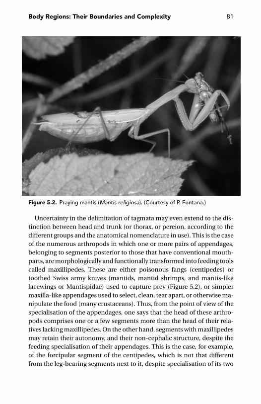
Body Regions: Their Boundaries and Complexity 81
Figure 5.2. Praying mantis (Mantis religiosa). (Courtesy of P. Fontana.)
Uncertainty in the delimitation of tagmata may even extend to the dis-tinction between head and trunk (or thorax, or pereion, according to thedifferent groups and the anatomical nomenclature in use). This is the caseof the numerous arthropods in which one or more pairs of appendages,belonging to segments posterior to those that have conventional mouth-parts, are morphologically and functionally transformed into feeding toolscalled maxillipedes. These are either poisonous fangs (centipedes) ortoothed Swiss army knives (mantids, mantid shrimps, and mantis-likelacewings or Mantispidae) used to capture prey (Figure 5.2), or simplermaxilla-like appendages used to select, clean, tear apart, or otherwise ma-nipulate the food (many crustaceans). Thus, from the point of view of thespecialisation of the appendages, one says that the head of these arthro-pods comprises one or a few segments more than the head of their rela-tives lacking maxillipedes. On the other hand, segments with maxillipedesmay retain their autonomy, and their non-cephalic structure, despite thefeeding specialisation of their appendages. This is the case, for example,of the forcipular segment of the centipedes, which is not that differentfrom the leg-bearing segments next to it, despite specialisation of its two

82 The Development of Animal Form
appendages as poison-claws, and the prothorax of the mantis, which main-tains its thoracic identity despite its unusual elongation and mobility andthe raptorial specialisation of its legs. In many crustaceans, integrationwithin the cephalic tagma of one or more segments with maxillipedes ismuch more advanced. This process has been regarded as a prolongationof a phylogenetically deeper event that led to the cephalisation of max-illar segments, a process regarded as still (primitively) incomplete in thelower crustacean lineage of the Cephalocarida (Dahl 1991). Integrationof further segments into the anterior tagma does not imply an unam-biguous extension of the head to embrace a number of segments higherthan usual, because the number of conventionally postcephalic segmentsfused to the head does not necessarily match the number of segmentswith maxillipedes. In Leptostraca, the first thoracic segment is fused to thecephalon, but its appendages are not specialised as maxillipedes and arenot different from the other thoracic appendages. In the Euphausiacea,there is a cephalothorax comprising the head and all eight segments of thepereion (as in crabs, lobsters, and shrimps), but none of these segmentshas masticatory maxillipedes.
Standard comparisons are still more uncertain when the transition be-tween two regions (say, thorax and abdomen) occurs at positions that areotherwise regarded as different (e.g., at different segment number).
A compromise solution adopted by some arthropod comparative anato-mists has been the use of the term pseudotagmata for body units suchas mesosoma and metasoma in scorpions, gnathosoma and idiosoma inmites and ricinuleans, and proterosoma and hysterosoma in actinotrichidmites (van der Hammen 1989). There is no harm if these terms are simplyused for descriptive purposes, but it is clear that this is simply a way toavoid problems in tracing homologies – not a way to solve them.
Boundaries between conventional tagmata are often less clear thananatomists would hope. Fuzzy boundaries between tagmata are quitecommon in polychaetes. In capitellids, a change in the orientation of theparapodia marks the transition from thorax to abdomen; but, in some gen-era, this change is diluted over several segments. In goniadids, the anteriorbody region is characterised by uniramous parapodia, the posterior regionby biramous parapodia, but the transition between the two types, whichis abrupt in some genera, is spread over 40 segments in others (Edmondset al. 2000).
Further difficulties are caused by a mismatch between the external andinternal organisations. In the internal anatomy of insects, for example, the

Body Regions: Their Boundaries and Complexity 83
thorax/abdomen boundary is often less than clear; the first abdominalganglion is often fused to the third thoracic ganglion, even if the latter isdistinct from those of the first and second thoracic segments (Hanstrom1928). Problems are sometimes caused by regressive evolutionary trendsthat cause an usually integrated region to be ‘decomposed’ into its origi-nal components. This is the case of the pontellid copepods, in which theantennary region is not incorporated into the cephalic tagma, as it is inall other copepods and in arthropods generally (Kabata 1979). Much morecommon, however, is the opposite trend, with an increasing number ofelements (segments) incorporated into a given tagma. This is observed inthe caligiform copepods, in which an increasingly larger anterior tagmaevolves, starting with the conventional head (cephalon) of their relatives.In Dissonus, head plus one maxillipedal segment fuse to form a cephalo-some, in Trebius one more trunk segment is incorporated in this anteriortagma to form a cephalothorax, whereas in Caligus this region extends tothe third thoracic segment (Kabata 1979).
Instability of Tagmatic BoundariesA principle illustrated by Bateson (1894) in his Materials for the Study ofVariation is that variation in the elements of a series is very often concen-trated at the end of the series. Variability at the end of series is manifestednot only in the frequency of cases of absence of terminal members, but alsoin the frequency of extra members in their neighbourhood. Bateson gavemany examples for mammal teeth and digits. This principle is virtuallyidentical to one of the ‘general laws of arthropod metamerism’ formulatedby Lankester (1904), stating that the most anterior and most posterior seg-ments of a tagma are particularly liable to regressive evolution. This is oftentrue of the first abdominal segment of insects, the first abdominal segmentof the arachnid opisthosoma and, in general, one or more segments at theposterior body end.
The anterior end of a tagma behaves very often as a ‘subduction zone’(in geological parlance). In many myriapod groups (Diplopoda, Pauropodaand Symphyla), the first postcephalic segmental unit is a legless collum. Areduction of the first postcephalic segment is also clear in the Collembola(Figure 5.3). Many arachnid groups lack the dorsal plate (tergum) of thefirst segment of the opisthosoma, and the reduction may extend to thenext two segments (scorpions, pseudoscorpions, and harvestmen). In anorthopteran suborder (Caelifera), the ventral plate (sternum) of the lastthoracic segment is largely fused to the corresponding sclerite of the first

84 The Development of Animal Form
Figure 5.3. In collembolans (a, Lipothrix lubbocki, Sminthuridae; b, Orchesellavillosa, Entomobryidae), the pronotum (i.e., the dorsal sclerite of the first thoracicsegment) is more or less extensively reduced. (Courtesy of R. Dallai, Siena.)
abdominal segment (Beier 1972). In the stick insects (Phasmodea), thedorsal plates of the same two segments are fused together (Beier 1968).In bees, wasps, and ants, as previously described, the first segment of theabdomen becomes the propodeum. In beetles, a reduction of the firstabdominal segments is commonplace.
A similar phenomenon was reported by Bateson (1894) for the teeth ofmammals, in which variability at the end of the series is manifested notonly in the frequency of cases of absence of terminal members, but alsoin the frequency of instances in which an extra teeth is present in theirneighbourhood.
Nowadays, it is easy to explain this ‘law’ in terms of Hox gene expression.As the transition from one tagma to the next corresponds to the anteriorboundary of expression of one or more Hox genes, we must be preparedto regard this transition as a morphogenetic ‘hot spot’, in which diversespecialisations may be expected, with segmental reduction included.
Lankester’s principle does not apply to arthropods only; in the mouse,the skeletal defects caused by systematically knocking out the Hox genesappear to be concentrated at certain ‘hot spots’, notably the boundaries be-tween different skeletal types (i.e., the boundaries between the anatomist’svertebral column regions; Chen and Capecchi 1997).
In the woodlouse Porcellio scaber, the expression domains defined by theHox genes Antennapedia, Ultrabithorax, and abdominal-A coincide with
Body Regions: Their Boundaries and Complexity 85
morphological boundaries between tagmata (Abzhanov and Kaufman1999, 2000b). However, these borders do not coincide with those of in-sects, and similar differences were observed for the Hox genes expressedin the posterior cephalic segments. For example, Sex combs reduced is ex-pressed in the maxillae of Porcellio, whereas the Hox gene proboscipedia isexpressed in the corresponding maxillary and labial segments of insects.
Homology of TagmataTerms such as head, thorax and abdomen have been used for the bodyregions of the most diverse animals, from vertebrates to polychaetes, frominsects to non-malacostracan crustaceans. Is there any degree of homologybetween the body regions of all these animals, or of bilaterians generally,to justify the generalised use of the same terms for their body regions? Asoften happens in respect to homology problems, the answer is yes and no.
There are problems even within a limited group, such as crustaceans(Kaestner 1970). Three main regions are primarily recognisable in thesearthropods – a head and two postcephalic tagmata – but the nature of thelatter ones is probably not the same in malacostracans and in the othercrustaceans. In malacostracans, both regions are typically provided withappendages; but, in most non-malacostracan crustaceans, appendagesare present in the first postcephalic region only. The latter condition hasbeen long regarded as equivalent to that of the insects, which have a tho-rax with three pairs of legs and an abdomen practically devoid of ap-pendages. Accordingly, the two terms used for the two tagmata of theinsect trunk – thorax and abdomen – are used for copepods and othernon-malacostracan crustaceans, but not for the malacostracans, wherethe two postcephalic regions are known instead as the pereion and thepleon. According to Gruner (1997), pereion and pleon together would beequivalent to the thorax of the basal crustaceans, whereas the abdomenwould have disappeared or nearly so.
For non-malacostracan crustaceans, some evidence is available aboutmolecular markers of the body regions and their boundaries. Averof andAkam (1995) compared the expression patterns of several Hox genes inArtemia franciscana (a non-malacostracan, branchiopod crustacean) withthose of the homologous genes in insects. They found that Antennape-dia, Ultrabithorax, and abdominal-A are expressed, in Artemia, in largelyoverlapping domains in the uniform thoracic (pregenital) region, whereastheir non-overlapping expression leads, in insects, to the specification ofdistinct segment types in the thorax and abdomen. Accordingly, Averof

86 The Development of Animal Form
and Akam tentatively suggested that the pregenital ‘thorax’ of Artemiawould correspond to the sum of the thoracic and the pregenital abdo-minal segments of insects. We shall also consider that non-malacostracancrustaceans with a limbless ‘abdomen’ (e.g., copepods and anostracans)do not have abdominal ganglia. Segments of their abdomen receive nervesfrom the thoracic part of the central nervous system. From this point ofview, this ‘abdomen’ would better compare with an appendage like thevertebrate tail, rather than with a part of the main body axis. In these crus-taceans, however, the anus opens at the end of these ‘abdominal’ segments,whereas it is anterior to the tail in vertebrates. It is therefore not advisableto call these segments a ‘tail’, as tentatively suggested by Averof and Akam(1995). Lack of individual neuromeres in this region invites enquiry as towhether this depends on a regional inhibition of the differentiation of seg-mentally distributed neural precursors or, what I regard as more likely, isthe whole of this ‘abdomen’ derived from secondary segmentation of justone primary segment (one eosegment, cf. pages 200–203).
Thus, we shall not expect a given region to comprise a fixed number ofsegments. This is true for vertebrates as well as for annelids and arthropods.It does not depend on the precise mechanism of segmentation adoptedby a particular animal group. I will return briefly to this phenomenon of‘transposition’ when discussing homology in the last chapter.
Number of Tagmata and ConvergenceThe precise correspondence between the anterior (or posterior) expres-sion boundary of a given gene along the main body axis and the anterior(or posterior) end of a given body region should not be construed as anargument in favour of the thesis that gene expression patterns are an infal-lible guide to the homology of body regions across the most diverse taxa.Tagmosis is probably constrained to a very limited number of alternativepossibilities, and the genomic organisation of gene families, such as theHox genes, offers a convenient and easily available patterning tool, so thatwe cannot rule out convergence.
The main constraint to tagmosis is perhaps the number of body regionsan animal may differentiate. Admittedly, the concept of tagma is to a largeextent arbitrary. This is clearly seen in the uncertainty of the morphologi-cal nomenclature applied to certain animal groups, in which some zoolo-gists recognise a smaller number of body regions and others recognise alarger number of body regions. For example, how many postcephalic re-gions do we recognise in a millipede? Some zoologists separate the first few

Body Regions: Their Boundaries and Complexity 87
segments as ‘thorax’, with zero to one pairs of legs each, from the remainingset of ‘double segments’ with two pairs of legs each. Yet other zoologists re-gard the millipede trunk as a single tagma. In scorpions, one might use thebasic arachnidan blueprint, thus recognising a prosoma and an opistho-soma, but the latter is clearly articulated into a broader mesosoma and anarrower metasoma, and so on.
However, even in the hands of the splitters, regional specialisation alongan animal’s main body axis does not reasonably allow for the distinctionof more than three to five regions. Thus, this question arises: is there anyreason why animals with a main body axis articulated into a dozen regionsdo not exist?
There is a reason for that. It might be found in a issue of complexity dis-cussed by Kauffman (1993) as an intrinsic limit of adaptive systems. Let’sconsider a system with many parts (in our case, body regions). As the num-ber of those parts and the richness of interactions among the parts increase,there will also be a rapid increase in the number of conflicting constraintsof design among the parts. Those conflicting constraints imply that optimi-sation can only attain increasingly poorer compromises. This limitation isintrinsic to the system’s design, that is, it cannot be overcome by strongeradaptive selection. If Kauffman (1993) is correct, no patterning system,even the richest and most versatile, could further increase the number ofdifferentiated parts; the upper limit depends instead on intrinsic structuralrules. This may be indeed the case. In hypothetical, strictly combinatorialterms, a Hox code would permit differentiation of many body regions, butthis is not what we observe. Interestingly, strict limits to the number ofkinds of parts apply also to the longitudinal axis of the appendages andthe other differentiated series of body units, such as tetrapod digits andmammal teeth. Many examples include just three elements, such as thetrimerous coelom (proto-, meso-, and metacoel; or axo-, hydro-, and so-matocoel) of bryozoans, echinoderms and other groups. No more thanfour kinds of elements – to borrow an example from botany – are presentin the flowers, with their sepals, petals, stamens, and carpels (the latterbeing the elements of the ovary). There are four kinds of teeth in the mouthof a mammal (incisors, canines, premolars, and molars). There are four tofive main transversal bands of the basic wing pattern in butterflies (Nijhout1991), or the kinds of segments in the polychaete Chaetopterus (Figure 3.3).There are five kinds of fingers in tetrapod vertebrates. And, there are up tosix – and this seems to be an unbridgeable upper level – main veins in theinsect wing.

88 The Development of Animal Form
Interestingly, five, or something close to five, is also the upper numberof ‘mental boxes’ into which perceptual schemes and naive taxonomiesare organised. We have no difficulty organising a mental image of therelative position of three to five objects distributed along a horizontal orvertical axis. Of three objects, one is in the middle, one is on the left andone is on the right. Or, there is one in front, one below and one above.Things are less complicated, but still manageable, if two more objects areadded (e.g., one more to the left and one more to the right). Additionalelements, however, would easily disrupt our whole perceptual and mentalconstructs. A counterpart to these perceptual constraints is provided byour extremely frequent use of three to five categories in the most diversekind of taxonomies we may adopt, including periodisation of time (e.g.,past, present, future; or, at most, remote past, recent past, present, nearfuture, and far future; cf. also the number and names of the geological eras).Five is also the highest number of taxonomic levels (or categories) in themost elaborate of the main folk taxonomies of animals or plants examinedthus far around the world (Berlin, Breedlove, and Raven 1973, Berlin 1992).
One might speculate whether this upper limit around number five of ourperceptual schemes and basic mental constructs has anything to do withKauffman’s (1993) principle of “optimized compromise” among differenti-ated units at a given structural level. I suspect it has, but I do not know howfar the limit in complexity we see at the perceptual/mental level dependson the limit in complexity of the underlying biological systems (our fivefingers included) rather than on its own intrinsic structural constraints.It is tempting to imagine a causal cascade, beginning with the structuralconstraints of genetic networking, moving to the morphological level withthe determination of no more than five kinds of fingers and ending withrules governing the way we count or describe our perceptual universe. It ismore likely, however, that common structural rules apply to all these lev-els, so that the agreement between levels is more the effect of a canaliseddrift than the expression of a historically determined causal cascade.
The Complexity of Postembryonic DevelopmentA final speculation on this theme is that the putative limit in the com-plexity of body patterning along a given structural dimension descend-ing from Kauffman’s principle might also apply to the complexity of thetemporal dimension of animal development. That is, the same principlemight possibly explain why the number of basically different kinds of body

Body Regions: Their Boundaries and Complexity 89
organisation displayed by an animal along its whole life cycle is always verylow, seldom higher than two or three (e.g., tadpole and frog, pluteus larvaand sea urchin, polyp and medusa, caterpillar chrysalis and butterfly).Simple growth stages, even when separated by moults, as in the case ofarthropods or nematodes, or the mostly smooth contrast between a non-reproductive juvenile and a fully reproductive adult should not be takeninto account, because of the fundamental continuity of these life pro-cesses. Things are quite different, instead, when a life history is punctuatedby major metamorphic events, such as in many cnidarians, echinodermsand holometabolous insects. The most complex life cycles are probablythose of the so-called hypermetabolous insects.
Hypermetaboly occurs in Strepsiptera, in several beetles (all Meloidae,Rhipiphoridae, Micromalthidae, and a few Carabidae and Staphylinidae;Figure 5.4) and in some members of Neuroptera (Mantispidae), Lepi-doptera (Gracillariidae), Diptera (Bombyliidae, Acroceridae, Nemestrini-dae), and Hymenoptera (Chalcidoidea). A basic feature of the life cycle ofall these insects is the occurrence of two to four different kinds of larvae.In the case of the blister beetles (Meloidae), for example, only the firstlarval instar (called a triungulin because its legs seem to end with threeclaws; in fact, between the two true claws there is a robust seta of similarsize) is active, long-legged and very mobile. Its mobility is instrumentalin finding either a wild bee that will bring the tiny triungulin to its nest,or the egg mass laid by a grasshopper in the sandy soil of a hot dry re-gion. As soon as it makes contact with its future foodstuff, the triungulinmoults into a short-limb feeding larva that rapidly grows by feeding on thegrasshopper’s egg or on the egg (or the young larva) of the bee, togetherwith the pollen the latter had stored in the nest for its offspring’s benefit.What follows varies according to the genera; but, for our purposes, it isenough to say that in Meloe the feeding larva moults into a non-feeding,resting prepupa or hypnotheca. The latter, however, does not give rise tothe adult, as one might expect, but to another active but not feeding larvalinstar. It will eventually moult into a true pupa, which in due time, at last,will give rise to the adult. An extra resting stage, such as the prepupa of theblister beetles, does not occur in the majority of the life cycles of the otherhypermetamorphic insects. In all of them, the larval stages preceding thepupa are not structurally uniform, as in the conventional caterpillars orin the larvae of the majority of beetles, flies, and wasps, because the first-instar larva (generally, the most mobile segment of the insect’s preadult

90 The Development of Animal Form
Figure 5.4. Larval and pupal stages of Mylabris variabilis, a representative of thehypermetamorphic blister beetles (Meloidae). a, larva I (triungulin); b, larva II;c, larva III; d, hypnotheca or pseudopupa; e, last larval stage; f, pupa. (Redrawnafter Bologna 1991 and other sources.)
life) is followed by a larva of a very different kind. Further complicationsmay occur, as in the beetle Micromalthus debilis, in which as much as fourdifferent larval kinds have been described.
Life cycles of a complexity comparable with that of hypermetabolousinsects are known from flatworms (flukes and tapeworms) and from therecently described phylum Cycliophora. Interestingly, all flukes and

Body Regions: Their Boundaries and Complexity 91
tapeworms are parasites, as are most of the hypermetabolous insects pre-viously described. The only cycliophoran thus far described, Symbion pan-dora, is probably a commensal, living on the mouthparts of the Norwaylobster (Funch and Kristensen 1995). The sporozoans or Apicomplexa (theprotist phylum to which the malaria agent Plasmodium belongs) are alsoparasitic, and it is directly in this phylum that the most complex life cy-cles among unicellular protists are encountered, with up to three differentkinds of reproduction and four or five different kinds of cells.
One might speculate whether Dawkins’s (1982) concept of extendedphenotype might help explain this generalised (but far from universal)co-occurrence of very complex life cycles and parasitic habits; but this isnot a point to be discussed in this book. What matters, for the argument Iam developing in this chapter, is the very low limit (around four or five) ofthe number of different kinds of stages that might occur, even in the mostcomplex life cycles. In my opinion, it is not by chance that this upper limitis of the same order of magnitude as the upper limit of differentiated partsin any given series or body dimension, be it the number of tagmata or thenumber of kinds of fingers in a tetrapod limb.
Number of kinds, of course, is not the same as number of individual parts.There is no clear upper limit, for example, of the number of body segments.Among the polychaetes, the Phyllodocidae may have up to 600 segments,the Oenonidae up to more than 1000, and the Eunicidae up to about 1500(Edmonds et al. 2000). Among the oligochaetes, more than 600 segmentsare present in Rhinodrilus (Glossoscolecidae; Hartmann-Schroder 1982).The arthropods with the highest number of segments are some centipedes(Gonibregmatus plurimipes with up to 191 pairs of legs, Himantariumgabrielis with up to 189 pairs of legs) and millipedes (Illacme plenipes withup to 375 pairs of legs (Hoffman 1982, Minelli, Pasqual, and Etonti 1984,Enghoff 1990, Minelli and Golovatch 2001). Large numbers of vertebrae arepresent in some sharks (up to ca. 400; Fiedler 1991), bony fishes (at leastca.750 in the eel relative Nemichthys; Nelson 1984), amphibians (more than200 in some caecilians; Noble 1931), and snakes (more than 300 in sometyphlopids; Gans, Laurent, and Pandit 1965). In all these cases, however, thetagmosis of these longest of animals is never more complex than in theirrelatives with fewer segments. Indeed, if a trend in complexity is present atall, it runs the other way. Thus, an increase in the number of body segmentsis often accompanied by a reduction in the complexity of the patterningalong the main body axis, something that snakes, eels, centipedes, andmillipedes exemplify at will.

92 The Development of Animal Form
Williston’s RuleIn the literature on macroevolution (e.g., Saunders and Ho 1984),‘Williston’s rule’ is sometimes cited, according to which there should bea general trend from polymerous and homonomous to oligomerous andheteronomous organisation (e.g., towards a reduction in the number ofserial parts, such as segments or teeth and a corresponding increase intheir specialisation). This ‘rule’, however, is far from universal. Oppositetrends are found, for example, in many fish groups (Lindsey 1988) andin myriapods (Berto, Fusco, and Minelli 1997, Fusco & Minelli 2000a).There is also a curious historical point, i.e., that the attribution of thisprinciple to Williston is incorrect. Williston (1914) observed that no ‘new’bone ever appeared in the skulls of reptiles, birds, and mammals, whereas,at the same time, many bones of the skulls of the so-called lower ver-tebrates have apparently got lost in evolution. But in another work hewrote: “We may hardly venture to guess as to the primitive number ofvertebrae in reptiles. We are quite sure that there has been an increasein number in some, a decrease in others” (Williston 1925:94). A trendtowards the reduction in number and the increrase in specialisation ofserial parts had been formulated long before Williston by naturalists-philosophers like Treviranus (1820–22), Meckel (1821), von Baer (1828),and Bronn (1858).
Developmental Time and Body AxesCorrespondence between the temporal sequence of morphogenetic eventsand the growth and patterning of the main body axis is easily observed. Inmany, if not most, of the metazoans, differentiation proceeds in antero-posterior order. This progression begins in the embryonic segment of de-velopment, but it may extend through postembryonic development to theattainment of the adult condition. The best example of antero-posteriorprogression of growth and differentiation during postembryonic life is pos-sibly the anamorphic arthropods, in which new segments differentiate,moult after moult, at the posterior end of the trunk. Many crustaceans, forexample, begin their postembryonic life as nauplius larvae, with only a fewanterior segments fully differentiated and just three pairs of appendages.The remaining segments and their appendages will progressively appearwith later moults. Behind a fully differentiated head, most newly hatchedmillipedes have a short trunk, with less than the final number of segments,and just three (rarely four or more) pairs of legs. The remaining segmentsand leg pairs will appear progressively at later stages.
Body Regions: Their Boundaries and Complexity 93
Developmental time is also mirrored by the proximo-distal axis of theappendages. Here, differentiation of the proximal structures often pre-cedes differentiation of more distal ones; but no simple rule applies. Inarthropods, the joint between the femur and tibia appears before the moreproximal joint between the trochanter and femur; the latter joint, in turn,precedes differentiation of more distal elements: the tibia and the tarsus(Norbeck and Denburg 1991).
There is no need to look for special mechanisms as an explanation of thecorrespondence between the temporal and spatial dimension of body pat-terning and differentiation. Animals have developed ways of controllingthe timing of cell activities (mitosis, cell migration, etc.), and the orderlyprogression of differentiation along the main body axis, or the axis of theappendages, is simply a ‘mechanical’ by-product of the timing of cellularprocesses. Nevertheless, correspondence between the complexity of de-velopmental schedules and the patterning of the main body axis is oftenfar from trivial.
Recent developments in the study of complex systems have revealedthat all definitions of complexity are bound to be partial and subjective.This limit notwithstanding, complexity can be often defined and – whatmatters more – measured in a way suitable for attacking a defined setof problems. This has been shown, for example, by Daniel McShea in aseries of important papers on the structural (morphological) complexity ofmulticellular animals (e.g., McShea 1991, 1992, 1996a, 1996b, 2000, 2001).The epistemological background to these questions about complexity hasbeen discussed by Ahl and Allen (1996), moving from the observation thatcomplexity is a function of the relationships between the units and levelswe identify in a system; but these units and levels depend on the observer’scriteria and do not exist independent of them. The concept of level isrelative to the particular point of view taken by the observer. Complexityis not a feature of the external world, but strictly depends on the way weask questions about the world.
Studying the origin and evolution of animal form confronts us with dif-ferent kinds of complexity. On the one hand, we are interested in com-paring the complexity of the body organisation of animals belonging todifferent species, or the complexity of different developmental stages ofthe same animal. On the other hand, we would like to measure the com-plexity of the developmental process itself.
Let’s start with morphological complexity. Comparisons of closely re-lated species seem to be amenable to simple metrics, such as those
94 The Development of Animal Form
proposed by McShea (1993) and Fusco and Minelli (2000a) to measure themorphological complexity of serial structures (e.g., the vertebral columnof mammals or the segments of centipedes). Difficulties, however, growenormously when we look for criteria for comparing the architectural com-plexity of more distantly related species. A rough criterion that applies, inprinciple, to all living beings is the number of cell types (Bonner 1988), butthis criterion suffers from several shortcomings – the most obvious beingthe subjectivity in classifying an organism’s cells into distinct types worthyof a name. For example, there are just 302 neurons in the nervous systemof Caenorhabditis elegans, but these few neurons can be classified into notless than 118 structural classes (Thomas 1994). Furthermore, the diversityof specialised cell types increases with the animal’s size, independent ofphylogeny (Bell and Mooers 1997). Whatever metrics we might devise todescribe an animal’s morphological complexity, it will hardly reveal dif-ferences in complexity between closely related animals only different intotal size, irrespective of size-dependent differences in cell-type diversity.Therefore, we cannot expect to learn too much from this criterion for esti-mating complexity. It seems more meaningful, even if limiting, to restrictour comparisons within the range of a class or an order, as I will do in thenext few paragraphs. Criteria for measuring morphological complexity willremain subjective. I will take a very simple, semiqualitative approach andfollow some largely intuitive principles (Minelli 1996a): (1) complexity isnot dependent on the number of parts (strictly homonomous sequencesof segments have the same complexity, irrespective of differences in thenumber of units); (2) there is an increase in complexity when changingfrom unsegmented to segmented organisation; (3) there is little differencein complexity between a strictly homonomous pattern and a gradient-likepattern, such as in those insect antennae in which the size of the articlesregularly decreases from the base to the tip; and (4) there is, instead, a highincrease in complexity when a strong structural boundary occurs, such asbetween head and thorax, or thorax and abdomen.
These rules may be used to obtain a comparative estimate of complexityalong an animal’s main body axis, as well as along the proximo-distal axisof an appendage. Interestingly, there is no difficulty in applying an identi-cal set of rules to the temporal rather than to the spatial dimension, thusobtaining a comparative estimate of the complexity of a developmentalschedule. Subjectivity of periodisation notwithstanding, there seems to beno problem in saying that cnidarians with both polyp and medusa havea more complex life cycle than those with polyp only or medusa only;

Body Regions: Their Boundaries and Complexity 95
that the postembryonic development of holometabolous insects – such asflies, butterflies, beetles, and bees – is more complex than the postembry-onic development of heterometabolous insects, such as grasshoppers orcockroaches. This is little more than a truism. But we will see soon thatthis approach may reveal unexpected correspondence among the relativedegrees of complexity found along different dimensions.
To begin with, in many arthropods, the complexity of postembryonicdevelopment and the morphological complexity of the main body axis ofthe adult appear to be coupled.
The Cambrian crustacean Rehbachiella kinnekullensis developedthrough a very high number of moults, perhaps more than 30, but themorphological changes following each moult were minimal. The highnumber of moults is distinctly mirrored by the high number of body seg-ments, whereas the smooth changes in body organisation along the wholepostembryonic development had its equivalent in the animal’s faint tag-mosis (Waloßek 1993).
In crustaceans, we can identify an ideal (but not strictly phylogenetic)series of taxa with an increasingly smaller number of moults, proceedingfrom Rehbachiella to Anostraca (e.g., Artemia) to Conchostraca to Ostra-coda. At the same time, the amount of change (posterior addition of newsegments, differentiation of appendages) brought about by each moultingevent increases in the same order. That is, the moulting sequence changesfrom merely quantitative control of growth to qualitative control of bodypatterns. This is expressed, along the main body axis, by a progressivereduction in the number of body segments and appendages, togetherwith increasing morphological and functional specialisations. Were sucha sequence actually supported by phylogeny (what is not at stake in thiscontext, however), we would have an example of evolution according toWilliston’s law.
In millipedes (Diplopoda), postembryonic development follows a moreor less complex and tightly fixed schedule. Enghoff, Dohle, and Blower(1993) distinguish three developmental modi. The first is euanamorpho-sis. Here the number of postembryonic moults is not strictly fixed; thereis also some individual variation in the number of segments added aftera given moult, and the number of body segments continues to increasefrom moult to moult, even after the animal has reached sexual maturity.The number of segments in the adult is also variable within a species.More canalised is teloanamorphosis, in which there is a fixed number ofpostembryonic moults, with a regular increase in the number of trunk

96 The Development of Animal Form
segments after each moult. The last moult coincides with the attainmentof sexual maturity and a fixed number of segments. In the third kind ofanamorphosis, called hemianamorphosis, postembryonic development isdivided into two parts: a strictly programmed series of early larval stages,through which, as in teloanamorphosis, a fixed number of segments isachieved, followed by some more postlarval stages which do not acquireadditional segments or appendages, but just increase in size and finallyreach sexual maturity. These differences in the complexity of postem-bryonic development correspond to the different degrees of complexityin adult morphology: millipedes with euanamorphosis have a worm-liketrunk, with the lowest level of regional differentiation, whereas those withhemianamorphosis have the most advanced regionalisation. This doesnot imply, however, that hemianamorphic millipedes are phylogeneticallymore advanced than those with euanamorphosis. Cladistic analyses showthat the most elongate, worm-like forms of millipedes, with a trunk madeof many similar segments, are more derived than those with shorter andmore extensively regionalised bodies. The same trend is observed in cen-tipedes (Enghoff 1990, Berto, Fusco, and Minelli 1997, Foddai and Minelli2000, Regier and Shultz 2001). In this case, Williston’s law is clearly notsupported.
In Hymenoptera, the most primitive forms (Symphyta) have many mor-phologically similar larval stages, whereas adults possess a more or lessprimitive multisegmented abdomen. In the more advanced Apocrita, thecomplexity of postembryonic development is higher, and the number ofpostembryonic stages is reduced. In some Parasitica, the younger and olderlarva of the same animal are completely different. At the same time, thereare major changes in the abdomen. The dorsal plate (tergum) of the firstabdominal segment becomes incorporated into the thorax, the followingsegment(s) may form one or two knot(s), as in the ants, and some segmentsof the abdomen may more or less extensively fuse together, as the tergalplates of some abdominal segments do in Braconidae (Figure 5.5).
A trend towards the reduction of the number of moults is also seen inDiptera. There are six moults in Simuliidae, four in the remaining Nema-tocera, five to eight in Orthorrhapha, but only three in Cyclorrhapha. Thisis broadly matched by a corresponding reduction of the number of freeabdominal segments in the adult.
Among the arachnids, Acari and Ricinulei are those with a more complexdevelopmental schedule (in particular, these are the only arachnid groupsin which the newly hatched animal – currently termed a larva – has less than

Body Regions: Their Boundaries and Complexity 97
Figure 5.5. Mismatch between dorsal and ventral segmentation in an adeline bra-conid wasp. (Redrawn after Goulet & Huber 1993.)
the final number of legs; i.e., three pairs rather than four). At the same time,these are also the arachnids with the most complex tagmatisation. This isreflected in anatomical nomenclature: instead of the usual terms (prosomaand opisthosoma) used for the body regions of most of the remainingarachnids (e.g., the spiders), no less than six different terms (gnathosoma,hysterosoma, idiosoma, metapodosoma, propodosoma, and podosoma)are used to identify the different body regions recognisable in the mites.
The Time Axis of Development and the Patterningof the Proximo-Distal Axis of the AppendagesThere are several examples of agreement between the complexity of poste-mbryonic development and the morphological complexity of the axis ofadult appendages (Minelli 1996a).
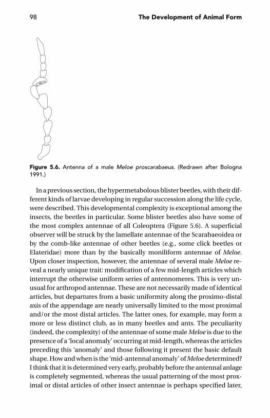
98 The Development of Animal Form
Figure 5.6. Antenna of a male Meloe proscarabaeus. (Redrawn after Bologna1991.)
In a previous section, the hypermetabolous blister beetles, with their dif-ferent kinds of larvae developing in regular succession along the life cycle,were described. This developmental complexity is exceptional among theinsects, the beetles in particular. Some blister beetles also have some ofthe most complex antennae of all Coleoptera (Figure 5.6). A superficialobserver will be struck by the lamellate antennae of the Scarabaeoidea orby the comb-like antennae of other beetles (e.g., some click beetles orElateridae) more than by the basically moniliform antennae of Meloe.Upon closer inspection, however, the antennae of several male Meloe re-veal a nearly unique trait: modification of a few mid-length articles whichinterrupt the otherwise uniform series of antennomeres. This is very un-usual for arthropod antennae. These are not necessarily made of identicalarticles, but departures from a basic uniformity along the proximo-distalaxis of the appendage are nearly universally limited to the most proximaland/or the most distal articles. The latter ones, for example, may form amore or less distinct club, as in many beetles and ants. The peculiarity(indeed, the complexity) of the antennae of some male Meloe is due to thepresence of a ‘local anomaly’ occurring at mid-length, whereas the articlespreceding this ‘anomaly’ and those following it present the basic defaultshape. How and when is the ‘mid-antennal anomaly’ of Meloe determined?I think that it is determined very early, probably before the antennal anlageis completely segmented, whereas the usual patterning of the most prox-imal or distal articles of other insect antennae is perhaps specified later,
Body Regions: Their Boundaries and Complexity 99
through molecular signalling from either end of the appendage. We knowvery little about the way a beetle antenna is segmented. For now, we canonly record the coexistence, in Meloe, of an unusually complex life history(hypermetamorphosis) with an unusual degree of morphological com-plexity in the antenna.
Some arachnids show a corresponding agreement between the com-plexity of the postembryonic development and the morphological com-plexity of the appendages. Upon hatching, most arachnids already havetheir definitive organisation, with a full complement of body segmentsand appendages. There are two exceptions, however: the Ricinulei and themites (Acari). In both groups, as previously described, the first postembry-onic stage is a six-legged larva, morphologically distinct (in some mites,to a dramatic degree) from the following stages. In the Acari, up to threenymphal stages are usually interposed between the larva and the adult.Interestingly enough, segmentation and patterning of the legs in Ricinuleiare more complicated than in the other arachnids. There is no parallel inthe mites, but these (most species are less than 1 mm long) are probablytoo small for a more complex patterning of the leg segments; however, mitelegs are frequently provided with apical structures of unusual complexity.
Topology
Morphology and Developmental Topology“Whatever sense may exist to the phrase ‘neighboring developmental pro-gram,’ distant regions on the fate map can have neighboring programs”(Kauffman 1993: 511). A developmental analysis of form often reveals anintrinsic geometry which is very different from that of the resulting animalform. Topographically close body parts can be topologically far away, andvice versa, in the network of developmental interactions that link themtogether. For example, why not speak of ontogenetic proximity of bodystructures found at opposite body ends when reading that, in Drosophila,the gene head involution defective is required for embryonic head involu-tion, as well as for the rotation of male terminalia in the pupal stage (Abbottand Lengyel 1991)? The same is true for Drosophila lacking both maternaland zygotic hunchback functions (Lehmann and Nusslein-Volhard 1987)or headless mutant embryos of the parasitic wasp Nasonia, with anteriorand posterior gap defects (Pultz, Pitt, and Alto 1999).
Metaplastic transformations, or conversions of a cell or group of cellsfrom an expected developmental fate to another, suggest the proximity ofthe specific developmental programs of the two cell types (Kauffman 1993).
100 The Development of Animal Form
Slack (1985) summarized 21 types of homeotic transformations (epithelialheterotopia and metaplasia) in humans, concentrated in the gastrointesti-nal, urinary and female reproductive systems. Most, but not all, of thesetransformations are between tissues whose precursors are neighbouringregions of a common cell sheet during early embryogenesis, which aretherefore likely to have neighbouring patterns of gene expression or devel-opmental fates. Three rules were identified by Kauffman (1993) in terms ofthe possible metaplastic transitions from one cell type to another:(1) limitation – no cell type can reach all cell types; (2) connectedness – thereis a large, strongly connected set of cell types which can mutually reachone another; and (3) asymmetry – some cell types can join this stronglyconnected set from outside, but can not be joined from inside.
In the tiny parasitic wasp Nasonia vitripennis, the minus stripes mu-tation causes small gaps in two thoracic segments (T1 and T2, pro- andmesothorax), as well as in the sixth and seventh abdominal segment (A6and A7), but not in those in between (Pultz et al. 2000). Thus, in a sense,T2 is closer to A6 than it is to T3. In this question of topology, a factorialapproach (see chapter 10 for a corresponding approach to homology) isthe best way to analyze the facts. We can recognise topological proximitybetween T2 and A6 from the point of view of the minus stripes gene ex-pression, but other genes would certainly suggest otherwise. There is noone functional topology of body parts; there are, instead, many topologiescross-linked by the functional relationships of genetic networking.
Maps of the phenotypic effects of a pleiotropic gene are possibly thebest way to demonstrate the intricacies of the topology of body morpho-genesis. More accessible, but also more limited, evidence is obtained fromthe very often discontinuous expression patterns of some genes (e.g., thepair-rule genes). Another way to ‘materialise’ the internal topology of de-velopmental events is the mapping of mitotic domains. These are devel-opmental units found at early developmental stages (before gastrulation),characterised by the synchronisation of mitotic activity of a cluster of cellsembedded in a context of cells which start dividing at an earlier or latertime. In the blue-bottle Calliphora, in which mitotic domains have beenaccurately studied, there are several simple domains formed by one areaeach or by two bilaterally symmetrical areas of synchronously dividingcells. But a few domains, such as those labelled as δ1416, δ1417, δ1421 andδ1425, comprise many serially repeated cell clusters. Besides dividing at thesame time, all the cells in these domains occupy homologous positions inthe presumptive segments of the embryo (Foe and Odell 1989).
Body Regions: Their Boundaries and Complexity 101
Developmental proximity is often a matter of communication. The ver-tebrate hindbrain (rhombencephalon) is divided in a series of rhombome-res whose persistence as independent units depends on the recognitionproperties of their constituent cells. If reciprocally put in contact, cellsfrom even-numbered rhombomeres would easily mix together, as wouldcells from different, odd-numbered rhombomeres. But cells from odd-and even-numbered rhombomeres sort out spontaneously (Guthrie andLumsden 1991, Guthrie, Prince, and Lumsden 1993). This is enough to keepthe cell populations of the different compartments separate. But this is alsoan example of the contrast between spatial and functional proximities.Similarly, sorting-out experiments with cells from dissociated imaginaldiscs of Drosophila demonstrated that differences between the differentlegs (which belong to the three thoracic segments) were not as great asthe differences between wing cells and leg cells from the same segment(mesothorax). Interestingly, whereas the cells destined to form the threepairs of legs were capable of sorting out regardless, the wing cells were notable to sort out from those destined to form the wing-homologous struc-ture, the haltere, despite the much more obvious morphological differ-ences existing between a wing and a haltere, with respect to the differencesbetween the fore-, mid- and hind legs (Fehon, Gauger, and Schubiger 1987).
Among serially repeated body parts, the topological closest relatives aresometimes arranged in a selectively discontinuous series rather than in theobvious series of contiguous segments which defines a tagma such as theinsect thorax or abdomen. One example is the ten pairs of tracheal placodeswhich form in those (para)segments in which the expression of the spaltgene does not prevent their differentiation (Kuhnlein and Schuh 1996).
Ghysen et al. (1993) have shown that specification of cell fates within agiven tissue is not the result of a progressive subdivision of the tissue intoincreasingly smaller developmental units down to the level of individualcells. For example, the formation of sense organs in Drosophila is not theresult of a subdivision of the organ-forming material into compartmentswithin compartments within compartments, but is accomplished by in-structing cells at particular discontinuous locations within the tissue toadopt a particular fate.
Thinking in terms of topology of developmental processes may help es-tablish a conceptual frame for discussing the possible independence ofdifferent sets of characters – not only developmental, but also morpho-logical. This issue is of primary concern to systematists, in their effortsto fill in and analyze data matrices in which mutual independence of the

102 The Development of Animal Form
characters recorded and coded represents one of the single best qualitiesof the work.
Topology of CoaptationsA peculiar problem of topology is offered by those body parts which mustmutually fit together to perform their function (coaptations). These partsmay belong to the same animal or to two sexual partners. The vertebrateheart originates as two separate halves, left and right, at either side of theembryo, which are destined to fuse together eventually following migrationtowards the midline. Another example is the dorsal closure of the embryoin arthropods such as Drosophila, which also requires a correct fittingtogether of the two halves. Coaptations are also common, and more easyto observe, in postembryonic stages. Conspicuous examples are those ofanimals able to roll up into a sphere, such as the pill millipedes (Figure 5.7)and many woodlice and armadillos, or those of the bivalve shells, in whichthe two valves are generally provided with the precisely interlocking teethand dental sockets that help them fit together at closure. Many insects canaccommodate some of their appendages, when at rest, within perfectlyshaped ventral or latero-ventral grooves, such as those accommodatingthe rostrum of many true bugs, or those used by weevils for protecting theantennae.
The coaptations most extensively studied to date are those that involvesexual appendages and sexual openings (e.g., Eberhard 1985). Attention,however, has been focussed primarily on the complementary structureof male and female parts, according to the traditional (and a bit naivelyadaptationist) key-and-lock model. Much less attention has been aimedat the positions these parts occupy in the body architecture of each part-ner. Many pairs of sexual structures are located at equivalent positions inboth the male and female. This seems to be easily explained with paral-lel determination of this position through equivalent expression of genes,such as Abdominal-B (see page 181). There are many exceptions, however.Spider males and dragonfly males have secondary penises (cf. page 177)whose position does not correspond in any obvious sense to the positionof the female (and male!) genital opening in the same animals. Close in-teractions between mating dragonflies are not limited to those betweenthe secondary penis of the male and the genital opening of the female, butextend to the male’s caudal appendages which interlock with the female’sprothorax during the whole nuptial flight. Thus, the question is whetherthere is any correlation between the relative positions of these interlocking
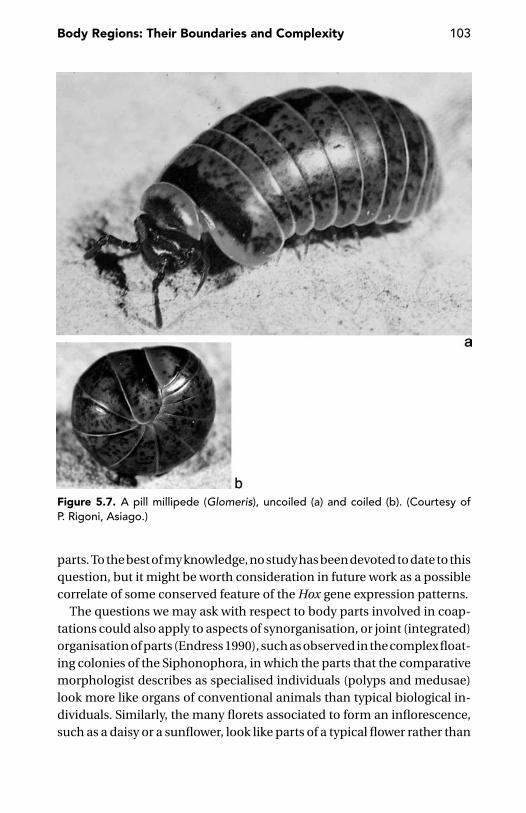
Body Regions: Their Boundaries and Complexity 103
Figure 5.7. A pill millipede (Glomeris), uncoiled (a) and coiled (b). (Courtesy ofP. Rigoni, Asiago.)
parts. To the best of my knowledge, no study has been devoted to date to thisquestion, but it might be worth consideration in future work as a possiblecorrelate of some conserved feature of the Hox gene expression patterns.
The questions we may ask with respect to body parts involved in coap-tations could also apply to aspects of synorganisation, or joint (integrated)organisation of parts (Endress 1990), such as observed in the complex float-ing colonies of the Siphonophora, in which the parts that the comparativemorphologist describes as specialised individuals (polyps and medusae)look more like organs of conventional animals than typical biological in-dividuals. Similarly, the many florets associated to form an inflorescence,such as a daisy or a sunflower, look like parts of a typical flower rather than

104 The Development of Animal Form
whole flowers. The rules according to which these synorganised parts fittogether are still to be approached from an evo-devo perspective.
Topological BreakdownThe body architecture of most animals is based on the simple topologyof linearly arranged and/or branching elements, however extensive theirexternal or internal pattern. Sometimes this simple topology is disruptedduring development.
An example is provided by dragonflies, in which the usual antero-posterior sequence of dorsal plates (terga) is interrupted by the lateralplates of the second thoracic segment (the left and right mesopleura),which join together dorsally, pushing the tergum of the mesothorax back,apart from the corresponding dorsal plate of the prothorax (see page 198and Figure 9.3).
Casanova (1991, 1993) suggested a similar or, rather, inverse interpre-tation for the carapace of malacostracan crustaceans. The terga of thegnathal segments (i.e., those of the posterior half of the head, bearing themandibles and two pairs of maxillae) would be longitudinally split intohalves that are pushed to occupy a medio-lateral position, whereas thedorsal surface of the carapace would be derived from the antennal seg-ments anteriorly and from the terga of the first segments of the pereionposteriorly.
Another curious disruption of the basic topology of body parts is pro-vided by the anastomosing tracheae of the geophilomorphs (Fuller 1960,Minelli 1985). The leg-bearing segments of these centipedes have one pairof spiracles each. Two dorsal tracheae originating from the close proxim-ity of each spiracle anastomose with the corresponding tracheae of theother body side, thus producing a network that may help the animal re-main provided with oxygen over its whole length even if some segments –due to compression when burrowing in the soil – may be impaired intheir ventilatory function. The topological consequences of these trachealanastomoses emerge when the animal moults. The tracheae, as all ecto-dermal derivatives, are covered by a cuticle, which can be cast off onlyfollowing breakage at the points in which the right and left branches areanastomosed. In these places, concentric fragments of cuticle similar tothe annual rings of a tree accumulate, moult after moult, thus leaving apermanent record of the number of moults the animal has undergone.
Some important developmental processes require real topologicalbreakdown. This happens not so much in the cavitation of a previously
Body Regions: Their Boundaries and Complexity 105
solid mass of cells (e.g., in the formation of a typical blastula), as with thetransformation of the animal into a veritable tube, when the digestive tractobtains a second opening in addition to the mouth.
The most obvious way to obtain a topological breakdown is by pro-grammed cell death. The morphogenetic consequences of this mecha-nism are less disruptive. Think of its role in separating the digits of a ver-tebrate hand or feet (a process recently investigated at the molecular levelby Ganan et al. 1998) or in destroying a tadpole’s tail. Programmed celldeath is also involved in very subtle refinements of form, as in the hind-brain of mammals, in which segmental boundaries between rhombomeresare established in part by selective removal of misplaced cells (Rusconi,Hays, and Cagan 2000) and during segmentation of the Drosophila embryo(Pazdera et al. 1998).
CHAPTER SIX
Differentiation and Patterning
Arguably the cell is the most fundamental biological unit. Thecell is also the unit of ontological primacy in molecular biology.
G.P. Wagner and M.D. Laublicher 2001: 144–5
The complexity of development is really about the complexityof the cell. Cells are much more complicated than embryos –what’s going on in a cell is much more complicated than what’sgoing on between cells in an embryo. And morphogenesis,which is about changes in form, is largely about cell mecha-nics, and the types of forces that cells generate are very few:tensions, occasionally extensions, and changes of neighbors.
L. Wolpert, in L. Wolpert,A. Ghysen and A. Garcia-Bellido 1998: 515
Although morphogenesis appears to be quite deterministic ona macroscopic scale, on a microscopic scale cellular activitiesduring the formation of the limb appear to be nearly random.Order emerges only as an average outcome, a biasing of manyindividual motions, each of which has a large stochastic com-ponent. Therefore, we can only speak of the probability of acertain developmental process occurring in a given setting.
G.F. Oster et al. 1988: 862–3
Cells as Units of Differentiation
Differentiation is how the different cell types in a developing animal areestablished. I will not discuss whether there is a finite and possibly smallnumber of discrete cell types, as virtually everyone seems to tacitly as-sume. This is not strictly so for Kauffman (1993), who maintains that celltypes are discretely different only because he restricts his definition (“Acell type corresponds to a state cycle attractor in the dynamical behavior of
106

Differentiation and Patterning 107
the genomic system”; italics as in the original) to “only those patterns [ofgene expression] which are recurrent asymptotic behaviors of the genome”(page 467).
In Rosine Chandebois’s (1977) metaphor, pattern formation and devel-opment in general, is the product of the rules of cell sociology. One mightrefrain from using such an anthropomorphic language in respect to cells;nevertheless, Chandebois’s image captures a basic concept that forcefullyemerges from our current awareness of developing systems. That is, thatthe cell is the basic unit of development, and that understanding cell prop-erties and functions is the main key to understanding the emergence ofmulticellular systems (Bonner 2000).
Akam (in the discussion of Wray 1999) observed that to jump straightfrom a gene network to a morphological structure, as is common practicetoday, amounts to missing a critical step (i.e., analysis at the level of the cell).I would push the argument further by saying that we should start with cells,as the main actors in the developmental play. Genes and their transcriptionand translation products are the words and sentences in the dialoguesamong the cells on the developmental scene. Words and sentences arecontinually updated along the unending series or performances offeredby life, one generation after another.
Many questions arise. Is there a list of basic cellular properties that ex-haustively account for the emerging properties of multicellular organisms?If so, to what extent are these properties cell-autonomous rather than in-teractive?
I will review a few basic properties and functions of cells which areconspicuously involved in the generation of animal form. We will see thatthese properties and functions are general properties of cells, but, at thesame time, we will see that their details have been fine-tuned in individualsystems. Fine-tuned, but still at the level of cell properties and behaviours(i.e., cell cycle) or cell–cell communication. Look at the incredible subtletyof detail in the single joints of a fly’s leg. How is all this detail specified andcontrolled? “There’s not such a level of control, it is a fallacy. There is nobodycontrolling the things in this detail. The actual shape of a bone, or of a fly leg,is the result of a series of genes doing vulgar things. As Lewis [Wolpert] said,it’s a combination of genes working in many places in the fly, and mutationsin any element of the combination will perturb the whole process.” (Garcıa-Bellido 1998: 515, in Wolpert, Ghysen, and Garcıa-Bellido 1998).
How can vulgar genes or vulgar processes account for the emergence ofexquisite patterns?

108 The Development of Animal Form
Cell Cycle LengthLet’s start with cell cycle length. At first sight, this might appear as a triviallytechnical affair. To the extent in which cell cycle length can be adjusted,we might expect this length to be shorter in early embryogenesis than laterin development, but these differences can only be expected to affect cellnumber and cell size, without any obvious morphogenetic effect. But thisis not so. The length of the cell cycle, as such, can be critical for morpho-genesis.
Without a minimum number of mitoses, morphogenesis could not pro-ceed. A proliferative phase, beginning with the egg’s first cleavage division,does precede most important morphogenetic events. The two things – cellproliferation and differentiation – are, to a large extent, mutually exclusive.In many, if not most, embryos, the first divisions occur at such a pace as tonot allow differentiation to occur at the same time. This is simply due tocompetition between DNA replication and transcription, but also perhapsto the total reorganisation of the cytoskeleton that occurs during mitosis,probably precluding a mitotic cell from contributing to morphogeneticmechanics (Foe 1989).
In the earliest phases of embryonic development, during cleavage in par-ticular, cell cycles are unusually short. Ten minutes is the average lengthof a cleavage nuclear division in Drosophila, what is obviously a very shortinterval for a eukaryotic cell. Some animals can go even quicker: In someappendicularians, tiny marine planktonic invertebrates closely related tothe ascidians, the length of a cell cycle during cleavage is just four to fiveminutes (at 22◦C; Fenaux 1976). Very short mitotic cycles do not give thecell the time to be engaged in transcription, but even somewhat longercell cycles may be quite selective in this respect. The time required for agene to be transcribed is proportional to its length; and for some genescontaining very long introns, transcription may be simply impossible un-less the mitotic cycle reaches a suitable length. Some examples have beensummarized by O’Farrell (1992): working at a rate of about 3 kilobases perminute (at 37◦C), the mammalian RNA polymerase requires 11 hours tocopy the 2,000 kilobases of the dystrophin gene, whereas the Drosophilapolymerase, progressing at about 1.4 kilobases per minute, may require55 minutes (at 25◦C) for transcription of the Ultrabithorax gene. Timelytranscription, therefore, requires coordination of cell cycle length and genesize (Rothe et al. 1992). This, in turn, can obviously have far-reaching con-sequences on differentiation and patterning. In fact, it has been shown(Ohsugi, Gardiner, and Bryant 1997) that growth factors may affect pattern

Differentiation and Patterning 109
formation by acting on the cell cycle. The effects of the length of the cellcycle on pattern formation in developing chick limb buds were studiedby locally slowing the process. When the cell cycle of anterior cells waslengthened (e.g., by reversibly inhibiting DNA replication), digit duplica-tion was observed, following the local expression of genes characteristicof the posterior polarising region.
Cell TypesClassification of cell types is possibly one of the most arbitrary taxonomicexercises in biology, nevertheless it may be safe to say that there are manystable states that cells can enter and in which they can proliferate (Gurdon1992). Once a cell has differentiated, it and its daughters never normallychange to another cell type; that is, transdifferentiation is rare. A musclecell does not change into a nerve or a gut cell, but the nucleus of a musclecell, if transplanted into the cytoplasm of an egg, will generate all major celltypes of a Xenopus embryo (Gurdon 1986, 1999).
Cell Autonomy, Induction, and RepatterningThere is little reason to maintain the old distinction between cell-autonomous and inductive development. Even at the level of gene ac-tion, a distinction between inductive and cell-autonomous events is oftenunwarranted, the same for the functions of Notch in the developmentof embryonic muscle and epidermis in Drosophila (Baker and Schubiger1996).
Nematodes have been described as typical metazoans with strictlylineage-dependent development, but a few decades of close investigationon the development of Caenorhabditis elegans have demonstrated howpoorly grounded was that belief. Schnabel (1996) identifies at least teninductive events during the blastula stage of this nematode, more numer-ous than the inductive events observed before gastrulation in amphibians(i.e., in animals whose development has always been described as heavilydependent on inductions). According to Schnabel, the only difference be-tween determinate and indeterminate embryogenesis is perhaps, in theend, the number of cells involved in determination events. The differencebetween the stereotyped cell lineage in the nematodes (or, at least, in manyspecies of this group; see page 11) and the much less precise cell lineage inother animals probably does depend on the very limited degree of topo-logical freedom due to the fact that regions of the early embryo are notrepresented by groups of cells, but by single blastomeres.
110 The Development of Animal Form
Stereotyped cell lineage is a common feature of developmental systems.Often it provides only raw material that is later patterned independently ofits genealogical origin. One example is provided by leeches, in which it isimpossible to draw segment boundaries that include all the progeny of justone set of primary blast cells (Weisblat, Price, and Wedeen 1988). Anotherexample is peracarid crustacean embryos, in which the intersegmentalfurrows do not correspond to genealogical limits, and the anlagen of theappendages are composed of parts of different clones (Dohle and Scholtz1988, 1997).
Further examples of ontogenetic repatterning are provided by Rieppel’s(1992, 1993a, 1993b) studies on ossification patterns in fossil and extantreptiles. Here, the pattern of chondrogenetic condensations is sometimesmodified at the later stage of ossification, with the secondary fusion ofsome originally separate primary elements or the lack of ossification inothers. More complex are the co-ordinate ontogenetic repatternings in thefeeding system of plethodontid salamanders illustrated by Wake in a seriesof papers (reviewed in Wake and Hanken 1996). The phenomenon affectsthe reduction or remodelling of gill archs and associated epibranchial car-tilages, as well as the reorganisation of the visual system and the motornuclei of the brainstem.
Cell Contacts and Cell CommunicationCell contacts can orient cell division axes (Goldstein 2000), influence theduration of the cell cycle or even determine its arrest (Serras, Dictus andVan den Biggelaar 1990).
In early embryonic stages, one can often identify communication com-partments, as in the ectoderm of Patella and Lymnaea (Serras et al. 1989,Serras, Notenboom, and Van den Biggelaar 1990). In Caenorhabditis ele-gans, the original pair of germ line cells is completely uncoupled fromthe soma since the beginning of morphogenesis; further subdivision ofthe soma into communication compartments is delayed until the laterstages (Bossinger and Schierenberg 1992). Morphogenetic specificity ofthese early cell–cell contacts is enhanced by the fact that topology, tem-poral sequence and way of functioning of these communication compart-ments are little conserved phylogenetically, as shown by the differences ex-isting between two nematode genera (Caenorhabditis and Cephalolobus)studied by Bossinger and Schierenberg (1996). The mechanisms throughwhich cell–cell interactions may influence development are diverse, butthe signalling molecules involved in the specification of positional
Differentiation and Patterning 111
information across cell membranes are basically the same across the meta-zoans (Livingston and Wilt 1993).
Asymmetric Cell DivisionsOne of the major processes leading to differentiation is asymmetric celldivision (for a review, see Horvitz and Herskowitz 1992).
In principle, two distinct types of mechanisms may generate asymmet-ric cell division (Posakony 1994). In the first type, the mother cell is in-trinsically polarised with respect to the plane of division (e.g., because ofsegregation of a particular molecule to one side of the mother cell) so thatit would be distributed asymmetrically to the daughters. Consequently,the two daughter cells are different since their birth. Remember Rax2,the molecular marker of cell polarisation recently described in yeast cells(Chen et al. 2000; see page 28). In the second type of asymmetric division,the dividing cell is asymmetrically influenced by some external cue, eitherfrom the substrate or from interactions with other cells. For example, inter-cellular contacts generate cortical differences leading to asymmetric celldivision in the embryo of the freshwater annelid Tubifex (Takahashi andShimizu 1997). In Caenorhabditis elegans, the asymmetry of cell divisionduring embryogenesis is controlled by the product of the lit-1 gene which,for up to six consecutive divisions, causes one of the two cells produced ateach cleavage to assume a posterior fate (Kaletta, Schnabel, and Schnabel1997).
Positional Homology and the Hot Spots of Differentiation
Positional Information or Informational Position?One of the most important concepts that has dominated developmentalbiology over the last three decades is Lewis Wolpert’s concept of positionalinformation, first proposed in 1969 (see also Wolpert 1989). According tothis model, each cell is defined by its positional value, a parameter re-lated to its position in the developing system. Cells interpret their posi-tional value by differentiating into a particular cell type or by exhibitingchanges in their state, growth, or migratory behaviour. One could arguethat Wolpert’s positional information would be better described as infor-mational position (Minelli 1975, Garcıa-Bellido in Wolpert, Ghysen, andGarcıa-Bellido 1998). Cells do not need to know their position as such, buttheir different positions provide them with different cues that the cells willtranslate into different behaviours or states.
112 The Development of Animal Form
The main question is whether cells are really mapping in such great detailtheir position within the developing organism. There is evidence that, in adynamic context, such as provided by a morphogen gradient, cells make adirect and continuous assessment of their position (Gurdon, Mitchell, andMahony 1995). Such an assessment of axial identity is also important inregeneration, because the blastema only gives rise to structures distal toits level of origin (Brockes 1997).
The organism’s self-description is much less precise and thus more eco-nomical than a mapping of individual cell positions. There is little reason(Gubb 1998) why adult cells with the same developmental fate should havedifferent positional information.
Zootype and the Patterning of the Nervous SystemWhen introducing the concept of positional information, Wolpert (1969)hypothesised the possible existence of a ‘universal positional field’ sharedby all developing animals. Years later, following the growing knowledge ofexpression patterns and developmental functions of the Hox genes, Akam(1989) and Awgulewitsch and Jacobs (1992) came close to formulatingwhat Slack, Holland, and Graham (1993) later defined as the zootype, thebasic antero-posterior patterning of the body common to all animals. Itis defined by the phylogenetically conserved and ontogenetically earlyexpression of a restricted set of genes. It is doubtful if and how the zootypeconcept could apply to cnidarians (but also to some bilaterians, such asascidians and sea urchins; see Hodin 2000). A ‘perennial embryo’ such asHydra may have the equivalent of the anterior and posterior regions of abilaterian, but no central or trunk region. If so, the Hox genes may not havethe same role in cnidarians as they have in bilaterians. Their roles may bemixed, as suggested by the joint expression of Cnox-3, which seems to playa role in anterior patterning, whereas Cnox-2 antagonises it in preventinganterior patterning (Bode 2001). Some authors (Deutsch and Le Guyader1998, Martınez et al. 1998) explicitly restrict the zootype concept to thebilaterians or even regard the antero-posterior patterning system basedon Hox gene expression as a developmental synapomorphy of bilaterians(Davidson 2001).
The zootype concept stands despite changing views on the value andsignificance of a Hox code and the apparently counterintuitive results ofincreasingly extensive interphylum and intraphylum comparisons. If ageneral increase in the number of Hox genes accompanied the main events

Differentiation and Patterning 113
in animal phylogeny (Kourakis and Martindale 2000, Ferrier and Holland2001b), culminating in the putative quadruplication of the whole set atthe very root of vertebrate radiation (Spring 1997), there is little doubtabout a reduction in the number of Hox genes in the phyletic line of thenematodes or a loss of the original role in antero-posterior body patterningin what are now the genes zen, bicoid, and fushi tarazu of Drosophilaand other dipterans. In addition, in arthropods with very little patternedtrunks (such as centipedes), there is no shortage of Hox genes. All 10 Hoxgenes to be expected in an arthropod were found in Lithobius (Hughes andKaufman 2002) and even in the worm-like geophilomorph Pachymerium(Bastianello et al. 2001).
The main question concerning the zootype concept is thus not so muchwhether this generalisation is worthwhile or not, but rather how this gen-eralised and evolutionarily conserved antero-posterior body patterningfirst arose and how it evolved. Deutsch and Le Guyader (1998) introducedthe interesting hypothesis of the neuronal zootype, according to whichthe major function of the zootype genes is to ensure a correct wiring of theneuronal network. Deutsch and Le Guyader postulate that the primordialfunction of the zootype genes is to specify the neuronal network in bilate-rian animals. A colinearity of the neuraxis and the genome organisation ofthe Hox genes would establish a link between the molecular colinearity ofthe genomic organisation and expression of the Hox genes with the arrowof developmental time and further anatomical colinearity which exists be-tween various parts of the central nervous system and the parts of the bodythey innervate. Some Hox genes such as the leech Lox2 and Lox4 genes andthe amphioxus Hox3 are only expressed in neuronal tissues. This wouldhave been the primary expression and thus function of the Hox genes ofthe zootype which only in a second step would have been recruited to con-trol other morphological features. That the original role of Hox genes wasthe patterning of the nervous system is also suggested by the generalisedexpression of these genes in the developing neural axis, whereas their ex-pression and involvement in the specification of other features seemedto have evolved secondarily, in different phyla, in divergent ways (Wada,Garcıa-Fernandez, and Holland 1999).
The central role of the nervous system in the origin and evolution of bodypatterning is shown by the daily discoveries that genes that have an impor-tant role in patterning other structures – not only ectodermal, but also oftenmeso- or endodermal ones – also have expression in the nervous system.
114 The Development of Animal Form
After closer scrutiny, they appear to have been involved originally in thepatterning of the nervous system, whereas all other roles they might havein different tissues are likely to be secondary. Among the many examples,there are the Lox6 gene of the leech Hirudo medicinalis, which is an or-thologue of the Drosophila Deformed gene (Kourakis et al. 1997, Wong andMacagno 1998) and the vertebrate Otx, originarily expressed in mesendo-derm and the central nervous system (Williams and Holland 1998). vander Hoeven et al. (1996) believe that, in vertebrates, a few Hox genes wereoriginally involved in the determination of the oral-aboral polarity in thedigestive tract and were later recruited in the patterning of other axialstructures, including the nervous system and the somitic mesoderm.
It has been said that about seventy ‘essential’ genes are required forproper development of the peripheral nervous system in the Drosophilaembryo (Salzberg et al. 1994), and that much of a vertebrate’s neural struc-ture depends on the dynamic and differential expression of tens to hun-dreds of different adhesion molecules responsible for neuron-neuron andneuron-glia adhesion, neurite fasciculation and nerve guidance (Takeichi1990, Edelmann and Crossin 1991, Crossin 1994). But these figures arelikely to underestimate the complexity of the genetic control in nervegrowth and patterning, because more than 50% of an animal’s genes areexpressed in its central nervous system (Tata 1993). The sense organs havetheir substantial share in this massive gene expression. Halder, Callaerts,and Gehring (1995) estimate that perhaps 2,500 genes are involved in build-ing and maintaining the Drosophila eye. This would amount to ca. 18% ofthe fly’s whole genome (Adams et al. 2000).
This behaviour is possibly typical of bilaterians, whereas in cnidarianssuch as Hydra, the nervous system does not seem to have a correspond-ingly important patterning role. Schaller, Rau, and Bode (1980) found thatHydra specimens consisting almost exclusively of epithelial cells had nor-mal morphogenetic behaviour. Despite the lack of nerves, these polypswere able to reproduce asexually by budding and regenerate a head orfoot without troubling the original polarity.
It is possible that, in some lower bilaterians, the muscle pattern is estab-lished independent of the positional information provided by the nervoussystem. In the acoel flatworm Convoluta pulchra, embryonic musculatureis developed in two steps (Ladurner and Rieger 2000). A primary ortho-gonal grid of short, isolated, circular muscle fibres is established, whicheventually elongates to encircle the embryo completely. The first longitu-dinal fibres are formed later, along with some new primary circular fibres.
Differentiation and Patterning 115
This primary orthogonal muscle grid serves as a template for myoblast dif-ferentiation. The whole process runs without using positional informationfrom the nervous system.
Cellularity and Positional InformationIn the early 1980s, when the expression patterns of Drosophila segmenta-tion genes were revealed and the syncytial nature of the early blastodermstages was definitively ascertained, it was soon realised that the transcrip-tion factors encoded by those genes can rapidly diffuse through the blas-toderm and reach their target nuclei, all because of the syncytial natureof the early blastoderm. Soon thereafter, cellularisation of the blastodermdramatically reduces mobility of the macromolecules across the embryo,thus mechanically contributing to consolidation of the precise patternof molecular markers (or, equivalently, of locally differentiated cellularstates) dynamically achieved during the first few hours of embryonic de-velopment.
However, subsequent study of the expression patterns of segmentationgenes in the embryos of other insects such as the little flour beetle Tri-bolium – in which the blastoderm is cellular since its first appearance –required a revisitation of this initial speculation. There is little doubt thatmacromolecules will move much more freely across a syncytium thanacross a cellularised cell layer, but the syncytial condition of the earlyDrosophila embryo should not be regarded as an adaptation to a particularway of patterning. The latter interpretation would be one more concessionto the fashionable adultocentric view of development. Much more likely,the syncytial organisation found in Diptera, such as Drosophila, can be ex-plained as an adaptation for speedy development. An enormous numberof dipteran larvae exploit rapidly decaying (and rapidly disappearing) foodsources, such as carrion, dung, soft mushrooms and the like. It is thereforeadvantageous to them to have an embryonic life as short as possible, sothat the newly hatched larvae can successfully compete for food soon afterthe mother has discovered the food source and laid her eggs on or in it.
In fact, the seven-stripe expression pattern of the fushi tarazu (ftz) genedevelops in Drosophila, even if cellularisation is prevented (although, in asyncytial context, the pattern is stable only if ftz RNA is rapidly degraded;Edgar, Odell, and Schubiger 1987).
That a syncytium is not required for segment formation and patterningin insects is also clear from the development of the tiny wasp Copidosomafloridanum, which is polyembryonic, with up to 2,000 individuals forming

116 The Development of Animal Form
from one egg. Following partitioning of the egg material into so manyunits, the antero-posterior axis is (re-)established within each embryo, ina cellularised environment. Segmentation and segment patterning genes,such as engrailed, Ultrabithorax/abdominal A, and even-skipped, are notexpressed during the early proliferative phase. But the transcription fac-tors for which they code are present later, in each embryo, with basicallyconserved patterns of expression (Grbic et al. 1996).
A broad comparison of those insects for which we know enough of bothembryo organisation (cellular vs. syncytial) and expression pattern of seg-mentation genes and Hox genes does not seem to suggest any qualitativedifference between the two kinds of organisation as for the opportunitiesthey offer in the patterning of the embryo.
Any suggestion about the specific advantages or disadvantages of thesyncytial organisation with respect to body patterning is derived frombroader comparisons, with nothing known about ‘developmental gene’expression in largely syncytial animals, such as the silicosponges and theacoel flatworms. Little insight comes from other systems, such as nema-todes, in which the advantages of syncytial organisation in organising thebody plan have been suggested with reference to the tail tip in the maleC. elegans (Nguyen et al. 1999).
Transpatterning
Provisional ScaffoldingIn the standing dynamics of multicellular systems, pattern generates pat-tern generates pattern. Ontogeny is full of processes that the current adul-tocentric view of development would describe as ways to make provisionalscaffolds for some definitive adult feature.
During limb development in vertebrates, future long bones are first laiddown as continuous condensations of cartilage precursor cells. The earliestcondensations appear in the proximal region of the future limb, with tissuethat will eventually differentiate into the humerus (or femur). Mesenchy-mal condensations then expand and branch, giving rise to the primordiaof the radius and ulna (tibia and fibula). Further branching of these boneswill give rise to the carpals (tarsals), metacarpals (metatarsals), and pha-lanxes. This branching process is strictly sequential, in that the formationof one joint is necessary to determine the position of the next (Spitz andDuboule 2001). The whole cartilage scaffold, in turn, is necessary for sub-sequent ossification.

Differentiation and Patterning 117
During neuronal development, transient glial boundaries are found aro-und functional groups of neurons and their outgrowths (dendrites andaxons), in vertebrates and insects alike. The specialised glial cells formingthis early scaffolding for neural growth express both inhibitors of neuritegrowth and attractant molecules that guide growing neurites to regionswhere they fasciculate, thus providing identity and positional informationto migratory cells and their growing processes. These glial boundaries,although fated to disappear with progressing development, reappear fol-lowing brain lesions in the adult (Steindler 1993).
Segments are obvious scaffolds for morphogenesis. One of the rolesof the segment border cells is guiding migrating muscle fibres to theirattachment sites (Volk and VijayRaghavan 1994). In vertebrates, a tran-sient segmental organisation is used as provisional scaffolding for theorderly growth of many ultimately unsegmented features. In the adult,only the spinal nerves and the vertebral column retain the original seg-mental organisation. But in the early embryo, a segmental arrangementis evident (e.g., in the distribution of motor axons growing out from thecentral nervous system in the rhombomeres) in the branchial arches andin the somitic organisation of the mesoderm (Ingham and Martınez Arias1992). Recent investigations of the expression of ‘developmental genes’have shown that loss of segmentation of the vertebral column is preventedby the persisting expression of Pax1 at the level of the intervertebral discs(Christ et al. 1998). Fading out of the expression of the same gene in otherparts of the skeleton (e.g., at the level of the basioccipital bone or betweenthe dens axis and the body of the axis) precedes and possibly prepares thefusion of the corresponding bones (Wilting et al. 1995).
A recent discovery in cnidarians suggests that we should look for thepossible occurrence of transient segmentation, even in animals where theadult organisation is farthest away from that of conventional segmentedmetazoans. In the planula larva of the hydrozoan Podocoryne carnea,Groger and Schmid (2001) found a set of cytochemically defined nervecells arranged into a serially repeated nerve net along the antero-posteriorbody axis. This pattern disappears when the planula metamorphoses intoa polyp.
In the development of adult muscles during the metamorphosis of Dros-ophila, nearly all larval muscles degenerate and are replaced by new adultmuscles. In the larval thorax, there is a morphologically recognisable classof myoblasts (the imaginal pioneers) associated with the imaginal discs.By attaching to the epidermis at sites corresponding to the future muscle
118 The Development of Animal Form
insertions, these myoblasts serve as foci for myoblast fusion and thus actas a scaffold for the developing adult muscles (Rivlin, Schneiderman, andBooker 2000).
Unavowed residues of Haeckelian recapitulationism will probably sug-gest that temporary scaffolding used in development may represent a phy-logenetically old feature, by now reduced to a simple ‘preparatory role’,but the true history is sometimes the other way. This is the case of theholometabolous insects, whose larva is evolutionarily younger than thecorresponding adult. In Drosophila, some larval muscles actually escapefrom the generalised destruction at metamorphosis and form the core ofthe dorsal longitudinal flight muscles of the adult, used as scaffolding forthe correct placement of the new adult fibres. What is phylogeneticallynew, in this case, is not the production of the flight muscles, but the useof phylogenetically younger larval scaffolding during adult myogenesis(Fernandes and Keshishian 1996).
The fact that provisional scaffolding is often provided by cells destinedto disappear at a later stage of development is likely to be per se an evolu-tionarily derived feature. In some instances, we still see what is probablythe original version of provisional scaffolding, that is the presence of em-bryonic cells that acquire transient states influencing the specification ofother cells in the embryo, but are not reflected in the developmental po-tentials of the cells themselves.
None of these examples of ‘provisional scaffolding’ provided by embry-onic or larval features towards the construction of adult structures rep-resents an obstacle for a non-adultocentric view of development. In thecase of the flight muscles of Drosophila, phylogeny shows that we shallactually read the relationship the other way. As the fly’s larva is evolution-arily younger than the adult, it is the larval muscles that originated fromthe adult muscles and found a way to develop a divergent morphology,transiently used during preimaginal life. No wonder larval and adult mor-phologies are compatible to the extent that the latter builds on what is notdestroyed of the first. More generally, in all cases discussed in this section,one could advance an explanation for the ‘scaffold’ structures in terms ofcongruence with the developmental dynamics of the ontogenetic stagesat which these structures appear, rather than simply seeing them in thelight, or to the service, of the adult structures that will later develop. Thisreversal of perspective applies even to the commonly acknowledged roleof the nervous system as providing a prepattern for the later developmentof musculature or other features. This ‘nerves first’ principle is far from
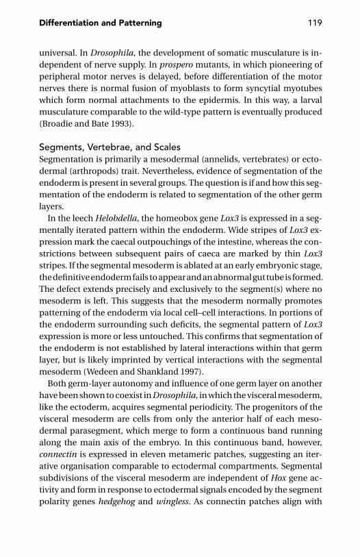
Differentiation and Patterning 119
universal. In Drosophila, the development of somatic musculature is in-dependent of nerve supply. In prospero mutants, in which pioneering ofperipheral motor nerves is delayed, before differentiation of the motornerves there is normal fusion of myoblasts to form syncytial myotubeswhich form normal attachments to the epidermis. In this way, a larvalmusculature comparable to the wild-type pattern is eventually produced(Broadie and Bate 1993).
Segments, Vertebrae, and ScalesSegmentation is primarily a mesodermal (annelids, vertebrates) or ecto-dermal (arthropods) trait. Nevertheless, evidence of segmentation of theendoderm is present in several groups. The question is if and how this seg-mentation of the endoderm is related to segmentation of the other germlayers.
In the leech Helobdella, the homeobox gene Lox3 is expressed in a seg-mentally iterated pattern within the endoderm. Wide stripes of Lox3 ex-pression mark the caecal outpouchings of the intestine, whereas the con-strictions between subsequent pairs of caeca are marked by thin Lox3stripes. If the segmental mesoderm is ablated at an early embryonic stage,the definitive endoderm fails to appear and an abnormal gut tube is formed.The defect extends precisely and exclusively to the segment(s) where nomesoderm is left. This suggests that the mesoderm normally promotespatterning of the endoderm via local cell–cell interactions. In portions ofthe endoderm surrounding such deficits, the segmental pattern of Lox3expression is more or less untouched. This confirms that segmentation ofthe endoderm is not established by lateral interactions within that germlayer, but is likely imprinted by vertical interactions with the segmentalmesoderm (Wedeen and Shankland 1997).
Both germ-layer autonomy and influence of one germ layer on anotherhave been shown to coexist in Drosophila, in which the visceral mesoderm,like the ectoderm, acquires segmental periodicity. The progenitors of thevisceral mesoderm are cells from only the anterior half of each meso-dermal parasegment, which merge to form a continuous band runningalong the main axis of the embryo. In this continuous band, however,connectin is expressed in eleven metameric patches, suggesting an iter-ative organisation comparable to ectodermal compartments. Segmentalsubdivisions of the visceral mesoderm are independent of Hox gene ac-tivity and form in response to ectodermal signals encoded by the segmentpolarity genes hedgehog and wingless. As connectin patches align with
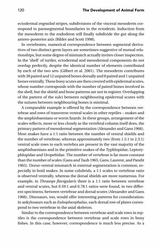
120 The Development of Animal Form
ectodermal engrailed stripes, subdivisions of the visceral mesoderm cor-respond to parasegmental boundaries in the ectoderm. Induction fromthe mesoderm to the endoderm will finally subdivide the gut along theantero-posterior axis (Bilder and Scott 1998).
In vertebrates, numerical correspondence between segmental deriva-tives of two distinct germ layers are sometimes suggestive of mutual rela-tionships, but some degree of mismatch actually invites closer inspection.In the ‘shell’ of turtles, ectodermal and mesodermal components do notoverlap perfectly, despite the identical number of elements contributedby each of the two sets (Gilbert et al. 2001). The mesoderm contributeswith 38 paired and 12 unpaired bones dorsally and 8 paired and 1 unpairedbones ventrally. These bony scutes are then covered with epidermal scutes,whose number corresponds with the number of paired bones involved inthe shell, but the shield and bone patterns are not in register. Overlappingof the pattern of the sulci between neighbouring epidermal scutes withthe sutures between neighbouring bones is minimal.
A comparable example is offered by the correspondence between ver-tebrae and rows of transverse ventral scales in other reptiles – snakes andthe amphisbaenians or worm lizards. In these groups, arrangement of thescales reflects, more or less closely as the vertebral column itself does, theprimary pattern of mesodermal segmentation (Alexander and Gans 1966).Most snakes have a 1:1 ratio between the number of ventral shields andthe number of vertebrae, whereas approximately two (from 1.5:1 to 2.3:1)ventral scale rows to each vertebra are present in the vast majority of theamphisbaenians and in the primitive snakes of the Typhlopidae, Leptoty-phlopidae and Uropeltidae. The number of vertebrae is far more constantthan the number of scales (Gans and Taub 1965, Gans, Laurent, and Pandit1965). Dorso-ventral mismatch in external segmentation is common, es-pecially in boid snakes. In some colubrids, a 1:1 scales to vertebrae ratiois observed ventrally, whereas the dorsal shields are more numerous. Forexample, in Thrasops flavigularis there is a 1:1 ratio between vertebraeand ventral scutes, but 0.59:1 and 0.78:1 ratios were found, in two differ-ent specimens, between vertebrae and dorsal scutes (Alexander and Gans1966). Dinosaurs, too, would offer interesting patterns for consideration:in ankylosaurs such as Euhoplocephalus, each dorsal row of plates corres-pond to two vertebrae in the axial skeleton.
Similar to the correspondence between vertebrae and scale rows in rep-tiles is the correspondence between vertebrae and scale rows in bonyfishes. In this case, however, correspondence is much less precise. As a

Differentiation and Patterning 121
rule, the number of vertebrae and the number of scales are both stabilisedearly in development. But at least in the case of the European minnow(Phoxinus phoxinus), an increase in scale count throughout life has beensuspected (Repa 1974). Apparently the spacing of the scales rows, whichbecome visible well after the pattern of myomeres has been established, isdictated by the spacing of the latter. In some cases, the number is secon-darily doubled by intercalation of new elements between two scale initialsof the first run (Lindsey 1988).
Guidelines to FollowOf all bilaterians, vertebrates are, by far, those in which cell migration hasthe most important role in morphogenesis. Extensive migrations, such asthose of the neural crest cells (Hall 1999), are virtually absent in inverte-brates. In the latter animals, most cells are formed in their final position.But there are exceptions. In Caenorhabditis elegans, migrations of someneuroblasts during larval life span the animal’s whole body length and areinfluenced by Hox gene expression (Salser and Kenyon 1994).
Cell migration may occur through different mechanisms (e.g., Levi,Duband and Thiery 1990, Hynes and Lander 1992): as migration of a wholecell sheet, as migration of isolated cells through an extracellular matrix oras migration of isolated cells over the cellular processes of another cell.The latter mechanism is especially relevant in the case of the developingnervous system. This is far from a trivial problem, as the growing axonsare seeking a target that also moves in the meantime (Jacob, Hacker, andGuthrie 2001). Growth cones are guided by four different mechanisms:contact attraction, chemoattraction, contact repulsion and chemorepul-sion (Tessier-Lavigne and Goodman 1996).
What guides motor neurons in their migration – extrinsic cues or the dif-ferent genes they express? The question is not that different from anotherquestion often raised in biology, whether the shape of a gall depends on(the genome of) the plant where it develops or on (the genome of) theinsect inducing its production. Both questions are actually ill-founded, asboth agents are jointly responsible for the outcome of their interaction,despite the contrary views of some authors.
As for motor neuron migrations, Sharma et al.’s (2000) experimentspoint to a major role of the genes expressed by the neurons themselves,rather than the matrix. As distinct motor neuron subtypes normally expressunique combinations of LIM-type homeodomain factors that are likelyinvolved in cell migration, axon navigation, or both, Sharma et al. forced

122 The Development of Animal Form
all motor neurons, irrespective of their potential subtype, to express ectopi-cally a LIM gene combination appropriate for the subgroup that normallyinnervates axial muscles. This genetic alteration was sufficient to convertall motor neurons to the cell migration behaviour of the axial subtype.Concordance with this type was also indicated by gene expression profileand the pattern of their axonal projections.
Genetic control of cell migration has been demonstrated in many cases.In Hydra, for instance, an Otx homologue plays a role in cell movementsleading to the establishment of a new body axis (Smith et al. 1999). In theDrosophila embryo, even-skipped and islet constitute a bimodal switchregulating axonal growth in such a way that motor axons are specificallydirected to ventral or dorsal regions of a muscle field (Landgraf et al. 1999).In Caenorhabditis elegans, the unc-129 and unc-6 netrin genes are requiredto guide pioneer motor axons along the dorso-ventral axis (Wadsworth andHedgecock 1996, Codavita et al. 1998). Genes controlling cell migrationmay exercise their effects by modifying the properties of the environmentthrough which cells will migrate. The product of the mig-17 gene in C. ele-gans, the metalloprotein MIG-17, directs migration of the distal tip cells ofthe two U-shaped arms of the gonad by remodelling the basement mem-brane (Nishiwaki, Hisamoto, and Matsumoto 2000).
To alter cell behaviour, the influence of the matrix does not need tobe molecularly specific. Chen et al. (1997) cultured human and bovinecapillary endothelial cells on micropatterned substrates that containedadhesive islands coated with extracellular matrix. Larger islands allowedcells to grow and expand, whereas smaller islands negatively influencedcell growth, and a very small island of extracellular matrix forced cell apop-tosis. Thus, cell shape decided whether individual cells grew or died. Butthis influence was the same, regardless of the type of matrix protein orantibody to integrin used to mediate adhesion.
In Drosophila, establishment of the final somatic muscle pattern de-pends on reciprocal signalling between the epidermal muscle attachmentcells and the approaching myotube. The first signals come from the epider-mal muscle attachment cells, inducing myotube attraction and adhesionto their target cells. As soon as they are attached to their target cell, themuscle cells send back a signal to the muscle attachment cells; this signalinduces their terminal differentiation into tendon-like cells (Becker et al.1997).
If, in most cases, the behaviour of migrating cells is the result of theirintrinsic specificities, mediated by interactions with the substrate and
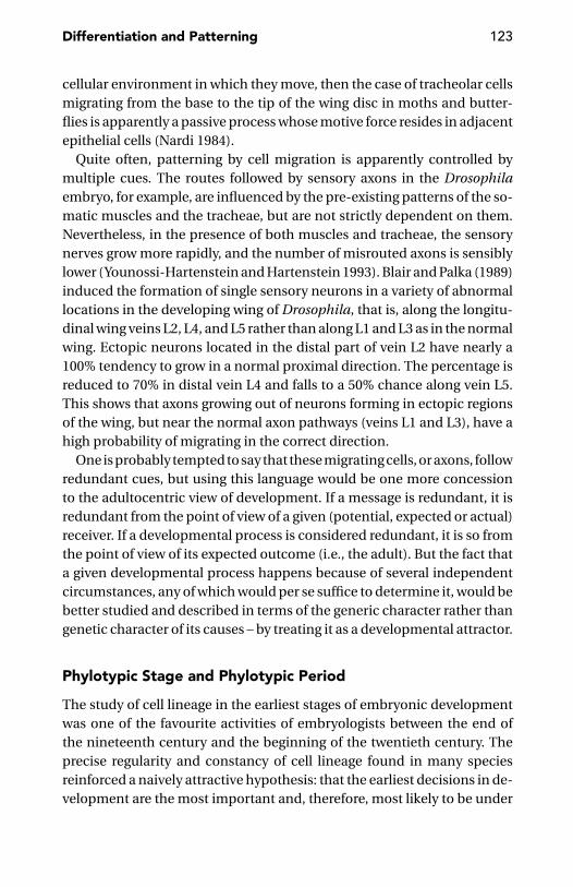
Differentiation and Patterning 123
cellular environment in which they move, then the case of tracheolar cellsmigrating from the base to the tip of the wing disc in moths and butter-flies is apparently a passive process whose motive force resides in adjacentepithelial cells (Nardi 1984).
Quite often, patterning by cell migration is apparently controlled bymultiple cues. The routes followed by sensory axons in the Drosophilaembryo, for example, are influenced by the pre-existing patterns of the so-matic muscles and the tracheae, but are not strictly dependent on them.Nevertheless, in the presence of both muscles and tracheae, the sensorynerves grow more rapidly, and the number of misrouted axons is sensiblylower (Younossi-Hartenstein and Hartenstein 1993). Blair and Palka (1989)induced the formation of single sensory neurons in a variety of abnormallocations in the developing wing of Drosophila, that is, along the longitu-dinal wing veins L2, L4, and L5 rather than along L1 and L3 as in the normalwing. Ectopic neurons located in the distal part of vein L2 have nearly a100% tendency to grow in a normal proximal direction. The percentage isreduced to 70% in distal vein L4 and falls to a 50% chance along vein L5.This shows that axons growing out of neurons forming in ectopic regionsof the wing, but near the normal axon pathways (veins L1 and L3), have ahigh probability of migrating in the correct direction.
One is probably tempted to say that these migrating cells, or axons, followredundant cues, but using this language would be one more concessionto the adultocentric view of development. If a message is redundant, it isredundant from the point of view of a given (potential, expected or actual)receiver. If a developmental process is considered redundant, it is so fromthe point of view of its expected outcome (i.e., the adult). But the fact thata given developmental process happens because of several independentcircumstances, any of which would per se suffice to determine it, would bebetter studied and described in terms of the generic character rather thangenetic character of its causes – by treating it as a developmental attractor.
Phylotypic Stage and Phylotypic Period
The study of cell lineage in the earliest stages of embryonic developmentwas one of the favourite activities of embryologists between the end ofthe nineteenth century and the beginning of the twentieth century. Theprecise regularity and constancy of cell lineage found in many speciesreinforced a naively attractive hypothesis: that the earliest decisions in de-velopment are the most important and, therefore, most likely to be under
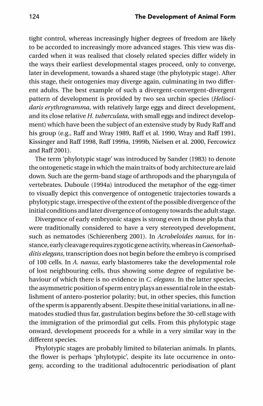
124 The Development of Animal Form
tight control, whereas increasingly higher degrees of freedom are likelyto be accorded to increasingly more advanced stages. This view was dis-carded when it was realised that closely related species differ widely inthe ways their earliest developmental stages proceed, only to converge,later in development, towards a shared stage (the phylotypic stage). Afterthis stage, their ontogenies may diverge again, culminating in two differ-ent adults. The best example of such a divergent-convergent-divergentpattern of development is provided by two sea urchin species (Helioci-daris erythrogramma, with relatively large eggs and direct development,and its close relative H. tuberculata, with small eggs and indirect develop-ment) which have been the subject of an extensive study by Rudy Raff andhis group (e.g., Raff and Wray 1989, Raff et al. 1990, Wray and Raff 1991,Kissinger and Raff 1998, Raff 1999a, 1999b, Nielsen et al. 2000, Fercowiczand Raff 2001).
The term ‘phylotypic stage’ was introduced by Sander (1983) to denotethe ontogenetic stage in which the main traits of body architecture are laiddown. Such are the germ-band stage of arthropods and the pharyngula ofvertebrates. Duboule (1994a) introduced the metaphor of the egg-timerto visually depict this convergence of ontogenetic trajectories towards aphylotypic stage, irrespective of the extent of the possible divergence of theinitial conditions and later divergence of ontogeny towards the adult stage.
Divergence of early embryonic stages is strong even in those phyla thatwere traditionally considered to have a very stereotyped development,such as nematodes (Schierenberg 2001). In Acrobeloides nanus, for in-stance, early cleavage requires zygotic gene activity, whereas in Caenorhab-ditis elegans, transcription does not begin before the embryo is comprisedof 100 cells. In A. nanus, early blastomeres take the developmental roleof lost neighbouring cells, thus showing some degree of regulative be-haviour of which there is no evidence in C. elegans. In the latter species,the asymmetric position of sperm entry plays an essential role in the estab-lishment of antero-posterior polarity; but, in other species, this functionof the sperm is apparently absent. Despite these initial variations, in all ne-matodes studied thus far, gastrulation begins before the 30-cell stage withthe immigration of the primordial gut cells. From this phylotypic stageonward, development proceeds for a while in a very similar way in thedifferent species.
Phylotypic stages are probably limited to bilaterian animals. In plants,the flower is perhaps ‘phylotypic’, despite its late occurrence in onto-geny, according to the traditional adultocentric periodisation of plant

Differentiation and Patterning 125
Figure 6.1. Polyps (a) and medusa (b) of the hydrozoan Turritopsis nutricula. Undercertain conditions, the medusa of this species may revert to polyp. (Courtesy ofS. Piraino, Lecce.)
development from the germinating seed to the maturing fruit; but for thevegetative structures, there is probably no phylotypic stage. In sponges, thelack of a phylotypic stage is suggested by the fact that a complete spongecan be reconstituted from dissociated cells. In cnidarians, by the phe-nomenon of transdifferentiation – illustrated by Bavestrello, Sommer, andSara (1992) and Piraino et al. (1996) in Turritopsis nutricula – the medusa,under certain conditions, can literally reverse its developmental history,by transforming back into a polyp (Figure 6.1). Two groups of bilaterians,

126 The Development of Animal Form
however, should be investigated closer from this point of view. One is bry-ozoans, in which freshwater forms rebuild their complex architecture fromvery simple multicellular resting stages, the statoblasts. The other is ne-merteans, in which large forms, such as Lineus, when severely starved,may regress to a microscopic cluster of cells from which a new worm islater produced. The integration of developmental processes that shouldcharacterise the phylotypic stage (Galis 1999) is a kind of morphogeneticratchet from which these animals seem to escape. Different is the case ofsome tunicates (e.g. the ascidian genus Botryllus and the thaliacean genusDoliolum) in which perfectly comparable ‘adults’ are built by embryoge-nesis or vegetative reproduction (blastogenesis; Manni et al. 1999). In thiscase, the question is not so much if a phylotypic stage exists, but ratherhow ‘generic’ this stage is, as it is not simply achieved through differentearly embryogenetic processes, but also from developmental processesother than embryogenesis.
This last case offers an opportunity to introduce regeneration. It has longbeen speculated that vegetative reproduction is not that different fromregeneration (e.g., Dehorne 1916, Berrill 1952, Herlant-Meewis 1953). Inmodern terms, this view has been reformulated as the hypothesis that fis-sion can be derived from regeneration processes recruited for a new rolein reproduction. This amounts to imagining (Bely and Wray 2001) thatparatomic fission in annelids is accomplished by initiating regenerationin the middle of an undamaged worm. Similarities between the two pro-cesses have been demonstrated in developmental genetic terms. In theannelid Pristina leidyi, for example, the role of Otx-class genes in pat-terning anterior body structures is largely the same in both contexts. InHydra, HyBra1, a homologue of the Brachyury gene, is associated with theformation of the hypostome (the apical region of the polyp) in differentdevelopmental situations, such as embryogenesis, the budding process,and the very earliest phases of regeneration (Technau and Bode 1999).
Sanchez Alvarado (2000) speculates that the genetic organisation oper-ating in the blastemas of simpler organisms during regeneration or asexualreproduction may have been co-opted in sexually reproductive animals toproduce novel embryonic structures, such as the limb bud.
An equivalence between limbs and buds is also implicit in Dewel’s (2000)suggestion that colonial cnidarians, such as the sea pens (Pennatulacea),may provide a blueprint for reconstructing the organisation of the first bila-terians, in which growth would have occurred along the primary axis (cf. asea pen’s primary polyp), as well as along lateral branches perpendicular to
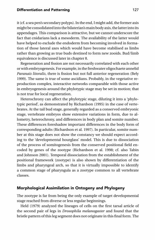
Differentiation and Patterning 127
it (cf. a sea pen’s secondary polyps). In the end, I might add, the former axismight be consolidated into the bilaterian’s main body axis, the latter into itsappendages. This comparison is attractive, but we cannot underscore thefact that cnidarians lack a mesoderm. The availability of the latter wouldhave helped to exclude the endoderm from becoming involved in forma-tion of those lateral axes which would have become stabilised as limbsrather than growing as true buds destined to form new zooids. Bud/limbequivalence is discussed later in chapter 8.
Regeneration and fission are not necessarily correlated with each otheror with embryogenesis. For example, in the freshwater oligochaete annelidParanais litoralis, there is fission but not full anterior regeneration (Bely1999). The same is true of some ascidians. Probably, in the vegetative re-production complex, interactive networks comparable with those activein embryogenesis around the phylotypic stage may be set in motion; thatis not true for local regeneration.
Heterochrony can affect the phylotypic stage, diluting it into a ‘phylo-typic period’, as demonstrated by Richardson (1995) in the case of verte-brates. At the tail bud stage, generally regarded as a conserved embryonicstage, vertebrate embryos show extensive variations in form, due to al-lometry, heterochrony, and differences in body plan and somite number.These differences foreshadow important differences in the body form ofcorresponding adults (Richardson et al. 1997). In particular, somite num-ber at this stage does not show the constancy we should expect accord-ing to the ‘developmental hourglass’ model. This is due to dissociationof the process of somitogenesis from the conserved positional field en-coded by genes of the zootype (Richardson et al. 1998; cf. also Tabinand Johnson 2001). Temporal dissociation from the establishment of thepositional framework (zootype) is also shown by differentiation of thelimbs and pharyngeal arch, so that it is virtually impossible to identifya common stage of pharyngula as a zootype common to all vertebrateclasses.
Morphological Assimilation in Ontogeny and Phylogeny
The zootype is far from being the only example of target developmentalstage reached from diverse or less regular beginnings.
Held (1979) analyzed the lineages of cells on the first tarsal article ofthe second pair of legs in Drosophila melanogaster and found that thebristle pattern of this leg segment does not originate in this final form. The

128 The Development of Animal Form
bristle cells of each row are arranged at first in a jagged line, which is laterstraightened by cell movements.
Remodelling of ‘draft organs’ during embryonic development is com-mon. An example is vasculature in vertebrates (Roman and Weinstein2000). What actually deserves our attention are those organs which areapparently built by bringing together pieces originally grown as physi-cally separated units. Cell biology has accustomed us to self-assembly,both intracellular (microtubuli, cytoskeleton) and intercellular (as exten-sively demonstrated by cell self-sorting within reaggregating mixtures ofdifferent kinds of cells). But a large leap separates these phenomena fromthe actual formation of organs from previously isolated precursors. This iswhat the individual components of the neural crest actually do (Hall 1999).
Another example is offered by the leech Helobdella. I have already des-cribed (page 38) the founding contribution of the progeny of seven pairsof different blast cells to the cell population of the leech’s 32 segments.This requires an alignment of the different teloblast lineages, but this isnot provided by a co-ordinated progression of the blast cells in generatingthe corresponding longitudinal bandelets. The latter acquires axial pat-terns independently and is later brought into alignment along the antero-posterior axis through a process of morphogenetic assembly (Nardelli-Haefliger, Bruce, and Shankland 1994).
Organs forming from bilaterally symmetrical anlagen, which for a whiledevelop independently of one another, are known from different phyla.In Drosophila, for example, the brain originates from two bilaterally sym-metrical neurogenic regions, which are initially separated from each otherand from the ventral nerve cord (Therianos et al. 1995). In Drosophila,again, the dorsal tracheal trunk is generated by fusion of adjacent tra-cheal metameres, following migration of a distinct subset of tracheal cells(the so-called dorsal trunk cells), under the influence of a transcriptionfactor encoded by the gene spalt (Kuhnlein and Schuh 1996). In verte-brates, as previously described, heart precursors originating at either sideof the embryo migrate towards the midline where they eventually fuse. Inzebrafish, this migration is controlled by the miles apart gene (Kuppermanet al. 2000).
If these morphogenetic events compare favourably with the phenomenaof self-assembly better known at a lower level of organisation, there are oth-ers for which the most obvious term of comparison is instead the semicon-servative duplication of DNA. This is what happens during blastogenesis(multiplication by budding) in the ascidian family Didemnidae. In these
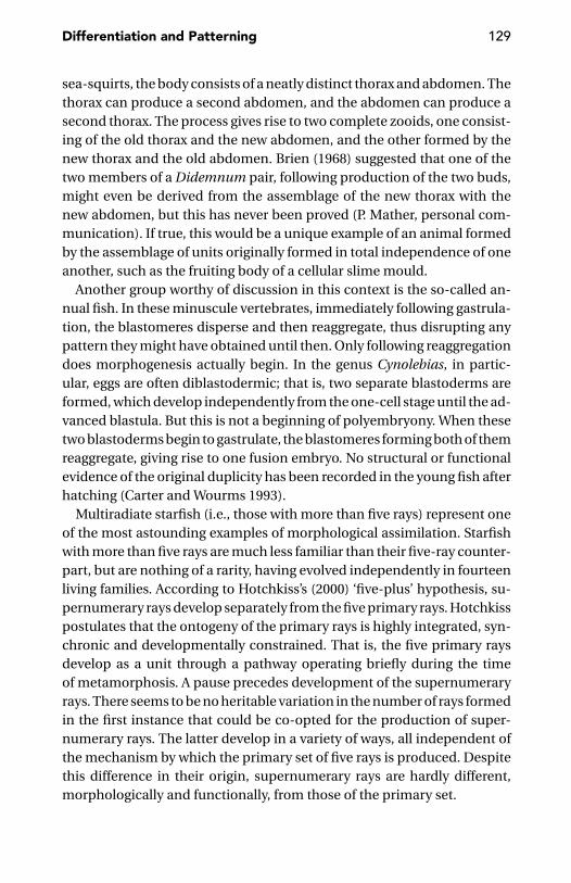
Differentiation and Patterning 129
sea-squirts, the body consists of a neatly distinct thorax and abdomen. Thethorax can produce a second abdomen, and the abdomen can produce asecond thorax. The process gives rise to two complete zooids, one consist-ing of the old thorax and the new abdomen, and the other formed by thenew thorax and the old abdomen. Brien (1968) suggested that one of thetwo members of a Didemnum pair, following production of the two buds,might even be derived from the assemblage of the new thorax with thenew abdomen, but this has never been proved (P. Mather, personal com-munication). If true, this would be a unique example of an animal formedby the assemblage of units originally formed in total independence of oneanother, such as the fruiting body of a cellular slime mould.
Another group worthy of discussion in this context is the so-called an-nual fish. In these minuscule vertebrates, immediately following gastrula-tion, the blastomeres disperse and then reaggregate, thus disrupting anypattern they might have obtained until then. Only following reaggregationdoes morphogenesis actually begin. In the genus Cynolebias, in partic-ular, eggs are often diblastodermic; that is, two separate blastoderms areformed, which develop independently from the one-cell stage until the ad-vanced blastula. But this is not a beginning of polyembryony. When thesetwo blastoderms begin to gastrulate, the blastomeres forming both of themreaggregate, giving rise to one fusion embryo. No structural or functionalevidence of the original duplicity has been recorded in the young fish afterhatching (Carter and Wourms 1993).
Multiradiate starfish (i.e., those with more than five rays) represent oneof the most astounding examples of morphological assimilation. Starfishwith more than five rays are much less familiar than their five-ray counter-part, but are nothing of a rarity, having evolved independently in fourteenliving families. According to Hotchkiss’s (2000) ‘five-plus’ hypothesis, su-pernumerary rays develop separately from the five primary rays. Hotchkisspostulates that the ontogeny of the primary rays is highly integrated, syn-chronic and developmentally constrained. That is, the five primary raysdevelop as a unit through a pathway operating briefly during the timeof metamorphosis. A pause precedes development of the supernumeraryrays. There seems to be no heritable variation in the number of rays formedin the first instance that could be co-opted for the production of super-numerary rays. The latter develop in a variety of ways, all independent ofthe mechanism by which the primary set of five rays is produced. Despitethis difference in their origin, supernumerary rays are hardly different,morphologically and functionally, from those of the primary set.

130 The Development of Animal Form
This is not that different from the mechanism by which some amniotes(e.g., moles and panda) generate supernumerary digits. Galis, van Alphen,and Metz (2001) prefer to call these appendages digit-like structures ratherthan extra digits, but it is also possible that early tetrapods used morethan one mode of digit development at the same time (Wagner, Chiu, andLaubichler 2000). This is a suggestion that the polydactylous appendagesof Acanthostega, with a set of larger and smaller digits (Coates and Clack1990), could support.
The homonomous vertebral column of snakes, the homodont teeth ofwhales and the distal part of the ichthyosaur fins – in which any distinctionbetween carpals, metacarpals and phalanxes is lost – are the result of a phy-logenetic reduction in the degree of differentiation among the elementsof a series, that is, a loss of modularity (Wagner 1996). Therefore, theseare examples of phylogenetic, rather than ontogenetic, assimilation. Thesame is also seen in the secondary homonomy of body segments in somecentipedes (the scolopendromorph Plutonium and the geophilomorphsgenerally) and in the vermiform larvae of several insects [e.g., many bitingmidges (Ceratopogonidae) and click beetles (Elateridae)].
A striking example of phylogenetic assimilation, quite likely dependenton homeotic co-option, is provided by the coelacanth (Latimeria chalum-nae), in which the dorsal and anal fins have secondarily acquired the skele-tal pattern previously evolved in the paired fins (Ahlberg 1992).
Patterning in Regeneration
Embryonic Patterning Versus Patterning in RegenerationMuch of the experimental work on animal regeneration has little or noth-ing to do with the animal’s real life. A recent review on regeneration ininsects (Marsh and Theisen 1999), for example, only deals with the regen-eration power of the imaginal discs in Drosophila, a hidden virtue thatthe fruit fly will never use to its own benefit. The fact that the power ofregeneration extends beyond the repair routines that the animal will likelyuse during its lifetime seems to justify Goss’s (1992) view of regenerationas an epiphenomenon of development – that is, something that is not aspecial, adaptively evolved phenomenon. Cells multiply, move or differ-entiate at their best, in the context of the cycling system of which they arepart. Regeneration, like the basic cellular properties from which it results,may proceed irrespective of its adaptive value.
The problem is that regeneration, much like illness and many otherphysiological or pathological conditions and processes (and what about

Differentiation and Patterning 131
to be a male or female?), is not homologous across the different groups (aview defended by Sanchez Alvarado 2000). It is simply a convenient termfor a class of events involving multicellular organisms. That regenerationis mainly ‘basic cell biology’ is shown by experiments demonstrating thatregeneration does not necessarily imply any memory of the original form.Experiments with reaggregated cells in the Hydra, which sorted out even-tually to form a new polyp, failed to show any preferential incorporation ofcells deriving from the apical region (‘head’ with tentacles) into the corre-sponding region of the newly formed polyp (Sato et al. 1992, Technau andHolstein 1992). In Botrylloides, a colonial sea squirt, any minute fragmentof a peripheral blood vessel containing a few blood cells isolated from anadult zooid may give rise to a complete organism with (the equivalent of)all three embryonic layers (Rinkevich, Shlemberg, and Fishelson 1995). Inthe flatworm Dugesia tigrina, the same set of patterning Hox genes is acti-vated in embryonic development and in regeneration, but with a differenttime schedule (Bayascas et al. 1998). In more complex organisms, suchas vertebrates, in which the regenerative power is primarily limited to theappendages (limbs and tail), regeneration may recapitulate normal devel-opment. In newts, for example, the regenerating limb blastema producesa zone of polarising activity, that is a signalling centre of antero-posteriorpatterning, as a normal developing limb would do (Imokawa and Yoshizato1997).
Terminal or Apical Control Versus RegenerationArthropods, nematodes and the other moulting animals do not regenerateany missing part of the main body axis. Itow’s (1986) claim to have obtainedsupernumerary segments in the horseshoe crab Tachypleus by applyinginhibitors of DNA synthesis cannot be used as an argument to disprovethe universality of this rule. The observed behaviour may simply be derivedfrom induced secondary subdivision of the animal’s primary segments.
That ecdysozoans do not regenerate parts of the main body axis doesnot mean that they lack regenerative powers completely. Collembolans,for example, regenerate all appendages without trouble and renovate thewhole midgut epithelium at each of their numerous moults (Schaller 1970).
In many cases, the simplest explanation for the lack of regeneration isthe lack of mitotic activity in the tissues, regions or stages in which theamputation occurred (Rockett and Woodring 1972). Ticks, for example,regenerate limbs completely; but in all other mite families, there is littleregeneration power or none at all. No regeneration, for example, is possi-ble in the tetranychids where, interestingly, there seems to be no mitotic
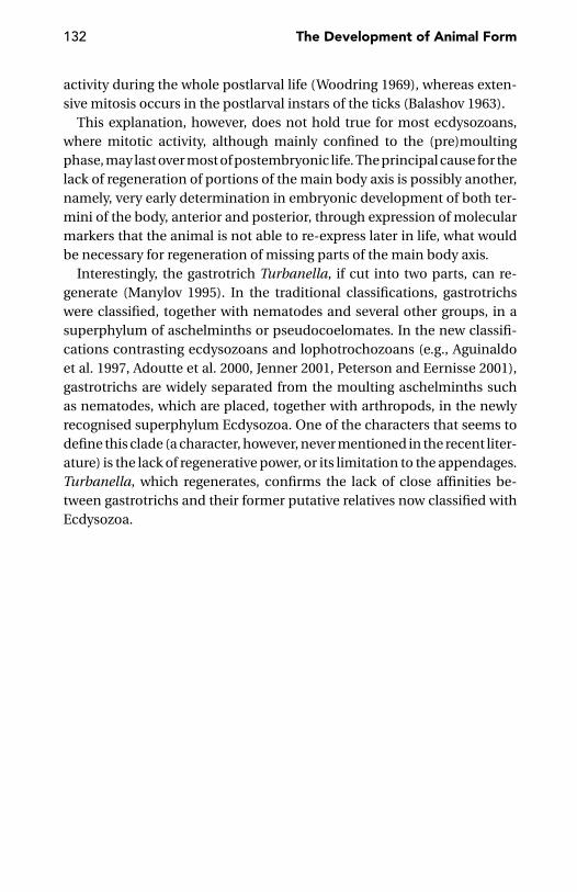
132 The Development of Animal Form
activity during the whole postlarval life (Woodring 1969), whereas exten-sive mitosis occurs in the postlarval instars of the ticks (Balashov 1963).
This explanation, however, does not hold true for most ecdysozoans,where mitotic activity, although mainly confined to the (pre)moultingphase, may last over most of postembryonic life. The principal cause for thelack of regeneration of portions of the main body axis is possibly another,namely, very early determination in embryonic development of both ter-mini of the body, anterior and posterior, through expression of molecularmarkers that the animal is not able to re-express later in life, what wouldbe necessary for regeneration of missing parts of the main body axis.
Interestingly, the gastrotrich Turbanella, if cut into two parts, can re-generate (Manylov 1995). In the traditional classifications, gastrotrichswere classified, together with nematodes and several other groups, in asuperphylum of aschelminths or pseudocoelomates. In the new classifi-cations contrasting ecdysozoans and lophotrochozoans (e.g., Aguinaldoet al. 1997, Adoutte et al. 2000, Jenner 2001, Peterson and Eernisse 2001),gastrotrichs are widely separated from the moulting aschelminths suchas nematodes, which are placed, together with arthropods, in the newlyrecognised superphylum Ecdysozoa. One of the characters that seems todefine this clade (a character, however, never mentioned in the recent liter-ature) is the lack of regenerative power, or its limitation to the appendages.Turbanella, which regenerates, confirms the lack of close affinities be-tween gastrotrichs and their former putative relatives now classified withEcdysozoa.
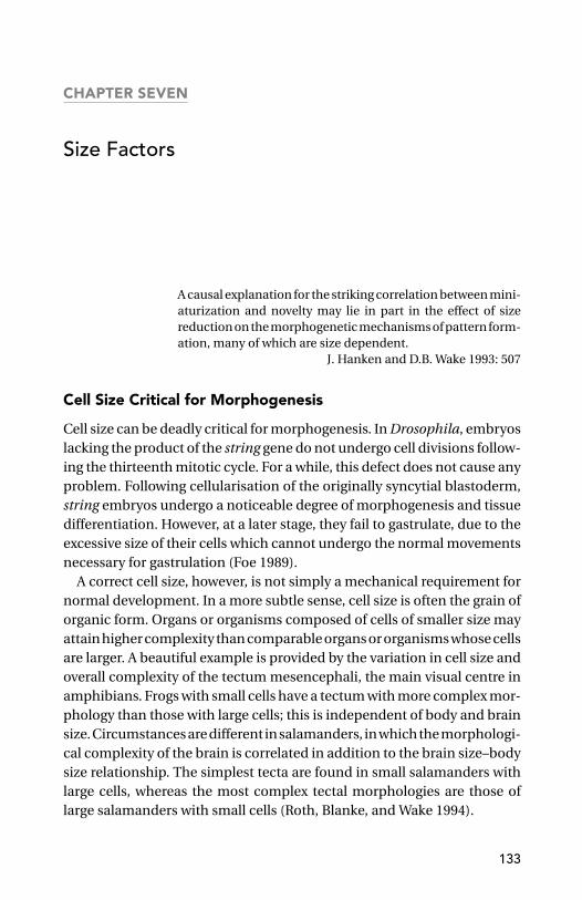
CHAPTER SEVEN
Size Factors
A causal explanation for the striking correlation between mini-aturization and novelty may lie in part in the effect of sizereduction on the morphogenetic mechanisms of pattern form-ation, many of which are size dependent.
J. Hanken and D.B. Wake 1993: 507
Cell Size Critical for Morphogenesis
Cell size can be deadly critical for morphogenesis. In Drosophila, embryoslacking the product of the string gene do not undergo cell divisions follow-ing the thirteenth mitotic cycle. For a while, this defect does not cause anyproblem. Following cellularisation of the originally syncytial blastoderm,string embryos undergo a noticeable degree of morphogenesis and tissuedifferentiation. However, at a later stage, they fail to gastrulate, due to theexcessive size of their cells which cannot undergo the normal movementsnecessary for gastrulation (Foe 1989).
A correct cell size, however, is not simply a mechanical requirement fornormal development. In a more subtle sense, cell size is often the grain oforganic form. Organs or organisms composed of cells of smaller size mayattain higher complexity than comparable organs or organisms whose cellsare larger. A beautiful example is provided by the variation in cell size andoverall complexity of the tectum mesencephali, the main visual centre inamphibians. Frogs with small cells have a tectum with more complex mor-phology than those with large cells; this is independent of body and brainsize. Circumstances are different in salamanders, in which the morphologi-cal complexity of the brain is correlated in addition to the brain size–bodysize relationship. The simplest tecta are found in small salamanders withlarge cells, whereas the most complex tectal morphologies are those oflarge salamanders with small cells (Roth, Blanke, and Wake 1994).
133

134 The Development of Animal Form
Figure 7.1. Habitus of a loricipheran. (Redrawn after Kristensen 1991.)
The extremely small cell size typical of the loriciferans – miniscule ma-rine invertebrates first described just twenty years ago (Kristensen 1983),which measure as adults at 300 µm, but are made of more than 10,000 cells(Kristensen 1991) – is a precondition for an exquisitely fine-grained mor-phology. The complexity these animals achieve despite their very smallsize is clearly seen in Figure 7.1. One is tempted to suggest that small cellsize has been critical in this respect; not so much that it allows carvingthe finest details of structure, but rather because of the strict limits a tinysize might impose on the total complexity of mRNA sequence in each cell,as found by Ernst et al. (1980) in sea urchin micromeres. Another groupof marine invertebrates of very small size, the appendicularians, have fol-lowed a completely different strategy. Their body is made of a very smallnumber of cells, some of which are of enormous size. Less than a dozencells, for example, are enough for lining their relatively robust stomach(Carlo Brena and Paolo Burighel, personal communication; Figure 7.2).
An inverse correlation between cell number and cell size, is far frombeing a universal rule. From the viewpoint of morphogenesis, there arecases such as the chico mutant in Drosophila, which is less than half thesize of a wild-type fly, owing to fewer but also smaller cells. For example, inthe case of the wing area, the reduction in cell number accounts for 68%,
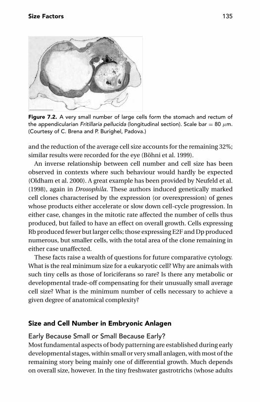
Size Factors 135
Figure 7.2. A very small number of large cells form the stomach and rectum ofthe appendicularian Fritillaria pellucida (longitudinal section). Scale bar = 80 µm.(Courtesy of C. Brena and P. Burighel, Padova.)
and the reduction of the average cell size accounts for the remaining 32%;similar results were recorded for the eye (Bohni et al. 1999).
An inverse relationship between cell number and cell size has beenobserved in contexts where such behaviour would hardly be expected(Oldham et al. 2000). A great example has been provided by Neufeld et al.(1998), again in Drosophila. These authors induced genetically markedcell clones characterised by the expression (or overexpression) of geneswhose products either accelerate or slow down cell-cycle progression. Ineither case, changes in the mitotic rate affected the number of cells thusproduced, but failed to have an effect on overall growth. Cells expressingRb produced fewer but larger cells; those expressing E2F and Dp producednumerous, but smaller cells, with the total area of the clone remaining ineither case unaffected.
These facts raise a wealth of questions for future comparative cytology.What is the real minimum size for a eukaryotic cell? Why are animals withsuch tiny cells as those of loriciferans so rare? Is there any metabolic ordevelopmental trade-off compensating for their unusually small averagecell size? What is the minimum number of cells necessary to achieve agiven degree of anatomical complexity?
Size and Cell Number in Embryonic Anlagen
Early Because Small or Small Because Early?Most fundamental aspects of body patterning are established during earlydevelopmental stages, within small or very small anlagen, with most of theremaining story being mainly one of differential growth. Much dependson overall size, however. In the tiny freshwater gastrotrichs (whose adults

136 The Development of Animal Form
are between 70 and 1,500 µm long), all mitoses are apparently completedby the time the juvenile hatches from the egg (but some mitoses happenpostembryonically in the marine forms of the same group).
The segmental organisation of the Drosophila embryo is generated thefirst five hours after fertilisation (Akam 1987). Dipterans go through em-bryonic development at an unusually speedy pace, but the very early com-pletion of the basic body architecture of the Drosophila embryo is but anexample of the very general rule that the most important decisions inshaping an animal are made very early in development.
Even apparently trifling details, such as distribution of sensory setae onthe body surface of mites, are apparently made very early in embryonicdevelopment (van der Hammen 1988).
It has been believed (e.g., Cooke 1980) that, in vertebrate embryos, manyimportant decisions about body architecture are taken not so early as inother animals (e.g., nematodes or insects); but some recent studies suggestotherwise. In the mouse, anterior identity is probably established beforegastrulation, as it is first manifested in extraembryonic tissue which isessential for normal development of anterior structures, such as the fore-brain (Beddington and Robertson 1998). Izpisua-Belmonte et al. (1993)found that the chick homeobox gene goosecoid (gsc) is expressed in a smallcell population near the posterior margin (Koller’s sickle) of the unincu-bated egg, long before its expression can be detected in Hensen’s node(the so-called chick organiser). Transplantation experiments suggest thatgsc-expressing cells have inducing activity even before Hensen’s node isdifferentiated; therefore, the development of the chick organiser startsearlier than previously thought.
Still, in vertebrates, the proximo-distal pattern of the limb is also likely setup very early in development (Hartmann and Tabin 2001). What has beentraditionally described as the developmental production of this proximo-distal pattern is simply a later elaboration of an already existing pattern,brought about by cell proliferation. This is demonstrated by the behaviourof the lipophilic dye in which cells at the limb bud tip (the so-called progresszone) were labelled at the early stages. The dye remained in the labelledcellular population rather than becoming distributed among cells with adifferent fate, as would have occurred if cells at the distal tip of the limb werecontinually respecified as limb outgrowth proceeds, as previously thought.
Further results supporting an early determination of the whole limbpattern in tetrapods – possibly within a cell layer only a few hundred µmthick – have been recently published by Sun, Mariani, and Martin (2002)and by Dudley, Ross, and Tabin (2002).
Size Factors 137
It is no surprise that directional asymmetry is often established at thevery beginning of embryonic development, no matter if the situs viscerumof a vertebrate or the direction of shell coiling in a mollusc is involved(Palmer 1996). But this extends to many other fundamental events, suchas those establishing polarity of the main body axis or differentiation ofthe organiser.
A question arises as to what the critical factor is: either the size of theanlage, and the number of cells in it, or the relative developmental timeper se. The question is similar to one of the basic issues in the analysisof allometry (e.g., Fairbairn 1992): is selection acting on shape as such, orthrough its allometric dependence on a selectively affected size parameter?
Davidson (2001) believes that the basic developmental strategy of mostbilaterians is to divide the egg into polyclonal lineages of differentiatingcells at the earliest possible time. This fits well within the traditional adul-tocentric view of development. I think, however, that size and cell numberare the relevant factors. There are cases in which important decisions aredelayed, relative to embryogenesis, but this still happens within small cir-cumscribed clusters of set-aside cells, such as the adult primordium in thesea urchin larva or the imaginal discs in the larva of the holometabolous in-sects. There are obvious mechanistic reasons for limiting decisions aboutpatterning within small cell clusters. One of the reasons is the limitedrange of action of the morphogens, which is in the order of 10 (Gurdonet al. 1994) or at most 30 cell diameters or less than 1 mm in the maxi-mum linear dimension (Wolpert 1989). In sea urchin larvae, Hox gene ex-pression is limited to the somatocoel, when this is only 2–300 µm across(Arenas-Mena, Cameron, and Davidson 2000). Consequently, I think thatthe widespread occurrence of most patterning events at early stages indevelopment is a simple by-product of the fact that early developmentalstages are small and rapidly get to comprise a suitable number of cells;not the other way around. In the same vein one could suggest that the seg-mentation of very large eggs is partial not because of mechanical problemsbut mainly because a dialogue between very large blastomeres would bedifficult, due to the limited range of action of morphogens.
Critical Number of Cells in Embryonic AnlagenToo low a cell number would clearly limit the chance of establishing asufficient number of subunits with different patterns of gene expressionand different developmental fate. In appendicularians, in which the wholedigestive tract is made of very few cells, the stomach and rectum aremorphologically distinct, but the histochemical properties of their cells
138 The Development of Animal Form
are unusually similar (Carlo Brena and Paolo Burighel, personal commu-nication).
In the imaginal discs of Drosophila, Distal-less expression (Beermanet al. 2001) and competence to differentiate adult structures (Minder andNothiger 1973) require a minimum of previous cell divisions in the disc.Following Minder and Nothiger’s suggestion, Kurushima and Ohtari (1975)investigated the behaviour of wing discs in the silkworm (Bombyx) andfound that these acquire competence to pupal-type behaviour when thenumber of cells is four times as large as after the third ecdysis. In theirinterpretation, the acquisition of competence to develop into the pupaltype may parallel the increase in cell number.
In vertebrates, adult long bone size are somehow dependent on thenumber of mitotically active cells in the original condensation. In turn,this number can often be related to the timing of the condensation pro-cess (Moss-Salentijn 1974, Kember 1978). If condensation is reduced belowa critical threshold, skeletogenesis may not even begin (Hall and Miyake2000). Goodwin and Trainor (1983) developed a physico-mathematicalmodel for the possible dependence on the size (i.e., cell number) of thelimb primordium of the number of cartilage condensations which will giverise to carpal/tarsal, metacarpal/metatarsal and finger/toe bone elements.This model aimed to prove the global nature of the limb field, hence theinapplicability of one-to-one correspondences between tetrapods limbswith different number of digits. Subsequent studies of limb developmenthave rejected applicability of Goodwin and Trainor’s model to the gener-ality of tetrapods. In some cases it works. This is suggested, for example,by the fact that extra digits are known to occur occasionally in St. Bernardsand other big dogs, but not in small size poodles (Alberch 1985).
Cell number at a critical stage may influence overall adult size. Bjorklund(1996) analysed growth in two closely related species of birds: the great tit(Parus major) and the blue tit (P. caeruleus). The former species is largerthan the latter in all external traits, but this difference is not due to dif-ferences in growth rate and neither to differences in the time at whichthe offset of growth occurs. Size differences are already manifested uponhatching and remain so throughout ontogeny.
In principle, intraspecific and interspecific differences in organ or bodysize depend on differences in the number of cells, their size or both. Mam-mal data summarised by Stevenson, Hill, and Bryant (1995) point to amajor role of cell number rather than cell size differences. These data in-clude measurement of cells in the liver, thyroid, and renal epithelia and
Size Factors 139
red blood cells in mammal species spanning a big range in body size.Things, however, are different in Drosophila. Stevenson et al. (1995) – bycomparing organ and cell allometry in the wing, eye and basitarsus of sev-eral Hawaiian Drosophila – found that cell size may contribute betweenone-third and two-thirds to evolutionary changes in organ and body size.This problem was also investigated experimentally. Partridge et al. (1994)studied replicated lines of Drosophila that had been maintained for fiveyears at 25◦C or at 16.5◦C. Those kept at the lower temperature showedhigher thorax length and wing area. The evolutionary effect of tempera-ture on wing area was entirely a consequence of an increase in cell area,with a small effect on cell number in males only.
A minimum cell number is also required for regeneration. A square flatsheet excised from a Hydra magnopapillata polyp gradually rounds up,turning into a hollow spherical ‘shell’ with a continuous ectodermal layeroutside and a continuous endodermal layer inside. This occurs, however,provided that the fragment is large enough. The smallest spherical shellthat could be produced contained 300 epithelial cells (Shimizu, Sawada,and Sugiyama 1993).
Miniaturisation
Miniaturisation and Body PatterningMiniaturisation may affect the expression or even the presence of a givenbody feature. Bateson (1894) wondered what the least size might be inwhich a given tooth can be present in a species which sometimes has itand sometimes not. His data were detailed enough to show that the leastsize of a tooth is different for different teeth and for different animals. Someminimum seems to exist for any body feature.
Before causing the total disappearance of a given feature, miniaturisa-tion sometimes causes a loss of regularity. An example is the asymmetry inthe distribution of the usually symmetrically arranged slit sensory organsin the tiny spider Comaroma bertkaui, just 1.6 mm long (Kropf 1998). Asimilar loss of symmetry does not occur in the dorsal bristle patterns ofmites, even in those species, which are thousands, that are much smallerthan Comaroma. One wonders if this difference only correlates with the re-cent miniaturisation of the spider, as opposed to an extremely long historyof the mite lineage as one of very small arthropods. In other words, mitesmight have long since adjusted their bristle patterning mechanisms totheir tiny size, whereas miniaturisation of Comaroma is probably a recent
140 The Development of Animal Form
event whose developmental consequences have not yet been assimilatedwell enough.
Similarly, the very small body size (1 mm total length) may explain thepeculiar distribution of the setae on the body surface (the terga especially)of the larvae of some tiny scydmaenid beetles (e.g., Cephennium and itsrelatives). Instead of the setal pattern usually found in beetle larvae, with afew pairs of regularly spaced setae per segment (cf. page 36), in these scy-dmaenids the entire dorsal surface is nearly uniformly covered by a largenumber of very coarsely patterned microscopic setae. This is counterintu-itive – one of the tiniest beetles having larvae with much more abundantsetae than its larger relatives – but this may depend on the impossibility ofthe usual spacing signals between setae-producing cells (lateral inhibitioneffects?) to take place within too small a field.
Miniaturisation may have far-reaching consequences on an animal’sbiology (cf. Rensch 1959). Some body features (e.g., the nervous system) donot seem to be amenable to any arbitrary reduction in size. Consequently,there is less relative (not just absolute!) space for the reproductive organsthan in a larger animal. In the female, this effect may be hard to accept,unless a suitable reproductive strategy is adopted. If there is no chanceof producing a large number of eggs, as the total amount of cell materialavailable for reproduction is very limited and the individual eggs cannotbe arbitrarily small as other cell types could be, miniaturised females mayadopt a k reproductive strategy – that is to produce very few but very largeeggs, thus maximising the chance of individual survival of the offspring.This explains one more apparent paradox: why many miniaturised animalsproduce larger eggs than their non-miniaturised relatives.
Miniaturisation, Segments, and CellsIn both annelids and arthropods, miniaturisation is often (but not univer-sally) coupled to a reduction in the number of body segments (oligomeri-sation). Examples among the annelids include the enchytraeid oligochaeteMarionina eleonorae (16 segments, the lowest number in the family) andsome ‘polychaetes’, such as Parergodrilus (8–9 segments), Dinodrilus(0.4 mm total length, ca. 9 indistinct body segments) and the Nerillidae(0.3–2 mm total length, 7–9 segments). It may not be by chance that thesegment number 8, or its double, is ‘nodal’ in these miniaturised annelids.
In centipedes, segment number is invariable in the Lithobiomorpha,irrespective of size (up to 45 mm total length in Eupolybothrus, but only3 mm in Catanopsobius), but it is often prone to oligomerisation in the

Size Factors 141
smallest representatives of the Geophilomorpha. A good example is pro-vided by the genus Schendylops, which includes species in the 6–70 mmrange of total length and 27–87 leg-bearing segments. The lowest segmentnumbers (27 or 29 in the males, 29 or 31 in the females) are found in the tinySchendylops oligopus, which is also the smallest member of the genus. In-terestingly, oligomerisation of very small geophilomorphs occurs in cladesin which the number of segments is intraspecifically and interspecificallyvariable; but it does not occur in a clade (family Mecistocephalidae) inwhich intraspecific variability is virtually absent and interspecific differ-ences are also very limited. In this group, a very small species, such asNannarrup hoffmani (10 mm in length), has the same number (41) of leg-bearing segments as its closest relatives that are up to 75 mm long (Anarrupsp.; Foddai et al. 2002).
One may wonder if originally segmented animals undergo such a degreeof miniaturisation that segmentation is actually impossible or useless. Thisis possibly true of the pseudosegmentation of many collembolans andsome groups of mites, such as the eriophyids, whose total length may beno more than 100 µm. A similar consequence of small body size is perhapsthe incomplete articulation (pseudoarthrosis) of some appendages: thetarsus in the Monotarsobius centipedes (Verhoeff 1902–25) and the labialpalp in the tiny rove beetle Atlantostiba franzi (Pace 1994).
This indiscernibility in miniaturised animals of otherwise clearly artic-ulated features raises a question of comparative method. In some of them,it may be impossible to identify the homologues of individual features thatare distinct in their non-miniaturised relatives. In these dwarfs, there mightnot be an amount of tissue, or a number of cells, large enough to supportspecification of some of the developmental (and morphological) units.
The number of cells available in specific anlagen may explain the reduc-tion or even the complete atrophy of some organs in miniaturised animals(Hanken and Wake 1993). The cells of the minute marine snail Caecumglabrum, only 1 mm long, are of approximately the same size as thoseof large marine snails such as the edible winkle Littorina littorea (Goetze1938). We do not expect that the reduction in body size be accompaniedby a corresponding reduction in average cell size, at least when miniatur-isation is a phylogenetically recent event involving one or a few speciesin a lineage. Circumstances may be different in taxa with a long historyof miniaturisation, as in the exceptional case of Loricifera, described pre-viously, in which a dramatic reduction in cell size is the most obviouscorrelate of a very reduced body size.
CHAPTER EIGHT
Axes and Symmetries
The essential unity of the phenomenon of Repetition of Partsand of its companion-phenomenon, symmetry, wherever metwith, has been too little recognized, and needless difficulty hasthus been introduced into morphology.
W. Bateson 1894: 21
[Co-option] is the fundamental process by which evolutionarychange in bilaterian form has occurred.
E.H. Davidson 2001:164
The Animal’s Main Body Axis
Zoologists and lay persons alike would not hesitate in identifying the mainbody axis in animals such as an earthworm, a leech, a grasshopper, a fish,or a bird. However, difficulties will appear if one is asked to point to themain body axis of a sea star, a sea squirt, or a hydra.
What is an animal’s main body axis? It is the longitudinal axis uniting theanimal’s fore and rear ends. The fore end coincides with the animal’s head,where the mouth, brain, eyes, and other important sensory organs are lo-cated. The rear end, to the contrary, is reasonably identified with the siteof the animal’s anus and, perhaps, excretory and genital openings. In turn,the anterior position of both mouth and eyes, and the posterior positionof the anus, correspond to the animal’s polarity with respect to locomo-tion. The animal explores the environment with the aid of the anteriorsensory organs and is ready to use its anterior mouth to exploit any fooditems its sensory organs might discover. As for the faeces, it is convenientto leave them behind in such a way as to minimise further contact withthem. Corresponding advantages may be derived from a posterior location
142
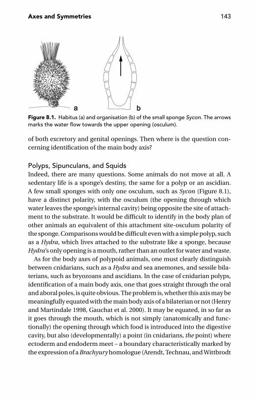
Axes and Symmetries 143
Figure 8.1. Habitus (a) and organisation (b) of the small sponge Sycon. The arrowsmarks the water flow towards the upper opening (osculum).
of both excretory and genital openings. Then where is the question con-cerning identification of the main body axis?
Polyps, Sipunculans, and SquidsIndeed, there are many questions. Some animals do not move at all. Asedentary life is a sponge’s destiny, the same for a polyp or an ascidian.A few small sponges with only one osculum, such as Sycon (Figure 8.1),have a distinct polarity, with the osculum (the opening through whichwater leaves the sponge’s internal cavity) being opposite the site of attach-ment to the substrate. It would be difficult to identify in the body plan ofother animals an equivalent of this attachment site-osculum polarity ofthe sponge. Comparisons would be difficult even with a simple polyp, suchas a Hydra, which lives attached to the substrate like a sponge, becauseHydra’s only opening is a mouth, rather than an outlet for water and waste.
As for the body axes of polypoid animals, one must clearly distinguishbetween cnidarians, such as a Hydra and sea anemones, and sessile bila-terians, such as bryozoans and ascidians. In the case of cnidarian polyps,identification of a main body axis, one that goes straight through the oraland aboral poles, is quite obvious. The problem is, whether this axis may bemeaningfully equated with the main body axis of a bilaterian or not (Henryand Martindale 1998, Gauchat et al. 2000). It may be equated, in so far asit goes through the mouth, which is not simply (anatomically and func-tionally) the opening through which food is introduced into the digestivecavity, but also (developmentally) a point (in cnidarians, the point) whereectoderm and endoderm meet – a boundary characteristically marked bythe expression of a Brachyury homologue (Arendt, Technau, and Wittbrodt
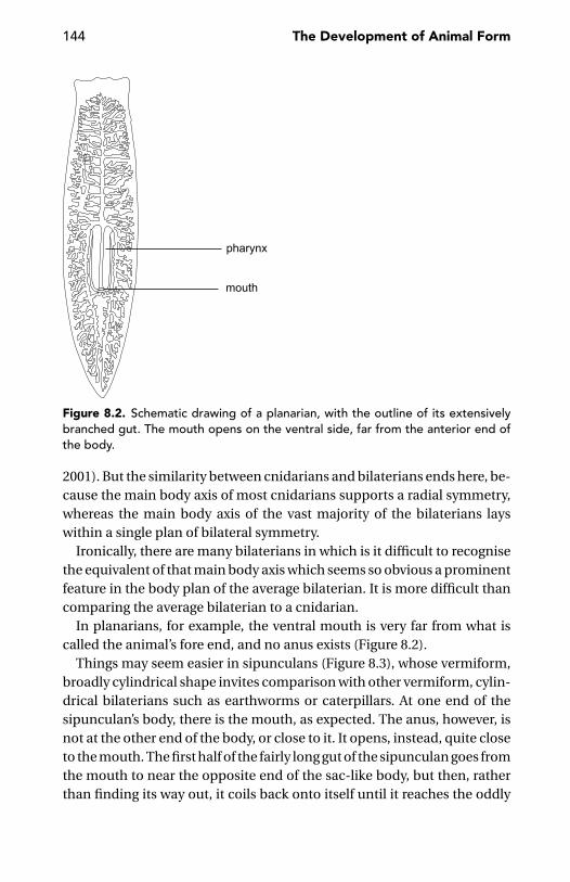
144 The Development of Animal Form
Figure 8.2. Schematic drawing of a planarian, with the outline of its extensivelybranched gut. The mouth opens on the ventral side, far from the anterior end ofthe body.
2001). But the similarity between cnidarians and bilaterians ends here, be-cause the main body axis of most cnidarians supports a radial symmetry,whereas the main body axis of the vast majority of the bilaterians layswithin a single plan of bilateral symmetry.
Ironically, there are many bilaterians in which is it difficult to recognisethe equivalent of that main body axis which seems so obvious a prominentfeature in the body plan of the average bilaterian. It is more difficult thancomparing the average bilaterian to a cnidarian.
In planarians, for example, the ventral mouth is very far from what iscalled the animal’s fore end, and no anus exists (Figure 8.2).
Things may seem easier in sipunculans (Figure 8.3), whose vermiform,broadly cylindrical shape invites comparison with other vermiform, cylin-drical bilaterians such as earthworms or caterpillars. At one end of thesipunculan’s body, there is the mouth, as expected. The anus, however, isnot at the other end of the body, or close to it. It opens, instead, quite closeto the mouth. The first half of the fairly long gut of the sipunculan goes fromthe mouth to near the opposite end of the sac-like body, but then, ratherthan finding its way out, it coils back onto itself until it reaches the oddly
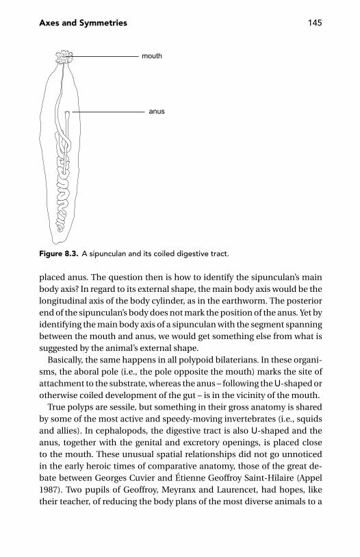
Axes and Symmetries 145
Figure 8.3. A sipunculan and its coiled digestive tract.
placed anus. The question then is how to identify the sipunculan’s mainbody axis? In regard to its external shape, the main body axis would be thelongitudinal axis of the body cylinder, as in the earthworm. The posteriorend of the sipunculan’s body does not mark the position of the anus. Yet byidentifying the main body axis of a sipunculan with the segment spanningbetween the mouth and anus, we would get something else from what issuggested by the animal’s external shape.
Basically, the same happens in all polypoid bilaterians. In these organi-sms, the aboral pole (i.e., the pole opposite the mouth) marks the site ofattachment to the substrate, whereas the anus – following the U-shaped orotherwise coiled development of the gut – is in the vicinity of the mouth.
True polyps are sessile, but something in their gross anatomy is sharedby some of the most active and speedy-moving invertebrates (i.e., squidsand allies). In cephalopods, the digestive tract is also U-shaped and theanus, together with the genital and excretory openings, is placed closeto the mouth. These unusual spatial relationships did not go unnoticedin the early heroic times of comparative anatomy, those of the great de-bate between Georges Cuvier and Etienne Geoffroy Saint-Hilaire (Appel1987). Two pupils of Geoffroy, Meyranx and Laurencet, had hopes, liketheir teacher, of reducing the body plans of the most diverse animals to a

146 The Development of Animal Form
common scheme in which the dorsal side of the vertebrates was equatedwith the ventral side of the arthropods. Briefly, Meyranx and Laurencetsuggested that to compare squids with vertebrates, you just have to imag-ine the straight body of the latter bent onto itself by dorsally turning theposterior half forward. In this way, the anus is brought to the same levelas the mouth, a comparison that Cuvier (1830), not surprisingly, outrightrejected. The comparison suggested by Meyranx and Laurencet still de-serves attention – not literally, of course. But the standard textbook sce-nario we offer to our students to explain the evolution of cephalopods froma monoplacophoran-like ancestor retains, from their courageous specu-lations, much more than we could readily admit. We are accustomed tosay that the functional (external) antero-posterior body axis of a squid isderived from the original dorso-ventral axis of primitive molluscs, whereasthe former antero-posterior axis is now reduced to the short span betweenthe mouth and the opening of the mantle cavity, where the anus is placed.This is a correct or, at least sensible, suggestion. This suggestion impliesuncoupling of the functional polarity of the body, corresponding to thelong axis of the squid’s cartilaginous pen and the anatomical polarity sug-gested by the positions occupied by the mouth and anus. I will argue thatthis dichotomy, no less than the inverted dorsal/ventral relationships invertebrates and arthropods, is probably intrinsic to the animal’s develop-mental rules, rather than to our descriptive frames.
Less extreme positional relationships, but still somewhat embarrassingwhen we try to define the animal’s main body axis, are much more frequentthan one might suppose – take for example the leeches (Figure 8.4). Fol-lowing the external shape, the rear end of the body is given by the posteriorsucker, because the anus opens dorsally, just before the sucker (an obvi-ously good position, in functional terms). Determining where the mainbody axis actually terminates is complicated by the fact that the posteriorsucker is formed by segments XXVII–XXXII, whereas the anus, despite itsactual position, would originally occur at the end of the last (XXXII) bodysegment.
There are problems for the vertebrates as well. Not so much in the caseof a bird, whose external ‘tail’, made of long feathers, does not obviouslybelong to the anatomical main body axis, but in the case of all vertebratesprovided with a true tail, be it neatly separated from the trunk as in alltailed mammals, or less so as in fishes and snakes. The gut does not runthrough a vertebrate tail, which is therefore wholly posterior to the anus.An obvious alternative to regarding the tail as a part of a vertebrate’s mainbody axis is to regard it as an appendage (see page 156).
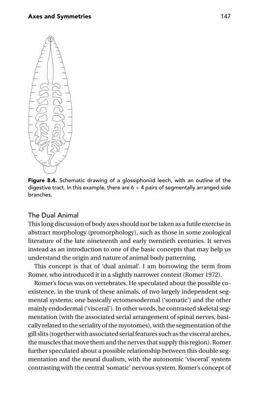
Axes and Symmetries 147
Figure 8.4. Schematic drawing of a glossiphoniid leech, with an outline of thedigestive tract. In this example, there are 6 + 4 pairs of segmentally arranged sidebranches.
The Dual AnimalThis long discussion of body axes should not be taken as a futile exercise inabstract morphology (promorphology), such as those in some zoologicalliterature of the late nineteenth and early twentieth centuries. It servesinstead as an introduction to one of the basic concepts that may help usunderstand the origin and nature of animal body patterning.
This concept is that of ‘dual animal’. I am borrowing the term fromRomer, who introduced it in a slightly narrower context (Romer 1972).
Romer’s focus was on vertebrates. He speculated about the possible co-existence, in the trunk of these animals, of two largely independent seg-mental systems: one basically ectomesodermal (‘somatic’) and the othermainly endodermal (‘visceral’). In other words, he contrasted skeletal seg-mentation (with the associated serial arrangement of spinal nerves, basi-cally related to the seriality of the myotomes), with the segmentation of thegill slits (together with associated serial features such as the visceral arches,the muscles that move them and the nerves that supply this region). Romerfurther speculated about a possible relationship between this double seg-mentation and the neural dualism, with the autonomic ‘visceral’ systemcontrasting with the central ‘somatic’ nervous system. Romer’s concept of

148 The Development of Animal Form
dual animal went practically unnoticed in mainstream zoological litera-ture, except for Jefferies (1986).
I will generalise this notion of ‘dual animal’ to contrast the somatic ec-tomesodermal system with the visceral endodermal system of triploblasticanimals generally. The reason for stressing this contrast is the large inde-pendence these two systems show in patterning. Mutual relationships be-tween the two ‘animals’ are obviously expected and are positively known,but the dialogue between the two does not obscure the large independenceof which either benefits in development and patterning.
The cases of cephalopods, sipunculans, and polypoid bilaterians areremarkable because they are conspicuous and far from exceptional. Thinkof flatworms such as the common freshwater planarians, with their mouthopening on the ventral side, about mid-length (with respect to the ‘somaticanimal’), and one of the three main intestinal branches extending forwardsin front of the mouth. Think of gastropods, which are worthy of a criticalreappraisal of the relative contributions of each of the two ‘animals’ to thetorsion of the visceral sac.
The functional independence of which an earthworm’s gut takes advan-tage in respect to the external epithelio-muscular sheath is an expressionof the visceral/somatic dualism of bilaterians. It is not by chance, that inleeches, where the coelomic cavities are reduced and the gut is surroundedby a solid mesodermal mass, rather than by a thin peritoneal cell layer, thedialogue between the somatic and visceral animal is much more intensethan in the case of earthworms. This has been documented experimen-tally (Wedeen 1995, Wedeen and Shankland 1997), but we do not needto look for gene expression patterns. The presence in many leech fami-lies of metamerically arranged gut coeca (Figure 8.4) is macroscopic proofof the patterning influence exercised by the segmentally arranged meso-derm on a ‘segmentally naive’ endoderm. Among the possible effects ofa strict dialogue between ectomesoderm and endoderm are also the so-called gastropore (a secondary external opening of the digestive tract) ofGastrostomobdella and other leeches (Sawyer 1986), the genito-intestinalchannel of some flatworms (Hyman 1951) and the branchial pharynx ofenteropneusts and chordates.
Buss (1987) was probably correct when suggesting that gastrulation (pro-cess by which a first separation of germ layers is accomplished) first de-veloped in early metazoans as a consequence of the divergence betweenan external lineage of ciliated undividing cells and an internal lineage ofnonciliated cells that retained the potential to undergo mitosis. No wonder

Axes and Symmetries 149
that such a primary distinction, one of survival value for a multicellularorganism otherwise at risk of mutiny from any of its own cell lineages,fell under the control of maternal determinants to the point that the twoprimary cell layers (the ectoderm and the endoderm) can be said to bepredetermined in the egg (Hall 1998b). What matters here, in the per-spective of the developing body architecture, is that a large degree of in-dependence in the morphogenetic role of ectodermal versus endodermalderivatives continued to be advantageous, even to animals whose embryosbecame very different from those early multicellular systems in whichsomething comparable with gastrulation first evolved. One wonders, inthis context, whether one of the advantages provided by the third germlayer (the mesoderm), when it first appeared in a developing animal, wasperhaps that of furthering and supporting the morphogenetic indepen-dence of ectodermal and mesodermal derivatives, even if, in due course,it becomes a third, largely autonomous player or, otherwise, a media-tor of ectodermal–endodermal interactions. But let us return to our dualanimal.
Many animals, especially those living on plant food, need a long digestivetract, but there might be constraints on their external shape (in particular,on their overall length), such that the gut cannot simply run straight fromthe mouth to the anus (if the anus lies opposite the mouth), parallel tothe main body axis of the ‘somatic animal’. A coiled gut is the obvioussolution – one adopted by animals as diverse as vertebrates, insects, andmolluscs. This is a solution that does not easily suggest the fundamentalindependence of the two systems, visceral and somatic, as shown insteadby sipunculans and polypoid animals. But the difference between theselatter animals and a vertebrate, insect or mollusc with a coiled gut is justmatter of degree. We must accept that the functional interdependenceof endodermal and ectomesodermal derivatives does not imply a strictinterdependence in morphogenesis.
Sipunculans and ParaHox GenesOne might speculate whether this duality is mirrored by the phylogeneticold splitting of a proto-Hox gene complex into two paralogous complexes:the Para-Hox genes and the Hox genes in the strict sense (Kourakis andMartindale 2000). Whereas the Hox complex seems to have evolved inassociation with the patterning of some component of the ectomesoder-mal ‘somatic animal’, there are suggestions as to a primary role of theParaHox complex in the patterning of the endodermal ‘visceral animal’
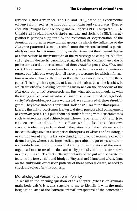
150 The Development of Animal Form
(Brooke, Garcıa-Fernandez, and Holland 1998),based on experimentalevidence from leeches, arthropods, amphioxus and vertebrates (Dupreyet al. 1988, Wright, Schnegelsberg and De Robertis 1989, Calleja et al. 1996,Offield et al. 1996, Brooke, Garcıa-Fernandez, and Holland 1998). This sug-gestion is perhaps supported by the reduction or ‘degeneration’ of theParaHox complex in some animal groups in which the influence of theHox gene-patterned ‘somatic animal’ onto the ‘visceral animal’ is partic-ularly evident. In this sense, I think, we shall interpret the different degreeof conservation or diversification of the ParaHox gene complex in differ-ent phyla. Phylogenetic parsimony suggests that the common ancestor ofprotostomes and deuterostomes had three ParaHox genes (Gsx, Xlox, andCdx). Three ParaHox genes have been cloned from a variety of deuteros-tomes, but (with one exception) all those protostomes for which informa-tion is available have either one or the other, or two at most, of the threegenes. This might be expected at least in animals such as the leeches, inwhich we observe a strong patterning influence on the endoderm of theHox gene-patterned ectomesoderm. But what about sipunculans, withtheir long gut freely coiling onto itself in the tissue vacuum of the large bodycavity? We should expect these worms to have conserved all three ParaHoxgenes. They have, indeed. Ferrier and Holland (2001a) found that sipuncu-lans are the only protostomes known to date to possess a full complementof ParaHox genes. This puts them on similar footing with deuterostomessuch as vertebrates and echinoderms, where the patterning of the gut [see,e.g., sea urchins and holothurians; Figure 8.5 (but also think of our ownviscera)] is obviously independent of the patterning of the body surface. Ininsects, the digestive tract comprises three parts, of which the first (foregutor stomodaeum) and the last one (hindgut or proctodaeum) are of ecto-dermal origin, whereas the intermediate part (the midgut or mesenteron)is of endodermal origin. Interestingly, for an interpretation of the insectorganisation in terms of the dual animal hypothesis, mutations are knownin Drosophila which affects left-right polarity of the gut with separate ef-fects on the fore-, mid-, and hindgut (Hayashi and Murakami 2001). Dataon the embryonic expression patterns of these genes is clearly needed tocheck the value of my hypothesis.
Morphological Versus Functional PolarityTo return to the opening question of this chapter (What is an animal’smain body axis?), it seems sensible to me to identify it with the mainlongitudinal axis of the ‘somatic animal’, irrespective of the concordant
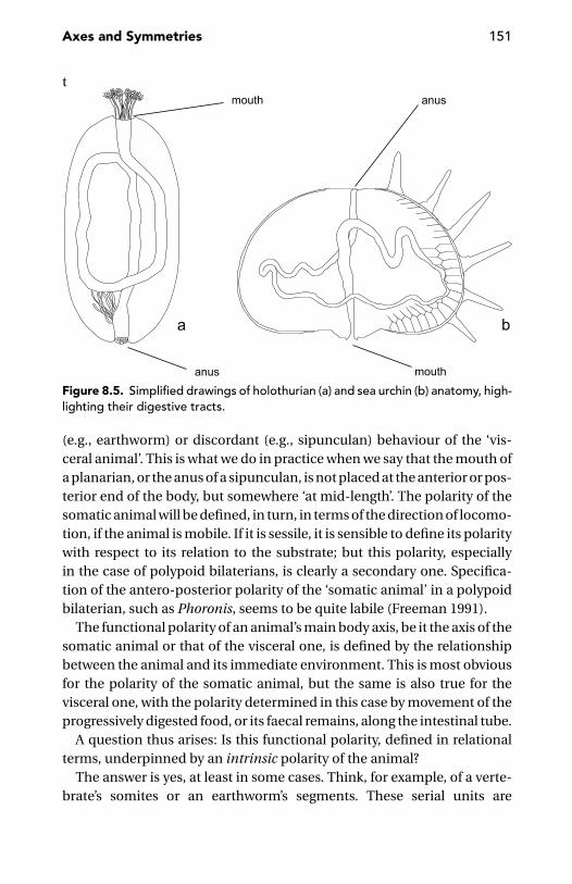
Axes and Symmetries 151
t
Figure 8.5. Simplified drawings of holothurian (a) and sea urchin (b) anatomy, high-lighting their digestive tracts.
(e.g., earthworm) or discordant (e.g., sipunculan) behaviour of the ‘vis-ceral animal’. This is what we do in practice when we say that the mouth ofa planarian, or the anus of a sipunculan, is not placed at the anterior or pos-terior end of the body, but somewhere ‘at mid-length’. The polarity of thesomatic animal will be defined, in turn, in terms of the direction of locomo-tion, if the animal is mobile. If it is sessile, it is sensible to define its polaritywith respect to its relation to the substrate; but this polarity, especiallyin the case of polypoid bilaterians, is clearly a secondary one. Specifica-tion of the antero-posterior polarity of the ‘somatic animal’ in a polypoidbilaterian, such as Phoronis, seems to be quite labile (Freeman 1991).
The functional polarity of an animal’s main body axis, be it the axis of thesomatic animal or that of the visceral one, is defined by the relationshipbetween the animal and its immediate environment. This is most obviousfor the polarity of the somatic animal, but the same is also true for thevisceral one, with the polarity determined in this case by movement of theprogressively digested food, or its faecal remains, along the intestinal tube.
A question thus arises: Is this functional polarity, defined in relationalterms, underpinned by an intrinsic polarity of the animal?
The answer is yes, at least in some cases. Think, for example, of a verte-brate’s somites or an earthworm’s segments. These serial units are
152 The Development of Animal Form
produced one after the other, in a progression that strictly follows theconventional antero-posterior axis of the ‘somatic animal’. In this case,the polarity-specifying reference does not reside in the interactions withthe external world, but flows directly from the progression of the animal’sown developmental time. Time’s arrow, in a sense, directly translates intospatial polarity.
There are many other ways in which time factors may determine spatialpolarity. Developmentally ‘higher’ regions (i.e., regions from which signalsare sent to developmentally ‘lower’ regions) may be those with longer cellcycles, in which the longest primary transcripts of genes full of long intronscan be produced – something forbidden to cells with too short cell cycles(Rothe et al. 1992).
Cartesian Axes, or Not
Cartesian co-ordinates dominate animal morphology and, to a large ex-tent, developmental biology as well. This attitude is quite naive and un-satisfactory, even for bilaterians, not to speak of radially symmetricalcnidarians.
One of the major problems with the conventional descriptions is repre-sented by the third axis that would complement the two less controversialones (i.e., the antero-posterior and the dorso-ventral ones). Most peoplewould recognise, as a third element, a left-right axis; but it is easy to showhow this is incorrect. Antero-posterior and dorso-ventral axes are unidi-rectional, whereas the so-called left-right axis is bidirectional. The first twoaxes identify the single plan of symmetry, which divides the animal intotwo mirror halves. If we wish to single out a third Cartesian axis, we needto consider only one-half of the animal without its mirror duplicate. In thisway, we discover the ‘inside-outside axis’, which proceeds out of the mir-ror symmetry plan perpendicular to it (Coen 1999). In no case is this axisstrictly equivalent to others. However, along this axis, there is no need tospecify positional values as it is along the other two axes. Eventual specifi-cation of the two semi-axes (midline-to-left and midline-to-right), givingrise to directional asymmetry, is something other than axis specificationor patterning. I will return to this point later on in this chapter.
My unease with the traditional Cartesian descriptions in terms of thethree axes of the body (or of an appendage) is not restricted to the lack ofequivalence between these axes. More generally, I believe that these aremuch more figments of our imagination than biological reality. In some

Axes and Symmetries 153
instances, these axes are not functionally separate in their roles, and cel-lular addresses and their interpretations cannot be so easily schematisedas usually assumed (Newman 1996).
In the proximo-distal patterning of the vertebrate limb, several HoxAand HoxD genes are involved jointly. However, the progressively restrictedexpression domains of progressively more 5′ genes of the two classes trans-late into progressively posterior-distal regions of the developing limbs(Gardiner et al. 1998), rather than into ‘pure’ antero-posterior or proximo-distal values. This continues into later limb bud stages, where skeletalelements are differentiating proximally and limb differentiation is aboutto commence distally; the expression of Hoxd11–13 moves towards moreproximal and anterior positions at the same time (Nelson et al. 1996). Ageneral multiaxial patterning system was suggested by Dolle et al. (1993),following the observation that mice mutants for the last gene in the HoxDcomplex (Hoxd13) display skeletal alteration affecting all ‘Cartesian’ bodyaxes.
In Drosophila, we have proof of the simultaneous involvement of onegene in the patterning of the antero-posterior axis and the dorso-ventralaxis (Munn and Steward 1995). This depends on events during mid-oogenesis, when the nucleus of the oocyte, then in a posterior positionwithin the ovariole, sends a chemical signal (the product of the gurkengene) to the nearest follicle cells causing them to assume a posterior iden-tity. At a later stage, the oocyte nucleus, now positioned at the anteriorend of the oocyte, sends a second Gurken signal to the follicle cells, whichthen acquire a dorsal identity. These inductive events are mediated by theproduct of another gene, spindle. The final polarity of the oocyte and theembryo is thus influenced by the asymmetries in the surrounding folliclecells (Anderson 1995, Gonzalez-Reyes, Elliott, and St. Johnston 1995, 1997).It is possible that gurken is not directly responsible for the establishmentof cell fates along a body axis, but restricts and orients a later axis-formingprocess (Roth and Schupbach 1994).
A role in the regulation of growth or patterning along more than oneaxis has also been postulated, in Drosophila, for genes involved in signalintegration, such as vestigial (Kim et al. 1996).
Similar to what happens along the main body axis, a multiaxial pattern-ing system is also at work along the appendages of Drosophila (Lecuit andCohen 1997).
Living organisms, or their parts, are sometimes described with referenceto co-ordinate systems other than Cartesian. Polar co-ordinate systems, in

154 The Development of Animal Form
particular, have often been applied in describing the organisation of theciliate cell (e.g., Frankel 1989) or the fate map of insect imaginal discs (e.g.,French, Bryant, and Bryant 1976, Bryant 1993, Couso, Bate, and Martınez-Arias 1993). Readers may be reassured about the meaningfulness of polarco-ordinate systems when reading about genes that might be involvedin their specification. In Drosophila, wingless is initially associated in theembryo with a pattern of stripes along the antero-posterior axis that fitswell into a Cartesian co-ordinate system; but later, during developmentof the imaginal discs, expression of the same gene is associated with apattern of sectors establishing a polar co-ordinate system (Couso, Bate,and Martınez-Arias 1993). Again, in Drosophila, Wilkins and Gubb (1991)proposed that, in the imaginal discs, segment polarity genes might specifythe angular component of the polar co-ordinate system. Held (1993) testedthis hypothesis by predicting that mutations in segment-polarity genesshould cause abnormal patterning within precise sectors of the imaginaldiscs. This was accurately confirmed by the defects (deletion of specificrows of chemosensory bristles or an abnormal increase in their number)found in specific sectors of the first tarsal segment of the second pair oflegs in segment-polarity mutants.
Cartesian geometry still dominates animal morphology, despite the factthat a straight antero-posterior axis is a poor reference when describing theanatomy of snails, squids or bryozoans. One may be skeptical before thedeformed reference grids used by D’Arcy Thompson (1942) or by modernmorphometricians (e.g., Bookstein 1991) to help comparisons betweenbody outlines of more or less strictly related animals; but the capriciouscurves in these grids may be less arbitrary than straight lines crossing atright angles, with the precision worthy of a geometry textbook.
Straight-jacketing different animals into a universal geometrical frame-work may suggest non-existing correspondences. For example, Hidalgo(1998) compared the embryonic expression of engrailed in Drosophila tothat of En-1, one of his vertebrate homologues. Whereas the former gene isexpressed in transversal stripes marking the anterior/posterior compart-ment boundary, the latter is expressed along a longitudinal stripe runningthe length of the embryo. In Cartesian terms, this can be described as ifthe expression of En-1 in vertebrates is shifted by 90◦, compared with itsequivalent in Drosophila. But what is really meant by this?
Completely different from these geometrical tricks is Geoffroy Saint-Hilaire’s (1822) old hypothesis, according to which the dorsal side of avertebrate is equivalent to the ventral side of an arthropod. Soon after
Axes and Symmetries 155
Arendt and Nubler-Jung (1994) revived this hypothesis in terms of molec-ular developmental genetics, extensive experimental data (e.g., Holley etal. 1995, Biehs, Francois and Bier 1996, De Robertis and Sasai 1996) pro-vided support for this view by demonstrating the phylogenetic conservedfunction of the Drosophila short gastrulation and decapentaplegic genesand the homologous Xenopus chordin and Bone Morphogenetic Protein-4(BMP-4) genes in subdividing the primitive embryonic ectoderm into neu-ral versus non-neural domains – but at opposite sides (dorsal and ventral)in the two kinds of animals. In most bilaterians, the initial dorso-ventral po-larity of the body is established in the ectodermal derivatives only, throughthe interaction of proteins from the neural side with morphogens (BMP-like) diffusing from the opposite side; this is as true of Drosophila as it isof amphioxus (Holland and Holland 1999). In the vertebrates, a parallelBMP-based system has evolved in the mesoderm (Graff 1997, Hemmati-Brivanlou and Melton 1997), so that dorso-ventral polarity is establishedin both germ layers simultaneously.
The Syntax of the Body
Despite the sheer diversity of body plans evolved in the different animallineages, there are some invariant features, in their body architecture,which go far beyond the obvious requirements of functional design. Forexample, why did no fish species evolve paired fins at a trunk level posteriorto the anus (Coates and Cohn 1999)?
Another generally overlooked feature of animal body syntax is that, inhermaphrodites and in gonochoric animals, where male and female geni-tal openings do not occur in identical position, the female gonopore orthe female gonad is nearly always anterior to the male gonopore or gonad(clitellate annelids seem to be an exception to this rule). In this contextis the organisation of the genital imaginal disc of Drosophila, which givesrise to the genital and anal structures of the adult fly. This disc is multi-segmental, in that it comprises cells from three abdominal segments: A8,corresponding to the primordia of the female genitalia; A9, correspondingto the primordia of the male genitalia; and A10, corresponding to the pri-mordia of anal structures (Casares et al. 1997). This multisegmental organi-zation preserves the anterior position of the female with respect to the malegenitalia.
There is no key to explain all these hard-wired points of animal anatomy.I hope that reviewing them in the context of this book may attract attention

156 The Development of Animal Form
to these overlooked features, which have perhaps a developmental, ratherthan an adaptive, explanation.
To answer these kinds of puzzles, we need not only the practical toolsof molecular developmental genetics, but also the theoretical tools of up-dated comparative morphology. Some of these concepts have already beendiscussed in these pages (e.g., the distinction between a somatic and a vis-ceral animal), and other concepts (such as axis paramorphism and doublesegmentation) will be introduced later.
Most important in the analysis of body syntax is distinguishing, as muchas possible, between appendages and main body axis.
How this distinction applies to echinoderms is not an easy question(e.g., Hotchkiss 1998, Popodi and Raff 2001). The problem of identifyingthe antero-posterior axis of these animals has been recently attacked byPeterson, Arenas-Mena, and Davidson (2000), using different lines of evi-dence, including palaeontology, comparative skeletal anatomy and theexpression pattern of a posterior class Hox gene in the coelomic cavitiesof the adult primordium. In their interpretation, the antero-posterior axisruns from the mouth through the adult coelomic compartments. This andother considerations lead these authors to the conclusion that the five raysof these animals are not primary (equivalent) body axes as some zoologistshad speculated, but outgrowths of the one main body axis. But what aboutthe tail of a vertebrate? Is it an appendage or part of the main body axis?
What Is a Tail?In vertebrate comparative anatomy, there has been a long controversy onwhether the tail forms by a developmental mechanism distinct from gas-trulation, which is responsible for the formation of the trunk. The verdictseems to be still out (Kanki and Ho 1997). Nevertheless, the large majorityof vertebrate zoologists would not hesitate to regard the tail as the poste-rior part of the animal’s main body axis. I am not ready to subscribe to thistraditional view.
A first argument is, that the tail, like the paired limbs of vertebrates, isonly made of ectodermal and mesodermal derivatives. No endodermalcomponent extends into it.
A second argument is that, in frogs, treatment with retinoic acid canswitch the identity of a tail blastema so that it gives rise to limbs rather thanto a new tail (Mohanty-Hejmadi, Dutta, and Mahapatra 1992, Maden 1993,Brockes 1997). This suggests that the tail, like the limbs, is an appendagerather than a part of the main body axis.

Axes and Symmetries 157
Extrapolating from this experiment straight to the deep phylogenetichistory of the chordates is perhaps too hazardous, but there was a seg-mented tail prolonging a short unsegmented body in Romer’s (1972) pu-tative vertebrate ancestor. This hypothetical animal (Romer’s ‘somatico-visceral animal’) is not that different from the tadpole larva of a sea squirt,or from one of those Palaeozoic calcichordates that Jefferies (1986) inter-pretes as ancestors of the vertebrates.
The most conspicuous component of a segmented tail is, of course,a segmented musculature, whose functional advantages do not need bestressed. One might easily conceive that the segmental musculature ofRomer’s hypothetical vertebrate ancestor was accompanied by segmen-tally arranged nerves. This might have been the case. In the tadpole larvastage of the ascidian Halocynthia roretzi, Wada, Holland and Satoh (1996)found a spatially iterative expression (15 ‘segmental’ spots) of a gene(HrPax-37) homologous to two vertebrate genes (Pax-3 and Pax-7) whichfunction in the differentiation of the dorsal neural tube. Also relevant inthis context is Crowther and Whittaker’s (1994) finding of serial repetitionof pairs of cilia along the epidermis of the tail of another ascidian larva(Ciona intestinalis).
In metazoans with dorsal neural cord, such as the chordates, it wouldbe easy to extend the neural axis of the trunk straight into the tail (i.e., intoa posterior appendage obviously dorsal to the anal opening). This topo-graphical circumstance places the tail in a privileged condition, relative toboth the lateral appendages and the posterior appendage of animals withventral neural cord, such as the telson of arthropods.
It is easy to think that a neural continuity between trunk and tail facil-itated morphological assimilation between trunk and tail. If vertebratesare derived from a tunicate-like ancestor (perhaps from neotenous formscomparable with the tadpole larva of present-day ascidians), then this as-similation implied the forward extension of the notochord from the tailto the trunk. This event paved the way to the development of the skeletal(vertebral) axis which evolved to replace the notochord, both morphologi-cally and functionally. On the other hand, the tail-to-trunk assimilationdid not proceed so far as to cancel the tail’s appendicular nature, as the taildid not acquire any endodermal derivative, including coelomic pouches(chordates are enterocoelic) and the viscera associated with them. Butthere was, apparently, no difficulty in extending to the trunk the segmentalcharacter originally associated with the musculature and the nervous sys-tem of the tail.

158 The Development of Animal Form
An indirect argument from developmental genetics potentially support-ing this hypothesis of morphological assimilation is that no Hox gene hasan anterior boundary of expression in the tail (Prince et al. 1998).
One more argument in favour of the appendicular nature of the verte-brate tail is the structural plasticity it may exhibit during postembryoniclife. Salamanders may add tail vertebrae continuously through life, as ob-served by Noble (1931) in the plethodontid Batrachoseps attenuatus, inwhich a young, but completely formed, specimen with a snout-to-ventlength of 2.3 cm had 28 caudals and an adult with a snout-to-vent lengthof 4.75 cm had 61 caudals; the number of dorsal vertebrae was 22 in both.The reverse phenomenon (reduction and fusion of tail vertebrae) is typicalof metamorphosing frogs.
It is thus possible to answer Coates and Cohn’s question: no fish evolvedfins caudal to the anus because the posterior body region, the tail, is simply(or mainly!) an appendage, like the fins themselves.
The Time Arrow of Growth and Differentiation
Along the main body axis, morphogenesis usually proceeds in an antero-posterior direction, along the appendages in a proximo-distal direction.This is the way vertebrates produce somites and annelids produce seg-ments. This is also the way teloblasts produce their rows of progeny cells.We could even say, following Dolle et al.’s (1993) reinterpretation ofWolpert’s (1969, 1989) concept of positional information, that the antero-posterior axis of the trunk and the proximo-distal axis of the appendagesare spatiotemporal axes defined by the progression of growth and pattern-ing. The notion of the Hox code (Kessel and Gruss 1990) has clearly playeda major role in the recent interpretations of this space-time correspon-dence. As all broad generalizations, however, this time-space correlationin morphogenesis has its exceptions.
First, the posterior area, or group of cells, from which new body parts areprogressively formed, is often not strictly terminal, but subterminal. Theposterior terminus is often one of the first body features to be specified,and this is important, especially if the animal will engage in active life evenbefore its main body axis is completed. This is the case, for example withcrustacean nauplii and the early postembryonic stages of millipedes andanamorphic centipedes (Lithobius, Scutigera).
Second, embryologists have long since identified, in some animal groups,nonterminal differentiation centres from which differentiation proceeds
Axes and Symmetries 159
both ways (i.e., in a rostral, as well as in a caudal, direction). This has beenalso seen, more recently, in the temporal changes of the expression pat-terns of some ‘developmental genes’, such as engrailed in the grasshopperSchistocerca (Patel, Kornberg, and Goodman 1989).
The Beginnings of Animal PolaritySymmetry-breaking events that establish the antero-posterior and/ordorso-ventral polarity of a developing animal may occur during oogenesis,during fertilization or later.
In Drosophila, as described previously, the two main axes of the body areestablished during oogenesis, when the oocyte comes to lie posterior tothe nurse cells and signals through the Gurken/Egfr pathway to induce theadjacent follicle cells to adopt a posterior fate. This directs the movementof the egg nucleus and associated gurken mRNA from a posterior to ananterior corner of the oocyte, where Gurken signals for a second timeinduce the dorsal follicle cells, thereby polarising the dorso-ventral axis(Gonzalez-Reyes, Elliott, and St. Johnston 1997, Micklem et al. 1997).
In Caenorhabditis elegans, as in many other animals, the primary spatialcue for antero-posterior polarity is derived from microtubules emanatingfrom the sperm asters (Wallenfang and Seydoux 2000).
Early specifications are not necessarily definitive. In the nemertean Cer-ebratulus lacteus, the dorso-ventral axis is set up prior to the first cleavagedivision, but blastomeres isolated at the two-cell stage will regulate toform normal, although miniature, pilidium larvae (Henry and Martindale1997). In this case, polarity is obviously established through extensive cel-lular interactions. One is reminded of the complete repatterning of the eggmaterial in polyembryonic wasps (page 115) and diblastodermic annualfish (page 129).
For a long time it has been believed (see Gurdon 1992) that, in the mam-malian embryo, establishment of the primary body axes is much delayedwith respect to the other animals and may even happen after implantation.However, a correlation has been demonstrated recently between the asym-metry of the fertilised oocyte, as defined by the position of the second polarbody and the entry point of the sperm, and the orientation of the animal-vegetal and embryonic-abembryonic axes in the preimplantation blasto-cyst. In turn, the animal-vegetal axis of the blastocyst does possibly corre-spond to the antero-posterior axis of the future gastrula (Tam et al. 2001).
Conventional descriptions following a Cartesian frame of mind acknowl-edge that the first axis to be formed is not always the same, more

160 The Development of Animal Form
Figure 8.6. A tiny adult tapeworm (Echinococcus granulosus). In the conventionalinterpretation of tapeworm polarity, with the scolex as the worm’s fore end, thetestes – within each proglottis – are anterior to the ovaries.
frequently the antero-posterior one, but sometimes (as in mammals) thedorso-ventral one. In any case, the first axis of the embryo is formed in verydifferent ways in different species (e.g., Gurdon 1992, L.Z. Holland 2000),following what in a first instance are probably generic cues, such as thesperm entrance point, or gravity, as in the chicken (Eyal-Giladi 1969).
Tapeworm PolarityGrowth and differentiation are sometimes intercalary rather than (sub) ter-minal. In some instances, this circumstance may even obscure the ‘real’antero-posterior polarity of the animal, as in the case of the tapeworms.All modern textbooks describe these animals as provided with an anteriorscolex and a posterior chain of proglottids (Figure 8.6), but is this descrip-tion really correct? Tapeworm polarity was debated in several papers at thebeginning of the twentieth century. Directionality in locomotion – the mostobvious test of antero-posterior polarity for a bilaterian – does not applyhere, due to the sessile habit of tapeworms. It is the same for the polarityof the ‘inner animal’, as tapeworms lack a mouth and digestive tract. Moreuseful is the position of the growth zone. Comparative arguments wouldsuggest that this is posterior (subterminal) rather than anterior (immedi-ately following a ‘head’), thus rejecting the conventional interpretation of
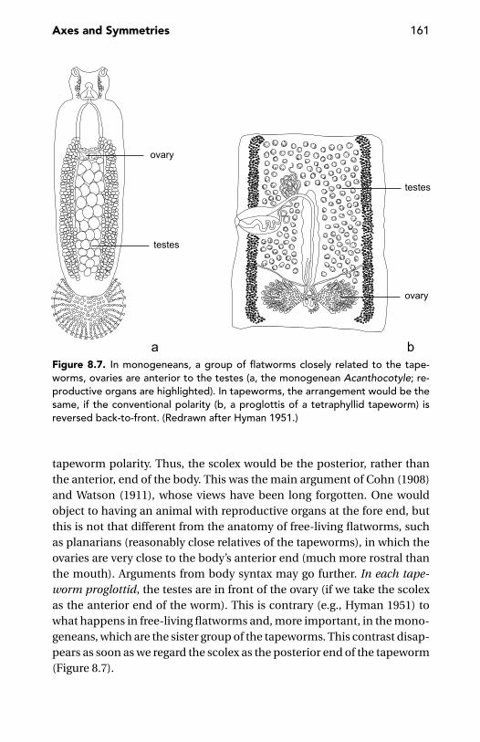
Axes and Symmetries 161
Figure 8.7. In monogeneans, a group of flatworms closely related to the tape-worms, ovaries are anterior to the testes (a, the monogenean Acanthocotyle; re-productive organs are highlighted). In tapeworms, the arrangement would be thesame, if the conventional polarity (b, a proglottis of a tetraphyllid tapeworm) isreversed back-to-front. (Redrawn after Hyman 1951.)
tapeworm polarity. Thus, the scolex would be the posterior, rather thanthe anterior, end of the body. This was the main argument of Cohn (1908)and Watson (1911), whose views have been long forgotten. One wouldobject to having an animal with reproductive organs at the fore end, butthis is not that different from the anatomy of free-living flatworms, suchas planarians (reasonably close relatives of the tapeworms), in which theovaries are very close to the body’s anterior end (much more rostral thanthe mouth). Arguments from body syntax may go further. In each tape-worm proglottid, the testes are in front of the ovary (if we take the scolexas the anterior end of the worm). This is contrary (e.g., Hyman 1951) towhat happens in free-living flatworms and, more important, in the mono-geneans, which are the sister group of the tapeworms. This contrast disap-pears as soon as we regard the scolex as the posterior end of the tapeworm(Figure 8.7).
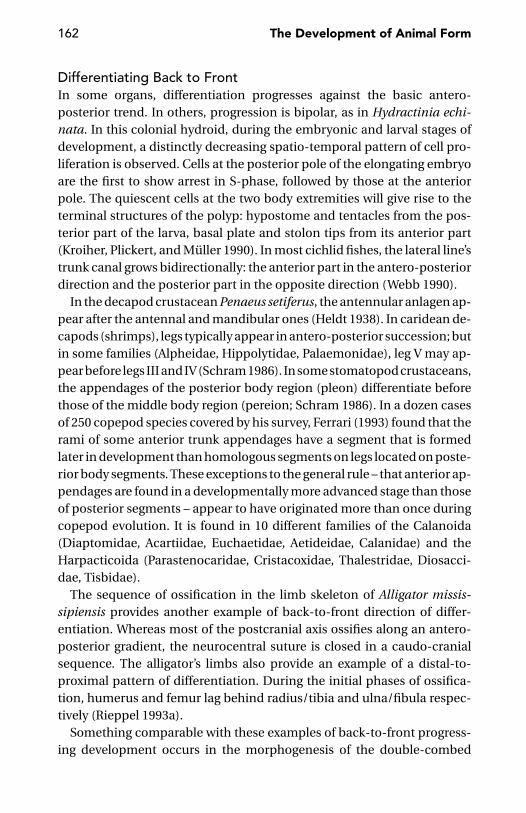
162 The Development of Animal Form
Differentiating Back to FrontIn some organs, differentiation progresses against the basic antero-posterior trend. In others, progression is bipolar, as in Hydractinia echi-nata. In this colonial hydroid, during the embryonic and larval stages ofdevelopment, a distinctly decreasing spatio-temporal pattern of cell pro-liferation is observed. Cells at the posterior pole of the elongating embryoare the first to show arrest in S-phase, followed by those at the anteriorpole. The quiescent cells at the two body extremities will give rise to theterminal structures of the polyp: hypostome and tentacles from the pos-terior part of the larva, basal plate and stolon tips from its anterior part(Kroiher, Plickert, and Muller 1990). In most cichlid fishes, the lateral line’strunk canal grows bidirectionally: the anterior part in the antero-posteriordirection and the posterior part in the opposite direction (Webb 1990).
In the decapod crustacean Penaeus setiferus, the antennular anlagen ap-pear after the antennal and mandibular ones (Heldt 1938). In caridean de-capods (shrimps), legs typically appear in antero-posterior succession; butin some families (Alpheidae, Hippolytidae, Palaemonidae), leg V may ap-pear before legs III and IV (Schram 1986). In some stomatopod crustaceans,the appendages of the posterior body region (pleon) differentiate beforethose of the middle body region (pereion; Schram 1986). In a dozen casesof 250 copepod species covered by his survey, Ferrari (1993) found that therami of some anterior trunk appendages have a segment that is formedlater in development than homologous segments on legs located on poste-rior body segments. These exceptions to the general rule – that anterior ap-pendages are found in a developmentally more advanced stage than thoseof posterior segments – appear to have originated more than once duringcopepod evolution. It is found in 10 different families of the Calanoida(Diaptomidae, Acartiidae, Euchaetidae, Aetideidae, Calanidae) and theHarpacticoida (Parastenocaridae, Cristacoxidae, Thalestridae, Diosacci-dae, Tisbidae).
The sequence of ossification in the limb skeleton of Alligator missis-sipiensis provides another example of back-to-front direction of differ-entiation. Whereas most of the postcranial axis ossifies along an antero-posterior gradient, the neurocentral suture is closed in a caudo-cranialsequence. The alligator’s limbs also provide an example of a distal-to-proximal pattern of differentiation. During the initial phases of ossifica-tion, humerus and femur lag behind radius/tibia and ulna/fibula respec-tively (Rieppel 1993a).
Something comparable with these examples of back-to-front progress-ing development occurs in the morphogenesis of the double-combed
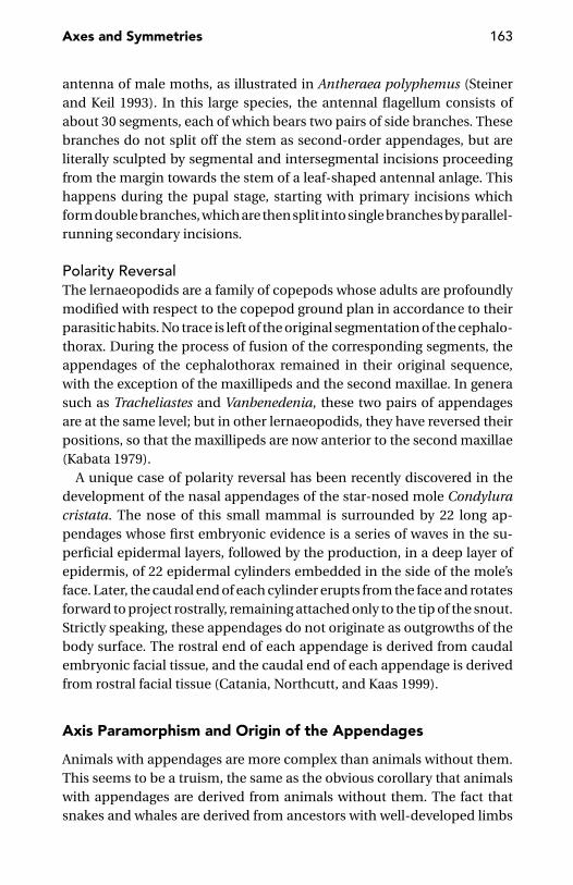
Axes and Symmetries 163
antenna of male moths, as illustrated in Antheraea polyphemus (Steinerand Keil 1993). In this large species, the antennal flagellum consists ofabout 30 segments, each of which bears two pairs of side branches. Thesebranches do not split off the stem as second-order appendages, but areliterally sculpted by segmental and intersegmental incisions proceedingfrom the margin towards the stem of a leaf-shaped antennal anlage. Thishappens during the pupal stage, starting with primary incisions whichform double branches, which are then split into single branches by parallel-running secondary incisions.
Polarity ReversalThe lernaeopodids are a family of copepods whose adults are profoundlymodified with respect to the copepod ground plan in accordance to theirparasitic habits. No trace is left of the original segmentation of the cephalo-thorax. During the process of fusion of the corresponding segments, theappendages of the cephalothorax remained in their original sequence,with the exception of the maxillipeds and the second maxillae. In generasuch as Tracheliastes and Vanbenedenia, these two pairs of appendagesare at the same level; but in other lernaeopodids, they have reversed theirpositions, so that the maxillipeds are now anterior to the second maxillae(Kabata 1979).
A unique case of polarity reversal has been recently discovered in thedevelopment of the nasal appendages of the star-nosed mole Condyluracristata. The nose of this small mammal is surrounded by 22 long ap-pendages whose first embryonic evidence is a series of waves in the su-perficial epidermal layers, followed by the production, in a deep layer ofepidermis, of 22 epidermal cylinders embedded in the side of the mole’sface. Later, the caudal end of each cylinder erupts from the face and rotatesforward to project rostrally, remaining attached only to the tip of the snout.Strictly speaking, these appendages do not originate as outgrowths of thebody surface. The rostral end of each appendage is derived from caudalembryonic facial tissue, and the caudal end of each appendage is derivedfrom rostral facial tissue (Catania, Northcutt, and Kaas 1999).
Axis Paramorphism and Origin of the Appendages
Animals with appendages are more complex than animals without them.This seems to be a truism, the same as the obvious corollary that animalswith appendages are derived from animals without them. The fact thatsnakes and whales are derived from ancestors with well-developed limbs

164 The Development of Animal Form
does not represent a serious objection to this generalisation, as embry-ological and anatomical evidence of the secondary loss of the appendagesis clearly present in these limbless vertebrates.
I will argue that animals provided with appendages were not necessar-ily derived from a common ancestor already endowed with appendages.The appendages of the vertebrates are not the same as those of the arthro-pods, any suggestion from developmental genetics notwithstanding. In myview, developmental and structural similarities between the appendagesof distantly related phyla are the transitive result of two different kinds ofrelationships: those between an animal’s main body axis and the axis of itsappendages, and those between the main body axes of animals belongingto different phyla, such as arthropods and vertebrates.
Axis ParamorphismThe core of this hypothesis is the notion of axis paramorphism, accordingto which the appendages are evolutionary duplicates of the main body axis(Minelli 2000b).
The existence of some degree of homology between the main body axisand the appendages of the same animal was advanced by a few authors[e.g., Dolle et al. (1993) and Held (1995)], but this idea was not exploredin depth; instead, evo-devo biologists began to reason in terms of theevolutionary co-option of individual genes or whole genetic cassettes (e.g.,Raff 1996, 1999b, Abouheif et al. 1997, Arthur, Jowett, and Panchen 1999). Tosay that genes otherwise used by the same animal (e.g., in the patterning ofits main body axis) were co-opted to specify features along an appendageimplies that the animal already possessed that appendage, and this raisesthe question of how the animal first acquired it. This question, to whichthe co-option hypothesis does not offer a solution, is perhaps explainedthrough the hypothesis of paramorphism.
Let’s begin with some evidence from comparative morphology.An important first generalisation is that no segmented appendage occurs
in non-segmented animals. The reciprocal correlation is a bit less strict,but we can safely state that most segmented animals have segmentedappendages.
In arthropods, segmentation is not limited to the paired limbs (anten-nae, mouthparts, legs), but extends to posterior unpaired appendages,such as the filum terminale of silverfish and the caudal appendage of sev-eral arachnids (thelyphonids, schizomids, and palpigrads; Figure 8.8). Inonychophorans (Figure 8.9) and tardigrades, the less distinct segmentation
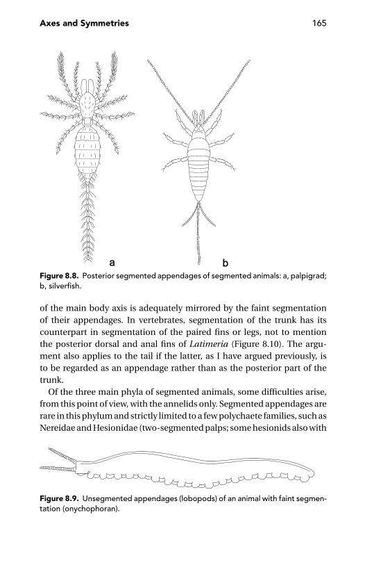
Axes and Symmetries 165
Figure 8.8. Posterior segmented appendages of segmented animals: a, palpigrad;b, silverfish.
of the main body axis is adequately mirrored by the faint segmentationof their appendages. In vertebrates, segmentation of the trunk has itscounterpart in segmentation of the paired fins or legs, not to mentionthe posterior dorsal and anal fins of Latimeria (Figure 8.10). The argu-ment also applies to the tail if the latter, as I have argued previously, isto be regarded as an appendage rather than as the posterior part of thetrunk.
Of the three main phyla of segmented animals, some difficulties arise,from this point of view, with the annelids only. Segmented appendages arerare in this phylum and strictly limited to a few polychaete families, such asNereidae and Hesionidae (two-segmented palps; some hesionids also with
Figure 8.9. Unsegmented appendages (lobopods) of an animal with faint segmen-tation (onychophoran).
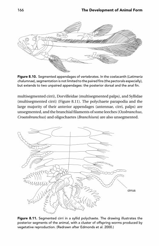
166 The Development of Animal Form
Figure 8.10. Segmented appendages of vertebrates. In the coelacanth (Latimeriachalumnae), segmentation is not limited to the paired fins (the pectorals especially),but extends to two unpaired appendages: the posterior dorsal and the anal fin.
multisegmented cirri), Dorvilleidae (multisegmented palps), and Syllidae(multisegmented cirri) (Figure 8.11). The polychaete parapodia and thelarge majority of their anterior appendages (antennae, cirri, palps) areunsegmented, and the branchial filaments of some leeches (Ozobranchus,Croatobranchus) and oligochaetes (Branchiura) are also unsegmented.
Figure 8.11. Segmented cirri in a syllid polychaete. The drawing illustrates theposterior segments of the animal, with a cluster of offspring worms produced byvegetative reproduction. (Redrawn after Edmonds et al. 2000.)

Axes and Symmetries 167
Figure 8.12. Unsegmented appendages of unsegmented animals: a, the priapulanTubiluchus; b, the actinotrocha larva of Phoronis; c, a temnocephalid flatworm; d,the cercaria larva of the digenean fluke Cotylophoron; e, an entoproct. (d, Redrawnafter Hyman 1951.)
There is no problem, of course, in contrasting the segmented appendagesof segmented animals with the unsegmented appendages of unsegmentedanimals. A list of the latter may include the tentacles of phoronids, ento-procts, ectoprocts, brachiopods, sipunculans, molluscs and pterobranchs,the tail of priapulans, the proboscis of nemerteans, and the different kindsof appendages (most commonly posterior) of flatworms, such as the tailof the cercariae, the caudal appendage of Stenostomum (Catenulida) andthe anterior lobes of the temnocephalids (Figure 8.12).
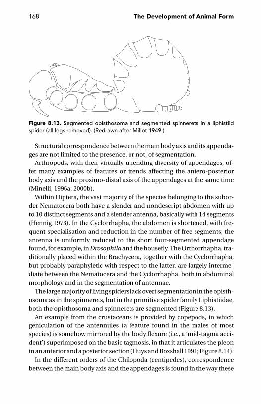
168 The Development of Animal Form
Figure 8.13. Segmented opisthosoma and segmented spinnerets in a liphistiidspider (all legs removed). (Redrawn after Millot 1949.)
Structural correspondence between the main body axis and its appenda-ges are not limited to the presence, or not, of segmentation.
Arthropods, with their virtually unending diversity of appendages, of-fer many examples of features or trends affecting the antero-posteriorbody axis and the proximo-distal axis of the appendages at the same time(Minelli, 1996a, 2000b).
Within Diptera, the vast majority of the species belonging to the subor-der Nematocera both have a slender and nondescript abdomen with upto 10 distinct segments and a slender antenna, basically with 14 segments(Hennig 1973). In the Cyclorrhapha, the abdomen is shortened, with fre-quent specialisation and reduction in the number of free segments; theantenna is uniformly reduced to the short four-segmented appendagefound, for example, in Drosophila and the housefly. The Orthorrhapha, tra-ditionally placed within the Brachycera, together with the Cyclorrhapha,but probably paraphyletic with respect to the latter, are largely interme-diate between the Nematocera and the Cyclorrhapha, both in abdominalmorphology and in the segmentation of antennae.
The large majority of living spiders lack overt segmentation in the opisth-osoma as in the spinnerets, but in the primitive spider family Liphistiidae,both the opisthosoma and spinnerets are segmented (Figure 8.13).
An example from the crustaceans is provided by copepods, in whichgeniculation of the antennules (a feature found in the males of mostspecies) is somehow mirrored by the body flexure (i.e., a ‘mid-tagma acci-dent’) superimposed on the basic tagmosis, in that it articulates the pleonin an anterior and a posterior section (Huys and Boxshall 1991; Figure 8.14).
In the different orders of the Chilopoda (centipedes), correspondencebetween the main body axis and the appendages is found in the way these
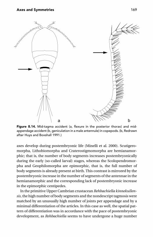
Axes and Symmetries 169
Figure 8.14. Mid-tagma accident (a, flexure in the posterior thorax) and mid-appendage accident (b, geniculation in a male antennule) in copepods. (b, Redrawnafter Huys and Boxshall 1991.)
axes develop during postembryonic life (Minelli et al. 2000). Scutigero-morpha, Lithobiomorpha and Craterostigmomorpha are hemianamor-phic; that is, the number of body segments increases postembryonicallyduring the early (so-called larval) stages, whereas the Scolopendromor-pha and Geophilomorpha are epimorphic, that is, the full number ofbody segments is already present at birth. This contrast is mirrored by thepostembryonic increase in the number of segments of the antennae in thehemianamorphic and the corresponding lack of postembryonic increasein the epimorphic centipedes.
In the primitive Upper Cambrian crustacean Rehbachiella kinnekullen-sis, the high number of body segments and the nondescript tagmosis werematched by an unusually high number of joints per appendage and by aminimal differentiation of the articles. In this case as well, the spatial pat-tern of differentiation was in accordance with the pace of postembryonicdevelopment, as Rehbachiella seems to have undergone a huge number
170 The Development of Animal Form
of growth moults, more than thirty perhaps, accompanied by very limitedadditions of new elements (segments, appendages or appendage articles)from one stage to the next (Waloßek 1993).
In vertebrates, van der Hoeven et al. (1996) observe an interesting simi-larity between the main body axis and the axis of the appendages, in thatall these axes obey a principle of proximal stability versus distal variability.This posteriorly (terminally) increased tolerance for functional variability,dubbed by van der Hoeven et al. as laxitas terminalis (terminal laxism), isparalleled by higher divergence in the Hox protein sequence.
There are several features in common, including WNT signalling, be-tween the pathways involved in the growth and patterning of the main bodyaxis, and the growth and patterning of a limb axis (Moon, Brown, and Torres1997, Shubin, Tabin, and Carroll 1997, Tabin, Carroll and Panganibam1999). In mice, the existence of a common multiaxial patterning systemis suggested by HoxD mutants, with skeletal alterations affecting all bodyaxes (Dolle et al. 1993). In Drosophila, wingless is involved in patterningboth the antero-posterior body axis and the proximo-distal axis of the leg(Campbell and Tomlinson 1995, Nagy and Williams 2001), and the wingdisc is subdivided into alternating sectors comparable with the stripes intowhich the expression of pair-rule genes partitions the antero-posterior axisof the embryo (Sturtevant et al. 1997). Held, Duarte, and Derakhshanian(1986) described the tarsi of the Drosophila mutant spiny legs, which maycontain up to eight joints, of which four correspond to those of the wild-type flies, whereas the others are extra joints dividing every tarsal arti-cle (the fifth excluded) into two subarticles. The most interesting featureis that nearly all extra joints have inverted polarity. By consequence, in thetarsus of spiny legs mutants, there is an alternation of normal and invertedjoints, perfectly comparable with the alternation of normal and extra seg-ment boundaries found along the main body axis in the embryonic lethalmutant patched, thus suggesting that the appendage and the main bodyaxis may share some patterning mechanism.
Tabin et al. (1999) suggested four different classes of explanations forthe similarities between arthropod and vertebrate limb development:(1) the common ancestor of arthropods and vertebrates was already pro-vided with appendages whose growth and patterning was controlled by thesame genes now acting in both arthropod and vertebrate limbs; (2) thesegenes, originally involved in the formation of body outgrowths other thantrue limbs, were independently co-opted to pattern fins, legs, and wings;(3) genes originally linked together in a stable cassette before being used,
Axes and Symmetries 171
in arthropods and vertebrates alike to pattern their appendages, becomeinvolved eventually in the latter process; and (4) the genes now involved inlimb patterning were individually recruited from other roles so that theirjoint operations in arthropod and vertebrate limb development is simplycoincidental.
According to Tabin et al. (1999), the last common ancestor of arthropodsand vertebrates was not provided with appendages from which those laterevolved in the two phyla might have derived as historical homologues.How then do we explain the large use of the same genetic informationin the developmental processes by which animals belonging to differentphyla form their body appendages?
According to the hypothesis of axis paramorphism, this may be ex-plained by regarding body appendages, such as arthropod and vertebratelimbs, as duplicates of the main body axis, although devoid of any endo-dermal component – that is, without involvement of the ‘visceral animal.’In my concept, a secondary axis is an axial paramorph of the main bodyaxis if it originated as a duplicate expression of genes already involved in thegrowth and patterning of the latter (Minelli 2000b). Later, divergence of theparamorphs with increasing specialisation of the appendages is obviouslyexpected in time, through the evolution of new regulatory interactions(Shubin and Marshall 2000).
The only serious alternative to axis paramorphism would be gene co-option: the animal ‘invented’ its appendages independent of the genesnow involved in its growth and patterning. These genes, already presentin the animal but involved in some other cellular machinery and devel-opmental process, later became associated with the development of theappendages, as in Tabin et al.’s (1999) second alternative.
There are problems with the co-option hypothesis. The main problem isnot the amount of co-option implied to have occurred during the evolutionof appendages, but how the animal ‘invented’ its body appendages in thefirst place. Where did the animal find the genetic tools needed to produceits secondary axes, if the latter had nothing to do, at the outset, with themain body axis? Still harder, what prevented the new tools now producingthe secondary axes from interfering with the growth and patterning of themain body axis?
The axis paramorphism hypothesis answers the first question. As to thesecond question, it is important to note that – with very minor exceptions,such as pycnogonids – the paired limbs of arthropods and vertebrates (aswell as the vertebrate tail) do not include the endodermal component
172 The Development of Animal Form
present in the main body axis. It is difficult to imagine an animal withlateral appendages similar to the main body axis in possession of endo-dermal derivatives (i.e., lateral branches of the gut). Rather than a solutionfor more effective interactions with water or substrate (e.g., locomotion orfood manipulation), this could be, at most, a way to reproduce asexually.There are a few annelids whose organization is not too far from this hypo-thetical monster. One example is the polychaete Syllis ramosa, a sedentaryparasite of sponges whose lateral body axes will eventually give rise to newindividuals (Beklemishev 1969). It is interesting to note that Syllis belongsto the annelids, a phylum where the dialogue between ectomesoderm(Romer’s ‘somatic animal’) and endoderm (Romer’s ‘visceral animal’) isprobably more extensive than in arthropods (or ecdysozoans generally).This may explain why some annelids (and flatworms, such as catenulids)produce buds with endodermal derivatives, something that ecdysozoansnever do. As undetached buds may change a conventional individual intoa colony, this circumstance may help explain why, in an enormous cladeas the Ecdysozoa, there is no instance of colonial organization, not evenin a sessile group as the barnacles.
The presence or absence of endodermal derivatives may well be thecriterion separating asexual reproduction from the production of bodyappendages. I have recently suggested (Minelli 2000b) that differentiationof a body outgrowth as a bud destined for asexual reproduction or as anappendage may depend on the presence or absence of markers similarto those in the Hydra, where the position of the mouth remains fixed,although it literally disappears, morphologically, when the animal is di-gesting freshly engulfed prey (Technau and Holstein 1995).
This is the scenario against which we can discuss the phylogenetic re-lationships between vertebrate and arthropod limbs. The discovery thatDistal-less is involved in initiating the outgrowth of limbs in vertebrates(Beauchemin and Savard 1992, Dolle, Price, and Duboule 1992) and in-sect alike (Cohen et al. 1989, Cohen 1990) seemed to support a commonorigin of these appendages, but Distal-less expression was soon found tomark the prospective tip of many kinds of appendages in different phyla(Popadic et al. 1996). Panganiban et al. (1997) suggested that the earliestbilaterians already possessed some component of the genetic machineryfor the production of appendages and even ventured to hypothesise (seealso De Robertis 1997) that Urbilateria, the putative common ancestorof vertebrates and arthropods, already possessed some kind of ‘humble’
Axes and Symmetries 173
appendage. Dong, Chu and Panganiban (2001) even suggest the existence,in the appendage of the common ancestor of arthropods and vertebrates,of proximal and distal domains under distinct genetic control.
What arthropods and vertebrates share in terms of organisation andgenetic control of their appendages is likely to be the secondary conse-quence of three distinct relationships: (1) phylogenetic conservation, be-tween arthropods and vertebrates, of genes and control pathways involvedin laying down the basic features of their main body axis; (2) paramorphismof arthropod limbs in respect to the main body axis of arthropods; and(3) paramorphism of vertebrate limbs with respect to the main body axis ofthe vertebrates. Vertebrate limbs and arthropod limbs are not true histori-cal homologues, but homoplastic paramorphs of historical homologues(Minelli 2000b). This is not the same as calling them ‘developmental par-alogues’ (Shubin, Tabin, and Carroll 1997), because the latter concept onlycaptures the homoplasy of the appendages without linking them to theorigin, patterning and evolution of the main body axis.
In any case, the dorso-ventral axis of the appendage is probably not thesame in arthropods and vertebrates (Minelli 2000b). In arthropods (primi-tively at least), the dorso-ventral polarity of the limbs is largely comparablewith the dorso-ventral polarity of the trunk, but things seem to be differentin vertebrates. In particular, the genes involved in patterning the antero-posterior and proximo-distal axes of vertebrate and arthropod limbs aresomewhat the same (Lawrence and Struhl 1996, Shubin, Tabin, and Carroll1997), but this is not true for the dorso-ventral axis.
Terminal Control and Axis ParamorphismMany, but not all, bilaterians have strong morphological markers at bothbody ends and both body ends, not just the anterior one, are defined veryearly in development. This is shown well by Drosophila, in which both bodytermini are defined by gradients of maternally encoded gene products.
Animals with early and clearly defined termini are also provided withappendages with a strongly and early defined tip. This allows patterning ofthe axis of the appendage, between its proximal and apical end, somehowcomparable with the patterning of the main body axis. This is what seemsto be excluded in the case of appendages, such as polychaete cirri, foundin metazoans with a less defined posterior body end. A strongly markedposterior body end, translated by paramorphism into a strongly marked tipof the appendages, is perhaps one of the keys to the evolutionary success
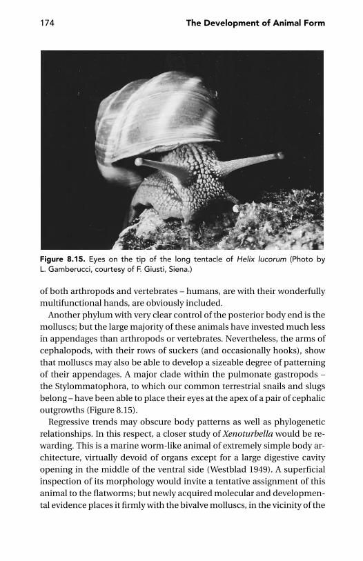
174 The Development of Animal Form
Figure 8.15. Eyes on the tip of the long tentacle of Helix lucorum (Photo byL. Gamberucci, courtesy of F. Giusti, Siena.)
of both arthropods and vertebrates – humans, are with their wonderfullymultifunctional hands, are obviously included.
Another phylum with very clear control of the posterior body end is themolluscs; but the large majority of these animals have invested much lessin appendages than arthropods or vertebrates. Nevertheless, the arms ofcephalopods, with their rows of suckers (and occasionally hooks), showthat molluscs may also be able to develop a sizeable degree of patterningof their appendages. A major clade within the pulmonate gastropods –the Stylommatophora, to which our common terrestrial snails and slugsbelong – have been able to place their eyes at the apex of a pair of cephalicoutgrowths (Figure 8.15).
Regressive trends may obscure body patterns as well as phylogeneticrelationships. In this respect, a closer study of Xenoturbella would be re-warding. This is a marine worm-like animal of extremely simple body ar-chitecture, virtually devoid of organs except for a large digestive cavityopening in the middle of the ventral side (Westblad 1949). A superficialinspection of its morphology would invite a tentative assignment of thisanimal to the flatworms; but newly acquired molecular and developmen-tal evidence places it firmly with the bivalve molluscs, in the vicinity of the
Axes and Symmetries 175
nuculids (Israelsson 1997, 1999, Noren and Jondelius 1997). Of all the mol-luscs, bivalves are probably those in which the anterior and posterior bodyends are less strongly marked by complex features, but they still preservequite distinct body ends which should be defined by molecular markersexpressed early in ontogeny. It would be interesting to know whether any-thing of these putative markers’ expression is left in Xenoturbella.
Gene Co-optionSomehow intermediate between the notion of axis paramorphism and themore fashionable explanations in terms of gene co-option is Arthur, Jowettand Panchen’s (1999) suggesion that any outgrowth, whether external or in-ternal, can be ectopically copied to produce lateral limbs. The cellular basisof outgrowing, in terms of polarised mitoses and/or cell migration, couldbe the same, irrespective of the germ layer in which outgrowing occurs. Ifso, one might speculate whether there is any relationship between the fac-ulty of outpouching by which deuterostomes form their coelomic cavitiesand the ease with which these animals produce appendages, either simpleas the podia of the echinoderms, or much more elaborate as the limbs andtail of the vertebrates. One might even read the argument the other wayaround and ask whether systems originally used for producing externalappendages (i.e., outgrowths involving the ectoderm and the mesoderm)have been ever co-opted to give rise to endodermal outgrowths. A casein point is possibly offered by the leeches. In most species of this group,there is no external appendage comparable with the parapodia of the poly-chaetes, the exceptions being represented by the (putative) respiratoryappendages of some representatives of Piscicolidae and Ozobranchidae,and the exceptional erpobdellid Croatobranchus (Sket et al. 2001). Leechesare often provided with segmental gut coeca, whose patterning is con-trolled by the Hox gene Lox3 (Wedeen and Shankland 1997).
If the gene co-option hypothesis is too weak to explain the correspon-dence between the main body axis and its appendages, it is obviouslyattractive in other simpler contexts.
Morphogenetic signals do not need to be specific for the structures theyinduce and may even be phylogenetically older than the anatomical char-acters they specify (Muller and Wagner 1996).
The role of Hox genes in specifying regional identities of the body axisis more primitive than its role in the appendages, in which these geneshave been secondarily co-opted (Shubin, Tabin, and Carroll 1997, Cohnand Coates 1999, Shubin and Marshall 2000). Cohn and Coates (1999),
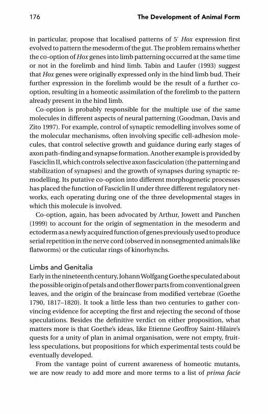
176 The Development of Animal Form
in particular, propose that localised patterns of 5′ Hox expression firstevolved to pattern the mesoderm of the gut. The problem remains whetherthe co-option of Hox genes into limb patterning occurred at the same timeor not in the forelimb and hind limb. Tabin and Laufer (1993) suggestthat Hox genes were originally expressed only in the hind limb bud. Theirfurther expression in the forelimb would be the result of a further co-option, resulting in a homeotic assimilation of the forelimb to the patternalready present in the hind limb.
Co-option is probably responsible for the multiple use of the samemolecules in different aspects of neural patterning (Goodman, Davis andZito 1997). For example, control of synaptic remodelling involves some ofthe molecular mechanisms, often involving specific cell-adhesion mole-cules, that control selective growth and guidance during early stages ofaxon path-finding and synapse formation. Another example is provided byFasciclin II, which controls selective axon fasciculation (the patterning andstabilization of synapses) and the growth of synapses during synaptic re-modelling. Its putative co-option into different morphogenetic processeshas placed the function of Fasciclin II under three different regulatory net-works, each operating during one of the three developmental stages inwhich this molecule is involved.
Co-option, again, has been advocated by Arthur, Jowett and Panchen(1999) to account for the origin of segmentation in the mesoderm andectoderm as a newly acquired function of genes previously used to produceserial repetition in the nerve cord (observed in nonsegmented animals likeflatworms) or the cuticular rings of kinorhynchs.
Limbs and GenitaliaEarly in the nineteenth century, Johann Wolfgang Goethe speculated aboutthe possible origin of petals and other flower parts from conventional greenleaves, and the origin of the braincase from modified vertebrae (Goethe1790, 1817–1820). It took a little less than two centuries to gather con-vincing evidence for accepting the first and rejecting the second of thosespeculations. Besides the definitive verdict on either proposition, whatmatters more is that Goethe’s ideas, like Etienne Geoffroy Saint-Hilaire’squests for a unity of plan in animal organisation, were not empty, fruit-less speculations, but propositions for which experimental tests could beeventually developed.
From the vantage point of current awareness of homeotic mutants,we are now ready to add more and more terms to a list of prima facie

Axes and Symmetries 177
improbable, but experimentally supported, serial homologues. This listalready includes petals and leaves, legs and antennae, and halteres andwings. The latest addition to this list is limbs and genitalia (Minelli 2002).
About fifteen years ago, Shearn, Hersperger, and Hersperger (1987) re-ported that mutations at two loci on the third chromosome of Drosophilamelanogaster caused a lot of homeotic transformations, not just those ofthe more usual kinds (antenna to leg, proboscis to leg and/or antenna, firstand third legs to second leg, haltere to wing), but also genitalia to leg and/orantenna. Similar reports became more frequent in subsequent years, thusenlarging the series of homologous ventrally paired appendages of insectsto include genitalia, in addition to antennae, mouthparts, and legs. Com-parable evidence is now available for vertebrates. The question then arisesas to what extent animal genitalia may fit into this scheme of homology.
Some preliminary observations are required, as animals with internalfertilisation have evolved a great diversity of genital appendages, eitherpaired (such as the copulatory appendages found on the parapodia ofsome pisionid polychaetes, or the hemipenes of many snakes and thepelvic claspers of chondrichthyes) or unpaired, as those of mammals andmost insects. These genital appendages are not necessarily located in closeproximity to the genital opening, but sometimes occur close to the oppo-site end of the body. This sheer diversity of genital appendages suggestsmultiple origins; nevertheless, some degree of homology can still be tracedamong them.
Three main classes of genital appendages can be distinguished: (1) ap-pendages, mostly unpaired, developed from the genital opening’s rim (ex-ample: the genital papilla of many flatworms; Figure 8.16); (2) appendagesderived from specialisation of non-genital appendages already existingin the proximity of the genital opening (example: the pelvic claspers ofchondrichthyes; Figure 8.17); and (3) appendage(s) derived from speciali-sation of non-genital appendages topographically unrelated to the genitalopening [examples: the palps of male spiders (Figure 8.18), the anten-nae of sminthurid springtails (Figure 8.19), and the secondary penis ofdragonflies (Figure 8.20)].
At first sight, there seems to be little hope of regarding these appendagesas other than analogous, but perspective changes if we adopt a combinato-rial approach to homology (see page 224). To this aim, we need to consider,alongside the appendages (both genital and non-genital), another compo-nent of the animal’s architecture: the main body axis. Problems are easierto solve if we accept the principle of axis paramorphism and are ready to
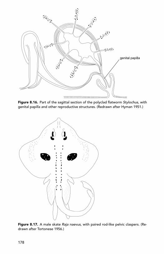
Figure 8.16. Part of the sagittal section of the polyclad flatworm Stylochus, withgenital papilla and other reproductive structures. (Redrawn after Hyman 1951.)
Figure 8.17. A male skate Raja naevus, with paired rod-like pelvic claspers. (Re-drawn after Tortonese 1956.)
178
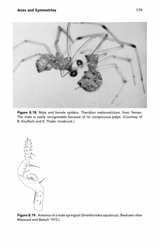
Axes and Symmetries 179
Figure 8.18. Male and female spiders, Theridion melanostictum, from Yemen.The male is easily recognisable because of its conspicuous palps. (Courtesy ofB. Knoflach and K. Thaler, Innsbruck.)
Figure 8.19. Antenna of a male springtail (Sminthurides aquaticus). (Redrawn afterMassoud and Betsch 1972.)

180 The Development of Animal Form
Figure 8.20. Thorax and anterior abdomen of a male dragonfly (a, top view; b, lat-eral view). The arrow in (b) marks the position of the secondary penis. In this insectorder, two originally lateral sclerites (the mesopleurae) expand towards the dorsalmidline, where they come in contact, thus separating the pronotum (the dorsalsclerite of the first thoracic segment) from the mesonotum (the dorsal sclerite ofthe second thoracic segment). (Redrawn after Watson and O’Farrell 1991.)
acknowledge that two features may share only one major component ofhomology: either positional or special homology.
For example, the secondary penis on the ventral side of the second ab-dominal segment of male dragonflies has very little to do, morphologicallyas well as functionally, with the gonopods, one or two pairs of specialisedlegs (between the eight and tenth pairs) in the male juliform millipedes.Gonopods are claspers used by the male when embracing the female dur-ing copulation, but these appendages have nothing to do with sperm trans-fer. To the contrary, the latter function is performed by male dragonfliesthrough their secondary penis. As for the origin (more or less clearly re-flected in the shape of the appendage), gonopods are derived (in ontogenyas well as in phylogeny) from normal locomotory legs, whereas the drag-onfly’s penis lacks a limb-like ontogenetic precursor (although in phyloge-netic perspective, it might be serially homologous to a limb pair). Despitethese big differences, the two kinds of genital appendages (Figures 8.20 and8.21) seem to share positional homology (Minelli and Schram 1994, Minelli2001), because they are located on the ventral side of probably equivalentsegments (for segment equivalence in arthropods, see pages 204–208).

Axes and Symmetries 181
Figure 8.21. A male millipede (Macrosternodesmus palicola). Arrow marks themodified sexual appendages or gonopods. (Redrawn after Blower 1985.)
The opposite relationship, of morphologically equivalent genitalia de-veloping in two animal groups in different positions, is harder to find.Something approaching the case is found in nematodes, in which the rel-ative position of the gonopores is different in the two sexes. In the males, itis subterminal, as in the majority of metazoans, whereas the female gono-pore occurs at mid-body length, in a site that possibly corresponds, interms of relative position, to the site occupied by millipede gonopods andthe dragonfly’s penis.
More generally, but still in terms of positional homology, along the mainbody axis there seem to be just two ‘hot spots’ where genital structuresmay develop.
If genital openings are mainly at the level of the posterior hot spot, theanterior one is often the site of sexual appendages. Neither spot seemsto be a genital hot spot per se. Rather, it is as if the animal was providedwith two ‘morphogenetic hooks’ to fix the position of different, importantfeatures of its body architecture. The phylogenetic conservation of thesetwo hot spots suggests a very ancient origin of the mechanisms by whichthese positions are specified. Recent data on the differentiation of externalgenital structures in mice (Kondo et al. 1997), spiders (Damen and Tautz1999a, 1999b) and nematodes (Kagoshima, Cassata, and Burglin 1999) sug-gest that differentiation of external genital structures is controlled by Hoxgenes of the posterior (Abdominal-B) class. In Drosophila, the absence ofAbdominal-B in the genital disc leads to the replacement of genitalia withlegs or, less frequently, with antennae (Estrada and Sanchez-Herrero 2001).This brings us straight to the topic of homology of limbs and genitalia. Inthe case of the replacement of genitalia by legs or antennae in the absenceof Abdominal-B expression in the genital disc, these transformations areaccompanied by the ectopic expression of genes, such as Distal-less ordachshund, which are normally required in these appendages.

182 The Development of Animal Form
In humans, a hand-foot-genital syndrome is known to depend on a Hoxgene mutation. Del Campo et al. (1999) reported about two unrelated chil-dren with reduction in the number of all fingers and toes, accompanied byhypomorphic development of the external genitalia. Both children wereheterozygous for a deletion that eliminates many members (including allposterior ones) of the HoxD gene cluster.
These concordant reports from different phyla suggest that the differ-entiation of external genital structures is under the control of Hox genesof the posterior (Abdominal-B) class, but we know very little about themechanisms involved. The concept of axis paramorphism may help usunderstand some aspects of the localisation of genital or sexually relatedstructures in many animals.
In terms of axis paramorphism, a distal position along an arthropod limb(or a distal/posterior position in a vertebrate limb) corresponds to a pos-terior position along the main body axis. Therefore, as genital structuresmostly evolve in a posterior position along the main body axis, we could ex-pect some genital or, at least, sexually related feature to evolve, along a limbaxis, in a subapical position in arthropods and in a subapical/posteriorposition in vertebrates. On the other hand, there is no reason why thesestructures should be restricted to the posterior legs. Examples of anteriorappendages bearing specialised genital/sexual features close to their distalend are all but rare. Examples include the swelling thumb (a distal elementof the forelimbs) of many male anurans (Figure 8.22), the terminal articleof the palps (anterior appendages) of male spiders (Figure 8.18), and theenlarged tarsi of the anterior legs of many male beetles (Figure 8.23).
Common genetic control of patterning in limbs and genitalia may evenextend to the analia. In mice, Hox13 and Evx-2 are expressed in the gen-italia and the internal anal sphincter (Herault et al. 1996, Kondo et al.1996). In Caenorhabditis elegans, the Evx-2 homologue vab-7 is requiredfor generation of rectal muscle cells, where its expression largely overlapsthe expression of egl-5, a nematode cognate of the posterior HoxD genesof vertebrates (Ahringer 1996).
Symmetry and Asymmetry
If development is nothing but the temporal deployment of cellular dyna-mics occurring in multicellular systems – and these systems do not nec-essarily coincide with whole embryos, but with developmental units ofdifferent size, as suggested in chapter 1 – then a symmetrical pattern can
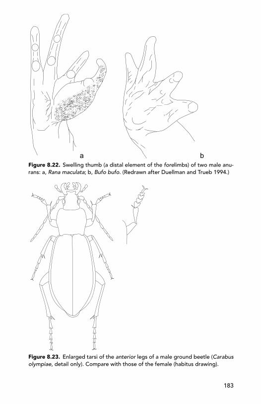
Figure 8.22. Swelling thumb (a distal element of the forelimbs) of two male anu-rans: a, Rana maculata; b, Bufo bufo. (Redrawn after Duellman and Trueb 1994.)
Figure 8.23. Enlarged tarsi of the anterior legs of a male ground beetle (Carabusolympiae, detail only). Compare with those of the female (habitus drawing).
183
184 The Development of Animal Form
often be expected as the ‘default’ product of largely independent devel-opmental trajectories going on in parallel within separate units (modules,see page 234). It is a pity that morphologists and developmental biologistsfailed to pay attention to Bateson’s (1894: 21) remark about “The essentialunity of the phenomenon of Repetition of Parts and of its companion-phenomenon, symmetry.” That symmetrically growing structures, despitetheir more or less extended developmental independence, do not have in-dividuality (if by individuality, we mean the possession of traits by whichwe can distinguish one trait from the other; Nijhout 2001) is clearly beyondthe point I am making here.
In birds, fluctuating asymmetry in feather length increases rapidly whenfeathers have just begun to grow, but it decreases again when the feathers’development approaches completion. Aparicio (1998) induced one of theseven primary feathers in the left wing of house sparrows to moult two daysapart from the corresponding feather in the right wing. Feathers of bothwings thus grew in a similar way, independent of the growth on the otherbody side. Asymmetries in feather length were less and less conspicuouswith the progress in growth, but differences in growing time persistedthroughout development. This experiment disproves the existence of amechanism of compensatory growth. The more likely explanation of theconvergence of the two sides towards a similar feather length is targetedgrowth: the feathers on the two sides follow a similar but independenttrajectory, which tends towards a fixed maximum potential size.
This brings us to one of the most interesting, but difficult, questionsin developmental biology: To which extent is the main body axis pro-duced by an integrated behaviour of the whole embryo, rather than bythe concurrent but largely independent development of its two halves? Anoverlapping of autonomy and interactions is revealed by Khaner’s (1996)study on the formation of separate body axes in separated left and righthalves of a chick embryo. That a complete embryo is not necessary to theformation of the main body axis is an argument in favour of local morpho-genetic autonomy. Interactions are nevertheless obvious. If the two em-bryo halves are separated at an early stage, then shifted along the midlineand finally reunited in staggered fashion, a single embryonic axis devel-ops if in the shifted-paired halves the posterior end of one half is adjoiningthe posterior area pellucida region of the other half. Two embryonic axesdevelop if the shift is so large that the posterior end of one half is fused tothe central area pellucida of the other half.
Axes and Symmetries 185
Directional AsymmetryFluctuating asymmetry notwithstanding, symmetry is the default state,although evolution can be expected to bring about reductions in the deg-ree of symmetry of cells and multicellular systems. Ciliates with their ele-gantly skewed cortex, and gastropods with their beautifully coiled shells,are masterworks of specialisation. Hermit crabs are clearly derived fromcrustaceans with bilaterally symmetrical pleon.
Living beings do not escape the general rule that the most highly sym-metrical systems are also the most random (Ball 1999). What we need toexplain is asymmetry rather than symmetry. But this does not rule outphylogenetic reversals, such as the symmetrisation of the pleon by whichan hermit crab lineage (the king crabs) resumed the symmetrical habitus ofordinary crabs (Cunningham, Blackstone, and Buss 1992). Contrary to com-mon belief, the radial symmetry of hydrozoans, cubozoans, and scypho-zoans is probably secondary, as suggested by the phylogenetically basalposition of Anthozoa, whose polyps are biradial to bilateral (Martindaleand Henry 1998).
Directional asymmetry may affect the whole body, as in some para-sitic isopods (Bopyridae), but this is rare. More often, it is limited to onetagma, as in the corixid water bugs (asymmetric abdomen) and hermitcrabs (asymmetric pleon). There is little need to recall the asymmetry ofthe situs viscerum in vertebrates (see Ryan and Izpisua-Belmonte 2000for a good review), or the visceral mass of gastropods, which becomesasymmetrical following the torsion of the visceral mass with respect to thefoot which maintains its original symmetry. I will only call attention to thatstrange opisthobranch, the ‘bivalve limpet’ Tamanovalva limax, which hasdextrally arranged viscera, but a small sinistral spiral at the apex of the shell.
More frequently, directional asymmetry affects the appendages, gen-erally one pair only, including the antennae of some springtails, the an-tennules of many male copepods, the mandibles of several termites andbeetles, and the chelipeds of many decapod crustaceans (Figure 8.24). Inthe cross-bills (the bird genus Loxia), the upper and lower halves of the billare asymmetric and turned into opposite directions (which helps them alot in removing seeds from larch or spruce cones).
Directional asymmetry does sometimes affect either the male or thefemale sex. Male-only asymmetries include terminalia of several insects[e.g., embiopterans and dipterans, the copulatory organ (aedeagus) ofmany beetles], and several features (vas deferens, gonopore, fifth foot)
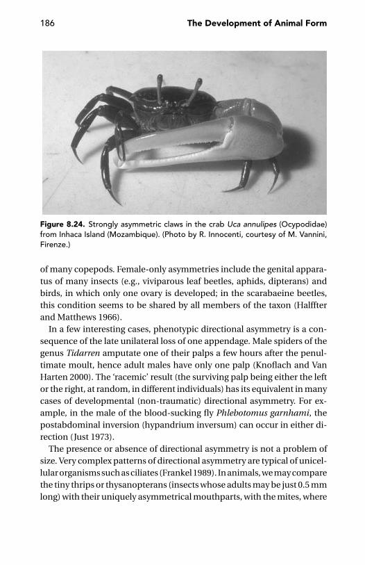
186 The Development of Animal Form
Figure 8.24. Strongly asymmetric claws in the crab Uca annulipes (Ocypodidae)from Inhaca Island (Mozambique). (Photo by R. Innocenti, courtesy of M. Vannini,Firenze.)
of many copepods. Female-only asymmetries include the genital appara-tus of many insects (e.g., viviparous leaf beetles, aphids, dipterans) andbirds, in which only one ovary is developed; in the scarabaeine beetles,this condition seems to be shared by all members of the taxon (Halffterand Matthews 1966).
In a few interesting cases, phenotypic directional asymmetry is a con-sequence of the late unilateral loss of one appendage. Male spiders of thegenus Tidarren amputate one of their palps a few hours after the penul-timate moult, hence adult males have only one palp (Knoflach and VanHarten 2000). The ‘racemic’ result (the surviving palp being either the leftor the right, at random, in different individuals) has its equivalent in manycases of developmental (non-traumatic) directional asymmetry. For ex-ample, in the male of the blood-sucking fly Phlebotomus garnhami, thepostabdominal inversion (hypandrium inversum) can occur in either di-rection (Just 1973).
The presence or absence of directional asymmetry is not a problem ofsize. Very complex patterns of directional asymmetry are typical of unicel-lular organisms such as ciliates (Frankel 1989). In animals, we may comparethe tiny thrips or thysanopterans (insects whose adults may be just 0.5 mmlong) with their uniquely asymmetrical mouthparts, with the mites, where
Axes and Symmetries 187
virtually no case of asymmetry is found, not even in the largest represen-tatives of the group – the ticks.
The phylogenetic distribution of directional asymmetry (Palmer 1996)reveals some systematic bias, something we should perhaps investigatemore closely as the possible expression of some hitherto unrecogniseddevelopmental principle (cf. Arthur 2002). In two large arthropod clades(Chelicerata, Myriapoda), directional asymmetry is virtually absent. Theonly case of which I am aware is Freyanella plataleae (male only?), a mitewhich clings on feather barbs of the host thanks to an enormous scapulararticulated seta, only present on the right side, close to the posterior borderof the propodosoma (Dubinin 1953). Directional asymmetry is commonin Crustacea and Insecta. Curiously, no snail-eating beetle has evolvedasymmetrical cephalic structures. Alternative morphological strategies, asfar as we can see, were more readily available: cychrisation in particular,that is an exteme narrowing of the anterior half of the body, thus allowingthe beetle to prosecute its prey well into the shell.
In their controversial book on Asymmetry, Developmental Stability, andEvolution, Møller and Swaddle (1997) discussed the ontogenetic plasticityof directional asymmetry in some crustaceans, such as the robber crabBirgus. At the beginning of its postlarval life, Birgus has a symmetrical ab-domen that develops asymmetry once the crab starts living in a gastropodshell, but it reverts to symmetry when the animal changes its life-styleagain by abandoning the shell. Govind (1992) suggested that, in lobsters,the more active claw became the crusher and its less active counterpartthe cutter; bilateral differences in predominantly mechanoreceptive inputto the paired claws would somehow lateralise claw ganglion and, throughit, the musculature of the appendage and its whole structure.
CHAPTER NINE
Segments
Segment identity is a subjective concept that originates fromthe observation that in a particular species, a number of cellcharacteristics are always associated in a given segment.
J. Castelli-Gair 1998: 441
What Is a Segment?
From a developmental point of view, segments are something differentfrom the modular building blocks we are used to seeing in millipedes,earthworms, and caterpillars. Generally, we regard segments as modules ofbody architecture advantageous to, and perhaps evolved ‘in view of’, someadvanced mechanisms of locomotion. In this chapter, I will suggest a verydifferent perspective. Basic to reformulation of our views about segmentsis the appreciation that this term has been and is still currently appliedto many (perhaps too many) different kinds of units (Minelli and Fusco1995).
Segments are often skeletomuscular units used in locomotory mecha-nics. Segments are also those units, often quite well circumscribed, thatbehave (or seem to behave) as convenient homologues in comparativeanatomy, even if the traditionally held principle – that segments shouldcorrespond to (pairs of) coelomic pouches – suffers too many exceptions.Segments are also developmental units, whose nature, origin, and evolu-tionary potential are the subject of this chapter.
There is nothing wrong with identifying segments with anatomical orfunctional units, or in describing segmentation as a kind of symmetry(Beklemishev 1969) or, more precisely, as translational symmetry (Coen1999, Fusco and Minelli 2000b). There is a danger, however, that thesepurely descriptive or abstract approaches bring us to the point that weregard all segments as basically the same, were it not for the specialisations
188
Segments 189
that make the difference (e.g., between thoracic and abdominal segmentsin an insect). To say that these segments are different because different setsof genes are expressed in each of them does not help with understandingthe nature of segments as developmental units, or whether all segmentsare basically the same or not.
According to Chaudonneret (1979), a typical insect metamere impliesthe coexistence of an ectodermic segment delimited by intersegmental fur-rows, a vascular segment or angiomere, a muscular segment or myomere, anephridial segment or nephromere, a nervous segment or neuromere, anda unit bearing one pair of appendages. But do all these elements necessarilycome in perfect register? That is, are there true ‘eusegments’ in the animalkingdom? Even restricting our attention to morphology (developmentalaspects of segmentation will be discussed later), segment boundaries aremore similar to hybrid zones between closely related species than to theneat borders of national states: between neighbouring segments we canexpect ‘introgression’ of muscles, tracheae, blood vessels, and other. Thisexplains the historical contrast between the Gratiolet-Whitman school(Gratiolet 1862, Whitman 1884, 1892) and the Moore-Castle school (Castle1900, Moore 1900) in delimiting segments in the leeches. According toCastle and Moore, the ventral ganglia occupy the longitudinal middleof the segment, whereas Gratiolet and Whitman regarded the ganglia aslocated at the anterior limit of the segment.
Budd (2001) has recently argued against the very idea of ‘eusegmenta-tion’ as a typological hindrance to understanding animal segments andcorrectly stressed the fact that individual parts or organ systems may havedeveloped segmentation independently.
Students of arthropod morphology are often in doubt as to the ‘true’segmental nature of some serial units of an animal’s main body axis. Thisis why Kabata (1979), for example, remarks that segmentation of the cope-pod Synergasilus is sometimes obscured by the presence of ‘false-rings’,whereas Huys and Boxshall (1991), in describing the structural modifica-tions of some other copepods (e.g., Paramesochra mielkei, Intermedop-syllus intermedius, and Psammocyclopine hindleyi), with respect to theirmore generalised relatives, recognise the presence of a secondary ‘pseu-dosomite’ separated from the anterior end of the genital ‘double somite’.
In other cases, the difference between ‘true’ and ‘false’ segments seemsto be obvious, as in the case of the leeches, with their annulated seg-ments, but probably – as we will see – too much has been made of thisdistinction.
190 The Development of Animal Form
Similar problems are sometimes found in interpreting the segments ofarthropod appendages, paramorphs of the main body axis. Not too rare,for example, are instances of pseudoarthrosis, in which a suture in thecuticle suggests subdivision into two segments. But no articular membranebetween the two parts is found, for example, in the tarsi of legs I–XIII ofseveral lithobiid centipedes (see page 141).
The basic problem is, what do we mean by segment?I will follow Alberch and Kollar’s (1988) suggestion in defining segmen-
tation as the subdivision of an embryonic field into sharply defined popu-lations of cells linearly arranged along the antero-posterior axis. What thissubdivision actually means, in terms of the future development of the em-bryo, is not necessarily the same in all instances. In the case of insects, seg-mentation is a system of mutual activation of cell states that locally excludeeach other (Meinhardt and Gierer 2000). In the case of rhombomeres, thesegmental units of the vertebrate hindbrain, it is a way to sequester cells,limiting their subsequent migration as neural crest cells to neighbouringareas (Lumsden, Sprawson, and Graham 1991, Takeichi et al. 1997). Thesame is possibly true for the diencephalic neuromere boundaries, whichact as a scaffold for early axon pathways in the neuroepithelium of thisvertebrate brain region (Boncinelli 1994).
From an evo-devo point of view, of particular value is the fact thatAlberch and Kollar’s definition of segmentation also applies to the regu-larly patterned arrangement of selected groups of cells in animals thatlack any morphologically overt segmentation. Wood and Edgar (1994) de-scribe the organisation of the first larva of Caenorhabditis elegans as partlymetameric, in that blast cells with the same or similar developmental po-tential are regularly spaced along the antero-posterior axis. During subse-quent development, these cells will give rise to homologous cell lineages.In particular, dorsal, lateral, and ventral ectoderm cells are each repeatedsix times along the animal’s length (Salser and Kenyon 1994). The six pairsof ventral cells (P cells) interdigitate at the midline, forming a single rowwhich contributes neurons to the ventral nerve cord.
Virtual Versus Physical Segmental Boundaries
What is primary: the segment or the limit between segments? In terms ofmorphology, the boundaries between compartments are virtual bound-aries, with nothing likely to be seen through the lenses of the micro-scope and nothing to be seen in fossils. But compartment boundaries may

Segments 191
be secondarily elaborated into physical boundaries, something one canactually see in the animal. At this stage, what in fact exists is an alternatingpattern of longer and shorter units, with the longer units corresponding toconventional morphological segments and the shorter units correspond-ing to the ‘walls’ between them. Physical boundaries are not required forfunctional integration within each segment and/or for functionally isolat-ing its global metabolism. Local integration, if elicited by a specific localtrigger, may rest on a higher frequency of interactions within the segmentwith respect to the interactions between segments. A conversation doesnot require partners to be physically isolated within a room, but peopleinvolved in a lively conversation, even if in a crowded room, are neverthe-less functionally isolated from all the other people around them. Physicalboundaries may offer an opportunity for stabilising a segmental patternwhich may be later translated, as prepattern, into more advanced serialarchitectures. In Drosophila, for example, the parasegmental boundaryhas been regarded as an organising centre since it is used as a referenceto pattern neighbouring cells (Ingham 1991, Ingham and Martınez-Arias1992, Dahl, Koseki, and Balling 1997).
Rather than as a frozen structure conserved throughout development,boundaries should be thought of as dynamic features. In the Drosophilaembryo, parasegmental boundaries are maintained by the pair-rule geneexpression that established them in the first place, for the first three hoursonly, when many of the pair-rule genes switch off and their products disap-pear. These boundaries are dynamically maintained by segment polaritygenes. This persistence of parasegmental boundaries is critical to avoidmixing of neighbouring cell populations that are still dividing, to set limitsto gene expression, and to establish gradients of positional information(Lawrence and Sampedro 1993). The basis of insect segmentation is thus ajuxtaposition of distinct cells to form a boundary that is self-perpetuating(Akam 1994).
The boundary between the histoblasts and the surrounding larval cellsand segment boundaries is also a self-perpetuating unit. Old experimentswith Drosophila demonstrated that a histoblast nest grafted in a larvalsegment in inverted antero-posterior polarity could not affect the polarityof the host abdomen; but this effect was obtained if the inverted histo-blasts were accompanied by the polyploid larval epidermal cells whichsurround them and which will be destroyed during metamorphosis. Thiswas interpreted as proof that the positional information responsible fora normal pattern resides in the polyploid epidermal cells (Milner and

192 The Development of Animal Form
Bleasby 1985). It is quite clear that it resided instead in the boundarybetween histoblast and polyploid cells: When polyploid epidermal cellsalone are grafted into the wrong position, histoblast differentiation isunaffected.
How Many Times Did Metazoans Evolve Segmentation?
There might well be more than one way to subdivide an embryonic fieldinto sharply defined populations of cells linearly arranged along the antero-posterior axis. If so, there is little reason to expect that the segments ofall segmented animals will be strictly comparable from a developmentalpoint of view. I will briefly review evidence suggesting that segmentationmechanisms are so diverse as to imply a multiple origin of segments alongthe evolutionary history of metazoans. The common features undeniablyshared by all segmented animals will be explained as due to two differentcauses: the ‘generic’ character of segmentation (see page 35) and the mul-tiple, independent involvement of similar genes, or gene batteries, in thesegmentation processes.
As soon as we realize that all segments are not necessarily the productof a single developmental process, we may also ask whether all segmentswithin one and the same animal – be it an annelid, an arthropod or avertebrate – are strictly equivalent from a developmental point of view.This point will be discussed later.
During the last two decades, our knowledge of expression patterns anddevelopmental roles of the so-called segmentation genes have grown quiteconsiderably, at least for some ‘model’ animals. It is unfortunate that westill know very little about the role these genes play in animals that a com-parative morphologist would suggest as representative of peculiar kinds ordegrees of segmentation, as are the myriapods. The first evidence, at last, isbeing published for the expression of the segment-polarity gene engrailedin the geophilomorph centipede Strigamia maritima (Kettle et al., in press)(Figure 9.1) and for the Hox genes – which are marginally involved, in thiscase, in segmentation – of the lithobiomorph centipede Lithobius (Hughesand Kaufman 2002).
One of the most important generalisations obtained thus far regardingthe identity and roles of segmentation genes in different metazoan groupsis the extensive similarity of genes and control cascades found in the dif-ferent phyla of segmented animals. This has been often construed as proofthat, contrary to the traditional phylogenetic views, segmentation might

Segments 193
Figure 9.1. Expression pattern of the segment-polarity gene engrailed inthe growing germ band of the geophilomorph centipede Strigamia maritima.(Courtesy of C. Kettle and W. Arthur, Sunderland, UK.)
have arisen only once in the history of metazoans. This view has developedinto a key component of the concept of Urbilateria, the putative commonancestor of all bilaterian (triploblastic) animals (De Robertis and Sasai1996).
Reconstructing UrbilateriaA segmented Urbilateria has been suggested, more or less explicitly, byseveral recent authors (Kimmel 1996, De Robertis 1997, L.Z. Holland et al.1997, Palmeirim et al. 1997, Christ et al. 1998, Holland and Holland 1998).Less explicit are Valentine, Erwin, and Jablonski (1996), when creditingthis bilaterian ancestor with ‘some form of serial structure’, whereas Dewel(2000) goes so far as to speak, more specifically, of segments ontogenetically
194 The Development of Animal Form
expressed as blocks of mesoderm, somites and probably adjoining fieldsof ectoderm or neuroectoderm.
In addition to segmentation, the following set of traits has been at-tributed to Urbilateria:
� a ‘humble appendage or antenna-like outgrowth’ (De Robertis 1997,based on the presence of fringe, serrate and other genes; Panganibanet al. 1997, with doubt; Morata and Sanchez-Herrero 1999) or even a pat-terned outgrowth (Dewel 2000, Dong, Chu, and Panganiban 2001); butthis is rejected by Mittman and Scholtz (2001)
� a hemocoel (Valentine, Erwin and Jablonski 1996), or a more articulateseries of internal cavities, including at least two large undivided lateral ordorso-lateral coeloms and serially repeated lateral gonocoels, each witha gonoduct and gonopore to the exterior; perhaps also serially repeatedcoelomic cavities with connections (gill slits and pores) to both the gutand the exterior (Dewel 2000)
� a contractile blood vessel or heart (De Robertis 1997, based on the pres-ence of genes such as Tinman/Nkx2.5 and DMEF2, which are involvedin the specification of a heart)
� a skeleton (Jacobs et al. 2000)� a primitive photoreceptor (Bolker and Raff 1996, De Robertis 1997,
Gehring and Ikeo 1999, Neumann and Nusslein-Volhard 2000, Kumar2001), based on the presence of some homologue of Pax6/eyeless (seepages 25–26)
� brain and brain areas (Arendt and Nubler-Jung 1996)� development through a primary ciliated larva with a tube-shaped gut
divided into anterior, middle and posterior portions (Arendt, Technau,and Wittbrodt 2001)
� cephalisation (Finkelstein and Boncinelli 1994, Dewel 2000)� antero-posterior polarity (De Robertis 1997, based on the presence of
Hox gene complexes with antero-posterior colinear expression; cf.page 47)
� dorso-ventral patterning (De Robertis 1997, based on the presence ofpairs of dorso-ventral patterning genes homologous to sonic hedge-hog/chordin and decapentaplegic/Bone Morphogenetic Protein-4; cf.page 155)
I have listed these features in approximate order of increasing likelihood,starting with segmentation and the presence of appendages [two features

Segments 195
that no student of phylogeny (e.g., Ax 1995) would probably ascribe tothe common ancestor of all bilaterians] and finishing with features suchas antero-posterior and dorso-ventral patterning, which are, in a sense,defining features of the bilaterian organisation. However, several otherauthors (e.g., Akam 1989, Abouheif 1997, Laufer et al. 1997, Shubin, Tabin,and Carroll 1997, Arthur, Jowett, and Panchen 1999) have regarded mostof these putative features of Urbilateria as independently acquired by sev-eral phyletic lines, through independent co-option of the same, or similargenes and control pathways. Axis paramorphism (Minelli 2000b) may havecontributed to the spread of these convergent features.
It must be noted that none of the authors who have expressed their viewson the putative organisation of Urbilateria used any of the standard meth-ods currently available for phylogeny reconstruction. Therefore, from amethodological point of view, none of these Urbilaterias qualify as reliableground-plan reconstruction. The more features are added to the picture,the more Urbilateria becomes similar to the Urpflanze (the archetypalplant) drawn by P.J.F. Turpin for a French edition of Goethe’s natural his-tory work (1837). Turpin’s plant is dramatically overloaded by samples ofall kinds of leaves, roots, flowers, and inflorescences (Figure 9.2). Typologi-cal thinking (cf. Richardson, Minelli, and Coates 1999) collapses featuresover features onto one archetypal form, but it does not help understandingtheir actual origin or their changes in evolution.
Davidson (2001) has forcefully argued against the fashionable recon-structions of the Urbilateria, which sum up to produce “an impossible andillogical image of the bilaterian common ancestor” (p. 189). It is clear thatthese reconstructions underestimate (and sometimes simply ignore) howmuch animal phyla may differ in important details of the developmentalprocesses by which they obtain segments, eyes, hearts, and so on. Whatis conserved across the whole of bilaterians are the differentiation genebatteries involved in specification of equivalent body parts in differentphyla, but the actual ways they develop may differ no less than their finalmorphologies.
Segments in Annelids, Arthropods, and VertebratesThis is not the place for reviewing in detail the mechanisms by whichsegments are made in annelids, arthropods, and vertebrates. A few notesmay suffice for the purpose of later discussion. In annelids and in verte-brates, segmentation is basically a mesodermal affair, whereas in arthro-pods it is primarily ectodermal (Minelli and Bortoletto 1988, Dewel 2000).
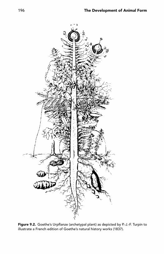
196 The Development of Animal Form
Figure 9.2. Goethe’s Urpflanze (archetypal plant) as depicted by P.-J.-F. Turpin toillustrate a French edition of Goethe’s natural history works (1837).
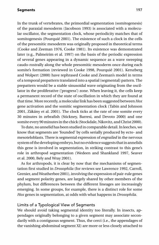
Segments 197
In the trunk of vertebrates, the primordial segmentation (somitogenesis)of the paraxial mesoderm (Jacobson 1993) is associated with a molecu-lar oscillator, the segmentation clock, whose periodicity matches that ofsomitogenesis (Pourquie 2001). The existence of such a clock in the cellsof the presomitic mesoderm was originally proposed in theoretical terms(Cooke and Zeeman 1976, Cooke 1981). Its existence was demonstratedlater (e.g., Palmeirim et al. 1997) on the basis of the periodic expressionof several genes appearing in a dynamic sequence as a wave sweepingcaudo-rostrally along the whole presomitic mesoderm once during eachsomite’s formation (reviewed in Cooke 1998, Pourquie 2001). Kerszbergand Wolpert (2000) have rephrased Cooke and Zeeman’s model in termsof a temporal prepattern translated into a spatial (segmental) pattern. Theprepattern would be a stable sinusoidal wave originating from the oscil-lator in the proliferative (‘progress’) zone. When leaving it, the cells keepa permanent record of the state of oscillation in which they are found atthat time. More recently, a molecular link has been suggested between Hoxgene activation and the somitic segmentation clock (Tabin and Johnson2001, Zakany et al. 2001). The clock ticks at the rate of one somite every30 minutes in zebrafish (Stickney, Barresi, and Devoto 2000) and onesomite every 90 minutes in the chick (Stockdale, Nikovitz, and Christ 2000).
To date, no annelid has been studied in comparable detail. In leeches, weknow that segments are ‘founded’ by cells serially produced by ecto- andmesoteloblasts. There is segmental expression of engrailed in the nervoussystem of the developing embryo, but no evidence suggests that in annelidsthis gene is involved in segmentation, in striking contrast to this gene’srole in arthropod segmentation (Wedeen and Shankland 1997, Seaveret al. 2000, Bely and Wray 2001).
As for arthropods, it is clear by now that the mechanisms of segmen-tation first studied in Drosophila (for reviews see Lawrence 1992, Carroll,Grenier, and Weatherbee 2001), involving the expression of pair-rule genesand segment polarity genes, are largely shared by other members of thephylum, but differences between the different lineages are increasinglyemerging. In some groups, for example, there is a distinct role for someHox genes in segmentation, at odds with what happens in Drosophila.
Limits of a Typological View of SegmentsWe should avoid taking segmental identity too literally. In insects, ap-pendages originally belonging to a given segment may associate secon-darily with a contiguous segment. Thus, the cerci (i.e., the appendages ofthe vanishing abdominal segment XI) are more or less closely attached to

198 The Development of Animal Form
segment X. Even more conspicuous is the fusion of mandible and maxilla,two appendages belonging to different segments, in the larvae of some or-thorrhaphous Diptera (Chaudonneret 1979, Teskey 1981). In silverfish anddragonflies, some tergal muscles are transsegmental, crossing the segmen-tal border to become attached to the tergum of the next segment (Matsuda1970). During embryonic or postembryonic development, some body seg-ments seem literally to disappear, at least on their dorsal (or ventral) aspect.This is frequent in the posterior body segments (genital region) of manyinsect orders. The most conspicuous case is probably that of the entomo-bryomorph Collembola, in which the dorsal sclerite of the first thoracicsegment (pronotum) disappears completely during embryonic develop-ment, therefore the first dorsal sclerite behind the head is the mesonotum(Matsuda 1979).
The anatomical proximity of originally neighbouring segments some-times gets lost following rearrangement of some segmental components.In the thorax of dragonflies, the two wing-bearing segments (meso- andmetathorax) are strongly associated to form a complex called the pterotho-rax. The ‘true’ dorsal sclerites (terga) of these two segments are quite smalland do not cover the whole dorsal surface of the pterothorax. The an-terior half of this complex is covered instead by the pleurae (originallylateral) of the mesothorax, which are joined along the midline, in front ofwhat remains of the mesotergum. In this way, the dorsal sclerites of thefirst thoracic segment (prothorax) are widely separated from the corre-sponding mesothoracic sclerite (Figure 8.20). In some sphecid wasps, theusual wasp waist becomes a long peduncle, entirely formed by the secondsegment of the abdomen, despite the fact that it seems to comprise twosegmental units, rather than one. This is because the dorsal and ventralsclerites of this segment have shifted longitudinally apart in such a waythat the anterior part of the peduncle is covered by the sternal plate onlyand the posterior part is covered by the tergal plate only (Figure 9.3).
In other cases, unexpected segment composition is the result of pro-found metamorphoses, such as those undergone by Drosophila, in whichthree pairs of imaginal discs (clypeo-labral, eye-antennal and labial) pro-duce the whole imaginal head. Therefore, only three of the theoretical sixhead segments (labral, antennal and labial) do actually contribute to theimaginal head, whereas the other three segments (intercalary, mandibu-lar and maxillary) do not seem to be involved. The contribution of eachimaginal disc goes beyond its expected segmental domain. For instance,the eye-antennal disc gives rise, in addition to the eye and the antenna,
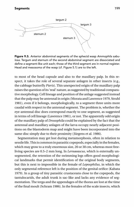
Segments 199
Figure 9.3. Anterior abdominal segments of the sphecid wasp Ammophila sabu-losa. Tergum and sternum of the second abdominal segment are dissociated anddefine a segment-like unit each; those of the third segment are in normal register.Head and mesosoma of the wasp (cf. Figure 5.1) are to the left.
to most of the head capsule and also to the maxillary palp. In this re-spect, it takes the role of several separate anlagen in other insects (e.g.,the cabbage butterfly Pieris). This unexpected origin of the maxillary palpraises the question of its ‘real’ nature, as suggested by traditional compara-tive morphology. Cell lineage and position of the anlage suggested insteadthat the palp may be antennal in origin (Morata and Lawrence 1979, Struhl1981), even if it belongs, morphologically, to a segment three units morecaudal with respect to the antennal segment. The problem is, whether theeye-antennal disc does correspond exactly to one segment, as suggestedin terms of cell lineage (Lawrence 1981), or not. The apparently odd originof the maxillary palp of Drosophila could be explained by the fact that theantennal and maxillary anlagen of the larva occupy nearly adjacent posi-tions on the blastoderm map and might have been incorporated into thesame disc simply due to their proximity (Jurgens et al. 1986).
Segmentation may get lost during metamorphosis, often in relation tosessile life. This is common in parasitic copepods, especially in the females,which may grow to a truly enormous size, 20 or 30 cm, whereas most free-living species are 0.5–2 mm long. In Lernaeascus, which is externally un-segmented, the retention of the swimming legs offers good morphologi-cal landmarks that permit identification of the original body segments,but this is next to impossible in the female of Leposphilus, in which theonly segmental reference left is the position of the genital orifice (Kabata1979). In a group of tiny parasitic crustaceans close to the copepods, thetantulocarids, the adult trunk is sac-like and lacks any evidence of seg-mentation. The terga and the appendages of the thorax are lost at the timeof the final moult (Schram 1986). In the females of the scale insects, which

200 The Development of Animal Form
are completely sessile following the first active larval instar, the abdomi-nal segments are more or less completely fused (Strumpel 1983). The mostdramatic loss of segmentation during development is offered by rhizo-cephalans such as Sacculina. When the female cypris larva of these crus-taceans fixes itself on a crab, the thoracic appendages and the abdomenare literally amputated and cast off, and the rhizocephalan changes into arhizoid branching body that expands through the victim’s tissues, only toemerge from it later in the form of a bag into which a male larva will at lastdeliver its equally shapeless content.
Segmentation: One Animal, More than One Mechanism
To acknowledge that segmentation processes are not the same in ani-mals as different as insects and vertebrates should not come as a surprise,whereas the notion that not all the segments of a segmented animal arethe product of the same mechanism may meet more resistance. There aregood reasons, however, to acknowledge the widespread coexistence of twodifferent kinds of segments within the same animal.
Double Segmentation: Eosegments and MerosegmentsLeeches, with their secondarily annulated segments, may help in intro-ducing the notion of double segmentation (Minelli 2000a).
A mid-body segment (those closer to the two body ends have reducedannulation or no annulation at all) is basically split into three first-orderannuli (a1a2a3), as in most species of the family Glossiphoniidae. In themedicinal leech and in many other leech species, the first and the last ofthese three primary annuli split into secondary annuli, giving rise to a five-annulated segment (b1b2a2b5b6). More complex patterns are also known, asin the six-annulated b1b2a2b5c11c12 segments of Dina lineata or the seven-annulated c1c2b2a2b5c11c12 segments of Trocheta subviridis dalmatina, withtwo third-order c annuli replacing one or more secondary b annuli.
Zoology textbooks are clear in pointing out the difference between the‘true’ segments – identified by the repetition of nephridia, nervous ganglia,and so on – and the superficial annuli. This is correct, but it obscures thefact that many kinds of ‘segments’ found in other animals are much moresimilar to leech annuli than to leech segments.
An example is the rhombomeres, the segmental units of the vertebratehindbrain. An interesting feature of rhombomeres is that they do not formin regular antero-posterior progression (or synchronously), but through a
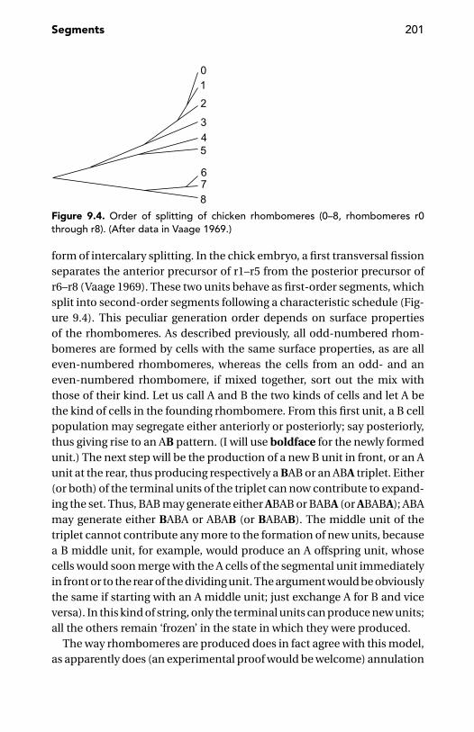
Segments 201
Figure 9.4. Order of splitting of chicken rhombomeres (0–8, rhombomeres r0through r8). (After data in Vaage 1969.)
form of intercalary splitting. In the chick embryo, a first transversal fissionseparates the anterior precursor of r1–r5 from the posterior precursor ofr6–r8 (Vaage 1969). These two units behave as first-order segments, whichsplit into second-order segments following a characteristic schedule (Fig-ure 9.4). This peculiar generation order depends on surface propertiesof the rhombomeres. As described previously, all odd-numbered rhom-bomeres are formed by cells with the same surface properties, as are alleven-numbered rhombomeres, whereas the cells from an odd- and aneven-numbered rhombomere, if mixed together, sort out the mix withthose of their kind. Let us call A and B the two kinds of cells and let A bethe kind of cells in the founding rhombomere. From this first unit, a B cellpopulation may segregate either anteriorly or posteriorly; say posteriorly,thus giving rise to an AB pattern. (I will use boldface for the newly formedunit.) The next step will be the production of a new B unit in front, or an Aunit at the rear, thus producing respectively a BAB or an ABA triplet. Either(or both) of the terminal units of the triplet can now contribute to expand-ing the set. Thus, BAB may generate either ABAB or BABA (or ABABA); ABAmay generate either BABA or ABAB (or BABAB). The middle unit of thetriplet cannot contribute any more to the formation of new units, becausea B middle unit, for example, would produce an A offspring unit, whosecells would soon merge with the A cells of the segmental unit immediatelyin front or to the rear of the dividing unit. The argument would be obviouslythe same if starting with an A middle unit; just exchange A for B and viceversa). In this kind of string, only the terminal units can produce new units;all the others remain ‘frozen’ in the state in which they were produced.
The way rhombomeres are produced does in fact agree with this model,as apparently does (an experimental proof would be welcome) annulation
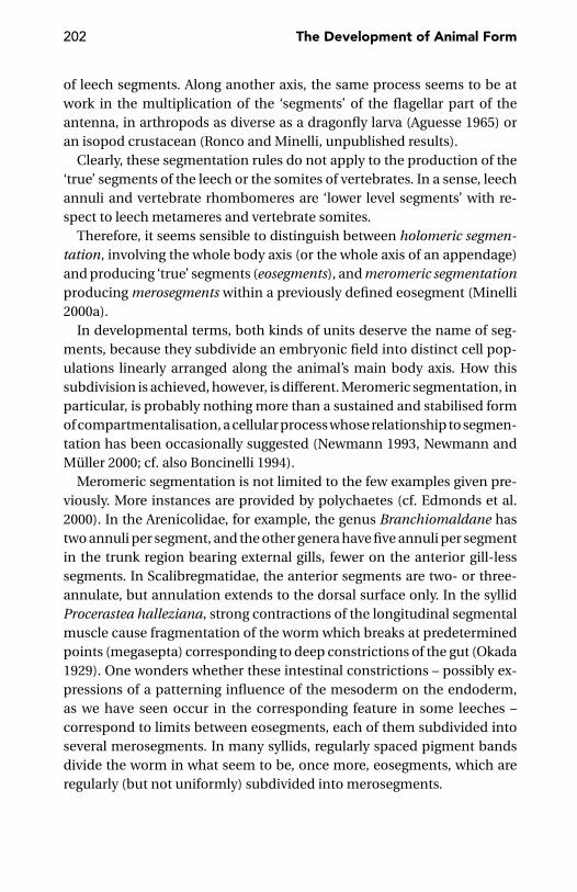
202 The Development of Animal Form
of leech segments. Along another axis, the same process seems to be atwork in the multiplication of the ‘segments’ of the flagellar part of theantenna, in arthropods as diverse as a dragonfly larva (Aguesse 1965) oran isopod crustacean (Ronco and Minelli, unpublished results).
Clearly, these segmentation rules do not apply to the production of the‘true’ segments of the leech or the somites of vertebrates. In a sense, leechannuli and vertebrate rhombomeres are ‘lower level segments’ with re-spect to leech metameres and vertebrate somites.
Therefore, it seems sensible to distinguish between holomeric segmen-tation, involving the whole body axis (or the whole axis of an appendage)and producing ‘true’ segments (eosegments), and meromeric segmentationproducing merosegments within a previously defined eosegment (Minelli2000a).
In developmental terms, both kinds of units deserve the name of seg-ments, because they subdivide an embryonic field into distinct cell pop-ulations linearly arranged along the animal’s main body axis. How thissubdivision is achieved, however, is different. Meromeric segmentation, inparticular, is probably nothing more than a sustained and stabilised formof compartmentalisation, a cellular process whose relationship to segmen-tation has been occasionally suggested (Newmann 1993, Newmann andMuller 2000; cf. also Boncinelli 1994).
Meromeric segmentation is not limited to the few examples given pre-viously. More instances are provided by polychaetes (cf. Edmonds et al.2000). In the Arenicolidae, for example, the genus Branchiomaldane hastwo annuli per segment, and the other genera have five annuli per segmentin the trunk region bearing external gills, fewer on the anterior gill-lesssegments. In Scalibregmatidae, the anterior segments are two- or three-annulate, but annulation extends to the dorsal surface only. In the syllidProcerastea halleziana, strong contractions of the longitudinal segmentalmuscle cause fragmentation of the worm which breaks at predeterminedpoints (megasepta) corresponding to deep constrictions of the gut (Okada1929). One wonders whether these intestinal constrictions – possibly ex-pressions of a patterning influence of the mesoderm on the endoderm,as we have seen occur in the corresponding feature in some leeches –correspond to limits between eosegments, each of them subdivided intoseveral merosegments. In many syllids, regularly spaced pigment bandsdivide the worm in what seem to be, once more, eosegments, which areregularly (but not uniformly) subdivided into merosegments.

Segments 203
Figure 9.5. Harmothoe sp. (Polynoidae) from Massachusetts. (Courtesy of F.Pleijel, Paris.)
In other polychaetes (aphroditids, eulepethids, polynoids, and sigalion-ids), segments with and without large dorsal appendages (elytra; Fig-ure 9.5) alternate regularly, but with one ‘accident’ (elytra on both seg-ments 4 and 5) and a change of pace at a critical spot. In these families,elytra are generally present on segments 2, 4, 5, 7, 9, 11, 13, 15, 17, 19, 21,and 23, and sometimes on segments 25 and 27. Posterior to this segment,the pattern changes and is less universal, with elytra occurring, for in-stance, on all further segments in sigalionids, but on every third segment inaphroditids (Edmonds et al. 2000, Fauchald 2000). Once more, this periodicpattern is probably the consequence of double segmentation. Compara-ble examples are found in the oligochaetes. In Pontoscolex and some otherglossoscolecid earthworms, the setae are not arranged in continuous lon-gitudinal rows, as in the majority of the oligochaetes; but pairs of setae arealternatively closely and widely paired in successive segments (Sims 1982).‘Double segments’ that might exemplify merosegmentation are also foundin some tardigrades [e.g., in some species of Diphascon and Milnesium(Maucci 1986; Figure 9.6)].
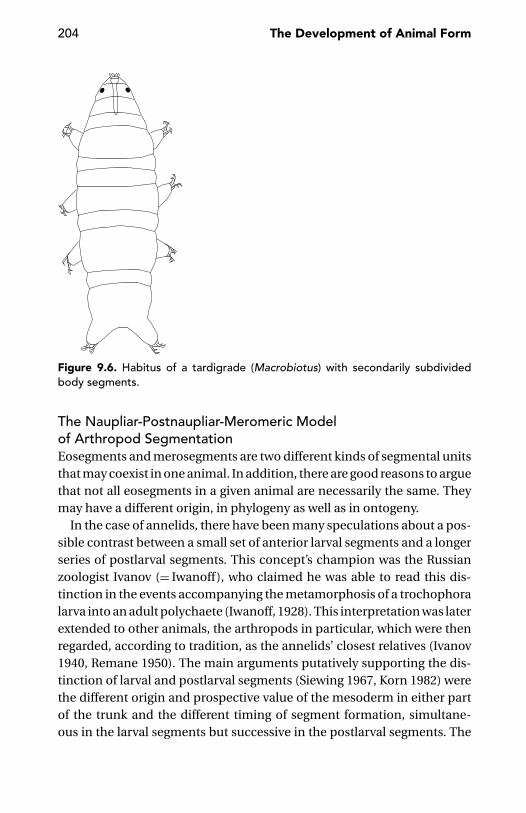
204 The Development of Animal Form
Figure 9.6. Habitus of a tardigrade (Macrobiotus) with secondarily subdividedbody segments.
The Naupliar-Postnaupliar-Meromeric Modelof Arthropod SegmentationEosegments and merosegments are two different kinds of segmental unitsthat may coexist in one animal. In addition, there are good reasons to arguethat not all eosegments in a given animal are necessarily the same. Theymay have a different origin, in phylogeny as well as in ontogeny.
In the case of annelids, there have been many speculations about a pos-sible contrast between a small set of anterior larval segments and a longerseries of postlarval segments. This concept’s champion was the Russianzoologist Ivanov (= Iwanoff), who claimed he was able to read this dis-tinction in the events accompanying the metamorphosis of a trochophoralarva into an adult polychaete (Iwanoff, 1928). This interpretation was laterextended to other animals, the arthropods in particular, which were thenregarded, according to tradition, as the annelids’ closest relatives (Ivanov1940, Remane 1950). The main arguments putatively supporting the dis-tinction of larval and postlarval segments (Siewing 1967, Korn 1982) werethe different origin and prospective value of the mesoderm in either partof the trunk and the different timing of segment formation, simultane-ous in the larval segments but successive in the postlarval segments. The

Segments 205
factual basis of these arguments has been seriously undermined by Dohle’s(1979) criticism, but there is some ground for reopening the question,whether the segments in one given segmented animal are all necessarilydetermined the same way. I will examine mainly the phylum with which Iam more familiar – the arthropods.
That the process of segmentation of the anterior head (the so-calledprosocephalon) of insects is basically different from the process responsi-ble for segmentation of the posterior head and the whole trunk (the thoraxand abdomen) is by now beyond any reasonable doubt (Grossniklaus,Cadigan, and Gehring 1994, Vincent, Blankenship, and Wieschaus 1997,Rogers and Kaufman 1997, Wimmer et al. 1997). Therefore, despite pre-vailing contrary views (e.g., Scholtz 1997), segments of the anterior headare not strictly homologous to those posterior to them.
In terms of genes whose early expression is involved in determiningthe insect’s segmental architecture, the anterior head lacks any expres-sion of the pair-rule genes, whose role in segmentation has been widelystudied in Drosophila (reviewed, e.g., in Lawrence 1992, Carroll, Grenier,and Weatherbee 2001). This evidence (cf. Jurgens and Hartenstein 1993,Rogers and Kaufman 1996, 1997, Wimmer et al. 1997) contributed to thedevelopment of a five-tagma model of insect architecture, in which thehead proper or prosocephalon was considered as basically distinct fromthe posteriorly adjoining gnathocephalon, and the traditional abdomenof insect morphologists was split into ‘true abdomen’ and postabdomen(Akam, Dawson, and Tear 1988).
If the anterior head lacks any expression of the pair-rule genes, it corre-sponds to the domains of expression of other genes, such as orthodenticle,empty spiracles and buttonhead. It is probably not without significancethat homologues of these genes are also expressed in the forehead of ver-tebrates. The boundary between the anterior head and the rest of the bodycould be homologous in insects and vertebrates (Holland, Ingham, andKrauss 1992, Finkelstein and Boncinelli 1994). This correspondence is pos-sibly ancestral; but in my view, it has only to do with axial patterning, notwith segmentation per se, due to the basic difference in the segmentationprocess in the two phyla.
But a distinction between anterior head and the rest of the body is notjust suggested by gene expression domains. The independent segmenta-tion of the first few body segments is also suggested, in arthropods, bythe organisation and development of the nauplius, the typical larva ofmany crustacean groups whose very short body bears just three pairs of

206 The Development of Animal Form
appendages (antennules, antennae and mandibles) corresponding to asmany body segments. Caudal to the mandibular segment is a growth zonefrom which the remaining segments will form later.
The fact that the anterior half lacks the pair-rule gene expression whichmarks the differentiation of segments in the remainder of the body is likelyto be the cause of major developmental differences between the two setsof segments: the naupliar and postnaupliar ones (Minelli 2001).
Transcriptional control and expression patterns of the pair-rule genesare adequately known from one model species only (Drosophila melano-gaster), but there is evidence enough to suggest that the spatial and tem-poral expression of these genes may be different in other insects and inarthropods at large. However, despite the limited evidence available todate, there seem to be good reasons to expect some pair-rule effect inarthropods generally. The role of pair-rule genes in the segmentation of thegerm band has been demonstrated, for example, in the beetle Tribolium(Sommer and Tautz 1993), in spiders (Damen, Weller, and Tautz 2000) andalso in the grasshopper embryo (Davis, Jaramillo, and Patel 2001), in whichsegmentally smooth progression in the expression of engrailed stripes hadpreviously induced doubt regarding the presence of any pair-rule pattern.To introduce the argument, it is better to put the fruit fly temporarily asideto focus instead on multisegmented arthropods, such as millipedes andcentipedes.
Segment number in these arthropods is extremely diverse. Centipedes,for example, range from the 15 leg-bearing segments of the house cen-tipede (Scutigera) and the brown centipede (Lithobius) up to 191 segmentsin a geophilomorph species, whereas millipedes may have just 13 (somepenicillates or pin-cushion millipedes) or as many as 375 pairs of legs, asdescribed in a previous chapter. What is most puzzling, however, is notthis numerical diversity. More puzzling is the lack of diversity in segmentnumber in the other arthropod groups, were it not modest and obviouslysecondary reduction at either extremity of the abdomen (insects) or theopisthosoma (arachnids). Why should a scorpion, a spider and a tick havethe same number of body segments? Why should we find the same numberof segments in a caterpillar, a mayfly, and a cockroach? Numerical diver-sity rules nearly all serial structures, from the vertebral column to the teetharcades in mammals, from the segments of the annelids to the petals andstamens of flowers. But let’s go back to myriapods. These arthropods, fi-nally, are diverse in the number of body segments. But this is right wheresomething unexpected awaits us.
Segments 207
First, many myriapods, despite their fairly large number of segments, donot have any individual variation in segment number. For example, Dicel-lophilus carniolensis is a centipede with 43 pairs of legs: no exception hasever been recorded, despite the thousands of specimens collected acrossits extensive geographic range. In the family (Mecistocephalidae) to whichDicellophilus belongs, most species have either 41 or 45 or 49 pairs of legs,but this number is rigorously fixed for each individual species; there areone or two exceptions among the few species with more than 49 segments(Bonato, Foddai and Minelli in press). The same is true for many millipedefamilies, within which the number of leg pairs is rigorously 60 or 64, forall individuals of all species in the family (Hoffman 1982). How can theseanimals develop their full complement of segments without introducingthe slightest error? In vertebrates, for comparison, the vertebral numberis mostly quite stable but, as expected, it becomes increasingly variable asits mean value increases, behaving as a typical meristic character undergenetic control. The heritability of vertebral number in vertebrates hasbeen recently shown by Lindell (1996) for the European common viper(Vipera berus), by Jockusch (1997) for the plethodontid newts of the genusBatrachoseps and by Billerbeck, Orti, and Conover (1997) for a small fish,the Atlantic silverside (Menidia menidia).
The puzzling question of the possible origin of the astonishing stabi-lity in segment number shown by some myriapods was first addressed byMaynard Smith (1960), who suggested this precision could be only ex-pected if the animal generates segments through a two-step process. Thefirst step would be the production of a small number of primary units,the second the splitting of these primary units into a fixed number of sec-ondary units corresponding to the animal’s conventional (morphological)segments. It seems to be much easier to control the production of a fewprimary units and their subsequent duplication than to get a rigorouslyunerring output from a single source producing a few dozen elements se-rially. Maynard Smith’s suggestion looks obviously attractive, as a possibleexplanation for the lack of variation in the number of body segments inseveral myriapod lineages, but there are further arguments to support it.
Look, for instance, at centipedes. Oddly enough, there is no adult cen-tipede with an even number of segments. Moreover, ca. 1,000 centipedespecies with 15 pairs of legs are known, but none with 17 or 19. The nexthigher number recorded in nature is 21 (as in most scolopendromorphs),then 23 (as in the remaining scolopendromorphs). Another gap (no cen-tipede species with 25 pairs of legs is known) must be bridged before we
208 The Development of Animal Form
reach the lowest extreme of the very large range (27–191), over which thenumber of leg-bearing segments of the worm-like geophilomorph cen-tipedes is distributed. Of the numbers in this range (27, 29, 31, . . . . , 191),some are distinctly preferred over those immediately lower or higher. Wehave already seen that, in the Mecistocephalidae, the large majority of thespecies have either 41 or 45 or 49 pairs of legs – those with either 43 or 47 be-ing very few. In all centipedes with 15, 21, or 23 pairs of legs and in nearly allMecistocephalidae, segment number is common to all individuals withinthe species. In the remaining geophilomorphs, there is intraspecific vari-ability in segment number and this variability is generally accompanied bydifferences between the sexes, with the segment number distribution forthe females shifted towards higher numbers (often 2 or 4 units higher thanin the conspecific males). This picture clearly points to the existence ofdevelopmental constraints (Arthur and Farrow 1999): more precisely, ofmultiplicative mechanisms based on the production of a common setof primary segmental units (eosegments) subsequently subdivided, by astereotyped process of subdivision, into secondary units (merosegments:Maynard Smith 1960, Minelli and Bortoletto 1988, Minelli 2000a).
The expression patterns of pair-rule genes are suggestive of molecu-lar and cellular mechanisms that could materialise this multiplicativesegmentation process. In Drosophila, an earlier 7-stripe pattern sets theblueprint for a later 14-stripe pattern; in Tribolium, 8 and 16 stripes arerespectively seen at earlier and later stages of pair-rule gene expression.However, a few comments are required.
First, pair-rule gene expression, suggesting early determination of pri-mary segments and their subsequent multiplication, does not extend tothe anterior head. Merosegmentation would only affect the postnaupliareosegments, but not the anterior naupliar ones.
Second, the degree of merosegmentation is possibly the same in allspecies within very large groups where adult segment number does notshow interspecific diversity, as in insects and malacostracan crustaceans.But it may vary, even between closely related species, as in the centipedegenus Mecistocephalus, in which most species have 49 pairs of legs, manyhave only 45 and others have more than 49.
The problem is whether there is any factual evidence as to the productionof arthropod segments in other than a plain anterior-to-posterior progres-sion. Admittedly, evidence is vanishingly scanty, but this may be due, inaddition to the expected technical difficulty, to the fact that this questionhas never been seriously addressed. Nevertheless, the expression patternsof segmentation genes in the tobacco hornworm Manduca sexta suggests

Segments 209
(Kraft and Jackle 1994) that segment formation in this insect may not bedue to sequential addition from a posterior budding zone.
There are several reasons to believe that the number of eosegments ismuch more strictly controlled than the pattern of merosegmentation. Mystrongest argument is derived from centipedes. It is reasonably easy tofit the recorded pattern of diversity in segment number in this class to amodel where the leg-bearing segments (plus the first trunk segment whoseappendages are transformed into poison claws) is derived from eight post-naupliar eosegments. The degree of merosegmentation would be two forthe eosegments of Scutigeromorpha, Lithobiomorpha, and Craterostig-momorpha (15 leg-bearing segments); three (but probably two for a fewanterior eosegments) for the Scolopendromorpha (21 or 23 leg-bearingsegments); and 4 to 24 for the Geophilomorpha (27–191 leg-bearing seg-ments; Minelli et al. 2000). To account for the segments forming the pos-terior head and the legless terminal segments, we may hypothesise theinvolvement of two more eosegments. This makes a possibly fixed total of10 eosegments, giving rise to a variable number of postnaupliar meroseg-ments. Segmental organisation of the crustacean nauplius and of the foreend of arthropods generally suggests that three more (eo)segments formthe naupliar part of the body (Minelli 2001). Thus, we have a grand total of13 eosegments, possibly basic to the body architecture of all arthropods.If so, we will be forced to reject (at the level of eosegments at least) thecurrent view that individual segments of arthropods can be gained or lostfrom the body plan within particular lineages (Brusca and Brusca 1990,Nagy and Williams 2001).
These 13 eosegments might even be derived from an equal number ofserially repeated elements already present in a primitive ecdysozoan, be-fore the development of overt segmentation in the arthropod lineage. Sug-gestions come from two different groups of non-arthropod ecdysozoans:nematodes and kinorhynchs. Thirteen is the number of pioneer motoneu-rons which migrate in the developing ventral nerve cord of Caenorhabditiselegans (Sulston and Horvitz 1977, Walthall 1995). Thirteen, again, is thenumber of body units (imperfect segments or ‘zonites’) forming the bodyof the adult kinorhynchs (Kristensen and Higgins 1991; Figure 9. 7).
Reliable Patterning of Eosegments and the VariableSchedule of MerosegmentationIn centipedes, the basically uniform series of trunk segments is gener-ally punctuated by a ‘mid-body anomaly’ (Minelli et al. 2000) which onlyaffects one or a few segments. In lithobiomorphs, craterostigmomorphs

210 The Development of Animal Form
Figure 9.7. A kinorhynch (Campyloderes sp.). (Courtesy of B. Neuhaus, Berlin.)
and scolopendromorphs, the basic alternation of segments with long dor-sal plate (tergum) and lateral spiracles and segments with short tergumand no lateral spiracles is ‘disturbed’ by what seems to be the lack of ashort tergum segment. In scutigeromorphs, a corresponding ‘anomaly’is given by one long tergum covering three leg-bearing segments ratherthan two, as the other terga do. In most geophilomorphs, the mid-bodyanomaly is generally less conspicuous and sometimes hardly visible at all,but it is very visible in some species, as in Stigmatogaster gracilis, in whicha short range of segments are ventrally marked by conspicuous ventralgrooves. I demonstrated (Minelli 1992) that the range of segments with ster-nal grooves expands with age, from a virtual source at about 0.43 relative
Segments 211
segment position along the trunk. This relative position is the same as theposition of the mid-body anomaly in all remaining centipedes, irrespec-tive of the actual ordinal number of the segment(s) involved. This positionis approximately the same as the position of the genital opening in otherarthropods, whereas the genital opening in centipedes is at the posteriorbody end and the mid-body anomaly could correspond to the originalexpression of Abdominal-B (a Hox gene whose relation to genital/sexualstructures has been described on page 181; Minelli et al. 2000). Hughes andKaufman (2002) have demonstrated that in the centipede genus Lithobius,Abdominal-B is expressed twice, first at a mid-trunk level (i.e., in a regioncorresponding to the future mid-body anomaly) and later at the posteriorend of the trunk, where the genital opening and the sexual appendageswill differentiate. My interpretation is that the anterior spot of expressionof Abdominal-B is specified very early in development, before meroseg-mentation. The anterior spot of expression, marking the position of themid-body anomaly, does apparently coincide with the same eosegment inall centipedes; the different segmental position of the anomaly in the dif-ferent taxa will only depend on the different degree of merosegmentationof their eosegments (Minelli 2001).
It is important to stress that, in my interpretation, eosegmentation isachieved very early in development, whereas merosegmentation – as theactualisation of a developmental fate already assigned to each eosegment –may be extensively delayed and occur in an antero-posterior progressionover long spans of embryonic and also possibly postembryonic life. In thegrowth zone of a tiny nauplius, there should be space enough for all post-naupliar eosegments, but we cannot expect to see all full-fledged meroseg-ments from very early on. The same is true for anamorphic myriapods.One may wonder, in the latter case, whether all myriapods actually exploittheir full potential of merosegmentation. Doubts are warranted at leastin the case of juloid millipedes, with their cylindrical body mostly termi-nating with a variable number of apodous segments and with a very largeintraspecific variability in the number of adult segments. In this group,adult specimens without apodous rings are documented in a few species(Sahli 1969), but this condition is quite unusual (Enghoff, Dohle, andBlower 1993).
There is a relationship, but not necessarily a simple one, between merose-gmentation and the pace at which postembryonic development proceeds.Enghoff, Dohle, and Blower (1993) believe that, in millipede evolution, aregular addition of sets of two segments (or multiples thereof) with thecompletion of each moult is a later acquisition rather than an early feature.
212 The Development of Animal Form
But we need to consider that this acquisition paralleled the change fromprimitive millipedes with a relatively low number of segments, as depictedin Enghoff’s (1990) millipede (chilognathan) ground plan, to clades withmuch larger segment numbers, such as polyzoniids and julids. No speci-men with an odd number of segments was found by David and Couret(1984) in a vast material of Polyzonium germanicum. The same occurs inthe Chordeumatida, in which only even numbers of leg-bearing sterniteshave been recorded, starting from the third postembryonic stadium on-wards. In all Juliformia, except for a few secondarily modified cases, onlyodd leg-pair numbers are found.
In arthropods, merosegmentation does not necessarily end with the de-termination of as many units as are the conventional segments the mor-phologist recognises in their adults. In many cases, the process continueswith further division of most, or some, of the trunk segments (never, how-ever, those corresponding to the naupliar part of the body) in subsegmentalunits. Things are a bit equivocal in the millipedes. Equivocal, at least, interms of the conventional standard, according to which a segment shouldbear just one pair of appendages. The majority of the segmental units in amillipede’s trunk are provided with two pairs of legs each, hence the termdiplosegments currently given them. Closer inspection reveals that theduplicity so evident in the appendages – and mirrored by internal featuressuch as ganglia, spiracles, tracheae, and heart ostia – is also present, insome way, in the dorsolateral aspect of the body, with more or less evidentdistinction, within each diplosegment, of an anterior prozonite and a pos-terior metazonite. Dorsal praeterga and ventral praesterna are also presentin some centipedes, in front of the main dorsal and ventral plates of eachsegment respectively. This duplication of segments also affects the insects.This is often seen in the thoracic segments (dorsally divided into the scu-tum and scutellum, laterally in the episternum and epimeron) and some-times also in one or more segments of the abdomen, as in many neuropter-ans (Achtelig 1975). Among the crustaceans, a few copepods appear to havefive to eight abdominal somites, rather than the usual four. This is regardedas the consequence of secondary annulation (= merosegmentation) of thebasic number of abdominal somites (Huys and Boxshall 1991).
Heterogeneous Segments in Vertebrates and AnnelidsQuite probably, there are ‘two heads’ in vertebrates (Duboule 1995) asthere are two in arthropods. The distinction between an anterior and aposterior head is clearly marked in the central nervous system, in which the
Segments 213
expression domains of the homeobox genes of the Otx and Emx families,homologous to the Drosophila orthodenticle and empty spiracle genes,are restricted to the developing rostral brain. Patterning of the developinghindbrain and the corresponding branchial region and spinal chord areunder the influence of Hox gene expression (e.g., Bally-Cuif and Boncinelli1997, P.W.H. Holland 2000).
Xenopus provides a wonderful example of how serially homologous ele-ments, such as the trunk somites of a vertebrate, may be produced by dif-ferent mechanisms within the same animal (Cooke 1980). Embryos whichhave started somite formation as artificially small neurulae with less thanthe usual number of cells manage to adapt the size of their anterior somitesappropriately, but this regulation is forgotten in later axial morphogenesis.Cell number is the same in somites numbered from 20 onwards in initiallysmall embryos and in their normal siblings. This experiment demonstratesthat two different modes of pattern control operate, in different times, inthe generation of one series of somites.
In mice, the formation of the second pharyngeal arch involves a distinctdevelopmental process from that responsible for the production of thethird and fourth arches (Wendling et al. 2000).
The vertebrate peripheral nervous system consists of two groups ofnerves which have a metameric series of proximal roots along the bodyaxis: the branchial and the spinal nerves. Spinal nerve metamerism is pat-terned after the somites, whereas segmental distribution of the branchialnerves is in part intrinsic to the rhombomeres, the segmental compart-ments of the hindbrain.
Still more impressive is the change in segmentation modes in the poly-chaete Spirorbis moerchi, in which larval segments develop simultane-ously in the ectoderm, followed by segmentation of the mesoderm. Post-larval segments are initiated in the mesoderm, followed by segmentationof the ectoderm (Potswald 1981).
The few cases described in this chapter show that, in vertebrates and an-nelids alike, different sets of segments originate through different mecha-nisms in the same animals, as with naupliar and postnaupliar eosegmentsin arthropods; but the similarities between the three animal groups alsoextend to double segmentation (eo- vs. merosegmentation). Even if dou-ble segmentation seems to account adequately for the numerical idiosyn-crasies of centipedes and millipedes, as well as for many other aspects ofarthropod segmentation, there is no reason to regard it as a unique trait ofthe arthropodan clade.
214 The Development of Animal Form
I have already mentioned leeches, with their annulated segments. Atvariance with the outcome of merosegmentation of arthropod eosegments,the secondary units of the leeches are not ‘promoted’ to the status of fullsegments, probably because the main segmentally arranged structures,such as nephridia or the ganglia of the ventral nervous chain, are alreadyspecified when merosegmentation begins.
In other annelids, however, things are possibly different and more sim-ilar to what we have seen in arthropods. In the Oligochaeta, the clitellumgenerally includes segment 16; but in the few exceptions to this rule, itusually includes segment 32. This looks like the effects of early meroseg-mentation (2 merosegments per eosegment), followed by the developmentof the merosegments as full segments. Also interesting, although less con-vincing, is the comparison of some of the shortest annelids in terms oftotal number of setigerous segments: there are 8 segments in the femaleof Parergodrilus (but 9 in the male) and 16 in Hrabeiella and Marioninaeleonorae, the shortest of the Enchytraeidae (Rota 1997).
Polychaetes could be the best group in which to study the different layersof segmentation potentially existing within the same animal. In these an-nelids, besides the examples of merosegmentation previously described(see page 202), there are genera in which a seemingly opposite patternhas been reported [i.e., two or three pairs of ganglia per morphologicalsegment (Pectinaria; Nilsson 1912)].
In vertebrates, the first two halves of the rhombencephalon (anterior =r1–r5 and posterior = r6–r8) behave like eosegments, within which mero-segments (rhombomeres) are progressively established in a characteristicsequence.
Comparable with the cases thus far discussed is the origin of dentition inmammals. In most mammals (but not, for example, in mice), the perma-nent dentition is of mixed developmental origin (McCollum and Sharpe2001), because incisors, canines, and premolars develop from successionallaminae that differentiate from the remnant of the primary dental laminaof their deciduous precursors, and molars develop directly from extensionsof the primary dental lamina.
As a footnote, if homologous parts, such as segments or teeth, are thusfrequently generated by more than one mechanism within the same ani-mal, we shall expect that the same will be true of cell types. Morphologicallyand functionally equivalent cells may have different origins within thesame animal. Smith, Kachinsky, and Miller (1994) found that myogenic

Segments 215
cells in the different regions of a mouse somite (dermatome, myotome,and sclerotome, as well as the dorsal versus ventral halves of somites)have different patterns of expression of four muscle regulatory factor pro-teins; therefore, they suggest that myogenic cells in the somites may havemultiple sites of origin and form through multiple molecular pathways.
Germ Layers and Segmentation
In different animals, segmentation affects body architecture more or lessextensively, generally involving both ectodermal and mesodermal, but notendodermal, derivatives. Tissues derived from the different germ layers aresometimes segmented independently. When segmentation of one germlayer’s derivatives is in register with segmentation of another germ layer’sderivatives, we expect this to be due to transpatterning, but there seemsto be no general rule (Holland 1988).
Segmentation of the endoderm is admittedly rare, the most conspicu-ous example being the segmental gut caeca of rhynchobdellid and manygnathobdellid leeches. These are in register with as many ectomesoder-mal segments. This endodermal segmentation is in part autonomous, inso far as a periodic pattern of expression of the Lox3 gene precedes and co-incides spatially with the segmental (intercoecal) constrictions of the gut.But this endodermal segmentation is also dependent on the mesoderm, asthe ablation of segmental mesoderm prevents differentiation of the defini-tive endoderm and expression of Lox3 RNA (Wedeen 1995, Wedeen andShankland 1997; cf. page 119).
Joint involvement of ectodermal (epidermis, nervous system) and meso-dermal derivatives (musculature, coelomic cavities, skeletons) is the rule.Ectoderm and mesoderm, in turn, follow largely independent routes inacquiring segmentation, with ectoderm leading the process in arthropodsand mesoderm leading the process in annelids (mostly) and vertebrates.An exception to this generalisation is the rhombomeres in vertebrates.Another exception is polychaetes such as Nereidae, Eunicidae, Tomopteri-dae, and Serpulidae (Korn 1982). The problem thus arises as to how the ec-todermal and mesodermal (and, when involved, also endodermal) deriva-tives are put in register during the development of segmented animals.
In the leech Helobdella robusta, normal development of the segmentallyrepeated cell clones derived from one of the blast cells does not dependon signals from adjacent clones derived from the other blast cells (Seaver

216 The Development of Animal Form
and Shankland 2000). As previously described (page 128), the longitudinalbandelets generated by the individual blast cells are brought into align-ment during a later process of morphogenetic assembly.
In vertebrates, the most conspicuous evidence of segmentation is foundin the derivatives of the mesodermal somites – in the vertebral column es-pecially, also often in the arrangement of the trunk musculature. Head seg-mentation is less obvious, but segmental interpretation of cephalic struc-tures has been a recurrent theme since Spix’s (1815) and Goethe’s (1820)speculations about a vertebral origin of the skull bones. Segmentation isnow clearly demonstrated in articulation of the hindbrain in neuromeres,but this has nothing to do with patterning of the skull bones. In the trunk,segmentation is first expressed in the mesodermic somites, which impartsegmentation to the spinal cord. The latter is not primarily organised inneuromeric segments analogous to the rhombomeres. Roles are reversedin the posterior (branchial) region of the head, where bone and connec-tive tissue are generated by the neural crest, which is derived from thehindbrain rhombomeres. Experiments demonstrate that the cranial neu-ral crest is prepatterned or imprinted with positional information beforemigration (Lumsden, Sprawson, and Graham 1991, Krumlauf 1993).
Segmental Mismatches and Resegmentation
If we still need arguments in favour of a less typological view of segmen-tation than has been fashionable up to now, we can find some in thewidespread mismatch between the dorsal and ventral expressions of seg-mentation in the same animal and in the phenomenon of resegmentation.
Dorso-Ventral MismatchesThe archetypal or, better, text-book arthropod segment is covered by a ter-gum dorsally and a sternum ventrally. Elaborations upon this basic schemeare very common. These may include additional (pleural) sclerites placedbetween the tergum and the sternum, or the more or less distinct subdivi-sion of these primary sclerites into an anterior and a posterior half each.These additional features notwithstanding, the basic architecture of thesegment remains nevertheless recognisable. But often, things are not thatsimple. I do not discuss body regions, such as the head, which are usuallydescribed as resulting from the ‘fusion’ of segments. Instead, I will notesome examples of dorso-ventral mismatch in the number, arrangementor patterning of segments.
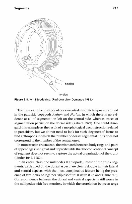
Segments 217
Figure 9.8. A millipede ring. (Redrawn after Demange 1981.)
The most extreme instance of dorso-ventral mismatch is possibly foundin the parasitic copepods Aethon and Norion, in which there is no evi-dence at all of segmentation left on the ventral side, whereas traces ofsegmentation persist on the dorsal side (Kabata 1979). One could disre-gard this example as the result of a morphological deconstruction relatedto parasitism, but we do not need to look for such ‘degenerate’ forms tofind arthropods in which the number of dorsal segmental units does notcorrespond to the number of the ventral ones.
In notostracan crustacean, the mismatch between body rings and pairsof appendages is so great and unpredictable that the conventional conceptof segment does not seem to capture the actual organisation of the trunk(Linder 1947, 1952).
In an entire class, the millipedes (Diplopoda), most of the trunk seg-ments, as defined on the dorsal aspect, are clearly double in their lateraland ventral aspects, with the most conspicuous feature being the pres-ence of two pairs of legs per ‘diplosomite’ (Figure 8.22 and Figure 9.8).Correspondence between the dorsal and ventral aspects is still worse inthe millipedes with free sternites, in which the correlation between terga
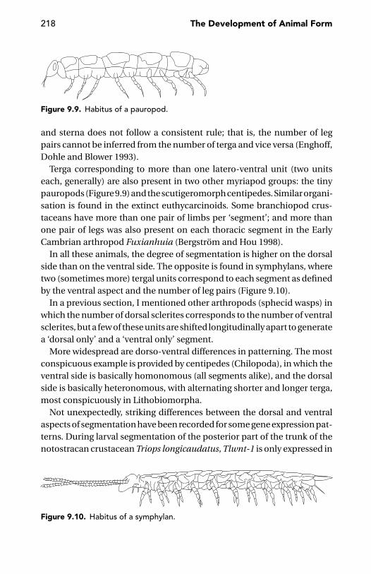
218 The Development of Animal Form
Figure 9.9. Habitus of a pauropod.
and sterna does not follow a consistent rule; that is, the number of legpairs cannot be inferred from the number of terga and vice versa (Enghoff,Dohle and Blower 1993).
Terga corresponding to more than one latero-ventral unit (two unitseach, generally) are also present in two other myriapod groups: the tinypauropods (Figure 9.9) and the scutigeromorph centipedes. Similar organi-sation is found in the extinct euthycarcinoids. Some branchiopod crus-taceans have more than one pair of limbs per ‘segment’; and more thanone pair of legs was also present on each thoracic segment in the EarlyCambrian arthropod Fuxianhuia (Bergstrom and Hou 1998).
In all these animals, the degree of segmentation is higher on the dorsalside than on the ventral side. The opposite is found in symphylans, wheretwo (sometimes more) tergal units correspond to each segment as definedby the ventral aspect and the number of leg pairs (Figure 9.10).
In a previous section, I mentioned other arthropods (sphecid wasps) inwhich the number of dorsal sclerites corresponds to the number of ventralsclerites, but a few of these units are shifted longitudinally apart to generatea ‘dorsal only’ and a ‘ventral only’ segment.
More widespread are dorso-ventral differences in patterning. The mostconspicuous example is provided by centipedes (Chilopoda), in which theventral side is basically homonomous (all segments alike), and the dorsalside is basically heteronomous, with alternating shorter and longer terga,most conspicuously in Lithobiomorpha.
Not unexpectedly, striking differences between the dorsal and ventralaspects of segmentation have been recorded for some gene expression pat-terns. During larval segmentation of the posterior part of the trunk of thenotostracan crustacean Triops longicaudatus, Tlwnt-1 is only expressed in
Figure 9.10. Habitus of a symphylan.

Segments 219
the ventral portion of the epidermis (Nulsen and Nagy 1999). In insects,the anterior boundary of expression of Antennapedia resides in the poste-rior labial segment ventrally, but in the anterior prothoracic segment andappendages laterally (Peterson et al. 1999).
ResegmentationIn all segmented animals, the first signs of serial patterning are mani-fest since early or very early developmental stages. The segments seen inthe body architecture of many adult metazoans do not necessarily havethe same anterior and posterior boundaries as the first segmental unitswhich are formed in the same animals. The problem is not simply one ofmerosegmentation (see page 202), but also one of resegmentation.
The concept of resegmentation was introduced by Remak (1855), whoregarded the adult vertebrae of amniotes as shifted by one half unit withrespect to the sclerotomal (bone-forming) portion of the somite. In otherwords, the posterior half of the sclerotome of one segment would fuseto the anterior half of the sclerotome of the next segment, so that thedefinitive centrum (the body of the vertebra) will be intersegmental ratherthan segmental. According to Williams (1959), such a resegmentation doesnot affect the whole of the vertebra, as it would apply to the bulk of theneural arch and the pleurocentrum, but not to the prezygapophysial regionof the neural arch and neither to the ventral arch and to the intervertebraldisc. The latter structures would be strictly segmental; that is, they wouldretain the primary segmentation. In this sense, a modern amniote vertebrawould be a mosaic of pieces derived from primary segmentation, togetherwith pieces subjected to resegmentation.
The resegmentation hypothesis has been alternatively accepted (e.g.,Carroll 1989, O’Higgins et al 1997), based partly on new embryologicalevidence (e.g., Bagnall, Higgins, and Sanders 1988, Ewan and Everett 1992,Huang et al. 1995), and doubted (e.g., Keynes and Stern 1988) or out-right refuted (e.g., Verbout 1985). It is also possible that differences existbetween different amniote lineages. Convincing evidence for resegmenta-tion is available, for example, for the dorsal vertebrae of the turtles (Rieppel2001), in which the neural arches shift forward by half a segment carryingthe ribs with them (Goette 1899).
According to Verbout (1985), an alternative explanation to the chang-ing pattern of segmentation in the vertebral column of amniotes could bea simple realignment of the relative positions of the sclerotome and my-otome, rather than actual fusion of half sclerotomes from adjacent somites.

220 The Development of Animal Form
There is no clear experimental evidence in favour of his hypothesis (Tamand Trainor 1994).
The final verdict on this question does not seem to have been formulatedyet, but molecular developmental genetics seems to come in support toRemak’s hypothesis. In the mouse, starting 10 days postcoitum, the seri-ally repeated expression of the homeobox gene Hlx is out of register withthe intersomitic fissures (the original segmental boundaries), suggestinga molecular blueprint for resegmentation (Lints et al. 1996).
In arthropods, the issue of resegmentation has been introduced byMartınez-Arias and Lawrence (1985). They noticed that the segments ofDrosophila, as marked by the intersegmental membranes in both larva andadult, do not correspond to the serial units defined by the segmentationgenes, but are shifted by one half unit in respect to them. Martınez-Ariasand Lawrence introduced the term parasegment for the original units ofsegmentation to distinguish them from the ‘true’ segments of larva andadult. As each parasegment comprises two compartments (anterior andposterior), resegmentation in Drosophila means the association of theposterior compartment of parasegment i with the anterior compartmentof parasegment i + 1 to produce a definitive (morphological) segment.Lawrence (1992) regards parasegments as the only fundamental unit ofsegmentation, in developmental terms. Segments, conspicuous as theseunits may be in morphological and functional terms, “are largely a figmentof our imagination” when considered from a developmental point of view(Lawrence in discussion to Riechmann et al. 1997: 2922). This is certainlytrue for the abdomen of Drosophila, but it is not clear how pervasive re-segmentation occurs in insects or in arthropods at large. Parasegmentshave been found in malacostracan crustaceans (Scholtz and Dohle 1996).In the centipede Lithobius, Cynthia Hughes (personal communication)found both segmental and parasegmental patterns, but the latter seem tocome out later than the first, thus making resegmentation unlikely. Evenin Drosophila, where resegmentation is obvious in the abdomen, this isnot necessarily true for the thorax and the head (Rogers and Kaufman1996, 1997). As in many other aspects of development, however, decid-ing whether an animal undergoes resegmentation or not may criticallydepend on evidence from the correct temporal layer. Parasegmental pat-terns may appear during a restricted temporal window only to be swampedsoon by resegmentation. A quick transition from a parasegmental patternto a segmental pattern may invest the expression of the same class ofgenes. In Drosophila, early expression of fushi tarazu and even-skipped
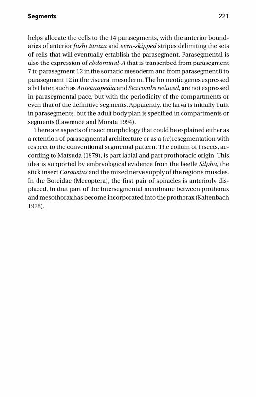
Segments 221
helps allocate the cells to the 14 parasegments, with the anterior bound-aries of anterior fushi tarazu and even-skipped stripes delimiting the setsof cells that will eventually establish the parasegment. Parasegmental isalso the expression of abdominal-A that is transcribed from parasegment7 to parasegment 12 in the somatic mesoderm and from parasegment 8 toparasegment 12 in the visceral mesoderm. The homeotic genes expresseda bit later, such as Antennapedia and Sex combs reduced, are not expressedin parasegmental pace, but with the periodicity of the compartments oreven that of the definitive segments. Apparently, the larva is initially builtin parasegments, but the adult body plan is specified in compartments orsegments (Lawrence and Morata 1994).
There are aspects of insect morphology that could be explained either asa retention of parasegmental architecture or as a (re)resegmentation withrespect to the conventional segmental pattern. The collum of insects, ac-cording to Matsuda (1979), is part labial and part prothoracic origin. Thisidea is supported by embryological evidence from the beetle Silpha, thestick insect Carausius and the mixed nerve supply of the region’s muscles.In the Boreidae (Mecoptera), the first pair of spiracles is anteriorly dis-placed, in that part of the intersegmental membrane between prothoraxand mesothorax has become incorporated into the prothorax (Kaltenbach1978).

CHAPTER TEN
Evo-devo Perspectives on Homology
Cells may have different characters now because they were indifferent situations in the past. They are like identical twinsreared apart; they remember their separate experiences.
J.H. Lewis and L. Wolpert 1976: 467
The initial progenitor field is transformed into a mosaic ofregulatory subdomains, and, remarkably, these prefigure themorphological pieces of the body part.
E.H. Davidson 2001: 110
Concepts and Interpretations
From a structuralist perspective, Muller and Newman (1999) have devel-oped the interesting argument that homologous structures, in so far asthey are integrated as fundamental parts of a given body plan, become‘attractors of morphological design’, a kind of backbone to which furtherelements of body design may be added.
Hierarchies and BeyondA hierarchical frame of mind is taken for granted in nearly all approachesto homology. Analysis in terms of hierarchies of structural parts or func-tional subsystems has been a common strategy in the study of complexsystems since the dawn of modern science (Bechtel and Richardson 1993,Zawidzki 1998). However, as forcefully argued by Kauffman (1993), withbiological systems this is not the most satisfactory strategy. Ahl and Allen(1996) define a system as hierarchical if it is composed of stable, observ-able subunits unified by a superordinate relation. It would be fortunateif the features we try to compare were always stable and unified by a
222
Evo-devo Perspectives on Homology 223
superordinate relation. I suspect, however, that stable and hierarchicallyunified features would make the world much duller and less interestingthan the real biological world.
Advocacy of hierarchy in homology is not universal. Striedter (1999)admits that biological systems consist of a complex network of multiplehierarchies, such that the hierarchical nature of the homology conceptremains potentially confusing. Nagy and Grbic (1999) acknowledge thatwe know very few gene networks in development functioning in a trulyhierarchical manner.
Kauffman (1993) predicted that genomic regulatory architectures aremore likely to be rich in feedback loops rather than purely hierarchical.This has been repeatedly demonstrated by recent studies. Arnone andDavidson (1997) observe that the linkages between internal genes in ge-nomic networks may connect a given gene to others at several differentlevels in the same network, something that makes the network other thanstrictly hierarchical.
Homology: Absolute or Relative?Until recently, homology was universally regarded as an absolute relation-ship. Structures either are, or are not, homologous (Striedter and Northcutt1991, Bolker and Raff 1996). However, by contrasting genes affecting pre-pattern with genes affecting process, Maynard Smith and Sondhi (1960)have long ago distinguished, in a sense, between positional and specialhomology. Long before them, Bateson (1894: 24) already had the followingobservation to offer: “I am aware that Meristic and Substantive Variationsoften occur together, and that there is a point at which it is not possi-ble to separate [them] satisfactorily. [..] For example, we may see that itis through Meristic Variation that the vertebral column of a Dog may bedivided into a number of Vertebrae greater or less than the normal; andthough in such cases all the Vertebrae have distinctly canine characters,yet there are nearly always Substantive Variations occurring in correlationwith the Meristic Variations, manifesting themselves in a re-arrangementof the points of division between the several groups of Vertebrae, and caus-ing individual Vertebrae to assume characters which are not proper to theirordinal positions”.
The first recent author to seriously challenge the all-or-nothing notionof homology was Roth (1984). Her paper opened the way to the moreflexible views defended later, for example, by Haszprunar (1992) and myself(Minelli 1996b, 1998).

224 The Development of Animal Form
It has long been acknowledged that homologous parts may be integratedto form nonhomologous complexes. This is one of the arguments leadingto the widespread hierarchical notion of homology. On this basis, Striedterand Northcutt (1991) criticised the notion that homologous charactersmust be based on the same, or homologous, causal elements and mecha-nisms (a view that had been championed, for instance, by Spemann 1915,Atz 1970, Hodos 1976, Roth 1984 and Wagner 1989). In their opinion, thiswould be a reductionist approach to the concept of homology, becauseof the claim that the homology of characters at any one level of organisa-tion can be reduced to the homology of their causal mechanisms. Striedterand Northcutt argued instead that evolution may bring about any level ofchange at any level of biological organisation, and that these changes mayinclude the causal mechanisms that generate them. This view seems tooabstract. To say that an aspect of biological organisation remains the same,irrespective of any evolutionary change it may undergo, will inevitably re-duce characters to Platonic ideas. Striedter and Northcutt (1991) pointto the complexity of causal relationships that may interrelate the variouslevels of biological organisation (e.g., because of pleiotropy or multigeniccontrol of morphological traits). Furthermore, during the course of evolu-tion, the causal relationships between characters at different levels of or-ganisation may change. A character at any given level of organisation maythus change by phylogenetic addition, deletion or substitution of elementsand causal factors at a lower level. Therefore, Striedter and Northcutt con-cluded that characters at the same level of organisation may be homolo-gous, even if some of their causal factors or component elements at a lowerlevel are not homologous. I would argue that these circumstances force usto adopt a factorial, or combinatorial, view of homology (Minelli 1998).
Shubin and Wake (1996), Abouheif (1999) and Pigliucci (2001) have madeexplicit mention of partial homology as the possible relationship betweenthe two traits we are comparing. Wake (1999: 44–45) even goes so far as toaffirm that, “Because evolution is a continuous process, [..] homology canonly ever be partial, in any real sense.” Comparative morphology, especiallycomparative developmental biology, offer lots of examples to support thisview.
Breidbach and Kutsch’s (1990) comparative study of the neurones inner-vating the dorsal longitudinal muscles in several insects is a nice exampleof factorial analysis of homology. Breidbach and Kutsch compared neu-rons in juvenile and adult locusts, in larval and adult beetles and in variousbody segments of these insects. In all species, instars and segments, they
Evo-devo Perspectives on Homology 225
found a common set of 11 neurons: seven motoneurons stemming fromthe next anterior ganglion and four neurons located in the ganglion of thesegment containing the muscles. Also common was the basic structure ofthe dendritic field. This identical neuronal plan was found between dif-ferent segments (serial homology), between the nymphal or larval stageand the adult of the same species (ontogeny), and among different insectspecies, both hemi- and holometabolous (phylogeny).
In vertebrates, the development of spinal and cranial motor neuronsoffers an intriguing mix of similarities and differences (Jacob, Hacker, andGuthrie 2001). At a molecular level, there is abundant evidence that severaltranscription factors relevant for body patterning operate in a synergisticand combinatorial way (Garcıa-Bellido 1994b, Davidson 2001). Dubouleand Morata (1994) suggested the existence of a combinatorial componentin the control exercised by homeotic genes on the patterning of the mainbody axis to the point that analysis of the phenotypic expression maybecome difficult. An example is the combined function of Ultrabithoraxand abdominal-A in the epidermis of abdominal segments II to VII inDrosophila. In this body region, the expression of both genes is required,although not necessarily by the same cells. But in the visceral mesoderm,these genes are not co-expressed and have distinct effects, with Ultrabitho-rax being expressed only in parasegment 7 and abdominal-A in paraseg-ments 8–12.
When dissecting homology into its components, one of the most funda-mental distinctions is between positional and special homology (Minelliand Schram 1994, Minelli 1998, 2002). According to classic comparativeanatomy, body regions are homologous among amniotes despite varia-tion in the number of vertebrae and the position along the body axis, aconcept known as ‘transposition’ (Goodrich 1906, 1913, Burke et al. 1995,Muller and Wagner 1996, P.H.W. Holland 2000). For example, the transi-tion between the cervical and thoracic vertebrae occurs at very differentsegmental positions in the different species. The equivalence of this tran-sition, irrespective of segmental position, is shown by the circumstancethat in mammals as in birds, in fishes as in amphibians, this transition(less clear in fishes than in tetrapods; van der Hoeven et al. 1996) corre-sponds to the anterior boundary of expression of the Hoxc6 gene (Gaunt1994, Burke et al. 1995).
This distinction between positional homology and special homology,obviously more important at the level of organs and body parts, is alreadyrelevant at cellular level (Wolpert 1996). For example, homologous cell
226 The Development of Animal Form
types may be located in non-homologous brain regions, while homologousbrain regions may contain non-homologous cell types (Striedter 1998). Cellmigration, as in the case of the neural crest derivatives, modifies positions,but migrating cells are known to be (or expected to be) specifically labelledbefore moving, thus offering a further example of dissociation betweenpositional and special homology.
This dissection of homology components is, basically, one of temporallayers. Striedter (1999) acknowledges that homology of adult characters isconceptually independent of the homology of their embryonic precursors.Drawing from his favourite field of comparative neurology of vertebrates,he shows that homology of the embryonic telencephalic zone is a separateissue from the homology of the adult neocortex. Homology of neocor-tex (as neocortex) is not disproved by the fact that some of its neuronsare derived from embryonic regions other than the dorsal telencephaliczone (Anderson et al. 1997). (A question of periodisation nevertheless re-mains: how are developmental stages defined, e.g., embryo and adult? cf.chapter 4).
Dissection of temporal layers of homology is what Wagner and Gauthier(1999) suggested with respect to the problem of digit homology in the avianhand. It has been long disputed whether bird digits represent digit I, II, andIII (conventional view) or digit II, III, and IV (Burke and Feduccia 1997)of the tetrapod pentadactyl limb. Wagner and Gauthier believe that dif-ficulties with this homologisation may be derived from dissociationbetween the developmental origin of the repeated elements (primordialcartilage condensations) and their subsequent individualisation into fullyfunctional characters (digits). Thus, there would be causal independence(cf. Tabin 1992) between morphogenetic processes creating chondrogeniccondensations in the limb and the later developmental individualisationof those elements when they are specified as functional fingers. In theavian case, comparative embryological evidence identifies the primor-dial condensations as CII, CIII, and CIV, but later in development thesecondensations differentiate following a frame shift in the developmentalidentities such that CII becomes digit I, CIII becomes digit II, and CIVbecomes digit III. This agrees with Hardy et al.’s (1995) study, which sug-gests, in the chick limb, the independence of a prepatterning mechanismspecifying the digits from a positional information-based process whichspecifies digit identity.
The question of homology of the amphibian digits is also tricky. Wagneret al. (1999) move from the finding that in the digit-forming region of the
Evo-devo Perspectives on Homology 227
newt Notophthalmus viridescens, there is a phase of Hoxa-11 expressionstarting with the development of digit III, whereas no corresponding ex-pression is known for either frogs (Xenopus) or amniotes. This unique traitof molecular developmental biology would suggest a peculiar origin forthe digits of recent urodeles. They would be derived from a lineage of am-phibians with two digits only (digits III and IV of the original pentadactylappendage), which would survive as urodele digits I and II, and the re-maining urodele digits would represent an evolutionary innovation.
Another dissection of homology components is invited by the circum-stance that in the epidermis (and cuticle) of insects, patterns of differ-entiation and patterns of antero-posterior or proximo-distal polarity areto some extent under separate control (Nubler-Jung 1987, Nubler-Jungand Grau 1987). This has been shown in Drosophila through the analy-sis of suitable mutants (Gubb and Garcıa-Bellido 1982, Held, Duarte andDerakhshanian 1986) and in the cotton seed bug Dysdercus by experimen-tal grafting of rotated pieces of cuticle (Nubler-Jung and Grau 1987). Thedifferentiation pattern is determined at the scale of the entire segment(Locke 1960, Nubler-Jung 1977), whereas the cell polarity pattern is de-pendent on local interactions between cells.
Most ontogenetic features evolve in a mosaic fashion, because develop-mental integration is historically labile. In the phylogenetic time dimen-sion, characters which appear to be integrated in ontogeny may evolveas separate features. I do not see why this dissociation should specifi-cally characterise the speciation events, as suggested by Fink and Zelditch(1996).
Temporal Serial HomologyLarval and adult homologues are sometimes derived from two temporallydistinct phases of activation and differential specification of subsets ofa same anlage. The most familiar example is that of the polyphyodontvertebrates (i.e., those that have two or more dentitions). Humans sharethis trait with most mammals but also with many fishes, amphibians andreptiles. Less obvious is the example of the insect compound eyes andtheir larval counterparts. In insects such as a cockroach or a grasshopper,there is an obvious morphological and developmental continuity betweenthe compound eyes of the first postembryonic stage and those of laterstages, adult included. We just noticed the addition of new ommatidiafollowing each moult. Things are different in the holometabolous insects,whose larvae do not have true compound eyes, but a small cluster of visual
228 The Development of Animal Form
organs called the stemmata. The developmental relationships between thelarval stemmata and the ommatidia of the adult compound eyes have beenrevealed by Paulus (1989) with his study of Chaoborus (Diptera). Paulusdemonstrated that existence of a common anlage is responsible for pro-duction of both larval stemmata and adult ommatidia. The visual organprecursors develop serially in two main waves, in different developmentalcontexts: embryonic/larval (producing stemmata) and pupal/adult (pro-ducing ommatidia).
Most dipteran larvae (e.g., those of Drosophila) have a reduced headand no eye at all, nevertheless they have other organs in a relationshipof temporal serial homology with corresponding adult structures. The so-called Keilin’s organs of the larvae of Drosophila have been interpreted asrudimentary legs, topographically corresponding to the legs of the adultfly. Keilin’s organs do not grow to become the fly’s legs, but disappear atmetamorphosis, whereas the leg/wing imaginal discs become activatedand give rise to adult appendages. Cohen, Wimmer, and Cohen (1991)demonstrated that the primordia of Keilin’s organs and those of the adultleg primordia are part of a single cluster of cells expressing both Distal-less and disconnected. The two kinds of organs are thus temporal serialhomologues.
We can probably apply the concept of temporal serial homology to manystructures that undergo complete restructuring during amphibian meta-morphosis (Wake and Hanken 1996). In the plethodontid Eurycea bislin-eata, for example, there is compartmentalisation between cells fated toform the larval epibranchial cartilage and cells which will form its adultequivalent (Alberch, Lewbart, and Gale 1985, Alberch, Gale, and Larsen1986, Alberch and Gale 1986, Alberch 1987, 1989), whereas in the jaw mus-cles of the anurans, the larval myofibres degenerate and the new adultmuscles are formed, at metamorphosis, by myofibers derived from quies-cent cells within the larval muscles (Alley 1989).
Underlying temporal serial homology, there is probably a reactivation,later in development, of patterning genes already expressed at earlierstages. A comparable behaviour is known in Drosophila for many genesinvolved in the early embryo in the patterning of the main body axis whichare expressed again in the imaginal discs, where they are involved in thepatterning of the (paramorphic) body appendages (Bryant 1996).
Genes and Homology“Can homology ever be definitively demonstrated? I think not” (Wake 1999:27). In David Wake’s opinion, to speak of homology is nothing more, and
Evo-devo Perspectives on Homology 229
nothing less, than to speak of phylogenetic continuity. But continuity ofwhat? Of structures, of genes, of mechanisms?
There is no question that many structures whose homology cannot bemeaningfully challenged in terms of comparative morphology are pro-duced by different mechanisms in different animals (e.g., Wilson 1894,Remane 1952, de Beer 1958, 1971, Sander 1983, Roth 1984, Hanken 1986,Henry and Raff 1990, Wray and Raff 1990, Striedter and Northcutt 1991,Striedter 1998). In various amphibians, somites differentiate and developin the most diverse ways, including 90◦ rotation as in Xenopus (Malacinskiet al. 1989). In bony fishes, such as zebrafish, fin muscles arise from migrat-ing precursor cells equivalent to the limb muscle precursors of tetrapodspecies. But in dogfish embryos, the same muscles are produced throughdirect epithelial somitic extensions (Neyt et al. 2000). As observed long agoby Baltzer (1952), xenoplastic grafts often give rise to partially equivalentstructures, sometimes even in organs that in normal development do notseem to be homologous or are altogether absent in one of the partners ina chimera.
A comprehensive theory of morphological evolution cannot deal withhomologues in structural terms only, but it must also explain their originand fixation (Muller and Newman 1999). No wonder that many attemptshave been made at solving this problem by looking at shared developmen-tal genes and pathways.
Nowadays, the concept of homology is no longer restricted to the toolkitof the morphologist, but has colonised the fields of genetics and molecularbiology extensively (e.g., Egel 2000, Fitch 2000). I will not discuss the com-mon abuse of the term, when used to denote simple sequence similarity be-tween two macromolecules, a position forcefully rejected by Reeck et al.’s(1987) classic commentary. Rather, I wish briefly to comment on the in-creasingly common identification of homology with shared expressionpatterns of some regulatory gene at a given developmental stage. Bolkerand Raff (1996) remark that this definition of homology is basically differentfrom the traditional ones, in that it is based on mechanism rather than onstructure, and tends to ignore phylogeny. However, Morata and Sanchez-Herrero (1999) find that Snodgrass’s (1935) subdivision of the arthropodappendage into coxopodite and telopodite coincides with the two regionsdefined by the expression of the extradenticle gene and the functioning ofHedgehog signalling.
An interesting experimental approach to homology is Smith et al.’s (2000)research on the histology of the stomach and gastrointestinal tract inXenopus, chicken and mice, coupled with the study of the expression
230 The Development of Animal Form
patterns of several genes in the developing guts of the same animals. In allthree species, the anterior part of the stomach was histologically similarand showed a common expression pattern, during embryonic develop-ment, for two secreted factors, Wnt5a and BMP-4. Also comparable, inboth histological and molecular phenotypes, were the posterior nonglan-dular stomach and pyloric sphincter regions of the chicken and mouse,with the chicken expressing the six2, BMPR1B, and Barx1 genes, and themouse expressing the Nkx2.5 gene.
Success notwithstanding, it is still doubtful how confidently we can relyon gene expression patterns for the assessment of homology. At no stageduring development is there a precise genetic description of the adultorganism (Gubb 1998). We have positive proof of how misleading thesemolecular data may be. In the rhizocephalan parasitic crustacean Sac-culina carcini, for example, the gene caudal is expressed in the thorax, butnot in the abdomen – that is, it shows a spatial pattern completely differentfrom that of the other arthropods (Rabet et al. 2001). Abzhanov, Popadic,and Kaufman (1999) performed comparative analysis of the expressionpatterns of Ultrabithorax/abdominal-A in representatives of the differentclasses of arthropods and found great differences in the anterior bound-ary of expression of these genes. These authors therefore concluded thatHox expression patterns are far from being a safe criterion for establishingthe homology of segments and appendages of arthropods belonging todifferent classes.
A positive consequence of the current interest in comparative gene ex-pression patterns is increased attention to the comparison of processes.This reminds me of Garcıa Bellido’s (in Wolpert, Ghysen, and Garcıa-Bellido1998) answer to the question, exactly what is conserved in evolution? Hisanswer was: the operation!
I share Gilbert and Bolker’s (2001) view that homology of process will bea critically important concept in the future of evolutionary developmen-tal biology. However, I do not wish to restrict the meaning of homologyof process, as they do, “to describ[ing] the relationship between patternsthat are composed of homologous proteins and that are related by com-mon ancestry” (p. 439). When looking at gene expression patterns as away to unravel homology, one should be aware that “a gene performs ahomologous function in two animals if at least some of its upstream orits downstream linkages (or both) remain the same in the two genomes,and the function it performs is descendant from their common ancestor.”The pertinent question thus becomes: “what is a homologous use of a given
Evo-devo Perspectives on Homology 231
gene in a comparison of diverse organisms?” (Both quotes are fromDavidson 2001: 201). But this question is still at the level of the gene. Redu-cing organ homology to gene homology is conceptually and methodologi-cally equivalent to reducing species phylogeny to gene phylogeny, a fault ofwhich systematists are increasingly aware (Maddison 1997, Nichols 2001).
I think that we shall apply the concept of process homology to devel-opmental processes at all levels. This will help us avoid the pitfalls ofpleiotropy and co-option. As noted by Galis (1996), when we comparedistantly related animals, such as insects and vertebrates, homologoushomeobox genes often appear to be involved with the production of non-homologous structures, because of the events happening, in the two sys-tems, downstream of the Hox gene expression. Consideration of the wholedevelopmental pathway is required for a sensible assessment of homology.To take the whole developmental pathway into account, it is not enoughto consider the expression patterns of more than one gene and the con-trol cascades in which these genes are involved. It is necessary to study, inaddition, the epigenetic properties of development (Muller and Newman1999), that is, the generic properties of molecules, cells and tissues, andthe interaction dynamics among them.
Genetic Redundancy, Network Degeneracy, and HomologyLiving beings do not care much which gene is encoding a particular func-tion (Akam in the discussion of Wray 1999). Within one developing or-ganism, there often seems to be many equivalent ways of doing the samething – a circumstance that is usually described as an instance of redun-dancy. Tautz (1992) did not hesitate to regard redundancy as a necessaryrequirement for the development and evolution of complex life forms.Redundancy, in particular, has been predicated of genetic networks, butGreenspan (2001) has shown that network behaviours usually attributedto redundancy are due to a different property. For example, knockout mu-tations are often found to have no apparent phenotypic effect, but thisis not an example of redundancy, because the latter implies preservationof the overall network structure and its functional outcome through thesubstitution of identical elements. Genetic networks are systems of highlyinterconnected non-identical elements related by non-uniform patternsof connectivity. It is because of this property that the network can producethe same result using different strategies (‘degeneracy’, as in the case of thegenetic code), whereas redundant systems always produce the same resultby the same strategy. According to Greenspan (2001), degeneracy of living

232 The Development of Animal Form
systems is not limited to genetic networks. It is amplified, in multicellularorganisms, by the plurality of their interacting cell types and organ systems.This is why whole developmental systems can evolve without necessarilyshowing morphological change, as discovered by Sommer (1997) in vulvalfate specification in Caenorhabditis elegans.
An interesting difference between redundant systems and degeneratesystems is that the former are likely to produce clearly identifiable ho-mologues, whereas the latter are likely to be features with complex andperhaps contradictory homologous components.
Evolutionary NoveltiesMuller and Wagner (1991: 243) defined a morphological novelty as “a struc-ture that is neither homologous to any structure in the ancestral speciesnor homologous to any other structure of the same organism”. If we takethis definition too literally, of course, there is hardly an evolutionary nov-elty in the whole history of life. We can easily relax it by adopting a factorialapproach to homology. The novel structure will share some homologouscomponents with other structures in the ancestral species, or in the sameorganism, but these components will be relatively minor with respect toother unique features it will possess.
Meyer (1999) identifies different ways by which evolutionary noveltiesmay originate: by ‘reawakening’ of existing but silent genetic programmes;through gene duplication and by changing the control of the expressionof some genes or by the co-option of genes into a new gene network; andthrough changes in regulatory elements. His gene-based view seems toorestrictive. A touch of epigenetic mechanisms may help to widen the win-dow on the origin of evolutionary novelties. According to Newman andMuller (2000), morphological innovations are literally produced by epige-netic mechanisms rather than by genetic change. Novelties produced byepigenetic factors may later serve as templates around which the overde-termining genetic factors will accumulate.
One of the most extraordinary features of morphological evolution isthe explosion of structural disparity among close relatives, in a trait thatis otherwise conserved across major taxa. A most conspicuous example isprovided by the number and arrangement of microtubules in the axonemeof cilia. The basic structure, with two microtubules in the middle of acrown of nine doublets, is found in such diverse organisms as mammalsand ciliate protozoans. Many exceptions do occur, scattered across the
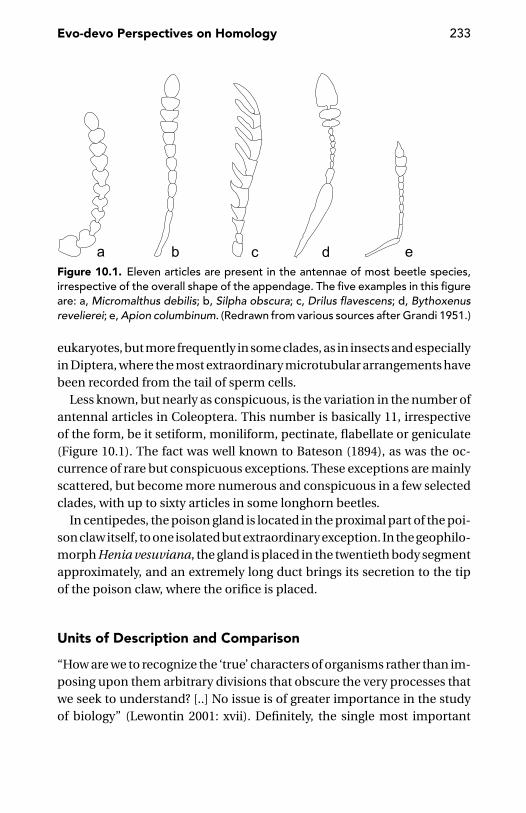
Evo-devo Perspectives on Homology 233
Figure 10.1. Eleven articles are present in the antennae of most beetle species,irrespective of the overall shape of the appendage. The five examples in this figureare: a, Micromalthus debilis; b, Silpha obscura; c, Drilus flavescens; d, Bythoxenusrevelierei; e, Apion columbinum. (Redrawn from various sources after Grandi 1951.)
eukaryotes, but more frequently in some clades, as in insects and especiallyin Diptera, where the most extraordinary microtubular arrangements havebeen recorded from the tail of sperm cells.
Less known, but nearly as conspicuous, is the variation in the number ofantennal articles in Coleoptera. This number is basically 11, irrespectiveof the form, be it setiform, moniliform, pectinate, flabellate or geniculate(Figure 10.1). The fact was well known to Bateson (1894), as was the oc-currence of rare but conspicuous exceptions. These exceptions are mainlyscattered, but become more numerous and conspicuous in a few selectedclades, with up to sixty articles in some longhorn beetles.
In centipedes, the poison gland is located in the proximal part of the poi-son claw itself, to one isolated but extraordinary exception. In the geophilo-morph Henia vesuviana, the gland is placed in the twentieth body segmentapproximately, and an extremely long duct brings its secretion to the tipof the poison claw, where the orifice is placed.
Units of Description and Comparison
“How are we to recognize the ‘true’ characters of organisms rather than im-posing upon them arbitrary divisions that obscure the very processes thatwe seek to understand? [..] No issue is of greater importance in the studyof biology” (Lewontin 2001: xvii). Definitely, the single most important
234 The Development of Animal Form
requirement, when addressing a problem of homology, is probably theidentification of meaningful units to be compared. Until recently, virtu-ally all assessments of homology involved morphological units, either inadults or earlier developmental stages. New perspectives were exploredby the recent turn towards developmentally defined morphological units(e.g., Wagner 1989, Schwenk 2001) or processes (e.g., Butler and Saidel2000, Gilbert and Bolker 2001).
ModulesCell–cell interactions, feedback behaviours, short-distance inductions,and long-distance morphogenetic effects notwithstanding, not to men-tion all forms of neural or hormonal communication, it does not seem tobe the case to embrace too literally a holistic view of the organism and itsdevelopment. If changes in every part of an organism had a non-trivial de-velopmental effect on every other part of the body, evolution could neverhave occurred. Selection acting of the smallest part would have producedsimultaneous selective pressure on all the other body parts. Consequently,the organism would have to be totally rebuilt in response to the slightestselective change (Lewontin 2001).
To describe any of those parts of the phenotype which behave as a rela-tively independent functionally interacting group of traits, Wagner intro-duced the concept of module (Wagner 1996, Wagner and Altenberg 1996).A module produces an integrated character complex and is thus both adevelopmental and an evolutionary unit. Characters within such a com-plex evolve together in a co-ordinate fashion because they are geneticallycorrelated (Cheverud 2001). Modules may arise by differential integrationof previously independent characters serving a common functional role,or by parcellation of an originally larger character complex, by selectiveelimination of pleiotropic effects among characters (Wagner 1996). Com-plex organs are made by adding new modules, each controlled by a fewgenes (Fishman and Olson 1997). Based on quantitative trait loci, Mezey,Cheverud and Wagner (2000) found modularity in the correspondencebetween genotype and the phenotype map of the ascending ramus of themandible in the mouse.
If modules are units within which developmental interactions are pri-marily confined, it should be possible to determine the spatial extentof modules by studying patterns of covariation in morphometric data,for example by comparatively analysing the fluctuating asymmetry ofpresumptive modules. An example of this approach is Klingenberg et al.’s
Evo-devo Perspectives on Homology 235
(2001) study demonstrating that the forewings and hindwings of bumble-bees are distinct developmental modules.
Evolutionary Changes in the Discernibility of DevelopmentalModules: Fusion Versus Non-disjunctionThe ontogeny of our braincase provides a good example of the fusion ofbones. Many distinct ossification centres give rise to the skull bones whoselines of contact become progressively interdigitated and which, at last, fusetogether.
In the literature, the same fusion term is often used to denote what, morecorrectly, should be called a non-disjunction. This is the case, for example,of a curious feature found in several scattered groups of arthropods – that is,the presence of segments covered by ring-like sclerites, without any trace ofthe usual distinction between a dorsal (tergal) and a ventral (sternal) com-ponent. This behaviour is generally confined to some posterior segmentsof the body, as in scorpions (segments XIV–XVIII), in thelyphonids (XVI–XVIII), in male snake flies and many caddis-flies (abdominal segment IX),in some psychodid Diptera (abdominal segment IX plus tergum X) and inseveral Mecoptera (again, ring-like segments in the posterior abdomen). Inthe miniaturised leptotyphline rove-beetles (Staphylinidae), all sclerites ofthe prothorax are united together. In phylogenetic terms, these rings (or,at least, a majority of them) may be regarded as the ‘fused’ equivalentof two or more sclerites that were probably distinct in the ancestors ofthese arthropods. Strictly speaking, ‘fusion’ is a process, not a pattern, asimplied by these phylogenetic comparisons. In terms of a process (i.e., interms of developmental events), it is quite probable that most of these ringsarise because no distinction between dorsal and ventral sclerites actuallydevelops.
Generally speaking, lack of disjunction (pattern) is much more frequentthan real fusion (process). Thus, instances of real fusion are events ofgreater evo-devo interest.
During the metamorphosis of the European grass frog (Rana tempo-raria), several sets of larval muscles undergo a process of fusion. Thelevator mandibulae posterior superficialis and levator mandibulae pos-terior profundus give rise to the levator mandibulae posterior; the levatorhyoideus suspensoriohyoideus, levator hyoideus suspensorioangularis,levator hyoideus quadratoangularis and levator hyoideus hyoangularistogether become the inner part of the depressor mandibulae; theconstrictores branchiales I–IV and levatores arcus branchialis I–III give

236 The Development of Animal Form
rise to the petrohyoidei I–IV. In Xenopus, the interarcuales are fused atmetamorphosis to form the longissimus dorsi; in adult anurans, the rec-tus abdominis superficialis and r.a. profundus are fused into a single rectusabdominis (Cannatella 1999).
In the leptostracan crustacean Nebalia, the peduncle of the second an-tenna comprises four articles in the advanced larva, but only three articlesin the adult, due to the fusion of the third and fourth larval articles (Olesenand Walossek 2000). In the females of the great majority of the harpacti-coid copepods, during the moult from the fifth copepodite to the adult, thegenital (= last thoracic) segment and the first abdominal segment fuse toform a large genital double somite. In some genera, this process is delayedand a fused double somite does not form (Huys and Boxshall 1991).
Patterns of gene expression help distinguish between non-disjunctionand fusion. Lack of segmental disjunction may be derived from the lack of aspecific gene activity, as demonstrated in vertebrates for lunatic fringe mu-tants, which fail to form boundaries between individual somites (Evrardet al. 1998).
Analysis of the expression patterns of presumptive homologues of theDrosophila genes extradenticle and dachshund in the limbs of the wood-louse Porcellio scaber and the spider Steatoda triangulosa suggests thatappendages such as the mouthparts derived from a leg-like ground planvia the elimination or fusion of the intermediate and distal podomeres(Abzhanov, Popadic, and Kaufman 1999). Such modification has been ac-tually seen in the process (Abzhanov and Kaufman 2000a) during differen-tiation of the maxillipede in P. scaber. In early development, this appendagedisplays expression patterns identical to those in the following pereiopods.Later, the extradenticle-expressing cells of the basis expand, and the dis-tal part of the appendage (the telopodite) begins to lose its segmentedcharacter.
Germ Layers and HomologyGerm layers are ‘temporary developmental modules’ which can differ-entiate, to a more or less large extent, independent of what is going onin the derivatives of the other germ layers. If the development of meso-dermal structures is reversibly inhibited in newly hatched nauplius lar-vae of Artemia, ectodermal and endodermal structures go on with differ-entiation and the missing mesodermal structures are formed, althoughdelayed, when the inhibitory treatment is removed (Hernandorena andMarco 1991). Germ layers are characterised by the exclusive expression of
Evo-devo Perspectives on Homology 237
genes, for example serpent, a gene with homeotic properties which speci-fies endoderm versus ectoderm in the developing gut of Drosophila (Reuter1994).
Traditionally, we recognize two primary germ layers (the ectoderm andthe endoderm) and a secondary germ layer (the mesoderm). Lack versuspresence of the latter is a criterion for distinguishing two main levels oforganisation: diploblastic (e.g., cnidarians) and triploblastic (bilaterians).This simple textbook picture is open to many questions. I am not con-cerned with recurrent claims, and correspondingly recurrent disclaims,that the mesoderm is present in Ctenophora, which have been traditionallyranked with diploblasts. The question is obviously relevant for our effortsto fit comb-jellies into a phylogenetic scheme, but this is less important,from the evo-devo perspective, than questions such as, are the ectoderm,endoderm and (where present) mesoderm homologous throughout theanimal kingdom? Are there animals with more than three germ layers?
As Hall (1998b) clearly describes, germ layers are not immutable build-ing blocks constraining development, but dynamic entities subjected tointensive evolutionary change.
Germ layers are just the most conspicuous of the many levels and waysthrough which a developing system becomes subdivided into functionalunits more or less extensively uncoupled from their neighbours.
One of the lowest levels of local autonomy is expressed by mitotic do-mains. In Drosophila, mitotic domain boundaries are cell fate boundaries(Cambridge, Davis, and Minden 1997). Subdivision of the Drosophila em-bryo into two dozen mitotic domains occurs with the fourteenth mitoticcycle prior to segregation of the germ layers (Foe and Odell 1989).
A kind of ‘postembryonic mitotic domain’ was observed in the beetleOryzaephilus surinamensis, in which ectodermally derived tissues un-dergo mitotic waves at the end of the first half of each larval stage, whereasin the tissues of mesodermal origin cells divide shortly before ecdysis(Romer 1964). In Hydra, the direction of cell division in the ectoderm issomewhat biased in the axial direction, and the endoderm is strongly bi-ased in the circumferential direction (Shimizu, Bode, and Bode 1995). Mi-totic domains are not exclusive of insects. In zebrafish, three spatially sep-arate mitotic domains with distinctive cell cycle length and rhythm ariseat the so-called midblastula transition. These domains, rather than speci-fying different cell fates, seem to correspond to distinctive roles in the sub-sequent morphogenetic movements (Kane et al. 1992). In Drosophila, cellsin different mitotic domains exhibit specific morphogenetic behaviours,
238 The Development of Animal Form
but many domains with unique morphogenetic activity are functionallyidentifiable even before their cells start dividing synchronously. Mitoticsynchrony is possibly just a superficial but delayed symptom of a specificdetermination of a cluster of cells (Foe and Odell 1989).
Between the germ layers and the mitotic domains, there is a whole arrayof more or less independent developmental units, in which we could repeatwhat Svacha (1992) said of the insect imaginal discs: that these groups ofcells do not represent qualitatively different structures, but merely one endof a continuum of more or less autonomous developmental units. Nomen-clature notwithstanding, there is no clear-cut difference between the threetraditional germ layers, the neural crest of the vertebrates, the imaginaldiscs of the holometabolous insects, and the set-aside cells from which theadult will be formed in marine invertebrates (such as nemerteans and seaurchins). But there are also processes by which specific morphogeneticfactors are allocated to distinct embryonic lineages which are not givenany special name. This happens, for example, following the stereotypedcleavage programs adopted by many animal groups, in which signallingcenters are reliably positioned together with the cells that respond to theirsignals (Henry and Martindale 1998).
Brian Hall (1998b, 1999, 2000) has strongly argued in favour of treat-ing the neural crest as a fourth germ layer. The neural crest has been de-fined as the quintessential feature of vertebrates (Gans and Northcutt 1983,Hall 1999, Butler 2000, Holland and Chen 2001), one of the main keys totheir extraordinary adaptive success. In fact, the neural crest parallels themesoderm as a major source of internal structures, tissues and organs.Its peculiar ‘strategy’ is the targeted migratory behaviour of its multiplecomponents.
The visceral mesoderm of insects has been also regarded as a separategerm layer (Lawrence 1992, Chauvet et al. 2000). The main homeotic geneshave different patterns of expression in somatic and visceral mesodermrespectively (Bienz 1994).
According to Hall (1998b), secondary germ layers, such as the mesodermor the neural crest, are ‘set-aside layers’. I think that we must both acceptand reverse this equation. Mesoderm and neural crest represent large clus-ters of cells which are set aside and thus excluded from the developmentalprocesses that will go on in ectodermal and endodermal derivatives. There-fore, there is no clear-cut criterion for assigning them a different status,with respect to smaller or more localised clusters of cells, such as the adultprimordium of the sea urchin or the imaginal discs of the fly.
Evo-devo Perspectives on Homology 239
Groups of cells comparable with the imaginal discs (or the histoblasts)of the holometabolous insects are probably more common than generallybelieved. For example, the ‘nasus’, a conspicuous anterior projection foundon the head of many termite soldiers, develops from a ‘soldier-nasus disc’within just one moult (the last one in the insect’s life), as described byMiura and Matsumoto (2000) for Hospitalitermes medioflavus.
As for imaginal discs, these are not necessarily limited to only one seg-ment. For example, the so-called genital disc of Drosophila consists ofthree primordia (female genital, male genital and anal) corresponding toas many segments (Chen and Baker 1997).
The typical example of set-aside cells is the adult primordium found inthe larvae of type 1 (or maximal indirect development) embryos (Davidson1991, Cameron, Peterson, and Davidson 1998) belonging to marine in-vertebrates of the most diverse phyletic lines, including echinoderms,nemerteans and bryozoans. In this kind of development, cell division po-tentialities and fate specification of the embryonic cell lineages are com-pletely different from those of the set-aside cells. Whereas embryonic celllineages have a limited mitotic potential and are precociously destined toa fixed fate, the set-aside cells retain extensive proliferative capacities andescape the fate specification affecting the embryonic cells. The embryon-ically active cells will form the larva, and the set-aside cells will form theadult. According to Peterson, Cameron, and Davidson (1997), separate setsof homologous relationships relate the larval and the adult traits of theseanimals. These authors argue that the diversity of the adult body plans ofthese invertebrates reflects diverse pattern formation processes going onin their set-aside cells.
We know (page 47) that the larval body of these invertebrates is notpatterned after the zootypic Hox gene expression. Problems in tracing ho-mologies between larval and adult features in these animals are thereforeserious, to an extent that is not always appreciated. Illuminating in thisrespect is Lacalli’s (1994) revisitation of Garstang’s (1894) auricularia hy-pothesis of the origin of chordates. Garstang distinguished three fieldsin this larva’s body surface: the ventral oral field (possibly homologousto the apical organ of other larvae), the ciliary band and the aboral ec-toderm. In chordates, the oral field would have been internalised, givingrise to the neural tube, and the oral field would have been enlarged toform most of the animal’s body surface. According to Lacalli, this meansthat the body cover of a vertebrate is different from that of the auricu-laria. The Hox gene expression formerly associated with the ciliary band

240 The Development of Animal Form
became internalised with it and did not extend to the new ectodermal bodycover.
The origin of set-aside cells fits nicely within a Darwinian view of devel-opment (cf. Buss 1987), where there is a selective advantage to individualcells and cell clones that are able to reduce the effects of competitions fromtheir neighbours – be it competition for morphogens, nutrients, or other.Evolution of type I development in metazoans (Davidson 1991) was accom-panied by the evolution of the ‘sequestration’ of the definitive germ linewithin the set-aside cells that will give rise to the adult (Ransick, Cameron,and Davidson 1996, Blackstone and Ellison 2000).
Set-aside cells may be finely interspersed within a sheet of ‘ordinary’cells. In Artemia, the general epidermis is comprised of proliferating diplo-id cells. Among them, we find tetra- or octoploid setal cells which do notdivide during the first two instars (Freeman and Chronister 1988). Thisis comparable with the behaviour of the diploid histoblasts – destined toform most of the body wall, musculature and other body parts of the adultDrosophila – which are interspersed within polyploid larval cells, destinedinstead to be destroyed at metamorphosis. Maintenance of diploidy ofthe imaginal cells is apparently one of the functions of escargot gene ex-pression (Hayashi et al. 1993).
The presence of set-aside cells allows a large developmental indepen-dence of the ‘container’ (the larva) from its ‘content’ (the set-aside cells). Asthe container develops before its content, we expect that the instar derivedfrom the set-aside cells will be ‘more advanced’ than the instar in whichthe set-aside cells were contained, but this is not necessarily true. In thecase of the holometabolous insects, as described previously (page 77), themorphology of the larva is generally less conservative than the morphologyof the adult. This is not limited to the gross features of external morphol-ogy but extends sometimes to the internal anatomy. For example, in manyDiptera (Stratiomyidae, Tabanidae, Muscidae), Neuroptera (Myrmeleonti-dae), and the beetle Melolontha, the ventral nerve cord is more primitivelysegmental in the adult than in the larva (Hanstrom 1928).
Conventional germ layers and set-aside cells, such as those forming theprimordium of the future adult of a sea urchin, represent one extreme ofa continuum of morphogenetic individualisation of groups of cells withina developing animal, the other extreme being represented by ‘delocalised’unspecialised cells such as sponge archaeocytes or the interstitial cells ofthe Hydra.
Evo-devo Perspectives on Homology 241
Let’s re-examine the question of the homology of germ layers and thestructures to which they give rise. Homologous structures in different or-ganisms often arise from different germ layers (Hall 1998b), to such anextent that Wagner (1989) considered undermining the germ layer con-cept itself. Salvini-Plawen and Splechtna (1979) also denied the generalpossibility of drawing homology between germ layers of different phyla.More cautious is Hall (1998b), who invites distinction between facts andtheory, with ‘facts’ being in his view the existence of homologous germ lay-ers across the animal kingdom and ‘theory’ being the now disproved beliefthat homologous structures in different animals are derived from the samegerm layer. This distinction between facts and theory is perhaps less clear-cut than Hall (1998b, 2000) would admit. At least a systematic descriptionof all animal embryos in terms of classic germ layers is not immune froma certain danger of typological thinking (Richardson et al. 1999).
Lesser Developmental UnitsDifferent criteria have been used to characterise lesser developmentalunits for which a complex terminology has been introduced that includesfields, progenitor fields, equivalence groups, territories of gene expressionor of action of a morphogen and compartments.
A field is a group of cells provided with self-organising and self-regulatingproperties (Ingham and Martınez-Arias 1992), thus forming a discrete unitof embryonic development (Gilbert, Opitz, and Raff 1996). In a sense, a fieldis comparable with biological species (for a review of species concepts, see,e.g., Minelli 1993, Mayden 1997). First, it is possible to define a field, or aspecies, either in terms of what happens inside or in terms of what cuts offthe field, or the species, from the surrounding world. In other words, a fieldis spatially confined within specific boundaries (cf. the isolation speciesconcepts), and cells inside it share gene expression patterns allowed bydiffusion, cell adhesion molecules, intercellular electric coupling and soon (cf. the recognition species concept). Consequently, if a species can bedefined in terms of a shared gene pool, a field can be defined in terms ofshared gene expression. A morphogenetic field is possibly the lowest termin a whole hierarchy of systems in which evolutionary novelties may arise.This happens following quantitative or qualitative changes in one or moregene expression patterns (Gilbert et al. 1996).
Davidson (1993: 666) introduced the concept of progenitor field, definedas “a region of an embryo composed of cells whose progeny will constitute a

242 The Development of Animal Form
given morphological structure”. Progenitor fields have well-defined spatialboundaries. The term is basically equivalent to the traditional term anlage,but Davidson stresses the dynamic biochemical properties of these units,their inception depending on transcription control functions, positive butespecially negative, mediated by intercellular signalling. Negative controlof transcription prevents specific gene expression in cell lineages otherthan those constituting the territory. These negative functions are thusrequired to define the relevant lineage interfaces as field boundaries. Forexample, in Drosophila, the anterior limit of knirps expression dependson several interactions with cis-regulatory sites to which the negativelyacting hunchback protein binds, and the posterior limit of expression ofthe same gene depends on multiple negative interactions at a differentcis-regulatory locus with the product of the gap gene tailless (Pankratzet al. 1992).
Basically identical to a progenitor field is an equivalence group, defined(Horvitz and Sternberg 1991) as a set of multipotent cells whose fate isdetermined by cell interactions between members of the same set. Ex-amples of equivalence groups have been identified in the most diverseanimal embryos and are usually singled out from the surrounding cellsby interactions involving the Notch/Delta signalling system (Jan and Jan1995). Lewis and Wolpert (1976) had previously observed that cells thatlook alike histologically may yet be non-equivalent because of their differ-ent position.
From a slightly different perspective, Theisen et al. (1996) proposed theterm territory to describe the set of cells under the influence of a par-ticular morphogen. Without necessarily identifying a gene product as amorphogen, Raff and Sly (2000) refer to gene expression territories.
Rather than in terms of gene expression, compartments have been clas-sically defined in terms of cell lineage (Garcıa-Bellido, Ripoll, and Morata1973), as body units uniquely formed by the descendants of a pool offounder cells confined within unbridgeable spatial limits.
Cell populations from adjacent compartments maintain their positionbecause they are unable to mix (Keynes and Stern 1988). This has beenshown by grafting experiments in chick embryos, in which cell populationsfrom anterior sclerotome halves only mix with cells from anterior halvesof other sclerotomes, and those from posterior halves mix with cells fromthe posterior halves of other sclerotomes. Anterior and posterior cells nonot mix, but form a boundary between them (Stern and Keynes 1987).This property applies to all vertebrates that have been studied. The strictly
Evo-devo Perspectives on Homology 243
similar behaviour of odd and even rhombomeres has been described pre-viously (page 101). Cell sorting based on properties of the cell membraneis at the root of compartmentalisation, but this is a complex process alsoinvolving signalling between neighbour compartments, as demonstratedfor the boundary between anterior and posterior wing discs in Drosophila(Blair and Ralston 1997).
There is no necessary correspondence between compartments andfields, although the very existence of compartments depends on the loca-lised expression of a few genes which gives rise to the boundary betweenneighbour compartments and maintains it throughout development.However, fields and compartments may overlap whenever gene expres-sion is actually limited by compartment boundaries. In the mouse embryo,for instance, the MesP genes and the genes encoding the receptor EphA4are only expressed in the anterior half of the somite, whereas HES1 andDelta1 are limited to the posterior half (Pourquie 2000).
Cells at compartment boundaries – e.g., those between the anteriorand posterior compartments in the wing disc, or those between the al-ternating anterior and posterior compartments in the ventral epidermisof the Drosophila embryo – are the source of morphogens, such as Wingless(Lawrence, Sanson, and Vincent 1996).
That compartments are elements of pattern formation but are not in-volved in differentiation per se, at variance with the behaviour of the de-velopmental units defined as fields, is shown by the fact that in mice com-partmentalisation is essential for the maintenance of segment borders inparaxial mesoderm-derived structures, but not for the differentiation ofdermomyotome and sclerotome (Hrabe de Angelis, McIntyre, and Gossler1997). In Drosophila, the dorso-ventral patterning of the wing may dependon cell interactions independent of compartment formation (Brook andCohen 1996). Since the segment rather than the compartment is a unit ofmorphological pattern, and two compartments are involved in formingan insect segment, cells on either side of the compartment boundariesmust communicate with one another to generate a coherent morpholog-ical structure (Ingham and Martınez Arias 1992).
Frames of Reference: Muscles and Nerves
Before the advent of developmental genetics and the study of the spatialexpression patterns of the so-called developmental genes, comparativeanatomists were keen to identify phylogenetically or ontogenetically more
244 The Development of Animal Form
conservative organs or tissues which could provide a reference frame-work for assessing the homology of other anatomical features. Theoreticalsupport for such a choice was not always obvious. Often, especially inpre-Darwinian literature, it was loaded by metaphysical overtones. Forexample, the choice of innervation patterns as a frame of reference inestablishing positional relationships of vertebrate muscles and bones ofarthropod sclerites was sometimes little more than a tribute paid to themost prestigious of the organic systems – a system whose complexity wasseen growing along the scala naturae to culminate eventually with thehuman brain.
The choice of the nervous system as the main frame of reference in theassessment of homology was far from universal. Muscles and blood vesselshave enjoyed their share of success, in some animal groups at least.
Due to the wealth of functions traditionally assigned to the heart andblood, to the point that the latter was regarded as the essence of life it-self, it is not a surprise that some old scientists, such as the eighteenthcentury Swiss physiologist and polymath Albrecht von Haller, regardedvessels, rather than nerves, as the primary feature of body organisation(Nordenskiold 1926). This appreciation was not widely shared by later re-searchers, although vascularisation was mentioned by Remane (1963) inhis list of morphological criteria of homology and was used occasionally byvertebrate comparative embryologists. There is some reason for paying at-tention to these anatomical features, however, because recent studies havedemonstrated that blood vessels, in addition to supplying nutrients to thedeveloping organs, may also provide information for morphogenesis. Thishas been shown in the organogenesis of the pancreas by Lammert, Cleaver,and Melton (2001) and in the development of the liver by Matsumotoet al. (2001).
Muscles and HomologyIn the comparative anatomical literature, homologies determined by pat-terns of muscle insertion are much more frequent than those determinedby patterns of vascularisation. Reliance on these patterns has often beenbacked with the result of investigations on the phylogenetic and ontoge-netic continuity of muscular patterns. This was obviously crucial to mono-graphic works such as Matsuda’s (1970) volume on the insect thorax, wherewe find several reassuring examples. Nymphal and adult muscles of thecricket Gryllus are nearly the same, with only one muscle being lost dur-ing development: this is clearly a good example of muscle continuity in

Evo-devo Perspectives on Homology 245
ontogeny. In other orthopterans, several muscles are lost, either during thelast nymphal stage or after the adult stage is reached, but no new muscleis formed during development. The predominant trend in the evolutionof the adult thoracic musculature in insects has been a reduction in mus-cle number. Some muscles present in the primitive wingless insects arestill present in the immature winged insects, but are absent in the adult.Of course, some muscles have been added to the thoracic musculature inconnection with flight.
Examples of the frequent use of musculature in establishing segmentalunits in arthropods are Hafner’s (1971) study of the dorsal musculaturepattern as a series of landmarks to recognise segmental borders in the piglouse Haematopinus suis and Shultz’s (1993) analysis of the segmentationof the opisthosoma in the uropygid arachnid Mastigoproctus giganteus.In this case, muscle morphology provided criteria for distinguishing ‘true’sterna from other ventral sclerites. Shultz regarded criteria derived frommusculature as unambiguous, even when conflicting with previous inter-pretations based on external morphology.
Widespread success notwithstanding, analysis of musculature patternsis not immune from problems, technical and conceptual alike. Malicky(1974) observed that it is not that easy to trace homologies of trunk musclesin caddis fly larvae, because their insertions are not strictly conserved. Itis not always possible to say whether a given muscle is a single one or abundle of many different muscles.
Particularly disturbing are the difficulties in tracing homologies betweenlarval and imaginal sclerites and musculature. Problems may be derivedfrom an advanced reduction of some of the presumptive sclerites to whicha muscle should be attached, as in the case of the male copulatory organsof insects. As these organs get their musculature from the ninth abdomi-nal segment, they might be interpreted as belonging to it. However, as thetenth sternum is reduced, it is also possible that the phallic musculatureis derived from intersegmental muscles originally running between ster-num IX and sternum X (Snodgrass 1957). In the honeybee, the imaginalmusculature of the thorax develops during the larval stages prior to theproduction of the skeletal elements to which it will be eventually attached.Most muscles do not grow between final sites of attachment, but manymyoblasts are supported by larval muscles already suspended across thebody cavity (Daly 1964). In the blackfly Simulium, in which some futureflight muscles are already developed and attached to the epidermis in thesecond larval stage, differences in growth rate of the epidermis of different
246 The Development of Animal Form
parts of the mesothorax cause later changes in the position and orienta-tion of these muscles (Hinton 1959, 1961). Morphologists have often useddifferent names for these basically homologous muscles with new pointsof attachment (Matsuda 1970).
Another problem in tracing homologies of muscles and their attach-ment points is that a muscle may undergo duplication, as in the case ofthe jaw muscles in tetraodontiform fishes. The plesiomorphic conditionin this order is two main adductor mandibulae muscles; but in some sub-groups, these are duplicated, one or more times. The number of adduc-tor mandibulae muscles thus ranges from the original two in triplespines(Triacanthidae) to as many as eight muscles in some filefishes (Monacan-thidae). Based on their origins, insertions and relative masses in represen-tative taxa and their congruence with a phylogeny for these taxa, Friel andWainwright (1997) recognise morphological orthologues and paraloguesof the original muscles. Owing to the specific significance given to the word‘orthologous’ and ‘paralogous’ in molecular biology (Fitch 1970), I recom-mend qualifying these muscles instead as paramorphs, through an easygeneralisation of the concept of paramorphism I have originally applied(Minelli 2000b) to the relationships between appendages and main bodyaxis (cf. page 164).
There are even cases in which evidence from muscular insertion pat-terns seems to point in the wrong direction. This may happen in thosegroups in which a body part has extensively departed from its morpholog-ical setting in the close relatives. In male dipterans, abdominal segmentsIX and X are often curved ventrally and are associated with the genitalia toform a complex known as the hypandrium. In some groups, a 180◦ twistgives rise to a hypandrium inversum, with the anus in a position ventral tothe genital opening. In the case of the little blood-sucking fly Phlebotomusgarnhami, it has been claimed that, to allow the postabdominal inversionto occur, the abdominal longitudinal muscles from abdominal segment VIhave moved their insertions to segment VII (Just 1973). Bigger problemswere faced by Kimsey (1992) in her study of the cuckoo wasps (or goldwasps, Chrysididae). This family is characterised by modification of twoor more abdominal segments that are telescoped within the abdomen andmay function as an ovipositor. Accompanying the internalisation of thesesegments are shifts in the position of those muscles which provide for theirunusual mobility. Kimsey assumed that a shift in position was more likelyto occur than the evolution of an entirely new muscle; nevertheless, oneof these muscles does not appear to be homologous to any muscle seen
Evo-devo Perspectives on Homology 247
in the ground plan of the aculeate hymenopterans. One more exampleof muscles shifting their insertion during development comes from thetendons of the levator mandibulae posterior profundus and the levatormandibulae externus in the frogs, which shift to insert on Meckel’s carti-lage when the suprarostral cartilage disintegrates during metamorphosis(De Jongh 1968).
Nerves and HomologyIn his classic monograph on bird comparative anatomy, Max Furbringer(1888) established the principle that homologies between muscles in dif-ferent birds can be correctly established only by reference to their inner-vation patterns.
One general reason for expecting that the nervous system should bemore conserved than other aspects of morphology is its complexity(Nishikawa et al. 1992, Tierney 1996). In vertebrates, further justificationfor relying on nerves rather than muscles or bones may be that the centralnervous system often differentiates far in advance of bones and muscles,as in the head of eutherian mammals (Smith 1996), but this does not applyto animals in other phyla.
In a sense, different body regions are recognised as non-equivalent bythe nerves that innervate them. This is the lesson Lewis and Wolpert (1976)derived from Miner’s (1956) experiment with a tadpole of the frog Ranapipiens, in which a piece of skin was rotated in such a way that skin fromthe animal’s ventral side now covered the back and vice versa. After meta-morphosis, the frog reacted to mechanical stimulation of the back or thebelly as if the pieces of skin were still in their normal place; that is, theanimal wiped its belly when stimulated on the back and vice versa.
In arthropods, innervation is often used as a criterion for identifiying thesegmental origin or composition of fused, reduced, or otherwise highlyspecialised body parts. In the female cockroach Leucophaea maderae,for example, Engelmann (1963) recognised six free abdominal ganglia,the latter of them also innervating segments VII through IX, includinggenital structures. Following the innervation pathways, the author identi-fies the segmental origin of the different parts of the female reproductivesystem.
There are problems with this use of innervation as a criterion for delimit-ing segments. Matsuda (1970), while sometimes using innervation as a keyto homology in his comparative analysis of the insect thorax, remarked onthe scanty developmental evidence supporting this practice and stressed
248 The Development of Animal Form
the need for adequate studies, extending back to the earliest stages ofneurogenesis, but in Drosophila, the arrangement of neuroblasts does nothelp to understand head segmentation (Hartenstein and Campos-Ortega1984). Matsuda also observed that there may be considerable individualvariation in the connections to the muscles a nerve develops. Other dif-ficulties may be derived from the more or less extensive fusion of twoneighbouring nerves or by the multiple innervation of one and the samemuscle (see also Schmitt 1962, Markl 1966).
Innervation may shift during development (Striedter 1998). Sewertzoff(1931), for example, reported that, in a lizard, the anlage of the forelimbsare initially innervated by neck nerves. But later these nerves degenerate,and the definitive innervation is provided by more caudal nerves.
There are also problems in arthropods, where motor nerves are usuallyrestricted to the segments in which their respective ganglia are found. Butthis restriction does not necessarily apply to the integumental sensorynerves. Thus, motor nerves are more reliable indices of segmental limitsthan sensory nerves, but comparative morphologists should be aware ofexceptions, as in the case of the moth Telea polyphemus, where the secondthoracic ganglion gives off nerves to all three thoracic segments (Snodgrass1960). Still more important, examples of reorganisation of the patternsof synaptic connectivity during development are known from animals asdiverse as insects (Truman 1990, Levine, Morton, and Restifo 1995), frogs(Alley 1990), and Caenorhabditis elegans (Hallam and Jin 1998). Lauder(1986) regarded the vertebrate brain as highly plastic and possibly notsufficiently robust to permit phylogenetic inferences.
There are molecular mechanisms supporting the traditional view. In mu-tant mouse embryos, it has been demonstrated that lack of Hoxb-1, oneof the Hox genes whose expression domain is restricted to one segmentalunit (in this case, the fourth rhombomere in the hindbrain) causes alteredsegmental identity and abnormal migration of motor neurons. In mutants,motor neurons differentiate, but the controlateral vestibuloacoustic effer-ent and the facial branchiomotor neurons fail to migrate to their properpositions (Studer et al. 1996).
The phylogenetic stability of neuroanatomical characters has been re-cently tested by Buschbeck (2000). In her elegant study, the phylogeneticrelationships of 23 species of Diptera, representing a diversity of lineagessuch as crane flies, robber flies, hover flies and house flies, were recon-structed based on fine neuroanatomical details of the visual system only.
Evo-devo Perspectives on Homology 249
The phylogenetic relationships suggested by this analysis are similar tothose suggested by the traditional macroanatomical traits or by moleculardata. The ‘phylogenetic signal’ contributed by the different parts of thenervous system is not the same. Phylogenetic relationships suggested bythe neurons of the second-order visual neuropil, the medulla, are betterdefined in respect to what is suggested by characters of the deeper visualcentre, the lobular plate. These differences, according to Buschbeck (2000),may relate to different functional constraints in the two neuropils.
Summary and Conclusions
If we wish to foster a dialogue between evolutionary biology and develop-mental biology, we need to ensure that the two disciplines stand on equalmetaphysical footing. I think that developmental biology is still heavilybiased by finalism, whereas an equation of evolution to progress is by nowa matter of the past. A discussion of this problem and some suggestionsfor a new view of development was the subject of chapter 1.
In this view, development acquires a meaning of its own, rather thansimply being the process required to obtain an adult. Consequently, I havesuggested that the very origin of many, if not most, of the basic features ofan animal’s body must be searched for in the adaptive significance thesefeatures initially had for development as such, rather than as adult featuresnecessarily prepared through development. This idea was the subject ofchapter 2.
If development is not the way by which a programme encoded in the eggis deployed to reach the final, adult condition, we may question whetherthe metaphor of the programme (more precisely, the metaphor of the ge-netic programme) is indeed adequate. I think it is not. This is not to deny, ofcourse, the role of genes in development, but to question instead currentnotions such as the developmental gene and the master control gene. I amsympathetic towards those views according to which the development ofthe earliest multicellular organisms was mainly caused by generic, ratherthan genetic, causes. In the course of evolution, the latter acquired anincreasingly larger importance and acted as canalisers and stabilisers ofdevelopmental processes. These questions were discussed in chapter 3.
Chapter 4 was devoted to developmental time. I suggested that devel-opmental systems first acquired ways to control and pattern the temporalprogression of their dynamics and that this offered a blueprint to the sub-sequent evolution of ways through which control over spatial patterning
250

Summary and Conclusions 251
was achieved. Further problems concerning developmental time are howto establish homology between developmental stages or events, how todefine a larva and what is metamorphosis.
Chapter 5 dealt with the most elementary level of body complexity (i.e.,regionalisation or tagmosis). I discussed briefly, in light of comparativemorphology and molecular developmental genetics, topics such as num-ber and homology of tagmata, the nature and temporal stability of theboundaries between them and the mostly overlooked relationships be-tween developmental time and the development and patterning ofprimary and secondary body axes, as well as some problems in morpho-logical and developmental topology.
Cell properties and behaviours, as basic to differentiation and pattern-ing, were briefly analysed in chapter 6, which was devoted to issues ofdifferentiation and body patterning. This was also the place to discusspositional information – a notion I suggested to replace the reciprocalnotion of informational position, transpatterning, phylotypic stages, andmorphological assimilation in ontogeny and phylogeny, with a coda onregeneration.
Size factors – cell size, cell number, and miniaturisation – were the topicsof chapter 7.
Chapters 8 and 9 – respectively devoted to body axes and symmetryand to the nature and origin of segments – included most of the newoutlooks, quite probably controversial and hopefully provocative, throughwhich I believe that evo-devo biology should approach the study of animalorganisation, in light of joint suggestions from developmental geneticsand comparative embryology and morphology. Hot spots of these twochapters were the concept of the dual animal, a relativistic revisitation ofthe definition of the animal’s main body axis, a critique of the conventionaldescription of body structure and morphogenesis in terms of Cartesianaxes, and a discussion of two concepts (axis paramorphism and doublesegmentation) I first introduced in research papers (Minelli 2000a, 2000b,2001). Unconventional views derived from this way of looking at animalmorphology and morphogenesis include the suggestions that the tail ofvertebrates is an appendage rather than a part of the main body axis, andthe scolex may be the posterior rather than the anterior end of a tapeworm’sbody.
The last chapter was devoted to homology. Based on evo-devo argu-ments, I defended the view that we shall follow a factorial approach tohomology. That is, that homology is a relative, rather than an absolute
252 The Development of Animal Form
concept. I also discussed, in evo-devo perspective, the merits and weak-nesses of homologies drawn on patterns of muscle insertion and innerva-tion. I also suggested that, in the multilayered mosaic of developmentalsubunits that we can recognise within a developing animal, there is a vir-tual continuum between the conventional germ layers and the clusters ofset-aside cells, such as the imaginal discs of holometabolous insects andthe adult primordium of sea urchin larvae.
Following the conceptual background introduced in the first two chap-ters, I have been articulating a view of development in which there islittle space for many old questions which are, in my view, a simple by-product of an unnecessarily complex holistic view of the organism andits development. For example, I have dismissed the ‘problem’ of the ori-gin of symmetry as simply due to failure to acknowledge that symmetryis the expected ‘default product’ of largely independent developmentaltrajectories occurring in parallel within separate modules.
I have also dismissed the fashionable concept that most major develop-mental ‘decisions’ are taken early in ontogeny, because this is the way toavoid irreversible mistakes in the long journey towards the adult. I have ar-gued instead that such ‘decisions’ (i.e., patterning) are only possible withinvery small systems. But the animal is adequately small only at very earlystages. Therefore, patterning is ‘decided’ at early stages, because at earlystages the embryo is small, not the other way around.
With this book, I hope I have succeeded at least in infecting the readerwith three ideas.
First, that evolutionary developmental biology urgently needs a bulkyinjection of facts and concepts from disciplines such as comparative mor-phology, descriptive embryology and the study of postembryonic devel-opment.
Second, that we cannot continue speaking of segments, tagmata or lar-vae without qualifying these concepts, and many others, adequately – whatmight not necessarily be the same way in every context.
Third, that evolutionary developmental biology requires much morethan the study of a handful of model species, however sophisticated ourexperimental approaches might eventually be. Animal evolution has ex-plored many more solutions than the chicken, mouse, zebrafish, Droso-phila, and Caenorhabditis would suggest. As Arthur (2002) stated, this isnot a reason to despair, but a plea for caution. Wide-ranging comparisonsand a combined interdisciplinary approach are required before evolution-ary developmental biology may finally be written ‘from first principles’.

Summary and Conclusions 253
Honestly, I do not expect that my criticism of the current adultocen-tric view of development will be accepted easily. I wonder, however, howfollowers of this view will be able to produce a consistent and sober expla-nation for the many facts I have tried to interpret in these pages from a lessfinalistic perspective. Let’s see, a few years from now, where the balancebetween these opposite views will eventually settle.

References
Abbott, M.K. & Lengyel, J.A. (1991) Embryonic head involution and rotation of maleterminalia require the Drosophila locus head involution defective. Genetics, 129,783–789.
Abouheif, E. (1997) Developmental genetics and homology: a hierarchical ap-proach. Trends in Ecology & Evolution, 12, 405–408.
Abouheif, E. (1999) Establishing homology criteria for regulatory gene networks:prospects and challenges. In Homology (Novartis Foundation Symposium 222),ed. G.R. Bock & G. Cardew, pp. 207–225. Chichester: Wiley.
Abouheif, E., Akam, M., Dickinson, W.J., Holland, P.W.H., Meyer, A., Patel, N.H., Raff,R.A., Roth, V.L. & Wray, G.A. (1997) Homology and developmental genes. Trendsin Genetics, 13, 432–433.
Abzhanov, A. & Kaufman, T.C. (1999) Novel regulation of the homeotic gene Scrassociated with a crustacean leg-to-maxilliped appendage transformation. De-velopment, 126, 1121–1128.
Abzhanov, A. & Kaufman, T.C. (2000a) Homologs of Drosophila appendage genesin the patterning of arthropod limbs. Developmental Biology, 227, 673–689.
Abzhanov, A. & Kaufman, T.C. (2000b) Crustacean (malacostracan) Hox genes andthe evolution of the arthropod trunk. Development, 127, 2239–2249.
Abzhanov, A. & Kaufman, T.C. (2000c) Embryonic expression of the Hox genes of thecrayfish Procambarus clarkii (Crustacea, Decapoda). Evolution & Development,2, 271–283.
Abzhanov, A., Popadic, A. & Kaufman, T.C. (1999) Chelicerate Hox genes and thehomology of arthropod segments. Evolution & Development, 1, 77–89.
Achtelig, M. (1975) Die Abdomenbasis der Neuropteroidea (Insecta, Holome-tabola). Eine vergleichend-anatomische Untersuchung des Skeletts und derMuskulatur. Zoomorphologie, 82, 201–242.
Adams, M.D., Celniker, S.E., Holt, R.A., Evans, C.A., Gocayne, J.D., Amanatides,P.G., Scherer, S.E., Li, P.W., Hoskins, R.A., Galle, R.F., George, R.A., Lewis, S.E.,Richards, S., Ashburner, M., Henderson, S.N., Sutton, G.G., Wortman, J.R., Yan-dell, M.D., Zhang, Q., Chen, L.X., Brandon, R.C., Rogers, Y.H.C., Blazej, R.G.,Champe, M., Pfeiffer, B.D., Wan, K.H., Doyle, C., Baxter, E.G., Helt, G., Nelson,C.R., Miklos, G.L.G., Abril, J.F., Agbayani, A., An, H.J., Andrews Pfannkoch, C.,
255

256 References
Baldwin, D., Ballew, R.M., Basu, A., Baxendale, J., Bayraktaroglu, L., Beasley,E.M., Beeson, K.Y., Benos, P.V., Berman, B.P., Bhandari, D., Bolshakov, S., Borkova,D., Botchan, M.R., Bouck, J., Brokstein, P., Brottier, P., Burtis, K.C., Busam, D.A.,Butler, H., Cadieu, E., Center, A., Chandra, I., Cherry, J.M., Cawley, S., Dahlke,C., Davenport, L.B., Davies, A., de Pablos, B., Delcher, A., Deng, Z.M., Mays, A.D.,Dew, I., Dietz, S.M., Dodson, K., Doup, L.E., Downes, M., Dugan Rocha, S., Dunkov,B.C., Dunn, P., Durbin, K.J., Evangelista, C.C., Ferraz, C., Ferriera, S., Fleischmann,W., Fosler, C., Gabrielian, A.E., Garg, N.S., Gelbart, W.M., Glasser, K., Glodek, A.,Gong, F.C., Gorrell, J.H., Gu, Z.P., Guan, P., Harris, M., Harris, N.L., Harvey, D.,Heiman, T.J., Hernandez, J.R., Houck, J., Hostin, D., Houston, D.A., Howland, T.J.,Wei, M.H., Ibegwam, C., Jalali, M., Kalush, F., Karpen, G.H., Ke, Z.X., Kennison,J.A., Ketchum, K.A., Kimmel, B.E., Kodira, C.D., Kraft, C., Kravitz, S., Kulp, D.,Lai, Z.W., Lasko, P., Lei, Y.D., Levitsky, A.A., Li, J.Y., Li, Z.Y., Liang, Y., Lin, X.Y.,Liu, X.J., Mattei, B., McIntosh, T.C., McLeod, M.P., McPherson, D., Merkulov, G.,Milshina, N.V., Mobarry, C., Morris, J., Moshrefi, A., Mount, S.M., Moy, M., Murphy,B., Murphy, L., Muzny, D.M., Nelson, D.L., Nelson, D.R., Nelson, K.A., Nixon, K.,Nusskern, D.R., Pacleb, J.M., Palazzolo, M., Pittman, G.S., Pan, S., Pollard, J., Puri,V., Reese, M.G., Reinert, K., Remington, K., Saunders, R.D.C., Scheeler, F., Shen,H., Shue, B.C., Siden Kiamos, I., Simpson, M., Skupski, M.P., Smith, T., Spier,E., Spradling, A.C., Stapleton, M., Strong, R., Sun, E., Svirskas, R., Tector, C.,Turner, R., Venter, E., Wang, A.H.H., Wang, X., Wang, Z.Y., Wassarman, D.A.,Weinstock, G.M., Weissenbach, J., Williams, S.M., Woodage, T., Worley, K.C.,Wu, D., Yang, S., Yao, Q.A., Ye, J., Yeh, R.F., Zaveri, J.S., Zhan, M., Zhang, G.G.,Zhao, Q., Zheng, L.S., Zheng, X.Q.H., Zhong, F.N., Zhong, W.Y., Zhou, X.J., Zhu,S.P., Zhu, X.H., Smith, H.O., Gibbs, R.A., Myers, E.W., Rubin, G.M., & Venter, J.C.(2000) The genome sequence of Drosophila melanogaster. Science, 287, 2185–2195.
Adoutte, A., Balavoine, G., Lartillot, N., Lespinet, O., Prud’homme, B. & de Rosa, R.(2000) The new animal phylogeny: reliability and implications. Proceedings of theNational Academy of Sciences of the United States of America, 97, 4453–4456.
Aguesse, P. (1965) La division des antennes chez les larves d’Odonates. Bulletin dela Societe Entomologique de France, 70, 8–12.
Aguinaldo, A.M.A., Turbeville, J.M., Linford, L.S., Rivera, M.C., Garey, J.R., Raff, R.A. &Lake, J.A. (1997) Evidence for a clade of nematodes, arthropods and other moult-ing animals. Nature, 387, 489–493.
Ahl, V. & Allen, T.F.H. (1996) Hierarchy Theory: A Vision, Vocabulary, and Epistemol-ogy. New York: Columbia University Press.
Ahlberg, P. (1992) Coelacanth fins and evolution. Nature, 358, 459.Ahn, D. & Gibson, G. (1999a) Axial variation in the threespine stickleback: relation-
ships to Hox gene expression. Development, Genes and Evolution, 209, 473–481.Ahn, D. & Gibson, G. (1999b) Expression patterns of the threespine stickleback Hox
genes and insights into the evolution of the vertebrate body axis. Development,Genes and Evolution, 209, 482–494.
Ahringer, J. (1996) Posterior patterning by the Caenorhabditis elegans even-skippedhomolog vab-7. Genes and Development, 10, 1120–1130.
References 257
Akam, M. (1987) The molecular basis for metameric pattern in the Drosophilaembryo. Development, 101, 1–22.
Akam, M. (1989) Hox and HOM: homologous gene clusters in insects and verte-brates. Cell, 57, 347–349.
Akam, M. (1994) Is pairing the rule? Nature, 367, 410–411.Akam, M. (1998) Hox genes, homeosis and the evolution of segment identity: no
need for hopeless monsters. International Journal of Developmental Biology, 42,445–451.
Akam, M., Dawson, I. & Tear, G. (1988) Homeotic genes and the control of segmentdiversity. Development, 104(Suppl.), 123–134.
Alberch, P. (1985) Developmental constraints: why St. Bernards often have an extradigit and poodles never do. American Naturalist, 126, 430–433.
Alberch, P. (1987) Evolution of a developmental process–irreversibility and redun-dancy in amphibian metamorphosis. In Development as an Evolutionary Process,eds. R.A. Raff and E.C. Raff, pp. 23–46. New York: Alan R. Liss.
Alberch, P. (1989) Development and the evolution of amphibian metamorphosis.In Trends in Vertebrate Morphology, eds. H. Splechtna & H. Hilgers, pp. 163–173.Stuttgart: Gustav Fischer. (Fortschritte der Zoologie, 35.)
Alberch, P. & Gale, E.A. (1986) Pathways of cytodifferentiation during the meta-morphosis of the epibranchial cartilage in the salamander Eurycea bislineata.Developmental Biology, 117, 233–244.
Alberch, P., Gale, E.A. & Larsen, P.R. (1986) Plasma T4 and T3 levels in naturallymetamorphosing Eurycea bislineata (Amphibia, Plethodontidae). General andComparative Endocrinology, 61, 153–163.
Alberch, P. & Kollar, E. (1988) Strategies of head development: workshop report.Development, 103(Suppl.), 25–30.
Alberch, P., Lewbart, G.A. & Gale, E.A. (1985) The fate of larval chondrocytes duringthe metamorphosis of the epibranchial in the salamander Eurycea bislineata.Journal of Embryology and Experimental Morphology, 88, 71–83.
Albert, R., Jeong, H. & Barabasi, A.-L. (2000) Error and attack tolerance of complexnetworks. Nature, 406, 378–382.
Alexander, A.A. & Gans, C. (1966) The pattern of dermal-vertebral correlation insnakes and amphisbaenians. Zoologische Mededeelingen, 41, 171–190.
Alley, K.E. (1989) Myofiber turnover is used to retrofit frog jaw muscles duringmetamorphosis. American Journal of Anatomy, 184, 1–12.
Alley, K.E. (1990) Retrofitting larval neuromuscular circuits in the metamorphosingfrog. Journal of Neurobiology, 21, 1092–1107.
Ambros, V. (1989) A hierarchy of regulatory genes controls a larva-to-adult devel-opment switch in C. elegans. Cell, 57, 49–57.
Ambros, V. & Moss, E.G. (1994) Heterochronic genes and the temporal control ofC. elegans development. Trends in Genetics, 10, 123–127.
Amirthalingam, K., Lorens, J.B., Saetre, B.O., Salaneck, E. & Fjose, A. (1995) Em-bryonic expression and DNA-binding properties of zebrafish pax-6. Biochemicaland Biophysical Research Communications, 215, 122–128.
Amos, T.M. & Mesce, K.A. (1994) Reorganization of the ventral nerve cord in the

258 References
moth Manduca sexta (L.) (Lepidoptera: Sphingidae). International Journal ofInsect Morphology & Embryology, 23, 21–37.
Amundson, R. & Lauder, G.V. (1994) Function without purpose: the uses of causalrole function in evolutionary biology. Biology and Philosophy, 9, 443–469.
Anderson, H. (1984) The development of sensory nerves within the wing of Droso-phila melanogaster. A light- and electron-microscope study. Roux’s Archives ofDevelopmental Biology, 193, 226–233.
Anderson, K. (1995) One signal two body axes. Science, 261, 489–490.Anderson, S.A., Eisenstat, D.D., Shi, L. & Rubenstein, J.L.R. (1997) Interneuron mi-
gration from basal forebrain to neocortex: dependence on Dlx genes. Science,278, 474–476.
Andre, H.M. (1988) Age-dependent evolution: from theory to practice. In Ontogenyand Systematics, ed. C.J. Humphries, pp. 137–187. New York: Columbia UniversityPress.
Andre, H.M. (1989) The concept of stase. In L’Ontogenese et le Concept de Stase chezles Arthropodes, eds. H.M. Andre & J.-C. Lions, pp. 3–16. Warre, Belgium: AGARPublishers.
Andre, H.M. (1992) Calyptostases as indicators of developmental constraints inmites and other arthropods. Acarologia, 33, 223–231.
Aparicio, J.M. (1998) Patterns of fluctuating asymmetry in developing primaryfeathers: a test of the compensatorial growth hypothesis. Proceedings of the RoyalSociety of London, B, 265, 2353–2357.
Appel, T.A. (1987) The Cuvier-Geoffroy Debate: French Biology in the Decades beforeDarwin. New York: Oxford University Press.
Arenas-Mena, C., Cameron, A.R. & Davidson, E.H. (2000) Spatial expression of Hoxcluster genes in the ontogeny of a sea urchin. Development, 127, 4631–4643.
Arenas-Mena, C., Martınez, P., Cameron, R.A. & Davidson, E.H. (1998) Expressionof the Hox gene complex in the indirect development of a sea urchin. Proceedingsof the National Academy of Sciences of the United States of America, 95, 13062–13067.
Arendt, D. & Nubler-Jung, K. (1994) Inversion of dorsoventral axis? Nature, 371, 26.Arendt, D. & Nubler-Jung, K. (1996) Common ground plans in early brain develop-
ment in mice and flies. BioEssays, 18, 255–259.Arendt, D., Technau, U. & Wittbrodt, J. (2001) Evolution of the bilaterian larval
foregut. Nature, 409, 81–85.Aristotle (1942) Generation of Animals (with an English translation by L.A. Peck).
Cambridge, Mass.: Harvard University Press.Arnone, M.I. & Davidson, E.H. (1997) The hardwiring of development: organization
and function of genomic regulatory systems. Development, 124, 1851–1864.Arthur, W. (1997) The Origin of Animal Body Plans. A Study in Evolutionary Devel-
opmental Biology. Cambridge: Cambridge University Press.Arthur, W. (2002) The emerging conceptual framework of evolutionary develop-
mental biology. Nature, 415, 757–764.Arthur, W. & Farrow, M. (1999) The pattern of variation in centipede segment
number as an example of developmental constraint in evolution. Journal of The-oretical Biology, 200, 183–191.

References 259
Arthur, W., Jowett, T. & Panchen, A. (1999) Segments, limbs, homology, and co-option. Evolution & Development, 1, 74–76.
Atz, J.W. (1970) The application of the idea of homology to behavior. In Develop-ment and Evolution of Behavior, eds. L.R. Aronson, E. Tobach, D.S. Lehrman &J.S. Rosenblatt, pp. 53–74. San Francisco: Freeman.
Audy, J.R., Nadchatram, M. & Vercammen-Grandjean, P.H. (1972) La ‘neosomie,’ unphenomene inedit de neoformation en acarologie, alliee a un cas remarquablede tachygenese. Journal of Medical Entomology, 9, 487–494.
Averof, M. & Akam, M. (1995) Hox genes and the diversification of insect and crus-tacean body plans. Nature, 376, 420–423.
Averof, M. & Patel, N.P. (1997) Crustacean appendage evolution associated withchanges in Hox genes expression. Nature, 388, 682–686.
Awgulewitsch, A. & Jacobs, D. (1992) Deformed autoregulatory element fromDrosophila functions in a conserved manner in transgenic mice. Nature, 358,341–344.
Ax, P. (1995) Multicellular Animals. A New Approach to the Phylogenetic Order inNature. Vol. 1. Berlin: Springer.
Azevedo, R.B.R. & Leroi, A.M. (2001) A power law for cells. Proceedings of the NationalAcademy of Sciences of the United States of America, 98, 5699–5704.
Bagnall, K.M., Higgins, S.J. & Sanders, E.J. (1988) The contribution made by a singlesomite to the vertebral column: experimental evidence in support of resegmen-tation using the chick-quail chimaera model. Development, 103, 69–85.
Bailey, W.J., Kim, J., Wagner, G.P. & Ruddle, F.H. (1997) Phylogenetic reconstructionof vertebrate Hox cluster duplication. Molecular Biology and Evolution, 14, 843–853.
Baker, R. & Schubiger, G. (1996) Autonomous and nonautonomous Notch functionsfor embryonic muscle and epidermis development in Drosophila. Development,122, 617–626.
Balashov, Yu.S. (1963) Anatomo-histological peculiarities of moulting of the tickHyalomma asiaticum (Acarina, Ixodoidea). Zoologicheskii Zhurnal, 42, 345–348.[In Russian]
Ball, P. (1999) The Self-made Tapestry: Pattern Formation in Nature. Oxford: OxfordUniversity Press.
Bally-Cuif, L. & Boncinelli, E. (1997) Transcription factors and head formation invertebrates. BioEssays, 19, 127–135.
Balss, H., V., Buddenbrock, W., Gruner, H.-E. & Korschelt, E. (1940–1961) Decapoda.In Bronn’s Klassen und Ordnungen des Tierreichs. Part 5(1)7. Leipzig: AkademischeVerlagsgesellschaft Geest & Portig.
Baltzer, F. (1952) Experimentelle Beitrage zur Frage der Homologie. XenoplastischeTransplantationen bei Amphibien. Experientia, 8, 285–297.
Barbieri, M. (2001) The Organic Codes. An Introduction to Semantic Biology.Cambridge: Cambridge University Press.
Bard, J.B.L. (1990) Traction and the formation of mesenchymal condensations invivo. BioEssays, 12, 389–395.
Bastianello, A., Ronco, M., Burato, P.A. & Minelli, A. (2001) Hox gene sequencesfrom the geophilomorph centipede Pachymerium ferrugineum (C.L. Koch, 1835)

260 References
(Chilopoda: Geophilomorpha: Geophilidae): implications for the evolution ofthe Hox class genes of arthropods. Molecular Phylogenetics and Evolution, 22,155–161.
Bateson, W. (1894) Materials for the Study of Variation Treated with Especial Regardto Discontinuity in the Origin of Species. London: Macmillan.
Bavestrello, G., Sommer, C. & Sara, M. (1992) Bi-directional conversion in Turritopsisnutricula. In Aspects of Hydrozoan Biology, eds. J. Bouillon, F. Boero, F. Cicogna,J.M. Gili & R.G. Hughes. Scientia Marina, 56, 137–140.
Bayascas, J.R., Castillo, E., Munoz-Marmol, A.M., Baguna, J. & Salo, E. (1998) Syn-chronous and early activation of planarian Hox genes and the re-specification ofbody axes during regeneration in Dugesia (G.) tigrina (Turbellaria, Tricladida).Hydrobiologia, 383, 125–130.
Beauchemin, M. & Savard, P. (1992) Two distal-less related homeobox-containinggenes expressed in regeneration blastemas of the newt. Developmental Biology,154, 55–65.
Bechtel, W. & Richardson, R.C. (1993) Discovering Complexity. Princeton: PrincetonUniversity Press.
Becker, S., Pasca, G., Strumpf, D., Min, L. & Volk, T. (1997) Reciprocal signalingbetween Drosophila epidermal muscle attachment cells and their correspondingmuscles. Development, 124, 2615–2622.
Beddington, R.S.P. & Robertson, E.J. (1998) Anterior patterning in mouse. Trends inGenetics, 14, 277–284.
Beermann, A., Jay, D.G., Beeman, R.W., Hulskamp, M., Tautz, D. & Jurgens, G. (2001)The Short antennae gene of Tribolium is required for limb development andencodes the orthologue of the Drosophila Distal-less protein. Development, 128,287–297.
Beier, M. (1968) Phasmida (Stab-oder Gespenstheuschrecken). In Handbuch derZoologie, eds. W. Kukenthal & T. Krumbach, Part 4(2)2/10. Berlin: de Gruyter.
Beier, M. (1972) Saltatoria (Grillen und Heuschrecken). In Handbuch der Zoologie,eds. W. Kukenthal & T. Krumbach, Part 4(2)2/9. Berlin: de Gruyter.
Beklemishev, W.N. (1969) Principles of Comparative Anatomy of Invertebrates.Edinburgh: Oliver & Boyd.
Bell, G. & Mooers, A.O. (1997) Size and complexity among multicellular organisms.Biological Journal of the Linnean Society, 60, 345–363.
Bely, A.E. (1999) Decoupling of fission and regenerative capabilities in an asexualoligochaete. Hydrobiologia, 406, 243–251.
Bely, A.E. & Wray, G.A. (2001) Evolution of regeneration and fission in annelids:insights from engrailed- and orthodenticle-class gene expression. Development,128, 2781–2791.
Bennet-Clark, H.C. (1971) The cuticle as a template for growth in Rhodnius prolixus.Journal of Insect Physiology, 17, 2421–2434.
Bergstrom, J. & Hou, X.-G. (1998) Changjiang arthropods and their bearing on earlyarthropod evolution. In Arthropod Fossils and Phylogeny, ed. G.D. Edgecombe,pp. 151–184. New York: Columbia University Press.
Berlese, A. (1913) Intorno alle metamorfosi degli insetti. Redia, 9, 121–136.Berlin, B. (1992) Ethnobiological Classification. Principles of Categorization of

References 261
Plants and Animals in Traditional Societies. Princeton: Princeton UniversityPress.
Berlin, B., Breedlove, D. & Raven, P. (1973) Folk taxonomies and biological classifi-cations. Science, 154, 273–275.
Bernays, E.A. (1986) Diet-induced head allometry among foliage-chewing insectsand its importance for graminivores. Science, 231, 495–497.
Berrill, N.J. (1952) Regeneration and budding in worms. Biological Reviews, 27,401–438.
Berrill, N.J. (1961) Growth, Development and Pattern. London: W.H. Freeman andCo.
Berto, D., Fusco, G. & Minelli, A. (1997) Segmental units and shape control inChilopoda. Entomologica Scandinavica, 51(Suppl.), 61–70.
Beverly, S.M. & Wilson, A.C. (1984) Molecular evolution in Drosophila and the higherDiptera. II. A time scale for fly evolution. Journal of Molecular Evolution, 21, 1–13.
Biehs, B., Francois, V. & Bier, E. (1996) The Drosophila short gastrulation gene pre-vents Dpp from autoactivating and suppressing neurogenesis in the neuroecto-derm. Genes and Development, 10, 2922–2934.
Bienz, M. (1994) Homeotic genes and positional signalling in the Drosophila viscera.Trends in Genetics, 10, 22–26.
Bilder, D. & Scott, M.P. (1998) Hedgehog and wingless induce metameric pattern inthe Drosophila visceral mesoderm. Developmental Biology, 201, 43–56.
Billerbeck, J.M., Orti, G. & Conover, D.O. (1997) Latitudinal variation in vertebralnumber has a genetic basis in the Atlantic silverside, Menidia menidia. CanadianJournal of Fisheries and Aquatic Sciences, 54, 1796–1801.
Bissen, S.T. & Weisblat, D.A. (1989) The durations and compositions of cell cyclesin embryos of the leech, Helobdella triserialis. Development, 106, 105–118.
Bjorklund, M. (1996) Similarity of growth among great tits (Parus major) and bluetits (P. caeruleus). Biological Journal of the Linnean Society, 58, 343–355.
Blackstone, N.W. & Ellison, A.M. (2000) Maximal indirect development, set-asidecells, and levels of selection. Journal of Experimental Zoology (Molecular andDevelopmental Evolution), 288, 99–104.
Blair, S.S. & Palka, J. (1989) Mosaic Drosophila wings reveal regional heterogeneityin the guidance of ectopic axons. Journal of Neurobiology, 20, 55–68.
Blair, S.S. & Ralston, A. (1997) Smoothened-mediated Hedgehog signalling is re-quired for the maintenance of the anterior-posterior lineage restriction in thedeveloping wing of Drosophila. Development, 124, 4053–4063.
Blower, J.G. (1985) Millipedes (Synopses of British Fauna (New Series) No. 35).London: E.J. Brill/Dr. W. Backhuys for The Linnean Society of London & TheEstuarine and Brackish-Water Sciences Association.
Bode, H.R. (2001) The role of Hox genes in axial patterning in Hydra. AmericanZoologist, 41, 621–628.
Bohni, R., Riesgo-Escovar, J., Oldham, S., Brogiolo, W., Stocker, H., Andruss, B.F.,Beckingham, K. & Hafen, E. (1999) Autonomous control of cell and organ size byCHICO, a Drosophila homolog of vertebrate IRS1–4. Cell, 97, 865–876.
Bolker, J. & Raff, R.A. (1996) Developmental genetics and traditional homology.BioEssays, 18, 489–494.
262 References
Bologna, M. (1991) Coleoptera Meloidae (Fauna d’Italia, XXVIII ). Bologna:Calderini.
Bonato, L., Foddai, D. & Minelli, A. (in press) A cladistic analysis of mecistocephalidcentipedes reveals an evolutionary trend in segment number (Chilopoda:Geophilomorpha: Mecistocephalidae). Systematic Entomology.
Boncinelli, E. (1994) Early CNS development: Distal-less related genes and forebraindevelopment. Current Opinion in Neurobiology, 4, 29–36.
Bonner, J.T. (1988) The Evolution of Complexity by Means of Natural Selection.Princeton: Princeton University Press.
Bonner, J.T. (1993) Life Cycles. Princeton: Princeton University Press.Bonner, J.T. (2000) First Signals. The Evolution of Multicellular Development.
Princeton: Princeton University Press.Bookstein, F.L. (1991) Morphometric Tools for Landmark Data. New York:
Cambridge University Press.Bosch, T.C.G. & Fujisawa, T. (2001) Polyps, peptides and patterning. BioEssays, 23,
420–427.Bossinger, O. & Schierenberg, E. (1992) Cell-cell communication in the embryo of
Caenorhabditis elegans. Developmental Biology, 151, 401–409.Bossinger, O. & Schierenberg, E. (1996) Cell-cell communication in nematode em-
bryos: differences between Cephalobus spec. and Caenorhabditis elegans. Devel-opment, Genes and Evolution, 206, 25–34.
Braun, B.R. & Johnson, A.D. (1997) Control of filament formation in Candida albi-cans by the transcriptional repressor TUP1. Science, 277, 105–109.
Breidbach, O. (1987) The fate of persisting thoracic neurons during metamorpho-sis of the meal beetle Tenebrio molitor (Insecta: Coleoptera). Roux’s Archives ofDevelopmental Biology, 196, 93–100.
Breidbach, O. & Kutsch, W. (1990) Structural homology of identified motoneuronsin larval and adult stages of hemi- and holometabolous insects. Journal of Com-parative Neurology, 297, 392–409.
Brentrup, D. & Wolf, R. (1993) Parabiotic development of fused eggs from thehymenopteron Pimpla turionellae, and of eggs injected with energids. Roux’sArchives of Developmental Biology, 203, 44–50.
Brien, P. (1968) Blastogenesis and morphogenesis. Advances in Morphogenesis, 7,151–203.
Briggs, D.E.G. & Fortey, R.A. (1982) The cuticle of the aglaspidid arthropods, a red-herring in the early history of vertebrates. Lethaia, 15, 25–29.
Broadie, K. & Bate, M. (1993) Muscle development is independent of innervationduring Drosophila embryogenesis. Development, 119, 533–543.
Brockes, J.P. (1997) Amphibian limb regeneration: rebuilding a complex structure.Science, 276, 81–87.
Bronn, H. (1858) Morphologische Studien uber die Gestaltungsgesetze. Leipzig:Winter.
Brook, W.J. & Cohen, S.M. (1996) Antagonistic interactions between wingless anddecapentaplegic responsible for dorsal-ventral pattern in the Drosophila leg.Science, 273, 1373–1377.

References 263
Brooke, N.M., Garcıa-Fernandez, J. & Holland, P.W.H. (1998) The ParaHox genecluster is an evolutionary sister of the Hox gene cluster. Nature, 392, 920–922.
Brusca, R.C. and Brusca, G.J. (1990) Invertebrates. Sunderland, Mass.: SinauerAssociates.
Bryant, P.J. (1993) The polar coordinate model goes molecular. Science, 259, 471–472.
Bryant, P.J. (1996) Cell proliferation control in Drosophila: flies are not worms.BioEssays, 18, 781–784.
Budd, G.E. (1999) Does evolution in body patterning genes drive morphologicalchange or vice versa? BioEssays, 21, 326–332.
Budd, G.E. (2001) Why are arthropods segmented? Evolution & Development, 3,332–342.
Budd, G.E. & Jensen, S. (2000) A critical reapprisal of the fossil record of the bilaterianphyla. Biological Reviews, 75, 253–295.
Burke, A.C. & Feduccia, A. (1997) Developmental patterns and the identification ofhomologies in the avian hand. Science, 278, 666–668.
Burke, A.C., Nelson, C.E., Morgan, B.A. & Tabin, C. (1995) Hox genes and the evolu-tion of vertebrate axial morphology. Development, 121, 333–346.
Buschbeck, E.K. (2000) Neurobiological constraints and fly systematics: how differ-ent types of neural characters can contribute to a higher level dipteran phylogeny.Evolution, 54, 888–898.
Buss, L.W. (1987) The Evolution of Individuality. Princeton: Princeton UniversityPress.
Butler, A.B. (2000) Chordate evolution and the origin of craniates: an old brain in anew head. Anatomical Record, 261, 111–125.
Butler, A.B. & Saidel, W.M. (2000) Defining sameness: historical, biological, andgenerative homology. BioEssays, 22, 846–853.
Calleja, M., Moreno, E., Pelaz, S. & Morata, G. (1996) Visualization of gene expressionin living adult Drosophila. Science, 274, 252–255.
Callery, E.M., Fang, H. & Elinson, R.P. (2001) Frogs without polliwogs: evolution ofanuran direct development. BioEssays, 23, 233–241.
Cambridge, S.B., Davis, R.L. & Minden, J.S. (1997) Drosophila mitotic domainboundaries as cell fate boundaries. Science, 277, 825–828.
Cameron, R.A., Peterson, K.J. & Davidson, E.H. (1998) Developmental gene reg-ulation and the evolution of large animal body plans. American Zoologist, 38,609–620.
Campbell, G. & Tomlinson, A. (1995) Initiation of the proximodistal axis in insectleg. Development, 121, 619–628.
Cannatella, D. (1999) Architecture: cranial and axial musculoskeleton. In Tadpoles.The Biology of Anuran Larvae, eds. R.W. McDiarmid & R. Altig, pp. 52–91. Chicago:The University of Chicago Press.
Carr, J.N. & Taghert, P.H. (1989) Pair-rule expression of a cell surface molecule duringgastrulation of the moth embryo. Development, 107, 143–151.
Carroll, R.L. (1989) Developmental aspects of lepospondyl vertebrae in paleozoictetrapods. Historical Biology, 3, 1–25.

264 References
Carroll, R.L. (1999) Homology among divergent Paleozoic tetrapod clades. InHomology (Novartis Foundation Symposium 222), eds. G.R. Bock & G. Cardew,pp. 47–64. Chichester: Wiley.
Carroll, S.B., Grenier, J.K. & Weatherbee, S.D. (2001) From DNA to Diversity: Molec-ular Genetics and the Evolution of Animal Design. Malden, Mass.: BlackwellScience.
Carroll, S.B., Weatherbee, S.D. & Langeland, J.A. (1995) Homeotic genes and theregulation and evolution of insect wing number. Nature, 375, 58–61.
Carter, C.A. & Wourms, J.P. (1993) Naturally occuring diblastodermic eggs in theannual fish Cynolebias: implications for developmental regulation and determi-nation. Journal of Morphology, 215, 301–312.
Cartwright, P., Bowsher, J. & Buss, L.W. (1999) Expression of a Hox gene, Cnox-2, andthe division of labor in a colonial hydroid. Proceedings of the National Academyof Sciences of the United States of America, 96, 2183–2186.
Casanova, B. (1991) Origine protocephalique antennaire de la carapace chezles Leptostraces, Mysidaces et Eucarides (Crustaces). Comptes Rendues del’Academie des Sciences, Paris, Serie III, 312, 461–468.
Casanova, B. (1993) L’origine protocephalique de la carapace chez les Thermos-baenaces, Tanaidaces, Cumaces et Stomatopodes. Crustaceana, 65, 144–150.
Casares, F. & Mann, R.S. (1998) Control of antennal versus leg development inDrosophila. Nature, 392, 723–726.
Casares, F. & Mann, R.S. (2001) The ground state of the ventral appendage inDrosophila. Science, 293, 1477–1480.
Casares, F., Sanchez, L., Guerrero, I. & Sanchez-Herrero, E. (1997) The genitaldisc of Drosophila melanogaster. I. Segmental and compartmental organization.Development, Genes and Evolution, 207, 216–228.
Casci, T. (2001) Servants to two masters. Nature Reviews Genetics, 2, 331.Castelli-Gair, J. (1998) Implications of the spatial and temporal regulation of Hox
genes on development and evolution. International Journal of DevelopmentalBiology, 42, 437–444.
Castle, W.L. (1900) The metamerism of the Hirudinea. Proceedings of the AmericanAcademy of Arts and Sciences, 35, 285–303.
Catania, K.C., Northcutt, R.G. & Kaas, J.H. (1999) The development of a biologicalnovelty: a different way to make appendages as revealed in the snout of the star-nosed mole Condylura cristata. Journal of Experimental Biology, 202, 2719–2726.
Caussanel, C. (1966) Etude du developpement larvaire de Labidura riparia (Derm.Labiduridae). Annales de la Societe Entomologique de France, Nouvelle Serie, 2,469–498.
Chandebois, R. (1977) Cell sociology and the problem of position effect: patternformation, origin and role of gradients. Acta Biotheoretica, 26, 203–238.
Charig, A. (1979) A New Look at the Dinosaurs. London: Heinemann.Chase, M.W. & Palmer, J.D. (1988) Chloroplast DNA variation, geographical dis-
tribution, and morphological parallelism in subtribe Oncidiinae (Orchidaceae).American Journal of Botany, 75, 163–164.
Chatterton, B.D.E. & Speyer, S.E. 1997. Ontogeny. In Treatise on InvertebratePaleontology, part O, Arthropoda 1. Trilobita, revised, ed. H.B. Whittington,
References 265
pp. 173–247. Boulder: Geological Society of America and Lawrence, Kan.: Uni-versity of Kansas.
Chaudonneret, J. (1979) La notion de metamere chez les Invertebres. Bulletin de laSociete Zoologique de France, 104, 241–270.
Chauvet, S., Merabet, S., Bilder, D., Scott, M.P., Pradel, J. & Graba, Y. (2000) DistinctHox protein sequences determine specificity in different tissues. Proceedings ofthe National Academy of Sciences of the United States of America, 97, 4064–4069.
Chen, C.S., Marksich, M., Huang, S., Whitesides, G.M. & Ingber, D.E. (1997) Geo-metric control of cell life and death. Science, 276, 1425–1428.
Chen, E.H. & Baker, B.S. (1997) Compartmental organization of the Drosophilagenital imaginal discs. Development, 124, 205–218.
Chen, F. & Capecchi, M.R. (1997) Targeted mutations in hoxa-9 and hoxb-9 revealssynergistic interactions. Developmental Biology, 181, 186–196.
Chen, T., Hiroko, T., Chaudhuri, A., Inose, F., Lord, D.M., Tanaka, S., Chant, J. &Fujita, S. (2000) Multigenerational cortical inheritance of the Rax2 protein inorienting polarity and division in yeast. Science, 290, 1975–1978.
Cheverud, J.M. (2001) The genetic architecture of pleiotropic relations and differen-tial epistasis. In The Character Concept in Evolutionary Biology, ed. G.P. Wagner,pp. 411–433. San Diego: Academic Press.
Chin-Sang, I.D. & Chisholm, A.D. (2000) Form of the worm: genetics of epidermalmorphogenesis in C. elegans. Trends in Genetics, 16, 544–551.
Chipman, A.D. (2001) Developmental exaptation and evolutionary change. Evolu-tion & Development, 3, 299–301.
Chisholm, A.D. & Horvitz, H.R. (1995) Patterning of the Caenorhabditis elegans headregion by the Pax-6 family member vab-3. Nature, 377, 52–55.
Chow, R.L., Altmann, C.R., Lang, R.A. & Hemmati-Brivanlou, A. (1999) Pax6 inducesectopic eyes in a vertebrate. Development, 126, 4213–4222.
Christ, B., Schmidt, C., Huang, R. & Wilting, J. (1998) Segmentation of the vertebratebody. Anatomy and Embryology, 197, 1–8.
Claypole, A.M. (1898) The embryology and oogenesis of Anurida maritima (Guer.).Journal of Morphology, 14, 219–300.
Coates, M.I. & Clack, J.A. (1990) Polydactyly in the earliest known tetrapod limbs.Nature, 347, 66–99.
Coates, M.I. & Cohn, M.J. (1999) Vertebral axial and appendicular patterning: theearly development of paired appendages. American Zoologist, 39, 676–685.
Codavita, A., Krishna, S., Hong, Z., Padgett, R.W. & Culotti, J.G. (1998) Pioneer axonguidance by UNC-129, a C. elegans TGF-β. Science, 281, 706–709.
Coen, E. (1999) The Art of Genes. How Organisms Make Themselves. Oxford: OxfordUniversity Press.
Cohen, B., Wimmer, E.A. & Cohen, S.M. (1991) Early development of leg and wingprimords in the Drosophila embryo. Mechanisms of Development, 33, 229–240.
Cohen, S.M. (1990) Specification of limb development in the Drosophila embryoby positional cues from segmentation genes. Nature, 343, 173–177.
Cohen, S.M., Bronner, G., Kuttner, F., Jurgens, G. & Jackle, H. (1989) Distal-lessencodes a homeodomain protein required for limb development in Drosophila.Nature, 338, 432–434.
266 References
Cohn, L. (1908) Die Orientierung der Cestoden. Zoologischer Anzeiger, 32, 51–66.Cohn, M.J. & Coates, M. (1999) Vertebrate axial and appendicular patterning: the
early development of paired appendages. American Zoologist, 39, 676–685.Cole, B.J. (1980) Growth ratios in holometabolous and hemimetabolous insects.
Annals of the Entomological Society of America, 73, 489–491.Conway Morris, S. (1998) Crucible of Creation. Oxford: Oxford University Press.Cooke, J. (1980) Compartments, ‘switches,’ ‘programs’ and vertebrate development.
Nature, 284, 216–217.Cooke, J. (1981) The problem of periodic patterns in embryos. Philosophical Trans-
actions of the Royal Society, London, B, 295, 509–524.Cooke, J. (1998) A gene that resuscitates a theory – somitogenesis and a molecular
oscillator. Trends in Genetics, 14, 85–88.Cooke, J. & Zeeman, E.C. (1976) A clock and wavefront model for control of the num-
ber of repeated structures during animal morphogenesis. Journal of TheoreticalBiology, 58, 455–476.
Cooper, A. & Fortey, R.A. (1998) Evolutionary explosions and the phylogenetic fuse.Trends in Ecology & Evolution, 13, 151–156.
Cottrill, C.P., Archer, C.W. & Wolpert, L. (1987) Cell sorting and chondrogenic ag-gregate formation in micromass culture. Developmental Biology, 122, 503–515.
Coucouvanis, E. & Martin, G.R. (1995) Signals for death and survival: a two-stepmechanism for cavitation in the vertebrate embryo. Cell, 83, 279–287.
Couso, J.B., Bate, M. & Martınez-Arias, A. (1993) A wingless-dependent polar coor-dinate system in Drosophila imaginal discs. Science, 259, 484–489.
Crossin, K.L. (1994) Morphoregulatory molecules and selectional dynamics duringdevelopment. International Review of Neurobiology, 37, 53–73.
Crowther, R.J. & Whittaker, J.R. (1994) Serial repetition of cilia pairs along the tailsurface of an ascidian larva. Journal of Experimental Zoology, 268, 9–16.
Cubbage, C.C. & Mabee, P.M. (1996) Development of the cranium and paired finsin the zebrafish Danio rerio (Ostariophysi, Cyprinidae). Journal of Morphology,229, 121–160.
Cunningham, C.W., Blackstone, N.W. & Buss, L.W. (1992) Evolution of king crabsfrom hermit crab ancestors. Nature, 355, 539–542.
Cuvier, G. (1830) Considerations sur les Mollusques, et en particulier sur lesCephalopodes. Annales des Sciences Naturelles, 19, 241–259.
Czerny, T. & Busslinger, M. (1995) DNA-binding and transactivation properties ofPax-6: three amino acids in the paired domain are responsbile for the differentsequence recognition of Pax-6 and BSAP (Pax-5). Molecular and Cellular Biology,15, 2858–2871.
Dahl, E. (1991) Crustacea Phyllopoda and Malacostraca: a reappraisal of cephalicand thoracic shield and fold systems and their evolutionary significance. Philo-sophical Transactions of the Royal Society, London, B, 334, 1–26.
Dahl, E., Koseki, H. & Balling, R. (1997) Pax genes and organogenesis. BioEssays, 19,755–765.
Daly, H.V. (1964) Skeleto-muscular morphogenesis of the thorax and wings ofthe honey bee Apis mellifera (Hymenoptera: Apidae). University of CaliforniaPublications in Entomology, 39, 1–77.

References 267
Damen, W.G.M. & Tautz, D. (1999a) Abdominal-B expression in a spider suggestsa general role for Abdominal-B in specifying the genital structure. Journal ofExperimental Zoology (Molecular and Developmental Evolution), 285, 85–91.
Damen, W.G.M. & Tautz, D. (1999b) Comparative molecular embryology of arthro-pods: the expression of Hox genes in the spider Cupiennius salei. InvertebrateReproduction and Development, 36, 203–209.
Damen, W.G.M., Weller, M. & Tautz, D. (2000) Expression patterns of hairy, even-skipped, and runt in the spider Cupiennius salei imply that these genes weresegmentation genes in a basal arthropod. Proceedings of the National Academyof Sciences of the United States of America, 97, 4515–4519.
David, J.-F. & Couret, T. (1984) La fin du developpement post-embryonnaireen conditions naturelles de Polyzonium germanicum (Brandt) (Diplopoda,Polyzoniida). Bulletin du Museum National d’Histoire Naturelle, 4e Serie, 6, A,1067–1076.
Davidson, E.H. (1991) Spatial mechanisms of gene regulation in metazoan embryos.Development, 113, 1–26.
Davidson, E.H. (1993) Later embryogenesis: regulatory circuity in morphogeneticfields. Development, 118, 665–690.
Davidson, E.H. (2001) Genomic Regulatory Systems: Development and Evolution.San Diego: Academic Press.
Davis, C.A., Holmyard, D.P., Millen, K.J. & Joyner, A.L. (1991) Examining patternformation in mouse, chicken and frog embryos with an En-specific antiserum.Development, 111, 287–298.
Davis, G.K., Jaramillo, C.A. & Patel, N.H. (2000) Pax group III genes and the evolutionof insect pair-rule patterning. Development, 128, 3445–3458.
Dawkins, R. (1982) The Extended Phenotype. San Francisco: W.H. Freeman and Co.de Beer, G.R. (1958) Embryos and Ancestors. 3rd edition. Oxford: Clarendon Press.de Beer, G.R. (1971) Homology: An Unsolved Problem (Oxford Biology Readers
No. 11). London: Oxford University Press.Dehorne, L. (1916) Les naıdimorphes et leur reproduction asexuee. Archives de
Zoologie Experimentale et Generale, 56, 25–157.de Jongh, H.J. (1968) Functional morphology of the jaw apparatus of larval and
metamorphosing Rana temporaria L. Netherlands Journal of Zoology, 18, 1–103.Del Campo, M., Jones, M.C., Veraksa, A.N., Curry, C.J., Jones, K.L., Mascarello, J.T.,
Ali-Kahn-Catts, Z., Drumheller, T. & McGinnis,W. (1999) Monodactylous limbsand abnormal genitalia are associated with hemizygosity for the human 2q31region that includes the HOXD cluster. American Journal of Human Genetics, 65,104–110.
Demange, J.-M. (1981) Les Mille-Pattes – Myriapodes. Paris: Societe des NouvellesEditions Boubee.
De Robertis, E.M. (1997) The ancestry of segmentation. Nature, 387, 25–26.De Robertis, E.M. & Sasai, Y. (1996) A common plan for dorsoventral patterning in
Bilateria. Nature, 380, 37–40.Detzel, P. (1998) Die Heuschrecken Baden-Wurttembergs. Stuttgart: Ulmer.Deutsch, J. & Le Guyader, H. (1998) The neuronal zootype. A hypothesis. Comptes
Rendues de l’Academie des Sciences, Paris, Sciences de la Vie, 321, 713–719.

268 References
Deutsch, J.S. (2001) Are Hexapoda members of the Crustacea? Evidence from com-parative developmental genetics. Annales de la Societe Entomologique de France,Nouvelle Serie, 37, 41–49.
Dewel, R.A. (2000) Colonial origin for Eumetazoa: major morphological transitionsand the origin of bilaterian complexity. Journal of Morphology, 243, 35–74.
Dirsh, V.M. (1967) The post-embryonic ontogeny of Acridomorpha (Orthoptera).Eos, 43, 413–514.
Dogiel, V. (1913) Embryologische Studien an Pantopoden. Zeitschrift fur wis-senschaftliche Zoologie, 107, 575–741.
Dohle, W. (1979) Vergleichende Entwicklungsgeschichte des Mesoderms beiArticulaten. Zeitschrift fur Zoologische Systematik und Evolutionsforschung, Bei-heft 1, 120–140.
Dohle, W. & Scholtz, G. (1988) Clonal analysis of the crustacean segment: the discor-dance between genealogical and segmental borders. Development, 104(Suppl.),147–160.
Dohle, W. & Scholtz, G. (1997) How far does cell lineage influence cell fate speci-fication in crustacean embryos? Seminars in Cell and Developmental Biology, 8,379–390.
Dolle, P., Dierich, A., Lemeur, M., Schimmang, T., Schuhbaur, B., Chambon, P. &Duboule, D. (1993) Disruption of the Hoxd-13 gene induces localized hete-rochrony leading to mice with neotenic limbs. Cell, 75, 431–441.
Dolle, P., Price, M. & Duboule, D. (1992) Expression of the mouse Dlx-1 homeoboxgene during facial, ocular and limb development. Differentiation, 49, 93–99.
Dong, P.D.S., Chu, J. & Panganiban, G. (2001) Proximodistal domain specificationand interactions in developing Drosophila appendages. Development, 128, 2365–2372.
Dorn, A. & Hoffmann, P. (1981) The ‘embryonic moults’ of the milkweek bug as seenby the S.E.M. Tissue and Cell, 13, 461–473.
Dover, G. (2000) Dear Mr. Darwin. London: Phoenix.Dover, G.A. (1992) Observing development through evolutionary eyes: a practical
approach. BioEssays, 14, 281–287.Driesch, H. (1892) Entwicklungsmechanische Studien. I. Der Werth der beiden ers-
ten Furchungszellen in der Echinodermentwicklung. Experimentelles Erzeugenvon Theil- und Doppelbildungen. Zeitschrift fur wissenschaftliche Zoologie, 53,160–178.
Dubinin, W.B. (1953) Arachnoidea–Feather Mites (Analgesoidea). Part 2. FamiliesEpidermoptidae and Freyanidae. Moscou: Akademija Nauk SSSR [In Russian](Fauna SSSR, 6(6)).
Duboule, D. (1994a) Temporal colinearity and the phylotypic progression: a basis forthe stability of a vertebrate Bauplan and the evolution of morphologies throughheterochrony. Development, 1994 (Suppl.), 135–142.
Duboule, D. (ed.) (1994b) Guidebook to the Homeobox Genes. Oxford: Oxford Uni-versity Press.
Duboule, D. (1995) Vertebrate Hox genes and proliferation: an alternative pathwayto homeosis? Current Opinion in Genetics and Development, 5, 525–528.
References 269
Duboule, D. & Morata, G. (1994) Colinearity and functional hierarchy among genesof the homeotic complexes. Trends in Genetics, 10, 358–364.
Dudley, A.T., Ros, M.A. & Tabin, C.J. (2002) A re-examination of proximodistal pat-terning during vertebrate limb development. Nature, 418, 539–544.
Duellman, W.B. & Trueb, L. (1994) Biology of Amphibians. Baltimore: The JohnsHopkins University Press.
Duncan, D.M., Burgess, E.A. & Duncan, I. (1998) Control of distal antennal identityand tarsal development in Drosophila by spineless-aristapedia, a homolog of themammalian dioxin receptor. Genes and Development, 12, 1290–1303.
Duprey, P., Chowdhury, K., Dressler, G.R., Balling, R., Simon, D., Guenet, J.L. &Gruss, P. (1988) A mouse gene homologous to the Drosophila gene caudal isexpressed in epithelial cells from embryonic intestine. Genes and Development,2, 1647–1654.
Eberhard, W.G. (1985) Sexual Selection and Animal Genitalia. Cambridge, Mass.:Harvard University Press.
Ebert, G. & Rennwald, E. (eds.) (1991) Die Schmetterlinge Baden-Wurttembergs.Band 1:Tagfalter 1. Stuttgart: Ulmer.
Edelmann, G.M. (1987) Neural Darwinism: The Theory of Neuronal Group Selection.New York: Basic Books.
Edelmann, G.M. & Crossin, K.L. (1991) Cell adhesion molecules: implications for amolecular histology. Annual Review of Biochemistry, 60, 155–190.
Edgar, B.A., Odell, G.M. & Schubiger, G. (1987) Cytoarchitecture and the patterningof fushi tarazu expression in the Drosophila blastoderm. Genes and Development,1, 1226–1237.
Edmonds, S.J., Fauchald, K., Glasby, C.J., Grygier, M.J., Hutching, P.A., Paxton,H., Rouse, G.W., Southward, E.C., Watson-Russell, C. & Wilson, R.C. (2000)Polychaetes and Allies: The Southern Synthesis (Fauna of Australia Volume 4A).Canberra: Australian Biological Resources Study.
Egel, R. (2000) How “homology” entered genetics. Trends in Genetics, 16, 437–439.Eldredge, N. & Cracraft, J. (1980) Phylogenetic Patterns and the Evolutionary Process.
Methods and Theory in Comparative Biology. New York: Columbia UniversityPress.
Emlen, D.J. (2001) Costs and the diversification of exaggerated animal structures.Science, 291, 1534–1536.
Endress, P.K. (1990) Patterns of floral construction in ontogeny and phylogeny.Biological Journal of the Linnean Society, 39, 153–175.
Engelmann, F. (1963) Die Innervation der Genital- und Postgenitalsegmente beiWeibchen der Schabe Leucophaea maderae. Zoologische Jahrbucher, Abteilungfur Anatomie, 81, 1–16.
Enghoff, H. (1990) The ground-plan of the chilognathan millipedes (external mor-phology). In Proceedings of the 7th International Congress of Myriapodology, ed.A. Minelli, pp. 1–21. Leiden: E.J. Brill.
Enghoff, H., Dohle, W. & Blower, J.G. (1993) Anamorphosis in millipedes(Diplopoda). The present state of knowledge and phylogenetic considerations.Zoological Journal of the Linnean Society, 109, 103–234.
270 References
Ernst, S.G., Hough-Evans, B.R., Britten, R.J. & Davidson, E.H. (1980) Limited com-plexity of the RNA in micromeres of sixteen-cell sea urchin embryos. Develop-mental Biology, 79, 119–127.
Estrada, B. & Sanchez-Herrero, E. (2001) The Hox gene Abdominal-B antagonizesappendage development in the genital disc of Drosophila. Development, 128,331–339.
Evans, G.O. (1992) Principles of Acarology. Wallingford UK: CAB International.Evrard, Y.A., Lun, Y., Aulehla, A., Lin, G. & Johnson, R.L. (1998) Lunatic fringe is
an essential mediator of somite segmentation and patterning. Nature, 394, 377–381.
Ewan, K.B.R. & Everett, A.W. (1992) Evidence for resegmentation in the formationof the vertebral column using the novel approach of retroviral-mediated genetransfer. Experimental Cell Research, 80, 371–378.
Eyal-Giladi, H. (1969) Differentiation potencies of the young chick blastoderm asrevealed by differential manipulations. Journal of Embryology and ExperimentalMorphology, 21, 177–192.
Fairbairn, D.J. (1992) The origin of allometry: size and shape polymorphism inthe common waterstrider, Gerris remigis Say (Heteroptera, Gerridae). BiologicalJournal of the Linnean Society, 45, 167–186.
Falciani, F., Hausdorf, B., Schroder, R., Akam, M., Tautz, D., Denell, R. & Brown, S.(1996) Class 3 Hox genes in insects and the origin of zen. Proceedings ofthe National Academy of Sciences of the United States of America, 93, 8479–8484.
Fauchald, K. (2000) Distribution of scales in polynoid scale-worms (Annelida,Polychaeta). American Zoologist, 40, 1012–1013.
Fehon, R.G., Gauger, A. & Schubiger, G. (1987) Cellular recognition and adhesion inembryos and imaginal discs of Drosophila melanogaster. In Genetic Regulationof Development, ed. W.F. Loomis, pp. 141–170. New York: Alan R. Liss.
Fenaux, R. (1976) Cycle vital d’un appendiculaire: Oikopleura dioica Fol, 1872.Annales de l’Institut Oceanographique, Paris, 52, 89–107.
Fercowicz, M.J. & Raff, R.A. (2001) Wnt gene expression in sea urchin development:heterochronies associated with the evolution of developmental mode. Evolution& Development, 3, 24–33.
Fernandes, J.J. & Keshishian, H. (1996) Patterning the dorsal longitudinal flightmuscles (DLM) of Drosophila: insights from the ablation of larval scaffolds.Development, 122, 3755–3763.
Ferrari, F.D. (1993) Exceptions to the rule of development that anterior is olderamong serially homologous segments of postmaxillipedal legs in copepods. Jour-nal of Crustacean Biology, 13, 367–368.
Ferrier, D.E.K. & Holland, P.W.H. (2001a) Sipunculan ParaHox genes. Evolution &Development, 3, 263–270.
Ferrier, D.E.K. & Holland P.W.H. (2001b) Ancient origin of the Hox gene cluster.Nature Reviews Genetics, 2, 33–38.
Fiedler, K. (1991) Lehrbuch der Speziellen Zoologie (begrundet von A. Kaestner).Band II: Wirbeltiere, ed. D. Starck. Teil 2: Fische. Jena, Gustav Fischer.
References 271
Fiehn, O., Kopka, J., Dormann, P., Altmann, T., Trethewey, R.N. & Willmitzer, L.(2000) Metabolite profiling for plant functional genomics. Nature Biotechnology,18, 1157–1161.
Fink, W.L. & Zelditch, M.L. (1996) Historical patterns of developmental integrationin piranhas. American Zoologist, 36, 61–69.
Finkelstein, R. & Boncinelli, E. (1994) From fly head to mammalian forebrain: thestory of otd and Otx. Trends in Genetics, 10, 310–315.
Finnegan, E.J. (2001) Is plant gene expression regulated globally? Trends in Genetics,17, 361–365.
Fishman, M.C. & Olson, E.N. (1997) Parsing the heart: modules for organ assembly.Cell, 91, 153–156.
Fitch, W.M. (1970) Distinguishing homologous from analogous proteins. SystematicZoology, 19, 99–113.
Fitch, W.M. (2000) Reply to Egel. Trends in Genetics, 16, 439.Fleig, R., Walldorf, U., Gehring, W.J. & Sander, K. (1992) Development of the
Deformed protein pattern in the embryo of the honeybee Apis mellifera L.(Hymenoptera). Roux’s Archives of Developmental Biology, 201, 235–242.
Flessa, K.W. & Jablonski, D. (1983) Extinction is here to stay. Paleobiology, 9, 315–321.Fleury, V. (2001) Sur une symetrie fondamentale entre la morphogenese et le fonc-
tionnement des organes arborises. Comptes Rendus de l’Academie des Sciences,Paris, Sciences de la Vie/Life Sciences, 324, 405–412.
Flickinger, R.A. (1994) Cell differentiation: an evolutionary perspective. Interna-tional Review of Cytology, 148, 81–118.
Foddai, D., Bonato, L., Pereira, L.A. & Minelli, A. (2002) Phylogeny and systemat-ics of the Arrupinae (Chilopoda: Geophilomorpha: Mecistocephalidae) with thedescription of a new dwarfed species. Journal of Natural History.
Foddai, D. & Minelli, A. (2000) Phylogeny of geophilomorph centipedes: old wisdomand new insights from morphology. In Progress in Studies in Myriapoda andOnychophora, eds. J. Wytwer & S. Golovatch, pp. 61–71. [Fragmenta Faunistica(Warszawa), 43(Suppl.)].
Foe, V.E. (1989) Mitotic domains reveal early commitment of cells in Drosophilaembryos. Development, 107, 1–22.
Foe, V.E. & Odell, G.M. (1989) Mitotic domains partition fly embryos reflecting earlycell biological consequences of determination in progress. American Zoologist,29, 617–652.
Frankel, J. (1989) Pattern Formation: Ciliate Studies and Models. Oxford: OxfordUniversity Press.
Franzl, S., Locke, M. & Huie, P. (1984) Lenticles: innervated secretory structures thatare expressed at every other larval moult. Tissue & Cell, 16, 251–268.
Freeman, G. (1991) The bases for and timing of regional specification during larvaldevelopment in Phoronis. Developmental Biology, 147, 157–173.
Freeman, J.A. & Chronister, R.B. (1988) Cell-specific endopolyploidy in developingArtemia. Roux’s Archives of Developmental Biology, 197, 490–495.
Freeman, M. (2000) Feedback control of intercellular signalling in development.Nature, 408, 313–319.
272 References
French, V., Bryant, P. & Bryant, S.V. (1976) Pattern regulation in epimorphic fields.Science, 193, 969–981.
Friel, J.P. & Wainwright, P.C. (1997) A model system of structural duplication: ho-mologies of adductor mandibulae muscles in tetraodontiform fishes. SystematicBiology, 46, 441–463.
Fuller, H. (1960) Uber die Chiasmen des Tracheensystems der Geophilomorphen.Zoologischer Anzeiger, 165, 289–297.
Funch, P. & Kristensen, R.M. (1995) Cyliophora is a new phylum with affinities toEntoprocta and Ectoprocta. Nature, 378, 711–714.
Furbringer, M. (1888) Untersuchungen zur Morphologie und Systematik der Vogel:Zugleich ein Beitrag zur Anatomie der Stutz- und Bewegungsorgane. Amsterdam:T.J. van Holkema.
Fusco, G., Brena, C. & Minelli, A. (2000) Cellular processes in the growth of lithobio-morph centipedes (Chilopoda: Lithobiomorpha). A cuticular view. ZoologischerAnzeiger, 239, 91–102.
Fusco, G. & Minelli, A. (2000a) Measuring morphological complexity of segmentedanimals: centipedes as model systems. Journal of Evolutionary Biology, 13,38–46.
Fusco, G. & Minelli, A. (2000b) Developmental stability in geophilomorph cen-tipedes. In Progress in Studies on Myriapoda and Onychophora, eds. J. Wytwer &S. Golovatch pp. 73–82. [Fragmenta Faunistica (Warszawa), 43(Suppl.)]
Galis, F. (1996) The evolution of insects and vertebrates: homeobox genes and ho-mology. Trends in Ecology & Evolution, 11, 402–403.
Galis, F. (1999) Why do almost all mammals have seven cervical vertebrae? Devel-opmental constraints, Hox genes, and cancer. Journal of Experimental Zoology(Molecular and Developmental Evolution), 285, 19–26.
Galis, F., van Alphen, J.J.M. & Metz, J.A.J. (2001) Why five fingers? Evolutionaryconstraints on digit numbers. Trends in Ecology & Evolution, 16, 637–646.
Galton, F. (1884) Hereditary Genius: An Enquiry into its Laws and Consequences.New and revised edition. New York: Appleton.
Ganan, Y., Macias, D., Basco, R.D., Merino, R. & Hurle, J.M. (1998) Morphologicaldiversity of the avian foot is related with the pattern of msx gene expression inthe developing autopod. Developmental Biology, 196, 33–41.
Gans, C., Laurent, R.F. & Pandit, H. (1965) Notes on a herpetological collectionfrom the Somali Republic. Musee Royal de l’Afrique Centrale, Tervuren, Annales –Sciences Zoologiques, 134, (i–iii)+ 1–93(+1).
Gans, C. & Northcutt, R.G. (1983) Neural crest and the origin of vertebrates: a newhead. Science, 220, 268–274.
Gans, C. & Taub, A.M. (1965) Segmental correlation between integument and ver-tebral column in typhlopids (Reptilia, Squamata). Copeia, 1965, 107–108.
Garcıa-Bellido, A. (1994a) Genetica del desarrollo y de la evolucion. Arbor, 147,97–110 & 577–578.
Garcıa-Bellido, A. (1994b) How organisms are put together. European Review, 2(1),15–21.
Garcıa-Bellido, A., Ripoll, P. & Morata, G. (1973) Developmental compartmentali-sation of the wing disk of Drosophila. Nature New Biology, 245, 251–253.
References 273
Garcıa-Fernandez, J. & Holland, P.W.H. (1994) Archetypal organization of theamphioxus Hox gene cluster. Nature, 370, 563–566.
Gardiner, D.M., Torok, M.A., Mullen, L.M. & Bryant, S.V. (1998) Evolution of verte-brate limbs: robust morphology and flexible development. American Zoologist,38, 659–671.
Garstang, W. (1894) Preliminary note on a new theory of the phylogeny of theChordata. Zoologischer Anzeiger, 27, 122–125.
Gauchat, D., Mazet, F., Berney, C., Schummer, M., Kreger, S., Pawlowski, J. &Galliot, B. (2000) Evolution of Antp-class genes and differential expression ofHydra Hox/ParaHox genes in anterior patterning. Proceedings of the NationalAcademy of Sciences of the United States of America, 97, 4493–4498.
Gaunt, S.J. (1994) Conservation in the Hox code during morphological evolution.International Journal of Developmental Biology, 38, 549–552.
Gehring, W.J. (1998) Master Control Genes in Development and Evolution.New Haven: Yale University Press.
Gehring, W. (2000) Reply to Meyer-Rochow. Trends in Genetics, 16, 245.Gehring, W.J. & Ikeo, K. (1999) Pax 6 mastering eye morphogenesis and eye evolu-
tion. Trends in Genetics, 15, 371–377.Geoffroy Saint-Hilaire, E. (1822) Considerations generales sur la vertebre. Memoires
du Museum d’Histoire Naturelle (Paris), 9, 89–119.Gerhart, J.C. & Kirschner, M.W. (1997) Cells, Embryos, and Evolution. Malden, Mass.:
Blackwell Science.Ghazi, A. & VijayRaghavan, K. (2000) Control of combinatorial codes. Nature, 408,
419–420.Ghysen, A., Dambly-Chaudiere, C., Aceves, E., Jan, L.Y. & Jan, Y.N. (1986) Sensory
neurons and peripheral pathways in the Drosophila embryo. Roux’s Archives ofDevelopmental Biology, 195, 281–289.
Ghysen, A., Dambly-Chaudiere, C., Jan, L.Y. & Jan, Y.N. (1993) Cell interactions andgene interactions in peripheral neurogenesis. Genes & Development, 7, 723–733.
Gibson, G. (1999) Insect evolution: redesigning the fruitfly. Current Biology, 9, R86–R89.
Gierer, A. & Meinhardt, H. (1972) A theory of biological pattern formation.Kybernetik, 12, 30–39.
Gilbert, S.F. (2000) Developmental Biology. 6th edition. Sunderland, Mass.: SinauerAssociates.
Gilbert, S.F. & Bolker, J.A. (2001) Homologies of process and modular elements ofembryonic construction. Journal of Experimental Zoology (Molecular and Devel-opmental Evolution), 291, 1–12.
Gilbert, S.F., Loredo, G.A., Brukman, A. & Burke, A.C. (2001) Morphogenesis of theturtle shell: the development of a novel structure in tetrapod evolution. Evolution& Development, 3, 47–58.
Gilbert, S.F., Opitz, J.M. & Raff, R.A. (1996) Resynthesizing evolutionary and devel-opmental biology. Developmental Biology, 173, 357–372.
Glardon, S., Holland, L.Z., Gehring, W.J. & Holland, N.D. (1998) Isolation and devel-opmental expression of the amphioxus Pax-6 gene (AmphiPax-6): insights intoeye and photoreceptor evolution. Development, 125, 2701–2710.
274 References
Gluesenkamp, A.G. (1997) Limb reduction and vertebral elongation: a developmen-tal approach to the interpretation of correlated characters. Journal of Morphol-ogy, 232, 258.
Goethe, J.W. von (1790) Versuch die Metamorphose der Pflanzen zu klaren. Gotha:Ettinger.
Goethe, J.W. (1817–1820) Zur Naturwissenschaft uberhaupt, besonders zurMorphologie. (Vol. I, part 1, 1817; part 2, 1820). Stuttgart: Cotta.
Goethe, J.W. (1837) Oeuvres d’Histoire Naturelle [traduites par Ch. Fr. Martins]. Paris:Cherbuliez.
Goette, A. (1899) Uber die Entwicklung des knochernen Ruckenschildes (Carapax)der Schildkroten. Zeitschrift fur Wissenschaftliche Zoologie, 66, 407–434.
Goetze, E. (1938) Bau und Leben von Caecum globrum (Montagu). ZoologischeJahrbucher, Abteilung fur Systematik, 71, 55–122.
Goldstein, B. (2000) When cells tell their neighbors which direction to divide.Developmental Dynamics, 218, 23–29.
Goldstein, B., Leviten, M.W. & Weisblat, D.A. (2001) Dorsal and Snail homologs inleech development. Development, Genes and Evolution, 211, 329–337.
Gonzalez-Reyes, A., Elliott, H. & St. Johnston, D. (1995) Polarization of both ma-jor body axes in Drosophila by gurken-torpedo signalling. Nature, 375, 654–658.
Gonzalez-Reyes, A., Elliott, H. & St. Johnston, D. (1997) Oocyte determination andthe origin of polarity in Drosophila: the role of the spindle genes. Development,124, 4927–4937.
Goodman, C.S., Davis, G.W. & Zito, K. (1997) The many faces of fasciclin II: geneticanalysis reveals multiple roles for a cell adhesion molecule during generation ofneuronal specificity. Cold Spring Harbor Symposia on Quantitative Biology, 62,479–491.
Goodrich, E.S. (1906) Notes on the development, structure, and origin of the medianand paired fins in fish. Quarterly Journal of Microscopic Science, 50, 227–248.
Goodrich, E.S. (1913) Metameric segmentation and homology. Quarterly Journalof Microscopic Science, 59, 227–248.
Goodwin, B.C. (2000) The life of form. Emergent patterns of morphological transfor-mation. Comptes Rendus de l’Academie des Sciences, Paris, Sciences de la Vie/LifeSciences, 323, 15–21.
Goodwin, B.C. & Kauffman, S.A. (1990) Spatial harmonics and pattern specifica-tion in early Drosophila development. Part I. Bifurcation sequences and geneexpression. Journal of Theoretical Biology, 144, 303–319.
Goodwin, B.C., Kauffman, S.A. & Murray, J.D. (1993) Is morphogenesis an intrinsi-cally robust process? Journal of Theoretical Biology, 163, 135–144.
Goodwin, B.C. & Trainor, L. (1983) The ontogeny and phylogeny of the pentadactyllimb. In Development and Evolution, eds. B. Goodwin, N. Holder & C. Wylie,pp. 75–98. Cambridge: Cambridge University Press.
Gordon, R. (1999) The Hierarchical Genome and Differentiation Waves. Singapore:World Scientific & London: Imperial College Press.
Goss, R.J. (1992) The evolution of regeneration: adaptive or inherent? Journal ofTheoretical Biology, 159, 241–260.
References 275
Gould, S.J. (1977) Ontogeny and Phylogeny. Cambridge, Mass.: The Belknap Pressof Harvard University Press.
Gould, S.J. (2000) Of coiled oysters and big brains: how to rescue the terminologyof heterochrony, now gone astray. Evolution & Development, 2, 241–248.
Gould, S.J. (2002) The Structure of Evolutionary Theory. Cambridge, Mass. andLondon: The Belknap Press of Harvard University Press.
Gould, S.J. & Lewontin, R.C. (1979) The spandrels of San Marco and the Panglossianparadigm: a critique of the adaptationist programme. Proceedings of the RoyalSociety of London, B, Biological Sciences, 205, 581–598.
Goulet, H. & Huber, J.T. (eds.) (1993) Hymenoptera of the World: An IdentificationGuide to Families. Ottawa: Centre for Land and Biological Resources Research.
Govind, C.K. (1992) Claw asymmetry in lobsters: case study in developmentalneuroethology. Journal of Neurobiology, 23, 1123–1145.
Graff, J.M. (1997) Embryonic patterning: to BMP or not to BMP, that is the question.Cell, 89, 171–174.
Grandi, G. (1951) Introduzione allo Studio dell’Entomologia. Bologna: EdizioniAgricole.
Grandjean, F. (1938) Sur l’ontogenie des acariens. Comptes Rendus de l’Academiedes Sciences, Paris, 206D, 146–150.
Grandjean, F. (1951) Les relations chronologiques entre ontogenese et phylogenesed’apres les petites caracteres discontinus des Acariens. Bulletin Biologique deFrance et Belgique, 85, 269–292.
Gratiolet, P. 1862. Recherches sur l’organisation du systeme vasculaire dans la sang-sue medicinale et l’aulastome vorace. Annales des Sciences Naturelles, Zoologie,17, 174–225.
Graveley, B.R. (2001) Alternative splicing: increasing diversity in the proteomicworld. Trends in Genetics, 17, 100–107.
Grbic, M., Nagy, L.M., Carroll, S.B. & Strand, M. (1996) Polyembryonic development:insect pattern formation in a cellularized environment. Development, 134, 795–804.
Greenspan, R.J. (2001) The flexible genome. Nature Reviews Genetics, 2, 383–387.Grenier, J.K. & Carroll, S.B. (2000) Functional evolution of the Ultrabithorax protein.
Proceedings of the National Academy of Sciences of the United States of America,97, 704–709.
Griesemer, J. (2000) Reproduction and the reduction of genetics. In The Con-cept of the Gene in Development and Evolution, eds. P. Buerton, R. Falk & H.-J.Rheinberger, pp. 240–285. Cambridge: Cambridge University Press.
Griesemer, J.R. (1996) Periodization and models in historical biology. In New Per-spectives in the History of Life: Essays on Systematic Biology as Historical Narrative,eds. M.T. Ghiselin & G. Pinna, pp. 19–30. (Memoirs of the California Academy ofSciences, 20) San Francisco: California Academy of Sciences.
Groger, H. & Schmid, V. (2001) Larval development in Cnidaria: a connection toBilateria? genesis, 29, 110–114.
Grossniklaus, U., Cadigan, K.M. & Gehring, W.J. (1994) Three maternal coordinatesystems cooperate in the patterning of the Drosophila head. Development, 120,3155–3171.
276 References
Gruner, H.-E. (1997) Les Crustaces. Segmentation, tagmes et appendices. In Traitede Zoologie, ed. P.P. Grasse, vol. 7(1), pp. 9–47. Paris: Masson.
Gubb, D. (1998) Cellular polarity, mitotic synchrony and axes of symmetry dur-ing growth. Where does the information come from? International Journal ofDevelopmental Biology, 42, 369–377.
Gubb, D. & Garcıa-Bellido, A. (1982) A genetic analysis of the determination ofcuticular polarity during development in Drosophila melanogaster. Journal ofEmbryology and Experimental Morphology, 68, 37–57.
Gurdon, J.B. (1986) Nuclear transplantation in eggs and oocytes. Journal of CellBiology, 4(Suppl.), 287–318.
Gurdon, J.B. (1992) The generation of diversity and pattern in animal development.Cell, 68, 185–199.
Gurdon, J.B. (1999) Genetic reprogramming following nuclear transplantation inAmphibia. Seminars in Cell and Development Biology, 10, 239–243.
Gurdon, J.B., Harger, P., Mitchell, A. & Lemaire, P. (1994) Activin signalling andresponse to a morphogen gradient. Nature, 371, 487–492.
Gurdon, J.B., Mitchell, A. & Mahony, D. (1995) Direct and continuous assessmentby cells of their position in a morphogen gradient. Nature, 376, 520–521.
Guthrie, S. & Lumsden, A. (1991) Formation and regeneration of rhombomereboundaries in the developing chick hindbrain. Development, 112, 221–229.
Guthrie, S., Prince, V. & Lumsden, A. (1993) Selective dispersal of avian rhombomerecells in orthotopic and heterotopic grafts. Development, 118, 527–538.
Guyenot, E. (1929) Le mecanisme de l’evolution et l’experience. In L’Evolutionen Biologie, ed. Fondation ‘Pour la Science,’ Centre International de Synthese,pp. 35–58. Paris: Alcan.
Hafner, P. (1971) Muskeln und Nerven des Abdomens besonders desmannlichen Geschlechtsapparates von Haematopinus suis (Anoplura).Zoologische Jahrbucher, Abteilung fur Anatomie, 88, 421–449.
Halder, G., Callaerts, P. & Gehring, W.J. (1995) Induction of ectopic eyes by targetedexpression of the eyeless gene in Drosophila. Science, 267, 1788–1792.
Halffter, G. & Matthews, E.G. (1966) The natural history of dung beetles of the sub-family Scarabaeinae (Coleoptera, Scarabaeidae). Folia entomologica Mexicana,12–14, 1–312.
Halfon, M.S., Carmena, A., Gisselbracht, S. Sackerson, C.M., Jimenez, F., Baylies,M.K. & Michelson, A.M. (2000) Ras pathway specificity is determined by the in-tegration of multiple signal-activated and tissue-restricted transcription factors.Cell, 103, 65–74.
Hall, B.K. (1992) Evolutionary Developmental Biology. London: Chapman & Hall.Hall, B.K. (1995) Homology and embryonic development. Evolutionary Biology, 28,
1–37.Hall, B.K. (1998a) Evolutionary Developmental Biology. 2nd edition. London:
Chapman & Hall.Hall, B.K. (1998b) Germ layers and the germ-layer theory revisited: primary and
secondary germ layers, neural crest as a fourth germ layer, homology, and demiseof the germ-layer theory. Evolutionary Biology, 30, 121–186.
Hall, B.K. (1999) The Neural Crest in Development and Evolution. New York: Springer.
References 277
Hall, B.K. (2000) The neural crest as a fourth germ layer and vertebrates as quadrob-lastic not triploblastic. Evolution & Development, 2, 3–5.
Hall, B.K. (2001) The gene is not dead, merely orphaned and seeking a home.Evolution & Development, 3, 225–228.
Hall, B.K. & Miyake, T. (2000) All for one and one for all: condensation and theinitiation of skeletal development. BioEssays, 22, 138–147.
Hallam, S.J. & Jin, Y. (1998) lin-14 regulates the timing of synaptic remodelling inCaenorhabditis elegans. Nature, 395, 78–82.
Hanken, J. (1986) Developmental evidence for amphibian origins. EvolutionaryBiology, 20, 389–417.
Hanken, J. (1993) Model systems versus outgroups: alternative approaches to thestudy of head development and evolution. American Zoologist, 33, 448–456.
Hanken, J. (1999) Larvae in amphibian development and evolution. In The Originand Evolution of Larval Forms, eds. B.K. Hall & M.H. Wake, pp. 61–108. San Diego:Academic Press.
Hanken, J. & Wake, D.B. (1993) Miniaturization of body size: organismal conse-quences and evolutionary significance. Annual Review of Ecology and Systemat-ics, 24, 501–519.
Hansen, H. (2000) Beitrag zur Kenntnis der Biologie von Evarcha jucunda (Lucas,1846) (Arachnida Araneae Salticidae). Memorie della Societa EntomologicaItaliana, 78, 499–513.
Hanstrom, B. (1928) Weitere Beitrage zur Kenntnis des Gehirns und derSinnesorgane der Polychaten (Polygordius, Tomopteris, Scolecolepis). Zeitschriftfur Morphologie und Okologie der Tiere, 13, 329–358.
Harbecke, R., Meise, M., Holz, A., Klapper, R., Naffin, E., Nordhoff, V. & Janning,W. (1996) Larval and imaginal pathways in early development of Drosophila.International Journal of Developmental Biology, 40, 197–204.
Hardy, A., Richardson, M.K., Francis-West, P.H., Rodriguez, C., Izpisua-Belmonte,J.C., Duprez, D. & Wolpert, L. (1995) Gene expression, polarising activity andskeletal patterning in reaggregated hind limb mesenchyme. Development, 121,4329–4337.
Harold, F.M. (1995) From morphogenes to morphogenesis. Microbiology, 141, 2765–2778.
Harris, A.K., Stopak, D. & Wild, P. (1980) Fibroblast traction as a mechanism forcollagen morphogenesis. Nature, 290, 249–251.
Harris, W.A. (1997) Pax-6: where to be conserved is not conservative. Proceedings ofthe National Academy of Sciences of the United States of America, 94, 2098–2100.
Hart, M.W. (1996) Testing cold fusion of phyla: maternity in a tunicate × sea urchinhybrid determined from DNA comparison. Evolution, 50, 1713–1718.
Hartenstein, V. & Campos-Ortega, J.A. (1984) Early neurogenesis in wild-typeDrosophila melanogaster. Roux’s Archives of Developmental Biology, 193, 308–325.
Hartmann, C. & Tabin, C.J. (2001) Wnt-14 plays a pivotal role in inducing synovialjoint formation in the developing appendicular skeleton. Cell, 104, 341–351.
Hartmann-Schroder, O. (1982) Stamm Annelida, Ringelwurmer oderGliederwurmer. In Lehrbuch der speziellen Zoologie (begrundet von A. Kastner),
278 References
Band I. Wirbellose Tiere, 3. Teil: Mollusca, Sipunculida, Echiurida, Annelida,Onychophora, Tardigrada, Pentastomida, ed. H.-E. Gruner, pp. 276–469.Stuttgart: Gustav Fischer.
Harvey, A.W. (1998) Genes for asymmetry easily overruled. Nature, 392, 345–346.Hassall, M. & Grayson, F.W.L. (1987) The occurrence of an additional instar in the
development of Chorthippus brunneus (Orthoptera: Gomphocerinae). Journalof Natural History, 21, 329–337.
Haszprunar, G. (1992) The types of homology and their significance for evolutionarybiology and phylogenetics. Journal of Evolutionary Biology, 5, 13–24.
Hayashi, S., Hirose, S., Metcalfe, T. & Shirras, A.D. (1993) Control of imaginal celldevelopment by the escargot gene of Drosophila. Development, 118, 105–115.
Hayashi, T. & Murakami, R. (2001) Left-right asymmetry in Drosophila melanogastergut development. Development, Growth and Differentiation, 43, 239–246.
Held, L.I. Jr. (1979) A high-resolution morphogenetic map of the second-legbasitarsus in Drosophila melanogaster. Roux’s Archives of Developmental Bio-logy, 187, 129–150.
Held, L.I. Jr. (1993) Segment-polarity mutations cause stripes of defects along a legsegment in Drosophila. Developmental Biology, 157, 240–250.
Held, L.I. Jr. (1995) Axes, boundaries and cordinates: the ABCs of fly leg develop-ment. BioEssays, 17, 721–732.
Held, L.I. Jr., Duarte, C.M. & Derakhshanian, K. (1986) Extra tarsal joints and abnor-mal cuticular polarities in various mutants of Drosophila melanogaster. Roux’sArchives of Developmental Biology, 195, 145–157.
Heldt, J.H. (1938) La reproduction chez les crustaces decapodes de la famille desPeneıdes. Annales de l’Institut Oceanographique, 18, 31–206.
Hemmati-Brivanlou, A. & Melton, D. (1997) Vertebrate embryonic cells will becomenerve cells unless told otherwise. Cell, 88, 13–17.
Hennig, W. (1973) Ordnung Diptera (Zweiflugler). In Handbuch der Zoologie, eds.W. Kukenthal & T. Krumbach, Part 4(2)2/31. Berlin: de Gruyter.
Henry, J.J. & Raff, R.A. (1990) Evolutionary change in the process of dorsoventral axisdetermination in the direct developing sea urchin, Heliocidaris erythrogramma.Developmental Biology, 141, 55–69.
Henry, J.Q. & Martindale, M.Q. (1997) Regulation of the modification of axial prop-erties in partial embryos of the nemertean, Cerebratulus lacteus. Development,Genes and Evolution, 207, 42–50.
Henry, J.Q. & Martindale, M.Q. (1998) Evolution of cleavage programs in relation-ship to axial specification and body plan evolution. Biological Bulletin, 195, 363–366.
Herault, Y., Hraba-Renevey, S., van der Hoeven, F. & Duboule, D. (1996) Functionof the Evx-2 gene in the morphogenesis of vertebrate limb. EMBO Journal, 15,6727–6738.
Herlant-Meewis, H. (1953) Contribution a l’etude de la regeneration chez lesoligochetes Aeolosomatidae. Annales de la Societe Royale Zoologique de Belgique,84, 117–161.
Hernandorena, A. & Marco, R. (1991) Heat induced developmental uncoupling ofmesoderm from ectoderm and endoderm germ layer derivatives during Artemia
References 279
postembryonic segmentation. Roux’s Archives of Developmental Biology, 200,300–305.
Heuer, J.G. & Kaufman, T.C. (1992) Homeotic genes have specific functional rolesin the establishment of the Drosophila embryonic peripheral nervous system.Development, 115, 35–47.
Hickman, C.S. (1999) Larvae in invertebrate development and evolution. InThe Origin and Evolution of Larval Forms, eds. B.K. Hall & M.H. Wake, pp. 21–59.San Diego: Academic Press.
Hidalgo, A. (1998) Growth and patterning from the engrailed interface. Interna-tional Journal of Developmental Biology, 42, 317–324.
Hill, A.A., Hunter, C.P., Tsung, B.T., Tucker-Kellogg, G. & Brown, E.L. (2000) Genomicanalysis of gene expression in C. elegans. Science, 290, 809–812.
Hinton, H.E. (1959) Origin of indirect flight muscle in primitive flies. Nature, 183,557–558.
Hinton, H.E. (1961) The role of the epidermis in the disposition of tracheae andmuscles. Science Progress, 49, 329–339.
Hodin, J. (2000) Plasticity and constraints in development and evolution. Jour-nal of Experimental Zoology (Molecular and Developmental Evolution), 288,1–20.
Hodos, W. (1976) The concept of homology and the evolution of behavior. In Evo-lution, Brain, and Behavior: Persistent Problems, eds. R.B. Masterton, W. Hodos& H. Jerison, pp. 153–167. Hillsdale, N.J.: Lawrence Erlbaum Association Publi-cations.
Hoffman, R.L. (1982) Diplopoda. In Synopsis and Classification of Living Organisms,Vol. 2, ed. S.P. Parker, pp. 689–724. New York: McGraw-Hill.
Hogan, B.L.M., Grindley, J., Bellusci, S., Dunn, N.R., Emoto, H. & Itoh, N. (1997)Branching morphogenesis of the lung: new models for a classical problem. ColdSpring Harbor Symposia on Quantitative Biology, 62, 249–256.
Holland, L.Z. (2000) Body-plan evolution in the Bilateria: early antero-posteriorpatterning and the deuterostome-protostome dichotomy. Current Opinion inGenetics and Development, 10, 434–442.
Holland, L.Z. & Holland, N.D. (1998) Developmental gene expression in amphioxus:new insights into the evolutionary origin of vertebrate brain regions, neural crest,and rostrocaudal segmentation. American Zoologist, 38, 647–658.
Holland, L.Z., Kene, M., Williams, N.A. & Holland, N.D. (1997) Sequence and em-bryonic expression of the amphioxus engrailed gene (AmphiEn): the metamericpattern of transcription resembles that of its segment-polariry homolog inDrosophila. Development, 124, 1723–1732.
Holland, N.D. & Chen, J. (2001) Origin and early evolution of the vertebrates: newinsights from advances in molecular biology, anatomy, and palaeontology. BioEs-says, 23, 142–151.
Holland, N.D. & Holland, L.Z. (1999) Amphioxus and the utility of molecular geneticdata for hypothesizing body part homologies between distantly related animals.American Zoologist, 39, 630–640.
Holland, P., Ingham, P. & Krauss, S. (1992) Mice and flies head to head. Nature, 358,627–628.

280 References
Holland, P.W.H. (1988) Homeobox genes and the vertebrate head. Development,103(Suppl.), 17–24.
Holland, P.H.W. (1992) Homeobox genes in vertebrate evolution. BioEssays, 14, 267–272.
Holland, P.W.H. (1997) Vertebrate evolution: something fishy about Hox genes.Current Biology, 7, R570–R572.
Holland, P.W.H. (2000) Embryonic development of heads, skeletons and amphioxus:Goodrich revisited. International Journal of Developmental Biology, 44, 29–34.
Holland, P.W.H. & Garcıa-Fernandez, J. (1996) Hox genes and chordate evolution.Developmental Biology, 173, 382–395.
Holley, S.A., Jackson, P.D., Sasai, Y., Lu, B., De Robertis, E., Hoffmann, E. & Ferguson,E.L. (1995) A conserved system for dorsal-ventral patterning in insects and ver-tebrates involving sog and chordin. Nature, 376, 249–253.
Horvitz, H.R. & Herskowitz, I. (1992) Mechanisms of asymmetric cell division: twoBs or not two Bs, that is the question. Cell, 68, 237–255.
Horvitz, H.R. & Sternberg, P.W. (1991) Multiple intercellular signalling systems con-trol the development of the Caenorhabditis elegans vulva. Nature, 351, 533–541.
Hotchkiss, F.H.C. (1998) A ‘rays-as-appendages’ model for the origin of pen-tamerism in echinoderms. Paleobiology, 24, 200–214.
Hotchkiss, F.H.C. (2000) On the number of rays in starfish. American Zoologist, 40,340–354.
Howard, K., Ingham, P.W. & Rushlow, C. (1988) Region-specific alleles of theDrosophila segmentation gene hairy. Genes and Development, 2, 1037–1046.
Hrabe De Angelis M., McIntyre, J. II & Gossler, A. (1997) Maintenance of somiteborders in mice requires the Delta homologue Dll1. Nature, 386, 717–721.
Huang, R., Zhi, Q., Neubuser, A., Muller, T.S., Brand-Saberi, B., Christ, B., & Wilting, J.(1995) Function of somite and somitocoele cells in the formation of the vertebralmotion segment in avian embryos. Acta Anatomica, 155, 231–241.
Hughes, C.L. & Kaufman, T.C. (2000) RNAi analysis of Deformed, proboscipedia andSex combs reduced in the milkweed bug Oncopeltus fasciatus: novel roles for Hoxgenes in the hemipteran head. Development, 127, 3683–3694.
Hughes, C.L. & Kaufman, T.C. (2002) Exploring the myriapod body plan: expres-sion patterns of the ten Hox genes in a centipede. Development, 129, 1225–1238.
Hughes, N.C. (1991) Morphological plasticity and genetic flexibility in a Cambriantrilobite. Geology, 19, 913–916.
Hughes, N.C., Chapman, R.E. & Adrain, J.M. (1999) The stability of thoracic seg-mentation in trilobites: a case study in developmental and ecological constraints.Evolution & Development, 1, 24–25.
Hughes, N.C., Gunderson, G.O. & Weedon, M.J. (2000) Late Cambrian conulariidsfrom Wisconsin and Minnesota. Journal of Paleontology, 74, 828–838.
Hutchison, C.A. III, Peterson, S.N., Gill, S.R., Cline, R.T., White, O., Fraser, C.M.,Smith, H.O. & Venter, J.C. (1999) Global transposon mutagenesis and a minimalmycoplasma genome. Science, 286, 2165–2169.
Huys, R. & Boxshall, G.A. (1991) Copepod Evolution. London: Ray Society.

References 281
Hyman, L.H. (1951) The Invertebrates. III. Platyhelminthes and Rhynchocoela.New York: McGraw-Hill.
Hynes, R.O. & Lander, A.D. (1992) Contact and adhesive specificities in the associ-ations, migrations, and targeting of cells and axons. Cell, 68, 303–322.
Imokawa, Y. & Yoshizato, K. (1997) Expression of Sonic hedgehog gene in regenerat-ing newt limb blastemas recapitulates that in developing limb buds. Proceedingsof the National Academy of Sciences of the United States of America, 94, 9159–9164.
Ingber, D.E. (1993) The riddle of morphogenesis: a question of solution chemistryor molecular cell engineering? Cell, 75, 1249–1252.
Ingham, P.W. (1991) Segment polarity genes and cell patterning within theDrosophila body segment. Current Opinion in Genetics and Development, 1, 261–267.
Ingham, P.W. & Martınez Arias, A. (1992) Boundaries and fields in early embryos.Cell, 68, 221–235.
Ingrisch, S. (1976) Beitrag zur Kenntnis der Larvenstadien mitteleuropaischerLaubheuschrecken (Orthoptera Tettigoniidae). Zeitschrift fur AngewandteZoologie, 63, 459–501.
Irvine, K.D. & Wieschaus, E. (1994) Cell intercalation during Drosophila germbandextension and its regulation by pair-rule segmentation genes. Development, 120,827–841.
Irvine, S.Q. & Martindale, M.Q. (2000) Expression patterns of anterior Hox genesin the polychaete Chaetopterus: correlation with morphological boundaries.Developmental Biology, 217, 333–351.
Ishii, N., Yamamoto, M., Lahm, H.W., Iizumi, S., Yoshihara, F., Nakayama, H.,Arisawa, M. & Aoki, K. (1997) A DNA-binding protein from Candida albicansthat binds to the RPG box of Saccharomyces cerevisiae and the telomeric repeatsequence of C. albicans. Microbiology, 143, 417–427.
Israelsson, O. (1997) Xenoturbella’s molluscan relatives. Nature, 390, 32.Israelsson, O. (1999) New light on the enigmatic Xenoturbella (phylum unknown)
ontogeny and phylogeny. Proceedings of the Royal Society of London, B, 266, 835–841.
Itow, T. (1986) Inhibitors of DNA synthesis change the differentiation of bodysegments and increase the segment number in horse shoe crab embryos(Chelicerata, Arthropoda). Roux’s Archives of Developmental Biology, 195, 323–333.
Iwanoff, P.P. (1928) Die Entwicklung der Larvalsegmente bei den Anneliden.Zeitschrift fur Morphologie und Okologie der Tiere, 10, 62–161.
Ivanov, P.P. (1940) Embryonic development of Scolopendra with reference to theembryology and morphology of Tracheata. Izvestyia Akademii Nauk SSSR, 1940,831–861. [In Russian]
Izpisua-Belmonte, J.C., De Robertis, E.M., Storey, K.G. & Stern, C.D. (1993) Thehomeobox gene goosecoid and the origin of organizer cells in the early chickblastoderm. Cell, 74, 645–659.
Jablonski, D. (2000) Micro- and macroevolution: scale and hierarchy in evolution-ary biology and paleobiology. In Deep Time. Paleobiology’s Perspective, eds. D.H.Erwin & S.L. Wing. [Paleobiology, 26(4/Suppl.), 15–52.]
282 References
Jacob, J., Hacker, A. & Guthrie, S. (2001) Mechanisms and molecules in motor neuronspecification and axon pathfinding. BioEssays, 23, 582–595.
Jacobs, D.K., Wray, C.G., Wedeen, C.J. & Kostriken, R. (2000) Molluscan engrailedexpression, serial organization, and shell evolution. Evolution & Development, 2,340–347.
Jacobson, A.G. (1993) Somitomeres: mesodermal segments of the head and trunk. InThe Skull, Volume 1: Development, eds. J. Hanken & B.K. Hall, pp. 42–76. Chicago:University of Chicago Press.
Jagersten, G. (1972) Evolution of the Metazoan Life Cycle. New York: Academic Press.Jan, Y.N. & Jan, L.Y. (1995) Maggot’s hair and bug’s eye: role of cell interactions and
intrinsic factors in cell fate specification. Neuron, 14, 1–5.Jefferies, R.P.S. (1986) The Ancestry of the Vertebrates. London: British Museum
(Natural History).Jenner, R.A. (2001) Bilaterian phylogeny and uncritical recycling of morphological
data sets. Systematic Biology, 50, 730–742.Jenner, R.A. & Schram, F.R. (1999) The grand game of metazoan phylogeny: rules
and strategies. Biological Reviews, 74, 121–142.Jeong, H., Tombor, B., Albert, R., Oltvai, Z.N. & Barabasi, A.-L. (2000) The large-scale
organization of metabolic networks. Nature, 407, 651–654.Jiang, T.-X., Jung, H.-S., Widelitz, R.B. & Chuong, C.-M. (1999) Self-organization of
periodic pattern by dissociated feather mesenchymal cells and the regulation ofsize, number and spacing of primordia. Development, 126, 4997–5009.
Jockusch, E.L. (1997) Geographic variation and phenotypic plasticity of num-ber of trunk vertebrae in slender salamanders, Batrachoseps (Caudata:Plethodontidae). Evolution, 51, 1966–1982.
Jurgens, G. & Hartenstein, V. (1993) The terminal region of the body plan. In TheDevelopment of Drosophila melanogaster, Vol. 1, eds. M. Bate & A. Martınez Arias,pp. 687–746. New York: Cold Spring Harbor Laboratory Press.
Jurgens, G., Lehmann, R., Schardin, M. & Nusslein-Volhard, C. (1986) Segmentalorganization of the head in the embryo of Drosophila melanogaster. A blastodermfate map of the cuticle structure of the larval head. Roux’s Archives of Develop-mental Biology, 195, 359–377.
Just, J.P. (1973) Die Anatomie der Postabdomina von Phlebotomus garnhamiHeisch, Guiggsberg und Teesdale, Pericoma palustris Meigen und Liriopelacustris Meigen (Diptera, Nematocera). Zoologische Jahrbucher, Abteilung furAnatomie, 91, 305–388.
Justice, R.W., Zilian, O., Woods, D.F., Noll, M. & Bryant, P.J. (1995) The Drosophilatumor suppressing gene warts encodes a homolog of human myotonic dystrophykinase and is required for the control of cell shape and proliferation. Genes andDevelopment, 9, 534–546.
Kabata, Z. (1979) Parasitic Copepoda of British Fishes. London: Ray Society.Kaestner, A. (1970) Invertebrate Zoology. Volume 3. Crustacea. New York: Wiley
Interscience.Kagoshima, H., Cassata, G. & Burglin, T.R. (1999) A Caenorhabditis elegans home-
obox gene expressed in the male tail, a link between pattern formation and sexualdimorphism? Development, Genes and Evolution, 209, 59–62.
References 283
Kaletta, T., Schnabel, H. & Schnabel, R. (1997) Binary specification of the embryoniclineage in Caenorhabditis elegans. Nature, 390, 294–298.
Kaltenbach, A. (1978) Mecoptera (Schnabelhafte, Schnabelfliegen). In Handbuchder Zoologie, eds. W. Kukenthal & T. Krumbach, Part 4(2)2/28. Berlin: de Gruyter.
Kane, D.A., Warga, R.M. & Kimmel, C.B. (1992) Mitotic domains in the early embryoof the zebrafish. Nature, 360, 735–737.
Kanki, J.P. & Ho, R.K. (1997) The development of the posterior body in zebrafish.Development, 124, 881–893.
Kauffman, S.A. (1993) Origins of Order: Self-organization and Selection in Evolution.New York: Oxford University Press.
Keller, E.F. (2000) The Century of the Gene. Cambridge, Mass.: Harvard UniversityPress.
Keller, E.F. & Segel, L.A. (1970) Initiation of slime mold aggregation viewed as aninstability. Journal of Theoretical Biology, 26, 399–416.
Kember, N.F. (1978) Cell kinetics and the control of growth in long bones. Cell andTissue Kinetics, 11, 477–485.
Kenyon, C.J., Austin, J., Costa, M., Cowing, D.W., Harris, J.M., Honigberg, L., Hunter,C.P., Maloof, J.N., Muller-Immergluck, M.M., Salser, S.J., Waring, D.A., Wang, B.B.& Wrischnik, L.A. (1997) The dance of the Hox genes: patterning the anteroposte-rior axis of Caenorhabditis elegans. Cold Spring Harbor Symposia on QuantitativeBiology, 62, 293–305.
Kerszberg, M. & Wolpert, L. (2000) A clock and trail model for somite formation,specialization and polarization. Journal of Theoretical Biology, 205, 505–510.
Kessel, M. & Gruss, P. (1990) Murine developmental control genes. Science, 249,374–379.
Kettle, C., Johnstone, J., Jowett, T., Arthur, H. & Arthur, W. (in press). The patternof segment formation, revealed by engrailed expression, in centipede with a vari-able number of segments. Evolution & Development, 5.
Kettle, C., Arthur, W., Jowett, T. & Minelli, A. (1999) Homeotic transformation in acentipede. Trends in Genetics, 15, 393.
Kettle, C., Arthur, W., Jowett, T. & Minelli, A. (2000) A homeotically-transformedspecimen of Strigamia maritima, and its morphological, developmentaland evolutionary implications. Fragmenta Faunistica (Warszawa), 43(Suppl.),105–112.
Keynes, R.J. & Stern, C.D. (1988) Mechanisms of vertebrate segmentation. Develop-ment, 103, 413–429.
Khaner, O. (1996) Axis formation in half blastoderms of the chick: stage at separationand the relative positions of fused halves influence axis development. Roux’sArchives of Developmental Biology, 205, 364–370.
Kim, J., Sebring, A., Esch, J.J., Kraus, M.E., Vorwerk, K., Magee, J. & Carroll, S.B. (1996)Integration of positional signals and regulation of wing formation and identityby Drosophila vestigial gene. Nature, 382, 133–138.
Kimmel, C. B. (1996) Was Urbilateria segmented? Trends in Genetics, 12, 329–331.Kimsey, L.S. (1992) Functional morphology of the abdomen and phylogeny of chry-
sidid wasps (Hymenoptera: Chrysididae). Journal of Hymenoptera Research, 1,165–174.

284 References
Kispert, A., Herrmann, B.G., Leptin, M. & Reuter, R. (1994) Homologs of the mouseBrachyury gene are involved in the posterior terminal structures in Drosophila,Tribolium and Locusta. Genes and Development, 8, 2137–2150.
Kissinger, J.C. & Raff, R.A. (1998) Evolutionary changes in sites and timing of actingene expression in embryos of the direct- and indirect-developing sea urchins,Heliocidaris erythrogramma and H. tuberculata. Development, Genes and Evolu-tion, 208, 82–93.
Klapper, R. (2000) The longitudinal visceral musculature of Drosophila melano-gaster persists through metamorphosis. Mechanisms of Development, 95, 47–54.
Klingenberg, C.P., Badyaev, A.V., Sowry, S.M. & Beckwith, N.J. (2001) Inferring de-velopmental modularity from morphological integration: analysis of individ-ual variation and asymmetry in bumblebee wings. American Naturalist, 157,11–23.
Klingenberg, C.P. & Nijhout, H.F. (1998) Competition among growing organs anddevelopmental control of morphological asymmetry. Proceedings of the RoyalSociety of London, B, 265, 1135–1139.
Klinger, M., Soong, J., Butler, B. & Gergen, J.P. (1996) Disperse versus compactelements for the regulation of runt stripes in Drosophila. Developmental Biol-ogy, 177, 73–84.
Kmita-Cunisse, M., Loosli, F., Bierne, J. & Gehring, W.J. (1998) Homeobox genes inthe ribbonworm Lineus sanguineus: evolutionary implications. Proceedings ofthe National Academy of Sciences of the United States of America, 95, 3030–3035.
Knoflach, B. & van Harten, A. (2000) Palpal loss, single palp copulation and oblig-atory mate consumption in Tidarren cuneolatum (Tullgren, 1910) (Araneae,Theridiidae). Journal of Natural History, 34, 1639–1659.
Kondo, T., Dolle, P., Zakany, J. & Duboule, D. (1996) Function of posterior HoxDgenes in the morphogenesis of the anal sphincter. Development, 122, 2651–2659.
Kondo, T., Zakany, J., Innis, J.W. & Duboule, D. (1997) Of fingers, toes and penises.Science, 390, 29.
Korn, H. (1982) Annelida. In Morphogenese der Tiere. Erste Reihe: DeskriptiveMorphogenese, ed. F. Seidel, Lief. 5:H-I. Stuttgart: Fischer.
Kourakis, M.J. & Martindale, M.Q. (2000) Combined-method phylogenetic analy-sis of Hox and ParaHox genes of the Metazoa. Journal of Experimental Zoology(Molecular and Developmental Evolution), 288, 175–191.
Kourakis, M.J., Master, V.A., Lokhorst, D.K., Nardelli-Haefliger, D., Wedeen, C.J.,Martindale, M.A. & Shankland, M. (1997) Conserved anterior boundaries of Hoxgene expression in the central nervous system of the leech Helobdella. Develop-mental Biology, 190, 284–300.
Kraft, R. & Jackle, H. (1994) Drosophila mode of metamerization in the embryo-genesis of the lepidopteran insect Manduca sexta. Proceedings of the NationalAcademy of Sciences of the United States of America, 91, 6634–6638.
Kramer, J.M., French, R.P., Park, E.-C. & Johnson, J.J. (1990) The Caenorhabditiselegans rol-6 gene, which interacts with the sqt-1 collagen gene to determineorganismal morphology, encodes a collagen. Molecular Cell Biology, 10, 2081–2089.
References 285
Krasnov, M.A. (1997) Genes that control organ form: lessons from bone and branch-ing morphogenesis. Cold Spring Harbor Symposia on Quantitative Biology, 62,235–240.
Kremen, C. (1989) Patterning during pupal commitment of the epidermis in thebutterfly Precis coenia: the role of intercellular communication. DevelopmentalBiology, 133, 336–347.
Kristensen, R.M. (1983) Loricifera, a new phylum with Aschelminthes charactersfrom the meiobenthos. Zeitschrift fur zoologische Systematik und Evolutions-forschung, 21, 163–180.
Kristensen, R.M. (1991) Loricifera. In Microscopic Anatomy of Invertebrates,Volume 4. Aschelminthes, eds. F.W. Harrison & E.E. Ruppert, pp. 351–375. NewYork: Wiley-Liss.
Kristensen, R.M. & Higgins, R.P. (1991) Kinorhyncha. In Microscopic Anatomyof Invertebrates, Volume 4. Aschelminthes, eds. F.W. Harrison & E.E. Ruppert,pp. 377–404. New York: Wiley-Liss.
Kroiher, M., Plickert, G. & Muller, W.A. (1990) Pattern of cell proliferation in embryo-genesis and planula development of Hydractinia echinata predicts the postmeta-morphic body pattern. Roux’s Archives of Developmental Biology, 199, 156–163.
Kropf, C. (1998) Slit sense organs of Comaroma simonii Bertkau: a morphologicalatlas (Araneae, Anapidae). In Proceedings of the 17th European Colloquium ofArachnology, Edinburgh 1997, ed. P.A. Selden, pp. 151–159. Burnham Beeches,UK: British Arachnological Society.
Krumlauf, R. (1992) Evolution of the vertebrate Hox homeobox genes. BioEssays,14, 245–252.
Krumlauf, R. (1993) Hox genes and pattern formation in the branchial region of thevertebrate head. Trends in Genetics, 9, 106–112.
Krumlauf, R. (1994) Hox genes in vertebrate development. Cell, 78, 191–201.Kuhnlein, R.P. & Schuh, R. (1996) Dual function of the region-specific homeotic
gene spalt during Drosophila tracheal system development. Development, 122,2215–2223.
Kumar, J.P. (2001) Signalling pathways in Drosophila and vertebrate retinal devel-opment. Nature Reviews Genetics, 2, 846–857.
Kupperman, E., An, S., Osborne, N., Waldron, S. & Stainier, D.Y.R. (2000) Asphingosine-1-phosphate receptor regulates cell migration during vertebrateheart development. Nature, 406, 192–195.
Kurushima, M. & Ohtaki, T. (1975) Relation between cell number and pupaldevelopment of wing disks in Bombyx mori. Journal of Insect Physiology, 21, 1705–1712.
Lacalli, T.C. (1994) Apical organs, epithelial domains, and the origin of the chordatecentral nervous system. American Zoologist, 34, 533–541.
Lacalli, T.C., Wilkinson, D.A. & Harrison, L.G. (1988) Theoretical aspects of stripeformation in relation to Drosophila segmentation. Development, 103, 105–113.
Ladurner, P. & Rieger, R. (2000) Embryonic muscle development of Convoluta pul-chra (Turbellaria-Acoelomorpha, Platyhelminthes). Developmental Biology, 222,359–375.
286 References
Lammert, E., Cleaver, O. & Melton, D. (2001) Induction of pancreatic differentiationby signals from blood vessels. Science, 294, 564–567.
Landgraf, M., Roy, S., Prokop, A. & VijayRaghavan, K. (1999) Even-skipped determinsthe dorsal growth of motor axons in Drosophila. Neuron, 22, 43–52.
Lankester, E.R. (1904) The structure and classification of the Arthropoda. QuarterlyJournal of Microscopic Science, New Series, 47, 523–582.
Laubichler, M.D. & Wagner, G.P. (2001) How molecular is molecular developmentalbiology? A reply to Alex Rosenberg’s reductionism redux: computing the embryo.Biology & Philosophy, 16, 53–68.
Lauder, G.V. (1986) Analogy and the evolution of behavior. In Evolution of AnimalBehavior, eds. M.H. Nitecki & J.A. Kitchell, pp. 9–40. New York: Oxford UniversityPress.
Laufer, E., Dahn, R., Orozco, O.E., Yeo, C.-Y., Pisenti, J., Henrique, D., Abbott, U.K.,Fallon, J.F. & Tabin, C. (1997) Expression of Radical fringe in limb-bud ectodermregulates apical ectodermal ridge formation. Nature, 386, 366–373.
Lawrence, P.A. (1981) The cellular basis of segmentation in insects. Cell, 26, 3–10.Lawrence, P.A. (1992) The Making of a Fly. Oxford: Blackwell.Lawrence, P.A. (2001) Science or alchemy? Nature Reviews Genetics, 2, 139–142.Lawrence, P.A. & Johnston, P. (1986) Observations on cell lineage of internal organs
of Drosophila. Journal of Embryology and Experimental Morphology, 91, 251–266.
Lawrence, P.A. & Morata, G. (1994) Homeobox genes: their function in Drosophilasegmentation and pattern formation. Cell, 78, 181–189.
Lawrence, P.A. & Sampedro, J. (1993) Drosophila segmentation: after the first threehours. Development, 119, 971–976.
Lawrence, P.A., Sanson, B. & Vincent, J.P. (1996) Compartments, wingless andengrailed: patterning the ventral epidermis of Drosophila embryos. Development,122, 4095–4103.
Lawrence, P.A. & Struhl, G. (1996) Morphogens, compartments, and pattern: les-sions from Drosophila? Cell, 85, 951–961.
Lecuit, T. & Cohen, S.M. (1997) Proximal-distal axis formation in the Drosophilaleg. Nature, 388, 139–145.
Lederberg, J. (1966) Remarks. Current Topics in Developmental Biology, 1, ix–xiii.Lehmann, R. & Nusslein-Volhard, C. (1987) Hunchback, a gene required for segmen-
tation of anterior and posterior regions of the Drosophila embryo. DevelopmentalBiology, 119, 402–417.
Levi, G., Duband, J.-L. & Thiery, J.P. (1990) Modes of cell migration in the vertebrateembryo. International Review of Cytology, 123, 201–252.
Levine, R.B., Morton, D.B. & Restifo, L.L. (1995) Remodeling of the insect nervoussystem. Current Opinion in Neurobiology, 5, 28–35.
Levine, R.B. & Truman, J.W. (1982) Metamorphosis of the insect nervous system:changes in morphology and synaptic interactions of identified neurones. Nature,299, 250–252.
Lewis, E.B. (1992) Clusters of master control genes regulate the developmentof higher organisms. Journal of the American Medical Association, 267, 1524–1531.
References 287
Lewis, J.H. & Wolpert, L. (1976) The principle of non-equivalence in development.Journal of Theoretical Biology, 62, 479–490.
Lewontin, R. (2001) Foreword. In The Character Concept in Evolutionary Biology,ed. G.P. Wagner, pp. xvii–xxiii. San Diego: Academic Press.
Li, H.S., Yang, J.M., Jacobson, R.D., Pasko, D. & Sundin, O. (1994) Pax-6 is firstexpressed in a region of ectoderm anterior to the early neural plate: implicationsfor stepwise determination of the lens. Developmental Biology, 162, 181–194.
Liang, Z. & Biggin, M.D. (1998) Eve and ftz regulate a wide array of genes in blasto-derm embryos: the selector homeoproteins directly or indirectly regulate mostgenes in Drosophila. Development, 125, 4471–4482.
Lindell, L.E. (1996) Vertebral number in adders, Vipera berus: direct and indirecteffect on growth. Biological Journal of the Linnean Society, 59, 69–85.
Linder, F. (1947) Abnormal body-rings in Branchiopoda Notostraca. ZoologiskaBidrag fran Uppsala, 25, 378–385.
Linder, F. (1952) Contribution to the morphology and taxonomy of theBranchiopoda Notostraca, with special reference to the North American species.Proceedings of the United States National Museum, 102(3291), 1–69.
Lindsey, C.C. (1988) Factors controlling meristic variation. In Fish Physiology,Vol. XIB, eds. W.S. Hoar & D.J. Randall, pp. 197–274. San Diego: Academic Press.
Lints, T.J., Hartley, L., Parson, L.M. & Harvey, R.-P. (1996) Mesoderm-specific ex-pression of the divergent homeobox gene Hlx during murine embryogenesis.Developmental Dynamics, 205, 457–470.
Livingston, B.T. & Wilt, F. (1993) Cell interactions in invertebrate development.Current Opinion in Cell Biology, 5, 839–843.
Lnenicka, G.A. & Murphey, R.K. (1989) The refinement of invertebrate synapsesduring development. Journal of Neurobiology, 20, 339–355.
Locke, M. (1960) The cuticular pattern in an insect – the intersegmental membranes.Journal of Experimental Biology, 37, 398–406.
Locke, M. (1990) Is there somatic inheritance of intracellular patterns? Journal ofCell Science, 96, 563–567.
Lohmann, J.U. & Bosch, T.C.G. (2000) The novel peptide HEADY specifies apical fatein a simple radially symmetric metazoan. Genes and Development, 14, 2771–2777.
Lotz, G. & Buckmann, D. (1968) Die Hautung und die Exuvie von Pycnogonumlitorale (Strom) (Pantopoda). Zoologische Jahrbucher, Abteilung fur Anatomie,85, 529–536.
Louvet, J.-P. (1974) Observation en microscopie electronique des cuticules edifieespar l’embryon, et discussion du concept de ‘mue embryonnaire’ dans les cas duphasme Carausius morosus Br. (Insecta, Phasmida). Zeitschrift fur Morphologieder Tiere, 78, 159–179.
Lowe, C.J. & Wray, G.A. (1997) Radical alterations in the roles of homeobox genesduring echinoderm evolution. Nature, 389,718–721.
Lowenstam, H.A. & Weiner, S. (1989) On Biomineralization. New York: Oxford Uni-versity Press.
Lumsden, A., Sprawson, N. & Graham, A. (1991) Segmental origin and migration ofneural crest cells in the hindbrain region of the chick embryo. Development, 113,1281–1291.

288 References
Mabee, P.M. & Trendler, T.A. (1996) Development of the cranium and paired fins inBetta spendens (Teleostei: Percomorpha): intraspecific variation and interspecificcomparisons. Journal of Morphology, 227, 249–287.
Maclean, N. & Hall, B.K. (1987) Cell Commitment and Differentiation. Cambridge:Cambridge University Press.
Maddison, W.P. (1997) Gene trees in species trees. Systematic Biology, 46, 523–536.
Maden, M. (1993) The homeotic transformation of tails into limbs in Rana tempo-raria by retinoids. Developmental Biology, 159, 379–391.
Magee, P.T. (1997) Which came first, the hypha or the yeast? Science, 277, 52–53.Malacinski, G.M., Neff, A.W., Radice, G. & Chung, H.-M. (1989) Amphibian somite
development: contrast of morphogenetic and molecular differentiation pat-terns between the laboratory archetype species Xenopus (anuran) and axolotl(urodele). Zoological Science, 6, 1–14.
Malicky, H. (1974) Trichoptera (Kocherfliegen). In Handbuch der Zoologie, eds.W. Kukenthal & T. Krumbach, Part 4(29). Berlin: de Gruyter.
Malakhov, V.V. (1994) Nematodes: Structure, Development, Classification, and Phy-logeny. Washington, D.C.: Smithsonian Institution Press.
Manni, L., Lane, N.J., Sorrentino, M., Zaniolo, G. & Burighel, P. (1999) Mechanismsof neurogenesis during the embryonic development of a tunicate. Journal ofComparative Neurology, 412, 527–541.
Manylov, O.G. (1995) Regeneration in Gastrotricha. I. Light microscopical observa-tion on the regeneration in Turbanella sp. Acta Zoologica, 76, 1–6.
Markl, H. (1966) Peripheres Nervensystem und Muskulatur der Arbeiterin von Apismellifera L., Formica polyctena Foerster und Vespa vulgaris L., und der Grundplander Innervierung des Insektenthorax. Zoologische Jahrbucher, Abteilung furAnatomie, 66, 63–128.
Marsh, J.L. & Theisen, H. (1999) Regeneration in insects. Seminars in Cell and De-velopmental Biology, 10, 365–375.
Martindale, M.Q. & Henry, J.Q. (1998) The development of radial and biradial sym-metry: the evolution of bilaterality. American Zoologist, 38, 672–684.
Martınez, D.E., Bridge, D. & Masuda-Nakagawa, L.M. (1998) Cnidarian homeoboxesand the zootype. Nature, 393, 748–749.
Martınez-Arias, A. & Lawrence, P.A. (1985) Parasegments and compartments in theDrosophila embryo. Nature, 313, 639–642.
Massoud, Z. & Betsch, J.M. (1972). Etude sur les Insectes Collemboles. II. Lescaracteres secondaires des antennes des Symphypleones. Revue d’Ecologie etBiologie du Sol, 9, 55–97.
Mastick, G., McKay, R., Oligino, T., Donovan, K. & Lopez, A. (1995) Identification oftarget genes regulated by homeotic proteins in Drosophila melanogaster throughgenetic selection of Ultrabithorax protein-binding sites in yeast. Genetics, 139,349–369.
Matsuda, R. (1970) Morphology and evolution of the insect thorax. Memoirs of theEntomological Society of Canada, 76, 1–431.
Matsuda, R. (1979) Morphologie du thorax et des appendices thoraciques des in-sectes. In Traite de Zoologie, Vol. 8, ed. P.P. Grasse, pp. 1–289. Paris: Masson.

References 289
Matsumoto, K., Yoshitomi, H., Rossant, J. & Zaret, K.S. (2001) Liver organogenesispromoted by endothelial cells prior to vascular function. Science, 294, 559–563.
Maucci, W. (1986) Tardigrada (Fauna d’Italia, XXIV ). Bologna: Calderini.Mayden, R.L. (1997) A hierarchy of species concepts: the denouement in the saga
of the species problem. In Species: The Units of Biodiversity, eds. M.F. Claridge,H.A. Dawah & M.R.Wilson, pp. 381–424. London: Chapman & Hall.
Maynard Smith, J. (1960) Continuous, quantised and modal variation. Proceedingsof the Royal Society of London, B, 152, 397–409.
Maynard Smith, J. & Sondhi, K.C. (1960) The genetics of a pattern. Genetics, 45,1039–1050.
Mayr, E. (1982) The Growth of Biological Thought. Diversity, Evolution, and Inheri-tance. Cambridge: The Belknap Press of Harvard University Press.
McCollum, M.A. & Sharpe, P.T. (2001) Developmental genetics and early hominidcraniodental evolution. BioEssays, 23, 480–493.
McEdward, L.R. & Janies, D.A. (1993) Life cycle evolution in asteroids: what is alarva? Biological Bulletin, 184, 255–268.
McKenzie, H.L. (1932) The biology and feeding habits of Hyperaspis lateralisMulsant (Coleoptera – Coccinellidae). University of California Publications inEntomology, 6, 9–21.
McKinney, M.L. (ed.) (1988) Heterochrony in Evolution: A Multidisciplinary Ap-proach. New York: Plenum Press.
McKinney, M.L. & McNamara, K.J. (1991) Heterochrony. The Evolution of Ontogeny.New York: Plenum Press.
McNamara, K.J. (1986) A guide to the nomenclature of heterochrony. Journal ofPaleontology, 60, 4–13.
McNamara, K.J. (ed.) (1990) Evolutionary Trends. Tucson: The University of ArizonaPress.
McNamara, K.J. (ed.) (1995) Evolutionary Change and Heterochrony. Chichester:John Wiley & Sons.
McNeill, H. (2000) Sticking together and sorting things out: adhesion as a force indevelopment. Nature Reviews Genetics, 1, 100–108.
McShea, D.W. (1991) Complexity and evolution: what everybody knows. Biologyand Philosophy, 6, 303–324.
McShea, D.W. (1992) A metric for the study of evolutionary trends in the complexityof serial structures. Biological Journal of the Linnean Society, 45, 39–55.
McShea, D.W. (1993) Evolutionary change in the morphological complexity of themammalian vertebrate column. Evolution, 47, 730–740.
McShea, D.W. (1996a) Complexity and homoplasy. In Homoplasy: The Recurrenceof Similarity in Evolution, eds. M.J. Sanderson & L. Hufford, pp. 207–225. SanDiego: Academic Press.
McShea, D.W. (1996b) Metazoan complexity and evolution: is there a trend? Evolu-tion, 50, 477–492.
McShea, D.W. (2000) Functional complexity in organisms: parts as proxies. Biology& Philosophy, 15, 641–668.
McShea, D.W. (2001) The hierarchical structure of organisms: a scale and docu-mentation of a trend in the maximum. Paleobiology, 27, 405–423.

290 References
Meckel, J. (1821) System der Vergleichenden Anatomie, Vol. 1. Halle: Regner.Meier, P., Finch, A. & Evan, G. (2000) Apoptosis in development. Nature, 407, 796–
801.Meinhardt, H. (1982) Models of Biological Pattern Formation. London: Academic
Press.Meinhardt, H. (1992) Pattern-formation in biology – a comparison of models and
experiments. Report on Progress in Physics, 55, 797–849.Meinhardt, H. (1996) Models in biological pattern formation: common mechanism
in plant and animal development. International Journal of Developmental Biol-ogy, 40, 123–134.
Meinhardt, H. & Gierer, A. (2000) Pattern formation by local self-activation andlateral inhibition. BioEssays, 22, 753–760.
Menshenina, L.L. (1990) Some correlations of limb development in Euphausia lar-vae (Euphausiacea). Crustaceana, 58, 1–16.
Metzger, R.J. & Krasnow, M.A. (1999) Genetic control of branching morphogenesis.Science, 284, 1635–1639.
Meyer, A. (1999) Homology and homoplasy: the retention of genetic programmes. InHomology (Novartis Foundation Symposium 222), eds. G.R. Bock and G. Cardew,pp. 141–157. Chichester: Wiley.
Meyer-Rochow, V.B. (2000) The eye: monophyleyic, polyphyletic or perhaps bi-phyletic? Trends in Genetics, 16, 244–245.
Mezey, J.G., Cheverud, J.M. & Wagner, G.P. (2000) Is the genotype-phenotype mapmodular? A statistical approach using mouse quantitative trait loci data. Genetics,156, 305–311.
Micklem, D.R., Dasgupta, R., Elliott, H., Gergely, F., Davidson, C., Brand, A.,Gonzales-Reyes, A. & St. Johston, D. (1997) The mago nashi gene is requiredfor the polarisation of the oocyte and the formation of perpendicular axes inDrosophila. Current Biology, 7, 468–478.
Millot, J. (1949) Araneıdes. In Traite de Zoologie, Vol. 6, ed. P.P. Grasse, pp. 589–743.Paris: Masson.
Milner, M.J. & Bleasby, A.J. (1985) The alignment of imaginal anlagen during themetamorphosis of Drosophila melanogaster. In Morphogenesis, eds. M. Balls &M. Bownes, pp. 20–35. Oxford: Clarendon Press.
Minder, G. & Nothiger, R. (1973) Parameters influencing the acquisition ofcompetence for metamorphosis in imaginal disks of Drosophila. Journal of InsectPhysiology, 19, 1711–1720.
Minelli, A. (1971) Memory, morphogenesis and behaviour. Scientia, 106, 798–806.
Minelli, A. (1975) Cell contacts and pattern formation. Bollettino di Zoologia, 42,381–393.
Minelli, A. (1985) Post-embryonic development and the phylogeny of geophilo-morph centipedes (Chilopoda). Bijdragen tot de Dierkunde, 55, 143–148.
Minelli, A. (1992) Towards a new morphology of myriapods. Bericht des naturwis-senschaftlich-medizinischen Vereins in Innsbruck, 10(Suppl.), 37–46.
Minelli, A. (1993) Biological Systematics: The State of the Art. London: Chapman &Hall.

References 291
Minelli, A. (1996a) Segments, body regions and the control of development throughtime. In New Perspectives on the History of Life, eds. M.T. Ghiselin & G. Pinna,pp. 55–61. (Memoirs of the California Academy of Sciences, 20). San Francisco:California Academy of Sciences.
Minelli, A. (1996b) Some thoughts on homology, 150 years after Owen’s definition.Memorie della Societa Italiana di Scienze Naturali e del Museo Civico di StoriaNaturale di Milano, 27(1), 71–79.
Minelli, A. (1998) Molecules, developmental modules and phenotypes: a combina-torial approach to homology. Molecular Phylogenetics and Evolution, 9, 340–347.
Minelli, A. (2000a) Holomeric vs. meromeric segmentation: a tale of centipedes,leeches, and rhombomeres. Evolution & Development, 2, 35–48.
Minelli, A. (2000b) Limbs and tail as evolutionarily diverging duplicates of the mainbody axis. Evolution & Development, 2, 157–165.
Minelli, A. (2001) A three-phase model of arthropod segmentation. Development,Genes and Evolution, 211, 509–521.
Minelli, A. (2002) Homology, limbs and genitalia. Evolution & Development, 4, 127–132.
Minelli, A. & Bortoletto, S. (1988) Myriapod metamerism and arthropod segmen-tation. Biological Journal of the Linnean Society, 33, 323–343.
Minelli, A., Foddai, D., Pereira, L.A. & Lewis, J.G.E. (2000) The evolution of segmen-tation of centipede trunk and appendages. Journal of Zoological Systematics andEvolutionary Research, 38, 103–117.
Minelli, A. & Fusco, G. (1995) Body segmentation and segment differentiation: Thescope for heterochronic change. In Evolutionary Change and Heterochrony, ed.K.J. McNamara, pp. 49–63. London: John Wiley & Sons.
Minelli, A. & Golovatch, S.I. (2001) Myriapods, biodiversity of. In Encyclopaedia ofBiodiversity, Vol. 4, ed. S.A. Levin, pp. 291–303. San Diego: Academic Press.
Minelli, A., Pasqual, C. & Etonti, G. (1984) I Chilopodi Geofilomorfi del gen.Himantarium C.L. Koch con particolare riferimento alle popolazioni italiane.Lavori - Societa Veneziana di Scienze Naturali, 9, 73–84.
Minelli, A. & Schram, F.R. (1994) Owen revisited: a reappraisal of morphology inevolutionary biology. Bijdragen tot de Dierkunde, 64, 65–74.
Miner, N. (1956) Integumental specification of sensory fibers in the developmentof cutaneous local sign. Journal of Comparative Neurology, 105, 161–170.
Minot, C.S. (1897) Cephalic homologies. A contribution to the determination of theancestry of vertebrates. American Naturalist, 31, 927–943.
Mittmann, B. & Scholtz, G. (2001) Distal-less expression in embryos of Limu-lus polyphemus (Chelicerata, Xiphosura) and Lepisma saccharina (Insecta,Zygentoma) suggests a role in the development of mechanoreceptors, chemo-receptors, and the CNS. Development, Genes and Evolution, 211, 232–243.
Miura, T. & Matsumoto, T. (2000) Soldier morphogenesis in a nasute termite: dis-covery of a disc-like structure forming a soldier nasus. Proceedings of the RoyalSociety of London, B, 267, 1185–1189.
Mohanty-Hejmadi, P., Dutta, S.K. & Mahapatra, P. (1992) Limbs generated at site oftail amputation in marbled balloon frog after vitamin A treatment. Nature, 355,352–353.
292 References
Møller, A.P. & Swaddle, J.P. (1997) Asymmetry, Developmental Stability, and Evolu-tion. Oxford: Oxford University Press.
Moon, R.T., Brown, J.D. & Torres, M. (1997) WNTs modulate cell fate and behaviorduring vertebrate development. Trends in Genetics, 13, 157–162.
Moore, J. & Willmer, P. (1997) Convergent evolution in invertebrates. BiologicalReviews, 72, 1–60.
Moore, J.P. (1900) A description of Microbdella biannulata with especial regardto the constitution of the leech somite. Proceedings of the Academy of NaturalSciences of Philadelphia, 5, 50–73.
Morata, G. & Lawrence, P. (1979) Development of eye-antenna imaginal disc ofDrosophila. Developmental Biology, 70, 355–370.
Morata, G. & Sanchez-Herrero, E. (1999) Patterning mechanisms in the body trunkand the appendages of Drosophila. Development, 126, 2823–2828.
Moreira-Leite, F., Sherwin, T., Kohl, L. & Gull, K. (2001) A trypanosome structureinvolved in transmitting information during cell division. Science, 294, 610–612.
Moreno, E., Basler, K. & Morata, G. (2002) Cells compete for Decapentaplegic sur-vival factor to prevent apoptosis in Drosophila wing development. Nature, 416,755–759.
Moss-Salentijn, L. (1974) Studies on long bone growth. I. Determination of differ-ential elongation in paired growth plates. Acta Anatomica, 90, 145–160.
Muller, G.B. & Newman, S.A. (1999) Generation, integration, autonomy: three stepsin the evoluton of homology. In Homology (Novartis Foundation Symposium 222),eds. G.R. Bock & G. Cardew, pp. 65–79. Chichester: Wiley.
Muller, G.B. & Wagner, G.P. (1991) Novelty in evolution: restructuring the concept.Annual Review of Ecology and Systematics, 22, 259–256.
Muller, G.B. & Wagner, G.P. (1996) Homology, Hox genes, and developmental inte-gration. American Zoologist, 36, 4–13.
Munn, K. & Steward, R. (1995) The anterior-posterior and dorsal-ventral axes havea common origin in Drosophila melanogaster. BioEssays, 17, 920–922.
Nagy, L.M. & Grbic, M. (1999) Cell lineages in larval development and evolutionof holometabolous insects. In The Origin and Evolution of Larval Forms, eds.B.K. Hall & M.H. Wake, pp. 275–300. San Diego: Academic Press.
Nagy, L.M. & Williams, T.A. (2001) Comparative limb development as a tool forunderstanding the evolutionary diversification of limbs in arthropods: challeng-ing the modularity paradigm. In The Character Concept in Evolutionary Biology,ed. G.P. Wagner, pp. 455–488. San Diego: Academic Press.
Nardelli-Haefliger, D., Bruce, A.E.E. & Shankland, M. (1994) An axial domain ofHom/Hox gene expression is formed by morphogenetic alignment of indepen-dently specified cell lineages in the leech Helobdella. Development, 120, 1839–1849.
Nardi, J.B. (1984) Tracheole migration in an insect wing. Evidence for guidance byepithelial processes. Roux’s Archives of Developmental Biology, 194, 1–8.
Nassif, C., Daniel, A., Lengyel, J.A. & Hartenstein, V. (1998) The role of morpho-genetic cell death during Drosophila embryonic head development. Develop-mental Biology, 197, 170–186.

References 293
Nelson, C.E., Morgan, B.A., Burke, A.C., Laufer, E., Dimambro, E., Murtaug, L.C.,Gonzales, E., Tessarollo, L., Parada, L.F. & Tabin, C. (1996) Analysis of Hox geneexpression in the chick limb bud. Development, 122, 1449–1466.
Nelson, J.S. (1984) Fishes of the World. 2nd edition. New York: John Wiley & Sons.Neufeld, T.P., Delacruz, A.F.A., Johnston, L.A. & Edgar, B.A. (1998) Coordination of
growth and cell division in the Drosophila wing. Cell, 93, 1183–1193.Neumann, C.J. & Nusslein-Volhard, C. (2000) Patterning of the zebrafish retina by
a wave of sonic hedgehog activity. Science, 289, 2137–2139.Neumann-Held, E.M. (1999) The gene is dead–long live the gene! Conceptualizing
genes the constructionist way. In Sociobiology and Bioeconomics. The Theory ofEvolution in Biological and Economic Theory (Studies in Economic Ethics andPhilosophy, vol. 20), ed. P. Koslowski, pp. 105–137. Berlin: Springer.
Newman, S.A. (1993) Is segmentation generic? BioEssays, 15, 277–283.Newman, S.A. (1994) Generic physical machanisms of tissue morphogenesis: a
common basis for development and evolution. Journal of Evolutionary Biology,7, 467–488.
Newman, S.A. (1996) Sticky fingers: Hox genes and cell adhesion in vertebrate limbdevelopment. BioEssays, 18, 171–174.
Newman, S.A. & Comper, W.D. (1990) ‘Generic’ physical mechanisms of morpho-genesis and pattern formation. Development, 110, 1–18.
Newman, S.A. & Muller, G.B. (2000) Epigenetic mechanisms of character origina-tion. Journal of Experimental Zoology (Molecular and Developmental Evolution),288, 304–317.
Neyt, C., Jagla, K., Thisse, C., Thisse, B., Haines, L. & Currie, P.D. (2000) Evolutionaryorigins of vertebrate appendicular muscle. Nature, 408, 82–86.
Ng, Y.-K. & Iannaccone, P.M. (1992) Fractal geometry of mosaic pattern demon-strates liver regeneration is a self-similar process. Developmental Biology, 151,419–430.
Nguyen, C.Q., Hall, D.H., Yang, Y. & Fitch, D.H.A. (1999) Morphogenesis of theCaenorhabditis elegans male tail tip. Developmental Biology, 207, 86–106.
Nichols, R. (2001) Gene trees and species trees are not the same. Trends in Ecology& Evolution, 16, 358–364.
Nielsen, C. (2001) Animal Evolution: Interrelationships of the Living Phyla.2nd edition. Oxford: Oxford University Press.
Nielsen, M.G., Wilson, K.A., Raff, E.C. & Raff, R.A. (2000) Novel gene expression pat-terns in hybrid embryos between species with different modes of development.Evolution & Development, 2, 133–144.
Niemuth, J. & Wolf, R. (1995) Developmental asynchrony caused by steep tempera-ture gradients does not impair pattern formation in the wasp, Pimpla turionellaeL. Roux’s Archives of Developmental Biology, 204, 444–452.
Nieto, M.A., Bennett, M.F., Sargent, M.G. & Wilkinson, D.G. (1992) Cloning anddevelopmental expression of Sna, a murine homologue of the Drosophila snailgene. Development, 116, 227–237.
Nijhout, H.F. (1975) A threshold size for metamorphosis in the tobacco hornworm,Manduca sexta. Biological Bulletin, 149, 214–225.
294 References
Nijhout, H.F. (1990) Metaphors and the role of genes in development. BioEssays,12, 441–446.
Nijhout, H.F. (1991) The Development and Evolution of Butterfly Wing Patterns.Washington, D.C.: Smithsonian Institution Press.
Nijhout, H.F. (1999) Hormonal control in larval development and evolution –insects. In The Origin and Evolution of Larval Forms, eds. B.K. Hall andM.H. Wake, pp. 217–254. San Diego: Academic Press.
Nijhout, H.F. (2001) Origin of butterfly wing patterns. In The Character Conceptin Evolutionary Biology, ed. G.P. Wagner, pp. 511–529. San Diego: AcademicPress.
Nijhout, H.F. & Emlen, D.J. (1998) Competition among body parts in the develop-ment and evolution of insect morphology. Proceedings of the National Academyof Sciences of the United States of America, 95, 3685–3689.
Nijhout, H.F. & Wheeler, D.E. (1996) Growth models of complex allometries inholometabolous insects. American Naturalist, 148, 40–56.
Nijhout, H.F. & Williams, C.M. (1974) Control of moulting and metamorphosis inthe tobacco hornworm, Manduca sexta (L.): Growth of the last instar larva andthe decision to pupate. Journal of Experimental Biology, 61, 481–491.
Nilsson, D. (1912) Beitrage zur Kenntniss des Nervensystems der Polychaeten.Zoologiska Bidrag fran Uppsala, 1, 85–161.
Nishikawa, K.C., Anderson, C.W., Deban, S.M. & O’Reilly, R.C. (1992) The evolu-tion of neural circuits controlling feeding behavior in frogs. Brain Behavior andEvolution, 40, 125–140.
Nishiwaki, K., Hisamoto, N. & Matsumoto, K. (2000) A metalloprotease disintegrinthat controls cell migration in Caenorhabditis elegans. Science, 288, 2205–2208.
Noble, G.K. (1931) The Biology of the Amphibia. New York: McGraw-Hill.Nogi, T. & Watanabe, K. (2001) Position-specific and non-colinear expression of the
planarian posterior (Abdominal-B) gene. Development, Growth and Differentia-tion, 43, 177–184.
Nonaka, S., Tanaka, Y., Okada, Y., Takeda, S., Harada, A., Kanai, Y., Kido, M. &Hirokawa, N. (1998) Randomization of left-right asymmetry due to loss of nodalcilia generating leftward flow of extraembryonic fluid in mice lacking KIF3Bmotor protein. Cell, 95, 829–837.
Norbeck, B.A. & Denburg, J.L. (1991) Pattern formation during insect leg segmen-tation: studies with a prepattern of a cell surface antigen. Roux’s Archives of De-velopmental Biology, 199, 476–491.
Nordenskiold, E. (1926) Die Geschichte der Biologie. Ein Uberblick. Jena, Germany:Gustav Fischer.
Noren, M. & Jondelius, U. (1997) Xenoturbella’s molluscan relatives. Nature, 390,31–32.
Nubler-Jung, K. (1987) Tissue polarity in an insect segment: denticle patternsresemble spontaneously forming fibroblast patterns. Development, 100, 171–177.
Nubler-Jung, K. & Grau, V. (1987) Pattern control in insect segments: superimposedfeatures of the pattern may be subject to different control mechanisms. Roux’sArchives of Developmental Biology, 196, 290–294.
References 295
Nulsen, C. & Nagy, L.M. (1999) The role of wingless in the development of multi-branched crustacean limbs. Development, Genes and Evolution, 209, 340–348.
Nunn, C.L. & Smith, K.K. (1998) Statistical analyses of developmental sequences: thecraniofacial region in marsupial and placental mammals. American Naturalist,152, 82–101.
O’ Farrell, P.H. (1992) Big genes and little genes and deadlines for transcription.Nature, 359, 366–367.
Offield, M.F., Jetton, T.L., Labosky, T.A., Ray, M., Stein, R., Magnuson, M.A., Hogan,B.L.M. & Wright, C.V.E (1996) Pdx-1 is required for pancreatic outgrowth anddifferentiation of the rostral duodenum. Development, 122, 983–995.
Ogasawara, M., Shigetani, Y., Hirano, S., Satoh, N. & Kuratani, S. (2000) Pax1/Pax9-related genes in an agnathan vertebrate, Lampetra japonica: espression patternof LjPax9 implies sequential evolutionary events towards the gnathostome bodyplan. Developmental Biology, 223, 299–410.
O’Higgins, P., Milne, N., Johnson, D.R. & Runnion, C.K. (1997) Adaptation in thevertebral column: a comparative study of patterns of metameric variation inmice and men. Journal of Zoology, 190, 105–113.
Ohsugi, K., Gardiner, D.M. & Bryant, S.V. (1997) Cell cycle length affects gene ex-pression and pattern formation in limbs. Developmental Biology, 189, 13–21.
Okada, Y.K. (1929) Regeneration and fragmentation in the syllidian polychaetes.Roux’s Archiv fur Entwicklungsmechanik der Organismen, 115, 542–600.
Oldham, S., Bohni, R., Stocker, H., Brogiolo, W., & Hafen, E. (2000) Genetic controlof size in Drosophila. Philosophical Transactions of the Royal Society of London,B, 355, 945–952.
Olesen, J. & Walossek, D. (2000) Limb ontogeny and trunk segmentation in Nebaliaspecies (Crustacea, Malacostraca, Leptostraca). Zoomorphology, 129, 47–64.
Oliver, J.H. Jr. (1989) Biology and systematics of ticks (Acari: Ixodida). Annual Reviewof Ecology and Systematics, 20, 397–430.
Oliver, P.G. & Meechan, C.J. (1993) Woodlice (Synopses of British Fauna (New Series)No. 49). Shrewsbury UK: Field Studies Council.
Ostenfeld, T.H., Mclean, E. & Devlin, R.H. (1998) Transgenesis changes body andhead shape in Pacific salmon. Journal of Fish Biology, 52, 850–854.
Oster, G.F., Shubin, N., Murray, J.D. & Alberch, P. (1988) Evolution and morpho-genetic rules: the shape of the vertebrate limb in ontogeny. Evolution, 42, 862–84.
Oyama, S. (1985) The Ontogeny of Information. Cambridge: Cambridge UniversityPress.
Oyama, S. (2000a) The Ontogeny of Information. 2nd edition. Durham, N.C.: DukeUniversity Press.
Oyama, S. (2000b) Evolution’s Eye. Durham, N.C.: Duke University Press.Pace, R. (1994) Atlantostiba franzi gen. n., sp. n. delle Canarie (Coleoptera,
Staphylinidae). Lavori–Societa Veneziana di Scienze Naturali, 19, 71–75.Pakkasmaa, S. & Piironen, J. (2001) Water velocity shapes juvenile salmonids. Evo-
lutionary Ecology, 14, 721–730.Palmeirim, I., Henrique, D., Ish-Horowicz, D. & Pourquie, O. (1997) Avian hairy
gene expression identifies a molecular clock linked to vertebrate segmentationand somitogenesis. Cell, 91, 639–648.
296 References
Palmer, A.R. (1996) From symmetry to asymmetry: phylogenetic patterns of asym-metry variation in animals and their evolutionary significance. Proceedings of theNational Academy of Sciences of the United States of America, 93, 14279–14286.
Panganiban, G., Irvine, S.M., Lowe, C., Roehl, H., Corley, L.S., Sherbon, B., Grenier,J.K., Fallon, J.F., Kimble, J., Walker, M., Wray, G.A., Swalla, B.J., Martindale, M.Q. &Carroll, S.B. (1997) The origin and evolution of animal appendages. Proceedings ofthe National Academy of Sciences of the United States of America, 94, 5162–5166.
Panganiban, G., Nagy, L. & Carroll, S.B. (1994) The role of the Distall-less gene inthe development and evolution of insect limbs. Current Biology, 4, 671–675.
Pankratz, M.J., Busch, M., Hoch, M., Seifert, E. & Jackle, H. (1992) Spatial control ofthe gap gene knirps in the Drosophila embryo by posterior morphogen system.Science, 255, 986–989.
Papageorgiou, S. (2001) The hierachical genome. BioEssays, 23, 559.Partridge, L., Barrie, B., Fowler, K. & French, V. (1994) Evolution and development
of body size and cell size in Drosophila melanogaster in response to temperature.Evolution, 48, 1269–1276.
Patel, N.H., Ball, E.E. & Goodman, C.S. (1992) Changing role of even-skipped duringthe evolution of insect pattern formation. Nature, 357, 339–342.
Patel, N.H., Kornberg, T.B. & Goodman, C.S. (1989) Expression of engrailed duringsegmentation in grasshopper and crayfish. Development, 107, 201–212.
Paulus, H.F. (1989) Das Homologisieren in der Feinstrukturforschung: das Bolwig-Organ der hoheren Dipteren und seine Homologisierung mit Stemmata undOmmatidien eines ursprunglichen Fazettenauges der Mandibulata. ZoologischeBeitrage, Neue Folge, 32, 437–478.
Pazdera, T.M., Janardhan, P. & Minden, J.S. (1998) Patterned epidermal cell death inwild-type and segment polarity mutant Drosophila embryos. Development, 125,3427–3436.
Percival Smith, A., Weber, J., Gilfoyle, E. & Wilson, P. (1997) Genetic characterizationof the role of the two HOX proteins, Proboscipedia and Sex Comb Reduced, indetermination of adult antennal, tarsal, maxillary palp and proboscis identitiesin Drosophila melanogaster. Development, 124, 5049–5062.
Peterson, K.J., Arenas-Mena, C. & Davidson, E.H. (2000) The A/P axis in echino-derm ontogeny and evolution: evidence from fossils and molecules. Evolution &Development, 2, 93–101.
Peterson, K.J., Cameron, R.A. & Davidson, E.H. (1997) Set-aside cells in maximalindirect development: evolutionary and developmental significance. BioEssays,19, 623–631.
Peterson, K.J. & Eernisse, D.J. (2001) Animal phylogeny and the ancestry of bilate-rians: inferences from morphology and 18S rDNA gene sequences. Evolution &Development, 3, 170–205.
Peterson, K.J., Irvine, S.Q., Cameron, R.A. & Davidson, E.H. (2000) Quantitativeassessment of Hox complex expression in the indirect development of the poly-chaete annelid Chaetopterus sp. Proceedings of the National Academy of Sciencesof the United States of America, 97, 4487–4492.
Peterson, M.D., Rogers, B.T., Popadic, A. & Kaufman, T.C. (1999) The embryonicexpression pattern of labial, posterior homeotic complex gene and the teashirt

References 297
homologue in an apterygote insect. Development, Genes and Evolution, 209, 77–90.
Pfennig, D.W. & Murphy, P.J. (2000) Character displacement in polyphenic tadpoles.Evolution, 54, 1738–1749.
Pigliucci, M. (2001) Characters and environments. In The Character Conceptin Evolutionary Biology, ed. G.P. Wagner, pp. 363–388. San Diego: AcademicPress.
Pineda, D., Gonzalez, J., Callaerts, P., Ikeo, K., Gehring, W.J. & Salo, E. (2000) Search-ing for the prototypic eye genetic network: Sine oculis is essential for eye regener-ation in planarians. Proceedings of the National Academy of Sciences of the UnitedStates of America, 97, 4525–4529.
Piraino, S., Boero, F., Aeschbach, B. & Schmid, V. (1996) Reversing the life cycle:medusae transforming into polyps and cell transdifferentiation in Turritopsisnutricula (Cnidaria, Hydrozoa). Biological Bulletin, 190, 302–312.
Popadic, A., Abzhanov, V., Rusch, D. & Kaufman, T.C. (1998) Understanding thegenetic basis of morphological evolution: the role of homeotic genes in the di-versification of the arthropod bauplan. International Journal of DevelopmentalBiology, 42, 453–461.
Popadic, A., Rusch, D., Peterson, M., Rogers, B.T. & Kaufman, T.C. (1996) Origin ofthe arthropod mandible. Nature, 380, 385.
Popodi, E. & Raff, R.A. (2001) Hox genes in a pentameral animal. BioEssays, 23,211–214.
Posakony, J.W. (1994) Nature versus nurture: asymmetric cell divisions inDrosophila bristle development. Cell, 76, 415–418.
Postwald, H.E. (1981) Abdominal segment formation in Spirorbis moerchi (Poly-chaeta). Zoomorphology, 97, 225–245.
Pourquie, O. (2000) Vertebrate segmentation: is cycling the rule? Current Opininionin Cell Biology, 12, 747–751.
Pourquie, O. (2001) The vertebrate segmentation clock. Journal of Anatomy, 199,169–175.
Prince, V.E., Joly, L., Ekker, M. & Ho, R.K. (1998) Zebrafish hox genes: genomic orga-nization and modified colinear expression patterns in the trunk. Development,125, 407–420.
Pultz, M.A., Pitt, J.N. & Alto, N.M. (1999) Extensive zygotic control of the anteropos-terior axis in the wasp Nasonia vitripennis. Development, 126, 701–710.
Pultz, M.A., Zimmerman, K.K., Alto, N.M., Kaeberlein, M., Lange, S.K., Pitt, J.N.,Reeves, N.L. & Zehrung, D.L. (2000) A genetic screen for zygotic embryoniclethal mutations affecting cuticular morphology in the wasp Nasonia vitripennis.Genetics, 154, 1213–1229.
Quennedey, A. & Quennedey, B. (1993) The precocious commitment of wing anla-gen in Tenebrio molitor revealed by the addition of 20–hydroxyecdysone. Tissueand Cell, 25, 219–236.
Rabet, N., Gibert, J.-M., Queinnec, E., Deutsch, J.S. & Mouchel-Vielh, E. (2001) Thecaudal gene of the barnacle Sacculina carcini is not expressed in its vestigialabdomen. Development, Genes and Evolution, 211, 172–178.
Raff, M.C. (1992) Social controls on cell survival and cell death. Nature, 356, 397–400.

298 References
Raff, M.C., Barres, B.A., Burne, J.F., Coles, H.S., Ishizaki, Y. & Jacobson, M.D. (1993)Programmed cell death and the control of cell survival: lessons from the nervoussystem. Science, 262, 695–700.
Raff, M.C., Durand, B. & Gao, F.-B. (1998) Cell number control and timing in animaldevelopment: the oligodendrocyte cell lineage. International Journal of Devel-opmental Biology, 42, 263–267.
Raff, R.A. (1996) The Shape of Life: Genes, Development, and the Evolution of AnimalForm. Chicago: The University of Chicago Press.
Raff, R.A. (1999a) Cell lineages in larval development and evolution of echinoderms.In The Origin and Evolution of Larval Forms, eds. B.K. Hall & M.H. Wake, pp. 255–273. San Diego: Academic Press.
Raff, R.A. (1999b) Larval homology and radical evolutionary changes in early devel-opment. In Homology (Novartis Foundation Symposium 222), eds. G.R. Bock &G. Cardew, pp. 110–124. Chichester: Wiley.
Raff, R.A., Parr, B.A., Parks, A.L. & Wray, G.A. (1990) Heterochrony and other mech-anisms of radical evolutionary change in early development. In EvolutionaryInnovations, ed. M.H. Nitecki, pp. 71–98. Chicago: University of Chicago Press.
Raff, R.A. & Sly, B.J. (2000) Modularity and dissociation in the evolution of geneexpression territories in development. Evolution & Development, 2, 102–113.
Raff, R.A. & Wray, G.A. (1989) Heterochrony: developmental mechanisms and evo-lutionary results. Journal of Evolutionary Biology, 2, 409–434.
Ransick, A., Cameron, R.A. & Davidson, E.H. (1996) Postembryonic segregation ofthe germ line in sea urchins in relation to indirect development. Proceedings ofthe National Academy of Sciences of the United States of America, 93, 6759–6763.
Reber-Muller, S., Ono, S-I, Wehrle-Haller, M. & Schmid, V. (1994) Transdifferenti-ation of striated muscle of jellyfish to smooth muscle and nerve cells: the roleof cell-ECM interactions and carbohydrates revealed by a monoclonal antibody.Differentiation, 57, 77–87.
Reeck, G.R.C., De Haen, C., Doolittle, D.C., Fitch, W.M., Dickerson, R.E., Chambon,P., McLachlan, A., Margoliash, E., Jukes, T.H. & Zuckerkandl, E. (1987) ‘Homology’in proteins and nucleic acids: a terminology muddle and a way out of it. Cell, 50,667.
Regier, J.C. & Shultz, J.W. (2001) A phylogenetic analysis of Myriapoda (Arthropoda)using two nuclear protein-encoding genes. Zoological Journal of the LinneanSociety, 132, 469–486.
Remak, R. (1855) Untersuchungen uber die Entwicklung der Wirbelthiere. Berlin:Reimer.
Remane, A. (1950) Die Entstehung der Metamerie der Wirbellosen. Verhandlun-gen der Deutschen Zoologischen Gesellschaft, 1949, Mainz, pp. 16–23. Leipzig:Akademische Verlagsgesellschaft Geest and Portig.
Remane, A. (1952) Die Grundlagen des Naturlichen Systems, der VergleichendenAnatomie und der Phylogenetik. Zweite Auflage. Leipzig: Akademische Verlags-gesellschaft Geest and Portig.
Remane, A. (1963) Uber die Homologisierungsmoglichkeiten bei Verbindungs-strukturen (Muskeln, Blutgefaßen, Nerven) und Hohlraumen. ZoologischerAnzeiger, 166, 481–489.
References 299
Rensch, B. (1959) Evolution Above the Species Level. New York: Columbia UniversityPress.
Repa, P. (1974) Zunahme der Schuppenzahl an der Seitenlinie bei einigenSusswasserfischarten in der Abhangigkeit von deren Korperlange. VestnikCeskoslovenske Spolecnosti Zoologicke, 38, 295–308.
Reuter, R. (1994) The gene serpent has homeotic properties and specifies endodermversus ectoderm within the Drosophila gut. Development, 120, 1123–1135.
Richardson, M.K. (1995) Heterochrony and the phylotypic period. DevelopmentalBiology, 172, 412–421.
Richardson, M.K. (1999) Vertebrate evolution: the developmental origins of adultvariation. BioEssays, 21, 604–613.
Richardson, M.K., Allen, S.P., Wright, G.M., Raynaud, A. & Hanken, J. (1998) Somitenumber and vertebrate evolution. Development, 125, 151–160.
Richardson, M.K., Hanken, J., Gooneratne, M.L., Pieau, C., Raynaud, A., Selwood, L.& Wright, G.M. (1997) There is no highly conserved embryonic stage in the verte-brates: implications for current theories of evolution and development. Anatomyand Embryology, 196, 91–106.
Richardson, M.K., Minelli, A. & Coates, M. (1999) Some problems with typologicalthinking in evolution and development. Evolution & Development, 1, 5–7.
Riechmann, V., Irion, V., Wilson, R., Grosskortenhaus, R., Leptin, M., Bate, M. &Frasch, M. (1997) Control of cell fates and segmentation in the Drosophila meso-derm. Development, 124, 2915–2922.
Rieppel, O.C. (1988) Fundamentals of Comparative Biology. Basel: Birkhauser.Rieppel, O. (1992) Studies on skeleton formation in reptiles. III. Patterns of ossifica-
tion in the skeleton of Lacerta vivipara Jacquin (Reptilia, Squamata). Fieldiana:Zoology, 68, 1–25.
Rieppel, O. (1993a) Studies on skeleton formation in reptiles. V. Patterns of ossifi-cation in the skeleton of Alligator mississippiensis Daudin (Reptilia, Crocodyla).Zoological Journal of the Linnean Society, 109, 301–325.
Rieppel, O. (1993b) Studies on skeleton formation in reptiles. II. Chamaeleohoehnelii (Squamata: Chamaeleoninae) with comments on the homology ofcarpal and tarsal bones. Herpetologica, 49, 66–78.
Rieppel, O. (2001) Turtles as hopeful monsters. BioEssays, 23, 987–991.Rinkevich, B., Shlemberg, Z. & Fishelson, L. (1995) Whole-body protochordate re-
generation from totipotent blood cells. Proceedings of the National Academy ofSciences of the United States of America, 92, 7695–7699.
Rivlin, P.K., Schneiderman, A.M. & Booker, R. (2000) Imaginal pioneers prefigure theformation of adult thoracic muscles in Drosophila melanogaster. DevelopmentalBiology, 222, 450–459.
Robbins, J.R., Evanko, S.P. & Vogel, K.G. (1997) Mechanical loading and TGF-β reg-ulate proteoglycan synthesis in tendon. Archives of Biochemistry and Biophysics,342, 203–211.
Robert, J.S., Hall, B.K. & Olson, W.M. (2001) Bridging the gap between developmentalsystems theory and evolutionary developmental biology. BioEssays, 23, 954–962.
Rockett, C.L. & Woodring, J.P. (1972) Comparative studies of acarine limb regener-ation, apolysis and ecdysis. Journal of Insect Physiology, 18, 2319–2336.
300 References
Rogers, B.T. & Kaufman, T.C. (1996) Structure of the insect head as revealed by theEN protein pattern in developing embryos. Development, 122, 3419–3432.
Rogers, B.T. & Kaufman, T.C. (1997) Structure of the insect head in ontogeny andphylogeny: a view from Drosophila. International Review of Cytology, 174, 1–84.
Roman, B.L. & Weinstein, B.M. (2000) Building the vertebrate vasculature: researchis going swimmingly. BioEssays, 22, 882–893.
Romer, A.S. (1972) The vertebrate as a dual animal–somatic and visceral. Evolu-tionary Biology, 6, 121–156.
Romer, F. (1964) Untersuchungen zur Cytologie des postembryonalen Wachs-tums von Oryzaephilus surinamensis L. Zoologische Jahrbucher, Abteilung furAnatomie, 81, 529–577.
Rota, E. (1997) First Italian record of the terrestrial polychaete Parergodrilus heideriReisinger, with anatomical and ecological notes. Italian Journal of Zoology, 64,91–96.
Roth, G., Blanke, J. & Wake, D.B. (1994) Cell size predicts morphological complexityin the brains of frogs and salamanders. Proceedings of the National Academy ofSciences of the United States of America, 91, 4796–4800.
Roth, S. & Schupbach, T. (1994) The relationship between ovarian and embryonicdorsoventral patterning in Drosophila. Development, 120, 2245–2257.
Roth, V.L. (1984) On homology. Biological Journal of the Linnean Society, 22, 13–29.Roth, V.L. & Mercer, J.M. (2000) Morphometrics in development and evolution.
American Zoologist, 40, 801–810.Rothe, M., Pehl, M., Taubert, H. & Jackle, H. (1992) Loss of gene function through
rapid mitotic cycles in the Drosophila embryo. Nature, 359, 156–159.Rubenstein, J.L.R. & Puelles, L. (1994) Homeobox gene expression during develop-
ment in the vertebrate brain. Current Topics in Developmental Biology, 29,1–63.
Rusconi, J.C., Hays, R. & Cagan, R.L. (2000) Programmed cell death and patterningin Drosophila. Cell Death and Differentiation, 7, 1063–1070.
Ruvkun, G. & Giusto, J. (1989) The Caenorhabditis elegans heterochronic gene lin-14encodes a nuclear protein that forms a temporal developmental switch. Nature,338, 313–319.
Ryan, A.K. & Izpisua-Belmonte, J.C. (2000) Establishing a left-right axis in the em-bryo. International Union of Biochemistry and Molecular Biology: Life, 50, 1–11.
Sahli, F. (1969) Contribution a l’etude du developpement post-embryonnairedes Diplopodes Iulides. Annales Universitatis Saraviensis, mathematisch-wissenschaftliche Fakultat, 7, 1–154.
Sahli, F. (1990) On post-adult moults in Julida (Myriapoda, Diplopoda). Why doperiodomorphosis and intercalaries occur in males? In Proceedings of the 7thInternational Congress of Myriapodology, ed. A. Minelli, pp. 135–156. Leiden:Brill.
Salser, S.J. & Kenyon, C. (1994) Patterning C. elegans: homeotic cluster genes, cellfates and cell migrations. Trends in Genetics, 10, 159–164.
Salser, S.J. & Kenyon, C. (1996) A C. elegans Hox gene switches on, off, on and offagain to regulate proliferation, differentiation and morphogenesis. Development,122, 1651–1661.
References 301
Salvini-Plawen, L. von & Splechtna, H. (1979) Zur Homologie der Keimblatter.Zeitschrift fur Zoologische Systematik und Evolutionsforschung, 17, 10–30.
Salzberg, A., D’Evelyn, D., Schulzen, K.L., Lee, J.-K., Strumpf, D., Tsai, L. & Bellen, H.J.(1994) Mutations affecting the pattern of the PNS in Drosophila reveal novelaspects of neuronal development. Neuron, 13, 269–287.
Samakovlis, C., Hacohen, N., Manning, G., Sutherland, D.C., Guillemin, K. &Krasnow, M.A. (1996a) Development of the Drosophila tracheal system occursby a series of morphologically distinct but genetically coupled branching events.Development, 122, 1395–1407.
Samakovlis, C., Manning, G., Steneberg, P., Hacohen, N., Cantera, R. & Krasnow,M.A. (1996b) Genetic control of epithelial tube fusion during Drosophila trachealdevelopment. Development, 122, 3531–3536.
Sanchez Alvarado, A. (2000) Regeneration in the metazoans: why does it happen?BioEssays, 22, 578–590.
Sander, K. (1983) The evolution of patterning mechanisms: gleanings from in-sect embryogenesis and spermatogenesis. In Development and Evolution, eds.B.C. Goodwin, N. Holder & C.C. Wylie, pp. 124–137. Cambridge: Cambridge Uni-versity Press.
Sato, M., Tashiro, H., Oikawa, A. & Sawada, Y. (1992) Patterning in hydra cell aggre-gates without the sorting of cells from differential axial origins. DevelopmentalBiology, 151, 111–116.
Saunders, P.T. & Ho, M.V. (1984) The complexity of organisms. In Evolutionary The-ory: Paths into the Future, ed. J.W. Pollard, pp. 121–139. Chichester: Wiley.
Sawyer, R.T. (1986) Leech Biology and Behaviour. Volume I. Anatomy, Physiology,and Behaviour. Oxford: Clarendon Press.
Sbrenna Micciarelli, A. & Sbrenna, G. (1976) The embryonic apolyses of Schistocercagregaria (Orthoptera). Journal of Insect Physiology, 18, 1027–1037.
Schaller, F. (1970) Collembola (Springschwanze). In Handbuch der Zoologie, eds.W. Kukenthal & T. Krumbach, Part (2)2/1. Berlin: de Gruyter.
Schaller, H.C., Rau, T. & Bode, H. (1980) Epithelial cells in nerve-free hydra producemorphogenetic substances. Nature, 283, 589–591.
Schejter, E.D. & Wieschaus, E. (1993) Bottleneck acts as a regulator of the micro-filament network governing cellularization of the Drosophila embryo. Cell, 75,373–385.
Schierenberg, E. (2001) Three sons of fortune: early embryogenesis, evolution andecology of nematodes. BioEssays, 23, 841–847.
Schlichting, C.D. & Pigliucci, M. (1998) Phenotypic Evolution: A Reaction Norm.Sunderland, Mass.: Sinauer Associates.
Schmitt, J.B. (1962) The comparative anatomy of the insect nervous system. AnnualReview of Entomology, 7, 137–156.
Schnabel, R. (1996) Pattern formation: regional specification in the early C. elegansembryo. BioEssays, 18, 591–594.
Schnabel, R., Hutter, H., Moerman, D. & Schnabel, H. (1997) Assessing normalembryogenesis in Caenorhabditis elegans using a 4D microscope: variabilityof development and regional specification. Developmental Biology, 184, 243–265.

302 References
Scholtz, G. (1997) Cleavage, germ band formation and head segmentation: theground pattern of the Euarthropoda. In Arthropod Relationships, eds. R.A. Fortey& R.H. Thomas, pp. 317–332. London: Chapman & Hall.
Scholtz, G. & Dohle, W. (1996) Cell lineage and cell fate in crustacean embryos–acomparative approach. International Journal of Developmental Biology, 40, 211–220.
Schram, F.R. (1986) Crustacea. New York: Oxford University Press.Schubart, O. (1934) Tausendfussler oder Myriapoda. 1. Diplopoda. In Tierwelt
Deutschlands, ed. F. Dahl, Band 28. Jena: Fischer.Schubert, F.R., Nieselt-Struwe, K. & Gruss, P. (1993) The Antennapedia-type home-
obox genes have evolved from three precursors separated early in metazoanevolution. Proceedings of the National Academy of Sciences of the United States ofAmerica, 90, 143–147.
Schwenk, K. (2001) Functional units and their evolution. In The Character Conceptin Evolutionary Biology, ed. G.P. Wagner, pp. 165–198. San Diego: Academic Press.
Seaver, E.C., Paulson, D., Irvine, S.Q. & Martindale, M.Q. (2000) Sequence and de-velopmental expression of the engrailed gene in the polychaete Chaetopterus.American Zoologist, 40, 1204–1205.
Seaver, E.C. & Shankland, M. (2000) Leech segmental repeats develop normally inthe absence of signals from either anterior or posterior segments. DevelopmentalBiology, 224, 339–353.
Sehnal, F. (1985) Growth and life cycles. In Comprehensive Insect Physiology,Biochemistry and Pharmacology, eds. G.A. Kerkut & L.I. Gilbert, volume 2,pp. 1–86. Oxford: Pergamon.
Sehnal, F., Svacha, P. & Zrzavy J. (1996) Evolution of insect metamorphosis. In Meta-morphosis: Postembryonic Reprogramming of Gene Expression in Amphibiansand Insect Cells, eds. L.I. Gilbert, J.R. Tata & B.G. Atkinson, pp. 3–58. San Diego:Academic Press.
Serras, F., Damen, P., Dictus, W.J.A.G., Notenboom, R.G.E. & Van den Biggelaar,J.A.M. (1989) Communication compartments in the ectoderm of embryos ofPatella vulgata. Roux’s Archives of Developmental Biology, 198, 191–200.
Serras, F., Dictus, W.J.A.G. & Van den Biggelaar, J.A.M. (1990) Changes in junc-tional communication associated with cell cycle arrest and differentiation of tro-choblasts in embryos of Patella vulgata. Developmental Biology, 137, 207–216.
Serras, F., Notenboom, R.G.E. & Van den Biggelaar, J.A.M. (1990) Communicationcompartments in the post-trochal ectoderm of the mollusc Lymnaea stagnalis.Experimental Cell Research, 187, 284–291.
Sewertzoff, A.N. (1931) Morphologische Gesetzmassigkeiten der Evolution. Jena:Fischer.
Sharma, K., Leonard, A.E., Lettieri, K. & Pfaff, S.L. (2000) Genetic and epigeneticmechanisms contribute to motor neuron pathfinding. Nature, 406, 515–519.
Shearn, A., Hersperger, E. & Hersperger, G. (1987) Genetic studies of mutationsat two loci of Drosophila melanogaster which cause a wide variety of homeotictransformations. Roux’s Archives of Developmental Biology, 196, 231–242.
Shigetani, Y., Sugahara, F., Kawakami, Y., Murakami, Y., Hirano, S. & Kuratani, S.
References 303
(2002) Heterotopic shift of epithelial-mesenchymal interactions in vertebratejaw evolution. Science, 296, 1316–1319.
Shimizu, H., Bode, P.M. & Bode, H.R. (1995) Patterns of oriented cell division duringthe steady-state morphogenesis of the body column in hydra. DevelopmentalDynamics, 204, 349–357.
Shimizu, H., Sawada, Y. & Sugiyama, T. (1993) Minimum tissue size required forhydra regeneration. Developmental Biology, 155, 287–296.
Shubin, N.H. & Marshall, C.R. (2000) Fossils, genes, and the origin of novelty. InDeep Time. Paleobiology’s perpsective, eds. D.H. Erwin & S.L. Wing. Paleobiology,26(4 Suppl.), 324–340.
Shubin, N., Tabin, C. & Carroll, S. (1997) Fossils, genes and the evolution of animallimbs. Nature, 388, 639–648.
Shubin, N. & Wake, D. (1996) Phylogeny, variation, and morphological integration.American Zoologist, 36, 51–60.
Shultz, J.M. (1993) Muscular anatomy of the giant whipscorpion Mastigoproctusgiganteus (Lucas) (Arachnida: Uropygi) and its evolutionary significance. Zoo-logical Journal of the Linnean Society, 108, 335–365.
Siewing, R. (1967) Diskussionsbeitrag zur Phylogenie der Coelomaten. ZoologischerAnzeiger, 179, 132–176.
Sims, R.W. (1982) Lumbricina. In Synopsis and Classification of Living Organisms,ed. S.P. Parker, Vol. 2, pp. 55–61. New York: McGraw-Hill.
Skeath, J.B. & Carroll, S.B. (1994) The achaete-scute complex: generation of cellularpattern and fate within the Drosophila nervous system. FASEB Journal, 8, 714–721.
Sket, B., Dovc, P., Jalzic, B., Kerovec, M., Kucinic, M. & Trontelj, P. (2001) A cave leech(Hirudinea Erpobdellidae) from Croatia with unique morphological features.Zoologica Scripta, 30, 223–229.
Slack, F. & Ruvkun, G. (1997) Temporal pattern formation by heterochronic genes.Annual Review of Genetics, 31, 611–634.
Slack, J.M.W. (1985) Homeotic transformations in man: implications for the mech-anism of embryonic development and for the organization of epithelia. Journalof Theoretical Biology, 114, 463–490.
Slack, J.M.W., Holland, P.W.H. & Graham, C.F. (1993) The zootype and the phylotypicstage. Nature, 361, 490–492.
Smith, A.B. (1994) Systematics and the Fossil Record. Oxford: Blackwell.Smith, D.M., Grasty, R.C., Theodosiou, N.A., Tabin, C. & Nascone-Yoder, N.M.
(2000) Evolutionary relationships between the amphibian, avian, and mam-malian stomachs. Evolution & Development, 2, 348–359.
Smith, H. & French, V. (1991) Pattern regulation during the development of thedorsal abdomen in the flesh fly, Sarcophaga agryostoma [sic, pro argyrostoma].Roux’s Archives of Developmental Biology, 200, 256–268.
Smith, K.K. (1996) Integration of craniofacial structures during development inmammals. American Zoologist, 36, 70–79.
Smith, K.K. (1997) Comparative patterns of craniofacial development in eutherianand metatherian mammals. Evolution, 51, 1663–1678.

304 References
Smith, K.K. (2001) Heterochrony revisited: the evolution of developmental se-quences. Biological Journal of the Linnean Society, 73, 169–186.
Smith, K.M., Gee, L., Blitz, I.L. & Bode, H.R. (1999) CnOtx, a member of the Otxgene family, has a role in cell movement in Hydra. Developmental Biology, 212,392–404.
Smith, T.H., Kachinsky, A.M. & Miller, J.B. (1994) Somite subdomains, muscle cellorigins, and the four muscle regulatory factor proteins. Journal of Cell Biology,127, 95–105.
Snodgrass, R.E. (1935) Principles of Insect Morphology. New York: McGraw-Hill.Snodgrass, R.E. (1956) Crustacean metamorphoses. Smithsonian Miscellaneous
Collections, 131(10), 1–78.Snodgrass, R.E. (1957) A revised interpretation of the external reproductive organs
of male insects. Smithsonian Miscellaneous Collections, 135(6), i–iii + 1–60.Snodgrass, R.E. (1960) Facts and theories concerning the insect head. Smithsonian
Miscellaneous Collections, 142(1), i–iii + 1–61.Sommer, R.J. (1997) Evolutionary changes of developmental mechanisms in the
absence of cell lineage alterations during vulva formation in the Diplogastridae(Nematoda). Development, 124, 243–251.
Sommer, R.J. & Tautz, D. (1993) Involvement of an orthologue of the Drosophilapair-rule gene hairy in segment formation of the short germ-band embryo ofTribolium (Coleoptera). Nature, 361, 448–450.
Sonneborn, T.M. (1930) Genetic studies in Stenostomum incaudatum. II. The effectsof lead acetate on the hereditary constitution. Journal of Experimental Zoology,57, 409–439.
Sonneborn, T.M. (1964) The differentiation of cells. Proceedings of the NationalAcademy of Sciences of the United States of America, 51, 915–929.
Spemann, H. (1915) Zur Geschichte und Kritik des Begriffes der Homologie. In DieKulture der Gegenwart, 3, IV/1, Allgemeine Biologie, ed. P. Hinneberg, pp. 63–86.Leipzig: Teubner.
Spemann, H. (1938) Embryonic Development and Induction. New Haven: YaleUniversity Press.
Spitz, F. & Duboule, D. (2001) The art of making a joint. Science, 291, 1713–1714.Spix, J.B. (1815) Cephalogenesis, sive Capitis Ossei Structura, Formatio et Significatio
per Omnium Animalium Classes, Familias, Genera ac Aetates Digesta. Monachii[=Munchen], Hubschmann.
Spring, J. (1997) Vertebrate evolution by interspecific hybridisation: are we poly-ploid? FEBS Letters, 400, 2–8.
Srinivasan, J., Pires-daSilva, A., Gutierrez, A., Zheng, M., Jungblut, B., Witte, H.,Schlak, I. & Sommer, R.J. (2001) Microevolutionary analysis of the nematodegenus Pristionchus suggests a recent evolution of redundant developmentalmechanisms during vulva formation. Evolution & Development, 3, 229–240.
Stauber, M., Taubert, H. & Schmidt-Ott, U. (2000) Function of bicoid and hunchbackhomologs in the basal cyclorrhaphan fly Megaselia (Phoridae). Proceedings of theNational Academy of Sciences of the United States of America, 97, 10844–10849.
Stehr, F.W. (1991) Immature Insects. Volume 2. Dubuque, Ia.: Kendall/Hunt.
References 305
Steinberg, M.S. (1970) Does differential adhesion govern self-assembly processesin histogenesis? Journal of Experimental Zoology, 173, 395–434.
Steindler, D.A. (1993) Glial boundaries in the developing nervous system. AnnualReview of Neurosciences, 16, 445–470.
Steiner, C. & Keil, T.A. (1993) Morphogenesis of the antenna of the male silkmoth,Antheraea polyphemus. IV. Segmentation and branch formation. Tissue and Cell,25, 447–464.
Stent, G.S. (1998) Developmental cell lineage. International Journal of Develop-mental Biology, 42, 237–241.
Stern, C.D. & Keynes, R.J. (1987) Interactions between somite cells: the formationand maintenance of segment boundaries in the chick embryo. Development, 99,261–272.
Stevenson, R.D., Hill, M.F. & Bryant, P.J. (1995) Organ and cell allometry in HawaiianDrosophila: how to make a big fly. Proceedings of the Royal Society of London, B,239, 105–110.
Stickney, H.L., Barresi, M.J.F. & Devoto, S.H. (2000) Somite development in zebrafish.Developmental Dynamics, 219, 287–303.
Stockdale, F.E., Nikovits, W. jr. & Christ, B. (2000) Molecular and cellular biology ofavian somite development. Developmental Dynamics, 219, 304–321.
Strathmann, R.R. (1993) Hypotheses on the origins of marine larvae. Annual Reviewof Ecology and Systematics, 24, 89–117.
Striedter, G.F. (1998) Stepping into the same river twice: homologues as recur-rent attractors in epigenetic landscapes. Brain Behavior and Evolution, 52, 218–231.
Striedter, G.F. (1999) Homology in the nervous system: of characters, embryo-logy and levels of analysis. In Homology (Novartis Foundation Symposium 222),eds. G.R. Bock & G. Cardew, pp. 158–172. Chichester: Wiley.
Striedter, G.F. & Northcutt, R.G. (1991) Biological hierarchies and the concept ofhomology. Brain Behavior and Evolution, 38, 177–189.
Struhl, G. (1981) A blastoderm fate map of compartments and segments of theDrosophila head. Developmental Biology, 84, 386–396.
Strumpel, N. (1983) Homoptera (Pflanzensauger). In Handbuch der Zoologie, eds.W. Kukenthal & T. Krumbach, Part 4(2)2/28. Berlin: de Gruyter.
Stuart, J.J., Brown, S.J., Beeman, R.W. & Denell, R.E. (1991) A deficiency of thehomeotic complex of the beetle Tribolium. Nature, 350, 72–74.
Studer, M., Lumsden, A., Ariza-McNaughton, L., Bradley, A. & Krumlauf, R. (1996)Altered segmental identity and abnormal migration of motor neurons in micelacking Hoxb-1. Nature, 384, 630–634.
Sturtevant, M.A., Biehs, B., Marin, E. & Bier, E. (1997) The spalt gene links theA/P compartment boundary to a linear adult structure in the Drosophila wing.Development, 124, 21–32.
Sulston, J. & Horvitz, H.R. (1977) Postembryonic cell lineages of the nematodeCaenorhabditis elegans. Developmental Biology, 56, 110–156.
Sun, X., Mariani, F.V. & Martin, G.R. (2002) Functions of FGF signalling from theapical ectodermal ridge in limb development. Nature, 418, 501–508.

306 References
Sutherland, D., Samakovlis, C. & Krasnow, M.A. (1996) Branchless encodes aDrosophila FGF homolog that controls tracheal cell migration and the pattern ofbranching. Cell, 87, 1091–1101.
Svacha, P. (1992) What are and what are not imaginal discs: reevaluation of somebasic concepts (Insecta, Holometabola). Developmental Biology, 154, 101–117.
Tabin, C.J. (1992) Why we have (only) five fingers per hand: Hox genes and theevolution of paired limbs. Development, 116, 289–296.
Tabin, C.J., Carroll, S.B. & Panganiban, G. (1999) Out on a limb: parallels in vertebrateand invertebrate limb patterning and the origin of the appendages. AmericanZoologist, 39, 650–663.
Tabin, C.J. & Johnson, R.L. (2001) Clocks and Hox. Nature, 412, 780–781.Tabin, C.J. & Laufer, E. (1993) Hox genes and serial homology. Nature, 361, 692–
693.Takahashi, H. & Shimizu, T. (1997) Role of intercellular contacts in generating an
asymmetric mitotic apparatus in the Tubifex embryo. Development, Growth andDifferentiation, 39, 351–362.
Takeichi, M. (1990) Cadherins: cell-cell adhesion molecules controlling animalmorphogenesis. Annual Review of Biochemistry, 59, 237–252.
Takeichi, M., Uemura, T., Iwai, Y., Uchida, N., Inoue, T., Tanaka, T. & Suzuki, S.C.(1997) Cadherins in brain patterning and neural network formation. Cold SpringHarbor Symposia on Quantitative Biology, 62, 505–510.
Tam, P.P.L., Gad, J.G., Kinder, S.J. & Tsang, S.J. (2001) Morphogenetic tissue move-ment and the establishment of body plan during development from blastocystto gastrula in the mouse. BioEssays, 23, 508–517.
Tam, P.P.L. & Trainor, P.A. (1994) Specification and segmentation of the paraxialmesoderm. Anatomy and Embryology, 189, 275–305.
Tanaka, A. (1981) Regulation of body size during larval development in the Germancockroach, Blattella germanica. Journal of Insect Physiology, 27, 587–592.
Tartar, V. (1961) The Biology of Stentor. Oxford: Pergamon Press.Tata, J.R. (1993) Gene expression during metamorphosis: an ideal model for post-
embryonic development. BioEssays, 15, 239–248.Tautz, D. (1992) Redundancies, development and the flow of information. BioEssays,
14, 263–266.Technau, U. & Bode, H.R. (1999) HyBra1, a Brachyury homologue, acts during head
formation in Hydra. Development, 126, 999–1010.Technau, U. & Holstein, T.W. (1992) Cell sorting during the regeneration of Hydra
from raggregated cells. Developmental Biology, 151, 117–127.Teskey, H.J. (1981) Morphology and terminology–larvae. In Manual of Nearctic
Diptera, Vol. 1, ed. J.F. McAlpine, pp. 65–88. Research Branch Agriculture CanadaMonograph No. 27. Ottawa: Biosystematics Research Institute.
Tessier-Lavigne, M. & Goodman, C.S. (1996) The molecular biology of axon guid-ance. Science, 274, 1123–1133.
Theisen, H., Haerry, T.E., O’Connor, M.B. & Marsh, J.L. (1996) Developmental ter-ritories created by mutual antagonism between wingless and decapentaplegic.Development, 122, 3939–48.

References 307
Theißen, G. (2001) Genetics of identity. Nature, 414, 491.Therianos, S., Leuzinger, S., Hirth, F., Goodman, C.S. & Reichert, H. (1995) Em-
bryonic development of the Drosophila brain: formation of commissural anddescending pathways. Development, 121, 3849–3860.
Thomas, J.H. (1994) The mind of a worm. Science, 264, 1698–1699.Thompson, D’A.W. (1942) On Growth and Form. 2nd edition. Cambridge: University
Press.Thorens, P. (1991) Developpement et morphologie compares de Chorthippus mollis
(Charp.) (Orthoptera, Acrididae). Mitteilungen der Schweizerischen Entomologis-chen Gesellschaft, 64, 9–25.
Tierney, A.J. (1996) Evolutionary implications of neural circuits structure and func-tion. Behavioural Processes, 35, 173–182.
Tomarev, S.I., Callaerts, P., Kos, L., Zinovieva, R., Halder, G., Gehring, W. &Piatigorsky, J. (1997) Squid Pax-6 and eye development.Proceedings of the Na-tional Academy of Sciences of the United States of America, 94, 2421–2426.
Tortonese, E. (1956) Leptocardia, Cyclostomata, Selachii (Fauna d’Italia, II).Bologna: Calderini.
Treviranus, G.R. (1802–22) Biologie oder Philosophie der Lebenden Natur. Gottingen:Rower.
Truman, J. (1990) Metamorphosis of the central nervous system of Drosophila.Journal of Neurobiology, 21, 1072–1084.
Truman, J.W. & Riddiford, L.M. (1999) The origins of insect metamorphosis. Nature,401, 447–452.
Turing, A.M. (1952) The chemical basis of morphogenesis. Philosophical Transac-tions of the Royal Society of London, B, 237, 37–72.
Uvarov, B. (1966) Grasshoppers and Locusts. A Handbook of General Acridology.Volume 1. Anatomy, Physiology, Development, Phase Polymorphism, Introductionto Taxonomy. Cambridge: University Press.
Vaage, S. (1969) The segmentation of the primitive neural tube in chick embryos(Gallus domesticus). Advances in Anatomy, Embryology and Cell Biology, 41, 1–88.
Valentine, J.W., Erwin, D.H. & Jablonski, D. (1996) Developmental evolution of meta-zoan bodyplans: the fossil evidence. Developmental Biology, 173, 373–381.
van Auken, K., Weaver, D.C., Edgar, L.G. & Wood, W.B. (2000) Caenorhabditis elegansembryonic axial patterning requires two recently discovered posterior-group Hoxgenes. Proceedings of the National Academy of Sciences of the United States ofAmerica, 97, 4499–4503.
van der Hammen, L. (1980) Glossary of Acarological Terminology. I. General Termi-nology. The Hague: W. Junk.
van der Hammen, L. (1988) Unfoldment and Manifestation. Seven Essays on Evolu-tion and Classification. Den Haag, The Netherlands: SPB Academic Publishing.
van der Hammen, L. (1989) An Introduction to Comparative Arachnology. Den Haag,The Netherlands: SPB Academic Publishing.
van der Hoeven, F., Sordino, P., Fraudeau, N., Izpisua-Belmonte, J.-C. & Duboule, D.(1996) Teleost HoxD and HoxA genes: comparison with tetrapods and functionalevolution of the HOXD complex. Mechanisms of Development, 54, 9–21.

308 References
Van Essen, D.C. (1997) A tension-based theory of morphogenesis and compactwiring in the central nervous system. Nature, 385, 313–318.
Van Valen, L.M. (1970) An analysis of developmental fields. Developmental Biology,23, 456–477.
Velhagen, W.A. Jr. (1997) Analyzing developmental sequences using sequence units.Systematic Biology, 46, 204–210.
Verbout, A.J. (1985) The development of the vertebral column. Advances inAnatomy, Embryology and Cell Biology, 90, 1–122.
Verhoeff, K.W. (1923) Periodomorphose. Zoologischer Anzeiger, 56, 233–238 & 241–254.
Verhoeff, K.W. (1902–25) Chilopoda. In: Bronn’s Klassen und Ordnungen des Tier-reiches. Leipzig: Akademische Verlagsgesellschaft.
Vincent, A., Blankenship, J.T. & Wieschaus, E. (1997) Integration of the head andtrunk segmentation systems controls cephalic furrow formation in Drosophila.Development, 124, 3747–3754.
Vogel, K.G. & Koob, T.J. (1989) Structural specialization in tendon under compres-sion. International Review of Cytology, 115, 267–293.
Volk, T. & VijayRaghavan, K. (1994) A central role for epidermal segment border cellsin the induction of muscle patterning in the Drosophila embryo. Development,120, 59–70.
Von Allmen, G., Hogga, I., Spierer, A., Karch, F., Bender, W., Gyurkovics, H. & Lewis, E.(1996) Splits in fruitfly Hox gene complexes. Nature, 380, 116.
von Baer, K.E. (1828) Uber Entwicklungsgeschichte der Thiere: Beobachtung undReflexion. Vol. 1. Konigsberg: Borntrager.
Voronov, D.A. & Panchin, Y.V. (1998) Cell lineage in marine nematode Enoplus brevis.Development, 125, 143–150.
Wada, H., Garcıa-Fernandez, J. & Holland, P.W.H. (1999) Colinear and segmentalexpression of amphioxus Hox genes. Developmental Biology, 213, 131–141.
Wada, H., Holland, P.W.H. & Satoh, N. (1996) Origin of patterning in neural tubes.Nature, 384, 123.
Wada, H., Saiga, H., Satoh, N. & Holland, P.W.H. (1998) Tripartite organization of theancestral chordate brain and the antiquity of placodes: insights from ascidiansPax-2/5/8, Hox and Otx genes. Development, 125, 1113–1122.
Wadsworth, W.G. & Hedgecock, E.M. (1996) Hierarchical guidance cues in the de-veloping nervous system of C. elegans. BioEssays, 18, 355–362.
Wagner, G.P. (1989) The biological homology concept. Annual Review of Ecologyand Systematics, 20, 51–69.
Wagner, G.P. (1996) Homologues, natural kinds and the evolution of modularity.American Zoologist, 36, 36–43.
Wagner, G.P. (2000) What is the promise of developmental evolution? Part I. Why isdevelopmental biology necessary to explain evolutionary innovations? Journalof Experimental Zoology (Molecular and Developmental Evolution), 288, 95–98.
Wagner, G.P. (2001) Characters, units and natural kinds: an introduction. In TheCharacter Concept in Evolutionary Biology, ed. G.P. Wagner, pp. 1–10. San Diego:Academic Press.

References 309
Wagner, G.P. & Altenberg, L. (1996) Perspective: complex adaptations and the evo-lution of evolvability. Evolution, 50, 967–976.
Wagner, G.P. & Gauthier, J.A. (1999) 1,2,3 = 2,3,4: a solution to the problem of thehomology of the digits in the avian hand. Proceedings of the National Academyof Sciences of the United States of America, 96, 5111–5116.
Wagner, G.P., Khan, P.A., Blanco, M.J., Misof, B. & Liversage, R.A. (1999) Evolutionof Hoxa-11 expression in amphibians: is the urodele autopodium an innovation?American Zoologist, 39, 686–694.
Wagner, G.P. & Laubichler, M.D. (2001) Character identification: the role of theorganism. In The Character Concept in Evolutionary Biology, ed. G.P. Wagner,pp. 141–163. San Diego: Academic Press.
Wagner, P.A., Chiu, C.-H. & Laubichler, M. (2000) Developmental evolution as amechanistic science: the inference from developmental mechanisms to evolu-tionary processes. American Zoologist, 40, 819–831.
Wake, D.B. (1991) Homoplasy: The result of natural selection or evidence of designlimitation? American Naturalist, 138, 543–567.
Wake, D.B. (1999) Homoplasy, homology and the problem of ‘sameness’ in biology.In Homology (Novartis Foundation Symposium 222), eds. G.R. Bock & G. Cardew,pp. 24–46. Chichester: Wiley.
Wake, D.B. & Hanken, J. (1996) Direct development in the lungless salamanders:what are the consequences for developmental biology, evolution and phylogen-esis? International Journal of Developmental Biology, 40, 859–869.
Wallenfang, M.R. & Seydoux, G. (2000) Polarization of the anterior-posterior axis ofC. elegans is a microtubule-directed process. Nature, 408, 89–92.
Waloßek, D. (1993) The Upper Cambrian Rehbachiella and the phylogeny of Bran-chiopoda and Crustacea. Fossils & Strata, 32, 1–202.
Walthall, W.W. (1995) Repeating patterns of motoneurons in nematodes: the ori-gin of segmentation? In The Nervous System of Invertebrates: An Evolutionaryand Comparative Approach, eds. O. Breidbach & W. Kutsch, pp. 61–75. Basel:Birkhauser.
Warren, R.W., Nagy, L., Selegue, J., Gates, J. & Carroll, S. (1994) Evolution ofhomeotic gene regulation and function in flies and butterflies. Nature, 372, 458–461.
Watson, E. (1911) The genus Gyrocotyle and its significance for problems of cestodestructure and phylogeny. University of California Publications in Zoology, 6, 353–468.
Watson, J.A.L. & O’Farrell, A.F. (1991) Odonata (dragonflies and damselflies). InThe Insects of Australia, Vol. 1, 2nd edition, eds. E.D. Naumann, P.B. Carne, J.F.Lawrence, E.S. Nielsen, J.P. Spradbery, R.W. Taylor, M.J. Whitten & M.J. Littlejohn,pp. 294–310. Melbourne: Melbourne University Press.
Webb, J.F. (1990) Ontogeny and phylogeny of the trunk lateral line system in cichlidfishes. Journal of Zoology, 221, 405–418.
Webb, J.F. (1999) Larvae in fish development and evolution. In The Origin andEvolution of Larval Forms, eds. B.K. Hall & M.H. Wake, pp. 109–158. San Diego:Academic Press.

310 References
Wedeen, C.J. (1995) Regionalization and segmentation of the leech. Journal ofNeurobiology, 27, 277–293.
Wedeen, C.J. & Shankland, M. (1997) Mesoderm is required for the formation of asegmented endodermal cell layer in the leech Helobdella. Developmental Biology,191, 202–214.
Wehrli, M. & Tomlinson, A. (1995) Epithelial planar polarity in the developingDrosophila eye. Development, 121, 2451–2459.
Weisblat, D.A. & Huang, F.Z. (2001) An overview of glossiphoniid leech development.Canadian Journal of Zoology, 79, 218–232.
Weisblat, D.A., Price, D.J. & Wedeen, C.J. (1988) Segmentation in leech development.Development, 104(Suppl.), 161–168.
Weisblat, D.A. & Shankland, M. (1985) Cell lineage and segmentation in the leech.Philosophical Transactions of the Royal Society of London, B, 312, 39–56.
Wendling, O., Dennefeld, C., Chambon, P. & Mark, M. (2000) Retinoid signaling isessential for patterning the endoderm of the third and fourth pharyngeal arches.Development, 127, 1553–1562.
Westblad, E. (1949) Xenoturbella bocki n.g., n.sp., a peculiar, primitive turbellariantype. Arkiv for Zoologi, 1, 11–29.
Wheeler, Q.D. (1990) Ontogeny and character phylogeny. Cladistics, 6, 225–268.Whitman, C.O. (1884) The external morphology of the leech. Proceedings of the
American Academy of Arts and Sciences, 20, 76–87.Whitman, C.O. (1892) The metamerism of Clepsine. In Festschrift zum 70. Geburt-
stage R. Leuckart’s, pp. 385–395. Leipzig: Engelmann.Wilkin, M.B., Becker, M.N., Mulvey, D., Phan, I., Chao, A., Cooper, K., Chung, H.-J.,
Campbell, I.D., Baron, M. & MacIntyre, R. (2000) Drosophila Dumpy is a giganticextracellular protein required to maintain tension at epidermal-cuticle attach-ment sites. Current Biology, 10, 559–567.
Wilkins, A.S. & Gubb, D. (1991) Pattern formation in the embryo and imaginal discsof Drosophila: what are the links? Developmental Biology, 145, 1–12.
Williams, E.E. (1959) Gadow’s arcualia and the development of tetrapod vertebrae.Quarterly Review of Biology, 34, 1–32.
Williams, N.A. & Holland, P.W.H. (1998) Gene and domain duplication in the chor-date Otx gene family: insights from Amphioxus Otx. Molecular Biology and Evo-lution, 15, 600–607.
Williamson, D.I. (1992) Larvae and Evolution. Toward a New Zoology. New York:Chapman & Hall.
Williamson, D.I. (2001) Larval transfer and the origins of larvae. Zoological Journalof the Linnean Society, 131, 111–122.
Williston, S.W. (1914) Water Reptiles of the Past and Present. Chicago: University ofChicago Press.
Williston, S.W. (1925) The Osteology of the Reptiles (arranged and edited byW.K. Gregory). Cambridge: Harvard University Press.
Wilson, E.B. (1894) The embryological criterion of homology. Biological LecturesDelivered at the Marine Biological Laboratory of Wood’s Hole, [1893], 101–124.
Wilson, E.T., Cretekos, C.J. & Helde, K.A. (1995) Cell mixing during early epiboly inthe zebrafish embryo. Developmental Genetics, 17, 6–15.

References 311
Wilting, J., Ebensperger, C., Muller, T.S., Koseki, H., Wallin, J. & Christ, B. (1995)Pax-1 in the development of the cervico-occipital transitional zone. Anatomyand Embryology, 192, 221–227.
Wimmer, E.A., Cohen, S.M., Jackle, H. & Desplan, C. (1997) Buttonhead does notcontribute a combinatorial code proposed for Drosophila head development.Development, 124, 1509–1517.
Wolpert, L. (1969) Positional information and the spatial pattern of cellular differ-entiation. Journal of Theoretical Biology, 25, 1–47.
Wolpert, L. (1989) Positional information revisited. Development, 1989 Suppl., 3–12.Wolpert, L. (1994) The evolutionary origin of development: cycles patterning, pri-
vilege and continuity. Development, 1994 suppl., 79–84.Wolpert, L. (1996) One hundred years of positional information. Trends in Genetics,
12, 359–364.Wolpert, L. (1998) Pattern formation in epithelial development: the vertebrate
limb and feather bud spacing. Philosophical Transactions of the Royal Society ofLondon, B, 353, 871–875.
Wolpert, L., Beddington, R., Brockes, J., Jessell, T. & Meyerowitz, E. (1998) Principlesof Development. London: Current Biology & Oxford: Oxford University Press.
Wolpert, L., Ghysen, A. & Garcıa-Bellido, A. (1998) Debatable issues. InternationalJournal of Developmental Biology, 42, 511–518.
Wong, V.Y. & Macagno, E.R. (1998) Lox6, a leech Dfd ortholog, is expressed in thecentral nervous system and in peripheral sensory structures. Development, Genesand Evolution, 208, 51–55.
Wood, W.B. & Edgar, L.G. (1994) Patterning in the C. elegans embryo. Trends inGenetics, 10, 49–54.
Woodring, J.P. (1969) Preliminary observations on moulting and limb regenerationin the mite Cataglyphus boharti. Journal of Insect Physiology, 15, 1719–1728.
Wray, G.A. (1999) Evolutionary dissociations between homologous genes andhomologous structures. In Homology (Novartis Foundation Symposium 222),eds. G.R. Bock & G. Cardew, pp. 189–206. Chichester: Wiley.
Wray, G.A. & Raff, R.A. (1990) Novel origins of lineage founder cells in the direct-developing sea urchin Heliocidaris erythrogramma. Developmental Biology, 141,41–54.
Wray, G.A. & Raff, R.A. (1991) Rapid evolution of gastrulation mechanisms in a seaurchin with lecithotrophic larvae. Evolution, 45, 1741–1750.
Wright, C.V.E., Schnegelsberg, P. & De Robertis, E.M. (1989) XlHbox8–a novel Xeno-pus homeo protein restricted to a narrow band of endoderm. Development, 105,787–794.
Young, J.Z. (1981) The Life of Vertebrates. 3rd edition. Oxford: Clarendon Press.Younossi-Hartenstein, A. & Hartenstein, V. (1993) The role of the tracheae and mus-
culature during pathfinding of Drosophila embryonic sensory axons. Develop-mental Biology, 158, 430–447.
Zackson, S.L. (1982) Cell clones and segmentation in leech development. Cell, 31,761–770.
Zackson, S.L. (1984) Cell lineage, cell-cell interaction, and segment formation in theectoderm of a glossiphoniid leech embryo. Developmental Biology, 104, 143–160.
312 References
Zakany, J., Fromenthal-Ramain, C., Warot, X. & Duboule, D. (1997) Regulation ofnumber and size of digits by posterior Hox genes: a dose-dependent mechanismwith potential evolutionary implications. Proceedings of the National Academyof Sciences of the United States of America, 94, 13695–13700.
Zakany, J. Kmita, M., Alarcon, P., de la Pompa, J.-L. & Duboule, D. (2001) Localizedand transient transcription of Hox genes suggests a link between patterning andthe segmentation clock. Cell, 106, 207–217.
Zawidzki, T.W. (1998) Competing models of stability in complex, evolving systems:Kauffman vs. Simon. Biology and Philosophy, 13, 541–554.
Zhang, J. & Nei, M. (1996) Evolution of Antennapedia class homeobox genes.Genetics, 142, 295–303.

Index
abdominal-A, 30, 45, 46, 47, 84, 85, 221,225, 230
expression in polyembryonic insects,116
Abdominal-B, 45, 47, 49, 102, 181putative role in centipede mid-body
anomaly, 211acanthocephalans
cuticle discarded, 15Acanthostega, 130Acari, see also mites, 99
6-legged larva, 99adultocentric view of development,
123, 137amphibians
larvae, 65anamorphosis, 95annelids
miniaturisation, 140segmented appendages, 165segments, 195
number, 214annual fish, 129, 159anostracans
post-embryonic stages, 63antenna
segmentation of flagellum, 202Antennapedia, 46, 47, 84, 85, 219, 221
duplication, 43apoptosis, 78appendages
origin, 163appendicularians
cell size, 134
Arabidopsis thalianahomeotic genes in flower
development, 50multiple effects of single-locus
mutations, 54arachnids
number of post-embryonic stages, 62post-embryonic development and
body complexity, 96archaeocytes, 240archetypes, 195armadillo
coaptations, 102Artemia
body regions, 85arthropods
Hox genes and tagmosis, 45Lankester’s general law of
segmentation, 83moults, 66resegmentation, 220segments, 195type III embryos, 69
ascidiansblastogenesis, 128segmented features, 157
assimilation, 127morphological, 157phylogenetic, 130
asymmetry, 182in miniaturized animals, 139
atavism, 76auricularia hypothesis, 239axis paramorphism, 163
313
314 Index
back-to-front differentiation, 162basement membrane
morphogenetic role, 19bicoid, 32
origin, 43, 113bilaterians
phylotypic stage, 124blastogenesis, 128blastomeres
reaggregation in annual fish, 129blister beetles
hypermetaboly, 89body axes, 92, 142
Cartesian, 152echinoderms, 156polar, 154
body patterning, multiaxial, 153body regions, see also tagmata, 79body size
dependance on cell number, 138early bilaterians, 17mites, 99
body syntax, 155Bone Morphogenetic Protein-4, 155, 194bony fishes
vertebrae vs. scales, 120Brachyury, 23, 143branching morphology, 51breathless, 52bryozoans
phylotypic stage, 126buttonhead, 205
Caenorhabditis elegansbody axes, 159cell lineage and Hox gene
expression, 56heterochronic mutants, 61migrating neuroblasts, 121number of pioneer motoneurons, 209segmental spacing of blast cells, 190variability in embryonic cell
behaviour, 10Cartesian axes, 152cartilage condensations, 116, 138caterpillars
lenticles, 64caudal, 46
expression in Sacculina, 230Cdx, 150
cellbasic unit of development, 4physical properties, 33
cell adhesiongeneric properties, 34
cell contactsmorphogenetic consequences, 110
cell cycle length, 108cell division
asymmetric, 111direction, 237stereotyped, 110
cell matrix, 122cell migration, 121cell number, 134cell size, 133
appendicularians, 134loriciferans, 134
cell sociology, 107cell sorting, 10, 243
rhombomeres, 101cell types, 109
number, 106as a criterion of morphological
complexity, 94topology of transitions, 99
cell-cell recognitiontopological consequences, 101
cellular Darwinism, 19centipedes
axis paramorphism, 168engrailed, 192Hox genes, 113, 192loss of modularity, 130mid-body anomaly, 210miniaturisation, 140number of body segments, 91poison claw segment, 81segmental mismatch, 218segments, 206, 208topology of tracheae, 104
cephalopodsbody axes, 145
checkerboard patterns, 36CH-Hox1, 45CH-Hox2, 45chico, 134chordin, 155, 194cilia
developmental role, 13

Index 315
ciliatescytoplasmic memory, 28morphogenetic similarity to insect
embryos, 32Cnox-2, 49, 112Cnox-3, 112coelacanth
phylogenetic assimilation, 130Collembola, see also springtails
post-embryonic stages, 63pronotum, 198
collumsegmental origin, 221
communication compartments, 110compartmentalisation, 202compartments, 101, 220, 242
boundaries, 243competition, 8
sharpening boundaries, 70complexity, 93
body architecture, 87developmental schedules, 70life cycle, 90post-embryonic development, 88
connectin, 119convergence, 32, 51, 86co-option, 130copepods
axis paramorphism, 168back-to-front differentiation, 162equivalence of antennular articles, 39false rings, 189Lazarus appendages, 76meromeric segmentation, 212polarity reversal, 163segmental mismatch, 217segmentally decomposed tagmata, 83
Copidosoma floridanumsegmentation, 116
crustaceanscephalisation, 82homology of tagmata, 85post-embryonic development and
body complexity, 95cuticle
embryonic, 14morphostatic role, 15origin, 14polygonal patterns, 29
Cuvier G., 145
cycliophoranslife cycle, 91
cytotaxis, 28
dachshund, 27, 181, 236Darwinian view of development, see also
competition, 240Davidson’s classification of embryo
types, 68decapentaplegic, 155, 194decapods
back-to-front differentiation, 162Lazarus appendages, 75
default morphology, 6, 30Deformed, 48dentition
origin, 214desynchronisation of embryonic
development, 59development
definitions, 1explained by development itself, 12holistic aspects, 9non-adultocentric view, 6stochasticity, 10
developmental canalisation, 40developmental competition of body
parts, 8developmental genes, 21
combinatorial action, 44generic roles, 44
developmental programme, 4concept criticized, 31
developmental stagesas units of competition, 68comparison, 62
developmental time, 56, 92, 136, 158diblastodermic embryo, 129, 159differentiation, 106
back-to-front, 162bipolar, 162
differentiation waves, 35digits
homology, 226Diptera
axis paramorphism, 168post-embryonic development and
body complexity, 96directional asymmetry, 185disconnected, 228
316 Index
dissogony, 6Distal-less, 45, 76, 138, 172, 181, 228DMEF2, 194dorsal, 43dorsal closure of embryo, 102dorso-ventral inversion, 155double segmentation, 200doublet worms, 29dragonflies
secondary penis, 102topology of thoracic sclerites,
104, 198Drosophila
neural architecture throughmetamorphosis, 68
positional information and Hox geneexpression, 56
segmental composition of adulthead, 198
dual animal, 147, 172dumpy
effects on Drosophila cuticle, 15Dyar’s coefficient, 74dystrophin gene
transcription, 108
Ecdysozoadevelopmental consequences of the
lack of cilia, 133evolution of moulting schedules, 71growth without moults, 15internal fertilisation, 16lack of colonial representatives, 172moults, 66origin of cuticle, 14regeneration, 131
echinodermsbody axes, 156
egg-timer metaphor, 124egl-5, 182Eleutherodactylus coqui
origin of direct development, 62embryo
definition, 66embryonic cuticle, 14empty spiracles, 205, 213Emx, 213En-1, 154endoderm
segmentation, 119, 215
engrailed, 43, 45, 120, 154, 159, 192,197, 206
expression in polyembryonicinsects, 116
eosegments, 200, 202, 204, 208epigenesis, 231, 232equivalence cell groups, 242escargot, 53, 240euanamorphosis, 95even-skipped, 43, 52, 220
expression in polyembryonic insects,116
role on axon growth, 122evolutionary Lazarus features, 76evolutionary novelty, 232Evx-2, 182extended phenotype, 91extradenticle, 31, 229, 236eye gone, 27eyeless, 25, 26, 194
false rings, 189field, 241finalism, 2five-plus hypothesis, 129fluctuating asymmetry, 184, 235folk taxonomies, 88fractal structure
in branching structures, 52fringe, 194fushi tarazu, 115, 220
origin, 113fusion, 235, 247
of mandible and maxilla, 198of symmetrical anlagen, 128
Fuxianhuiasegmental mismatch, 218
gastropore, 148gastrulation
caused by differential cell adhesion, 33gene co-option, 126, 171, 175gene expression territory, 242gene networks, 4, 231
developmental effects of length, 152generic forms, 35generic phenomena, 34
developmental role, 3essential, 114responsible for homology, 228
Index 317
genetic assimilation, 39genetic networks, 53genetic programme, 3, 25
concept criticized, 31genitalia
paramorphism, 176genome size
effects on developmentalcomplexity, 70
genomorphen, 23genotype/phenotype relationships,
41Geoffroy Saint-Hilaire E., 145, 154germ layers
autonomy, 119relation to homology, 236relation to segmentation, 215
germ line sequestration, 240germ-band stage
as phylotypic stage, 124gnathosoma, 82goosecoid, 136growth factors influencing cell cycle
length, 109Gsx, 150gurken, 153, 159
hairy, 52head involution defective, 99headless, 99hedgehog, 119Heliocidaris
heterochrony, 124hemianamorphosis, 96heterochronic mutants, 61heterochrony, 58, 60, 66, 127histoblasts
checkerboard patterns, 38holometabolous insects
larva, 65as evolutionary novelty, 118
origin of developmental schedule, 71homology, 222
absolute, 223of developmental stages, 58experimental approach, 229factorial analysis, 224depending on gene expression, 228germ layers, 236hierarchical view, 222
based on innervation, 247of metamorphosing structures, 228based on muscle attachment, 244partial, 224positional, 181, 223of process, 230relative, 223special, 223temporal layers, 226temporal serial, 227based on vascular patterns, 244
homoplasy, 32, 51homothorax, 31Hox code, 44, 87Hox genes, 156, 192
role in arthropod segmentation, 197influence on cell migration, 121control of gene expression, 56role in non-bilaterians, 112number, 112in regeneration, 131expression in sea urchin larvae, 137expression at tagmatic border, 83in Urbilateria, 194
Hox1 genes, 48Hox3, 113Hox3-zen
evolution, 43Hox-11, 226Hox14, 182HoxA, 153Hoxb-1, 248Hoxc6, 48HoxD, 153, 170Hoxd11-13, 153hunchback, 99, 242HyBra1, 126Hydra
body axes, 142cell migration, 122interstitial cells, 240peptides in cell-cell communication, 40permanent embryo, 5, 112reaggregation, 131regeneration, 139specification of hypostome, 126
Hymenopterapost-embryonic development and
body complexity, 96thorax, 80

318 Index
hypermetaboly, 89, 99hysteromorphosis, 75hysterosoma, 8
idiosoma, 82imaginal discs, 154, 238
multisegmental, 155insects
embryonic cuticle, 14meromeric segmentation, 212
intercalary post-embryonic stages, 64internal fertilisation
constraints on body size, 17interneurons
persistence at metamorphosis, 68interstitial cells, 240introns
length influencing development, 152islet
role on axon growth, 122
Keilin’s organs, 228kinorhynchs
number of zonites, 209knirps, 242
labial, 45Lankester’s general law of arthropod
segmentation, 83larva
cross-phylum hybridisation, 67definitions, 65holometabolous insects, 118
larval musculaturefate at metamorphosis, 68
lateral linepolarity of growth, 162
Latimeriaphylogenetic assimilation, 130
laxitas terminalis, 170Lazarus appendages, 75Lazarus developmental features, 75leeches
annuli, 200number of blast cells, 38body axes, 146engrailed, 197segmental gut coeca, 175segmentation, 214
endoderm, 215
segments, 189origin, 197
somatic vs. visceral animal, 148teloblasts, 128temporal control of Hox gene
expression, 56lens crystalline
convergent use of aldehydedehydrogenase, 32
lenticlescyclical changes, 64
lepidopteransneural architecture through
metamorphosis, 68life cycle, 4limb reduction
associated to vertebral elongation, 8LIM-type homeodomain factors, 121Loricifera
cell size, 134miniaturisation, 141
loss of modularity, 130Lox2, 113Lox3, 175, 215
expression in endoderm, 119Lox4, 113Lox6, 114lunatic fringe, 236
mab-5, 51mab-18, 27malacostracans
topology of terga, 104mammals
body axes, 159marine invertebrates
type I embryos, 69master control genes, 25maxillary palp
segmental origin, 199maxillipedes, 81maximum indirect development, 69, 239Meloe
antenna, 98mental boxes, 88merosegments, 200, 202, 204, 208mesosoma
scorpions, 82wasps, 80
metabolic networks, 33

Index 319
metachronous differentation ofappendages, 57
metagenesis, 67metamorphosis, 65
consequences for homology, 228spatio-temporal progression, 59
metaplastic transformations, 99metasoma, 82Meyranx and Laurencet, 146
origin of pharyngeal archs, 213Micromalthus debilis
hypermetaboly, 90mig-17
role on axon growth, 122migration
heart precursors, 128tracheal cells, 128
millipedesnumber of body segments, 91periodomorphosis, 76post-embryonic development
relation to body complexity, 95interspecific comparison, 60
segments, 211, 217mineralized skeletons
origin, 18miniaturisation, 139minus stripes, 100mites, see also Acari, 99
effect of small size on legcomplexity, 99
growth without moults, 16Lazarus developmental features, 75post-embryonic stages, 62
mitosisminimum number, 108
mitotic domains, 100, 237modules, 234morphallaxis, 65morphogens, 112, 242morphological assimilation, 127morphological novelty, 232motor neurons
persistence at metamorphosis, 68moults
adult to adult, 72evolution of schedule, 71number, 74unequal meaning, 64
multiaxial body patterning, 153, 170
multicellularitydevelopmental consequences, 1
muscle attachmentas cue to homology, 244
muscledifferentiation depending on
extracellular matrix, 19duplication, 246pattern independent of
innervation, 115replacement at metamorphosis in
Drosophila, 117transsegmental, 198
nasusontogeny, 239
natural selectionstabilizing effect on developmental
sequences, 7nauplius, 92
segmental composition, 205neighbour developmental
programmes, 99nematodes
cell lineage, 11divergence of early developmental
stages, 124embryonic cuticle, 15growth without moults, 15lineage-dependent development, 109number of moults, 17phylotypic stage, 126reduced number of Hox genes, 113
nemerteansbody axes, 159type I embryos, 69
nervesas a cue to homology, 247serially repeated net in a cnidarian, 117
nervous systemchanges during insect metamorphosis,
68role in body patterning, 113
networkdegeneracy, 231scale-free, 11, 33
neural crestas fourth cell layer, 238migration, 121
neuronal zootype, 113
320 Index
non-disjunction, 235non-genetic inheritance, 29notostracans
segments, 217NPM model of arthropod segmentation,
204number of body regions, 87number of parts
deceptive similarities, 38
octonary patterns, 39oligochaetes
meromeric segmentation, 203number of body segments, 91
oligomerisation, 140ontogenetic plasticity, 187ontogenetic repatterning, 110optix, 27organic codes, 50organic matrix
origin, 19orthodenticle, 45, 205, 213orthologous genes, 51orthology
morphological, 246orthopterans
post-embryonic stages, 63Otx, 213
role in cell migration, 122
paedogenesis, 6pair-rule genes, 4, 197, 205
4-segment periodicity, 34ParaHox genes, 47, 149parallelism, 32paralogy
morphological, 246paramorphism, 163, 246
genitalia, 176parasegments, 101, 191, 220parasitic copepods
loss of segmentation, 199paratomic fission, 126patched, 170pauropods
segmental mismatch, 218Pax1, 117Pax6, 26, 194Pax9, 43peptides in cell-cell communication, 40
perceptual schemes, 88pereion, 85periodisation of development, 55periodomorphosis, 76pharyngula
as phylotypic stage, 124phenotypic plasticity, 41Phoronis
polarity, 151phylotypic period, 127phylotypic stage, 10, 123physico-chemical processes in
morphogenesis, 34pill millipedes
coaptations, 102pleiotropy, 100pleon, 85plethodontid salamanders
homoplasy, 32ontogenetic repatterning, 110post-embryonic development of the
tail, 158Plox4-Dj, 48Plox5-Dj, 48Podocoryne carnea
muscle differentiation depending onextracellular matrix, 19
serially repeated nerve net, 117pointed, 52polar co-ordinates, 154polarity, 159
of body axes, 150reversal, 163
polychaetesfuzzy tagmatic boundaries, 82heterogeneous segments, 213meromeric segmentation, 202number of body segments, 91transdifferentiation, 76
polydactylous appendages, 130polyembryonic insects
segmentation, 116polyembryonic wasps, 159polyploidy
of undividing epidermalcells, 240
polypsbody axes, 142
positional information, 111in syncytial organisms, 115

Index 321
post-embryonic development, 67complexity, 88millipedes, 60
pre-Mendelian world, 40proboscipedia, 31, 85progenitor field, 241programmed cell death
topological consequences, 105propodeum, 80prosocephalon
segmentation, 205prospero, 119protein networks, 54proterosoma, 82provisional scaffolding, 116pseudarthrosis, 141pseudoscorpions
post-embryonic stages, 62pseudotagmata, 82pycnogonids
growth without moults, 16Lazarus appendages, 76
Rax2, 28reaggregation of blastomeres, 129reaper, 78recapitulation, 77reductionism, 24redundancy, 123, 231regeneration, 126, 130, 139
ecdysozoans, 131gastrotrichs, 132reassessment of positional
information, 112Rehbachiella
axis paramorphism, 169post-embryonic development,
74relation to body complexity, 95
remodelling of draft organs, 128reptiles
ontogenetic repatterning, 110scales vs. vertebrae, 120
resegmentation, 216, 219rhombomeres, 101, 190, 200, 214
origin of boundaries, 105Ricinulei
6-legged larva, 99post-embryonic stages, 62
ring-like sclerites, 235
robustness of morphogenesis, 10Romer’s dual animal, 172runt, 52
Sacculinaexpression of caudal, 230loss of segmentation, 200
scale insectsfusion of abdominal segments, 199
scale-free networks, 11, 33scales
number, 120schizomids
post-embryonic stages, 62sea urchins
type I embryos, 69segment periodicity, 34segment polarity genes, 154, 191, 197segmental boundaries, 190
self-perpetuating, 191segmental mismatches, 216segmentation clock, 197segmentation
as a way to control morphogenesis, 18definition, 190double, 200endoderm, 119holomeric, 202lost during metamorphosis, 199meromeric, 202NPM model, 204phylogeny, 192Tribolium, 115trunk vs. appendages, 164
segments, 188as generic forms, 35
heterogeneous, 212larval, 204in miniaturized animals, 140non-sequential addition, 208postlarval, 204as scaffolds for morphogenesis, 117supernumerary, 131
self-assembly, 27self-similarity in branching structures, 52sensory axons
migration, 123sensory organs
checkerboard patterns, 36SEPALLATA, 50

322 Index
serpent, 237serrate, 194set-aside cells, 65, 238, 239sex combs reduced, 31, 46, 48,
85, 221sexual appendages
coaptations, 102sexual opening
coaptations, 102short gastrulation, 155Siamese twin cells, 29sine oculis, 27Siphonophora, 103sipunculans
body axes, 144ParaHox genes, 150
situs viscerum, 56, 185size factors, 133skull
vertebral origin, 216snail, 43snakes
loss of modularity, 130somatic animal, 147somatic muscles
origin of pattern in Drosophila, 122somites
homology, 229somitogenesis, 197sonic hedgehog, 194spalt, 101, 128Sphaerularia bombi
avoiding cuticle constraint, 15sphecid wasps
segmental composition ofpeduncle, 198
spidersaxis paramorphism, 168secondary male copulatory organ, 102
spindle, 153spineless, 31spiny legs, 170sponges
archaeocytes, 240lack of phylotypic stage, 125
springtails, see also Collembola, 198starfish
supernumerary arms, 129star-nosed mole
polarity reversal, 163
stase, 65stemmata, 227stochasticity of development, 10stomatopods
back-to-front differentiation, 162supernumerary
arms, 129digits, 130larval stages, 63
symmetry, 182synapses
modified by axon competition, 8syncytial organisation, 115
compatible with local autonomy, 9synorganisation, 103syntagma, 53
tagmata s. also body regions, 86boundaries, 83
fuzzy in polychaetes, 82number, 86segmental decomposition, 83
tagmosis, 79tail, 156tailless, 242tapeworms
polarity, 160tardigrades
meromeric segmentation, 203targeted growth, 73teloanamorphosis, 95teloblasts, 197temporal mosaics, 59terminal control, 173termite soldiers
ontogeny of nasus, 239territory, 242ticks
post-embryonic stages, 63Tinman/Nkx2.5, 194Tlwnt-1, 218topological breakdown, 104topology, 99
coaptations, 102developmental processes, 101
tracheolar cellsmigration, 123
transdifferentiation, 76, 125transient glial boundaries, 117transpatterning, 116
Index 323
Triboliumengrailed, 206pair-rule genes, 206segmentation, 115
trilobitesdevelopmental canalisation, 41
triungulin larva, 89Trypanosoma brucei
cytotaxis, 28tunicates
blastogenesis, 126Turing mechanisms, 10Turritopsis nutricula
transdifferentiation, 125turtles
resegmentation, 219shell, 120
twin of eyeless, 27type 1 embryos, 239typological thinking, 195, 197, 241
Ultrabithorax, 30, 45, 46, 47, 48, 53, 84, 85,108, 225, 230
expression in polyembryonicinsects, 116
unc-6role on axon growth, 122
unc-129role on axon growth, 122
Urbilateria, 172, 193uropygids
post-embryonic stages, 62Urpflanze, 195
vab-3, 27vab-7, 182vascular patterns
as cue to homology, 244
vegetative reproduction, 126vertebrae
number, 91, 120heritability, 207
vertebratesaxis paramorphism, 170body axes, 146resegmentation, 219segments, 195origin of skeleton, 40type II embryos, 69
vestigial, 153visceral animal, 147visceral mesoderm
as germ layer, 238segmented, 119
wartsrole in cell competiton, 9
Williston’s rule, 92wing veins
origin of pattern, 123wingless, 119, 154, 170woodlice
coaptations, 102
Xenopusheterogeneous somites, 213
Xenoturbella, 174Xlox, 150
yeastmolecular markers of cell polarity, 28
zenorigin, 113
zootype, 112, 239neuronal, 113