MPL115A1, Miniature SPI Digital Barometer - Data Sheet - ENIB
Upload
independentCategory
view
1download
0
Articles
Pathogenesis of progressive rod-conedegeneration in miniature poodles
Gustavo Aguirre, James Alligood, Paul O'Brien, and Ned Buyukmihci
Visual cell pathologic changes and outer segment renewal were investigated in miniaturepoodles with progressive rod-cone degeneration. Early in this disease, visual cells in theposterior pole and equatorial regions show outer segment lamellar disorientation and vesicularprofiles. Visual cells are normal in the periphery. Outer segment renewal determined afterintravitreal injection of3H-leucine was abnormally slower in affected animals than in controls.This renewal abnormality was similar in structurally normal and diseased photoreceptors,suggesting that the renewal defect is the earliest recognizable abnormality in the disease. Thepigment epithelium was normal; the presence and density of pigment did not appear to affectthe extent and severity of the disease or modify the abnormal renewal rate. As the diseaseprogressed, photoreceptor outer segments were lost, and the remaining diminutive photorecep-tors accumulated label in the inner segment and perinuclear zones. Sodium dodecyl sulfate gelelectrophoresis of crude rod outer segment preparations showed no differences in opsin syn-thesis between normal and affected retinas early in the disease, but opsin synthesis decreased inthe late stage of the disease. (INVEST OPHTHALMOL VIS SCI 23:610-630, 1982.)
Key words: autoradiography, cone, dog, electroretinogram, outer segmentrenewal, progressive retinal atrophy, progressive rod-cone degeneration,
retinitis pigmentosa, retinal degeneration, rod
I rogressive rod-cone degeneration (PRCD)is one of several recessively inherited retinaldiseases of dogs, known clinically as progres-sive retinal atrophy.1 In PRCD, the visualcells differentiate normally but soon developlamellar disorientation and vesicular profilessuggestive of ruptures of and damage to the
From the Section of Ophthalmology, School of Veteri-nary Medicine, and Scheie Eye Institute and Depart-ment of Ophthalmology, School of Medicine, Univer-sity of Pennsylvania, Philadelphia, Pa. (G. A.); Lab-oratory of Vision Research, National Eye Institute,N.I.H., Bethesda, Md. (J. A., P. O.); and Departmentof Surgery, School of Veterinary Medicine, Universityof California, Davis, Calif. (N. B.).
Supported in part by National Eye Institute grants EY1244, EY 01583, and EY 03299.
Submitted for publication Sept. 2, 1981.Reprint requests: Gustavo Aguirre, V.M.D., School of
Veterinary Medicine, University of Pennsylvania,Philadelphia, Pa. 19104.
610
outer segment cell membrane. With time,visual cells degenerate; the rate of degenera-tion is more extensive and rapid for rods thanfor cones.
Rod visual cells continually renew theirouter segments by the addition of new mem-branous discs at the base. This process isdemonstrable by autoradiography (ARG) astritium-labeled amino acid precursors are in-corporated into visual pigment protein. Thenewly synthesized protein, represented by adense band of radioactivity, is progressivelydisplaced sclerad along the outer segmentswith time.2' 3 Membrane bound packets ofdiscs are shed from the tips of the outer seg-ments. These are subsequently phagocytized(phagosomes) by the retinal pigment epithe-lium and enzymatically degraded by the lyso-somal system.4
The purpose of this study is to characterizethe sequence of morphologic abnormalities in

Volume 23Number 5 Rod-cone degeneration in poodles 611
Table I. Dogs utilized in the study
Dog No.
Affected-PRCD123
4
5
67
8
Sex
MFM
M
F
MF
M
Controls (heterozygotes9
1011
Other controlsMongrel
Hemeralope
FFM
M
M
Eye
OSOSODOSODOSOSODODOSODOSOD
)OSOSOD
ODOSOD
ERG(age)
13.5 wk13.5 wk
28 wk
28 wk7% mo10 mo18 mo42 mo
55 mo
13.5 wk13.5 wk33 mo
28 wk28 wk
—
ARG
Enucleation(age)
14.5 wk14.5 wk30 wk30 wk30 wk30 wk10 mo
—18V4 mo42 mo42 mo60 mo60 mo
14.5 wk14.5 wk33 mo
30 wk30 wk30 wk
Post-injectioninterval (days)
4.24.248262
22233
4.24.22
261
Amount injected(fxd/eye)
ROS SDS elec-trophoresis
100 +100 +5050505080
-100 +100 +100 +100100
100 +90 +
120 +
656590
— = not done; + done; OS = left eye; OD = right eye; ROS = rod outer segment.
PRCD-affected animals and to determinewhether these precede or follow changes inthe outer segment renewal process.
Materials and methods
PRCD-affected miniature poodles were ob-tained by breeding affected (homozygous reces-sive) dogs with either affected animals or carriers(heterozygotes). Matings to heterozygote malesproduced both affected and carrier progeny; mat-ing affected animals to genetically normal animalsproduced only heterozygotes. The phenotypicallynormal heterozygotes were used as controls. Asadditional controls for rod outer segment renewalstudies, a mongrel and a hemeralopic Alaskanmalamute were used. Although the hemeralopehas a selective inherited cone degeneration, rodsare morphologically and functionally normal.5' 6
Pertinent details of the dogs utilized in this studyare presented in Table I.
Electroretinography was used to stage the dis-ease and to determine the extent of functional ret-inal damage. Dogs were anesthetized with ashort-acting thiobarbiturate, entubated, and keptanesthetized with halothane.7 The electroret-inogram (ERG) was recorded from PRCD-affectedand normal dogs (Table I) as previously de-
scribed.6 In summary, the ERG was elicited dur-ing dark adaptation by stimulation with low-intensity red light. The dark-adapted retina wasthen stimulated with scotopically balanced stimuli(red and blue) and white light. In most animals,rod and cone flicker stimuli were also used to bet-ter isolate the rod and cone components of theERG.
At different times after the ERG procedure(Table I), dogs were anesthetized with intravenousthiopental sodium and the globes were massageddigitally through the closed eyelids to reduce in-traocular pressure. The eyes were injected in-travitreally with 3H-leucine (30 to 50 Ci/mmolespecific activity; New England Nuclear, Boston,Mass.) in either 0.01N HC1 or Kreb's Ringer bi-carbonate buffer (pH 7.4) at a concentration of 1mCi/ml. Approximately half of the control and af-fected eyes received the radioisotope in the buf-fer. We previously showed8 no differences in re-newal rate or morphologic appearance betweeneyes injected with the radioisotope in either 0.01NHCl or Kreb's Ringer bicarbonate buffer; this wasconfirmed in the present study. An estimation ofvitreous volume was made prior to the injection bymeasuring the vertical corneal diameter.8 The in-jected radioisotope concentration ranged from0.025 to 0.05 mCi/ml of vitreous; most animals,

612 Aguirre et at.Invest. Ophthalmol. Vis. Set.
November 1982
Scotopic red Scotopic blue
#618 months
Fig. 1. Dark adapted ERGs recorded from normalcontrols and PRCD-affected dogs in response toscotopically balanced red and blue stimuli andwhite light. Normal dogs respond to the threestimuli. The lower amplitude responses recordedfrom the 33-month-old animal are normal forminiature poodles of that age; with aging, there isa decrease in the ERG amplitude in normal dogs.No functional abnormalities are present in the13.5-week-old affected animal. The ERG of the28-week-old animal shows a decreased amplitudebut normal waveform to all three stimuli used.The 18-month-old animal has only a very smallamplitude response. Vertical black lines indicateonset of 20 msec duration stimuli. Vertical cali-bration, 100 /u,V; horizontal calibration, 50 msec.
however, received the higher dose rate. In all butthree dogs, the intravitreal injections were doneduring the first 4 hr after the onset (7:00 A.M.) ofillumination; in the remaining three dogs (Table I,dogs 6, 7, and 11) the eyes were injected 4 to 6 hrafter the onset of illumination. The dogs were thenallowed to recover and were housed under routinelaboratory conditions with cyclic (12 hr on, 12 hroff) fluorescent lights.
At specific intervals after intravitreal injection(Table I), the dogs were anesthetized, the globeswere enucleated, and the eyecup was fixed in glu-taraldehyde and osmium tetroxide and embeddedin an epoxy resin (Epon 812) as previously de-scribed.7 Sections (1 /xm thick) of the tissues wereprocessed for light microscopic examination andARG using either Ilford L4 or Kodak NTB-3 emul-sions. There were no differences in interpretation
between the light microscopic autoradiograms withthe two emulsions; nevertheless, the affected andcontrol groups were equally represented in both.
Sections were taken from the posterior pole,equatorial, and peripheral retinal locations. In allcases the tissues were so oriented that the sectionsextended along the vertical meridian from theoptic disc to the periphery in the superior (12o'clock) and inferior (6 o'clock) positions. In eighteyes (six affected, two control) the horizontalmeridians in the temporal and nasal quadrantswere also available for morphologic and ARG stud-ies. Attempts were made to include the ora serratain all peripheral sections; it was not possible to doso reliably in all cases because it was often re-moved with the anterior ocular segment.
To avoid examiner bias, the sections werecoded and the examiner was masked as to status ofanimal and time interval between intravitreal in-jection and enucleation. For each set of slides, amasked determination was made of the presenceand stage of disease (using uncoated slides) and thepresence and location of a band of silver grains inthe outer segment layer. Band displacement wasdetermined in sections with good longitudinalorientation of the outer segments by means of aZeiss X63 oil immersion objective and 10x mi-crometer eyepiece. The distance from the outersegment base to the leading edge of a distinct bandof silver grains in the outer segment layer wasdetermined for groups of photoreceptors in differ-ent areas of the same section. After examinationand measurements of all sections in an experi-ment, the code was broken and animals were as-signed to the affected or control groups. The rateof outer segment renewal was determined forpostinjection intervals of 2 days and longer by cal-culating the displacement of the band of radioac-tivity per 24 hr period (using the time of in-travitreal injection as time 0). The displacementand renewal data were expressed as micrometersand as micrometers per 24 hr ± 1 S.D., respec-tively.
For electron microscopic examination, thinsections were cut from appropriate areas of thesame blocks, stained with uranyl acetate and leadcitrate, and examined with a Zeiss EM 9S2 elec-tron microscope. Electron microscopic ARG wasdone in selected areas to verify the light mi-croscopic observations. Thin sections for electronmicroscopic ARG were carbon coated after stain-ing, coated with Ilford L4 emulsion, stored in arefrigerator, and developed in Dektol after 3 to 12
Volume 23Number 5 Rod-cone degeneration in poodles 613
PE
IS
rONL
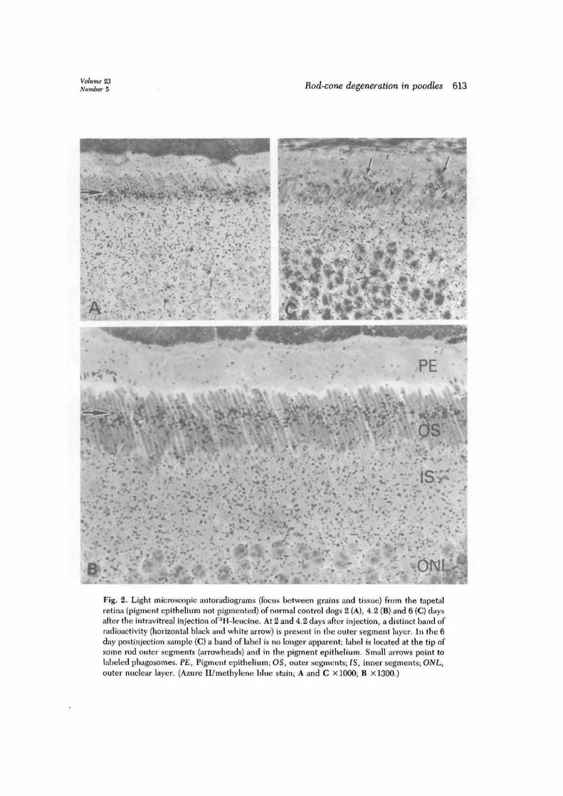
Fig. 2. Light microscopic autoradiograms (focus between grains and tissue) from the tapetalretina (pigment epithelium not pigmented) of normal control dogs 2 (A), 4.2 (B) and 6 (C) daysafter the intravitreal injection of 3H-leucine. At 2 and 4.2 days after injection, a distinct band ofradioactivity (horizontal black and white arrow) is present in the outer segment layer. In the 6day postinjection sample (C) a band of label is no longer apparent; label is located at the tip ofsome rod outer segments (arrowheads) and in the pigment epithelium. Small arrows point tolabeled phagosomes. PE, Pigment epithelium; OS, outer segments; IS, inner segments; ONL,outer nuclear layer. (Azure II/methylene blue stain; A and C XlOOO; B X1300.)

614 Aguirre et at.Invest. Ophthalmol. Vis. Sri.
November 1982
c S 142 Eu
o o
E S
.2 o
Normal controls
^ ̂ 30 weekold affected
10 month oldaffected
1 2 3 4 5 6 7 8Days following intravitreai 3H-Leucine
Fig. 3. Scleral displacement of labeled discs fromthe base of the rod outer segment with time. Themean ± 1 S.D. (vertical hatch marks) is given forthe location of the leading edge of the band ofradioactivity at each postinjection interval for bothcontrol and affected animals. The line for normalsis drawn for best fit and represents data at differ-ent postinjection intervals in six eyes from fivedogs. The affected dogs represent different agegroups and postinjection intervals (line for 30-week-old affected animals drawn for best fit). The 10-month-old affected band displacement point istaken from the peripheral retina, which is morpho-logically normal. Disease status more centrally pre-vented accurate determination of band position.
weeks. Prior to examination, the sections wereagain stained in uranyl acetate and lead citrate.
At the time of enucleation of four affected andthree control animals that had received the in-travitreai 3H-leucine (Table I), the nasal half of theeye was placed on dry ice and the sclera was re-moved from the retina-choroid. For the 42 month-affected animal, the nasal retina-choroid of the lefteye was combined with the entire retina-choroid ofits right eye. The retina-choroid from each samplewas suspended in 5 ml of 40% (w/v) sucrose andswirled on a Vortex mixer for 25 1-sec intervals toshear off the rod outer segments. The suspensionwas centrifuged for 30 min at 27,000 x g. Thecrude rod outer segments floated on the surface andthe remainder of the retina-choroid sedimented asa pellet. The crude rod outer segments were resus-pended in the supernatant fluid, diluted with 10 mlof 66 mM phosphate buffer, pH 7.2, and sedi-mented for 15 min at 27,000 x g. Each crude rodouter segment pellet was suspended in 100 fi\ of 4
mM Tris-acetate buffer, pH 7.4, containing 0.2mM EDTA (25 t̂l for the 42 month-affected ani-mal). Concentrated sodium dodecyl sulfate (SDS)and /3-mercaptoethanol were added to final concen-trations of 2.5% and 2%. After 16 hr at room tem-perature the samples were diluted with an equalvolume of 50% glycerol in water containing 0.002%bromphenol blue. Samples of 25 or 40 fi\ wereapplied to 7.5% polyacrylamide gels for electro-phoresis.9 The gels were stained with Coomassieblue, scanned, and cut into 1 mm slices, whichwere digested with NCS solubilizer (Amersham)and counted in a toluene-based scintillation fluid.
Results
Electroretinography. No abnormalitieswere noted in the ERG responses of 13.5-week- old PRCD-affected dogs. At 28 weeks ofage, however, affected dogs had ERG re-sponses that failed to increase in amplitudewith dark adaptation (not shown), and thedark-adapted responses were lower in ampli-tude than normal (Fig. 1). With progressionof the disease the response amplitude de-creased; by 18 months of age only small am-plitude b-wave responses were recordedfrom the affected dog with red and whitelight stimuli.
Morphology and ARGOuter segment renewal—controls. Twenty-
four hours after the intravitreai injectionof 3H-leucine, the control photoreceptorsshowed an intense accumulation of silvergrains within the inner segments, and a dis-tinct band of radioactivity was located approx-imately 2.2 /Ltm from the base of the rod outersegments. With increasing postinjection in-tervals, the band of radioactive label was dis-placed sclerad (Fig. 2, A and B). By 6 daysafter injection the band was located near thetip of some rod outer segments, approxi-mately 15.6 /xm from the base. In some areaswith short rod outer segments there was noband of radioactivity; instead, these areasshowed increased labeling over the pigmentepithelium, including labeled phagosomes(Fig. 2, C).
As is typical in the dog, outer segmentlength varied within the same retinal region.8
In the photoreceptors located in the verticalmeridian, outer segment length varied be-

Volume 23Number 5 Rod-cone degeneration in poodles 615
tween 10 to 23 fim. This variation did notappear to be dependent on retinal locationexcept for a reduction in outer segmentlength peripherally near the ora serrata.
Progressive sclerad displacement of theband of radioactivity in the rod outer seg-ment layer with time was measured and ex-pressed as renewal rate (ju,m displaced/24 hr);a mean renewal rate for all retinal regionsexamined in the normal controls was 2.3 ±0.28 (Fig. 3). Although there were slightvariations in band displacement and renewalrates within the same eye, these did not ap-pear to be dependent on retinal location (cen-tral vs. peripheral; tapetal vs. nontapetal; su-perior vs. inferior) (Fig. 4, control).
PRCD—Pathology and outer segment re-newal. Light microscopic examination of theretina of the 14.5-week-old affected animalsshowed no morphologic differences from con-trols that could be attributed to the disease.Individual rod outer segments appeared nor-mal; the sides were parallel and staining in-tensity was uniform throughout the entirelength (Fig. 5, A). The slight disorganizationof the outer segments in some specimens wasinterpreted as possible fixation artifact ratherthan disease. By electron microscopy, how-ever, abnormalities characteristic of the earlystages of PRCD1 were readily evident in theseretinas (Fig. 5, C). Vesicular profiles werepresent primarily in the sclerad half of theouter segment layer, where they were in closeapposition to the outer segment cell mem-brane and within the apical microvilli of thepigment epithelium. Lamellar disorientationand disorganization were evident in some rodphotoreceptors; in most, however, lamellardisc abnormalities were minimal or absent.Cone photoreceptors were normal.
Distinct abnormalities were evident bylight microscopy in 30-week-old dogs; the foureyes from two affected dogs were readily dif-ferentiated in the masked examination fromthe age-matched control eyes (Fig. 6, A). Inmost regions examined at this age, abnormal-ities were restricted to the outer segments.Although the outer segments were aligned inthe parallel arrangement characteristic ofnormal, their sides were indented and irregu-
Table II. Renewal rates for superior retinain the posterior pole of affected dogsand age-matched controls
Control
Age(wk)
Renewal rate(/xm/24 hr)
Affected
Age(wk)
Renewal rate(ixml24 hr)
14.514.53030
Mean
2.42.42.62.5
2.4 ± 0.09
14.514.5303030
1.61.61.31.51.1
1.42 ± 0.19*
*0.01 > p null > 0.001 (t test).
lar (Fig. 7). These changes appeared to affectthe entire outer segment, although they weremore extensive in the sclerad half.
In some instances, visual cell disease af-fected both the inner and outer segments. Aslight decrease in the density of rods resultedin an increased prominence of the cone innersegments. The outer segment layer in theseareas generally contained more markedly dis-organized and disoriented rod outer seg-ments, with a concomitant reduction in den-sity. Although some cones showed thesestructural abnormalities, most were spared.The outer segment disease primarily affectedrods at this time.
The severity and distribution of thesephotoreceptor-degenerative changes are il-lustrated in Fig. 4, which represents a spatialmap of disease for the left eye of a 30-week-old affected dog (Table I, dog 3). The distri-bution of the disease was independent ofwhether the pigment epithelium was pig-mented or nonpigmented. There was, how-ever, a difference between the central andperipheral areas of the retina. In some eyes,the peripheral photoreceptors were normalin the four quadrants examined; in othereyes, the peripheral photoreceptors werenormal in some but not all quadrants (Figs. 4and 6, A and C). Electron microscopic exam-ination confirmed the light microscopic ob-servations.
Phagocytic cells were found in the photo-receptor layer of the tapetal and nontapetalzones of the 30-week-old affected retina. Cy-

(CS O
D - 13.4yR - 2.2p/24 hrs.
PI interval - 6 days
s oR-D - ) P E
S O
R -
S O
hrs. SOD - 16.3p
R - 2.7|j/24 hrs.
soD-15pR - 2.5p/24 hrs.
SODR ) P E
Left Eye30 week old mongrel
Control
S O
3000p
PI interval - 8 days
S - 1D - 10.9R- 1.4/24 hrs.
S - 1D - 12.5MR-1.6p/24hrs.
S - 1D- 12.1pR- 1.5 /̂24 hrs.
S - 1 S - 1
NAS - 1D - 9.5pR- 1.2p/24hrs.
S - 1D - 10.5p
R - 1.3p/24 hrs.
NA
S O
R- 1.5p/24hrs.
30O0H
Left Eye30 week old affected
Miniature Poodle
Fig. 4. For legend see facing page.
Volume 23Number 5 Rod-cone degeneration in poodles 617
tologically they were different from pigmentepithelial cells; they were nonpigmented andtheir irregularly shaped pale nuclei had adense chromatin margin.
Light microscopic ARG of the retina in theyoung (14.5 and 30 weeks) affected dogsshowed the same general pattern of label in-corporation as in the controls (Figs. 5, B, and6, B and D). Label was found within theinner segments and, with increasing postin-jection intervals, the band of radioactivity inthe outer segment layer was displacedsclerad. As with controls, a decreasing con-centration of silver grains was located vitreadto the distinct band of radioactivity. Theseobservations were confirmed by electron mi-croscopic ARG.
A consistent difference between affected
and control dogs was evident in the dis-placement of the band of radioactivity; in af-fected dogs the radioactive label was dis-placed a shorter distance than in controls foreach postinjection interval studied (Fig. 3).For example, the radioactive band in theouter segment layer was displaced 15.6 ± 0 . 6/urn 6 days after the intravitreal injection in thecontrol, but in the 30-week-old affected ani-mals, the band was displaced only 6.5 ± 0.4and l l .2 ± 1.0/urn6and8days, respectively,after the injection (Figs. 3 and 4).
Table II illustrates the displacement resultsin terms of renewal rates (/am/24 hr) for thesuperior retina in the posterior pole of controland affected (14.5 and 30 weeks of age) ani-mals. This area was chosen because it wasuniformly represented in both groups. The
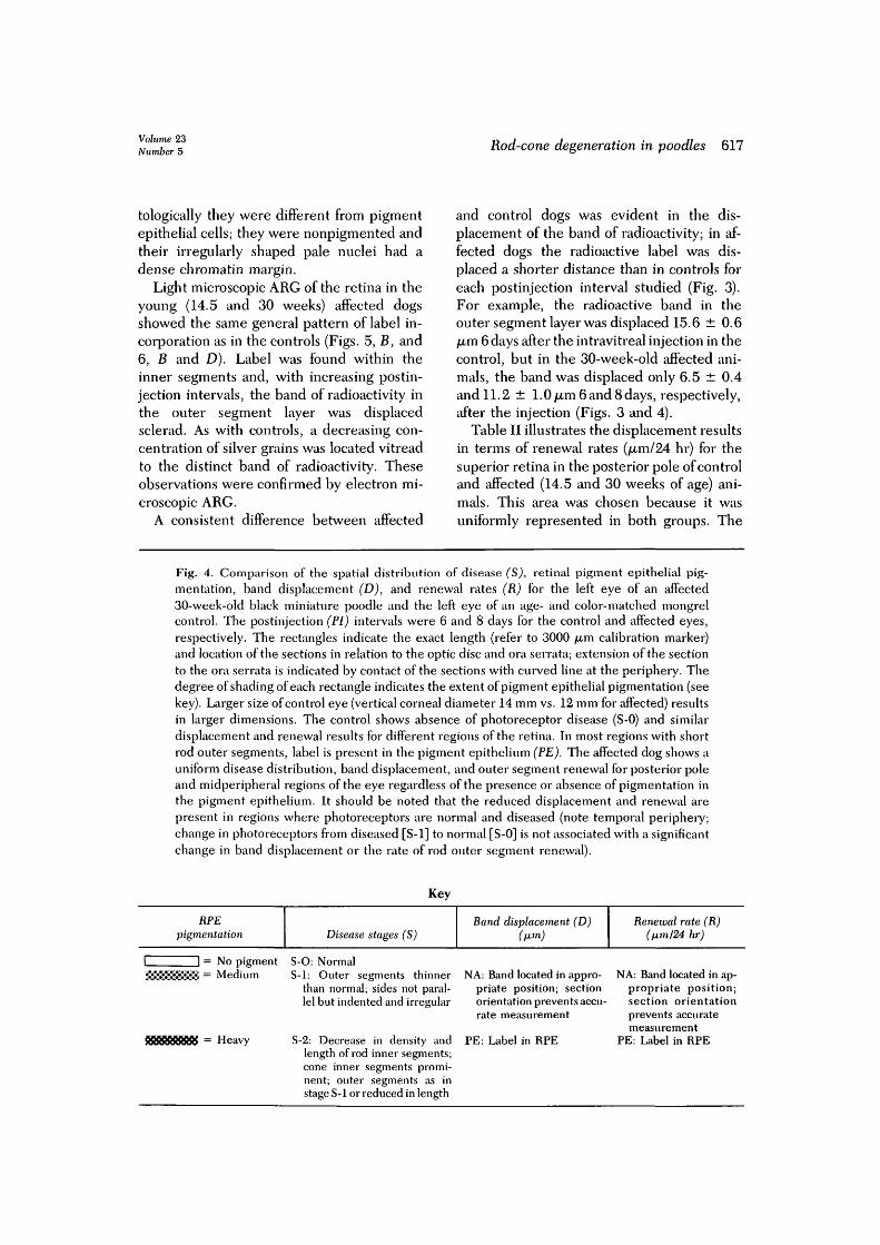
Fig. 4. Comparison of the spatial distribution of disease (S), retinal pigment epithelial pig-mentation, band displacement (D), and renewal rates (R) for the left eye of an affected30-week-old black miniature poodle and the left eye of an age- and color-matched mongrelcontrol. The postinjection (PI) intervals were 6 and 8 days for the control and affected eyes,respectively. The rectangles indicate the exact length (refer to 3000 /xm calibration marker)and location of the sections in relation to the optic disc and ora serrata; extension of the sectionto the ora serrata is indicated by contact of the sections with curved line at the periphery. Thedegree of shading of each rectangle indicates the extent of pigment epithelial pigmentation (seekey). Larger size of control eye (vertical corneal diameter 14 mm vs. 12 mm for affected) resultsin larger dimensions. The control shows absence of photoreceptor disease (S-0) and similardisplacement and renewal results for different regions of the retina. In most regions with shortrod outer segments, label is present in the pigment epithelium (PE). The affected dog shows auniform disease distribution, band displacement, and outer segment renewal for posterior poleand midperipheral regions of the eye regardless of the presence or absence of pigmentation inthe pigment epithelium. It should be noted that the reduced displacement and renewal arepresent in regions where photoreceptors are normal and diseased (note temporal periphery;change in photoreceptors from diseased [S-1] to normal [S-0] is not associated with a significantchange in band displacement or the rate of rod outer segment renewal).
Key
RPEpigmentation
1 1 = No pigment!*!»!$JS8*S*8 = Medium
9RB65BB& = Heavy
Disease stages (S)
S-O: NormalS-1: Outer segments thinner
than normal; sides not paral-lel but indented and irregular
S-2: Decrease in density andlength of rod inner segments;cone inner segments promi-nent; outer segments as instage S-1 or reduced in length
Band displacement (D)(fim)
NA: Band located in appro-priate position; sectionorientation prevents accu-rate measurement
PE: Label in RPE
Renewal rate (R)(lxm/24 hr)
NA: Band located in ap-propriate position;section orientationprevents accuratemeasurement
PE: Label in RPE
618 Aguirre et at. Invest, Ophthahnol. Vis. Sci.November 1982
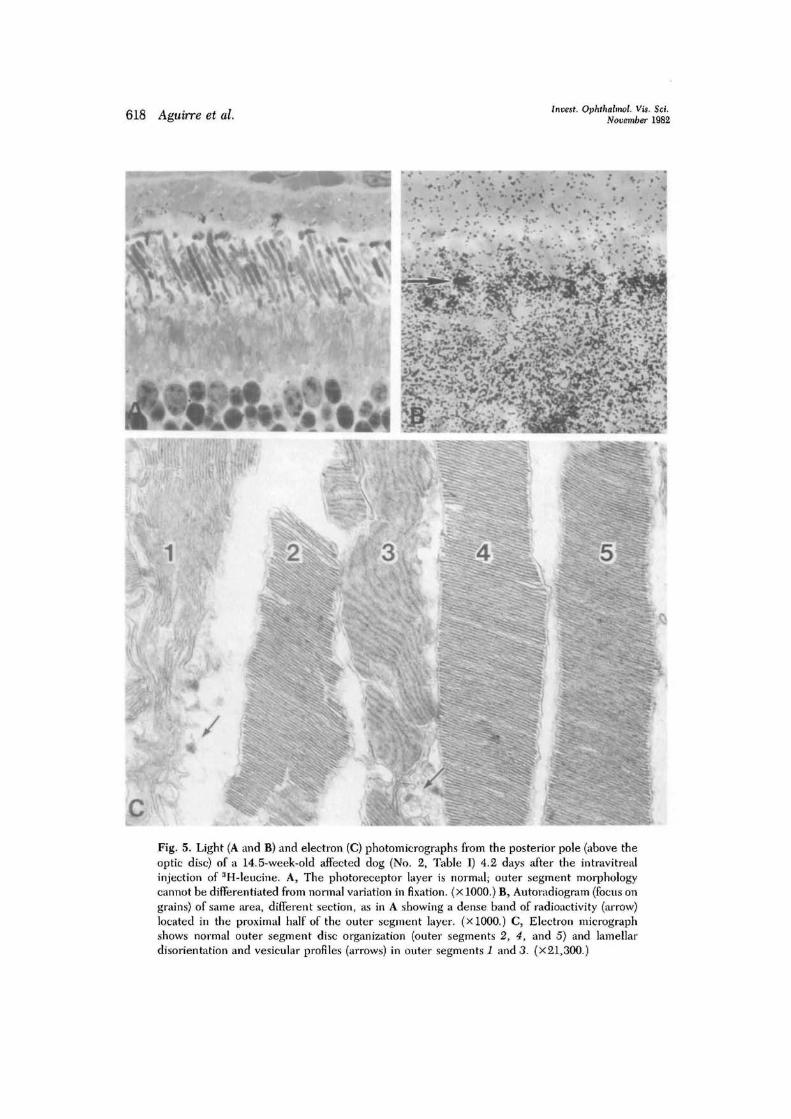
Fig. 5. Light (A and B) and electron (C) photomicrographs from the posterior pole (above theoptic disc) of a 14.5-week-old affected dog (No. 2, Table I) 4.2 days after the intravitrealinjection of aH-leucine. A, The photoreceptor layer is normal; outer segment morphologycannot be differentiated from normal variation in fixation, (x 1000.) B, Autoradiogram (focus ongrains) of same area, different section, as in A showing a dense band of radioactivity (arrow)located in the proximal half of the outer segment layer. (xlOOO.) C, Electron micrographshows normal outer segment disc organization (outer segments 2, 4, and 5) and lamellardisorientation and vesicular profiles (arrows) in outer segments 1 and 3. (x21,300.)

Volume 23Number 5 Rod-cone degeneration in poodles 619
Fig. 6. Light microscopic sections and autoradiograms (focus on grains) from the left retina of30-week-old affected dog (No. 3, Table I) 8 days after the intravitreal injection of 3H-leucine. Aand B, Paired sections from the posterior pole above the optic nerve head showing outersegment disorientation and disorganization (A) and a dense band of radioactivity (arrow) in thescleral half of the outer segment layer (B). The vacuolation and vesiculation of photoreceptorinner segments in A are a fixation artifact. C and D, Paired sections from the temporalperiphery adjacent to the ora serrata. Photoreceptor structure is normal (C). A dense band ofradioactivity (arrow) is present in the tips of the outer segments adjacent to the pigmentepithelium (D). (All XlOOO.)
renewal rate in the affected animals was signif-icantly slower than that in controls. Similarreductions in renewal rates were present inother retinal locations of the affected dogs(Fig. 4). A comparison of the renewal rates forall retinal areas of control (2.3 ± 0.28 /um/24
hr) and 14.5- and 30-week-old affected ani-mals (1.3 ± 0.25/am/24 hr) indicated that theslower renewal rate of affected animals wassignificantly different from that of controls atthe 98% confidence level (t test, 0.02 > pnull > 0.01).

620 Aguirre et al.Invest. Ophthalmol. Vis. Sci.
November 1982
\
Fig. 7. Light and electron photomicrographs of the right nasal midperipheral retina of a30-week-old affected dog {No. 3, Table I). Outer segment cell membranes are not parallel, andthe lamellar discs are disoriented. Vesicular profiles (arrows) are distinct and associated withabnormal rod outer segments. A phagosome (P) is present within an apical process of thepigment epithelium. M, Melanin granule, (x 14,400). Inset, Most rod outer segments aredisorganized and have irregular sides (stage I). C, cone; R, rod. (X1250.)
The posterior pole retina from the 10-month-old affected dog (No. 5, Table I)showed a marked progression of the rod pho-toreceptor disease. The rod inner segmentswere shortened and had disorganized anddisoriented outer segments. In some areas,rods were lost from the visual cell layer, re-sulting in broadening and increased promi-nence of the cone inner segments (Fig. 8, A).Concomitant with the loss of rod inner seg-
ments, there was a decrease in the density ofouter segment material and a narrowing ofthe outer nuclear layer.
The extent of the disease in the central andequatorial retina of the 10-month-old animalprevented the identification of a band of ra-dioactivity in the outer segment layer (Fig. 8,A to D). Although label was present at alllevels of the outer segment layer, its distribu-tion was diffuse, preventing differentiation

Volume 23Number 5 Rod-cone degeneration in poodles 621
between molecular and membrane renewaland randomly distributed outer segment layerlabel. In these areas, shortened and diminu-tive rod inner segments had intense labeldistributed over the perinuclear and innersegment regions. The now-prominent conesaccumulated label intensely in the inner seg-ments and diffusely over the outer segments.
Peripherally, the rod disease in the 10-month-old animal was confined to the outersegments. These showed structural changessimilar to those present in the central retinalregions of the younger affected animals (Fig.8, C and D). In the far periphery adjacent tothe ora serrata, a narrow transition zone (Fig.8, E) separated structurally normal periph-eral photoreceptors from more diseased cen-tral ones. Electron microscopy confirmedthat the peripheral rod photoreceptors wereindeed normal (Fig. 9, A and C).
The structurally normal peripheral rodphotoreceptors (Fig. 9, B) of the 10-month-old affected animal also had abnormal outersegment renewal; 2 days after the intravitrealinjection the label was displaced 2.5 /im intothe outer segment layer (renewal rate, 1.25/nm/24 hr). A sharp contrast in labeling char-acteristics was seen between these cells andthe more diseased central ones at the periph-eral transition zone (Fig. 8, £); the more cen-trally located rod photoreceptors showeddiffuse labeling of the outer segment layerrather than a distinct band (Fig. 8, F).
Progressive photoreceptor degenerativechanges in the older affected animals resultedin loss of outer and then inner segments andbrought the external limiting membrane intoclose proximity to the pigment epithelium.The outer nuclear layer subsequently disap-peared; depending on the retinal location,inner retinal layer organization was preservedor lost (Fig. 10, A, C, andE.).
In contrast to the younger affected animals,the peripheral retina was more diseased in theolder affected animals (181A months and older)(Fig. 10). In the 60-month-old animal the ret-ina was best preserved in the posterior pole,where only broad, club-shaped cone photore-ceptors and their nuclei remained (Fig. 11, D).
These findings, however, were not uniform.Intact cone photoreceptors were absent fromthe superior and inferior parapapillary retinabut were present temporally and nasally fordistances of approximately 5000 and 800 fim,respectively, from the optic disc (compareFigs. 10, A, and 11, D).
Autoradiographic studies of the retina inthese older animals showed dense incorpora-tion of label into retinal neurons. The distri-bution of photoreceptor layer label dependedon the age of the animal and the stage of thedisease. In areas where photoreceptors werepreserved, outer segment layer label waspresent but a distinct band of radioactivitywas not evident (Fig. 11, A and B). In morediseased areas, radioactive label accumulatedin the perinuclear regions of the remainingphotoreceptor nuclei (Fig. 10, B, D, andE).
Biochemistry. The use of crude rod outersegment preparations provided membraneproteins from retinal structures other thanthe outer segments. These proteins served asreference points for determining the relativelabeling of opsin by gel electrophoresis. Oneuseful reference was the prominent Coomas-sie blue-staining peak seen to the left ofopsin in Fig. 13 and resolved into its twocomponents in Fig. 12. Relative to the fastermoving band of this doublet, opsin had a mo-bility of 1.26 ± 0.03. Four different dogcrude rod outer segment preparations wereused for this comparison. In each case, puri-fied bovine rod outer segment opsin coin-cided with the canine opsin peak so labeledin Figs. 12 and 13.
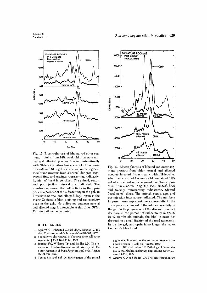
In the 14.5-week-old littermate affectedand control dogs injected intravitreally with3H-leucine, the SDS gels of the crude rodouter segment preparations showed that themajor radioactive band in the gels coincidedwith the major Coomassie blue-stain ingpeak for opsin (Fig. 12). The distribution oflabel and membrane proteins within the gelswas the same in the affected and controldogs. Moreover, when the radioactivity inthe opsin peak was expressed as a percent ofthe total radioactivity in the gel, no differ-ence between affected and control dogs was

622 Aguirre et al. Invest. Ophthalmol. Vis. Set.November 1982
Fig. 8. For legend see ikcing page.
Volume 23Number 5 Rod-cone degeneration in poodles 623
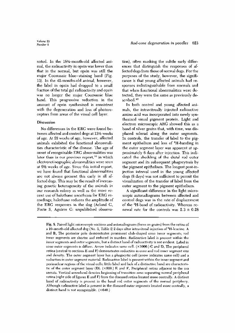
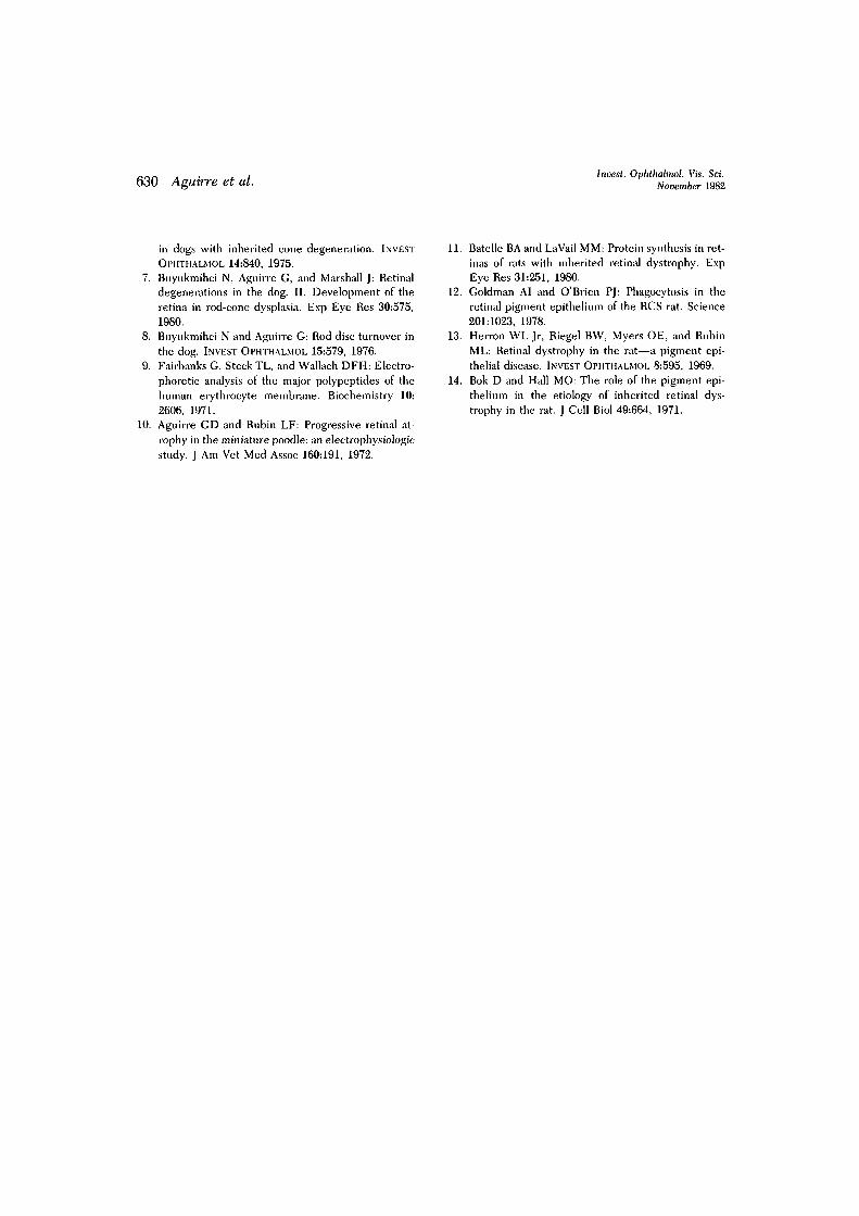
noted. In the l^A-month-old affected ani-mal, the radioactivity in opsin was lower thanthat in the normal, but opsin was still themajor Coomassie blue-staining band (Fig.13). In the 42-months-old animal, however,the label in opsin had dropped to a smallfraction of the total gel radioactivity and opsinwas no longer the major Coomassie blueband. This progressive reduction in theamount of opsin synthesized is consistentwith the degeneration and loss of photore-ceptors from areas of the visual cell layer.
Discussion
No differences in the ERG were found be-tween affected and control dogs at 13V2 weeksof age. At 28 weeks of age, however, affectedanimals exhibited the functional abnormali-ties characteristic of the disease. The age ofonset of recognizable ERG abnormalities waslater than in our previous report,10 in whichelectroretinographic abnormalities were seenat 9V2 weeks of age. Since this initial report,we have found that functional abnormalitiesare not always present this early in all af-fected dogs. This may be the result of increas-ing genetic heterogeneity of the animals inour research colony as well as the more re-cent use of halothane anesthesia for ERG re-cordings; halothane reduces the amplitude ofthe ERG responses in the dog (Acland G,Forte S, Aguirre G: unpublished observa-
tion), often masking the subtle early differ-ences that distinguish the responses of af-fected dogs from those of normal dogs. For thepurposes of the study, however, the signifi-cance is that young affected animals had re-sponses indistinguishable from normals andthat when functional abnormalities were de-tected, they were the same as previously de-scribed. l0
In both control and young affected ani-mals, the intravitreally injected radioactiveamino acid was incorporated into newly syn-thesized visual pigment protein. Light andelectron microscopic ARG showed this as aband of silver grains that, with time, was dis-placed sclerad along the outer segments.In controls, the transfer of label to the pig-ment epithelium and loss of 3H-banding inthe outer segment layer was apparent at ap-proximately 6 days after injection. This indi-cated the shedding of the distal rod outersegment and its subsequent phagocytosis bythe pigment epithelium. The longest post-in-jection interval used in the young affecteddogs (8 days) was not sufficient to permit thevisualization of the transfer of label from theouter segment to the pigment epithelium.
A significant difference in the light micro-scopic autoradiograms between affected andcontrol dogs was in the rate of displacementof the 3H-band of radioactivity. Whereas re-newal rate for the controls was 2.3 ± 0.28
Fig. 8. Paired light microscopic sections and autoradiograms (focus on grains) from the retina ofa 10-month-old affected dog (No. 5, Table I) 2 days after intravitreal injection of 3H-leucine. Aand B, The posterior pole demonstrates prominent club-shaped cone inner segments; rodinner segments are shorter and reduced in number. Radioactive label is present within theinner segments and outer segments, but a distinct band of radioactivity is not evident. Label incone outer segments is diffuse. Arrow indicates same cell. (xlOOO.) C and D, The peripheralretina (central to sections E and F) demonstrates reduction in cone and rod inner segment sizeand density. The outer segment layer has a phagocytic cell (arrow indicates same cell) and areduction in outer segment material. Radioactive label is present within the inner segment andperinuclear regions of the visual cells; little label and lack of a distinctive band are characteris-tic of the outer segment layer (D). (x800.) E and F, Peripheral retina adjacent to the oraserrata. Vertical arrowhead denotes beginning of transition zone separating normal peripheralretina (right side of figures E and F) from the diseased retina located more centrally. A distinctband of radioactivity is present in the basal rod outer segments of the normal periphery.Although radioactive label is present in the diseased outer segments located more centrally, adistinct band is not recognizable. (X640.)

624 Aguiire et al. Invest, Ophthalmol. Vis. Sci.November 1982
Fig, 9. For legend see tacing page.

Volume 23Number 5 Rod-cone degeneration in poodles 625
/am/24 hr, affected dogs showed a signifi-cantly slower rate of renewal (1.3 ± 0.25/Am/24 hr). This abnormality was similar in theposterior pole and equatorial and peripheralzones in areas where the rod photoreceptorswere structurally normal or diseased. In addi-tion, the presence of pigment epithelial pig-mentation did not appear to affect the extentand severity of the disease or to modify theabnormal renewal rate. The selected tissuesexamined by electron microscopic ARGconfirmed the light microscopic observations.
Light microscopic ARG demonstrated thatcones distributed label diffusely throughoutthe outer segment; no apparent differences inlabel intensity and distribution were evidentbetween PRCD-affected dogs and normalcontrols. This was, however, an imprecise as-sessment of the renewal of cone outer seg-ment membranes in the disease. Althoughcone degeneration is a feature of PRCD, ourlight microscopic studies were unable to de-termine whether cone outer segment renewalwas abnormal.
At stages of the disease when outer seg-ment disorganization was extensive, a dis-tinct band of 3H-label was not recognizable inthe outer segment layer. Instead, 3H-labelwas present over outer segment profiles at alllevels of the outer segment layer and ap-peared diffuse. In terms of characterizingouter segment renewal by means of measur-ing 3H-band displacement, advanced diseaseprecluded the determination of further re-newal abnormalities in degenerating photo-receptors.
With further progression of the disease,photoreceptor degeneration became moreextensive. These degenerative and retinalatrophic changes began peripherally and pro-gressed centrally. Thus the central-to-pe-ripheral gradation of disease characteristic of
young affected animals was reversed in theolder ones.
The biochemical studies of crude rod outersegment preparations demonstrated that inyoung littermate affected and control dogs,the major radioactive peak in the gel wascoincident with opsin. This confirmed thatthe intense 3H-band of label located over theouter segment layer in the autoradiogramsrepresented the newly synthesized visualpigment protein.
It is noteworthy, however, that the per-cent radioactivity in opsin compared with thetotal gel radioactivity was the same in 14%-week-old affected and control animals eventhough the 3H-band displacement and outersegment renewal rates were reduced in af-fected dogs. Although we have no definiteexplanation for this possible discrepancy, onemust remember that two different parame-ters were being evaluated. The percent ra-dioactivity in the opsin peak represented thesynthesis of new visual pigment protein rela-tive to other proteins on the gel that werederived from rod outer segments and othermembranes, possibly including opsin-con-taining inner segment vesicles. Determi-nation of band displacement and renewalrates measured the assembly and displace-ment of the newly formed disc membranes.It is possible that the rates of protein synthe-sis and disc assembly are different. We havefound no difference in the rod outer segmentwidth and disc packing density between theaffected and control dogs.* The lack ofcorrelation between the opsin synthesisand renewal data suggests that a possible
*Width = 1.21 ± 0.17/im for affected (n = 56), 1.15 ±0.17 /xm for control (n = 79); packing density = 28.6 ±4 discs//i,m for affected (n = 29), 27.8 ± 2 . 8 discs//imfor controls (n = 27).
Fig. 9. Paired light microscopic sections and autoradiograms (A and B) from the peripheralretina adjacent to the ora serrata from a 10-month-old affected dog (No. 5, Table I) 2 days afterthe intravitreal injection of 3H-leucine. Photoreceptor structure is normal. A band of radioac-tivity (arrow) is located in the basal aspect of the outer segment layer. (X800.) C, Electronphotomicrograph of the same area demonstrates normal rod outer segments (ROS). Aphagocytized outer segment tip (arrowhead F) is seen in a cross-section of a broad pigmentepithelial process. M, Melanin granules, (x 15,200.)

626 Aguirre et at. Invest, Ophthahnol. Vis. Sci.November 1982
Fig. 10. Paired light microscopic sections and autoradiograms of the right superior retina froma 60-month-old affected dog (No. 8, Table I) 3 days after the intravitreal injection of aH-leucine. A and B, Superior parapapillary regions show loss of photoreceptor inner and outersegments. A single row of cone nuclei remains in the outer nuclear layer; radioactive labelaccumulates over the perinuclear regions of these cells (arrows). C and D, Superior equatorialregion shows loss of layer organization but preservation of the pigment epitheliun (PE). Radio-active label accumulates in remaining retinal neurons. E and F, Superior peripheral region,3000 jLtm central to the ora serrata. Focal retinal loss results in isolation of the pigmentepithelial layer (area in brackets). In contrast to regions where retinal tissue is better pre-served, pigment epithelium accumulates radioactive label. (X800.)
Volume 23Number 5 Rod-cone degeneration in poodles 627
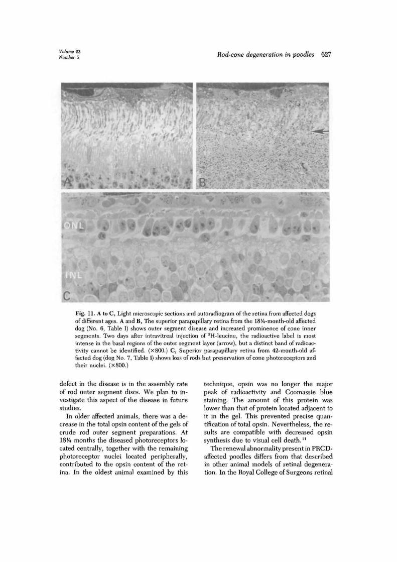
Fig. 11. A to C, Light microscopic sections and autoradiogram of the retina from affected dogsof different ages. A and B, The superior parapapillary retina from the 18%-month-old affecteddog (No. 6, Table I) shows outer segment disease and increased prominence of cone innersegments. Two days after intravitreal injection of 3H-leucine, the radioactive label is mostintense in the basal regions of the outer segment layer (arrow), but a distinct band of radioac-tivity cannot be identified. (x800.) C, Superior parapapillary retina from 42-month-old af-fected dog (dog No. 7, Table I) shows loss of rods but preservation of cone photoreceptors andtheir nuclei. (X800.)
defect in the disease is in the assembly rateof rod outer segment discs. We plan to in-vestigate this aspect of the disease in futurestudies.
In older affected animals, there was a de-crease in the total opsin content of the gels ofcrude rod outer segment preparations. AtI8V4 months the diseased photoreceptors lo-cated centrally, together with the remainingphotoreceptor nuclei located peripherally,contributed to the opsin content of the ret-ina. In the oldest animal examined by this
technique, opsin was no longer the majorpeak of radioactivity and Coomassie bluestaining. The amount of this protein waslower than that of protein located adjacent toit in the gel. This prevented precise quan-tification of total opsin. Nevertheless, the re-sults are compatible with decreased opsinsynthesis due to visual cell death.11
The renewal abnormality present in PRCD-affected poodles differs from that describedin other animal models of retinal degenera-tion. In the Royal College of Surgeons retinal
628 Aguirre et al. Incest. Ophthalmol. Vis. Sci.November 1982
GCL
NFL
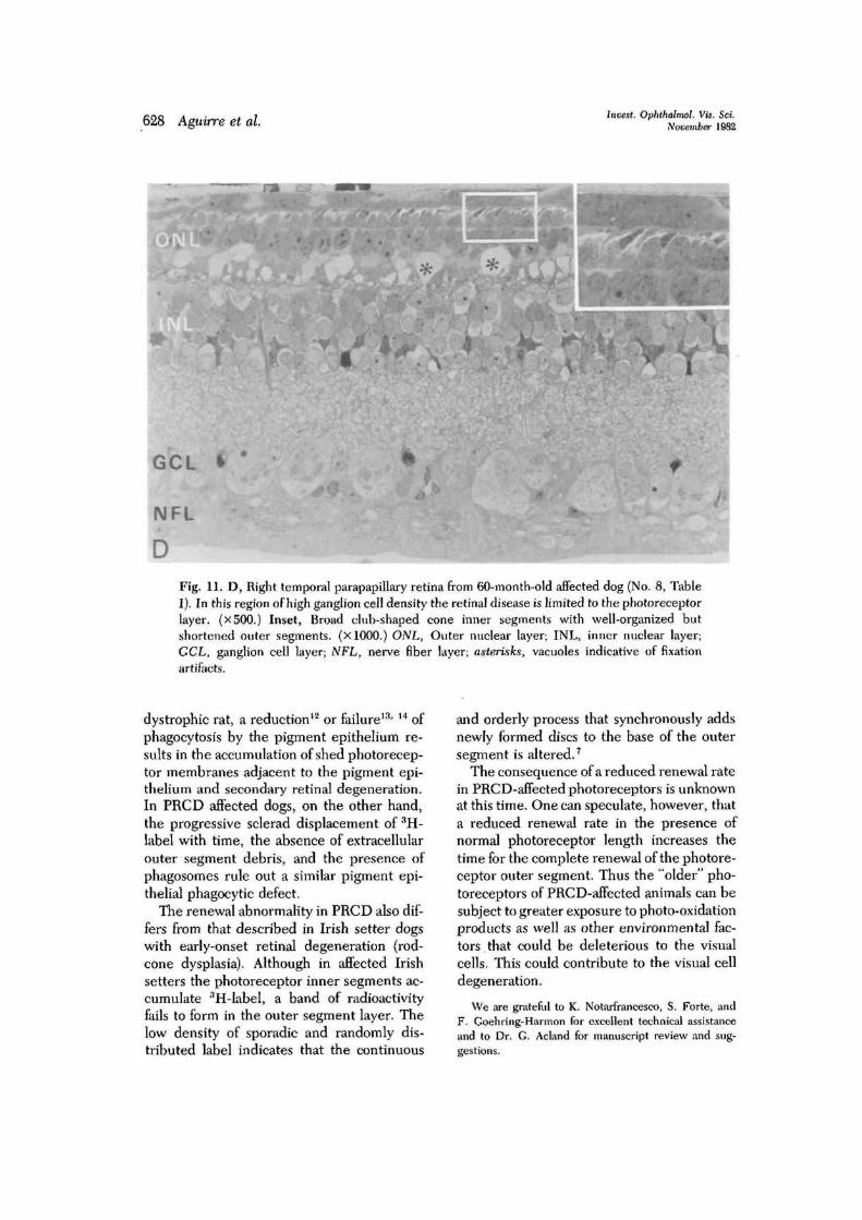
DFig. 11. D, Right temporal parapapillary retina from 60-month-old affected dog (No. 8, TableI), In this region of high ganglion cell density the retinal disease is limited to the photoreceptorlayer. (x500.) Inset, Broad club-shaped cone inner segments with well-organized butshortened outer segments. (X1000.) ONL, Outer nuclear layer; INL, inner nuclear layer;CCL, ganglion cell layer; NFL, nerve fiber layer; asterisks, vacuoles indicative of fixationartifacts.
dystrophic rat, a reduction12 or failure13' I4 ofphagocytosis by the pigment epithelium re-sults in the accumulation of shed photorecep-tor membranes adjacent to the pigment epi-thelium and secondary retinal degeneration.In PRCD affected dogs, on the other hand,the progressive sclerad displacement of 3H-label with time, the absence of extracellularouter segment debris, and the presence ofphagosomes rule out a similar pigment epi-thelial phagocytic defect.
The renewal abnormality in PRCD also dif-fers from that described in Irish setter dogswith early-onset retinal degeneration {rod-cone dysplasia). Although in affected Irishsetters the photoreceptor inner segments ac-cumulate 3H-label, a band of radioactivityfails to form in the outer segment layer. Thelow density of sporadic and randomly dis-tributed label indicates that the continuous
and orderly process that synchronously addsnewly formed discs to the base of the outersegment is altered.7
The consequence of a reduced renewal ratein PRCD-affected photoreceptors is unknownat this time. One can speculate, however, thata reduced renewal rate in the presence ofnormal photoreceptor length increases thetime for the complete renewal of the photore-ceptor outer segment. Thus the "older" pho-toreceptors of PRCD-affected animals can besubject to greater exposure to photo-oxidationproducts as well as other environmental fac-tors that could be deleterious to the visualcells. This could contribute to the visual celldegeneration.
We are grateful to K. Notarfrancesco, S. Forte, andF. Goehring-Harmon for excellent technical assistanceand to Dr. G. Acland for manuscript review and sug-gestions.
Volume 23Number 5 Rod-cone degeneration in poodles 629
1500
1000 -
1000
MINIATURE POODLES14% weeks old
- Post-injectionInterval 4.2 days
1000
1000
Fig. 12. Electrophoresis of labeled rod outer seg-ment proteins from 14^-week-old littermate nor-mal and affected poodles injected intravitreallywith 3H-leucine. Absorbance scan of a Coomassieblue-stained SDS gel of crude rod outer segmentmembrane proteins from a normal dog (top scan,smooth line) and tracings representing radioactiv-ity (clotted lines) in gel slices. The animal, status,and postinjection interval are indicated. Thenumbers represent the radioactivity in the opsinpeak as a percent of the radioactivity in the gel. Inlittermate normal and affected dogs, opsin is themajor Coomassie blue-staining and radioactivitypeak in the gels. No difference between normaland affected dogs is detectable at this time. DPM,Disintegrations per minute.
R E F E R E N C E S
1. Aguirre G: Inherited retinal degenerations in thedog. Trans Am Acad Ophthalmol Otol 81:667, 1976.
2. Young RW: The renewal of photoreceptor cell outersegments. J Cell Biol 33:61, 1967.
3. Bargoot FG, Williams TP, and Beidler LM: The lo-calization of radioactive amino acid taken up into theouter segments of frog (Rana pipiens) rods. VisionRes 9:385, 1969.
4. Young RW and Bok D: Participation of the retinal
6000
5000
4000
3000
MINIATURE POODLES" Post-injection
Interval 2 days
-Opsin
Fig. 13. Electrophoresis of labeled rod outer seg-ment proteins from older normal and affectedpoodles injected intravitreally with 3H-leucine.Absorbance scan of Coomassie blue-stained SDSgel of crude rod outer segment membrane pro-teins from a normal dog (top scan, smooth line)and tracings representing radioactivity (dottedlines) in gel slices. The animal, status, age, andpostinjection interval are indicated. The numbersin parentheses represent the radioactivity in theopsin peak as a percent of the total radioactivity inthe gel. With progression of the disease there is adecrease in the percent of radioactivity in opsin.In 42-months-old animals, the label in opsin hasdropped to a small fraction of the total radioactiv-ity on the gel, and opsin is no longer the majorCoomassie blue band.
pigment epithelium in the rod outer segment re-newal process. J Cell Biol 42:392, 1969.
5. Aguirre GD and Rubin LF: Pathology of hemeralo-pia in the Alaskan malamute dog. INVEST OPHTHAL-MOL 13:231, 1974.
6. Aguirre GD and Rubin LF: The electroretinogram
630 Aguirre et al.Invest. Ophthaltnol. Vis. Sci.
November 1982
in dogs with inherited cone degeneration. INVESTOPHTHALMOL 14:840, 1975.
7. Buyukmihci N, Aguirre G, and Marshall J: Retinaldegenerations in the dog. II. Development of theretina in rod-cone dysplasia. Exp Eye Res 30:575,1980.
8. Buyukmihci N and Aguirre G: Rod disc turnover inthe dog. INVEST OPHTHALMOL 15:579, 1976.
9. Fairbanks G, Steck TL, and Wallach DFH: Electro-phoretic analysis of the major polypeptides of thehuman erythrocyte membrane. Biochemistry 10:2606, 1971.
10. Aguirre GD and Rubin LF: Progressive retinal at-rophy in the miniature poodle: an electrophysiologicstudy. J Am Vet Med Assoc 160:191, 1972.
11. Batelle BA and La Vail MM: Protein synthesis in ret-inas of rats with inherited retinal dystrophy. ExpEye Res 31:251, 1980.
12. Goldman AI and O'Brien PJ: Phagocytosis in theretinal pigment epithelium of the RCS rat. Science201:1023, 1978.
13. Herron WL Jr, Riegel BW, Myers OE, and RubinML: Retinal dystrophy in the rat—a pigment epi-thelial disease. INVEST OPHTHALMOL 8:595, 1969.
14. Bok D and Hall MO: The role of the pigment epi-thelium in the etiology of inherited retinal dys-trophy in the rat. J Cell Biol 49:664, 1971.
Copyright © 2022 FDOKUMEN