Partitioning of Bacterial Communities between Seawater and Healthy, Black Band Diseased, and Dead...
15
APPLIED AND ENVIRONMENTAL MICROBIOLOGY, May 2002, p. 2214–2228 Vol. 68, No. 5 0099-2240/02/$04.000 DOI: 10.1128/AEM.68.5.2214–2228.2002 Partitioning of Bacterial Communities between Seawater and Healthy, Black Band Diseased, and Dead Coral Surfaces Jorge Frias-Lopez, Aubrey L. Zerkle, George T. Bonheyo, and Bruce W. Fouke* Department of Geology, University of Illinois, Urbana, Illinois 61801 Received 21 December 2001/Accepted 25 February 2002 Distinct partitioning has been observed in the composition and diversity of bacterial communities inhabiting the surface and overlying seawater of three coral species infected with black band disease (BBD) on the southern Caribbean island of Curaçao, Netherlands Antilles. PCR amplification and sequencing of bacterial 16S rRNA genes (rDNA) with universally conserved primers have identified over 524 unique bacterial se- quences affiliated with 12 bacterial divisions. The molecular sequences exhibited less than 5% similarity in bacterial community composition between seawater and the healthy, black band diseased, and dead coral surfaces. The BBD bacterial mat rapidly migrates across and kills the coral tissue. Clone libraries constructed from the BBD mat were comprised of eight bacterial divisions and 13% unknowns. Several sequences repre- senting bacteria previously found in other marine and terrestrial organisms (including humans) were isolated from the infected coral surfaces, including Clostridium spp., Arcobacter spp., Campylobacter spp., Cytophaga fermentans, Cytophaga columnaris, and Trichodesmium tenue. Infectious disease in scleractinian corals has emerged as one of the primary causes of the accelerating global destruction of coral reef ecosystems (22, 25, 27, 56, 74). Black band disease (BBD) is one of the most widespread and destructive of these coral infections (see review in reference 50). The diagnostic symptom of BBD is the development of a narrow 0.1- to 7-cm- wide ring-shaped black to red microbial mat that migrates from top to bottom across massive coral colonies, killing healthy coral tissue at rates of as much as 1 cm per day (47, 53). BBD preferentially affects corals such as Montastrea annularis, Mon- tastrea cavernosa, and Diploria strigosa (6, 15, 53). These spe- cies, known as framework building corals, form large structures that become the dominant physical elements of reefs. As a result, coral mortality caused by BBD is a potent force in restructuring coral reef ecosystems (15, 36). There is considerable controversy as to whether BBD is caused by physical and chemical environmental stresses or is an infectious disease or both (50, 56). However, an impediment to determining the cause of BBD has been the lack of informa- tion about the diversity and distribution of microbial popula- tions that inhabit normal healthy coral tissue and the BBD bacterial mat. It is known from studies of infectious disease in marine and terrestrial invertebrates, fish, and mammals (in- cluding humans) that pathogens are most effectively studied within an ecological context of interactions among microbes, their hosts, and the environmental conditions in which they live (25, 54). Accurate diagnosis and eventual treatment and pre- vention of BBD will therefore require a basic knowledge of the composition and distribution of the microbial communities associated with healthy as well as diseased organisms. This type of community-based comparative analysis of the microorgan- isms associated with infectious diseases in corals has not pre- viously been completed. The purpose of the present report was to complete the first culture-independent 16S rRNA phylogenetic survey of the bac- terial communities inhabiting seawater and healthy, BBD-in- fected, and dead coral surfaces. The coral reef chosen for analysis was on the leeward reef tract of Curaçao, Netherlands Antilles (Fig. 1). A factor that may contribute to BBD is the daily dumping of sewage and other pollutants directly onto the reef from the major commercial, municipal, and military har- bor of St. Annabaai (Fig. 1). The following three fundamental questions have been addressed: (i) is there a coral-specific microbial population that is different from that of the bacte- rioplankton community in the overlying seawater column; (ii) are the microbial communities comprising the BBD mat and the dead coral surfaces distinct from each other; and (iii) have sewage microbes colonized the diseased coral, as might be expected if sewage microbes were contributing to the disease process? Results are presented that indicate the healthy coral tissue is colonized by a microbial population that is unique and distinct from that found in the water column and the BBD mat and dead coral surfaces. Also, bacteria associated with sewage were detected only in the BBD bacterial mat. MATERIALS AND METHODS Field work and sample collection. Field sampling using standard scuba tech- niques was conducted in August 2000 on the leeward reef tract of Curaçao, Netherlands Antilles (Fig. 1). Diseased corals were studied at the Playa Kalki and Water Plant sites in back reef environments approximately 0.5 km offshore (33). These included BBD-infected colonies of M. annularis (Fig. 2A and B) and M. cavernosa (Fig. 2C and D) at Playa Kalki at a 5-m water depth, as well as a colony of D. strigosa (Fig. 2E and F) at Water Plant at a 4-m water depth. Three individual BBD-infected coral heads, one each of M. annularis, M. cavernosa, and D. strigosa, were analyzed. From each coral head, four individual samples were collected in situ from the following environments: the overlying seawater, the healthy coral surface, the black band diseased coral surface, and the dead coral surface. Seawater was sampled by collecting 2 liters of seawater in cleaned 1-liter Nalgene high-density polyethylene bottles from directly above the coral colonies. The water was pumped through a sterile 0.45-m-filter-loaded cup (Pall/ Gelman) when brought on shore. All filters and centrifuge tubes were then immediately frozen at 20 o C, transported to Illinois on dry ice, and stored at 40 to 80°C. The coral surface samples were collected by removing a 2-cm by * Corresponding author. Mailing address: Department of Geology, University of Illinois, 1301 W. Green St., Urbana, IL 61801. Phone: (217) 244-5431. Fax: (217) 244-4996. E-mail: [email protected]. 2214
Transcript of Partitioning of Bacterial Communities between Seawater and Healthy, Black Band Diseased, and Dead...
APPLIED AND ENVIRONMENTAL MICROBIOLOGY, May 2002, p. 2214–2228 Vol. 68, No. 50099-2240/02/$04.00�0 DOI: 10.1128/AEM.68.5.2214–2228.2002
Partitioning of Bacterial Communities between Seawater and Healthy,Black Band Diseased, and Dead Coral Surfaces
Jorge Frias-Lopez, Aubrey L. Zerkle, George T. Bonheyo, and Bruce W. Fouke*Department of Geology, University of Illinois, Urbana, Illinois 61801
Received 21 December 2001/Accepted 25 February 2002
Distinct partitioning has been observed in the composition and diversity of bacterial communities inhabitingthe surface and overlying seawater of three coral species infected with black band disease (BBD) on thesouthern Caribbean island of Curaçao, Netherlands Antilles. PCR amplification and sequencing of bacterial16S rRNA genes (rDNA) with universally conserved primers have identified over 524 unique bacterial se-quences affiliated with 12 bacterial divisions. The molecular sequences exhibited less than 5% similarity inbacterial community composition between seawater and the healthy, black band diseased, and dead coralsurfaces. The BBD bacterial mat rapidly migrates across and kills the coral tissue. Clone libraries constructedfrom the BBD mat were comprised of eight bacterial divisions and 13% unknowns. Several sequences repre-senting bacteria previously found in other marine and terrestrial organisms (including humans) were isolatedfrom the infected coral surfaces, including Clostridium spp., Arcobacter spp., Campylobacter spp., Cytophagafermentans, Cytophaga columnaris, and Trichodesmium tenue.
Infectious disease in scleractinian corals has emerged as oneof the primary causes of the accelerating global destruction ofcoral reef ecosystems (22, 25, 27, 56, 74). Black band disease(BBD) is one of the most widespread and destructive of thesecoral infections (see review in reference 50). The diagnosticsymptom of BBD is the development of a narrow 0.1- to 7-cm-wide ring-shaped black to red microbial mat that migrates fromtop to bottom across massive coral colonies, killing healthycoral tissue at rates of as much as 1 cm per day (47, 53). BBDpreferentially affects corals such as Montastrea annularis, Mon-tastrea cavernosa, and Diploria strigosa (6, 15, 53). These spe-cies, known as framework building corals, form large structuresthat become the dominant physical elements of reefs. As aresult, coral mortality caused by BBD is a potent force inrestructuring coral reef ecosystems (15, 36).
There is considerable controversy as to whether BBD iscaused by physical and chemical environmental stresses or is aninfectious disease or both (50, 56). However, an impediment todetermining the cause of BBD has been the lack of informa-tion about the diversity and distribution of microbial popula-tions that inhabit normal healthy coral tissue and the BBDbacterial mat. It is known from studies of infectious disease inmarine and terrestrial invertebrates, fish, and mammals (in-cluding humans) that pathogens are most effectively studiedwithin an ecological context of interactions among microbes,their hosts, and the environmental conditions in which they live(25, 54). Accurate diagnosis and eventual treatment and pre-vention of BBD will therefore require a basic knowledge of thecomposition and distribution of the microbial communitiesassociated with healthy as well as diseased organisms. This typeof community-based comparative analysis of the microorgan-isms associated with infectious diseases in corals has not pre-viously been completed.
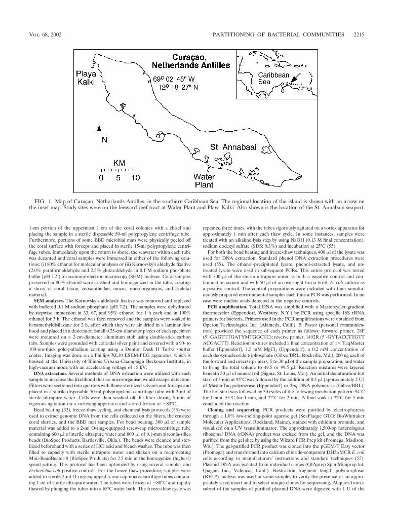
The purpose of the present report was to complete the firstculture-independent 16S rRNA phylogenetic survey of the bac-terial communities inhabiting seawater and healthy, BBD-in-fected, and dead coral surfaces. The coral reef chosen foranalysis was on the leeward reef tract of Curaçao, NetherlandsAntilles (Fig. 1). A factor that may contribute to BBD is thedaily dumping of sewage and other pollutants directly onto thereef from the major commercial, municipal, and military har-bor of St. Annabaai (Fig. 1). The following three fundamentalquestions have been addressed: (i) is there a coral-specificmicrobial population that is different from that of the bacte-rioplankton community in the overlying seawater column; (ii)are the microbial communities comprising the BBD mat andthe dead coral surfaces distinct from each other; and (iii) havesewage microbes colonized the diseased coral, as might beexpected if sewage microbes were contributing to the diseaseprocess? Results are presented that indicate the healthy coraltissue is colonized by a microbial population that is unique anddistinct from that found in the water column and the BBD matand dead coral surfaces. Also, bacteria associated with sewagewere detected only in the BBD bacterial mat.
MATERIALS AND METHODS
Field work and sample collection. Field sampling using standard scuba tech-niques was conducted in August 2000 on the leeward reef tract of Curaçao,Netherlands Antilles (Fig. 1). Diseased corals were studied at the Playa Kalkiand Water Plant sites in back reef environments approximately 0.5 km offshore(33). These included BBD-infected colonies of M. annularis (Fig. 2A and B) andM. cavernosa (Fig. 2C and D) at Playa Kalki at a 5-m water depth, as well as acolony of D. strigosa (Fig. 2E and F) at Water Plant at a 4-m water depth. Threeindividual BBD-infected coral heads, one each of M. annularis, M. cavernosa, andD. strigosa, were analyzed. From each coral head, four individual samples werecollected in situ from the following environments: the overlying seawater, thehealthy coral surface, the black band diseased coral surface, and the dead coralsurface. Seawater was sampled by collecting 2 liters of seawater in cleaned 1-literNalgene high-density polyethylene bottles from directly above the coral colonies.The water was pumped through a sterile 0.45-�m-filter-loaded cup (Pall/Gelman) when brought on shore. All filters and centrifuge tubes were thenimmediately frozen at �20oC, transported to Illinois on dry ice, and stored at�40 to �80°C. The coral surface samples were collected by removing a 2-cm by
* Corresponding author. Mailing address: Department of Geology,University of Illinois, 1301 W. Green St., Urbana, IL 61801. Phone:(217) 244-5431. Fax: (217) 244-4996. E-mail: [email protected].
2214
1-cm portion of the uppermost 1 cm of the coral colonies with a chisel andplacing the sample in a sterile disposable 50-ml polypropylene centrifuge tube.Furthermore, portions of some BBD microbial mats were physically peeled offthe coral surface with forceps and placed in sterile 15-ml polypropylene centri-fuge tubes. Immediately upon the return to shore, the seawater within each tubewas decanted and coral samples were immersed in either of the following solu-tions: (i) 80% ethanol for molecular analyses or (ii) Karnovsky’s aldehyde fixative(2.0% paraformaldehyde and 2.5% glutaraldehyde in 0.1 M sodium phosphatebuffer [pH 7.2]) for scanning electron microscopy (SEM) analyses. Coral samplespreserved in 80% ethanol were crushed and homogenized in the tube, creatinga slurry of coral tissue, zooxanthellae, mucus, microorganisms, and skeletalmaterial.
SEM analyses. The Karnovsky’s aldehyde fixative was removed and replacedwith buffered 0.1 M sodium phosphate (pH 7.2). The samples were dehydratedby stepwise immersion in 33, 67, and 95% ethanol for 1 h each and in 100%ethanol for 3 h. The ethanol was then removed and the samples were soaked inhexamethyldisilazane for 2 h, after which they were air dried in a laminar flowhood and placed in a desiccator. Small 0.25-cm-diameter pieces of each specimenwere mounted on a 2-cm-diameter aluminum stub using double-stick carbontabs. Samples were grounded with colloidal silver paint and covered with a 90- to100-nm-thick gold-palladium coating using a Denton Desk II Turbo sputtercoater. Imaging was done on a Phillips XL30 ESEM-FEG apparatus, which ishoused at the University of Illinois Urbana-Champaign Beckman Institute, inhigh-vacuum mode with an accelerating voltage of 15 kV.
DNA extraction. Several methods of DNA extraction were utilized with eachsample to increase the likelihood that no microorganism would escape detection.Filters were sectioned into quarters with flame-sterilized scissors and forceps andplaced in a sterile disposable 50-ml polypropylene centrifuge tube with 3 ml ofsterile ultrapure water. Cells were then washed off the filter during 3 min ofvigorous agitation on a vortexing apparatus and stored frozen at �80°C.
Bead beating (32), freeze-thaw cycling, and chemical lysis protocols (55) wereused to extract genomic DNA from the cells collected on the filters, the crushedcoral slurries, and the BBD mat samples. For bead beating, 300 �l of samplematerial was added to a 2-ml O-ring-equipped screw-cap microcentrifuge tubecontaining 600 �l of sterile ultrapure water and 800 �l of 0.1-mm zirconia-silicabeads (BioSpec Products, Bartlesville, Okla.). The beads were cleaned and ster-ilized beforehand with a series of HCl acid and bleach washes. The tube was thenfilled to capacity with sterile ultrapure water and shaken on a reciprocatingMini-BeadBeater-8 (BioSpec Products) for 2.5 min at the homogenize (highest)speed setting. This protocol has been optimized by using several samples andEscherichia coli-positive controls. For the freeze-thaw procedure, samples wereadded to sterile 2-ml O-ring-equipped screw-cap microcentrifuge tubes contain-ing 1 ml of sterile ultrapure water. The tubes were frozen at �80oC and rapidlythawed by plunging the tubes into a 65°C water bath. The freeze-thaw cycle was
repeated three times, with the tubes vigorously agitated on a vortex apparatus forapproximately 1 min after each thaw cycle. In some instances, samples weretreated with an alkaline lysis step by using NaOH (0.13 M final concentration),sodium dodecyl sulfate (SDS; 0.3%) and incubation at 25°C (55).
For both the bead beating and freeze-thaw techniques, 400 �l of the lysate wasused for DNA extraction. Standard phenol DNA extraction procedures wereused (55). The ethanol-precipitated lysate, phenol-extracted lysate, and un-treated lysate were used in subsequent PCRs. This entire protocol was testedwith 300 �l of the sterile ultrapure water as both a negative control and con-tamination screen and with 50 �l of an overnight Luria broth E. coli culture asa positive control. The control preparations were included with their simulta-neously prepared environmental samples each time a PCR was performed. In nocase were nucleic acids detected in the negative controls.
PCR amplification. Total DNA was amplified with a Mastercycler gradientthermocycler (Eppendorf, Westbury, N.Y.) by PCR using specific 16S rRNAprimers for bacteria. Primers used in the PCR amplifications were obtained fromOperon Technologies, Inc. (Alameda, Calif.). B. Paster (personal communica-tion) provided the sequence of each primer as follows: forward primer, 28F(5�-GAGTTTGATYMTGGCTC); reverse primer, 1492R (5�-GYTACCTTGTTACGACTT). Reaction mixtures included a final concentration of 1� TaqMasterbuffer (Eppendorf), 1.5 mM MgCl2 (Eppendorf), a 0.2 mM concentration ofeach deoxynucleoside triphosphate (Gibco/BRL, Rockville, Md.), 200 ng each ofthe forward and reverse primers, 5 to 30 �l of the sample preparation, and waterto bring the total volume to 49.5 or 99.5 �l. Reaction mixtures were layeredbeneath 50 �l of mineral oil (Sigma, St. Louis, Mo.). An initial denaturation-hotstart of 5 min at 95°C was followed by the addition of 0.5 �l (approximately 2 U)of MasterTaq polymerase (Eppendorf) or Taq DNA polymerase (Gibco/BRL).The hot start was followed by 30 cycles of the following incubation pattern: 94°Cfor 1 min, 55°C for 1 min, and 72°C for 2 min. A final soak at 72°C for 5 minconcluded the reaction.
Cloning and sequencing. PCR products were purified by electrophoresisthrough a 1.0% low-melting-point agarose gel (SeaPlaque GTG; BioWhittakerMolecular Applications, Rockland, Maine), stained with ethidium bromide, andvisualized on a UV transilluminator. The approximately 1,500-bp heterologousribosomal DNA (rDNA) product was excised from the gel, and the DNA waspurified from the gel slice by using the Wizard PCR Prep kit (Promega, Madison,Wis.). The gel-purified PCR product was cloned into the pGEM-T Easy vector(Promega) and transformed into calcium chloride-competent DH5�MCR E. colicells according to manufacturers’ instructions and standard techniques (55).Plasmid DNA was isolated from individual clones (QIAprep Spin Miniprep kit;Qiagen, Inc., Valencia, Calif.). Restriction fragment length polymorphism(RFLP) analysis was used in some samples to verify the presence of an appro-priately sized insert and to select unique clones for sequencing. Aliquots from asubset of the samples of purified plasmid DNA were digested with 1 U of the
FIG. 1. Map of Curaçao, Netherlands Antilles, in the southern Caribbean Sea. The regional location of the island is shown with an arrow onthe inset map. Study sites were on the leeward reef tract at Water Plant and Playa Kalki. Also shown is the location of the St. Annabaai seaport.
VOL. 68, 2002 PARTITIONING OF BACTERIAL COMMUNITIES 2215
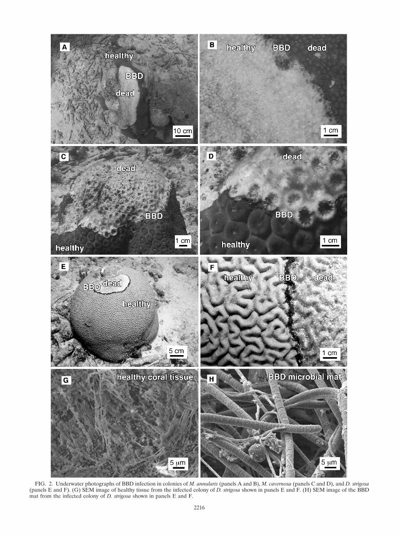
FIG. 2. Underwater photographs of BBD infection in colonies of M. annularis (panels A and B), M. cavernosa (panels C and D), and D. strigosa(panels E and F). (G) SEM image of healthy tissue from the infected colony of D. strigosa shown in panels E and F. (H) SEM image of the BBDmat from the infected colony of D. strigosa shown in panels E and F.
2216
restriction enzyme EcoRI in 1� REact 3 buffer (Gibco/BRL) for more than 3 hat 37°C, and the digested product was separated by electrophoresis on a 0.8%agarose gel (SeaKem LE; BioWhittaker Molecular Applications). After stainingwith ethidium bromide, the bands were visualized on a UV transilluminator andthe RFLP patterns were analyzed to select clones containing the appropriatelysized insert. Plasmid DNA from these clones was then digested with the four-base recognition site enzymes MspI and HinP1I in 1� NEB buffer 2 (NewEngland Biolabs, Beverly, Mass.) under the conditions described above. Thedigest products were then separated by electrophoresis on a 3.0% agarose gel(MetaPhor; BioWhittaker Molecular Applications) and stained with ethidiumbromide, and the RFLP patterns were used to identify unique clones to besubmitted for sequence analysis. Multiple samples with seemingly identicalRFLP patterns were selected for sequence analysis in an effort to capture dif-ferent sequences with similar RFLP patterns. Clones selected for sequenceanalysis were patched onto Luria broth agar petri dishes supplemented with 100�g of ampicillin/ml (Roche Molecular Biochemicals, Indianapolis, Ind.) andincubated overnight at 37°C.
Inoculation, cell culturing, template preparation, and sequencing were per-formed in the high-throughput laboratory of the W. M. Keck Center for Com-parative and Functional Genomics of the University of Illinois at Urbana-Cham-paign. The plates were used to inoculate 2-ml 96-well culture blocks containingCircle Grow medium (Bio100) supplemented with ampicillin (100 �m/ml). Plas-mid template DNA was purified from the cultures by using an automated systemand a QIAwell 96 Turbo Prep BioRobot kit (Qiagen). Sequencing was completedby using T7(�26) primer, which was synthesized in house (63). Sequence reac-tions were performed on the plasmid templates by using a Qiagen Bio Robot9600 and Big Dye Terminator chemistry apparatus (v.2.0) from ABI. Sequencingwas performed on an ABI 3700 capillary sequencer, and then the results wereprocessed in the Bioinformatics Unit of the W. M. Keck Center.
Sequence analyses. The rDNA sequences were first compared with those inthe GenBank database with the basic local alignment search tool (BLAST)network service (4). From the alignments created by this search, the orientationof each cloned 16S rRNA gene could be determined and a rough phylogeneticassociation was established. Each sequence was analyzed using the CHIMERA_CHECK program (version 2.7) available at the Ribosomal Database Projectweb site (39). Those sequences deemed to be chimeric were culled from the dataset. A 97 to 100% match of the unknown clone with the GenBank dataset wasconsidered an accurate identification to the species level, 93 to 96% similaritywas accepted as a genus-level identification, and a 86 to 92% match was consid-ered an accurate identification of a related organism (21).
Nucleotide sequence accession numbers. The GenBank accession numbers forthe 16S rRNA sequences generated in this study are AY037873 throughAY038581 and AF441866 through AF442078.
RESULTS
Optical and SEM analyses. Visual assessment of the surfaceof infected M. annularis, M. cavernosa, and D. strigosa coloniesindicate that healthy coral tissues contain microbes with dif-ferent morphologies than those of the microbes inhabiting theBBD microbial mats. Bacteria inhabiting healthy coral tissueoccurred within a stringy exopolymer matrix. In contrast, theBBD mat is dominated by large (2- to 5-�m by 0.5- to 2-mm)filamentous nonheterocystous cyanobacteria that have previ-ously been optically identified as Phormidium corallyticum (52).A smaller filamentous bacterium that has previously been op-tically identified as a member of the Beggiatoa spp. (14, 53)occurs intertwined with P. corallyticum. Rare 0.5-mm-diameterbundles of P. corallyticum and Beggiatoa spp. were sometimesobserved on healthy coral tissue within a few centimeters of theleading edge of the migrating BBD mat, which is consistentwith previous observations (48).
16S rRNA clone library diversity. High-diversity assem-blages of bacterial sequences were detected in seawater andhealthy, BBD-infected, and dead coral surfaces in M. annularis,M. cavernosa, and D. strigosa colonies (Tables 1, 2, and 3,respectively). A total of 295 partial 16S rRNA gene sequenceswere identified: 65 bacterial sequences in seawater directly
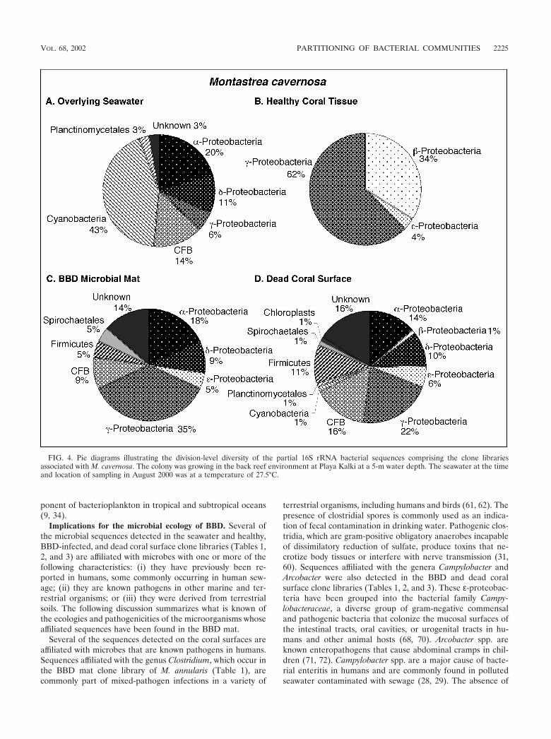
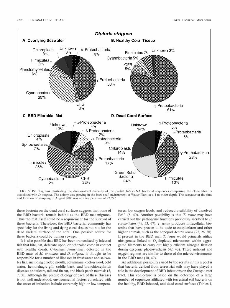
overlying the coral colonies and a total of 230 sequences fromhealthy (64 sequences), BBD-infected (64 sequences), anddead (102 sequences) coral surfaces. Microorganism identifi-cations were based on comparison of these sequences with theGenBank database. Division-level microbial diversity in eachsample was estimated by dividing the number of clones repre-senting each division by the total number of clones in theseawater and healthy, BBD diseased, and dead coral surfacelibraries associated with M. annularis, M. cavernosa, and D.strigosa (shown as pie diagrams in Fig. 3, 4, and 5, respectively).
Microbial communities inhabiting overlying seawater. Bac-terioplankton clone libraries were constructed from samples ofseawater collected at 10 cm immediately above the surfaces ofBBD-infected M. annularis, M. cavernosa, and D. strigosa col-onies (Fig. 3A, 4A, and 5A, respectively). The most abundantsequences in the seawater clone libraries at both locationsrepresented cyanobacteria (30 to 43%). The next most abun-dant clones in the seawater libraries represented the �-pro-teobacteria (6 to 38%) and �-proteobacteria (6 to 31%) divi-sions. Chloroplast sequences, which comprised 3 to 8% of thelibraries, were likely derived from the rDNA of chlorophyll-containing organelles in planktonic algae and free-living zoox-anthellae just above the coral colony surfaces (45). Only twosequences were common to all three seawater clone libraries:those which represented the genera Prochlorococcus and Syn-echococcus (Tables 1, 2, and 3).
Microbial communities inhabiting healthy coral tissue.Clone libraries from healthy tissues indicated that each of thethree coral species contained a significantly different assem-blage of bacterial sequences (Fig. 3B, 4B, and 5B). A maximumof only 5% of the sequences were representative of cyanobac-teria, in contrast to the dominance of cyanobacteria sequencesin the overlying seawater clone libraries (Fig. 3A and B, 4Aand B, and 5A and B). Chloroplasts in these libraries weremost likely derived from zooxanthellae inhabiting the coraltissues. M. annularis sequences (Fig. 3B) exhibited significantlymore microbial diversity than those of either M. cavernosa orD. strigosa. (Fig. 4B and 5B, respectively) and were dominatedby green sulfur bacteria (19%), �-proteobacteria (16%), firmi-cutes (16%), and planctomycetales (13%). Healthy tissue li-braries from M. cavernosa and D. strigosa contained sequencesrepresenting only three and five divisions, respectively, both ofwhich were dominated by �-proteobacteria (62 to 81%). Noneof the same bacterial sequences were detected in all three ofthe healthy coral species (Tables 1, 2, and 3).
Microbial communities inhabiting the BBD microbial mat.Clone libraries constructed from the BBD microbial mat on M.annularis, M. cavernosa, and D. strigosa colonies consistentlyexhibit high division-level diversity (Fig. 3C, 4C, and 5C, re-spectively). Sequences were dominated by firmicutes (5 to26%), Cytophaga-Flavobacterium-Bacteroides group (CFB) (9to 29%), �-proteobacteria (17 to 35%), and �-proteobacteria(2 to 15%). The chloroplasts (4 to 7%) were most likely de-rived from coral zooxanthellae. These results indicate that theBBD microbial communities are completely distinct fromthose inhabiting healthy coral tissue, sharing no common 16SrRNA sequences on any individual coral colony (Tables 1, 2,and 3). These clone libraries also confirm that the BBD matscontain significantly more microbial diversity than that sug-gested by optical analyses (Fig. 2D). Sequences representing
VOL. 68, 2002 PARTITIONING OF BACTERIAL COMMUNITIES 2217
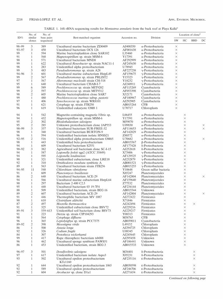
TABLE 1. 16S rRNA sequencing results for Montastrea annularis from the back reef at Playa Kalkia
ID%No. ofsimilarclones
No. ofbase pairssequenced
Best-matched organism Accession no. DivisionLocation of cloneb
SW HC BBD DC
98–99 3 389 Uncultured marine bacterium ZD0409 AJ400350 �-Proteobacteria �93–97 3 450 Uncultured bacterium OCS 126 AF001638 �-Proteobacteria �99 1 594 Marine bacterioplankton clone SAR102 L35460 �-Proteobacteria �91 1 568 Magnetospirillum sp. strain MSM-6 Y17391 �-Proteobacteria �90 1 571 Uncultured bacterium MND8 AF292999 �-Proteobacteria �99 1 622 Uncultured Roseobacter sp. strain NAC11-1 AF245630 �-Proteobacteria �98 1 283 Unidentified alpha proteobacterium U78945 �-Proteobacteria �99 3 483 Pseudoalteromonas sp. strain A28 AF227238 �-Proteobacteria �94–96 3 601 Uncultured marine eubacterium HstpL49 AF159675 �-Proteobacteria �99 2 567 Pseudoalteromonas sp. strain PRLIST2 Y15323 �-Proteobacteria �97 1 567 Alteromonas macleodii strain CH-518 Y18232 �-Proteobacteria �95 1 640 Uncultured bacterium CHAB-I-7 AJ240911 �-Proteobacteria �99 6 589 Prochlorococcus sp. strain MIT9202 AF115269 Cyanobacteria �99 2 597 Prochlorococcus sp. strain MIT9312 AF053398 Cyanobacteria �98 2 605 Marine bacterioplankton clone SAR7 X52171 Cyanobacteria �99 1 480 Prochlorococcus marinus subsp. pastoris AF180967 Cyanobacteria �97 1 406 Synechococcus sp. strain WR8101 AJ292905 Cyanobacteria �91 1 621 Cytophaga sp. strain JTB250 AB015264 CFB �96 1 619 Unidentified eukaryote OM8 1 U70717 Chloroplasts �
94 1 542 Magnetite-containing magnetic Vibrio sp. L06455 �-Proteobacteria �94 1 432 Magnetospirillum sp. strain MSM-6 Y17391 �-Proteobacteria �95 2 302 Rhodothalassium salexigens M59070 �-Proteobacteria �95 2 285 Unknown proteobacterium clone JAP553 U09830 �-Proteobacteria � �98–99 3 337 Uncultured bacterium SUR-FREE-32 AF114653 �-Proteobacteria � �98 1 340 Uncultured bacterium BURTON-9 AF142829 �-Proteobacteria �96 1 594 Unidentified bacterium isolate HNSS21 Z88572 �-Proteobacteria �92–95 2 516 Unidentified alpha proteobacterium OM65 U70682 �-Proteobacteria � �98 1 443 Unidentified beta proteobacterium AB015567 -Proteobacteria �94 1 609 Uncultured bacterium S2551 AF177428 �-Proteobacteria �90–92 3 382 Agricultural soil bacterium clone SC-I-15 AJ252618 �-Proteobacteria �94 1 587 Legionella feeleii sgp2 (ATCC 35849) X73406 �-Proteobacteria �94 1 595 Legionella londiniensis AF129525 �-Proteobacteria �90 1 321 Unidentified eubacterium, clone LRE18 AJ232879 �-Proteobacteria �92 1 310 Ornithodoros moubata symbiote A AB001521 �-Proteobacteria �89 1 563 Uncultured bacterium strain JTB256 AB015255 �-Proteobacteria �94–95 11 548 Chlorobium vibrioforme Y10648 Green sulfur bacteria �91 1 609 Planctomyces brasiliensis X85247 Planctomycetales �86 2 648 Uncultured bacterium ACE-29 AF142804 Planctomycetales � �94 1 566 Uncultured marine eubacterium HstpL64 AF159640 Planctomycetales �90 1 297 Bacterium 2-400 C2.5 Z77532 Planctomycetales �95 1 448 Uncultured bacterium 03 19-7F4 AF234144 Planctomycetales �88 1 509 Unidentified bacterium, strain BD2-16 AB015544 Unknown �89 2 607 Uncultured bacterium ACE-29 AF142804 Planctomycetales � �91 1 446 Thermophilic bacterium MV 1087 AJ272422 Firmicutes �90 1 610 Clostridium aldrichii X71846 Firmicutes �87 2 497 Moorella thermoacetica AJ242494 Firmicutes � �91 1 325 Unidentified eubacterium clone BSV72 AJ229216 Firmicutes �89–91 3 559–613 Unidentified soil bacterium clone BSV73 AJ229217 Firmicutes �91 1 223 Dietzia sp. strain CIP104293 Y08313 Firmicutes �90 1 564 Cytophaga diffluens M58765 CFB �96 1 365 Leptolyngbya sp. strain PCC7375 AB039011 Cyanobacteria �89–92 2 398 Mesostigma viride L49152 Chloroplasts �86 1 500 Astasia longa AJ294725 Chloroplasts �92 1 326 Codium fragile U08345 Chloroplasts �84 1 307 Prototheca wickerhamii AJ245645 Chloroplasts �99 1 417 Rape rhizosphere bacterium tsb088 AJ295458 Unknown �96 1 462 Uncultured sponge symbiont PAWS51 AF186441 Unknown �86 1 453 Unidentified bacterium, strain BD2-3 AB015533 Unknown �
96–97 3 501 Desulfovibrio salexigens M34401 �-Proteobacteria �97 1 617 Unidentified bacterium isolate Aspo3 X95231 �-Proteobacteria �93 1 502 Uncultured epsilon proteobacterium
KTc1160AF235116 ε-Proteobacteria �
92 1 644 Uncultured epsilon proteobacterium 1065 AB030598 ε-Proteobacteria �92 1 589 Uncultured epsilon proteobacterium AF246706 ε-Proteobacteria �92 1 604 Arcobacter sp. clone D1a1 AJ271654 ε-Proteobacteria �
Continued on following page
2218 FRIAS-LOPEZ ET AL. APPL. ENVIRON. MICROBIOL.
Desulfovibrio and the associated genus Desulfobotulus weredetected in clone libraries from all three coral species (Tables1, 2, and 3). Sequences associated with Trichodesmium andClostridium occurred in BBD mat clone libraries from M. an-nularis and D. strigosa (Tables 1 and 3, respectively).
Microbial communities inhabiting dead coral surfaces.Clone libraries from dead surfaces of the M. annularis, M.cavernosa, and D. strigosa colonies also contained a high diver-sity of sequences, representing 6 to 9 microbial divisions (Fig.3D, 4D, and 5D, respectively). However, the relative propor-tions of sequences representing each division varied signifi-cantly between species. In general, clone libraries were domi-nated by �-proteobacteria (0 to 42%), �-proteobacteria (5 to17%), �-proteobacteria (0 to 28%), and CFB (0 to 17%).Sequences detected on dead coral surfaces were 95% distinctfrom those detected in the overlying seawater, healthy coraltissues, and the BBD microbial mats.
DISCUSSION
The optical and molecular analyses completed in this studyof corals infected with BBD on Curaçao indicate that bacterialcommunities are distinctly partitioned between overlying sea-water and healthy, diseased, and dead coral surfaces. The fol-lowing discussion evaluates the implications of this partitioningwith respect to the proportion of cyanobacterium-related se-quences in reef tract seawater clone libraries, the possibility ofcoral species-specific microbial communities, and the microbialecology of BBD in M. annularis, M. cavernosa, and D. strigosacolonies.
Reef tract bacterioplankton. Sequences affiliated with twoglobally distributed bacterioplankton were detected in all sea-water clone libraries collected from the Curaçao reef tract. Themost common was Prochlorococcus, which is the smallest yetmost abundant oxygenic photoautotroph in tropical and sub-
TABLE 1—Continued
ID%No. ofsimilarclones
No. ofbase pairssequenced
Best-matched organism Accession no. DivisionLocation of cloneb
SW HC BBD DC
91–94 7 551 Cytophaga fermentans M58766 CFB � �92–93 3 430 Uncultured Cytophaga kps30 AF195441 CFB �92 1 601 Cytophaga sp. strain BD1-16 AB015525 CFB �93 1 595 Trichodesmium tenue AF013029 Cyanobacteria �92 3 441 Bacterium strain 77003 AF227847 Unknown �91 1 594 Clostridium fimetarium AF126687 Firmicutes �91 1 637 Clostridium paradoxum clone para99 Z69939 Firmicutes �89 1 349 Uncultured marine bacterium 90d10 AF295117 Firmicutes �90–91 2 487 Guillardia theta AF041468 Chloroplasts �87 1 318 Unidentified eubacterium clone BSV85 AJ229229 Unknown �
97 1 496 Roseobacter sp. (P. decipiens symbiont) AF107210 �-Proteobacteria �96 1 568 Roseobacter sp. (P. filiformis symbiont) AF107209 �-Proteobacteria �95 1 626 Sulfitobacter pontiacus AF182018 �-Proteobacteria �96 1 507 Uncultured Agrobacterium sp. strain kpc102rc AF194391 �-Proteobacteria �99 1 515 Uncultured marine eubacterium HstpL78 AF159652 �-Proteobacteria �86–93 9 454 Desulfocella halophila AF022936 �-Proteobacteria �96–98 4 487 Desulforhopalus singaporensis AF118453 �-Proteobacteria �97–98 4 549 Desulfobotulus sp. strain BG14 U85470 �-Proteobacteria �91 2 483 Desulfofrigus fragile AF099065 �-Proteobacteria �98 1 524 Desulfobacterium catecholicum AJ237602 �-Proteobacteria �94 1 218 Uncultured bacterium ODPB-B3 AF121088 �-Proteobacteria �94 1 612 Unidentified bacterium clone NB1-k AB013832 �-Proteobacteria �91 1 587 Plesiomonas shigelloides X60418 �-Proteobacteria �93 1 583 Uncultured bacterium strain BD1-7 AB015519 �-Proteobacteria �92 1 564 Pseudoalteromonas sp. strain A25 AF227237 �-Proteobacteria �98 1 652 Vibrio alginolyticus (ATCC 17749T) X56576 �-Proteobacteria �89 1 582 Unidentified bacterium, clone NB1-o AB013836 �-Proteobacteria �94 1 398 Uncultured bacterium adhufec29.25 AF153855 �-Proteobacteria �91 1 571 Cytophaga sp. strain JTB244 AB015262 CFB �86 1 325 Flexibacter tractuosus M58789 CFB �94 1 487 Microscilla furvescens M58792 CFB �91 1 331 Psychroserpens burtonensis ACAM188 U62913 CFB �96 1 503 Uncultured bacterium KC305 AB022513 CFB �92 1 589 Uncultured cytophagales QSSC9L-1 AF170779 CFB �96 1 602 Clostridium halophilum DSM 5387 X77837 Firmicutes �89 1 411 Uncultured soil bacterium clone K20-71 AF145861 Firmicutes �87 1 415 Uncultured bacterium Sva0855 AJ240981 Firmicutes �87 1 566 Uncultured eubacterium clone vadinBB35 U81761 Unknown �
a Listed are the percent identities (ID%) to previously identified sequences, the numbers of similar clones, the numbers of base pairs sequenced (bp), and theaccession numbers and divisions of the best s-matched organism in GenBank.
b � signs indicate whether the clone occurred in the overlying seawater (SW), healthy coral tissues (HC), black band diseased coral tissue (BBD), or dead coralskeleton surfaces (DC).
VOL. 68, 2002 PARTITIONING OF BACTERIAL COMMUNITIES 2219

TABLE 2. 16S rRNA sequencing results for a Montastrea cavernosa colony from the back reef at Playa Kalkia
ID%No. ofsimilarclones
No. ofbase pairssequenced
Best-matched organism Accession no. DivisionLocation of Cloneb
SW HC BBD DC
98 2 594 Uncultured bacterium SAR102 L35460 �-Proteobacteria �94–98 2 534 Uncultured bacterium OCS126 AF001638 �-Proteobacteria �91 1 543 Caedibacter caryophilus AJ238683 �-Proteobacteria �92 1 573 Olavius loisae endosymbiont 2 AF104473 �-Proteobacteria �99 1 596 Uncultured Roseobacter NAC11-1 AF245630 �-Proteobacteria �98 1 610 Uncultured bacterium EBAC31A08 AF268219 �-Proteobacteria �92 1 334 Uncultured bacterium CHAB-II-49 AJ240909 �-Proteobacteria �98–99 2 546 Unknown marine bacterioplankton SAR7 X52171 �-Proteobacteria �94 2 587 Unidentified marine bacterium OM60 U70696 �-Proteobacteria �98–99 9 587 Prochlorococcus sp. strain MIT9202 AF115269 Cyanobacteria �98 3 546 Uncultured Synechococcus sp. strain NAC1-5 AF245618 Cyanobacteria �96 1 454 Prochlorococcus marinus pastori AF180967 Cyanobacteria �99 1 603 Prochlorococcus sp. strain MIT9312 AF053398 Cyanobacteria �99 1 602 Synechococcus WH8101 AF001480 Cyanobacteria �92 2 675 Uncultured Cytophagales CRE-FL75 AF141488 CFB �93 1 688 Marine psychrophile sp. SW17 AF001368 CFB �93 1 645 Uncultured marine bacterium ZD0403 AJ400347 CFB �99 1 588 Unidentified cytophagales OM188 U70687 CFB �94 1 601 Unidentified planctomycete OM190 U70712 Planctomycetales �98 1 567 Unidentified bacterium isolate HRV16 Z88588 Unknown �
95 12 564 Unidentified beta proteobacterium OPB30 AF026979 -Proteobacteria �94 6 558 Hydrogenophilus thermoluteolus TH-4 AB009829 -Proteobacteria �95 1 478 Unidentified beta proteobacterium OPS140 AF026983 -Proteobacteria �95–96 2 664 Uncultured epsilon proteobacterium KTc1160 AF235116 ε-Proteobacteria �98–99 34 539 Chromatium sp. RW AF384210 �-Proteobacteria �99 1 406 Escherichia coli K12 MG1655 AE000345 �-Proteobacteria �
95 1 527 Uncultured alpha proteobacterium SIC.926 AF277517 �-Proteobacteria �99 1 583 Uncultured proteobacterium clone CD5H5 AY038412 �-Proteobacteria �93 1 588 Maricaulis sp. strain MCS18 AJ227806 �-Proteobacteria �94 1 607 Uncultured alpha proteobacterium KTc0993 AF235129 �-Proteobacteria �95 1 583 Uncultured proteobacterium clone CD4D6 AY038529 �-Proteobacteria �99 2 515 Desulfovibrio sp. strain TBP-1 AF090830 �-Proteobacteria � �98 1 582 Uncultured proteobacterium clone CD5B11 AY038410 ε-Proteobacteria �96 1 321 Shewanella sp. clone NB65-G AB013842 �-Proteobacteria �93 1 606 Marinobacter hydrocarbonoclasticus Y18240 �-Proteobacteria �99 1 566 Pseudomonas stutzeri AY017341 �-Proteobacteria �93–94 2 443 Uncultured gamma proteobacterium clone 26 AF369718 �-Proteobacteria �96 1 542 Unidentified proteobacterium strain NKB4 AB013256 �-Proteobacteria �92 2 616 Oceanospirillum linum M22365 �-Proteobacteria �88 1 530 Marine bacterium BBFL7 AY028207 CFB �99 1 630 Uncultured Cytophagales clone CD4B12 AY038511 CFB �92 1 597 Parasporobacterium paucivorans sp. SYR1 AJ272036 Firmicutes �92–95 2 478 Spirochaeta litoralis M88723 Spirochaetales � �91 1 220 Uncultured rumen bacterium 4C3d-12 AB034093 Unknown �91 1 612 Unidentified butyrate-producing L1-92 AJ270487 Unknown �97 1 613 Uncultured Crater Lake bacterium CL0-45 AF316686 Unknown �
98 2 435 Silicibacter lacuscaerulensis U77644 �-Proteobacteria �97 1 489 Alpha proteobacterium MBIc1876 AB026194 �-Proteobacteria �88 1 489 Uncultured alpha proteobacterium MB13E08 AY033327 �-Proteobacteria �95 2 497 Caulobacter sp. strain MCS33 AJ227811 �-Proteobacteria �96 1 341 Uncultured Rhodobacter CtaxPhil-16 AF259624 �-Proteobacteria �97 1 477 Agrobacterium gelatinovorum D88523 �-Proteobacteria �95 1 342 Uncultured alpha proteobacterium CHAB-I-5 AJ240910 �-Proteobacteria �97 1 373 Sulfitobacter pontiacus AF182018 �-Proteobacteria �90 1 399 Uncultured ferromanganous bacterium MND AF292999 �-Proteobacteria �99 1 333 Beta proteobacterium OS-ac-15 U46749 -Proteobacteria �87 1 296 Desulfovibrio zosterae Y18049 �-Proteobacteria �90–91 2 555 Desulfocella halophila AF022936 �-Proteobacteria �91 1 425 Delta proteobacterium S2551 AF177428 �-Proteobacteria �89 1 302 Desulfovibrio alaskensis NCIMB13491 Y11984 �-Proteobacteria �87 1 462 Uncultured delta proteobacterium Sva0447 AJ240999 �-Proteobacteria �92 1 535 Desulfofrigus oceanense strain ASv26 AF099064 �-Proteobacteria �96 1 371 Desulfobotulus sp. strain BG14 U85470 �-Proteobacteria �
Continued on following page
2220 FRIAS-LOPEZ ET AL. APPL. ENVIRON. MICROBIOL.
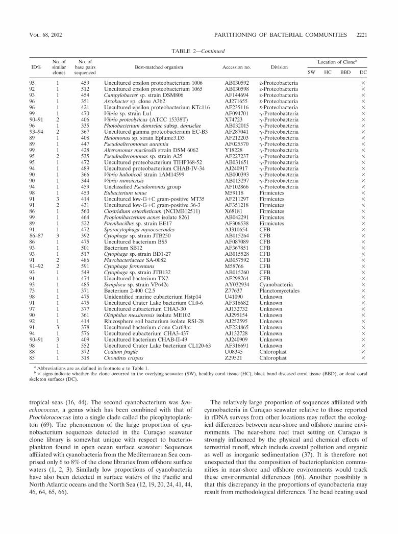
tropical seas (16, 44). The second cyanobacterium was Syn-echococcus, a genus which has been combined with that ofProchlorococcus into a single clade called the picophytoplank-ton (69). The phenomenon of the large proportion of cya-nobacterium sequences detected in the Curaçao seawaterclone library is somewhat unique with respect to bacterio-plankton found in open ocean surface seawater. Sequencesaffiliated with cyanobacteria from the Mediterranean Sea com-prised only 6 to 8% of the clone libraries from offshore surfacewaters (1, 2, 3). Similarly low proportions of cyanobacteriahave also been detected in surface waters of the Pacific andNorth Atlantic oceans and the North Sea (12, 19, 20, 24, 41, 44,46, 64, 65, 66).
The relatively large proportion of sequences affiliated withcyanobacteria in Curaçao seawater relative to those reportedin rDNA surveys from other locations may reflect the ecolog-ical differences between near-shore and offshore marine envi-ronments. The near-shore reef tract setting on Curaçao isstrongly influenced by the physical and chemical effects ofterrestrial runoff, which include coastal pollution and organicas well as inorganic sedimentation (37). It is therefore notunexpected that the composition of bacterioplankton commu-nities in near-shore and offshore environments would trackthese environmental differences (66). Another possibility isthat this discrepancy in the proportions of cyanobacteria mayresult from methodological differences. The bead beating used
TABLE 2—Continued
ID%No. ofsimilarclones
No. ofbase pairssequenced
Best-matched organism Accession no. DivisionLocation of Cloneb
SW HC BBD DC
95 1 459 Uncultured epsilon proteobacterium 1006 AB030592 ε-Proteobacteria �92 1 512 Uncultured epsilon proteobacterium 1065 AB030598 ε-Proteobacteria �93 1 454 Campylobacter sp. strain DSM806 AF144694 ε-Proteobacteria �96 1 351 Arcobacter sp. clone A3b2 AJ271655 ε-Proteobacteria �96 1 421 Uncultured epsilon proteobacterium KTc116 AF235116 ε-Proteobacteria �99 1 470 Vibrio sp. strain Lu1 AF094701 �-Proteobacteria �90–91 2 406 Vibrio proteolyticus (ATCC 15338T) X74723 �-Proteobacteria �96 1 335 Photobacterium damselae subsp. damselae AB032015 �-Proteobacteria �93–94 2 367 Uncultured gamma proteobacterium EC-B3 AF287041 �-Proteobacteria �89 1 408 Halomonas sp. strain Eplume3.D3 AF212203 �-Proteobacteria �89 1 447 Pseudoalteromonas aurantia AF025570 �-Proteobacteria �99 1 428 Alteromonas macleodii strain DSM 6062 Y18228 �-Proteobacteria �95 2 535 Pseudoalteromonas sp. strain A25 AF227237 �-Proteobacteria �95 1 472 Uncultured proteobacterium TIHP368-52 AB031651 �-Proteobacteria �94 1 489 Uncultured proteobacterium CHAB-IV-34 AJ240917 �-Proteobacteria �90 1 366 Vibrio halioticoli strain 1AM14599 AB000393 �-Proteobacteria �90 1 344 Vibrio rumoiensis AB013297 �-Proteobacteria �94 1 459 Unclassified Pseudomonas group AF102866 �-Proteobacteria �98 1 453 Eubacterium tenue M59118 Firmicutes �91 3 414 Uncultured low-G�C gram-positive MT35 AF211297 Firmicutes �91 2 431 Uncultured low-G�C gram-positive 36-3 AF351218 Firmicutes �86 1 560 Clostridium estertheticum (NCIMB12511) X68181 Firmicutes �99 1 464 Propionibacterium acnes isolate 8261 AB042291 Firmicutes �89 1 522 Paenibacillus sp. strain EE17 AF306538 Firmicutes �91 1 472 Sporocytophaga myxococcoides AJ310654 CFB �86–87 3 392 Cytophaga sp. strain JTB250 AB015264 CFB �86 1 475 Uncultured bacterium BS5 AF087089 CFB �93 1 501 Bacterium SB12 AF367851 CFB �93 1 517 Cytophaga sp. strain BD1-27 AB015528 CFB �91 2 486 Flavobacteriaceae SA-0082 AB057592 CFB �91–92 2 591 Cytophaga fermentans M58766 CFB �93 1 549 Cytophaga sp. strain JTB132 AB015260 CFB �91 1 474 Uncultured bacterium TX2 AF298764 CFB �93 1 485 Symploca sp. strain VP642c AY032934 Cyanobacteria �73 1 371 Bacterium 2-400 C2.5 Z77637 Planctomycetales �98 1 475 Unidentified marine eubacterium Hstp14 U41090 Unknown �91 1 475 Uncultured Crater Lake bacterium CL0-6 AF316682 Unknown �97 1 377 Uncultured eubacterium CHA3-30 AJ132732 Unknown �90 1 361 Oleiphilus messinensis isolate ME102 AJ295154 Unknown �92 1 414 Rhizosphere soil bacterium isolate RSI-28 AJ252595 Unknown �91 3 378 Uncultured bacterium clone Car68rc AF224865 Unknown �94 1 576 Uncultured eubacterium CHA3-437 AJ132728 Unknown �90–91 3 409 Uncultured bacterium CHAB-II-49 AJ240909 Unknown �98 1 552 Uncultured Crater Lake bacterium CL120-63 AF316691 Unknown �88 1 372 Codium fragile U08345 Chloroplast �85 1 318 Chondrus crispus Z29521 Chloroplast �
a Abbreviations are as defined in footnote a to Table 1.b � signs indicate whether the clone occurred in the overlying seawater (SW), healthy coral tissue (HC), black band diseased coral tissue (BBD), or dead coral
skeleton surfaces (DC).
VOL. 68, 2002 PARTITIONING OF BACTERIAL COMMUNITIES 2221
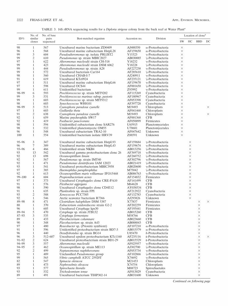
TABLE 3. 16S rRNA sequencing results for a Diploria strigosa colony from the back reef at Water Planta
ID%No. ofsimilarclones
No. of basepairs
sequencedBest-matched organism Accession no. Division
Location of cloneb
SW HC BBD DC
98 1 567 Uncultured marine bacterium ZD0409 AJ400350 �-Proteobacteria �96 1 568 Uncultured marine eubacterium HstpL28 AF159650 �-Proteobacteria �99 3 604 Pseudoalteromonas sp. isolate PRLIST2 Y15323 �-Proteobacteria �99 2 610 Pseudomonas sp. strain MBIC2027 AB030085 �-Proteobacteria �97 1 622 Alteromonas macleodii strain CH-518 Y18232 �-Proteobacteria �99 1 423 Alteromonas macleodii strain DSM 6062 Y18228 �-Proteobacteria �99 1 444 Pseudoalteromonas sp. strain A28 AF227238 �-Proteobacteria �99 1 572 Uncultured bacterium Car164 AF285610 �-Proteobacteria �98 1 560 Uncultured CHAB-I-7 AJ240911 �-Proteobacteria �91 1 619 Uncultured KTc0924 AF235121 �-Proteobacteria �97 1 511 Uncultured marine eubacterium HstpL66 AF159670 �-Proteobacteria �97 1 598 Uncultured OCS44 AF001650 �-Proteobacteria �99 1 611 Unidentified bacterium Z93992 �-Proteobacteria �98–99 6 593 Prochlorococcus sp. strain MIT9202 AF115269 Cyanobacteria �99 3 531 Prochlorococcus marinus subsp. pastoris AF180967 Cyanobacteria �99 1 603 Prochlorococcus sp. strain MIT9312 AF053398 Cyanobacteria �98 1 603 Synechococcus WH8101 AF397728 Cyanobacteria �98–99 3 513 Cyanophora paradoxa cyanelle M19493 Chloroplasts � �97 1 603 Guillardia theta AF041468 Chloroplasts �91 2 638 Cyanophora paradoxa cyanelle M19493 Chloroplasts � �92 1 659 Marine psychrophile SW17 AF001368 CFB �92 1 419 Fusibacter paucivorans AF050099 Firmicutes �98 1 594 Unidentified eubacterium clone SAR276 U65915 Planctomycetales �98 1 532 Unidentified planctomycete OM55 U70681 Planctomycetales �96 1 548 Uncultured eubacterium TRA2-10 AF047642 Unknown �100 1 554 Unidentified bacterium isolate HRV39 Z88591 Unknown �
95–96 3 347 Uncultured marine eubacterium HstpL93 AF159684 �-Proteobacteria �96 7 389 Uncultured marine eubacterium HstpL43 AF159674 �-Proteobacteria �93–96 4 466 Unidentified strain NKB4 AB013256 �-Proteobacteria �93 13 542 Uncultured gamma proteobacterium clone 26 AF369718 �-Proteobacteria �92 13 698 Oceanospirillum linum AF260752 �-Proteobacteria �92 1 567 Pseudomonas sp. strain IMT40 AF302796 �-Proteobacteria �92 1 471 Pseudomonas denitrificans IAM 12023 AB021419 �-Proteobacteria �93 1 541 Uncultured proteobacterium MBIC3958 AB020600 �-Proteobacteria �97 1 385 Haemophilus paraphrophilus M75042 �-Proteobacteria �92 1 613 Oceanospirillum maris williamsae IFO15468 AB006763 �-Proteobacteria �99–100 4 604 Propionibacterium acnes AF154832 Firmicutes �97 1 373 Uncultured Cytophagales clone CRE-PA10 AF141499 CFB �91 1 576 Flexibacter aggregans M64628 CFB �98 1 590 Uncultured Cytophagales clone CD4E12 AY038534 CFB �91 1 635 Planktothrix sp. strain FPI AF212922 Cyanobacteria �93 2 588 Xenococcus PCC7305 AF132783 Cyanobacteria �92 1 546 Arctic seawater bacterium R7366 AJ293826 Unknown �89–98 8 471 Clostridium halophilum DSM 5387 X77837 Firmicutes � �88 1 370 Eubacterium oxidoreducens strain G2-2 AF202259 Firmicutes �96 1 605 Uncultured Cytophaga kps30 AF195441 Firmicutes �89–94 5 470 Cytophaga sp. strain JTB132 AB015260 CFB �87–93 3 535 Cytophaga fermentans M58766 CFB �89 1 433 Flavobacterium columnare AB023660 CFB �90 1 348 Flavobacterium sp. strain A43 AB008043 CFB �97 1 480 Roseobacter sp. (Prionitis symbiont) AF107210 �-Proteobacteria �91 1 596 Unidentified proteobacterium strain BD7-3 AB015579 �-Proteobacteria �96 1 440 Desulfobotulus sp. strain BG14 U85470 �-Proteobacteria �96 3 512–607 Uncultured epsilon proteobacterium KTc1160 AF235116 ε-Proteobacteria � �91–92 2 624 Uncultured proteobacterium strain BD1-29 AB015529 ε-Proteobacteria �94–99 3 557 Alteromonas macleodii AF025957 �-Proteobacteria �94–95 2 462 Oceanospirillum sp. strain ME113 AJ302700 �-Proteobacteria �92 2 499 Neptunomonas naphthovorans AF053734 �-Proteobacteria �95 1 489 Unclassified Pseudomonas group AF102866 �-Proteobacteria �99 1 565 Vibrio campbelli ATCC 25920T X74692 �-Proteobacteria �82 1 547 Spinacia oleracea M21453 Chloroplasts �89 1 473 Nephroselmis olivacea X74754 Chloroplasts �93 1 577 Spirochaeta litoralis M88723 Spirochaetales �93 1 532 Trichodesmium tenue AF013029 Cyanobacteria �89 1 491 Uncultured bacterium TIHP302-14 AB031600 Unknown �
Continued on following page
2222 FRIAS-LOPEZ ET AL. APPL. ENVIRON. MICROBIOL.
in the present study may have more effectively lysed the toughcyanobacterial cell walls than the techniques used for the off-shore studies, none of which applied bead beating. Further-more, the consistently high proportion of cyanobacteria in allthree of the seawater samples analyzed from Curaçao (Fig. 3A,4A, and 5A) suggests that this is an accurate representation ofthe reef tract bacterioplankton clone library.
Healthy coral microbial communities. Sequencing results inthe present study indicate that the microbial communities in-habiting healthy coral tissue have the following characteristics:(i) they are unique in that they are distinct from the bacterio-plankton in overlying seawater, especially with respect to cya-nobacteria sequences; and (ii) they are markedly differentamong the three coral species. Significant reductions in theproportion of cyanobacterium-affiliated sequences from theseawater clone libraries (30 to 43%) to those from the healthycoral tissue clone libraries (0 to 5%) (Fig. 3A and B, 4A and B,and 5A and B, respectively) are likely to be the result ofchemical defense mechanisms that inhibit coral tissue coloni-zation by cyanobacteria (35). This partitioning is also consis-tent with the results of previous optical and culture-basedstudies of the mucus-rich biofilm covering coral tissue, which iscalled the coral surface microlayer (CSM) (13, 14, 38, and 45).Expelled and free-living zooxanthellae are prominent compo-nents of the CSM, which protects coral from light, exposure,and sedimentation and is the first line of defense against dis-ease infection (11, 38, 45, 56).
Inferred metabolisms from the M. annularis clone librarysequences suggest that mixtures of aerobic and anaerobic mi-crobes inhabit the healthy coral tissue (Tables 1, 2, and 3). Thistype of mixed metabolic assemblage is similar to those of thediverse microbial communities detected in the tissues of ma-rine sponges, where low-oxygen microniches occur in the poresof well-oxygenated sponge surfaces (73). In addition, severalother bacteria inhabiting healthy M. annularis tissue have notpreviously been observed in marine environments. Specifically,sequences of several microbial strains, which comprise 3% of
the M. annularis healthy tissue clone library, were previouslyisolated exclusively from terrestrial soils (e.g., agricultural soilbacterium SC-I-iS and unidentified soil bacterium clonesBSV72 and BSV73; Tables 1, 2, and 3).
Sequences from clone libraries constructed from healthy M.annularis tissue exhibit significantly higher bacterial diversity(10 divisions) than those from clone libraries constructed fromhealthy M. cavernosa and D. strigosa tissue (3 and 4 divisions,respectively; Fig. 3B, 4B, and 5B, respectively). These prelim-inary results imply that scleractinian corals may contain spe-cies-specific microbial communities. The abundance of �-pro-teobacteria and -proteobacteria and the low overall sequencediversity levels observed in M. cavernosa and D. strigosa (Fig.4B and 5B) are somewhat similar to those of the microbialcommunities detected with molecular screening of healthy tis-sues from a single Montastrea franksi colony in Panama (4divisions) (51). Sequences affiliated with Silicibacter lacuscae-rulensis were also detected in several other healthy M. franksicolonies by using denaturing gradient gel electrophoresis tech-niques (51). However, S. lacuscaerulensis sequences were onlydetected in the present study on Curaçao in the dead coralsurface clone library of M. cavernosa (Table 2). This furthersupports the possibility that individual coral species containunique microbial consortia. An important difference from theCuraçao corals is that M. franksi healthy tissue clone librariesfrom Panama were dominated by cyanobacteria (51), which issurprising given the chemical mechanisms inhibiting cyanobac-terial settlement on healthy coral tissues (35). In addition, M.franski did not contain chloroplast-affiliated sequences in thehealthy tissue clone libraries (51), which was unexpected giventhe abundance of zooxanthellae in the coral tissue and theCSM (45) and their use of primers similar to those applied inthe present study.
Microbes associated with BBD. The BBD microbial mat isdominated by large filamentous cyanobacteria originally identi-fied as Oscillatoria submembranaceae (A. Antonius, Abstr. 10thMeet. Assoc. Isl. Mar. Lab. Caribb., p. 3, 1973 [abstr.]) and
TABLE 3—Continued
ID%No. ofsimilarclones
No. of basepairs
sequencedBest-matched organism Accession no. Division
Location of cloneb
SW HC BBD DC
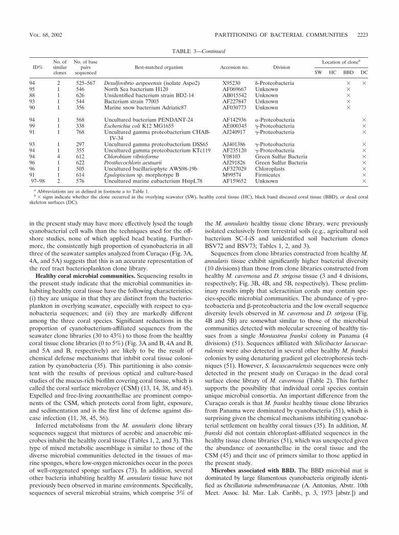
94 2 525–567 Desulfovibrio aespoeensis (isolate Aspo2) X95230 �-Proteobacteria � �95 1 546 North Sea bacterium H120 AF069667 Unknown �88 1 626 Unidentified bacterium strain BD2-14 AB015542 Unknown �93 1 544 Bacterium strain 77003 AF227847 Unknown �90 1 356 Marine snow bacterium Adriatic87 AF030773 Unknown �
94 1 568 Uncultured bacterium PENDANT-24 AF142936 �-Proteobacteria �99 1 338 Escherichia coli K12 MG1655 AE000345 �-Proteobacteria �91 1 768 Uncultured gamma proteobacterium CHAB-
IV-34AJ240917 �-Proteobacteria �
93 1 297 Uncultured gamma proteobacterium DSS65 AJ401386 �-Proteobacteria �94 1 355 Uncultured gamma proteobacterium KTc119 AF235120 �-Proteobacteria �94 4 612 Chlorobium vibrioforme Y08103 Green Sulfur Bacteria �96 1 622 Prosthecochloris aestuarii AJ291826 Green Sulfur Bacteria �96 1 505 Uncultured bacillariophyte AWS98-19b AF327029 Chloroplasts �91 1 614 Epulopiscium sp. morphotype B M99574 Firmicutes �97–98 2 576 Uncultured marine eubacterium HstpL78 AF159652 Unknown �
a Abbreviations are as defined in footnote a to Table 1.b � signs indicate whether the clone occurred in the overlying seawater (SW), healthy coral tissue (HC), black band diseased coral tissue (BBD), or dead coral
skeleton surfaces (DC).
VOL. 68, 2002 PARTITIONING OF BACTERIAL COMMUNITIES 2223

later systematically reclassified as Phormidium corallyticum(52). Other bacteria previously identified in the BBD matinclude motile, nonphotosynthetic sulfide-oxidizing Beggiatoaspp. (optically identified) (14, 18, and 52) and the sulfate-reducing Desulfovibrio spp. (identified optically and with 16SrRNA oligonucleotide probes) (18 and 53 and S. Schnell, B.Assmus, and L. L. Richardson, abstract from the Annual Meet-ing of the VAAM [Vereinigung fuer Allgemeine und Ange-wandte Mikrobiologie] and GBCH [Gesellschaft fuer Biolo-gische Chemie], Biospecktrum, p. 116, 1996 [abstr.]). AlthoughSEM imaging for the present report indicated that the BBDmicrobial mat was predominantly composed of filamentouscyanobacteria (Fig. 2G and H), cyanobacterial sequences rep-resented only 0 to 4% of the BBD mat clone libraries (Table1). Furthermore, neither P. corallyticum or Beggiatoa sp. se-quences were detected in the BBD mat (Tables 1, 2, and 3).
One explanation for these discrepancies may be that P. cor-
allyticum has not been previously sequenced. While severalspecies of terrestrial Phormidium have been detected in desertsoil crusts (17), no marine species have previously been re-ported in GenBank. Similarly, multiple Beggiatoa species havebeen sequenced from other marine environments (3) but theirsequences were not similar to those in the BBD mat clonelibrary. Another explanation could be that P. corallyticum andBeggiatoa spp. were represented by one of the unknown bac-terial sequences in the BBD mat clone library (Fig. 3, 4, and 5).Other possibilities are that they are present in the BBD matbut their rDNA was not extracted, amplified, and sequencedand they are not present in the BBD mat. It is important tonote that sequences affiliated with the filamentous cyanobac-terium Trichodesmium tenue were found in the BBD mats(Tables 1, 2, and 3). Members of the family Trichodesmium(formerly Oscillatoria) are nonheterocystous nitrogen-fixing(diazotrophic) cyanobacteria that are a globally abundant com-
FIG. 3. Pie diagrams illustrating the division-level diversity of the partial 16S rRNA bacterial sequences comprising the clone librariesassociated with M. annularis. The colony was growing in the back reef environment at Playa Kalki at a 5-m water depth. The seawater at the timeand location of sampling in August 2000 was at a temperature of 27.5°C.
2224 FRIAS-LOPEZ ET AL. APPL. ENVIRON. MICROBIOL.
ponent of bacterioplankton in tropical and subtropical oceans(9, 34).
Implications for the microbial ecology of BBD. Several ofthe microbial sequences detected in the seawater and healthy,BBD-infected, and dead coral surface clone libraries (Tables 1,2, and 3) are affiliated with microbes with one or more of thefollowing characteristics: (i) they have previously been re-ported in humans, some commonly occurring in human sew-age; (ii) they are known pathogens in other marine and ter-restrial organisms; or (iii) they were derived from terrestrialsoils. The following discussion summarizes what is known ofthe ecologies and pathogenicities of the microorganisms whoseaffiliated sequences have been found in the BBD mat.
Several of the sequences detected on the coral surfaces areaffiliated with microbes that are known pathogens in humans.Sequences affiliated with the genus Clostridium, which occur inthe BBD mat clone library of M. annularis (Table 1), arecommonly part of mixed-pathogen infections in a variety of
terrestrial organisms, including humans and birds (61, 62). Thepresence of clostridial spores is commonly used as an indica-tion of fecal contamination in drinking water. Pathogenic clos-tridia, which are gram-positive obligatory anaerobes incapableof dissimilatory reduction of sulfate, produce toxins that ne-crotize body tissues or interfere with nerve transmission (31,60). Sequences affiliated with the genera Campylobacter andArcobacter were also detected in the BBD and dead coralsurface clone libraries (Tables 1, 2, and 3). These ε-proteobac-teria have been grouped into the bacterial family Campy-lobacteraceae, a diverse group of gram-negative commensaland pathogenic bacteria that colonize the mucosal surfaces ofthe intestinal tracts, oral cavities, or urogenital tracts in hu-mans and other animal hosts (68, 70). Arcobacter spp. areknown enteropathogens that cause abdominal cramps in chil-dren (71, 72). Campylobacter spp. are a major cause of bacte-rial enteritis in humans and are commonly found in pollutedseawater contaminated with sewage (28, 29). The absence of
FIG. 4. Pie diagrams illustrating the division-level diversity of the partial 16S rRNA bacterial sequences comprising the clone librariesassociated with M. cavernosa. The colony was growing in the back reef environment at Playa Kalki at a 5-m water depth. The seawater at the timeand location of sampling in August 2000 was at a temperature of 27.5°C.
VOL. 68, 2002 PARTITIONING OF BACTERIAL COMMUNITIES 2225
these bacteria on the dead coral surfaces suggests that none ofthe BBD bacteria remain behind as the BBD mat migrates.Thus the mat itself could be a requirement for the survival ofthese bacteria. Therefore, the BBD bacterial community hasspecificity for the living and dying coral tissues but not for thedead skeletal surface of the coral. One possible source forthese bacteria could be human sewage.
It is also possible that BBD has been transmitted by infectedfish that bite, eat, defecate upon, or otherwise come in contactwith healthy corals. Cytophaga fermentans, detected in theBBD mats of M. annularis and D. strigosa, is thought to beresponsible for a number of illnesses in freshwater and saltwa-ter fish, including eroded mouth, columnaris, cotton wool, coldwater, hemorrhagic gill, saddle back, and branchionephritisdiseases and ulcers, tail and fin rot, and black patch necrosis (5,7, 30). Although the precise etiology of each of these diseasesis not well understood, environmental factors correlated withthe onset of infection include extremely high or low tempera-
tures, low oxygen levels, and reduced availability of dissolvedFe3� (8, 40). Another possibility is that T. tenue may havecarried out the pathogenic functions previously ascribed to P.corallyticum (49, 53, 67). T. tenue produces intracellular bio-toxins that have proven to be toxic to zooplankton and otherhigher animals, such as the copepod Acartia tonsa (23, 26, 58).If present in the BBD mat, T. tenue would primarily utilizenitrogenase linked to O2-depleted microzones within aggre-gated filaments to carry out highly efficient nitrogen fixationduring oxygenic photosynthesis (42, 43). These nutrient andoxygen regimes are similar to those of the microenvironmentsin the BBD mat (10, 49).
An additional possibility raised by the results in this report isthat bacteria derived from terrestrial soils may have played arole in the development of BBD infections on the Curaçao reeftract. This conjecture is based on the detection of a largenumber of sequences affiliated with terrestrial soil bacteria onthe healthy, BBD-infected, and dead coral surfaces (Tables 1,
FIG. 5. Pie diagrams illustrating the division-level diversity of the partial 16S rRNA bacterial sequences comprising the clone librariesassociated with D. strigosa. The colony was growing in the back reef environment at Water Plant at a 4-m water depth. The seawater at the timeand location of sampling in August 2000 was at a temperature of 27.5°C.
2226 FRIAS-LOPEZ ET AL. APPL. ENVIRON. MICROBIOL.
2, and 3). It is possible that these soil bacteria were attached tosoil-derived particles that were transported via terrestrial run-off onto the reef tract, settled onto coral colonies, and inocu-lated healthy coral tissues. However, it is unknown if thesebacteria were alive or dead at the time that they were collected.The feasibility of this mechanism is exemplified by the resultsof studies of the terrestrial fungus Aspergillus sydowii, an or-ganism that was derived from terrestrial runoff, which has beenidentified as a pathogen in marine sea fans (59; G. W. Smith,L. D. Ives, I. A. Nagelkerken, and K. B. Ritchie, Letter, Nature383:487, 1996). It has been further hypothesized that A. sydowiican be attached to dust particles and can thus cause coraldisease, as aerosols settle on the sea surface and become sus-pended sediment (57).
In summary, the culture-independent molecular methodsapplied in this study indicate that microbiota are distinctlypartitioned between seawater and the surface of BBD-infectedcorals. Sequences affiliated with known pathogens in humansand other organisms detected in the BBD mat suggest thathuman sewage, infection from other marine organisms, andterrestrial runoff may all have contributed to the developmentof the disease. Further advances in understanding BBD etiol-ogy will require tracking variations in these microbial commu-nities, as the host corals and zooxanthellae respond to envi-ronmental changes in seawater temperature and pollutionchemistry.
ACKNOWLEDGMENTS
This work was supported by research grants from the Office of NavalResearch (N00014-00-1-0609), the Petroleum Research Fund of theAmerican Chemical Society (34549-G2), and the Geological Society ofAmerica (7058-01).
Ongoing discussions with A. Salyers and C. Woese throughout theproject significantly added to all aspects of the data collection andinterpretation. Reviews by A. Salyers and A. Murray significantly im-proved the manuscript. S. Schaffers and M. Fortwengler are thankedfor taking the underwater photographs.
REFERENCES
1. Acinas, S. G., F. Rodriguez-Valera, and C. Pedros-Alio. 1997. Spatial andtemporal variation in marine bacterioplankton diversity as shown by RFLPfingerprinting of PCR amplified 16S rRNA. FEMS Microb. Ecol. 24:27–40.
2. Acinas, S. G., J. Anton, and F. Rodriguez-Valera. 1999. Diversity of free-living and attached bacteria in offshore western Mediterranean waters asdepicted by analysis of genes encoding 16S rRNA. Appl. Environ. Microbiol.65:514–522.
3. Ahmad, A., J. P. Barry, and D. C. Nelson. 1999. Phylogenetic affinity of awide, vacuolate, nitrate-accumulating Beggiatoa sp. from Monterey Canyon,California, with Thioploca spp. Appl. Environ. Microbiol. 65:270–277.
4. Altschul, S. F., W. Gish, W. Miller, E. W. Meyers, and D. J. Lipman. 1990.Basic local alignment search tool. J. Mol. Biol. 59:143–169.
5. Anderson, J. I. W., and D. A. Conroy. 1969. The pathogenic myxobacteriawith special reference to fish diseases. J. Appl. Bacteriol. 32:30–39.
6. Antonius, A. 1981. The “band” diseases in coral reefs, p. 7–14. FourthInternational Coral Reef Symposium, vol. 2, Manila, Philippines.
7. Becker, C. D., and M. P. Fujihara. 1978. The bacterial pathogen Flexibactercolumnaris and its epizootiology among Columbia River fish. MonographNo. 2. American Fishery Society, Washington, D.C.
8. Campbell, A. C., and J. A. Buswell. 1982. An investigation into the bacterialetiology of “black patch necrosis” in Dover sole, Solea solea. J. Fish Dis.5:495–508.
9. Capone, D. G., J. P. Zehr, H. W. Paerl, B. Bergman, and E. J. Carpenter.1997. Trichodesmium, a globally significant marine cyanobacterium. Science276:1221–1229.
10. Carlton, R. G., and L. L. Richardson. 1995. Oxygen and sulfide dynamics ina horizontally migrating cyanobacterial mat: black band disease of corals.FEMS Microbiol. Ecol. 18:155–162.
11. Coffroth, M. A. 1990. Mucous sheet formation on poritid corals: an evalua-tion of coral mucous as an energy source on reefs. Mar. Biol. 105:39–49.
12. Cottrell, M. T., and D. L. Kirchman. 2000. Natural assemblages of marineproteobacteria and members of the Cytophaga-Flavobacter cluster consum-ing low- and high-molecular-weight dissolved organic matter. Appl. Environ.Microbiol. 66:1692–1697.
13. Ducklow, H. W., and D. Mitchell. 1979. Composition of mucus released bycoral coelenterates. Limnol. Oceanogr. 24:706–714.
14. Ducklow, H. W., and R. Mitchell. 1979. Bacterial populations and adapta-tions in the mucus layers on living corals. Limnol. Oceanogr. 24:715–725.
15. Edmunds, P. J. 1991. Extent and effect of black band disease on Caribbeanreefs. Coral Reefs 10:161–165.
16. Fuhrman, J. A., and L. Campbell. 1998. Marine ecology: microbial diversity.Nature 393:410–411.
17. Garcia-Pichel, F., and O. Pringault. 2001. Cyanobacteria track water indesert soils. Nature 413:380–381.
18. Garrett, P., and P. Ducklow. 1975. Coral disease in Bermuda. Nature 253:349–350.
19. Giovannoni, S., and M. S. Rappé. 2000. Evolution, diversity, and molecularecology of marine prokaryotes. p. 47–84. In D. L. Kirchman (ed.), Microbialecology of the oceans. Wiley-Liss, Inc., New York, N.Y.
20. Glöckner, F. O., B. M. Fuchs, and R. Amann. 1999. Bacterioplankton com-positions of lakes and oceans: a first comparison based on fluorescence insitu hybridization. Appl. Environ. Microbiol. 65:3721–3726.
21. Goebel, B. M., and E. Stackebrandt. 1994. Cultural and phylogenetic analysisof mixed microbial populations found in natural and commercial bioleachingenvironments. Appl. Environ. Microbiol. 60:1614–1621.
22. Goreau, T. J., J. Cervino, M. Goreau, R. Hayes, M. Hayes, L. Richardson, G.Smith, K. DeMeyer, I. Nagelkerken, F. J. Garzon, D. Gil, G. Garrison, E. H.Williams, W. L. Bunkley, C. Quirolo, K. Patterson, J. W. Porter, and K.Porter. 1998. Rapid spread of diseases in Caribbean coral reefs. Rev. Biol.Trop. 46:157–171.
23. Guo, C., and P. A. Tester. 1994. Toxic effect of the bloom-forming Trichodes-mium sp. (Cyanophyta) to the copepod Acartia tonsa. Nat. Tox. 2:222–227.
24. Hagström, A., J. Pinhassi, and U. L. Zweifel. 2000. Biogeographical diversityamong marine bacterioplankton. Aquat. Microb. Ecol. 21: 231–244.
25. Harvell, C. D., K. Kim, J. M. Burkholder, R. R. Colwell, P. R. Epstein, D. J.Grimes, E. E. Hofmann, E. K. Lipp, A. D. M. E. Osterhause, R. M. Over-street, J. W. Porter, G. W. Smith, and G. R. Vasta. 1999. Emerging marinediseases—climate links and anthropomorphic factors. Science 285:1505–1510.
26. Hawser, S. P., J. M. O’Neil, M. R. Roman, and G. A. Codd. 1992. Toxicity ofblooms of the cyanobacterium Trichodesmium to zooplankton. J. Appl. Phy-col. 4:79–86.
27. Hayes, R. L., and N. I. Goreau. 1998. The significance of emerging diseasesin the tropical coral reef ecosystem. Rev. Biol. Trop. 46:173–185.
28. Healing, T. D., M. H. Greenwood, and A. D. Pearson. 1992. Campylobactersand enteritis. Rev. Med. Microbiol. 3:159–167.
29. Hernandez, J., A. Fayos, J. L. Alonso, and R. J. Rowen. 1996. Ribotypes andAP-PCR fingerprints of thermophilic campylobacters from marine recre-ational waters. J. Appl. Bacteriol. 80:157–164.
30. Hikida, M., H. Wakabayashi, S. Egusa, and S. Masumura. 1979. Flexibactersp., a gliding bacterium pathogenic to some marine fishes in Japan. Bull. Jpn.Soc. Sci. Fish. 45:421–428.
31. Hippe, H., J. R. Andreesen, and G. Gottschalk. 2001. The genus Clostridium.In The Prokaryotes. Springer-Verlag, New York, N.Y.
32. Huggenholtz, P., C. Pitulle, K. L. Hershberger, and N. R. Pace. 1998. Noveldivision level bacterial diversity in a Yellowstone hot spring. J. Bacteriol.180:366–376.
33. James, N. P., and P. A. Bourque. 1992. Reefs and Mounds, p. 323–348. InR. G. Walker and N. P. James (ed.), Facies models: response to sea levelchange. Geological Association of Canada, St. John’s, Canada.
34. Janson, S., B. Bergman, E. J. Carpenter, S. J. Giovannoni, and K. Vergin.1999. Genetic analysis of natural populations of the marine diazotrophiccyanobacterium Trichodesmium. FEMS Microbiol. Ecol. 30:57–65.
35. Koh, E. G. L. 1997. Do scleractinian corals engage in chemical warfareagainst microbes? J. Chem. Ecol. 23:379–398.
36. Kuta, K. G., and L. L. Richardson. 1997. Black band disease and the fate ofdiseased coral colonies in the Florida Keys, p. 601–606. Eighth InternationalCoral Reef Symposium, vol. 1, Balboa, Republic of Panama.
37. Laws, E. A. 2000. Aquatic pollution. John Wiley and Sons, New York, N.Y.38. Lyons, M. M., P. Aas, J. D. Pakulski, L. Van Waasbergen, R. V. Miller, D. L.
Mitchell, and W. H. Jeffrey. 1998. DNA damage induced by ultravioletradiation in coral-reef microbial communities. Mar. Biol. 130:537–543.
39. Maidik, B. L., G. J. Olsen, N. Larsen, R. Overbeek, M. J. McCaughey, andC. R. Woese. 1997. The RDP (Ribosomal Database Project). Nucleic AcidsRes. 25:109–110.
40. McVicar, A. H., and P. G. White. 1982. The prevention and cure of aninfectious disease in cultivated juvenile Dover sole, Solea solea. Aquaculture26:213–222.
41. Olson, R. J., S. W. Chisholm, E. R. Zettler, and E. V. Armburst. 1990.Pigments, size and distribution of Synechococcus in the North Atlantic andPacific Oceans. Limnol. Oceanogr. 35:45–58.
42. Paerl, H. W., B. M. Bebout, and L. E. Prufert. 1989. Bacterial associations
VOL. 68, 2002 PARTITIONING OF BACTERIAL COMMUNITIES 2227
with marine Oscillatoria sp. (Trichodesmium sp.) populations: ecophysiologi-cal implications. J. Phycol. 25:773–784.
43. Paerl, H. W., B. M. Bebout, and L. E. Prufert. 1989. Naturally occurringpatterns of oxygenic photosynthesis and N2 fixation in a marine microbialmat, p. 326–341. In Y. Cohen and E. Rosenberg (ed.), Microbial mats:physiological ecology of benthic microbial communities. American Societyfor Microbiology, Washington, D.C.
44. Partensky, F., W. R. Hess, and D. Vaulot. 1999. Prochlorococcus, a marinephotosynthetic prokaryote of global significance. Microbiol. Mol. Biol. Rev.63:106–127.
45. Paul, J. H., M. F. DePlaun, and W. H. Jeffrey. 1986. Elevated levels ofmicrobial activity in the coral surface microlayer. Mar. Ecol. Prog. Ser.33:29–40.
46. Rappé, M. S., K. Vergin, and S. Giovannoni. 2000. Phylogenetic comparisonsof a coastal bacterioplankton community with its counterparts in open oceanand freshwater systems. FEMS Microbiol. Ecol. 33:219–232.
47. Richardson, L. L. 1996. Horizontal and vertical migration patterns of Phor-midium corallyticum and Beggiatoa spp. associated with black-band disease ofcorals. Microb. Ecol. 32:323–335.
48. Richardson, L. L. 1997. Occurrence of the black band disease cyanobacte-rium on healthy corals of the Florida Keys. Bull. Mar. Sci. 61:485–490.
49. Richardson, L. L., K. G. Kuta, S. Schnell, and R. G. Carlton. 1997. Ecologyof the black band disease microbial consortium, p. 597–600. Eighth Interna-tional Coral Reef Symposium, vol. 1, Balboa, Panama.
50. Richardson, L. L. 1998. Coral disease: what is really known? Trends Ecol.Evol. 13:438–443.
51. Rohwer, F., M. Breitbart, J. Jara, F. Azam, and N. Knowlton. 2001. Diversityof bacteria associated with the Caribbean coral Montastrea franksi. CoralReefs 20:85–91.
52. Rützler, K., and D. L. Santavy. 1983. The black band disease of Atlantic reefcorals. I. Description of the cyanophyte pathogen. Mar. Ecol. 4:301–319.
53. Rützler, K., D. L. Santavy, and A. Antonius. 1983. The black band disease ofAtlantic reef corals. III. Distribution, ecology, and development. Mar. Ecol.4:329–358.
54. Salyers, A. A., and D. D. Whitt. 1994. Regulation of virulence genes. In A. A.Salyers and D. D. Whitt (ed.), Bacterial pathogenesis: a molecular approach.ASM Press, Washington, D.C.
55. Sambrook, J., E. F. Fritsch, and T. Maniatis. 1989. Molecular cloning: alaboratory manual, 2nd ed. Cold Spring Harbor Laboratory Press, ColdSpring Harbor, N.Y.
56. Santavy, D. L., and E. C. Peters. 1997. Microbial pests: coral disease in theWestern Atlantic, p. 607–612. Eighth International Coral Reef Symposium,vol. 1, Balboa, Panama.
57. Shinn, E. A., G. W. Smith, J. M. Prospero, P. Betzer, M. L. Hayes, V.Garrison, and R. T. Barber. 2000. African dust and the demise of Caribbeancoral reefs. Geophys. Res. Lett. 27:3029–3032.
58. Skulberg, O. M., B. Underdal, and H. Utkilen. 1994. Toxic waterblooms withcyanophytes in Norway—current knowledge. Archiv Hydrobiol. Suppl. 105:279–289.
59. Smith, G. W. 1998. Response of sea fans to Aspergillus sp. Rev. Biol. Mar.46:205–208.
60. Smith, L. D. S. 2001. The genus Clostridium—medical. In The Prokaryotes.Springer-Verlag, New York, N.Y.
61. Smith, L. D. S., and B. L. Williams. 1984. The pathogenic anaerobic bacteria.Charles C Thomas Publishers, Springfield, Mass.
62. Sterne, M., and I. Batty. 1975. Pathogenic clostridia. Butterworths, London,England.
63. Studier, W. F., and J. J. Dunn. 1983. Complete nucleotide sequence ofbacteriophage T7 DNA and the location of T7 genetic elements. J. Mol. Biol.166:477–535.
64. Suzuki, M., M. S. Rappé, Z. W. Haimberger, H. Winfield, N. Adair, J.Strobel, and S. Giovannoni. 1997. Bacterial diversity among small-subunitrRNA gene clones and cellular isolates from the same seawater sample.Appl. Environ. Microbiol. 63:983–989.
65. Suzuki, M., M. S. Rappé, and S. Giovannoni. 1998. Kinetic bias in estimatesof coastal picroplankton community structure obtained by measurements ofsmall-subunit rRNA gene PCR amplicon length heterogeneity. Appl. Envi-ron. Microbiol. 64:4522–4529.
66. Suzuki, M. T., O. Beja, L. T. Taylor, and E. F. Delong. 2001. Phylogeneticanalysis of ribosomal RNA operons from uncultivated coastal marine bac-terioplankton. Environ. Microbiol. 3:323–331.
67. Taylor, D. L. 1983. The black band disease of Atlantic reef corals. II. Isola-tion, cultivation, and growth of Phormidium corallyticum. Mar. Ecol. 4:320–328.
68. Trust, T. J., S. M. Logan, C. E. Gustafson, P. J. Romanuik, N. W. Kim, V. L.Chan, M. A. Ragan, P. Guerry, and R. R. Gutell. 1994. Phylogenetic andmolecular characterization of a 23S rRNA gene positions the genus Campy-lobacter in the epsilon subdivision of the Proteobacteria and shows that thepresence of transcribed spacers is common in Campylobacter spp. J. Bacte-riol. 176:4597–4609.
69. Urbach, E., D. J. Scanlan, D. L. Distel, J. B. Waterbury, and S. W. Chisholm.1998. Rapid diversification of marine picophytoplankton with dissimilar lightharvesting structures inferred from sequences of Prochlorococcus and Syn-echococcus. J. Mol. Evol. 46:188–201.
70. VanDamme, P., and J. De Ley. 1991. Proposal for a new family, Campy-lobacteraceae. Int. J. Syst. Bacteriol. 41:451–455.
71. Vandamme, P., P. Pugina, G. Benzi, R. Van Etterijck, L. Vlaes, K. Kersters,J. P. Butzler, H. Lior, and S. Lauwers. 1992. Outbreak of recurrent abdom-inal cramps associated with Arcobacter butzleri in an Italian school. J. Clin.Microbiol. 30:2335–2337.
72. Wassenaar, T. M., and D. G. Newell. 2001. The genus Campylobacter. In TheProkaryotes. Springer-Verlag, New York, N.Y.
73. Webster, N. S., K. J. Wilson, L. L. Blackall, and R. T. Hill. 2001. Phyloge-netic diversity of bacteria associated with the marine sponge Rhopaloeidesodorabile. Appl. Environ. Microbiol. 67:434–444.
74. Williams, E. H., and L. Bunkley-Williams. 1990. The world-wide coral reefbleaching cycle and related sources of coral mortality. Atoll Res. Bull. 335:1–71.
2228 FRIAS-LOPEZ ET AL. APPL. ENVIRON. MICROBIOL.