Using pH Abnormalities in Diseased Skin to Trigger and Target Topical Therapy
29
1 Using pH abnormalities in diseased skin to trigger and target topical therapy Khalida Rizi a , Rebecca Green a , Michael Donaldson b , Adrian Williams a,* a Reading School of Pharmacy, University of Reading, Whiteknights, P.O. Box 226, Reading RG6 6AP, UK b Stiefel Laboratories, Eurasia Headquarters, Concorde Road, Maidenhead, SL6 4BY, UK Suggested running head: pH-triggered topical therapy *Corresponding author Professor Adrian Williams Reading School of Pharmacy PO Box 226 Whiteknights Reading RG6 6AP UK Tel: +44 0 118 378 6196 Fax: +44 0 118 378 6562. E-mail: [email protected]
-
Upload
independent -
Category
Documents
-
view
1 -
download
0
Transcript of Using pH Abnormalities in Diseased Skin to Trigger and Target Topical Therapy

1
Using pH abnormalities in diseased skin to trigger and
target topical therapy
Khalida Rizi a, Rebecca Green
a, Michael Donaldson
b, Adrian Williams
a,*
a Reading School of Pharmacy, University of Reading, Whiteknights, P.O. Box 226, Reading
RG6 6AP, UK
b Stiefel Laboratories, Eurasia Headquarters, Concorde Road, Maidenhead, SL6 4BY, UK
Suggested running head: pH-triggered topical therapy
*Corresponding author
Professor Adrian Williams
Reading School of Pharmacy
PO Box 226
Whiteknights
Reading
RG6 6AP
UK
Tel: +44 0 118 378 6196
Fax: +44 0 118 378 6562.
E-mail: [email protected]

2
Abstract
Purpose: The pH discrepancy between healthy and atopic dermatitis skin was identified as a
site specific trigger for delivering hydrocortisone from microcapsules.
Methods: Using Eudragit L100, a pH-responsive polymer which dissolves at pH 6,
hydrocortisone-loaded microparticles were produced by oil-in-oil microencapsulation or
spray drying. Release and permeation of hydrocortisone from microparticles alone or in gels
was assessed and preliminary stability data was determined.
Results: Drug release from microparticles was pH-dependent though the particles produced
by spray drying also gave significant non-pH dependent burst release, resulting from their
porous nature or from drug enrichment on the surface of these particles. This pH-responsive
release was maintained upon incorporation of the oil-in-oil microparticles into Carbopol- and
HPMC-based gel formulations. In-vitro studies showed 4 to 5-fold higher drug permeation
through porcine skin from the gels at pH 7 compared to pH 5.
Conclusions: Permeation studies showed that the oil-in-oil generated particles deliver
essentially no drug at normal (intact) skin pH (5.0 – 5.5) but that delivery can be triggered
and targeted to atopic dermatitis skin where the pH is elevated. The incorporation of these
microparticles into Carbopol- and HPMC-based aqueous gel formulations demonstrated good
stability and pH-responsive permeation into porcine skin.
Keywords
pH-responsive microparticles, Eudragit L100, hydrocortisone, Carbopol, hydroxypropyl
methyl cellulose.

3
1 Introduction
The introduction of anti-inflammatory topical steroids into medicine about 50 years ago
represents a significant landmark in dermatologic therapy and remains to date the mainstay of
atopic dermatitis management (1). Although hydrocortisone is regarded as a safe alternative
to the more potent corticosteroids, its “over-the-counter” availability makes it particularly
liable for abuse. The use of more potent steroids, especially when treating paediatric patients,
can cause serious systemic side effects such as hypothalamic-pituitary-adrenal axis
suppression and it has been reported that as little as 2 g per day of clobetasol propionate,
0.05% w/w cream, can decrease morning cortisol levels after only a few days (2).
There is a clear need to optimise these therapies using drug delivery strategies. Ideally, new
glucocorticoids with a better therapeutic index need to be developed. However, this is
difficult to achieve as the receptors responsible for drug activity are also responsible for the
occurrence of side effects. Novel selective glucocorticoid receptor ligands with better
therapeutic profiles are being developed (3), but site-specific delivery strategies, such as drug
encapsulation, offers an alternative approach for minimising adverse reactions.
The pH discrepancy between normal and atopic dermatitis skin provides a potential trigger
for a controlled release formulation. Normal pH on the surface of adult skin is in the range of
5.0 to 5.9, depending on age, gender and body area (4-6). However, in the case of atopic
dermatitis, there is a trend of increased stratum corneum surface alkalinity with reported pH
values approximately 0.5 unit higher in patients with AD than in controls with healthy skin
(pH of ~ 6 or over in diseased skin) (4, 7). A correlation between disease severity and surface
skin pH was also observed. For instance, surface skin pH increases up to 7.3-7.4 in acute
eczema with erosion (8). A trend towards more elevated pH values was also observed in non-
involved skin of seborrhetic dermatitis, atopic dermatitis and xeroderma patients (9).

4
This study explores the use of pH-responsive polymeric microparticulates for site-specific
corticosteroid delivery to treat atopic dermatitis. Eudragit L100, a polymer with a dissolution
threshold of pH 6, was used to produce hydrocortisone-loaded microparticles by spray drying
and oil-in-oil solvent evaporation methods. Drug release from both batches of microparticles
was first evaluated to determine the degree of their pH-responsiveness and the mechanisms of
drug release. The microparticles with the best pH-controlled release profiles were formulated
into several aqueous gel preparations. In-vitro hydrocortisone permeation from the
microparticles and formulations was assessed and a preliminary stability study was
performed to examine drug leakage from the microparticles which would result in a loss of
pH-responsiveness.
2 Materials and methods
2.1 Materials
Hydrocortisone was purchased from Sigma-Aldrich (UK). Eudragit L100 was kindly
provided by Röhm (Germany). Ethanol, hexane (laboratory grades) and sorbitan sesquioleate
were obtained from Sigma-Aldrich (UK). Sodium dodecyl sulfate and Liquid Paraffin BP
were purchased from Fisher Scientific (UK). Sodium phosphate dibasic heptahydrate and
sodium phosphate monobasic dehydrate (Sigma-Aldrich, UK) were used to prepare the
dissolution media. Carbopol 940 (Acros Organics, Belgium) and Metolose 90SH-SR
(hydroxypropyl methyl cellulose, HPMC) (Shin Etsu, Japan) were used for aqueous gel
preparation. Nitrocellulose membranes (MF membrane, pore size 0.22 µm, 25 mm diameter)
used in diffusion testing were purchased from Sigma Aldrich, UK.

5
2.2 Preparation of pH-responsive microparticles
Hydrocortisone-loaded Eudragit L100 microparticles were produced using the spray drying
and oil-in-oil solvent emulsification methods as detailed in our previous articles (10, 11).
Briefly, a 2.5% w/v polymeric feed solution was spray dried from a 50:50 v/v ethanol/water
co-solvent system using the following parameters; a 1.5 ml/min feed rate, a 28 m3/h flow of
heated nitrogen and an inlet temperature of 70oC. To obtain the oil-in-oil microparticles, a
10% w/v polymer ethanolic solution was emulsified in liquid paraffin and stirred overnight at
1200 rpm to allow for complete solvent evaporation and particle solidification (12). The drug
was incorporated into the polymeric microparticles at 2.5, 10 and 25% w/w loading with
respect to polymer weight.
2.3 Preparation of gel formulations
Eudragit L100 microparticles prepared from the oil-in-oil emulsification process at 10% w/w
drug loading were incorporated into aqueous formulations gelled with either 1.5% w/v
Carbopol 940 or 2% w/v Metolose 90SH-SR and containing 1% w/v phenoxyethanol
(ethyleneglycol-monophenyl ether) as a preservative. The final concentration of
hydrocortisone within all gel preparations was 1% w/w.
2.4 Release studies
Release testing of hydrocortisone-loaded Eudragit L100 microparticles used nitrocellulose
membranes in Franz-type diffusion cells with receptor compartments containing 15 ml of 0.1
M phosphate buffer solutions with pH’s ranging from 5 to 7. A finite amount of the
microparticles was used in the donor compartment: 0.5 mg, 3.3 mg and 12 mg for 25, 10 and
2.5% w/w drug-loaded microparticles (C<0.1 Cs upon complete drug release), over a surface
area of 3.14 cm2. The Franz-cells were immersed in a water bath at 37±1
0 C in order to
maintain the membrane surface temperature at 32±10 C. Samples (1ml) were taken

6
periodically from the receptor compartment, replaced with fresh pre-heated phosphate buffer
and analysed using UV spectroscopy at 248 nm.
2.5 Permeation studies
Although human skin is the most relevant membrane for percutaneous drug absorption, due
to its limited availability for experimental use, a wide range of animal models has been
investigated as a replacement. Porcine skin was found to be a good surrogate for human skin
in a number of in-vitro studies (e.g. 13, 14) and was used in this study. Pig ears were
obtained from a local abattoir (within 6 hrs of animal sacrifice), cleaned under cold running
water before full thickness skin was removed from the underlying cartilage (frozen skin was
used within 2 month). Permeation experiments were performed using Franz-type diffusion
cells (receptor volume 15 ml, surface area 3.14 cm2). Full thickness skin (mean thickness
1.20±0.15 mm, n=20) was mounted between the donor and receptor compartments with the
stratum corneum side up before equilibration with the receptor buffer pH for 2 hours prior to
the experiment. 10% w/w Hydrocortisone-loaded microparticles or 1% w/w hydrocortisone
Carbopol- and Metolose-based gel preparations were then applied to the skin at a finite dose
(3.3 mg of microparticles and 30 mg of gel). The permeation studies were performed under
occlusion with Parafilm to ensure hydration and equilibration of the stratum corneum with
the phosphate buffer receptor at pH 5 or 7. Samples (1ml) of receptor solution were taken
periodically, replaced with fresh pre-heated buffer and analysed with reverse-phase high
performance liquid chromatography (Hypersil ODS, 5 µm particle size, 250 x 4 mm) using
UV detection at 248 nm. The high performance liquid chromatography (HPLC) system
consisted of a Spectra-Physics SP800 ternary HPLC pump, a Spectroflow 757 UV detector
and an HP 3395 integrator. A mobile phase of 60:40 v/v methanol/water was used at 1.1
ml/min which gave a retention time (Rt) of 5.2 min. The calibration curve was linear over 0-

7
25 µg/ml (R2> 0.99) with a limit of detection (LoD) and quantification (LoQ) of 0.87 µg/ml
and 2.89 µg/ml respectively. The injection volume was set at 20 µl.
2.6 Stability testing
Stability testing of the 1% w/w hydrocortisone gel formulations (Carbopol- and Metolose-
based) was carried out over twelve weeks at room temperature. Gel samples were diluted
with water, centrifuged then filtered before the filtrate was analysed by reverse-phase HPLC
to determine drug leakage from the particles into the gel, whereas the collected microparticles
were dissolved in ethanol and analysed by UV to establish the percentage of encapsulated
drug remaining.
2.7 Statistical analysis
Data are expressed as mean ± standard deviation (SD). Statistical significance was assessed
using the Student’s t-test, for two batch drug release comparison, and one way analysis of
variance (ANOVA), when the difference between the results of more than two
microparticulate systems are evaluated (Genstat; version 12). The correlation coefficient (R2)
is reported in these cases. For permeation experiments, statistical analysis was carried out
using the Mann-Whitney U test. In all cases, p < 0.05 denotes significance.
3 Results
The characteristics of our pH-responsive microparticles were described in our previous
articles (10, 11). Briefly, the encapsulation efficiency of the drug was relatively high with
over 70% of the drug encapsulated in all cases (10, 11). Differential scanning calorimetry and
X-ray powder diffraction data showed that hydrocortisone was encapsulated as a solid
solution, except for the oil-in-oil microparticles produced at 25% w/w drug-loading where
about 50% of the drug was incorporated in its crystalline form (10). Scanning electron

8
microscopy confirmed these results with visible drug crystals only observed at the surface of
25% w/w drug-loaded oil-in-oil microparticles and not in images of particles with lower drug
contents.
Hydrocortisone release profiles from the microparticles at different pH values are presented
in Figure 1. Eudragit L100 microparticles prepared from the oil-in-oil emulsification method
showed better pH-controlled release than the spray dried microparticles, with negligible
hydrocortisone release at pH 5 at 2.5% and 10% w/w drug-loading (Figure 1, A and B) and in
agreement with results obtained from dissolution testing (10). In contrast, significant drug
release was observed at both pH 5.0 and 5.5 for 2.5% w/w drug-loaded Eudragit L100
microparticles produced from the spray drying method (Figure 1, D). In both cases, the drug
was incorporated at a level well below its solubility within the polymer matrix (11)
suggesting that differences in drug release profiles are related to the method of
microencapsulation rather than the level of drug loading. In fact, solvent evaporation during
the spray drying process occurs rapidly (due to the high temperatures used) leading to the
formation of porous microparticles. The porosity of these microparticles might be in part
implicated in the burst release effect observed at low pH values (5.0 and 5.5), where the
polymers are not soluble. Another explanation for this phenomenon is the possible deposition
of the drug on or near the surface of the microparticles leading to non-pH-controlled drug
leakage. Loading the microparticles with 25% hydrocortisone exceeds the solubility of the
drug in the polymer and results in uncontrolled burst release of the excess drug from both the
spray dried and oil-in-oil generated materials (Figures 1C and F)
INSERT FIGURE 1
Despite differences in drug release profiles, further analysis of hydrocortisone release from
the 2.5% w/w drug-containing Eudragit L100 microparticles at different time points (Table 1)
showed no statistical difference in terms of pH-responsiveness (p>0.05). The pH-

9
responsiveness of Eudragit L100 is reported in Table 1 as percentage hydrocortisone release
per pH unit. These values were obtained by plotting the percentage drug release against pH
and calculated from the gradient of the line of best fit. These data confirm that the preparation
method did not alter the pH-responsiveness of the polymer and is supported by DSC, X-ray
and Raman analysis reported in our previous study (10) as no differences were observed
between unprocessed polymer and the microparticles obtained from the different preparation
methods at the various drug loadings.
INSERT TABLE 1
Maximum release rates (Jmax) for the different microparticles batches are reported in Table 2
At pH 5, maximum drug release rates were significantly higher for the spray dried
microparticles than the oil-in-oil produced material (p<0.05). At pH7, where the polymer
dissolves, the maximum release rate and time to achieve this were not significantly different
(P>0.05) between the two production methods. These findings support the view that apart
from the burst release effect observed with the spray dried microparticles (about 30% after 3
hrs, Figure 1, D), which is responsible for the relatively high flux values at pH 5, the two
preparation methods produce equally pH-responsive particles.
INSERT TABLE 2
Release testing showed that incorporating 10% w/w hydrocortisone-loaded microparticles
into the Carbopol- and HPMC-based gel formulations maintained pH-responsiveness (Figure
2, A and C), similar to that observed for the microparticles alone (Figure 1, B). However,
drug release from the neutralised Carbopol gel showed significantly lower drug release at pH
7 (Figure 2, B), which is probably due to increased viscosity of the formulation. Because the
pKa of Carbopol is 6.0±0.5, the carboxylate moiety on the polymer backbone ionizes with
increased pH, resulting in repulsion between the negative charges, which adds to the swelling

10
of the polymer thus increases viscosity (15). This also demonstrates that adjusting
formulation viscosity can confer further slow-release properties to the pH-responsive
formulation.
INSERT FIGURE 2
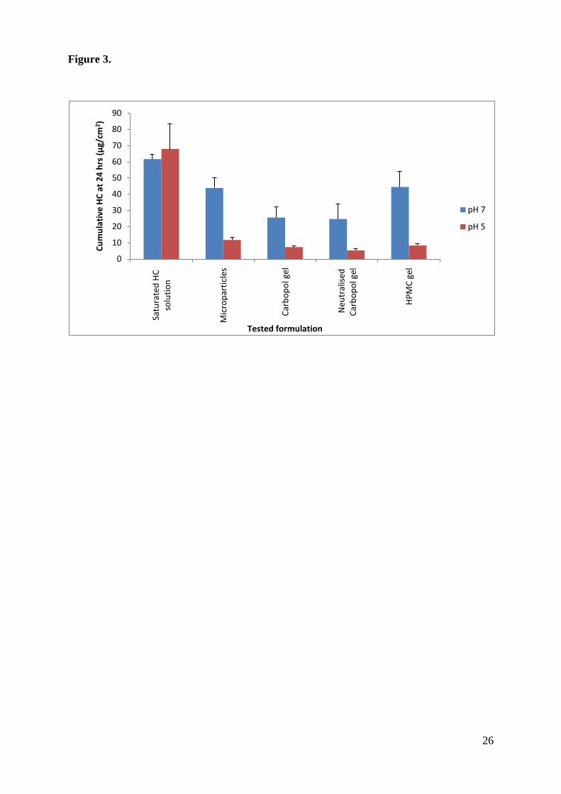
Permeation across full thickness porcine skin was measured using receptor compartments
containing phosphate buffer at pH 5 or 7. The skin was equilibrated with the buffer for 2 hrs
prior to the experiment to develop the appropriate surface pH. A comparison of the
cumulative amount of hydrocortisone permeating the skin after 24 hours is given in Figure 3.
Permeation from Carbopol hydrogels before and after neutralisation showed similar pH-
responsiveness to the free powder, with about 4-fold greater permeation of the drug after 24
hours at pH 7 compared to pH 5 (p<0.05). Similarly, the HPMC-based gel formulation
showed significantly higher drug permeation at pH 7 (about 5-fold greater than the amount
permeated after 24 hours at pH 5). In contrast to drug permeation from the particles, the
difference in hydrocortisone permeation from a saturated aqueous solution at pH 5 and 7 is
statistically non-significant. This is expected as the solubility of the drug at pH 5 and 7 was
found to be essentially the same (0.252±0.01 mg/ml) (11).
INSERT FIGURE 3
Increasing the pH of the Carbopol formulation to pH 5, in order to prevent skin pH alteration,
did not affect the amount of drug permeating the skin at pH 5 and 7 (Figure 3). Nonetheless,
increasing the pH of the formulation from 3 to 5 could potentially negatively influence the
efficient encapsulation of the drug within Eudragit L100 microparticles. Eudragit L100 has a
reported pKa value of 6.45±0.03, this means that a greater degree of carboxyl group
ionisation will be present at pH 5 compared to pH 3. Increased polymer ionisation might lead
to greater swelling and potentially a greater degree of drug loss into the external hydrogel
phase through diffusion; drug leakage from the incorporated microparticles can result in the

11
loss of formulation pH-responsive properties. In this study, the possible effects of Carbopol
hydrogel neutralisation on formulation stability was monitored before and after the addition
of the base (Figure 4, A), and compared to drug leakage from microparticles in the HPMC gel
(Figure 4, B).
INSERT FIGURE 4
4 Discussion
In recent years, polymeric nano- and micro-particulate systems have emerged as promising
systems for site-specific drug delivery and they have been extensively studied for oral,
parenteral and pulmonary drug administration (e.g. 12, 16). The application of such
polymeric systems in topical drug delivery is thought to offer a number of advantages,
including control of drug release and protection of unstable compounds (17).
Microencapsulation, using polymers such as poly(lactide-co-glycolide acid) (PLGA), for
controlled-release topical drug delivery has previously been reported, for example to prolong
the residence time of sunscreen agents in the stratum corneum (18) or to target vitamin A to
the upper layers of the skin (19). However, reports concerning percutaneous uptake of nano-
and micro-particles are contradictory (20, 21), though deposition into the follicles is well
documented (22, 23).
Unlike previous studies where polymeric particulate systems have been designed to cross the
intact skin layer or to accumulate in follicular openings, this study relies on a targeted drug
delivery approach whereby microparticle dissolution occurs on the surface of the skin
according to pH and results in drug release at the diseased site. Thus, the discrepancy in skin
surface pH observed in normal and diseased skin is used to trigger drug release at the target
site and so minimises delivery to non-diseased sites.

12
4.1 Hydrocortisone release from Eudragit L100 microparticles
The usefulness of drug release data in formulation design has been discussed by Flynn and
Poulsen (24, 25). Because the efficiency of creams, ointments and gel products is dependent
on drug release, in-vitro release testing constitutes an important and valuable tool for quality
control. This is particularly important for controlled release drug formulations. In fact, the use
of the diffusion model for polyvinyl alcohol (PVA) microparticles was shown to better
differentiate between samples in terms of controlled-drug release kinetics compared to other
methods of release testing such as dissolution studies (26).
The data in Figure 1 and Tables 1 and 2 clearly illustrate pH control over drug release from
the Eudragit microparticles. Comparing Figures 1A and 1D, the spray dried materials give
significantly greater drug release at pH 5 than from the oil-in-oil particles. However, with
increasing pH, release from particles produced by either process were similarly pH
responsive (Table 1) which indicates that the initial non-pH controlled release from the spray
dried materials is due to poor drug encapsulation, perhaps from surface enrichment or the
production of porous materials in which the polymer does not control release. Well-
controlled drug release from oil-in-oil manufactured particles was achieved; for example, at a
drug loading of 10%, only 1.02 µg cm-2
hr-1
hydrocortisone was released at pH 5 whereas the
same particles liberated 26.6 µg cm-2
hr-1
at pH 7.
4.2 Formulation development
There are numerous examples on the market for powdered topical formulations such as, talc,
zinc oxide and kaolin (27). However, such formulations are applied as drying, protective or
lubricant agents. Difficulties associated with the application and dosing of powders can be
overcome by incorporating them into a vehicle such as a cream, ointment, lotion or a gel.

13
The main criteria for incorporating powders intended for topical drug delivery into
conventional formulations is that the individual powder particles should be impalpable, i.e.
incapable of being perceived as individual particles by our sense of touch (28). In general,
particles less than 50 µm in their longest dimension are invisible to the senses (17). A coarser
powder can make the preparation gritty and cosmetically unacceptable. Microparticles
prepared from the oil-in-oil emulsification process at 2.5% and 10% w/w drug-content fulfil
this criterion with a mean equivalent circular area particle diameter of 41.5±0.6 and
39.3±11.2 µm respectively. The mean size of these microparticles can be further reduced
using a higher homogenisation speed during the manufacturing process.
In this study, 10% w/w hydrocortisone-loaded Eudragit L100 microparticles were
incorporated into a gel formulation to obtain a final drug content of 1% w/w, which is within
the 0.5 to 2.5% w/w topical concentration commonly used for hydrocortisone. The use of
microparticles with a lower drug-loading, e.g. 2.5% w/w, will require the incorporation of a
larger amount of powder into the gel formulation, which would result in a preparation with a
paste-like consistency that is difficult to spread (29).
For formulation development, two commonly used gelling materials were investigated;
Carbopol 940 and Metolose 90SH-SR (HPMC). The carbomer (Carbopol 940) used in this
study is an acrylic acid polymer cross-linked with allyl ethers of pentaerythritol (30, 31). The
individual Carbopol cross-linked polymer particles swell when dispersed in an aqueous
solution forming a colloidal, mucilage-like dispersion. Dermatological applications of
Carbopol polymers are numerous; examples include formulations of local anaesthetics and
cosmetics (e.g. 32-34). Carbopol polymers are also reported to have good suspending
properties (35). For example, a permanent sand (average particle size of 0.06 cm) suspension
was achieved using as little as 0.25% Carbopol 934 (36). The same physical stability was

14
reported for other insoluble compounds such as, pentobarbital sodium, chloramphenicol and
sulphadimidine (34, 37).
Metolose 90SH-SR, a high-viscosity grade of hydroxypropyl methyl cellulose (HPMC), was
also used. This is an example of a fully soluble polymer, which dissolves in aqueous media to
form a highly viscous gel due to its relatively high molecular weight. The use of HPMC for
dermatological gels has also been reported in the literature (34, 38). However, unlike
Carbopol, this is a neutral polymer. In fact, the pH of the two prepared gels using 1.5% w/w
Carbopol 940 or 2% w/w Metolose 90SH-SR were found to be 3.23±0.01 and 7.40±0.09
respectively. The addition of Eudragit L100 microparticles did not change the pH of the
Carbopol gel, but for the HPMC-containing gel the pH was reduced to about 5.2 due to the
anionic nature of Eudragit L100.
Carbopol 940 is also an anionic polymer whose use in a topical drug formulation can also
modulate skin surface pH. This effect can be detrimental for the controlled pH-responsive
release of hydrocortisone from the microparticles, as a reduced pH can result in reduced or no
drug release at the affected areas of the skin. To circumvent this problem, the Carbopol
hydrogel formulation was neutralised to pH 5.0 using 0.5 M NaOH. The use of other bases
for Carbopol neutralisation, such as monoethaloamine and triethylamine, has been reported in
the literature (28, 32, 33). However, these stronger bases are usually used for gel formulations
prepared from a hydro-alcoholic co-solvent system, as the inclusion of an alcohol can affect
polymer solubility leading to the precipitation of the neutralised Carbopol salt (39). It is
therefore essential to select the appropriate neutraliser depending on the alcohol content of
the formulation. Since, in this study an aqueous Carbopol formulation was employed, sodium
hydroxide was selected to neutralise the polymer.

15
4.3 Release and permeation testing of microparticle-containing
formulations
The results in Figure 2 illustrate that pH responsive release of hydrocortisone from the oil-in-
oil generated particles was maintained when incorporated into formulations gelled with
Carbopol or HPMC, though neutralizing the Carbopol gel did negatively impact on drug
release. As described above, release from topical preparations provides a good indication as
to permeation across skin membranes and the release data did indeed mirror the percutaneous
absorption values in Figure 3. Thus the particles alone or in gelled formulations all delivered
significantly greater amounts of hydrocortisone through porcine skin at pH 7 than at pH 5, in
contrast to a standard saturated aqueous solution from which drug permeation was the same
at both pH values. It is notable that the 10% particles showed a 26-fold increase in drug
release at pH 7 compared to pH 5 (Table 2) but only a 4-fold increase in permeation at the
higher pH value. This discrepancy reflects the presence of the stratum corneum as the rate
limiting barrier to permeation of the liberated drug whereas the release studies provide no
such barrier.
Our approach demonstrates that it is feasible to deliver the active ingredient to diseased skin
sites with minimal delivery to surrounding (non-involved) tissue. Current formulations, such
as typical creams, contain various ingredients which can act as permeation enhancers,
including surfactants and fragrance agents such as terpenes. Such enhancers could promote
the formation of a drug depot within the non-involved skin and hence increase potential
adverse effects. Whilst we have not explored the potential for depot formation on long term
use at diseased skin sites, our in-vitro proof-of-principle studies demonstrate that an in-vivo
evaluation of our pH-triggered and targeted drug delivery formulation is merited.

16
4.4 Stability testing
The Eudragit L100 microparticles were predominantly stable within the Carbopol hydrogel
with about 5% drug leakage into the bulk of the formulation before neutralisation (Figure 4).
This is probably due to the high glass transition temperature of Eudragit L100 (1500 C, (40))
which limits drug movement and diffusion into the external environment. In fact, the
prevention of drug molecule aggregation, which can lead to crystal growth, through
encapsulation into polymers with high transition temperatures, has been extensively studied
(17, 41, 42). The limited diffusion of hydrocortisone from the Eudragit L100 microparticles
coupled with the poor solubility of the drug in aqueous media explains the minimal drug
leakage from the microparticles.
After Carbopol hydrogel neutralisation at week 6, drug leakage from the microparticles
increased to about 15%. This is probably due to increased Eudragit L100 ionisation and
swelling as discussed earlier. However, this value remained constant over the next 6 weeks,
with 85% of the drug originally encapsulated remaining within the microparticles. With the
HPMC-based gel formulation, hydrocortisone leakage from Eudragit L100 microparticles
was minimal over the 12 weeks testing period, as demonstrated in Figure 4, B. A longer-term
stability study is nonetheless necessary to fully evaluate the commercial viability of these
novel pH-responsive formulations.
5 Conclusion
Eudragit L100 microparticles loaded with hydrocortisone intended for targeted delivery to
atopic dermatitis areas of the skin were produced by two different methods, oil-in-oil
microencapsulation and spray drying. Release from both batches of microparticles was
equally pH-dependent though the particles produced by spray drying also gave significant

17
non-polymer controlled burst release, probably due to their porous nature or as a result of
drug enrichment on the surface of these particles.
The release studies showed that the oil-in-oil generated particles deliver essentially no drug at
normal (intact) skin pH (5.0 – 5.5) but that delivery can be targeted and controlled to atopic
dermatitis skin where the pH is elevated to around pH 6.0-6.5. This pH-responsive release
was maintained upon the incorporation of these microparticles into Carbopol- and HPMC-
based gel formulations. In-vitro hydrocortisone permeation studies from these novel pH-
responsive formulations showed a 4 to 5-fold increase in drug permeation after 24 hours at
pH 7 compared to pH 5. A 12-week preliminary stability study monitored drug leakage from
the incorporated microparticles which would result in a loss of pH-responsiveness. The study
demonstrated relatively good stability of both formulations with better stability of the
microparticles incorporated in the HPMC gel. Nonetheless, a long-term stability study and a
blanching assay are necessary to fully evaluate the commercial viability and clinical efficacy
of such formulations.
6 Acknowledgements
The authors thank Stiefel laboratories Ltd, a GSK company, for their financial support.

18
7 References
1. Hengge UR, Ruzicka T, Schwartz RA, Cork MJ. Adverse effects of topical
glucocorticosteroids. J Am Acad Dermatol. 2006;54(1):1.
2. Ohman EM, Rogers S, Meenan FO, McKenna TJ. Adrenal suppression following
low-dose topical clobetasol propionate. J R Soc Med. 1987;80(7):422-4.
3. Schäcke H, Schottelius A, Döcke W-D, Strehlke P, Jaroch S, Schmees N, et al.
Dissociation of transactivation from transrepression by a selective glucocorticoid receptor
agonist leads to separation of therapeutic effects from side effects. PNAS. 2004;101(1):227-
32.
4. Fluhr JW, Elias PM. Stratum corneum pH: formation and function of the "acid
mantle". Exogenous Dermatol. 2002;1:163-75.
5. Eberlein-König B, Schafer T, Huss-Marp J, Darsow U, Mohrenschlager M, Herbert
O, et al. Skin surface pH, stratum corneum hydration, trans-epidermal water loss and skin
roughness related to atopic eczema and skin dryness in a population of primary school
children. Acta Derm Venereol. 2000;80:188-91.
6. Schmid-Wendtner MH, Korting HC. The pH of the Skin Surface and Its Impact on the
Barrier Function. Skin Pharmacol Physiol. 2006;19(6):296-302.
7. Sparavigna A, Setaro M, Gualandri V. Cutaneous pH in children affected by atopic
dermatitis and in healthy children: a multicenter study. Skin Res Technol. 1999;5(4):221-7.
8. Chikakane K, Takahashi H. Measurement of skin pH and its significance in cutaneous
diseases. Clin Dermatol. 1995 1995/8//;13(4):299-306.
9. Anderson D. The acid-base balance of the skin. Br J Dermatol. 1951;63:283-96.
10. Rizi K, Green RJ, Khutoryanskaya O, Donaldson M, Williams AC. Mechanisms of
burst release from pH-responsive polymeric microparticles. J Pharm. Pharmacol., 2010:In
Press.

19
11. Rizi K, Green RJ, Donaldson M, Williams AC. Production of pH-responsive
microparticles by spray drying: Investigation of experimental parameter effects on
morphological and release properties. J Pharm Sci. 2010;100(2):566-79.
12. Kendall RA, Alhnan MA, Nilkumhang S, Murdan S, Basit AW. Fabrication and in
vivo evaluation of highly pH-responsive acrylic microparticles for targeted gastrointestinal
delivery. Eur J Pharm Sci. 2009;37(3-4):284-90.
13. Jacobi, U., Kaiser, M., Toll, R., Mangelsdorf, S., Audring, H., Otberg, N., Sterry, W.,
Lademann, J.,2007.Porcine ear skin: an in vitro model for human skin. Skin Res and Tech,
13, 19-24.
14. Muhammad, F., Brooks, J. D., Riviere, J. E.,2004.Comparative mixture effects of
JP(100) additives on the dermal absorption and disposition of jet fuel hydrocarbons in
different membrane model systems. Toxicology Letters, 150, 351-365.
15. French DL, Himmelstein KJ, Mauger JW. Physicochemical aspects of controlled
release of substituted benzoic and naphthoic acids from Carbopol® gels. J Controlled
Release. 1995;37(3):281-9.
16. Salama R, Hoe S, Chan H-K, Traini D, Young PM. Preparation and characterisation
of controlled release co-spray dried drug-polymer microparticles for inhalation 1: Influence
of polymer concentration on physical and in vitro characteristics. Eur J Pharm Biopharm.
2008;69(2):486-95.
17. Klee SK, Farwick M, Lersch P. Triggered release of sensitive active ingredients upon
response to the skin's natural pH. Colloids Surf Physicochem Eng Aspects. 2009;338(1-
3):162-6.
18. Alvarez-Roman R, Barre G, Guy RH, Fessi H. Biodegradable polymer nanocapsules
containing a sunscreen agent: preparation and photoprotection. Eur J Pharm Biopharm.
2001;52:191-5.

20
19. Jenning V, Gysler A, Schafer-Korting M, Gohla S. Vitamin A loaded solid lipid
nanoparticles for topical use:occlusive properties and drug targeting to the upper skin. Eur J
Pharm Biopharm. 2000;49:211-8.
20. Alvarez-Roman R, Naik A, Kalia YN, Guy RH, Fessi H. Skin penetration and
distribution of polymeric nanoparticles. J Controlled Release. 2004;99(1):53.
21. Zhao Y, Brown MB, Jones SA. Pharmaceutical foams: are they the answer to the
dilemma of topical nanoparticles? Nanomed Nanotechnol Biol Med. 2010;6(2):227-36.
22. Knorr F, Lademann J, Patzelt A, Sterry W, Blume-Peytavi U, Vogt A. Follicular
transport route - Research progress and future perspectives. Eur J Pharm Biopharm.
2008;71(2):173-80.
23. Lademann J, Richter H, Teichmann A, Otberg N, Blume-Peytavi U, Luengo J, et al.
Nanoparticles - An efficient carrier for drug delivery into the hair follicles. Eur J Pharm
Biopharm. 2007;66(2):159.
24. Flynn GL. Comparision between in vivo techniques. Acta Pharm Suce. 1983;20:54-9.
25. Poulsen BJ, Flynn GL. In vitro methods to study dermal delivery and percutaneous
absorption. In: Bronaugh RL, Maibach HI, editors. Percutaneous absorption: Mechanism,
absorption and drug delivery. New York: Marcel Dekker; 1985. p. 431-59.
26. Salama RO, Traini D, Chan H-K, Young PM. Preparation and characterisation of
controlled release co-spray dried drug-polymer microparticles for inhalation 2: Evaluation of
in vitro release profiling methodologies for controlled release respiratory aerosols. Eur J
Pharm Biopharm. 2008;70(1):145-52.
27. British National Formulary. 59 ed: British Medical Association & Royal
Pharmaceutical Society of Great Britain; March 2010.
28. Barry BW. Dermatological formulations:percutaneous absorption. 1st ed. New York:
Marcel Dekker Inc; 1993.
29. Stankler L. Diseases of the skin. Br Med J. 1974;1:27-9.

21
30. Guo J-H. Carbopol polymers for pharmaceutical drug delivery applications. Drug
Deliv Technol. 2003.
31. Labanda J, Marco P, Llorens J. Rheological model to predict the thixotropic
behaviour of colloidal dispersions. Colloids Surf Physicochem Eng Aspects. 2004;249(1-
3):123-6.
32. Glavas-Dodov M, Goracinova K, Mladenovska K, Fredro-Kumbaradzi E. Release
profile of lidocaine HCl from topical liposomal gel formulation. Int J Pharm. 2002;242(1-
2):381-4.
33. Lu G, Jun HW. Diffusion studies of methotrexate in Carbopol and Poloxamer gels. Int
J Pharm. 1998;160(1):1-9.
34. Shin S-C, Cho C-W, Yang K-H. Development of lidocaine gels for enhanced local
anesthetic action. Int J Pharm. 2004;287(1-2):73-8.
35. Dolan MM, Steelman RL, Tumilowicz RR. Carbopol 934: An improved suspending
agent for insoluble test compounds. Toxicol Appl Pharmacol. 1960;2(3):331-7.
36. Meyer RJ, Cohen L. The rheology of natural and synthetic hydrophilic polymer
solutions as related to suspending ability. J Soc Cosmet Chem. 1959;10:143-54.
37. Berney BM, Deasy PB. Evaluation of carbopol 934 as a suspending agent for
sulphaoimidine suspensions. Int J Pharm. 1979;3(2-3):73-80.
38. El-Kattan AF, Asbill CS, Michniak BB. The effect of terpene enhancer lipophilicity
on the percutaneous permeation of hydrocortisone formulated in HPMC gel systems. Int J
Pharm. 2000;198(2):179-89.
39. Formulating hydroalcoholic gels with Carbopol polymers. Noveon Technical Data
Sheet. 2009.
40. Lin S-Y, Liao C-M, Hsiue G-H, Liang R-C. Study of a theophylline-Eudragit L
mixture using a combined system of microscopic Fourier-transform infrared spectroscopy
and differential scanning calorimetry. Thermochim Acta. 1995;254:153-66.

22
41. Eerikainen H, Kauppinen EI. Preparation of polymeric nanoparticles containing
corticosteroid by a novel aerosol flow reactor method. Int J Pharm. 2003;263(1-2):69.
42. Friesen DT, Shanker R, Crew M, Smithey DT, Curatolo WJ, Nightingale JAS.
Hydroxypropyl methylcellulose acetate succinate-based spray-dried dispersions: An
overview. Mol Pharm. 2008;5(6):1003-19.

23
Legend to Figures
Figure 1. Hydrocortisone release profiles from: A, B and C) 2.5%, 10% and 25% w/w
hydrocortisone-containing microparticles produced from the oil/oil method and D, E and F)
2.5%, 10% and 25% w/w drug-loaded microparticles obtained from the spray drying
technique (mean ± SD, n=3).
Figure 2. Release of hydrocortisone from: A) Carbopol hydrogel, B) neutralised Carbopol
hydrogel and C) HPMC gel. Results are presented as mean ± SD, n=3-5.
Figure 3. Cumulative hydrocortisone (HC) permeation at pH 5 and 7 after 24 hrs from a
saturated solution of hydrocortisone, 10% w/w hydrocortisone-loaded Eudragit L100
microparticles and various microparticle-containing gel formulations. Data are shown as
mean ± SD, n=5.
Figure 4. Stability testing of 10% w/w hydrocortisone-loaded Eudragit L100 microparticles
incorporated into; Top) Carbopol hydrogel and Bottom) HPMC gel. The percentage of drug
leaking into the bulk of the formulation and that remaining within the microparticles was
determined before and after Carbopol neutralisation at 6 weeks. Data is presented as mean ±
SD, n= 3.

24
Figure 1
0
20
40
60
80
100
0 1 2 3 4 5 6
% H
C r
ele
ased
Time (hrs)
pH 7
pH 6.5
pH 6
pH 5.5
pH 5
A)
0
20
40
60
80
100
0 1 2 3 4 5 6
% H
C r
ele
ased
Time (hrs)
D)
0
20
40
60
80
100
0 1 2 3 4 5 6
% H
C r
ele
ase
d
Time (hrs)
B)
0
20
40
60
80
100
0 1 2 3 4 5 6
% H
C r
ele
ase
d
Time (hrs)
E)
0
20
40
60
80
100
0 1 2 3 4 5 6
% H
C r
ele
ase
d
Time (hrs)
C)
0
20
40
60
80
100
0 1 2 3 4 5 6
% H
C r
ele
ased
Time (hrs)
F)

25
Figure 2
0
20
40
60
80
100
0 1 2 3 4 5
% R
ele
ase
Time (hrs)
0
20
40
60
80
100
0 1 2 3 4 5
% R
ele
ase
Time (hrs)
0
20
40
60
80
100
0 1 2 3 4 5
% R
ele
ase
Time (hrs)
pH 7
pH 5
A) B)
C)
26
Figure 3.
0
10
20
30
40
50
60
70
80
90
Satu
rate
d H
C
solu
tio
n
Mic
rop
arti
cles
Car
bo
po
l gel
Neu
tral
ised
C
arb
op
ol g
el
HP
MC
gel
Cu
mu
lati
ve H
C a
t 2
4 h
rs (
µg/
cm2)
Tested formulation
pH 7
pH 5

27
Figure 4.
0
20
40
60
80
100
120
0 2 4 6 8 10 12 14
% h
ydro
cort
iso
ne
Time (weeks)
% remaining
% leakagepH increased to 5 at week 6
0
20
40
60
80
100
120
0 2 4 6 8 10 12 14
% h
ydro
ocr
tiso
ne
Time (weeks)
% remaining
% leakage

28
Table 1. pH-responsiveness of 2.5% w/w hydrocortisone-loaded Eudragit L100
microparticles reported as percentage hydrocortisone released per unit pH, calculated at 3, 4
and 5 hours.
Time Spray drying Oil/oil method
3 hours 31.2±3.66 37.2±8.62
4 hours 33.7±3.14 44.3±6.69
5 hours 31.9±4.19 48.8±5.33

29
Table 2. Maximum release rates (Jmax) (µg cm-2
hr-1
) and time to maximum release rates
(Tmax) calculated from the release profile of hydrocortisone for the different microparticles
prepared from the spray drying and oil-in-oil emulsification methods.
Method Drug
loading
(% w/w)
pH 5 pH 7
Jmax Tmax Jmax Tmax
Spray drying 2.5% 7.23±2.11 1.5 hr 17.3±2.94 1.5 hr
10% 18.5±1.81 1.5 hr 24.6±4.48 45 min
25% 12.6±0.71 45 min 16.6±5.98 45 min
Oil/oil
method
2.5% 1.26±0.16 1.5 hr 19.6±3.54 1.5 hr
10% 1.02±0.24 1.5 hr 26.6±4.50 1.5 hr
25% 5.20±2.26 1.5 hr 18.8±1.30 1.5 hr