Palmitoylation is the switch that assigns calnexin to quality control or ER Ca2+ signaling
37
Palmitoylation is the Switch that Assigns Calnexin to Quality Control or ER Calcium Signaling Emily M. Lynes 1 , Arun Raturi 1 , Marina Shenkman 2 , Carolina Ortiz Sandoval 1 , Megan C. Yap 1 , Jiahui Wu 3 , Aleksandra Janowicz 1 , Nathan Myhill 1 , Matthew D. Benson 1 , Robert 5 E. Campbell 3 , Luc G. Berthiaume 1 , Gerardo Z. Lederkremer 2 and Thomas Simmen 1* 1 Department of Cell Biology, University of Alberta, Edmonton, Alberta, T6G2H7, Canada 10 2 Department of Cell Research and Immunology, George Wise Faculty of Life Sciences, Tel Aviv University, Tel Aviv 69978, Israel 3 Department of Chemistry, University of Alberta, Edmonton, Alberta, T6G2G2, Canada 15 * Corresponding Author: Tel.: (780) 492-1546 Fax: (780) 492-0450 e-mail: [email protected] 20 Word count excluding references: 8103 Running title: The Function of Calnexin Palmitoylation Keywords: Endoplasmic reticulum, mitochondria, quality control, calcium signaling, ERp57, SERCA2b 25 © 2013. Published by The Company of Biologists Ltd. Journal of Cell Science Accepted manuscript JCS Advance Online Article. Posted on 10 July 2013
-
Upload
independent -
Category
Documents
-
view
3 -
download
0
Transcript of Palmitoylation is the switch that assigns calnexin to quality control or ER Ca2+ signaling
Palmitoylation is the Switch that Assigns Calnexin to Quality Control or ER
Calcium Signaling
Emily M. Lynes1, Arun Raturi1, Marina Shenkman2, Carolina Ortiz Sandoval1, Megan C.
Yap1, Jiahui Wu3, Aleksandra Janowicz1, Nathan Myhill1, Matthew D. Benson1, Robert 5
E. Campbell3, Luc G. Berthiaume1, Gerardo Z. Lederkremer2 and Thomas Simmen1*
1Department of Cell Biology, University of Alberta, Edmonton, Alberta, T6G2H7,
Canada
10 2 Department of Cell Research and Immunology, George Wise Faculty of Life Sciences,
Tel Aviv University, Tel Aviv 69978, Israel
3 Department of Chemistry, University of Alberta, Edmonton, Alberta, T6G2G2, Canada
15
* Corresponding Author:
Tel.: (780) 492-1546
Fax: (780) 492-0450
e-mail: [email protected] 20
Word count excluding references: 8103
Running title: The Function of Calnexin Palmitoylation
Keywords: Endoplasmic reticulum, mitochondria, quality control, calcium signaling,
ERp57, SERCA2b
25
© 2013. Published by The Company of Biologists Ltd.Jo
urna
l of C
ell S
cien
ceA
ccep
ted
man
uscr
ipt
JCS Advance Online Article. Posted on 10 July 2013

Abstract
The palmitoylation of calnexin serves to enrich calnexin on the mitochondria-associated
membrane (MAM). Given a lack of information on the significance of this finding, we
have investigated how this endoplasmic reticulum (ER)-internal sorting signal affects the
functions of calnexin. Our results demonstrate that palmitoylated calnexin interacts with 5
sarcoendoplasmic reticulum (SR) calcium transport ATPase (SERCA) 2b and that this
interaction determines ER calcium content and the regulation of ER-mitochondria
calcium crosstalk. In contrast, non-palmitoylated calnexin interacts with the
oxidoreductase ERp57 and performs its well-known function in quality control.
Interestingly, our results also show that calnexin palmitoylation is an ER stress-dependent 10
mechanism. Following a short term ER stress, calnexin quickly becomes less
palmitoylated, which shifts its function from the regulation of calcium signaling towards
chaperoning and quality control of known substrates. These changes also correlate with a
preferential distribution of calnexin to the MAM under resting conditions or the rough
ER and ER quality control compartment (ERQC) following ER stress. Our results have 15
therefore identified the switch that assigns calnexin either to calcium signaling or to
protein chaperoning.
Jour
nal o
f Cel
l Sci
ence
Acc
epte
d m
anus
crip
t

Introduction
The main functions of the endoplasmic reticulum (ER) are the production of secretory
and membrane proteins as well as lipids, and the storage of calcium. An integral
requirement for these functions is the interaction of the ER with mitochondria, which
occurs on the mitochondria associated membrane (MAM), a subdomain of the ER that 5
makes close contacts between the ER and the mitochondria (Raturi and Simmen, 2012;
Simmen et al., 2010). Here, the ER exchanges calcium with mitochondria via the ER
calcium handling proteins inositol 1,4,5-triphosphate receptors (IP3R) and
sarcoendoplasmic reticulum (SR) calcium transport ATPase (SERCA) (Rizzuto et al.,
2009). Under resting conditions, calcium delivery from the ER to the mitochondrial 10
matrix is needed to activate the mitochondrial enzyme pyruvate dehydrogenase (PDH),
which drives the tricarboxylic acid (TCA) cycle (Cardenas et al., 2010). Given the critical
role that mitochondrial metabolism plays for death and survival of the cell (Glancy and
Balaban, 2012; Zhivotovsky and Orrenius, 2011), ER-mitochondria calcium exchange
has to be regulated at the source. Various ER chaperones and oxidoreductases perform 15
this function by dictating calcium channels and pumps to open or close dependent on ER
redox and calcium homeostasis (Simmen et al., 2010). Examples are Ero1α that activates
inositol 1,4,5 trisphosphate receptors (IP3Rs) (Li et al., 2009) and ERp44 that inhibits
IP3Rs (Higo et al., 2005).
Calnexin is another key ER chaperone that binds to nascent glycoproteins 20
(Rutkevich and Williams, 2011). This binding slows down client protein folding and
prevents their aggregation via the retention of folding intermediates in the ER. Upon
release from calnexin, glycoproteins may follow one of three pathways: they may fold
rapidly and correctly, in which case they will be exported to the Golgi apparatus, or if
they are not yet correctly folded they may be re-glucosylated and returned to the calnexin 25
cycle (Ruddock and Molinari, 2006). Proteins that have been trapped in the calnexin
cycle for a prolonged period are eventually subject to extensive mannose trimming by
mannosidase I and ER-associated degradation enhancing α-mannosidase-like protein
(EDEM), followed by retrotranslocation from the ER to the cytosol and degradation by
the proteasome, a process known as ER-associated degradation (ERAD) (Lederkremer, 30
2009). All along, calnexin works in partnership with ERp57, an oxidoreductase that is a
Jour
nal o
f Cel
l Sci
ence
Acc
epte
d m
anus
crip
t

member of the protein disulfide isomerase (PDI) family (Coe and Michalak, 2010; Oliver
et al., 1997; Zapun et al., 1998). Studies have shown that a certain subset of disulfide-
bonded, heavily glycosylated proteins such as the low density lipoprotein receptor
(LDLR) depend specifically on this partnership for efficient folding and subsequent
trafficking through the secretory system (Jessop et al., 2007; Rutkevich and Williams, 5
2011).
Surprisingly, our laboratory and others have determined that calnexin is not only
found on the rough ER, but is often enriched on the MAM (Hayashi and Su, 2007; Myhill
et al., 2008; Wieckowski et al., 2009). This finding further reinforces the notion that
calnexin is a central player for the regulation of ER calcium signaling, and is also 10
consistent with the fact that calnexin interacts with the SERCA calcium pump (Roderick
et al., 2000). Interestingly, calnexin’s role in calcium signaling could depend on ER
homeostasis, since ER stress results in the relocation of calnexin towards the ER quality
control compartment (ERQC) and its association with slowly folding proteins (Frenkel et
al., 2004). 15
Therefore, we have examined whether the targeting of calnexin to different ER
subdomains correlates with an ER stress-dependent role in ER calcium signaling towards
mitochondria. We further examined whether such a role correlated with the extent of
binding of calnexin to its known partners ERp57 and SERCA2b. Calnexin targeting to
the MAM depends on a juxtamembrane palmitoylation motif (Lynes et al., 2012) that is 20
implicated in mediating the interaction of calnexin with the translocon (Lakkaraju et al.,
2012) and with lipid rafts on the plasma membrane as well (Ferrera et al., 2008). In
addition to calnexin, palmitoylation is known to modify several ER proteins, including
heme oxygenase-1 and ORAI1, but the consequences of this modification are currently
unknown (Dowal et al., 2011; Kang et al., 2008). We had shown earlier that the 25
interference with palmitoylation relocates heme oxygenase-1 (Lynes et al., 2012), but
ER-associated palmitoylation might have multiple, diverse effects (Lakkaraju et al.,
2012). To investigate the functional significance of palmitoylation-dependent targeting of
calnexin, we characterized the functional consequences of calnexin’s palmitoylation state
for the cell in terms of cellular calcium signaling and quality control of the known 30
calnexin substrates LDLR and asialoglycoprotein receptor (ASGPR).
Jour
nal o
f Cel
l Sci
ence
Acc
epte
d m
anus
crip
t

Jour
nal o
f Cel
l Sci
ence
Acc
epte
d m
anus
crip
t
Results
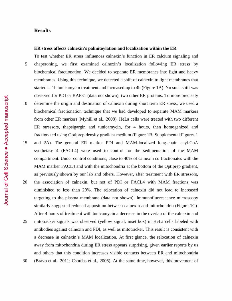
ER stress affects calnexin’s palmitoylation and localization within the ER
To test whether ER stress influences calnexin’s function in ER calcium signaling and
chaperoning, we first examined calnexin’s localization following ER stress by 5
biochemical fractionation. We decided to separate ER membranes into light and heavy
membranes. Using this technique, we detected a shift of calnexin to light membranes that
started at 1h tunicamycin treatment and increased up to 4h (Figure 1A). No such shift was
observed for PDI or BAP31 (data not shown), two other ER proteins. To more precisely
determine the origin and destination of calnexin during short term ER stress, we used a 10
biochemical fractionation technique that we had developed to separate MAM markers
from other ER markers (Myhill et al., 2008). HeLa cells were treated with two different
ER stressors, thapsigargin and tunicamycin, for 4 hours, then homogenized and
fractionated using Optiprep density gradient medium (Figure 1B, Supplemental Figures 1
and 2A). The general ER marker PDI and MAM-localized long-chain acyl-CoA 15
synthetase 4 (FACL4) were used to control for the sedimentation of the MAM
compartment. Under control conditions, close to 40% of calnexin co-fractionates with the
MAM marker FACL4 and with the mitochondria at the bottom of the Optiprep gradient,
as previously shown by our lab and others. However, after treatment with ER stressors,
the association of calnexin, but not of PDI or FACL4 with MAM fractions was 20
diminished to less than 20%. The relocation of calnexin did not lead to increased
targeting to the plasma membrane (data not shown). Immunofluorescence microscopy
similarly suggested reduced apposition between calnexin and mitochondria (Figure 1C).
After 4 hours of treatment with tunicamycin a decrease in the overlap of the calnexin and
mitotracker signals was observed (yellow signal, inset box) in HeLa cells labeled with 25
antibodies against calnexin and PDI, as well as mitotracker. This result is consistent with
a decrease in calnexin’s MAM localization. At first glance, the relocation of calnexin
away from mitochondria during ER stress appears surprising, given earlier reports by us
and others that this condition increases visible contacts between ER and mitochondria
(Bravo et al., 2011; Csordas et al., 2006). At the same time, however, this movement of 30
Jour
nal o
f Cel
l Sci
ence
Acc
epte
d m
anus
crip
t
calnexin away from the MAM suggests a specific function that is not due to known
structural changes of the ER.
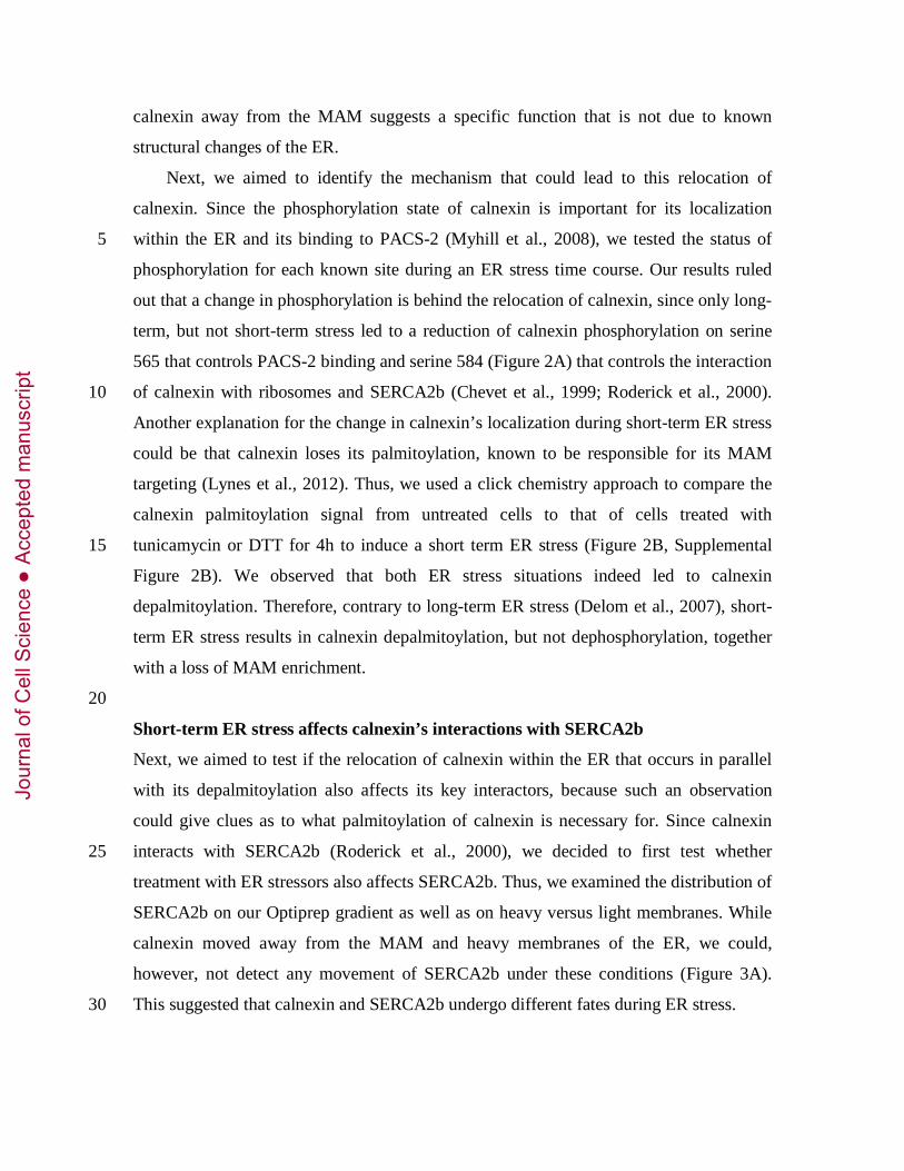
Next, we aimed to identify the mechanism that could lead to this relocation of
calnexin. Since the phosphorylation state of calnexin is important for its localization
within the ER and its binding to PACS-2 (Myhill et al., 2008), we tested the status of 5
phosphorylation for each known site during an ER stress time course. Our results ruled
out that a change in phosphorylation is behind the relocation of calnexin, since only long-
term, but not short-term stress led to a reduction of calnexin phosphorylation on serine
565 that controls PACS-2 binding and serine 584 (Figure 2A) that controls the interaction
of calnexin with ribosomes and SERCA2b (Chevet et al., 1999; Roderick et al., 2000). 10
Another explanation for the change in calnexin’s localization during short-term ER stress
could be that calnexin loses its palmitoylation, known to be responsible for its MAM
targeting (Lynes et al., 2012). Thus, we used a click chemistry approach to compare the
calnexin palmitoylation signal from untreated cells to that of cells treated with
tunicamycin or DTT for 4h to induce a short term ER stress (Figure 2B, Supplemental 15
Figure 2B). We observed that both ER stress situations indeed led to calnexin
depalmitoylation. Therefore, contrary to long-term ER stress (Delom et al., 2007), short-
term ER stress results in calnexin depalmitoylation, but not dephosphorylation, together
with a loss of MAM enrichment.
20
Short-term ER stress affects calnexin’s interactions with SERCA2b
Next, we aimed to test if the relocation of calnexin within the ER that occurs in parallel
with its depalmitoylation also affects its key interactors, because such an observation
could give clues as to what palmitoylation of calnexin is necessary for. Since calnexin
interacts with SERCA2b (Roderick et al., 2000), we decided to first test whether 25
treatment with ER stressors also affects SERCA2b. Thus, we examined the distribution of
SERCA2b on our Optiprep gradient as well as on heavy versus light membranes. While
calnexin moved away from the MAM and heavy membranes of the ER, we could,
however, not detect any movement of SERCA2b under these conditions (Figure 3A).
This suggested that calnexin and SERCA2b undergo different fates during ER stress. 30
Jour
nal o
f Cel
l Sci
ence
Acc
epte
d m
anus
crip
t
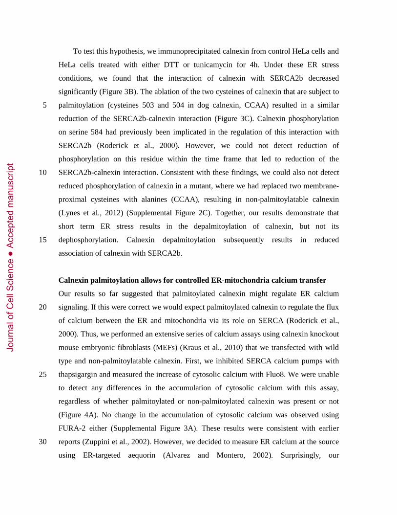
To test this hypothesis, we immunoprecipitated calnexin from control HeLa cells and
HeLa cells treated with either DTT or tunicamycin for 4h. Under these ER stress
conditions, we found that the interaction of calnexin with SERCA2b decreased
significantly (Figure 3B). The ablation of the two cysteines of calnexin that are subject to
palmitoylation (cysteines 503 and 504 in dog calnexin, CCAA) resulted in a similar 5
reduction of the SERCA2b-calnexin interaction (Figure 3C). Calnexin phosphorylation
on serine 584 had previously been implicated in the regulation of this interaction with
SERCA2b (Roderick et al., 2000). However, we could not detect reduction of
phosphorylation on this residue within the time frame that led to reduction of the
SERCA2b-calnexin interaction. Consistent with these findings, we could also not detect 10
reduced phosphorylation of calnexin in a mutant, where we had replaced two membrane-
proximal cysteines with alanines (CCAA), resulting in non-palmitoylatable calnexin
(Lynes et al., 2012) (Supplemental Figure 2C). Together, our results demonstrate that
short term ER stress results in the depalmitoylation of calnexin, but not its
dephosphorylation. Calnexin depalmitoylation subsequently results in reduced 15
association of calnexin with SERCA2b.
Calnexin palmitoylation allows for controlled ER-mitochondria calcium transfer
Our results so far suggested that palmitoylated calnexin might regulate ER calcium
signaling. If this were correct we would expect palmitoylated calnexin to regulate the flux 20
of calcium between the ER and mitochondria via its role on SERCA (Roderick et al.,
2000). Thus, we performed an extensive series of calcium assays using calnexin knockout
mouse embryonic fibroblasts (MEFs) (Kraus et al., 2010) that we transfected with wild
type and non-palmitoylatable calnexin. First, we inhibited SERCA calcium pumps with
thapsigargin and measured the increase of cytosolic calcium with Fluo8. We were unable 25
to detect any differences in the accumulation of cytosolic calcium with this assay,
regardless of whether palmitoylated or non-palmitoylated calnexin was present or not
(Figure 4A). No change in the accumulation of cytosolic calcium was observed using
FURA-2 either (Supplemental Figure 3A). These results were consistent with earlier
reports (Zuppini et al., 2002). However, we decided to measure ER calcium at the source 30
using ER-targeted aequorin (Alvarez and Montero, 2002). Surprisingly, our
Jour
nal o
f Cel
l Sci
ence
Acc
epte
d m
anus
crip
t
measurements showed about 80% more calcium in MEFs transfected with wild type
calnexin, but not with calnexin that cannot be palmitoylated (Figure 4B). Since these
cells did not show altered accumulation of cytosolic calcium from ER stores, our findings
suggested that mitochondrial calcium uptake could be different as well in cells expressing
calnexin compared to calnexin knockout MEFs (Arnaudeau et al., 2001; de Brito and 5
Scorrano, 2008). Therefore, we tested whether mitochondria show differences in calcium
uptake following the inhibition of ER calcium pumps. Indeed, we found that
mitochondria in wild type MEFs take up significantly less calcium than knockout MEFs
(Supplemental Figure 3B). Moreover, our results with the mitochondrial calcium
indicator dye Rhod2 in the presence of thapsigargin show that the presence of wild type 10
calnexin dampened mitochondrial calcium uptake following thapsigargin administration,
when compared to calnexin knockout MEFs (Figure 4C). Importantly, this effect was not
seen using non-palmitoylated calnexin. Since our results indicate that the presence of
palmitoylated calnexin reduced the ability of mitochondria to import calcium from the
ER, we next tested whether this was also true under the condition of ER stress, when cells 15
normally increase mitochondrial calcium import from ER sources (Bravo et al., 2011;
Csordas et al., 2006). Our results show that calnexin knockout MEFs were unable to
boost the import of IP3R-released calcium into mitochondria after a 4h tunicamycin
stress. However, the transfection of wild type calnexin, but not of non-palmitoylatable
calnexin rescued this deficiency (Figure 4D), thus making calnexin knockout MEFs 20
stress-responsive again. Together, our results suggested that calnexin must be
palmitoylatable to control the activity of SERCA and thus the rate of calcium import into
mitochondria.
Since efficient calcium transfer from the ER to mitochondria is a prerequisite for the
mitochondrial membrane potential (Cardenas et al., 2010), we next tested the hypothesis 25
that calnexin knockout cells could exhibit an abnormally high proton gradient across the
mitochondrial membranes due to their high constitutive calcium transfer from the ER to
mitochondria. By labeling calnexin knockout cells as well as their counterparts
transfected with wild type calnexin and non-palmitoylatable calnexin with TMRM, we
were indeed able to confirm this hypothesis by demonstrating that only wild type 30
calnexin transfection resulted in a specific reduction of the mitochondrial membrane
Jour
nal o
f Cel
l Sci
ence
Acc
epte
d m
anus
crip
t

potential (Supplemental Figure 3C). This was also the case in the presence of KCl that
equilibrates plasma membrane uptake of TMRM (data not shown). Throughout our
experiments, we verified that expression of the two calnexin constructs was even (Figure
4E) and that our transfections did not result in the induction of the unfolded protein
response (Supplemental Figure 3D). Therefore, ER-mitochondria calcium transfer is 5
more efficient in the absence of calnexin, but the boosted calcium transfer observed under
ER stress requires palmitoylated calnexin that increases the ER calcium content.
Calnexin’s palmitoylation state regulates its interaction with ERp57
In addition to its interaction with SERCA2b, calnexin interacts with the oxidoreductase 10
ERp57 in the ER to mediate protein folding. This role of calnexin is particularly
important for slow-folding substrates (Frenkel et al., 2004). Thus, we decided to also test
whether calnexin’s interaction with ERp57 is similarly affected by ER stress conditions.
First, we examined the distribution of calnexin and ERp57 by Optiprep gradient and
fractionation of cellular membranes into heavy and light membranes. Contrary to 15
SERCA2b (Figure 3A), the distribution of ERp57 closely matched the one by calnexin
and both turned out to move from heavy to light membranes following an ER stress
(Figure 5A), suggesting that these two proteins might share functions under ER stress.
Indeed, we observed that when cells are treated with either DTT or tunicamycin for 4h,
calnexin’s interactions with ERp57 increased (Figure 5B). ER stress could lead to the 20
misfolding of calnexin itself, which could then trigger its association with other
chaperones. To test this possibility, we expressed FLAG-tagged calnexin and mutant
calnexin that cannot be palmitoylated. As shown in Figure 5C, the calnexin CCAA
mutant that cannot be palmitoylated indeed interacted with ERp57 almost twice as well.
Likewise, the binding of calnexin to ERp57 was about doubled in the presence of 2-25
bromopalmitate, an inhibitor of palmitoylation (Supplemental Figure 4A).
Our results suggested that the change of interaction between calnexin and ERp57
dependent on palmitoylation was of a functional nature. We therefore sought to identify a
folding substrate that is particularly dependent on the calnexin/ERp57 folding pathway.
For this purpose, we first chose the low density lipoprotein receptor (LDLR) that is 30
known to form mixed disulfides with ERp57 and depend on calnexin folding (Jessop et
Jour
nal o
f Cel
l Sci
ence
Acc
epte
d m
anus
crip
t

al., 2007). To determine whether LDLR maturation proceeds differently whether calnexin
is palmitoylated or not, we tested this in the calnexin knockout MEFs that we transfected
with wild type calnexin or the calnexin CCAA mutant. Here, we first determined that the
expression level of the LDLR in lysates was unaltered (Figure 5D). Despite this however,
the amount of LDLR on the cell surface increased with wild type calnexin, but decreased 5
with CCAA calnexin (Figure 5D), suggesting non-palmitoylated calnexin is indeed more
stringent in ER quality control retention than the at least partially palmitoylated wild type
calnexin.
Efficient intracellular retention of LDLR by non-palmitoylated calnexin could indicate
that this form of calnexin interacts with unfolded domains of proteins that await exit from 10
the ER or alternatively ER-associated degradation (ERAD). Thus, we analyzed the
interaction of calnexin with an ERAD substrate, the uncleaved precursor of
asialoglycoprotein receptor (ASGPR) H2a, which is normally completely ER-retained
(Kamhi-Nesher et al., 2001; Shenkman et al., 1997). CNX CCAA interacted much more
robustly than CNX wild type with H2a (Figure 6A). CNX CCAA also caused a slight 15
increase of total H2a, likely due to decreased targeting of this protein for ERAD.
Pulse/chase analysis showed that dissociation of H2a from calnexin followed a different
pattern when we transfected nonpalmitoylatable calnexin compared to wild type (Figure
6B). Whereas wild type calnexin dissociated efficiently with time from the ERAD
substrate H2a, non-palmitoylatable CCAA calnexin reassociated and remained 20
complexed to H2a for extended periods of time. For the pulse samples two bands can be
seen for H2a precursor molecules, the lower one corresponding to an underglycosylated
species (one of the glycosylation sites unoccupied). As we had shown before (Frenkel et
al., 2004), the fully glycosylated species shifts progressively to a faster migration because
of the trimming of mannose residues on its N-glycans, whereas the underglycosylated 25
lower species is quickly degraded.
We next tested whether palmitoylation influences calnexin targeting to the pericentriolar
ERQC, where calnexin and ERAD substrates like H2a accumulate upon inhibition of
their degradation or under ER stress (Frenkel et al., 2004; Kamhi-Nesher et al., 2001;
Kondratyev et al., 2007). Thus, we compared the localization of the non-palmitoylatable 30
mutant with wild type calnexin. Whereas in untreated cells both proteins showed a
Jour
nal o
f Cel
l Sci
ence
Acc
epte
d m
anus
crip
t

disperse ER pattern, upon short term proteasomal inhibition for 3h, calnexin CCAA
showed a more pronounced relocation to the juxtanuclear ERQC and colocalization with
H2a linked to monomeric RFP (H2a-RFP), according to the Manders coefficient (Figure
6C-F). Together, our results demonstrate the preferential interaction of non-palmitoylated
calnexin with ERAD substrates that extends to the ERQC, where dissociation from the 5
misfolded glycoprotein would require the ability of calnexin to undergo palmitoylation.
Discussion
The results presented herein uncover a mechanistic description of the role of calnexin 10
palmitoylation during ER stress. We demonstrate that reversible palmitoylation assigns
calnexin to the interaction with SERCA2b and a role in the regulation of calcium transfer
to mitochondria, whereas non-palmitoylated calnexin tends to interact with ERp57 to
fulfill its role in protein folding and quality control (Figure 7). Our results therefore
illustrate for the first time how palmitoylation can restrict calnexin to either role and 15
propose palmitoylation as a key mechanism of ER stress signaling. Our observations
identify palmitoylation as a quick signal that reassigns calnexin to specific tasks within
the ER, unlike phosphorylation that determines calnexin interaction with the ribosome,
and another ER chaperone, BAP31, in a less dynamic way . Changes in calnexin
phosphorylation require significantly longer exposure to ER stress when compared to the 20
changes in palmitoylation (Figure 2). Moreover, whereas the calnexin phosphorylation
state results in intra-ER relocations mediated by the cytosolic sorting protein PACS-2
(Myhill et al., 2008), palmitoylation may regulate the calnexin localization by allowing
(or not) its interaction with its effectors SERCA2b and ERp57, localized to sites of
calcium signaling or protein folding, respectively. Interestingly, calnexin appears to 25
follow a targeting pattern that is likely relatively unique, since ER and mitochondria
move closer to each other during ER stress, suggesting an enrichment of ER proteins at
the MAM or a tightening of ER-mitochondria contacts (Bravo et al., 2011; Csordas et al.,
2006).
So far, the interaction of calnexin with its substrates has been explained from the 30
substrate’s point of view, since calnexin binds exclusively monoglucosylated substrates
Jour
nal o
f Cel
l Sci
ence
Acc
epte
d m
anus
crip
t

(Lederkremer, 2009). This property, together with the enzymatic action of
UDPGlc:glycoprotein glucosyltransferase and glucosidases I and II leads to a cyclic
interaction of folding substrates with calnexin until proper folding is reached or ERAD is
triggered. Our new data now indicates that calnexin itself undergoes a palmitoylation-
dependent cycle within the ER and shuttles back and forth from regulating calcium 5
signaling at or close to the MAM and mediating protein folding and quality control at or
close to the rER, including the targeting of unfolded or misfolded proteins to the ERQC
(Leitman et al., 2012). Interestingly, these results could help explain the anti-stress
function of 2-bromopalmitate (2BP) treatment that attenuates the induction of ER stress
transcription factors and the progression of apoptosis (Baldwin et al., 2012). In this 10
scenario, calnexin would become more efficient as a chaperone and would hence mitigate
the effects of any additionally introduced ER stressor. Consistent with such an idea, we
observed that the inhibition of palmitoylation with 2BP leads to a 2-fold increase in the
binding of ERp57 with calnexin (Supplemental Figure 4A), increases the association with
H2a and reduces the amount of mature LDLR on the surface (Supplemental Figure 4B). 15
These findings, together with the decreased amount of surface LDLR with CCAA
calnexin, suggest that palmitoylation facilitates the dissociation of calnexin from folding
intermediates.
Accordingly, palmitoylated calnexin appears to be less important for protein
folding, consistent with its demonstrated predominant localization to the MAM (Lynes et 20
al., 2012). However, our results also reinforce the close relationship between ER folding
assistants and the control of mitochondrial metabolism (Simmen et al., 2010). In our
specific example, calnexin expression and reversible palmitoylation are required to allow
the ER to properly signal via calcium towards mitochondria. It is apparently due to the
presence of palmitoylated calnexin that the ER can signal a state of stress to 25
mitochondria, likely to alleviate the accumulation of unfolded proteins within the ER
(Bravo et al., 2011). However, this function is also critical for mitochondria metabolism,
as suggested by the abnormally high mitochondria membrane potential in the absence of
palmitoylated calnexin. Surprisingly, our results contradict previous reports that calnexin
acts as an inhibitor of SERCA2b in Xenopus oocytes: direct measurement of ER calcium 30
with aequorin suggests that at the basis of our effects is an increased activity of
Jour
nal o
f Cel
l Sci
ence
Acc
epte
d m
anus
crip
t

SERCA2b in the presence of palmitoylatable calnexin, but not of the non-palmitoylated
calnexin CCAA mutant (Figure 4B). A potential explanation for this discrepancy could
lie in special properties of Xenopus oocytes or in the determination of calnexin’s
influence on SERCA2b via the measurement of cytosolic calcium waves following
calnexin over-expression used in the earlier study (Roderick et al., 2000) rather than 5
direct measurements of ER calcium or assays of ER-mitochondria calcium cross-talk.
Despite this rather marked difference in ER calcium content, we have, however, not
been able to detect differences in the induction of the unfolded protein response
dependent on the presence or absence of calnexin (Supplemental Figure 3D). This is
consistent with earlier findings on calnexin knockout cells (Coe et al., 2008). Rather, our 10
findings confirm the paramount role the ER plays for the regulation of mitochondria
metabolism as elegantly demonstrated by the Foskett lab (Cardenas et al., 2010).
While the enzyme that palmityolates calnexin has been identified as DHHC6
(Lakkaraju et al., 2012), our results predict that depalmitoylation enzymes of the
thioesterase family are probably also important regulators of the ER stress response 15
(Baekkeskov and Kanaani, 2009). However, since the APT inhibitor palmostatin B
(Rusch et al., 2011) did not influence our observations (data not shown), it has to be yet
unknown thioesterases that mediate calnexin shuttling between the MAM and the
remainder of the ER. Given the demonstrated efficacy of interference with the ER stress
response in various clinical applications (Wang and Kaufman, 2012), the interference 20
with ER protein palmitoylation now provides a new avenue with new characteristics and
opportunities.
Jour
nal o
f Cel
l Sci
ence
Acc
epte
d m
anus
crip
t
Materials and Methods
Antibodies and Reagents
All chemicals were from Sigma (Oakville, ON) except Optiprep (Axis Shield, Norton, 5
MA) and Lactacystin (EMD Millipore, Billerica, MA). Rainbow [14C]-labeled methylated
protein standards were obtained from GE Healthcare (Little Chalfont, Buckinghamshire,
United Kingdom). Promix cell labeling mix ([35S]Met plus [35S]Cys), >1000 Ci/mmol
was from PerkinElmer Life and Analytical Sciences (Boston, MA). Protein A-Sepharose
was from Repligen (Needham, MA). The antibodies have been purchased as follows: 10
mouse anti-ERp57 (StressMarq, Victoria, BC), mouse and rabbit anti-FLAG (Rockland,
Gilbertsville, PA; Sigma, Oakville, ON), mouse anti-SERCA2b (EMD Millipore,
Billerica, MA), mouse anti-tubulin (Sigma, St Louis, MO), rabbit anti-LDLR (Biovision,
Milpitas, CA), goat anti-FACL4 (abcam, Cambridge, UK), mouse anti-PDI (Thermo-
Pierce, Rockford, IL), mouse anti-complex 2 (Mitosciences, Eugene, OR), rabbit anti-15
calnexin antibody (Lynes et al., 2012). The phospho-calnexin antibodies were from
abcam (P563 dog, P583 human respectively) or generated by 21st Century Biochemicals
(Marlboro, MA) on behalf of us (P534, P544 dog). Goat anti-mouse IgG conjugated to
agarose was from Sigma. Goat anti-mouse/rabbit secondary fluorescent antibodies were
from Life Technologies (Carlsbad, CA). The rabbit polyclonal anti-H2a carboxyterminal 20
antibody was used in earlier studies (Tolchinsky et al., 1996). Goat anti-mouse IgG
antibody conjugated to Cy2 was from Jackson Labs (West Grove, PA).
Cell culture and transfections
HeLa, mouse embryonic fibroblasts (MEFs) and human embryonic kidney (HEK) 293 25
cells were grown in DMEM plus 10% fetal calf serum (FCS) and NIH 3T3 cells in
DMEM plus 10% new born calf serum. All cells were grown at 37˚C under an
atmosphere of 5% CO2. HeLa cells were transiently transfected with Metafectene
(Biontex, Martinsried, Germany). Transient transfection of NIH 3T3 cells was performed
using an MP-100 Microporator (Digital Bio) according to the manufacturer's instructions. 30
Jour
nal o
f Cel
l Sci
ence
Acc
epte
d m
anus
crip
t

Transient transfection of HEK 293 cells was done according to the calcium phosphate
method. The experiments were performed 24-48 hours after transfection.
Biotin labeling and pulldown of surface proteins
Cells were washed twice with cold PBS with calcium and magnesium (PBS++) and then 5
incubated with 0.3 mg/mL EZ Link Sulfo-NHS-LC-Biotin (Thermo-Pierce, Rockford,
IL) in cold PBS++ for 30 minutes on ice at 4˚C. The biotinylation reaction was then
quenched for 5 minutes with 50 mM Glycine in PBS++, and rinsed twice with cold PBS.
The cells were then lysed in mRIPA buffer with Complete Protease Inhibitors (Roche,
Basel, Switzerland) and scraped into microcentrifuge tubes. Post-nuclear supernatants 10
were obtained by centrifuging the lysates at 800 g for 5 minutes at 4˚C. In parallel,
lysates of non-biotinylated cells from each experimental condition were prepared as
above. The biotinylated samples were then incubated at 4˚C overnight on a rocker with
25 μL of 40% streptavidin-agarose beads (Sigma, St Louis, MO) prepared in PBS. The
beads were washed once with PBS, and resuspended in Laemmli buffer, and heated at 15
100˚C for 5 minutes. The lysates of the non-biotinylated cells were denatured in a similar
manner. All samples were then analyzed by SDS-PAGE and Western blot.
Detection of palmitoylation by click chemistry
HeLa cells transfected for 48h with FLAG-Calnexin were treated with tunicamycin (10 20
μM) or DTT (2 mM) for 4 hours. Next, cells were labeled with 100 μM alkynyl-palmitate
or palmitate conjugated to BSA for 3 hours. Cells were harvested in 0.1% SDS-RIPA
buffer (containing CPI protease inhibitor, EDTA-free, Roche, Laval, QC) and calnexin
was immunoprecipitated using the anti-FLAG antibody (Rockland). The click reaction
was then carried out on the immunoprecipitated, labeled proteins by incubating them for 25
30 minutes at 37°C with 2mM TBTA, 50 mM CuSO4, 50 mM TCEP and 2 mM Biotin-
azide. Samples were then separated in duplicate by SDS-PAGE, and transferred to PVDF
membranes which were washed with either 0.1 M Tris-HCl ph 7.0 or 0.1 M KOH. This
alkali treatment removes fatty acids incorporated into proteins via thioester bonds but not
via amide bonds and contributes to ensure the specificity of the signal. Palmitoylation 30
was detected by probing both membranes with HRP-conjugated Neutravidin using ECL.
Jour
nal o
f Cel
l Sci
ence
Acc
epte
d m
anus
crip
t

Immunofluorescence microscopy
HeLa cells were treated with tunicamycin (10 μM) or thapsigargin (1.5 μM) for 4 hours,
and immunofluorescence microscopy was performed using the indicated primary
antibodies, with the protocol described in (Gilady et al., 2010). LDLR surface binding 5
was detected by binding of a 1:100 dilution of anti-LDLR antibody in DMEM, 10% FBS,
1% BSA. Intracellular FLAG-tagged calnexin was detected after permeabilization with
1% Triton X-100. The procedures employed with NIH 3T3 cells were described
previously (Avezov et al., 2008; Kamhi-Nesher et al., 2001). Confocal microscopy was
done on a Zeiss laser scanning confocal microscope (LSM 510; Carl Zeiss, Jena, 10
Germany) as described before (Avezov et al., 2008). Colocalization analysis (Manders)
was done using ImageJ and Imaris softwares.
Co-immunoprecipitation experiments
Cells were washed twice with PBS++ and incubated for 30 minutes at room temperature 15
with 2 mM Dithiobis (succinimidyl proprionate) (Thermo Scientific, Rockford, IL) in
PBS++ to crosslink protein-protein interactions. The cells were then washed twice more
and incubated in 10 mM NH4Cl in PBS++ for 10 minutes to quench the crosslinking
reaction. The cells were then washed a final time in PBS++ and harvested in CHAPS
lysis buffer (1% CHAPS, 10 mM Tris pH 7.4, 150 mM NaCl, 1 mM EDTA) containing 20
Complete protease inhibitors (Roche, Basel, Switzerland). Post-nuclear supernatants were
obtained by centrifuging the lysates for 5 minutes at 4˚C at 800g, and were subsequently
incubated with the indicated antibodies for one hour at 4˚C on a rocker. Protein A
Sepharose beads were then added and the lysates incubated for a further hour. The beads
were then washed 3 times in CHAPS buffer and resuspended in Laemmli buffer and 25
analyzed by SDS-PAGE and Western Blot. For immunoprecipitation from HEK 293
cells, cell lysis was done in 2% sodium cholate for 30 min on ice, and debris and nuclei
were pelleted in a microfuge for 30 min at 4°C. The samples were immunoprecipitated
with anti-FLAG and goat anti-mouse IgG-agarose overnight, followed by washes and
elution by boiling with sample buffer containing β-mercaptoethanol. 30
Jour
nal o
f Cel
l Sci
ence
Acc
epte
d m
anus
crip
t

Metabolic labeling
Subconfluent (90%) cell monolayers in 100-mm dishes were labeled with [35S]Cys. Co-
immunoprecipitation with anti-calnexin antibody was performed as described before
(Frenkel et al., 2004). Briefly, after metabolic labeling, cells were lysed in HBS buffer
pH 7.5, containing 2% sodium cholate, cell lysates were immunoprecipitated with anti-5
FLAG antibody, boiled in 1% SDS, then diluted with 10 volumes of 1% Triton X-100,
0.5% sodium deoxycholate in HBS and reimmunoprecipitated with anti-H2a antibody.
SDS-PAGE gels were analyzed by fluorography using 20% 2,5-diphenyloxazole and
were exposed to Biomax MS film using a transcreen-LE from Kodak (Vancouver, BC).
Quantitation was performed in a Fujifilm FLA 5100 phosphorimager (Japan). 10
Reverse transcriptase polymerase chain reaction to assay the unfolded protein
response (UPR)
Calnexin knockout cells transfected as indicated were treated with 1.5 μM thapsigargin
for 16h or left as controls. Total RNA was isolated using TRIzol (Life Technologies, 15
Carlsbad, CA) and amplified with primers for Xbp-1 (CCTTGTGGTTGAGAACCAGG
and CTAGAGGCTTGGTGTATAC) and GAPDH, respectively
(AACTTTGGCATTGTGGAAGG and ACACATTGGGGGTAGGAACA). PCR
products were separated on 7.5% and 1% acrylamide gels, respectively.
20
Plasmid-based ER and Mitochondria Calcium Measurements
ER calcium was measured with pHSVerAEQ (gift from Antonio Cuadrado, Madrid,
Spain). Cell were transfected with this plasmid (and constructs expressing calnexin as
indicated) and processed for measurements 24h following transfection as published
(Alvarez and Montero, 2002). Following the depletion of ER calcium with 0.5 mM 25
EGTA and 1μM 2,5-di-tert-butyl-benzohydroquinone (Sigma), aequorin was
reconstituted with 10μM coelenterazine hcp (Sigma). After leaving the cells for 1h at RT
in the dark, cells were perfused with medium containing 1mM calcium. Luminescence
from this condition and total luminescence following digitonin permeabilization was
assayed on a Lumat luminometer (Berthold, Bad Wildbad, Germany). For mitochondrial 30
calcium, mitochondrially targeted R-GECO1 was used (Zhao et al., 2011). Calnexin wild
Jour
nal o
f Cel
l Sci
ence
Acc
epte
d m
anus
crip
t

type and knockout MEFs were transfected with a plasmid encoding mitochondrial R-
GECO1, whose fluorescence was measured on a Olympus Fluoview FV1000 microscope
and quantified using the FV10-ASW 3.1 software (Olympus, Richmond Hill, ON).
Fluorescence measurements were initialized after establishment of a baseline, followed
by the addition of 1μM thapsigargin. 5
Construction of mitochondria-targeted R-GECO1 plasmid
To construct a plasmid expressing mitochondria-targeted R-GECO1, the gene for R-
GECO1 in pTorPE1 was used as a template. PCR was carried out by using primers:
‘GCaMP_FW_BamH1_mito’ (sequence: 10
GAGGATCCAACCATGGTCGACTCATCACGTC) and ‘GCaMP_RV_HindIII’
(sequence: CGCAAGCTTCTACTTCGCTGTCATCATTTGTAC). PCR products were
purified using 1% agarose gel (Agarose S, Nippon Gene Co.) electrophoresis. DNA was
extracted from the appropriate gel slice using the GeneJet DNA Purification Kit (Thermo
Scientific). The PCR product was then digested with BamH1 and HindIII (Thermo 15
Scientific), repurified as described above, and ligated into the the CMV-mito-GEM-
GECO11 vector previously digested with the same restriction enzymes. The resulting
ligation products were transformed into DH10B E. coli by electroporation, and the
transformed E. coli were grown on agar plates (with 400 μg/ml ampicillin) overnight. E.
coli colonies on agar plates were then picked up and cultured in LB media (with 100 20
μg/ml ampicillin, 250 rpm shaking) for 12 to 16 hours. DNA plasmid purification was
performed using the GeneJET Plasmid Miniprep Kit (Thermo Scientific), and the purified
mitochondria-targeting R-GECO1 DNA plasmids were verified by sequencing at the
University of Alberta Molecular Biology Services Unit.
25
Flow Cytometry
Cells were loaded with either 1 μM Fluo8 (AAT Bioquest, Sunnyvale, CA) 1 μM Rhod2
(Life Technologies, Carlsbad, CA) or 20 nM TMRM (Sigma, St Louis, MO) and
incubated in DMEM (Life Technologies, Carlsbad, CA) for 30 minutes at 37˚C. Cells
were then harvested in HEPES Buffered Saline (0.1% glucose, 0.1% BSA) and subjected 30
to flow cytometry using a FACS Scan cytometer (BD Biosciences, Mississauga, ON)
Jour
nal o
f Cel
l Sci
ence
Acc
epte
d m
anus
crip
t
before treatment and 10 (50 μM histamine) or 20 seconds (1.5 μM thapsigargin) after
drug administration.
Optiprep Gradient Fractionations
HeLa cells were treated with the indicated stressors and then washed twice in PBS++ and 5
collected in mitochondria homogenization buffer (10mM HEPES pH 7.4, 250 mM
sucrose, 1mM EDTA, 1mM EGTA). The cell suspension was passed 15 times through an
18 μM clearance ball bearing homogenizer (Isobiotech, Heidelberg, Germany). The cells
were subsequently centrifuged for 10 minutes at 800xg at 4˚C, to pellet nuclei and
unbroken cells. The supernatant was layered over a 10%-30% continuous gradient of 10
Optiprep density gradient medium (Axis-Shield, Norton, MA), and was centrifuged in an
SW55 Ti rotor (Beckman Coulter, Mississauga, ON) for 3 hours at 32700 rpm. Fractions
were taken from the top of the gradient and analysed by SDS-PAGE and Western Blot.
Acknowledgements 15
We thank Marek Michalak for helpful discussions. The plasmid encoding ER-targeted
aequorin was a generous gift by Antonio Cuadrado, Madrid, Spain. Research in the
Simmen lab is supported by Alberta Cancer Foundation grant #25018, CCSRI grant
2010-700306 and Alberta Innovates Health Solutions scholarship 200500396. EML was
supported by Alberta Cancer Foundation studentships 24136 & 25370. REC is a Tier II 20
CRC in Bioanalytical Chemistry and is supported by CIHR NHG 99085. LGB was
supported by CIHR grant MOP 81248 and Alberta Cancer Research Institute (ACRI)
grant 24425. GZL is supported by grant No. 1070/10 from the Israel Science Foundation.
25
Jour
nal o
f Cel
l Sci
ence
Acc
epte
d m
anus
crip
t

Figure Legends
Figure 1: ER stress affects calnexin’s ER-internal distribution. A. Heavy and light
membrane fractionation following ER stress. Homogenized HeLa cell lysates were 5
separated into heavy and light membranes following a 1h or 4h treatment with 10 μM
Tunicamycin (Tuni). Membrane fractions were analyzed as indicated by SDS-PAGE and
Western Blot for PDI, calnexin and mitochondrial complex 2. The graph shows the
distribution time course for PDI and calnexin. B. Optiprep fractionation following ER
stress. Homogenized HeLa cell lysates were separated via Optiprep following a 4h 10
treatment with 10 μM Tunicamycin (Tuni) into 6 fractions. Membrane fractions were
analyzed as indicated by SDS-PAGE and Western Blot for PDI (pan-ER) and FACL4
(MAM), as well as calnexin. The graph shows the calnexin amount in the MAM fraction
(6) from 3 independent experiments (Statistics: P=0.035). C. Calnexin signal proximal to
mitochondria depends on ER stress. Control HeLa cells and cells treated with 10 μM 15
Tunicamycin were processed for immunofluorescence microscopy as described and
analyzed for the signals of calnexin, PDI and mitochondria (mitotracker). The merged
images (calnexin = green, PDI = blue, mitochondria = red) are shown on the right with a
zoomed area of just calnexin and mitotracker signals. Manders coefficients: 0.36 for
CNX wild type, 0.30 for CNX CCAA (from 14 cells). 20
Figure 2: Analysis of calnexin phosphorylation and palmitoylation during an ER
stress time course. A. Calnexin phosphorylation of its three sites is individually altered
during an ER stress time course. HeLa cells treated for the indicated times with 10 μM
Tunicamycin were snap-lysed with sample buffer and analyzed via Western blot using 25
phospho-specific antibodies against the three known sites serine 555, 565, 584 (dog
nomenclature). B. Calnexin palmitoylation is reduced during ER stress. HeLa cells were
incubated for 4h with 10 μM Tunicamycin and then processed for click chemistry as
described (Lynes et al., 2012).
30
Jour
nal o
f Cel
l Sci
ence
Acc
epte
d m
anus
crip
t

Figure 3: ER stress and palmitoylation affect calnexin’s interaction with SERCA2b.
A. Optiprep and membrane fractionation following ER stress. Homogenized HeLa cell
lysates were separated via Optiprep following a 4h treatment with 10 μM Tunicamycin
(Tuni) into 6 fractions. Membrane fractions were analyzed as indicated by SDS-PAGE
and Western Blot for SERCA2b. Homogenized HeLa cell lysates were separated into 5
heavy and light membranes following a 4h treatment with 10 μM Tunicamycin (Tuni).
Membrane fractions were analyzed as indicated by SDS-PAGE and Western Blot for
calnexin and SERCA2b. The graph shows the calnexin and SERCA2b amounts in the
heavy membrane fraction from 3 independent experiments (Statistics: P=0.013 for
calnexin, P=0.395 for SERCA2b). B. Calnexin-SERCA2b co-immunoprecipitation 10
following ER stress. HeLa cells were treated for 4 h with either 2 mM Dithiothreitol
(DTT) or 10 μM Tunicamycin (Tuni). DSP-crosslinked lysates (5% inputs) and calnexin
immunoprecipitates were analyzed for calnexin and co-immunoprecipitating SERCA2b.
The graph shows results from 3 independent experiments (Statistics: P=0.0073 for DTT,
P<0.001 for Tunicamycin). C. Calnexin/calnexin palmitoylation mutant (CCAA)-15
SERCA2b co-immunoprecipitation. HeLa cells were transfected with FLAG-tagged wild
type or CCAA calnexin. DSP-crosslinked lysates (5% inputs) and FLAG
immunoprecipitates were analyzed for FLAG-tagged calnexin and co-
immunoprecipitating SERCA2b. The graph shows results from 3 independent
experiments (Statistics: P=0.006). 20
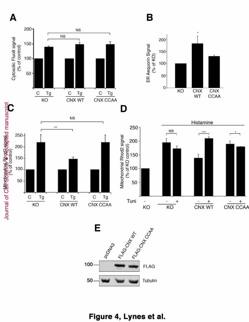
Figure 4: Palmitoylated calnexin regulates ER calcium signaling. A. Measurement of
cytosolic calcium following thapsigargin-mediated ER calcium release. Calnexin
knockout (ko) MEFs and ko cells transfected with FLAG-tagged wild type or CCAA
calnexin were loaded with Fluo8. Cells were then treated with 1.5 μM thapsigargin and 25
probe fluorescence was recorded before and after thapsigargin treatment by flow
cytometry (Statistics: P=0.8419 for calnexin wild type, P=0.8816 for calnexin CCAA). B.
Measurement of ER calcium content. Calnexin knockout (ko) MEFs and ko cells
transfected with FLAG-tagged wild type or CCAA calnexin were co-transfected with a
plasmid encoding ER-targeted aequorin. Luminescence was plotted from three 30
independent experiments following the protocol outlined in Materials and Methods.
Jour
nal o
f Cel
l Sci
ence
Acc
epte
d m
anus
crip
t
P=0.08 C. Measurement of mitochondrial calcium following thapsigargin-mediated ER
calcium release. Calnexin knockout (ko) MEFs and ko cells transfected with FLAG-
tagged wild type or CCAA calnexin were loaded with Rhod2. Cells were then treated
with 1.5 μM thapsigargin and probe fluorescence was recorded before and after
thapsigargin treatment by flow cytometry (Statistics: P=0.02 for calnexin wild type, 5
P=0.8583 for calnexin CCAA). D. Measurement of mitochondrial calcium following
histamine-mediated ER calcium release. Calnexin knockout (ko) MEFs and ko cells
transfected with FLAG-tagged wild type or CCAA calnexin were loaded with Rhod2.
Cells were then treated with 50 μM histamine and probe fluorescence was recorded
before and after histamine treatment by flow cytometry (Statistics: P=0.2581 for calnexin 10
knockout, P<0.001 for calnexin wild type, P=0.088 for calnexin CCAA). E. Calnexin
expression levels of representative cells from flow cytometry experiments. Calnexin
knockout (ko) MEFs and ko cells transfected with FLAG-tagged wild type or CCAA
calnexin were lysed and processed for Western blot using the FLAG antibody.
15
Figure 5: ER stress and palmitoylation affect calnexin’s interaction with ERp57. A.
Optiprep and membrane fractionation following ER stress. Homogenized HeLa cell
lysates were separated via Optiprep following a 4h treatment with 10 μM Tunicamycin
(Tuni) into 6 fractions. Membrane fractions were analyzed as indicated by SDS-PAGE
and Western Blot for ERp57, PDI and calnexin. Homogenized HeLa cell lysates were 20
separated into heavy and light membranes following a 4h treatment with 10 μM
Tunicamycin (Tuni). Membrane fractions were analyzed as indicated by SDS-PAGE and
Western Blot for ERp57, PDI and calnexin. The graph shows the ERp57, PDI and
calnexin amounts in the heavy membrane fraction from 3 independent experiments
(Statistics: P=0.056 for ERp57, P=0.088 for PDI, P=0.013 for calnexin). B. Calnexin-25
ERp57 co-immunoprecipitation following ER stress. HeLa cells were treated for 4 h with
either 2 mM Dithiothreitol (DTT) or 10 μM Tunicamycin (Tuni). DSP-crosslinked
lysates (5% inputs) and calnexin immunoprecipitates were analyzed for calnexin and co-
immunoprecipitating ERp57. C. Calnexin/calnexin palmitoylation mutant (CCAA)-
ERp57 co-immunoprecipitation. HeLa cells were transfected with FLAG-tagged wild 30
type or CCAA calnexin. DSP-crosslinked lysates (5% inputs) and FLAG
Jour
nal o
f Cel
l Sci
ence
Acc
epte
d m
anus
crip
t

immunoprecipitates were analyzed for FLAG-tagged calnexin and co-
immunoprecipitating ERp57. The graph shows results from 3 independent experiments
(Statistics: P=0.003). D. LDLR surface biotinylation. Calnexin knockout (ko) MEFs and
ko cells transfected with FLAG-tagged wild type or CCAA calnexin were lysed and
processed for Western blot using the LDLR antibody. In parallel, the same set of cells 5
was processed for surface biotinylation as described in Materials & Methods and probed
for biotinylated surface LDLR. The bar graph shows the means of three independent
experiments (P=0.07). Cells were also processed for LDLR surface binding, followed by
permeabilization and detection of intracellular transfected FLAG-tagged calnexin as
shown at the bottom. 10
Figure 6. Involvement of non-palmitoylated calnexin in glycoprotein quality control.
A. Increased interaction of an ERAD substrate with CNX CCAA as compared to CNX
WT. HEK-293 cells were cotransfected with a vector encoding for an ERAD substrate
glycoprotein, H2a and FLAG-tagged CNX wt or CNX CCAA. The cells were lysed in 15
2% sodium cholate (Methods) and 10% of the lysates were run on 10% SDS-PAGE and
immunoblotted with anti-H2a antibody (bottom panel). The rest of the lysates were
immunoprecipitated with mouse anti-FLAG and goat anti-mouse IgG-agarose, subjected
to 10% SDS-PAGE and immunoblotted with anti-H2a (top panel) or with anti-FLAG
(middle panel). Relative amounts of H2a co-immunoprecipitated with anti-FLAG were 20
plotted relative to the total amounts. B. Slower dissociation of H2a from non-
palmitoylated CNX. HEK 293 cells were transfected as in (A). Two days post-
transfection the cells were pulse-labeled for 20 min with [35S]-cys and chased for the
indicated times in the presence of the proteasome inhibitor MG-132. After the pulse (0 h
chase) or the chase periods, the cells were lysed, H2a was immunoprecipitated from 15% 25
of the cell lysates (right panels) and the remainders were immunoprecipitated with anti-
FLAG antibody followed by elution and re-immunoprecipitation with anti-H2a, as
described in Methods (left panels). Control antibody was used instead of anti-FLAG in a
control sample (Cont.). All immunoprecipitates were separated in 12% SDS-PAGE
followed by phosphorimaging. For the pulse samples, two bands can be seen for H2a 30
precursor molecules, the lower ones corresponding to underglycosylated species (one of
Jour
nal o
f Cel
l Sci
ence
Acc
epte
d m
anus
crip
t
the glycosylation sites unoccupied). The fully glycosylated species shifts progressively to
a faster migration because of the trimming of mannose residues on its N-glycans. The
graph shows percent of H2a coimmunoprecipitated with CNX WT and CNX CCAA
normalized to total amounts (IP anti-H2a), after chase relative to the pulse, from
phosphorimager quantitations of the gels, average of three independent experiments ± 5
SE. C-F. Increased ERAD substrate colocalization at the juxtanuclear ERQC, with CNX
CCAA as compared to CNX WT upon proteasomal inhibition. Plasmids encoding for
H2a linked to monomeric RFP (H2a-RFP) and FLAG-tagged CNX wt (C, D) or CNX
CCAA (E, F) were cotransfected in NIH 3T3 cells. One day after transfection cells were
incubated for 3h in the absence (C, E) or presence of 25 μM Lac (D, F), fixed, 10
permeabilized and incubated with mouse anti-FLAG and Cy2-conjugated goat-anti-
mouse IgG. The samples were analyzed in an LSM confocal microscope. Representative
confocal optical slices are shown. Bar=10μm. Manders coefficients: 0.77 for CNX wild
type, 0.88 for CNX CCAA (from 21 cells).
15
Figure 7: Model for the calnexin shuttling between calcium signaling and quality
control. Palmitoylated calnexin interacts with SERCA on the MAM to regulate ER
calcium signaling and to a lesser extent on the rER with the translocon. Non-
palmitoylated calnexin interacts with ERp57 on the rER to mediate protein folding and
quality control. Additionally, non-palmitoylated calnexin can interact with ERAD 20
substrates on the ERQC. Palmitoylation redirects chaperoning calnexin back to its role in
calcium signaling.
Supplemental Figure 1: Full gels for Figure 1B. Representations of the control gels
seen in Figure 1B for calnexin, FACL4, and PDI. Molecular weight markers are shown 25
on the left and arrows indicate the bands of interest.
Supplemental Figure 2: Additional analysis of calnexin palmitoylation and
phosphorylation. A. Optiprep fractionation following thapsigargin treatment.
Homogenized HeLa cell lysates were separated via Optiprep following a 4h treatment 30
with 1.5 μM Thapsigargin into 6 fractions. Membrane fractions were analyzed as
Jour
nal o
f Cel
l Sci
ence
Acc
epte
d m
anus
crip
t

indicated by SDS-PAGE and Western Blot for PDI (pan-ER), SERCA2b and FACL4
(MAM), as well as calnexin. B. Calnexin palmitoylation is reduced during DTT stress.
HeLa cells were incubated for 4h with 5 mM DTT and then processed for click chemistry
as described (Lynes et al., 2012). C. Neither calnexin palmitoylation nor the
phosphorylation status of serines 554 and 564 influence serine 583 phosphorylation. 5
Calnexin knockout (ko) MEFs and ko cells transfected with FLAG-tagged wild type
CCAA, M3 (S554, 564�A), or M4 (S554, 564�D) calnexin were lysed and processed
for Western blot using the phospho-serine 583 antibody.
Supplemental Figure 3: Influence of calnexin presence on mitochondria calcium 10
import, mitochondria membrane potential and ER ability to trigger the unfolded
protein response. A. Measurement of cytosolic calcium following thapsigargin-mediated
ER calcium release. Calnexin knockout (ko) MEFs and ko cells transfected with FLAG-
tagged wild type or CCAA calnexin were loaded with FURA-2. Cells were then treated
with 1.5 μM thapsigargin and probe fluorescence was recorded before and after 15
thapsigargin treatment by flow cytometry. The increases of fluorescence for the three
conditions were not statistically different from each other. B. Plasmid-based
measurement of mitochondrial calcium content following thapsigargin. Calnexin
wildtype and knockout (ko) MEFs were transfected with a plasmid encoding
mitochondria-targeted R-GECO-1. The increase in relative fluorescence units was 20
assayed from three independent experiments following the protocol outlined in Materials
and Methods. A representative curve for wild type and knockout cells is shown on the
right. C. Measurement of mitochondrial membrane potential. Calnexin knockout (ko)
MEFs and ko cells transfected with FLAG-tagged wild type or CCAA calnexin were
loaded with TMRM and probe fluorescence was recorded by flow cytometry (Statistics: 25
P=0.001 for calnexin wild type, P=0.4135 for calnexin CCAA). D. Xbp-1 splicing
measured by reverse transcriptase PCR. Calnexin knockout (ko) MEFs and ko cells
transfected with FLAG-tagged wild type or CCAA calnexin were treated with 1.5 μM
thapsigargin for 16h, followed by analysis of the Xbp-1 mRNA. PCR products were
separated on a 7.5% acrylamide gel for Xbp-1 and a 1% acrylamide gel for GAPDH. 30
Jour
nal o
f Cel
l Sci
ence
Acc
epte
d m
anus
crip
t
Supplemental Figure 4: 2BP promotes the calnexin chaperone activity. A. Calnexin-
ERp57 co-immunoprecipitation following 2BP treatment. HeLa cells were treated for 4 h
with 100 μM 2BP. DSP-crosslinked lysates (5% inputs) and calnexin immunoprecipitates
were analyzed for calnexin and co-immunoprecipitating ERp57. P=0.07. B. LDLR
surface biotinylation following 2BP treatment. HeLa cells were lysed and processed for 5
Western blot using the LDLR antibody. In parallel, the same set of cells was processed
for surface biotinylation and probed for biotinylated surface LDLR.
10
References
Alvarez, J. and Montero, M. (2002). Measuring [Ca2+] in the endoplasmic reticulum with aequorin. Cell Calcium 32, 251-60. Arnaudeau, S., Kelley, W. L., Walsh, J. V., Jr. and Demaurex, N. (2001). 15 Mitochondria recycle Ca(2+) to the endoplasmic reticulum and prevent the depletion of neighboring endoplasmic reticulum regions. J Biol Chem 276, 29430-9. Avezov, E., Frenkel, Z., Ehrlich, M., Herscovics, A. and Lederkremer, G. Z. (2008). Endoplasmic reticulum (ER) mannosidase I is compartmentalized and required for N-glycan trimming to Man5-6GlcNAc2 in glycoprotein ER-associated degradation. 20 Mol Biol Cell 19, 216-25. Baekkeskov, S. and Kanaani, J. (2009). Palmitoylation cycles and regulation of protein function (Review). Mol Membr Biol 26, 42-54. Baldwin, A. C., Green, C. D., Olson, L. K., Moxley, M. A. and Corbett, J. A. (2012). A role for aberrant protein palmitoylation in FFA-induced ER stress and beta-cell 25 death. Am J Physiol Endocrinol Metab 302, E1390-8. Bravo, R., Vicencio, J. M., Parra, V., Troncoso, R., Munoz, J. P., Bui, M., Quiroga, C., Rodriguez, A. E., Verdejo, H. E., Ferreira, J. et al. (2011). Increased ER-mitochondrial coupling promotes mitochondrial respiration and bioenergetics during early phases of ER stress. J Cell Sci 124, 2143-52. 30 Cardenas, C., Miller, R. A., Smith, I., Bui, T., Molgo, J., Muller, M., Vais, H., Cheung, K. H., Yang, J., Parker, I. et al. (2010). Essential regulation of cell bioenergetics by constitutive InsP3 receptor Ca2+ transfer to mitochondria. Cell 142, 270-83. Chevet, E., Wong, H. N., Gerber, D., Cochet, C., Fazel, A., Cameron, P. H., 35 Gushue, J. N., Thomas, D. Y. and Bergeron, J. J. (1999). Phosphorylation by CK2 and MAPK enhances calnexin association with ribosomes. EMBO J 18, 3655-66. Coe, H., Bedard, K., Groenendyk, J., Jung, J. and Michalak, M. (2008). Endoplasmic reticulum stress in the absence of calnexin. Cell Stress Chaperones 13, 497-507. 40
Jour
nal o
f Cel
l Sci
ence
Acc
epte
d m
anus
crip
t

Coe, H. and Michalak, M. (2010). ERp57, a multifunctional endoplasmic reticulum resident oxidoreductase. Int J Biochem Cell Biol 42, 796-9. Csordas, G., Renken, C., Varnai, P., Walter, L., Weaver, D., Buttle, K. F., Balla, T., Mannella, C. A. and Hajnoczky, G. (2006). Structural and functional features and significance of the physical linkage between ER and mitochondria. J Cell Biol 174, 5 915-21. de Brito, O. M. and Scorrano, L. (2008). Mitofusin 2 tethers endoplasmic reticulum to mitochondria. Nature 456, 605-10. Delom, F., Fessart, D. and Chevet, E. (2007). Regulation of calnexin sub-cellular localization modulates endoplasmic reticulum stress-induced apoptosis in MCF-7 10 cells. Apoptosis 12, 293-305. Dowal, L., Yang, W., Freeman, M. R., Steen, H. and Flaumenhaft, R. (2011). Proteomic analysis of palmitoylated platelet proteins. Blood. Ferrera, D., Panigada, M., Porcellini, S. and Grassi, F. (2008). Recombinase-deficient T cell development by selective accumulation of CD3 into lipid rafts. Eur J 15 Immunol 38, 1148-56. Frenkel, Z., Shenkman, M., Kondratyev, M. and Lederkremer, G. Z. (2004). Separate roles and different routing of calnexin and ERp57 in endoplasmic reticulum quality control revealed by interactions with asialoglycoprotein receptor chains. Mol Biol Cell 15, 2133-42. 20 Gilady, S. Y., Bui, M., Lynes, E. M., Benson, M. D., Watts, R., Vance, J. E. and Simmen, T. (2010). Ero1alpha requires oxidizing and normoxic conditions to localize to the mitochondria-associated membrane (MAM). Cell Stress Chaperones 15, 619-29. Glancy, B. and Balaban, R. S. (2012). Role of mitochondrial Ca2+ in the 25 regulation of cellular energetics. Biochemistry 51, 2959-73. Hayashi, T. and Su, T. P. (2007). Sigma-1 receptor chaperones at the ER-mitochondrion interface regulate Ca(2+) signaling and cell survival. Cell 131, 596-610. Higo, T., Hattori, M., Nakamura, T., Natsume, T., Michikawa, T. and Mikoshiba, K. (2005). Subtype-Specific and ER Lumenal Environment-Dependent 30 Regulation of Inositol 1,4,5-Trisphosphate Receptor Type 1 by ERp44. Cell 120, 85-98. Jessop, C. E., Chakravarthi, S., Garbi, N., Hammerling, G. J., Lovell, S. and Bulleid, N. J. (2007). ERp57 is essential for efficient folding of glycoproteins sharing common structural domains. EMBO J 26, 28-40. Kamhi-Nesher, S., Shenkman, M., Tolchinsky, S., Fromm, S. V., Ehrlich, R. 35 and Lederkremer, G. Z. (2001). A novel quality control compartment derived from the endoplasmic reticulum. Mol Biol Cell 12, 1711-23. Kang, R., Wan, J., Arstikaitis, P., Takahashi, H., Huang, K., Bailey, A. O., Thompson, J. X., Roth, A. F., Drisdel, R. C., Mastro, R. et al. (2008). Neural palmitoyl-proteomics reveals dynamic synaptic palmitoylation. Nature 456, 904-9. 40 Kondratyev, M., Avezov, E., Shenkman, M., Groisman, B. and Lederkremer, G. Z. (2007). PERK-dependent compartmentalization of ERAD and unfolded protein response machineries during ER stress. Exp Cell Res 313, 3395-407. Kraus, A., Groenendyk, J., Bedard, K., Baldwin, T. A., Krause, K. H., Dubois-Dauphin, M., Dyck, J., Rosenbaum, E. E., Korngut, L., Colley, N. J. et al. 45 (2010). Calnexin deficiency leads to dysmyelination. J Biol Chem 285, 18928-38.
Jour
nal o
f Cel
l Sci
ence
Acc
epte
d m
anus
crip
t

Lakkaraju, A. K., Abrami, L., Lemmin, T., Blaskovic, S., Kunz, B., Kihara, A., Dal Peraro, M. and van der Goot, F. G. (2012). Palmitoylated calnexin is a key component of the ribosome-translocon complex. EMBO J 31, 1823-35. Lederkremer, G. Z. (2009). Glycoprotein folding, quality control and ER-associated degradation. Curr Opin Struct Biol 19, 515-23. 5 Leitman, J., Ron, E., Ogen-Shtern, N. and Lederkremer, G. Z. (2012). Compartmentalization of Endoplasmic Reticulum Quality Control and ER-Associated Degradation Factors. DNA Cell Biol. Li, G., Mongillo, M., Chin, K. T., Harding, H., Ron, D., Marks, A. R. and Tabas, I. (2009). Role of ERO1-alpha-mediated stimulation of inositol 1,4,5-triphosphate 10 receptor activity in endoplasmic reticulum stress-induced apoptosis. J Cell Biol 186, 783-92. Lynes, E. M., Bui, M., Yap, M. C., Benson, M. D., Schneider, B., Ellgaard, L., Berthiaume, L. G. and Simmen, T. (2012). Palmitoylated TMX and calnexin target to the mitochondria-associated membrane. EMBO J 31, 457-70. 15 Myhill, N., Lynes, E. M., Nanji, J. A., Blagoveshchenskaya, A. D., Fei, H., Carmine Simmen, K., Cooper, T. J., Thomas, G. and Simmen, T. (2008). The Subcellular Distribution of Calnexin Is Mediated by PACS-2. Mol Biol Cell 19, 2777-88. Oliver, J. D., van der Wal, F. J., Bulleid, N. J. and High, S. (1997). Interaction of the thiol-dependent reductase ERp57 with nascent glycoproteins. Science 275, 86-8. 20 Raturi, A. and Simmen, T. (2012). Where the endoplasmic reticulum and the mitochondrion tie the knot: The mitochondria-associated membrane (MAM). Biochim Biophys Acta. Rizzuto, R., Marchi, S., Bonora, M., Aguiari, P., Bononi, A., De Stefani, D., Giorgi, C., Leo, S., Rimessi, A., Siviero, R. et al. (2009). Ca(2+) transfer from the ER 25 to mitochondria: when, how and why. Biochim Biophys Acta 1787, 1342-51. Roderick, H. L., Lechleiter, J. D. and Camacho, P. (2000). Cytosolic phosphorylation of calnexin controls intracellular Ca(2+) oscillations via an interaction with SERCA2b. J Cell Biol 149, 1235-48. Ruddock, L. W. and Molinari, M. (2006). N-glycan processing in ER quality 30 control. J Cell Sci 119, 4373-80. Rusch, M., Zimmermann, T. J., Burger, M., Dekker, F. J., Gormer, K., Triola, G., Brockmeyer, A., Janning, P., Bottcher, T., Sieber, S. A. et al. (2011). Identification of acyl protein thioesterases 1 and 2 as the cellular targets of the Ras-signaling modulators palmostatin B and M. Angew Chem Int Ed Engl 50, 9838-42. 35 Rutkevich, L. A. and Williams, D. B. (2011). Participation of lectin chaperones and thiol oxidoreductases in protein folding within the endoplasmic reticulum. Curr Opin Cell Biol 23, 157-66. Shenkman, M., Ayalon, M. and Lederkremer, G. Z. (1997). Endoplasmic reticulum quality control of asialoglycoprotein receptor H2a involves a determinant for 40 retention and not retrieval. Proc Natl Acad Sci U S A 94, 11363-8. Simmen, T., Lynes, E. M., Gesson, K. and Thomas, G. (2010). Oxidative protein folding in the endoplasmic reticulum: tight links to the mitochondria-associated membrane (MAM). Biochim Biophys Acta 1798, 1465-73.
Jour
nal o
f Cel
l Sci
ence
Acc
epte
d m
anus
crip
t

Tolchinsky, S., Yuk, M. H., Ayalon, M., Lodish, H. F. and Lederkremer, G. Z. (1996). Membrane-bound versus secreted forms of human asialoglycoprotein receptor subunits. Role of a juxtamembrane pentapeptide. J Biol Chem 271, 14496-503. Wang, S. and Kaufman, R. J. (2012). The impact of the unfolded protein response on human disease. J Cell Biol 197, 857-67. 5 Wieckowski, M. R., Giorgi, C., Lebiedzinska, M., Duszynski, J. and Pinton, P. (2009). Isolation of mitochondria-associated membranes and mitochondria from animal tissues and cells. Nat Protoc 4, 1582-90. Zapun, A., Darby, N. J., Tessier, D. C., Michalak, M., Bergeron, J. J. and Thomas, D. Y. (1998). Enhanced catalysis of ribonuclease B folding by the interaction of 10 calnexin or calreticulin with ERp57. J Biol Chem 273, 6009-12. Zhao, Y., Araki, S., Wu, J., Teramoto, T., Chang, Y. F., Nakano, M., Abdelfattah, A. S., Fujiwara, M., Ishihara, T., Nagai, T. et al. (2011). An expanded palette of genetically encoded Ca(2)(+) indicators. Science 333, 1888-91. Zhivotovsky, B. and Orrenius, S. (2011). Calcium and cell death mechanisms: a 15 perspective from the cell death community. Cell Calcium 50, 211-21. Zuppini, A., Groenendyk, J., Cormack, L. A., Shore, G., Opas, M., Bleackley, R. C. and Michalak, M. (2002). Calnexin deficiency and endoplasmic reticulum stress-induced apoptosis. Biochemistry 41, 2850-2858. 20
Jour
nal o
f Cel
l Sci
ence
Acc
epte
d m
anus
crip
t

Jour
nal o
f Cel
l Sci
ence
Acc
epte
d m
anus
crip
t

Jour
nal o
f Cel
l Sci
ence
Acc
epte
d m
anus
crip
t

Jour
nal o
f Cel
l Sci
ence
Acc
epte
d m
anus
crip
t
Jour
nal o
f Cel
l Sci
ence
Acc
epte
d m
anus
crip
t

Jour
nal o
f Cel
l Sci
ence
Acc
epte
d m
anus
crip
t

Jour
nal o
f Cel
l Sci
ence
Acc
epte
d m
anus
crip
t

Jour
nal o
f Cel
l Sci
ence
Acc
epte
d m
anus
crip
t