Phosphonated Carbocyclic 2‘-Oxa-3‘-azanucleosides as New Antiretroviral Agents
Upload
independentCategory
view
0download
0
O
C
a
b
c
a
ARRAA
K45ANGSG
1
5ikaeOVOndOl
Sf
0d
Toxicology 290 (2011) 59– 68
Contents lists available at SciVerse ScienceDirect
Toxicology
jou rn al hom epage: www.elsev ier .com/ locate / tox ico l
xazolone (OXA) is a respiratory allergen in Brown Norway rats
. Frieke Kupera,∗, Marijana Radonjica, Jos van Trielb, Rob Stieruma, Rianne J. de Groota,1, Josje H.E. Artsc
TNO, Quality and Safety, Zeist, The NetherlandsTNO Triskelion, BV, Zeist, The NetherlandsAkzo Nobel NV, Arnhem, The Netherlands
r t i c l e i n f o
rticle history:eceived 17 June 2011eceived in revised form 16 August 2011ccepted 22 August 2011vailable online 26 August 2011
eywords:-Ethoxymethylene-2-phenyl-2-oxazoline--onellergic laryngitiseutrophils
a b s t r a c t
Oxazolone (OXA) is a potent contact allergen in man, and it is used as a model Th1-allergen to test(Q)SAR’s and screening assays for allergenic potential of chemicals. However, it elevates serum IgE levelsand Thelper2 cytokines at relatively low doses in test animals, suggesting that it has also respiratoryallergenic potential. The lack of human data on respiratory allergenic potential of OXA may be due tolack of significant inhalation exposure. Here, female Brown Norway rats (BN) were sensitized by two orfive dermal applications of OXA at the same total dose of 3.75 mg. Controls received vehicle. All animalswere challenged by inhalation to 45 mg/m3 OXA on day 21 and necropsy was performed on day 22. Allsensitized animals had increased serum IgE. OXA challenge decreased breathing frequency, and inducedapnoeic breathing in the sensitized animals – a hallmark of respiratory allergy in our model. An exudative,granulocytic inflammation was observed primarily in the larynx of the sensitized and challenged rats.
ene expressionELPZMB
Microarray analysis of lung tissue, sampled 24 h after challenge, revealed upregulation of several genesand activation of Gene Ontology (GO) pathways, which resembled more closely those found previously inlung tissue of rats sensitized and challenged by the respiratory allergen trimellitic anhydride than by thecontact allergen dinitrochlorobenzene. The results indicate that the contact allergen OXA can also be arespiratory allergen, provided that it is inhaled. Its use as a model contact sensitizer must be reconsidered.
© 2011 Elsevier Ireland Ltd. All rights reserved.
. Introduction
Oxazolone (OXA), 4-ethoxymethylene-2-phenyl-2-oxazoline--one (CAS reg. no. 15646-46-5) is a heterocyclic compound and
s a chemical intermediate in the production of drugs. It is a well-nown, strong contact sensitizer in man, and is frequently used as
model contact allergen in in vivo and in vitro screening assays toxamine the sensitizing potential of chemicals (Roggen et al., 2008).XA has an EC3 of 0.01% in the mouse dermal LLNA (ECETOC, 2003;an Och et al., 2002). Reports on respiratory allergenic properties ofXA in humans are absent, which may indicate that OXA is indeedo respiratory allergen or that significant exposures by inhalation
o not take place. In contrast to acid anhydrides and diisocyanates,XA is not a bulk chemical and it is mostly presented as relativelyarge flakes.
∗ Corresponding author at: TNO, Quality and Safety, Research Group Quality andafety, PO Box 360, 3700 AJ, The Netherlands. Tel.: +31 0888661736;ax: +31 0888644986.
E-mail address: [email protected] (C.F. Kuper).1 Presently: Radboud Medical University, Nijmegen, The Netherlands.
300-483X/$ – see front matter © 2011 Elsevier Ireland Ltd. All rights reserved.oi:10.1016/j.tox.2011.08.017
There are no validated tests to identify the respiratory allergenicpotential of chemicals, but the IgE test and cytokine fingerprint-ing test have been used regularly (rat: Arts and Kuper, 2007;mouse: Dearman et al., 1992; Kimber et al., 1996). OXA was neg-ative in a standard IgE test in mice with two topical applications(Dearman and Kimber, 1992), but induced pronounced increasesin serum IgE, especially but not exclusively after 5 and 15 timesapplication (mouse: Webb et al., 1998; rat: Mommers et al., 2006).Two skin applications followed by a single intranasal challengeincreased also serum IgE levels (Vanoirbeek et al., 2003a,b). OXAinduced the synthesis of both IFN-gamma (Th1-related) and Il-4 (Th2-related) cytokines in the lymph nodes draining the skinsite of application (Thomson et al., 1993). Interestingly, the OXA-induced ulcerative colitis mouse model is considered to be aTh2-mediated process (Waldner and Neurath, 2009; Heller et al.,2002).
Although an increased total serum IgE level upon skin applica-tion (IgE test) has been proposed as being a predictive parameter of
airway hypersensitivity caused by low molecular weight chemicals,it is unknown if total serum IgE translates directly to potential riskof inducing respiratory allergy (Farraj et al., 2007, 2004; Selgradeet al., 2006). Therefore, it is questionable if OXA is a respiratory
60 C.F. Kuper et al. / Toxicology 290 (2011) 59– 68
Table 1Experimental scheme.
Group designationa Sensitizationb Day 21: challengec Day 22: necropsy
Veh2/OXAUnsensitized 2x/challenged
Vehicle 2x 45 mg/m3 OXA - Lung functiond
- Total IgE in serum- Liver, kidneys, left lung weights- BAL of right lung lobes- Formalin-fixation,paraffin-embedding of nasal passages,larynx, trachea (all animals)- Left lungs:1. Formalin-fixation,paraffin-embedding (all groups;n = 3/group)2. Immunohisto-chemistry onsnap-frozen tissue (veh5/OXA andOXA5/OXA5; n = 3/group)3. Transcriptomics on snap-frozentissue (veh2/OXA, OXA2/OXA, OXA2/−and OXA5/−; n = 3/group)
Veh5/OXAUnsensitized 5x/challenged
Vehicle 5x 45 mg/m3 OXA
OXA2/OXASensitized 2x/challenged (allergic group)
Day 0: 300 �l on flanks1% (w/v) OXADay 7: 150 �l on ears0.5% (w/v) OXA(total dose 3.75 mg)
45 mg/m3 OXA
OXA5/OXA Sensitized 5x/challenged (allergicgroup)
Days 0, 4, 11, 145 × 150 �l 0.5% (w/v) OXA,alternating on flanks and ear(total dose 3.75 mg)
45 mg/m3 OXA
OXA2/−Sensitized 2x/unchallenged
Day 0: 300 �l on flanks(1%, w/v, OXA)Day 7: 150 �l on ears(0.5% (w/v) OXA(total dose 3.75 mg)
–
OXA5/−Sensitized 5x/unchallenged
Days 0, 4, 7, 11, 145 × 150 �l 0.5% (w/v) OXA,alternating on flanks and ear(total dose 3.75 mg)
–
a Six female BN rats per group.
afi
ems2eac(p
ipewwD
2
2
Ctlat
2
o(mDegert
b Vehicle acetone–olive oil (AOO) 4:1 (v/v).c OXA dissolved in acetone, concentration based on RF test (1–50 mg/m3).d Lung function parameters were determined before, during and after challenge.
llergen, based on increases in total serum IgE (and Th2 cytokinengerprinting) alone.
Challenge tests in the skin and the respiratory tract are used toxamine if an allergic response is really provoked in sensitized ani-als and if so, of what type and severity it is. Using a protocol that
uccessfully identified respiratory allergens (rat: Arts and Kuper,007; Pauluhn et al., 2002; Zhang et al., 2004; mouse: Vanoirbeekt al., 2003a,b: Satoh et al., 1995) the respiratory allergen trimelliticnhydride (TMA) and the contact allergen/skin allergen dinitro-hlorobenzene (DNCB) resulted in distinctly divergent responsesArts et al., 1998; Kuper et al., 2008a,b). The divergence was alsoresent in gene expression patterns in the lungs.
OXA was expected to behave like the respiratory allergen TMAn an inhalation challenge test as mentioned above, because of itsotential to elevate IgE levels in serum. Breathing parameters, genexpression in the lungs and histopathology of the respiratory tractere examined and compared with the results found previouslyith the respiratory allergen TMA and the contact (skin) allergenNCB.
. Materials and methods
.1. Animals and maintenance
Female, 7–8-week-old, inbred Brown Norway (BN) rats were purchased fromharles River Deutschland GmbH (Sulzfeld, Germany). The animals were acclima-ized for 7 days before the start of the study. They were kept under conventionalaboratory conditions and received the Institute’s grain-based open-formula dietnd unfluoridated tap water ad libitum. All animal procedures were approved byhe TNO Committee of Animal Welfare.
.2. Treatment schedules and groups
The study was conducted with 6 groups of rats (Table 1). Blood was collectedne day before the start of the study. Female BN rats received 150 �l of oxazoloneOXA; purity 90%; Sigma–Aldrich Chemie GmbH Steinheim, Germany) in a 4:1 (v/v)
ixture of acetone (Merck; Darmstadt, Germany) and refined olive oil (AOO) (Sigmaiagnostics, Inc. St. Louis, USA) as the vehicle on each flank (approximately 12 cm2
ach). Flanks had been shaved with an electrical razor at least 2–3 days earlier. Theroups, that were sensitized twice, received 75 �l of OXA on the dorsum of bothars, seven days after the first sensitization. Groups that were sensitized five timeseceived OXA alternating on the dorsum and the flanks (see Table 1). The sensitiza-ion concentrations were based on a preliminary study with OXA in which increased
IgE levels were observed especially after 5 and 15 times application, but two timeswas effective also in most of the animals, all at the same total dose of 3.75 mg(Mommers et al., 2006). On day 20 or 21, basal breathing parameters (frequency,tidal volume and pattern) were assessed, followed by OXA inhalation challenge andassessment of the same breathing parameters during and immediately post chal-lenge. Animals were challenged by inhalation of target concentration of 50 mg/m3 ofOXA for 15 min. Twenty-four hours later, breathing parameters were assessed again,whereafter necropsy was performed. The challenge concentration of OXA was basedon a range-finding study with four reserve rats that were exposed to 1 mg/m3 on thefirst day, 5 mg/m3 on the second day, 15 mg/m3 on the third day and 50 mg/m3 onthe fourth day. Only the concentration of 50 mg/m3 turned out to be slightly irritat-ing (slight decreased breathing frequency) and was therefore chosen as the targetconcentration.
2.3. Atmosphere generation and analysis
During inhalation challenge, an all glass nebulizer, Institute’s design, wasused to generate the test atmospheres from freshly prepared solutions of OXAin acetone. The acetone concentration was kept between 3000 and 5000 ppm(∼7–12 g/m3), which levels are far below the level inducing sensory irritation(Alarie, 1973). Atmospheric concentrations of OXA were determined gravimetri-cally by filter sampling and those of acetone by calculations based on the nominalconcentration, assuming the usual 100% generation efficiency for this vapour.The particle size distribution of OXA in the test atmosphere was determinedusing a 10-stage cascade impactor (Andersen, Atlanta, GA). Due to the small sam-pling air flow rate and the large total volume required for analytical and particlesize determinations, samples were not collected during the challenge itself butimmediately prior to or after challenge. The mean concentration of OXA turnedout to be 45 mg/m3; the mass median aerodynamic diameter (MMAD) of theaerosolized OXA particles was 0.8 �m with a geometric standard deviation (gsd) of2.7 and 3.7.
2.4. Inhalation challenge and measurements of breathing parameters
Rats were individually restrained in Battelle tubes and each tube was placed intoone of four whole body plethysmographs connected to the central exposure unit.Using this experimental set up, two OXA-sensitized (OXA2/OXA or OXA5/OXA) andtwo vehicle-treated (veh2/OXA or veh5/OXA) rats at a time were first exposed tofresh air for at least 25 min (pre-challenge period) and then to the OXA atmospherefor exactly 15 min (challenge period), followed by a recovery (post challenge) periodof 30 min as described previously (Arts et al., 1998; Kuper et al., 2008a,b).
As a surrogate for changes in lung function, breathing pattern (Arts and Kuper,
2007) was monitored by means of recording the pressure signal in the plethysmo-graphs. Frequency and tidal volume were measured and respiratory flow (minuteventilation) was calculated from the breathing pattern. Before challenge, the respi-ration was monitored approximately 20 s each min starting 6 min prior to the actualchallenge. During challenge respiration was monitored approximately 20 s during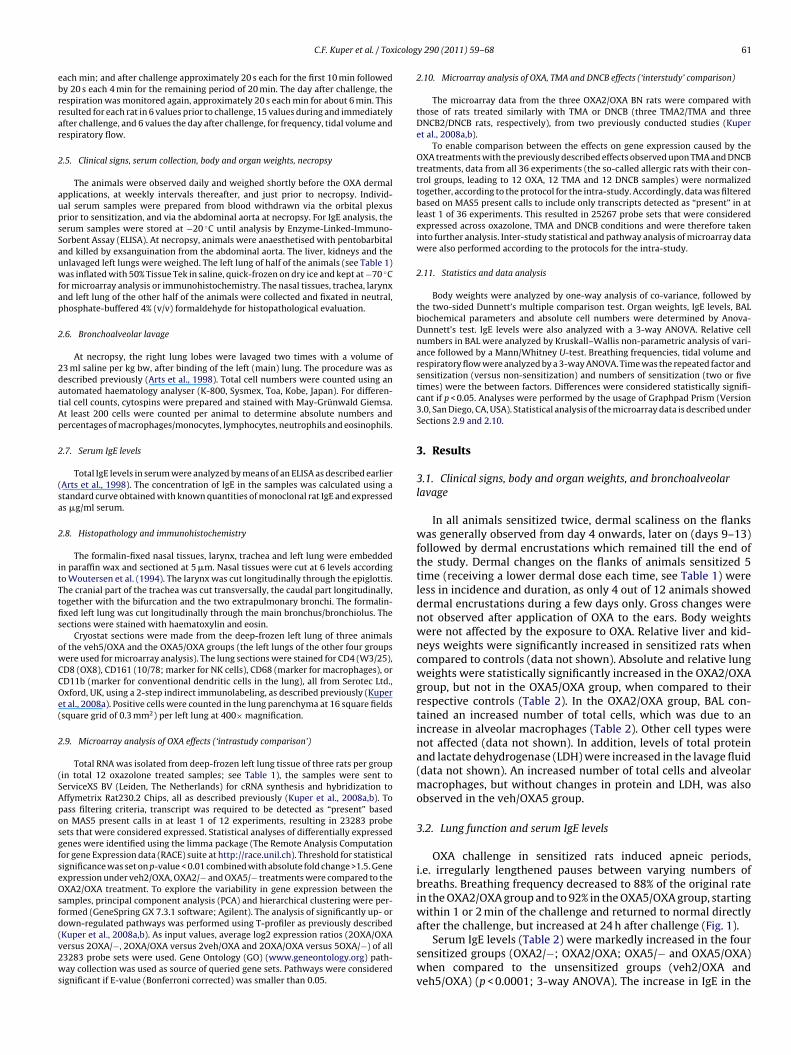
icolog
ebrrar
2
aupsSauwfap
2
2datAp
2
(sa
2
itTtfis
owCCOe(
2
(SAposgfseOsfd(v2ws
C.F. Kuper et al. / Tox
ach min; and after challenge approximately 20 s each for the first 10 min followedy 20 s each 4 min for the remaining period of 20 min. The day after challenge, theespiration was monitored again, approximately 20 s each min for about 6 min. Thisesulted for each rat in 6 values prior to challenge, 15 values during and immediatelyfter challenge, and 6 values the day after challenge, for frequency, tidal volume andespiratory flow.
.5. Clinical signs, serum collection, body and organ weights, necropsy
The animals were observed daily and weighed shortly before the OXA dermalpplications, at weekly intervals thereafter, and just prior to necropsy. Individ-al serum samples were prepared from blood withdrawn via the orbital plexusrior to sensitization, and via the abdominal aorta at necropsy. For IgE analysis, theerum samples were stored at −20 ◦C until analysis by Enzyme-Linked-Immuno-orbent Assay (ELISA). At necropsy, animals were anaesthetised with pentobarbitalnd killed by exsanguination from the abdominal aorta. The liver, kidneys and thenlavaged left lungs were weighed. The left lung of half of the animals (see Table 1)as inflated with 50% Tissue Tek in saline, quick-frozen on dry ice and kept at −70 ◦C
or microarray analysis or immunohistochemistry. The nasal tissues, trachea, larynxnd left lung of the other half of the animals were collected and fixated in neutral,hosphate-buffered 4% (v/v) formaldehyde for histopathological evaluation.
.6. Bronchoalveolar lavage
At necropsy, the right lung lobes were lavaged two times with a volume of3 ml saline per kg bw, after binding of the left (main) lung. The procedure was asescribed previously (Arts et al., 1998). Total cell numbers were counted using anutomated haematology analyser (K-800, Sysmex, Toa, Kobe, Japan). For differen-ial cell counts, cytospins were prepared and stained with May-Grünwald Giemsa.t least 200 cells were counted per animal to determine absolute numbers andercentages of macrophages/monocytes, lymphocytes, neutrophils and eosinophils.
.7. Serum IgE levels
Total IgE levels in serum were analyzed by means of an ELISA as described earlierArts et al., 1998). The concentration of IgE in the samples was calculated using atandard curve obtained with known quantities of monoclonal rat IgE and expresseds �g/ml serum.
.8. Histopathology and immunohistochemistry
The formalin-fixed nasal tissues, larynx, trachea and left lung were embeddedn paraffin wax and sectioned at 5 �m. Nasal tissues were cut at 6 levels accordingo Woutersen et al. (1994). The larynx was cut longitudinally through the epiglottis.he cranial part of the trachea was cut transversally, the caudal part longitudinally,ogether with the bifurcation and the two extrapulmonary bronchi. The formalin-xed left lung was cut longitudinally through the main bronchus/bronchiolus. Theections were stained with haematoxylin and eosin.
Cryostat sections were made from the deep-frozen left lung of three animalsf the veh5/OXA and the OXA5/OXA groups (the left lungs of the other four groupsere used for microarray analysis). The lung sections were stained for CD4 (W3/25),D8 (OX8), CD161 (10/78; marker for NK cells), CD68 (marker for macrophages), orD11b (marker for conventional dendritic cells in the lung), all from Serotec Ltd.,xford, UK, using a 2-step indirect immunolabeling, as described previously (Kupert al., 2008a). Positive cells were counted in the lung parenchyma at 16 square fieldssquare grid of 0.3 mm2) per left lung at 400× magnification.
.9. Microarray analysis of OXA effects (‘intrastudy comparison’)
Total RNA was isolated from deep-frozen left lung tissue of three rats per groupin total 12 oxazolone treated samples; see Table 1), the samples were sent toerviceXS BV (Leiden, The Netherlands) for cRNA synthesis and hybridization toffymetrix Rat230.2 Chips, all as described previously (Kuper et al., 2008a,b). Toass filtering criteria, transcript was required to be detected as “present” basedn MAS5 present calls in at least 1 of 12 experiments, resulting in 23283 probeets that were considered expressed. Statistical analyses of differentially expressedenes were identified using the limma package (The Remote Analysis Computationor gene Expression data (RACE) suite at http://race.unil.ch). Threshold for statisticalignificance was set on p-value < 0.01 combined with absolute fold change >1.5. Genexpression under veh2/OXA, OXA2/− and OXA5/− treatments were compared to theXA2/OXA treatment. To explore the variability in gene expression between the
amples, principal component analysis (PCA) and hierarchical clustering were per-ormed (GeneSpring GX 7.3.1 software; Agilent). The analysis of significantly up- orown-regulated pathways was performed using T-profiler as previously described
Kuper et al., 2008a,b). As input values, average log2 expression ratios (2OXA/OXAersus 2OXA/−, 2OXA/OXA versus 2veh/OXA and 2OXA/OXA versus 5OXA/−) of all3283 probe sets were used. Gene Ontology (GO) (www.geneontology.org) path-ay collection was used as source of queried gene sets. Pathways were consideredignificant if E-value (Bonferroni corrected) was smaller than 0.05.
y 290 (2011) 59– 68 61
2.10. Microarray analysis of OXA, TMA and DNCB effects (‘interstudy’ comparison)
The microarray data from the three OXA2/OXA BN rats were compared withthose of rats treated similarly with TMA or DNCB (three TMA2/TMA and threeDNCB2/DNCB rats, respectively), from two previously conducted studies (Kuperet al., 2008a,b).
To enable comparison between the effects on gene expression caused by theOXA treatments with the previously described effects observed upon TMA and DNCBtreatments, data from all 36 experiments (the so-called allergic rats with their con-trol groups, leading to 12 OXA, 12 TMA and 12 DNCB samples) were normalizedtogether, according to the protocol for the intra-study. Accordingly, data was filteredbased on MAS5 present calls to include only transcripts detected as “present” in atleast 1 of 36 experiments. This resulted in 25267 probe sets that were consideredexpressed across oxazolone, TMA and DNCB conditions and were therefore takeninto further analysis. Inter-study statistical and pathway analysis of microarray datawere also performed according to the protocols for the intra-study.
2.11. Statistics and data analysis
Body weights were analyzed by one-way analysis of co-variance, followed bythe two-sided Dunnett’s multiple comparison test. Organ weights, IgE levels, BALbiochemical parameters and absolute cell numbers were determined by Anova-Dunnett’s test. IgE levels were also analyzed with a 3-way ANOVA. Relative cellnumbers in BAL were analyzed by Kruskall–Wallis non-parametric analysis of vari-ance followed by a Mann/Whitney U-test. Breathing frequencies, tidal volume andrespiratory flow were analyzed by a 3-way ANOVA. Time was the repeated factor andsensitization (versus non-sensitization) and numbers of sensitization (two or fivetimes) were the between factors. Differences were considered statistically signifi-cant if p < 0.05. Analyses were performed by the usage of Graphpad Prism (Version3.0, San Diego, CA, USA). Statistical analysis of the microarray data is described underSections 2.9 and 2.10.
3. Results
3.1. Clinical signs, body and organ weights, and bronchoalveolarlavage
In all animals sensitized twice, dermal scaliness on the flankswas generally observed from day 4 onwards, later on (days 9–13)followed by dermal encrustations which remained till the end ofthe study. Dermal changes on the flanks of animals sensitized 5time (receiving a lower dermal dose each time, see Table 1) wereless in incidence and duration, as only 4 out of 12 animals showeddermal encrustations during a few days only. Gross changes werenot observed after application of OXA to the ears. Body weightswere not affected by the exposure to OXA. Relative liver and kid-neys weights were significantly increased in sensitized rats whencompared to controls (data not shown). Absolute and relative lungweights were statistically significantly increased in the OXA2/OXAgroup, but not in the OXA5/OXA group, when compared to theirrespective controls (Table 2). In the OXA2/OXA group, BAL con-tained an increased number of total cells, which was due to anincrease in alveolar macrophages (Table 2). Other cell types werenot affected (data not shown). In addition, levels of total proteinand lactate dehydrogenase (LDH) were increased in the lavage fluid(data not shown). An increased number of total cells and alveolarmacrophages, but without changes in protein and LDH, was alsoobserved in the veh/OXA5 group.
3.2. Lung function and serum IgE levels
OXA challenge in sensitized rats induced apneic periods,i.e. irregularly lengthened pauses between varying numbers ofbreaths. Breathing frequency decreased to 88% of the original ratein the OXA2/OXA group and to 92% in the OXA5/OXA group, startingwithin 1 or 2 min of the challenge and returned to normal directlyafter the challenge, but increased at 24 h after challenge (Fig. 1).
Serum IgE levels (Table 2) were markedly increased in the foursensitized groups (OXA2/−; OXA2/OXA; OXA5/− and OXA5/OXA)when compared to the unsensitized groups (veh2/OXA andveh5/OXA) (p < 0.0001; 3-way ANOVA). The increase in IgE in the

62 C.F. Kuper et al. / Toxicology 290 (2011) 59– 68
Fig. 1. Mean breathing frequency, tidal volume and respiratory flow ± SD of BN rats before, during challenge, and within 30 min and 24 h after the challenge. The maineffect was a change in time in sensitized animals. Frequency: time p < 0.001; time × sensitization p < 0.01. Tidal volume: time p < 0.001; time × sensitization p < 0.001;time × sensitization × number of sensitizations p < 0.001; Respiratory flow: time p < 0.001; time × sensitization p < 0.001; time × sensitization × number of sensitizationsp < 0.01, 3-way ANOVA analysis.
Table 2Left lung weights, bronchoalveolar lavage parameters, and serum IgE levels in BN rats exposed to OXA.
Groupa Lung weightrelative to bodyweight
BAL total cells(×106)
BAL macrophages(×106)
BAL total protein(mg/L)
BAL LDH(U/L)
Pre treatmenttotal serum IgE(�g/ml)
After treatmenttotal serum IgE(�g/ml)
Veh2/OXA 2.50 ± 0.07 0.88 ± 0.09 0.82 ± 0.09 174 ± 21 68 ± 5 0.40 ± 0.14 0.72 ± 0.19Veh5/OXA 2.76 ± 0.08 1.37 ± 0.12** 1.30 ± 0.11* 234 ± 16 114 ± 24 0.36 ± 0.12 0.79 ± 0.21OXA2/OXA 2.98 ± 0.07** 1.40 ± 0.11** 1.32 ± 0.11** 294 ± 21** 143 ± 25* 0.45 ± 0.13 1.78 ± 0.35**
OXA5/OXA 2.78 ± 0.11 1.08 ± 0.07 0.99 ± 0.06 258 ± 44 102 ± 12 0.30 ± 0.18 2.13 ± 0.86**
OXA2/− 2.73 ± 0.04 1.02 ± 0.09 0.96 ± 0.08 167 ± 15 65 ± 15 0.55 ± 0.32 2.01 ± 0.43**
OXA5/− 2.82 ± 0.08 1.12 ± 0.14 1.02 ± 0.12 211 ± 20 92 ± 5 0.32 ± 0.05 2.60 ± 0.69**
a Six female rats per group.* p < 0.05; Anova + Dunnetts test’s (two-sided).
** p < 0.01; Anova + Dunnetts test’s (two-sided).

C.F. Kuper et al. / Toxicology 290 (2011) 59– 68 63
Table 3Histopathological changes and their incidences in BN rats treated dermally with vehicle or OXA, left unchallenged or followed by an inhalation challenge with OXA.
Veh2/OXA Veh5/OXA OXA2/OXA OXA5/OXA OXA2/− OXA5/−Nasal tissues, anterior levels (6)a (6) (6) (6) (6) (6)Inflammatory cell infiltrates
Very slight, focal 1 – 1 3 1 –Very slight to slight, multifocal – – 5 3 – –
LarynxUlceration
Slight/moderate 3 6 – – – –Severe – – 6 6 – –
Mucopurulent exudateSlight 4 5 – – – –Severe – – 6 6 – –
Inflammatory cellsVery slight – 1 – – 1 1Slight 2 2 – – – 1
gscs
3
hur
F(ae
Moderate/severe – –
a The number of animals examined is given in brackets.
roups sensitized twice was more than twofold, and in the groupsensitized five times it was about threefold (statistically signifi-ant difference between the groups sensitized twice and the groupsensitized five times, p < 0.05; 3-way ANOVA).
.3. Histopathology and immunohistochemistry
The unchallenged (OXA2/− and OXA5/−) animals did not exhibitistopathological changes in the respiratory tract, except for a gran-lomatous inflammation in the lungs, which is common in naive BNats of this age (Germann et al., 1998).
ig. 2. Histopathology of the larynx of BN rats, 24 h after challenge. The larynx is sectioned lOXA2/−) control animal; (B) overview of unsensitized, OXA-challenged (veh2/OXA) animt the base of the epiglottis; (C) overview of twice-sensitized, OXA-challenged (OXA2/Opiglottis. (D) Detail of the extensive inflammation in the twice-sensitized, OXA-challeng
6 6 – –
The most prominent effect of the challenge was observed inthe larynx of the sensitized (OXA2/OXA and OXA5/OXA) animals:a dense, ulcerative inflammation with exudation at the ventralside of the epiglottis and the vocal cords. Numerous granulocytes,mainly neutrophils and a few eosinophils, lymphocytes, mono-cytes and macrophages were observed in the mucosa of almostthe entire ventral side of the epiglottis (Table 3 and Fig. 2A–D). In
addition, hemorrhages were present in almost all animals. Simi-lar epithelial effects were observed in unsensitized/challenged rats(veh2/OXA), but the number of inflammatory cells was markedlyless and hemorrhages were absent. In the nasal passages, the chal-ongitudinally through the epiglottis. (A) Overview of twice-sensitized, unchallengedal with epithelial ulceration and minimal neutrophilic cell involvement, primarilyXA) animal with an extensive inflammation along the entire ventral side of the
ed (OXA2/OXA) animal.

64 C.F. Kuper et al. / Toxicology 290 (2011) 59– 68
Table 4Differentially expressed genes in lung tissue of allergic (twice sensitized/challenged; OXA2/OXA) BN rats, 24 h after challenge.
Description Gene name Accession no. Gene group/pathway (GO, KEGG) Mean log 2ratio > 1.00a,b
Cytokine (chemokine) 10 Cxcl10/IP-10 U22520 Positive regulation of leukocyte chemotaxis; chemotaxis;inflammatory response; immune response; signaltransduction. Cytokine–cytokine receptor interaction;Chemokine signaling pathway; Toll-like receptor signalingpathway; RIG-I-like receptor signaling pathway;
5.14***
Cytokine (chemokine) 11 Cxcl11/I-TAC BF281987 Chemotaxis; inflammatory response; chemokine activity;immune response; signal transduction; G-protein coupledreceptor protein signaling pathway. Toll-like receptorsignaling; cytokine–cytokine receptor interaction; Chemokinesignaling pathway
5.30***
Cytokine (chemokine) 9 Cxcl9 Al044222 Chemotaxis; defense response; inflammatory response;immune response; cellular defense response.Cytokine–cytokine receptor interaction; chemokine signalingpathway; Toll-like receptor signaling pathway
3.96***
Cxcl9 Al170387 3.80***
Ubiquitin D Ubd NM 053299 Antimicrobial humoral response; protein modificationprocess; proteolysis
3.78***
Cytokine (chemokine) 7 – monocytechemotactic protein 3
Ccl7/MCP3/SCYA7 BF419899 Cellular calcium ion homeostasis; chemotaxis; chemokineactivity; immune response; inflammatory response; signaltransduction. Cytokine–cytokine receptor interaction;Chemokine signaling pathway; NOD-like receptor signalingpathway
2.69*
Agrin G1p2 predicted BE096523 Plasma membrane organization; signal transduction;muscarinic acetylcholine receptor signaling; synapseassembly; neuromuscular junction development.ECM-receptor interaction
2.14***
Similar to Ac2-233 Igtp predicted Bl300770 – −2.09**
Homologous to some humaninterferon-inducible proteins
MGC94037 AW531805 – 1.89***
Similar to interferon-inducible GTPase MGC108823 Al408440 – 1.80***
RGD1309362 AA955213 – 1.71***
Proteasome subunit, beta type 9 Psmb9 Al599350 Immune response; antigen processing and presentation;anaphase-promoting complex-dependent proteasomalubiquitin-dependent protein catabolic process; interspeciesinteraction between organisms; negative regulation ofubiquitin-protein ligase activity during mitotic cell cycle.
1.68***
Granzyme B Gzmb M34097 Apoptosis; extracellular space; proteolysis; cleavage of lamin;serine-type (endo)peptidase activity; natural killer cellmediated cytotoxicity; type I diabetes mellitus; autoimmunethyroid disease; allograft rejection
1.60***
Interferon regulatory factor 7 (predicted) Irf7 predicted BF411036 Negative regulation of transcription from RNA polymerase IIpromoter; regulation of transcription, DNA-dependent;response to virus; immunoglobulin mediated immuneresponse; interspecies interaction between organisms.Toll-like receptor signaling pathway; RIG-I-like receptorsignaling pathway; Cytosolic DNA-sensing pathway
1.44***
Granzyme B Gzmb Al029386 See above under ‘Granzyme B M34097’ 1.36***
Signal transducer and activator oftranscription 1
Stat1 AW434718 Regulation of transcription, DNA-dependent; transcriptionfrom RNA polymerase II promoter; induction of apoptosis;activation of caspase activity; signal transduction. Chemokinesignaling pathway; Toll-like receptor signaling pathway;Jak-STAT signaling pathway; pathways in cancer
1.27***
Ubiquitin-activating enzyme E1-like(predicted)
Ube1l predicted Al177396 – 1.21***
Granzyme M Gzmm L05175 Cytolysis; proteolysis; extracellular space 1.20**
Signal transducer and activator oftranscription 1
Stat1 BM386875 See above under ‘Stat1 AW434718’ 1.20**
Interferon regulatory factor 1 Irf1 NM 012591 Immune response; negative regulation of progression throughcell cycle; positive regulation of IL-12 biosynthesis; regulationof transcription, DNA-dependent; transcription from RNApolymerase II promoter; CD8-positive, alpha-beta T celldifferentiation
1.19***
Guanylate nucleotide binding protein 2 Gbp2 NM 133624 Immune response 1.19***
Similar to hypothetical protein MGC29390 RGD1310490 BF284106 – 1.18***
Natural killer cell group 7 sequence Nkg7 NM 133540 Extracellular space; integral to membrane 1.17**
Uridine phosphorylase 1 (predicted) Upp1 predicted Bl292558 Nucleobase, nucleoside, nucleotide and nucleic acid metabolicprocess; pyrimidine nucleotide metabolic process; nucleosidemetabolic process; nucleotide catabolic process; uridinemetabolic process. Pyrimidine metabolism; Drug metabolism -other enzymes; Metabolic pathways
1.12**
Similar to 5830458K16Rik protein(predicted)
RGD1306974 predicted AA819788 – 1.12***
Putative ISG12(b) protein Isg12(b)/Ifl27l2 AA819034 Membrane; integral to membrane 1.05***
a OXA2/OXA versus veh2/OXA.b Significances ***p < 0.001; **p < 0.01 or *p < 0.05 OXA2/OXA versus either of all intrastudy control groups (veh2/OXA, OXA2/− and OXA5/−).

C.F. Kuper et al. / Toxicology 290 (2011) 59– 68 65
Table 5T-values of differentially expressed groups of genes (Gene Ontology) in left lung tissue of BN rats, sensitized twice and subsequently challenged with OXA (OXA2/OXA).
GO ontology pathway OXA2/OXA versus OXA2/− OXA2/OXA versus OXA5/− OXA2/OXA versus veh2/OXA
Chemokine activity 14.41*** 8.36*** 14.91***
Chemokine receptor binding 14.16*** 7.88*** 14.59***
Immune response 10.57*** 4.75* 17.00***
Cytokine activity 8.09*** 5.12* 8.02***
Defense response 7.18*** 0.05 10.67***
Inflammatoryresponse 6.18*** −0.46 8.89***
Extracellular matrix −5.28** −8.52*** −3.80Blood vessel development −5.71*** −5.48*** 3.44Anatomical structure development −6.49*** −8.42*** −1.08Muscle contraction −3.91 −9.63*** −6.53***
lgtmctcpnsCs
Cn(i8
3
3
eagTa
3
a‘ngsct
3
(rtTT
Fig. 3. The number of genes differentially expressed by OXA, TMA and DNCB in thetwice-sensitized and challenged (compound 2x/compound) groups compared to theunsensitized, but challenged (veh2x/compound) groups, and the number of genes
** E-value < 0.001.*** E-value < 0.0001.
enge in the sensitized animals caused a very slight to slight mixedranulocytic–monocytic inflammatory cell infiltrate, whereas inhe unsensitized animals (veh2/OXA and veh5/OXA) the inflam-
ation was only very slight (Table 3). Moreover, the inflammatoryell infiltrates in the unsensitized animals were only focal, whereashe infiltrates in about half of the sensitized animals were multifo-al. The infiltrates in the sensitized animals did not show a distinctredilection site, other than being located in the anterior part of theasal passages (Levels 1–3): they were observed ventrally on theeptum, the lateral wall and/or the maxilla- and nasal turbinates.hallenge-related effects were not observed in the H&E-stainedections of the trachea and lungs.
The number of immunohistochemically stained CD4+, CD8+,D161+, and CD68+ cells in the alveolar septa of the left lungs didot differ between the sensitized (OXA5/OXA) and unsensitizedveh5/OXA) animals. The number of CD11b+ cells was slightly lowern the sensitized animals (OXA5/OXA: 70.7 ± 2.1 and veh5/OXA:1.7 ± 2.9 per total surface area of 1.4 mm2).
.4. Effect of OXA on lung tissue transcriptome
.4.1. Intrastudy comparison: single genes analysisComparison between the groups resulted in total of 1150 differ-
ntially expressed genes in at least one of the veh2/OXA, OXA2/−nd OXA5/− treatments compared to the sensitized/challengedroup (OXA2/OXA). The genes affected most are listed in Table 4.he mean fold changes were highest for the cytokines Cxcl9, Cxcl10nd Cxcl11 and for ubiquitin.
.4.2. Gene group analysisThe GO Ontology groups/pathways of genes ‘Chemokine
ctivity’, ‘Chemokine receptor binding’, ‘Immune response’ andCytokine activity’ were upregulated considerably, and highly sig-ificantly in the OXA2/OXA group, compared to the other threeroups (Table 5). The pathways ‘extracellular matrix’, ‘blood ves-el development’, ‘anatomical structure development’ and ‘muscleontraction’ were downregulated significantly compared to two ofhe groups (Table 5).
.4.3. Interstudy comparisonSingle gene comparison between OXA, TMA and DNCB
groups sensitized/challenged versus unsensitized/challenged rats)
evealed an overlap of 112 genes between OXA and TMA, and 6 ofhese genes were shared by OXA, DNCB and TMA (Fig. 3 and Table 6).he overall gene expression profile of OXA resembled more that ofMA than of DNCB.shared by OXA, TMA and/or DNCB.
4. Discussion
The breathing pattern, IgE levels and inflammation induced byOXA in this study indicated that OXA behaved comparable to therespiratory allergen TMA in previous studies, using the same pro-tocol (Arts et al., 1998, 2003, 2004; Kuper et al., 2008a). Animalssensitized five times had higher IgE levels and tended to have amore decreased breathing frequency during challenge than ani-mals sensitized twice, but the type and degree of inflammation wasunaffected by the number of sensitizations.
The allergic inflammation induced by the inhalation challengeof OXA in the OXA-sensitized animals was localized primarily in thelarynx. Allergic laryngitis was observed also in TMA-allergic BrownNorway rats and DNCB-allergic Wistar rats. Allergic laryngitis isnot diagnosed often in man, perhaps because particle impactionpatterns in the respiratory tract differ from those of rats. However,
it is more plausible that the reported low incidence in man is due tounderdiagnosis of allergic laryngitis (Hellings and Fokkens, 2006;Krouse and Altman, 2010; Leino et al., 1998; Randhawa et al., 2010).
66 C.F. Kuper et al. / Toxicology 290 (2011) 59– 68
Table 6Overlap of genes in lung tissue of allergic (twice sensitized/challenged) BN rats: comparison OXA, TMA and/or DNCB.a
Descriptionb Gene name Accession no. Description
The most significant differentially expressed genes shared by OXA and TMA, but not DNCBc
Cxcl9 AI170387Cxcl10 U22520Cxcl11 BF281987
Amiloride binding protein 1 Abp1 NM 022935 Amine metabolic process; oxidation reduction, arginine and prolinemetabolism; histidine metabolism; tryptophan metabolism
G1p2-predicted BE096523Gbp2 NM 133624Irf1 NM 012591Irf7-predicted BF411036Isg12(b)/Ifl27l2 AA819034
Interferon inducible protein 1(predicted)
If1-predicted AI407339 Autophagy; inflammatory response; innate immune response
Oligoadenylate synthetase-like 1 Oasl1-predicted BF419319Proteasome (prosome, macropain)
subunit, beta type, 8Psmb8 NM 080767 Immune response; antigen processing and presentation, anaphase-promoting
complex-dependent proteasomal ubiquitin-dependent protein catabolicprocess; interspecies interaction between organisms; negative regulation ofubiquitin-protein ligase activity during mitotic cell cycle
Psmb9 AI599350– Psmb10-predicted BG373505 –Member RAS oncogene family Rab20-predicted BI293985 Intracellular protein transport; nucleocytoplasmic transport; signal
transduction; small GTPase mediated signal transduction; protein transportSignal transducer and activator of
transcription 2Stat2-predicted BG666368 Regulation of transcription, DNA-dependent; regulation of transcription from
RNA polymerase II promoter; signal transduction; JAK-STAT cascade;chemokine signaling pathway; Jak-STAT signaling pathway
Selectin P Selp BI296054 Positive regulation of leukocyte migration; regulation of cellular extravasation;inflammatory response; cell adhesion; heterophilic cell–cell adhesion.
Transporter 1 Tap1 X57523 Protein complex assembly; transport; intracellular protein transport; defenseresponse; immune response. ABC transporters; antigen processing andpresentation; primary immunodeficiency
Transporter 2 Tap2 NM 032056 positive regulation of T cell mediated cytotoxicity; response to molecule ofbacterial origin; antigen processing and presentation of exogenous proteinantigen via MHC class Ib, TAP-dependent; protein complex assembly; positiveregulation of antigen processing and presentation of peptide antigen via MHCclass I. ABC transporters; Antigen processing and presentation; Primaryimmunodeficiency
TNF receptor superfamily member;B-cell surface antigen CD40
Tnfrsf5-predicted/CD40 AW433947 Immune response-regulating cell surface receptor signaling pathway; proteincomplex assembly; inflammatory response; platelet activation; positiveregulation of B cell proliferation. Cytokine–cytokine receptor interaction; Celladhesion molecules; Toll-like receptor signaling pathway; Intestinal immunenetwork for IgA production; asthma
Upp1-predicted BI292558Superoxide dismutase 3, extracellular Sod3 (down-regulated) NM 012880 Response to hypoxia; superoxide metabolic process; response to copper ion;
oxidation reductionEngulfment adaptor PTB Gulp1-predicted
(down-regulated)BM390519 Lipid transport; phagocytosis, engulfment; apoptosis; metabolic process;
oxidation reductionFlavin containing monooxygenase 2
(non-functional); pulmonaryflavin-containing monooxygenase
Fmo2 (down-regulated) BM389350 Organic acid metabolic process; NADP metabolic process; oxygen and reactiveoxygen species metabolic process; xenobiotic metabolic process; toxinmetabolic process; Drug metabolism – cytochrome P450
The six differentially expressed genes shared by OXA, TMA and DNCBUbd NM 053299Gzmb AI029386Ccl7/MCP3 BF419899
Runt-related transcription factor Runx3 AI709792 Regulation of transcription, DNA-dependent; transcription from RNApolymerase II promoter; induction of apoptosis; axon guidance; cellproliferation
– RGD1309596 predicted BG377140 –– NA AW525366 –
a For all three allergens, the same protocol was used. The genes were measured in lung tissue, 24 h after challenge with the allergen.b Only for the genes not included in Table 4.c Of the 106 genes expressed by both OXA and TMA, 24 genes were selected on the basis of (i) being significantly expressed in the twice sensitized/challenged groups
( e dir
answesp
OXA- and TMA-allergic rats) compared to all other groups, (ii) expressed in the sam
The histopathology of the laryngitis pointed to an intriguingspect of respiratory allergy. The challenge with OXA induced largeumbers of neutrophils in the larynx of the sensitized animals, butome neutrophils were observed in the unsensitized animals as
ell (Fig. 2), suggesting that the sensitization made the animalsxceptionally sensitive for the irritating properties of OXA. Thisensitization-aggravated irritation is in line with the concept pro-osed by Noble (2009) that there may be memory for danger signals
ection (up- or down-regulated for both OXA and TMA), and (iii) at p < 0.01.
as well as for specific antigens. The concept of Noble (2009) couldexplain the presence of the large numbers of neutrophils in theOXA-induced allergic laryngitis as well as the neutrophils in mod-erate to severe asthma and the aspecific hyperreactivity (AHR) in
asthmatic individuals.The development of respiratory allergy is considered to be regu-lated predominantly by Th2-polarized immune reactions in whichIgE plays a dominant role. The OXA-induced increased total IgE
icolog
lpchehtpplpca
trgTTagenaaDe(asewedpaSlr
wrura(cnis
attamwaTpcipmio
Krouse, J.H., Altman, K.W., 2010. Rhinogenic laryngitis, cough and the unified airway.Otolaryngol. Clin. North America 43, 111–121.
C.F. Kuper et al. / Tox
evels in serum and the breathing pattern were in line with a Th2-olarized mechanism. Neutrophils in the lungs are associated withertain types of Th2-mediated asthma, but also with Th1-mediatedypersensitivity pneumonitis/allergic alveolitis (HP/AA; De Vooghtt al., 2011; Fireman, 2006; Matsuoka et al., 2010). The observedemorrhages in the OXA-induced laryngitis are more in line withhe hemorrhages found in the lungs of acute and subchronic HP/AAatients (Bogaert et al., 2009; Jancar and Crespo, 2005). This com-lex picture is in accordance with the increasing awareness that
ung allergies like asthma and HP/AA have considerable overlap inathogenesis and histopathology, depending on the severity andhronicity of the diseases and this may be the case with laryngealllergies as well.
In the present study, allergic inflammation was not observed inhe lungs of the OXA-sensitized and -challenged rats, but microar-ay analysis of lung tissue revealed strong upregulation of severalenes (Table 4). There was considerable overlap between OXA- andMA-induced differentially expression of genes (Table 6), althoughMA induced more strongly Th2-associated genes and OXA Th1-ssociated genes, namely Cxcl9, Cxcl10 and Cxcl11 (Table 4). Theseenes are regulated by STAT1 (Chen and Hershey, 2007; Fulkersont al., 2004; Mikhak et al., 2006). Interestingly, these genes wereot upregulated by the contact allergen DNCB (Table 6). This inccordance to the findings of Ku et al. (2009), who found Cxcl9nd Cxcl10 upregulation in the skin by OXA and TDI but not byNCB. Cxcl10 has been found in a murine asthma model (Fulkersont al., 2004) and in the late-phase reaction in human asthmatic lungLai et al., 2008). The Th1-related chemokines may reflect a neg-tive feedback mechanism by which Th1 counterbalances Th2 toelf-limit the allergic inflammation (Dharajiva et al., 2009; Wellst al., 2007). OXA and TMA induced upregulation of Ccl7 (Table 6),hich is consistently associated with respiratory allergy (Bloemen
t al., 2007; Fulkerson et al., 2004; Johnson et al., 2004), but soid DNCB. In contrast to TMA, OXA did not activate the knownathways for remodeling, which is considered a significant healthspect of asthma (Davies et al., 2003). However, shared genes likeELP and GZMB suggest that OXA, like TMA, interfered with vascu-ar permeability and leukocyte extravasation and maybe even lungemodeling via platelet activation (Pitchford et al., 2008).
Although the GO Ontology pathways related to chemokinesere strongly upregulated in the OXA-sensitized and -challenged
ats, no genes were identified for those chemokines that are beingsed in current cytokine profiling tests for distinguishing typicalespiratory from typical contact allergens. In addition, the genesnd pathways were dissimilar to those found by Verstraelen et al.2009a,b,c) in in vitro studies, exposing macrophages and epithelialells to chemical respiratory allergens. Microarray analyses haveot provided us yet with markers to be applied in a robust test to
dentify respiratory allergens, but they clearly add in our under-tanding of the complex issue of respiratory allergy.
In summary, the sensitizer OXA behaved like the respiratoryllergen TMA in our inhalation challenge model in rats, with respecto the induction of elevated serum IgE levels and apneas, andhe type of allergic laryngeal inflammation. However, microarraynalysis of the lung suggested that OXA acts through differentechanisms than TMA, although the overlap in genes and path-ays between OXA and TMA was larger than with the contact
llergen DNCB. It is possible that the variability in balance betweenh1- and Th2-associated genes reflects different subtypes of res-iratory allergies (Bogaert et al., 2009; Truyen et al., 2006). It isoncluded that, although the mechanisms may differ from thosenduced by the recognized respiratory allergen TMA, OXA has res-iratory allergenic potential and should therefore not be used as aodel contact allergen to develop and validate (Q)SAR models and
n vivo and in vitro screening tests to identify sensitization potentialf chemicals.
y 290 (2011) 59– 68 67
Conflict of interest
All authors declare that there is no conflict of interest.
Acknowledgements
The authors gratefully acknowledge the CEFIC-LRI Brussels,Belgium, and the Dutch Ministry of Social Affairs and Employmentfor financial support. The authors thank E. Duistermaat, L. van Oost-rum, G. Roverts and M. Schijf for expert technical assistance.
References
Alarie, Y., 1973. Sensory irritation by airborne chemicals. CRC Crit. Rev. Toxicol. 2,299–363.
Arts, J.H.E., Kuper, C.F., Spoor, S., Bloksma, N., 1998. Airway morphology and func-tion of rats following dermal sensitization and respiratory challenge with lowmolecular weight chemicals. Toxicology 152, 66–76.
Arts, J.H.E., Bloksma, N., Leusink-Muis, T., Kuper, C.F., 2003. Respiratory allergy andpulmonary irritation to trimellitic anhydride in brown Norway rats. Toxicol.Appl. Pharmacol. 187, 38–49.
Arts, J.H.E., Koning de, M.W., Bloksma, N., Kuper, C.F., 2004. Respiratory allergyto trimellitic anhydride in rats: concentration–response relationships duringelicitation. Inhal. Toxicol. 16, 259–269.
Arts, J.H.E., Kuper, C.F., 2007. Animal models to test respiratory allergy to low molec-ular weight chemicals: a guidance. Methods 41, 61–71.
Bloemen, K., Verstraelen, S., Van den Heuvel, R., Witters, H., Nelissen, I., Schoeters,G., 2007. The allergic cascade: review of the most important molecules in theasthmatic lung. Immunol. Lett. 113, 6–18.
Bogaert, P., Tournoy, K.G., Naessens, T., Grooten, J., 2009. Where asthma and hyper-sensitivity pneumonitis meet and differ. Am. J. Pathol. 174, 3–13.
Chen, W., Hershey, G.K.K., 2007. Signal transducer and activator of transcriptionsignals in allergic disease. J. Allergy Clin. Immunol. 119, 529–541.
Davies, D.E., Wicks, J., Powell, R.M., Puddicombe, S.M., Holgate, S.T., 2003. Airwayremodeling in asthma: new insights. J. Allerg. Clin. Immunol. 111, 215–226.
Dharajiva, N., Vaidya, S., Sinha, M., Luxon, B., Boldogh, I., Sur, S., 2009. Allergen chal-lenge induces Ifng dependent GTPases in the lungs as part of a Th1 transcriptomeresponse in a murine model of allergic asthma. Plos One 4, 1–8.
Dearman, R.J., Kimber, I., 1992. Divergent immune responses to respiratory and con-tact chemical allergens: antibody elicited by phthalic anhydride and oxazolone.Clin. Exp. Allergy 22, 241–250.
Dearman, R.J., Basketter, D.A., Kimber, I., 1992. Variable effects of chemical allergenson serum IgE concentration in mice. Preliminary evaluation of a novel approachto the identification of respiratory sensitizers. J. Appl. Toxicol., 317–323.
De Vooght, V., Haenen, S., Verbeken, E., Nemery, B., Hoet, P.H.M., Vanoirbeek, J.A.J.,2011. Succesful transfer of chemical-induced asthma by adoptive transfer of lowamounts of lymphocytes in a mouse model. Toxicology 279, 85–90.
ECETOC, 2003. Contact sensitisation: classification according to potency. EuropeanCentre for Ecotoxicology and Toxicology of Chemicals, Brussels, Belgium, Tech-nical Report No. 87.
Farraj, A.K., Harkema, J.R., Kaminski, N.E., 2004. Allergic rhinitis induced by intranasalsensitization and challenge with trimellitc anhydride but not with dinitrochlo-robenzene or oxazolone in A/J mice. Toxicol. Sci. 79, 315–325.
Farraj, A.K., Boykin, E., Haykal-Coates, N., Gavett, S.H., Doerfler, D., Selgrade, M., 2007.Th2 cytokines in skin draining lymph nodes and serum IgE do not predict airwayhypersensitivity to intranasal isocyanate exposure in mice. Toxicol. Sci. 100,99–108.
Fireman, P., 2006. Atlas of Allergies and Clinical Immunology. Mosby Elsevier,Philadelphia, US.
Fulkerson, P.C., Zimmerman, N., Hassman, L.M., Finkelman, F.D., Rothenberg, M.E.,2004. Pulmonary chemokine expression is coordinately regulated by STAT1,STAT6, and IFN�. J. Immunol. 173, 7565–7574.
Germann, P.-G., Haefner, D., Hanauer, G., Drommer, W., 1998. Incidence and sever-ity of granulomatous pneumonia in Brown Norway (BN) rats: breeder relatedvariations. J. Exp. Anim. Sci. 39, 22–33.
Heller, F., Fuss, I.J., Nieuwenhuis, E.E., Blumberg, R.S., Strober, W., 2002. Oxazolonecolitis, a Th2 model resembling ulcerative colitis, is mediated by Il-13 producingNK-T cells. Immunity 17, 629–638.
Hellings, J., Fokkens, W., 2006. Allergic rhinitis and its impact on otorhinolaryngol-ogy. Eur. J. Allergy Clin. Immunol. 61, 6.
Jancar, S., Sanchez Crespo, M., 2005. Immune complex-mediated tissue injury: amultistep paradigm. Trends immunol. 26, 48–55.
Johnson, V.J., Matheson, J.M., Luster, M.I., 2004. Animal models for diisocyanateasthma: answers for lingering questions. Curr. Opin. Allergy Clin. Immunol. 4,105–110.
Kimber, I., Hilton, J., Basketter, D.A., Dearman, R.J., 1996. Predictive testing for res-piratory sensitization in the mouse. Toxicol. Lett. 86, 193–198.
Ku, H.-U., Jeong, S.-H., Kang, H.-G., Pyo, H.-M., Cho, J.-H., Son, S.-W., Yun, S.M., Ryu, D.-Y., 2009. Gene expression profiles and pathways in skin inflammation inducedby three different sensitizers and an irritant. Toxicol. Lett. 190, 231–237.

6 icolog
K
K
L
L
M
M
M
N
P
P
R
R
S
S
T
8 C.F. Kuper et al. / Tox
uper, C.F., Heijne, W.H.M., Dansen, M., Verhoeckx, K.C.M., Boorsma, A., Radonjic,M., Bruijntjes, J.P., Stierum, R., Muijser, H., Arts, J.H.E., 2008a. Molecular charac-terization of trimellitic anhydride-induced respiratory allergy in Brown Norwayrats. Toxicol. Pathol. 36, 985–999.
uper, C.F., Stierum, R.H., Boorsma, A., Schijf, M.A., Prinsen, M., Bruijntjes, J.P.,Bloksma, N., Arts, J.H.E., 2008b. The contact allergen dinitrochlorobenzene(DNCB) and respiratory allergy in the Th2-prone Brown Norway rat. Toxicology246, 213–221.
ai, S.-T., Hung, C.-H., Hua, Y.-M., Hsu, S.-H., Jong, Y.-J., Suen, J.-L., 2008. T-helper1-related chemokines in the exacerbation of childhood asthma. Pediatr. Intern.50, 99–102.
eino, T., Tammilehto, L., Hytonen, M., Sala, E., Paakulainen, H., Kanerva, L., 1998.Occupational skin and respiratory diseases among hairdressers. Scand. J. Work.Environ. Health 24, 398–406.
atsuoka, H., Niimi, A., Matsumoto, H., Takemura, M., Yamaguchi, M., Jinnai, M.,Inoue, H., Ito, I., Chin, K., Mishima, M., 2010. Inflammatory subtypes in cough-variant asthma: association with maintenance doses of inhaled corticosteroids.Chest 138, 1418–1425.
ikhak, Z., Fleming, C.M., Medoff, B.D., Thomas, S.Y., Tager, A.M., Campanella, G.S.,Luster, A.D., 2006. STAT1 in peripheral tissue differentially regulated homing ofantigen-specific Th1 and Th2 cells. J. Immunol. 176, 4959–4967.
ommers, C., Arts, J., Schijf, M., Kuper, F., 2006. Exposure is the proof of the pudding:oxazolone is a potential respiratory allergen. Toxicologist (Suppl. Tox. Sci.) 90,76.
oble, A., 2009. Do we have memory of danger as well as antigen? Trends Immunol.30, 150–156.
auluhn, J., Eidmann, P., Freyberger, G., Wasinska-Kempka, G., Vohr, H.W., 2002.Respiratory hypersensitivity to trimellitic anhydride in brown Norway rats: acomparison of endpoints. J. Appl. Toxicol. 22, 89–97.
itchford, S.C., Momi, S., Baglioni, S., Casali, L., Giannini, S., Rossi, R., Page, C.P., Gresele,P., 2008. Allergen induces the migration of platelets to lung tissue in allergicasthma. Am. J. Respir. Crit. Care Med. 177, 604–612.
andhawa, P.S., Nouraei, S., Mansuri, S., Rubin, J.S., 2010. Allergic laryngitis as a causeof dysphonia: a preliminary report. Logoped. Phoniatrics Vocol. 35, 169–174.
oggen, E., Aufderheide, M., Cetin, Y., Dearman, R.J., Gibbs, S., Hermanns, I., Kimber,I., Regal, J.F., Rovida, C., Warheit, D.B., Uhlig, S., Casati, S., 2008. The developmentof novel approaches to the identification of chemical and protein respiratoryallergens. ATLA 36, 591–598.
atoh, T., Kramarik, J.A., Tollerud, D.J., Karol, M.H., 1995. A murine model for assessingthe respiratory hypersensitivity potnetial of chemical allergens. Toxicol. Lett. 78,57–66.
elgrade, M.K., Boykin, E.H., Haykal-Coates, N., Woolhiser, M.R., Wiescinski, C.,Andrews, D.L., Farraj, A.K., Doerfler, D.L., Gavett, S.H., 2006. Inconsistencies
between cytokine profiles, antibody responses, and respiratory hyperrespon-siveness following dermal exposure to isocyanates. Toxicol. Sci. 94, 108–117.homson, J.A., Troutt, A.B., Kelso, A., 1993. Contact sensitization to oxazolone:involvement of both interferon-� and interleukin-4 in oxazolone-specific Ig andT-cell responses. Immunology 78, 185–192.
y 290 (2011) 59– 68
Truyen, E., Coteur, L., Dilissen, E., Overbergh, L., Dupont, L.J., Ceuppens, J.L., Bul-lens, D.M.A., 2006. Evaluation of airway inflammation by quantitative Th1/Th2cytokine mRNA measurement in sputum of asthma patients. Thorax 61,202–208.
Van Och, F.M., Van Loveren, H., De Jong, W.H., Vandebriel, R.J., 2002. Cytokineproduction induced by low-molecular-weight chemicals as a function of thestimulation index in a modified local lymph node assay: an approach to discrim-inate contact sensitizers from respiratory sensitizers. Toxicol. Appl. Pharmacol.184, 46–56.
Vanoirbeek, J.A.J., Mandervelt, C., Cunninghal, A.R., Hoet, P.H.M., Xu, H., Van-hooren, H.M., Nemery, B., 2003a. Validity of methods to predict the respiratorysensitizing potential of chemicals: a study with a piperidinyl chlorotriazinederivative that caused an outbreak of occupational asthma. Toxicol. Sci. 76, 338–346.
Vanoirbeek, J.A.J., Tarkowski, M., Ceuppens, J.L., Verbeken, E.K., Nemery, B., Hoet,P.H.M., 2003b. Respiratory response to toluene disiocyanate depends on priorfrequency and concetration of dermal sensitization in mice. Toxicol. Sci. 80,310–321.
Verstraelen, S., Nelissen, I., Hooyberghs, J., Witters, H., Schoeters, G., Van Cauwen-berghe, P., Van den Heuvel, R., 2009a. Gene profiles of THP-1 macrophagesafter in vitro exposure to respiratory (non-)sensitizing chemicals: identifica-tion of discriminating genetic markers and pathway analysis. Toxicol. In Vitro23, 1151–1162.
Verstraelen, S., Nelissen, I., Hooyberghs, J., Witters, H., Schoeters, G., Van Cauwen-berghe, P., Van den Heuvel, R., 2009b. Gene profiles of a human alveolar epithelialcell line after in vitro exposure to respiratory (non-)sensitizing chemicals: iden-tification of discriminating genetic markers and pathway analysis. Toxicol. InVitro 23, 1151–1162.
Verstraelen, S., Nelissen, I., Hooyberghs, J., Witters, H., Schoeters, G., Van Cauwen-berghe, P., Van den Heuvel, R., 2009c. Gene profiles of a human bronchialepithelial cell line after in vitro exposure to respiratory (non-)sensitizing chem-icals: Identification of discriminating genetic markers and pathway analysis.Toxicol. In Vitro 23, 1151–1162.
Waldner, M.J., Neurath, M.F., 2009. Chemically-induced mouse models of colitis.Curr. Protocols Pharmacol. Suppl. 46, 1–15.
Webb, E.F., Tzimas, M.N., Newsholme, S.J., Griswold, D.E., 1998. Intralesionalcytokines in chronic oxazolone-induced contact sensitivity suggest roles fortumor necrosis factor � and interleukin-4. J. Invest. Dermatol. 111, 86–92.
Wells, J.W., Cowled, C.J., Giorgini, A., Kemeny, D.M., Noble, A., 2007. Regulation ofallergic airway inflammation by class I-restricted allergen presentation and CD8T-cell infiltration. J. Allergy Clin. Immunol. 119, 226–234.
Woutersen, R.A., van Garderen-Hoetmer, A., Slootweg, P.J., Feron, V.J., 1994. Upperrespiratory tract carcinogenesis in experimental animals and humans. In:
Waalkens, M.P., Ward, J.M. (Eds.), Carcinogenesis, Target Organ ToxicologySeries. Raven Press, New York, pp. 27–36.Zhang, X.D., Fedan, J.S., Lewis, D.M., Siegel, P.D., 2004. Asthmalike biphasic airwayresponses in Brown Norway rats sensitized by dermal exposure to dry trimelliticanhydride powder. J. Allergy Clin. Immunol. 113, 320–326.
Copyright © 2022 FDOKUMEN