Characterising Pavement Surface Damage Caused by Tyre Scuffing Forces
Upload
independentCategory
view
1download
0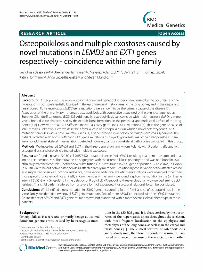
Baasanjav et al. BMC Medical Genetics 2010, 11:110http://www.biomedcentral.com/1471-2350/11/110
Open AccessR E S E A R C H A R T I C L E
Research articleOsteopoikilosis and multiple exostoses caused by novel mutations in LEMD3 and EXT1 genes respectively - coincidence within one familySevjidmaa Baasanjav†1,6, Aleksander Jamsheer†2,3, Mateusz Kolanczyk*†1,4, Denise Horn1, Tomasz Latos5, Katrin Hoffmann1,4, Anna Latos-Bielenska2,3 and Stefan Mundlos1,4
AbstractBackground: Osteopoikilosis is a rare autosomal dominant genetic disorder, characterised by the occurrence of the hyperostotic spots preferentially localized in the epiphyses and metaphyses of the long bones, and in the carpal and tarsal bones [1]. Heterozygous LEMD3 gene mutations were shown to be the primary cause of the disease [2]. Association of the primarily asymptomatic osteopokilosis with connective tissue nevi of the skin is categorized as Buschke-Ollendorff syndrome (BOS) [3]. Additionally, osteopoikilosis can coincide with melorheostosis (MRO), a more severe bone disease characterised by the ectopic bone formation on the periosteal and endosteal surface of the long bones [4-6]. However, not all MRO affected individuals carry germ-line LEMD3 mutations [7]. Thus, the genetic cause of MRO remains unknown. Here we describe a familial case of osteopoikilosis in which a novel heterozygous LEMD3 mutation coincides with a novel mutation in EXT1, a gene involved in aetiology of multiple exostosis syndrome. The patients affected with both LEMD3 and EXT1 gene mutations displayed typical features of the osteopoikilosis. There were no additional skeletal manifestations detected however, various non-skeletal pathologies coincided in this group.
Methods: We investigated LEMD3 and EXT1 in the three-generation family from Poland, with 5 patients affected with osteopoikilosis and one child affected with multiple exostoses.
Results: We found a novel c.2203C > T (p.R735X) mutation in exon 9 of LEMD3, resulting in a premature stop codon at amino acid position 735. The mutation co-segregates with the osteopoikilosis phenotype and was not found in 200 ethnically matched controls. Another new substitution G > A was found in EXT1 gene at position 1732 (cDNA) in Exon 9 (p.A578T) in three out of five osteopoikilosis affected family members. Evolutionary conservation of the affected amino acid suggested possible functional relevance, however no additional skeletal manifestations were observed other then those specific for osteopoikilosis. Finally in one member of the family we found a splice site mutation in the EXT1 gene intron 5 (IVS5-2 A > G) resulting in the deletion of 9 bp of cDNA encoding three evolutionarily conserved amino acid residues. This child patient suffered from a severe form of exostoses, thus a causal relationship can be postulated.
Conclusions: We identified a new mutation in LEMD3 gene, accounting for the familial case of osteopoikilosis. In the same family we identified two novel EXT1 gene mutations. One of them A598T co-incided with the LEMD3 mutation. Co-incidence of LEMD3 and EXT1 gene mutations was not associated with a more severe skeletal phenotype in those patients.
BackgroundOsteopoikilosis is a rare and primarily benign autosomaldominant genetic entity caused by heterozygous muta-
tions in the LEMD3 gene. It is characterised by the occur-rence of the hyperostotic spots throughout the skeleton,with most frequent localization in the epiphyses andmetaphyses of the long bones, as well as in the carpal andtarsal bones [1]. The clinical features of osteopoikilosisare relatively mild, therefore the condition is usually diag-nosed by chance or because of the association with other
* Correspondence: [email protected] Institute of Medical Genetics, Charité Berlin, Humboldt University, Augustenburger Platz 1, 13353 Berlin, Germany† Contributed equallyFull list of author information is available at the end of the article
© 2010 Baasanjav et al; licensee BioMed Central Ltd. This is an Open Access article distributed under the terms of the Creative CommonsAttribution License (http://creativecommons.org/licenses/by/2.0), which permits unrestricted use, distribution, and reproduction inany medium, provided the original work is properly cited.
Baasanjav et al. BMC Medical Genetics 2010, 11:110http://www.biomedcentral.com/1471-2350/11/110
Page 2 of 8
medical problems (fractures, joint dislocations, etc.). Inaddition to spotty bone changes, some patients affectedby osteopoikilosis develop the superficial skin lesions(elastic-type nevi) and/or subcutaneous foci of dermato-fibrosis. Such combination of clinical features is catego-rized as a separate condition named the Buschke-Ollendorff syndrome [3]. Osteopoikilosis has also beenfound in association with a more severe and detrimentalbone disease called melorheostosis. Melorheostosis man-ifests with predominantly asymmetric depositions ofdense compact bone on the periosteal and endosteal sur-face of the long bones, resembling a dripping wax of acandle. Bone deformations are often associated with theossification of the soft tissue in the joint proximity, whichcan cause compression of the adjacent nerves, and resultin pain. Heterozygous LEMD3 gene mutations weredetected in all such cases. In contrast, no germlineLEMD3 mutations were found in the isolated cases ofmelorheostosis [7,8]. Thus, genetic cause of isolatedmelorheostosis remains unknown. Melorheostosisbelongs to a group of osteogenic lesions together withanother disease called hereditary multiple exostoses(HME) [9]. Multiple exostoses (enchondromas) arecaused by heterozygous mutations in EXT1, EXT2 and/orEXT3 genes. The EXT proteins function in the proteogly-can synthesis and play tumour suppressor roles. EXT1and EXT2 have both been shown to encode a heparansulphate polymerase with both D-glucuronyl (GlcA) andN-acetyl-D-glycosaminoglycan (GlcNAC) transferaseactivities and their functions are indispensable for hepa-rin-sulphate biosynthesis [10]. The nature of the tumoursuppressor effects of the heparan sulphate biosynthesis isnot entirely clear, however the regulation of Ihh signallingwas proposed to play an important role [11]. Here wedescribe a family with 5 patients affected by osteopoikilo-sis caused by novel mutation in the LEMD3 gene. Inter-estingly, three of the patients affected with this newLEMD3 mutation additionally carry a new mutation inthe EXT1 gene. We discuss possible implications.
MethodsPatientsWe studied a two-branch family of Polish descent. Themain branch comprised three generations with five indi-viduals affected by osteopoikilosis. We also examined amore distant kindred affected by the severe deforma-tional condition of the long bones. The local ethics com-mittee approved the study and written, informed consentwas obtained from all participants or their legal guardiansfor publication of this case report, including clinical data,pedigree and X-ray images. Copies of all written consentsare available for review on request.
DNA sequencingThe LEMD3 and EXT1 genes were analyzed by bidirec-tional sequencing with primers listed in the see (Table 1).PCR amplification of LEMD3 gene was performed in a 20μl final volume, contained 1 U of Taq polymerase (FIREPol) with buffer supplemented with 1.5 mM Mg2+, 0.4mM dNTP, 8 pmol of each forward and reverse primer,and 30 ng of DNA. The exons were amplified as follow-ing: 5 min at 94°C, 35 cycles (30 sec 94°C, 30 sec 60°C, 30sec 72°C) 10 min 72°C. PCR amplifications of EXT1 genewere performed in a 20 μl final volume, contained 1 U ofTaq polymerase (Invitek) with buffer supplemented with2 mM Mg2+, 0.4 mM dNTP, 8 pmol of each forward andreverse primer, and 30 ng of DNA. PCR conditions usedwere as in case of LEMD3, with exception of exon1-2 andexon 6 amplification, where annealing temperature variedfrom 55°C to 61°C (touchdown PCR). PCR products weresequenced with the DNA Sequencing Kit BigDye™ Termi-nator v3.0 Cycle Sequencing (Applied Biosystems) on anABI 3730 automated sequencer. All exons were comparedto genomic sequence (NM_014319.3 for LEMD3 geneand NM_000127.2 for EXT1 gene) and variations werenumbered according to Ensembl ENSG00000174106 forLEMD3 and ENSG00000182197 for EXT1. Healthy con-trol subjects were screened for the identified mutations inthe LEMD3 and EXT1 genes via sequence analysis. cDNAwas synthesised using random hexamer primers asdescribed below. PCR amplification of the EXT1 cDNAfragments was done with primer pairs listed in (Table 2).The amplification conditions were like for the genomicDNA sequencing. PCR products were sequenced withthe DNA Sequencing Kit BigDye™ Terminator v3.0 CycleSequencing (Applied Biosystems) on an ABI 3730 auto-mated sequencer.
Isolation of the primary osteoblast cellsSurgically removed exostosis was placed in alphaMEMmedium supplemented with 10% FCS, Penicilin-Strepto-mycin and Glutamine and transported to laboratorywithin 24 h. Bone was cleaned of any remaining connec-tive tissue and 3 rounds of collagenase IV (2 mg/ml)digest 1× 5 min. and 2× 20 min. each at 37°C were per-formed. Cells from the first digestion were discardedwhereas second and third digests were pooled and seededin the alpha-MEM medium. Cells were cultured till con-fluent.
RNA isolation and cDNA synthesisTotal RNA was isolated with peqGOLD TriFast™ reagent(PeqLab) according to supplied protocol. cDNAs weresynthesised from 1 μg total RNAs with SuperscriptII(Invitrogen) according to manufacturer's guidelines.

Baasanjav et al. BMC Medical Genetics 2010, 11:110http://www.biomedcentral.com/1471-2350/11/110
Page 3 of 8
ResultsClinical HistoryIn the current study we identified a family with congeni-tal osteopoikilosis (Figure 1). The affected family mem-bers suffered from moderate to intermittent pain in thehands and feet, with onset of the symptoms varying from15 (Patient IV:17) to 26 years of age (Patient III:13). X-rayexamination revealed disseminated sclerotic foci in the
bones of the hands and feet, in the epiphyseal parts of thelong bones as well as pelvis and sacrum (Figure 2). Clini-cal features observed in the affected family members aresummarized in (Table 3), and involve several findings:dermatofibrosis, tetralogy of fallot (TOF), ovarian andsinus cysts, diabetes mellitus type 2, and vitiligo. Interest-ingly these various features were presented by thepatients who carried both LEMD3 and EXT1 gene muta-
Table 1: Sequences of the primers used for LEMD3 and EXT1 gene amplification and sequencing.
Exon name F Primer sequence 5'- 3' R Primer sequence 5' - 3'
LEMD3_e1-1 CTCAGGTGAGCTCCTCCC CGACTCGTCCGAGCTGAAG
LEMD3_e1-2 GCGACCTCTCCTACTTACGG GTCGTCGTCGTCCTCTTCC
LEMD3_e1-3 AGGAGAGGGACCCGGAG GGGGAGTCCACACTGAAGG
LEMD3_e1-4 AGGAGGGTGTGATCAAGTGG GCGCAAATAGTCTTTCAGGG
LEMD3_e2 TTTAGCAAAGTACATGCTGGC TTATACGACAGTTAGGGAATACTCAG
LEMD3_e3 TTCAGATTATGTGGCTTCTGTG TTCACAAATATAACACTGGACTTGG
LEMD3_e4 TGTGGTTAATGTAATGGTAGTTGTTTG GGAACAAGAGCGAAACTGTG
LEMD3_e5-6 TTGGAGTAGTGGGAAAATGC GCTGTGACTTATGTGGCAACC
LEMD3_e7-8 GAAGGTTCATTCCGTTGTGG AGTTGAGAAGGGTCACAGCTC
LEMD3_e9 CATCTAAATCTTCTTTGAACAAACTCC CAGAACGAGAGAGTTTTGCC
LEMD3_e10 CTAACCAGGGGTCTGGCTC TTTGCTTGGAATTTAATGAAAGAG
LEMD3_e11-12 TCTACCTCCTGTTAGTCAACAAGC TGGTAAAAGACATATGAGCACAAAAC
LEMD3_e13 ATTGCATGGCTCTTGGTTTG GCTGCCTCACTGCTAAATCC
EXT1_e1-1 TCTTTACAGGCGGGAAGATG TGTTCCACAAGTGGAGACTCTG
EXT_e1-2 CCAGGTTCTACACCTCGGAC CTCAGTTCCAGGCTCAAAGG
EXT1_e2 CTGGTGGCTTTCCCGAG AAGGGAAACCACACCTTCTC
EXT1_e3 AAGCTTCCTTTCCTTCTGGC CCATGACACAGGTAATTTTCTCC
EXT1_e4 TGCTAGAAGCCAAATGCTATG TGGACCAATCACACATCCC
EXT1_e5 CTCTGACTGCCACCATCTTTC AAGCAATCTTCAATGCAGGG
EXT1_e6 ATTTGCTCCAGCATGAGGC TGAATGAAAGGGAGTAGCAGG
EXT1_e7 GCTGAGATTTCCAGCTCCTC AACAGGGAGAAGATATCTAGGGC
EXT1_e8 AGATTCCTTCGGTGTTGAGG CAAGGCACGGCTAAAAGAAG
EXT1_e9 CCGGATTTTGCATTATGAATTAG ATCAGCAAAACTTAAGCGGG
EXT1_e10 GGGATTCAAAGAATGGGTATG CTGGGTGGAACAGCTAGAGG
EXT1_e11 TGCTCATTTGCCTGACTCC ACAATCTGGCTCTGCTGATG
Table 2: Sequences of the primers used for EXT1 cDNA amplification and sequencing.
PCR name F Primer sequence 5'- 3' R Primer sequence 5' - 3'
EXT1 cDNA1 GCTGCTCGCCCGCCCTGGGTG GTGGTGCAAGCCATTCCTAC
EXT1 cDNA2 CTCAGCTGGCTCTTGTCTCG CTCGGTGTAGTCAGGCCAAG
EXT1 cDNA3 CTTGTGGAACAATGGTAGG CCTATGACGGCAGCTTGGTTC
EXT1 cDNA4 GTATGATTATCGGGAAATG CTGGGCACAGTACTGGGACTTGG
EXT1 cDNA5 CTGGTCTCTCAGTCCCAGC GTCCCATCATTGTCTCCTTATAC
EXT1 cDNA6 GCCTCCAATCAAAGTGACCC CTCTGCTGATGAGTGGATCTGC
Baasanjav et al. BMC Medical Genetics 2010, 11:110http://www.biomedcentral.com/1471-2350/11/110
Page 4 of 8
tions (with exception of dermatofibrosis which was pre-sented by a patient affected with LEMD3 mutation only).
We also consulted a more distant relative of this family(V:1), who was independently referred to a clinical genet-icist at the age of 7 years. X-ray examination showed largemultiple exostoses predominantly localized in the ends ofthe long bones (Figure 2). The boy was operated at theage of 7 years for a large exostosis affecting proximal partof the right humerus. Histopathological examination ofthe removed bone was suggestive of enchondromatosis.
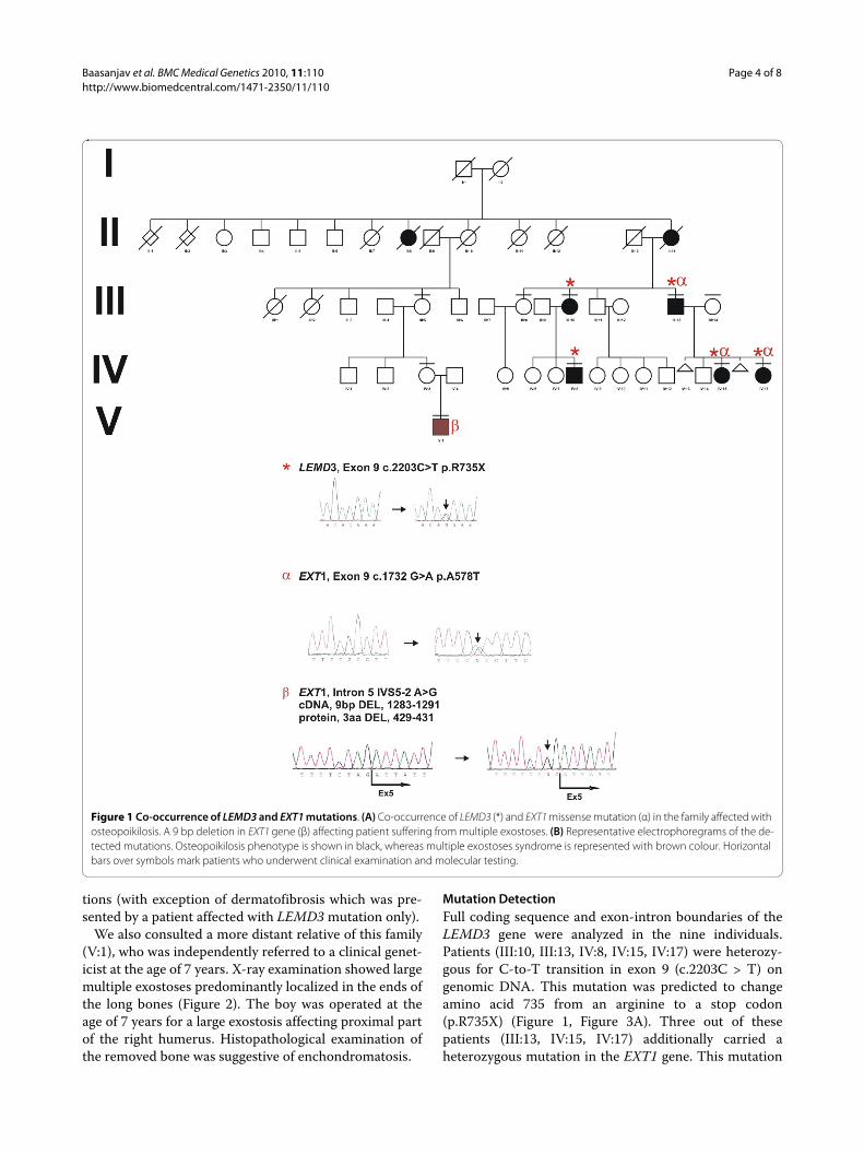
Mutation DetectionFull coding sequence and exon-intron boundaries of theLEMD3 gene were analyzed in the nine individuals.Patients (III:10, III:13, IV:8, IV:15, IV:17) were heterozy-gous for C-to-T transition in exon 9 (c.2203C > T) ongenomic DNA. This mutation was predicted to changeamino acid 735 from an arginine to a stop codon(p.R735X) (Figure 1, Figure 3A). Three out of thesepatients (III:13, IV:15, IV:17) additionally carried aheterozygous mutation in the EXT1 gene. This mutation
Figure 1 Co-occurrence of LEMD3 and EXT1 mutations. (A) Co-occurrence of LEMD3 (*) and EXT1 missense mutation (α) in the family affected with osteopoikilosis. A 9 bp deletion in EXT1 gene (β) affecting patient suffering from multiple exostoses. (B) Representative electrophoregrams of the de-tected mutations. Osteopoikilosis phenotype is shown in black, whereas multiple exostoses syndrome is represented with brown colour. Horizontal bars over symbols mark patients who underwent clinical examination and molecular testing.

Baasanjav et al. BMC Medical Genetics 2010, 11:110http://www.biomedcentral.com/1471-2350/11/110
Page 5 of 8
altered G-to-A in exon 9 (c.1732G > A) on genomic DNAand predicted to change amino acid 578 from an alanineto a threonine (p.A578T). Presence of both LEMD3 andEXT1 variants was excluded among 81 and 247 healthyPolish and German controls respectively. Bioinformaticanalysis of the EXT1 sequence with SIFT http://sift.jcvi.org and PolyPhen http://genetics.bwh.har-vard.edu/pph/ software indicated a high probability of
the mutation being deleterious for the protein function(PolyPhen - PSIC score difference for A578T: 1.688) - see(Figure 3B) for sequence conservation. However, affectedpatients did not exhibit exostoses and no additional skele-tal manifestations beyond hyper-mineralized foci weredetected. Interestingly, patients affected with bothLEMD3 and EXT1 mutations presented spectrum ofadditional non-skeletal pathologies, which included:(IV:15) TOF and ovarian cysts, (IV:17) sinus cysts, and(III:13) diabetes melitus type 2 and vitiligo (Table 3).
A different mutation in EXT1 gene was identified in thepatient (V:1), diagnosed with multiple exostoses syn-drome. The patient carried a heterozygotic splice sitemutation in intron 5 (IVS5-2 A > G), as detected in bloodlymphocytes and primary osteoblast progenitor cellsobtained from the surgically removed exostoses.Sequencing of the exostoses derived cDNA (obtainedfrom the cells isolated from the affected bone) showedthat splice site mutation resulted in the in-frame deletionof 9 bp of the exon 5 leading to a deletion of three aminoacids (pos. 429-431 - two conserved isoleucin residuesand a conserved glutamic acid residue) (Figure 3B). Func-tional relevance of the deleted amino acids was predictedbased on their evolutionary conservation.
DiscussionWe identified a family with five members affected byosteopoikilosis caused by a novel nonsense heterozygousmutation (p.R735X) localised in exon 9 of the LEMD3gene. In other branch of this family we identified a boyaffected by more severe bone deformations. The boy wasinitially suspected of the melorheostosis, but upon X-rayexamination, the diagnosis was corrected to the multipleexostoses syndrome. Clinical diagnosis was subsequentlyconfirmed by sequence analysis of the EXT1 gene andidentification of a previously undescribed splice sitemutation (IVS5-2 A>G). This finding led us to sequenceEXT1 gene in the rest of the family. Surprisingly, three ofthe osteopoikilosis affected patients additionally to
Figure 2 Sclerotic changes in the hands, feet, and pelvis of the os-teopoikilosis affected patients positive for c.2203C > T (p.R735X) LEMD3 mutation. (A) Hyperostotic spots are seen bilaterally in the dis-tal parts of radius and carpal bones (arrows) as well as in the phalanges of hands and feet and in the pelvis (arrowheads). (B) Radiological ap-pearance of the right hand of the proband (V:I - carrying intron 5 IVS5-2 A > G mutation) at the age of 5 years. Large exostoses in the proximal part of the humerus, as well as in the proximal and distal ends of the ulna and radius are demarcated with arrows.
Table 3: Clinical symptoms identified in the patients presenting with osteopoikilosis.
Patient IV:15 Patient IV:17 Patient III:13 Patient III:10 Patient IV:8
(female; 26 years) (female; 24 years) (male; 60 years) (female; 54 years) (male; 19 years)
EXT1 mutation status p.A578T p.A578T p.A578T - -
Painful hands and feet + + + + +
Dermatofibrosis - - - + -
Additional skin changes - - Vitiligo - -
Other symptoms/disorders TOF, Ovarian cyst Sinus cyst DM2 - -
Laboratory tests (Ca, P, AP, ACP) NE NE NE Normal NE
TOF - tetralogy of Fallot; DM2 - diabetes mellitus type 2; NE - not examined; Ca - calcium; P - phosphate; AP - alkaline phosphatase; ACP - acid phosphatase

Baasanjav et al. BMC Medical Genetics 2010, 11:110http://www.biomedcentral.com/1471-2350/11/110
Page 6 of 8
Figure 3 Schematic representation of the LEMD3 and EXT1 protein structure with the protein motifs and domains assigned. (A) The LEMD3 R735X (*) mutation localizes in SMAD binding domain of LEMD3. EXT1 mutation A578T (α) localizes in the glycotransferase domain of EXT1. EXT1 mu-tation IVS5-2 A > G (p. DEL 429-431) (β) is located in the conserved region of the protein in-between two catalytically active domains. (B) Evolutionary conservation of the mutated amino acid residues in the EXT1. The block of absolute sequence conservation surrounding mutation sites is demarcated with the red bracket.
Baasanjav et al. BMC Medical Genetics 2010, 11:110http://www.biomedcentral.com/1471-2350/11/110
Page 7 of 8
LEMD3 mutation carried a yet unreported amino acidvariant (p.A578T) in the EXT1 gene. Of note was a widespectrum of the clinical symptoms observed in thesefamily members, which ranged from heart defect, diabe-tes mellitus, vitiligo to ovarian and sinus cyst formation.None of these pathologies was observed in the examinedfamily members who carried LEMD3 mutation only(patients III:10 and IV:8), nor in the family members whowere free of mutations in both genes (patients III:5, III:8,III:14, IV:3). However, since we were unable to examineother unaffected family members, the relevance of thisobservation remains uncertain. The EXT1 splice sitemutation and other identified mutations must haveoccurred independently in the two branches of the family.
The exact mechanism by which LEMD3 gene muta-tions lead to the formation of the bone lesions is not clear.LEMD3 inactivation in mice was recently shown to resultin the mid-gestation lethality [12]. However, heterozy-gous mice were healthy and no bone lesions reminiscentof osteopoikilosis could be detected, leaving questionmark over patho-mechanism of the disease. Co-occur-rence of the LEMD3 gene mutation with the mutation inanother gene has not yet been reported. Presented caseconstitutes first such report. Following considerationsappear relevant based on the review of the available liter-ature. It has been shown that LEM domain containingproteins interact with the barrier-to-autointegration fac-tor (BAF) [13]. BAF is a component of the chromatinremodelling complex, which uses energy from ATP todismantle DNA-histone complexes [14]. This is on onehand necessary for initiation of transcription, and it hasbeen postulated that LEMD3, through BAF and SMADinteractions might regulate the expression of osteogenicgenes [2]. On the other hand it is known that chromatinremodelling is necessary for the efficient DNA repair[15]. LEMD3 closely associates with the intranuclear lam-ina and mutations in other lamin interacting proteins areknown to result in the DNA damage accumulation [16].Indeed, it has been suggested that lamin complexes actsas assembly scaffolds for DNA repair machinery [17].Thus, it seems legitimate to ask if inactivation of LEMD3could also result in an increased mutational susceptibilityand increased frequency of the post-zygotic second hitmutation occurrence. In this context it is interesting tonote that osteopoikilosis was previously reported to coin-cide with other pathological entities, including varioustypes of cancers: synovial chondromatosis [18], syn-oviosarcoma [19], chondrosarcoma [20], osteosarcoma[21], giant cell tumor [22], metastatic breast carcinoma[23], as well as developmental dysplasias: dental, facialabnormalities, coarctation of the aorta, double urether,mental retardation and other reviewed by Gunal et.al.[24].
Clearly, further research is needed to address possibleassociation of the LEMD3 loss of function with DNAmutation susceptibility. Presented study constitutes firstexample of the LEMD3 gene mutation co-occurrencewith additional genetic alteration, which could potentiallymodify and/or constitute the nature of the osteopoikilo-sis.
ConclusionsThe presented case points to importance of the thoroughclinical evaluation of the osteopoikilosis patients as phe-notypic features of osteopoikilosis with melorheostosismight be confused with the co-occurrence of osteopoiki-losis and multiple exostoses. The data encourage re-eval-uation of the known osteopoikilosis families for thepossible co-occurrence of other than Buschke-Ollendorffand melorheostosis disease entities and investigation ofthe possible LEMD3 function in the DNA repair.
Competing interestsThe authors declare that they have no competing interests.
Authors' contributionsSV: performed sequencing and helped in manuscript preparation AJ: Con-sulted the family, collected and processed clinical material, conceived themanuscript. MK: coordinated sample processing, performed histological analy-sis of the surgically removed exostosis material, isolated primary cells from theexostoses tissue material and prepared DNA out of primary cells, and con-ceived the manuscript. DH: provided expert consultations critical in diagnosingmultiple exostoses syndrome. KH: provided advice on sequencing, nuclearenvelope proteins and helped in manuscript preparation. TL: Referred the fam-ily to a clinical geneticist. ALB: consulted the family, critically revised the manu-script. SM: critically revised the manuscript. All authors read and approved thefinal manuscript.
AcknowledgementsM.K. and N.K. were supported by the by the Young Investigator Award from Children Tumour Fundation - New York (Grant #2007-01-038) and Bundesmin-isterium für Bildung und Forschung; Grant (NF1-01GM0844).This work was also supported by the Sixth Framework of the European Com-mission (EuroGrow project LSHM-CT-2007-037471) and by a grant from the Polish Ministry of Science and Higher Education (495/N-NIEMCY/2009/0)We thank Monika Osswald and Carola Dietrich for excellent technical assis-tance.
Author Details1Institute of Medical Genetics, Charité Berlin, Humboldt University, Augustenburger Platz 1, 13353 Berlin, Germany, 2Center for Medical Genetics in Poznań, ul. Grudzieniec 4, 60-601 Poznań, Poland, 3Chair and Department of Medical Genetics, University of Medical Sciences in Poznań, ul. Grunwaldzka 55 paw.15, 60-352 Poznań, Poland, 4Max Planck Institute for Molecular Genetics, Development and Disease, Ihnestraße 63-73, 14195 Berlin, Germany, 5Department of Radiology and Diagnostic Imaging, Nicolaus Copernicus University, Collegium Medicum, Bydgoszcz, ul. Curie-Skłodowskiej 9, 85-094 Bydgoszcz, Poland and 6Division of Nephrology, Department of Internal Medicine, University Clinic Leipzig, Philipp-Rosenthal-Str. 27, 04103 Leipzig, Germany
References1. Melnick JC: Osteopathia condensans disseminata (osteopoikilosis);
study of a family of 4 generations. Am J Roentgenol Radium Ther Nucl Med 1959, 82(2):229-238.
Received: 20 January 2010 Accepted: 9 July 2010 Published: 9 July 2010This article is available from: http://www.biomedcentral.com/1471-2350/11/110© 2010 Baasanjav et al; licensee BioMed Central Ltd. This is an Open Access article distributed under the terms of the Creative Commons Attribution License (http://creativecommons.org/licenses/by/2.0), which permits unrestricted use, distribution, and reproduction in any medium, provided the original work is properly cited.BMC Medical Genetics 2010, 11:110
Baasanjav et al. BMC Medical Genetics 2010, 11:110http://www.biomedcentral.com/1471-2350/11/110
Page 8 of 8
2. Hellemans J, Preobrazhenska O, Willaert A, Debeer P, Verdonk PC, Costa T, Janssens K, Menten B, Van Roy N, Vermeulen SJ, et al.: Loss-of-function mutations in LEMD3 result in osteopoikilosis, Buschke-Ollendorff syndrome and melorheostosis. Nat Genet 2004, 36(11):1213-1218.
3. Ehrig T, Cockerell CJ: Buschke-Ollendorff syndrome: report of a case and interpretation of the clinical phenotype as a type 2 segmental manifestation of an autosomal dominant skin disease. J Am Acad Dermatol 2003, 49(6):1163-1166.
4. Debeer P, Pykels E, Lammens J, Devriendt K, Fryns JP: Melorheostosis in a family with autosomal dominant osteopoikilosis: report of a third family. Am J Med Genet A 2003, 119A(2):188-193.
5. Butkus CE, Michels VV, Lindor NM, Cooney WP: Melorheostosis in a patient with familial osteopoikilosis. Am J Med Genet 1997, 72(1):43-46.
6. Nevin NC, Thomas PS, Davis RI, Cowie GH: Melorheostosis in a family with autosomal dominant osteopoikilosis. Am J Med Genet 1999, 82(5):409-414.
7. Hellemans J, Debeer P, Wright M, Janecke A, Kjaer KW, Verdonk PC, Savarirayan R, Basel L, Moss C, Roth J, et al.: Germline LEMD3 mutations are rare in sporadic patients with isolated melorheostosis. Hum Mutat 2006, 27(3):290.
8. Zhang Y, Castori M, Ferranti G, Paradisi M, Wordsworth BP: Novel and recurrent germline LEMD3 mutations causing Buschke-Ollendorff syndrome and osteopoikilosis but not isolated melorheostosis. Clin Genet 2009, 75(6):556-561.
9. Bovee JV: Multiple osteochondromas. Orphanet J Rare Dis 2008, 3:3.10. Nadanaka S, Kitagawa H: Heparan sulphate biosynthesis and disease. J
Biochem 2008, 144(1):7-14.11. Koziel L, Kunath M, Kelly OG, Vortkamp A: Ext1-dependent heparan
sulfate regulates the range of Ihh signaling during endochondral ossification. Dev Cell 2004, 6(6):801-813.
12. Dheedene A, Deleye S, Hellemans J, Staelens S, Vandenberghe S, Mortier G: The Heterozygous Lemd3 (+/GT) Mouse Is Not a Murine Model for Osteopoikilosis in Humans. Calcif Tissue Int 2009.
13. Margalit A, Brachner A, Gotzmann J, Foisner R, Gruenbaum Y: Barrier-to-autointegration factor--a BAFfling little protein. Trends Cell Biol 2007, 17(4):202-208.
14. Lusser A, Kadonaga JT: Chromatin remodeling by ATP-dependent molecular machines. Bioessays 2003, 25(12):1192-1200.
15. Zhang L, Zhang Q, Jones K, Patel M, Gong F: The chromatin remodeling factor BRG1 stimulates nucleotide excision repair by facilitating recruitment of XPC to sites of DNA damage. Cell Cycle 2009, 8(23):.
16. Vlcek S, Foisner R: Lamins and lamin-associated proteins in aging and disease. Curr Opin Cell Biol 2007, 19(3):298-304.
17. Manju K, Muralikrishna B, Parnaik VK: Expression of disease-causing lamin A mutants impairs the formation of DNA repair foci. J Cell Sci 2006, 119(Pt 13):2704-2714.
18. Havitcioglu H, Gunal I, Gocen S: Synovial chondromatosis associated with osteopoikilosis--a case report. Acta Orthop Scand 1998, 69(6):649-650.
19. Stadt J Van de, Thoua Y, Spiegl G, Rasquin C, Burny F: Synoviosarcoma: presentation of a case and review of the literature. Rev Chir Orthop Reparatrice Appar Mot 1984, 70(8):643-648.
20. Grimer RJ, Davies AM, Starkie CM, Sneath RS: Chondrosarcoma in a patient with osteopoikilosis. Apropos of a case. Rev Chir Orthop Reparatrice Appar Mot 1989, 75(3):188-190.
21. Mindell ER, Northup CS, Douglass HO Jr: Osteosarcoma associated with osteopoikilosis. J Bone Joint Surg Am 1978, 60(3):406-408.
22. Ayling RM, Evans PE: Giant cell tumor in a patient with osteopoikilosis. Acta Orthop Scand 1988, 59(1):74-76.
23. Kennedy JG, Donahue JR, Aydin H, Hoang BH, Huvos A, Morris C: Metastatic breast carcinoma to bone disguised by osteopoikilosis. Skeletal Radiol 2003, 32(4):240-243.
24. Gunal I, Kiter E: Disorders associated with osteopoikilosis: 5 different lesions in a family. Acta Orthop Scand 2003, 74(4):497-499.
Pre-publication historyThe pre-publication history for this paper can be accessed here:http://www.biomedcentral.com/1471-2350/11/110/prepub
doi: 10.1186/1471-2350-11-110Cite this article as: Baasanjav et al., Osteopoikilosis and multiple exostoses caused by novel mutations in LEMD3 and EXT1 genes respectively - coinci-dence within one family BMC Medical Genetics 2010, 11:110
Copyright © 2022 FDOKUMEN