Ultrasound Lung Comets: A Clinically Useful Sign of Extravascular Lung Water
Upload
khangminh22Category
view
0download
0
Human Lung Cell Responses Caused by
Roadside Particle Types
by
Xuan Jia
M.Sc., Fudan University, 2012
Thesis Submitted in Partial Fulfillment of the
Requirements for the Degree of
Doctor of Philosophy
in the
Department of Chemistry
Faculty of Science
© Xuan Jia 2019
SIMON FRASER UNIVERSITY
Spring 2019
Copyright in this work rests with the author. Please ensure that any reproduction or re-use is done in accordance with the relevant national copyright legislation.

ii
Approval
Name: Xuan Jia
Degree: Doctor of Philosophy (Chemistry)
Title: Human Lung Cell Responses Caused by Roadside Particle Types
Examining Committee:
Chair: Charles Walsby Associate Professor
George R. Agnes Senior Supervisor Professor
Byron D. Gates Supervisor Associate Professor
Hua-Zhong Yu Supervisor Professor
Frank Lee Internal Examiner Associate Professor Faculty of Health Sciences
Chung-Wai Chow External Examiner Associate Professor Dalla Lana School of Public Health University of Toronto
Date Defended/Approved: January 22, 2019

iii
Abstract
Particulate matter (PM), especially traffic-derived particles, is associated with adverse
effects on human health. An in vitro dose-response methodology using human lung cells
A549 was adopted to investigate lung cell culture responses [cytokine expression
Interleukin (IL) –6, IL-8, and cell death] following incubation with traffic-derived particles.
The basis of this study was to investigate interactions between the known components
on ambient particles proximal to roadways. In using ambient particle type ERM-CZ120,
and laboratory mimics of PM, cellular responses clearly indicate the importance of
insoluble particle types that are internalized via endocytosis. Particle size appears to not
be a principal factor, but particle-air interface chemistries, while not investigated in this
work, are likely important. The soluble species used herein did not effect a response
when introduced alone, but when combined with insoluble particle types, the cellular
response in excess of the insoluble particle alone was measured. A probable
mechanism is that the insoluble particles function as carriers, via endocytosis, and that
process provides an access route for internalization of soluble species. As evidenced by
one set of experiments, prediction of overall cellular response to a given dose of a
specific particle type is not trivial. Ferrous iron, when introduced with silica particles,
effected significant down-regulation of expressed cytokines, whereas lead ions effected
significant up-regulation, but when ferrous iron and lead ions were co-administered with
silica particles, cytokine expression was down-regulated. These results indicate the
necessity to measure specific cellular responses as an outcome following a dose with a
specific particle composition of insoluble and soluble components for which detailed
physical and chemical composition information is known, and not to extrapolate to other
particle types.
Keywords: Particulate matter; traffic-derived particles; health effect; human lung cell
response; chemical composition; compositional interaction

iv
Dedication
For My Parents

v
Acknowledgements
I must acknowledge my senior supervisor Dr. George R. Agnes for providing me
the opportunity to work on this project, and giving me guidance and support throughout
the entire research. His positive attitude, humor and willingness to encourage me to
keep exploring and balance my life and work at the same time created an enjoyable
environment that allowed me to conduct research with so much freedom. I consider
myself extremely fortunate to have been a student under his supervision.
My appreciation also goes to my supervisory committee members, Dr. Byron
Gates and Dr. Hua-Zhong (Hogan) Yu, and examining committee members, Dr. Charles
Walsby, Dr. Frank Lee and Dr. Chung-Wai Chow for their advice and suggestions.
I would like to thank all my fellow Agnes group lab mates for making our lab as a
second home. I am particularly grateful to Dr. Neil Draper for helping me to get familiar
with the new environment quickly, Dr. Sean Fenwick for training me in cell culturing and
characterization techniques, and always proving insightful suggestions, Femi Akintola for
keeping my mind open in the later years.
I am also very glad to have built friendships with colleagues in the department of
chemistry, especially with Lalangi Asha Chandrasena and Ameya Ranade. Their support
and accompany provides me lots of courage to keep going as a scientist and as a
person. And a special thank you goes to Dr. Jamie Scott Laboratory for letting me
continue using their plate reader.
Finally, I am extremely thankful to my family and friends for respecting my
decision and always being there. The journey to complete my studies is long and
arduous, and I can never make it happen without their unconditional love and support.

vi
Table of Contents
Approval .......................................................................................................................... ii
Abstract .......................................................................................................................... iii
Dedication ...................................................................................................................... iv
Acknowledgements ......................................................................................................... v
Table of Contents ........................................................................................................... vi
List of Tables .................................................................................................................. ix
List of Figures.................................................................................................................. x
List of Acronyms ............................................................................................................ xii
Chapter 1. Introduction .............................................................................................. 1
1.1. Motivation .............................................................................................................. 1
1.2. Overview of Particulate Matter ............................................................................... 2
1.3. Characteristics of Particulate Matter ...................................................................... 2
1.3.1. Size ............................................................................................................... 3
1.3.2. Chemical composition .................................................................................... 5
1.3.3. Surface properties ......................................................................................... 6
1.4. Traffic-derived particles ......................................................................................... 8
1.4.1. Main particle types/components .................................................................... 8
1.4.2. Ambient level of traffic-derived particle ........................................................ 10
1.4.3. Health effects of traffic-derived particle ........................................................ 13
1.5. Overview of the human immune response to the inhalation of particulate matter . 14
1.5.1. Human respiratory system ........................................................................... 14
1.5.2. Agency guidelines and policy ....................................................................... 16
1.5.3. Particle deposition and clearance ................................................................ 19
1.5.4. Cellular internalization of particles ............................................................... 21
1.5.5. Particle-induced oxidative stress and inflammation ...................................... 22
1.5.6. From Inflammation to Disease ..................................................................... 24
1.6. Summary and Study Objectives ........................................................................... 25
Chapter 2. Methodology ........................................................................................... 27
2.1. Tools and Methods for in vitro dose Response Studies ....................................... 27
2.2. Particles and compounds .................................................................................... 29
2.2.1. Certified reference material ERM-CZ120 ..................................................... 29
2.2.2. Crystalline silica Min-U-Sil® 5 ....................................................................... 31
2.2.3. Nanoparticles .............................................................................................. 31
2.2.4. Chemical compounds .................................................................................. 31
2.3. Cell culture .......................................................................................................... 32
2.4. Tissue culture reagents ....................................................................................... 33
2.4.1. Growth medium ........................................................................................... 33
2.4.2. Serum-free medium ..................................................................................... 33
2.4.3. PBS solution ................................................................................................ 34

vii
2.4.4. Tumor necrosis factor - α ............................................................................. 34
2.5. Exposure of Cell Cultures to Materials ................................................................. 34
2.6. Measurement of IL-6 and IL-8 concentrations in supernatants ............................. 37
2.6.1. Brief Background Rationale for Selection of IL-6 and IL-8 for Measurement 37
2.6.2. Quantitation of IL-6 and IL-8 using enzyme-linked immunosorbent assay .... 37
2.7. Trypan blue assay ............................................................................................... 39
2.8. Statistical analysis ............................................................................................... 40
2.9. Summary ............................................................................................................. 40
Chapter 3. Human lung cell responses induced through dosage with whole, or fractions of, an ambient particle type sampled adjacent to a highway .......... 41
3.1. Abstract ............................................................................................................... 41
3.2. Introduction .......................................................................................................... 42
3.3. Methodology ........................................................................................................ 44
3.3.1. Reagents used in culturing .......................................................................... 44
3.3.2. Particles and compounds ............................................................................ 45
3.3.3. Cell culture .................................................................................................. 46
3.3.4. Exposure to particles/ particle fraction ......................................................... 46
3.3.5. Measurement of IL-6 and IL-8 concentrations in supernatants ..................... 47
3.3.6. Trypan blue assay ....................................................................................... 47
3.3.7. Statistical analysis ....................................................................................... 47
3.4. Results and Discussions ...................................................................................... 47
3.5. Conclusion........................................................................................................... 52
Chapter 4. Human lung cell responses caused by insoluble particle types that are mimics of major particle types adjacent to roadways .............................. 53
4.1. Abstract ............................................................................................................... 53
4.2. Introduction .......................................................................................................... 54
4.2.1. Background on Carbon Black ...................................................................... 55
4.2.2. Background on Silica ................................................................................... 59
4.2.3. Background on Nickel .................................................................................. 63
4.3. Methodology ........................................................................................................ 65
4.3.1. Reagents used in culturing .......................................................................... 65
4.3.2. Particles and compounds ............................................................................ 66
4.3.3. Cell culture .................................................................................................. 67
4.3.4. Exposure to particles ................................................................................... 67
4.3.5. Measurement of IL-6 and IL-8 concentrations in supernatants ..................... 67
4.3.6. Trypan blue assay ....................................................................................... 68
4.3.7. Statistical analysis ....................................................................................... 68
4.4. Results and Discussions ...................................................................................... 68
4.4.1. CB nanoparticles ......................................................................................... 68
4.4.2. Crystalline silica ........................................................................................... 70
4.4.3. Nickel nanoparticles..................................................................................... 73
4.4.4. Is there a synergy between different particle types? .................................... 75
4.4.4.1 CB nanoparticle plus crystalline silica ......................................................... 75

viii
4.4.4.2 Nickel nanoparticle plus crystalline silica .................................................... 81
4.5. Conclusion........................................................................................................... 83
Chapter 5. Human lung cell responses measured after incubation with particles plus soluble metal ion salts .............................................................................. 85
5.1. Abstract ............................................................................................................... 85
5.2. Introduction .......................................................................................................... 86
5.2.1. Background on ammonium nitrate ............................................................... 87
5.2.2. Background on zinc ..................................................................................... 89
5.2.3. Background on lead ..................................................................................... 92
5.2.4. Background on iron ..................................................................................... 94
5.3. Methodology ........................................................................................................ 96
5.3.1. Tissue Culture Reagents ............................................................................. 96
5.3.2. Reagents, Particles, and Compounds .......................................................... 96
5.3.3. Cell culture .................................................................................................. 97
5.3.4. Cell exposure to particles and soluble salts ................................................. 97
5.3.5. Measurement of IL-6 and IL-8 concentrations in supernatants ..................... 98
5.3.6. Trypan blue assay ....................................................................................... 98
5.3.7. Statistical analysis ....................................................................................... 98
5.4. Results and Discussions ...................................................................................... 99
5.4.1. Ammonium nitrate ....................................................................................... 99
5.4.2. Zinc ........................................................................................................... 102
5.4.3. Lead .......................................................................................................... 104
5.4.4. Iron ............................................................................................................ 109
5.4.5. Combined doses using iron and lead ......................................................... 115
5.5. Conclusion......................................................................................................... 118
Chapter 6. Summary and Future work .................................................................. 120
6.1. Summary ........................................................................................................... 120
6.2. Future work ....................................................................................................... 122
6.2.1. Method modification: co-culture, new biomarkers, and data analysis ....... 122
6.2.2. Characterization of liquid-solid interface .................................................... 125
6.2.3. Selection of particle types/components ...................................................... 125
6.2.4. Method development of long-term effect study .......................................... 126
6.3. Concluding remarks ........................................................................................... 127
References ................................................................................................................. 129

ix
List of Tables
Table 1.1 Summary of annual emissions of total and traffic-derived PM2.5 in Canada from 1990 to 2016 ..................................................................... 12
Table 1.2 National Ambient Air Quality Standards of lead and particulate matter ... 17
Table 2.1 Mass fraction of certified PAHs in ERM-CZ100 ...................................... 30
Table 2.2 Materials and mass range of the materials used to dose cell cultures. ... 36
Table 3.1 Mass fraction of the certified elements in ERM-CZ120 ........................... 45
Table 4.1 Differences between CB and Soots ........................................................ 58

x
List of Figures
Figure 1.1 Classification of the particulate matter based on size ............................... 4
Figure 1.2 Annual emissions of traffic-derived PM10 and PM2.5 in the United States from 1997 to 2017 .................................................................................. 11
Figure 1.3 Schematics of the human respiratory system ......................................... 14
Figure 1.4 Total and regional particle deposition in the human respiratory system under mouth breathing pattern ............................................................... 20
Figure 2.1 A549 cells as viewed after an 18 hour incubation period (a) with 125 μg/ml CB nanoparticles, and (b) without any particles/particle components ............................................................................................................... 35
Figure 2.2 Sample calculation of the concentrations of IL-6 of the positive/negative control and samples using the method of external standards and least squares regression analysis ................................................................... 39
Figure 3.1 Normalized cell viability following 18 hrs incubation of A549 cells as a function of dose of particle type EMR-CZ120, and its water-insoluble fraction ................................................................................................... 49
Figure 3.2 Normalized a) IL-6 and b) IL-8 expression following 18 hrs incubation of A549 cell cultures as a function of dose of particle type EMR-CZ120, the water-soluble fraction and the insoluble fraction of EMC-CZ120 ............ 50
Figure 4.1 Normalized cell viability after 18 hrs incubation of A549 cells as a function of dose of CB nanoparticles ................................................................... 69
Figure 4.2 Normalized IL-6 and IL-8 expression after 18 hrs incubation of A549 cell cultures as a function of dose of CB nanoparticles ................................. 70
Figure 4.3 Normalized cell viability after 18 hrs incubation of A549 cells as a function of dose of crystalline silica ...................................................................... 71
Figure 4.4 Normalized IL-6 and IL-8 expression after 18-hour incubation of A549 cell cultures as a function of dose of crystalline silica ................................... 72
Figure 4.5 Normalized IL-6 and IL-8 expression after 18 hrs incubation of A549 cell cultures as a function of dose of nickel nanoparticles ............................. 73
Figure 4.6 A549 cells as viewed after an 18 hrs incubation period (a) without any particles/particle components (negative control) (b) with 100 μg/ml nickel nanoparticles. (c) close-up image of the area with circles ....................... 74
Figure 4.7 Normalized cell viability after 18 hrs incubation of A549 cells as a function of dose of CB nanoparticles plus crystalline silica at different mass ratios ............................................................................................................... 75
Figure 4.8 Normalized a) IL-6 and b) IL-8 expression after 18 hrs incubation of A549 cell cultures as a function of dose of CB nanoparticle plus crystalline silica at different mass ratios ........................................................................... 78
Figure 4.9 Normalized cell viability after 18 hrs incubation of A549 cells as a function of dose of CB nanoparticles, crystalline silica, and CB nanoparticles plus crystalline silica ...................................................................................... 79

xi
Figure 4.10 Normalized a) IL-6 and b) IL-8 expression after 18 hrs incubation of A549 cell cultures as a function of dose of CB nanoparticles, crystalline silica, and CB nanoparticles plus crystalline silica ............................................ 80
Figure 4.11 Normalized a) IL-6 and b) IL-8 expression after 18 hrs incubation of A549 cell cultures as a function of dose of nickel nanoparticles plus crystalline silica ....................................................................................................... 81
Figure 4.12 Normalized cell viability after 18 hrs incubation of A549 cells as a function of dose of nickel nanoparticles plus crystalline silica .............................. 82
Figure 5.1 IL-6 and IL-8 expression of A549 cells after 18 hrs exposure to 0.23 and 0.94 μM NH4NO3 .................................................................................... 99
Figure 5.2 Normalized (a) IL-6 and (b) IL-8 expression after 18 hrs incubation of A549 cell cultures as a function of dose of NH4NO3 + 125 µg/ml of either CB nanoparticle or crystalline silica ...................................................... 101
Figure 5.3 Normalized (a) IL-6 and (b) IL-8 expression after 18 hrs incubation of A549 cell cultures as a function of dose of CB nanoparticle/ crystalline silica + 100 μM Zn(NO3)2 ...................................................................... 103
Figure 5.4 Normalized (a) IL-6 and (b) IL-8 expression after 18 hrs incubation of A549 cell cultures as a function of dose of CB nanoparticles or crystalline silica alone, or plus 100 μM PbCl2. ....................................................... 106
Figure 5.5 Normalized cell viability after 18 hrs incubation of A549 cells as a function of dose of either crystalline silica or CB nanoparticle alone, or with 100 μM PbCl2 .............................................................................................. 107
Figure 5.6 Normalized (a) IL-6 and (b) IL-8 expression after 18 hrs incubation period for A549 cell cultures as a function of a low and a high dose of crystalline silica, and the same dose plus varying concentrations of PbCl2 ........... 108
Figure 5.7 Normalized (a) IL-6 and (b) IL-8 expression after 18 hrs incubation period for A549 cell cultures with either CB nanoparticles or crystalline silica alone, and together with 100 μM FeCl2 ................................................ 110
Figure 5.8 Normalized (a) IL-6 and (b) IL-8 expression after 18 hrs incubation of A549 cell cultures as a function of dose of CB nanoparticle or crystalline silica particles alone, and together with 100 μM FeCl3 .......................... 111
Figure 5.9 Normalized (a) IL-6 and (b) IL-8 expression after 18 hrs incubation of A549 cell cultures as a function of [FeCl2] plus CB nanoparticles or crystalline silica particles. ..................................................................... 114
Figure 5.10 Normalized cell viability after 18 hrs incubation of A549 cell cultures as a function of [FeCl2] plus crystalline silica particles or CB nanoparticles…. ............................................................................................................. 115
Figure 5.11 Normalized (a) cell viability (b) IL-6 and IL-8 expression after 18 hrs incubation of A549 cell cultures as a function of 100 µM FeCl2 and 100 µM PbCl2 plus 125 µg/ml crystalline silica ............................................ 117
Figure 6.1 IL-6 and IL-8 expression after 18-hour incubation of A549 cell cultures as a function of dose of crystalline silica: (a) without normalization against the number of viable cells (b) Normalized against the number of viable cells .............................................................................................. …….124
xii
List of Acronyms
# Number
% Percent
~ Approximately
< Less than
> Greater than
≤ Less than or equal to
≥ Greater or equal to
± Plus-minus
° Degrees
°C Degrees Celsius
σ or SD Standard deviation
μg Microgram
μl Microlitre
μm Micrometre or micron
μM Micromolar
A549 Human lung carcinoma alveolar type II pneumocyte cell line
AI Alveolar-interstitial
AIRS Aerometric Information System
AQC Air Quality Guideline
AQMS Air Quality Management System
ATBS 2,2’-Azino-bis(3-ethylbenzothiazoline-6-sulfonic acid)
ATSDR Agency for Toxic Substances and Disease Registry
Avg. Average
BET Brunauer–Emmett–Teller
BSA Bovine serum albumin
CAA Clean Air Act
CAAQS Canada Ambient Air Quality Standards
CAT Catalase
CB Carbon black
CCME Canadian Council of the Ministers of the Environment
cm Centimeter

xiii
COPD Chronic obstructive pulmonary disease
DALYs Disability-adjusted life years
DCFH-DA Dichlorodihydrofluorescein diacetate
dl Decilitre
DNA Deoxyribonucleic acid
e.g. For example
EC Elemental carbon
ED Emergency department
EDTA Ethylenediaminetetraacetic acid
EHC-93 Environmental Health Centre-93
ELF Epithelium lining fluid
ELISA Enzyme-linked immunosorbent assay
EPA Environmental Protection Agency
ERM-CRM120 European reference material-certified reference material 120
ESR Electron spin resonance
ET Extrathoracic
etc. And so on
FBS Fetal bovine serum
GBD Global burden of disease
GI Gastrointestinal
GM-CSF Granulocyte-macrophage colony-stimulating factor
GPx Glutathione peroxidase
HBECs Human bronchial epithelial cells
HEI Health Effects Institute
HIF Hypoxia-inducible factor
hrs Hours
IARC International Agency for Research on Cancer
i.e. That is
IL Interleukin
IPN Inhalable Particulate Network
JRC-IRMM Joint Research Centre – Institute for Reference Materials and Measurement
kDa Kilodaltons
kg Kilogram
km Kilometer

xiv
L Litre
LCT50 Lethal concentration and time, 50%
LPS Lipopolysaccharides
LRI Lower respiratory infection
m Meter
MCM Menu of Control Measures
MFF Metal fume fever
mg Milligram
mins Minutes
ml Millilitre
mm Millimeter
mM Millimolar
mph Miles per hour
MS Mass spectrometry
MT Metallothionein
MW Molecule weight
N Negative control
n Number of samples
NAAQS National Ambient Air Quality Standards
NAPS National Air Pollution Surveillance
NEI National Emissions Inventory
NF-κB Nuclear factor kappa beta
ng Nanogram
NHAPS National human activity pattern survey
nm Nanometer
NOM Natural organic matter
NOx Nitrogen oxides
vol. Volume
OC Organic carbon
P Positive control
PAHs Polycyclic aromatic hydrocarbons
PAMPs Pathogen-associated molecular patterns
PBS Phosphate-buffered saline
pH Numeric scale for acidity

xv
PM Particulate matter
PM0.1 or UFPs Ultrafine particles, with aerodynamic diameters smaller than 0.1 µm
PM2.5 Fine fraction particles, with aerodynamic diameters smaller than 2.5 µm
PM2.5-10 Coarse fraction particles, with aerodynamic diameters from 2.5-10 μm
PM10 Thoracic fraction particles, with aerodynamic diameters smaller than 10 μm
ppm Parts per million
PTFE Polytetrafluoroethylene
RDE Real-Driving Emissions
RNS Reactive nitrogen species
ROFA Residual oil fly ash
ROS Reactive oxygen species
S Sample
SEM Scanning electron microscope
SMEs Small and medium-sized enterprises
SOA Secondary organic aerosol
SOD Superoxide dismutase
SOF Soluble organic fraction
t Tons
TB Tracheobronchial
TC Total carbon
THP-1 Human peripheral blood acute monocytic leukemia monocyte cell line
TLR2 Toll-like receptor 2
TLR4 Toll-like receptor 4
TLRs Toll-like receptors
TNF Tumor necrosis factor
TPM Total particulate matter
TSP Total suspended particulate
UK United Kingdom
USA or U.S. United States of America
v/v Volume over volume
WHO World Health Organization

xvi
wt% Weight percent
XPS X-ray photoelectron spectroscopy

1
Chapter 1. Introduction
1.1. Motivation
Ambient air pollution, especially ambient particulate matter (PM), is identified as
a leading contributor to the global burden of disease (GBD), and this is particularly
pronounced in low/middle-income countries.1-2 According to a GBD study completed in
2016, which assessed the attributable deaths and disability-adjusted life years (DALYs)
of 84 behavioural, environmental and occupational, and metabolic risk factors across
195 countries and territories from 1990 to 2016, the rank of ambient particles has
increased from seventh in 1990 to sixth in 2016 in terms of DALYs. With respect to
death data, the ambient particle was consistently among the top ten ranked risk factors
in 2016. For instance, it was fourth and third place in developing counties such as China
and India, respectively.2 Another GBD study concluded that, in 2015, the fifth-ranking
risk factor for mortality was exposure to PM2.5 (PM with a median aerodynamic diameter
smaller than 2.5 µm), based on data implicating approximately 4.2 million deaths and
103.1 million DALYs, which attribute 7.6% and 4.2% of the total global deaths and
DALYs, respectively. A trend of increasing contribution of PM2.5 to the GBD, was
observed, based on 25 years data (from 1990 to 2015).1 In Canada, ambient PM
exposure was attributed to approximately 6958 deaths (2.56% of total deaths), and
109,373 DALYs (1.24% of total DALYs) in 2016, which ranked 12th and 14th,
respectively.3 Extensive data sets associate exposure to PM with several chronic
diseases including lower respiratory infection (LRI), chronic obstructive pulmonary
disease (COPD), cardiovascular disease, and lung cancer, all of which factor in DALYs.1-
2, 4-5
In urban areas, traffic is a major source of PM. Higher PM concentrations are
typically measured at sites near roadways versus sites further distant from roadways.6-7
This suggests that individuals near the roadside or travelling in vehicles are likely to be
exposed to higher levels of PM. According to the data from national human activity
pattern survey (NHAPS), the time that individuals spend in vehicles and near vehicles
based on the 95th percentile is 270 and 425 minutes per day in the United States (U.S.),

2
respectively.8-9 Certain traffic-derived particle types and components, such as soots
containing transition metals, are reported highly associated with adverse health
effects.10-11
However, it remains to be clearly delineated through controlled experimentation
the identity of the main contributors to the toxicity of the whole particle. In the limits, are
specific particulate components significantly more toxic than others, versus, is it simply
the overall cumulative effect of the component complexity of PM?
1.2. Overview of Particulate Matter
Particulate matter is defined as a heterogeneous mixture of fine solid and liquid
particles suspended in the air.12 Individual particles emitted directly to the atmosphere
are known as primary particles. Particles formed through gas-to-particle conversion from
gaseous precursors such as sulfur dioxide, ammonia, oxides of nitrogen, and volatile
organics are termed secondary.12-13
Both types of particles can arise from natural and anthropogenic sources. The
main natural sources include volcanic eruptions, wind erosion, dust storms, forest fires,
ocean sprays, and some natural organic compounds such as soil humic materials,
fungal spores, and living vegetation. Anthropogenic sources include fossil fuel
combustion, transportation, industrial processes, construction activities, biomass
burning, agricultural operations, and mining and quarrying. Of the anthropogenic
sources, the traffic-related emission is one of the major sources of PM afflicting a large
fraction of the planet's human population.13-15 Generally, the characteristics of the
particles can be highly variable due to different sources and changes in time and space.
For instance, ambient particles can be further modified by homogeneous and
heterogeneous tropospheric chemistry, or for re-suspended particles which may also
carry with them back into the atmosphere compounds such as biologics, humic
materials, and other dusts.12, 14
1.3. Characteristics of Particulate Matter
In this section, the discussion is restricted to particle characteristics that are
associated with health outcomes.

3
1.3.1. Size
An ideal particle is defined as having a spherical shape and standard density of
1.0 g/cm3.16 However, in the atmosphere, each suspended particle is usually non-
spherical and having an irregular shape and varied density. Thus, the size of ambient
particles is described as an “equivalent” diameter (e.g., an ideal particle with that
“equivalent” diameter would have the same physical behavior with the measured
ambient particle).16-17 Depending on the physical property being measured, several
diameter types can be used to describe the size of particles. For instance, the optical
diameter is determined by the optical properties of the particles, the thermodynamic
diameter is determined by the diffusion coefficient of the particles in the air, and Stokes
diameter and aerodynamic diameter are determined by the particle’s aerodynamic
behavior, or diffusion and gravitational settling, respectively.17-18 Generally, the size of
ambient particles can vary from nanometers to tens of micrometers.12 Larger particles
are suspended in exceptional circumstances, such as very high winds.
Aerodynamic diameter is the most commonly used size-characterization
parameter. It is defined as the diameter of an ideal particle that settles at equal terminal
gravitational velocity as the particle of interest.16-17, 19 Particles’ aerodynamic properties
can determine the transportation and removal of the particles in the atmosphere, and
their ability to deposit and retain in the human respiratory system. It is also associated
with the sources and chemical compositions of particles.5, 12, 14, 16 Based on their
aerodynamic diameter, particles can be classified into different fractions (Figure 1.1). For
instance, PM10 refers to PM with a median aerodynamic diameter less than 10 μm. PM10
is also known as thoracic particles due to their ability to penetrate beyond the larynx and
deposit in the primary bronchi, while the larger particles with size up to 100 μm primarily
deposit in the nasopharynx.15, 19 Particles with aerodynamic diameters from 2.5 to 10 μm
(PM2.5-10) are defined as coarse fraction particles, and generally deposit in the
extrathoracic and upper tracheobronchial regions. They are predominantly generated
from natural sources through mechanical processes such as sea sprays and road dusts,
but this size category can also include some secondary particles that have grown large
under suitable environmental conditions.15, 19 Important biological particle components
such as endotoxin and pollen are mainly found in coarse fraction.15 PM2.5, the fine
fraction particles, stand for PM with a median aerodynamic diameter < 2.5 μm. Particles
with these sizes have the ability to reach the small airways and alveoli and are also

4
referred to as respirable particles. They are usually from anthropogenic sources, such as
high-temperature combustion. Primary combustion particles usually grow through
coagulation and condensation (become secondary particles) proximal to the emission
source where particle-particle collisions are more probable.14-15
In recent years, a considerable amount of attention has been paid to ultrafine
particles (UFPs) that are size-categorized as ≤ 0.1 μm in diameter (PM0.1). UFPs are
usually formed through combustion processes, and can grow into larger particles
through coagulation and condensation quickly. However, there is increasing use of
nanoparticles in industrial commodities, and not only are the emissions under scrutiny,
but also the fate of nanoparticles in the commodities. The UFPs that deposit in the
alveoli can translocate into the circulatory system. How UFPs, versus PM2.5 and other
size classifications of ambient particles, factor the pathogenesis of cardiovascular
disease continues to receive considerable research attention, because a considerable
body of epidemiological evidence correlates suspended particles with adverse effects on
human health.14-15, 20
Figure 1.1 Classification of the particulate matter based on size, source: American Heart Association, Inc.15
Generally, it is believed that small particles could elicit greater toxicity than larger
ones on a mass basis for the following reasons: small particles can penetrate into deep
regions of the respiratory tract, bypass mucociliary clearance, and deposit in the alveolar

5
region of the lungs where they potentially come into contact with more cells than a
comparable mass of larger particles. Smaller particles also have large surface areas
which can absorb and retain toxic substances through heterogeneous surface chemistry
while they are in the atmosphere.5, 14-15
However, definitive conclusions remain elusive. For instance, reports exist that
conclude coarse PM leads to higher pulmonary responses than the fine and ultrafine PM
on the comparative mass basis. In these studies, the size-specific chemistry of coarse
PM, that includes chemical components such as endotoxin and crustal materials, appear
prominently in the overall toxicity of particles.21-23 A goal of the studies reported herein
was to identify additional components that are toxic.
1.3.2. Chemical composition
The atmospheric particulate matter is a mixture of assorted chemical
components that vary in time and space. In general, the main components that
constitute fine particles include sulfates, ammonium, organic compounds such as
polycyclic aromatic hydrocarbons (PAHs), elemental carbon, and transition metals, while
sea salts, biogenic components (e.g. pollen and spores), and crustal elements including
silicon, calcium, aluminium, magnesium, and iron are usually observed in coarse
particles. Other common particle components include ammonium ions, nitrates,
hydrogen ions, and particle-bound water.12, 24-27 Among the trace elements that can be
found in ambient particles, lead, iron, and copper have the highest concentrations.12
Certain particle components have been reported as playing an important role in
the health effect of ambient particles. Metals, for instance in traffic-generated particles,
are introduced in particles that were produced through the combustion of fuels, fuel
additives, and in particles generated through wear and abrasion of brake and automobile
tire wear. With these sources of metals in particles, there has also been research focus
on redox-active transition metals, such as Fe, Cu, and Ni, due to their capability to
generate reactive oxygen species (ROS) directly through redox-cycling in lung tissues,
specifically mitochondria, which, can lead to subsequent oxidative stress, inflammation,
and in turn factor in the pathogenesis of chronic respiratory diseases.5, 10, 14-15, 26, 28-29
Non-redox-active metals, such as Zn, Al, and Pb influence the toxicity of the particles by
either decreasing or increasing oxidative stress, as well as they have direct interaction

6
with functional groups on biological molecules, or displacement of normative metals ions
from proteins.5, 14, 29-32
In addition to metals, organic compounds also are suggested as toxic. These
compounds may originate from combustion, hydrocarbon emission, secondary
formation, and biological sources. Over 200 organic compounds have been identified in
ambient particulate matter, including alkanes, alkenes, oxygenated organic compounds,
nitro-compounds, aromatics such as PAHs and their derivatives.4, 12, 14, 26 Several
compounds are quinones that can generate ROS through redox cycling in the presence
of biological reductants. The PAHs that have no direct oxidative activity can undergo
biotransformation by cytochrome P450 and dihydrodiol dehydrogenase intracellularly to
form quinones.5, 26 Other organic compounds, including PAHs, are known as high
carcinogenic and mutagenic. They are associated with an increased risk of chronic
diseases such as cardiovascular disease and lung cancer.4, 26, 33
Biological particle components can also contribute to the toxicity of the particles.
An essential constituent of the outer cell wall of Gram-negative bacteria, endotoxin, is
found absorbed to the surface of coarse particles.25, 34 When endotoxin comes into
contact with cells, the TLR4 (Toll-like receptors 4) pathway can be activated with the
secretion of pro-inflammatory cytokines as the outcome. In turn, the cellular response
increases oxidative stress and triggers a lung inflammatory response.5, 15, 34 However,
the role of each component in a mixture sorbed to an ambient particle in the overall
toxicity remains to be clarified.
1.3.3. Surface properties
Increasing attention has been paid to the surface properties of the particles,
especially the ultrafine particles and nanoparticles. The rationale is for the same mass of
particles, a population of smaller particles will have a higher total particle number and
larger surface area, assuming similar surface roughness. For instance, to reach an
airborne mass concentration of 10 µg/m3, the particle concentration would be 1200 per
cm3 of 250 µm diameter particles, having a total surface area of ~240 µm2/cm3, whereas
for smaller particles having a diameter of 5 µm, the particle concentration would be
1.53x10⁸ particles per cm3 having a total surface area of ~1200 µm2/cm3.35 With the
larger total surface area, the ultrafine/nanoparticles are capable of absorbing greater

7
quantities of toxic components, and undergo more reactions on their surface, hence are
potentially more reactive and toxic than the equivalent larger particles at the same total
mass.36-37 It is suggested that the amount of surface molecules increases exponentially
as the particle diameter decreases, and with that, several other studies have speculated
that ultrafine/nanoparticles could cause greater toxicity than larger particles of identical
chemical compositions and mass dosages.36, 38-42 Due to its significant role, the particle
surface area is suggested as a dose metric to assess the toxicity of low solubility
particles, especially those that are alone of low-toxicity.41-44
The surface charge of particles, usually measured as the zeta-potential, is
defined as the electric potential between the charged groups on the particle surface and
the particle suspension medium.42, 45 The zeta potential will change dynamically based
on the absorption salts and proteins from a supporting solution. For example,
nanoparticles adsorb macromolecules having opposite charge in biological fluids, such
as proteins, to form the corona on the particle surfaces, and thereby reduce the surface
charge. This process forms an electrical double layer at the solid particle/liquid interface
(e.g. electrolyte solution, lung lining fluid etc.) as an outcome of the interaction between
the ions/molecules in the solution and the atoms/molecular features presented at the
particle surface. Counter-ions in the solution are attracted to the particle surface (Stern
layer). The counter-ions screen the particle’s surface charge, and in turn, another layer,
termed the diffuse layer, forms. This is directly analogous to the double layer that forms
at an electrode immersed in a conductive solution. In acting to neutralize the particle
surface charge, the concentration of counter-ions in the Stern layer can be enriched by
factors of ~ 10 or more relative to the bulk electrolyte concentration.46-47 The pH of the
suspension medium will also affect the particle’s surface charge.42, 45 The zeta-potential
of most metal oxide nanoparticles is negative in neutral solution with pH of 7.4, and
slightly negative in lung lining fluids, and positive in acidic medium (pH = 5.6).42
The surface charge on a particle influences cellular uptake. In general, with the
cell membrane being negatively charged, positively charged particles tend to be taken
up by the cells more favorably than neutral or negatively charged particles due to
electrostatic attractions, and thus tend to be more toxic.42, 45, 48-49

8
Surface reactivity is a direct determinant of particles’ toxicity, especially for the
low-solubility particles such as crystalline silica, as it describes the capability of the
particles to induce ROS through interaction with the immediate environment.42, 50-51
Surface reactivity can be affected by several particle characteristics, including size,
surface area, and active surface sites, and chemical components absorbed on the
particle surface such as transition metals.42, 51-52 Electron spin resonance (ESR) (cell-free
environment), hemolytic potential assay (in vitro), and 2’-7’ dichlorodihydrofluorescein
diacetate (DCFH-DA) assay (cell-free environment or in vitro) can be used to assess the
surface reactivity of the particles.42, 45
1.4. Traffic-derived particles
Traffic is a major source of PM, including both fine and coarse fraction, especially
in urban areas.10 Proximal to major roadways, particulate matter is ubiquitous and at
elevated concentrations as compared to locations further from roadways.8 Due to their
special characteristics, traffic-derived particles are reported highly associated with
adverse health effects.10-11, 53
1.4.1. Main particle types/components
Particles derived from traffic can be divided into two general types: I. vehicle
exhaust particles, usually known as soot; II. non-exhaust emissions, including particles
from brake, tire and road wear, and the re-suspended road dust due to the turbulence
generated by the motion of an automobile.8, 53-54
Soot is defined as an unwanted by-product of incomplete combustion or pyrolysis
of carbon-containing materials, such as fuel oil, diesel, gasoline, coal, and wood.55-56 Its
physical and chemical properties can be highly variable, depending on the starting
materials and combustion conditions. For traffic-derived soot, factors include engine
type, fuel (e.g. gasoline, diesel, biofuel, and natural gas), additives, operation condition,
and emission control technologies.8, 53, 57 Engine exhaust soot is mainly composed of
elemental carbon (nominally < 60%), inorganic compounds including transition metals
and ammonium sulfate and nitrate, and adsorbed organic material.53, 55 There are
hundreds of organic compounds that have been identified in soot. Most of them are
PAHs and their derivatives which are known to have strong carcinogenic or mutagenic

9
potential. Example compounds are benzo(a)anthracene, benzo(a)-pyrene,
benzo(k)fluoranthene, 1-nitropyrene, and 3-nitrofluoranthene.10, 33, 53 Other organic
components identified in soot include phenols, heterocyclic compounds, nitroarenes, and
other nitrogen- and oxygen-containing derivatives. In addition, the transition metals, e.g.
Fe, Cu, and Ni have also been reported to contribute to the toxicity of the particles
through the generation of oxidative stress.5, 10, 29
Compare to gasoline fuel engine exhaust soot, the numbers of soot particles
emitted from diesel fuel engine can be more than 100-fold larger due to the greater
proportion of fine and ultrafine particles per unit mass of the total particulate matter.53, 58
As mentioned before, small particles can penetrate deeper into the lung and have larger
surface area, thus could absorb and retain more toxic substances than large particles.
Therefore, diesel soot represents one of the main harmful traffic-derived particle types
due to its large number concentration and adverse health effects. It is for this reason, as
of ~2015, that most diesel fuel automobiles sold in North America have exhaust systems
that filter and trap many of these particles.
As previously mentioned, non-exhaust vehicle emissions including particles from
brake, tire and road surface wear, and the re-suspended road dust are predominately
generated through mechanic processes. As such, the size range of these particle types
spans several hundreds of nanometers to tens of micrometers, except brake wear
generated particles that can be smaller in size.8, 53-54, 59 It is reported that 27% of the
PM10 mass of brake wear particles are coarse fractions (PM2.5-10), 35% are fine fractions
(PM2.5-0.1), and the rest 38% are ultrafine particles (PM0.1).60 Metals including Fe, Cu, Pb,
and Zn are reported as the most abundant and ubiquitous in brake wear particles.54, 59
Other main element components include phosphorus, silicon, sulfur, and chlorine.60
Organic compounds are also main components of brake wear particles as they’ve been
used as binders and reinforcing fibres in brake lining manufacture. However, there is
very limited information about what specific organic compounds are present.53-54, 59-60
Tire wear particles are generated through the friction between the road surface
and tire tread. Their amount and properties are affected by tire characteristics, road
surface condition, and vehicle operation.8, 54 Generally, most tire wear emissions are
coarse particles, with less than 10% being fine particles.54 Tire wear particles primarily
consist of organic components, including benzothiazole, styrene-butadiene-rubber,

10
natural rubber, n-alkanes, n-alkanoic acid PAHs.8, 54 They also contain approximately
13% of inorganic components from accelerators, curing agents and other additives, such
as Cd, Cu, Pb, and Zn. Among them, Zn has been considered as a marker for tire wear
particles due to its wide usage in tire manufacture.54
Road dusts refer to particles from various sources, including all of the
aforementioned (e.g., vehicle exhaust, tire and brake wear), plus mineral particles from
both road wear and other crustal sources, particle types from proximal industrial related
activities, as well as background particle types from distal emission sources. All of these
particle types can deposit on the road surface, and are thus available for resuspension
due to wind and turbulence generated by vehicles.8, 10, 14, 53-54, 59 Among all the non-
exhaust particles, road dust is the largest contributor to the total mass of particulate
matter, especially in dry climates.53, 59
Despite their complex sources, roadway dusts are found to be mainly associated
with crustal materials.8, 54 An analysis of road dusts from both paved-road and unpaved-
roads showed that approximately 10% of the total suspended particulate (TSP) is PM2.5,
while 50% of TSP is PM10, and the rest of the mass consists of particles with diameters
larger than 10 µm.61 Han et al. found that the ratio of PM2.5 to PM10 concentration in re-
suspended road dusts ranged from 0.25 to 0.40.62 The composition of road dusts varies
greatly depending on the local condition. In general, road dusts consist of organic
compounds (an important constituent of both soil and vehicle exhaust, e.g. PAHs),
crustal elements such as Si, Ca, Al, and Fe, metals from the vehicles (e.g. Pb, Cu, Cd,
and Zn), and some biogenic components such as endotoxin.8, 54, 62
1.4.2. Ambient level of traffic-derived particle
Traffic emission is one of the main contributors of particulate matter, especially in
urban areas. It is reported that traffic-derived particles account for 14% to 48% of PM10
and 9% to 49% of PM2.5 in urban areas, and 1% to 4% of PM10 and 5% to 7% of PM2.5 in
rural areas in Europe (data quoted was a 24-hr mass concentration).53 The annual
emissions of traffic-derived PM10 and PM2.5 in the United States in 2017 were 4.5×10⁵
and 2.9×10⁵ t, respectively, and these values have decreased by approximately 27%
and 45%, respectively, since 1997 (Figure 1.2). From 1997 to 2017, the contribution
from traffic sources to the total emission of particles in the United States ranges from

11
2.2% to 3.5% for PM10 and from 5.4% to 11.5% for PM2.5.63 Compare to the U.S. and
Europe, the contribution of traffic sources is lower in Canada (Table 1.1). The annual
emissions of total particulate matter (TPM), PM10 and PM2.5 from transportation are
7.4×10⁵, 4.9×10⁵and 3.6×10⁵ t, respectively, in Canada in 2016. These values account
for 0.32%, 0.68% and 2.3% of the total emissions of TPM, PM10 and PM2.5,
respectively.64
Figure 1.2 Annual emissions of traffic-derived PM10 and PM2.5 in the United States from 1997 to 2017. Data source: U.S. Environmental Protection Agency, National Emissions Inventory (NEI) Air Pollutant Emissions Trends Data, retrieved from https://www.epa.gov/air-emissions-inventories/air-pollutant-emissions-trends-data63

12
Table 1.1 Summary of annual emissions of total and traffic-derived PM2.5 in Canada from 1990 to 201664
Year Total PM2.5
Emission (t) Traffic-derived PM2.5
Emission (t) The contribution of Traffic-derived
PM2.5 (%)
1990 2.0× 10⁶ 9.6× 10⁵ 4.8
2000 1.7× 10⁶ 9.7× 10⁵ 5.7
2005 1.5× 10⁶ 8.0× 10⁵ 5.3
2011 1.6× 10⁶ 6.0× 10⁵ 3.8
2012 1.7× 10⁶ 5.5× 10⁵ 3.2
2013 1.7× 10⁶ 5.3× 10⁵ 3.2
2014 1.6× 10⁶ 5.1× 10⁵ 3.2
2015 1.6× 10⁶ 4.0× 10⁵ 2.5
2016 1.6× 10⁶ 3.6× 10⁵ 2.3
The concentration of traffic-derived particles is affected significantly by the
volume of automobiles per unit time.8, 10, 53 Lin et al. reported that, at a roadside site with
high traffic volume (72,000 vehicles per day, and 542 vehicles per km2), the
concentrations of PM10 and PM2.5 ranged from 135 to 289 µg/m3 with mean of 192 µg/m3,
and 88.9 to 210 µg/m3 with an average of 141 µg/m3, respectively. The particle
concentrations adjacent to a high volume roadside site was ~3 times of those at a rural
site.6 Janssen et al. investigated the mass concentrations of particles in two traffic-
volume roadside locations, of 8,900 and 15,000 vehicles per day, and both sites were
deemed low volume by the investigators. The average concentrations of PM10 were 39.3
and 74.5 µg/m3 at the two sites, with ranges from 16 to 56 µg/m3 and 34 to 147 µg/m3,
respectively. Those concentrations were 1.3 times higher than the background.7
Generally, the mean roadside PM10 concentrations are below 50 µg/m3, and for PM2.5, it
usually ranges from 14.4 to 59.0 µg/m3.8
Particle concentrations tend to exhibit a decay gradient based on distance from a
roadway, especially for ultrafine particles.8, 65-66 Meteorology also plays an important role.
Cho et al. investigated the effect of size-fractionated PM collected at distances of 20 m
and 275 m from an interstate highway, categorized as near and far roadway,
respectively. The overall chemical compositions of near road PM and far road PM were
similar, however, the near road PM had larger particle concentrations, and endotoxin
and metal abundance as compared to the far road PM.22 In general, the particle
concentrations drop to background levels within ~200 m away from the roadway on the
upwind side, whereas for the downwind side, this distance varies from 300 up to 500 m.8

13
1.4.3. Health effects of traffic-derived particle
Numerous epidemiological studies have demonstrated the association of traffic
emissions with adverse health outcomes. Exposure to traffic emissions may increase the
risk of mortality and morbidity of lung cancer, respiratory and cardiovascular diseases,
including asthma, bronchitis, and arteriosclerosis, and exacerbate the symptoms of
existed diseases.8, 11, 67-69 Schwartz et al. found a linear relationship between
concentrations of traffic-related PM2.5 and daily mortality by analyzing the data of six
U.S. cities using a hierarchical model. Across all observed exposure concentrations of
particulate matter, an increase of 10 μg/m3 traffic-related PM2.5 is associated with an
~3.4% increase in daily death.67 Highway proximity study, which focus on people living
near major roadways and the commensurate exposure to a higher level of traffic
emissions as compared to people living farther from roadways, indicates significantly
greater risk of cardiovascular morbidity and all-cause mortality for people living proximal
to major roadways, defined as 50 to 100 m, versus those living at distances > 200 m.11,
70-72
The generation of oxidative stress is one of the mechanisms involved in the
health effects induced by traffic-derived particles. As ubiquitous components of both
exhaust and non-exhaust traffic-derived particles, transition metals, especially the redox-
active metals, such as Fe, Cu and Ni, can generate ROS directly through redox-cycling
in lung tissues. The ROS can lead to subsequent oxidative stress and factors in
inflammation and pathogenesis of chronic respiratory diseases.8, 10, 14, 29 Non-redox-
active metals, such as Pb, and biological components, e.g. endotoxin, that can be found
on road dust, can also influence the toxicity of the particles by interacting with functional
groups on biological molecules and increasing oxidative stress indirectly.5, 10, 14, 31
PAHs are organic compounds that are mainly found in soots from vehicle
exhaustion. The number of different molecules of PAHs is large. Following internalization
within cells, PAHs that are labile (e.g., not bound to a particle) undergo biotransformation
to form quinones, which can generate ROS through redox cycling in the presence of
biological reductants.5, 26, 58 Several of the PAHs, and nitro-PAHs, are known to be highly
mutagenic and genotoxic.10 Therefore, soots, especially the ultrafine ones, are
considered as having high toxicity potential due both their size and chemical
compositions.10-11, 34, 58

14
1.5. Overview of the human immune response to the inhalation of particulate matter
1.5.1. Human respiratory system
Figure 1.3 Schematics of the human respiratory system, source: Hofmann et al.73 ET1: anterior nasal passages; ET2: posterior nasal passages, naso-oropharynx, and larynx; BB: bronchial region, including trachea and bronchi; bb: bronchiolar region consisting of bronchioles and terminal bronchioles; AI: alveolar-interstitial region
The respiratory system generally includes the extrathoracic (ET) region,
tracheobronchial (TB) region and alveolar-interstitial (AI) region.5, 74 A schematic of the
human respiratory system is depicted in Figure 1.3. Based on a functional perspective,
the respiratory tract can be divided into a conducting zone (ET region and TB region)
which is a passageway for air to move into and out of the lung, and a respiratory zone

15
(AI region) where gas exchange occurs. In an analogous manner, an anatomical division
of the human respiratory system includes the upper respiratory tract (e.g., ET region)
and lower respiratory tract (e.g., the TB and the AI region).74-76
The extrathoracic region includes the nose, mouth, nasal and oral cavities,
pharynx (throat), and larynx (voice box).77 The tracheobronchial region starts from the
trachea (windpipe) which is a single airway that bifurcates into the two bronchi, which
connect to the left and right lung respectively. In continuing further into the lung, the
bronchi branch into small airways and then further into smaller airways that are termed
bronchioles, and the terminal bronchioles. The whole structure is known as the
respiratory tree. The division point that an airway branch into smaller airways is called a
generation. The human respiratory tree consists of 23 generations, on average.74, 76 The
alveolar-interstitial region begins with respiratory bronchioles, and continues with
alveolar ducts and alveoli which are tiny sacs rich with capillaries. The exchange of
gases, i.e. oxygen and carbon dioxide, between the respiratory system and the
circulatory system takes place in this region. The lung tissue of a healthy adult consists
of millions of densely packed alveoli. This design maximizes the lung surface area and
provides optimal conditions for the gases exchange.74, 76, 78
The lower respiratory tract is covered by ciliated, pseudostratified columnar
epithelium. It is mainly made up by three types of cells, ciliated cells, goblet (mucus)
cells and basal cells.78-79 The respiratory epithelium is lined with a layer of mucus, which
is known as epithelium lining fluid (ELF). ELF is approximately 5 to 20 µm deep in a
healthy human adult. It consists of two phases, a low-viscosity periciliary fluid produced
by ciliated cells, and a thick viscous mucus layer above it that is secreted by goblet cells
and tracheal glands.78, 80-81 The ELF contains granulocytes, lymphocytes, surface
macrophages and many biomolecular species such as mucins and glutathione.78-79, 82
ELF plays an important role in capturing and interacting with the inhaled particles.5, 83
Cilia extend from the respiratory epithelium to the mucus layer. Through oscillatory
motion, cilia can transport the inhaled particles which were trapped by ELF upward to
the pharynx and clear them from the respiratory system. This process is known as
mucociliary clearance.5, 74 Basal cells are firmly attached to the basal membrane. It has
been suggested that a basal cell is a stem cell of the respiratory epithelium that can
differentiate to other epithelium cell types.84

16
In the alveolar-interstitial region, the epithelial is not ciliated.5 There are three
main cell types, alveolar macrophages, and two human lung epithelial cell types, the
membranous pneumocytes that are Type I cells and the granular pneumocytes that are
Type II cells.79, 85 96% of the pulmonary epithelium surface area is covered by Type I
cells, and the remainder is Type II cells.86 Type I cells have very thin structures to
facilitate gas exchange between alveoli and the blood capillaries. And they are unable to
divide.79 Together with pulmonary endothelium lining close to alveolar epithelium, Type I
cells function as the blood-gas barrier.87 In comparison to Type I cells, Type II cells are
more numerous and have special functions (e.g., Type I cells are branched with multiple
cytoplasmic plates and have large diameter relative to Type II cells).85-86, 88-90 Type II
cells have a cuboidal structure with microvilli. They are capable of synthesis and
secretion of pulmonary surfactant that reduces alveolar surface tension, and they can
divide and differentiate into Type I cells to replace injured cells.5, 79, 86, 88 In response to
inhaled particles, type II cells are able to endocytose particles, and secrete pro-
inflammatory mediators to stimulate the recruitment of macrophages.5, 91-92 Alveolar
macrophages mediate defense against inhaled particles by internalization into cells via
phagocytosis.5, 79 In addition, the pulmonary endothelium has also been reported to
interact with inhaled particles.93-94 It is a thin layer of squamous pulmonary endothelial
cells, lining the interior surface of pulmonary blood vessels. Pulmonary endothelium
plays a critical role in regulating vascular homeostasis. The interaction between the
pulmonary endothelial cells and inhaled particles can contribute to the local pulmonary
injury as well as the systemic effect via releasing mediators to the circulation.87, 95
1.5.2. Agency guidelines and policy
The World Health Organization (WHO) Air Quality Guideline (AQG) for
particulate matter suggests maximum concentrations of 20 µg/m3 and 50 µg/m3 for PM10
as annual mean and 24-hour mean, respectively. The PM2.5 guideline values of 10 µg/m3
and 25 µg/m3 with respect to annual mean and 24-hour mean, respectively. They were
calculated using PM2.5 = PM10 × 0.5, which is based on a PM2.5/PM10 ratio typically
measured in an urban area of developing countries. Those guideline values are selected
based on relationships between exposure to particulate matter and their long-term
(corresponding to annual mean guideline values) and short-term (corresponding to 24-
hour mean guideline values) adverse health outcomes.96 For instance, the long-term

17
guideline value of PM2.5, 10 µg/m3, represents the lower threshold that significant effects
on mortality which was observed in the study of American Cancer Society.96-97
Different countries tend to have their own national guidelines to control the
emissions of particulate matter. The United States Environmental Protection Agency
(EPA) set the National Ambient Air Quality Standards (NAAQS) for six principal
pollutants including particulate matter and lead (Table 1.3).98 Based on the Clean Air Act
(CAA), two types of standards are defined, primary standards, which provide health
protection, e.g. health of sensitive populations, and secondary standards, which provide
public welfare protection, e.g. damage to animals, vegetation and buildings caused by
decreased visibility.99 It is interesting that lead concentrations are specifically monitored
in the atmosphere, because lead is toxic. It was an additive in gasoline post World War
II. Since 1996, it has been phased out as an additive in the United States. 100
Table 1.2 National Ambient Air Quality Standards of lead and particulate matter98
Pollutant Primary/Secondary Averaging Time Level Form
Lead (Pb)
Primary and secondary
Rolling 3 month average
0.15 µ g/m3
Not to be exceeded
PM2.5
primary 1 year 12.0
µ g/m3 Annual mean, averaged over 3 years
secondary 1 year 15.0
µ g/m3 Annual mean, averaged over 3 years
Primary and secondary
24 hours 35
µ g/m3 98th percentile1, averaged over 3
years
PM10 Primary and secondary
24 hours 150
µ g/m3 Not to be exceeded more than once
per year on average over 3 years 1 nth percentile refers to the nth highest 24-hour mean concentration
To assist the reduction of existing emissions, a Menu of Control Measures
(MCM) has been released by the EPA to provide information of emission reduction
control measures and their efficiency and cost-effectiveness for different PM sources.
Traffic-related PM source categories include paved roads, unpaved roads, and on-road
and non-road vehicles. Detailed control measures are provided based on specific
conditions, for example, a maximum speed limit of 25 mph, road surface stabilization,
and dust suppressant is suggested for unpaved roads, and continuous inspection and
maintenance is required for vehicles.101 Since the 1970s, the federal emission standards
for on-road and nonroad engines and vehicles have been established by EPA under the
18
Clean Air Act to regulate exhaust emissions from vehicles and other forms of
transportation.99
In Canada, the Canadian Council of the Ministers of the Environment (CCME)
adopted a Canada-wide Air Quality Management System (AQMS) to manage air issues
in 2012. Under AQMS, more stringent standards as defined by Canada Ambient Air
Quality Standards (CAAQS) for fine particulate matter, sulfur dioxide, and ozone were
adopted to replace the previous Canada-wide Standards, which had been developed in
2000.102 According to CAAQS, the standards of PM2.5 are 28 µg/m3 and 27 µg/m3 for 24-
hour mean and 98th percentile, averaged over 3 years by 2015 and 2020, respectively,
and 10 µg/m3 and 8.8 µg/m3 annual mean and averaged over 3 years by 2015 and 2020,
respectively.103 No national standard for PM10 has been developed so far in Canada. To
regulate the transportation emissions, the AQMS mainly works on reducing emissions
from mobile sources through adopting advanced transportation technologies, vehicle
inspection, and maintenance, increased utilization of green fleets, and addressing in-use
emissions from diesel-engine automobiles and generators.102
The European Commission adopted an air quality standard as 50 µg/m3 (annual
mean) and 8.8 µg/m3 (24-hour mean) with respect to PM10, and 25 µg/m3 (annual mean)
with an additional exposure concentration obligation of 20 µg/m3 (annual mean,
averaged over 3 years) by 2015, and a reduction of 0-20% by 2020 with respect to
PM2.5.104 Emission regulations are adopted as a part of a common legal framework for
approval of road vehicles after 2007. Also, Real-Driving Emissions test procedures
(RDE) were developed in 2017 to provide better knowledge of actual on-road
emissions.105
In summary, guidelines of particulate matter vary between nations, and as such,
it is difficult to compare them directly due to the different conditions of nations, e.g.
populations, geography, automobile roadways, and usages, etc. Though numerous
regulations and measures have been adopted to control the traffic-derived particles,
most of these are focused on vehicular high-temperature combustion exhaust emissions.

19
1.5.3. Particle deposition and clearance
In traversing the respiratory tract, inhaled particles have several physical
mechanisms that act to remove them from the air stream, causing them to deposit on the
surrounding airway walls. The principal ones are impaction due to inertial forces,
sedimentation due to gravitational forces, and Brownian diffusion which depends on the
thermal properties of air molecules. The magnitude of above deposition mechanisms
and the particle deposition probability on different regions/ sites of the respiratory system
depends on the dynamics of inhaled particles (size, density, and shape), geometry of the
respiratory tract (e.g. radius, branching angle), and the breathing pattern which would
determine the velocity of airflow and the residence time of the particles.5, 73 For instance,
impaction is effective in the ET region and upper bronchial airways due to the relatively
high ratio of (air/particle) velocity, whereas in lower bronchial airways and AI region,
diffusion and sedimentation processes dominate. Fast breathing leads to higher particle
velocity, thus would benefit impaction, whereas slow breathing relies on diffusion and
sedimentation due to longer residence time to remove particulates before the air mass
enters the ET.73, 106
In general, large particles with aerodynamic diameters > 1.5 µm primarily deposit
in the ET region and large airways due to impaction. The inhalation flow rate plays an
important role in this process. Smaller particles with aerodynamic diameters > 0.5 µm
can pass through the bifurcation zone and deposit in small airways and deep inside the
lung (TB region and AI region) due to sedimentation. For very small particles with
(thermodynamic) diameters < 0.5 µm, Brownian diffusion is the most effective
mechanism for their deposition. Very small particles behave similarly to diffusing gas
molecules and can thus penetrate deep into lung where they ultimately deposit in
alveolar region. Ultrafine particles (< 100 nm) can even translocate across the air-
epithelium barrier. Such particles are thus distributed throughout the organism. 5, 107
Figure 1.4 indicates the total and regional deposition of inhaled particles of different
sizes in the human respiratory system under mouth breathing pattern. Total deposition
refers to the mean probability of an inhaled particle depositing in the respiratory
system.106

20
Figure 1.4 Total and regional particle deposition in the human respiratory system under mouth breathing pattern. Source: Hussain et al.108
After depositing in the respiratory system, inhaled particles can be cleared
through several mechanisms, e.g. chemical clearance through the mucosal layer
(dissolution, leaching, and protein-binding), mucociliary escalator transport, and
phagocytosis. Three routes are generally followed: absorption to the circulatory system
(in case of soluble particle components, and ultrafine particles), transport to the
gastrointestinal (GI) tract, and transport to the lymphatic system.5, 107, 109
An ambient particle is usually a mixture of multiple chemical components. When
a particle deposits on the respiratory tract or lung, it will interact with the ELF or
pulmonary surfactant first, and its water-soluble and lipid-soluble fractions would be
expected to dissolve in the liquid, and undergo any possible biochemical reactions with
the ions and biomolecules in the ELF or pulmonary surfactant as well as the cell
membrane receptors of the epithelium cells. Eventually, those soluble particulates will
pass through the fluid layer, epithelial cells, the interstitium and endothelial cells, and
ultimately be cleared by the bronchial and pulmonary circulations.5, 107, 110 This process is
usually rapid and can be observed immediately after inhalation. The physicochemical
properties of the particle do play important roles in the aforementioned process.107 For
example, ultrafine particles either interacting directly with epithelial cells by endocytosis

21
or pass through the junctions between adjacent cells, enter into circulation and
translocate to secondary organs, such as the liver, brain, spleen, kidney, and heart.5, 111
Two main mechanisms are responsible for the clearance of the insoluble
particles: mucociliary clearance, and the alveolar macrophage-mediated phagocytosis.
The insoluble particles that deposited in TB region can be trapped by the ELF, and
transported by cilia towards the pharynx, from where they can be swallowed into the GI
tract, or blown from the nose. This mucociliary clearance process is also known as rapid
clearance, as the particles usually can be cleared within 24 hours.5, 107 Small particles
have a probability to penetrate the fluid and be engulfed by airway macrophages, or they
can enter epithelium cells resulting in a slower clearance.5 In the AI region, where there
is no mucociliary action, the particles are phagocytized by the alveolar macrophages,
and go through lysosomal digestion within the cells. Macrophages loaded with particles
move towards the most distal ciliated airways and there they are removed by mucociliary
clearance. Also, macrophages penetrate into the interstitium and migrate to the
lymphatics and lymph nodes, where the retention time in the organism is long.107
Macrophage-mediated clearance can take ~100 days, thus it is also known as a chronic
clearance process.110, 112 Several studies show that excessive alveolar dust burdens of
insoluble particles do impair the alveolar macrophage-mediated clearance processes,
and even inhibit particle clearance completely as evidenced by 'black' lung tissues
observed in the human autopsy. This nonspecific, particle-induced phenomenon is
“particle-overload” in the lung.5, 107, 113-114
1.5.4. Cellular internalization of particles
After deposition in the human respiratory system, the ambient particles can be
taken up by the cells. The process that mammalian cells internalize extracellular
substances via various mechanisms is termed as endocytosis. Depending on different
mechanisms, there are several types of endocytosis, including phagocytosis, pinocytosis,
clathrin-dependent receptor-mediated endocytosis and clathrin-independent
endocytosis.115 Fine particles with micro-size, can be taken up via phagocytosis and
macropinocytosis.116
Phagocytosis, also known as “cell eating”, refers to the uptake of large particulate
ligands with the size larger than 1 µm.117 It is conducted primarily by specialized cells,
22
such as macrophages, neutrophils and monocytes, as a critical process for innate
immunity to clear pathogens. The phagocytosis is initiated by the interaction between the
phagocytic receptors on the cell membranes and the ligands on the particle surfaces.116-
117 It can lead to the rearrangements of the actin cytoskeleton, and the internalization of
the particle by forming a phagocytic vacuole, i.e. phagosome, surrounding the particle.116,
118-119 After internalization, most phagosome fuse with lysosomes which contains
degradative enzymes to degrade the vacuolar content.119 The surface chemistry of the
particles plays an important role in phagocytosis. 119 Macropinocytosis (“cell drinking”) is
a form of endocytosis that take up small antigens, nutrients and solute non-selectively.120
Similar to phagocytosis, macropinocytosis is also an actin-driven process that the
surface membrane of cells forms large endocytic vacuoles, i.e. macropinosomes, to
internalize a large amount of solute non-selectively.116, 120 And macropinosomes can fuse
with lysosome after internalization as well.121 Macropinocytosis is normally triggered by
growth factor signalling pathways, and can be highly active in macrophages for antigen
capture.120-121
Nanoparticles were reported to enter cells via both endocytic pathways and non-
endocytic pathways, of which the mechanisms are not clear yet. Some possible
mechanisms include diffusion through the cell membrane, adhesive interactions (related
to Van der Waals or steric interactions, and electrostatic forces)116, 122 It is reported that
nanoparticles can active or disrupt cell functions through interacting with expressed
membrane proteins, cytoskeletal and intercellular junctional proteins.123-124 Thus, it is
possible that nanoparticles may not have to be internalized to induce cellular
response.116
1.5.5. Particle-induced oxidative stress and inflammation
Reactive oxygen species refers to oxygen-atom-containing radicals including
hydroxyl (OH˙), oxyl (RO˙), peroxyl (RO2˙), hydroperoxyl (HO2˙) radicals, and superoxide
anion (O2˙-), plus non-radical oxidizing agents that can convert to free radicals easily,
such as hydrogen peroxide (H2O2), hypochlorous acid (HOCl) and ozone (O3).125 They
can be produced intracellularly during metabolism via cytosolic enzymatic reactions,
activation of nuclear transcription factors, proliferative regulation pathways, and
mitochondrial electron transport, as well as be triggered by exogenous sources such as
inhaled particles. ROS are involved as host defence with phagocytic cells.125-126 They

23
can function as signalling molecules to trigger specific pathways under both
physiological and pathophysiological conditions. At the same time, ROS can interact with
various cell components, including cell membranes, lipids, proteins, and DNA, and
cause damage due to their high reactivity. The extent of damage depends on the
availability of the antioxidant defense system which consists of several enzymatic and
non-enzymatic antioxidants, e.g. catalase (CAT), superoxide dismutase (SOD), and
glutathione peroxidase (GPx) that, ideally stoichiometrically exceed ROS, and thus react
with ROS and maintain the physiological homeostasis.126
In situations where the quantity of ROS is elevated relative to the physiological
homeostasis level, adverse biological effects including oxidation of cell components and
inflammation are defined as oxidative stress. The degree of oxidative stress is
determined by the relative quantity of ROS and the antioxidant defences.4-5, 126 Studies
have demonstrated that ambient particles are capable of inducing ROS production and
thus oxidative stress through several pathways.4-5, 26 For instance, quinones can
generate ROS through redox cycling in the presence of biological reductants directly.5, 26
Redox-active transition metals, such as Fe, can generate ROS directly through Fenton
reaction.127-128 Particle types like freshly fractured quartz may have Si˙ and Si-O˙ radicals
on their surfaces. Through interaction with lung lining fluid, those particle-surface
radicals can give rise to ROS such as OH˙, and H2O2.129-131 The increase of ROS and
oxidative stress can trigger inflammation through activation of redox-sensitive signalling
pathways, while the inflammation itself can further stimulate the generation of ROS due
to the recruitment and activation of phagocytes, and thus lead to a cycle of oxidative
impairment.4-5
Inflammation is an important form of innate immunity. It usually involves a
multifactorial network that includes chemical signalling by cytokines/chemokines,
activation and migration of leukocytes, and host response to clear exogenous antigens
and heal the injured tissue.132-133 In the case of exposure to inhaled particles, the
particles may deposit in the upper airways, interact with ELF and the bronchial epithelial
cells, and be cleared through mucociliary clearance as mentioned in the particle
clearance section. 5, 107 In contrast, particles depositing in AI regions which are not
ciliated can lead to inflammation. The particles interact with alveolar epithelial cells (type
I and type II cells) first and lead to the secretion of pro-inflammation
cytokines/chemokines [e.g. human tumor necrosis factor-α (TNF-α), interleukin-6 (IL-6),

24
and interleukin-8 (IL-8)] by type II cell to recruit phagocytic cells. The physiological
functions of these cytokines are summarized in Chapter 2. Alveolar macrophages then
migrate to the location of the particles to phagocytose particles in response to the
stimulation of the cytokines/chemokines. Those engulfed particles are expected to be
cleared from the lung subsequently through the migration of macrophages either to
lymph nodes or to the mucociliary escalator.5, 109
This acute inflammation process requires a complex network of multiple
signalling pathways which can be activated by inhaled particles through several
mechanisms, such as the generation of ROS and oxidative stress, and the macrophage
toll-like receptors (TLRs) pathways.4-5, 109 TLRs are a family of antigen-specific receptors
that can recognize molecular motifs of pathogens [pathogen-associated molecular
patterns (PAMPs)] directly. As the first step of the innate immune response, TLRs can
initiate dendritic cells and phagocytes, and lead to the production of inflammatory
mediators. Both lung epithelial cells and macrophages express TLRs.5, 109 Among them,
TLR4 (Toll-like receptor 4) and TLR2 (Toll-like receptor 2) are the main receptors that
bind particulates and initiate inflammatory signalling pathways in response to certain
particulate bio-components that are usually associated with coarse particles. Examples
are endotoxin of which lipopolysaccharides (LPS) the main bioactive component, and β-
glucans. Endotoxin is found in the outer cell wall of gram-negative bacteria, and β-
glucans is the peptidoglycan of gram-positive bacteria.34, 109, 134
1.5.6. From Inflammation to Disease
The acute inflammation process mentioned above can be modulated by
environmental factors such as the pathogenic particles and their toxicity. For instance,
the ROS generated by inhaled particles interact with cell membranes and cause damage
to macrophage receptors expressed on the cell surfaces.109 Nanoparticles may have a
long residence time in the AI region due to their size. The prolonged exposure of lung
epithelial cells to nanoparticles could cause hypersecretion of pro-inflammatory
mediators, disruption of the chemotactic gradient, and decreased migration of particle-
laden macrophages from the lung. Instead of being cleared by the mucociliary escalator,
those macrophages remain within the respiratory region.5 In general, decreased particle
clearance is associated with chronic inflammation and pathogenesis of various diseases.

25
Chronic inflammation is defined as a prolonged pathological condition with
persistent active inflammation response that can cause tissue damage and contribute to
fibrosis and carcinogenesis.5, 135 The ROS induced by inflammation interact with cell
components, e.g. cell membrane, protein and DNA, potentially lead to mutation.4-5, 135
One of the products of the inflammatory process is mitogenic stimuli which can simulate
the proliferation of epithelial cells. As the cell proliferation increases, the risk that
damaged DNA cannot be repaired successfully before cell division and a mutation could
be passed to the daughter cells increases.5 As a consequence, it may lead to fibrosis
and structural change of airways which would result in respiratory diseases such as
asthma, interstitial pulmonary fibrosis, and COPD.5, 136-137 The pro-inflammatory
mediators secreted by lung cells can also enter circulation to recruit leukocytes and
monocytes, thus resulting in systemic inflammation, and potentially cause adverse
effects on the cardiovascular system. For instance, the formation of atheroma and
arterial damage can lead to cardiovascular diseases such as atherosclerosis and heart
disease.4-5 It is suggested that chronic inflammation could be a significant cause of
cancer and some chronic diseases, and the ROS and oxidative stress induced by
inflammation plays an important role.4-5, 132, 135
1.6. Summary and Study Objectives
Exposure to ambient particulate matter is correlated positively with adverse
health effects. Within the lung, interactions between type II epithelial cells and deposited
particles in the lower respiratory tract can lead to inflammation, secretion of pro-
inflammatory mediators, and lung injury.5, 138 Known effects span pulmonary
inflammation to chronic diseases such as asthma, COPD, lung cancer and
cardiovascular diseases, and even death.2, 4-5, 139 Traffic-derived particles are suggested
to have a high correlation with adverse health effects, either as an outcome of their
chemistry and/or the fact that their concentrations are elevated adjacent to roadways.8,
10-11, 53 Identifying prominent toxic particle components/types will help to improve the
understanding of how ambient particles cause adverse health effects, and ideally
motivate change in existing regulations to reduce the burden of this environmental
exposure risk.
In vitro dose-response methodology has been widely used to investigate acute
effects of ambient particles, and evaluate toxic particle types and components by
26
introducing single component, or single material (e.g., commercially available particles)
to measure a specific cellular response to a specific dose.79, 140-141 Most previous studies
focus on the cellular response of either whole particles or single particle component.14, 79,
141-142 However, the cytotoxicity of how individual components factor, specifically in the
context of cytotoxicity of the whole particle, is not well defined.
Based on the hypothesis that the cellular response caused by the ambient
particle, which is a mixture of assorted chemical components and particle types, does
not simply equal to the sum of the response caused by each individual particle
components/types, rather it could be modified by the interactions between different
particle components/types, we adopted an in vitro methodology (details described in
Chapter 2) to enable investigation of two general questions: i. Do interactions between
insoluble particle types affect toxicity? We chose this question to address experimentally
because others have shown the physical and chemical properties of insoluble particles,
such as size, surface area, and chemistry are important. ii. Do interactions between
insoluble particle types and soluble components, and between soluble components
affect toxicity?
Certified reference material ERM-CZ120 (European reference material-certified
reference material 120), an ambient particle type collected in a high-volume automobile
tunnel, was used to illustrate the relative roles of the insoluble and soluble fractions of
ambient particulate matter (Chapter 3). Specific insoluble particle types, including
commercially available carbon black nanoparticles, crystalline silica particles, and nickel
nanoparticles were chosen as laboratory mimics of the insoluble fraction of diesel soots,
a re-suspended roadside dust type, and an insoluble metal particle type, respectively.
Investigation of the cellular response of different insoluble particle types alone, as well
as their effects when combined, has been conducted and described in Chapter 4.
Chapter 5 mainly focuses on how specific soluble components affect cytotoxicity when
they were dosed either alone, or in varied combinations together with an insoluble
particle type. Inorganic water-soluble components including nitrate or chloride salts of
Zn, Fe and Pb and ammonium nitrate were chosen for the study.

27
Chapter 2. Methodology
2.1. Tools and Methods for in vitro dose Response Studies
Different approaches to study the health effects of ambient particulate matter
have been described, spanning epidemiology to the human clinical/animal to tissue
cultures.4, 14, 140-141 Each experimental platform is able to address specific questions that
often provide unique information.
Epidemiological studies provide the most relevant population-based information
regarding acute and chronic toxic effects of inhaling ambient particles. Examples of
acute exposures are industrial accidents, natural forest fires, or volcanic eruptions.
Chronic exposures also include occupation exposures, as well as lifestyle exposures
such as residential living adjacent to a major roadway. This method delineates impact
factors that provide a robust correlation between exposure to ambient particles and the
adverse health effect, but this method does have difficulty addressing detailed
methodological questions.140, 143
In vivo studies provide important, complementary information with respect to
epidemiological studies. This approach has been shown to establish biological
plausibility and provide more detailed information about underlying mechanisms. Ethical
consideration around its use is real and significant, and experimentation is rigorously
reviewed. In vivo studies involving non-human subjects are limited to the extent that
extrapolation to human health and risk is tenuous at best, to not realistic.79, 140-141, 144
In vivo studies, and epidemiology studies, are both complex, time-consuming,
and expensive.79, 140 By comparison, in vitro methodology is less complex, easier to
control experimental factors, faster, and cost-efficient. Several lung cell in vitro models
have been developed to investigate the cytotoxicity of the ambient particles as well as
the related cellular pathway and biochemical processes at the molecular level.79, 140-141,
144-145 A significant caveat with in vitro experimental methods is that extrapolation to
system responses, especially inflammation, is not possible, but could be possible in the

28
future with advanced multi-cell type tissue culture methods. However, the triggers to
initiate many cellular regulation pathways are activated similarly for both in vitro and in
vivo methods. In addition, in vitro methods can be tailored specifically to investigate
details of evidence gleaned/suggested from in vivo and epidemiology studies.140-141 In
vitro methodology is not well suited to the effect of chronic exposure to ambient particles.
In vitro methodology continues to be used extensively to investigate acute effects
of ambient particles, and by comparison rapid screening of toxic particle types and
components.79, 140-141 Many strategies involve exposing cell cultures to ambient particles
or the extracted particulate fraction or a single component found in ambient particles, by
introducing an aliquot of a solution or suspension into the culture media.14, 79, 141-142
Doses using the whole particle or a particle fraction provide information about the
cellular response induced by these components. In intending this approach,
investigators can introduce single compounds, or single materials (e.g., commercially
available particles) to measure a specific cellular response to a specific dose. Despite
the long-term use of this methodology, how the cytotoxicity of individual components
factor, specifically in the cytotoxicity of the whole particle, is not well defined in in vitro
studies, and to a considerably lesser extent, in in vivo and epidemiology studies.
In this work, we adopted an in vitro methodology to enable investigation of the
cellular responses of different particle types and components alone, as well as their
effects when combined. Certified reference material ERM-CZ120, an ambient particle
type, was used, albeit not in chronological order, to illustrate the relative roles of the
insoluble and soluble fractions of ambient particulate matter. The ERM-CZ120 results
emphasize the critical need to evaluate ambient particle - human health implications
under conditions that approximate as closely as possible, the nature of ambient particle
exposure. As delineated in Chapter 1, we built our experimental methodology to
generate data regarding components of ambient particle types commonly measured in
locations at or near major automobile roadways. For the particles, commercially
available carbon black nanoparticles, crystalline silica particles, and nickel nanoparticles
were chosen as laboratory mimics of the insoluble fraction of diesel soots, a re-
suspended roadside dust type, and an insoluble metal particle type, respectively.
Regarding water-soluble components, with biologics like endotoxin having proven
toxicity, and organic compound groups such as polycyclic aromatic hydrocarbons also
having proven deleterious effect on human health, this study focused on soluble

29
transition metal ions, nitrate or chloride salts of Zn, Fe, and Pb. In addition, ammonium
nitrate was also studied as a reference soluble particle component because it is near-
ubiquitous in ambient particle samples. The aim of this work was to study the effect of
these different particle components on lung cell cultures, and especially interactions
between them, to increase the understanding of how specific soluble compounds affect
cytotoxicity when they were dosed in varied combinations together with a particle type.
2.2. Particles and compounds
2.2.1. Certified reference material ERM-CZ120
Certified Reference Material ERM-CZ120 was purchased from Sigma–Aldrich
(Oakville, Ontario, Canada). It is an ambient particle type that had been collected in a
high-volume automobile tunnel, named the “Wislostrada”, in Warsaw, Poland. The
particles were sieved and ground with a jet mill to obtain a PM10-like fine dust with 50
vol.% of the particles’ aerodynamic diameter below 7.59 µm. The morphology of the
particles is varying from each other, including sphere, cubes, fibres, and irregular
shapes.
ERM-CZ120 has been certified by the Joint Research Centre – Institute for
Reference Materials and Measurement, European Commission (JRC-IRMM). The
concentrations by mass of all of the elements have been certified (Table 3.1). Note the
original material was separated, and one fraction only was certified regarding organic
components such as PAHs, named as ERM-CZ100, thus in the ERM-CZ120 we used,
there are likely organic components present (Table 2.1). In addition, in the samples of
ERM-CZ120 used in our laboratory, contamination with bacteria was observed when the
cells were exposed without antibiotics presenting in the growth medium, but no
confirmation of any bio-components in ERM-CZ120 was provided by JRC-IRMM.

30
Table 2.1 Mass fraction of certified PAHs in ERM-CZ100
PAH Mass Fraction (mg/kg)
Benzo[a]anthracene 0.91
Benzo[a]pyrene 0.72
Benzo[b]fluoranthene 1.42
Benzo[j]fluoranthene 0.75
Benzo[k]fluoranthene 0.67
Dibenzo[a,h]anthracene 0.18
Indeno[1,2,3-c,d]pyrene 1.07
Sum of benzo[b] fluoranthene, Benzo[k]fluoranthene and Benzo[j]fluoranthene
2.84
The ERM-CZ120 material was stored in a 5 ml amber glass vial with a rubber
stopper [coated with polytetrafluoroethylene (PTFE)] and an aluminium cap at 18 ± 5 °C
in the dark. As recommended by JRC-IRMM, this material was set in an air-conditioned
environment with a temperature of 20 ± 1 °C and a relative humidity of 50 ± 5 % to reach
equilibrium for a period of 48 hours. Then after being shaken for the purpose of re-
homogenisation, a small fraction of the material was taken from the vial immediately for
cell exposure.
The water-soluble fraction and the insoluble fraction of ERM-CZ120 were
prepared by suspending 0.1000 ± 0.0005 g whole particles in 10 ml sterilized deionized
water at a concentration of 10 mg/ml. The suspension was sonicated in an ultrasonic
water bath for 1 hour and shaken overnight before being centrifuged at 209 × g for 5
mins. The supernatant of this suspension was collected, filtered with a syringe filter
(diameter 25 mm, polypropylene membrane, pore size 0.2 µm, Lot #12472914, Pall
Corporation, New York, USA) and then subsequently used as the soluble fraction. The
particles that were recovered from the suspension as a pellet were washed 3 times with
10 ml sterilized deionized water each time, freeze dried and used as the insoluble
fraction of ERM-CZ120. Approximately 54% of the total particle mass was recovered
from the suspension as insoluble fraction. Both the defined soluble and insoluble
fractions of ERM-CZ120, prepared as described above, were reconstituted in serum-free
medium freshly and vortexed prior to use.

31
2.2.2. Crystalline silica Min-U-Sil® 5
Commercially available crystalline silica (model: Min-U-Sil® 5) was provided by
the U.S. Silica Company (Berkley Springs, WV, USA). This material is declared as high
purity, inert, white crystalline silica from a natural source. Based on the information
provided by U.S. Silica Company, at least 98-99% of the particles are SiO2 with a
median diameter around 1.7 µm. The diameters of 92% of the particles are less than or
equal to 5 µm.
Min-U-Sil® 5 is well-characterized and widely used as a model for investigating
the toxicity caused by inhalation of crystalline silica.146-148 The purity of Min-U-Sil ® 5 is
99%,and the impurities may vary due to different geological sources. According to the
manufacturer, the main impurities of our sample include aluminium oxide (< 1%), iron
oxide (< 0.1%) and titanium oxide (< 0.1%) which may also contribute to the toxicity of
the quartz particles. There is evidence to suggest that the surface properties of particles,
including silica, influence significantly the toxicity of the insoluble particles which is
discussed in Chapter 4.30, 131, 149-152
2.2.3. Nanoparticles
The sample of carbon black (CB) nanoparticles (Lot #1211NH, 100 nm) was
purchased from Nanostructured & Amorphous Materials Inc. (Houston, USA). The purity
of CB nanoparticles is 88.1%, and impurities include ash (5.8%) and water (1.06%).
The sample of nickel nanopowder (Lot #577995, < 100 nm) was purchased from
Sigma-Aldrich (Oakville, Ontario, Canada). The purity of nickel nanopowder is ≥ 99%
based on the trace metals analysis.
2.2.4. Chemical compounds
Reagent grade soluble salts [NH4NO3, FeCl2, FeCl3, Zn(NO3)2, and PbCl2] with
purity ≥ 99% were purchased from Sigma–Aldrich (Oakville, Ontario, Canada).

32
2.3. Cell culture
There are two main human lung epithelial cell types, the membranous
pneumocytes that are Type I cells and the granular pneumocytes that are Type II cells.85
96% of the pulmonary epithelium surface area is covered by Type I cells, and the
remainder are Type II cells.86 Although Type II cells cover much less surface area than
Type I cells, they are more numerous and have special functions, especially in response
to lung injury.85-86, 88-89
Type II cells are capable of synthesis and secretion of pulmonary surfactant, and
can differentiate into Type I cells to replace injured cells.79, 86, 88 When type II cells
interact with cytotoxic compounds, such as metal ions, these cells can generate storage
proteins and superoxide dismutase, which together act to mitigate and eliminate the
toxic species.89 They are also able to endocytose particles, and secrete pro-
inflammatory mediators to stimulate the recruitment of macrophages, and therefore have
an important role in the process of inflammation induced by particles.5, 91-92 The secretion
of cytokines, and observation of irreparable damage to these cells when exposed to
species that injure these cells is the experimental basis of using them in the experiment
to evaluate the cytotoxicity of a toxin.
The human lung alveolar epithelium cell line A549 (CCL-185, American Type
Culture Collection, Rockville, MD, USA) used in this work is a type II pulmonary epithelial
cell model. The cells originated from lung carcinomatous tissue, removed from a 58-
year-old male in 1972.153-154 These cells have been immortalized, and are termed A549.
The A549 cells retain similar features as the primary Type II cells as they were isolated
directly from biopsy, remain biologically relevant, but as such they do not have a
proliferation limitation, can be passaged continually, and have been used extensively in
in vitro studies.79, 85-86, 89, 141
In this work, the A549 cells were cultured in 10 ml growth medium at 37 °C in a
humidified atmosphere of 5% CO2 in air using Petri dishes (100×20 mm, Lot #664161,
Greiner Bio-one, Monroe, USA). Prior to dosing the cells in a culture, cells were first
seeded into 6-well culture dishes (surface area/well 8.87 cm2, Lot #83.1839.300,
Sarstedt, Nümbrecht, Germany) with 2.5 ml of growth medium in each well and grown to
confluence.

33
The glassware was bleached, rinsed with distilled deionized water 3 times and
autoclaved before being used to store cell culture solutions. And all the sterilized
plasticware used for cell culturing was disposable.
2.4. Tissue culture reagents
The culture medium, Nutrition Mixture F12 HAM Kaighn’s modification (model: F-
12K, Lot #N3520), penicillin-streptomycin (10,000 units/ml, Lot #15140122), human
tumor necrosis factor-α (Lot #H8916), sodium bicarbonate (bioreagent, Lot #S5761,
99.5%-100.5%) and trypan blue solution (Lot #T8154) were purchased from Sigma–
Aldrich (Oakville, Ontario, Canada). Phosphate-buffered saline (PBS, Lot
#OXBR0014G), fetal bovine serum (FBS, Lot #12483) and trypsin-EDTA solution
(0.05%, phenol red, Lot #25300-054) were obtained from Fisher Scientific/Life
Technologies (Pittsburgh, PA, USA).
2.4.1. Growth medium
The growth medium was used for cell proliferation. It was prepared with F-12K
medium supplemented with 10% (v/v) fetal bovine serum. 1% of penicillin-streptomycin
(10,000 units/ml) was added to the growth medium for cells exposed to ERM-CZ120.
Sodium bicarbonate was used to maintain physiological pH of the medium based on the
instruction from the manufacturer. The growth medium was sterilized through filtration
(Rapid-Flow filter units, diameter 75 mm, SFCA membrane, pore size 0.2 µm, Lot
#0974028C, Fisher Scientific, Pittsburgh, PA, USA).
2.4.2. Serum-free medium
The serum-free medium was used during the period that A549 cells were
exposed to a dose of particles and/or soluble particle components. No cell proliferation
was expected during this period, and it was not observed. This medium was prepared
with the F-12K medium, and only for cells exposed to ERM-CZ120, it also contained 1%
penicillin-streptomycin (10,000 units/ml). Sodium bicarbonate was used to maintain
physiological pH of the medium based on the instruction from the manufacturer. The
growth medium was sterilized through filtration.

34
2.4.3. PBS solution
The PBS solution was used as a wash solution, and prepared from PBS salt
tablets. 10 tablets per 1 L distilled deionized water was adopted as per the
manufacturer’s instructions. The solution was autoclaved for 20 mins at 121 °C prior to
use.
2.4.4. Tumor necrosis factor - α
TNF-α is a potent pro-inflammatory cytokine produced by many cell types,
including macrophages, T lymphocytes, and epithelial cells.155-156 It plays a significant
role in the recruitment of inflammatory cells in the lung, including induction of cytokine
networking between alveolar macrophages and the pulmonary epithelium.157 When
exposure to inhaled allergens, airway macrophages may release TNF-α, which would
then stimulate the expression of other cytokines such as interleukin (IL) -6 and IL-8.155-157
In this study, a stock solution of TNF-α (10 μg/ml) was prepared with sterilized
deionized water. And then 10 µl aliquot of the stock solution was added to the cultures
incubated with 2 ml serum-free medium to a final concentration of 50 ng/ml TNF-α.
Those cultures incubated with 2 ml serum-free medium containing 50 ng/ml human TNF-
α for 18 hours are served as the positive controls.
2.5. Exposure of Cell Cultures to Materials
Aliquots of stock solutions containing particle fractions, particles and/or different
soluble salts were prepared, diluted to specific concentrations, and then introduced to
A549 cell cultures in 6-well culture dishes with an 18-hour exposure period. Serial
dilutions of stock solutions were performed using the serum-free medium. The volume of
serum-free medium/solutions that bath a culture in each dish is 2 ml. A representative
set of examples are presented in Figure 2.1 for A549 cultures that have been incubated
with and without CB nanoparticles added to the serum-free medium.

35
(a) (b)
Figure 2.1 A549 cells as viewed after an 18 hour incubation period (a) with 125 μg/ml CB nanoparticles, and (b) without any particles/particle components (negative control). Images were obtained using a bright-field microscope, at 100X magnification.
For exposure to a single particle type or particle fraction, cell cultures were
incubated with particles suspended in serum-free medium at dosages from 0 to 700
μg/ml for CB nanoparticles and crystalline silica, and from 0 to 100 μg/ml for Ni
nanoparticles. Regarding ERM-CZ120, its whole particles and water-insoluble fractions,
the ‘concentrations’ ranged from 0 to 1250 μg/ml. Regarding the water-soluble fraction,
cell cultures were incubated with the supernatant of particle suspensions at equivalent
dosages from 0 to 1250 μg/ml, where the original stock solution was obtained using
ERM-CZ120 particles at 10 mg/ml. For doses involving a mixture of CB nanoparticles
and crystalline silica particles, the mass ratios of CB nanoparticles to crystalline silica
particles used were fixed at 0.7, 1.4 and 2.8, and the total mass dosages of the particles
were varied from 0 to 700 μg/ml. Regarding dosages of crystalline silica particles plus Ni
nanoparticles, silica was used from 62.5 to 125 μg/ml, and nickel nanoparticles from 10
to 40 μg/ml.
Metal salts at 100 μM for FeCl3, and Zn(NO3)2, from 0 to 1000 μM for FeCl2, and
from 0 to 500 μM for PbCl2 in serum-free medium were used for cells exposed to the
water-soluble particle components. And the dosages of the particles mixed with the salts
are from 0 to 175 μg/ml (details in Chapter 5). For NH4NO3, two concentrations, 0.23 and
0.94 μM in serum-free medium, were used to dose cultures.

36
Table 2.2 Materials and mass range of the materials used to dose cell cultures.
ERM-CZ120
(mass ratio %) Laboratory mimics
(mass ratio %) Laboratory mimics
(dosage)
Elemental carbon (CB nanoparticle)
4.54 25.93 - 84.85A 0 - 700 µ g/ml
Silicon (Crystalline silica
particle) 22.90
15.15 - 74.07A
(CB/SiO2 mixture)
60.98 – 92.59A (Ni/SiO2 mixture)
0 - 700 µ g/ml
Ni (Ni nanoparticle)
0.0058 7.41 – 39.02A 10 - 40 µ g/ml
NH4NO3 - 13.04 – 37.50B 18.75 – 75.00 µ g/ml
0.23 - 0.94 μM
Zn 0.12 3.60 - 9.47C 6.54 µ g/ml (100 µ M)
Fe(II) 3.81 (total iron) 0.032 - 30.88C 0.056 - 55.85µ g/ml
( 1-1000 µ M)
Fe(III) 3.81 (total iron) 3.09 - 8.94C 5.58 µ g/ml (100 µ M)
Pb 0.011 0.12 - 62.37C 0.21 - 103.60 µ g/ml
( 1-500 µ M) Note: for ERM-CZ120, data represent the mass ratio of respective elements in the particles [for Fe(II) and Fe(III), only the data of total iron was given]. A The mass ratio of particles in the mixture of insoluble particle types. B The mass ratio of NH4NO3 salt in the mixture. C The mass ratio of Zn, Fe(II), Fe(III) and Pb in the mixture added to the serum-free medium.
Cultures incubated with particle-free serum-free medium, and serum-free
medium containing human TNF-α at 50 ng/ml served as the negative and positive
controls, respectively. All samples/controls were run in duplicate per experiment. Three
or more independent experiments with the same dose were performed throughout.
Delineation of the dosages and the mass ratios of the above chemical components are
summarized in Table 2.2. All the sterilized plasticware used for solution preparing, cell
exposure, and cell response assays was disposable.

37
2.6. Measurement of IL-6 and IL-8 concentrations in supernatants
2.6.1. Brief Background Rationale for Selection of IL-6 and IL-8 for Measurement
IL-6 is a pro-inflammatory cytokine that has an important role in both human
innate and adaptive immunity.158 It is involved in acute-phase immune response through
several pathways, including the activation and growth of T cells, and the differentiation of
both T and B cells. For example, it is speculated to exert an anti-inflammatory effect via
inhibiting the expression of pro-inflammatory cytokines and chemokines.155, 158-159 Known
activators of IL-6 expression are IL-1β and TNF. Regarding the production of IL-6, it is
widely produced in organs in any organism as almost all stromal cells, which form
connective tissues, produce it.158
IL-8 is a chemotactic cytokine that can be released by monocytes and epithelial
cells upon stimulation of pro-inflammatory cytokines such as IL-1 and TNF-α.155 Its
characterized function is as a neutrophil chemoattractant and activator, and it thus plays
a causative role in acute inflammation.155, 160
Both IL-6 and IL-8 have been reported to be produced by A549 cells that have
been stimulated.161-162 In this study, the concentrations of these two cytokines in the
supernatant of A549 cells following exposure to particles and/or particle components
were used to assess the extent of stimulation of the cells as a function of the dose.
2.6.2. Quantitation of IL-6 and IL-8 using enzyme-linked immunosorbent assay
Enzyme-linked immunosorbent assay (ELISA) is a widely used technique to
detect and quantify biological molecules presented at physiological concentrations,
which for cytokines are typically ng/ml.163 Following the dose to the particles, the
incubation period was 18 hours in duration, after which the supernatants of A549 cells
were collected and stored at –80°C until analysis. The longest period of supernatant
storage prior to analysis was < 1 month. The concentrations of IL-6 and IL-8 in the
supernatants were quantified using ELISA kits (IL-6, model #900-M16, IL-8, model #900-

38
M18, Cedarlane, Burlington, Ontario, Canada) as per the manufacturer’s
instructions/protocols.
A sandwich ELISA was adopted in this study. In general, the surfaces of 96-well
microtiter plates were covered with a known quantity of capture antibody of IL-6 or IL-8
(goat anti-human IL-6 + d-mannitol or rabbit anti-human IL-8 + d-mannitol) and
incubated overnight. Bovine serum albumin (BSA, Lot #A7030, Sigma-Aldrich, Oakville,
Ontario, Canada) was then introduced to the plates for the purpose of interacting with
specific and nonspecific binding sites. Following that, the samples (cell supernatants) or
standard solutions (recombinant human IL-6 + BSA + d-mannitol or recombinant human
IL-8 + BSA + d-mannitol) containing antigen, IL-6 and/or IL-8, were applied to the plates.
These antigens displace the albumin at binding sites specific to these antigens. A known
amount of specific primary antibodies (detection antibody, biotinylated goat anti-human
IL-6 + d-mannitol or biotinylated rabbit anti-human IL-8 + d-mannitol) that “sandwiched”
the antigen was then added to the plates. Enzyme-linked secondary antibodies (avidin-
horseradish peroxidase conjugates for both IL-6 and IL-8) were then added to bind to the
primary antibodies. Chromogenic 2,2’-Azino-bis(3-ethylbenzothiazoline-6-sulfonic acid)
liquid substrate system (ABTS liquid substrate, Lot #A3129, Sigma-Aldrich, Oakville,
Ontario, Canada), were introduced. In the presence of active enzyme, peroxidase, which
was linked to the secondary antibodies, the ABTS subtract could become a soluble
green product via redox reactions. This visible colour change was associated with the
concentrations of antigen in the samples/standards, and was then quantified through the
plate reader (Infinite M200Pro, Tecan) at the wavelengths 405 nm and 650 nm (as a
reference). A minimum of 3 washes was adopted between each step to wash off the
extra/unbound samples/reagents/antibodies. All the ELISA assays were performed in
triplicate.
The mean and standard deviation (SD) of the concentrations of IL-6 and IL-8
were determined using the method of external standards and least squares regression
analysis (Figure 2.2). The values obtained were then normalized against the positive and
negative control, using the equation sfinal=S-N
P-N (S: samples, N: negative control, P:
positive control).

39
Figure 2.2 Sample calculation of the concentrations of IL-6 of the positive/negative control and samples using the method of external standards and least squares regression analysis. The IL-6 concentrations of the positive/negative control and 2 samples were calculated as 341.18, 31.43, 417.46 and 0.09 pg/ml, respectively.
2.7. Trypan blue assay
A dye exclusion test with trypan blue was used to measure the viability of the
A549 cells after 18-hour exposure period. Trypan blue is a dye that is added to a cell
suspension to visualize non-viable cells. Live cells, having intact cell membranes
exclude trypan blue, thus have clear transparent cytoplasm, whereas dead cells have
compromised membrane integrity, and will therefore have blue cytoplasm via taking up
the dye.164
After collection of supernatant, the culture in each well of a 6-well plate was
washed three times with 2 ml PBS, and a trypan blue viability assay was immediately
performed. Cells were incubated with 2 ml of 5% (v/v) trypan blue in PBS solution at 37
°C for up to 5 minutes, washed three times with 2 ml of PBS solution, and then kept
moist with 2 ml PBS. The numbers of viable and non-viable cells were counted using a
hemacytometer (0.1 mm, Bright-Line™) and a bright-field microscope (TMS-F, Nikon)

40
following the procedure of trypan blue exclusion test.164 The hemacytometer and its
coverslip were cleaned with 70% ethanol, and then followed by distilled water.
The viabilities of cells for samples were normalized against negative controls as
viability (%) = total number of viable cells per ml of sample aliquot
total number of cells per ml of negative control aliquot×100%.
2.8. Statistical analysis
The data acquired on cell viability, and the expression of IL-6 and IL-8 are
reported as mean values ± standard deviation (1σ). The student t-test was used to
compare the cell viability and cytokine expression between samples, and controls and
samples. The threshold for statistical significance was set as p < 0.05, and when p <
0.01 was obtained it is reported.
2.9. Summary
An in vitro method using human lung alveolar epithelium cell line A549 cell was
developed and used to measure the response of cell cultures to doses of water-insoluble
and soluble components of ambient particles. Certified reference ambient particle ERM-
CZ120, including its insoluble and soluble fractions, was used. Commercially available
carbon black nanoparticles, crystalline silica and nickel nanoparticles which were chosen
as laboratory mimics of insoluble diesel soots, re-suspended roadside dusts and an
insoluble metal particle type, respectively. Metal nitrate or chloride salts of Zn, Fe, and
Pb, and ammonium nitrate were investigated. Cell viability and the expression of IL-6
and IL-8 were adopted to evaluate the cellular response to different particle
types/components or the combination of them.

41
Chapter 3. Human lung cell responses induced through dosage with whole, or fractions of, an ambient particle type sampled adjacent to a highway
3.1. Abstract
Ambient particle standard ERM-CZ120, a certified particle type, was
characterized with respect to effecting lung cell responses in vitro. Cultures of lung
epithelial cell type, A549, were exposed to the whole particle, the insoluble fraction, and
the soluble fraction, at mass concentrations in the supernatant spanning 0 to 1250
µg/ml. Using an incubation period of 18 hours, the induced expression of pro-
inflammatory cytokines IL-6, and IL-8, and the extent of cell death as a function of mass
dose were measured. At the same mass dosages, the whole particle induced similar cell
viability but higher IL-6 expression than the insoluble particle fraction. No differential
expression of cytokines and cell viability relative to the negative control was effected by
the water-soluble fraction. However, the whole particle effected a dose-dependent
expression of IL-6 that was consistently greater than the insoluble fraction, indicating
that the soluble fraction does, in fact, contribute to the overall particle toxicity. Thus, the
insoluble fraction of particle type ERM-CZ120 is the primary, mass dependent, factor in
determining the overall cytotoxicity. The soluble fraction of this particle type is a cytotoxic
non-factor when dosed alone, but via endocytosis when dosed together with the
insoluble fraction, is then another important factor that is also dose-dependent.

42
3.2. Introduction
Particulate matter is defined as a heterogeneous mixture of fine solid and liquid
material suspended in the atmosphere.12 Epidemiology studies have concluded that long
durations of time proximal to major roadways, either because of occupational or
residential or both reasons, is a factor in adverse effects on human health.
Consequently, the ambient particulate matter near roadways has been the subject of
many studies. Suspended particulate matter proximal to roadways is ubiquitous and at
elevated concentrations, as compared to locations further away from roadways.8 Particle
types identified in roadway air samples include soots from incomplete combustion of
fossil fuels, vehicular wear materials (e.g., metals, brakes, and tires), re-suspended
crustal dusts and road salts, particle types from proximal industrial activities, as well as
background particle types from distal emission sources.10, 14 Each of these particle types,
classified chemically based on their emission sources, can be further modified, by
homogeneous and heterogeneous tropospheric chemistry.165 In addition, re-suspended
particles can carry on them via sorption, biologics, humic materials, and other dusts.
Elucidating what ambient particle component effects what biological response is an open
research question.
The effects of different chemical fractions of ambient particles (e.g. water-soluble
and insoluble fractions) continue to be investigated. The scientific basis for these studies
is, after inhalation, the water-soluble fraction of the particles can separate from the
particle and re-distribute in the pulmonary fluid layer. Previous studies suggest it is
possible that the insoluble fraction can act independent of, as well as together with the
soluble fraction, in interactions with lung tissues and cells that effect downstream
responses.142, 166-168 Several studies have shown the water-soluble particle fraction alone
is capable of causing oxidative stress, induction of cytokine production, and
commensurate lung injury.166, 169-170 Specific soluble fraction components such as metal
cations, that have the potential to generate reactive oxygen species through a Fenton
reaction, were proposed as primary factors in the particles’ toxicity.166, 169, 171-172 Prieditis
et al. concluded that the toxicity of an atmospheric dust, Environmental Health Center
(EHC) -93, was mainly attributed to the soluble particle fraction. Interestingly, Prieditis et
al. attributed a majority of the toxicity to soluble zinc. Zn, Cu, V, Ni, Fe, Pb, and other
metal ions were found in this particulate matter, but Zn and Cu were suggested as the

43
main contributors to the lung injury and inflammation.173 A study by Kodavanti et al. on
the pulmonary responses to residual oil fly ash (ROFA) particles indicated that the water-
leachable metal content of the particle is highly associated with the toxic effect of the
particles.171 They suggested that the ions of V and Ni were important. Palleschi et al.
investigated the soluble fraction of fine airborne particulate matter using a bio-compatible
solution as the solvent used for isolation and recovery of the soluble fraction. Their
results illustrated that bioactive components induce significant cellular oxidative stress
and stimulate cell migration of lung epithelial cells in as short a period of time as a few
minutes after contact with the biocompatible solution. They suggested that cations of Cu
and Ni, and PAHs from PM2.5 were the most probable species responsible for the
measured cellular responses. Further, they noted that low molecule weight (MW) soluble
components (< 3 kDa) were principal contributors to the oxidative stress,168, 174 whereas
high MW soluble and/or insoluble compounds mainly contributed to oxidation of
extracellular thiols and an increase of homocysteine concentration.168
The insoluble fraction of ambient particulate matter exerts its toxicity primarily
through cell-particle interactions.142, 166, 175 Adamson et al. investigated the toxic effect of
different particulate fractions as well as the whole particle of atmospheric dust particle
type EHC-93 on rats (e.g., in vivo). All samples induced inflammation, however,
measurably different responses from different doses were observed. The insoluble
fraction was observed to readily recruit macrophages that subsequently lead to the
secretion of pro-inflammatory cytokines, whereas the soluble components mainly
interacted with lung epithelium.166 Zou et al. found that through cell-particle interactions,
the insoluble fraction of PM2.5 induced cell membrane damage, which was speculated to
allow increased access of water-soluble faction components to the interior of cells.142
However, there remains no clear definitive experimental result to indicate what species
are most important with respect to effecting lung cell responses. For example, other
studies have concluded that, in comparison to water-soluble fractions, the insoluble
fractions mediate a higher toxic response, via an outcome of endocytosis, whereby toxic
water/lipid-soluble compounds, such as endotoxin and metals, are co-internalized.167, 175-
178 In addition, Imrich et al. have reported that endotoxin primes alveolar macrophages,
and ultimately generate greater, adverse, cellular response.175 This is of interest as large
complex molecules, including and especially endotoxin, readily adsorb onto particles.

44
Based on this broad base of knowledge, a prevailing school of thought is that
both water-soluble and insoluble particulate fractions are capable of inducing toxic
effects on lung tissues, though different mechanisms are involved.142, 166, 177-178 In
contrast, many others have no consensus as to whether the water-soluble or the
insoluble fraction is the main contributor of the whole particle’s toxicity. This is likely due
to the heterogeneous nature of individual particles that collectively comprise an ambient
particle sample, which in turn makes it difficult to elucidate specifics regarding the
biological mechanisms of tissue injuries as to how individual components, and
specifically soluble fraction components, contribute to the overall toxicity in these
samples.10, 15, 168-169, 179
The basis of this thesis is to measure cellular responses following in vitro doses
of water-soluble and insoluble components of an ambient particle type (this chapter),
and then use the method in an effort to elucidate how mixtures of, and individual
components of ambient particles effect cellular responses (subsequent chapters). In this
chapter, an ambient particle type collected in a high-volume automobile tunnel, identified
as ERM-CZ120 was investigated. It is a material that is certified for its elemental
components that together constitute 100% of the mass of the material (Table 3.1). The
whole particle ERM-CZ120 was subjected to a single step water-soluble fractionation.
The two fractions obtained are termed as the water-soluble and the water-insoluble
fractions. Both of these fractions, as well as the whole particles, were incubated with
lung cells, in vitro (human lung epithelial cell line A549). Readouts used in these dose-
response experiments were two pro-inflammatory cytokines, IL-6 and IL-8, and cell
viability.
3.3. Methodology
3.3.1. Reagents used in culturing
The culture medium, Nutrition Mixture F12 HAM Kaighn’s modification (model: F-
12K, Lot #N3520), penicillin-streptomycin (10,000 units/ml, Lot #15140122), human
tumor necrosis factor-α (Lot #H8916), sodium bicarbonate (bioreagent, Lot #S5761,
99.5%-100.5%) and trypan blue solution (Lot #T8154) were purchased from Sigma–
Aldrich (Oakville, Ontario, Canada). Phosphate-buffered saline (PBS, Lot
#OXBR0014G), fetal bovine serum (FBS, Lot #12483) and trypsin-EDTA solution

45
(0.05%, phenol red, Lot #25300-054) were obtained from Fisher Scientific/Life
Technologies (Pittsburgh, PA, USA).
3.3.2. Particles and compounds
Certified Reference Material ERM-CZ120 was purchased from Sigma–Aldrich
(Oakville, Ontario, Canada). It is an ambient particle type collected in a high-volume
automobile tunnel, and certified by the Joint Research Centre – Institute for Reference
Materials and Measurement, European Commission. ERM- dust, CZ120 is a PM10-like
fine and it is certified for all elements including elemental carbon, silicon, nickel, iron,
lead, zinc, arsenic, and cadmium, such that the total mass composition of this material is
known (Table 3.1). Note, oxygen is not quantitated, but oxides have been assumed to
comprise the remaining mass difference calculated based on the values in Table 3.1.
ERM-CZ120 may also contain organic components such as PAHs, and endotoxin
(details in Chapter 2), each at low concentrations.
Table 3.1 Mass fraction of the certified elements in ERM-CZ120. Units for the mass fraction are mg/kg.
Element Mass Fraction Element Mass Fraction Element Mass Fraction
Al 34100 Fe 38144 Si 229000
As 7.1 Ga 8.7 Sm 4.1
Au 0.02 Hf 8.4 Sr 251
Ba 562.2 K 10998 Ta 1.0
Br 10.2 La 25.0 Tb 0.6
Ca 63043 Mg 13200 Th 7.0
Ce 56.8 Mn 611 Ti 4372
Cd 0.90 Mo 33.2 TC3 111333
Cl 10033 Na 14211 U 2.6
Co 14.3 Nd 22.2 V 72.4
Cr 201 Ni 58 W 4.1
Cs 3.1 OC2 76633 Yb 1.7
Cu 462 Pb 113 Zn 1240
Dy 3.3 Rb 52.3 Zr 341
EC1 45433 Sb 64.7
Eu 0.8 Sc 7.4 1 Element Carbon, 2 Organic Carbon, 3 Total Carbon
The water-soluble fraction and the insoluble fraction of ERM-CZ120 were
obtained by suspending whole particles in sterilized deionized water at a concentration
of 10 mg/ml. The suspension was sonicated in an ultrasonic water bath for 1 hour and
shaken overnight, at normal laboratory temperature, before being centrifuged at 209×g

46
for 5 mins. The supernatant of the suspension was then filtered and collected as the
soluble fraction. The particles that were recovered from the suspension as a pellet were
washed 3 times, freeze dried, and then later used as the insoluble fraction of ERM-
CZ120. Approximate 54% of the total particle mass was recovered from the suspension
as insoluble fraction. Both the soluble and insoluble fraction of ERM-CZ120 were
reconstituted in serum-free medium freshly and vortexed prior to use.
3.3.3. Cell culture
The human lung alveolar epithelium cell line A549 (CCL-185, American Type
Culture Collection, Rockville, MD, USA) was used in this work. These cells were cultured
in F12K medium supplemented with 10% (v/v) fetal bovine serum and 1% of penicillin-
streptomycin (10,000 units/ml) at 37 °C in a humidified atmosphere of 5% CO2 in the air.
Prior to dose-response assays, cells were seeded in 6-well culture dishes (surface
area/well 8.87 cm2) and grown to confluence. Cultures incubated with particle-free
serum-free medium, and serum-free medium containing 50 ng/ml TNF-α served as the
negative and positive controls, respectively.
3.3.4. Exposure to particles/ particle fraction
Stock suspensions of particles, or water-insoluble particle fraction, at 1.25 mg/ml
in serum-free medium were made fresh and vortexed for homogeneity. Serial dilutions
were performed using the serum-free medium. Aliquots of solutions containing
particles/particle fractions were then introduced to A549 cell cultures. For exposure to
whole particles and insoluble fractions, the cells were incubated with particles
suspended in 2 ml serum-free medium at dosages spanning 0 to 1250 μg/ml.
With respect to the water-soluble fraction, cell cultures were also incubated with
the filtered supernatant of particle suspensions in 2 ml serum-free medium, where the
dose of water-soluble fraction was calculated based on an equivalent dosage from 0 to
1250 μg/ml through knowledge that the original stock solution of the water-soluble
fraction was obtained using ERM-CZ120 particles at 10 mg/ml, and was serial diluted
using the serum-free medium.

47
3.3.5. Measurement of IL-6 and IL-8 concentrations in supernatants
Following the dose to the particles, the incubation period was 18 hours in
duration, and then supernatants were collected and stored at –80°C until analysis. The
concentrations of two pro-inflammatory mediators, IL-6 and IL-8, in the supernatants
were quantified using ELISA kits (IL-6, model #900-M16, IL-8, model #900-M18,
Cedarlane, Burlington, Ontario, Canada) as per the manufacturer’s instructions. Each
ELISA assay was performed in triplicate. The determined concentration of IL-6 and IL-8,
mean and standard deviation (SD), were obtained through normalization against the
positive and negative control, using the equation sfinal =S−N
P−N (S: samples, N: negative
control, P: positive control).
3.3.6. Trypan blue assay
After collection of the supernatant, each culture in a 6-well plate was washed
three times with 2 ml PBS, and a trypan blue viability assay was immediately performed
following the procedure of trypan blue exclusion test.164 The numbers of viable and non-
viable cells were counted using a hemacytometer (0.1 mm, Bright-Line™) and a bright-
field microscope (TMS-F, Nikon), respectively. The percent viabilities of samples were
normalized against negative controls.
3.3.7. Statistical analysis
The data acquired on cell viability, and the expression of IL-6 and IL-8 are
reported as mean values ± standard deviation (1σ). The student t-test was used to
compare the cell viability and cytokine expression between samples, and controls and
samples. The threshold for statistical significance was set as p < 0.05, and when p <
0.01 was obtained it is reported.
3.4. Results and Discussions
The cytotoxicity of certified ambient particles ERM-CZ120, as well as their water-
soluble and insoluble fraction, was measured as a function of the extent of stimulation of
lung epithelial cell cultures, type A549. Mass concentrations of the dose in the
48
supernatant spanned 0 to 1250 µg/ml, the incubation period was 18 hrs, and the
readouts were IL-6 and IL-8 by ELISA and cell viability by trypan blue assays.
For the whole particle and insoluble fraction, cell viability decreases with an
increase in the mass of particles introduced to the culture medium (Figure 3.1). At the
highest dosage used in this study, 1250 µg/ml, 30% of cells were viable after exposure
to the whole particles or the water-insoluble fraction. There was no significant difference
between the viability trends of the whole particle versus the insoluble particle fraction.
The water-soluble fraction of ERM-CZ120 alone had no effect on cell viability up to the
maximum dose accessed in this experiment, 1250 µg/ml.
Dose-dependent increases for IL-6 concentration were measured for both whole
particles and insoluble fraction (Figure 3.2a). At the same mass dosages, the whole
particle effected higher IL-6 expression versus the insoluble particle fraction, which
suggests a non-additive effect is realized from the soluble fraction in the whole particle.
Interestingly, these outcomes are cytokine specific as the IL-8 concentrations following
exposure to the whole particle, or either of its fractions, were not differentiable from the
negative control (Figure 3.2b). Similar dose-dependent correlation between doses of the
whole particle, water-soluble and insoluble particle fraction and the cellular response has
been reported by several previous studies.142, 174, 178
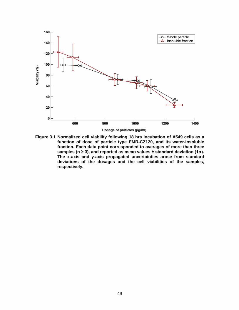
49
Figure 3.1 Normalized cell viability following 18 hrs incubation of A549 cells as a function of dose of particle type EMR-CZ120, and its water-insoluble fraction. Each data point corresponded to averages of more than three samples (n ≥ 3), and reported as mean values ± standard deviation (1σ). The x-axis and y-axis propagated uncertainties arose from standard deviations of the dosages and the cell viabilities of the samples, respectively.

50
Figure 3.2 Normalized a) IL-6 and b) IL-8 expression following 18 hrs incubation of A549 cell cultures as a function of dose of particle type EMR-CZ120, the water-soluble fraction and the insoluble fraction of EMC-CZ120. Each data point corresponded to averages of three or more samples (n ≥ 3), and reported as mean values ± standard deviation (1σ). The x-axis and y-axis propagated uncertainties are from standard deviations of the dosages, and the individual cytokine expression levels, IL-6 or IL-8, as a function of the dose, respectively.

51
The A549 responses to doses of the whole particle, the water-soluble fraction,
and the insoluble fraction of ambient particle type ERM-CZ120 illustrate the significant
role of endocytosis in health outcomes due to particulate air pollution. The water-soluble
fraction alone effects no differential expression of cytokines IL-6 and IL-8 relative to the
negative control, yet clearly, it has an assignable role in the overall response of the
whole particle type ERM-CZ120 (Figure 3.1 and Figure 3.2). This result, in general,
agrees with other studies of insoluble and soluble fractions of ambient particle types,
where cellular responses to the individual soluble plus insoluble fractions do not sum to
the response from the whole particle.142, 161, 169, 174, 176, 180 For instance, Fujii et al.
investigated the cytotoxicity of ambient particle EHC-93 on human bronchial epithelial
cells (HBECs), and measured dose-dependent trends for cytokine expression for the
whole particle, whereas the soluble fraction of EHC-93 did not simulate differentiable
cytokine expression relative to the control.181 Jalava et al. investigated the inflammatory
and cytotoxic effects of both fine and coarse particles, including their water-soluble and
insoluble fraction, as well as the organic-solvent-soluble and insoluble fraction. In that
study, the insoluble particle fraction effected the highest cellular response of the
macrophage cell line used.177 Huang et al. found that the cytotoxicity of both coarse and
fine particles was dependent on exposure time and concentration. Soluble components
such as inorganic ions (nitrates, sulfates, and ammonium) and organic components
might cause the cytotoxicity synergistically or additively with metals.174
Numerous studies have measured the role of the water-soluble components such
as metal ions for the purpose of investigating the hypothesis that the main contributor of
the toxicity was attributable to the water-soluble particulate fraction.170, 172-173 In fact, this
hypothesis has been shown for compounds such as biologics, where a receptor is
presented on the cell exterior that selectively recognizes the biologic compound, which
triggers an immune response.167, 175-178 Imrich et al. found that certain particle types
readily adsorbed components such as endotoxin, a biologic, that itself effects strong
cellular response.175 Conversely, with respect to the results presented in this chapter for
water-soluble metal ions, rather than inducing cytotoxicity directly, the water-soluble
metals, and other soluble components that may present, do not effect A549 cellular
response alone, but this fraction does effect synergistically cellular responses when
introduced together with insoluble components. The most probable mechanism is that

52
the water-insoluble particle components play a role as carriers, via endocytosis, for the
water/lipid-soluble components. In addition, the cell-particle interactions could cause cell
membrane damage which can facilitate greater internalization of the water-soluble
fraction compounds.142 These reports, as well as the current study, emphasize the
critical role of endocytosis when species that have no receptor expressed in the cell
membrane are presented to intact viable cells.
3.5. Conclusion
Following 18-hour incubation period for A549 cultures with ambient particle type
ERM-CZ120, pro-inflammatory cytokine IL-6 expression, and cell death was measured
for both the whole particle and the water-insoluble fraction in a dose-dependent manner.
No differential expression of cytokines and cell viability relative to negative control was
effected by the water-soluble fraction alone. However, the whole particles lead to higher
IL-6 expression than the insoluble fraction at the same mass dosages, suggesting that
the soluble fraction does have an assignable effect but only when dosed together with
the insoluble fraction. No differential expression of IL-8 relative to the negative control
was measured for either the whole particle or any of its fractions. This data is the basis
of our recommending studies that measure cellular responses to learn of interactions
between different particle components to identify the most significant components of
ambient particle types causing adverse effects on human health.

53
Chapter 4. Human lung cell responses caused by insoluble particle types that are mimics of major particle types adjacent to roadways
4.1. Abstract
Three insoluble particle types, carbon black nanoparticles, crystalline silica
particles, and nickel nanoparticles, and interactions between them have been measured
as a function of dose, by mass, with respect to effecting lung cell responses in vitro.
Responses measured were pro-inflammatory cytokines IL-6 and IL-8, and the extent of
cell death. No differentially measurable effect on cell viability or cytokine expression was
observed for nickel nanoparticles at dosages ≤ 100 µg/ml in the serum-free medium.
Crystalline silica, at a nominal diameter ~ 2 µm, lead to the lowest cell viability and the
greatest cytokine expression for a given total mass dosage as compared to CB alone, or
a mixture of CB nanoparticles and crystalline silica. Mixtures of CB and crystalline silica,
at three different mass ratios, were observed to effect additive mass effects with respect
to decreased cell viability and increased IL-8 expression. Interestingly, a non-addictive
lung cell response for IL-6 was measured for the mixture of CB plus crystalline silica in
specific doses. These results illustrate that specific mixtures of particle types can
generate cellular expression that is greater than an equivalent mass dose of either
particle type alone, but this result certainly has limits/restrictions. Similarly, 10 µg/ml
nickel nanoparticles plus crystalline silica induced significantly higher IL-6 expression at
both low and high silica dosages as compared to crystalline silica alone.

54
4.2. Introduction
The ambient particulate matter has been correlated positively with adverse health
effects through numerous, independent, epidemiology studies.10, 15, 26, 28, 182-183 Traffic-
related air pollutants, either a direct outcome of their chemical compositions and/or the
fact that the concentrations of these pollutants are elevated adjacent to roadways, have
a high correlation to adverse effects on human health.183-184 Short-term exposure to
roadside particles can lead to pulmonary inflammation and exacerbation of the
respiratory/pulmonary diseases, such as asthma; whereas chronic exposure is
associated with respiratory/pulmonary diseases, atherosclerosis, and cardiovascular
diseases.8, 10, 14, 183-185 Proximal to major roadways, ubiquitous particle types include
soots from incomplete combustion of fossil fuels, vehicular wear materials (e.g., metals,
brakes, and tires), re-suspended crustal dusts and road salts, particle types from
proximal industrial related activities, as well as background particle types from distal
emission sources.10, 14 Of the traffic-derived PMs, diesel exhaust particles can comprise
up to 90% of the particles in the world’s largest cities.186-187
Results obtained using reference ambient particle type ERM-CZ120 indicate that
the insoluble fraction of the particles induces IL-6 expression and cell death at high mass
dosages. The insoluble fraction has an important role in particles’ cytotoxicity, as has
been well documented in the literature, but, in addition, it is becoming clear that the
insoluble fraction plays a critical role of transporting soluble compounds into cells, via
endocytosis (e.g. Chapter 3). In this chapter, three laboratory mimics of ambient particle
types were used: CB nanoparticles to represent diesel soot, crystalline silica particles to
represent re-suspended road dusts, and nickel nanoparticles to represent an insoluble
metal particle type. The effects of dosing these particle types individually, as well as
binary mixtures thereof, were studied by measuring select lung cell responses in vitro.
Through the study of a single particle type dose, and then a binary particle type dose,
two questions were investigated: is there a measurable differential role of a particle
type’s physical and chemical characteristics relative to another particle type, and is there
a non-additive interaction between different particle types, as a function of total particle
mass dose, as ascertained using specific cellular responses?

55
4.2.1. Background on Carbon Black
Carbon black is a commercially available commodity made through a highly
defined and controlled partial combustion, thermal decomposition process of
hydrocarbons. Hydrocarbon combustion, from lower molecular weight fuels to coal tars,
in internal combustion engines rarely leads to complete combustion, and thus,
worldwide, the study of soots is an environmentally important area involving many
diverse themes of study.56, 188-189
CB is manufactured in the format of small elemental carbon particles. Primary
particles, as being described as nodules, are roughly spherical with diameters varying
from 5 to 500 nm.56, 123, 188-189 Inside the particle, there is a relatively disordered nucleus
oriented centrically with imperfect graphite layers. The carbon layers are generally
parallel to each other, but their relative positions and sizes are irregular.188-189 Through
particle-particle collisions and the further deposition of carbon, primary particles can
aggregate together into grapelike in appearance chains or clusters, termed aciniform
aggregates.56, 188-189 Approximately 99% of particulate carbon in CB is aciniform. In turn,
aciniform aggregates may form agglomerates through van der Waals forces.56 The
surface area of CB varies widely, and it is dependent on its structure. For example, due
to the large primary particle size and low degree of aggregation, coarse thermal black
particles have surface area as small as 8 m2/g, whereas pigment grade CB can have
surface area up to ~ 1000 m2/g.188 CB having large surface area is the result of a porous
structure, thus the aggregation tends to not affect the surface area and other
characteristics of particles generated by particular modification.56, 190
CB composition does depend on its raw material, manufacturing process, and
post-production processing.56, 188-189 More than 80 wt% of CB is element carbon. It may
also contain hydrogen, nitrogen, and sulfur, but the quantities of the impurities are
usually minimal, each at less than ~ 1 wt%.188 The presence of air in the manufacturing
process and oxidative after-treatment leads to oxygen content up to 15 wt%. These
oxidized CB particles have surface properties different from CB produced using other
processes due to the oxygen atoms bound to their surfaces, which present acidic or
basic functional groups depending on the conditions.188 Other impurities include alkali
and alkaline earth metal salts which are from the water used in the manufacturing
process, and trace amounts of organic compounds that strongly adsorb on the surfaces

56
of the particles.56, 188-189 It is reported that the soluble organic fraction of CB is less than
0.5 wt%.188 The dominant organic compounds in this fraction are PAHs.188-189 These
organic compounds are usually biologically inactive, and can only be isolated by
continuous extraction with strong organic solvents due to the large surface area of CB
particles and their low organic loading.56, 188, 191
In vitro studies suggest that CB nanoparticles cause oxidative stress, pro-
inflammatory effects, genotoxicity, and cell death.123-124, 190-195 One of the mechanisms is
the generation of reactive oxygen species through interactions between cells and the
particle surfaces.123-124, 191 Chuang et al. found that CB caused oxidative stress and DNA
damage in rats. Particle surface area and chemistry are expected to be important
regarding interactions with cells and generating self-mediated free radicals.196 Hussain et
al. measured a positive correlation between the oxidative stress and inflammatory
effects of CB nanoparticles and the Brunauer–Emmett–Teller (BET) surface area of
individual particles. Production of H2O2 was also observed, and it played an important
role in the process of ROS generation.190 In general, CB particles’ ability to generate
ROS is related to surface properties such as specific reactivity of available sites and
their abundance (e.g., surface area). However, mechanistic details are not yet
available.123, 191
CB nanoparticles can interact with expressed membrane proteins due to their
strong absorption ability, which is generally thought to lead to complicated
consequences.123 These non-specific interactions can activate or inhibit proteins by
causing denaturation and/or conformational changes on proteins, and as a result, lead to
a variety of cell functions to be activated or disrupted. For instance, through interacting
with enzyme and cytokines, CB particles have been documented to disturb cell
metabolism and immune response.123-124, 191, 197 Moreover, it is reported that
nanoparticles can also interact with cytoskeletal or bind to intercellular junctional
proteins, and induce alteration of cell signalling, migration, and autophagy.124
Simultaneously, interactions between CB nanoparticles and proteins also affect the
particle's surface properties, including reactivity and aggregation state, and thus
influence the process of particle internalization and the particle’s toxicity.123, 198
Both high levels of oxidative stress caused by excess ROS and the interactions
between CB particles and cell proteins can initiate inflammation, cytotoxicity and

57
apoptosis.123-124, 191, 199 Hussain et al. found that exposure to CB nanoparticles can lead
to apoptosis of bronchial epithelial cells through a ROS dependent mitochondrial
pathway. The process is known to involve H2O2 production, mitochondrial surface-
potential perturbation, caspase activation, the release of pro-apoptotic factors and DNA
fragmentation.124 Mroz et al. suggest that CB nanoparticles can induce DNA single-
strand breaks and pro-inflammatory transcription factors activation on A549 cells.195
As a nanoparticle, CB can cause adverse effects on the cardiovascular system
directly by translocating to the bloodstream and/or interacting with platelets and the
vascular endothelium, and/or indirectly through causing local inflammation which may
lead to further tissue inflammation and potential destabilization of pre-existing
atheromatous plaques.123, 188, 191, 199-201 An in vivo study reported that rat exposed to a
high dosage of CB with an exposure duration of 24 months developed increased lung
tumor incidence, which was believed to be due to the inability to clear the lung when
challenged by an excessive particle burden.202 This phenomenon was summarized as
“lung overload”, and it is not restricted to CB nanoparticles as evidenced through rat
exposure to high doses of any low-toxicity insoluble particle types.188, 203-204 In contrast to
the above-cited rat-based studies showing that insoluble particle types are toxic at high
dosages, but related studies on humans having workplace compliance with safety
regulations have shown that humans in the CB industry indicate no health hazards.188-189,
204
Generally, CB is viewed as an example of a low solubility, low toxicity particle
type and due to its high purity, it has used as a reference material in studies
investigating particle toxicity.123, 188, 191, 204 Recall that CB without oxidative treatment is >
95% by mass is elemental carbon, and adsorbed on the CB surfaces are metals and
organic compounds, whose overall toxicity is considered negligible.188 In the report of
IARC (international agency for research on cancer) in 2010, CB was classified as group
2B, which is defined as possibly carcinogenic to humans, based on the criteria that there
is inadequate evidence in humans, but sufficient carcinogenic evidence in experimental
animals as well as evidence on potential mechanisms for the carcinogenicity of CB.204
In this study, CB nanoparticles were adopted as a mimic of soots. However,
there are many differences between soots and CB. As an unwanted by-product, soots
are produced through uncontrolled partial combustion or pyrolysis of carbonaceous

58
materials, such as coal, gasoline and diesel fuels, wood, fuel oils, and waste oils.55, 189
Soot physical and chemical properties, as overviewed in Chapter 1, are highly variable
as they depend on the starting materials and the combustion conditions.55, 189, 205
Generally, CB nanoparticles and soots are distinct in physical structure, chemical
composition, and toxicity (Table 4.1).
Table 4.1 Differences between CB and Soots
CB Soots
Physical structure
Aciniform
aciniform carbonaceous microgel
carbon cenospheres coke and char fragment
Chemical compositions
> 95 wt% elemental carbon, with trace quantities of ash (< 1 wt%) and SOF(< 0.5 wt%) A
< 60 wt% elemental carbon, with variable percentages of SOF, metals, and other
components are viableB
Toxicity Low toxicity,
possibly carcinogenic to humans (Group 2B) C
High toxicity, carcinogenic to humans
(Group 1)C A Date from188, B data from56, C data from204 206. SOF = soluble organic fraction.
Although soots have similar generation processes as CB, including nucleation,
aggregation, and surface growing process, their particle structure can range from
amorphous to graphitic or even fullerenic due to different emission sources and thermal
history.55-56, 205 Aciniform carbon is predominant in diesel engine exhaust soots, and it
has been reported that the particulate portion of diesel soot has similar carbon structure
as CB.207-208 Recall, as previously mentioned, approximately 99% of the particulate
carbon in CB is aciniform, which can be characterized as colloidal size, grapelike
aggregates of imperfect graphitic structure particles.56, 189 However, the subsequent
deposition of carbonaceous materials, such as crosslinked resins, can cement the
aciniform carbon particles together, and form carbonaceous microgels (carbon or
carbonaceous material embedded with spheroidal colloidal dimension carbon particles),
termed xerogels that have large size distribution.189, 207, 209 Additional types of carbon
particles that are found in soots include carbon cenospheres (porous or hollow carbon
spheres with diameters of 10-100 µm, formed by carbonization of liquid drops without
shape change), coke and char fragments (fragments of carbonized wood or coal with
dimensions from millimeters to micrometers).56, 189, 207

59
CB particles have ~ 95 wt% element carbon, whereas the mass fraction of
elemental carbon in soots is less than 60 wt%.56, 188 A larger percentage of soluble
organic fraction (SOF) and inorganic impurities/ash can be found in soots relative to
CB.55-56, 189 For instance, diesel soot contains 20 to 40 wt% of organic compounds,
among them, PAHs and their derivatives comprise approximately < 1 wt%, and PAHs
are associated with adverse health effect. Nitrate, sulfate, transition metals, and other
trace elements may also be found in diesel soot.209 As a consequence, soots are much
more toxic than CB.55-56, 189, 206 For instance, numerous studies have demonstrated that
soots effect adverse outcomes on humans, such as respiratory irritation, pulmonary
inflammation, exacerbation of cardiovascular diseases and even cancer.55-56, 189, 206, 209
IARC evaluated soots as carcinogenic to humans (group 1, defined as carcinogenic to
humans).55, 206
4.2.2. Background on Silica
Silica and siliceous minerals are the major constituents of the earth’s crust. 12-14
wt% of the lithosphere is quartz, which is one of the crystalline forms of silica.210-211 In
PM10, silica is ubiquitous. Sources of airborne silica are construction activities, dust from
paved roads and unpaved roads, agricultural operations, wind erosion, volcanic
eruptions, and mining and quarrying.212-213 Several amorphous and crystalline forms of
silica are known.212 The majority of the free silica (pure crystalline silica) is in the form of
quartz. Other crystalline forms include tridymite and cristobalite.146, 210, 212-213 In this study,
quartz was selected for study due to its abundance in ambient particles. The phrase
“crystalline silica” implies quartz unless another form of crystalline form silica is
specified.
There are two dimorphs of quartz, α-quartz and β-quartz, which are temperature
dependent. The structure of quartz is a three-dimensional helix framework that is
composed of either three or six silicon tetrahedra along the trigonal c-axis. Below 573
°C, quartz is in the form of trigonal α-quartz, and above 573 °C, it converts to hexagonal
β-quartz.146, 213 Silicon tetrahedra are the basic structural units of quartz. In each unit, a
silicon atom is surrounded by four oxygen atoms, and each oxygen atom is common to
two tetrahedra.5, 146, 210-211

60
Generally, the purity of quartz is high, but the chemical compositions of quartz
are affected by its sources. For commercial products, the preparation process may also
affect their compositions.146, 212-213 Major impurities include Al, Fe, Li, Na, K and Ca, yet
their concentrations are usually below 1 wt%.146, 149 Metal ions such as Al3+ and Fe3+ can
substitute Si4+ in the tetrahedra, and at the same time introduce monovalent or divalent
cations including Li+, Na+, K+, Mg2+ and Ca2+ to the interstitial sites. Moreover, the metal
ions can also form non-bridging oxygen sites in the lattices.146, 210
Inhalation of crystalline silica is associated with adverse health effects. At the
cellular level, crystalline silica induces the generation of ROS, which in turn leads to the
secretion of cytokines and pro-inflammatory mediators, which may cause
membranolysis, lipid peroxidation, DNA damage, and cell death.131, 214-215 It is believed
that the surfaces of quartz, especially the silanol groups (Si-OH) and ionized silanol
groups (Si-O-) on the particle’s surfaces, play an important role.129-131 The quartz planes
can be cleaved or fractured when the quartz is ground, and lead to homolytic and/or
heterolytic cleavage of Si-O bonds, as a result, Si˙ and Si-O˙ radicals (for homolytic
cleavage) and Si+ and Si-O- ions (for heterolytic cleavage) can be generated. 131
Generally, silanol and ionized silanol are viewed as the main “active sites” attributing to
quartz toxicity.149-150 Freshly fractured quartz is speculated to be more toxic than an aged
one due to its relatively higher surface reactivity.5, 129, 149 In aqueous environments, such
as in lung lining fluid, those surface radicals can give rise to ROS including OH˙ and
H2O2 in the lung lining fluid. In the presence of water or H2O2, surface radicals of quartz
can form silanol and OH˙ through following reactions:
-SiO(•) + H2O → -SiOH + HO(•)-
-SiO2(•) + H2O2 → -SiOH + HO(•) + O2
-Si+O2(•)- + H2O2 → -SiOH + HO(•) + O2
109,110
A silanol can bind to bio-macromolecules though hydrogen bonding. The binding
can modify the particles’ toxicity in several ways, for example, reduce the toxicity of the
particles by preventing the direct contact between the particles and the molecules or
enhance the toxicity by activating the immune system.131, 215 Further, ionized silanol can
interact with the cell membranes.131

61
In interacting with phagocytic cells, crystalline silica can also induce oxidative
stress by stimulating a respiratory burst.130 Phagocytosis of particles is capable of
stimulating a sudden increase in oxygen consumption by the cells, and as a
consequence, there is an increased production of ROS such as O2(•)- and H2O2, and
NO.129-130, 216 The reaction between NO and O2(•)- yield NO2
- which is one of the
important reactive nitrogen species (RNS).129, 216 Both ROS and RNS are able to react
with various biological targets directly, such as DNA, proteins, and lipids, and thereby
cause damage to cells.5 Fanizza et al. found that short-term exposure to α-quartz can
induce direct oxidative DNA damage and cell death of A549 cells in a dose-related
manner.217 Chu et al. observed that cell damage caused by crystalline silica
nanoparticles, which included mitochondrion multiplication and DNA fragmentation, was
associated with an increased level of ROS in the cells.214 Duffin et al., in using DQ12
quartz, a commonly used commercial product in laboratory studies, measured increased
chemokine expression and activation of an important pathway in inflammation, nuclear
factor kappa beta (NF-κB), that depended on the particles’ surface reactivity.30 This type
of cell damage leads to the apoptosis of the cells, spanning alveolar macrophages to
epithelial cells, with subsequent further inflammation and fibrosis.131, 216, 218
One well-known occupational disease caused by exposure to crystalline silica is
silicosis, caused by inhalation of respirable crystalline silica (crystalline silica particles
with diameters < 10 μm), and can be categorized into four types: acute, chronic,
accelerated, and complicated silicosis.212-213 Silicosis is a fibrotic lung disease. Acute
silicosis is exposure to very high concentrations of silica dust, and is usually fatal over a
period of 1-3 years. Its cause is attributed to fluid accumulations from cell debris and
surfactants in the upper and middle regions of the lung, inducing further damage in
alveolar cells that manifest as shortness of breath and other symptoms characteristic of
pulmonary edema. Chronic silicosis is the most common type of silicosis. It is caused by
long-term exposure (≥ 10 years) to low concentrations of silica dust. The interaction
between quartz particles and lung tissues also induce the recruitment and activation of
pulmonary macrophages, that in turn leads to the secretion of fibrogenic factors such as
interleukins and growth factors which stimulate the production of collagen. The collagen
produces silicotic lesions through hyalinization. Further growth of silicotic lesions leads
to fibrotic scars in the lung tissue, which is the main characteristic of chronic silicosis.
The main symptoms include a dry cough, sputum production, and reduction of

62
pulmonary function. Accelerated silicosis is due to exposure to high levels of silica in a
period shorter than chronic silicosis (around 5-10 years). It shares some symptoms with
chronic silicosis, but the development processes are faster. Asymptomatic silicosis is
described as early onset stage of the disease, but there is no symptoms present.131, 212-
213, 216
In addition to silicosis, inhalation of silica can lead to other airway/lung diseases
such as bronchitis, pulmonary tuberculosis, rheumatoid arthritis, and lung cancer.212-213
IARC classified crystalline silica (quartz and cristobalite form) as group 1, which is
defined as carcinogenic to human, suggesting that sufficient evidence in both animal and
human exists.146
Metal impurities such as Al and Fe modify the toxicity of the quartz particles.131,
149 Aluminum salts decrease the toxicity of quartz particles. Duffin et al. reported that the
treatment of aluminium lactate on DQ12 quartz can reduce the generation of HO˙ and
thus decrease the damage of the cells.30 Bégin et al. in using a sheep tracheal lobe
model observed that quartz treated by aluminium lactate demonstrated less pathologic
score than untreated quartz. It was suggested that aluminium salts can bind on the
surfaces of the quartz particle, especially the “active sites” such as silanol, thus lower the
bio-activity of the particles, and accelerate the clearance of the particles from the lung.150
For iron, it appears more complicated. Iron ions can catalyze the generation of ROS
through the Fenton reaction, however, metallic iron [Fe(0)] is reported to protect against
the toxicity caused by quartz.5, 129, 131, 149 Castranova et al. found that freshly milled quartz
having high Fe contamination lead to higher ROS generation and stronger inflammatory
effect as compared to quartz having low Fe contamination.151 Cullen et al. investigated
the effect of particulate and ionic iron on the toxicity of quartz in vivo. Only particulate
iron showed the ability to decrease the inflammatory effect of quartz. No effect of
ferrous/ferric iron on quartz surface radical production was observed. However, the
mechanism of the protective effect is unclear yet.152 In conclusion, iron is believed to
react with the ROS generated by quartz particles in an indirect way, unlike aluminium
which interacts directly with active sites on particle surfaces.
In this study, crystalline silica particle was chosen as a mimic of re-suspended
road dust. In the environment, quartz is usually mixed with different kinds of particles
and chemical components. Those particles and components in the mixtures may modify

63
the surface properties of quartz, and as a result, affect its toxicity in different ways. The
average concentration of quartz in ambient air across 22 U.S. cities were measured by
EPA Inhalable Particulate Network (IPN) in 1980, and the mass ratio of quartz ranged
from 1.0% to 9.0% (avg. 4.9%) in coarse particles, and from 0% to 2.6% (avg. 0.4%) in
fine particles.212, 219 Near roadside, the main sources of quartz include the minerals used
for road pavement, and dusts from unpaved roads and the shoulders of paved roads.161,
212-213 Hetland et al. analyzed 4 types of road pavement minerals and roadside PM10
samples, and found that both the chemical components and the cytotoxicity of PM10
samples were similar to those of the minerals used for road pavement. The content of
quartz in the roadside PM10 samples is 24%. Metal components such as Fe and the
treatment during asphalt production may modify the toxicity of the mineral particles.161. In
general, the interaction between quartz particles and other types of particles, or particle
components, is not well characterized, and thus it should be investigated as it could be a
significant source of the observed adverse effects on human health.
4.2.3. Background on Nickel
Nickel is a silver-white metal that occurs naturally in the earth’ crust at an
abundance ~ 0.009%.220 Due to its ability to increase a metal solid's strength,
corrosion/temperature resistance, and overall toughness when alloyed with other
metallic elements, nickel is widely used in metallurgical, chemical processing and
nanomaterials industry in the manufacturing of stainless steels, alloys, electroplating,
catalysts, nickel-cadmium batteries, and pigments.220-222
Natural sources of atmospheric nickel include windblown dusts, volcanic
emissions, rocks/soils weathering, and forest fires.223-224 Anthropogenic sources include
industrial processes spanning mining, melting, refining, to alloy production, combustion
of fossil fuels, and waste incineration.222-223 Tobacco smoking is also a source of nickel
exposure.224 Among the different sources, fossil fuel combustion, specifically coals and
heavy oils, is reported as the main contributor of atmospheric nickel.222 Ambient particles
containing nickel deposit into surface waters and soils, and lead to the accumulation of
nickel by plants and animals.225
Airborne nickel is predominantly in the particle phase, and the concentration
varies depending on the source. It has been reported that the average airborne nickel

64
levels are 0.01-3 ng/m3 in remote areas, 3-30 ng/m3 in urban areas without metallurgical
industries, and 0.07-0.77 μg/m3 in industrialized areas that utilize Nickel.224 Based on a
survey conducted in 11 Canadian urban cities and one rural site from 1987 to 1990, the
annual mean concentration of nickel was 1 ng/m3 for the rural site, and ranged from 1 to
20 ng/m3 for urban areas, with maximum values from 6 to 77 ng/m3.226 A point source
study indicated that the background annual mean nickel concentrations in Canadian
Shield and Great Lakes region range from 0 to 1.46 ng/m3, while the nickel
concentrations measured close to the nickel/copper smelters in Sudbury are 46-151
ng/m3, which accounted for approximately 0.67% of the total PM10.227 For the general
population, food and water uptake is the main exposure route of nickel instead of
inhalation. It is reported that, for non-smoking urban residents, the daily uptake of nickel
is below 0.0008 mg through inhalation, while it is ~ 0.1-0.3 mg via food. However, for
occupational exposure to nickel, inhalation is the primary route.225 An occupational study
of nickel exposure in a nickel refinery in Norway indicated an average concentration of
nickel ≤ 0.7 mg/m3 in the breathing zone for all workers.228 Dermal contact is another
pathway for both occupational and general exposure.224
Inhalation of nickel involves the following forms: particles of insoluble nickel
compounds, aerosols derived from soluble nickel solutions, and gases containing nickel
species (e.g. nickel carbonyl).224 The different chemical formation and physical
properties, such as solubility, do affect the species toxicity. Soluble nickel species
dissolved in the mucus are cleared rapidly from tissues through ciliary transport.
However, less soluble nickel particles can enter cells by phagocytosis, and slowly
dissolve inside the cells and in so doing provide an extended exposure period to high
cellular levels of Ni2+.222, 224-225, 229 Generally, the less soluble species, including nickel
oxides and sulfides, are more carcinogenic than the soluble nickel species, such as
nickel chloride or nitrate.222
Inhalation exposure to nickel has been associated with various adverse health
effects. Short-term high-dose exposure can induce nasal cavity damage, lung irritation,
hyperplasia of pulmonary cells, fibrosis, pneumonia, and emphysema. Chronic exposure
is speculated to contribute to respiratory disorders such as asthma and bronchitis,
pneumoconiosis and lung cancer.222-223, 230 It has been reported that nickel refinery
workers have a significantly higher risk of pulmonary and nasal cavity cancers as
compared to a control population. Higher mortality due to non-cancerous lung diseases

65
has also been observed.230 The International Agency for Research on Cancer (IARC)
has classified nickel compounds as group 1 (carcinogenic to humans).222
The mechanism of nickel’s toxicity is not yet fully understood. It has been
suggested that Ni could be involved in Fenton chemistry, and lead to the generation of
ROS in the presence of strong oxidizers such as H2O2 and ascorbate in the cytosol and
nucleus.5, 229-230 Note that Ni2+ does not directly participate in redox cycling reactions.
However, through chelation with biological ligands, the reduction potential of Ni2+ can be
altered, thus allowing the oxidation of Ni2+ to Ni 3+ by a strong oxidant.5 Intracellular
glutathione depletion and oxidative damage including lipid peroxidation and DNA
depurination induced by Ni have been observed.229-232 As a carcinogen, Ni can also
interfere with cellular metabolism, and cause genotoxicity by replacing essential metals
such as Fe(II), Mg(II) and Ca(II) in enzymes and proteins, or binding to cellular
compounds including nuclear proteins and DNA.224, 229-230 Salnikow et al. found that
exposure of A549 cells to Ni2+ lead to the production of ROS and the activation of
Hypoxia-inducible factor (HIF) -1 transcription factor, which is associated with cell
survival and lung cancer. The activation of HIF-1 was proposed due to the substitution of
iron by nickel in the oxygen sensor.233 Through complexing with heterochromatin, Ni is
implicated in methylation of DNA, gene silencing and chrome condensation. The
mechanism is not clear, but the replacement of magnesium by nickel in the DNA
backbone is proposed to be a factor.229 Through interactions with repair enzymes, Ni can
also inhibit DNA repair.234 A former colleague of our group has studied the cellular
response to soluble nickel plus amorphous silica particle at a mass ratio of 5,000 ppm
using co-culture of THP-1 cell and A549 cell. No differential IL-6 or IL-8 expression was
measured for cells dosed with Ni2+ plus amorphous silica particles relative to the
response of cells dosed with amorphous silica particles alone.235 In this study, nickel
nanoparticle was chosen as a representation of insoluble metal particle.
4.3. Methodology
4.3.1. Reagents used in culturing
The culture medium, Nutrition Mixture F12 HAM Kaighn’s modification (model: F-
12K, Lot #N3520), human tumor necrosis factor-α (Lot #H8916), sodium bicarbonate
(bioreagent, Lot #S5761, 99.5%-100.5%) and trypan blue solution (Lot #T8154) were

66
purchased from Sigma–Aldrich (Oakville, Ontario, Canada). Phosphate-buffered saline
(PBS, Lot #OXBR0014G), fetal bovine serum (FBS, Lot #12483) and trypsin-EDTA
solution (0.05%, phenol red, Lot #25300-054) were obtained from Fisher Scientific/Life
Technologies (Pittsburgh, PA, USA).
4.3.2. Particles and compounds
Commercially available crystalline silica (model: Min-U-Sil® 5) was provided by
the U.S. Silica Company (Berkley Springs, WV, USA). It is declared as high purity, inert,
white crystalline silica derived from a natural source. According to the vendor, ~ 98-99%
of the particles are SiO2 with a median diameter of 1.7 µm. 92% of these particles have
diameters ≤ 5µm. The main impurities include aluminium oxide (< 1%), iron oxide (<
0.1%) and titanium oxide (< 0.1%) which may also contribute to the toxicity of the quartz
particles. Previous studies suggest that aluminium salts and metallic iron can decrease
the toxicity of quartz particles by coating the active sites at the particle-air interface as
well as the iron ions are capable of catalyzing ROS production by way of Fenton
reaction.30, 131, 149-152
Carbon black nanoparticles (Lot #1211NH , 100 nm) was purchased from
Nanostructured & Amorphous Materials Inc. (Houston, USA). The purity of CB
nanoparticles is 88.1%, and impurities include ash (5.8%) and water (1.06%). Nickel
nanoparticles (Lot #577995, < 100 nm) were purchased from Sigma-Aldrich (Oakville,
Ontario, Canada). The purity of nickel nanoparticles is ≥ 99% based on the trace metals
analysis.
In this study, CB nanoparticles and crystalline silica particles were adopted as
laboratory mimics of two insoluble particle types that are in roadside ambient particles,
diesel soot, and re-suspended roadside dusts, respectively. As outlined above in the
Introduction section, it is noted that CB nanoparticle and diesel soot are distinct with
respect to chemical compositions and toxicity. Thus, the particle chemical compositions
and the interactions between different particle types/components are much more
complicated in the ambient environment than with the CB used here in these laboratory
studies.

67
4.3.3. Cell culture
The human lung alveolar epithelium cell line A549 (CCL-185, American Type
Culture Collection, Rockville, MD, USA) was used in this work. These cells were cultured
in F12K medium supplemented with 10% (v/v) fetal bovine serum at 37 °C in a
humidified atmosphere of 5% CO2 in the air. Prior to dose-response assays, cells were
seeded in 6-well culture dishes (surface area/well 8.87 cm2) and grown to confluence.
Cultures incubated with particle-free serum-free culture medium, and serum-free
medium containing 50 ng/ml TNF-α served as the negative and positive controls,
respectively.
4.3.4. Exposure to particles
Stock suspensions of particles (1 mg/ml in serum-free medium) were made fresh
and vortexed for homogeneity. Serial dilutions of stock solutions were performed using
the serum-free medium. Aliquots of solutions containing particles were then introduced
to A549 cell cultures. For exposure to a single particle type, the cells were incubated
with particles suspended in 2 ml serum-free medium at dosages from 0 to 700 μg/ml for
CB nanoparticles and crystalline silica, and from 0 to 100 μg/ml for Ni nanoparticles. For
doses involving a mixture of CB nanoparticles and crystalline silica particles, the mass
ratios of CB nanoparticles to crystalline silica particles used were fixed at 0.7, 1.4 and
2.8, with a total mass dosage range from 0 to 700 μg/ml in 2 ml serum-free medium.
Regarding dosages of crystalline silica particles plus Ni nanoparticles, silica was used
from 62.5 to 125 μg/ml, and Nickel nanoparticles from 10 to 40 μg/ml. All samples were
run in duplicate per experiment. Three or more independent experiments with the same
dose were performed throughout.
4.3.5. Measurement of IL-6 and IL-8 concentrations in supernatants
Upon introducing the dose of particles to the cell cultures, the incubation period
was 18 hours in duration, at the end of which the supernatants were collected and stored
at –80°C until analysis. The concentrations of two pro-inflammatory mediators, IL-6 and
IL-8, in the supernatants were quantified using ELISA kits (IL-6, model #900-M16, IL-8,
model #900-M18, Cedarlane, Burlington, Ontario, Canada) as per the manufacturer’s
instructions. The ELISA assays were performed in triplicate. The determined

68
concentration of IL-6 and IL-8, mean and standard deviation, were obtained through
normalization against the positive and negative control, using the equation sfinal =S−N
P−N
(S: samples, N: negative control, P: positive control).
4.3.6. Trypan blue assay
After collection of the supernatant, each separate culture in a 6-well plate was
washed three times with 2 ml PBS, and a trypan blue viability assay was immediately
performed following the procedure of trypan blue exclusion test.164 The numbers of
viable and non-viable cells were counted using a hemacytometer (0.1 mm, Bright-
Line™) and a bright-field microscope (TMS-F, Nikon), respectively. The percent
viabilities of samples were normalized against negative controls.
4.3.7. Statistical analysis
The data acquired on cell viability, and the expression of IL-6 and IL-8 is reported
as mean values ± standard deviation (1σ). The student t-test was used to compare the
cell viability and cytokine expression between samples, and controls and samples. The
threshold for statistical significance was set as p < 0.05, and when p < 0.01 was
obtained it is reported.
4.4. Results and Discussions
4.4.1. CB nanoparticles
Figure 4.1 presents the measured cell viability as a function of the dosage of CB
nanoparticles. Generally, as the dosage of the particles increased, cell viability
decreased. 95% of the cells are non-viable when the dosage is ≥ 500 µg/ml. Two
cytokines, IL-6 and IL-8, were adopted to indicate trends in cell response (Figure 4.2),
because both are known as important pro-inflammatory mediators. As the dose of the
CB nanoparticles increased, the IL-6 and IL-8 expression increased, to a maximum, for
both IL-6 and IL-8, at a dosage of ~ 230 µg/ml. With further increases in the total mass
dose of particles, cell cytokine expression decreased. The dosage 230 µg/ml
corresponds to cell viability ~ 40% (Figure 4.1). The death of a large fraction of the cells
provides an explanation for why the IL-6 and IL-8 expression decreases at dosages >

69
230 µg/ml. The elevation of IL-6 and IL-8 expression due to exposure to CB
nanoparticles indicates a dose-dependent correlation between CB nanoparticle
exposure and its ability to induce inflammation in A549 cells.
Figure 4.1 Normalized cell viability after 18 hrs incubation of A549 cells as a function of dose of CB nanoparticles. Each data point corresponded to averages of more than three samples (n ≥ 3), and reported as mean values ± standard deviation (1σ). The x-axis and y-axis propagated uncertainties arose from standard deviations of the dosages and the cell viabilities of the samples, respectively.
CB nanoparticles contain a trace amount of metal salts and organic
compounds.56, 188-189 As such, they are classified as an example of low toxicity particle
which mainly induces cytotoxicity through interactions of particle surfaces with cells.124,
188-189 However, previous studies have generated results that were interpreted that CB
nanoparticles cause oxidative stress, pro-inflammatory effect, genotoxicity, and cell
death.124, 188-189, 191, 204 Val et al. investigated the inflammatory process caused by CB and
TiO2 nanoparticles on human bronchial epithelial cell lines and found that CB
nanoparticles were able to induce several kinds of pro-inflammatory cytokines in a dose-
dependent manner, including IL-6, TNF-α and granulocyte-macrophage colony-
stimulating factor (GM-CSF). They also suggested that the ability for nanoparticles to
absorb cytokines could affect the evaluation of the pro-inflammatory potential of the
particles.198 The work of Mroz et al. demonstrated that CB nanoparticles induced DNA

70
single-strand break and pro-inflammatory transcription factors activation on A549 cells.
Oxidative stress pathways were involved in the process.195 Hussain et al. found that
exposure to CB nanoparticles lead to apoptosis of bronchial epithelial cells through a
ROS dependent mitochondrial pathway. In review, two pathways, the generation of
ROS, and the interactions between particulate surfaces and cell proteins, are believed
as main contributors of CB’s cytotoxicity. CB solid-air interface properties such as
surface area and reactivity appear to play important roles in the process.124 However, the
detailed mechanisms are not fully understood yet. Our results are in general agreement
with the measurements made in previous studies, that CB induces pro-inflammatory
mediator expression and cell death in a dose-dependent relationship.
Figure 4.2 Normalized IL-6 and IL-8 expression after 18 hrs incubation of A549 cell cultures as a function of dose of CB nanoparticles. Each data point corresponded to averages of more than three samples (n ≥ 3), and reported as mean values ± standard deviation (1σ). The x-axis and y-axis propagated uncertainties arose from standard deviations of the dosages and the IL-6 or IL-8 expression of the samples, respectively.
4.4.2. Crystalline silica
Figure 4.3 and 4.4 show the cell viability, and selected cytokines expression, IL-6
and IL-8, as a function of the dosage of crystalline silica, respectively. The dose-

71
dependent results for silica have similarity to that was measured using CB nanoparticles.
For instance, cell viability decreases as the mass dosage of crystalline silica increases.
At dosages ≥ 360 μg/ml, the culture is non-viable. Also, regarding both IL-6 and IL-8
expression, at low mass doses, silica caused increased cytokine secretion up to
maxima, but, with further increases in particle mass dosage, the concentrations of
cytokines measured in the supernatant decreased steadily to become non-differentiable
relative to the negative controls. The measured maximum for interleukin secretion
corresponded to a trade-off between maximal stimulation of the cells versus minimal cell
death. The highest IL-6 and IL-8 expressions were observed at a dosage of 130 μg/ml
and 250 μg/ml respectively, which correspond to viabilities of ~ 50% and ~ 15%,
respectively.
Figure 4.3 Normalized cell viability after 18 hrs incubation of A549 cells as a function of dose of crystalline silica. Each data point corresponded to an average of more than three samples (n ≥ 3), and reported as mean values ± standard deviation (1σ). The x-axis and y-axis propagated uncertainties arose from standard deviations of the dosages and the cell viabilities of the samples, respectively.

72
Figure 4.4 Normalized IL-6 and IL-8 expression after 18-hour incubation of A549 cell cultures as a function of dose of crystalline silica. Each data point corresponded to an average of more than three samples (n ≥ 3), and reported as mean values ± standard deviation (1σ). The x-axis and y-axis propagated uncertainties arose from standard deviations of the dosages and the IL-6 and IL-8 expression of the samples, respectively.
On the cellular level, the toxicity of crystalline silica mainly depends on the
surface properties, especially the “active sites” with silanol groups.129-131 Either by
directly producing ROS at the particle surfaces, and/or stimulating the respiratory burst
of the cells and inducing ROS/RNS indirectly, quartz particles are capable of generating
oxidative stress, triggering pro-inflammatory cytokine expression, and causing cell
apoptosis.131, 212, 215 The study of Fanizza et al. indicated that short-term exposure of α-
quartz induced direct-oxidative DNA damage and cell death of A549 cells in a dose-
related manner.217 Herseth et al. investigated the cytotoxicity of silica using both mono-
culture (alveolar macrophages) and non-contact co-culture (alveolar macrophages and
endothelial cells). Pro-inflammatory cytokine response, i.e. the release of IL-1β, IL-6, and
IL-8, can be observed for both monoculture and co-culture. IL-1β is known to have an
important role in triggering the release of other cytokines.236

73
4.4.3. Nickel nanoparticles
A dosage range from 0-100 μg/ml was selected and adopted for nickel
nanoparticles. At this range, nickel nanoparticles induced no differentiable cytokine
expressions from negative controls (Figure 4.5). Also, there was no obvious effect on
cell viability was observed, but at dosage ≥ 40 μg/ml, transparent vacuoles were
observed inside the A549 cells (Figure 4.6). Abnormal cellular morphology induced by
nickel has been reported.223 Ahamed’s study on the toxic response of A549 cells
exposed to nickel nanoparticles indicated significant toxicity of Ni nanoparticles in which
the generation of oxidative stress plays an important role.231
Figure 4.5 Normalized IL-6 and IL-8 expression after 18 hrs incubation of A549 cell cultures as a function of dose of nickel nanoparticles. Each data point corresponded to averages of more than three samples (n ≥ 3), and reported as mean values ± standard deviation (1σ). The y-axis propagated uncertainties arose from standard deviations of the IL-6 and IL-8 expression of the samples, respectively.

74
Figure 4.6 A549 cells as viewed after an 18 hrs incubation period (a) without any particles/particle components (negative control) (b) with 100 μg/ml nickel nanoparticles. (c) close-up image of the area with circles. Images were obtained using a bright-field microscope, at 100X magnification. Circles show transparent vacuoles inside the A549 cells caused by dosage of nickel nanoparticles
Prior work by our group with soluble nickel, Ni(NO3)2·6H2O, which used a co-
culture of THP-1 cell and A549 cells, indicated that Ni2+ can induce significant higher
expression of both IL-6 and IL-8 than negative controls when the dosage ≥ 20 μg/ml.235
The different effects of nickel, spanning soluble nickel salts to insoluble nickel
nanoparticles indicate that the cytotoxicity of nickel depends on the cellular
concentration of bioavailable nickel ions.224-225, 229 Additional differences in the results
involving doses of nickel are likely due to different methodology (e.g., cell types used),
exposure time, and molecules measured in assessing the cellular response. For
insoluble nickel nanoparticles, the surface sites and overall particle charge are likely
important factors, with additional experimental investigation required to further delineate.

75
4.4.4. Is there a synergy between different particle types?
4.4.4.1 CB nanoparticle plus crystalline silica
For particle mixtures of CB nanoparticles plus crystalline silica particles, different
mass ratios (the mass dosages of CB nanoparticles over mass dosages of crystalline
silica) as 0.7, 1.4, and 2.8 were used. Cell viability (Figure 4.7), IL-6, and IL-8 expression
(Figure 4.8) are plotted out as a function of the dosage of the particle mixtures with
different mass ratios, respectively. For both cell viability and cytokine expression, there
was no measurable difference between cells dosed with CB nanoparticles plus
crystalline silica at different mass ratios. This mass ratio dose data was also averaged
and plotted out (Figures 4.9 and 4.10).
Figure 4.7 Normalized cell viability after 18 hrs incubation of A549 cells as a function of dose of CB nanoparticles plus crystalline silica at different mass ratios. Each data point corresponds to an average of more than three samples (n ≥ 3), and reported as mean values ± standard deviation (1σ). The y-axis propagated uncertainties arose from standard deviations of the cell viabilities of the samples.
The trend for all of the particle doses (CB nanoparticles, crystalline silica
particles, and mixtures of CB nanoparticles plus crystalline silica particles) is that cell
viability decreased with increased mass of particles introduced (Figure 4.9). At the same
76
mass dosage, CB nanoparticles led to highest cell viability, whereas crystalline silica and
mixtures of CB nanoparticles and crystalline silica induced similar cell viabilities, both
lower than CB nanoparticles. Note that all of these particles induce > 95% cell death at
mass loadings ≥ 500 µg/ml. Figure 4.10a and 4.10b plot IL-6 and IL-8 concentrations,
respectively, as a function of the dosage of different types of particles. The trends for
these two cytokines are similar, with low mass dosage of the particles inducing
increased cytokine secretion up to a maxima, and, then with further increases in particle
mass dosage, the concentrations of cytokines measured in the supernatant decreased
steadily to, at mass doses > 700 µg/mL, equivalent to negative controls. The maxima in
the interleukin secretion correspond to a trade-off between maximal stimulation of the
cells (Figure 4.10) versus cell death (Figure 4.9), as the maxima of interleukin secretion
occurred at cell viability ~ 30%.
In comparing cellular responses to crystalline silica particles versus CB
nanoparticles that were used in this study, the cytokine response to crystalline silica is
greater at an equivalent mass dose, a significant difference was observed with respect
to IL-8. Regarding cells dosed with CB nanoparticles and crystalline silica mixtures, the
IL-6 expression is generally the same at < 150 µg/ml doses, but greater than for cells
dosed with crystalline silica particles alone for doses > 150 µg/ml. For example, IL-6 at
230 µg/ml particle dose, was measured as having significantly higher cell expression for
the mixture of CB nanoparticles and crystalline silica versus CB nanoparticles or
crystalline silica alone (Figure 4.10b). This outcome illustrates that, for certain mixtures
of particle types and mass loadings, greater cellular expression can be measured for the
equivalent mass dose of either particle type alone. Conversely, for the IL-8
concentrations measured, though the CB alone gave a significantly lower response at all
doses, the crystalline silica versus the mixtures of crystalline silica and CB were not
measurably different, suggesting that the IL-8 response was effected predominantly by
mass of the crystalline silica in the dose. This hints at the potential complexity of
understanding cellular responses when presented with ambient particulate matter that is
heterogeneous in composition and size.
Several pathways have been implicated regarding the toxicity of CB
nanoparticles and crystalline silica.5, 123, 237 The particles can interact with cell membrane
proteins and activate or inhibit signalling pathways. Internalization of particles can lead
to lysosomal destabilization or accumulation.123 For silica, it is believed that the siloxyl

77
radicals on its surfaces can generate ROS such as hydrogen peroxide, hydroxyl and
superoxide radicals in an aqueous environment.5, 237 CB nanoparticles and crystalline
silica can each participate in intercellular reactions and lead to the further generation of
ROS which is related to the particle surface area and reactivity.5, 123 Our results suggest
that, certain mixtures of particle types may stimulate one or more pathways, which would
account for the observed greater cellular expression of mixtures than measured for the
equivalent mass dose of either particle type alone. Conversely, a study by Borm et al. on
coal mine dust and coal fly-ash indicated that, the quartz in the particle mixtures was not
as bio-active as the pure samples at equivalent doses both in vitro and in vivo.238 The
difference between their results and ours may due to the differences between different
methods, e.g., cell line, particle compositions, and readouts used. Regardless of the
details, our results specifically suggest interactions between different particle types,
mass loadings, and chemical compositions, play critically important roles in the
summative toxicity of each dose, with obvious health implications on ambient particle
exposures.

78
Figure 4.8 Normalized a) IL-6 and b) IL-8 expression after 18 hrs incubation of A549 cell cultures as a function of dose of CB nanoparticle plus crystalline silica at different mass ratios. Each data point is an average of more than three samples (n ≥ 3), and reported as mean values ± standard deviation (1σ). The y-axis propagated uncertainties arose from standard deviations of the IL-6 and IL-8 expression of the samples, respectively.
(a)
(b)

79
Figure 4.9 Normalized cell viability after 18 hrs incubation of A549 cells as a function of dose of CB nanoparticles, crystalline silica, and CB nanoparticles plus crystalline silica. Each data point is an average of more than three samples (n ≥ 3), and reported as mean values ± standard deviation (1σ). The x-axis and y-axis propagated uncertainties arose from standard deviations of the dosages and the cell viabilities of the samples, respectively.

80
Figure 4.10 Normalized a) IL-6 and b) IL-8 expression after 18 hrs incubation of A549 cell cultures as a function of dose of CB nanoparticles, crystalline silica, and CB nanoparticles plus crystalline silica. Each data point corresponded to averages of more than three samples (n ≥ 3), and reported as mean values ± standard deviation (1σ). The x-axis and y-axis propagated uncertainties arose from standard deviations of the dosages, and the IL-6 and IL-8 expression of the samples, respectively.
(b)
(a)
(b)

81
4.4.4.2 Nickel nanoparticle plus crystalline silica
Figure 4.11 Normalized a) IL-6 and b) IL-8 expression after 18 hrs incubation of A549 cell cultures as a function of dose of nickel nanoparticles plus crystalline silica. The data are reported as mean values ± standard deviation (1σ), with the number of samples ≥ 3. Asterisk symbols indicate the difference between the cytokine expression of the cells dosed with nickel nanoparticles plus crystalline silica and that of the cells dosed with crystalline silica alone is statistically significant. A single asterisk symbol (*) represents p < 0.05, two asterisks represent p < 0.01.
The effect of combined doses of nickel nanoparticles with crystalline silica was
characterized by varying the concentrations of nickel nanoparticles, at 10 and 40 µg/ml,
together with crystalline silica particles at 62.5 and 125 µg/ml (Figure 4.11). Compared
with crystalline silica alone, 10 µg/ml nickel nanoparticles plus crystalline silica induced
significant higher IL-6 expression at both low and high silica dosages, whereas no
difference with respect to cell viability was observed. However, when the dosage of
nickel increased to 40 µg/ml, the measured IL-6 expression levels of nickel nanoparticles
plus crystalline silica were lower than that for the respective lower mass nickel doses,
because many cells were no longer viable at the end of the 18-hr exposure (Figure
4.12). For IL-8, a mass dosage-dependent relationship between the dosage of particle
mixture and IL-8 expression was measured.

82
Figure 4.12 Normalized cell viability after 18 hrs incubation of A549 cells as a function of dose of nickel nanoparticles plus crystalline silica. The data are reported as mean values ± standard deviation (1σ), with the number of samples ≥ 3.
Nickel nanoparticles alone at both 10 and 40 µg/ml following incubation with
A549 cells did not induce IL-6 or IL-8 expression differential from the negative control.
When combined with crystalline silica at both low and high dosages, increased cellular
response was measured. However, both IL-6 and IL-8 of the crystalline silica alone
versus the crystalline silica and nickel nanoparticle were not measurably different at the
same total mass dosage (Figure 4.4 and 4.11). Further, our results suggest that the
mass of crystalline silica in the dose is a primary factor in the cellular response, but yet
added nickel does alter the response, which suggests more than one biological pathway
mechanisms could be involved. Silica particles have been proposed as cytotoxic
dependent on the solid-aqueous interface’s siloxyl radicals and silanol groups to
generate ROS, and trigger the expression of pro-inflammatory cytokines and
chemokines.5, 237 Nickel can generate ROS through Fenton chemistry in the presence of
strong oxidizers such as H2O2 and ascorbate in cytosol and nucleus. The ROS
generated by crystalline silica could be providing the oxidizer to active a Fenton
reaction.5, 230 It is reported that metallic nickel and nickel oxide nanoparticles are involved
in the regulation of inflammation signalling pathways such as NF-κB pathway.239-241
Multiple inflammation signalling pathways triggered by silica particles and nickel
nanoparticles, respectively, may work synergistically and lead to an increase of IL-6
expression.

83
4.5. Conclusion
To investigate the toxicity of insoluble fraction of ambient particles, three typical
particle types near the roadside, i.e. CB nanoparticles representing diesel soots,
crystalline silica particles representing re-suspended road dusts, and nickel
nanoparticles, have been chosen. A549 lung cell responses following incubation with
these particle types were studied in vitro. Dose-dependent correlations between particle
exposure and cell viability and pro-inflammatory cytokine expressions have been
measured for CB nanoparticles, crystalline silica and mixtures of them at the mass
dosage range varies from 0 to 700 µg/ml. Generally, cell viability decreased with
increased mass of particles introduced. At mass loadings > 500 µg/ml, > 95% cell death
was measured. The trends for IL-6 and IL-8 were similar. The low mass dosage of the
particles induced increased cytokine secretion up to a maxima, and, then with further
increases in particle mass dosage, the concentrations of cytokines measured in the
supernatant decreased steadily to, at mass doses > 700 µg/ml, equivalent to negative
controls. The maxima in the interleukin secretion correspond to a trade-off between
maximal stimulation of the cells versus their death. No obvious effect on cell viability or
cytokine expression has been observed for nickel nanoparticles alone at dosages ≤ 100
µg/ml.
For mixtures of CB nanoparticles and crystalline silica particles, all different
particle mass ratios generated dose-dependent cell viability and cytokine expression
trends that were similar to the individual particle type. The small differences observed
were, CB nanoparticles lead to highest cell viability and lowest cytokine expression at
the same mass dosage as either crystalline silica or mixtures of crystalline silica plus CB
nanoparticles. Mixtures of CB nanoparticles and crystalline silica particles lead to
increased cellular expression for IL-6 as compared to equivalent mass doses of either
particle type alone. This result illustrates that certain mixtures of particle types can lead
to greater cellular expression as compared to an equivalent mass dose of either particle
type alone. 10 µg/ml nickel nanoparticles plus crystalline silica induced significantly
higher IL-6 expression at both low and high silica dosages than crystalline silica alone.
For both the mixtures of CB nanoparticles and crystalline silica particles, and those of
nickel nanoparticles and crystalline silica particles, the mass of crystalline silica in the
dose effect the cellular response predominantly.

84
Though our studies did not investigate cellular mechanisms, the mechanisms are
not clear yet, but in drawing information from other studies we assume that the surface
chemistry of the particles plays an important role. Ambient particle surfaces could be
activated from the sources, or through heterogeneous radical chemistry in the
troposphere or deactivated through homogeneous and heterogeneous chemistry. For
instance, crustal dusts like silica could be deactivated through sorption of humic
materials through processes prior to their introduction to the troposphere. Conversely,
various organic compounds, such as natural organic matter (NOM), would sorb strongly
(e.g. multi-chelation) to the pH-dependent negatively charged, or polar, the particle-air
interface of silica. Thus, NOM could passivate the silica interface. In our case, the
interactions between different particle types could possibly modify chemistry on the
particle-air interface and thus modify the toxicity of the whole mixtures of particles.
Future studies need to be done to confirm this assumption.
Several general conclusions can be drawn based on our results. The insoluble
fraction of the particles, CB and crystalline silica particles are able to induce the
secretion of pro-inflammatory cytokines and cell death. At the same mass dosage,
crystalline silica particles (nominally < 5 µm diameter) lead to higher cytokine expression
and more cell death than CB or nickel nanoparticles (each nominally 100 nm diameter).
At the same mass dosage, the total surface area of CB nanoparticles can be more than
106 times larger than that of crystalline silica particles. It illustrates that particle size and
surface area do not pre-determine the cytotoxicity of the particles. Previous studies have
shown particle types of all size and composition, e.g. crystalline silica and CB
nanoparticles, effect different cellular responses.5, 123, 237 Results presented in this
chapter agree well with previous conclusions that the total mass of the particle types is
important, and from that, there was no measurable synergy between the different
particle types studied. Other factors including chemical composition, and especially
surface chemistry of the particles, should be taken into consideration when assessing
the overall toxicity of a given sample of ambient particles.

85
Chapter 5. Human lung cell responses measured after incubation with particles plus soluble metal ion salts
5.1. Abstract
Soluble compounds commonly measured on ambient particle types, including
ammonium nitrate, and soluble salts of zinc, lead, and iron were dosed onto human lung
cells in vitro, either alone, or with commercially available crystalline silica or carbon black
nanoparticles. The responses of A549 cultures to these doses were measured to be
dependent on the chemical composition of the dose. Ammonium nitrate effected no
differentiable cellular response relative to the negative control, either alone or in
combination with particles. The soluble transition metal salts, introduced alone at 100 µM
to the culturing medium, had no differential effect on cytokines IL-6 and IL-8 expression
relative to negative controls. However, when combined with particles, Zn or Fe each
effected cytoprotectant behavior. In contrast, Pb plus SiO2 induced a non-linear increase
in expression of these cytokines. The experiments yielded the following specific
outcomes. For cells dosed with 125 µg/ml crystalline silica plus Fe2+, measurably
different responses comparing against cells dosed with crystalline silica alone occurred
at [Fe2+] > 10 µM with respect to both IL-6 and IL-8. The analogous threshold for 175
µg/ml CB nanoparticles was [Fe2+] > 100 µM. For Pb2+, the IL-6 threshold to observe a
non-linear effect with 62.5 µg/ml silica was at 10 µM, and for 125 µg/ml silica it was 1
µM. For IL-8, the threshold for differential response due to crystalline silica particles plus
Pb2+ was 10 or 100 µM for the combined dose with 62.5 or 125 µg/ml silica, respectively.
High dosages of ferrous chloride (500 µM) and lead chloride (1000 µM) alone lead to a
significant decrease of cell viability, and an increase of cytokine expression, but obvious
precipitates were observed in these solutions so the outcomes were a combined effect
of amorphous precipitates plus soluble metal ions. High cell viability and low IL-6 and IL-
8 expression was observed for cells exposed to a ternary mixture of 100 µM Fe2+ and
Pb2+ plus 125 µg/ml crystalline silica. With this measured outcome, Fe2+ clearly plays an
important role with respect to cell response.
86
5.2. Introduction
Components measured in ambient particles span a wide range of source
materials, including water-soluble metals, acids, polyaromatic hydrocarbons, insoluble
particles, and biogenic compounds such as endotoxin. Each of these components has
been the subject of extensive dose-response investigation, and most of these
compounds/materials are classified as individually having considerable negative effect
on human health through several mechanisms.5, 10, 14, 26
Water-soluble metals typically range in concentration on ambient particles from
10 ng/m3 to ~10 μg/m3.14, 242 Redox-active transition metals, such as Fe, are capable of
generating reactive oxygen species directly through redox-cycling in lung tissues,
specifically mitochondria, where the ensuing emergence of oxidative stress markers
themselves factor in inflammation and pathogenesis of chronic respiratory diseases.5, 10,
14-15, 26, 28 Non-redox-active metals, such as Zn and Pb, can influence the toxicity of the
particles by either decreasing or increasing oxidative stress, as well as through direct
interactions with functional groups on biological molecules. For example, Pb influences
the nervous system, and interacts directly with enzymes that are involved in hemoglobin
manufacture.5, 31-32, 100, 243-245
Other particle components, nitrates and sulfates, are main contributors of the
total particle mass, but these anions alone are not considered toxic. Conversely, it has
been proposed that NOx and SOx following atmospheric oxidation, lower the pH of
ambient particle surfaces or aqueous phase, and also change the bioavailability of other
particle components. These gaseous emissions, therefore, induce an indirect, negative,
health effect.246-247
Based on these select examples of individual water-soluble components on
ambient particles exerting toxic effects, the general consensus is that each particle
component plays a role in a subsequent inhalation health outcome.248 Further elucidation
of particle toxicity, especially regarding interactions between the water-soluble species
on particles and the insoluble components of ambient particles, is the subject of this
chapter.8, 10, 14-15, 26, 28, 143, 179, 249 Our earlier study on reference ambient particle type
ERM-CZ120 suggests an assignable role to the water-soluble fraction, but only when
dosed with particles, regarding the overall response of A549 cells to doses of the whole

87
particle (Chapter 3). In this chapter, several soluble particle components, including
ammonium nitrate, and soluble salts of zinc, lead, and iron, and their respective
interactions with two insoluble particle types, CB nanoparticle and crystalline silica
particle, are studied with respect to effecting lung cell responses in vitro. Two questions
guided the experimental design for this study i. what is the respective role of a particle’s
chemical components when introduced individually? ii. How do water-soluble and
insoluble particle components interact with respect to either increasing or decreasing the
overall response of the cells?
Different pathways have been reported for the toxicity of CB nanoparticles and
crystalline silica particles.5, 123, 237 CB particles interact with cell membrane proteins, and
in so doing non-specifically activate or inhibit cell signalling pathways. The internalization
of CB nanoparticles can lead to lysosomal destabilization or accumulation.123 For
crystalline silica, it is believed that the silanol groups on the particle's solid-liquid
interface are capable of generating ROS, such as hydrogen peroxide, hydroxyl and
superoxide radicals in aqueous environments.5, 237 Soluble compounds co-administered
with these insoluble particle types are expected to have no effect, have an additive
effect, or a negative effect with respect to effecting cellular response.
5.2.1. Background on ammonium nitrate
Ammonium nitrate is classified as a secondary component of ambient particles,
meaning it is formed through chemical reactions in the atmosphere as compared to other
materials emitted directly from a primary source. Generally, ambient nitrogen oxides
(NOx) can be converted to gaseous nitric acid by homogeneous reaction with hydroxyl
radical or ozone, through a series of reactions, to nitrate radical and dinitrogen
pentoxide. Nitric acid, and analogously formed sulfuric acid, reacts with alkaline gaseous
substances, usually ammonia (NH3), in the atmosphere to form salts (NH4NO3).250-251
The salts then homogeneously condense as condensation nuclei or precipitate on
existing particles, depending on the local environmental conditions. Nitrogen oxides and
ammonia are introduced to the troposphere from both natural and anthropogenic
sources. The dominant anthropogenic source of NOx is high-temperature combustion,
which accounts for 54% of total NOx. 85% of total NH3 is from agricultural emissions.250,
252 On-road vehicle emission also happens to be another main source of atmospheric
ammonia. Nowadays, most automobiles are equipped with three-way catalysts that help

88
to reduce nitrogen oxide emissions, by oscillating the air-to-fuel ratio between oxidation
and reduction conditions in the engine. Ammonia is one of the by-products generated
under reducing conditions.252
Ammonium nitrates are present in both fine and coarse size ambient particles.
Based on source appointment studies across the United States, the contributions of
secondary nitrates vary from 2% to 36.6% for PM2.5, and for PM10 the range is from 0 to
33.4%.253 Another report found ammonium salts account for 10-30% of the fine particle
mass in a polluted urban area. 254 According to the data obtained from the U.S. EPA
Aerometric Information System (AIRS) 2004, the average annual concentration of
nitrates in PM2.5 of 25 largest U.S. cities was 2.1 µg/m3, which accounted for 14% of the
total PM2.5 mass. Note the range of nitrates in PM2.5 spans 5% to 38%).250 Harrison et al.
collected and analyzed both PM2.5 and PM10 in the urban background and roadside
locations in the UK. Their results suggested that there was no obvious difference
between the annual concentrations of ammonium nitrates in background sites versus
roadside sites. In that study, ammonium nitrates accounted for 9.1% and 14.5% of the
total mass of PM2.5 and PM10 respectively in background sites, and 5.7% and 8.5% of
the total mass of PM2.5 and PM10 at roadside sites.255
At most locations, nitrates and sulfates are significant contributors to the total
particle mass. Compared to sulfates, toxicity studies involving nitrates is limited. Based
on published epidemiological and laboratory studies, there appears to be no obvious
adverse health effect due to sulfates and nitrates at ambient levels.250-251 However,
several postulates regarding how nitrates and sulfates could factor in ambient particle
toxicity, especially the strong acids generated have been proposed.
Lipfert et al. have suggested that nitrate was a potential contributor to the
mortality of male U.S. military veterans during 1997-2002, because a statistically
significant relation had been found between mortality and nitrate concentration, based
on the results of their single-pollutant model. However, automobile density, which is
highly related to the nitrate concentration, has been found to be the most important
factor, but when it was added as a covariate to the model, the nitrate risk coefficient was
only elevated but was not significant.256 A hypothesized mechanism of nitrate and
sulfate-containing particles’ toxicity is the acidity of either the whole particles for liquid
particles, or the surface layer for solid particles. Several studies discussed the possibility

89
that (a critical mass of) inhaled acidic particles would generate an adverse effect by
altering the local pH and changing the properties of the tissue on which they deposit.257-
259 At doses less than a critical mass of acid to alter pH in lung tissues and fluids, the
acidity of particles would be neutralized by respiratory ammonia and airway fluids.250-251,
259
Several theories regarding the potential for indirect health effects due to
particulate nitrates/sulfates have been proposed. Jang et al. observed that an acidic
surface layer on particles can lead to a multifold increase of secondary organic aerosol
(SOA) mass.247 The acidic layer can also lead to an increase in the quantity of organic
components, such as organic peroxides, sorbed onto the particles, and thus an increase
in the overall toxicity of these particles. Friedlander et al. suggested that the hygroscopic
property of ammonium sulfates/nitrates can increase the quantity of gaseous peroxides
and other soluble reactive species to be retained in the particle phase. Particle-phase
peroxides can be transported into human cells by endocytosis and thereby effect a more
severe outcome relative to an exposure to gaseous peroxides at similar quantities.246
5.2.2. Background on zinc
Zinc metal is lustrous blue-white in appearance, but it is not found in this form in
nature due to its relatively high activity.260-261 Zinc metal has a high reduction potential (-
0.76 V), and the only oxidized form of zinc found in nature is plus two oxidation state
(Zn2+).262 Zinc metal reduces many metal ions to their metallic state. Based on this
capability, zinc is usually used to protect metals such as iron from corrosion.260 For
instance, zinc can be used as an anode/coating by galvanizing, electrodeposition, or
zinc dust paints, and corrodes sacrificially to protect other metals. Zinc plates are bolted
to the keels or hulls of marine vessels. Other important applications of zinc metal include
the production of alloys, die casting, and manufacturing brass and bronze products.244,
260, 263
Zinc is the 24th most abundant element in the planet’s crust, at an average
concentration in the earth’s crust of 65 g/t (0.0065%).261 The zinc concentration in the
waters, soils, and atmosphere of this planet is highly variable. The main natural sources
of zinc in the atmosphere are soils, volcanic emissions, forest fires, biogenic emissions,
and sea sprays. Anthropogenic sources of zinc in the atmosphere include zinc mining,
90
electroplating, smelting, iron/steel production, facilities utilizing zinc in their product
production, fuel and coal combustion, zinc-containing fertilizers and pesticides, corrosion
of galvanized solids, and waste disposal and incineration.244, 260-261, 263 In the atmosphere,
zinc is in, or adsorbed to, particles. It has been reported that the mass median diameter
of zinc-containing particles is ~1.5 μm for both rural and urban sites.264 Generally, the
natural background concentration of atmospheric zinc is ≤ 300 ng/m3. In urban areas,
the zinc concentrations in the atmosphere are generally < 1 μg/m3.244, 260-261 A survey of
the National Air Surveillance Network showed that the mean zinc atmospheric
concentration was 0.02 to 0.16 μg/m3 for urban areas, 0.01 to 0.05 μg/m3 for rural areas,
and 0.003 to 0.027 μg/m3 for remote areas in the United States from 1977 to 1979.244 In
Europe, the zinc atmospheric concentration ranged from 0.4 to 300 ng/ m3 in the rural
air, and 10 to 2400 ng/ m3 in the urban air. In samples taken over the Atlantic Ocean,
remote areas (depending on respective recent atmospheric transport histories), the
atmospheric zinc concentration range was 0.3 to 27 μg/m3.263 Regarding regulations, the
province of Ontario, Canada has an ambient air quality criteria, averaged over a 24
period, of Zn < 120 μg/m3.265
Zinc is an essential trace element. Zinc in organisms, is present as Zn2+, and is a
component in over 300 enzymes and proteins. The zinc ion is, thus, involved in many
cell processes, including DNA synthesis, membrane stability, cellular signalling, cell
proliferation, metabolism, differentiation and apoptosis.245, 266 Approximately 50% of zinc
is localized in the cytosol and cell membranes, and 30 to 40% is in the nucleus. 32 With
respect to health implications from particulate air pollution, a function of Zn2+ is cellular
protection from oxidative stress. As a non-redox metal, zinc itself is not directly involved
in free radical reactions, rather the Zn2+ role is as an antioxidant through several indirect
mechanisms. It is an essential structural component of superoxide dismutase (SOD),
and the primary inducer of the metal-binding protein metallothionein (MT).32, 266 Both of
these proteins are free radical scavengers. Metallothioneins are potent scavengers of
heavy metals, including cadmium, mercury, and copper.266 Zinc can also reduce the
generation of free radicals by replacing redox-active transition metals such as copper
and iron from biomolecules.267 It is reported that the increase of intracellular zinc
concentration can reduce the hydroxyl radical driven DNA damage by replacing iron ions
from the nucleoproteins.268 In addition, zinc stabilizes cell membrane structure, and
prevents lipid peroxidation.266

91
At moderate extracellular zinc concentration, zinc is an inhibitor of cell apoptosis
and protects cells against oxidative stress. Exposure to elevated concentrations of zinc
is toxic. The zinc homeostatic system can fail in instances when extracellular zinc
concentration is high, and cause the accumulation of intracellular zinc, trigger pro-
apoptotic molecules and in turn cell death.244, 266 The impact of zinc on cell apoptosis is
complex, and it remains ambiguous due to contradictory results.32, 266 High
concentrations of zinc prompt interaction with other metal ions, including copper, iron,
and calcium, that have similar properties as zinc, and compete for the same protein
binding sites.263 As a result, it can lead to a nutritional deficiency of other essential
metals. Copper deficiency and interfering of iron metabolism caused by excess zinc
have both been reported.32, 245, 263 Negative effects due to zinc deficiency are not
discussed here.
Acute and chronic toxicity due to inhaled zinc is usually associated with
occupational exposure. It is well-documented that intense exposure to zinc oxides can
lead to metal fume fever (MFF), a risk in certain occupational situations such as zinc
smelting and welding. The symptoms include fever, chest pain, cough, nausea, dyspnea,
fatigue, and pulmonary function impairment. Generally, those effects are reversible and
will not develop further into a chronic lung disease.244-245, 269 Inhalation of small zinc oxide
particles (< 1 μm) may cause inflammation, an increase of bronchiolar leukocytes, and
tissue damage.32, 244, 263 The zinc concentration that triggers MFF is not clear yet due to a
limited number of studies.32 Adult respiratory distress syndrome (ARDS), and human
death, as a result of smoke bomb explosions that have high concentrations of zinc, zinc
chloride vapor, plus other constituents, such as zinc oxide, hexachloroethane, and
calcium silicate, have been reported in several cases.270-272 Nevertheless, the evidence
is not unequivocal to prove zinc’s toxicity on humans. The LCT50 (lethal concentration
and time, 50%) of zinc chloride in mice is 11800 mg/min/m3.273
There are only a few studies that consider a chronic outcome due to inhaled zinc.
Ishiyama et al. investigated the toxicity of zinc hydroxide and zinc sulfate using single
intratracheal instillation. Rat lung morphological alterations have been observed after
being treated with 1 mM of zinc hydroxide for 7 days, while no effect was found for zinc
sulfate.274 Hirshon et al. have suggested that there was an association between the high
ambient air PM2.5 zinc level and the increasing emergency department (ED) visits and
hospital admissions for children having asthma the following day. In that study, a limited

92
number of particle components, including zinc, had been measured. The zinc
concentration in ambient particles was ≥ 8.63 ng/m3 and generally less than 1 μg/m3,
which does not compare to the occupational exposure level of zinc of 0.26 to 0.29 mg/m3
in order to lead to an outcome of clinical disease. 244, 266, 275 This association is not,
however, validation of zinc concentrations in ambient particles as being the cause of
increased risk for asthma.
5.2.3. Background on lead
Lead is a rare metal, its mean abundance in the earth’s crust is 16g/t
(0.0016%).276 It forms two series of compounds, having valence states of +2 and +4.
Generally, Pb(II) compounds are ionic, while Pb(IV) compounds are covalent.277 As a
lustrous metal, the surfaces of metallic lead is bluish white when freshly cut, but
undergoes oxidation rapidly to form an insoluble grey surface layer that resists corrosion
from oxygen in the air, water, and soil. It was this property, the resistance to corrosion,
that was the rationale behind using lead plumbing and piping.276-277 Besides resistance to
corrosion, metallic lead has other properties of commercial importance, such as high
malleability, softness, low melting point, high density, and ductility. As such, metallic lead
is widely used in different industries, including automotive, battery, pigments, coatings,
ceramic glazes, ammunition, alloys, gasoline additives, and radiation shields.100, 276-278
The single largest industrial use of metallic lead is in batteries.100
Natural sources of lead in the atmosphere include volcanic activity, forest fires,
earth’s crust weathering, sea sprays, and radioactive decay of radon.278-279 However,
human activities are the predominant sources of lead emissions into the atmosphere.
Emission from vehicles using leaded gasoline had been the main source of ambient
atmospheric lead for several decades following World War II,278 but this source has been
significantly reduced with the world-wide switch to unleaded gasoline for automobiles.100
Other anthropogenic sources include smelting and refining of lead, manufacturing
involving lead, and combustion of coal, biomass, and incineration of lead-containing
wastes.277-279
With the persistence of lead in the environment, re-mobilization in sediment, soil,
and water, is a risk.279 Roadside dusts, and lake beds that have received roadside
runoff, contain lead. Atmospheric lead is predominantly found in particles < 1 μm

93
diameter. It is reported that the concentration of atmospheric lead is 0.000076 μg/m3 in
remote areas, yet > 10 μg/m3 proximal to stationary sources. According to the data from
the EPA National Air Quality Monitoring Program, the average atmospheric lead
concentration of United States was < 0.05 μg/m3 in 2002.278 In European cities, the
average atmospheric lead levels were 0.15 to 0.5 μg/m3 in urban areas, and < 0.15
μg/m3 in nonurban areas.280 Environment Canada’s National Air Pollution Surveillance
(NAPS) program has measured the ambient air concentrations of lead in PM2.5
nationwide. Between 1984 to 2008, ambient air lead concentrations in Canada were
consistently below 0.02 μg/m3.281
Lead is a well-known toxin. It affects human body systems including central
nervous, hematopoietic, hepatic, cardiovascular, renal, and reproductive system, and
causes disorders of many functions, such as brain damages, delay growth for children,
anemia, nephropathy, cardiac and vascular damage, hypertension, infertility, and
miscarriage. Some of these adverse effects are irreversible.31, 100, 278, 281-283 The route for
lead internalization in humans is either through respiratory or gastrointestinal systems.
Regarding inhalation, approximately 30-40% of the lead will enter the bloodstream, and
then disperse to other tissues.284 Lead toxicity is now diagnosed through blood
concentration. Blood lead concentrations > 10 μg/dl are considered toxic. 267 Acute
exposure, such as lead concentrations at up to 100-120 μg/dl for a short period of time,
can lead to gastrointestinal disturbances, damage of hepatic and renal systems,
hypertension and neurological effects including malaise, drowsiness, and
encephalopathy, convulsions and even death. Chronic exposure at concentrations of 40-
60 μg/dl, which is more common, can cause anaemia, nephropathy, cardiac and
vascular damage, interference of reproductive systems and neurological disturbances
such as lethargy, convulsions, and paralysis.31, 279, 283 IARC has classified inorganic lead
compounds as group 2A, as probably carcinogenic to humans, meaning there is limited
evidence in humans but sufficient evidence in experimental animals to characterize
inorganic lead compounds these compounds as carcinogenic.278
Induction of oxidative stress is a prominent mechanism in the toxicity of lead.
Though it is a non-redox-active metal, lead has also been reported to generate ROS,
such as hydroperoxides, hydrogen peroxides, and singlet oxygen directly.31, 285 Through
inhibition of sulfhydryl-dependent enzymes, and replacement of zinc ions that serve as
co-factors in these antioxidant enzymes, lead can reduce the cellular antioxidant pools

94
including glutathione, SOD, and catalase (CAT), and thereby effect indirect stress on
cellular oxygen homeostasis.5, 31, 267, 285 Through binding to phosphatidylcholine in cellular
membranes, lead can also induce changes in the biophysical properties of the
membrane, such as lipid peroxidation.284-286
Lead also affects several intracellular system functions, from metabolism,
inflammation, apoptosis, and intracellular movement, by replacing cations such as Ca2+,
Zn2+, and Na+.31, 282 For example, it has been reported that lead can interfere with
calcium fluxes, and it replaces Ca2+ in proteins. In calcium-binding proteins such as
calmodulin, Pb2+ has a higher affinity, with an outcome that calcium-regulated activities
are disrupted.282, 284 Chelation of this nature may in part explain the observed lead
transport across the blood-brain barrier, which is known to cause damage to the nervous
system.31, 100 Lead can also outcompete sodium, and seriously impair the Na+ dependent
cellular functions.31
5.2.4. Background on iron
Iron is the fourth most abundant element in the earth’s crust.287 Its industrial use
is extensive. Pure iron is a relatively soft silvery-white metal, and thus often alloyed with
other elements, such as carbon, nickel, or concrete, for commercial applications.287-288
However, iron is usually found in oxygen and/or sulfur-containing compounds as oxides,
hydroxides, carbonates and sulfides in nature, with valences of +2 and +3.287, 289 The
main industrial application of iron is to manufacture steel, which is an iron-carbon alloy,
usually containing 0.3-2% of carbon and small amount of other additive elements, and is
widely used as a basic material in the construction and manufacturing of buildings,
railways, bridges, ships, automobiles, aircraft, spacecraft, machinery, pipes, household
appliances, furniture, tools, and weapons.290 Other applications of iron include pigments
in plastics and coatings, coagulants in water treatment, and nutritional supplies as an
essential trace element.289
Natural processes such as weathering of the earth crust weathering, volcanic
eruptions, forest fires, and sea sprays are the predominant sources of atmospheric iron.
An estimated 95% of the globally averaged atmospheric iron budget is from mineral
aerosol sources, and the largest sources are the North African deserts.289, 291
Anthropogenic sources including industrial emissions, combustion of fuels and coal, and

95
biomass burning contribute to the remaining 5% of the budget.291-292 Generally, the
ambient level of iron in the air is around 1.3 μg/m3 in urban areas, up to 12 μg/m3 near
stationary emission sources such as iron and steel manufacturing plants, and 50-90
ng/m3 in remote areas.289 M. Oakes et al. investigated the iron components in PM2.5 for
both urban and rural sites in Atlanta, United States. The total 24-hr average iron
concentrations in the PM2.5 ranged from 52.6 to 1743 ng/m3 for the urban sites, and from
15.4 to 78.0 ng/m3 for the rural sites. The fractional iron solubility was between 2 to 38%
for all the sites. Iron was presented as a mixture of Fe(II) and Fe(III) in atmospheric
particles, with a majority (estimated 74%) as Al-substituted Fe-oxides. The Fe(II) content
ranged from 5 to 35% with an average of 25%.293 B.J. Majestic et al. investigated iron
content in both coarse and fine particles sampled from three urban sites. The total 24-hr
average iron concentrations ranged from 58.2 to 241.0 ng/m3 (mean: 152.1 ng/m3) in
PM2.5, and from 16.2 to 417.8 ng/m3 in PM10 (mean: 158.9 ng/m3). The soluble fraction
accounted for 1% to 20% of the total iron. The majority of both insoluble and soluble iron
was in the form of Fe(III) in all the particle size fractions.294
Iron is an essential trace element for humans, which involves several
fundamental activities, including oxygen transport, DNA synthesis, and energy
metabolism.295 The minimum iron nutritional requirement ranges from 10 to 50 mg/day
depending on age, sex, and physiological status.289 Approximately 65% of all
internalized iron is bound to haemoglobin, another 10% is an integral component of
enzymes, cytochromes, and myoglobin, and the remainder is sequestered by iron
storage proteins such as hemosiderin and ferritin, or bound with transport proteins, such
as transferrin.296 In instances when these storage pathways are overwhelmed, free
redox-active iron can generate cellular damage mainly through inducing oxidative stress.
In the presence of hydrogen peroxide, iron can catalyze the production of hydroxyl
radicals via a Fenton reaction. Hydroxyl radicals are extremely active, and attack almost
every constituent in the cell, causing lipid peroxidation, DNA damage, and even cell
death. In addition, iron is a redox-active metal and the free ion reacts with lipid
hydroperoxides directly which results in lipid peroxidation.127-128, 296-297
A protective pathway of cells to eliminate oxidative damage caused by exposure
to excess iron is to sequester the iron within lysosomes, with subsequent removal
through lysosome-mediated biliary excretion. Interestingly, a hypothesis that an overload
of iron which leads to excessive accumulation of iron within lysosomes that causes

96
lysosomal fragility and function impairment has been proposed. With this hypothesis, the
speculation is that the sequestered iron, and hydrolytic enzymes, will be released to the
cytoplasm and induce cellular injury.127
Exposure to high doses of iron can lead to depression, shallow and rapid
respiration, respiratory failure, convulsions, and coma.289 Several pathological
conditions, including heart and liver diseases, neurodegenerative disorders, diabetes,
immune system abnormalities, and hormonal abnormalities have been reported.296 Iron
accumulation, ~10 mg per lung in hamsters, after exposure to 40 mg/m3 ferric oxide
dust, for 30 hours per week for 100 weeks. In these hamsters, pulmonary fibrosis has
been observed. For humans, inhalation exposure to high doses of iron/iron compounds
is usually restricted to occupational exposure risk.298 However, no relationship has been
found between the occupational exposure to free iron or iron compounds, and neither
lung function damage or mortality of steelworkers by epidemiological studies.299 Iron and
its compounds are classified as possible human carcinogens, but rather in the situation
of causing human lung cancer, it is probable that other dust and compounds aerosolized
during iron and steel manufacturing processes, including quartz dust, polycyclic aromatic
hydrocarbons, and non-ferrous metals, are responsible.299-300
5.3. Methodology
5.3.1. Tissue Culture Reagents
The culture medium, nutrition mixture F12 HAM Kaighn’s modification (model:
F12K), human tumor necrosis factor-α (Lot #H8916-10), sodium bicarbonate
(bioreagent, Lot #S5761, 99.5%-100.5%) and trypan blue solution (Lot #T8154) were
purchased from Sigma–Aldrich (Oakville, Ontario, Canada). Phosphate-buffered saline
(PBS, Lot #OXBR0014G), fetal bovine serum (FBS, Lot #12483) and trypsin-EDTA
solution (0.05%, phenol red, Lot #25300-054) were obtained from Fisher Scientific/Life
Technologies (Pittsburgh, PA, USA).
5.3.2. Reagents, Particles, and Compounds
Commercially available crystalline silica (model: Min-U-Sil® 5) was used as
provided (U.S. Silica Company, Berkley Springs, WV, USA). This material is declared as

97
high purity, inert, white crystalline silica from a natural source, having particles with a
median diameter of 1.7 µm, and 92% of the silica particle diameters are reported as ≤ 5
µm. Carbon black (CB) nanoparticles (100 nm diameter,Lot #1211NH) were purchased
from Nanostructured & Amorphous Materials Inc. (Houston, USA). The purity of CB
nanoparticles is 88.1%, and impurities include ash (5.8%) and water (1.06%). Reagent
grade soluble salts [NH4NO3, FeCl2, FeCl3, Zn(NO3)2, and PbCl2] with purity ≥ 99% were
purchased from Sigma–Aldrich (Oakville, Ontario, Canada).
5.3.3. Cell culture
The human lung alveolar epithelium cell line A549 (CCL-185, American Type
Culture Collection, Rockville, MD, USA) was used for dose-response studies. The cells
were cultured in F12K medium supplemented with 10% (v/v) fetal bovine serum (growth
medium) at 37 ◦C in a humidified atmosphere of 5% CO2 in the air. Cells were seeded in
6-well culture dishes (surface area per well 8.87 cm2, Lot #83.1839.300, Sarstedt,
Nümbrecht, Germany) and grown to confluence prior to dose-response assays. Cultures
incubated with 2 ml particle-free serum-free medium, and 2 ml serum-free medium
containing 50 ng/ml TNF-α, served as the negative and positive controls, respectively.
5.3.4. Cell exposure to particles and soluble salts
All particle stock solutions were made in serum-free medium fresh daily and
vortexed homogeneously prior to their use. For CB nanoparticles and crystalline silica,
the stock solution concentration used was 1 mg/ml. Stock solutions of 100 mM of
NH4NO3, FeCl2, FeCl3, Zn(NO3)2, and 10 mM of PbCl2 were prepared in sterilized
deionized water. Serial dilutions of stock solutions were performed using the serum-free
medium.
Aliquots of solutions containing particles and/or different soluble salts were then
introduced to A549 cell cultures. For exposure to a single particle type, the cells were
incubated with particles suspended in 2 ml serum-free medium at dosages from 0 to 175
μg/ml for CB nanoparticles, and 0 to 125 μg/ml for crystalline silica. For combination of
insoluble particle and soluble salts, dosages of particles are 0, 87.5, and 175 μg/ml for
CB nanoparticles, and 0, 62.5, and 125 μg/ml for crystalline silica, and soluble salts
concentrations used were 0.234 and 0.937 μM for NH4NO3, 100 μM for FeCl3 and

98
Zn(NO3)2, from 0 to 1000 μM for FeCl2, and from 0 to 500 μM for PbCl2 in 2 ml serum-
free medium in binary model. In the ternary model, 100 µM Fe2+ and 100 µM Pb2+ plus
125 µg/ml crystalline silica was adopted. All samples were run in duplicate per
experiment. Three or more independent experiments with the same dose were
performed throughout.
5.3.5. Measurement of IL-6 and IL-8 concentrations in supernatants
Following exposure of the culture to the dose that was 18 hours in duration,
supernatants were collected and stored at –80°C until analysis. The concentrations of
two pro-inflammatory mediators, IL-6 and IL-8, in the supernatants were quantified using
ELISA kits as per the manufacturer’s instructions (IL-8, model #900-M18, and IL-6,
model #900-M16, Cedarlane (Burlington, Ontario, Canada). The ELISA assays were
performed in triplicate. The determined concentration of IL-6 and IL-8, mean and
standard deviation (SD), were obtained through normalization against the positive and
negative control, using the equation sfinal =S−N
P−N (S: samples, N: negative control, P:
positive control).
5.3.6. Trypan blue assay
After the collection of the supernatants, cultures were washed three times with 2
ml PBS per single culture in a 6-well plate. A trypan blue viability assay was performed
immediately thereafter following the procedure of trypan blue exclusion test.164 The
numbers of viable and non-viable cells were counted using a hemacytometer (0.1 mm,
Bright-Line™) and a bright-field microscope (TMS-F, Nikon), respectively. The viabilities
of cells were calculated as viability (%) = total number of viable cells per ml of aliquot
total number of cells per ml of aliquot×100%, then
normalized against negative controls.
5.3.7. Statistical analysis
The data acquired on cell viability, and the expression of IL-6 and IL-8 is reported
as mean values ± standard deviation (1σ). The student t-test was used to compare the
cell viability and cytokine expression between samples, and controls and samples. The

99
threshold for statistical significance was set as p < 0.05, and when p < 0.01 was
obtained it is reported.
5.4. Results and Discussions
5.4.1. Ammonium nitrate
Based on reported ambient particulate concentrations of ammonium nitrates,
~15% of the total particle mass, two dosages were adopted. Cultures were bathed with
either 0.23 or 0.94 µM ammonium nitrate, which corresponds to 13% and 38% of the
total particle mass, respectively, used later in this work. This study, involving ammonium
nitrate, was performed because it is, by mass and by prevalence, a significant
component of ambient particles. The assumption made was that ammonium nitrate can
influence or change the physical and/or chemical properties of the ambient particles and,
therefore, have a role in their cytotoxicity.
Figure 5.1 IL-6 and IL-8 expression of A549 cells after 18 hrs exposure to 0.23 and 0.94 μM NH4NO3. The data are reported as mean values ± standard deviation (1σ), with the number of samples ≥ 3.
Ammonium nitrate alone at either high and low dosages induced no differentiable
cytokine expressions from negative controls (Figure 5.1). Also, there was no obvious

100
effect on cell viability observed. Figure 5.2 indicates the effect of combined doses of
ammonium nitrate at either 0.23 or 0.94 µM plus a particle type, either crystalline silica or
CB nanoparticles, at 125 µg/ml. In comparing the results from cells exposed to the
particles alone, the addition of ammonium nitrate as a secondary component caused no
change on the cell expression for both IL-6 and IL-8, for both the low and high
concentrations of ammonium nitrate used. This result is consistent with previous studies
of inhaled nitrates that have indicated that there was no measurable adverse effect from
nitrate on ambient particles.250-251 An interpretation of this result is that, for water-soluble
particle components, the mass dosage is not as important as chemical composition,
unlike the results presented in Chapter 4 that indicated ambient insoluble particle types
induce cytokine expression in a dose-dependent manner.

101
Figure 5.2 Normalized (a) IL-6 and (b) IL-8 expression after 18 hrs incubation of A549 cell cultures as a function of dose of NH4NO3 + 125 µg/ml of either CB nanoparticle or crystalline silica. The data are reported as mean values ± standard deviation (1σ), with the number of samples ≥ 3.
(a)
(b)

102
5.4.2. Zinc
Zinc nitrate alone at 100 μM induced no differentiable cytokine expression
relative to negative controls in A549 cultures. The cells were then dosed with zinc ions
plus particles, at 62.5 or 125 μg/ml for crystalline silica, and 87.5 or 175 μg/ml for CB
nanoparticles, respectively. The cell expressions measured are plotted in Figure 5.3.
Generally, in comparison to cells dosed with crystalline silica particles alone, the addition
of Zn2+ together with crystalline silica reduced supernatant concentrations for both IL-6
and IL-8, with statistical significance measured for IL-8. In contrast, no measurable
differential effect was found between cells dosed with CB nanoparticles alone versus
cells exposed to Zn2+ together with CB nanoparticles.
Zinc has been reported to protect the cell from free radicals by several
mechanisms, such as serving as a cofactor of enzymes that are believed to act as
protectants.5, 29, 245, 266, 301-302 Its possible activity as an antioxidant could explain the
reduction of cytokine expression of cells exposed to zinc plus crystalline silica. Another
possible mechanism is through hydrolysis of zinc on silica particle surfaces to generate
Zn(OH)+. The Zn(OH)+ would be retained on the particle surfaces and thus reduce the
surface reactivity of these particles.149, 303 Of the pathways that zinc may be involved in,
they do not appear to alter the toxicity of CB, because no significant effect in introducing
Zn2+ with CB nanoparticles was measured.

103
Figure 5.3 Normalized (a) IL-6 and (b) IL-8 expression after 18 hrs incubation of A549 cell cultures as a function of dose of CB nanoparticle/ crystalline silica + 100 μM Zn(NO3)2. The data are reported as mean values ± standard deviation (1σ), with the number of samples ≥ 3. The asterisks indicate statistical difference between the viability of the cells dosed with particles plus 100 μM Zn(NO3)2 and that of the cells dosed with particles alone. A single asterisk (*) represents p < 0.05, two asterisks (**) represent p < 0.01.
(a)
(b)

104
5.4.3. Lead
A549 cell cultures were exposed to 100 μM Pb2+ plus low and high dosages of
particles, 62.5 or 125 μg/ml crystalline silica, 87.5 or 175 μg/ml for CB nanoparticles,
respectively. Compared to cell cultures dosed with particles alone, Pb2+ plus either
crystalline silica or CB nanoparticles, at the respective lower mass dose for each of
these particle types, effected non-additive increases for both IL-6 and IL-8 expression
relative to cells dosed with these particles alone (Figure 5.4). Cell viability
measurements for the same doses are plotted in Figure 5.5. Lead chloride alone at 100
μM induced no differentiable cytokine expressions of A549 cultures as compared to the
negative control (Figure 5.6). Statistically significant increases in both IL-6 and IL-8 was
measured for the dose of the lower concentration used for silica plus lead ions. For
doses at the higher concentration of silica used plus Pb2+, the measured cytokine
expression levels were lower than that for the respective lower mass silica doses. This
result is rationalized with the measurement that many cells in the culture were no longer
viable at the end of the 18-hr exposure (Figure 5.5). The trend for CB plus Pb2+ was
similar as crystalline silica plus Pb2+ with respect to cytokine expression, except
differential cell death was not measurable.
The effect of combined doses of Pb2+ with crystalline silica was further
characterized by varying the concentration of Pb2+, from 0 to 500 µM, and dosed with
crystalline silica particles at 62.5 or 125 µg/ml (Figure 5.6). Lead chloride alone induced
no differentiable cytokine expression and cell viability relative to negative controls until
the dosage concentration of Pb2+ was 500 μM. At 500 µM lead chloride alone,
precipitates were observable in the solutions. This result suggests that there are different
pathways responsible for the cytotoxicity of soluble and insoluble lead, respectively. For
cells exposed to crystalline silica plus Pb2+, generally, as the dosage of Pb2+ increases,
the concentration of the specific cytokines measured increased. With respect to IL-6,
cells dosed with crystalline silica plus Pb2+, as compared against the cells dosed with
crystalline silica alone, measurably different responses occurred at ~10 and ~1 µM Pb2+
for combined doses with crystalline silica particles at 62.5, and 125 µg/ml, respectively
(Figure 5.6a). For IL-8, the threshold for differential response due to the addition of Pb2+
to crystalline silica particles was ~10 and ~100 µM Pb2+ for combined doses with 62.5 or
125 µg/ml silica, respectively (Figure 5.6b). At [Pb2+] = 500 µM, plus particles, the

105
measured cytokine expression levels were as low as the negative controls with respect
to IL-6, because most cells in the culture were no longer viable.
The interaction between crystalline silica particles and lead measured here, and
interpreted as a non-linear outcome, has also been measured by Chun-Feng Lu et al.
using 10 μg/ml of nano-SiO2 particles plus 100 μM lead acetate [Pb(AC)2], with neither
dose of silica or Pb2+ alone being cytotoxic. They suggested that the silica particles
functioned as a carrier, and provided a route for the soluble, hydrophilic, lead cations to
gain access to the interior of cells.243 In our study, larger silica particles at higher
concentrations were used, which appear to have a similar function. Recall the
cytotoxicity of silica particles themselves has been proposed as being directly associated
with the generation of ROS by the siloxyl radicals on its solid-liquid interface.5, 237 Though
a non-redox-active cation, Pb2+ has been reported to induce oxidative stress through
multiple ways. It can catalyze peroxidative reactions, lead to lipid hydroperoxide
production, generate ROS, and reduce the production of antioxidant compounds by
inhibiting sulfhydryl-dependent enzymes.5, 243, 285-286, 302, 304 Lead, internalized via
endocytosis of carrier particles, can itself affect and stimulate additional intracellular
functions, including inflammatory pathways and apoptosis by substituting for cations
such as Ca2+, Zn2+ and Na+.31, 282 Therefore, it is reasonable to assume that the addition
Pb2+ enhances the level of oxidative stress induced by crystalline silica particles through
different pathways, and/or enhancing these pathways.305-307

106
Figure 5.4 Normalized (a) IL-6 and (b) IL-8 expression after 18 hrs incubation of A549 cell cultures as a function of dose of CB nanoparticles or crystalline silica alone, or plus 100 μM PbCl2. The data are reported as mean values ± standard deviation (1σ), with the number of samples ≥ 3. Asterisks indicate statistical difference between the viability of the cells dosed with particles plus 100 μM PbCl2 versus that of the cells dosed with particles alone. One asterisk (*) represents p < 0.05, two asterisks (**) represent p < 0.01.

107
Figure 5.5 Normalized cell viability after 18 hrs incubation of A549 cells as a function of dose of either crystalline silica or CB nanoparticle alone, or with 100 μM PbCl2. The data are reported as mean values ± standard deviation (1σ), with the number of samples ≥ 3. Asterisks indicate statistical difference between the viability of the cells dosed with particles plus 100 μM PbCl2 versus cells dosed with particles alone. One asterisk above (*) represents p < 0.05, two asterisks (**) represent p < 0.01.

108
Figure 5.6 Normalized (a) IL-6 and (b) IL-8 expression after 18 hrs incubation period for A549 cell cultures as a function of a low and a high dose of crystalline silica, and the same dose plus varying concentrations of PbCl2. The data are reported as mean values ± standard deviation (1σ), with the number of samples ≥ 3. Asterisks indicate the difference between the cytokine expression of the cells dosed with crystalline silica plus PbCl2 and versus cells dosed with crystalline silica alone are statistically significant. One asterisk (*) represents p < 0.05, two asterisks (**) present p < 0.01.

109
5.4.4. Iron
The effect of either Fe(II) or Fe(III) ions on cellular expression have been
investigated and results are plotted in Figure 5.7 and 5.8, respectively. Cells exposed to
Fe2+ plus crystalline silica particles or CB nanoparticles at different particle dosages all
effected a significant reduction in IL-6 and IL-8 expression levels. The cytokine
expression level measured for all of the Fe2+ plus particles groups dosed dropped to the
level of the negative controls (Figure 5.7). Regarding ferric iron, cells dosed with 62.5
µg/ml crystalline silica plus Fe3+, the cytokine expressions were not differentiable from
those of the cells dosed with 62.5 µg/ml crystalline silica alone. At higher silica doses,
the Fe3+ plus 125 µg/ml silica resulted in an outcome similar to that measured for Fe2+,
with a significant reduction measured for IL-8. However, for both IL-6 and IL-8, the
reduction of cytokine expression was not as large for ferric iron as it was for ferrous iron
(with silica). For cultures dosed with Fe3+ plus CB nanoparticles at either concentration,
effected a reduction of cytokine expression, but the reduction was significant only with
respect to IL-8 (Figure 5.8).
In further measurements of the effect of ferrous iron, varying the concentration of
Fe2+ from 0 to 1000 µM was studied in dosing the cell cultures (at constant volume of
aliquot used), and also, the effect of combined doses of Fe2+ with 125 µg/ml crystalline
silica or 175 µg/ml CB nanoparticle was measured. Ferrous chloride alone induced no
differentiable cytokine expressions and cell viability from negative controls for doses <
1000 μM (Figure 5.9). At the dosage of 1000 μM, precipitates were observable in this
solution, and ferrous chloride alone lead to a significant decrease of cell viability (61%)
and an increase of IL-6 expression in comparison to negative controls. Similar
phenomena were measured at high concentrations of lead chloride. These results at
high concentrations of a soluble species, in which the solubility has been exceeded,
clearly illustrate the significance of particulates in providing a route to the interior of cells
via endocytosis for soluble, ionic, species.

110
Figure 5.7 Normalized (a) IL-6 and (b) IL-8 expression after 18 hrs incubation period for A549 cell cultures with either CB nanoparticles or crystalline silica alone, and together with 100 μM FeCl2. The data are reported as mean values ± standard deviation (1σ), with the number of samples ≥ 3. Asterisks indicate statistical difference between the viability of the cell cultures dosed with particles plus 100 μM FeCl2 versus cell cultures dosed with particles alone. One asterisk (*) represent p < 0.05, two asterisks (**) represent p < 0.01.

111
Figure 5.8 Normalized (a) IL-6 and (b) IL-8 expression after 18 hrs incubation of A549 cell cultures as a function of dose of CB nanoparticle or crystalline silica particles alone, and together with 100 μM FeCl3. The data are reported as mean values ± standard deviation (1σ), with the number of samples ≥ 3. Asterisks indicate statistical difference between the viability of the cell cultures dosed with particles plus 100 μM FeCl3 versus cell cultures dosed with particles alone. One asterisk (*) represents p < 0.05, two asterisks (**) represent p < 0.01.

112
Cell cultures exposed to increased concentrations of ferrous iron, up to [Fe2+] =
100 µM, plus fixed concentration of CB nanoparticles or crystalline silica particles,
responded with decreased abundance of cytokines (Figure 5.9), and increased cell
viability (Figure 5.10), relative to the responses measured for the same mass dose of
particles alone. This trend continued at [Fe2+] = 1000 µM for IL-8, but not for IL-6, which
was measured at high concentration, likely due to the increased fraction of cells
becoming non-viable. As compared to the same mass of particles alone, the threshold
for statistically reduced response for ferrous ion plus particles was 10 µM Fe2+ for both
IL-6 and IL-8 with silica particles, and 100 µM Fe2+ with CB particles, while for cell
viability, it was 100 µM Fe2+ with silica or CB particles.
Iron, under normal physiological conditions, is sequestered and thus considered
inactive by iron storage proteins such as hemosiderin and ferritin.296 Only when these
storage pathways are overwhelmed is free redox-active iron available to cause cellular
damage mainly through inducing oxidative stress. Previous studies have suggested that
iron can introduce oxidizing species directly through redox-cycling in lung tissues and
increase the production of ROS through Fenton reactions.5, 308-309 With the presence of
hydrogen peroxide, iron can catalyze the production of hydroxyl radicals by Fenton
reactions.128, 297 The quantity of hydroxyl radicals can be significantly affected by the
concentrations of intracellular antioxidants, such as ascorbate, citrate, and
glutathione.310 Wilson et al. found that the addition of both Fe2+ and Fe3+ enhanced ROS
generation by ultrafine CB particles in cell-free media. However, no increase of
inflammation was observed due to the addition of either Fe2+ or Fe3+ in vitro. They
suggested that the added ferric iron was sequestered by storage proteins, such as
ferritin.311 Ferrous iron is likely acting directly as a reducing agent, prior to being
sequestered by ferritin. In addition, the generation of ROS may not directly correlate to
the expression of pro-inflammatory cytokines.312-313 In a study by Øvrevik et al. on
mineral particles’ cytotoxicity, no correlation was observed between ROS generation and
cytokine expression or cell apoptosis, or in particular, the iron content of the particles.314
Hydrolysis products of iron, including Fe(OH)+ and Fe(OH)2+, can interact with the silanol
group/ionized silanol groups on the active site of silica particles, thus lowering the
surface reactivity of the particles.149, 303, 315 Experimental factors that have been proposed
include the surface reactivity of the particles, regulation pathways involving Fe and how
they can be affected, the concentration of storage/transport/receptor proteins, and

113
antioxidants. Additional experimentation is necessary to ascertain the validity of
proposed mechanisms of oxidative stress and cytokine expression that exist in the peer-
reviewed literature.

114
Figure 5.9 Normalized (a) IL-6 and (b) IL-8 expression after 18 hrs incubation of A549 cell cultures as a function of [FeCl2] plus CB nanoparticles or crystalline silica particles. The data are reported as mean values ± standard deviation (1σ), with the number of samples ≥ 3. Asterisks indicate statistical difference between the cytokine expression of the cells dosed with FeCl2 versus FeCl2 plus crystalline silica or CB nanoparticles. One Asterisk (*) represents p < 0.05, two asterisks (**) represent p < 0.01.

115
Figure 5.10 Normalized cell viability after 18 hrs incubation of A549 cell cultures as a function of [FeCl2] plus crystalline silica particles or CB nanoparticles. The data are reported as mean values ± standard deviation (1σ), with the number of samples ≥ 3. Asterisks indicate statistical difference between the cytokine expression of the cells dosed with FeCl2 plus crystalline silica particles or CB nanoparticles versus cells dosed with either particle type alone. One asterisk (*) represents p < 0.05, two asterisks (**) represent p < 0.01.
5.4.5. Combined doses using iron and lead
To further investigate the effect of iron(II) and lead ions, a ternary model using
100 µM Fe2+ and 100 µM Pb2+ plus 125 µg/ml crystalline silica particles was studied. The
cell viability measurements are plotted in Figure 5.11a. The addition of 100 µM Fe2+ to
the mixture of Pb2+ plus crystalline silica leads to an increase of cell viability relative to
the responses measured for doses of particles alone and Pb2+ plus crystalline silica.
Both Fe2+ plus crystalline silica and the mixture of Fe2+, Pb2+and crystalline silica lead to
a cell viability of approximately 100%. In comparison to cell cultures exposed to
crystalline silica alone and Pb2+ plus crystalline silica, Fe2+ plus Pb2+ and crystalline silica
particles effected significant decreases in both IL-6 and IL-8 expression levels. The
measured cytokine expression level is, however, higher than that of cells dosed with
Fe2+ plus crystalline silica particles, where the latter is as low as the negative controls
(Figure 5.11b).

116
Recall that results presented earlier in this chapter for Fe2+ with silica effected
cytoprotectant behaviour, whereas Pb2+ with silica-induced a non-linear additive cell
expression. An explanation for this result is that the insoluble crystalline silica particles
function as carriers and provide access to cell interiors for the water-soluble particle
components, such as iron and lead. Based on our results, Fe2+ is likely acting as a
sacrificial reducing agent, whereas Pb2+ induces oxidative stress and stimulates
inflammatory pathways and apoptosis through multiple pathways.31, 282, 285-286 However,
when Fe2+, Pb2+, and crystalline silica particles combine together, the presence of Fe2+ in
the dose appears to dominate the cytotoxicity of this ternary mixture, as assessed using
cell viability, and IL-6 and IL-8 expression. It is possible that more Fe2+ than Pb2+ was
adsorbed on the surface of crystalline silica particles and carried into the cells. If a
similar amount of Fe2+ and Pb2+ was transported into the cells, then these results
suggest that the generation or reduction of ROS plays an important role in determining
overall cell response and reduced redox species, such as Fe2+, may play an important
role in protecting the cell from oxidative stress. In addition, the ternary mixtures induced
significant higher cytokine expression than the combination of ferrous ions and silica. It
is possible that a certain amount of lead has been carried into the cells, which could be
highly toxic. However, the biomarkers that have adopted in this study are very general
pro-inflammatory cytokines, and may not be necessarily sufficient to indicate the
cytotoxicity of lead under this condition.
As a mixture, traffic-derived particles consist of assorted insoluble particle types
and water-soluble components. Riley et al. studied the interactions between combustion-
derived metals, Zn with Cu or V or Ni, and found that the mixtures could either lead to a
decrease or increase of the toxicity of the mixture as compared to the toxicity of Cu or V
or Ni alone. Fe lead to the lowest toxicity amongst the metal ions of V, Zn, Cu and Ni,
where the TC50 (toxic concentration, 50%) was used to provide quantitative
information.29 Studies focusing on measuring cellular outcomes using multiple particle
types/components model should be pursued in the future to provide an improved
understanding of the interaction between the components on a particle and the particles,
and assist in identification of toxins/important particle components in the mixture.

117
Figure 5.11 Normalized (a) cell viability (b) IL-6 and IL-8 expression after 18 hrs incubation of A549 cell cultures as a function of 100 µM FeCl2 and 100 µM PbCl2 plus 125 µg/ml crystalline silica. The data are reported as mean values ± standard deviation (1σ), with the number of samples ≥ 3. Asterisks indicate statistical difference between the viability or cytokine expression of the cells dosed with FeCl2 and PbCl2 plus crystalline silica versus cells dosed with crystalline silica alone or either FeCl2 or PbCl2 plus crystalline silica. One asterisk (*) represents p < 0.05, two asterisks (**) represent p < 0.01.

118
5.5. Conclusion
The combinations of particles plus various salts cause different cellular
outcomes. There was no measurable effect with ammonium nitrate as a secondary
component. Particles dosed with Zn or Fe each effected reduced cytokine expression.
For cells dosed with 125 µg/ml crystalline silica particles plus Fe2+, as compared against
the cells dosed with crystalline silica particles alone, measurably different responses
were at ≥ 10 µM of Fe2+ with respect to both IL-6 and IL-8. The analogous threshold for
differential cytokine expression due to the addition of Fe2+ plus 175 µg/ml CB
nanoparticles was ≥ 100 µM (Figure 5.9). Conversely, Pb2+ with crystalline silica particles
induced a non-linear additive expression of cytokines. The lead salt concentration
threshold to observe the non-linear additive effect with respect to IL-6 were at ~10 and
~1 µM of Pb2+, for combined doses with crystalline silica particles at 62.5 and 125 µg/ml,
respectively (Figure 5.6a). For IL-8, the threshold for differential response due to the
addition of Pb2+ plus crystalline silica particles was 10 and 100 µM (Figure 5.6b) plus
62.5 and 125 µg/ml silica, respectively. The ternary combination of 100 µM Fe2+ and 100
µM Pb2+ plus 125 µg/ml crystalline silica leads to near 100% cell viability (Figure 5.11)
and low IL-6 and IL-8 expression (Figure 5.12). Based on these results, it is concluded
that Fe2+ plays an important role in the mixture with respect to lowering cytokine
expression and maximizing cell viability.
The incubation of individual metal ions together with particles illustrates the
importance of endocytosis in assessing how particle composition affects overall cellular
responses. Ferrous chloride and lead chloride alone induced no differentiable cytokine
expression and cell viability from negative controls until the dosages were as high as
1000 μM and 500 μM, respectively, at which concentrations, obvious precipitates were
observable. At these high dosages, both ferrous chloride and lead chloride lead to a
significant decrease of cell viability, and elevated cytokine expression. Under this
condition, endocytosis provided a facile pathway for metal ions to access intracellular
space. It also indicates that even for the same particle component, different regulation
pathways may be activated by its soluble versus insoluble forms.
For an insoluble particle type plus water-soluble particle component system,
particles can function as carriers, and provide the pathway for the soluble, hydrophilic,
components to gain admission to the interior of cells. For example, the silanol group and

119
ionized silanol groups on silica particles will be negatively charged, and their interaction
with soluble particle components in solution act to neutralize the surface charge of the
particles, and thus enable bound ions, and others in the electrical double layer
surrounding the particles, to be transported via endocytosis into the interior of cells. It
has been reported that the cation concentration in the Stern layer of silica nanoparticles
can be elevated by factors of ~10 - ~20 because of the silica particle’s surface charge in
electrolyte solution of pH ~7 (surface potential ranges from -200 to -400 mV).47 Once
internalized by a cell, the pH drop to 4~5 within a lysosome would be expected to
displace metal cations bound in the Stern layers of a particle’s solid-liquid interface. This
is one mechanism for how soluble components alone are not toxic to cells, yet when
presented with particles, are cytotoxic.
At similar solution conditions (i.e. pH 7 in F12 medium), the surface potential of
CB nanoparticle with the diameter of 95 nm was measured as -5 mV, which could lead
to a lower concentration factor for metal ions in the Stern layer.190 Thus, in comparison to
crystalline silica, fewer metal ions could be transported by CB nanoparticles. This
assumption is supported by our results that show CB plus solutes, Pb2+ or Fe2+, effected
analogous, but smaller, cellular response than silica plus Pb2+
or Fe 2+
. Further, the
greater surface area of the CB nanoparticles versus the micrometer silica particles was
therefore a non-factor in this experiment. Rather, this outcome points to the particle
surface properties for a given set of experimental conditions is important.
Our studies support the conclusion that varied different cellular outcomes are
resultants followed by dosage with different combinations of particles plus various salts,
and other compounds on ambient particles. Related, Ghio et al. has suggested that
sulfate can increase the mobilization of metal ions, by functioning as new ligand in the
cell competing with physiological ligands, and thus contribute to the cytotoxicity of
particles in an indirect way.316 By extension, cellular responses to doses of whole
ambient particles are the summative outcomes of the respective species and their
abundances in causing injury to the cells. Future investigations that extend this basic
methodology can be done, to study more particle components and varied particle types
and their interactions. Results of that nature would likely elucidate more clearly if there
are specific particle components that contribute disproportionately to the cytotoxicity of
the whole system. This type of information is needed to inform policy development
regarding air quality.

120
Chapter 6. Summary and Future work
6.1. Summary
To investigate the toxic effect of traffic-derived ambient particles, an in vitro
methodology using human lung alveolar epithelium cell line A549 was adopted in this
work. Certified reference material ERM-CZ120, an ambient particle type collected in a
high traffic volume automobile tunnel, was used to illustrate the relative roles of the
water-insoluble and water-soluble fractions of ambient particulate matter. Laboratory
mimics of several insoluble particle types and water-soluble particle components that are
commonly measured in locations at or near major automobile roadways were adopted to
evaluate their health effect. In this study, the emphasis was on learning about the
interactions between different particle types and components as assessed using specific
cellular readouts (i.e. cell viability, and expression of two pro-inflammatory cytokine IL-6
and IL-8), and thus add to the understanding of how such interactions affect the
cytotoxicity of whole particles.
Following 18-hour incubation periods of A549 cultures with ERM-CZ120 (whole
particle), its water-insoluble fraction, and the water-soluble fraction at mass
concentrations in the supernatant spanning 0 to 1250 µg/ml, a dose-dependent pro-
inflammatory cytokine IL-6 expression and cell death was measured for both the whole
particle and the water-insoluble fraction, with the former effecting higher levels of IL-6.
No differential expression of cytokines and cell viability relative to negative control was
effected by the water-soluble fraction alone. IL-8 was also measured, but there was no
expression differentiable from the negative control. These results suggest that, for the
particle type ERM-CZ120, the insoluble fraction is the primary, mass dependent, factor
that determines the overall cytotoxicity, and the water-soluble fraction has an assignable
effect but only when dosed together with the insoluble fraction. Endocytosis clearly plays
an important role in this process.
Carbon black nanoparticles, crystalline silica particles, and nickel nanoparticles
were chosen as laboratory mimics of the insoluble fraction of diesel soots, a re-

121
suspended roadside dust type, and an insoluble metal particle type, respectively. Dose-
dependent correlations between particle exposure and cell viability and cytokine
expressions (both IL-6 and IL-8) have been measured for CB nanoparticles, crystalline
silica and the mixture of them at the mass dosages range varies from 0 to 700 µg/ml. In
comparison to crystalline silica, and mixtures of CB nanoparticles and crystalline silica,
crystalline silica particles lead to lowest cell viability and greatest cytokine expression at
the same mass dosage. Additive mass effects were observed for mixtures of CB and
crystalline silica at three different mass ratios with respect to cell viability and IL-8
expression, while non-addictive lung cell response for IL-6 was measured in specific
doses. No differentially measurable effect on cell viability or cytokine expression was
observed for nickel nanoparticles at dosage ≤ 100 µg/ml. 10 µg/ml nickel nanoparticles
plus crystalline silica induced significantly higher IL-6 expression at both low and high
silica dosages than crystalline silica alone. In these particle mixtures, the mass of
crystalline silica in the dose effect the cellular response predominantly.
Several water-soluble particle components, including ammonium nitrate, and
soluble salts of zinc, lead, and iron, and their interaction with two insoluble particle types,
CB nanoparticle and crystalline silica particle, have been studied on effecting lung cell
responses in vitro. No cellular response was caused by ammonium nitrate either alone
or in combination with particles. 100 µM of soluble metal salts alone had no differential
effect on both IL-6 and IL-8 compared to negative controls. However, cellular outcomes
dependent on the chemical composition of the dose were observed for the combination
of metal salts at the same concentration (100 µM) plus particles. Combination of
particles (CB nanoparticles or crystalline silica particles) with Zn or Fe each effect
cytoprotectant behavior, whereas Pb with crystalline silica induced a non-linear additive
expression of both IL-6 and IL-8. For cells exposed to 125 µg/ml crystalline silica plus
Fe2+, measurably different responses relative to crystalline silica alone occurred at [Fe2+]
> 10 µM with respect to both IL-6 and IL-8, and the analogous threshold for CB
nanoparticles at 175 µg/ml was [Fe2+] > 100 µM. For Pb2+, the IL-6 threshold to observe
a non-linear additive effect was 10 µM with 62.5 µg/ml crystalline silica, and 1 µM with
125 µg/ml crystalline silica, while the IL-8 threshold was 10 and 100 µM when combined
with 62.5 or 125 µg/ml crystalline silica, respectively. High dosages of FeCl2 (500 µM) or
PbCl2 (1000 µM) alone lead to a significant decrease of cell viability, and an increase of
cytokine expression. Obvious precipitates were observed in these solutions, which

122
suggested that the cellular response was caused by a combined effect of amorphous
precipitates plus soluble metals. The ternary mixture of 100 µM of Fe2+ and Pb2+ plus
125 µg/ml crystalline silica lead to a significantly lower IL-6 and IL-8 expression and
higher viability relative to crystalline silica alone or Pb2+ plus crystalline silica. Further,
the ternary combination had a higher level of cytokine expression but similar viability
which is around 100% as compared to cells exposed to Fe2+ plus crystalline silica. Fe2+
plays a significant role in these doses with respect to lowering cytokine expression and
maximizing cell viability.
6.2. Future work
6.2.1. Method modification: co-culture, new biomarkers, and data
analysis
In this study, human lung alveolar epithelium cell line A549 was dosed with
mimics of traffic-derived ambient particles. While this study illustrated that mixtures of
particle components lead to cellular outcomes that were not linear summations of
responses to the same components that were individually dosed onto the cells. The
methodology is a simply mono-culture system, and as such, it has a limitation in
reflecting the complexity of an organismal respiratory system and the interaction
between inhaled particles and the respiratory system. For instance, in the case of
particles depositing in the AI region, alveolar epithelial cells interact with particles and in
response secrete pro-inflammatory cytokines/chemokines that function to recruit
phagocytic cells. Alveolar macrophages clear the particles from the lung through
phagocytosis.5 During this process, a complex network of multiple extracellular and
intracellular signalling pathways can be activated that involves different types of cells.5,
109 In addition, the pulmonary endothelial cells has also been reported to interact with
inhaled particles and contribute to both the local pulmonary injury and the systemic
effect.87, 93-95 Adopting a co-culture of alveolar epithelial cells, macrophages, and/or
pulmonary endothelial cells, and possibly using air-liquid interface supports, could be
used to provide better mimicry of particle-cell interaction in the AI region.
Non-linear additive cellular expression, possibly by the involvement of more than
one biological pathway, has been measured following doses of a combination of certain
particle types and components. For instance, endocytosis possibly plays an important

123
role for water-insoluble particle fraction, and water-soluble particle components effected
different outcomes. The biomarkers, IL-6, IL-8 and cell viability, that were adopted to
assess the cellular response in this study, however, are insufficient to address the
underlying mechanisms by which particles interact with the cells. To enable a better
investigation of the biological outcomes following doses with different particle
components/types, especially to confirm the proposed mechanisms, other
factors/techniques are needed for the future study. For example, the scanning electron
microscope (SEM) and/or confocal microscopy can be used to observe the
internalization of particles. And special endocytosis assay can be applied to further
investigate the different pathways of particle internalization. The Intracellular level of
glutathione versus that of its oxidized state, glutathione disulfide, is a possible indicator
for evaluating the cellular oxidative stress.317 Biomarkers for specific biological
processes and pathways, e.g. apoptosis which is a type of programmed cell death, could
provide more information about the cell viability results in this study.318 Soft-ionization
mass spectrometry (MS) techniques could be applied to detect biologics, from small
molecules like glutathione to macromolecules such as proteins in the supernatants of
cell cultures.319-320 The methods accessible with modern instrumental methods have the
potential to provide new information regarding cellular responses.
In addition, some improvement can be done with the data analysis. For instance,
the cytokine expression of insoluble particle types, crystalline silica, CB nanoparticles
and the mixtures of them follows a mass-dependent manner. However, besides the
mass dosage of the particles, the amount of viable cells is another factor that affects the
IL-6 and IL-8 expression. The measured maximum for interleukin secretion
corresponded to a trade-off between maximal stimulation of the cells versus minimal cell
death. To better assess the effect of particle mass dosage, a normalization factor, i.e.
the number of viable cells, can be adopted for ELISA data. As showed in Figure 6.1,
after normalization, a better evaluation might be obtained for both IL-6 and IL-8
expression.
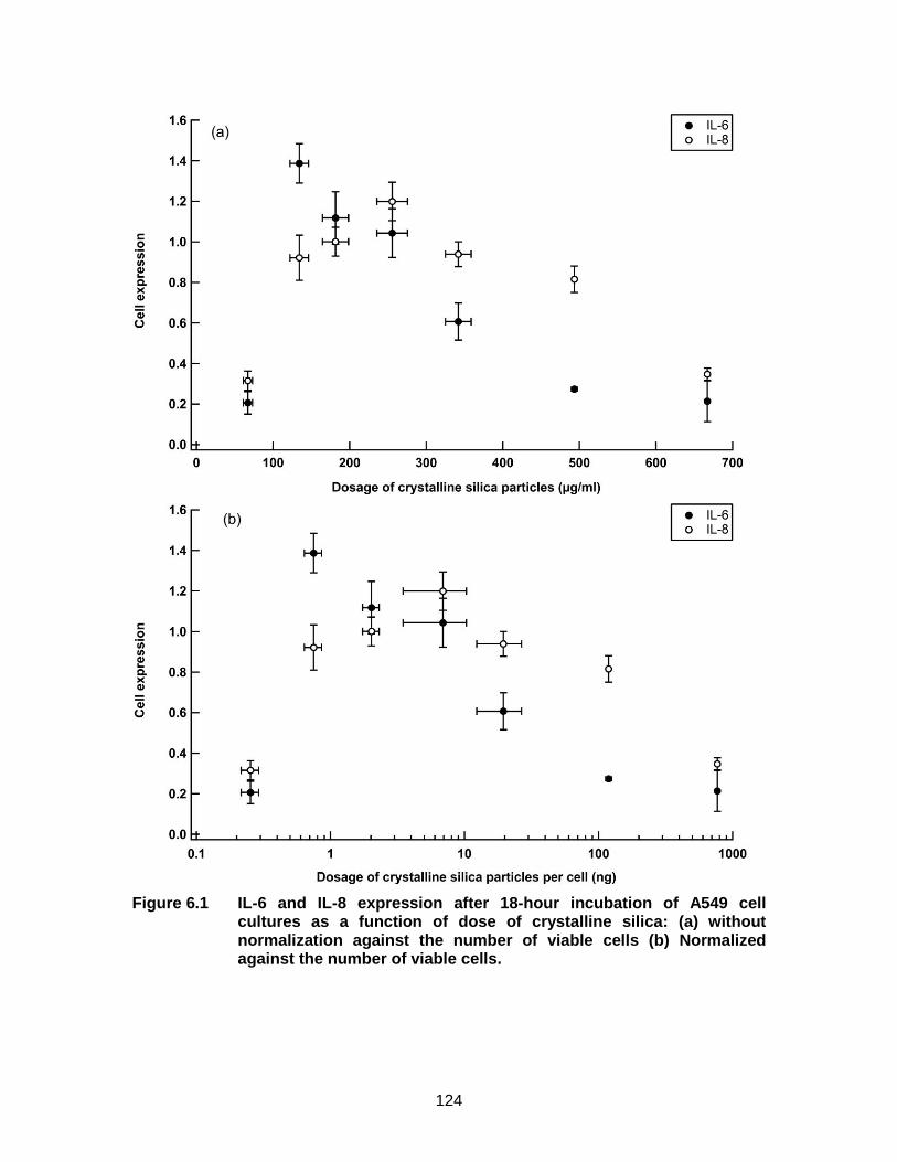
124
Figure 6.1 IL-6 and IL-8 expression after 18-hour incubation of A549 cell cultures as a function of dose of crystalline silica: (a) without normalization against the number of viable cells (b) Normalized against the number of viable cells.

125
6.2.2. Characterization of liquid-solid interface
The results of the combination of insoluble particles and specific soluble
components suggest that the solid-liquid interfacial properties, especially the sorption of
the soluble components on the particle surfaces plays an important role. To further
investigate that how the solid-liquid interfacial properties would affect the cytotoxicity of
the particles, some characterization of particle surfaces and the sorption of specific
soluble components on the particle surfaces need to be done in the future. For instance,
the X-ray photoelectron spectroscopy (XPS) can be applied to measure the elemental
compositions of the particle surfaces. SEM and particle size analyzer can be adopted to
obtain the size/size distribution of the particles. And certain experiments need to be
designed regarding factors that may modify the interfacial sorption of specific
components the particle on surfaces, including particle types, the effect of other solutes,
kinetic factors (e.g. temperature, and interaction time), and the speciation of the specific
components in the solution.
6.2.3. Selection of particle types/components
Several inorganic representatives of major particle types/components adjacent to
roadways were selected in this study, including carbon black nanoparticles, crystalline
silica particles, nickel nanoparticles, and water-soluble particle components ammonium
nitrate and soluble salts of zinc, lead, and iron. Through a binary particle
types/components model of particles (CB nanoparticle or crystalline silica particle) plus
water-soluble particle components, chemical composition dependent cellular response
was measured, e.g. cytoprotective effect was observed for Zn and Fe, whereas Pb plus
crystalline silica lead to a non-linear additive cell expression. To further investigate the
interaction between different particle types/components and screen the important
particle types/components, the number of component/particle types was increased to a
ternary particle types/components model with crystalline silica, iron(II) salt and lead salt.
Though lead and iron(II) salt each have an opposite effect on cell response, when
combined with crystalline silica, low cytokine expression and high cell viability were
measured for the ternary combination. This suggests the important role of reduced redox
species that protect the cell from oxidative stress. Studying different particle
types/components in an incrementally increasing number of species in the particle
types/components model can be expected to generate new information.

126
Other particle components that should be investigated in the future could include
organic compounds such as polycyclic aromatic hydrocarbons and biogenic components
such as endotoxin. These organic components are produced in the incomplete
combustion of fossil fuels.4, 12, 14, 26 With respect to the traffic-derived particulate matter,
hundreds of organic compounds have been identified in soot. Most of them are PAHs
and their derivatives, which have strong carcinogenic and mutagenic potential.10, 33, 53
They can also generate ROS through biotransformation and lead to inflammation.5, 26
Biological particle components such as endotoxin can induce oxidative stress and trigger
lung inflammatory response through a different mechanism, activation of TLRs
pathways.5, 15, 34 The interaction between organic components, endotoxin, water-soluble
components, and particles (e.g. CB nanoparticle or crystalline silica particle) should be
studied in the future.
6.2.4. Method development of long-term effect study
Long-term exposure to PM is associated with inflammation, respiratory and
cardiovascular diseases, and even deaths.1-2, 4-5 At the cellular level, several
mechanisms are believed to contribute to the chronic effect of ambient particles. For
instance, through the generation of ROS, the particles can lead to inflammation. During
this process, inflammatory cells such as macrophages may release mitogenic factors to
stimulate the proliferation of epithelial cells to replace those that have been damaged.
With cell proliferation rate increases, the chance of mutation increases as well.5, 321 It can
lead to fibrosis and structural changes of airways which eventually result in respiratory
diseases such as asthma and interstitial pulmonary fibrosis.5, 136-137
Investigations on the long-term health effects of ambient particles are mostly
epidemiological, whereas in vitro methodology, which takes considerably shorter time
and less cost, is useful for studying acute effects of ambient particles, and rapid
screening of toxic particle types and components due to its limitation of complexity. 79, 140-
141 For adherent cells such as A549, the exposure period is usually up to 72 hours.322
New in vitro methods continue to be introduced. General methods describe repeated
doses for weeks to assess chronic cellular effects of PM, including traditional sub-
cultured cell models and developed models using special bioreactors and
microcarriers.322-324 The question ‘how will a repeated low-doses of particles at each
stage of passaging, affect lung cell response?’ is a possible starting point for developing

127
an in vitro model to study the long-term effect of ambient particles on the cellular level in
future. CB nanoparticle can be used as the first particle type to be evaluated, as the
nanoparticle tends to not be cleared quickly from the respiratory system. A co-culture
cell model including A549 cells and macrophages is suggested to be adopted and
exposed to a constant low concentration of CB nanoparticles each time after cell
passaging. Multiple assays, such as viability assay, enzyme-linked immunosorbent
assay for measuring pro-inflammatory cytokines, and genetic assay, can all be adopted
in a single study to evaluate the chronic cellular effect of the particles.
6.3. Concluding remarks
An in vitro methodology was adopted to investigate the human lung cellular
response induced by traffic-derived particles. It was applied for both ambient particle
samples and laboratory mimics of multiple particle types/components to investigate two
research questions: i. Do interactions between water-insoluble particle types affect
toxicity? ii. Do interactions between water-insoluble particle types and water-soluble
components, and between water-soluble components affect toxicity?
Several conclusions can be drawn based on our results. The interaction between
different particle components/types affects the cellular response in different ways,
certain particle components/types including crystalline silica, lead and iron may dominate
the toxicity of the whole mixture. One probable mechanism of the compositional
interaction is that the water-insoluble particle components play a role as carriers, via
endocytosis, for the water/lipid-soluble components. In this study, due to the limitation of
the methodology, we don’t have sufficient evidence to confirm the internalization of the
particles, via endocytosis. All the statements in the results and discussion sections of
this thesis about endocytosis are proposed assumptions. Instead of suggesting the
importance of endocytosis, based on our results, it would be safer to conclude that the
water-insoluble particles are critical, especially when they combined with soluble species
that are presented to intact viable cells but have no receptor expressed in the cell
membrane. On the cellular level, mass dosage plays an important role in the toxicity of
water-insoluble particles. The particle size and surface area do not pre-determine the
cytotoxicity of the particles. However, other factors, including chemical compositions and
surface chemistry of the particles, should be taken into consideration. For the water-
soluble particle components, their toxicity is mainly determined by the chemical

128
composition. For a given sample of ambient particles, the overall toxicity is affected by
several factors and cannot simply be predicted by the sum of the toxicity of the individual
components in the particle.
A complexed multi-component model for acute response investigation and toxin
screening, and a method development for long-term health effect study are suggested
as future work. The goal of such future research is to further improve the understanding
of particle toxicity, and screen certain particle types/components which we hope would
help to reduce the adverse effect on human health caused by ambient particles.

129
References
1. Cohen, A. J.; Brauer, M.; Burnett, R.; Anderson, H. R.; Frostad, J.; Estep, K.; Balakrishnan, K.; Brunekreef, B.; Dandona, L.; Dandona, R., Estimates and 25-year trends of the global burden of disease attributable to ambient air pollution: an analysis of data from the Global Burden of Diseases Study 2015. The Lancet 2017, 389 (10082), 1907-1918.
2. Gakidou, E.; Afshin, A.; Abajobir, A. A.; Abate, K. H.; Abbafati, C.; Abbas, K. M.; Abd-Allah, F.; Abdulle, A. M.; Abera, S. F.; Aboyans, V., Global, regional, and national comparative risk assessment of 84 behavioural, environmental and occupational, and metabolic risks or clusters of risks, 1990–2016: a systematic analysis for the Global Burden of Disease Study 2016. The Lancet 2017, 390 (10100), 1345-1422.
3. Global Burden of Disease Collaborative Network, Global Burden of Disease Study 2016 (GBD 2016) GBD Compare | IHME Viz Hub - Data Visualizations. . Institute for Health Metrics and Evaluation (IHME): Seattle, United States, 2017.
4. Brook, R. D.; Rajagopalan, S.; Pope, C. A.; Brook, J. R.; Bhatnagar, A.; Diez-Roux, A. V.; Holguin, F.; Hong, Y.; Luepker, R. V.; Mittleman, M. A., Particulate matter air pollution and cardiovascular disease: an update to the scientific statement from the American Heart Association. Circulation 2010, 121 (21), 2331-2378.
5. Donaldson, K.; Borm, P., Particle toxicology. CRC Press: 2006.
6. Lin, C.-C.; Chen, S.-J.; Huang, K.-L.; Lee, W.-J.; Lin, W.-Y.; Liao, C.-J.; Chaung, H.-C.; Chiu, C.-H., Water-soluble ions in nano/ultrafine/fine/coarse particles collected near a busy road and at a rural site. Environmental Pollution 2007, 145 (2), 562-570.
7. Janssen, N. A.; Van Mansom, D. F.; Van Der Jagt, K.; Harssema, H.; Hoek, G., Mass concentration and elemental composition of airborne particulate matter at street and background locations. Atmospheric Environment 1997, 31 (8), 1185-1193.
8. HEI panel on the health effects of traffic-related air pollution, Traffic-related air pollution: a critical review of the literature on emissions, exposure, and health effects. Health Effects Institute: Boston, MA, 2010; Vol. HEI special report 17.
9. Tsang, A. M.; Klepeis, N. E. Descriptive statistics tables from a detailed analysis of the National Human Activity Pattern Survey (NHAPS) data; Lockheed Martin Environmental Systems and Technologies, Las Vegas, NV (United States); Environmental Protection Agency, National Exposure Research Lab., Las Vegas, NV (United States): 1996.

130
10. De Kok, T. M.; Driece, H. A.; Hogervorst, J. G.; Briedé, J. J., Toxicological assessment of ambient and traffic-related particulate matter: a review of recent studies. Mutation Research/Reviews in Mutation Research 2006, 613 (2), 103-122.
11. Grahame, T. J.; Schlesinger, R. B., Cardiovascular health and particulate vehicular emissions: a critical evaluation of the evidence. Air Quality, Atmosphere & Health 2010, 3 (1), 3-27.
12. Seinfeld, J. H.; Pandis, S. N., Atmospheric chemistry and physics: from air pollution to climate change. John Wiley & Sons: 2016.
13. Kim, K.-H.; Kabir, E.; Kabir, S., A review on the human health impact of airborne particulate matter. Environment international 2015, 74, 136-143.
14. Kelly, F. J.; Fussell, J. C., Size, source and chemical composition as determinants of toxicity attributable to ambient particulate matter. Atmospheric Environment 2012, 60, 504-526.
15. Brook, R. D.; Franklin, B.; Cascio, W.; Hong, Y.; Howard, G.; Lipsett, M.; Luepker, R.; Mittleman, M.; Samet, J.; Smith, S. C., Jr.; Tager, I.; Expert Panel on, P.; Prevention Science of the American Heart, A., Air pollution and cardiovascular disease: a statement for healthcare professionals from the Expert Panel on Population and Prevention Science of the American Heart Association. Circulation 2004, 109 (21), 2655-71.
16. DeCarlo, P. F.; Slowik, J. G.; Worsnop, D. R.; Davidovits, P.; Jimenez, J. L., Particle morphology and density characterization by combined mobility and aerodynamic diameter measurements. Part 1: Theory. Aerosol Science and Technology 2004, 38 (12), 1185-1205.
17. U.S. EPA, Air Quality Criteria for Particulate Matter (Final Report, 2004). U.S. Department of Environmental Protection Agency, Ed. EPA 600/P-99/002aF-bF: Washington, DC, 2004.
18. Mansfield, E.; Kaiser, D. L.; Fujita, D.; Van de Voorde, M., Metrology and Standardization for Nanotechnology: Protocols and Industrial Innovations. John Wiley & Sons: 2017.
19. Peters, T. M., Use of the aerodynamic particle sizer to measure ambient PM10–2.5: The coarse fraction of PM10. Journal of the Air & Waste Management Association 2006, 56 (4), 411-416.
20. Daigle, C. C.; Chalupa, D. C.; Gibb, F. R.; Morrow, P. E.; Oberdörster, G.; Utell, M. J.; Frampton, M. W., Ultrafine particle deposition in humans during rest and exercise. Inhalation toxicology 2003, 15 (6), 539-552.

131
21. Amatullah, H.; North, M. L.; Akhtar, U. S.; Rastogi, N.; Urch, B.; Silverman, F. S.; Chow, C.-W.; Evans, G. J.; Scott, J. A., Comparative cardiopulmonary effects of size-fractionated airborne particulate matter. Inhalation toxicology 2012, 24 (3), 161-171.
22. Cho, S.-H.; Tong, H.; McGee, J. K.; Baldauf, R. W.; Krantz, Q. T.; Gilmour, M. I., Comparative toxicity of size-fractionated airborne particulate matter collected at different distances from an urban highway. Environmental health perspectives 2009, 117 (11), 1682.
23. Happo, M.; Salonen, R.; Hälinen, A.; Jalava, P.; Pennanen, A.; Dormans, J.; Gerlofs-Nijland, M.; Cassee, F.; Kosma, V.-M.; Sillanpää, M., Inflammation and tissue damage in mouse lung by single and repeated dosing of urban air coarse and fine particles collected from six European cities. Inhalation toxicology 2010, 22 (5), 402-416.
24. World Health Organization, Health effects of particulate matter. Policy implications for countries in eastern Europe, Caucasus and central Asia World Health Organization: Copenhagen, Denmark, 2013.
25. Harrison, R. M.; Yin, J., Particulate matter in the atmosphere: which particle properties are important for its effects on health? Science of the total environment 2000, 249 (1-3), 85-101.
26. Valavanidis, A.; Fiotakis, K.; Vlachogianni, T., Airborne particulate matter and human health: toxicological assessment and importance of size and composition of particles for oxidative damage and carcinogenic mechanisms. Journal of Environmental Science and Health, Part C 2008, 26 (4), 339-362.
27. Li, C.; Martin, R. V.; van Donkelaar, A.; Boys, B. L.; Hammer, M. S.; Xu, J.-W.; Marais, E. A.; Reff, A.; Strum, M.; Ridley, D. A., Trends in chemical composition of global and regional population-weighted fine particulate matter estimated for 25 years. Environmental Science & Technology 2017, 51 (19), 11185-11195.
28. Agranovski, I., Aerosols: science and technology. John Wiley & Sons: 2011.
29. Riley, M. R.; Boesewetter, D. E.; Kim, A. M.; Sirvent, F. P., Effects of metals Cu, Fe, Ni, V, and Zn on rat lung epithelial cells. Toxicology 2003, 190 (3), 171-184.
30. Duffin, R.; Gilmour, P. S.; Schins, R. P.; Clouter, A.; Guy, K.; Brown, D. M.; MacNee, W.; Borm, P. J.; Donaldson, K.; Stone, V., Aluminium lactate treatment of DQ12 quartz inhibits its ability to cause inflammation, chemokine expression, and nuclear factor-κB activation. Toxicology and applied pharmacology 2001, 176 (1), 10-17.
31. Flora, G.; Gupta, D.; Tiwari, A., Toxicity of lead: a review with recent updates. Interdisciplinary toxicology 2012, 5 (2), 47-58.

132
32. Plum, L. M.; Rink, L.; Haase, H., The essential toxin: impact of zinc on human health. International journal of environmental research and public health 2010, 7 (4), 1342-1365.
33. Shiraiwa, M.; Selzle, K.; Pöschl, U., Hazardous components and health effects of atmospheric aerosol particles: reactive oxygen species, soot, polycyclic aromatic compounds and allergenic proteins. Free radical research 2012, 46 (8), 927-939.
34. Degobbi, C.; Saldiva, P. H. N.; Rogers, C., Endotoxin as modifier of particulate matter toxicity: a review of the literature. Aerobiologia 2011, 27 (2), 97-105.
35. Oberdörster, G.; Oberdörster, E.; Oberdörster, J., Nanotoxicology: an emerging discipline evolving from studies of ultrafine particles. Environmental health perspectives 2005, 113 (7), 823.
36. Bakand, S.; Hayes, A.; Dechsakulthorn, F., Nanoparticles: a review of particle toxicology following inhalation exposure. Inhalation toxicology 2012, 24 (2), 125-135.
37. Gwinn, M. R.; Vallyathan, V., Nanoparticles: health effects—pros and cons. Environmental health perspectives 2006, 114 (12), 1818.
38. Warheit, D. B., Nanoparticles: health impacts? Materials today 2004, 7 (2), 32-35.
39. Wallace, W. E.; Keane, M. J.; Murray, D. K.; Chisholm, W. P.; Maynard, A. D.; Ong, T.-m., Phospholipid lung surfactant and nanoparticle surface toxicity: Lessons from diesel soots and silicate dusts. In Nanotechnology and Occupational Health, Springer: 2006; pp 23-38.
40. Brown, D.; Kinloch, I.; Bangert, U.; Windle, A.; Walter, D.; Walker, G.; Scotchford, C.; Donaldson, K.; Stone, V., An in vitro study of the potential of carbon nanotubes and nanofibres to induce inflammatory mediators and frustrated phagocytosis. Carbon 2007, 45 (9), 1743-1756.
41. Monteiller, C.; Tran, L.; MacNee, W.; Faux, S.; Jones, A.; Miller, B.; Donaldson, K., The pro-inflammatory effects of low-toxicity low-solubility particles, nanoparticles and fine particles, on epithelial cells in vitro: the role of surface area. Occupational and environmental medicine 2007, 64 (9), 609-615.
42. Braakhuis, H. M.; Park, M. V.; Gosens, I.; De Jong, W. H.; Cassee, F. R., Physicochemical characteristics of nanomaterials that affect pulmonary inflammation. Particle and fibre toxicology 2014, 11 (1), 18.
43. Oberdörster, G.; Stone, V.; Donaldson, K., Toxicology of nanoparticles: a historical perspective. Nanotoxicology 2007, 1 (1), 2-25.

133
44. Schmid, O.; Möller, W.; Semmler-Behnke, M.; A. Ferron, G.; Karg, E.; Lipka, J.; Schulz, H.; Kreyling, W.; Stoeger, T., Dosimetry and toxicology of inhaled ultrafine particles. Biomarkers 2009, 14 (sup1), 67-73.
45. Sayes, C. M.; Warheit, D. B., Characterization of nanomaterials for toxicity assessment. Wiley Interdisciplinary Reviews: Nanomedicine and Nanobiotechnology 2009, 1 (6), 660-670.
46. Park, S.-J.; Seo, M.-K., Interface science and composites. Academic Press: 2011; Vol. 18.
47. Brown, M. A.; Abbas, Z.; Kleibert, A.; Green, R. G.; Goel, A.; May, S.; Squires, T. M., Determination of surface potential and electrical double-layer structure at the aqueous electrolyte-nanoparticle interface. Physical Review X 2016, 6 (1), 011007.
48. Kettler, K.; Veltman, K.; van de Meent, D.; van Wezel, A.; Hendriks, A. J., Cellular uptake of nanoparticles as determined by particle properties, experimental conditions, and cell type. Environmental toxicology and chemistry 2014, 33 (3), 481-492.
49. Shin, S. W.; Song, I. H.; Um, S. H., Role of physicochemical properties in nanoparticle toxicity. Nanomaterials 2015, 5 (3), 1351-1365.
50. Madl, A. K.; Pinkerton, K. E., Health effects of inhaled engineered and incidental nanoparticles. Critical reviews in toxicology 2009, 39 (8), 629-658.
51. Fubini, B., Surface reactivity in the pathogenic response to particulates. Environmental health perspectives 1997, 105 (Suppl 5), 1013.
52. Duffin, R.; Tran, C.; Clouter, A.; Brown, D.; MacNee, W.; Stone, V.; Donaldson, K., The importance of surface area and specific reactivity in the acute pulmonary inflammatory response to particles. Annals of Occupational Hygiene 2002, 46 (suppl_1), 242-245.
53. Amato, F.; Schaap, M.; Reche, C.; Querol, X., Road traffic: a major source of particulate matter in Europe. In Urban Air Quality in Europe, Springer: 2013; pp 165-193.
54. Thorpe, A.; Harrison, R. M., Sources and properties of non-exhaust particulate matter from road traffic: a review. Science of the total environment 2008, 400 (1-3), 270-282.
55. International Agency for Research on Cancer (IARC), IARC Monographs on the evaluation of the carcinogenic risk of chemicals to humans. . World Health Organization: Lyon, France, 1985; Vol. 35.

134
56. Watson, A. Y.; Valberg, P. A., Carbon black and soot: two different substances. AIHAJ-American Industrial Hygiene Association 2001, 62 (2), 218-228.
57. International Agency for Research on Cancer (IARC), IARC monographs on the evaluation of carcinogenic risks to humans. World Health Organization: Lyon, France, 2012; Vol. 100F.
58. Wilson, S. J.; Miller, M. R.; Newby, D. E., Effects of diesel exhaust on cardiovascular function and oxidative stress. Antioxidants & redox signaling 2018, 28 (9), 819-836.
59. Grigoratos, T.; Martini, G., Brake wear particle emissions: a review. Environmental Science and Pollution Research 2015, 22 (4), 2491-2504.
60. Garg, B. D.; Cadle, S. H.; Mulawa, P. A.; Groblicki, P. J.; Laroo, C.; Parr, G. A., Brake wear particulate matter emissions. Environmental Science & Technology 2000, 34 (21), 4463-4469.
61. Chow, J. C.; Watson, J. G.; Houck, J. E.; Pritchett, L. C.; Rogers, C. F.; Frazier, C. A.; Egami, R. T.; Ball, B. M., A laboratory resuspension chamber to measure fugitive dust size distributions and chemical compositions. Atmospheric Environment 1994, 28 (21), 3463-3481.
62. Han, S.; Youn, J.-S.; Jung, Y.-W., Characterization of PM10 and PM2. 5 source profiles for resuspended road dust collected using mobile sampling methodology. Atmospheric environment 2011, 45 (20), 3343-3351.
63. U.S. EPA, National Emissions Inventory (NEI) Air Pollutant Emissions Trends Data. https://www.epa.gov/air-emissions-inventories/air-pollutant-emissions-trends-data, 2017.
64. Environment and Climate Change Canada, 1990-2016 Air Pollutant Emission Inventory Report. Minsiter of Environment and Climate, C., Ed. Minsiter of Environment and Climate, Canada: Gatineau, QC, 2018.
65. Zhu, Y.; Hinds, W. C.; Kim, S.; Shen, S.; Sioutas, C., Study of ultrafine particles near a major highway with heavy-duty diesel traffic. Atmospheric environment 2002, 36 (27), 4323-4335.
66. Roorda-Knape, M. C.; Janssen, N. A.; De Hartog, J. J.; Van Vliet, P. H.; Harssema, H.; Brunekreef, B., Air pollution from traffic in city districts near major motorways. Atmospheric Environment 1998, 32 (11), 1921-1930.
67. Schwartz, J.; Laden, F.; Zanobetti, A., The concentration-response relation between PM (2.5) and daily deaths. Environmental health perspectives 2002, 110 (10), 1025.
135
68. Lipfert, F.; Wyzga, R.; Baty, J.; Miller, J., Traffic density as a surrogate measure of environmental exposures in studies of air pollution health effects: long-term mortality in a cohort of US veterans. Atmospheric Environment 2006, 40 (1), 154-169.
69. Heinrich, J.; Schwarze, P. E.; Stilianakis, N.; Momas, I.; Medina, S.; Totlandsdal, A. I.; Von Bree, L.; Kuna-Dibbert, B.; Krzyzanowski, M., Studies on health effects of transport-related air pollution. Health effects of transport-related air pollution 2005, 125.
70. Hoffmann, B.; Moebus, S.; Möhlenkamp, S.; Stang, A.; Lehmann, N.; Dragano, N.; Schmermund, A.; Memmesheimer, M.; Mann, K.; Erbel, R., Residential exposure to traffic is associated with coronary atherosclerosis. Circulation 2007, 116 (5), 489-496.
71. Finkelstein, M. M.; Jerrett, M.; Sears, M. R., Environmental inequality and circulatory disease mortality gradients. Journal of Epidemiology & Community Health 2005, 59 (6), 481-487.
72. Gehring, U.; Heinrich, J.; Krämer, U.; Grote, V.; Hochadel, M.; Sugiri, D.; Kraft, M.; Rauchfuss, K.; Eberwein, H. G.; Wichmann, H.-E., Long-term exposure to ambient air pollution and cardiopulmonary mortality in women. Epidemiology 2006, 17 (5), 545-551.
73. Hofmann, W., Modelling inhaled particle deposition in the human lung—a review. Journal of Aerosol Science 2011, 42 (10), 693-724.
74. Kleinstreuer, C.; Zhang, Z., Airflow and particle transport in the human respiratory system. Annual Review of Fluid Mechanics 2010, 42, 301-334.
75. Kleinstreuer, C.; Zhang, Z.; Donohue, J., Targeted drug-aerosol delivery in the human respiratory system. Annu. Rev. Biomed. Eng. 2008, 10, 195-220.
76. Ionescu, C. M., The human respiratory system. In The Human Respiratory System, Springer: 2013; pp 13-22.
77. Bair, W., The ICRP human respiratory tract model for radiological protection. Radiation Protection Dosimetry 1995, 60 (4), 307-310.
78. Smith, H., Human respiratory tract model for radiological protection. ICRP publication 66 1994, 24(1-3), 1-482.
79. BéruBé, K.; Aufderheide, M.; Breheny, D.; Clothier, R.; Combes, R.; Duffin, R.; Forbes, B.; Gaça, M.; Gray, A.; Hall, I., In vitro models of inhalation toxicity and disease. Alternatives to Laboratory Animals 2009, 37 (1), 89-141.

136
80. Widdicombe, J.; Bastacky, S.; Wu, D.; Lee, C., Regulation of depth and composition of airway surface liquid. European Respiratory Journal 1997, 10 (12), 2892-2897.
81. Franciosi, L.; Govorukhina, N.; ten Hacken, N.; Postma, D.; Bischoff, R., Proteomics of epithelial lining fluid obtained by bronchoscopic microprobe sampling. In Nanoproteomics, Springer: 2011; pp 17-28.
82. Cantin, A.; North, S.; Hubbard, R.; Crystal, R., Normal alveolar epithelial lining fluid contains high levels of glutathione. Journal of applied physiology 1987, 63 (1), 152-157.
83. Stanke, F., The contribution of the airway epithelial cell to host defense. Mediators of inflammation 2015, 2015.
84. Boers, J. E.; Ambergen, A. W.; Thunnissen, F. B., Number and proliferation of basal and parabasal cells in normal human airway epithelium. American journal of respiratory and critical care medicine 1998, 157 (6), 2000-2006.
85. Wallaert, B.; Fahy, O.; Tsicopoulos, A.; Gosset, P.; Tonnel, A., Experimental systems for mechanistic studies of toxicant induced lung inflammation. Toxicology letters 2000, 112, 157-163.
86. Foster, K. A.; Oster, C. G.; Mayer, M. M.; Avery, M. L.; Audus, K. L., Characterization of the A549 cell line as a type II pulmonary epithelial cell model for drug metabolism. Experimental cell research 1998, 243 (2), 359-366.
87. Mühlfeld, C.; Blank, F.; Rothen-Rutishauser, B.; Gehr, P., Particle-lung interactions. CRC Press: 2009.
88. Ryan, R. M.; Mineo-Kuhn, M. M.; Kramer, C. M.; Finkelstein, J. N., Growth factors alter neonatal type II alveolar epithelial cell proliferation. American Journal of Physiology-Lung Cellular and Molecular Physiology 1994, 266 (1), L17-L22.
89. Riley, M. R.; Boesewetter, D. E.; Turner, R. A.; Kim, A. M.; Collier, J. M.; Hamilton, A., Comparison of the sensitivity of three lung derived cell lines to metals from combustion derived particulate matter. Toxicology in Vitro 2005, 19 (3), 411-419.
90. Ward, H.; Nicholas, T., Alveolar type I and type II cells. Australian and New Zealand journal of medicine 1984, 14, 731-734.
91. Ito, T.; Kitamura, H.; Inayama, Y.; Nozawa, A.; Kanisawa, M., Uptake and intracellular transport of cationic ferritin in the bronchiolar and alveolar epithelia of the rat. Cell and tissue research 1992, 268 (2), 335-340.

137
92. Stearns, R. C.; Paulauskis, J. D.; Godleski, J. J., Endocytosis of ultrafine particles by A549 cells. American journal of respiratory cell and molecular biology 2001, 24 (2), 108-115.
93. Bai, Y.; Suzuki, A. K.; Sagai, M., The cytotoxic effects of diesel exhaust particles on human pulmonary artery endothelial cells in vitro: role of active oxygen species. Free Radical Biology and Medicine 2001, 30 (5), 555-562.
94. Wang, T.; Shimizu, Y.; Wu, X.; Kelly, G. T.; Xu, X.; Wang, L.; Qian, Z.; Chen, Y.; Garcia, J. G., Particulate matter disrupts human lung endothelial cell barrier integrity via Rho-dependent pathways. Pulmonary circulation 2017, 7 (3), 617-623.
95. Lenasi, H., Endothelial Dysfunction. IntechOpen: 2018.
96. World Health Organization, WHO Air quality guidelines for particulate matter, ozone, nitrogen dioxide and sulfur dioxide: global update 2005: summary of risk assessment. World Health Organization: Geneva, Switzerland, 2006.
97. Pope, C. A.; Thun, M. J.; Namboodiri, M. M.; Dockery, D. W.; Evans, J. S.; Speizer, F. E.; Heath, C. W., Particulate air pollution as a predictor of mortality in a prospective study of US adults. American journal of respiratory and critical care medicine 1995, 151 (3), 669-674.
98. U.S. EPA, National Ambient Air Quality Standards. U.S. Environmental Protection Agency, Ed. Washington, DC 2015.
99. U.S. EPA, Clean Air Act. U.S. Environmental Protection Agency, Ed. Washington, DC 1990.
100. Agency for Toxic Substances and Disease Registry (ATSDR), Toxicological profile for lead. U.S. Department of Health and Human Services, Public Health Service: Atlanta, Georgia, 2007.
101. U.S. EPA, Menu Of Control Measures. U.S. Environmental Protection Agency, Ed. Washington, DC 2012.
102. Canadian Council of Ministers of the Environment Air Quality Management System (AQMS). (accessed Jun 26).
103. Canadian Council of Ministers of the Environment, Canadian Ambient Air Quality Standards. Canadian Council of Ministers of the Environment, Ed. http://www.ec.gc.ca/default.asp?lang=En&n=56D4043B-1&news=A4B2C28A-2DFB-4BF4-8777-ADF29B4360BD (Accessed Jun 26, 2018), 2012.

138
104. Environment Directorate General of the European Commission, Air Quality Standards. Environment Directorate General of the European Commission, Ed. http://ec.europa.eu/environment/basics/home_en.htm (Accessed Jun 26, 2018), 2017.
105. Directorate General for Internal Market Industry Entrepreneurship and SMEs of European Commission Emissions in the automotive sector. (accessed June 26).
106. Heyder, J., Deposition of inhaled particles in the human respiratory tract and consequences for regional targeting in respiratory drug delivery. Proceedings of the American Thoracic Society 2004, 1 (4), 315-320.
107. Smith, H., Human respiratory tract model for radiological protection. ICRP publication 66. Ann. ICRP 1994, 24 (1-3), 1-482.
108. Hussain, M.; Madl, P.; Khan, A., Lung deposition predictions of airborne particles and the emergence of contemporary diseases, Part-I. Health 2011, 2 (2), 51-59.
109. Miyata, R.; van Eeden, S. F., The innate and adaptive immune response induced by alveolar macrophages exposed to ambient particulate matter. Toxicology and applied pharmacology 2011, 257 (2), 209-226.
110. Lippmann, M.; Yeates, D.; Albert, R., Deposition, retention, and clearance of inhaled particles. Occupational and Environmental Medicine 1980, 37 (4), 337-362.
111. Kumar, S.; Verma, M. K.; Srivastava, A. K., Ultrafine particles in urban ambient air and their health perspectives. Reviews on environmental health 2013, 28 (2-3), 117-128.
112. Smith, J.; Bailey, M.; Etherington, G.; Shutt, A.; Youngman, M., Effect of particle size on slow particle clearance from the bronchial tree. Experimental lung research 2008, 34 (6), 287-312.
113. Warheit, D.; Hansen, J.; Yuen, I.; Kelly, D.; Snajdr, S.; Hartsky, M., Inhalation of high concentrations of low toxicity dusts in rats results in impaired pulmonary clearance mechanisms and persistent inflammation. Toxicology and applied pharmacology 1997, 145 (1), 10-22.
114. Morrow, P., Possible mechanisms to explain dust overloading of the lungs. Toxicological Sciences 1988, 10 (3), 369-384.
115. Mukherjee, S.; Ghosh, R. N.; Maxfield, F. R., Endocytosis. Physiological reviews 1997, 77 (3), 759-803.
116. Gehr, P.; Clift, M.; Brandenberger, C.; Lehmann, A.; Herzog, F.; Rothen-Rutishauser, B., Endocytosis of environmental and engineered micro-and nanosized particles. Compr. Physiol 2011, 1, 1159-1174.

139
117. Greenberg, S.; Grinstein, S., Phagocytosis and innate immunity. Current opinion in immunology 2002, 14 (1), 136-145.
118. Aderem, A.; Underhill, D. M., Mechanisms of phagocytosis in macrophages. Annual review of immunology 1999, 17 (1), 593-623.
119. Silverstein, S. C.; Steinman, R. M.; Cohn, Z. A., Endocytosis. Annual review of biochemistry 1977, 46 (1), 669-722.
120. Lim, J. P.; Gleeson, P. A., Macropinocytosis: an endocytic pathway for internalising large gulps. Immunology and cell biology 2011, 89 (8), 836.
121. Kerr, M. C.; Teasdale, R. D., Defining macropinocytosis. Traffic 2009, 10 (4), 364-371.
122. Muhlfeld, C.; Gehr, P.; Rothen-Rutishauser, B., Translocation and cellular entering mechanisms of nanoparticles in the respiratory tract. Swiss medical weekly 2008, 138 (27/28), 387.
123. Boland, S.; Hussain, S.; Baeza-Squiban, A., Carbon black and titanium dioxide nanoparticles induce distinct molecular mechanisms of toxicity. Wiley interdisciplinary reviews. Nanomedicine and nanobiotechnology 2014, 6 (6), 641-52.
124. Hussain, S.; Thomassen, L. C.; Ferecatu, I.; Borot, M.-C.; Andreau, K.; Martens, J. A.; Fleury, J.; Baeza-Squiban, A.; Marano, F.; Boland, S., Carbon black and titanium dioxide nanoparticles elicit distinct apoptotic pathways in bronchial epithelial cells. Particle and fibre toxicology 2010, 7 (1), 10.
125. Bayr, H., Reactive oxygen species. Critical care medicine 2005, 33 (12), S498-S501.
126. Finkel, T.; Holbrook, N. J., Oxidants, oxidative stress and the biology of ageing. Nature 2000, 408 (6809), 239.
127. Britton, R. S.; Leicester, K. L.; Bacon, B. R., Iron toxicity and chelation therapy. International journal of hematology 2002, 76 (3), 219-228.
128. Dixon, S. J.; Stockwell, B. R., The role of iron and reactive oxygen species in cell death. Nature chemical biology 2014, 10 (1), 9-17.
129. Fubini, B.; Hubbard, A., Serial review: role of reactive oxygen and nitrogen species (ROS/RNS) in lung injury and diseases. Free Radical Biology & Medicine 2003, 34 (12), 1507-1516.
130. Shi, X.; Castranova, V.; Halliwell, B.; Vallyathan, V., Reactive oxygen species and silica‐induced carcinogenesis. Journal of Toxicology and Environmental Health, Part B Critical Reviews 1998, 1 (3), 181-197.

140
131. Dutta, D.; Moudgil, B. M., Crystalline silica particles mediated lung injury. KONA Powder and Particle Journal 2007, 25, 76-87.
132. Coussens, L. M.; Werb, Z., Inflammation and cancer. Nature 2002, 420 (6917), 860.
133. Gabay, C., Interleukin-6 and chronic inflammation. Arthritis research & therapy 2006, 8 (2), S3.
134. Astrakianakis, G.; Murray, E., Conflicting Effects of Occupational Endotoxin Exposure on Lung Health-A Hypothesis-Generating Review of Cancer and COPD Risk. J Environ Immunol Toxicol 2014, 1 (3), 128-139.
135. Khansari, N.; Shakiba, Y.; Mahmoudi, M., Chronic inflammation and oxidative stress as a major cause of age-related diseases and cancer. Recent patents on inflammation & allergy drug discovery 2009, 3 (1), 73-80.
136. Snider, G. L., Interstitial pulmonary fibrosis. Chest 1986, 89 (3), 115S-121S.
137. Bateman, E. D.; Hurd, S.; Barnes, P.; Bousquet, J.; Drazen, J.; FitzGerald, M.; Gibson, P.; Ohta, K.; O'byrne, P.; Pedersen, S., Global strategy for asthma management and prevention: GINA executive summary. European Respiratory Journal 2008, 31 (1), 143-178.
138. Bokulic, R. E., The lung: Scientific foundations. By Ronald G. Crystal and John D. West. New York: Raven Press, Ltd., 1991, 2 volumes, 2,224 pp. Wiley Online Library: 1992.
139. Pasanen, K.; Pukkala, E.; Turunen, A. W.; Patama, T.; Jussila, I.; Makkonen, S.; Salonen, R. O.; Verkasalo, P. K., Mortality among population with exposure to industrial air pollution containing nickel and other toxic metals. Journal of occupational and environmental medicine 2012, 54 (5), 583-591.
140. Devlin, R. B.; Frampton, M. L.; Ghio, A. J., In vitro studies: What is their role in toxicology? Experimental and Toxicologic Pathology 2005, 57, 183-188.
141. Nemmar, A.; Holme, J. A.; Rosas, I.; Schwarze, P. E.; Alfaro-Moreno, E., Recent advances in particulate matter and nanoparticle toxicology: a review of the in vivo and in vitro studies. BioMed research international 2013, 2013.
142. Zou, Y.; Jin, C.; Su, Y.; Li, J.; Zhu, B., Water soluble and insoluble components of urban PM2. 5 and their cytotoxic effects on epithelial cells (A549) in vitro. Environmental pollution 2016, 212, 627-635.
143. Valberg, P. A., Is PM more toxic than the sum of its parts? Risk-assessment toxicity factors vs. PM-mortality "effect functions". Inhalation toxicology 2004, 16 Suppl 1, 19-29.

141
144. Bakand, S.; Winder, C.; Khalil, C.; Hayes, A., Toxicity assessment of industrial chemicals and airborne contaminants: transition from in vivo to in vitro test methods: a review. Inhalation toxicology 2005, 17 (13), 775-787.
145. Paur, H.-R.; Cassee, F. R.; Teeguarden, J.; Fissan, H.; Diabate, S.; Aufderheide, M.; Kreyling, W. G.; Hänninen, O.; Kasper, G.; Riediker, M., In-vitro cell exposure studies for the assessment of nanoparticle toxicity in the lung—A dialog between aerosol science and biology. Journal of Aerosol Science 2011, 42 (10), 668-692.
146. International Agency for Research on Cancer (IARC), IARC Monographs on the evaluation of the carcinogenic risk of chemicals to humans. World Health Organization: Lyon, France, 1997; Vol. 68.
147. Antognelli, C.; Gambelunghe, A.; Del Buono, C.; Murgia, N.; Talesa, V. N.; Muzi, G., Crystalline silica Min-U-Sil 5 induces oxidative stress in human bronchial epithelial cells BEAS-2B by reducing the efficiency of antiglycation and antioxidant enzymatic defenses. Chemico-biological interactions 2009, 182 (1), 13-21.
148. Øvrevik, J.; Refsnes, M.; Namork, E.; Becher, R.; Sandnes, D.; Schwarze, P. E.; Låg, M., Mechanisms of silica-induced IL-8 release from A549 cells: initial kinase-activation does not require EGFR activation or particle uptake. Toxicology 2006, 227 (1-2), 105-116.
149. Donaldson, K.; Borm, P. J., The quartz hazard: a variable entity. The Annals of occupational hygiene 1998, 42 (5), 287-294.
150. Bégin, R.; Massé, S.; Sébastien, P.; Martel, M.; Bossé, J.; Dubois, F.; Geoffroy, M.; Labbe, J., Sustained efficacy of aluminum to reduce quartz toxicity in the lung. Experimental lung research 1987, 13 (2), 205-222.
151. Castranova, V.; Vallyathan, V.; Ramsey, D. M.; McLaurin, J. L.; Pack, D.; Leonard, S.; Barger, M. W.; Ma, J.; Dalal, N. S.; Teass, A., Augmentation of pulmonary reactions to quartz inhalation by trace amounts of iron-containing particles. Environmental Health Perspectives 1997, 105 (Suppl 5), 1319.
152. Cullen, R.; Vallyathan, V.; Hagen, S.; Donaldson, K., Protection by iron against the toxic effects of quartz. The Annals of Occupational Hygiene 1997, 41, 420-425.
153. Giard, D. J.; Aaronson, S. A.; Todaro, G. J.; Arnstein, P.; Kersey, J. H.; Dosik, H.; Parks, W. P., In vitro cultivation of human tumors: establishment of cell lines derived from a series of solid tumors. Journal of the National Cancer Institute 1973, 51 (5), 1417-1423.
154. Lieber, M.; Todaro, G.; Smith, B.; Szakal, A.; Nelson‐Rees, W., A continuous
tumor‐cell line from a human lung carcinoma with properties of type II alveolar epithelial cells. International journal of cancer 1976, 17 (1), 62-70.

142
155. Chung, K. F.; Barnes, P. J., Cytokines in asthma. Thorax 1999, 54 (9), 825-857.
156. Strieter, R. M.; Kunkel, S. L.; Bone, R. C., Role of tumor necrosis factor-alpha in disease states and inflammation. Critical care medicine 1993, 21 (10 Suppl), S447-63.
157. Standiford, T. J.; Kunkel, S.; Phan, S.; Rollins, B.; Strieter, R., Alveolar macrophage-derived cytokines induce monocyte chemoattractant protein-1 expression from human pulmonary type II-like epithelial cells. Journal of Biological Chemistry 1991, 266 (15), 9912-9918.
158. Hunter, C. A.; Jones, S. A., IL-6 as a keystone cytokine in health and disease. Nature immunology 2015, 16 (5), 448.
159. Scheller, J.; Chalaris, A.; Schmidt-Arras, D.; Rose-John, S., The pro-and anti-inflammatory properties of the cytokine interleukin-6. Biochimica et Biophysica Acta (BBA)-Molecular Cell Research 2011, 1813 (5), 878-888.
160. Harada, A.; Sekido, N.; Akahoshi, T.; Wada, T.; Mukaida, N.; Matsushima, K.,
Essential involvement of interleukin‐8 (IL‐8) in acute inflammation. Journal of leukocyte biology 1994, 56 (5), 559-564.
161. Hetland, R. B.; Refsnes, M.; Myran, T.; Johansen, B. V.; Uthus, N.; Schwarze, P. E., Mineral and/or metal content as critical determinants of particle-induced release of IL-6 and IL-8 from A549 cells. Journal of Toxicology and Environmental Health Part A 2000, 60 (1), 47-65.
162. Arnold, R.; Humbert, B.; Werchau, H.; Gallati, H.; König, W., Interleukin-8, interleukin-6, and soluble tumour necrosis factor receptor type I release from a human pulmonary epithelial cell line (A549) exposed to respiratory syncytial virus. Immunology 1994, 82 (1), 126.
163. Gan, S. D.; Patel, K. R., Enzyme immunoassay and enzyme-linked immunosorbent assay. J Invest Dermatol 2013, 133 (9), e12.
164. Strober, W., Trypan blue exclusion test of cell viability. Current protocols in immunology 2015, 111 (1), A3. B. 1-A3. B. 3.
165. Ravishankara, A., Heterogeneous and multiphase chemistry in the troposphere. Science 1997, 276 (5315), 1058-1065.
166. Adamson, I. Y.; Prieditis, H.; Vincent, R., Soluble and insoluble air particle fractions induce differential production of tumor necrosis factor α in rat lung. Experimental lung research 2004, 30 (5), 355-368.
167. Yi, S.; Zhang, F.; Qu, F.; Ding, W., Water‐insoluble fraction of airborne particulate matter (PM10) induces oxidative stress in human lung epithelial A549 cells. Environmental toxicology 2014, 29 (2), 226-233.

143
168. Palleschi, S.; Rossi, B.; Armiento, G.; Montereali, M. R.; Nardi, E.; Tagliani, S. M.; Inglessis, M.; Gianfagna, A.; Silvestroni, L., Toxicity of the readily leachable fraction of urban PM 2.5 to human lung epithelial cells: Role of soluble metals. Chemosphere 2017.
169. Adamson, I. Y.; Prieditis, H.; Vincent, R., Pulmonary toxicity of an atmospheric particulate sample is due to the soluble fraction. Toxicology and applied pharmacology 1999, 157 (1), 43-50.
170. Imrich, C.-A. W. G. A.; Ning, H. D. Y., Analysis of air pollution particulate-mediated oxidant stress in alveolar macrophages. Journal of Toxicology and Environmental Health Part A 1998, 54 (7), 529-545.
171. Kodavanti, U. P.; Hauser, R.; Christiani, D. C.; Meng, Z. H.; McGee, J.; Ledbetter, A.; Richards, J.; Costa, D. L., Pulmonary responses to oil fly ash particles in the rat differ by virtue of their specific soluble metals. Toxicological Sciences 1998, 43 (2), 204-212.
172. Okeson, C. D.; Riley, M. R.; Fernandez, A.; Wendt, J. O., Impact of the composition of combustion generated fine particles on epithelial cell toxicity: influences of metals on metabolism. Chemosphere 2003, 51 (10), 1121-1128.
173. Prieditis, H.; Adamson, I., Comparative pulmonary toxicity of various soluble metals found in urban particulate dusts. Experimental lung research 2002, 28 (7), 563-576.
174. Huang, M.; Kang, Y.; Wang, W.; Chan, C. Y.; Wang, X.; Wong, M. H., Potential cytotoxicity of water-soluble fraction of dust and particulate matters and relation to metal (loid) s based on three human cell lines. Chemosphere 2015, 135, 61-66.
175. Imrich, A.; Ning, Y.; Kobzik, L., Insoluble components of concentrated air particles mediate alveolar macrophage responses in vitro. Toxicology and applied pharmacology 2000, 167 (2), 140-150.
176. Ghio, A. J., Metals associated with both the water-soluble and insoluble fractions of an ambient air pollution particle catalyze an oxidative stress. Inhalation toxicology 1999, 11 (1), 37-49.
177. Jalava, P. I.; Salonen, R. O.; Pennanen, A. S.; Happo, M. S.; Penttinen, P.; Hälinen, A. I.; Sillanpää, M.; Hillamo, R.; Hirvonen, M.-R., Effects of solubility of urban air fine and coarse particles on cytotoxic and inflammatory responses in RAW 264.7 macrophage cell line. Toxicology and Applied Pharmacology 2008, 229 (2), 146-160.
178. Soukup, J. M.; Becker, S., Human alveolar macrophage responses to air pollution particulates are associated with insoluble components of coarse material, including particulate endotoxin. Toxicology and applied pharmacology 2001, 171 (1), 20-26.

144
179. Ayres, J. G.; Borm, P.; Cassee, F. R.; Castranova, V.; Donaldson, K.; Ghio, A.; Harrison, R. M.; Hider, R.; Kelly, F.; Kooter, I. M., Evaluating the toxicity of airborne particulate matter and nanoparticles by measuring oxidative stress potential—a workshop report and consensus statement. Inhalation toxicology 2008, 20 (1), 75-99.
180. Beck-Speier, I.; Kreyling, W. G.; Maier, K. L.; Dayal, N.; Schladweiler, M. C.; Mayer, P.; Semmler-Behnke, M.; Kodavanti, U. P., Soluble iron modulates iron oxide particle-induced inflammatory responses via prostaglandin E 2 synthesis: In vitro and in vivo studies. Particle and fibre toxicology 2009, 6 (1), 34.
181. Fujii, T.; Hayashi, S.; Hogg, J. C.; Vincent, R.; Van Eeden, S. F., Particulate matter induces cytokine expression in human bronchial epithelial cells. American Journal of Respiratory Cell and Molecular Biology 2001, 25 (3), 265-271.
182. Kunzli, N.; Kaiser, R.; Medina, S.; Studnicka, M., Public-health impact of outdoor and traffic-related air pollution: a European assessment. The Lancet 2000, 356 (9232), 795.
183. Hoek, G.; Brunekreef, B.; Goldbohm, S.; Fischer, P.; van den Brandt, P. A., Association between mortality and indicators of traffic-related air pollution in the Netherlands: a cohort study. The lancet 2002, 360 (9341), 1203-1209.
184. Künzli, N.; Kaiser, R.; Medina, S.; Studnicka, M.; Chanel, O.; Filliger, P.; Herry, M.; Horak, F.; Puybonnieux-Texier, V.; Quénel, P., Public-health impact of outdoor and traffic-related air pollution: a European assessment. The Lancet 2000, 356 (9232), 795-801.
185. Lippmann, M., Toxicological and epidemiological studies of cardiovascular effects of ambient air fine particulate matter (PM2. 5) and its chemical components: coherence and public health implications. Critical reviews in toxicology 2014, 44 (4), 299-347.
186. Riedl, M.; Diaz-Sanchez, D., Biology of diesel exhaust effects on respiratory function. Journal of Allergy and Clinical Immunology 2005, 115 (2), 221-228.
187. Shah, S. D.; Cocker, D. R.; Miller, J. W.; Norbeck, J. M., Emission rates of particulate matter and elemental and organic carbon from in-use diesel engines. Environmental Science & Technology 2004, 38 (9), 2544-2550.
188. Vohler, O.; GmbH, S.; Collin, G.; V., D. E.; Wege, E.; Gmbh, S.; Meitingen; Forhs, W., Ullmann's Encyclopedia of chemical technology. 6 ed.; Wiley-VCH: New York, 2003; Vol. 6.
189. Rivin, D.; Hutzinger, O.; Beek, B.; Metzler, M., The handbook of environmental chemistry. Springer: Berlin, 1986; Vol. 3.

145
190. Hussain, S.; Boland, S.; Baeza-Squiban, A.; Hamel, R.; Thomassen, L. C.; Martens, J. A.; Billon-Galland, M. A.; Fleury-Feith, J.; Moisan, F.; Pairon, J. C.; Marano, F., Oxidative stress and proinflammatory effects of carbon black and titanium dioxide nanoparticles: role of particle surface area and internalized amount. Toxicology 2009, 260 (1-3), 142-9.
191. Donaldson, K.; Tran, L.; Jimenez, L. A.; Duffin, R.; Newby, D. E.; Mills, N.; MacNee, W.; Stone, V., Combustion-derived nanoparticles: a review of their toxicology following inhalation exposure. Particle and fibre toxicology 2005, 2 (1), 10.
192. Aam, B. B.; Fonnum, F., Carbon black particles increase reactive oxygen species formation in rat alveolar macrophages in vitro. Archives of toxicology 2007, 81 (6), 441-6.
193. Foucaud, L.; Goulaouic, S.; Bennasroune, A.; Laval-Gilly, P.; Brown, D.; Stone, V.; Falla, J., Oxidative stress induction by nanoparticles in THP-1 cells with 4-HNE production: stress biomarker or oxidative stress signalling molecule? Toxicology in vitro : an international journal published in association with BIBRA 2010, 24 (6), 1512-20.
194. Garza, K. M.; Soto, K. F.; Murr, L. E., Cytotoxicity and reactive oxygen species generation from aggregated carbon and carbonaceous nanoparticulate materials. International journal of nanomedicine 2008, 3 (1), 83.
195. Mroz, R.; Schins, R.; Li, H.; Drost, E.; Macnee, W.; Donaldson, K., NANOPARTICLE CARBON BLACK DRIVEN DNA DAMAGE INDUCES GROWTH ARREST AND AP-1 AND NFκΒ DNA BINDING IN LUNG EPITHELIAL A549 CELL LINE. Journal of physiology and pharmacology 2007, 58 (5), 461-470.
196. Chuang, H.-C.; Chen, L.-C.; Lei, Y.-C.; Wu, K.-Y.; Feng, P.-H.; Cheng, T.-J., Surface area as a dose metric for carbon black nanoparticles: A study of oxidative stress, DNA single-strand breakage and inflammation in rats. Atmospheric Environment 2015, 106, 329-334.
197. Sager, T. M.; Castranova, V., Surface area of particle administered versus mass in determining the pulmonary toxicity of ultrafine and fine carbon black: comparison to ultrafine titanium dioxide. Part Fibre Toxicol 2009, 6, 15.
198. Val, S.; Hussain, S.; Boland, S.; Hamel, R.; Baeza-Squiban, A.; Marano, F., Carbon black and titanium dioxide nanoparticles induce pro-inflammatory responses in bronchial epithelial cells: need for multiparametric evaluation due to adsorption artifacts. Inhalation toxicology 2009, 21 (sup1), 115-122.
199. Nel, A.; Xia, T.; Mädler, L.; Li, N., Toxic potential of materials at the nanolevel. science 2006, 311 (5761), 622-627.
146
200. Kolling, A.; Ernst, H.; Rittinghausen, S.; Heinrich, U., Relationship of pulmonary toxicity and carcinogenicity of fine and ultrafine granular dusts in a rat bioassay. Inhalation toxicology 2011, 23 (9), 544-554.
201. Bonn, P. J.; Driscoll, K., Particles, inflammation and respiratory tract carcinogenesis. Toxicology letters 1996, 88 (1), 109-113.
202. Nikula, K.; Snipes, M.; Barr, E.; Griffith, W.; Henderson, R.; Mauderly, J., Comparative pulmonary toxicities and carcinogenicities of chronically inhaled diesel exhaust and carbon black in F344 rats. Fundamental and Applied Toxicology 1995, 25 (1), 80-94.
203. McCunney, R. J., Particle overload in the rat lung and lung cancer: implications for human risk assessment. CRC Press: 1996.
204. International Agency for Research on Cancer (IARC), IARC monographs on the evaluation of carcinogenic risks to humans. World Health Organization: Lyon, France, 2010; Vol. 93.
205. Vander Wal, R. L.; Bryg, V. M.; Hays, M. D., Fingerprinting soot (towards source identification): Physical structure and chemical composition. Journal of Aerosol Science 2010, 41 (1), 108-117.
206. International Agency for Research on Cancer (IARC), Overall evaluations of carcinogenicity: an updating of IARC monographs volumes 1 to 42. World Health Organization: Lyon, France, 1987; Vol. 7.
207. Medalia, A. I.; Rivin, D., Particulate carbon and other components of soot and carbon black. Carbon 1982, 20 (6), 481-492.
208. Medalia, A. I.; Rivin, D.; Sanders, D. R., A comparison of carbon black with soot. Science of the total environment 1983, 31 (1), 1-22.
209. Gilman, P., Health assessment document for diesel engine exhaust. US Environmental Protection Agency 2002.
210. Elvers, B.; Hawkins, S.; Russey, W., Ullmann's encyclopedia of industrial chemistry. Wiley Online Library: Weinheim, Germany, 2003; Vol. 32.
211. Uhrlandt, S., Encyclopedia of chemical technology. Wiley-Interscience: Hoboken, New Jersey, 2006; Vol. 22.
212. EPA., U. S., Health Effects of Inhaled Crystalline and Amorphous Silica. Development, U. S. E. P. A. O. o. R. a., Ed. National Center For Environmental Assessment, Research Triangle Park Office: Research Triangle Park, NC, 1996.
213. Rice, F., Crystalline silica, Quartz. World Health Organization: Geneva, 2000.

147
214. Chu, Z.; Huang, Y.; Li, L.; Tao, Q.; Li, Q., Physiological pathway of human cell damage induced by genotoxic crystalline silica nanoparticles. Biomaterials 2012, 33 (30), 7540-7546.
215. Hamilton, R. F.; Thakur, S. A.; Holian, A., Silica binding and toxicity in alveolar macrophages. Free Radical Biology and Medicine 2008, 44 (7), 1246-1258.
216. Rimal, B.; Greenberg, A. K.; Rom, W. N., Basic pathogenetic mechanisms in silicosis: current understanding. Current opinion in pulmonary medicine 2005, 11 (2), 169-173.
217. Fanizza, C.; Ursini, C. L.; Paba, E.; Ciervo, A.; Di Francesco, A.; Maiello, R.; De Simone, P.; Cavallo, D., Cytotoxicity and DNA-damage in human lung epithelial cells exposed to respirable α-quartz. Toxicology in vitro 2007, 21 (4), 586-594.
218. Kawasaki, H., A mechanistic review of silica-induced inhalation toxicity. Inhalation toxicology 2015, 27 (8), 363-377.
219. Davis, B. L.; Johnson, L. R.; Stevens, R. K.; Courtney, W. J.; Safriet, D. W., The quartz content and elemental composition of aerosols from selected sites of the EPA inhalable particulate network. Atmospheric Environment (1967) 1984, 18 (4), 771-782.
220. Howson, T. E.; Tien, J. K.; Tundermann, J. H., Encyclopedia of chemical technology. Wiley-Interscience: Hoboken, New Jersey, 2006; Vol. 17.
221. Elvers, B.; Hawkins, S.; Russey, W., Ullmann's encyclopedia of industrial chemistry. Wiley Online Library: Weinheim, Germany, 2003; Vol. 22.
222. International Agency for Research on Cancer (IARC), IARC monographs on the evaluation of carcinogenic risk of chemicals to man. World Health Organization: Lyon, France, 2017; Vol. 100C-10.
223. Agency for Toxic Substances and Disease Registry (ATSDR), Toxicological profile for nickel. U.S. Department of Health and Human Services, Public Health Service: Atlanta, Georgia, 2005.
224. Cempel, M.; Nikel, G., Nickel: A Review of Its Sources and Environmental Toxicology. Polish Journal of Environmental Studies 2006, 15 (3).
225. Schaumlöffel, D., Nickel species: analysis and toxic effects. Journal of Trace Elements in Medicine and Biology 2012, 26 (1), 1-6.
226. Environment Canada; Health Canada, Canadian Environmental Protection Act. Nickel and Its Compounds Canada, E.; Canada, H., Eds. Ottawa, CA, 1994.

148
227. Newhook, R.; Hirtle, H.; Byrne, K.; Meek, M., Releases from copper smelters and refineries and zinc plants in Canada: human health exposure and risk characterization. Science of the Total Environment 2003, 301 (1), 23-41.
228. Grimsrud, T. K.; Berge, S. R.; Resmann, F.; Norseth, T.; Andersen, A., Assessment of historical exposures in a nickel refinery in Norway. Scandinavian journal of work, environment & health 2000, 338-345.
229. Muñoz, A.; Costa, M., Elucidating the mechanisms of nickel compound uptake: a review of particulate and nano-nickel endocytosis and toxicity. Toxicology and applied pharmacology 2012, 260 (1), 1-16.
230. Das, K.; Das, S.; Dhundasi, S., Nickel, its adverse health effects & oxidative stress. Indian Journal of Medical Research 2008, 128 (4), 412.
231. Ahamed, M., Toxic response of nickel nanoparticles in human lung epithelial A549 cells. Toxicology in Vitro 2011, 25 (4), 930-936.
232. Barchowsky, A.; Soucy, N. V.; O'Hara, K. A.; Hwa, J.; Noreault, T. L.; Andrew, A. S., A novel pathway for nickel-induced interleukin-8 expression. Journal of Biological Chemistry 2002, 277 (27), 24225-24231.
233. Salnikow, K.; Su, W.; Blagosklonny, M. V.; Costa, M., Carcinogenic metals induce hypoxia-inducible factor-stimulated transcription by reactive oxygen species-independent mechanism. Cancer research 2000, 60 (13), 3375-3378.
234. Hartwig, A.; Krüger, I.; Beyersmann, D., Mechanisms in nickel genotoxicity: the significance of interactions with DNA repair. Toxicology letters 1994, 72 (1-3), 353-358.
235. Fenwick, S. L. Development of an In Vitro Method to Study Heterogeneous Particle Mimic Exposures. (Thesis) Ph.D., Simon Fraser University, BC, Canada, 2016.
236. Herseth, J.; Refsnes, M.; Låg, M.; Hetland, G.; Schwarze, P., IL-1β as a determinant in silica-induced cytokine responses in monocyte-endothelial cell co-cultures. Human & experimental toxicology 2008, 27 (5), 387-399.
237. Konecny, R.; Leonard, S.; Shi, X.; Robinson, V. A.; Castranova, V., Reactivity of free radicals on hydroxylated quartz surface and its implications for pathogenicity of silicas: experimental and quantum mechanical study. Journal of environmental pathology, toxicology and oncology 2001, 20 (Suppl. 1).
238. Borm, P. J., Toxicity and occupational health hazards of coal fly ash (CFA). A review of data and comparison to coal mine dust. The Annals of occupational hygiene 1997, 41 (6), 659-676.

149
239. Magaye, R.; Zhou, Q.; Bowman, L.; Zou, B.; Mao, G.; Xu, J.; Castranova, V.; Zhao, J.; Ding, M., Metallic nickel nanoparticles may exhibit higher carcinogenic potential than fine particles in JB6 cells. PloS one 2014, 9 (4), e92418.
240. Capasso, L.; Camatini, M.; Gualtieri, M., Nickel oxide nanoparticles induce inflammation and genotoxic effect in lung epithelial cells. Toxicology letters 2014, 226 (1), 28-34.
241. Magaye, R.; Zhao, J., Recent progress in studies of metallic nickel and nickel-based nanoparticles’ genotoxicity and carcinogenicity. Environmental toxicology and pharmacology 2012, 34 (3), 644-650.
242. Pongpiachan, S.; Iijima, A., Assessment of selected metals in the ambient air PM10 in urban sites of Bangkok (Thailand). Environmental Science and Pollution Research 2016, 23 (3), 2948-2961.
243. Lu, C.-F.; Yuan, X.-Y.; Li, L.-Z.; Zhou, W.; Zhao, J.; Wang, Y.-M.; Peng, S.-Q., Combined exposure to nano-silica and lead induced potentiation of oxidative stress and DNA damage in human lung epithelial cells. Ecotoxicology and environmental safety 2015, 122, 537-544.
244. Agency for Toxic Substances and Disease Registry (ATSDR), Toxicological profile for zinc. U.S. Department of Health and Human Services, Public Health Service: Atlanta, Georgia, 2005.
245. Walsh, C. T.; Sandstead, H. H.; Prasad, A. S.; Newberne, P. M.; Fraker, P. J., Zinc: health effects and research priorities for the 1990s. Environmental health perspectives 1994, 102 (Suppl 2), 5.
246. Friedlander, S.; Yeh, E., The submicron atmospheric aerosol as a carrier of reactive chemical species: case of peroxides. Applied occupational and environmental hygiene 1998, 13 (6), 416-420.
247. Jang, M.; Czoschke, N. M.; Lee, S.; Kamens, R. M., Heterogeneous atmospheric aerosol production by acid-catalyzed particle-phase reactions. Science 2002, 298 (5594), 814-817.
248. Rohr, A. C.; Wyzga, R. E., Attributing health effects to individual particulate matter constituents. Atmospheric Environment 2012, 62, 130-152.
249. Schwarze, P.; Øvrevik, J.; Låg, M.; Refsnes, M.; Nafstad, P.; Hetland, R.; Dybing, E., Particulate matter properties and health effects: consistency of epidemiological and toxicological studies. Human & experimental toxicology 2006, 25 (10), 559-579.
250. Reiss, R.; Anderson, E. L.; Cross, C. E.; Hidy, G.; Hoel, D.; McClellan, R.; Moolgavkar, S., Evidence of health impacts of sulfate-and nitrate-containing particles in ambient air. Inhalation toxicology 2007, 19 (5), 419-449.

150
251. Schlesinger, R. B.; Cassee, F., Atmospheric secondary inorganic particulate matter: the toxicological perspective as a basis for health effects risk assessment. Inhalation toxicology 2003, 15 (3), 197-235.
252. Committee on the Environment and Natural Resources; Air Quality Research Subcommittee, Atmospheric Ammonia: Sources and Fate. Laboratory, N. A., Ed. NOAA Aeronomy Laboratory: Boulder Colorado, USA, 2000.
253. United States Environmental Protection Agency, Air Quality Criteria for Particulate Matter. EPA., U. S., Ed. Office of Research and Development: Research Triangle Park, NC, 2004; Vol. I.
254. Wexler, A. S.; Seinfeld, J. H., The distribution of ammonium salts among a size and composition dispersed aerosol. Atmospheric Environment. Part A. General Topics 1990, 24 (5), 1231-1246.
255. Harrison, R. M.; Jones, A. M.; Lawrence, R. G., Major component composition of PM 10 and PM 2.5 from roadside and urban background sites. Atmospheric Environment 2004, 38 (27), 4531-4538.
256. Lipfert, F.; Baty, J.; Miller, J.; Wyzga, R., PM2. 5 constituents and related air quality variables as predictors of survival in a cohort of US military veterans. Inhalation toxicology 2006, 18 (9), 645-657.
257. Last, J. A.; Hyde, D. M.; Chang, D. P., A mechanism of synergistic lung damage by ozone and a respirable aerosol. Experimental lung research 1984, 7 (3-4), 223-235.
258. Hattis, D.; Wasson, J. M.; Page, G. S.; Stern, B.; Franklin, C. A., Acid particles and the tracheobronchial region of the respiratory system—an “irritation-signaling” model for possible health effects. JAPCA 1987, 37 (9), 1060-1066.
259. Dreher, K. L., Particulate matter physicochemistry and toxicology: In search of causality—A critical perspective. Inhalation toxicology 2000, 12 (sup3), 45-57.
260. Frank E.Goodwin, Encyclopedia of chemical technology. Wiley-Interscience: Hoboken, New Jersey, 2007; Vol. 26.
261. Gunter G. Graf, Ullmann's encyclopedia of industrial chemistry. Wiley Online Library: Weinheim, Germany, 2003; Vol. 39.
262. Zumdahl, S.; DeCoste, D. J., Chemical principles. Nelson Education: 2012.
263. WHO, Zinc. Envrionmental Health Criteria 221 Organization, W. H., Ed. Geneva, Switzerland, 2001.

151
264. Lioy, P. J.; Wolff, G. T.; Kneip, T. J., Toxic airborne elements in the New York metropolitan area. Journal of the Air Pollution Control Association 1978, 28 (5), 510-512.
265. Stands Development Branch Ontario Ministry of the Environment, Ontario's Ambient Air Quality Criteria. Environment, O. M. o. t., Ed. Ontario, Canada, 2012; Vol. PIBS # 6570e01.
266. Stefanidou, M.; Maravelias, C.; Dona, A.; Spiliopoulou, C., Zinc: a multipurpose trace element. Archives of toxicology 2006, 80 (1), 1.
267. Jomova, K.; Valko, M., Advances in metal-induced oxidative stress and human disease. Toxicology 2011, 283 (2), 65-87.
268. Dreosti, I. E., Trace elements, micronutrients, and free radicals. Springer Science & Business Media: 1991.
269. Cooper, R. G., Zinc toxicology following particulate inhalation. Indian journal of occupational and environmental medicine 2008, 12 (1), 10.
270. Homma, S.; Jones, R.; Qvist, J.; Zapol, W. M.; Reid, L., Pulmonary vascular lesions in the adult respiratory distress syndrome caused by inhalation of zinc chloride smoke: a morphometric study. Human pathology 1992, 23 (1), 45-50.
271. Freitag, A.; Caduff, B., ARDS caused by military zinc fumes exposure. Schweizerische medizinische Wochenschrift 1996, 126 (23), 1006-1010.
272. Hjortso, E.; Ovist, J.; Bud, M.; Thomsen, J.; Andersen, J.; Wiberg-Jørgensen, F.; Jensen, N.; Jones, R.; Reid, L.; Zapol, W., ARDS after accidental inhalation of zinc chloride smoke. Intensive care medicine 1988, 14 (1), 17-24.
273. Schenker, M. B.; Speizer, F. E.; Taylor, J. O., Acute upper respiratory symptoms resulting from exposure to zinc chloride aerosol. Environmental research 1981, 25 (2), 317-324.
274. Ishiyama, H.; Ogino, K.; Sato, M.; Ogura, M.; Dan, S.; Hobara, T., Histopathological changes induced by zinc hydroxide in rat lungs. Experimental and Toxicologic Pathology 1997, 49 (3-4), 261-266.
275. Hirshon, J. M.; Shardell, M.; Alles, S.; Powell, J. L.; Squibb, K.; Ondov, J.; Blaisdell, C. J., Elevated ambient air zinc increases pediatric asthma morbidity. Environmental health perspectives 2008, 116 (6), 826.
276. Charles A. Sutherland; Edward F. Milner; Robert C. Kerby; Herbert Teindl; Albert Melin, Ullmann's encyclopedia of industrial chemistry. Wiley Online Library: Weinheim, Germany, 2003; Vol. 19.

152
277. Joseph Breen; Stephen C. DeVito; Michael King; R. David Prengaman; venkoba Ramachandran, Encyclopedia of chemical technology. Wiley-Interscience: Hoboken, New Jersey, 2005; Vol. 14.
278. International Agency for Research on Cancer (IARC), IARC monographs on the evaluation of carcinogenic risk of chemicals to man. World Health Organization: Lyon, France, 2006; Vol. 87.
279. WHO, Expoure to Lead: a Major Public Health Concern. Organization, W. H., Ed. WHO: Geneva, Switzerland, 2010.
280. WHO, Air quality guidelines for Europe. second edition ed.; Organization, W. H., Ed. Geneva, Switzerland, 2000; pp 149-153.
281. Health Canada, Final Human Health State of the Science Report on Lead. Canada, H., Ed. Health Canada: Ottawa, Ontario, 2013.
282. Brochin, R.; Leone, S.; Phillips, D.; Shepard, N.; Zisa, D.; Angerio, A., The Cellular Effect of Lead Poisioning and Its Clinical Picture. Issues 2008, 5 (2).
283. Patrick, L., Lead toxicity, a review of the literature. Part I: exposure, evaluation, and treatment. Alternative medicine review 2006, 11 (1), 2-23.
284. Gillis, B. S.; Arbieva, Z.; Gavin, I. M., Analysis of lead toxicity in human cells. BMC genomics 2012, 13 (1), 344.
285. Patrick, L., Lead toxicity part II: the role of free radical damage and the use of antioxidants in the pathology and treatment of lead toxicity. Alternative Medicine Review 2006, 11 (2), 114.
286. Yedjou, C.; Haynes, L.; Dorsey, W.; McMurray, R.; Tchounwou, P. B., Lead-Induced Cytotoxicity and Oxidative Stress in Human Leukemia (HL-60) Cells. Archives of environmental contamination and toxicology 2003, 44 (3), 417.
287. Gordon H. Geiger; J. A. Lepinski; Jeffrey C. Myers; Alan M. Stolzenberg, Encyclopedia of chemical technology. Wiley-Interscience: Hoboken, New Jersey, 2005; Vol. 14.
288. Charles A. Sutherland; Edward F. Milner; Robert C. Kerby; Herbert Teindl; Albert Melin, Ullmann's encyclopedia of industrial chemistry. Wiley Online Library: Weinheim, Germany, 2003; Vol. 18.
289. WHO, lron in drinking-water. World Health Organization: 2003.
290. Paxton, H. W., Encyclopedia of chemical technology. Wiley-Interscience: Hoboken, New Jersey, 2007; Vol. 23.

153
291. Mahowald, N. M.; Engelstaedter, S.; Luo, C.; Sealy, A.; Artaxo, P.; Benitez-Nelson, C.; Bonnet, S.; Chen, Y.; Chuang, P. Y.; Cohen, D. D., Atmospheric iron deposition: global distribution, variability, and human perturbations. 2008.
292. Wang, R.; Balkanski, Y.; Boucher, O.; Bopp, L.; Chappell, A.; Ciais, P.; Hauglustaine, D.; Peñuelas, J.; Tao, S., Sources, transport and deposition of iron in the global atmosphere. Atmospheric Chemistry and Physics 2015, 15 (11), 6247-6270.
293. Oakes, M.; Weber, R.; Lai, B.; Russell, A.; Ingall, E., Characterization of iron speciation in urban and rural single particles using XANES spectroscopy and micro X-ray fluorescence measurements: investigating the relationship between speciation and fractional iron solubility. Atmospheric Chemistry and Physics 2012, 12 (2), 745-756.
294. Majestic, B.; Schauer, J.; Shafer, M., Application of synchrotron radiation for measurement of iron red-ox speciation in atmospherically processed aerosols. Atmospheric Chemistry and Physics 2007, 7 (10), 2475-2487.
295. Wang, J.; Pantopoulos, K., Regulation of cellular iron metabolism. Biochemical Journal 2011, 434 (3), 365-381.
296. Fraga, C. G.; Oteiza, P. I., Iron toxicity and antioxidant nutrients. Toxicology 2002, 180 (1), 23-32.
297. Eaton, J. W.; Qian, M., Molecular bases of cellular iron toxicity. Free Radical Biology and Medicine 2002, 32 (9), 833-840.
298. Nettesheim, P.; Creasia, D.; Mitchell, T., Carcinogenic and cocarcinogenic effects of inhaled synthetic smog and ferric oxide particles. Journal of the National Cancer Institute 1975, 55 (1), 159-169.
299. EPA., U. S., Health effects assessment for iron (and compounds). U.S. Environmental Protection Agency; Office of Reasearch and Development; Office of Health and Environmental Assessment; Environmental Criteria and Assessment Office; Office of Emergency and Remedial Response; Response, O. o. S. W. a. E., Eds. Cincinnati, OH, U.S., 1984.
300. International Agency for Research on Cancer (IARC), Occupational exposures during iron and steel founding. World Health Organization: Lyon, France, 2012; Vol. 100F-34.
301. Bray, T. M.; Bettger, W. J., The physiological role of zinc as an antioxidant. Free Radical Biology and Medicine 1990, 8 (3), 281-291.
302. Chai, F.; Truong-Tran, A. Q.; Ho, L. H.; Zalewski, P. D., Regulation of caspase activation and apoptosis by cellular zinc fluxes and zinc deprivation: A review. Immunology & Cell Biology 1999, 77 (3).

154
303. Stumm, W.; Morgan, J. J., Aquatic chemistry: chemical equilibria and rates in natural waters. John Wiley & Sons: 2012; Vol. 126.
304. Gurer, H.; Ercal, N., Can antioxidants be beneficial in the treatment of lead poisoning? Free Radical Biology and Medicine 2000, 29 (10), 927-945.
305. Kamimura, D.; Ishihara, K.; Hirano, T., IL-6 signal transduction and its physiological roles: the signal orchestration model. Reviews of physiology, biochemistry and pharmacology 2003, 149, 1-38.
306. Kunkel, S. L.; Standiford, T.; Kasahara, K.; Strieter, R. M., Interleukin-8 (IL-8): the major neutrophil chemotactic factor in the lung. Experimental lung research 1991, 17 (1), 17-23.
307. Suwa, T.; Hogg, J. C.; Klut, M. E.; Hards, J.; van EEDEN, S. F., Interleukin-6 changes deformability of neutrophils and induces their sequestration in the lung. American journal of respiratory and critical care medicine 2001, 163 (4), 970-976.
308. Guo, B.; Zebda, R.; Drake, S. J.; Sayes, C. M., Synergistic effect of co-exposure to carbon black and Fe 2 O 3 nanoparticles on oxidative stress in cultured lung epithelial cells. Particle and fibre toxicology 2009, 6 (1), 4.
309. Zhou, Y.-M.; Zhong, C.-Y.; Kennedy, I. M.; Leppert, V. J.; Pinkerton, K. E., Oxidative stress and NFκB activation in the lungs of rats: a synergistic interaction between soot and iron particles. Toxicology and applied pharmacology 2003, 190 (2), 157-169.
310. Vidrio, E.; Jung, H.; Anastasio, C., Generation of hydroxyl radicals from dissolved transition metals in surrogate lung fluid solutions. Atmospheric Environment 2008, 42 (18), 4369-4379.
311. Wilson, M. R.; Lightbody, J. H.; Donaldson, K.; Sales, J.; Stone, V., Interactions between ultrafine particles and transition metals in vivo and in vitro. Toxicology and applied pharmacology 2002, 184 (3), 172-179.
312. Lander, H. M., An essential role for free radicals and derived species in signal transduction. The FASEB journal 1997, 11 (2), 118-124.
313. Naik, E.; Dixit, V. M., Mitochondrial reactive oxygen species drive proinflammatory cytokine production. Journal of Experimental Medicine 2011, 208 (3), 417-420.
314. Ovrevik, J.; Hetland, R. B.; Schins, R. P.; Myran, T.; Schwarze, P. E., Iron release and ROS generation from mineral particles are not related to cytokine release or apoptosis in exposed A549 cells. Toxicology letters 2006, 165 (1), 31-8.

155
315. Stefánsson, A., Iron (III) hydrolysis and solubility at 25 C. Environmental Science & Technology 2007, 41 (17), 6117-6123.
316. Ghio, A. J.; Stoneheurner, J.; McGee, J. K.; Kinsey, J. S., Sulfate content correlates with iron concentrations in ambient air pollution particles. Inhalation toxicology 1999, 11 (4), 293-307.
317. Rahman, I.; MacNee, W., Oxidative stress and regulation of glutathione in lung inflammation. European Respiratory Journal 2000, 16 (3), 534-554.
318. Ward, T. H.; Cummings, J.; Dean, E.; Greystoke, A.; Hou, J.-M.; Backen, A.; Ranson, M.; Dive, C., Biomarkers of apoptosis. British journal of cancer 2008, 99 (6), 841.
319. Aebersold, R.; Mann, M., Mass spectrometry-based proteomics. Nature 2003, 422 (6928), 198.
320. Domon, B.; Aebersold, R., Mass spectrometry and protein analysis. science 2006, 312 (5771), 212-217.
321. BéruBé, K. A.; Quinlan, T. R.; Moulton, G.; Hemenway, D.; O'Shaughnessy, P.; Vacek, P.; Mossman, B. T., Comparative proliferative and histopathologic changes in rat lungs after inhalation of chrysotile or crocidolite asbestos. Toxicology and applied pharmacology 1996, 137 (1), 67-74.
322. Mrakovcic, M.; Meindl, C.; Roblegg, E.; Fröhlich, E., Reaction of monocytes to polystyrene and silica nanoparticles in short-term and long-term exposures. Toxicology research 2014, 3 (2), 86-97.
323. Mrakovcic, M.; Absenger, M.; Riedl, R.; Smole, C.; Roblegg, E.; Fröhlich, L. F.; Fröhlich, E., Assessment of long-term effects of nanoparticles in a microcarrier cell culture system. PLoS One 2013, 8 (2), e56791.
324. Thurnherr, T.; Brandenberger, C.; Fischer, K.; Diener, L.; Manser, P.; Maeder-Althaus, X.; Kaiser, J.-P.; Krug, H. F.; Rothen-Rutishauser, B.; Wick, P., A comparison of acute and long-term effects of industrial multiwalled carbon nanotubes on human lung and immune cells in vitro. Toxicology letters 2011, 200 (3), 176-186.
Copyright © 2022 FDOKUMEN