
ORIGINAL RESEARCHES - Via Medica Journals
182
The Journal is indexed in the following databases: Index Medicus/Medline, EMBASE, EBSCO, Emerging Sources Citation Index (ESCI), Scopus, Index Copernicus 121,38 (2019), MNiSW 2019 (20 points) www.journals.viamedica.pl ISSN 2451–4934 Year 2020 Vol. 88 No. 6 November–December Formerly Pneumonologia i Alergologia Polska Edited since 1926 Advances in Respiratory Medicine 2020, vol. 88, no 6, pages 477–650 ORIGINAL RESEARCHES • Predictors of successful weaning in patients requiring extremely prolonged mechanical ventilation • Study of genetic variants in chromosome 5p15.33 region in non-smoker lung cancer patients • Uncontrolled asthma in Ethiopia: a systematic review and meta-analysis • Effects on vital signs after twenty minutes of vaping compared to people exposed to second-hand vapor • Clinical outcome, viral response and safety profile of chloroquine in COVID-19 patients — initial experience • Knowledge, anxiety and the use of hydroxychloroquine prophylaxis among health care students and professionals regarding COVID-19 pandemic • Bronchodilatory effects of B-type natriuretic peptide in acute asthma attacks: a randomized controlled clinical trial • Incidence and predictors of chronic thromboembolic pulmonary hypertension following first episode of acute pulmonary embolism • Role of ultrasonography in assessment of anatomic upper airway changes in patients with obstructive sleep apnea • Study of inflammatory biomarkers in COPD and asthma exacerbations • High incidence of masked hypertension in patients with obstructive sleep apnoea despite normal automated office blood pressure measurement results • Human factors and usability of an incentive spirometer patient reminder (SpiroTimer™)
-
Upload
khangminh22 -
Category
Documents
-
view
0 -
download
0
Transcript of ORIGINAL RESEARCHES - Via Medica Journals

The Journal is indexed in the following databases: Index Medicus/Medline, EMBASE, EBSCO, Emerging Sources Citation Index (ESCI), Scopus, Index Copernicus 121,38 (2019), MNiSW 2019 (20 points)
w w w . j o u r n a l s . v i a m e d i c a . p l
ISSN 2451–4934Year 2020 Vol. 88 No. 6 November–December
Formerly Pneumonologia i Alergologia Polska Edited since 1926
Advances in Respiratory Medicine 2020, vol. 88, no 6, pages 477–650
ORIGINAL RESEARCHES• Predictors of successful weaning in patients requiring extremely
prolonged mechanical ventilation• Study of genetic variants in chromosome 5p15.33 region
in non-smoker lung cancer patients• Uncontrolled asthma in Ethiopia: a systematic review and meta-analysis• Effects on vital signs after twenty minutes of vaping compared
to people exposed to second-hand vapor• Clinicaloutcome,viralresponseandsafetyprofileofchloroquine
in COVID-19 patients — initial experience• Knowledge, anxiety and the use of hydroxychloroquine prophylaxis among
health care students and professionals regarding COVID-19 pandemic• Bronchodilatory effects of B-type natriuretic peptide in acute asthma attacks:
a randomized controlled clinical trial• Incidence and predictors of chronic thromboembolic pulmonary hypertension followingfirstepisodeofacutepulmonaryembolism
• Role of ultrasonography in assessment of anatomic upper airway changes in patients with obstructive sleep apnea
• StudyofinflammatorybiomarkersinCOPDandasthmaexacerbations• High incidence of masked hypertension in patients with obstructive sleep apnoea despitenormalautomatedofficebloodpressuremeasurementresults
• Human factors and usability of an incentive spirometer patient reminder (SpiroTimer™)


Copyright © 2020 PTChP
Formerly Pneumonologia i Alergologia Polska
Editor-in-Chief:Wojciech J. Piotrowski — Łódź[email protected]
Vice Editor-In-Chief:Claudio Pedone — [email protected]
Contributing Editors:Katarzyna Górska — [email protected] Kupczyk — Łódź[email protected] Majewski — Łódź[email protected] Sahni — New [email protected]
Statistical Editor:Agnieszka Skoczylas — WarsawMichał Poznański — ŁódźŁukasz Mokros — Łódź
Managing Editor:Anna Młynarczyk
Editorial Advisory Board:Nicolino Ambrosino — Pavia, ItalyAdam Antczak — Łódź, PolandEwa Augustynowicz-Kopeć — Warsaw, PolandHalina Batura-Gabryel — Poznań, PolandAndrey Belevskiy — Moscow, RussiaWojciech Biernacki — London, Great BritainAnna Bręborowicz — Poznań, PolandOtto Burghuber — Vienna, AustriaRyszarda Chazan — Warsaw, PolandIvane Chkhaidze — Tbilisi, GeorgiaJoanna Chorostowska-Wynimko — Warsaw, PolandElżbieta Chyczewska — Białystok, PolandEnrico Maria Clini — Modena, ItalyBrendan Cooper — Birmingham, Great BritainSven-Erik Dahlén — Solna, SwedenWilfried De Backer — Antwerp, Belgium
Anna Doboszyńska — Olsztyn, PolandAntonio M. Esquinas — Murcia, SpainDorota Górecka — Warsaw, PolandPaweł Górski — Łódź, PolandSylwia Hartl — Vienna, AustriaRaffaele Antonelli Incalzi — Rome, ItalyRenata Jankowska — Wrocław, PolandSabina Janciauskiene — Hannover, GermanyChrister Janson — Uppsala, SwedenEwa Jassem — Gdańsk, PolandKozui Kida — Tokyo, JapanJerzy Kozielski — Zabrze, PolandPiotr Kuna — Łódź, PolandJan Kuś — Warsaw, PolandHenryk Mazurek — Rabka, PolandFlorin Mihaltan — Bucharest, RomaniaJanusz Milanowski — Lublin, PolandTadeusz Orłowski — Warsaw, PolandBernard Panaszek — Wrocław, PolandWładysław Pierzchała — Katowice, PolandTadeusz Płusa — Warsaw, PolandVenerino Poletti — Forli, ItalyMichał Poznański — Lodz, PolandStephen Rennard — Omaha, United StatesKazimierz Roszkowski — Warsaw, PolandMonika Szturmowicz — Warsaw, PolandPaweł Śliwiński — Warsaw, PolandBranislava Savic — Belgrade, SerbiaNikos Siafakas — Heraclion, GreeceDragan Subotic — Belgrad, SerbiaAdam Torbicki — Otwock, PolandMichał Unger — Philadelphia, United StatesArunas Valiulis — Vilnius, LithuaniaMartina Vašáková — Praque, Czech RepublicJadwiga Wędzicha — London, Great BritainElżbieta Wiatr — Warsaw, PolandDariusz Ziora — Zabrze, PolandZofia Zwolska — Warsaw, PolandPast Editors-in-Chief (most recent first)Monika Szturmowicz — WarsawDorota Górecka — WarsawElżbieta Wiatr — WarsawTadeusz Płusa — Warsaw
www.journals.viamedica.pl
www.ptchp.org
Opinions presented in the articles not necessarily represent the opinions of the EditorsAdvances in Respiratory Medicine (ISSN 2451–4934) is published by VM Media sp. z o.o. VM Group sp.k., ul. Świętokrzyska 73, 80–180 Gdańsk phone: +48 58 320 94 94, fax +48 58 320 94 60, e-mail: [email protected] http://www.viamedica.plproject manager: [email protected] Address: Biuro ZG PTChP, ul. Wronia 45, lok. 132, Warszawa, PolandSubscription Rates: Paper subscription, 6 issues incl. package and postage individual 60EPaper subscription, 6 issues incl. package and postage institutional 90E.Payment should be made to: Fortis Bank Polska SA, Gdańsk, Poland, Acc.: PL 15 1600 1303 0004 1007 1035 9001; SWIFT: PPABPLPK.Single issues, subscriptions orders and requests for sample copies should be sendto e-mail: [email protected] orders option available at: https://journals.viamedica.pl/advances_in_respiratory_medicine/about/subscriptionsAdvertising: For details on media opportunities within this journal please contact the advertising sales department, ul. Świętokrzyska 73, 80–180 Gdańsk, Poland, phone: +48 58 320 94 94; e-mail: [email protected] Editors accept no responsibility for the advertisement contents.All rights reserved, including translation into foreign languages. No part of this periodical, either text or illustration, may be used in any form whatsoever. It is particularly forbidden for any part of this material to be copied or translated into a mechanical or electronic language and also to be recorded in whatever form, stored in any kind of retrieval system or transmitted, whether in an electronic or mechanical form or with the aid of photocopying, microfilm, recording, scanning or in any other form, without the prior written permission of the publisher. The rights of the publisher are protected by national copyright laws and by international conventions, and their violation will be punishable by penal sanctions.Editorial policy and information for authors available on https://journals.viamedica.pl/public/journals/30/0B_ARM_2017_1_zasady1.pdf.Polish Ministry of Science and Higher Education score: 20 pts.


Formerly Pneumonologia i Alergologia Polska
www.journals.viamedica.pl
www.ptchp.org
Contents
ORIGINAL RESEARCH
Predictors of successful weaning in patients requiring extremely prolonged mechanical ventilation
Yuval Leonov , Igor Kisil, Alona Perlov, Vladimir Stoichev, Yulia Ginzburg, Alla Nazarenko, Yuri Gimelfarb ...................................... 477
Study of genetic variants in chromosome 5p15.33 region in non-smoker lung cancer patients
Iman Mandour, Sabah Ahmed Mohamed Hussein, Rania Essam, Menna Ahmad El-Hossainy ...................................................... 485
Uncontrolled asthma in Ethiopia: a systematic review and meta-analysis
Degena Bahrey Tadesse, Melaku Negash, Ebud Ayele, Abrha Hailay, Kbrom Gemechu Kiros, Teklehaimanot Gereziher Haile, Mebrahtu Abay, Gebre Teklemariam Demoz................................................................................. 495
Effects on vital signs after twenty minutes of vaping compared to people exposed to second-hand vapor
Molly L McClelland, Channing S Sesoko, Douglas A MacDonald, Louis M Davis .......................................................................... 504
Clinical outcome, viral response and safety profile of chloroquine in COVID-19 patients — initial experience
Ram Niwas, Aneesa Shahul S, M K Garg, Vijaya Lakshmi Nag, Pradeep Kumar Bhatia, Naveen Dutt, Nishant Chauhan, Jaykaran Charan, Shahir Asfahan, Praveen Sharma, Pankaj Bhardwaj, Mithu Banerjee, Pawan Garg, Binit Sureka, Gopal Krishna Bohra, Maya Gopalakrishnan, Sanjeev Misra ......................................................................................................... 515
Knowledge, anxiety and the use of hydroxychloroquine prophylaxis among health care students and professionals regarding COVID-19 pandemic
Vinita Jindal, Saurabh Mittal, Tanvir Kaur, Avtar Singh Bansal, Prabhjot Kaur, Gurmeet Kaur, Hem C Sati, Avneet Garg .............................................................................................................................................................. 520
Bronchodilatory effects of B-type natriuretic peptide in acute asthma attacks: a randomized controlled clinical trial
Hassan Motamed, Arash Forouzan, Habib Heybar, Mohammad Javad Khorasani, Saeed Hesam ................................................. 531
Incidence and predictors of chronic thromboembolic pulmonary hypertension following first episode of acute pulmonary embolism
Sahar Asl Fallah, Saeed Ghodsi, Hamidreza Soleimani, Mehrnaz Mohebi, Ali Hossein Sabet, Hamid Ariannejad, Shahpour Shirani, Sakineh Jahanian, Yaser Jenab ........................................................................................................................ 539
Role of ultrasonography in assessment of anatomic upper airway changes in patients with obstructive sleep apnea
Sabah Ahmed Hussein, Khaled Mahmoud Kamel, Safy Zahid Kaddah, Emad Efat Abd El-Hamid, Marwa Moawad Shaban ......... 548
Study of inflammatory biomarkers in COPD and asthma exacerbations
Alice G Vassiliou, Vlassios Vitsas, Matina Kardara, Chrysi Keskinidou, Pinelopi Michalopoulou, Nikoletta Rovina, Ioanna Dimopoulou, Stylianos E Orfanos, Georgios Tsoukalas, Antonia Koutsoukou, Anastasia Kotanidou ................................... 558
High incidence of masked hypertension in patients with obstructive sleep apnoea despite normal automated office blood pressure measurement results
Milan Sova, Samuel Genzor, Marketa Sovova, Eliska Sovova, Katarina Moravcova, Shayan Nadjarpour, Jana Zapletalova .......... 567
Human factors and usability of an incentive spirometer patient reminder (SpiroTimer™)
Joshua Pangborn, Layla Kazemi, Adam E M Eltorai ....................................................................................................................... 574

REVIEW ARTICLES
Basics of mechanical ventilation for non-aneasthetists. Part 2: Clinical aspects
Zbigniew Putowski, Marcelina Czok, Piotr S Liberski, Łukasz J Krzych ......................................................................................... 580
The coincidence of diabetes mellitus and asthma, their probable causal relationships and therapeutic opportunities
Marcin Kosmalski, Monika Różycka-Kosmalska, Andrzej Witusik, Tadeusz Pietras ....................................................................... 590
Nintedanib — efficacy, safety and practical aspects of treatment for patients with idiopathic pulmonary fibrosis
Magdalena Martusewicz-Boros, Katarzyna Górska ....................................................................................................................... 599
CASE REPORTS
Pott disease: when lumbar pain is not innocent
Evgenia I Kalamara, Evangelos T Ballas, Guergana Petrova ........................................................................................................... 608
Cystic fibrosis or not? Familial occurrence of a rare mutation in the CFTR gene
Paweł Zapolnik, Beata Zapolnik ..................................................................................................................................................... 612
Asymptomatic patient with “lumpy and bumpy” airways. A case of pulmonary MALToma
Mehul Agarwal, Manohar Lal Gupta, Kunal Deokar, Neha Bharti ................................................................................................... 615
SARS-CoV-2 lung disease in a patient with pulmonary sarcoidosis — case report
Lucyna Magdalena Opoka, Dorota Wyrostkiewicz, Jolanta Winek, Katarzyna Błasińska, Joanna Miłkowska-Dymanowska, Monika Szturmowicz............................................................................................................... 620
CLINICAL VIGNETTES
Unexplained vascular pulmonary nodule
Fabio Davoli, Chiara Baldovini, Guido Caroli, Franco Stella, Maria Teresa Minguzzi, Giulio Rossi .................................................. 626
Miliary pulmonary tuberculosis after the first dose of intravesical BCG instillation in a patient with high-grade bladder cancer
Adam Stępień, Michał Brudło, Tomasz Stachura, Paulina Marcinek, Jerzy Soja, Krzysztof Sładek ................................................ 628
LETTERS TO THE EDITOR
The “masks for the ventilator” in the COVID-19 era
Rohit Kumar, Siddharth Raj Yadav, Amit Kumar, Pranav Ish, Nitesh Gupta, Shibdas Chakrabarti ................................................... 630
When attacked by a new enemy, do not forget the old ones. A tale of 2 diseases: tuberculosis and COVID-19
Gopal Chawla, Pratap Upadhya, Rohit Vadala, Arpitha Ananthraju ................................................................................................ 633
Fogging of goggles in PPE during COVID-19 pandemic. A practical problem with multiple possible solutions
Manu Madan, Nipun Malhotra, Nitesh Gupta, Somya Ish, Pranav Ish ........................................................................................... 636
Should emergency medical service staff use respirators with filtered valves during the COVID-19 pandemic?
Katarzyna Barycka, Tomasz Dzieciątkowski, Anna Drozd, Łukasz Szarpak, Miłosz J Jaguszewski, Krzysztof J Filipiak ................. 638
GUIDELINES AND RECOMMENDATIONS
Spirometry during the SARS-CoV-2 pandemic. Guidelines and practical advice from the expert panel of the Respiratory Pathophysiology Assembly of the Polish Respiratory Society
Monika Franczuk, Tadeusz Przybyłowski, Małgorzata Czajkowska-Malinowska, Jakub Radliński, Grażyna Bochenek, Stefan Wesołowski, Paweł Śliwiński ............................................................................................................................................ 640

ORIGINAL RESEARCHES
477www.journals.viamedica.pl
Address for correspondence: Kisil Igor, Chronic Mechanical Ventilation Department, Bayit Balev Geriatric and Rehabilitation Center, Maccabi Health Services, Bat Yam, Israel; e-mail: [email protected]: 10.5603/ARM.a2020.0151Received: 11.03.2020Copyright © 2020 PTChPISSN 2451–4934
Yuval Leonov1 , Igor Kisil1, Alona Perlov1*, Vladimir Stoichev1, Yulia Ginzburg1*, Alla Nazarenko1, Yuri Gimelfarb2
1Bayit Balev Geriatric and Rehabilitation Center, Maccabi Health Services, Bat Yam, Israel2AMHC, Bat Yam, affiliated to the Sackler Faculty of Medicine, Tel Aviv University, Israel*At the time of research
Predictors of successful weaning in patients requiring extremely prolonged mechanical ventilation
AbstractIntroduction: For patients on prolonged mechanical ventilation (PMV; > 21 days), successful weaning has been attributed to various factors. The aim of this study is to determine the usefulness of the rapid shallow breathing index (RSBI) and other potential predictors of successful weaning in patients unable to wean and requiring extreme PMV at a hospital-based long-term ventilator facility in Israel. Material and ethods: Retrospective analysis of prospectively collected data over 5 years. Results: A total of 150 subjects on PMV, ready to undergo a weaning process, were included in the study. Of them, 60 (40.0%) were males. The mean age of the whole study population was 76.5 years (SD = 13.6; range 22.0–96.0 years). The subjects were on MV for a mean period of 170.1 days (SD = 237.6; range 25.0–1624.0 days). Sixty patients (40%) were successfully weaned. The mean RSBI in the successfully weaned population was 41.9 breaths/min/L (SD = 12.3; range 13.0–80.4 breaths/min/L), in the population where weaning failed, it was 114.8 breaths/min/L (SD = 69.2; range 47.5–450.0 breaths/min/L). By univariate logistic regression analysis, younger age (p < 0.007), female gender (p < 0.001), decreased duration of MV (p < 0.023), re-spiratory rate (p < 0.001) and RSBI (p < 0.001), increased tidal volume/ideal body weight (p < 0.001) and minute ventilation (p < 0.01) were found to be factors that significantly predict successful weaning. By multivariate analysis, increased tidal volume/ /ideal body weight (p < 0.007) and decreased RSBI (p < 0.046) were found to be independent predictors of successful weaning (p < 0.001; R2 Nagelkerke = 0.90).Conclusions: Factors independently predicting successful weaning in patients requiring extreme PMV included increased tidal volume/ideal body weight and decreased RSBI.
Key words: mechanical ventilation, respiratory mechanics, mechanical ventilator weaning, respiratory rate, tidal volumeAdv Respir Med. 2020; 88: 477–484
Introduction
The timing of weaning from mechanical ventilation should be carefully considered. To initiate the process of weaning, patients should be able to support their own ventilation and oxy- genation, and this facility should be assessed continuously. Failed trials of discontinuation of mechanical ventilation may precipitate respira-tory muscle injury, and, ultimately, prolong the duration of mechanical ventilation. Moreover, failed trials of extubation have been associated
with prolonged hospital stay and excess hospital mortality [1].
One of the best studied and most commonly used weaning predictors over the last three de-cades, is rapid shallow breathing index (RSBI) — see Table 1. It is defined as the ratio of respi-ratory rate to tidal volume (f/VT). It was described in a prospective cohort study of mechanically ventilated patients which found that a RSBI > 105 breaths/min/L was associated with weaning failure, while a RSBI < 105 breaths/min/L predict-ed weaning success with a sensitivity, specificity,

Advances in Respiratory Medicine 2020, vol. 88, no. 6, pages 477–484
478 www.journals.viamedica.pl
positive predictive value and negative predictive value of 97%, 64%, 78%, 95%, respectively [2].
Despite relatively low specificity, RSBI is appropriate for most medical-surgical intensive care patients, but there are no specific and accu-rate criteria for objective parameters to look for when considering withdrawal, that generalize to all patients [1].
In recent studies, the predictive power of commonly known values of RSBI (< 100 or < 105 breaths/min/L) and more than 50 other known weaning measures (vital capacity, maximal inspi-ratory pressure, expired volume per minute, tidal volume, positive end-expiratory pressure, etc.) has been very poor [1]. The discrepancy in results between the original study [2] and more recent ones may be due to several factors, among them, differences in patient population (intensive care patients, patients requiring PMV [3–5] pediatric
[6] or elderly patients [7] etc.), methodology of the weaning process, the absence of objective criteria to determine the tolerance for a trial of discon-tinuation or extubation [1], the lack of objective criteria to clearly define weaning outcomes [8] and variation in definition of PMV [9] — see Table 1.
The prevalence rate of patients meeting the definition of PMV (> 21 days) will likely continue to increase [8]. The value of classic respiratory parameters as weaning predictors in PMV has not been demonstrated [1, 10]. Moreover, few studies have assessed the efficacy of the RSBI
in predicting successful weaning in this chronic population [4, 5] — see Table 1. Therefore, the nontraditional values of RSBI (other than < 100 or < 105 breaths/min/L) could be important in deter-mining the weanability of patients who have been mechanically ventilated for prolonged periods of time. The aim of this study is to determine the usefulness of RSBI and other potential predictors of successful weaning in patients requiring ex-tremely prolonged mechanical ventilation.
Material and methods
This is a retrospective study in a 29-bed long-term ventilator facility (LTVF) that is a part of a 282-bed Bayit Balev Geriatric and Rehabilita-tion community teaching Center (managed by the Maccabi Health care services group) in Bat Yam. This center serves as a tertiary referral center for patients in the central region of Israel. The study was approved by the Institutional Review Board-IRB (IRB approval number 14/2013). Since patient care was not influenced by this study, the IRB did not require informed consent.
Subjects
All study subjects had undergone tracheosto-my and were ventilator dependent for ≥ 21 consec-utive days, for > 6 hours per day, before admission to our LTVF (during the period between January
Table 1. Summary of studies with predictive measures of successful weaning by rapid shallow breathing index (RSBI), for adults admitted in facilities for long-term care only
Study Adults admitted in
N Age, mean years [SD]
Gender — males,
[%]
COPD [%]
MVprior to weaning,
days
RSBI[breath/ /min/L]
Weaning pro-cess: success and duration
Logistic regression
Gluc, Corgian 1996 [3]
LTVF 38 67.4(14.9)
na 58.0 Mean = 23.9SD = 8.9
na 1 na
Chao, Scheinhorn 2007 [4]
LTVF 191 70.2(13.0)
50.5 na Median = 29Range 5–136
≤ 97 2, ≥ 1 h na
Wu et al. 2009 [16]
LTVF 1307 73.2(15.3)
54.2 20.5 > 21 d 146.4† 2, ≥ 1 d OR0.99
Verceles et al. 2012 [5]
LTAC 52 57.9(15.7)
44.2 11.5 > 21 d ≤ 105 2, ≥ 2 d #
Dermot Frengley et al. 2014 [7]
LTAC 540 79.2(8.1)
43.0 19.4 Median = 39Range 26–57
<105 2, ≥ 28 d aOR0.99**
Current study LTVF 150 76.5(13.5)
40.0 8.7 Median = 84.5Range
25–1624
41.9† 2, ≥ 7 d
aOR0.83*
Success in weaning process: 1Successful extubation; 2Trial of unassisted breathing followed by extubation; #no association; †of success population; *p < 0.05; ** p < 0.001. aOR — (adjusted) odds ratio for the likelihood of being successfully weaned; MV — mechanical ventilation; na — not available; LTVF — long-term ventilator facility; LTAC — long-term acute care

Yuval Leonov et al., Predictors of successful weaning in patients requiring extremely prolonged mechanical ventilation
479www.journals.viamedica.pl
1st, 2012 and December 31st, 2016), consistent with the National Association for Medical Direction of Respiratory Care’s criteria for PMV [8].
For all of the study subjects before the admis-sion to our LTVF, at least three previous weaning attempts, within at least a week, had failed, they were with tracheostomy, hemodynamically sta-ble, with no need for intensive care, after at least 30 days of stay in general hospital, consistent with the Israeli Ministry of Health definition of patients on PMV [11].
All of the subjects were being supported with one of the following mechanical ventilators: the 740 Ventilator System (Covidien Puritan Bennet, Galway, Ireland); Vela (CareFusion, Viasys); Ham-ilton — C1, C2 & Raphael (Hamilton Medical AG, Bonaduz / Switzerland). At baseline, one of the following modes of ventilator support were em-ployed: Volume control intermittent mandatory ventilation (VC-IMV), 135 subjects; Continuous spontaneous ventilation (CSV) + pressure sup-port, 10 subjects; Volume control continuous mandatory ventilation (VC-CMV), 2 subjects; Other modes, 4 subjects.
All the subjects were ready to undergo a weaning process based on the following criteria:1. Stable hemodynamics without the need for va-
soactive or intravenous sedative agents [12–16];2. Core temperature < 38°C [10, 12, 13, 15, 16];3. Absence of acute psychiatric and/or acute
neurological disorders [3];4. Absence of catabolic state: a) severe decu-
bitus; b) serum albumin > 2.4 g/dL [3]; c) hemoglobin > 8 g/dL [3, 10, 13];
5. Adequate gas exchange, due to the fraction of inspired oxygen (FiO2) ≤ 0.4 [15, 16] with a positive end-expiratory pressure (PEEP) of less or equal than 5 cmH2O [15, 17];
6. Adequate serum electrolyte exchange [3];7. Being conscious and cooperative [12, 13];8. In case of chronic obstructive pulmonary
disease (COPD) — during the remission phase only;
9. In case of bulbar involvement (multiple scle-rosis, Guillain-Barre syndrome, post cerebro-vascular accident-CVA, etc.) — appropriate neurological treatment has been given in medical centers before admitting to LTVF. These patients received multidisciplinary treatment (language therapist, physiothera-pist and occupational therapist) in order to care for swallowing problems.The exclusion criteria were the following:
1. Obstructive sleep apnea, apnea due to im-paired respiratory drive, apnea due to hyper-
ventilation and anxiety before the weaning process;
2. All kinds of hypoventilation before the wean-ing process (such as due to morbid obesity, etc.). In case of successful treating of these conditions, the eligible subjects have been included in the weaning trail;
3. Abnormal blood gas analysis.
Data collection and measurements
Medical records were retrospectively re-viewed. Recorded data included demographic and clinical features. Pressure support and FiO2 were assessed at baseline.
The other pulmonary mechanics measures (Table 2) were assessed during spontaneous breathing trial. The trial of spontaneous breathing has been continued for 20 minutes approximately on continuous positive airway pressure mode (pressure support 0). During the trial of sponta-neous breathing tidal volume, respiratory rate and minute ventilation have been measured.
The ideal body weight (IBW) was calculated using the Stewart equation, based on patient’s height and body mass index [18]. In bed-ridden subjects the height was calculated from the value of patient’s knee height [19].
Interventions/weaning process
The weaning protocol was a basis for the grad-ual removal of ventilator support for all subjects:1. Disconnection of the patient from the ventila-
tor was carried out in the morning while sit-ting or lying, after essential parameters were measured and deep suction was performed;
2. The disconnection was carried out while be-ing monitored, after saturation, a few breaths and hemodynamic measurement;
3. The level of end tidal carbon dioxide (etCO2) was monitored by capnography;
4. In cases where etCO2 could not be measured, the CO2 level was measured by means of a blood test for gasses;
5. Each time, when the patient was disconnect-ed from the ventilator, the cannula balloon was deflated;
6. Supplemental O2 was supplied to keep arte-rial saturations > 90%;
7. In cases of excessive secretion and ineffective coughing, suction was performed as required until the patient learnt to cough effectively;
8. If, during the removal of the ventilator, the patient was able to maintain hemodynamic

Advances in Respiratory Medicine 2020, vol. 88, no. 6, pages 477–484
480 www.journals.viamedica.pl
stability (> 90 mm Hg systolic blood pressure, heart rate < 110 per min) and correct levels of etCO2 and saturation, the period of discon-nection was extended by 1–2 hours every few days, according the physician’s judgement (avoidance of psychomotor agitation, som-nolence, nonadherence to treatment regimen, and the absence of CO2 accumulation/eleva-tion). The patient was monitored as described above and was closely observed by a nurse;
9. During the weaning process, the subjects re-ceived adequate pharmacological treatment, respiratory physiotherapy, humidified inspi-ratory gas and appropriate staff attention;
10. In cases where there was a worsening of the patient’s condition, the disconnection was immediately terminated;
11. Discussion of the status of the weaning pro-cess and evaluation of the patient’s condition was held weekly by a multidisciplinary team and the conclusions were entered into the patient’s chart;
12. Even after the disconnection was complete, the patient remained in the unit for at least a week for follow-up before being transferred to another department. He was then given routine monitoring and was located near the nurses’ station for lung monitoring;
Weaning outcome measures
Successful weaning has been defined accord-ing to the National Association for Medical Direc-tion of Respiratory Care Consensus Conference
Table 2. The demographics and clinical features of the patient population (n = 150)
Characteristics Successfully weaned (n = 60)
Failed weaning (n = 90)
P-value
Age, median years (IQR) 76.0 (64.0–82.8)
82.5(74.5–88.0)
0.02
Gender — males, n [%] 35 (58.3) 25 (27.8) 0.001
Cause of PMV, n [%] 0.002
Acute lung disease 33 (70.2) 14 (29.8) 0.08
Chronic lung disease 6 (46.2) 7 (53.8) 0.29
Neurologic disease 36 (59.0) 25 (41.0) 0.84
Cardiac disease 15 (68.2) 7 (31.8) 0.40
Miscellaneous 0 (0.0) 7 (100.0) 0.001
Time from intubation to tracheostomy, median days (IQR)
19.5 (13.2–27.8)
23.0 (16.0–33.0)
0.07
Time from tracheostomy to admission in LTVF, median days (IQR)
19.0(12.5–39.0)
17.0 (10.0–37.8)
0.49
Duration of MV, median days (IQR) 74.5(52.5–126.2)
95.0 (57.5–251.5)
0.014
Baseline pulmonary functions
FiO2, median (IQR) 40.0(40.0–40.0)
40.0(40.0–40.0)
0.43
Pressure support, median cmH2O (IQR)
12.0(12.0–15.0)
14(12.0–16.0)
0.015
Pulmonary mechanics at spontaneous breathing trial
f, median breaths/min (IQR)
18.0 (16.0–20.8)
28.0(21.8–32.0)
0.001
VT/IBW, median mL/kg (IQR)
6.9(6.3–8.1)
4.6(3.8–5.5)
0.001
Minute ventilation, median L/min (IQR)
8.2(7.0–9.8)
7.1(5.7–8.6)
0.001
RSBI, median breaths/min/L (IQR)
41.3(35.1–49.4)
93.6(68.4–133.7)
0.001
FiO2 — fraction of inspired oxygen; IBT — ideal body weight; IQR — interquartile range; MV — mechanical ventilation; LTVF — long-term ventilator facility; PMV — prolonged mechanical ventilation; RSBI — rapid shallow breathing index; VT — tidal volume

Yuval Leonov et al., Predictors of successful weaning in patients requiring extremely prolonged mechanical ventilation
481www.journals.viamedica.pl
Guidelines [8]. Subjects were considered weaned if they were independent from mechanical venti-lation for 7 consecutive days. A weaning process was defined as the period of 7 days that a patient was considered as actively weaning by respiratory staff and has been classed as failed because at least one of the following criteria:1. Psychomotor agitation / change in mental
status [7, 20];2. New-onset tachypnea (f > 35 breaths/min)
[4, 15, 17, 21];3. Oxygen saturation (decrease < 90%, de-
spite supplemental oxygen with FiO2 60%) [4, 7, 15];
4. Increasing PetCO2 during capnography or increasing PaCO2 in arterial blood more than 50 mm Hg with clinical signs, sleeplessness or CO2 narcosis [22]. Hypercapnia (arterial CO2 >50 mm Hg) without symptoms was not considered as weaning failure. Hypoventila-tion during sleep was excluded;
5. Hypotension (decrease of < 90 mm Hg sys-tolic) [2, 7, 15, 20];
6. Evidence of increasing respiratory effort (accessory respiratory muscle involvement, diaphoresis, agitation etc.) [2, 4, 7, 12, 13, 15, 17, 20, 21].Subjects were considered “not weaned” if
they required continuous mechanical ventilation, were transferred to an acute care facility, or died during the study period [16].
Statistical analysis
Data were analyzed using the Statistical Pack-age for Social Sciences version 20.0 for Windows (SPSS, An IBM Company, version 20). Continuous data are expressed as median with range/inter-quartile range (IQR) or mean with Standard De-viation (SD), while categorical data are expressed as frequencies and percentage unless otherwise specified. Demographic and clinical character-istics were compared using the Mann-Whitney U test or Chi-square test, as appropriate.
Univariate and multivariate (while con-trolling for confounding factors) logistic re-gression analysis with odds ratio (OR) and 95% confidence interval (95% CI) was performed to determine the factors predictive of success-ful weaning. A model to predict successful weaning was then constructed using these fac-tors. A p-value < 0.05 was considered statisti-cally significant.
Results
Patient populationDuring the study period, there were 211 sub-
jects in our LTVF. After excluding those not eligible for inclusion, 150 subjects were includ-ed in this study. Out of them, 60 (40.0%) were males. The mean age of the study population was 76.5 years (SD = 13.6; range 22.0–96.0 years). The subjects were on MV for a mean period of 170.1 days (SD = 237.6; range 25.0–1624.0 days; IQR 54.0–158.0 days).
Out of the study population, 148 (98.7%) were ventilator dependent for 24 hours per day and 2 (1.3%) were ventilator dependent for 12 hours per day. All subjects required oxygen therapy.
The causes of PMV [16] were as follows (see Table 2):1. Acute lung disease (n = 47): pneumonia (n =
16), acute respiratory distress syndrome (n = 4), aspiration pneumonia (n = 3), pulmonary embolism (n = 2), hemothorax (n = 1), lung cancer (n = 1);
2. Chronic lung disease — COPD (n = 13);3. Neurologic disease (n = 61): post CVA (n =
37), anoxic brain damage (n = 9), traumatic intracranial hemorrhage (n = 5), vocal cold paresis (n = 2), Guillain-Barre syndrome (n = 2), Parkinson disease (n = 2), multiple sclerosis (n = 1), muscular dystrophy (n = 1), spinal cord injury (n = 1), Creutzfeldt–Jakob disease (n = 1);
4. Cardiac disease (n = 22): congestive heart failure (n = 20), cardiac arrest (n = 2);
5. Miscellaneous (n = 7): sepsis (n = 6), post-operative (n = 1).Out of the study population, 60 (40.0%)
became ventilator independent, without non-in-vasive support or temporal ventilation, and were discharged. In 30 (50.0%) of these subjects, tracheostomy was removed. Only 11 (18.3%) of them were oxygen dependent. The remaining 90 subjects (60.0%) were ventilator dependent. Additional patient characteristics, demographics, physiological and clinical variables are depicted in Table 2.
The subjects were grouped with respect to weaning status (successful or failed), and com-parisons were made. In the successfully weaned sub-population, there were more males (p < 0.001), the subjects were younger (p < 0.02), and they were mechanically ventilated for a shorter

Advances in Respiratory Medicine 2020, vol. 88, no. 6, pages 477–484
482 www.journals.viamedica.pl
period of time (p < 0.014). VT/IBW (p < 0.001) and minute ventilation (p < 0.001) were all signifi-cantly higher in the successfully weaned group.
The following were all significantly lower in the successfully weaned group: f (p < 0.001), pressure support (p < 0.015), RSBI (p < 0.001). The mean RSBI in the successfully weaned pop-ulation was 41.9 breaths/min/L (SD = 12.3; range 13.0–80.4 breaths/min/L), while in the failed weaned population 114.8 breaths/min/L (SD = 69.2; range 47.5–450.0 breaths/min/L).
No deaths occurred during the study period.
Predictive factors of successful weaning
Univariate logistic regression analysisThe results of univariate logistic regression
analysis are shown in Table 3. Younger age (p < 0.007), female gender (p < 0.001), decreased du-ration of MV (p < 0.023), f (p < 0.001) and RSBI (p < 0.001), increased VT/IBW (p < 0.001) and minute ventilation (p < 0.001) were found to be factors that significantly predict the successful weaning.
Multivariate logistic regression analysisThe results of multivariate logistic regression
analysis are presented in Table 4. Variables with significant differences between outcome groups were chosen. After controlling for confounding factors, increased VT/IBW (p < 0.007) and RSBI (p < 0.046) were found to be independent predictors of successful weaning (X2 = 164.5; df = 7; p < 0.001; -2 Log Likelihood = 37.3; R2 Nagelkerke = 0.90).
Discussion
The current study focused on the investi-gation of the efficiency of using predictors of successful weaning among patients after PMV. This study was one of the few that investigated the success of weaning of patients who had been ventilated for a very long time (up to nearly 4.5 years).
The findings of the current study showed that low RSBI values and a high VT/IW ratio were independent predictive factors of the success of weaning among this population of patients. With the increase of one unit of RSBI, the chance of successful weaning raised by 0.83, in other words, decreased. As the VT/IW ratio grew by one unit, the chance of successful weaning increased by 6.42. The predictive model of successful wean-ing in the current study (as shown in Table 4)
accounts for 90% of the variance of success in weaning from PMV. In other words, the variables that were not studied only accounted for 10% of the variance of success in weaning from PMV. Among the additional advantages of the study is the absence of mortality.
As has been found in previous studies [23–26], including among patients after PMV [27], in the current study, a decreased RSBI value has been established as an independent predictive factor of success in weaning. The OR of RSBI as a predictive factor in the current study was 0.83. This means that it has been found in the range of OR of RSBI as an independent predictive factor in previous studies: between 0.64 [26] and 0.99 [16, 23, 24, 27, 28].
In preceding studies, RSBI values lower than 50 breaths/min/L were found [13, 17, 20, 23, 25, 28, 29]. In our paper, the average RSBI among patients with successful weaning was
Table 3. Summary of univariate analysis revealing the pos-sible factors associated with successful weaning from extreme PMV (N = 150)
Characteristics OR 95%CI P–value
Age 0.96 0.94–0.99 0.007
Gender — females vs males 0.28 0.14–0.55 0.001
Duration of MV 0.997 0.995–0.999 0.023
f 0.74 0.67–0.82 0.001
VT/IBW 9.99 4.49–22.22 0.001
Minute ventilation 1.27 1.10–1.48 0.001
Pressure support 0.92 0.84–1.01 0.074
RSBI 0.83 0.77–0.89 0.001IBW — ideal body weight; MV — mechanical ventilation; PMV — prolonged mechanical ventilation; RSBI — rapid shallow breathing index; VT — tidal volume
Table 4. Summary of multivariate analysis revealing the pos-sible factors associated with successful weaning from extreme PMV (N = 150)
Characteristics OR 95%CI P–value
Age 1.01 0.93–1.09 0.89
Gender — females vs males 1.19 0.14–9.81 0.87
Duration of MV 0.99 0.99–1.00 0.23
f 1.02 0.43–2.42 0.97
VT/IBW 6.42 1.67–24.69 0.007
Minute ventilation 0.89 0.27–2.87 0.84
RSBI 0.83 0.69–0.99 0.046IBW — ideal body weight; MV — mechanical ventilation; PMV — prolonged mechanical ventilation; RSBI — rapid shallow breathing index; VT — tidal volume

Yuval Leonov et al., Predictors of successful weaning in patients requiring extremely prolonged mechanical ventilation
483www.journals.viamedica.pl
41.9 breaths/min/L. This was one of the lowest values shown in recently published studies, al-though among intensive care patients. The possi-ble explanation for that may lie in the difference between the study populations: the patients in the acute phase in the intensive care unit (ICU) as opposed to chronic patients in the LTVF.
Until now, no empirical evidence has been found of studies investigating the impact of the VT/IBW ratio on the success in weaning from PMV. That said, the recommended values of the VT/IBW ratio in cases of lung-protective mechan-ical ventilation in patients with severe lung dis-ease, such as ARDS, was in the range of between 4 and 8 mL/kg [30]. The average VT/IW ratio in the population with successful weaning in the current study was within this range. From this it can be concluded that there is a need for further investigation of predictive factors of success in weaning after PMV.
Among the possible explanations for the relatively low rate of success in weaning is the advanced age of the study population, which was one of the oldest when compared to those in previous studies [2, 4, 7, 10, 12, 13, 15, 16, 20, 21]. Only Krieger et al. [7] showed results of weaning in an older population, with a mean age of 79.6 years. An additional explanation may lie in the extremely long period of mechanical ventilation that these patients experienced and medical comorbidities.
Limitations
Firstly, this was a retrospective analysis of patients transferred to a single unit over a 5-year period. Secondly, the unit was hospital-based, but not community-based. Thirdly, several variables, such as chronic comorbidity or functioning level before the PMV, were not analyzed in this study. Middle-term and/or long-term outcomes, such as all-cause mortality, may be more meaningful and helpful in providing patients and significant others with realistic outcome expectations.
Conclusions
Factors independently predicting successful weaning in patients requiring extreme PMV in-cluded increased tidal volume/ideal body weight and decreased RSBI. The weaning process, also very prolonged, still falls in the gap between art and science. Additional clinical research with rigorous selection of subjects and a standard weaning protocol, along with long-term outcome
monitoring after PMV, is needed. The prevalence of patients requiring PMV is steadily increasing and this demands adequate funding and promo-tion of this kind of research.
Author contributions
YL — study design, review of manuscript; IK — literature search, data collection, study design, manuscript preparation, and takes responsibility for the integrity of the data and the accuracy of the data analysis, including and especially any adverse effects; AP — data collection; VS — data collection; YG — data collection; AN — data col-lection; YG — literature search, statistical analy-sis of data, writing of the manuscript and review.
Conflict of interest
None declared.
References:1. Meade M, Guyatt G, Cook D, et al. Predicting success in wean-
ing from mechanical ventilation. Chest. 2001; 120(6 Suppl): 400S–424S, doi: 10.1378/chest.120.6_suppl.400s, indexed in Pubmed: 11742961.
2. Yang KL, Tobin MJ. A prospective study of indexes predicting the outcome of trials of weaning from mechanical ventila-tion. N Engl J Med. 1991; 324(21): 1445–1450, doi: 10.1056/NEJM199105233242101, indexed in Pubmed: 2023603.
3. Gluck EH. Predicting eventual success or failure to wean in pa-tients receiving long-term mechanical ventilation. Chest. 1996; 110(4): 1018–1024, doi: 10.1378/chest.110.4.1018, indexed in Pubmed: 8874263.
4. Chao DC, Scheinhorn DJ. Determining the best threshold of rapid shallow breathing index in a therapist-implemented patient-specific weaning protocol. Respir Care. 2007; 52(2): 159–65.
5. Verceles AC, Diaz-Abad M, Geiger-Brown J, et al. Testing the prognostic value of the rapid shallow breathing index in pre-dicting successful weaning in patients requiring prolonged mechanical ventilation. Heart Lung. 2012; 41(6): 546–552, doi: 10.1016/j.hrtlng.2012.06.003, indexed in Pubmed: 22770598.
6. Baumeister BL, el-Khatib M, Smith PG, et al. Evaluation of predictors of weaning from mechanical ventilation in pediatric patients. Pediatr Pulmonol. 1997; 24(5): 344–352, doi: 10.1002/(sici)1099-0496(199711)24:5<344::aid-ppul7>3.0.co;2-i, in-dexed in Pubmed: 9407568.
7. Krieger BP, Isber J, Breitenbucher A, et al. Serial measure-ments of the rapid-shallow-breathing index as a predictor of weaning outcome in elderly medical patients. Chest. 1997; 112(4): 1029–1034, doi: 10.1378/chest.112.4.1029, indexed in Pubmed: 9377913.
8. MacIntyre NR, Epstein SK, Carson S, et al. Management of patients requiring prolonged mechanical ventilation: report of a NAMDRC consensus conference. Chest. 2005; 128(6): 3937–3954, doi: 10.1378/chest.128.6.3937, indexed in Pubmed: 16354866.
9. Rose L, McGinlay M, Amin R, et al. Variation in definition of prolonged mechanical ventilation. Respir Care. 2017; 62(10): 1324–1332, doi: 10.4187/respcare.05485, indexed in Pubmed: 28611229.
10. Vallverdú I, Calaf N, Subirana M, et al. Clinical characteristics, respiratory functional parameters, and outcome of a two-hour T-piece trial in patients weaning from mechanical ventilation. Am J Respir Crit Care Med. 1998; 158(6): 1855–1862, doi: 10.1164/ajrccm.158.6.9712135, indexed in Pubmed: 9847278.

Advances in Respiratory Medicine 2020, vol. 88, no. 6, pages 477–484
484 www.journals.viamedica.pl
11. Ministry of Health in Israel. Licensing and activation of long-term ventilator facility. Circular number 48/2008, folder num-ber 200/0/3. 2008.
12. Gandia F, Blanco J. Evaluation of indexes predicting the out-come of ventilator weaning and value of adding supplemental inspiratory load. Intensive Care Med. 1992; 18(6): 327–333, doi: 10.1007/BF01694360, indexed in Pubmed: 1469159.
13. Capdevila XJ, Perrigault PF, Perey PJ, et al. Occlusion pressure and its ratio to maximum inspiratory pressure are useful pre-dictors for successful extubation following T-piece weaning tri-al. Chest. 1995; 108(2): 482–489, doi: 10.1378/chest.108.2.482, indexed in Pubmed: 7634888.
14. Leitch EA, Moran JL, Grealy B. Weaning and extubation in the intensive care unit. Clinical or index-driven approach? Intensive Care Med. 1996; 22(8): 752–759, doi: 10.1007/BF01709517, indexed in Pubmed: 8880243.
15. Kuo PH, Wu HD, Lu BY, et al. Predictive value of rapid shallow breathing index measured at initiation and termination of a 2-hour spontaneous breathing trial for weaning outcome in ICU patients. J Formos Med Assoc. 2006; 105(5): 390–398, doi: 10.1016/S0929-6646(09)60135-2, indexed in Pubmed: 16638649.
16. Wu YK, Kao KC, Hsu KH, et al. Predictors of successful weaning from prolonged mechanical ventilation in Taiwan. Respir Med. 2009; 103(8): 1189–1195, doi: 10.1016/j.rmed.2009.02.005, in-dexed in Pubmed: 19359156.
17. Martinez A, Seymour C, Nam M. Minute ventilation recov-ery time: a predictor of extubation outcome. Chest. 2003; 123(4): 1214–1221, doi: 10.1378/chest.123.4.1214, indexed in Pubmed: 12684314.
18. Stewart TE, Meade MO, Cook DJ, et al. Evaluation of a ven-tilation strategy to prevent barotrauma in patients at high risk for acute respiratory distress syndrome. Pressure- and Volume-Limited Ventilation Strategy Group. N Engl J Med. 1998; 338(6): 355–361, doi: 10.1056/NEJM199802053380603, indexed in Pubmed: 9449728.
19. Chumlea WC, Roche AF, Steinbaugh ML. Estimating stature from knee height for persons 60 to 90 years of age. J Am Geri-atr Soc. 1985; 33(2): 116–120, doi: 10.1111/j.1532-5415.1985.tb02276.x, indexed in Pubmed: 3968366.
20. Del Rosario N, Sassoon CS, Chetty KG, et al. Breathing pattern during acute respiratory failure and recovery. Eur Respir J.
1997; 10(11): 2560–2565, doi: 10.1183/09031936.97.10112560, indexed in Pubmed: 9426095.
21. Lee KH, Hui KP, Chan TB, et al. Rapid shallow breathing (fre-quency-tidal volume ratio) did not predict extubation outcome. Chest. 1994; 105(2): 540–543, doi: 10.1378/chest.105.2.540, indexed in Pubmed: 8306759.
22. Boles JM, Bion J, Connors A, et al. Weaning from mechan-ical ventilation. Eur Respir J. 2007; 29(5): 1033–1056, doi: 10.1183/09031936.00010206, indexed in Pubmed: 17470624.
23. Tu X. [Application of multi-predictors in the ventilator wean-ing process]. Zhonghua Jie He He Hu Xi Za Zhi. 2004; 27(12): 829–832, indexed in Pubmed: 15730783.
24. Su CL, Chiang LL, Yang SH, et al. Preventive use of noninva-sive ventilation after extubation: a prospective, multicenter randomized controlled trial. Respir Care. 2012; 57(2): 204–210, doi: 10.4187/respcare.01141, indexed in Pubmed: 21762554.
25. Bien UD, Souza GF, Campos ES, et al. Maximum inspiratory pressure and rapid shallow breathing index as predictors of suc-cessful ventilator weaning. J Phys Ther Sci. 2015; 27(12): 3723–3727, doi: 10.1589/jpts.27.3723, indexed in Pubmed: 26834339.
26. Lai CC, Chen CM, Chiang SR, et al. Establishing predic-tors for successfully planned endotracheal extubation. Medicine (Baltimore). 2016; 95(41): e4852, doi: 10.1097/MD.0000000000004852, indexed in Pubmed: 27741103.
27. Dermot Frengley J, Sansone GR, Shakya K, et al. Prolonged mechanical ventilation in 540 seriously ill older adults: effects of increasing age on clinical outcomes and survival. J Am Geriatr Soc. 2014; 62(1): 1–9, doi: 10.1111/jgs.12597, indexed in Pubmed: 24404850.
28. Frutos-Vivar F, Ferguson ND, Esteban A, et al. Risk factors for extubation failure in patients following a successful sponta-neous breathing trial. Chest. 2006; 130(6): 1664–1671, doi: 10.1378/chest.130.6.1664, indexed in Pubmed: 17166980.
29. Seely AJE, Bravi A, Herry C, et al. Do heart and respiratory rate variability improve prediction of extubation outcomes in critically ill patients? Crit Care. 2014; 18(2): R65, doi: 10.1186/cc13822, indexed in Pubmed: 24713049.
30. Forel JM, Roch A, Marin V, et al. Neuromuscular blocking agents decrease inflammatory response in patients pre-senting with acute respiratory distress syndrome. Crit Care Med. 2006; 34(11): 2749–2757, doi: 10.1097/01.CCM.0000239435.87433.0D, indexed in Pubmed: 16932229.

ORIGINAL RESEARCHES
485www.journals.viamedica.pl
Address for correspondence: Sabah Ahmed Mohamed Hussein, Faculty of Medicine, Cairo University, Cairo, Egypt; e-mail: [email protected]: 10.5603/ARM.a2020.0161Received: 27.03.2020Copyright © 2020 PTChPISSN 2451–4934
Iman Mandour1, Sabah Ahmed Mohamed Hussein2, Rania Essam1, Menna Ahmad El-Hossainy1
1Clinical and Chemical Pathology Department, Faculty of Medicine, Cairo University, Cairo, Egypt2Chest Department, Faculty of Medicine, Cairo University, Cairo, Egypt
Study of genetic variants in chromosome 5p15.33 region in non-smoker lung cancer patients
AbstractIntroduction: Genome-wide association studies have identified that genetic polymorphisms in the telomerase reverse transcrip-tase (TERT) and cleft lip and palate transmembrane 1-like (CLPTM1L) genes may play important roles in the development of lung cancer in never smokers.Material and methods: This study was aiming to evaluate the associations between the risk of lung cancer in never smokers and single nucleotide polymorphisms in these genes by Real-Time Taqman assay, in forty lung cancer patients and forty appar-ently healthy age-matched controls selected from the chest department, Kasr Al-Ainy hospital from June 2018 to January 2019. Results: Adenocarcinoma was the most common histopathological subtype of lung cancer in the study patients. Also, the prevalence of females having adenocarcinoma was more common than males. The heterozygous form of the CLPTM1L occurred more frequently in the subjects aged above 46 years (P=0.019). There was a significant association between (rs 2730100) (c. 1574-3777C>A) TERT and CLPTM1L (rs 451360) (c.1532+ 1051C>A) genotypes and the incidence of lung cancer in never smokers, especially adenocarcinoma, a subtype of non-small cell lung carcinoma (NSCLC).Conclusions: Polymorphism in the telomerase reverse transcriptase (TERT) and cleft lip and palate transmembrane 1 like (CLPT-M1L) genes may play an important role in the development of NSCLC, especially adenocarcinoma subtype. The two genes are located in the chromosome 5p15.33.
Key words: genetic variation, 5p15.33 chromosome, lung cancer, non-smokersAdv Respir Med. 2020; 88: 485–494
Introduction
From data estimated in 2008, there were 16,632 newly diagnosed lung cancer cases among Arab league countries nationals. The majority of cases were reported in Arab countries in North Africa such as Egypt (20.6%), followed by Moroc-co (20.1%), Algeria (15.4%) and Tunisia (10%). Furthermore, there were a total of 15,421 deaths related to lung cancer in the Arab populations [1]. Cancer develops after genetic damage to DNA and epigenetic changes. Those changes affect the cell’s normal functions, including cell proliferation, programmed cell death and DNA repair. As more damage accumulates, the risk of cancer increases [2]. Studies have demonstrated differences in epidemiological characteristics and
histopathological subtypes between smokers and never smokers, which led to the suggestion of existence of non-tobacco-related risk factors in the pathogenesis of NSCLC. Possible risk factors included exposure to cooking fumes, hormones and viral infection. Additional evidence that suggested differences in tumor biology between never smokers and smokers lay in the mutational frequencies and spectra observed in the tumor tissue itself [3].
Genome-wide association studies (GWAS) have shown that the polymorphism in the telo-merase reverse transcriptase (TERT) and cleft lip and palate transmembrane 1 like (CLPTM1L) genes may play important roles in the develop-ment of lung cancer. These two genes are located in chromosome 5p15.33 [4]. Telomerase expres-

Advances in Respiratory Medicine 2020, vol. 88, no. 5, pages 485–494
486 www.journals.viamedica.pl
sion plays a role in cellular senescence as it is normally repressed in postnatal somatic cells resulting in shortening of telomeres. Deregulation of telomerase expression in somatic cells may be involved in oncogenesis [5]. CLPTM1L is a com-monly overexpressed anti-apoptotic factor in lung tumors. Knockdown of CLPTM1L transcript in NSCLC cells results in the increase in sensitivity to genotoxic stress-mediated apoptotic killing and diminishes expression of Bcl-xL in a manner dependent on a dose of CLPTM1L expression [6].
This study investigated the association between telomerase reverse transcriptase rs 2736100 (c.1574-3777C>A) and cleft lip and palate transmembrane 1 like protein rs 451360 (c.1532+1051C>A) single nucleotide polymorphism with lung cancer in never smokers. Moreover, it was aiming to figure out the relation between the genotypes and tumor histopathological subtypes.
Material and methods
This is a prospective study performed in the Chest Department in collaboration with the Clinical and Chemical Pathology Department, Faculty of Medicine, Cairo University during the period from June 2018 to January 2019. It was performed on 80 subjects divided into two groups; Group I included 40 lung cancer never smokers recruited from the chest department inpatients and diagnosed by bronchoscopic tissue biopsy; Group II included 40 age- and sex-matched never smokers healthy volunteers as a control group. Patients with type II respiratory failure, refractory hypoxemia, bleeding disorders, multiple organ system failure, recent angina or myocardial in-farction (< 6 weeks) were excluded. Institutional research ethics committee has approved the study and written informed consent was obtained from all participants. Detection of telomerase reverse transcriptase and cleft lip and palate transmem-brane 1 like protein was done by real-time PCR.
Methodology
Specimen collection: 3 mL of blood were withdrawn by aseptic venipuncture to a pre-chilled violet top EDTA vacutainer tubes for genomic DNA study. DNA samples were stored at -80˚C to be used for TaqMan real-time PCR.
Genetic analysis of telomerase reverse tran-scriptase rs 2736100 (c.1574-3777C>A) and cleft lip and palate transmembrane 1like protein rs 451360 (c.1532+1051C>A) by Real-Time PCR: Analysis of TERT and CLPTM1L Polymorphisms by
Real-Time PCR using TaqMan® probes and primers on Applied biosystems ®Step One Real-time PCR system: The test was done in two main steps:
A. DNA extraction from peripheral blood leucocytes of EDTA anti-coagulated blood
Principle: The kit (CinnaPure®DNA) con-tains all ingredients for quick preparation of pure DNA from blood.
Equipment: Mini spin columns 50×, collec-tion tubes (1.5 mL) 50×, lysis buffer 20 mL, pre-cipitation buffer 15 mL, wash buffer I 20 mL, wash buffer II 40 mL and elution buffer 2 × 1250 μL.
Protocol: Approximate time for total nucleic acid preparation from blood =15 min. Lysis buffer (400 μL) was added to 100 μL of the sample in a ster-ile 1.5 mL polypropylene tube. Precipitation solu-tion (300 μL) was then added and vortexed at max-imum speed for 5 seconds. The solution was then pipetted to a spin column with a collection tube. The tube was centrifuged at 12,100 × g for 1 min., after which the collection tube was discarded and replaced by a new one. Wash buffer I (400 μL) was added to the spin column, centrifuged at 12,100 × g for 1 min. and flow-through discarded. The spin column was washed with 400 μL wash buffer II, cen-trifuged for 1 min at 12,100 × g and flow-through discarded. This step was repeated twice. The column was carefully transferred to a new 1.5 mL tube. Preheated elution buffer (30 μL at 65˚C) was added to the center of the column which was then covered and incubated at 65˚C for 3–5 min. It was then centrifuged for 1min. at 12,100 × g to elute the DNA. Purified DNA was stored at -80°C. DNA con-centration was measured spectrophotometrically by measuring optical density when ultraviolet light is absorbed at 260 nm by Nano drop [7].
B. Amplification and real-time PCR allelic discrimination assays
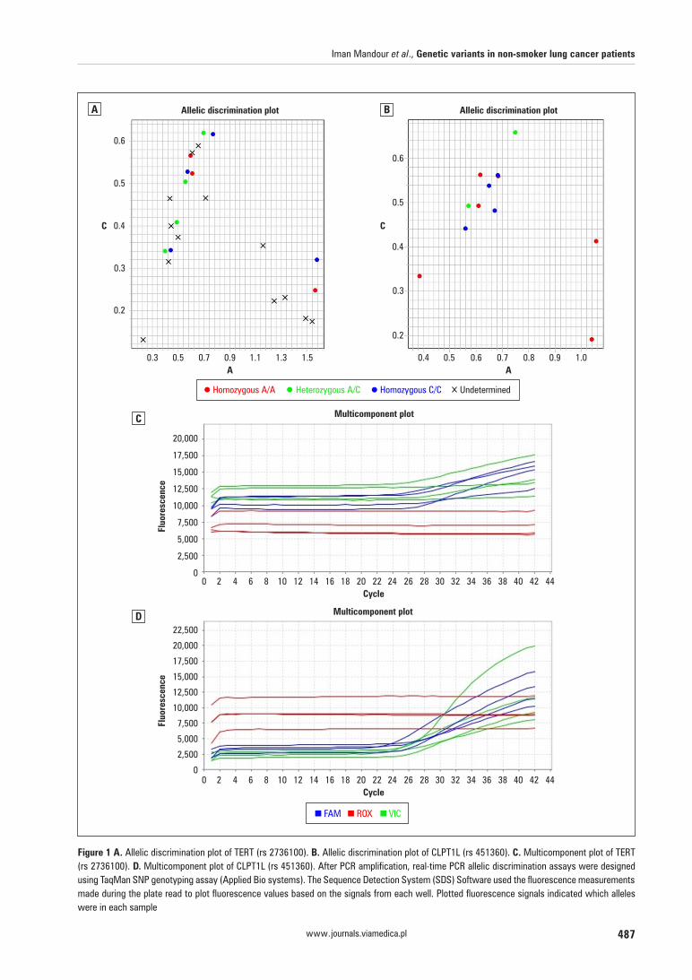
Real-time PCR with sequence-specific primers was used to assess the telomerase reverse tran-scriptase and cleft lip and palate transmembrane 1like protein. Real-time PCR allelic discrimination assay were designed using TaqMan SNP Genotyp-ing Assays (Applied Bio systems) (Figure 1).
Principle. TaqMan SNP Genotyping Assay1. Each TaqMan Minor groove binder (MGB)
probe anneals specifically to its complementa-ry sequence between the forward and reverse primer sites.
2. When the oligonucleotide probe is intact, the proximity of the reporter dye to quencher dye results in quenching of reporter fluorescence.
Iman Mandour et al., Genetic variants in non-smoker lung cancer patients
487www.journals.viamedica.pl
Figure 1 A. Allelic discrimination plot of TERT (rs 2736100). B. Allelic discrimination plot of CLPT1L (rs 451360). C. Multicomponent plot of TERT (rs 2736100). D. Multicomponent plot of CLPT1L (rs 451360). After PCR amplification, real-time PCR allelic discrimination assays were designed using TaqMan SNP genotyping assay (Applied Bio systems). The Sequence Detection System (SDS) Software used the fluorescence measurements made during the plate read to plot fluorescence values based on the signals from each well. Plotted fluorescence signals indicated which alleles were in each sample
A B
C
D

Advances in Respiratory Medicine 2020, vol. 88, no. 5, pages 485–494
488 www.journals.viamedica.pl
3. AmpliTaq Gold DNA polymerase extends prim-ers bound to template DNA.
4. AmpliTaq Gold DNA polymerase cleaves only probes that are hybridized to target.
5. Cleavage separates reporter dye from quencher dye, which results in increased fluorescence by reporter.
6. Increase in fluorescence signal occurs when probes that have hybridized to complementary sequence are cleaved. Thus, fluorescence sig-nal generated by PCR amplification indicates which alleles are present in the sample [8].
ReagentsEach of the 40× TaqMan SNP Genotyping Assay consists of a single tube containing:1. Sequence-specific forward and reverse primers
to amplify promoter region of CLPTM1L and TERT genes.
2. Two TaqMan MGB probes for distinguishing between the two alleles:
— One probe labeled with VIC dye detects Allele 1 sequence.
— One probe labeled with FAM dye detects Allele 2 sequence:• The context sequence for TERT was
Polymorphism: C > A, transition sub-stitution.
• The context sequence for CLPTM1L was Polymorphism: C > A, transversion substitution.
Each TaqMan MGB probe contains:1. Reporter dye at the 5’ end of each probe.
— FAM dye (6-carboxyfluorescein) is linked to the 5’ end of the Allele 1 (C) probe.
— VIC dye is linked to the 5’ end of the Allele 2 (A) probe.
2. Minor groove binder (MGB) at the 3’ end of each probe. This modification increases melt-ing temperature (Tm) for a given probe length
[9], which allows the design of shorter probes.3. Non-fluorescent quencher (NFQ) at the 3’ end
of each probe.
TechniqueAll reactions were performed in total volume
of 20 μL containing 10 μL of master mix, 0.5 μL of SNP-ready-made assay, (1–5 μL) purified DNA solution according to DNA concentration which was measured to be completed to 20 μL of nucle-ase-free water. Reaction mixture was prepared for each assay before transferring it to the optical reaction plate for thermal cycling. After adding reagents to DNA samples, they were mixed thor-oughly to avoid air bubbles in the well.
Allelic discrimination plate read and analysis
After PCR amplification, an endpoint plate read was performed using an Applied Bio systems Real-Time PCR System, The Sequence Detection System (SDS) Software used the fluorescence measurements made during the plate read to plot fluorescence values based on the signals from each well. Plotted fluorescence signals indicated which alleles were in each sample. Plate read document was analyzed. Automatic allele calls were made. Allele calls were converted to geno-types (Figure 1).
Statistical methodsData were coded and entered using the sta-
tistical package SPSS version 23. Data were summarized using mean, standard deviation, median, minimum and maximum for quantitative variables and frequencies (number of cases) and relative frequencies (percentages) for categori-cal variables. Comparisons between the groups were done using unpaired t test when comparing 2 groups and analysis of variance (ANOVA) with multiple comparisons post hoc test when com-paring more than 2 groups. For comparing cate-gorical data, Chi-square (c2) test was performed. Exact test was used instead when the expected frequency was less than 5. Genotype and allele frequencies were compared between the disease and the control groups using logistic regression. Odds ratio (OR) with 95% confidence intervals was calculated. P-values less than 0.05 were considered statistically significant [10].
Results
Group (I) included 40 lung cancer non-smok-ers, their mean age was 44.13 + 16.18. There were 20 females (50%) and 20 males (50%) in the group. Group (II) included forty healthy vol-unteers serving as a control group. Their mean age was 34.45 + 9.98 years, and there were 31 females (77.5%) and 9 males (22.5%). Never smoker lung cancer patients were classified into small-cell lung carcinoma (SCLC) constituting 5% (2 patients), undifferentiated lung carcino-ma constituting 5% (2 patients) and 36 (90%) non-smallcell lung carcinoma (NSCLC) cases, who were divided into 26 (65%) adenocarcinoma cases, 6 (15%) large-cell carcinoma and 4 (10%) squamous-cell carcinoma (Figure 2).
TERT genotype distribution showed that homozygous form of the wild genotype “CC” was found in 22 (55%) patients and 18 (45%)

Iman Mandour et al., Genetic variants in non-smoker lung cancer patients
489www.journals.viamedica.pl
individuals of the control group with no clinical significance (P=0.2). The heterozygous form of genotype “CA” was found in 12 (30%) patients and 19 (47.5%) control individuals (P=0.175) and the homozygous form of the mutant geno-type “AA” was found in 6 (16%) patients and in 3 (7.5%) controls (P=0.525). The allelic distribu-tion of TERT showed that “A” allele was present in 24 (30%) patients and 25 (31.3%) controls (P = 0.864), and allele “C” was found in 56 (70%) patients and 55 (68.8%) controls (P = 0.932) (Figure 3A). The genotype distribution of the CLPTM1L showed that the homozygous form of the mutant genotype “AA” was found in 9 (22%) patients in the never smoker lung cancer group, while it was found in 6 (12%) individuals in the control group with no clinical significance (P = 0.393). The heterozygous form “CA” was discovered in 31 (77.5%) patients, while it was found in 34 (85%) individuals of the control group (p = 0.53), and the homozygous form of the wild genotype “CC” was not detected in the two studied groups. The allelic distribution of the CLPTM1L revealed that allele “A” was found in 49 (61.3%) patients and 46 (57.5%) controls (P = 0.629), and the “C” allele was discovered in 31 (38.8%) patients and 34 (42.5%) controls (p = 0.512) (Figure 3B).
On combining genotypes of TERT and CLPT-M1L, the homozygous form of the wild genotype “CC” of the TERT and the homozygous form of the
mutant genotype “AA” of CLPTM1L was found in 1 patient and in 1 control individual constituting 2.5% of each group (P = 0.884). On joining the homozygous form of the mutant genotypes “AA” of both SNPs (single nucleotide polymorphisms), it was found in 3 patients (7.5%) and in 2 controls (5%) (P = 0.841). On combining the homozygous form of the mutant genotype “AA” of TERT with the heterozygous form “CA” of the CLPTM1L, it was found in 3 patients (7.5%) and in 1 (2.5%) of the controls (P = 0.460). On combining the heterozygous form “CA” of TERT with the ho-mozygous form “AA” of the mutant genotype of CLPTM1L, it was found in 5 (12.5%) lung cancer patients and in 3 (7.5%) controls (P = 0.708). On combining the heterozygous form ”CA” of the two SNPs, it was found in 7 (17.5%) patients and in 16 (40%) controls (P = 0.06), and on joining the homozygous form of the wild genotype “CC” of TERT with the heterozygous form “CA” of CLPT-M1L, it was found in 21 (52.5%) patients and in 17 (42.5%) controls (P = 0.978).
The relations between TERT and CLPTM1L genotypes and the histopathological subtypes of lung cancer were shown in Table 1 and 2. Also, relations between the combination of genotypes of two SNPs and lung cancer histopathology were shown in Table 3. The connection between the genotypes or the combination of genotypes of two SNPs and sex showed no statistical significance. The relation of the CLPTM1L genotypes and age uncovered that mean age of patients with het-erozygous form of CLPTM1L “CA” was 46.42 ± 17.28 years, while mean age of patients who had homozygous form of the mutant genotype was 36.22 ± 8.09 (P = 0.019). The relation between genotypes of TERT and age showed that mean age of patients with heterozygous form “CA” was 44.17 + 15.58 years, and mean age of homozygous form of wild genotype “CC” was 46.68 + 17.51, and these are older ages than the ages (34.67 + 9.18) of the patients showing homozygous form of mutant genotype “AA” (P = 0.27).
Discussion
Lung cancer is one of the commonest cancer types worldwide in respect of incidence and mortality. Global statistical data showed that lung cancer alone accounts for 13% of all newly diagnosed cancers and is responsible for 18% of all cancer deaths [11]. Lung cancer in never smokers is distinct from those in smokers in view of pathogenesis, molecular alterations, drug re-sponsiveness and prognosis. Significant portion
Figure 2. Classification of never smoker lung cancer patients accord-ing to the histopathological type of the tumor

Advances in Respiratory Medicine 2020, vol. 88, no. 5, pages 485–494
490 www.journals.viamedica.pl
Figure 3 A. Genotype and allelic distribution of TERT (rs 2736100) (c. 1574-3777A>C). B. Genotype and allelic distribution of CLPT1L (rs 451360) (c. 1532 + 1051A>C)
A
B
of lung cancer in never smokers harbor genetic variant in driving oncogene, to which molec-ular targeted drugs are dramatically sensitive. Therefore, genetic testing before the treatment is essential for lung cancer in never smokers to select the appropriate treatment option according to patient’s molecular characteristics [12].
Non-smoking-associated lung cancer not only occurs in never smokers, but also in cur-rent and former smokers. Recent genome-wide association studies (GWAS) have shown that the polymorphisms in the TERT and CLPTM1L genes
may play important roles in the development of lung cancer. The two genes are both located in chromosome 5p15.33 [13]. Three GWAS in Euro-pean populations showed consistent associations of polymorphisms in these two genes with lung cancer in non-smokers [14].
This study analyzed the distribution of TERT and CLPTM1L single nucleotide polymorphism in patients with lung cancer in never smokers and in healthy controls in attempt to find the association between these two polymorphisms and the development of lung cancer in never

Iman Mandour et al., Genetic variants in non-smoker lung cancer patients
491www.journals.viamedica.pl
Table 1. The relation between histopathological subtypes of lung cancer and TERT and CLPT1L genotypes among the study population
Histopathological subtype
TERT (rs 2736100)
AA(n = 6)
CA(n = 12)
CC(n = 22)
Total(n=40)
P-value
NSCLC (n = 36) Yes [no, %] 5 (83.33%) 11(91.67%) 20 (90.91%) 36 (90%) 0.837
No [no, %] 1 (16.67%) 1 (8.33%) 2 (9.09%) 4 (10%)
SCLC (n = 2) Yes [no, %] 0 0 2 (9.09%) 2 (5%)
0.422No [no, %] 6 (100%) 12 (100%) 20 (90.91%) 38 (95%)
Undifferentiated carcinoma (n = 2)
Yes [no, %] 1 (16.67%) 1 (8.33%) 0 2 (5%) 0.206
No [no, %] 5 (83.33%) 11 (91.67%) 22 (100%) 38 (95%)
P-value 0.005* 0.0001* 0.0001* 0.0001*
Histopathological subtype
CLPT1L (rs 451360) (c. 1532 + 1051G>T)
AA(n = 9)
CA(n = 31)
CC(n = 0)
Total(n = 40)
P-value
NSCLC (n = 36) Yes [no, %] 8 (88.89%) 28 (90.32%) 0 36 (90%) 0.899
No [no, %] 1 (11.11%) 3 (9.68%) 0 4 (10%)
SCLC (n = 2) Yes [no, %] 0 (0.0%) 2 (6.45%) 0 2 (5%) 0.434
No [no, %] 9 (100%) 29 (93.55%) 0 38 (95%)
Undifferentiated carcinoma (n = 2)
Yes [no, %] 1 (11.11%) 1 (3.23%) 0 2 (5%) 0.339
No [no, %] 8 (88.89%) 30 (96.77%) 0 38 (95%)
P-value 0.0007* 0.0001* — 0.0001**P value < 0.05 is considered significant.NSCLC — non-small cell lung carcinoma; TERT — telomerase reverse transcriptase; SCLC —small cell lung carcinoma
smokers. Never smokers lung cancer patients were classified according to histopathological subtype of lung cancer into small-cell lung car-cinoma (5%), undifferentiated lung carcinoma (5%) and non-small-cell lung carcinoma (90%). Then NSCLC patients were further divided into adenocarcinoma (65%), large-cell carcinoma (15%), squamous-cell carcinoma (10%) (Figure 2). Adenocarcinoma was most prevalent subtype. This agreed with Sun et al. [15] who stated that smoking-related carcinogens act on both proxi-mal and distal airways of the lung inducing all major types of lung cancer. Also, they stated that cancers arising in never smokers target distal airways and favor the adenocarcinoma type. Subramanian and Govindan [16] mentioned that adenocarcinoma is the most common type occurring in never smokers. Again, Landi et al. [17] have established that locus on chromosome 5p15.33 is distinctly associated with a risk of lung adenocarcinoma and not with other major histological types. Also, Henschke et al. [18] found that women are commonly diagnosed when screened, and Patel et al. [19] showed that wom-
en outnumber men among lung cancer patients who never smoked regularly. The study done by Samet et al. [20] showed that adenocarcinoma is more prevalent in females. This is explained by the hypothesis that females are more prone to exposure to second-hand smoke, household exposure of coal used for cooking, hormone re-placement therapy, use of antiaging drugs which elongate telomeres thus escaping apoptosis and lead to cancers [21].
In the present study, there was a statistically significant difference in mean age between the patients demonstrating heterozygous form of CLPTM1L “CA” and those showing homozygous form of mutant genotype AA” (P = 0.019). Al-though age difference between different genotype of TERT was statistically insignificant (P = 0.27), the patients with heterozygous form CA and homozygous form of wild genotype “CC” were older than patients showing homozygous form of mutant genotype “AA”. This agreed with Wakelee et al. [22] who mentioned that lung cancer in never smokers occurs more commonly after the age of forty.

Advances in Respiratory Medicine 2020, vol. 88, no. 5, pages 485–494
492 www.journals.viamedica.pl
Table 2. The relation between histopathological subtypes of non-small cell lung cancer and TERT and CLPT1L genotypes among the study population
NSCLC histopathological subtypes
TERT (rs 2736100)
AA(n = 5)
CA(n = 11)
CC(n = 20)
Total (n = 36)
P-value
Adenocarcinoma(n = 26)
Yes (no, %) 2 (40%) 8 (72.73%) 16 (80%) 26 (72.22%) 0.202
No (no, %) 3 (60%) 3 (27.27%) 4 (20%) 10 (27.78%)
Squamous cell car-cinoma (n = 4)
Yes (no, %) 2(40%) 0 2 (10%) 4 (11.11%) 0.004*
No (no, %) 3 (60%) 11 (100%) 18 (90%) 32 (88.89%)
Large cell carcinoma (n = 6)
Yes (no, %) 1 (20%) 3 (27.27%) 2 (10%) 6 (16.67%) 0.455
No (no, %) 4 (80%) 8 (72.73%) 18 (90%) 30 (83.33%)
P-value 0.740 0.001 0.0001 0.001
NSCLC histopathological subtypes
CLPT1L (rs 451360) (c. 1532 + 1051G>T)
AA(n = 8)
CA(n = 28)
CC(n = 0)
Total(n = 36)
P-value
Adenocarcinoma (n = 26)
Yes (no, %) 6 (75%) 20 (71.43%) 0 26 (72.22%) 0.841
No (no, %) 2 (25%) 8 (28.57%) 0 10 (27.78%)
Squamous cell carcinoma (n = 4)
Yes (no, %) 0 4 (14.29%) 0 4 (11.11%) 0.256
No (no, %) 8 (100%) 24 (85.71%) 0 32 (88.89%)
Large cell carcinoma (n = 6)
Yes (no, %) 2 (25%) 4 (14.29%) 0 6 (16.67%) 0.473
No (no, %) 6 (75%) 24 (85.71%) 0 30 (16.67%)
P-value 0.005* 0.0001* — 0.001**P value <0.05 is considered significant.NSCLC — non-small cell lung carcinoma; TERT — telomerase reverse transcriptase; SCLC — small cell lung carcinoma
Table 3. The combination of genotypes of two SNPs and the histopathological subtypes of lung cancer
Groups CC/AA(n = 1)
AA/AA(n = 3)
AA/CA(n = 3)
CA/AA(n = 5)
CA/CA(n = 7)
CC/CA(n = 21)
P
Adenocarcinoma 1 (100%) 2 (66.7%) 0 3 (60%) 5 (71.4%) 15 (71.4%) < 0.001*
Squamous cell carcinoma 0 0 2 (66.7%) 0 0 2 (9.5%) 0.15
Large cell carcinoma 0 1 (33.3%) 0 1 (20%) 2 (28.6%) 2 (9.5%) 0.54
SCLC 0 0 0 0 0 2 (9.5%) 0.07
Undifferentiated carcinoma 0 0 1 (33.3%) 1 (20%) 0 0 0.54
P 0.4 0.25 0.25 0.19 0.008* < 0.001**P value <0.05 is considered significant.SCLC —small cell lung carcinoma
There was no statistically significant differ-ence between never smoker lung cancer patients and the controls in TERT genotype distribution re-garding homozygous form of wild genotype “CC”, heterozygous form of genotype “CA” and homozy-gous form of mutant genotype “AA”. Also, allelic distribution of TERT showed that the “A” allele and allele “C” didn’t differ in distribution between the cases and controls. Wang et al. [23] showed that TERT SNPs is a risk factor for developing lung can-
cer in never smokers. Again, case–control study by Liao et al. [24] revealed the statistical significance between AC and CC genotypes, as well as C allele of rs2736100 in TERT gene and increased risks of lung cancer in never smokers. This is explained by various ethnic populations, besides, a difference in defining investigating groups and small sample size, a dissimilarity in their genetic backgrounds, geographical differences in allelic frequencies and complexity of the disease.

Iman Mandour et al., Genetic variants in non-smoker lung cancer patients
493www.journals.viamedica.pl
The distribution of CLPT1L genotypes and allelic distribution showed non-significant differ-ence between never smoker lung cancer patients and healthy controls. Also, Sun et al. [25] found no association of XRCC1 and CLPTM1L polymor-phisms with NSCLC in non-smoking Han Chinese population. However, Pande et al. [26] revealed additional SNPs that may be susceptibility mark-ers for lung cancer risk in smokers (rs4975615) and never smokers (rs451360) of clinical signifi-cance. Also, the case-control study by Liang et al. [27] found that rs451360, an intronic SNP within CLPTM1L gene, was significantly associated with lung cancer risk in never smokers. Possible explanations for the same polymorphism to have different roles in cancer susceptibility can occur because that allele under investigation can be masked by the presence of other genes involved in disease development. Thus, results available regarding effect of polymorphisms on cancer risk and development should be interpreted with caution.
TERT genotypes including homozygous form of mutant genotype “AA”, heterozygous form “CA” and homozygous form of wild genotype “CC” were significantly found in NSCLC patients constitut-ing 83.33%, 91.67% and 90.91%, respectively, of lung cancer non-smoker patients. CLPTM1L genotypes including homozygous form of mutant allele “AA” and heterozygous genotype “CA” were detected significantly in NSCLC patients consti-tuting 88.89% and 90.32%, respectively (Table 2). TERT and CLPTM1L genotypes were significantly presented in adenocarcinoma compared to squa-mous-cell carcinoma and large-cell carcinoma (Table 3). On combining homozygous form of wild genotypes “CC” of TERT and homozygous form of mutant genotype “AA”, was found in ad-enocarcinoma (100%). On joining homozygous form of mutant genotype “AA” in both SNPs, was found in adenocarcinoma (66.7%) and large-cell carcinoma (33.3%). On combining homozygous form of mutant genotype of TERT “AA” with het-erozygous form of CLPTM1L “CA”, was found in squamous-cell carcinoma (66.7%) and undifferen-tiated carcinoma (33.3%). On combining hetero-zygous form of TERT “CA” with homozygous form of mutant genotype of CLPTM1L “AA”, was found in adenocarcinoma (60%), large-cell carcinoma (20%) and undifferentiated carcinoma (20%). On combining heterozygous form “CA” of both SNPs, was found in adenocarcinoma 5 (71.5%) and large-cell carcinoma (28.6%) with statistical significance (P=0.008). On combining homozy-gous form of wild genotype of TERT “CC” with
heterozygous form of CLPT1L (CA), was found in adenocarcinoma (71.4%), squamous-cell carcino-ma (9.5%), large-cell carcinoma (9.5%), small-cell carcinoma (9.5%) with statistical significance (P < 0.001). Also, there was statistical significance regarding the association of adenocarcinoma and some combined genotypes, mainly “CC/AA, CA/CA and CC/CA” (P ≤ 0.001). This agreed with Yuan et al. [28] who demonstrated that TERT rs 2736100 polymorphism is a risk factor associ-ated with increased lung cancer susceptibility, particularly for lung adenocarcinoma. Also, Hsi-ung et al. [29] reported conclusive evidence that common genetic variants in TERT-CLPTM1L lo-cus on chromosome 5p15.33 are associated with a risk for lung adenocarcinoma in non-smoking Asian women. Again, Zhao et al. [30] showed a significant association of 5p15.33 (TERT–CLPT-M1L genes) with lung cancer in the Chinese Han population.
Again, Bhat et al. [31] suggested that rs2853677 of TERT was significantly associated with multiple cancers, and it could be a potential marker for diagnosis of non-small-cell lung cancer and leukemia. Our study together with Bhat et al study highlighted the role of telomere-associat-ed pathways in non-small-cell lung cancer. On the other hand, Hung et al. [32] suggested that SNPs/regions connected with lung cancer risk in never smokers are not specific for this type of can-cer but rather have pleiotropic effects. Also, they found that genetic susceptibility to lung cancer in never smokers is associated with genetic variants with pan-cancer risk effects, and they mentioned that the comparison with smokers discovered that top variants previously shown to be associated with lung cancer risk only confer risk in the presence of tobacco exposure, underscoring the importance of gene-environment interactions in the etiology of this disease. Similarly, Wang et al. [33] performed a pleiotropic analysis to explore the shared susceptibility mechanisms between non-lung cancers and lung cancer and found that genetic variants identified from other cancer types were also significantly associated with the risk of lung cancer.
In conclusion, polymorphism in TERT and CLPTM1L genes may play an important role in the development of NSCLC, especially the adenocar-cinoma subtype. The two genes are located in the chromosome 5p15.33. It is necessary to conduct large-scale population study on Egyptian patients to elucidate our results. It is recommended to do further studies using more advanced molecular techniques as complete sequence analysis for

Advances in Respiratory Medicine 2020, vol. 88, no. 5, pages 485–494
494 www.journals.viamedica.pl
proper evaluation and assessment of the TERT and CLPT1L single nucleotide polymorphism as a predictive marker of lung cancer in Egyptian non-smoker individuals for early diagnosis of lung cancer.
Conflict of interest
None declared.
References:4. Ferlay J, Shin HR, Bray F, et al. Estimates of worldwide
burden of cancer in 2008: GLOBOCAN 2008. Int J Cancer. 2010; 127(12): 2893–2917, doi: 10.1002/ijc.25516, indexed in Pubmed: 21351269.
5. Jackson M, Marks L, May GHW, et al. The genetic basis of disease. Essays Biochem. 2018; 62(5): 643–723, doi: 10.1042/EBC20170053, indexed in Pubmed: 30509934.
6. Gealy R, Zhang L, Siegfried JM, et al. Comparison of mutations in the p53 and K-ras genes in lung carcinomas from smoking and nonsmoking women. Cancer Epidemiol Biomarkers Prev. 1999; 8 (4 Pt 1): 297-302. Erratum in: Cancer Epidemiol Bio-markers Prev. 2001; 10(12): 1309.
7. Kachuri L, Amos CI, McKay JD, et al. EPIC Study. Lung cancer susceptibility locus at 5p15.33. Nat Genet. 2008; 40(12): 1404–1406, doi: 10.1038/ng.254, indexed in Pubmed: 18978790.
8. Bernal A, Tusell L. Telomeres: implications for cancer devel-opment. Int J Mol Sci. 2018; 19(1), doi: 10.3390/ijms19010294, indexed in Pubmed: 29351238.
9. Taylor JK, Zhang QQ, Monia BP, et al. Inhibition of Bcl-xL ex-pression sensitizes normal human keratinocytes and epithelial cells to apoptotic stimuli. Oncogene. 1999; 18(31): 4495–4504, doi: 10.1038/sj.onc.1202836, indexed in Pubmed: 10442640.
10. Huss VA, Festl H, Schleifer KH. Studies on the spectrophoto-metric determination of DNA hybridization from renaturation rates. Syst Appl Microbiol. 1983; 4(2): 184–192, doi: 10.1016/S0723-2020(83)80048-4, indexed in Pubmed: 23194591.
11. Pfaffl M, Hageleit M. Validities of mRNA quantification using recombinant RNA and recombinant DNA external calibration curves in real-time RT-PCR. Biotechnology Letters. 2001; 23(4): 275–282.
12. Afonina I, Zivarts M, Kutyavin I, et al. Efficient priming of PCR with short oligonucleotides conjugated to a minor groove bind-er. Nucleic Acids Res. 1997; 25(13): 2657–2660, doi: 10.1093/nar/25.13.2657, indexed in Pubmed: 9185578.
13. Chan YH. Biostatistics 101: data presentation. Singapore Med J. 2003; 44(6): 280–285, indexed in Pubmed: 14560857.
14. Bray F, Ferlay J, Soerjomataram I, et al. Global cancer statistics. CA Cancer J Clin. 2011; 61(2): 69–90, doi: 10.3322/caac.20107, indexed in Pubmed: 21296855.
15. Lee YJ, Kim JH, Kim SeK, et al. Lung cancer in never smokers: change of a mindset in the molecular era. Lung Cancer. 2011; 72(1): 9–15, doi: 10.1016/j.lungcan.2010.12.013, indexed in Pubmed: 21272954.
16. Kachuri L, Amos CI, McKay JD, et al. Lung cancer susceptibil-ity locus at 5p15.33. Nat Genet. 2008; 40(12): 1404–1406, doi: 10.1038/ng.254, indexed in Pubmed: 18978790.
17. Rafnar T, Sulem P, Stacey SN, et al. Sequence variants at the TERT-CLPTM1L locus associate with many cancer types. Nat Genet. 2009; 41(2): 221–227, doi: 10.1038/ng.296, indexed in Pubmed: 19151717.
18. Sun S, Schiller JH, Gazdar AF. Lung cancer in never smokers — a different disease. Nat Rev Cancer. 2007; 7(10): 778–790, doi: 10.1038/nrc2190, indexed in Pubmed: 17882278.
19. Subramanian J, Govindan R. Lung cancer in never smokers: a review. J Clin Oncol. 2007; 25(5): 561–570, doi: 10.1200/JCO.2006.06.8015, indexed in Pubmed: 17290066.
20. Landi MT, Zhao Y, Rotunno M, et al. MicroRNA expression dif-ferentiates histology and predicts survival of lung cancer. Clin Cancer Res. 2010; 16(2): 430–441, doi: 10.1158/1078-0432.CCR-09-1736, indexed in Pubmed: 20068076.
21. Henschke CI, Yankelevitz DF, Libby DM, et al. Early lung can-cer action project: annual screening using single-slice helical CT. Ann N Y Acad Sci. 2001; 952: 124–134, doi: 10.1111/j.1749-6632.2001.tb02733.x, indexed in Pubmed: 11795431.
22. Patel JD, Bach PB, Kris MG. Lung cancer in US women: a contemporary epidemic. JAMA. 2004; 291(14): 1763–1768, doi: 10.1001/jama.291.14.1763, indexed in Pubmed: 15082704.
23. Samet JM, Avila-Tang E, Boffetta P, et al. Lung cancer in never smokers: clinical epidemiology and environmental risk factors. Clin Cancer Res. 2009; 15(18): 5626–5645, doi: 10.1158/1078-0432.CCR-09-0376, indexed in Pubmed: 19755391.
24. Subramanian J, Regenbogen T, Nagaraj G, et al. Review of ongo-ing clinical trials in non-small-cell lung cancer: a status report for 2012 from the ClinicalTrials.gov Web site. J Thorac Oncol. 2013; 8(7): 860–865, doi: 10.1097/JTO.0b013e318287c562, in-dexed in Pubmed: 23478543.
25. Wakelee HA, Chang ET, Gomez SL, et al. Lung cancer inci-dence in never smokers. J Clin Oncol. 2007; 25(5): 472–478, doi: 10.1200/JCO.2006.07.2983, indexed in Pubmed: 17290054.
26. Wang Y, Broderick P, Webb E, et al. Common 5p15.33 and 6p21.33 variants influence lung cancer risk. Nat Genet. 2008; 40(12): 1407–1409, doi: 10.1038/ng.273, indexed in Pubmed: 18978787.
27. Liao Y, Guo S, Chen Y, et al. VSIG4 expression on macrophages facilitates lung cancer development. Lab Invest. 2014; 94(7): 706–715, doi: 10.1038/labinvest.2014.73, indexed in Pubmed: 24862966.
28. Sun Y, Zhang YJ, Kong XM. No association of XRCC1 and CLPTM1L polymorphisms with non-small cell lung cancer in a non-smoking Han Chinese population. Asian Pac J Cancer Prev. 2013; 14(9): 5171–5174, doi: 10.7314/apjcp.2013.14.9.5171, in-dexed in Pubmed: 24175795.
29. Pande M, Spitz MR, Wu X, et al. Novel genetic variants in the chromosome 5p15.33 region associate with lung cancer risk. Carcinogenesis. 2011; 32(10): 1493–1499, doi: 10.1093/carcin/bgr136, indexed in Pubmed: 21771723.
30. Liang Y, Thakur A, Gao L, et al. Correlation of CLPTM1L poly-morphisms with lung cancer susceptibility and response to cisplatin-based chemotherapy in a Chinese Han population. Tumour Biol. 2014; 35(12): 12075–12082, doi: 10.1007/s13277-014-2508-6, indexed in Pubmed: 25155038.
31. Yuan Y, Lu C, Xue L, et al. Association between TERT rs2736100 polymorphism and lung cancer susceptibility: evidence from 22 case-control studies. Tumour Biol. 2014; 35(5): 4435–4442, doi: 10.1007/s13277-013-1583-4, indexed in Pubmed: 24390616.
32. Hsiung CA, Lan Q, Hong YC, et al. The 5p15.33 locus is as-sociated with risk of lung adenocarcinoma in never-smoking females in Asia. PLoS Genet. 2010; 6(8), doi: 10.1371/journal.pgen.1001051, indexed in Pubmed: 20700438.
33. Zhao Z, Li C, Yang L, et al. Significant association of 5p15.33 (TERT-CLPTM1L genes) with lung cancer in Chi-nese Han population. Exp Lung Res. 2013; 39(2): 91–98, doi: 10.3109/01902148.2012.762436, indexed in Pubmed: 23368278.
34. Bhat GhR, Bhat A, Verma S, et al. Association of newly iden-tified genetic variant rs2853677 of TERT with non-small cell lung cancer and leukemia in population of Jammu and Kash-mir, India. BMC Cancer. 2019; 19(1): 493, doi: 10.1186/s12885-019-5685-2, indexed in Pubmed: 31126249.
35. Hung RJ, Spitz MR, Houlston RS, et al. Lung cancer risk in never-smokers of european descent is associated with genetic variation in the 515.33 tert-clptm1ll region. J Thorac Oncol. 2019; 14(8): 1360–1369, doi: 10.1016/j.jtho.2019.04.008, in-dexed in Pubmed: 31009812.
36. Wang L, Zhu M, Wang Y, et al. Cross-Cancer pleiotropic anal-ysis reveals novel susceptibility loci for lung cancer. Front Oncol. 2019; 9: 1492, doi: 10.3389/fonc.2019.01492, indexed in Pubmed: 32010612.

ORIGINAL RESEARCHES
495www.journals.viamedica.pl
Address for correspondence: Degena Bahrey Tadesse, Department of Adult Health Nursing, School of Nursing, Aksum University, Aksum, Ethiopia; e-mail: [email protected] DOI: 10.5603/ARM.a2020.0138Received: 05.05.2020Copyright © 2020 PTChPISSN 2451–4934
Degena Bahrey Tadesse1, Melaku Negash1, Ebud Ayele2, Abrha Hailay1, Kbrom Gemechu Kiros3, Teklehaimanot Gereziher Haile4, Mebrahtu Abay5, Gebre Teklemariam Demoz6
1Department of Adult Health Nursing, School of Nursing, Aksum University, Aksum, Ethiopia2Department of Human Nutrition, School of Public Health, Aksum University, Aksum, Ethiopia3Department of Adult Health Nursing, School of Nursing, Adigrat University, Adigrat, Ethiopia4Department of Maternal and Neonatology Nursing, School of Nursing, Aksum University, Aksum, Ethiopia5Department of Epidemiology, School of Public Health, Aksum University, Aksum, Ethiopia6Department of Clinical Pharmacy, School of Pharmacy, Aksum University, Aksum, Ethiopia
Uncontrolled asthma in Ethiopia: a systematic review and meta-analysis
AbstractIntroduction: Despite significant improvement in the diagnosis and management of this disorder, asthma in the majority of Ethiopians remains poorly controlled. Although the prevalence of uncontrolled asthma is a public health problem in Ethiopia, its reported prevalence varies from study to study. Hence, this review aims to determine the true prevalence of uncontrolled asthma among asthmatic patients in Ethiopia. Material and methods: Different database searching engines were used including PubMed, Scopus, Google Scholar, Africa jour-nal online, World Health Organization (WHO) afro library, and Cochrane review. They were systematically searched for published studies on uncontrolled asthma in Ethiopia from 2014 to 2019. Primary search terms were “asthma”, “uncontrolled asthma”, “uncontrolled wheezing”, and “Ethiopia”. The Preferred Reporting Items for Systematic Review and Meta-Analysis (PRISMA) guideline was followed. Publication bias was examined by the funnel plot. The random-effect model was fitted to estimate the pooled prevalence of uncontrolled asthma among asthmatic patients. All statistical analysis was done using R version 3.5.3 and the RStudio version 1.2.5033 software for Windows. Results: The overall pooled prevalence of uncontrolled asthma was found to be 71.67% [95% CI (0.6772; 0.7562)]. Potential associated factors were: unscheduled visits, frequency of short-acting beta2-agonist (SABA) use, type of treatment and perceived rate of asthma control, low monthly income, age group, presence of comorbidity, moderate persistent asthma, severe persistent asthma and use of SABA alone as anti-asthmatic medication, use of biomass fuel for cooking, longer duration of asthma (> 30 years), incorrect inhalation technique, and asthma exacerbation in the last 12 months. Self-perceived poor asthma control was associated with any activity limitation due to asthma, inconsistent inhaled corticosteroid use, and lack of health education on metered-dose inhaler technique [AOR =4.96; 95% CI (1.08–22.89)].Conclusions: Nearly two-thirds of patients were determined to have uncontrolled asthma. Thus, this evidence suggests that attention should be given to asthma patients and health care providers.
Key words: uncontrolled asthma, asthmatic patient, determinants, EthiopiaAdv Respir Med. 2020; 88: 495–503
Introduction
Approximately 300 million people world-wide have asthma and recent projections indi-cate that an additional 100 million people will be living with asthma by 2025 [1, 2]. The World Health Organization (WHO) reports that there are
approximately 250,000 asthma deaths per year, primarily in low-and middle-income countries (LMICs) [3, 4]. As with many other chronic dis-eases in Ethiopia, rapid urbanization rates have been related to a rise in the burden of asthma and other allergic diseases [3–6]. The incidence of these conditions can, in principle, have the

Advances in Respiratory Medicine 2020, vol. 88, no. 6, pages 495–503
496 www.journals.viamedica.pl
potential to reach higher rates than those seen in high-income countries (HICs) due to the pri-mary effects of parasitic helminthic infections on the immune system, as these infections are widespread in Ethiopian settings [5]. The Interna-tional Study of Asthma and Allergy in Childhood (ISAAC) stated that the burden of asthma among adults in Africa, including Ethiopia, has risen and has contributed most to the burden of disease through its effects on quality of life [3]. In-patient admissions and sales of medications account for much of the direct government expenses, while the lack of employment due to absenteeism from work and decreased education is responsible for many of the indirect costs [7, 8].
In Ethiopia, challenges, including those resulting from overuse of health facilities, lack of qualified personnel and testing equipment, and lack of access to and availability of inhaled medicines have hampered efforts to improve the management of asthma [8, 9]. The lack of coordinated health promotion initiatives, such as effective control strategies for environmental causes, air pollutants, and occupational dust, has also led to an increasing burden of disease [10]. The WHO has indicated that the rate of regula-tion of asthma and health responses combatting the diease on the continent have been below the required requirements and have led to the large size of the disease burden [3, 4]. Besides, while many African countries have national guidelines on the treatment of asthma and other CRDs, these guidelines have not been enforced in most rural areas [11, 12]. Economic analyses (Placeholder2) in many African settings have shown that the direct costs of asthma are typically higher than indirect costs. Nonetheless, indirect costs reflect a relatively higher proportion of overall costs for pediatric patients compared to adult patients [8]. Moreover, the broader economic strain on individ-uals, households, employers, and the community, owing to the lack of future potential sources of livelihood, has also been devastating in many resource-poor environments [7]. It is assumed that many children with asthma in Africa may fail to reach their full potential unless appropriate treat-ment and control measures are put in place [1]. It has been proposed that awareness by health care professionals and the public is a critical element in reacting to the challenge raised by asthma in Africa [3, 13].
By 2015, the urban population of the world is projected to rise from 45% to 59%, with more than half of this happening in Africa, including Ethiopia [8]. The prevalence of asthma and other
chronic diseases in Africa is also projected to in-crease due to this increasing population growth, impact of subsequent urbanization, and adoption of western lifestyles [14].
Because of reasons that include these chang-es, the low output of research, and limited quality of health services data on the burden of asthma in Africa, it is important to evaluate the available data through a systematic review of literature to attempt to measure the burden, direct the setting of health priorities, and inform the formulation of effective health policy responses. Understanding the determinants of uncontrolled asthma will also help to manage premature death from non-com-municable diseases (NCD) in general, as well as asthma in particular.
This result will also allow the Ministry of Health and Clinical Services, facility managers, and governmental and non-governmental orga-nizations to be informed of the determinants of uncontrolled asthma and to commit to taking action. Besides, this analysis should be used as a model for prospective researchers and other stakeholders.
Therefore, this study will be trying to assess the prevalence and determinants of uncontrolled asthma among asthma patients in Ethiopian hospitals.
Methods and materials
SettingEthiopia is an east African country and is di-
vided into 9 regions named Tigray, Afar, Amhara, Oromia, Somali, Benishangul-Gumuz, Southern Nations Nationalities and People Region (SNN-PR), Gambella, and Harari, as well as two admin-istrative states (Addis Ababa city administration and Dire Dawa city administration).
Search strategy
The search strategy has been applied using Online Databases (PubMed/MEDLINE, Google Scholar, Web of Science, Cochran Library, Afri-ca Wide Knowledge, and Africa Index Medicus) from 2014 to January 2020. Only articles written in English and full-texts published from peer-re-viewed journals were eligible for inclusion. Pri-mary search terms were “asthma”, “uncontrolled asthma”, and “Ethiopia”. Studies that reported associated factors or determinants or predictors of uncontrolled asthma only, or did not report the magnitude or prevalence of uncontrolled asthma were excluded.

Degena Bahrey Tadesse et al., Uncontrolled asthma in Ethiopia: a systematic review and meta-analysis
497www.journals.viamedica.pl
The literature search technique was devel-oped using the headings of the medical subject headings (Mesh), BOOLEAN (AND/OR) operator was used.
The last electronic search was run on 30 Jan-uary 2019. Although no complete study protocol was written before starting this review, we devel-oped and piloted a screening guide to make sure that the inclusion criteria were adhered to and consistently applied by all review authors. Two re-viewers (DBT and KGK) independently screened the titles, abstracts of all citations retrieved, and full-text search results to identify potentially eligible studies. The agreement between review authors was measured using Cohen’s k statistic. Disagreements were resolved by discussion after mutual consensus and third-party independent review (AH).
Inclusion and exclusion criteriaThe following factors were a part of the
inclusion criteria for our analysis: studies pre-sented as original articles; studies that assessed uncontrolled asthma among patients; studies conducted among Ethiopian asthmatic patients living in Ethiopia and aged 18 and above; studies conducted and published from 2014 to 30 January 2019; and studies written in English. Studies that did not explain the criteria for the level of asthma control and studies that didn’t state the number of patients with uncontrolled asthma were excluded.
The types of studies included were all pub-lished and unpublished observational studies (cross-sectional, prospective/retrospective cohort studies, or case-control studies). Baseline data from randomized controlled trials conducted in Ethiopia reporting on the prevalence of uncon-trolled asthma were also included. Experimental studies, letters, reviews, commentaries, editorials, case reports, or case series were not included. In the case of duplicate reports, the most compre-hensive and up-to-date version was taken into account.
The participants of the study included adult populations with known asthma on any form of anti-asthmatic medication. Study participants should be at least 18 years of age. Intervention(s)/exposure(s): On any form of an-ti-asthmatic medications.Outcome: Prevalence of uncontrolled asthma among people who report taking antihyperten-sive drugsSettings: Hospital-based studies.Publication date: 2014, to January 30, 2019.Language: No language restriction.
Exclusion criteriaStudies not performed on humans, qual-
itative studies, studies that lack relevant data needed to compute the prevalence of uncontrolled asthma, studies among children and adolescents < 18 years of age, and studies that did not use the disease control criteria through the ACQ, C-ACT, ACT, or GINA were excluded.
Data extraction and quality assessmentData extraction used a preconceived and
standardized data collection form and was per-formed by two independent authors (DBT and KGK). Any discrepancies between these authors were reconciled through discussion. Data ex-tracted comprised information about the year of publication, country, objective and design of the study, diagnostic criteria of uncontrolled asth-ma, mean age, sex (male proportion), duration of asthma, signs and symptoms, anti-asthmatic medications, complications, prevalence and/or incidence, and risk factors for uncontrolled asthma.
Quality assessment of included studiesThe methodological quality of the included
studies was evaluated using the Newcastle-Ot-tawa Scale. The Newcastle-Ottawa Scale was designed to assess the quality of non-randomized studies in meta-analyses. This scale is primarily formulated by a star allocation system, assigning a maximum of 10 stars for the risk of bias in three areas: a selection of study groups (4 or 5 stars), comparability of groups (2 stars), and ascer-tainment of the outcome of interest or exposure (3 stars). No validation study provides a cut-off score for rating low-quality studies. A priori, we arbitrarily established that 0–3, 4–6, and 7–10 stars would be considered as a high, mod-erate, and low risk of bias, respectively.
Data analysis and presentation of resultsData were analyzed using the R software
V.3.5.3. Data were summarized using ranges, means ± SDs, and frequencies (percentages) where appropriate. Forest plots were drawn to vi-sualize the combined prevalence of uncontrolled asthma and the extent of statistical heterogeneity between studies. Statistical heterogeneity was as-sessed using the c2 test on Cochrane’s Q statistic [20] and quantified by calculating the I2 statistic (with values of 25%, 50%, and 75% representative of low, medium, and high heterogeneity, respec-tively) [21]. There was a clinical heterogeneity between studies included in this study. The defi-

Advances in Respiratory Medicine 2020, vol. 88, no. 6, pages 495–503
498 www.journals.viamedica.pl
nition of uncontrolled asthma was different across studies. Consequently, we used a random-effects meta-analysis to estimate the overall pooled prev-alence of uncontrolled asthma. To assess possible publication bias, Egger weighted regression meth-ods were used. A p-value < 0.05 was considered indicative of statistically significant publication bias. Moreover, other relevant findings were sum-marized in a narrative format.
Data managementBased on the inclusion and exclusion criteria,
a tool has been developed a priori to guide the screening and selection process. The tool was be piloted and revised before data extraction begins. The search results were first be uploaded to EndNote software first to remove duplicates.
Selection processOnce data are obtained, two investigators
will independently screened the titles and ab-stracts of articles retrieved from the literature search against the inclusion criteria. Full texts for the eligible titles and/or abstracts including those where there is uncertainty were obtained for further assessment on whether to include in the study or not. Where necessary, authors were contacted for additional information to confirm the eligibility of studies. Disagreements were resolved through discussion and, when needed, there was arbitration by a third reviewer. Reasons for excluding articles were recorded.
Data collection processData were extracted using a standardized
data extraction form. From the studies included, two assessors were independently extracted data using the predefined standardized extraction form. Disagreements were resolved through dis-cussion and, when needed, there was arbitration by a third reviewer.
Where there is missing information, the cor-responding author of the study was contacted to request the missing information. A maximum of three emails will be sent to the corresponding au-thor to request for additional information before excluding the study. For studies appearing in more than one published article, we considered the one that is most recent, comprehensive, and with the largest sample size. For surveys appearing in one article with multiple surveys conducted at different time points, we shall treat each survey as a separate study. For multi-national studies, data were separated to show the estimate by country level.
Data itemsData on general information, authors, year,
country, region, type of publication, study char-acteristics (study design, setting, sample size, response rate, mean or median age, or age range), data on the diagnosis of asthma, information on the use of anti-asthmatic medication/therapy, and prevalence estimates of uncontrolled asthma among those on treatment were extracted. Where anti-asthmatic treatment or prevalence informa-tion relevant for estimating uncontrolled asthma among those on treatment are not available, we contacted the corresponding author of the study to request the missing information. The preva-lence of uncontrolled asthma were estimated as a percentage of all the participants on treatment with an anti-asthmatic.
Outcomes and prioritizationThe primary outcome is the prevalence of
uncontrolled asthma among people who report taking anti-asthmatic treatment in Ethiopia.
Risk of bias in individual studiesTo assess the risk of bias and quality of stud-
ies included in this review, a tool developed by Hoy et al. for prevalence studies was used [15]. The tool contains 11 items; items 1–4 assess the external validity, 5–10 assess the internal validity, and item 11 is a summary of the over-all risk by the reviewer based on the responses of the above 10 items which are scored 1 if yes and 0 if no. Studies were classified as having either a low (> 8), moderate, or high (≤ 5) risk of bias. This exercise was done by two reviewers and disagreements were solved by discussion and, where necessary, by arbitration involving a third reviewer/author.
For each included study, we estimated the precision (C) or margin of error considering the sample size (SS) and the observed prevalence (p) of uncontrolled asthma from the formula:
z2 × p × (1–p)
d2SS =
where z was the value fixed at 1.96 across studies (corresponding to 95% confidence inter-val). The desirable margin of error is 5% (0.05) or lower.
Data synthesisCrude numerators and denominators from
the individual studies were used to recalculate the study-specific prevalence. Prevalence estimates

Degena Bahrey Tadesse et al., Uncontrolled asthma in Ethiopia: a systematic review and meta-analysis
499www.journals.viamedica.pl
will be summarized by geographic regions and by comorbidities.
A meta-analysis were performed on variables that are similar across the included studies. Proportions were stabilized using the double arc-sine transformation, and then, a random-effects meta-analysis was performed to determine the pooled estimate of the prevalence of uncontrolled asthma among patients on anti-asthmatic treat-ment across studies in Ethiopia.
Heterogeneity was be explored using Co-chrane’s Q and quantified by I2 statistics [16]. Subgroup analyses were performed based on the following: Regions (Eastern, Western, Central, and Southern of Ethopia) to identify the possible sources of heterogeneity. The definitions of the comorbidities of interest were collected, and those with the same definitions were analyzed together.
The presence of publication bias was as-sessed using Egger’s test and funnel plots [17]. A p-value of < 0.10 on the Egger’s test was con-sidered statistically significant for publication bias. Inter-rater agreements between the research-ers involved in study selection and those involved in the identification of risk of bias were assessed using Cohen’s k coefficient.
Al l analyses were per formed using a “metaprop” routine using R version 3.5.3 for Windows [18]. Results were reported as pro-portions with corresponding 95% confidence intervals (CIs).
Results
Screening flow Figure 1 is a flow diagram outlining the pro-
cess of identification and selection of included studies. We identified 66 records through a com-prehensive search among which 35 duplicates were identified and removed. Subsequently, we screened 26 titles and abstracts and excluded 18 irrelevant papers. Then, the abstracts of thir-teen full-text articles were reviewed for eligibility, among which eight publications were excluded for not reporting the prevalence of uncontrolled asthma. At the end of the process, only five studies met the inclusion criteria and were thus retained for qualitative and quantitative analyses (Figure 2).
Prevalence of uncontrolled asthma All included studies were observationally
conducted from 2014 to 2019 in different re-gions. Five studies were included with a total of the sample being 1001. The sample sizes for each study were as follows: 243 in Oromia [19], 197 in Oromia [20], 182 in Addis Ababa [21], 182 in Addis Ababa [22], 206 in Amhara [23]. The prevalence for each study was as follows: Jimma University Specialized and Teaching Hos-pital(JUSH) (71.3%), JUSH (64.4%), Addis Ababa (75.8%), Addis Ababa (75.8%), and University of Gondar (70%) [19–23]. The overall mean pooled prevalence was 71.7% (Figure 2).
Figure 1. Process of the identification and selection of studies for inclusion in the review (PRISMA flow diagram)

Advances in Respiratory Medicine 2020, vol. 88, no. 6, pages 495–503
500 www.journals.viamedica.pl
Associated factors of uncontrolled asthma The first and second studies were conducted
in Jimma Specialized Hospital (Southeast Ethi-opia) from June 1, 2012 to July 31, 2012 and in 2016, respectively. The third and fourth studies were conducted in Addis Ababa (Central Ethi-opia) in 2017 and 2019, respectively. The fifth study was conducted in Gondar (Northwest Ethi-opia) in 2019 (Table 1).
Discussion
The present systematic review with me-ta-analysis was performed to produce pooled esti-mates of nationwide results of uncontrolled asth-ma in Ethiopia among patients with asthma. This review emphasizes the burden of uncontrolled asthma among patients with asthma in Ethiopia for a better understanding of the medical condi-tion which will help in mitigating the problem of uncontrolled asthma throughout the country. In addition, this review points out a critical lack of a high level of evidence regarding the burden of uncontrolled asthma in Ethiopia. Indeed, we have recorded only five studies that have assessed the prevalence and/or risk factors for uncontrolled asthma in Ethiopia. That being said, this review highlights the crucial and urgent need to focus on the epidemiology of uncontrolled asthma in Ethiopia to better understand the condition and address specific action plans that will surely result in mitigating the morbidity and mortali-ty due to asthma and its related complications throughout the country.
This meta-analysis showed that the pooled estimated of uncontrolled asthma in Ethiopia was found to be 71.7% with a 95% CI between 67.72% and 75.62%. Individually, the prevalence of uncontrolled asthma in the studies included in our work was varied and ranged from 64.4%
reported by Fenta et al[20] to 75.8.2% reported by Gebremariam [22]. The reason for these variations could be due to the types of diagnotic tools and assessment methods.
Our meta-analysis study result was higher than the studies conducted in Ethiopia [20, 24], Canada [25], Zimbabwe [25], and Morocco [26]. This might be due to differences in sample size and study design. This study result is lower than the studies conducted in Ethiopia [27] and Israel [28]. This difference might be due to a difference in setting since our hospital is the largest hospital in the country and the most complicated patients are treated here.
Age, unscheduled visits, frequency of SABA use, types of treatment, and perceived rate of asth-ma control [20]. Low monthly income, presence of comorbidities, moderate persistent asthma, severe persistent asthma, and use of SABA alone as anti-asthmatic medication [21]. Use of bio-mass fuel for cooking, longer duration of asthma (>30 years), incorrect inhalation technique, and asthma exacerbation in the last 12 months [22]. Self-perceived poor asthma control was associat-ed with any activity limitation due to asthma and inconsistent inhaled corticosteroid use [23]. Lack of health education on metered-dose inhaler tech-nique [AOR = 4.96; 95% CI (1.08–22.89)] [29].
Nonetheless, we conducted this review fol-lowing the rigor and standards of the industry. Besides, and to the best of our knowledge, this is the first systematic review and meta-analysis drawing a clear picture of the prevalence and risk factors for uncontrolled asthma in Ethiopia.
Our findings from the meta-analysis have implications in clinical practice because they contribute to giving attention to the prevention and care of patients with asthma. This pooled point of estimates for uncontrolled asthma in patients with asthma provides updated evidence
Figure 2. Forest plot for the pooled prevalence of uncontrolled asthma from 5 observational studies

Degena Bahrey Tadesse et al., Uncontrolled asthma in Ethiopia: a systematic review and meta-analysis
501www.journals.viamedica.pl
Tabl
e 1.
Cha
ract
eris
tics
of s
tudi
es in
clud
ed in
the
syst
emat
ic re
view
and
met
a-an
alys
is o
f the
pre
vale
nce
of u
ncon
trol
led
asth
ma
amon
g ad
ult a
sthm
atic
pat
ient
s in
Eth
iopi
a, 2
020
Aut
hor,
year
Stud
y ar
eaSt
udy
Des
ign
Sam
ple
size
Case
sPr
eval
ence
of u
ncon
trol
led
asth
ma
(%)
Ass
ocia
ted
fact
ors
Dia
gnos
tic c
riter
ia
Zem
edku
n K,
Wol
dem
icha
el K
, Te
fera
G 2
014
JUSH
CS23
416
771
.3Ag
e, u
nsch
edul
ed v
isit,
freq
uenc
y of
SA
BA u
se, t
ype
of tr
eatm
ent a
nd p
er-
ceiv
ed ra
te o
f ast
hma
cont
rol [
19]
Asth
ma
Cont
rol Q
uest
ionn
aire
(ACQ
), Gl
obal
Initi
ativ
e fo
r Ast
hma
(GIN
A) a
nd th
e As
thm
a Co
ntro
l Tes
t(C
-ACT
/ACT
)
Fant
a K,
Dab
a FB
201
6 JU
SHCS
197
127
64.4
Low
mon
thly
inco
me,
pre
senc
e of
co
mor
bidi
ty, m
oder
ate
pers
iste
nt a
sthm
a,
seve
re p
ersi
sten
t ast
hma
and
use
of
SABA
(sho
rt-ac
ting
beta
2 ag
onis
t) al
one
as a
nti-a
sthm
atic
med
icat
ion
[20]
Asth
ma
Cont
rol Q
uest
ionn
aire
(ACQ
), Gl
obal
Initi
ativ
e fo
r Ast
hma
(GIN
A) a
nd th
e As
thm
a Co
ntro
l Tes
t(C
-ACT
/ACT
)
Gebr
emar
iam
TH,
Bin
egdi
e AB
, Miti
ku A
S,
Asha
grie
AW
, Geb
rehi
wot
KG,
Hul
uka
DK,
Sher
man
CB,
Sch
luge
r NW
201
7
Addi
s Ab
aba
CS18
213
875
.8Us
e of
bio
mas
s fu
el fo
r coo
king
, lon
ger
dura
tion
of a
sthm
a (>
30
year
s), i
ncor
rect
in
hala
tion
tech
niqu
e, a
nd a
sthm
a ex
acer
-ba
tion
in th
e la
st 1
2 m
onth
[21]
Asth
ma
Cont
rol Q
uest
ionn
aire
(ACQ
), Gl
obal
Initi
ativ
e fo
r Ast
hma
(GIN
A) a
nd th
e As
thm
a Co
ntro
l Tes
t(C
-ACT
/ACT
)
Gebr
emar
iam
TH,
She
rman
CB,
Sch
luge
r N
W 2
019
Addi
s Ab
aba
CS18
213
875
.8Se
lf-pe
rcei
ved
poor
ast
hma
cont
rol w
as
asso
ciat
ed w
ith a
ny a
ctiv
ity li
mita
tion
due
to a
sthm
a an
d in
cons
iste
nt in
hale
d co
rti-
cost
eroi
d us
e [2
2]
Asth
ma
Cont
rol Q
uest
ionn
aire
(ACQ
), Gl
obal
Initi
ativ
e fo
r Ast
hma
(GIN
A) a
nd th
e As
thm
a Co
ntro
l Tes
t(C
-ACT
/ACT
)
Meb
raht
om M
, Mes
fin N
, Geb
reye
sus
H,
Tew
elde
med
hin
M 2
019
Gond
arCS
206
145
70La
ck o
f hea
lth e
duca
tion
on m
eter
ed-d
ose
inha
ler t
echn
ique
[AOR
= 4
.96;
95%
CI
(1.0
8–22
.89)
] [23
]
Asth
ma
Cont
rol Q
uest
ionn
aire
(ACQ
), Gl
obal
Initi
ativ
e fo
r Ast
hma
(GIN
A) a
nd th
e As
thm
a Co
ntro
l Tes
t(C
-ACT
/ACT
)

Advances in Respiratory Medicine 2020, vol. 88, no. 6, pages 495–503
502 www.journals.viamedica.pl
to advance prevention strategies, to serve as a key indicator of patient safety, to reflect the quality of healthcare service, and to further advance potential and appropriate treatment strategies for uncontrolled asthma in patients with asthma. Finding the accurate prevalence of uncontrolled asthma will help to improve the use of adher-ence interventions such as patient education and counseling on how to self- monitor, as well as lifestyle modification interventions such as exercise, weight reduction, and healthy diet. Therefore, the asthma treatment strategy should keep in mind that in addition to prescribing ap-propriate anti-asthmatic medications and asthma care practice, they need to include resources that help patients overcome individual challenges to reduce the development of chronic complications using self-care practice. The implication of this study, particularly the pronounced variation between studies (64.4% to 75.8%), reflects that patients with asthma require developed standards for management and implemented endorsed guidelines in clinical practice.
Conclusion
In this study, the prevalence of uncontrolled asthma among asthmatic patients was consid-erably high. Potential associated factors were: unscheduled visits, frequency of SABA use, type of treatment and perceived rate of asthma control, low monthly income, age, presence of comorbidities, moderate persistent asthma, severe persistent asthma, use of SABA alone as anti-asthmatic medication, use of biomass fuel for cooking, longer duration of asthma (>30 years),
an incorrect inhalation technique, and asthma exacerbation in the last 12 months. Self-perceived poor asthma control was associated with any activity limitation due to asthma, inconsistent inhaled corticosteroid use, and lack of education on metered-dose inhaler technique. In response to this finding, future interventions that target the prevalence and resolution of associated factors is required.
Strengths and limitations of this study
To the best of our knowledge, this is the first and only systematic review and meta-analysis that has focused on uncontrolled asthma in Ethiopia.
Strong and reliable methodological and sta-tistical procedures were used in this review.
Only nine studies were found eligible for inclusion in the qualitative and quantitative analyses.
The definition of uncontrolled asthma was different from one study to another, with a con-sequential high clinical heterogeneity across studies.
Different studies use different variables and this results in variation in the significant variables.
Most of the studies did have not separate.
Authors contribution
DBT, KGK, and AH conceived and designed the study, conducted the research for literature, and extracted and analyzed the data. AH, GTD, MA, EA, MN, and KGK drafted the manuscript. DBT, KGK, and AH critically revised the manu-script. All authors approved the final manuscript.
Figure 3. Funnel plot showing evidence of no publication bias across studies

Degena Bahrey Tadesse et al., Uncontrolled asthma in Ethiopia: a systematic review and meta-analysis
503www.journals.viamedica.pl
Conflict of interest
The authors declare that they have no com-peting interests.
References:1. Suruki RY, Daugherty JB, Boudiaf N, et al. The frequency
of asthma exacerbations and healthcare utilization in pa-tients with asthma from the UK and USA. BMC Pulm Med. 2017; 17(1): 74, doi: 10.1186/s12890-017-0409-3, indexed in Pubmed: 28449686.
2. Anandan C, Nurmatov U, van Schayck OCP, et al. Is the preva-lence of asthma declining? Systematic review of epidemiolog-ical studies. Allergy. 2010; 65(2): 152–167, doi: 10.1111/j.1398-9995.2009.02244.x, indexed in Pubmed: 19912154.
3. Bousquet J, Mantzouranis E, Cruz AA, et al. Uniform definition of asthma severity, control, and exacerbations: document pre-sented for the World Health Organization Consultation on Se-vere Asthma. J Allergy Clin Immunol. 2010; 126(5): 926–938, doi: 10.1016/j.jaci.2010.07.019, indexed in Pubmed: 20926125.
4. Braman SS. The global burden of asthma. Chest. 2006; 130(1 Suppl): 4S–412S, doi: 10.1378/chest.130.1_suppl.4S, indexed in Pubmed: 16840363.
5. van Gemert F, van der Molen T, Jones R, et al. The impact of asthma and COPD in sub-Saharan Africa. Prim Care Respir J. 2011; 20(3): 240–248, doi: 10.4104/pcrj.2011.00027, indexed in Pubmed: 21509418.
6. Chan KY, Adeloye D, Grant L, et al. How big is the ‘next big thing’? Estimating the burden of non-communicable dis-eases in low- and middle-income countries. J Glob Health. 2012; 2(2): 020101, doi: 10.7189/jogh.02.020101, indexed in Pubmed: 23289068.
7. Bahadori K, Doyle-Waters MM, Marra C, et al. Economic burden of asthma: a systematic review. BMC Pulm Med. 2009; 9: 24, doi: 10.1186/1471-2466-9-24, indexed in Pubmed: 19454036.
8. Baltussen R, Smith A. Cost effectiveness of strategies to com-bat vision and hearing loss in sub-Saharan Africa and South East Asia: mathematical modelling study. BMJ. 2012; 344: e615, doi: 10.1136/bmj.e615, indexed in Pubmed: 22389341.
9. Uijen AA, Schermer TR, van de, et al. Prevalentie en zorgcon-sumptie bij astma en COPD in relatie tot etniciteit [Prevalence of and health care consumption for asthma and COPD in relation to ethnicity]. Ned Tijdschr Geneeskd 2008;152(20. ; 1157: 1163.
10. Reddel HK, Taylor DR, Bateman ED, et al. An official Amer-ican Thoracic Society/European Respiratory Society state-ment: asthma control and exacerbations: standardizing end-points for clinical asthma trials and clinical practice. Am J Respir Crit Care Med. 2009; 180(1): 59–99, doi: 10.1164/rccm.200801-060ST, indexed in Pubmed: 19535666.
11. Mash B, Rhode H, Pather M, et al. Quality of asthma care: Western Cape Province, South Africa. S Afr Med J. 2009; 99(12): 892–896, indexed in Pubmed: 20460001.
12. Aït-Khaled N, Enarson DA, Ottmani S, et al. Chronic airflow limitation in developing countries: burden and priorities. Int J Chron Obstruct Pulmon Dis. 2007; 2(2): 141–150, indexed in Pubmed: 18044686.
13. Ndiaye M, Ndir M, Hane AA, et al. Application du programme « maladies respiratoires chroniques » de l’OMS en Afrique subsaharienne : problématique au Sénégal. Revue des Mala-dies Respiratoires. 2004; 21(3): 479–491, doi: 10.1016/s0761-8425(04)71352-2.
14. Beaglehole R, Bonita R, Horton R, et al. Priority actions for the non-communicable disease crisis. The Lancet. 2011; 377(9775): 1438–1447, doi: 10.1016/s0140-6736(11)60393-0.
15. Hoy D, Brooks P, Woolf A, et al. Assessing risk of bias in prev-alence studies: modification of an existing tool and evidence of interrater agreement. J Clin Epidemiol. 2012; 65(9): 934–939, doi: 10.1016/j.jclinepi.2011.11.014, indexed in Pubmed: 22742910.
16. Higgins JPT, Thompson SG, Deeks JJ, et al. Measuring incon-sistency in meta-analyses. BMJ. 2003; 327(7414): 557–560, doi: 10.1136/bmj.327.7414.557, indexed in Pubmed: 12958120.
17. Egger M, Davey Smith G, Schneider M, et al. Bias in meta-anal-ysis detected by a simple, graphical test. BMJ. 1997; 315(7109): 629–634, doi: 10.1136/bmj.315.7109.629, indexed in Pubmed: 9310563.
18. Nyaga VN, Arbyn M, Aerts M. Metaprop: a Stata command to perform meta-analysis of binomial data. Arch Public Health. 2014; 72(1): 39, doi: 10.1186/2049-3258-72-39, indexed in Pubmed: 25810908.
19. Zemedkun K, Woldemichael K, Tefera G. Assessing control of asthma in Jush, Jimma, South West Ethiopia. Ethiop J Health Sci. 2014; 24(1): 49–58, doi: 10.4314/ejhs.v24i1.7, indexed in Pubmed: 24591799.
20. Taressa KF, Daba KB. Uncontrolled asthma and associated factors among adult asthmatic patients on follow-up at chest clinic of jimma university specialized hospital, South-West Ethiopia. Pharm Res. 2016; 6(11): 1–5.
21. Gebremariam TH, Binegdie AB, Mitiku AS, et al. Level of asthma control and risk factors for poor asthma control among clinic patients seen at a Referral Hospital in Addis Ababa, Ethi-opia. BMC Res Notes. 2017; 10(1): 558, doi: 10.1186/s13104-017-2887-z, indexed in Pubmed: 29110731.
22. Gebremariam TH, Sherman CB, Schluger NW. Perception of asthma control among asthmatics seen inChest Clinic at Tertiary Hospital, Addis Ababa, Ethiopia. BMC Pulm Med. 2019; 19(1): 187, doi: 10.1186/s12890-019-0959-7, indexed in Pubmed: 31660922.
23. Mebrahtom M, Mesfin N, Gebreyesus H, et al. Status of me-tered dose inhaler technique among patients with asthma and its effect on asthma control in Northwest Ethiopia. BMC Res Notes. 2019; 12(1): 15, doi: 10.1186/s13104-019-4059-9, indexed in Pubmed: 30642378.
24. Bérubé D, Djandji M, Sampalis JS, et al. Effectiveness of mon-telukast administered as monotherapy or in combination with inhaled corticosteroid in pediatric patients with uncontrolled asthma: a prospective cohort study. Allergy Asthma Clin Im-munol. 2014; 10(1): 21, doi: 10.1186/1710-1492-10-21, indexed in Pubmed: 24932181.
25. Ndarukwa P, Chimbari MJ, Sibanda E. Assessment of levels of asthma control among adult patients with asthma at Chitung-wiza Central Hospital, Zimbabwe. Allergy Asthma Clin Immu-nol. 2020; 16: 10, doi: 10.1186/s13223-020-0405-7, indexed in Pubmed: 32042285.
26. Ghanname I, Chaker A, Cherkani Hassani A, et al. Factors associated with asthma control: MOSAR study (Multicenter Observational Study of Asthma in Rabat-Morocco). BMC Pulm Med. 2018; 18(1): 61, doi: 10.1186/s12890-018-0624-6, indexed in Pubmed: 29699541.
27. Al-Jahdali H, Anwar A, Al-Harbi A, et al. Factors associated with patient visits to the emergency department for asthma therapy. BMC Pulm Med. 2012; 12: 80, doi: 10.1186/1471-2466-12-80, indexed in Pubmed: 23244616.
28. Izbicki G, Grosman A, Weiler Z, et al. National asthma ob-servational survey of severe asthmatics in Israel: the no-air study. Allergy Asthma Clin Immunol. 2012; 8(1): 8, doi: 10.1186/1710-1492-8-8, indexed in Pubmed: 22656244.
29. Zewudie A, Nigussie T, Mamo Y, et al. Determinants of poorly controlled asthma among asthmatic patients in Jimma Univer-sity Medical Center, Southwest Ethiopia: a case control study. BMC Res Notes. 2019; 12(1): 525, doi: 10.1186/s13104-019-4571-y, indexed in Pubmed: 31429799.

PRACA ORYGINALNA
504
ORIGINAL PAPERS
www.journals.viamedica.pl
Address for correspondence: Molly L McClelland, University of Detroit Mercy, Detroit, United States; e-mail: [email protected]: 10.5603/ARM.a2020.0148Received: 18.05.2020Copyright © 2020 PTChPISSN 2451–4934
Molly L McClelland, Channing S Sesoko, Douglas A MacDonald, Louis M Davis
University of Detroit Mercy, Detroit, United States
Effects on vital signs after twenty minutes of vaping compared to people exposed to second-hand vapor
AbstractIntroduction: Very little is known about the immediate physiological implications of vaping or inhaling second-hand vapor. This study used a quantitative approach to understand the short-term physiological implications of vape use and exposure to sec-ond-hand vapor for people who do not vape. Material and methods: One hundred and forty-eight people participated in the study, 75 self-identified as non-vapers and 73 self-identified as people who vape. All participants were over the age of 18. Participants used or were exposed to non-flavored e-juice without nicotine in Sorin® vape devices. Heart rate, blood pressure, respiratory rate, blood oxygenation, blood glucose and pulmonary function tests were assessed. Physiological parameters were assessed prior to vape use or exposure to vapor and again after 20 minutes of vaping. Results: Findings indicated there were no significant changes in most health parameters except blood pressure which was reduced in both groups. Heart rate was also significantly reduced for vaping participants. Conclusion: Vaping without flavorings or nicotine do not appear to have an immediate negative health impact on vital signs. The physiological effects of long-term exposure and/or vape use requires additional investigation. Information was established regarding the physiological effects of non-flavored, non-nicotine vaping so future studies can compare the effects of vaping with assorted flavors and nicotine concentrations to the effects of vaping only the base ingredients (vegetable glycerin and propylene glycol). New knowledge was gleaned relating to exposure to vapor, a phenomenon not previously examined but common espe-cially among non-vaping people who attend social events where people are vaping.
Key words: vaping, second-hand vapor, e-cigarettes, e-juice, vital signsAdv Respir Med. 2020; 88: 504–514
Introduction
The public health impact of vaping has be-come a frequently discussed topic [1] but very little is currently known about the immediate physiological impact of vaping or second-hand exposure to vapor. The paucity of information related to the physiological effects of vaping is presenting problems for the medical community in knowing how to advise patients regarding vape use. This study was aimed at investigating the immediate physiological effects of vape use compared to second-hand exposure to vapor and to also better understand the demographics between the two groups.
Patented in 2003 but developed in 1963, vap-ing was established as an alternative to cigarette smoking [2]. The incidence of vape use is on the rise [3, 4]. and has recently been subjected to regulations under the United States (US) Federal Food, Drug and Cosmetic Act [5]. The new reg-ulations include restricting sales to minors and listing health warnings on labels. US Federal reg-ulations were implemented because, despite not containing any tobacco, the US Center for Disease Control and the US Federal Drug Administration consider vapes to be tobacco products and poten-tially harmful to health [3, 6, 7].
Since vaping is a fairly new phenomenon, there remains a lack of research regarding the

Molly L McClelland et al., Effects on vital signs after twenty minutes of vaping
505www.journals.viamedica.pl
effects of its use and medical implications. Some reports suggest that vaping helps people quit smoking [9, 10]. Other reports suggest that vaping can actually improve some health conditions such as tonsilitis [11], that it can reverse symptoms of chronic idiopathic neutrophilia [12], and that it can improve and enhance a sense of well-being [13].
Vaping is typically regarded as a safer alter-native to cigarette smoking under the assumption that vaping is less toxic than tobacco use [14–17]. Some researchers are suggesting that adult smok-ers could vape as a means to reduce or quit smok-ing tobacco cigarettes [18, 19].
But other studies indicate that vaping can actually lead to nicotine addiction and can have serious negative medical implications [20]. Emerging animal studies are suggesting that va-ping may have negative health consequences. For example, an increased susceptibility to infections was found in mice when exposed to vapor [21].
Increased bacterial growth, formation of bio-films, alterations in immunity, and disturbances in airway cytokines were other negative health effects found in animals exposed to vapor [22]. The high temperatures used to heat inhaled vape fluid are leading to the formation of toxins such as acrolein, acetaldehyde, and formaldehyde in the pulmonary system [23, 24]. Alterations in physiological hemostasis, hyperactive platelet activity, and an increased risk for thrombogenic events were also discovered [25].
Human vaping studies are suggesting con-cerning health outcomes including lung diseases [26], cancer, cardiovascular disorders [27], head-aches, bleeding from the nares, weight changes [28, 29], dizziness, nausea, and tremors [30]. Reports of heart attacks and seizures are also emerging as alarming health problems thought to be linked to vape use [31].
Additionally, the US FDA has deemed vaping to be potentially harmful to humans because of the nitrosamines, diethylene glycol, rimonabant, heavy metal, formaldehyde, acetaldehyde, and other contaminants found after using the devices [32]. As such, they have recently implemented new regulations and restrictions for sellers of vape products [5].
Health effects of base solvents in vape fluid
Most vape fluid contains vegetable glycer-ine and propolyene glycol as the base ingredi-dents. These FDA approved food ingredients may have concerning health effects when inhaled. Cer-vellati et al15 found that base humectants evoked
release of cytokines and pro-inflammatory medi-ators which is concerning even for vapers who do not add nicotine or flavorings to the vape fluid. Other researchers agree citing the discovery that propylene glycol is converted to propylene oxide which can cause a range of health problems rang-ing from symptoms of infection to carcinogenic effects [26, 27]. Vaporizing vegetable glycerine has been found to cause irritation of the skin, eyes, nares, and esophagus and may also be associated with the development of malignacies [33]. Some studies are finding that the elevated ratios of vege-table glycerin and propylene glycol can lead to re-active oxygen species formation which is linked to cardiovascular, neurodegenerative, sensory, and psychiatric disorders [34]. Other studies suggest that inhaled base ingredients can be converted to dangerous substances when heated. Some of these ingredients include: acetaldehyde, acrolein, acetone, formaldehyde and glyosal. They pose a risk of inducing systemic biological alterations which can lead to inflammation, central nervous system depression, malignancies, and alterations in circadian rhythms [35]. These concerning re-ports and variants in vape use require much more investigation to fully understand the implications of this new non-tobacco trend.
Gaps in the literature
Very few studies have been completed look-ing at the physiological effects of vape use or second-hand exposure to vapor despite a call for evidence on that end. A few studies discovered that vaping (with nicotine) increases heart rate (HR) and/or blood pressure (BP) [35, 36]. Var-davas’ group [37] determined that vaping led to respiratory impedance and flow resistance. Addi-tionally, more data are needed on the short-term physiological effects of vaping and second-hand inhalation of vapor in order to build up evidence to guide public health practices. The relative recent increase in popularity of vaping coupled with an insufficient amount of research surround-ing its effects is affecting the ability for health practitioners to guide treatment.
The identified deficiencies in knowledge about vaping and the need to contribute knowl-edge to inform health providers about the safety and health effects of vape use prompted the physiological variables of HR, BP, respiratory rate (RR), blood sugar (BS), oxygen saturation (O2%) and pulmonary function tests (PFT) to be chosen for examination in this study. Additionally, we were interested in understanding the effects of

Advances in Respiratory Medicine 2020, vol. 88, no. 6, pages 504–514
506 www.journals.viamedica.pl
second-hand vapor for people exposed to vapor but not actually using the vape as well. Finally, we attempted to identify demographic differences between the two groups.
Purpose and hypothesis
The purpose of this study was to examine the physiological effects of vaping and second-hand exposure to vapor when vaping for twenty min-utes without nicotine or added flavorings. It was expected that both direct inhalation of vegetable glycerine and propylene glycol vapor through a vape device, and inhalation of second-hand va-por by those not using a vape device but exposed to such vapor would contribute to an increase in HR, BP (systolic, diastolic, and mean arterial pressure), respiratory rate (RR), and blood sugar (BS). We also hypothesized that there would be a decrease in percent of blood oxygen saturation and pulmonary function test (PFT) results.
Materials and methods
Design and procedureThe study utilized a mixed-factorial experi-
mental design with one “between groups” factor (i.e., participants who directly vaped versus participants who did not directly vape but were exposed to second-hand vapor), and eight re-peated measures of ‘within subjects’ factors (i.e., measurements of each of the eight physiological variables both before and after vaping or exposure to second hand vapor). Institutional Review Board (IRB) approval was obtained from the institution of the first author (i.e., University of Blinded for Review) prior to the commencement of the study.
Participants from both the vape and non-vape groups were commingled in data gathering sessions such that non-vape participants were sat next to vape participants. For each session, participants first provided informed consent and then were asked to complete a demographic and health history questionnaire. Thereafter, all phys-iological variables were measured. Next, vaping participants were given a Sorin® vape device filled with a 70/30 mix of vegetable glycerin and pro-pylene glycol and asked to vape for twenty min-utes. The fluid contained no nicotine or flavorings in order to determine the physiological effects of the base vape fluid without additives. Non-vapers were asked to sit next to the vaping participants during the same time frame. At the conclusion of the twenty minutes of vaping, physiological measurements were taken again.
Sample
A convenience sample of adult volunteers was solicited using social media and direct re-cruitment by the researchers. A total of 148 vol-unteers agreed to participate. Approximately half of the participants self-identified as vape users and were assigned to the vape group, while the remaining participants self-identified as non-vape users and were assigned to the non-vape (i.e., vape-exposure) group. Participants were asked to not eat or vape at least sixty minutes prior to data collection.
Measures
Physiological variables measured included: HR measured in beats per minute (bpm), respi-ratory rate (RR) measured in breaths per minute, percent of blood oxygen saturation (% O2 sat) mea-sured using a pulse oximeter, BP measured using manual blood pressure cuff with ausculation for systolic (SBP) over diastolic (DBP) pressures (mm Hg), mean arterial pressure (MAP) determined by calculating SBP + 2(DBP)/3, pulmonary function tests (PFT) measured using a peak flow meter (% of personal best average of three attempts), and blood sugar (BS) measured using a glucom-eter (mg/dL).
Results
Demographic characteristics and participant responses to all health variables for the entire sample are reported separately for each experi-mental group (i.e., non-vape and vape) in Table 1. Analyses that were completed to determine if the two groups differed on any of these variables are also reported in the table. Of these analyses, only those involving gender and smoking habits emerged significant. The finding for smoking habits became non-significant when participants identified as former smokers were removed from the analysis. It was also interesting to note that while the two groups did not demonstrate statis-tically significant differences, a higher frequency of vapers reported health risk factors including alcoholism, use of alcohol, cigarette use, former cigarette use, and mental illness compared to the non-vaping participants. Conversely, participants in the non-vaping group reported a higher fre-quency of a positive family history for alcoholism.
Basic descriptive statistics were calculated for age and all outcome variables of interest for the entire sample, as well as for the non-vape and

Molly L McClelland et al., Effects on vital signs after twenty minutes of vaping
507www.journals.viamedica.pl
Table 1. Descriptive statistics and frequencies for all demographic and health variables for the pooled sample and the non-vape and vape groups separately
Total sample(N = 148)
Non-vape group (n = 75)
Vape group(n = 73)
Tests of group differences
Age
F (1,136) = 0.76, p = 0.386 Mean 23.19 22.64 23.76
Standard deviation 9.23 8.08 10.30
Minimum–maximum 18–78 18–78 18–62
Gender
c2 (1) = 10.30, p = 0.001 Male 63 22 41
Female 79 49 30
Missing 6 4 2
Race
c2 (1) = 3.36, p = 0.067 White 112 60 52
Not white/mixed 29 10 19
Missing 7 5 2
Family history of alcoholism
c2 (1) = 0.34, p = 0.561 No 118 58 60
Yes 29 16 13
Unsure/missing 1 1 0
Alcoholism
c2 (1) = 2.09, p = 0.149 No 142 73 69
Yes 2 0 2
Unsure/missing 4 2 2
Uses alcohol
c2 (1) = 2.98, p = 0.084 No 42 26 16
Yes 102 47 55
Missing 4 2 2
Smoking habits
c2 (2) = 8.55, p = 0.014
Non-smoker 133 72 61
Smoker 6 1 5
Former smoker 5 0 5
Missing 4 2 2
Drug addiction
c2 (1) = 0.00, p = 0.989 No 139 70 69
Yes 4 2 2
Unsure/missing 5 3 2
Mental illness
c2 (1) = 0.49, p = 0.485 No 111 58 53
Yes 31 14 17
Unsure/missing 6 3 3For age, four cases were missing data, so calculations based on N = 144 for the pooled sample, n = 73 for the non-vape group, and n = 71 for the vape group, respectively. For tests of group differences, one-way analysis of variance was used with age, and chi-square tests of independence were used for all other variables. For smoking habits, the chi-square analysis was run a second time after excluding former smokers. The result emerged non-significant; c2 (1) = 3.23, p =0.072

Advances in Respiratory Medicine 2020, vol. 88, no. 6, pages 504–514
508 www.journals.viamedica.pl
vape groups separately. As well, skew and kurtosis were computed for all variables. An examination of these statistics revealed evidence of a severe non-normality of score distributions for multiple variables as reflected in the elevated skew (i.e., values of 2 or greater) and/or kurtosis (i.e., values of 7 or greater). In addition, most variables appear to have extreme values (i.e., values that are three standard deviations below or above the mean).
Study outcomes as a function of demographic and health variables
Prior to running the main analyses, we thought it would be worthwhile to examine the extent to which all main study outcome variables may be influenced by demographic and health variables. As such, we computed product-moment correlations with age and outcome variables via one-way Analysis of Variance (ANOVA) where-in the outcome variables served as dependent variables, and the remaining demographic (e.g., gender, race) and health variables (i.e., smoking habit, family history of alcoholism, alcoholism, use of alcohol, addiction, and mental illness) were used as independent variables. While not reported in this article due to length restrictions, one or more statistically significant results was obtained with all variables except self-reported alcoholism. The most frequent significant results were found for gender, followed by age, addiction, family history of alcoholism, alcohol use, race, mental illness, and smoking habit, respectively.
Main analyses
Given the research design used in this study, the most straightforward and efficient approach to analysis would have involved the completion of eight mixed factorial ANOVAs with the groups (i.e., non-vape vs. vape) as the “between groups” independent variable, and the pre-post measure-ments of the outcome variables (i.e., pre-post systolic blood pressure, diastolic blood pressure, mean arterial pressure, heart rate, blood oxygen saturation, respiratory rate, blood sugar, and pul-monary functions) as the ‘within subjects’ vari-ables. However, problems with the non-normality of score distributions and/or extreme outlying values of many variables challenged the appro-priateness of using such a statistic because it is known to be sensitive to violations of assumptions regarding normality, and also because extreme values have a distorting influence on mean scores.
In lieu of the mixed factorial ANOVAs, we decided to adopt a multi-staged approach to data analysis. First, we completed one-way ANOVAs using each outcome variable as the dependent variable and experimental groups as the inde-pendent variable. Second, we re-ran ANOVAs after excluding extreme values on the dependent variable whenever extreme outliers were found. This was done to ensure that results were not skewed due to the influence of outliers. Third, we ran Mann-Whitney tests using all data including outliers. This is the non-parametric equivalent of the one-way ANOVA and is not influenced by non-normality. The results of these three sets of analyses using pre-test, post-test, and pre-post difference variables can be found in Tables 2 and 3, respectively.
An examination of the results for the pre-test variables in Table 2 revealed significant findings in all analyses for systolic blood pressure, mean arterial pressure, and pulmonary functions with the vape group obtaining the significantly higher mean score in all cases. Effect sizes as reflected in eta-squared are all small. In addition, while the one-way ANOVA for blood oxygen saturation was found to be non-significant in all the cases, the result became significant when excluding outliers. The Mann-Whitney test result was also significant. The effect size, however, is small. The non-vape group obtained the higher mean score.
In terms of the post-test variables in Table 2, consistently significant results with small effect sizes were obtained for systolic blood pressure, diastolic blood pressure, mean arterial pressure, and pulmonary function tests. In all cases, the vape group produced the higher mean score. Al-ternatively, an inspection of Table 3 shows that no significant results were found with the pre-post difference scores. This indicates that the degree of change in values from pre- to post-test was not markedly different across the non-vape and vape groups for any of the measured outcome variables.
To further test for the robustness of results, two additional sets of analyses were completed. In the first, outliers on both the dependent variable and age were excluded and one-way ANOVAs were re-computed. In the second, with the same outliers removed, we completed Analyses of Covariance (ANCOVA) controlling for age and gender. These variables were selected for use as covariates based upon the large number of statis-tically significant findings that we obtained when examining their association with the outcome variables.

Molly L McClelland et al., Effects on vital signs after twenty minutes of vaping
509www.journals.viamedica.pl
Tabl
e 2.
Re
sults
of a
naly
ses
exam
inin
g al
l pre
-test
and
pos
t-tes
t phy
siol
ogic
al v
aria
bles
as
a fu
nctio
n of
exp
erim
enta
l gro
up (n
on-v
ape
vs v
ape)
Non
-vap
e (n
= 7
5)Va
pe (n
= 7
3)On
e-w
ay A
NOV
AOn
e-w
ay A
NOV
A w
ith o
utlie
rs e
xclu
ded
Man
n-W
hitn
ey te
st
Mea
n [s
d]M
ean
[sd]
F (1
,146
)P
Eta2
F [d
f n/df
d]P
Eta2
U (z
)P
Pre-
test
phy
siol
ogic
al m
easu
rem
ents
Syst
olic
blo
od p
ress
ure
119.
81 (1
4.52
)12
5.93
(15.
45)
6.17
0.01
40.
045.
24 (1
, 145
)0.
024
0.03
2093
.00
(-2.4
7)0.
013
Dias
tolic
blo
od p
ress
ure
74.9
3 (8
.18)
77.7
0 (9
.73)
3.51
0.06
3—
——
2284
.00
(-1.7
4)0.
081
Mea
n ar
teria
l pre
ssur
e89
.89
(8.9
7)93
.78
(10.
73)
5.71
0.01
80.
044.
78 (1
, 145
)0.
030
0.03
2179
.00
(-2.1
4)0.
032
Hear
t rat
e85
.61
(16.
39)
86.6
2 (1
7.90
)0.
130.
723
0.13
(1, 1
44)
0.72
027
28.5
0 (-0
.03)
0.97
2
Bloo
d O 2
sat
urat
ion
98.1
6 (1
.17)
97.7
7 (1
.30)
3.74
0.05
54.
45 (1
, 143
)0.
037
0.03
2179
.50
(-2.2
7)0.
024
Resp
irato
ry ra
te11
.84
(1.6
7)12
.05
(1.6
3)0.
630.
430
0.91
(1, 1
44)
0.34
225
28.5
0 (-0
.93)
0.35
1
Bloo
d su
gar
99.0
9 (1
8.43
)98
.82
(23.
58)
0.01
0.93
80.
50 (1
, 143
)0.
483
2528
.50
(-0.8
0)0.
423
Pulm
onar
y fu
nctio
n te
st40
5.80
(111
.43)
458.
36 (1
24.8
8)7.
310.
008
0.05
9.48
(1, 1
45)
0.00
30.
0620
47.0
0 (-2
.65)
0.00
8
Post
-test
phy
siol
ogic
al m
easu
rem
ents
Syst
olic
blo
od p
ress
ure
115.
37 (1
3.85
)12
0.78
(13.
66)
5.71
0.01
80.
044.
83 (1
, 145
)0.
030
0.03
2127
.50
(-2.3
4)0.
019
Dias
tolic
blo
od p
ress
ure
72.3
7 (8
.06)
77.0
0 (1
0.77
)8.
79*
0.00
40.
067.
72*
(1, 1
45)
0.00
60.
0519
92.0
0 (-2
.87)
0.00
4
Mea
n ar
teria
l pre
ssur
e86
.71
(8.6
9)91
.59
(10.
79)
9.23
*0.
003
0.06
——
—19
92.0
0 (-2
.86)
0.00
4
Hear
t rat
e83
.04
(14.
00)
82.7
7 (1
6.43
)0.
01*
0.91
30.
02*
(1, 1
45)
0.88
227
13.0
0 (-0
.09)
0.92
5
Bloo
d O 2
sat
urat
ion
97.9
3 (1
.28)
97.7
4 (1
.14)
0.94
0.33
31.
62 (1
, 143
)0.
205
2384
.00
(-1.4
4)0.
149
Resp
irato
ry ra
te12
.36
(2.5
0)12
.19
(1.6
0)0.
240.
628
0.20
(1, 1
44)
0.65
526
42.5
0 (-0
.40)
0.69
0
Bloo
d su
gar
99.3
5 (1
7.73
)98
.93
(21.
93)
0.02
0.89
90.
08 (1
, 144
)0.
779
2666
.50
(-0.2
7)0.
785
Pulm
onar
y fu
nctio
n te
st40
3.05
(124
.16)
455.
18 (1
37.7
3)5.
850.
017
0.04
——
—20
46.0
0 (-2
.65)
0.00
8
One-
way
AN
OVAs
with
Out
liers
Exc
lude
d w
ere
only
com
pute
d w
hen
ther
e w
as e
vide
nce
of o
ne o
r mor
e ex
trem
e ou
tlier
s (i.
e., v
alue
s gr
eate
r tha
n 3
stan
dard
dev
iatio
ns fr
om th
e to
tal p
oole
d m
ean)
on
the
depe
nden
t var
iabl
e. m
ean
arte
rial p
ress
ure
was
est
imat
ed u
sing
the
form
ula
[sys
tolic
blo
od p
ress
+ (2
× d
iast
olic
blo
od p
ress
)] / 3
. As
teris
k (*
) mea
ns th
at L
even
e’s
test
was
sig
nific
ant s
o ho
mog
enei
ty o
f var
ianc
e ca
nnot
be
assu
med

Advances in Respiratory Medicine 2020, vol. 88, no. 6, pages 504–514
510 www.journals.viamedica.pl
Analyses that emerged non-significant in the first three sets of analyses continued to be non-significant in these additional analyses. The significant findings found with pre-test systolic blood pressure, pre-test mean arterial pressure, pre-test blood oxygen saturation, post-test systolic blood pressure, and post-test pulmonary func-tioning came out non-significant after excluding outliers on these variables and age and when controlling for age and gender. Pre-test pulmonary function, post-test diastolic blood pressure, and post-test mean arterial pressure were found to remain significant after excluding outliers on the variables as well as age but became non-signifi-cant in the ANCOVAs. Overall, it appears that all significant “between-group” results can be seen as a product of the influence of outlier scores and/or covariates.
To provide a more fulsome evaluation of the effects of vape exposure on the non-vape and vape groups, we elected to complete a number of repeated measures ANOVAs for each group separately. Akin to the between-groups analyses, we used a multi-staged approach where we first examined the pairs of pre-post outcome variables using all available data for each group. Thereafter, we ran a second set of ANOVAs excluding outliers on both the pre- and/or post-test outcome vari-
ables. Third, we ran Wilcoxon tests for each pre-post variable pair. This statistic is the nonpara-metric equivalent to a paired-samples t-test and repeated measures ANOVA and is not influenced by non-normality or extreme outliers. Results of these analyses can be found in Table 4.
Inspection of the findings for the non-vape group only showed significant results with small to medium effect sizes for systolic blood pressure, diastolic blood pressure, and mean arterial pres-sure. In all instances, pre-test mean scores were significantly higher. No other significant results were found for the non-vape group. For the vape group, systolic blood pressure, mean arterial pres-sure, and heart rate (all higher at pre-test) came out significant with small to medium effect sizes.
Lastly, we ran two additional sets of repeated measures ANOVAs. In one set, outlier pre- and/or post-test scores on the outcome variable were excluded, as were outliers on age. In the sec-ond, outliers continued to be excluded and both gender and age were used as covariates. Results were mostly the same as they were in the first three sets of analyses. For the non-vape group, systolic blood pressure, diastolic blood pressure, and mean arterial pressure remained statistically significant with small-to-medium effect sizes. For the vape group, systolic blood pressure remained
Table 3. Results of analyses examining all pre-post test difference scores as a function of experimental group (non-vape vs vape)
Non-vape (n = 75)
Vape(n = 73)
One-way ANOVA One-way ANOVA with outliers excluded
Mann-Whitney test
Mean [sd] Mean [sd] F (1,146) P Eta2 F [dfn/dfd] P Eta2 U (z) P
Systolic blood pressure
4.44(10.34)
5.15(12.14)
0.15 0.702 0.55(1, 145)
0.460 2612.50(-0.48)
0.631
Diastolic blood pressure
2.56(8.34)
0.70(7.99)
1.92 0.168 — — — 2348.50(-1.50)
0.135
Mean arterial pressure
3.19(7.70)
2.18(7.90)
0.61 0.435 — — — 2599.00 0.595
Heart rate 2.57(14.30)
3.85(14.08)
0.30 0.585 0.04(1, 145)
0.851 2687.00(-0.58)
0.564
Blood O2 saturation
0.23(1.44)
0.03(1.47)
0.69 0.406 0.04(1, 142)
0.840 2530.00(-0.83)
0.404
Respiratory rate
-0.52(2.81)
-0.14(2.04)
0.90 0.345 0.02(1, 143)
0.902 2688.00(-0.20)
0.843
Blood sugar -0.25(16.99)
-0.11(16.90)
0.00 0.959 0.03(1, 144)
0.867 2674.50(-0.24)
0.809
Pulmonary function test
2.75(42.37)
3.18(52.21)
0.00 0.956 0.44(1, 144)
0.507 2523.00(-0.82)
0.410
Pre-post difference scores were computed by subtracting post-test values from pre-test values for each variable. One-way ANOVAs with Outliers Excluded were only computed when there was evidence of one or more extreme outliers (i.e., values greater than 3 standard deviations from the total pooled mean) on the dependent variable. Mean arterial pressure was estimated using the formula [systolic blood press + (2 × diastolic blood press)] / 3. Asterisk (*) means that Levene’s test was significant so homogeneity of variance cannot be assumed

Molly L McClelland et al., Effects on vital signs after twenty minutes of vaping
511www.journals.viamedica.pl
Table 4. Results of analyses examining all dependent variables at pre-post test for experimental groups separately
Pre-test Post-test Repeated measures ANOVARepeated measures ANOVA
with outliers excluded Wilcoxon test
Mean [SD] Mean [SD] F [dfn/dfd] P Partial eta2 F [dfn/dfd] P Partial eta2 Z P
Non-vape group only (n = 75)
Systolic blood pressure
119.81 (14.52)
115.37 (13.85)
13.82(1, 74)
< 0.001 0.16 — — — -3.39 <0.001
Diastolic blood pressure
74.93 (8.18)
72.37 (8.06) 7.07(1, 74)
0.010 0.09 — — — -2.65 0.008
Mean arterial pressure
89.89(8.97)
86.71(8.69)
12.86 0.001 0.15 — — — -3.15 0.002
Heart rate 85.61 (16.39)
83.04 (14.00)
2.43(1, 74)
0.124 2.23(1, 73)
0.139 -1.22 0.223
Blood O2 saturation
98.16 (1.17)
97.93 (1.28) 1.86(1, 74)
0.177 3.52(1, 72)
0.065 -1.62 0.105
Respiratory rate 11.84 (1.67)
12.36 (2.50) 2.57(1, 74)
0.113 1.24(1, 71)
0.270 -1.07 0.285
Blood sugar 99.09 (18.43)
99.35 (17.73)
0.02(1, 74)
0.898 0.04(1, 71)
0.849 -0.03 0.975
Pulmonary function test
405.80 (111.43)
403.05 (124.16)
0.32(1, 74)
0.576 1.17(1, 71)
0.284 -1.25 0.212
Vape group only (n =73)
Systolic blood pressure
125.93 (15.45)
120.78 (13.66)
13.14(1, 72)
0.001 0.15 — — — -3.58 <0.001
Diastolic blood pressure
77.70 (9.73)
77.00 (10.77)
0.56(1, 72)
0.457 — — — -0.80 0.423
Mean arterial pressure
93.78(10.73)
91.59(10.79)
5.57 0.021 0.07 — — — -2.43 0.015
Heart rate 86.62 (17.90)
82.77 (16.43)
5.45(1, 72)
0.022 0.07 4.49 (1, 71)
0.038 0.06 -2.23 0.026
Blood O2 saturation
97.77 (1.30)
97.74 (1.14) 0.03(1, 72)
0.874 0.35(1, 69)
0.556 -0.83 0.407
Respiratory rate 12.05 (1.63)
12.19 (1.60) 0.33(1, 72)
0.567 0.58(1, 70)
0.450 -0.56 0.579
Blood sugar 99.82 (23.58)
98.93 (21.93)
0.00(1, 72)
0.956 0.06(1, 71)
0.806 -0.28 0.781
Pulmonary function test
458.36 (124.88)
455.18 (137.73)
0.27(1, 72)
0.605 — — — -0.21 0.833
Repeated measures ANOVAs with outliers excluded were only computed when there was evidence of one or more extreme outliers (i.e., values greater than 3 standard deviations from the group mean) on the dependent variable at pre- and/or post-test. Mean arterial pressure was estimated using the formula [systolic blood press + (2 × diastolic blood press)] / 3
significant in both sets of analyses with medium effect sizes. Mean arterial pressure continued to be significant when outliers were removed but dropped below statistical significance when covariates were included. Finally, heart rate emerged non-significant in both analyses.
Discussion
The findings of this investigation provide an interesting and somewhat unexpected set
of results. In relation to our hypothesis which predicted that HR, BP, respiratory rate (RR), and blood sugar (BS) would increase while the percent of blood oxygen saturation (O2%) and results of pulmonary function testing (PFT) would decrease as a function of vaping and/or exposure to second-hand vapor, our pre-post analyses indicated that, for the vape group, SBP, mean arterial pressure (MAP), and HR significantly decreased after vaping. However, only the result for SBP remained significant when controlling

Advances in Respiratory Medicine 2020, vol. 88, no. 6, pages 504–514
512 www.journals.viamedica.pl
for age and gender and/or excluding outliers. For the non-vape group, all three BP variables (SBP, DBP, and MAP) were significantly lower after ex-posure to second-hand vapor. When considering our ‘between-groups’ analyses, the vape group produced significantly higher pre- and post-va-ping mean values on SBP, MAP, and PFTs while obtaining a significantly higher mean DBP at post-vape only. Nevertheless, all of these differ-ences ceased being statistically significant when excluding outliers and/or when controlling for age and gender. As well, “between-group” analyses of mean pre-post difference scores were consistently non-significant indicating that the two groups did not substantively differ from each other in their physiological response to vaping/exposure to vapor. Based upon our findings, it appears that vegetable glycerine and propylene glycol e-juice without flavoring or nicotine does not seem to have any markedly negative immediate physi-ological effects regardless of whether they were directly vaped or inhaled through second-hand vapor.
The fact that robust decreases in one or more mean BP variables were found for both groups after vaping or exposure to vapor is counterin-tuitive but may be explained as a function of all participants becoming more relaxed with the ex-periment as participation in the study proceeded (e.g., participants in both groups became more comfortable with the experimental situation and interacting with each other). If this is correct, then it may be worthwhile for future researchers using a similar methodology to ensure that participants are at ease with each other and with the study conditions prior to vaping or exposing partici-pants to vapor. It might also be good to obtain two or more baseline (i.e., before vaping or exposure to vapor) physiological measurements so that the effects of any initial discomfort can be mitigated through the averaging of multiple measurements.
Demographic findings suggest that people who chose to vape show a trend toward report-ing higher levels of other health-risk behaviors including abusing alcohol and cigarette use. Va-pers also reported a higher frequency of drinking alcohol and have a higher incidence of mental illness compared to people who do not vape. Conversely, people who do not vape reported a higher incidence of a positive family history of alcoholism. However, the absence of statisti-cally significant differences between the groups on most of these variables (i.e all but smoking) makes it difficult to ascertain if the higher levels of health-risk behaviors seen with our sample are
reflective of robust differences that would be seen in the population at large.
Limitations of the study
The study was limited by the skewed gen-der and race distribution in the sample. There were significantly more females than males and a disproportionately high percentage of Cau-casian participants compared to other ethnic groups. A more equitably diverse sample may yield different results. Additionally, the age of participants was established as needing to be greater than 18. However, there was no upper limit and since age can affect some vital signs, similar future studies could consider capping the upper age limit.
Conclusions
One of the main goals of medical profes-sionals is to promote health and reduce risk. It is currently unclear if long-term vape use is a safe alternative to smoking or if the use of e-cigarettes can help smokers quit smoking [38, 39]. The out-break of lung diseases portrayed in many media outlets during summer 2019 created public aware-ness and concern regarding the health effects of vape use. Policy makers have been implementing regulations on vaping in an attempt to keep the public safe, but this is often done with insuffi-cient evidence [40]. Medical providers need to stay abreast of current vaping research in order to know how to best guide patients, the public at large, and contribute to evidenced based laws, policies, and regulations surrounding vape use.
Conflict of interest
None declared.
References:1. Levy DT, Cummings KM, Villanti AC, et al. A framework for
evaluating the public health impact of e-cigarettes and other vaporized nicotine products. Addiction. 2017; 112(1): 8–17, doi: 10.1111/add.13394, indexed in Pubmed: 27109256.
2. Rimer S. Chasing the facts about e-cigarette health risks. Boston University Research. Behind the Vapor, July 11, 2017. Available online: www.bu.edu/eng/2016/05/12/chasing-the-facts-about-e-cigarette-health-risks/. [Last accessed at: 05.09.2019].
3. Center for Disease Control. Chapter 1 Introduction, Conclu-sions, and Historical Background Relative to E-Cigarettes. In S. General, E-Cigarette Use Among Youth and Young Adults (pp. 1-24). Washington, DC: CDC.gov. Available online: www.cdc.gov/tobacco/data_statistics/sgr/e-cigarettes/pdfs/2016_SGR_Chap_1_508.pdf. [Last accessed at: 05.09.2019].
4. Nurasyikin MS, Leelavathi M, Tohid H. E-Cigarette use, its impact on tobacco smoking. Med & Health. 2019; 14(1): 78–90, doi: https://doi.org/10.17576/MH.2019.1401.07.

Molly L McClelland et al., Effects on vital signs after twenty minutes of vaping
513www.journals.viamedica.pl
5. Cai K. Juul and rivals, given 10 months to submit FDA appli-cation, face battle to keep selling e-cigs. Forbes. 16.07.2019. Available online: https://www.forbes.com/sites/kenrick-cai/2019/07/16/juul-rivals-10-month-deadline-fda-applica-tion-battle-to-sell-e-cigarettes/#5cd160361f3c. [Last accessed at: 20.09.2019].
6. Food and Drug Administration, HHS. Deeming tobacco prod-ucts to be subject to the federal food, drug, and cosmetic act, as amended by the family smoking prevention and tobacco control act; restrictions on the sale and distribution of to-bacco products and required warning statements for tobacco products. Final rule. Fed Regist. 2016; 81(90): 28973–29106, indexed in Pubmed: 27192730.
7. Martin EM, Clapp PW, Rebuli ME, et al. E-cigarette use results in suppression of immune and inflammatory-response genes in nasal epithelial cells similar to cigarette smoke. Am J Physiol Lung Cell Mol Physiol. 2016; 311(1): L135–L144, doi: 10.1152/ajplung.00170.2016, indexed in Pubmed: 27288488.
8. Pepper JK, Brewer NT. Electronic nicotine delivery system (elec-tronic cigarette) awareness, use, reactions and beliefs: a system-atic review. Tob Control. 2014; 23(5): 375–384, doi: 10.1136/tobaccocontrol-2013-051122, indexed in Pubmed: 24259045.
9. Polosa R, Caponnetto P, Cibella F, et al. Quit and smoking re-duction rates in vape shop consumers: a prospective 12-month survey. Int J Environ Res Public Health. 2015; 12(4): 3428–3438, doi: 10.3390/ijerph120403428, indexed in Pubmed: 25811767.
10. Stead L, Perera R, Bullen C, et al. Nicotine replacement ther-apy for smoking cessation. Cochrane Database of Systematic Reviews. 2012, doi: 10.1002/14651858.cd000146.pub4.
11. Miler JA, Hajek P. Resolution of recurrent tonsillitis in a non-smoker who became a vaper. A case study and new hy-pothesis. Med Hypotheses. 2017; 109: 17–18, doi: 10.1016/j.mehy.2017.09.006, indexed in Pubmed: 29150279.
12. Farsalinos KE, Romagna G. Chronic idiopathic neutrophilia in a smoker, relieved after smoking cessation with the use of electronic cigarette: a case report. Clin Med Insights Case Rep. 2013; 6: 15–21, doi: 10.4137/CCRep.S11175, indexed in Pubmed: 23439796.
13. Keane H, Weier M, Fraser D, et al. ‘Anytime, anywhere’: vaping as social practice. Critical Public Health. 2016; 27(4): 465–476, doi: 10.1080/09581596.2016.1250867.
14. Carter B, Clark A, Woessner J. CASAA submits comment on FDA’s smokeless tobacco standards. Consumer Advocates for Smoke Free Alternatives Association. Wasthington, D.C. Avail-able online: www.casaa.org/news/casaa-submits-comment-on-fdas-smokeless-tobacco-standards-rule/. [Last accessed at: 30.09.2019].
15. Cervellati F, Muresan XM, Sticozzi C, et al. Comparative effects between electronic and cigarette smoke in human keratino-cytes and epithelial lung cells. Toxicol In Vitro. 2014; 28(5): 999–1005, doi: 10.1016/j.tiv.2014.04.012, indexed in Pubmed: 24809892.
16. Chedekel L, Rimer S. Public Health professor Michael Siegel says concerns are premature and overblown. Behind the Vapor. Available online: www.bu.edu/research/articles/behind-the-va-por/. [Last accessed at: 24.09.2019].
17. McNeill A, Brose LS, Calder R, et al. E-cigarettes: an evi-dence updateA report commissioned by Public Health En-gland 2015. Available online: https://assets.publishing.service.gov.uk/government/uploads/system/uploads/attachment_data/file/733022/Ecigarettes_an_evidence_update_A_report_com-missioned_by_Public_Health_England_FINAL.pdf. [Last ac-cessed at: 30.09.2019].
18. Palmer A. Parsing the influences of nicotine and expectancies on the acute effects of e-cigarettes: A balanced-placebo experi-ment. Graduate Theses and Dissertations. Tampa: Clinical Psy-chology Commons. Available online: http://scholarcommons.usf.edu/etd/6923. [Last accessed at: 01.10.2019].
19. Palmer AM, Brandon TH. Nicotine or expectancies? Using the balanced-placebo design to test the immediate outcomes of vaping. Addictive Behaviors. 2019; 97: 90-96. doi: 10.1016/j.addbeh.2019.04.026.
20. Hua My, Alfi M, Talbot P. Health-related effects reported by electronic cigarette users in online forums. J Med Internet Res.
2013; 15(4): e59, doi: 10.2196/jmir.2324, indexed in Pubmed: 23567935.
21. Sussan TE, Gajghate S, Thimmulappa RK, et al. Exposure to electronic cigarettes impairs pulmonary anti-bacterial and anti-viral defenses in a mouse model. PLoS One. 2015; 10(2): e0116861, doi: 10.1371/journal.pone.0116861, indexed in Pubmed: 25651083.
22. Hwang JH, Lyes M, Sladewski K, et al. Electronic cigarette inhalation alters innate immunity and airway cytokines while increasing the virulence of colonizing bacteria. J Mol Med (Berl). 2016; 94(6): 667–679, doi: 10.1007/s00109-016-1378-3, indexed in Pubmed: 26804311.
23. Geiss O, Bianchi I, Barrero-Moreno J. Correlation of volatile carbonyl yields emitted by e-cigarettes with the temperature of the heating coil and the perceived sensorial quality of the generated vapours. Int J Hyg Environ Health. 2016; 219(3): 268–277, doi: 10.1016/j.ijheh.2016.01.004, indexed in Pubmed: 26847410.
24. Gillman IG, Kistler KA, Stewart EW, et al. Effect of variable power levels on the yield of total aerosol mass and formation of aldehydes in e-cigarette aerosols. Regul Toxicol Pharmacol. 2016; 75: 58–65, doi: 10.1016/j.yrtph.2015.12.019, indexed in Pubmed: 26743740.
25. Qasim H, Karim ZA, Silva-Espinoza JC, et al. Short-Term e-cig-arette exposure increases the risk of thrombogenesis and en-hances platelet function in mice. J Am Heart Assoc. 2018; 7(15), doi: 10.1161/JAHA.118.009264, indexed in Pubmed: 30021806.
26. Eltorai AEm, Choi AR, Eltorai AS. Impact of electronic cig-arettes on various organ systems. Respir Care. 2019; 64(3): 328–336, doi: 10.4187/respcare.06300, indexed in Pubmed: 30401756.
27. Glantz SA, Bareham DW. E-Cigarettes: use, effects on smoking, risks, and policy implications. Annu Rev Public Health. 2018; 39: 215–235, doi: 10.1146/annurev-publhealth-040617-013757, indexed in Pubmed: 29323609.
28. Centers for Disease Control and Prevention. New CDC study finds dramatic increase in e-cigarette-related calls to poison centers. Available online: www.cdc.gov/media/releases/2014/p0403-e-cigarette-poison.html. [Last accessed at: 15.09.2019].
29. Lanza HI, Pittman P, Batshoun J. Obesity and cigarette smok-ing: extending the link to e-cigarette/vaping use. Am J Health Behav. 2017; 41(3): 338–347, doi: 10.5993/AJHB.41.3.13, in-dexed in Pubmed: 28376978.
30. Voytko LF. Investigates 127 Seizure Reports Potentially Linked To Vaping. New York: Forbes. August 10, 2019. Available on-line: www.forbes.com/sites/lisettevoytko/2019/08/08/fda-in-vestigates-127-seizure-reports-potentially-linked-to-vap-ing/#4997f442454b. [Last accessed at: 05.09.2019].
31. Alzahrani T, Pena I, Temesgen N, et al. Association between electronic cigarette use and myocardial infarction. Am J Prev Med. 2018; 55(4): 455–461, doi: 10.1016/j.amepre.2018.05.004, indexed in Pubmed: 30166079.
32. Gottlieb S, Abernethy A. Understanding the Health Impact and Dangers of Smoke and ‘Vapor’. St. Louis: FDA News room. Food and Drug Administration. Available online: www.fda.gov/news-events/fda-voices-perspectives-fda-leadership-and-ex-perts/understanding-health-impact-and-dangers-smoke-and-vapor. [Last accessed at: 05.09.2019].
33. Ween M, Hodge G, Reynolds P. A26 Smoke, pollutants, and toxins: Insights from bench studies: The new kid on the block: E-cigarettes can cause damage to airway cells and cause airway macrophage dysfunction. Am J Resp and Critical Care Medi-cine. 2016; 193.
34. Brieger K, Schiavone S, Miller FJ, et al. Reactive oxygen spe-cies: from health to disease. Swiss Med Wkly. 2012; 142: w13659, doi: 10.4414/smw.2012.13659, indexed in Pubmed: 22903797.
35. Lechasseur A, Jubinville É, Routhier J, et al. Exposure to electronic cigarette vapors affects pulmonary and systemic expression of circadian molecular clock genes. Physiol Rep. 2017; 5(19), doi: 10.14814/phy2.13440, indexed in Pubmed: 29038357.
36. Vlachopoulos C, Ioakeimidis N, Abdelrasoul M, et al. Electronic cigarette smoking increases aortic stiffness and blood pressure

Advances in Respiratory Medicine 2020, vol. 88, no. 6, pages 504–514
514 www.journals.viamedica.pl
in young smokers. J Am Coll Cardiol. 2016; 67(23): 2802–2803, doi: 10.1016/j.jacc.2016.03.569, indexed in Pubmed: 27282901.
37. Vardavas CI, Anagnostopoulos N, Kougias M, et al. Short-term pulmonary effects of using an electronic cigarette: im-pact on respiratory flow resistance, impedance, and exhaled nitric oxide. Chest. 2012; 141(6): 1400–1406, doi: 10.1378/chest.11-2443, indexed in Pubmed: 22194587.
38. Ashford K, Rayens E, Wiggins AT, et al. Advertising exposure and use of e-cigarettes among female current and former to-bacco users of childbearing age. Public Health Nurs. 2017;
34(5): 430–436, doi: 10.1111/phn.12334, indexed in Pubmed: 28485553.
39. English TM, Smith TB, Song X, et al. Barriers to electronic cigarette use. Public Health Nurs. 2018; 35(5): 363–368, doi: 10.1111/phn.12406, indexed in Pubmed: 29984426.
40. LaVito A. Michigan becomes first state to ban sales of fla-vored e-cigarettes. [CNBC]. Available online: https://www.cnbc.com/2019/09/04/michigan-bans-sales-of-flavored-e-cig-arettes-restricts-vaping-marketing.html. [Last accessed: 05.09.2019].

ORIGINAL RESEARCHES
515www.journals.viamedica.pl
Address for correspondence: Ram Shahul Niwas, Department of Pulmonary Medicine, All India Institute of Medical Sciences Jodhpur, India; e-mail: [email protected]: 10.5603/ARM.a2020.0139Received: 26.06.2020Copyright © 2020 PTChPISSN 2451–4934
Ram Niwas1, Aneesa Shahul S1, M K Garg2, Vijaya Lakshmi Nag3, Pradeep Kumar Bhatia4, Naveen Dutt1, Nishant Chauhan1, Jaykaran Charan5, Shahir Asfahan1, Praveen Sharma6, Pankaj Bhardwaj7, Mithu Banerjee6, Pawan Garg8, Binit Sureka8, Gopal Krishna Bohra2, Maya Gopalakrishnan2, Sanjeev Misra9
1Department of Pulmonary Medicine, All India Institute of Medical Sciences Jodhpur, India2Department of General Medicine, All India Institute of Medical Sciences Jodhpur, India3Department of Microbiology, All India Institute of Medical Sciences Jodhpur, India4Department of Anaesthesiology & Critical Care, All India Institute of Medical Sciences Jodhpur, India5Department of Pharmacology, All India Institute of Medical Sciences Jodhpur, India6Department of Biochemistry, All India Institute of Medical Sciences Jodhpur, India7Department of Community Medicine and Family Medicine, All India Institute of Medical Sciences Jodhpur, India8Department of Diagnostic & Interventional Radiology, All India Institute of Medical Sciences Jodhpur, India9Department of Surgical Oncology, All India Institute of Medical Sciences Jodhpur, India
Clinical outcome, viral response and safety profile of chloroquine in COVID-19 patients — initial experience
AbstractIntroduction: Chloroquine and its analogues are currently being investigated for the treatment and post exposure prophylaxis of COVID-19 due to its antiviral activity and immunomodulatory activity.Material and methods: Confirmed symptomatic cases of COVID-19 were included in the study. Patients were supposed to re-ceive chloroquine (CQ) 500 mg twice daily for 7 days. Due to a change in institutional protocol, initial patients received chloroquine and subsequent patients who did not receive chloroquine served as negative controls. Clinical effectiveness was determined in terms of timing of symptom resolution and conversion rate of reverse transcriptase polymerase chain reaction (RT-PCR) on day 14 and day 15 of admission.Results: Twelve COVID-19 patients formed the treatment arm and 17 patients were included in the control arm. The duration of symptoms among the CQ treated group (6.3 ± 2.7 days) was significantly (p-value = 0.009) lower than that of the control group (8.9 ± 2.2 days). There was no significant difference in the rate of RT-PCR negativity in both groups. 2 patients out of 12 developed diarrhea in the CQ therapy arm. Conclusion: The duration of symptoms among the treated group (with chloroquine) was significantly lower than that of the control group. RT-PCR conversion was not significantly different between the 2 groups.
Key words: COVID-19, chloroquine, viral conversion, safety profileAdv Respir Med. 2020; 88: 515–519
Introduction
COVID-19 was first detected in the Wuhan province of China and it has since traversed all natural barriers and spread to all continents of the world. COVID-19 is caused by the SARS-CoV-2 vi-rus belonging to the family of Corona viruses and was previously referred to as the 2019 n-CoV. Within the limits of our current understanding, person to person spread of COVID-19 occurs
through droplets and fomites [1–3]. The incuba-tion period is presumed to be 2 to 14 days. The spectrum of disease ranges from a mild self-lim-iting upper respiratory infection to Multi-Organ Dysfunction Syndrome [3–5].
Chloroquine (CQ) and hydroxychloro-quine have been used in the management of COVID-19 due to their theoretical antiviral ac-tivity [6–9]. China’s National Health Commission had reported that CQ was associated with reduced

Advances in Respiratory Medicine 2020, vol. 88, no. 6, pages 515–519
516 www.journals.viamedica.pl
progression of the disease and a decreased dura-tion of symptoms [10, 11].
In light of the above, we considered it neces-sary to undertake a study to describe the clinical course and treatment outcomes in COVID-19 pa-tients treated with CQ. This was especially nec-essary in a malaria-endemic country like India where CQ is easily available and doctors have extensive experience using it.
Objectives
Our primary objective was to study the clini-cal and viral effectiveness of CQ in COVID-19 pos-itive patients. The secondary objective was to study its safety profile.
Material and methods
Study design This was an observational case-control study
done at a COVID referral center in western India.
Methodology and participantsAll COVID-19 positive patients were hospi-
talized as per protocol. Symptomatic hospitalized RT-PCR confirmed COVID-19 patients (> 18 years old) were included in the study after providing informed written consent. The study protocol was approved by the Institute Ethics Committee vide. AIIMS/IEC/2020-21/1035. Originally designed to be a descriptive observational study, we initially recruited patients who received CQ as per our protocol. Due to changes in institutional guide-lines, more patients could not be recruited into the CQ arm. Hence, patients who did not receive CQ served as controls in this study. Patients were excluded if they had a known hypersensitivity to CQ, cardiac arrhythmias, history of retinopathy, glucose-6-phosphate dehydrogenase deficiency, a prolonged corrected QT interval more than 450 milliseconds, and baseline aspartate trans-aminase, alanine transaminase levels > 5 times the upper limit of normal in the blood. Pregnant patients, lactating mothers, bone marrow trans-plant recipients, patients with stage 4 chronic kidney disease, and patients on maintenance hemodialysis were also excluded from the study.
The demographic details, clinical find-ings, and investigation results were recorded in a pre-designed proforma. A detailed history in-cluding symptoms and their duration was record-ed. Study participants in the CQ group received CQ 500 mg twice daily for 7 days.
Repeat RT-PCR was done on day 14 and 15. Clinical outcomes included the number of days of symptoms, RT-PCR conversion, and dis-ease-related complications. The safety profile of therapy was determined in terms of the incidence of therapy-related complications.
Statistical methodsData was analyzed using Python 3.6. Quanti-
tative data were presented as means and standard deviations (SD). Qualitative data were present-ed as percentages. The distribution of data on categorical variables like the patient’s clinical parameters was expressed as frequency and per-centages. The student’s t-test was used to assess the difference in duration of symptoms. Fisher’s exact test was used to compare viral clearance between the treatment group and the control group. A two-tailed p-value of less than 0.05 was considered statistically significant.
Results
We received 120 patients during the study period of which 62 had RT-PCR done on day 14 and 15 following national guidelines. Amongst those 62 patients, 29 of them were symptomatic without any comorbidities and were enrolled in our study as per protocol. Due to the unpredict-able efficacy of chloroquine in COVID-19, it was decided that chloroquine would only be adminis-tered to patients without comorbidities. In accor-dance with institutional guidelines, we treated 12 of them with CQ in addition to standard care. In the other group (17 out of 29), patients were treated with standard care as treatment protocol changed (Figure 1).
Baseline demographic data of both groups is presented in Table 1. There were 29 patients in the study population with a mean age of 45.54 years (SD 15.3 years). There was a predominance of males in the study population. 24.13% of the study participants were smokers. Major symp-toms included cough (71.4%), fever (59.52%), headache (14.2%), and hemoptysis (9.5%).
Among the disease-related complications, 2 patients (6.8%) had developed hypoxemic re-spiratory failure. No patient had developed adult respiratory distress syndrome, septic shock, renal failure, liver failure, or multi-organ dysfunction syndrome. 2 out of 12 (16.6%) patients in the CQ-treated group developed diarrhea.
The duration of symptoms among the CQ-treated group (6.3 ± 2.7 days) was signifi-

Ram Niwas et al., Chloroquine in COVID-19
517www.journals.viamedica.pl
cantly (p-value = 0.009) lower than that of the control group (8.9 ± 2.2 days) (Figure 2).
In our study, RT-PCR conversion on day 15 was similar in both groups (p-value > 0.05) with conversion seen in 10 out of 12 patients on CQ (83.3%) compared to 16 out of 17 in the control group (94.4%) (Figure 3).
Discussion
In the current pandemic situation, there is a rel-ative scarcity of high-quality reliable data on what constitutes optimal patient care in COVID-19. Drug therapy in COVID-19 remains important as the
global treatment recommendations were largely based on empiric evidence and unpowered stud-ies. Specific therapies which are currently consid-ered for treatment include remdesivir, favipiravir, CQ/hydroxychloroquine, interleukin-6 pathway inhibitors, convalescent plasma, favipravir, inter-feron-beta etc. The United States Food and Drug Administration has authorized conditional use of remdesivir for hospitalized children and adults with severe COVID-19. However, its anti-viral effect has only been proven in vitro and its use is sup-ported by only a few randomized clinical trials [12].
CQ and hydroxychloroquine are currently being considered for the treatment and post-ex-
Table 1. Baseline demographic and clinical characteristics of patients in the study group and in the control group
Characteristics Treated group(n = 12)
Number (%)
Control group(n = 17)
Number (%)
p value
Males 9 (75%) 14 (82.3%) 0.669
Females 3 (25%) 3 (17.7%) 0.669
Smokers 2 (16.7%) 5 (29.4%) 0.664
Mean age, years [SD] 41.25 (18.04) 47.64 (15.30) 0.6
Mean duration of symptoms in days [SD] 6.3 (2.7) 8.9 (2.2) 0.009
RT-PCR conversion rate on day 14 and 15 10 (83.3) 16 (94.4) 0.521
Figure 1. Allocation of participants in the study

Advances in Respiratory Medicine 2020, vol. 88, no. 6, pages 515–519
518 www.journals.viamedica.pl
posure prophylaxis of COVID-19 due to its theo-retical antiviral and immunomodulatory activity. CQ increases intracellular pH, reduces T cell activation, impedes the pro-inflammatory sig-naling pathway, and attenuates the production of TNF-alpha and interleukin-1. CQ was also found to inhibit the key steps of coronavirus entry into cells via angiotensin-converting enzyme 2 (ACE) receptor binding and membrane fusion [13, 14]. CQ has been used extensively in malaria treat-ment especially in a malaria-endemic country like India, and it has been proven to have an excellent safety profile. This study is the first reported ex-perience of CQ use for COVID-19 in India.
In our study, RT-PCR conversion on day 14 and 15 was similar in both groups (p-value > 0.05). Although Guatret et al showed early viral conversion in their study involving the use of CQ/HCQ, our results differed from this as we could not do serial RT-PCR testing owing to resource limitation inherent to a developing country [14]. Our result is concordant with the findings of Tang et al. who assessed the efficacy and safety of HCQ in 150 Chinese patients where they found that the negative conversion of RT-PCR was similar for their standard care group as well as the hydroxychloroquine group [15].
The duration of symptoms among the treated group was significantly lower than that of the control group (p-value = 0.009). There were no known studies in the literature that had reported time-to-symptom resolution. Our results suggest
that, although CQ is unable to cause an early negative PCR conversion in patients, it assists in early symptom relief.
In a study published by van den Broek et al regarding the safety profile and adverse re-actions, QTc interval prolongation greater than 500 milliseconds was seen in 22 of 95 patients (25%) [16]. In our study, the only adverse event related to drug therapy was diarrhea in 2 out of 12 patients. Tang et al also reported that diar-rhea was the most common adverse event noted [15]. Due to known drug interactions between hydroxychloroquine and azithromycin, digox-in, and metoprolol, we did not assess for these confounding factors in our study which may be elicited in larger trials.
Our results from the use of CQ in COVID-19 sug-gest a limited albeit safe role in early symptom resolution in COVID-19. A shortened duration of symptoms with CQ might decrease the infectivity of the disease as aerosols generated by coughing are an important source of spread in COVID-19.
Strengths and limitations
This study, being the first reported expe-rience of the use of CQ in the management of COVID-19 in India, and being bound by institu-tional protocol in COVID-19 management, could only recruit a limited number of patients. How-ever, since we could not find any larger studies from India regarding the use of HCQ/CQ, we
Figure 3. Bar plot showing RT-PCR conversion rate at the end of 2 weeks of hospitalization
Figure 2. Box-plot showing the distribution of time-to-symptom reso-lution between the CQ and control arms

Ram Niwas et al., Chloroquine in COVID-19
519www.journals.viamedica.pl
proceeded with reporting our limited experience with it. Other limitations included the fact that this was a single center design with limited long term follow up. A shortened duration of symp-toms with CQ might decrease the infectivity of the disease as aerosols generated by coughing are an important source of spread in COVID-19.
Conclusion
The duration of symptoms among the treat-ed group (with CQ) was significantly lower than that of the control group, though RT-PCR con-version was not significantly different between the two. CQ was used without serious adverse events. Larger and multicenter trials are needed to establish its definitive role in COVID-19.
Conflict of interest
None declared.
References: 1. Lu R, Zhao X, Li J, et al. Genomic characterisation and epidemi-
ology of 2019 novel coronavirus: implications for virus origins and receptor binding. Lancet. 2020; 395(10224): 565–574, doi: 10.1016/S0140-6736(20)30251-8, indexed in Pubmed: 32007145.
2. WHO. Coronavirus disease (COVID-2019) situation reports. 2020. https://www.who.int/emergencies/diseases/novel-coro-navirus-2019/situation-reports. [Last accessed 28.03.2020].
3. Wahba L, Jain N, Fire AZ, et al. A new coronavirus associat-ed with human respiratory disease in China. Nature. 2020; 579(7798): 265–269, doi: 10.1038/s41586-020-2008-3, indexed in Pubmed: 32015508.
4. Zhu Na, Zhang D, Wang W, et al. A novel coronavirus from patients with pneumonia in China, 2019. N Engl J Med. 2020; 382(8): 727–733, doi: 10.1056/NEJMoa2001017, indexed in Pubmed: 31978945.
5. Liu Z, Xiao X, Wei X, et al. Composition and divergence of coronavirus spike proteins and host ACE2 receptors predict potential intermediate hosts of SARS-CoV-2. J Med Virol. 2020; 92(6): 595–601, doi: 10.1002/jmv.25726, indexed in Pubmed: 32100877.
6. Guan WJ, Ni ZY, Hu Yu, et al. Clinical characteristics of coro-navirus disease 2019 in china. N Engl J Med. 2020; 382(18):
1708–1720, doi: 10.1056/NEJMoa2002032, indexed in Pubmed: 32109013.
7. Zumla A, Chan JFW, Azhar EI, et al. Coronaviruses — drug discovery and therapeutic options. Nat Rev Drug Discov. 2016; 15(5): 327–347, doi: 10.1038/nrd.2015.37, indexed in Pubmed: 26868298.
8. Zhonghua Jie He He Hu Xi Za Zhi. Multicenter Collaboration Group of Department of Science and Technology of Guangdong Province and Health Commission of Guangdong Province for chloroquine in the treatment of novel coronavirus pneumoni-aExpert consensus on chloroquine phosphate for the treatment of novel coronavirus pneumonia [in Chinese], 43 (2020).
9. Gao J, Tian Z, Yang Xu. Breakthrough: Chloroquine phosphate has shown apparent efficacy in treatment of COVID-19 as-sociated pneumonia in clinical studies. Biosci Trends. 2020; 14(1): 72–73, doi: 10.5582/bst.2020.01047, indexed in Pubmed: 32074550.
10. Savarino A, Trani LDi, Donatelli I, et al. New insights into the antiviral effects of chloroquine. The Lancet Infectious Diseas-es. 2006; 6(2): 67–69, doi: 10.1016/s1473-3099(06)70361-9.
11. Chinese Clinical Trial Registry. Available at: www.chictr.org.cn/ searchproj.aspx?title=%E6%B0%AF%E5%96%B9&of-fi cialname=&subjectid=&secondaryid=&applier=&study leader=ðicalcommitteesanction=&sponsor=&studya il-ment=&studyailmentcode=&studytype=0&studystage= 0&studydesign=0&minstudyexecutetime=&maxstudyexe cutetime=&recruitmentstatus=0&gender=0&agreetosign =&secsponsor=®no=®status=0&country=&prov in-ce=&city=&institution=&institutionlevel=&measure= &intercode=&sourceofspends=&createyear=0&isupload rf=&whetherpublic=&btngo=btn&verifycode=&page=1. [Last accessed: 09.03.2020]. .
12. Agostini ML, Andres EL, Sims AC, et al. Coronavirus suscepti-bility to the antiviral remdesivir (GS-5734) is mediated by the viral polymerase and the proofreading exoribonuclease. mBio. 2018; 9(2), doi: 10.1128/mBio.00221-18, indexed in Pubmed: 29511076.
13. Liu Y, Zhang C, Huang F, et al. 2019–novel coronavirus (2019-nCoV) infections trigger an exaggerated cytokine response aggravating lung injury. Available at: http://chinaxiv.org/abs/202002.00018. [Last accessed: 01.04.2020].
14. Rosendaal FR, Gautret P, Lagier JC, et al. Hydroxychloro-quine and azithromycin as a treatment of COVID-19: results of an open-label non-randomized clinical trial. Int J Anti-microb Agents. 2020; 56(1): 105949, doi: 10.1016/j.ijantimi-cag.2020.105949, indexed in Pubmed: 32205204.
15. Tang W, Cao Z, Han M, et al. Hydroxychloroquine in patients with mainly mild to moderate coronavirus disease 2019: open label, randomised controlled trial. BMJ. 2020; 369: m1849, doi: 10.1136/bmj.m1849, indexed in Pubmed: 32409561.
16. van den Broek MPH, Möhlmann JE, Abeln BGS, et al. Chloro-quine-induced QTc prolongation in COVID-19 patients. Neth Heart J. 2020; 28(7-8): 406–409, doi: 10.1007/s12471-020-01429-7, indexed in Pubmed: 32350818.

PRACA ORYGINALNA
520
ORIGINAL RESEARCHES
www.journals.viamedica.pl
Address for correspondence: Avneet Garg, Department of Pulmonary Medicine, Adesh Institute of Medical Sciences and Resaerch, Bathinda, Punjab, India; e-mail: [email protected]: 10.5603/ARM.a2020.0163Received: 07.08.2020Copyright © 2020 PTChPISSN 2451–4934
Vinita Jindal1, Saurabh Mittal2, Tanvir Kaur1, Avtar Singh Bansal1, Prabhjot Kaur1, Gurmeet Kaur1, Hem C Sati2, Avneet Garg1
1Adesh Institute of Medical Sciences and Resaerch, Bathinda, Punjab, India2All India Institute of Medical Scieneces, New Delhi, India
Knowledge, anxiety and the use of hydroxychloroquine prophylaxis among health care students and professionals regarding COVID-19 pandemic
AbstractIntroduction: Data regarding knowledge and attitude about COVID-19, the prevalence of acceptance of hydroxychloroquine prophylaxis and anxiety amidst COVID-19 pandemic among health care students/professionals in India is scarce.Material and methods: A cross-sectional study was conducted during May 2020, using an online survey via Google forms. A self-administered validated structured questionnaire was applied, which comprised 28 questions among health care students/ /professionals at a tertiary care centrein North India.Results: A total of 956 respondents were included (10.2% nurses, 45.2% medical students, 24.3% paramedical students, 11.7% resident doctors and 8.6% consultant doctors). Overall knowledge score was 9.3/15; the highest for preventive practices (4/5), followed by clinical knowledge (2.7/5) and the use of personal protective equipment (PPE) (2.6/5). The overall score was the highest in consultant doctors (10.8) while the lowest in nurses (8.5) and paramedical students (8.4) (p < 0.001). Less than half of the respondents had knowledge about the correct sequence of doffing PPE and the use of N95 mask. About 21.8% of the par-ticipants experienced moderate to severe anxiety; higher among nurses (38%), followed by paramedical students (29.3%); and anxiety was higher when knowledge score was low (27.6% vs 14.7%); both factors were independent predictors on multivariate analysis (p < 0.001). Only 18.1% of the respondents applied HCQ prophylaxis — the highest proportion constituted consultants (42.7%), and the least — paramedical students (5.2%); (p < 0.001) and HCQ use was more frequently used if they had a family member of extreme age group at home (23.3% vs 12.2%; p < 0.001). Conclusions: The knowledge about correct PPE usage is low among all groups of HCWs and students, and there is a high prev-alence of anxiety due to COVID-19. The lower COVID-19 knowledge scores were significantly associated with a higher likelihood of anxiety and inadequate use of HCQ prophylaxis. The appliance of HCQ prophylaxis had no significant association with anxiety levels of the respondents.
Key words: COVID-19, anxiety, knowledge, HCQ prophylaxis, health careAdv Respir Med. 2020; 88: 520–530
Introduction
The virus responsible for the global outbreak of COVID-19 pandemic is a novel strain discov-ered in 2019 and is believed to have originated from bats. The first case was reported in Wuhan, Hubei Province in China, then spread throughout the world. The disease was declared a pandemic on March 11th, 2020 by the World Health Organi-
zation (WHO) [1].The virus was initially named 2019-nCOV and subsequently, the name was changed to SARS-CoV-2 (severe acute respiratory syndrome coronavirus-2) by the International Committee on Taxonomy of Viruses [2]. This is a highly contagious virus and community trans-mission has been witnessed by many countries worldwide. The health care workers being the front liners, besides having the highest risk of

Vinita Jindal et al., COVID-19 pandemic in India
521www.journals.viamedica.pl
being infected, are also bearing a great amount of physical and psychological stress. Current-ly, India is witnessing a rapid rise in cases of COVID-19 and community transmission presence in some areas of the country. To tackle the situ-ation, the government of India and state admin-istrations are building up the infrastructure at a very rapid speed that includes the training and deployment of medical students in COVID care hospitals in the near future, if such need arises [3]. Hence, it is of utmost importance to create awareness, fill knowledge gaps, and address the mental health of medical students and the health care professionals. COVID-19 cases are increasing rapidly in Punjab, particularly after allowing re-laxations in lockdown, although the government has been able to manage exceedingly well till now. The Indian Council of Medical Research (ICMR) has recommended prophylactic use of hydroxy-chloroquine (HCQ) in high-risk contacts as well as health care workers [4, 5]. Currently, there is very scarce data from Indian perspective regard-ing knowledge, awareness, anxiety and the use of HCQ prophylaxis related to COVID-19 among health care workers.
This study was planned to assess the knowl-edge and anxiety regarding COVID-19 and HCQ prophylaxis among the health care students or professionals in the state of Punjab, India.
Material and methods
An online survey-based cross-sectional study was conducted during the month of May 2020 (13th– 24th May) in the period of extended lockdown but increasing relaxations across India, including the state of Punjab. The survey was conducted using a self-administered question-naire comprising 28 questions structured into various sections (demographics, knowledge, and anxiety) among health care students/professionals at Adesh University, Bathinda in North India.
The questionnaire was designed after de-tailed literature search on the disease, including updated information provided by the World Health Organization (WHO), the US Center for Disease Control and Prevention (CDC), ICMR, and other societies’ guidelines [1, 6, 7]. The question-naire was further validated in three steps. Firstly, it was sent to ten expert medical professionals and researchers for an opinion regarding the importance, relevance and acceptability of the questionnaire, and their comments were incorpo-rated. Subsequently, the questionnaire was sent to ten health care workers (HCWs) for pre-testing,
checking the language, clarity, simplicity, feasi-bility and time required to complete the ques-tionnaire, and their comments were incorporated too. Lastly, a pilot study was conducted among 100 respondents of the target population to check for statistical validity (Cronbach’s alpha score — 0.73). After validation, the questionnaire link was sent to each respondent via personal emails or using WhatsApp platform via a Google form. The target population included medical students, paramedical students, nurses, resident doctors and consultant doctors of the university. The respondents were asked to give their informed consent before filling in the questionnaire. The participants were briefed about the type of the research work and assured that the anonymity of the data was maintained. The persons who did not consent to the study or provided incomplete forms were excluded.
The demographic section included 5 ques-tions (1 question was related to age, gender, quali-fication, use of HCQ prophylaxis and family mem-ber of extreme age group at home). Knowledge section was further divided into 3 subdomains, i.e. clinical knowledge of COVID-19 (5 questions), the use of personal protective equipment (5 ques-tions) and understanding of preventive measures (5 questions). Each question was an option-based (either “yes/no” or “multiple option” type with the single correct answer), and “1” mark was given for every correct response and “0” mark for wrong answer. The Generalized Anxiety Disorder (GAD-7) questionnaire was used to assess anxiety among the respondents. The GAD-7 questionnaire is a seven-item, self-report anxiety questionnaire designed to assess the patient’s health status during the previous two weeks. The score ranges from 0 to 21 and the scores of 5, 10 and 15 repre-sent cut-off points for mild, moderate and severe anxiety, respectively [8]. A score of 10 or more points is considered referral for further evalua-tion [9, 10].
The study was undertaken after receiving permission from the Institutional Research and Ethical Committee (IRB no: AIMSR/MC/Estt/843).
Statistical analysis
Data was imported to Excel spreadsheet, and statistical software Stata 14.0 was used for data analysis. Numerical variables were expressed as mean and standard deviations, while categorical variables were expressed as frequencies (per-centages). One-way ANOVA analysis was used to analyse any difference in mean knowledge

Advances in Respiratory Medicine 2020, vol. 88, no. 6, pages 520–530
522 www.journals.viamedica.pl
scores. Bonferroni correction in p-value was done for multiple comparisons. Chi-square tests were used to compare the association between two cat-egorical variables. The logistic regression analysis was applied to find independent predictors of the presence of anxiety and the use of HCQ prophy-laxis among the respondents, and was expressed as odds ratio (OR) and 95% confidence interval (CI). The variables found statistically significant in univariate analysis and clinically relevant were included in stepwise multiple logistic regression model with the probability of entry 0.05 and probability of removal 0.10. A p-value of less than 0.05 was considered significant.
Results
A total of 956 participants were evaluated. Most participants (67.4%) were in the age group of 20-30 years, with females (52.4%) being slightly more common. Majority of the participants were medical students (45.2%), followed by paramed-ical students (24.3%) (Table 1).
Overall mean (SD) knowledge score was 9.3 (2.6), being the highest for preventive prac-tices 4 (1.0). The overall score was higher in consultants (10.8), and the least in nurses (8.5) and paramedical students (8.4) (p < 0.001). The knowledge scores were higher in the older age group but were not different between males and females. A similar trend of the association was seen when analysed for differences in scores relat-ed to knowledge of clinical aspects of the disease
and related preventive practices. However, not only the knowledge regarding the use of PPE re-mained poor across all participants, but also there were no significant differences in knowledge among any of the subgroups analysed (Table 2).
The doffing sequence of PPE and the need for fit testing of N95 masks was known to less than 50% of the respondents. The fact that most of the
Table 2. Comparison of knowledge/awareness scores about COVID-19 among various subgroups
Group Subgroup Total score(out of 15)
P value CSscore
(out of 5)
P value PPEscore
(out of 5)
P value PMscore
(out of 5)
P value
Age [years] < 20 8.8 (2.7) < 0.001 2.5 (1.2) < 0.001 2.6 (1.2) > 0.05 3.8 (1.1) < 0.001
20–30 9.3 (2.5) 2.7 (1.2) 2.5 (1.2) 4.0 (0.9)
> 30 10.9 (2.0) 3.5 (1.2) 2.8 (1.0) 4.5 (0.7)
Gender Male 9.2 (2.6) > 0.05 2.7 (1.3) > 0.05 2.5 (1.1) >.05 3.9 (1.0) > 0.05
Female 9.5 (2.5) 2.8 (1.2) 2.6 (1.2) 4.0 (0.9)
Profession/ /qualification
Nurses 8.5 (2.7) < 0.001 2.3 (1.3) < 0.001 2.4 (1.2) > 0.05 3.8 (1.0) < 0.001
MBBS students 9.7 (2.2) 3.0 (1.0) 2.6 (1.2) 4.0 (0.9)
Paramedical students 8.4 (2.9) 2.2 (1.3) 2.5 (1.2) 3.7 (1.1)
Resident doctors 9.6 (2.4) 2.8 (1.3) 2.6 (1.1) 4.1 (0.9)
Consultant doctors 10.8 (2.1) 3.6 (1.2) 2.8 (1.0) 4.4 (0.8)
Knowledge scores
9.3 (2.6) 2.7 (1.2) 2.6 (1.2) 4.0 (1.0)
MBBS — bachelor of medicine and bachelor of surgery
Table 1. Baseline and demographic characteristics
Group Subgroup Number [%]
Age [years] < 20 226 (23.6)
20–30 644 (67.4)
31–45 71 (7.4)
> 45 15 (1.6)
Gender Male 455 (47.6)
Female 501 (52.4)
Profession/ /qualification
Nurses 98 (10.2)
MBBS students 432 (45.2)
Paramedical students 232 (24.3)
Resident doctors 112 (11.7)
Consultant doctors 82 (8.6)
Having family mem-bers < 5 years or > 65 years at home
Yes 506 (52.9)
No 450 (47.1)
Use of HCQ as prophylaxis
Yes 173 (18.1)
No 783 (81.9)HCQ — hydroxychloroquine; MBBS — bachelor of medicine and bachelor of surgery

Vinita Jindal et al., COVID-19 pandemic in India
523www.journals.viamedica.pl
COVID-19 patients do not require hospitalisation and single negative RT-PCR done on nasal swab does not exclude the diagnosis of COVID-19 was poorly known among respondents. The knowl-edge of ICMR advisory regarding the use of HCQ prophylaxis was good among respondents, but was significantly higher in consultants (92.7%), followed by medical students (91.9%), interns/res-idents (88.4%) compared to nurses (78.6%) and paramedical students (82.8%) (p-value < 0.001).
Using the GAD-7 scale, the overall prevalence of anxiety in our study was 46.9% (25.1% had mild anxiety, 14.7% had moderate and 7% had severe anxiety). Using 10 as cut-off value for the presence of anxiety, 21.8% experienced moderate or severe anxiety, the highest was among nurses (38%), followed by paramedical students (29.3%). Anxiety was higher when knowledge score was low (27.6% vs 14.7%); both factor were signif-icant on univariate and multivariate analysis (p < 0.001). There was no significant association of anxiety in relation to age, gender or the use of HCQ prophylaxis by the respondents or those having family members of extreme age group at home (Table 3).
In our study, most common factor reported for causing emotional distress in health care
workers was found to be the non-availability of the definite cure/vaccine for COVID-19 (32.5%), followed by the fear of the infection to the dear ones (20.5%). A lot of information/misinformation on various media platforms was the most common responsible factor for anxiety for a significant proportion of the respondents (12.2%), whereas shortage of PPE and violence against doctors was responsible for anxiety among 4.7% and 6.0% of the respondents, respectively (Table 4).
HCQ prophylaxis was used by 18.1% of the respondents, which was significantly higher among consultants (p < 0.001), and those having family member less than 5 years or more than 65 years of age at home on multivariate analysis (p < 0.001). Older age group and higher overall knowledge scores were associated with a signifi-cantly higher frequency of taking HCQ prophy-laxis on univariate analysis but were not found significant on multivariate analysis (Table 5).
Discussion
In this study, we aimed to assess the knowl-edge gaps, anxiety perceptions and the use of HCQ prophylaxis among medical/ paramedical students, nurses, residents and doctors, and we
Table 3. Factors affecting anxiety levels among healthcare students and professionals (univariate and multivariate analysis)
Group Subgroup Anxiety present number [%]
Odds ratio (95%CI) P-value Odds ratio (95%CI)
Univariate analysis Multivariate
Age [years] < 20 55/226 (24.3) 1
> 0.0520–30 141/644 (21.9) 0.87 (0.61–1.24)
> 30 12/86 (13.9) 0.50 (0.25–1.1)
Gender Male 92/455 (20.2) 1> 0.05
Female 116/501 (23.1) 1.19 (0.87–1.62)
Profession/ /qualification
Nurses 37/98 (37.8) 1
< 0.001
1
MBBS students 64/432 (14.8) 0.28 (0.18–0.47) 0.32 (0.19–0.52)
Paramedical students 68/232 (29.3) 0.68 (0.41–1.12) 0.66 (0.40–1.1)
Resident doctors 24/112 (21.4) 0.45 (0.24-0.82) 0.49 (0.26–0.91)
Consultants 15/82 (18.3) 0.37 (0.18–0.73) 0.48 (0.24–0.98)
Having family member < 5 years or > 65 years of age at home
No 93/450 (20.7) 1
> 0.05Yes 115/506 (22.7) 1.12 (0.82–1.5)
Use of HCQ as prophylaxis
No 167/783 (21.3) 1> 0.05
Yes 41/173 (23.7) 1.14 (.77-1.69)
Overall knowledge score
Low 144/522 (27.6) 1< 0.001
1
High 64/434 (14.7) .45 (.32-.62) 0.52 (0.37–0.73)HCQ — hydroxychloroquine; MBBS — bachelor of medicine and bachelor of surgery

Advances in Respiratory Medicine 2020, vol. 88, no. 6, pages 520–530
524 www.journals.viamedica.pl
found a large gap in their knowledge. We also noted high prevalence of anxiety among the evaluated individuals.
We found a higher level of knowledge among consultant doctors. A study by Bhagavathula et al. showed greater and more sound knowledge regarding COVID-19 in doctors as compared to other health care workers. They also discovered that awareness regarding the symptoms of the
disease and the use of preventive measures in interrupting the transmission of COVID-19 was higher in doctors [11]. Another study demon-strated similar trends depicting wider knowledge in doctors compared to nurses and paramedical staff [12]. Health workers, especially consultant doctors, seemed well versed in the situation in our study but the workers from other spheres as well as students, needed awareness sessions conducted in a righteous and wholesome man-ner. This may be due to the fact that consultant doctors have more knowledge about infectious diseases because of their better professional development and experience to manage infec-tious diseases (particularly H1N1 infections in the Indian context) with growing age and time. Ironically, a study by Olum et al. did not find any difference in knowledge among health care workers of different cadres and professional qualifications. A widespread use of international and government media by the respondents as an information source was cited as one of the pos-sible reasons for that. To the contrary, the same study found greater knowledge in the younger age group (< 40 years) [13]. The better use of a wide and diverse source of information platforms by young people was attributed to the same. Fur-ther we assessed knowledge scores for various subdomains and similar trends of knowledge
Table 4. Factor reported as the strongest reason for anxie-ty or emotional distress
S.No. Factor responsible for anxiety or emotional distress
N [%]
1. No definite cure or vaccine 311 (32.5)
2. Fear of getting infected or transmitting the infection to your dear ones
196 (20.5)
3. A lot of information/misinformation on various media platforms
117 (12.2)
4. Improper health care facilities for patients 75 (7.9)
5. Extended lockdown period by government 74 (7.7)
6. Violence against doctors 57 (6.0)
7. Financial crisis 53 (5.6)
8. Shortage of PPE 45 (4.7)
9. Feeling of being isolated if get infected 28 (2.9)PPE — personal protective equipment
Table 5. Factors affecting the use of HCQ as prophylaxis among healthcare students and professionals (univariate and multivariate analysis)
Group Subgroup Use of HCQ as prophylaxis [%]
Odds ratio (95%CI) P-value Odds ratio (95%CI)
Univariate analysis Multivariate
Age [years] < 20 35/226 (15.5) 1
< 0.001
1
20–30 101/644 (15.7) 1.01 (0.67–1.54) 0.79 (0.51–1.24)
> 30 37/86 (43.0) 4.12 (2.36–7.2) 2.42 (0.97–6.07)
Gender Male 92/455 (20.2) 1> 0.05
Female 81/501 (16.2) 0.76 (0.55–1.05)
Profession/ /qualification
Nurses 12/98 (12.2) 1
< 0.001
1
MBBS students 84/432 (19.4) 1.73 (0.90–3.31) 2.08 (1.03–4.2)
Paramedical students 12/232 (5.2) 0.39 (0.17–0.90) 0.46 (0.19–1.11)
Resident doctors 30/112 (26.8) 2.62 (1.26–5.47) 3.36 (1.53–7.32)
Consultants 35/82 (42.7) 5.33 (2.53–11.25) 2.38 (0.95–5.98)
Having family member < 5 years or > 65 years of age at home
No 55/450 (12.2) 1< 0.001
1
Yes 118/506 (23.3) 2.18 (1.54–3.09) 1.78 (1.23–2.57)
Overall knowledge score Low 75/522 (14.4) 10.001
High 98/434 (22.6) 1.74 (1.25–2.42)HCQ — hydroxychloroquine; MBBS — bachelor of medicine and bachelor of surgery

Vinita Jindal et al., COVID-19 pandemic in India
525www.journals.viamedica.pl
gap were observed for the clinical spectrum of the disease and associated preventive measures in regard to age and professional qualification of the respondents. The most impactful finding of the study is that the knowledge regarding the use of masks and personal protective equipment (PPE) remained poor across all respondents. This highlights many areas which need special sen-sitisation; most common being the use of masks and PPE, as it is essential to know the correct use of PPEs for the safety of HCWs. Studies have shown that knowledge of disease can influence preventive practices and attitude of health care worker to the disease [14]. Poor or inadequate understanding is not only related with the high-er risk of acquiring infection among health care workers but can also lead to delayed or improper treatment of patients and further spread of infec-tion. Although various guidelines are available, the information dissemination among health care students and workers should be made accessible at local levels, particularly by arranging training lectures, seminars, journal clubs and educational campaigns. We organised a vertically integrated seminar on COVID-19 bridging the knowledge gaps in our institute after the results of our study were obtained.
The psychological effects related to the cur-rent pandemic are driven by many factors, includ-ing uncertainty about the duration of the crisis, lack of proven therapies or a vaccine, and poten-tial shortages of health care resources, including personal protective equipment [15]. Health care workers are also distressed by the effects of social distancing pitted against the desire to be present for their families, and the possibility of personal and family illness. They also experience consid-erable psychological distress as a result of the COVID-19 pandemic due to their involvement in patient care, vicarious trauma, quarantine or self-isolation.
The overall prevalence of anxiety in our study was about 47%, and about 22% suffered from moderate or severe anxiety. Out of the total participants in the present study, the nursing staff had significantly higher levels of moderate or severe anxiety and was also higher in those who had poor knowledge scores, both signifi-cant on multivariate analysis. Our findings were similar to a study by Lai J et al., which reported a 44.6% prevalence of anxiety among health care workers using the GAD-7 scale and was found to be significantly higher in nurses and front line health care workers [16]. That study also reported higher anxiety in the female population, but we
did not find any gender differences. In a study by Sogut et al., 5% of the female midwifery students had moderate or high levels of anxiety, and concern was higher if parents or relatives had some chronic disease, but no relation was found between knowledge and anxiety levels. In contrast, having a family member at extremes of age had no significant association with anxiety levels [17]. In a systemic review and meta-anal-ysis by Pappa et al., the overall reported anxiety prevalence was 37%, which was lower than that in our study group [18]. However, the nurses reported higher anxiety in comparison to the doctors in parallel with the findings of our study. The widely different demographic profile of the respondents in the studies may be responsible for these differences, as in our study, the females were coping with stress similar to their counter-parts. Another interesting observation was that the majority had mild anxiety, emphasising the need for early diagnosis to prevent the evolution into a potentially difficult clinical situation. The similar findings were documented in the systematic review and meta-analysis [18]. An important factor responsible for emotional dis-tress was a plethora of information as well as the misinformation of the same available in ample doses on the internet and social media. Hence, it is advisable to pertain to reliable sources of information like WHO, CDC, ICMR and national or state health ministry websites for authentic information in this era of widespread use of so-cial media and internet. Although various media platforms can be a good source of information for a novel disease like COVID-19 with daily emerg-ing literature and data, we need to be cautious about authenticity of the information, particular-ly to avoid rumour mongering, fear, chaos, panic and potentially harmful practices [19]. Staying connected online with loved ones, even as we cut back on in-person socialising, can help reduce the feelings of isolation and anxiety originating from it. Maintaining some kind of routine cou-pled with a healthy lifestyle along with a good diet can help maintain a sense of normalcy. Also indulging in relaxing and soothing activities like exercise and meditation for the more peaceful mind is definitely the need of the hour for the stressed-out health care workers. The sense of anxiety and uncertainty about the possibility of contracting COVID-19 is a dynamic process, and response from health care workers is likely to vary, as they get accustomed to the pandemic, although the importance of such knowledge and relieving the anxiety continue to be there.

Advances in Respiratory Medicine 2020, vol. 88, no. 6, pages 520–530
526 www.journals.viamedica.pl
We found that only about 18% of the respon-dents in our study were taking HCQ prophylax-is. Although a recent RCT did not show benefit of HCQ prophylaxis, the ICMR guidelines (Indian Council of Medical Research) continue to recom-mend the same in India, supported by a case con-trol study [20, 21]. HCQ intake was significantly higher in consultant doctors, interns, residents, medical students compared to nursing staff and paramedical students; and higher among the re-spondents having extreme age family members at home, both factors were statistically significant on multivariate analysis. The higher use of HCQ in the respondents having extreme age family members may be associated with fear of trans-mitting the infection to the age group vulnerable to the disease. The higher knowledge about the disease was associated with higher HCQ use on univariate analysis, but that factor was not signif-icant on multivariate analysis. A low use of HCQ in nursing staff despite their direct involvement in suspected patients’ care is a concern. On the other hand, among MBBS students who were not involved directly in the care of suspected patients, a significant proportion (19.4%) took HCQ prophylaxis. Better awareness of ICMR advi-sory among consultant doctors/medical students compared to nurses/paramedical students can be one of the reasons for that finding.
Our study uniquely assessed independent predictors of the use of HCQ prophylaxis among health care workers and students. The study draws attention towards the need for better aware-ness and knowledge regarding ICMR advisory on the use of HCQ prophylaxis along with potential benefits and side-effects associated with the drug. The use of HCQ prophylaxis had no relation to the anxiety levels of the participants.
There are several limitations to the study. A large proportion of staff did not respond, sug-gesting a response bias. Subjective variations of the individuals and recall bias may have some impact on the results of the study. The study was conducted in only one medical college and hospital of Northern India. Hence, the results may not be generalised for health care workers from other hospitals or medical colleges.
Conclusions
The lower COVID-19 knowledge scores were found to be significantly associated with a higher likelihood of anxiety and potentially harmful preventive practices towards COVID-19 epidemic in this study. The knowledge about masks and
PPE remained poor among all respondents. Our findings clearly emphasise the importance of pro-viding health education to health care students and professionals to increase COVID-19 knowl-edge, which may also result in improvements in their anxiety levels and practices towards COVID-19. The study uniquely assessed inde-pendent predictors of the use of HCQ prophylaxis among health care workers and students.
Conflict of interest
None declared.
References:1. Cucinotta D, Vanelli MW. Declares COVID-19 a pandemic. Acta
Bio-Medica Atenei Parm. 2020; 91(1): 157–160.2. Wu Y, Ho W, Huang Y, et al. SARS-CoV-2 is an appropriate
name for the new coronavirus. The Lancet. 2020; 395(10228): 949–950, doi: 10.1016/s0140-6736(20)30557-2.
3. Agarwal A, Nagi N, Chatterjee P, et al. Guidance for building a dedicated health facility to contain the spread of the 2019 nov-el coronavirus outbreak. Indian J Med Res. 2020; 151(2 & 3): 177–183, doi: 10.4103/ijmr.IJMR_518_20, indexed in Pubmed: 32362643.
4. Tilangi P, Desai D, Khan A, et al. Hydroxychloroquine pro-phylaxis for high-risk COVID-19 contacts in India: a pru-dent approach. Lancet Infect Dis. 2020; 20(10): 1119–1120, doi: 10.1016/S1473-3099(20)30430-8, indexed in Pubmed: 32450054.
5. Rathi S, Ish P, Kalantri A, et al. Hydroxychloroquine prophy-laxis for COVID-19 contacts in India. Lancet Infect Dis. 2020; 20(10): 1118–1119, doi: 10.1016/S1473-3099(20)30313-3, in-dexed in Pubmed: 32311324.
6. Cook TM, El-Boghdadly K, McGuire B, et al. Consensus guide-lines for managing the airway in patients with COVID-19: Guidelines from the Difficult Airway Society, the Association of Anaesthetists the Intensive Care Society, the Faculty of In-tensive Care Medicine and the Royal College of Anaesthetists. Anaesthesia. 2020; 75(6): 785–799, doi: 10.1111/anae.15054, indexed in Pubmed: 32221970.
7. Nicola M, O’Neill N, Sohrabi C, et al. Evidence based manage-ment guideline for the COVID-19 pandemic - Review article. Int J Surg. 2020; 77: 206–216, doi: 10.1016/j.ijsu.2020.04.001, indexed in Pubmed: 32289472.
8. Rutter LA, Brown TA. Psychometric properties of the general-ized anxiety disorder scale-7 (GAD-7) in outpatients with anx-iety and mood disorders. J Psychopathol Behav Assess. 2017; 39(1): 140–146, doi: 10.1007/s10862-016-9571-9, indexed in Pubmed: 28260835.
9. Spitzer RL, Kroenke K, Williams JBW, et al. A brief mea-sure for assessing generalized anxiety disorder: the GAD-7. Arch Intern Med. 2006; 166(10): 1092–1097, doi: 10.1001/archinte.166.10.1092, indexed in Pubmed: 16717171.
10. Hakim A, Tak H, Nagar S, et al. Assessment of prevalence of depression and anxiety and factors associated with them in undergraduate medical students of Dr. S. N. Medical College, Jodhpur. International Journal Of Community Medicine And Public Health. 2017; 4(9): 3267, doi: 10.18203/2394-6040.ijc-mph20173826.
11. Bhagavathula AS, Aldhaleei WA, Rahmani J, et al. Knowl-edge and perceptions of COVID-19 among health care work-ers: cross-sectional study. JMIR Public Health Surveill. 2020; 6(2): e19160, doi: 10.2196/19160, indexed in Pubmed: 32320381.
12. Zhang M, Zhou M, Tang F, et al. Knowledge, attitude, and prac-tice regarding COVID-19 among healthcare workers in Henan, China. J Hosp Infect. 2020; 105(2): 183–187, doi: 10.1016/j.jhin.2020.04.012, indexed in Pubmed: 32278701.

Vinita Jindal et al., COVID-19 pandemic in India
527www.journals.viamedica.pl
13. Olum R, Chekwech G, Wekha G, et al. Coronavirus dis-ease-2019: knowledge, attitude, and practices of health care workers at Makerere University Teaching Hospitals, Uganda. Front Public Health. 2020; 8: 181, doi: 10.3389/fpubh.2020.00181, indexed in Pubmed: 32426320.
14. McEachan R, Taylor N, Harrison R, et al. Meta-Analysis of the reasoned action approach (RAA) to understanding health be-haviors. Ann Behav Med. 2016; 50(4): 592–612, doi: 10.1007/s12160-016-9798-4, indexed in Pubmed: 27169555.
15. Wu PE, Styra R, Gold WL. Mitigating the psychological effects of COVID-19 on health care workers. CMAJ. 2020; 192(17): E459–E460, doi: 10.1503/cmaj.200519, indexed in Pubmed: 32295761.
16. Lai J, Ma S, Wang Y, et al. Factors associated with mental health outcomes among health care workers exposed to coronavirus disease 2019. JAMA Netw Open. 2020; 3(3): e203976, doi: 10.1001/jamanetworkopen.2020.3976, indexed in Pubmed: 32202646.
17. Sögüt S, Dolu İ, Cangöl E. The relationship between COVID-19 knowledge levels and anxiety states of midwifery students during the outbreak: A cross-sectional web-based survey. Per-
spect Psychiatr Care. 2020 [Epub ahead of print], doi: 10.1111/ppc.12555, indexed in Pubmed: 32537821.
18. Pappa S, Ntella V, Giannakas T, et al. Prevalence of depression, anxiety, and insomnia among healthcare workers during the COVID-19 pandemic: A systematic review and meta-analy-sis. Brain Behav Immun. 2020; 88: 901–907, doi: 10.1016/j.bbi.2020.05.026, indexed in Pubmed: 32437915.
19. Blendon RJ, Benson JM, DesRoches CM, et al. The public’s response to severe acute respiratory syndrome in Toronto and the United States. Clin Infect Dis. 2004; 38(7): 925–931, doi: 10.1086/382355, indexed in Pubmed: 15034821.
20. Boulware DR, Pullen MF, Bangdiwala AS, et al. A randomized trial of hydroxychloroquine as postexposure prophylaxis for covid-19. N Engl J Med. 2020; 383(6): 517–525, doi: 10.1056/NEJMoa2016638, indexed in Pubmed: 32492293.
21. Chatterjee P, Anand T, Singh KhJ, et al. Healthcare workers & SARS-CoV-2 infection in India: A case-control investigation in the time of COVID-19. Indian J Med Res. 2020; 151(5): 459–467, doi: 10.4103/ijmr.IJMR_2234_20, indexed in Pubmed: 32611916.

Advances in Respiratory Medicine 2020, vol. 88, no. 6, pages 520–530
528 www.journals.viamedica.pl
Appendix 1. Proforma questionnaire
1. Your age (in years)* � < 20 � 20–30 � 31–45 � > 45
2. Your gender* � Male � Female
3. Your professional qualification* � 1st year MBBS student � 2nd year MBBS student � Final part 1 MBBS student � Final part 2 MBBS student � Intern/resident � Faculty/professional doctor � Nursing staff � Paramedical staff
CLINICAL SPECTRUM OF COVID-19
4. What are the most common symptoms of COVID-19?*(1 point)
� Nasal congestion, sneezing and sore throat � Fever, diarrhoea and abdominal pain � Breathing difficulty, haemoptysis and chest pain
� Fever, dry cough and fatigue
5. “Most of the COVID-19+ patients are sick and need hospitalisation”. This statement is…*(1 point)
� True � False
6. “A negative RT-PCR test performed on prop-erly collected nasopharyngeal swab ex-cludes the diagnosis of COVID-19 in sus-pected patient”. This statement is…*(1 point)
� True � False
7. ICMR has recommended ……… for pro-phylaxis of COVID-19 infection in high risk group?*(1 point)
� Ivermectin � Lopinavir/Ritonavir � Hydroxychloroquine � Oseltamivir � Azithromycin
8. The following drugs have showed some promising effects in clinical trials for treat-ment of COVID-19, except?*(1 point)
� Remdesivir � Tocilizumab � Convalescent plasma � Ocrelizumab � Hydroxychloroquine
PROTECTIVE PERSONAL EQUIPMENT (PPE) AND MASKS
9. PPE includes:*(1 point)
� N95 masks and surgical masks � Gown and gloves � Cap, shoe cover and face shield � All of the above
10. Tick the correct sequence for donning (put-ting on) PPE*1 point
� Gown, face-mask, goggles, gloves � Face-mask, gloves, gown, goggles � Goggles, face-mask, gloves, gown � Gloves, face-mask, goggles, gown
11. Tick the correct sequence for doffing (re-moving) PPE*(1 point)
� Face-mask, gloves, goggles, gown � Goggles, face-mask, gown, gloves � Gown, gloves, face-mask, goggles � Gloves, goggles, gown, face-mask

Vinita Jindal et al., COVID-19 pandemic in India
529www.journals.viamedica.pl
12. Which layer acts as anti-viral layer in three- -layered surgical mask? (1 point)
� Inner layer � Middle layer � Outer layer � All layers act equally
13. The following statement about N95 masks is true?*(1 point)
� N95 masks are less effective than surgical masks
� The particles size that is used for testing Respirator Filter Efficiency are 0.5 microns
� N95 masks are not needed during aero-sol-generating procedures
� It is required to fit test N95 mask before wearing
PREVENTIVE MEASURES
14. What is the preferred method for good hand hygiene to prevent COVID-19 transmission?*(1 point)
� Alcohol-based sanitiser or hand rub for 10 seconds
� Non-alcohol-based sanitiser or hand rub for 10 seconds
� Alcohol-based sanitiser or hand rub for 20 seconds
� Non-alcohol-based sanitiser or hand rub for 20 seconds
15. Precautions that need to be followed to re-duce chances of being infected or spread of COVID-19 include?*(1 point)
� Good hand hygiene � Cover mouth and nose with bent elbow or tissue paper while coughing or sneezing
� Encouraging people to stay at home � All of the above
16. To practise social distancing, the minimum distance to be maintained away from others? (1 point)
� 2 metres � 3 metres � 4 metres � 6 metres
17. To best reduce the transmission of COVID-19 in OPD’s…?*(1 point)
� Both doctor and patient should wear the mask
� Doctor should wear the mask � Patient should wear the mask � Either doctor or patient should wear the mask
18. The minimum percentage of alcohol need-ed in the handsanitisers for prevention of COVID-19?*(1 point)
� 30% � 50% � 70% � 90%
ANXIETY RELATED TO COVID-19 PANDEMIC
19. Feeling nervous, anxious or on edge over last 2 weeks?*
� Not at all � Several days � More than half the days � Nearly every day
20. Not being able to stop or control worrying over last 2 weeks?*
� Not al all � Several days � More than half the days � Nearly every day
21. Worrying too much about different things over last 2 weeks?*
� Not al all � Several days � More than half the days � Nearly every day
22. Feel trouble while relaxing over last 2 weeks?*
� Not al all � Several days � More than half the days � Nearly every day

Advances in Respiratory Medicine 2020, vol. 88, no. 6, pages 520–530
530 www.journals.viamedica.pl
23. Being so restless that it is hard to sit still over last 2 weeks?*
� Not al all � Several days � More than half the days � Nearly every day
24. Becoming easily annoyed or irritable over last 2 weeks?*
� Not al all � Several days � More than half the days � Nearly every day
25. Feeling afraid as if something awful might happen over last 2 weeks?*
� Not al all � Several days � More than half the days � Nearly every day
26. Do you have family members < 5 years or > 65 years of age at home?*
� Yes � No
27. Have you or any of your family members have started HCQ/chloroquine therapy as prophylaxis for COVID-19? Mark yes, if taken even a single dose*
� Yes � No
28. Which among the following factors you con-sider the strongest reason for your anxiety or emotional distress?*
� Shortage of PPE � No definite cure or vaccine � A lot of information/misinformation on various media platforms
� Extended lockdown period by government � Violence against doctors � Fear of getting infected � Fear of transmitting the infection to your dear ones
� Feeling of being isolated if get infected � Improper health care facilities for patients � Financial crisis

ORIGINAL RESEARCH
531www.journals.viamedica.pl
Address for correspondence: Hassan Motamed, Emergency Medicine Department, Ahvaz Jundishapur University of Medical Sciences, Ahvaz, Iran; e-mail: [email protected]: 10.5603/ARM.a2020.0178Received: 24.04.2020Copyright © 2020 PTChPISSN 2451–4934
Hassan Motamed1, 2, Arash Forouzan1, 2, Habib Heybar3, Mohammad Javad Khorasani1, Saeed Hesam4
1Emergency Medicine Department, Ahvaz Jundishapur University of Medical Sciences, Ahvaz, Iran2Air Pollution and Respiratory Diseases Research Center, Ahvaz Jundishapur University of Medical Sciences, Ahvaz, Iran3Cardiology Department, Faculty of Medicine, Ahvaz Jundishapur University of Medical Sciences, Ahvaz, Iran4Biostatistics Department, Ahvaz Jundishapur University of Medical Sciences, Ahvaz, Khouzestan, Iran
Bronchodilatory effects of B-type natriuretic peptide in acute asthma attacks: a randomized controlled clinical trial
AbstractIntroduction: B-type natriuretic peptide (BNP) regulates different physiological processes such as blood pressure, cardiac growth, and neural and skeletal development. Thus, the aim of this study w as to evaluate the effect of BNP in the treatment of acute asthma attacks.Material and methods: In this randomized clinical trial, patients with acute asthma attacks were enrolled. The patients were divided randomly into two groups. Patients in the interventional group received BNP via intravenous infusion. Two µg/kg of BNP was injected as a bolus in 60 seconds. Then, infusion of BNP immediately began and was given in 0.01, 0.02, and 0.03 µg/kg/min doses every 30 minutes for the first 1.5 hours. The patients in the control group received nebulized salbutamol. Afterwards, peak flow meter findings, hemodynamic parameters, and estimation of the clinical severity of asthma in both groups were checked every 30 minutes.Results: In total, 40 patients were included in this study. The values of PEFR in the 60th and 90th minutes in the control group were lower than those in the interventional group. In the 60th minute, the mean of PEFR was 377.3 in the BNP group but 335.95 in the control group (P = 0.049). Moreover, this difference remained significant in the 90th minute (P = 0.021). However, forced expiratory volume in one second (FEV1) did not differ between the groups at any time (p > 0.05). Conclusion: Although a large experimental study is needed to verify our hypothesis, it seems that BNP might be a therapeutic option in asthma exacerbations, particularly in those with b2 agonist receptor polymorphism.
Key words: bronchodilator agents, asthma, B-type natriuretic peptide, emergency medicineAdv Respir Med. 2020; 88: 531–538
Introduction
Today, chronic lung diseases such as asthma are one of the most common causes of disability and death in human societies. Asthma is the most common chronic disorder of the respiratory tract and, in 2011 alone, resulted in 51% of 26 million US adults diagnosed with the disease reporting an asthma attack [1]. The burden caused by severe asthma results in a greater number of resonances, healthcare utilization, and expenditures [2] which increase the frequency of hospital admissions by nearly 50% [3].
High dose b2 agonists, inhaled anticholi-nergics, and oral corticosteroids are often re-commended to manage acute exacerbations
[4–8]. However, these are not always effective [9] because of the resistance due to b2 adrenorecep-tor gene polymorphism in 30% of patients [10, 11–15] which also impacts the frequently delayed response to corticosteroids [16–18]. Therefore, finding other drugs which are also effective in controlling signs and symptoms in this high-risk population is necessary.
Natriuretic peptides (NPs), which include atrial natriuretic peptide (ANP), brain natriuretic peptide (BNP), and C-type natriuretic peptide (CNP), regulate different physiological proces-ses such as blood pressure, cardiac growth, and neural and skeletal development [19–21]. Mo-reover, BNP influences a variety of animal and human respiratory cells by NP receptors such

Advances in Respiratory Medicine 2020, vol. 88, no. 6, pages 531–538
532 www.journals.viamedica.pl
as type II alveolar cells, airway epithelial cells, and airway smooth muscle (ASM) cells [22–25]. Therefore, the administration of BNP can cause bronchorelaxation in patients with asthma. Cal-zetta et al. demonstrated that BNP stimulated the release of acetylcholine (210.7 ± 11.1%) from human bronchial epithelial (BEAS-2B) cells. This resulted in increased myosin phosphatase target subunit 1 and nitric oxide synthase gene/protein expression which enhanced nitric oxide levels in asthmatic airway smooth muscle supernatant (35.0 ± 13.0%). This shows that BNP protects against bronchial hyperresponsiveness via an interaction between the respiratory epithelium and airway smooth muscle in subjects with asth-ma [26]. To the best of our knowledge, there are hardly any sufficient prospective studies on this subject. Therefore, the present study was designed to evaluate the clinical efficacy of BNP in allevia-ting acute asthma attacks in asthmatic patients.
Material and methods
Study design and target groupThis prospective, double-blind clinical trial
was conducted in the Emergency Department of Ahvaz Golestan Hospital in Ahvaz, a city in the southwest of Iran, from March 2015 to June 2016. The asthma symptoms and peak flow me-ter findings in patients receiving BNP combined with standard treatment (intervention group) were compared to those of patients receiving just standard treatment (control group). The inclusion criteria consisted of being referred to the Emer-gency Department of Ahvaz Golestan Hospital with a diagnosis of asthma which was confir-med based on clinical and para-clinical British guidelines on the management of asthma [12% or 200cc increase in forced expiratory volume in one second (FEV1) levels after 15 min following admi-nistration of inhaled short-acting beta-2-agonists such as salbutamol], having a history of asthma symptoms (i.e. wheezing, shortness of breath, and cough), signing a consent form to participate in the study, being aged between 18–55, not consu-ming bronchodilators 6 hours before admission to the emergency department, and having the ability to perform peak flow meter tests.
Exclusion criteria consisted of having an un-derlying pulmonary disease (i.e. cancer or laryn-geal edema), left ventricular dysfunction (diasto-lic dysfunction with preserved ejection fraction), eosinophilic pneumonia, systemic vasculitides (i.e. polyarteritis nodosa), chronic obstructive pulmonary disease (i.e. chronic bronchitis or em-
physema), interstitial lung disease, a lung mass, history of chronic bronchitis, coronary artery disease, a cardiac arrhythmia, pregnancy, lacta-tion, and/or a mild or severe asthma exacerbation (predicted FEV1 < 25%) that occurred in patients with a known asthma diagnosis in response to a “trigger” (i.e. a viral upper respiratory infection, allergen or irritant exposure, lack of adherence to medication, or an unknown stimulus). Patients were also excluded in the case that they were criti-cally ill and required CPR, had a lack of favorable response to treatment and a deteriorating clinical condition requiring the use of other complemen-tary therapies for the treatment of asthma (i.e. magnesium sulfate/epinephrine, intubation, or intermittent positive pressure ventilation), blood pressure < 100 mm Hg at admission, an allergic history to BNP, a history of smoking (10 packs per year), age either below 18 years or more than 55 years, and/or a dissatisfaction to continue par-ticipation in the study. We also excluded patients with uncompleted data.
ParticipantsThe participants were randomly allocated
into two groups using a block randomization procedure with matched subjects in each block based on age and sex. The study received ethics approval from the Ethics Committee of Ahvaz University of Medical Sciences. All participants gave written informed consent.
After obtaining informed consent, eligible patients were enrolled. The peak flow meter and estimation of clinical severity of asthma were performed in both the control and interventional groups before administering any drug. Then, ba-sed on the severity of the asthma attack, asthma treatment was performed according to standard protocols. Patients with a mild to moderate severity of attacks were treated with 2.5 mg of nebulized racemic salbutamol over 20 minutes in three doses, as well as 0.5 mg of inhaled ipratro-pium in 3 doses within 20 minutes. Patients with extremely severe attacks were treated with 5 mg of inhaled racemic salbutamol via an inhaler in three doses over 20 minutes, 0.5 mg of inhaled ipratropium in three doses over 20 minutes, and 50 mg of oral prednisolone.
Patients in the interventional group received BNP via intravenous infusion. For this purpose, 2 μg/kg of BNP was injected as a bolus over 60 se-conds. Standard treatment was the base treatment in both groups, but BNP infusion was the additive adjunctive treatment for the case group. BNP in-fusion consisted of 0.01, 0.02, and 0.03 μg/kg/min

Hassan Motamed et al., B-type natriuretic peptide on acute asthma attacks
533www.journals.viamedica.pl
each for 30 minutes in the first 1.5 hours. If the patient had a systolic blood pressure < 100 mm Hg on two separate readings, the infusion was di-scontinued and the patient was excluded from the study. Also, if the systolic blood pressure decreased to 20 mmHg, the infusion was interrupted. If the patient’s systolic blood pressure improved, infusion was resumed and the patient was included in the study. Throughout the study, all patients under-went cardiac monitoring and pulse oximetry. The patients’ blood pressure was measured by monitors approximately every 10 minutes. For all patients, drugs were injected through a catheter inserted in the elbow. Patients were treated in a semi-upright position at an angle of 45 degrees. The patients in the control group received standard treatment (nebulized salbutamol). Then, peak flow meter findings (peak expiratory flow rates [PEFR] and FEV1), hemodynamic parameters, and estimation of the clinical severity of asthma in the both groups were checked every 30 minutes.
Sample sizeThe sample sized used in these studies was
justified in previous similar studies [29, 30]. For our study, we determined the sample size accor-ding to other reviews in literature which showed
that the minimum size for every group was to be no less than 20 patients.
Data analysisData were analyzed and reported only for
patients who completed the trial. Statistical analysis of the data was performed using SPSS. To compare qualitative variables between groups, analysis of non-independent observations were used which included variance with repeated measurements (parametric), the Friedman test (non-parametric), and ANOVA with repeated measurements for multifactorial, separately for each group. The two-tailed p-value of < 0.05 was considered significant.
Results
Forty patients completed the study. Five pa-tients were removed because they declined to par-ticipate and did not meet the inclusion criteria. 20 patients were placed into the interventional group and 20 were placed into the control group (Figure 1). The mean age of the patients in the interventional and control groups was 42.7 ± 10.38 and 36.65 ± 11.25, respectively. 19 patients (47.5 %) were male (Table 1). Before intervention,
Figure 1. Flow diagram

Advances in Respiratory Medicine 2020, vol. 88, no. 6, pages 531–538
534 www.journals.viamedica.pl
Table 1. Studied variables during different periods of time in both control and B-type natriuretic peptide group
Groupvariables
BNP (n = 20) Control (n = 20) P-value
Age, year [mean ± SD]* 42.7 ± 10.38 36.65 ±11.25 0.085
Sex, male [N (%)]* 9 (45) 10 (50) 0.752
Asthma duration, year [mean ± SD]* 3.7 ± 1.68 3.95 ±2.06 0.677
FEV1 [mean ± SD]
30th min
60th min
90th min
Before intervention 1.67 ± 0.43 1.67 ± 0.33 0.955
2.04 ± 0.44 1.96 ± 0.34 0.54
2.36 ± 0.43 2.29 ± 0.44 0.596
2.7 ± 0.45 2.58 ± 0.47 0.4
PEFR [mean ± SD]
30th min
60th min
90th min
Before intervention 289.95 ± 66.99 279.15 ± 54.05 0.578
335.15 ± 66.26 302.45 ± 53.82 0.095
377.3 ± 73.62 335.95 ± 53.05 0.049
427.7 ± 78.2 376.9 ± 53.42 0.021
PR, per min [mean ± SD]
30th min
60th min
90th min
Before intervention 89.25 ± 7.05 91.4 ± 5.16 0.278
95.9 ± 3.07 95.45 ± 2.21 0.598
99.3 ± 4.06 98.45 ± 2.85 0.498
95.9 ± 2.16 95.45 ± 1.46 0.598
RR, per min [mean ± SD]
30th min
60th min
90th min
Before intervention 28.65 ± 2.47 28.85 ± 2.13 0.786
22 ± 1.58 22.2 ± 1.5 0.685
19.65 ± 1.3 19.35 ± 0.81 0.39
15.1 ± 0.78 15.05 ± 0.75 0.839
SBP [mm Hg] [mean ± SD]
30th min
60th min
90th min
Before intervention 121.25 ± 5.59 118.5 ± 7.45 0.195
120.5 ± 4.26 121.5 ± 6.09 0.551
118.75 ± 6.85 119.25 ± 4.06 0.781
118.3 ± 6.57 118.65 ± 6.99 0.871
DBP, mm Hg [mean ± SD]
30th min
60th min
90th min
Before intervention 79.5 ± 12.55 79.5 ± 10.5 0.99
75 ± 11.35 79 ± 4.7 0.158
75.25 ± 9.24 79.5 ± 9.3 0.156
78.75 ± 12.96 80.75 ± 9.77 0.585
O2 saturation [%], [mean ± SD]
30th min
60th min
90th min
Before intervention 90.8 ± 0.76 91.05 ± 0.88 0.347
93.45 ± 1.14 93.75 ± 1.16 0.417
95.75 ± 0.91 95.25 ± 1.61 0.236
96.75 ± 0.85 96.8 ± 0.89 0.857
Dyspnea [mean ± SD]At discharge
Before intervention 4 ± 0.79 4.25 ± 0.5 0.241
0.72 ± 0.59 0.75 ± 0.57 0.893
Speaking, N [%] Before intervention
Sentence 3 (15 %) 3 (15 %) 0.667
Phrase 13 (65 %) 15 (75 %)
Word 4 (20 %) 2 (10 %)
At discharge Sentence 16 (80%) 16 (80%) 0.99
Phrase 4 (20 %) 4 (20 %)
Wheezing, N [%] Before intervention
Mild 2 (10 %) 0 0.127
Moderate 16 (80 %) 14 (70 %)
Severe 2 (10 %) 6 (30 %)
At discharge Mild 18 (90 %) 18 (90 %) 0.99
Moderate 2 (10 %) 2 (10 %)*Parametric. DBP —diastolic blood pressure; , FEV1 — forced expiratory volume in one second; PEFR — peak expiratory flow rates RR — respiratory rate; PR — pulse rate; SBP — systolic blood pressure;

Hassan Motamed et al., B-type natriuretic peptide on acute asthma attacks
535www.journals.viamedica.pl
Figure 2. Peak expiratory flow rate (PEFR) trend during the study
Figure 3. Forced expiratory volume in one second (FEV1) trend during the study
the studied variables including peak flow meter readings, hemodynamic measurements, and clini-cal findings did not show a significant difference between the groups (P > 0.05).
Results showed that the means of PEFR in the 60th and the 90th minutes in the control group were lower than those in the interventional group. In the 60th minute, the mean of PEFR was 377.3 in the BNP group but 335.95 in the control group (P = 0.049). Moreover, this difference remained si-gnificant in the 90th minute (P = 0.021) (Figure 2). However, FEV1 did not differ between the groups at any time (p > 0.05) (Figure 3). Hemodynamic parameters such as respiratory rate, pulse rate, and systolic and diastolic blood pressures did not differ between the two groups in different periods of time (p > 0.05) (Figure 4–7). Furthermore, we found that clinical findings such as speaking and wheezing were similar in both groups at discharge
(p > 0.05). Finally, the severity of dyspnea was not different between the two groups at discharge (0.72 vs 0.75, P = 0.893).
Discussion
According to our results, administering BNP increased PEFR significantly without having serio-us side effects or changing hemodynamic parame-ters. However, the severity of dyspnea at discharge did not differ in comparison with the control group.
Akerman et al. showed that intravenous (IV) nesiritide (BNP) is an effective bronchodilator in patients with asthma. They found that after 180 min of nesiritide infusion, FEV1 and FVC expanded to 2.41 L and 3.65 L, respectively [27]. Orlandi et al. showed that the effect of BNP on relaxing bronchial smooth muscle cells is mediated from the epithelium and is associated with rapid changes in EGFR and

Advances in Respiratory Medicine 2020, vol. 88, no. 6, pages 531–538
536 www.journals.viamedica.pl
Figure 4. Pulse rate (PR) trend during the study
Figure 5. Systolic blood pressure (SBP) trend during the study
Figure 6. Diastolic blood pressure (DBP) trend during the study

Hassan Motamed et al., B-type natriuretic peptide on acute asthma attacks
537www.journals.viamedica.pl
Figure 7. Percent of O2 saturation trend during the study
calcium homeostasis-associated gene levels [28]. Calzetta et al. demonstrated that supernatant from BEAS-2B cells treated with BNP reduced the hyper-reactivity of asthmatic smooth muscle cells by shi-fting the potency of histamine by 1.19-fold but had no effect in healthy smooth muscle cells. BNP did not have a direct effect on smooth muscle cells. Bloc-king muscarinic M2-receptors and iNOS abolished the protective role of supernatant from BEAS-2B treated with BNP. BNP stimulated the release of ace-tylcholine (210.7 ± 11.1%) from BEAS-2B cells that, in turn, increased MYPT1 and iNOS gene/protein expression and enhanced NO levels in asthmatic ASM supernatant (35.0 ± 13.0%) [26]. Another study conducted by Matera et al. showed that BNP induced a weak relaxant activity on carbachol-con-tracted bronchi in non-sensitized (relaxation: 4.23 ± 0.51%) and passively sensitized bronchi (relaxation: 11.31 ± 2.22%). On the other hand, BNP induced a relaxant activity on his-contracted bronchi in non-sensitized (relaxation: 42.52 ± 9.03%) and in passi-vely sensitized bronchi (relaxation: 60.57 ± 9.58%). Finally, they acknowledged the modest relaxant role of BNP in asthma and, possibly, COPD [29]. These four studies confirmed our results.
However, Nishimura et al. showed a modest elevation of plasma BNP during acute exacerba-tions of chronic obstructive pulmonary disease. It appears that acute exacerbations of chronic ob-structive pulmonary disease may have an impact on plasma BNP levels that are not attributable to heart failure [30].
Study limitation
The results of this study are in contrast with other studies. This may be due to different sample
sizes, different races with different demographic features with different chief complaints, and a lack of controlling for risk factors common in both conditions.
Conclusions
Although a large experimental study is ne-eded to verify our hypothesis, it seems that BNP could be a therapeutic option in the treatment of asthma exacerbations, particularly in those with b2 agonist receptor polymorphism.
Acknowledgment
This study was financially supported by the Office of Vice-chancellor for Technology and Research Development of Jundishapur Medical Sciences University, Ahvaz, Southwest Iran as part of Mohammad Javad Khorasani’s thesis under the research code U-94070. We gratefully acknowledge the dedicated efforts of the investi-gators, the coordinators, the volunteer patients who participated in this study, and the Clinical Research Development Units (CRDU) of Ahvaz Golestan Hospital.
Conflict of interest
None declared.
References:1. American Lung Association Epidemiology and Statistics Unit,
Research and Program Services Division 2012.2. National Asthma Education and Prevention Program. Third Expert
Panel on the Diagnosis and Management of Asthma. Expert panel report 3: guidelines for the diagnosis and management of asthma. Bethesda: National Heart, Lung, and Blood Institute. 2007.

Advances in Respiratory Medicine 2020, vol. 88, no. 6, pages 531–538
538 www.journals.viamedica.pl
3. Ivanova JI, Bergman R, Birnbaum HG, et al. Effect of asth-ma exacerbations on health care costs among asthmatic pa-tients with moderate and severe persistent asthma. J Aller-gy Clin Immunol. 2012; 129(5): 1229–1235, doi: 10.1016/j.jaci.2012.01.039, indexed in Pubmed: 22326484.
4. National Heart, Lung and Blood Institute. Global Initiative for asthma. Global strategy for asthma management and preven-tion. NHLBI/WHO workshop report. Bethesda, Md: NIH. 2002.
5. National Institutes of Health and National Heart, Lung, and Blood Institute. Guidelines for the diagnosis and management of asthma–update on selected topics 2002. Washington, DC: US Dept of Health and Human Services. 2002.
6. National Asthma Council. Asthma Management Handbook 2002. National Asthma Council Australia, Ltd, Melbourne 2002.
7. British Thoracic Society. Scottish Intercollegiate Guidelines Network British Guideline on the Management of Asthma. Thorax. 2003; 58(Suppl 1): i1–94.
8. Becker A, Lemière C, Bérubé D, et al. Summary of recom-mendations from the Canadian Asthma Consensus guidelines, 2003. CMAJ. 2005; 173(6 Suppl): S3–11, indexed in Pubmed: 16157733.
9. Kercsmar CM, McDowell KM. Love it or lev it: levalbuterol for severe acute asthma--for now, leave it. J Pediatr. 2009; 155(2): 162–164, doi: 10.1016/j.jpeds.2009.03.062, indexed in Pubmed: 19619747.
10. Fischl MA, Pitchenik A, Gardner LB. An index predicting relapse and need for hospitalization in patients with acute bronchial asthma. N Engl J Med. 1981; 305(14): 783–789, doi: 10.1056/NEJM198110013051402, indexed in Pubmed: 7266631.
11. Hall IP. Pharmacogenetics of asthma. Eur Respir J. 2000; 15: 449–451.
12. Drazen JM, Silverman EK, Lee TH. Heterogeneity of thera-peutic responses in asthma. Br Med Bull. 2000; 56(4): 1054–1070, doi: 10.1258/0007142001903535, indexed in Pubmed: 11359637.
13. Palmer LJ, Silverman ES, Weiss ST, et al. Pharmacogenetics of asthma. Am J Respir Crit Care Med. 2002; 165(7): 861–866, doi: 10.1164/ajrccm.165.7.2109096, indexed in Pubmed: 11934710.
14. Koga T, Kamimura T, Oshita Y, et al. Determinants of broncho-dilator responsiveness in patients with controlled asthma. J Asthma. 2006; 43(1): 71–74, doi: 10.1080/02770900500448662, indexed in Pubmed: 16448969.
15. Tsai HJ, Shaikh N, Kho JY, et al. Beta 2-adrenergic receptor polymorphisms: pharmacogenetic response to bronchodila-tor among African American asthmatics. Hum Genet. 2006; 119(5): 547–557, doi: 10.1007/s00439-006-0169-2, indexed in Pubmed: 16596417.
16. Tantisira KG, Hwang ES, Raby BA, et al. TBX21: a functional variant predicts improvement in asthma with the use of inha-led corticosteroids. Proc Natl Acad Sci U S A. 2004; 101(52): 18099–18104, doi: 10.1073/pnas.0408532102, indexed in Pub-med: 15604153.
17. Tantisira KG, Lake S, Silverman ES, et al. Corticosteroid phar-macogenetics: association of sequence variants in CRHR1 with improved lung function in asthmatics treated with inhaled corticosteroids. Hum Mol Genet. 2004; 13(13): 1353–1359, doi: 10.1093/hmg/ddh149, indexed in Pubmed: 15128701.
18. Tantisira KG, Silverman ES, Mariani TJ, et al. FCER2: a phar-macogenetic basis for severe exacerbations in children with asthma. J Allergy Clin Immunol. 2007; 120(6): 1285–1291, doi: 10.1016/j.jaci.2007.09.005, indexed in Pubmed: 17980418.
19. Pandey KN, Pandey KN. Stoichiometric analysis of internali-zation, recycling, and redistribution of photoaffinity-labeled guanylate cyclase/atrial natriuretic factor receptors in cultu-red murine Leydig tumor cells. J Biol Chem. 1993; 268(6): 4382–4390, indexed in Pubmed: 8095048.
20. Potter LR, Abbey-Hosch S, Dickey DM. Natriuretic peptides, their receptors, and cyclic guanosine monophosphate-depen-dent signaling functions. Endocr Rev. 2006; 27(1): 47–72, doi: 10.1210/er.2005-0014, indexed in Pubmed: 16291870.
21. Potter LR, Yoder AR, Flora DR, et al. Natriuretic peptides: their structures, receptors, physiologic functions and thera-peutic applications. Handb Exp Pharmacol. 2009(191): 341–366, doi: 10.1007/978-3-540-68964-5_15, indexed in Pubmed: 19089336.
22. Hellermann G, Kong X, Gunnarsdóttir J, et al. Mechanism of bronchoprotective effects of a novel natriuretic hormone pepti-de. J Allergy Clin Immunol. 2004; 113(1): 79–85, doi: 10.1016/j.jaci.2003.10.009, indexed in Pubmed: 14713911.
23. Bianchi C, Gutkowska J, Thibault G, et al. Radioautographic localization of 125I-atrial natriuretic factor (ANF) in rat tissues. Histochemistry. 1985; 82(5): 441–452, doi: 10.1007/BF02450479, indexed in Pubmed: 3161851.
24. Kawaguchi S, Uchida K, Ito T, et al. Immunohistochemical lo-calization of atrial natriuretic peptide receptor in bovine kidney and lung. J Histochem Cytochem. 1989; 37(11): 1739–1742, doi: 10.1177/37.11.2553804, indexed in Pubmed: 2553804.
25. Matera MG, Calzetta L, Passeri D, et al. Epithelium integrity is crucial for the relaxant activity of brain natriuretic peptide in human isolated bronchi. Br J Pharmacol. 2011; 163(8): 1740–1754, doi: 10.1111/j.1476-5381.2011.01339.x, indexed in Pubmed: 21410689.
26. Calzetta L, Passeri D, Kanabar V, et al. Brain natriuretic pepti-de protects against hyperresponsiveness of human asthmatic airway smooth muscle via an epithelial cell-dependent me-chanism. Am J Respir Cell Mol Biol. 2014; 50(3): 493–501, doi: 10.1165/rcmb.2013-0119OC, indexed in Pubmed: 24074453.
27. Akerman MJ, Yaegashi M, Khiangte Z, et al. Bronchodilator effect of infused B-type natriuretic peptide in asthma. Chest. 2006; 130(1): 66–72, doi: 10.1378/chest.130.1.66, indexed in Pubmed: 16840384.
28. Orlandi A, Calzetta L, Doldo E, et al. Brain natriuretic peptide modulates calcium homeostasis and epidermal growth factor receptor gene signalling in asthmatic airways smooth muscle cells. Pulm Pharmacol Ther. 2015; 31: 51–54, doi: 10.1016/j.pupt.2015.02.005, indexed in Pubmed: 25722070.
29. Matera MG, Calzetta L, Parascandolo V, et al. Relaxant effect of brain natriuretic peptide in nonsensitized and passively sen-sitized isolated human bronchi. Pulm Pharmacol Ther. 2009; 22(6): 478–482, doi: 10.1016/j.pupt.2009.04.005, indexed in Pubmed: 19393327.
30. Nishimura K, Nishimura T, Onishi K, et al. Changes in plasma levels of B-type natriuretic peptide with acute exacerbations of chronic obstructive pulmonary disease. Int J Chron Obstruct Pulmon Dis. 2014; 9: 155–162, doi: 10.2147/COPD.S55143, indexed in Pubmed: 24523584.

ORIGINAL RESERCH
539www.journals.viamedica.pl
Address for correspondence: Yaser Jenab, Department of Cardiology, Tehran Heart Center, Tehran University of Medical Sciences, Tehran, Iran; e-mail: [email protected]: 10.5603/ARM.a2020.0200Received: 07.05.2020Copyright © 2020 PTChPISSN 2451–4934
Sahar Asl Fallah1, Saeed Ghodsi1, Hamidreza Soleimani1, Mehrnaz Mohebi2, Ali Hossein Sabet1, Hamid Ariannejad1, Shahpour Shirani2, Sakineh Jahanian1, Yaser Jenab1
1Department of Cardiology, Tehran Heart Center, Tehran University of Medical Sciences, Tehran, Iran2Department of Radiology, Tehran Heart Center, Tehran University of Medical Sciences, Tehran, Iran
Incidence and predictors of chronic thromboembolic pulmonary hypertension following first episode of acute pulmonary embolism
AbstractIntroduction: Late obstructive pulmonary artery remodeling presented as CTEPH portends adverse sequelae and therapeutic challenges. Although progressive dyspnea on exertion beyond three-month period of treatment with anticoagulants is a diagnostic cornerstone, uncertainty still surrounds early identification and risk factors. Material and methods: We have conducted a prospective study among survivors of acute pulmonary embolism (PE) who were treated by anticoagulants for at least 3 months. Patients with preexisting pulmonary hypertension (PH), severe chronic obstructive pulmonary disease (COPD), and low ejection fraction (EF) in baseline echocardiography (EF < 30%) were excluded. Complete follow-up for 290 subjects were performed. According to a predetermined stepwise diagnostic protocol, patients with exertional Dyspnea and PH probable features in echocardiography underwent lung perfusion scan. Results: Cumulative two-year incidence of CTEPH was 8.6% (n = 25). There was no patient with normal baseline right ventricular (RV) function in CTEPH group. In the same way, none of these patients had only segmental involvement in baseline CT angiography (CTA) in CTEPH group. Greater proportion of CTEPH group received fibrinolytic therapy, however the difference was not significant (2.6% vs 8 %, P = 0.16). Multivariate logistic regression demonstrated significant association of RV diameter, and PAP in baseline echocardiography as well as RV strain in CTA with development of CTEPH. Corresponding odds ratios were 1.147 (1.063–1.584) P < 0.0001) , 1.062 (1.019–1.106, P = 0.004), and 2.537 (1.041–6.674), P = 0.027), respectively. Conclusions: We found that incidence of CTEPH was relatively high in the present investigation. RV diameter, baseline PAP and RV dysfunction were independent predictors of CTEPH.
Key words: pulmonary hypertension, pulmonary embolism, CTEPH, echocardiography, predictor, RV dysfunctionAdv Respir Med. 2020; 88: 539–547
“The Known” facts regarding CTEPH risk factors are relatively inconsistent. However, history of recurrent PE or VTE, RV dysfunction, elevated PAP, RVD and the (RV/LV) ratio >1 are frequent. Unprovoked pulmonary embolism, ol-der age, and splenectomy have been mentioned, too. Besides the incidence varies widely among different populations.
“The New” findings in our prospective long-term study were high incidence of CTEPH after first index PTE and exploration of some important risk factors. Of those, were RV diameter, baseline PAP and RV dysfunction as determined via CT angiographic measures. Furthermore, relative
risks for developing CTEPH were persistent in majority of subgroups.
Introduction
Chronic thromboembolic pulmonary hyper-tension (CTEPH) is a serious chronic form of pul-monary hypertension (PHTN) which is thought to be caused by deposition of fibrotic material and vascular remodeling following the initial pathologic insult of an acute pulmonary embo-lism (APE). Consequently, a cascade of events pertaining to inflammation and healing process occurs leading to elevation of pulmonary arte-

Advances in Respiratory Medicine 2020, vol. 88, no. 6, pages 539–547
540 www.journals.viamedica.pl
rial pressure and right ventricular failure [1, 2]. Incidence of CTEPH in observational studies has been reported in a wide range to be as low as 0.5% or as high as 9.1% [3–6]. Given that CTEPH is one of the few etiologies of PHTN potentially curable by means of pulmonary end artrectomy (PEA) in addition to high levels of morbidity and mortal-ity in untreated patients [5], a timely diagnosis and management is of great value with eminent prognostic implications.
An essential question is when and how to screen APE patients for detection of CTEPH [7]. There has been multiple studies [8, 9] linking several medical and surgical conditions to devel-opment of CTEPH following an APE episode . Like-wise, there are also clinical risk scores predicting the occurrence of CTEPH after APE [10]. However, limited number of patients and lack of focus on laboratory data, making their results either un-representative or incomplete, might have plagued most of them. In the current study, we sought risk factors and potential clinical predictors of CTEPH in APE patients who were followed in Tehran Heart Center. The main purpose was to form a better understanding of risk markers as encountered in clinical practice in a patient with a history of APE to alert the physician regarding possibility – and indeed the peril – of developing CTEPH.
Material and methods
A prospective cohort structure was designed in the present research. The study population consisted of all consecutive patients who were diagnosed with first episode of APE between 2014 and 2017 in our hospital. We enrolled all those patients with first episode of APE who sur-vived and were fully anticoagulated for at least three months after admission. Patients who were already diagnosed with PHTN, those with severe COPD based on GOLD criteria and patients with a Left Ventricular Ejection Fraction (LVEF) of < 30 % were excluded from the study. Then we performed a scheduled follow-up program for eligible patients with unresolved pulmonary hy-pertension who were at increased risk of CTEPH. Primary endpoint of the study was incidence of CTEPH according to pulmonary perfusion scan. We have also investigated the occurrence of this diagnosis using right heart catheterization among those with positive scan findings.
The diagnosis of CTEPH was made according to the existing guidelines [7, 11]. In summary, we defined the diagnosis of CTEPH based on abnor-mal lung ventilation/perfusion scans despite at
least three months of anticoagulation in patients with a previous history of APE. The diagnosis was confirmed if mean pulmonary artery pressure exceeded ≥ 25 mm Hg at rest with a pulmonary wedge pressure < 15 mm Hg in right heart cathe-terization or any of the following criteria was met:— abnormal ventilation/perfusion scan with at
least one or more segmental perfusion defect;— an abnormal computed tomography scan.
Experienced echocardiography physicians to assess right ventricular (RV) size and function did echocardiographic assessment. RV function indices like Tricuspid annular plane systolic excursion (TAPSE), right ventricular systolic motion (RVSM) and subjective parameters like tricuspid regurgitation (TR) severity and inferior vena cava (IVC) plethora were applied. Laboratory data including D-dimer, high-sensitivity cardiac troponin (hs-CTnT) and N-terminal proBNP (NT-ProBNP) levels were gathered, at both base-line and at prespecified certain points during follow up. All laboratory measurements were done using Tehran Heart Center’s Central Labo-ratory equipment. All demographic, clinical and laboratory data of patients were extracted from Tehran Heart Center’s Data Bank (THC-DB). Defi-nition of right heart strain (RHS) was performed based on the definition of recent studies which encompasses different RV to LV size ratios in addition to IVC(inferior vena cava) plethora and interventricular septal bowing [12].
Continuous variables were presented as mean ± SD and categorical variables were expressed as a percentage. Continuous variables were com-pared using the standard t-test. Categorical vari-ables were compared using the chi-square test or Mann-Whitney U test regarding the presence or absence of normal distribution. A P value of less than 0.05 was considered significant. All statistical analysis was done using SPSS Statis-tics 25.0 (SPSS Inc, Chicago, IL). Multivariable Logistic regression analysis with and without bootstrap was recruited in order to determine potential risk factors of CTEPH. We have also evaluated adjusted effects of two major predictors using subgroup analysis due to decline interac-tions. Receiver Operating Curves (ROC) graphs were applied to show association of continuous variables predicting CTEPH either via pulmonary scan or right heart catheterization.
Results
Overall, during the study period 359 patients were initially admitted with a diagnosis of APE.

Sahar Asl Fallah et al., Incidence and predictors of CTEPH following first episode of acute PE
541www.journals.viamedica.pl
Of these patients, 49 were excluded on different grounds (Figure 1), of 310 remaining patients 20 (6.5%) were lost to follow-up for various reasons. To calculate the incidence of CTEPH in study population we evaluated all patients three months after their initial presentation, they were asked about their functional capacity and their functional class according to New York Heart As-sociation (NYHA) were determined. All patients who were in NYHA functional class of 2 and greater were evaluated with trans thoracic echo-cardiography (TTE), based on their TTE results and their RV function we performed lung perfu-sion scan to detect possible CTEPH in patients who were symptomatic and had RV dysfunction in their follow up period. Figure 1 shows steps in this diagnostic work up and its results.
Tables 1 and 2 have shown the demographic and baseline clinical characteristics of patients,
physical examination findings and initial lab data results, electrocardiography, echocardiography and CT Angiography results upon admission re-spectively. Mean length of follow-up period was 21 months, which was similar for patients with CTEPH and the others as well.
Overall, of 290 patients who met the inclu-sion criteria and whose complete data was avail-able to us 25 were diagnosed with CTEPH (8.6%). However, the incidence rate was 3.79% according to diagnosis via mean systolic PAP ≥ 25 mm Hg in right heart catheterization. Of note there was no patient in CTEPH group who had a normal base-line RV function in echocardiography, they all had at least some grades of TR and all of them had more than segmental involvement of pulmonary vasculature based on CT angiography findings.
Among echocardiography parameters, that we examined an increased diameter of RV was
Figure 1. Scheme of study population and reasons for exclusion. APE — acute pulmonary embolism; HTN — hypertension

Advances in Respiratory Medicine 2020, vol. 88, no. 6, pages 539–547
542 www.journals.viamedica.pl
associated with development of CTEPH in fol-low-up period. In fact multivariate analysis revealed that with each one millimeter increase in RV diameter the risk of developing CTEPH escalates by 10–14%.While a normal IVC diam-eter and respiratory collapse were protective for
development of CTEPH, these two parameters being abnormal were predictive of CTEPH de-velopment in the future. A diagnosis of RV strain by echocardiography also is predictive of CTEPH in the follow-up period, based on results from multivariate analysis it increased risk of CTEPH
Table 1. Clinical characteristics including lab parameters of patients at baseline of the study
Non-CTEPH CTEPH Total P value
Gender (female) 46.4% (n = 123) 44% (n = 11) 46.2% (n = 134) 0.81
Age (mean) 56.8 61.4 57.2 0.19
Diabetes mellitus 20.4% (n = 54) 28% (n = 7) 21% (n = 61) 0.30
Hypertension 41.1% (n = 109) 52% (n = 13) 42.1% (n = 122) 0.29
History of smoking 25.3% (n = 67) 24% (n = 6) 25.2% (n = 73) 0.88
Body mass index (Kg/m2) 29.8 30.3 29.8 0.64
Systolic blood pressure (mm Hg) 129 130 129 0.81
Oxygen saturation 93.1% 90.8% 92.9% 0.053
Neutrophil to lymphocyte ratio 2.78 2.46 2.76 0.39
High sensitivity troponin (ng/ml) 65.9 35.5 63.1 0.12
D-dimer (mg/L) 7 5.4 6.9 0.30
NT-proBNP (pg/mL) 3228 5035 3406 0.38
Episode of unprovoked acute PE 45.5% (n = 120) 52% (n = 13) 46% (n = 133) 0.53
Fibrinolytic therapy 2.6% (n = 7) 8% (n = 2) 3.1% (n = 9) 0.16
Symptom duration (days) 5.3 6.7 5.5 0.29
Table 2. Electrocardiographic, echocardiographic and CT angiographic results of patients enrolled in the study upon admission
Non-CTEPH CTEPH Total P value
ECG RBBB Incomplete 14.3% (n = 38) 24% (n = 6) 15.2% (n = 44) 0.27
Complete 7.5 % (n = 20) 0% (n = 0) 6.9% (n = 20)
T wave inversion in precordial leads 38.9 % (n = 103) 44% (n = 11) 39.3% (n = 114) 0.61
S1Q3T3 47.9% (n = 127) 52% (n = 13) 48.3% (n = 140) 0.69
ECHO RV dysfunction Yes 60 % (n = 156) 100% (n = 25) 63.5% (n = 181) < 0.001
No 40% (n = 104) 0% (n = 0) 36.5 % (104)
TR Yes 88.5 % (n = 231) 100% (n = 25) 89.5% (256) 0.014
No 11.5% (n = 29) 0% (n = 0) 10.5% (n = 29)
IVC plethora Non 51.8% (n = 127) 25 % (n = 5) 49.8% (n = 132) 0.015
Severe 24.1% (n = 59) 55% (n = 11) 26.4% (n = 70) 0.006
RVSM (mean) 10.6 9.4 10.3 0.08
TAPSE (mean) 17.7 15.4 17.7 0.016
RV (strain) 33.3% (n = 88) 64% (n = 16) 36% (n = 104) 0.004
RVD 35.4 mm 41.9 mm 35.9 mm < 0.001
CT angiog-raphy
Segmental involvement 16.3% (n = 44) 0% (n = 0) 14.9% (n = 43) < 0.001
More than segmental involvement 83.7% (n = 220) 100% (n = 25) 85.1 % (n = 245)
CT — computed tomography; ECHO — echocardiography; IVC — inferior vena cava; RBBB — right bundle branch block; RV — right ventricle; RVD — RV diameter; RVSM — right ventricular peak systolic velocity; TAPSE — tricuspid annular plane systolic excursion

Sahar Asl Fallah et al., Incidence and predictors of CTEPH following first episode of acute PE
543www.journals.viamedica.pl
by 2.53 folds. Table 3 have summarized the inte-grated impacts of different risk factors of CTEPH derived via two adjusted models. Figures 2 and 3 illustrates the relationship between major risk factors and incidence of CTEPH .
Discussion
This prospective cohort study was conducted to: a) establish the incidence of CTEPH in APE patients who are diagnosed and followed in our center; and b) find possible clinical, imaging or laboratory predictors that can help in distinguish-ing patients who are at high risk of developing CTEPH in the post admission period.
Incidence of CTEPH was 8.6% among pa-tients who participated in the present study. This rate is surprisingly higher than that of outstanding European and US registries [13–20] that reported a weighted average of 4%. Nevertheless, it was lower than values reported in Japanese patients in a systematic literature review. CTEPH inci-dence from a Chinese registry was 11% which is also higher that what we discovered among our patients [21–23]. The observed discrepancy in
results can be due to several factors, including quality of patient care, timing of treatment initi-ation, follow up protocols (routine vs per symp-tom screening for CTEPH), and modalities used to detect CTEPH and environmental and genetic factors that are not measured or adjusted are all variables which play their own roles.
To date diversity of proposed risk factors for chronic thromboembolic pulmonary hypertension has emerged as a substantial issue. A recent sys-tematic review and meta-analysis have explored multiple relevant factors extracted from eight studies. History of recurrent PE or VTE, initial dysfunction of right ventricle (RV) were the most frequent ones followed by elevated PAP, right ventricular diameter and the (RV/LV) ratio > 1. Unprovoked pulmonary embolism, older age, and size heterogeneity of erythrocytes (RDW) were also indicated in at least two studies. Other uncommon correlates stated were as following :large perfusion defects, higher BNP, having vari-cose veins, intermediate-risk PE , CT obstruction index over 30%, hypothyroidism, prolonged symptom onset prior to index PE, diabetes mel-litus, history of fibrinolysis or surgical embolec-
Table 3. Multivariate regression models with and without bootstrap method to determine main risk factors of chronic thromboembolic pulmonary hypertension
Model 1 Model 2
OR (95% CI ) P value OR (95% CI) P value
WBC 0.97 (0.89–1.210) 0.185 0.95 (0.92–1.340) 0.072
hs-CTnT 0.968 (0.943–0.994) 0.017 0.954 (0.876–1.070) 0.066
NT-proBNP 1.060 (1.010–1.170) 0.033 1.11 (0.96–1.42) 0.169
PAP (per 5 mm Hg increase)
1.079 (1.024–1.138) 0.005 1.062 (1.019–1.106) 0.004
Sex (male vs female) 0.410 (0.091–1.858) 0.248 0.748 (0.515–1.739) 0.146
Syncope 0.874 (0.636–2.088) 0.297 0.76 (0.69– 1.17) 0.178
RVD(per 1 mm increase)
1.104 (1.038–1.175) 0.002 1.147 (1.063–1.584) 0.000
RV strain in CTA 7.577 (1.668–14.418) 0.009 2.537 (1.041–6.674) 0.027
O2 saturation(per 5 % increase)
1.062 (0.951–1.186) 0.162 0.93 (0.731–1.06) 0.063
Systolic BP(per 1 mm Hg)
1.018 (0.976–1.061) 0.408
Platelet 1.230 (0.920–1.870) 0.223
Symptom duration 0.933 (0.796–1.094) 0.394
CAD 6.440 (0.609–68.136) 0.122Model 1 represents the multivariate logistic regression while Model 2 refers to the same analysis using bootstrapping method. Both models have been adjusted fora, BMI, heart rate, hemoglobin, baseline creatinine, D-dimer, RVSM, RV dysfunction, diabetes, hypertension, smoking, LVEF, CAD, RBBB and other specific ECG results, TR severity, initial fibrinolysis, beta blocker use, unprovoked PTE, and statin therapy. WBC — white blood cells; PAP — pulmonary alveolar proteinosis; RVD — RV diameter; RV — right ventricular; CTA — CT angiography; CAD — computer aided diagnosis

Advances in Respiratory Medicine 2020, vol. 88, no. 6, pages 539–547
544 www.journals.viamedica.pl
tomy [19]. In our study, there was no association between Unprovoked PE, symptom duration, DM or thrombolytic therapy with incidence of CTEPH. However consistent with some of previous reports, we have also revealed that RV diameter, baseline PAP, and RV strain comprise a considerable part of its risk factors [10, 24, 25]. A crucial principle in this regard is the combination of tests applied to diagnose CTEPH. Since it is not feasible to conduct catheterization for all suspected individ-uals, a constellation of perfusion-ventilation scan, follow-up echocardiography, and CTA have been considered in most studies. In our study, RHC was performed in subjects with a positive lung scan. Another caveat in researches in this line appears when asymptomatic patients or those with mild symptoms develop CTEPH [26]. Thus, relatively silent CTEPH might be missed if only patients
with persistent dyspnea of function class ≥ 2 enter the screening as we did. There is not an agreement neither about common classifications for RV dys-function, which we have focused on, nor regarding the severity of PTE. In fact, beside influence of in-ter-observer errors, potential measurement biases and subjective findings in echocardiography, indi-ces of RV dysfunction such as TAPSE, RVSM, RVD, RV/LV ratio are not yet consistent. Furthermore, interpretation of CT angiography and perfusion scan requires an optimal expertise as well as stan-dard criteria. Yongping Yu et al. with a prospective cohort have declared that symptoms- to-treatment over 1 month, intermediate to high risk embolism, segmental and sub‐segmental involvement were more likely to develop CTEPH [23]. Likewise, the severity of PTE is composed of clinical PESI score, hemodynamic status, biomarkers particularly hs-CTNT, and RV dysfunction according to TTE or CTA. Thus, a stringent comparison in terms of PTE
Variables AUC ± SE 95 % CI P-value
O2 saturation 0.318 ± 0.048 (0.223–0.413) 0.007
NT pro-BNP 0.643 ± 0.054 (0.537–0.750) 0.033
RVD [mm] 0.716 ± 0.052 (0.595–0.797) 0.004
PAP [mm Hg] 0.782 ± 0.051 (0.653–0.851) 0.000
Figure 2. Receiver operating curves showing the predictors of chronic thromboembolic pulmonary hypertension diagnosed via lung scan. AUC — area under the curve; PAP — pulmonary alveolar proteinosis; RVD — RV diameter; RV — right ventricular
Variables AUC ± SE 95 % CI P-value
NT pro-BNP 0.602 ± 0.065 (0.474–0.730) 0.215
RVD [mm] 0.785 ± 0.057 (0.604–0.847) 0.009
PAP [mm Hg] 0.811 ± 0.066 (0.612–0.873) 0.003
Figure 3. Receiver operating curves showing the predictors of chronic thromboembolic pulmonary hypertension diagnosed via right heart catheterisation

Sahar Asl Fallah et al., Incidence and predictors of CTEPH following first episode of acute PE
545www.journals.viamedica.pl
severity is not available. Although high-risk PTE patients had not a greater likelihood of CTEPH in the present study (despite Yongping Yu et al.), this stratification was applied in our multivariate analysis. Baseline proportion of patients who had received thrombolytic agents representing a high-risk category in their research was comparable to our results (3.1 vs 5%). However, thrombolytics might have also accelerated salvage of the clots
but the paradoxical theory describes distal em-bolization of degraded particles. On the contrary, we had no patient with IVC filter. Besides 4.5% of our participants underwent treatment with Novel oral anticoagulant (NOAC) medications mainly rivaroxaban. The data regarding the use of NOAC agents were not available for evaluation of our findings against that of previous studies. A sub-stantial difference between our study and previous
Figure 4. Subgroup analysis of major risk factors of CTEPH including PAP and RV strain in CTA [missing reference to the table in text]

Advances in Respiratory Medicine 2020, vol. 88, no. 6, pages 539–547
546 www.journals.viamedica.pl
ones pertains to duration of symptoms or symptom onset to treatment interval. It was 5.5 days show-ing that timely diagnosis was made for the major-ity of patients and it was also identical for those with and without CTEPH. In addition, only 4.48% of our subjects were symptomatic for 3 weeks or more while 90.6 % of cases were treated after 1 month in a recent study [23]. Since segmental and sub segmental branches involvement might serve as an independent predictor of pulmonary hypertension, experts have suggested a link between delayed treatment of PTE and CTEPH. This concept is explained through propagation or embolization of the thrombus particles into distal pulmonary vasculature following deferred anticoagulation. Therefore, the aggregated clots become organized in an underlying structure, which is fulfilled with inflammatory cytokines as well as fibrosis triggering factors. Although, all patients identified as CTEPH in this study had sub segmental obstruction, calculated average of symptom duration was the same as non-CTEPH cases.
Although we failed to show the association of oxygen saturation of individuals at presentation with incidence of CTEPH in multivariate models, a borderline statistical significance was achieved. Indeed, there was a trend toward protective effect of higher oxygen supply at the time of index PTE. It was in agreement with few previous reports [6]. In addition, the receiver operating curve (ROC) analysis have confirmed such an association. We have also demonstrated that relative risk of CTEPH incidence was modified via sex. In other words, the association of RV strain and outcome was significant in male subjects. By contrast, greater risk of CTEPH in patients with higher baseline PAP was only significant for female gender. In the same way diabetes mellitus and statin regimen after PTE diagnosis have influenced the association of PAP and incidence of subsequent CTEPH. Thus, these relationships were observed in diabetics as well as those who did not used statins.
Study limitations
There were several limitations and challeng-es in the present study. We had not collected the data regarding inflammatory conditions such as biomarkers like CRP, blood groups, history of splenectomy, thyroid disorders, anti-phospholip-id syndrome, Ventriculo-atrial shunts, infected chronic intravenous lines or pacemakers and objective evidence of malignancy. Furthermore, target population did not subtend patients with
recurrent thromboembolism so the results could be generalized only to survivors of first acute PTE. The subgroup of patients for whom fibrino-lytic treatment was applied at the time of index PTE diagnosis constituted a small fraction of the total number. However, this feature was similar when compared between patients with and with CTEPH. A wide variety of determinants has not been indicated here since the exact pathophysi-ology of this long-term complication are unclear, yet. Adherence to anticoagulant therapy, which was mainly based on subjective patient reports, could not be verified neither in the present study nor in prior reports.
Conclusions
Due to uncertainty and controversy sur-rounding contemporary risk factors of CTEPH along with insidious clinical course of this entity, we still need aggregate body of evidence to iden-tify its major determinants. Moreover, validation of the predictors in prospective investigations as well as targeting the appropriate subset of patients who have survived pulmonary embolism suffer-ing chronic symptoms beyond 3 months is of great value. To best of our knowledge, this is the first prospective study in Iran, which have focused on a structured model of diagnosis, incidence and risk factors of CTEPH.
In a brief look we recruited a stepwise algo-rithm in prospective diagnosis and follow-up of the patients: First step was screening echocardiog-raphy (at baseline and follow up): N = 290. Sec-ond step included V-Q scan plus pulmonary CT angiography in all patients with PH (PAP > 40 mm Hg in echocardiography): N = 53. Third step was RHC for positive results of step 2 which comprised 25 patients.
Herein, we have demonstrated that a consid-erable proportion of PTE survivors (8.6%) were at increased hazard of developing CTEPH over 2 years. This estimate was expected according to previously reported ranges of 0.1–9%. However, it was greater than that observed in Europe but less than the values reported in latest study in China. However, the incidence rate was 6.55 % according to diagnosis via systolic PAP > 25 in right heart catheterization. Furthermore, we found that base-line PAP, RV strain detected via CT angiography, and RV diameter were independent measures predicting CTEPH. Prevailing well-established stepwise approach to execute screening for pul-monary hypertension among PTE survives after 3 months appears to be effective.

Sahar Asl Fallah et al., Incidence and predictors of CTEPH following first episode of acute PE
547www.journals.viamedica.pl
Conflict of interest
None declared.
References:1. Hoeper MM, Mayer E, Simonneau G, et al. Chronic thrombo-
embolic pulmonary hypertension. Circulation. 2006; 113(16): 2011–2020, doi: 10.1161/CIRCULATIONAHA.105.602565, in-dexed in Pubmed: 16636189.
2. Kim NH, Lang IM. Risk factors for chronic thromboembolic pul-monary hypertension. Eur Respir Rev. 2012; 21(123): 27–31, doi: 10.1183/09059180.00009111, indexed in Pubmed: 22379171.
3. Lang IM, Madani M. Update on chronic thromboembolic pul-monary hypertension. Circulation. 2014; 130(6): 508–518, doi: 10.1161/CIRCULATIONAHA.114.009309, indexed in Pubmed: 25092279.
4. Klok FA, Zondag W, van Kralingen KW, et al. Patient outcomes after acute pulmonary embolism. A pooled survival analysis of different adverse events. Am J Respir Crit Care Med. 2010; 181(5): 501–506, doi: 10.1164/rccm.200907-1141OC, indexed in Pubmed: 19965808.
5. Barco S, Klok FA, Konstantinides SV, et al. Chronic thrombo-embolic pulmonary hypertension. J Am Coll Cardiol. 2013; 62(25 Suppl): D92–D99, doi: 10.1016/j.jacc.2013.10.024, in-dexed in Pubmed: 24355646.
6. Martí D, Gómez V, Escobar C, et al. Incidence of symptomatic and asymptomatic chronic thromboembolic pulmonary hy-pertension. Arch Bronconeumol. 2010; 46(12): 628–633, doi: 10.1016/j.arbres.2010.07.012, indexed in Pubmed: 20926172.
7. Konstantinides SV, Torbicki A, Agnelli G, et al. 2014 ESC guidelines on the diagnosis and management of acute pulmo-nary embolism. Eur Heart J. 2014; 35(43): 3033–3069, 3069a, doi: 10.1093/eurheartj/ehu283, indexed in Pubmed: 25173341.
8. Bonderman D, Jakowitsch J, Adlbrecht C, et al. Medical con-ditions increasing the risk of chronic thromboembolic pulmo-nary hypertension. Thromb Haemost. 2005; 93(3): 512–516, doi: 10.1160/TH04-10-0657, indexed in Pubmed: 15735803.
9. Jaïs X, Ioos V, Jardim C, et al. Splenectomy and chronic throm-boembolic pulmonary hypertension. Thorax. 2005; 60(12): 1031–1034, doi: 10.1136/thx.2004.038083, indexed in Pubmed: 16085731.
10. Klok FA, Dzikowska-Diduch O, Kostrubiec M, et al. Deriva-tion of a clinical prediction score for chronic thromboem-bolic pulmonary hypertension after acute pulmonary embo-lism. J Thromb Haemost. 2016; 14(1): 121–128, doi: 10.1111/jth.13175, indexed in Pubmed: 26509468.
11. Galiè N, Humbert M, Vachiery JL, et al. Guidelines on diag-nosis and treatment of pulmonary arterial hypertension. The Task Force on Diagnosis and Treatment of Pulmonary Arterial Hypertension of the European Society of Cardiology. Eur Heart J. 2004; 25(24): 2243–2278, doi: 10.1016/j.ehj.2004.09.014, in-dexed in Pubmed: 15589643.
12. Shams A, Hung J, Bahl A. Ability of computed tomography to predict right heart strain on an echocardiogram in patients with acute pulmonary embolus. J Biol Regul Homeost Agents. 2018; 32(2): 365–370.
13. Gall H, Hoeper MM, Richter MJ, et al. An epidemiological analysis of the burden of chronic thromboembolic pulmonary
hypertension in the USA, Europe and Japan. Eur Respir Rev. 2017; 26(143), doi: 10.1183/16000617.0121-2016, indexed in Pubmed: 28356407.
14. Korkmaz A, Ozlu T, Ozsu S, et al. Long-term outcomes in acute pulmonary thromboembolism: the incidence of chronic thromboembolic pulmonary hypertension and associated risk factors. Clin Appl Thromb Hemost. 2012; 18(3): 281–288, doi: 10.1177/1076029611431956, indexed in Pubmed: 22275389.
15. Otero R, Oribe M, Ballaz A, et al. Echocardiographic assess-ment of pulmonary arterial pressure in the follow-up of pa-tients with pulmonary embolism. Thromb Res. 2011; 127(4): 303–308, doi: 10.1016/j.thromres.2010.12.010, indexed in Pubmed: 21247617.
16. Coquoz N, Weilenmann D, Stolz D, et al. Multicentre obser-vational screening survey for the detection of CTEPH fol-lowing pulmonary embolism. Eur Respir J. 2018; 51(4), doi: 10.1183/13993003.02505-2017, indexed in Pubmed: 29563171.
17. Surie S, Gibson NS, Gerdes VEA, et al. Active search for chron-ic thromboembolic pulmonary hypertension does not appear indicated after acute pulmonary embolism. Thromb Res. 2010; 125(5): e202–e205, doi: 10.1016/j.thromres.2009.12.016, in-dexed in Pubmed: 20085846.
18. Dentali F, Donadini M, Gianni M, et al. Incidence of chronic pulmonary hypertension in patients with previous pulmonary embolism. Thromb Res. 2009; 124(3): 256–258, doi: 10.1016/j.thromres.2009.01.003, indexed in Pubmed: 19193397.
19. Zhang M, Wang N, Zhai Z, et al. Incidence and risk factors of chronic thromboembolic pulmonary hypertension after acute pulmonary embolism: a systematic review and meta-analysis of cohort studies. J Thorac Dis. 2018; 10(8): 4751–4763, doi: 10.21037/jtd.2018.07.106, indexed in Pubmed: 30233847.
20. Noble S, Pasi J. Epidemiology and pathophysiology of can-cer-associated thrombosis. Br J Cancer. 2010; 102 Suppl 1: S2–S9, doi: 10.1038/sj.bjc.6605599, indexed in Pubmed: 20386546.
21. Opitz I, Ulrich S. Chronic thromboembolic pulmonary hyper-tension. Swiss Med Wkly. 2018; 148: w14702, doi: 10.4414/smw.2018.14702, indexed in Pubmed: 30576568.
22. Tiede H, Hoeper MM, Richter M, Cacheris W, Hinzmann B, Mayer E. Global burden of chronic thromboembolic pulmo-nary hypertension (CTEPH): An epidemiological analysis.
23. Yu Y, Yang Li, Zhang Y, et al. Incidence and risk factors of chronic thromboembolic pulmonary hypertension in patients with diagnosis of pulmonary embolism for the first time in real world. Clin Respir J. 2018; 12(11): 2551–2558, doi: 10.1111/crj.12955, indexed in Pubmed: 30160381.
24. Yang S, Yang Y, Zhai Z, et al. Incidence and risk factors of chronic thromboembolic pulmonary hypertension in patients after acute pulmonary embolism. J Thorac Dis. 2015; 7(11): 1927–1938, doi: 10.3978/j.issn.2072-1439.2015.11.43, indexed in Pubmed: 26716031.
25. Guérin L, Couturaud F, Parent F, et al. Prevalence of chronic thromboembolic pulmonary hypertension after acute pulmo-nary embolism. Prevalence of CTEPH after pulmonary embo-lism. Thromb Haemost. 2014; 112(3): 598–605, doi: 10.1160/TH13-07-0538, indexed in Pubmed: 24898545.
26. Kayaalp I, Varol Y, Çimen P, et al. The incidence of chronic thromboembolic pulmonary hypertension secondary to acute pulmonary thromboembolism. Tuberk Toraks. 2014; 62(3): 199–206, indexed in Pubmed: 25492817.

PRACA ORYGINALNA
548
ORIGINAL RESERCH
www.journals.viamedica.pl
Address for correspondence: Sabah Ahmed Hussein, Chest Department, Faculty of Medicine, Cairo University, Egypt; e-mail: [email protected]: 10.5603/ARM.a2020.0187Received: 08.05.2020Copyright © 2020 PTChPISSN 2451–4934
Sabah Ahmed Hussein1, Khaled Mahmoud Kamel1, Safy Zahid Kaddah1, Emad Efat Abd El-Hamid2, Marwa Moawad Shaban1
1Chest Department, Faculty of Medicine, Cairo University, Egypt2Shebin Elkom Chest Hospital, Egypt
Role of ultrasonography in assessment of anatomic upper airway changes in patients with obstructive sleep apnea
AbstractInstroduction: Obstructive sleep apnea is a common disorder, characterized by recurrent narrowing and closure of the upper airway accompanied by intermittent oxyhemoglobin desaturation and sympathetic activation. Ultrasound imaging of the airways has advantages of being safe, quick, repeatable, portable and widely available. Airway ultrasound can visualize and assess the mouth and tongue, oropharynx, hypopharynx, epiglottis, larynx, vocal cords, cricothyroid membrane, cricoid cartilage, trachea, and cervical esophagus. Material and methods: This study assessed the role of ultrasonography in detecting the level and degree of obstruction of airway passages in patients with obstructive sleep apnea (OSA) and its relation to OSA severity. It included thirty-three patients diagnosed as OSA, and ten healthy subjects as a control group. All participants were ≥ 18 years and were subjected to full medical history, Epworth sleepiness score (ESS), thorough clinical examination, complete overnight polysomnography and neck ultrasonography. Results: Ultrasonography findings showed a statistically significant increase in lateral parapharyngeal wall thickness (LPWT) (P < 0.001) and a significant increase in distance between lingual arteries (DLA) (P < 0.01) among OSA patients. Moreover, there was a significant statistical decrease in the retropalatal pharynx transverse diameter (RPD) (P < 0.05) in the OSA group compared to those without OSA. LPWT and DLA are parameters that can be used to predict the severity of OSA. Combination of LPWT and RPD can achieve a 100% sensitivity and specificity.Conclusions: Ultrasound is more objective and convenient than the questionnaire because it doesn’t require overnight time consumption. It is also more relevant than pulse oximetry for examining pharyngeal airspace. Also, this study demonstrated that submental ultrasonography is sufficiently sensitive for differentiating OSA severity.
Key words: obstructive sleep apnea, neck ultrasonography, upper airway anatomy Adv Respir Med. 2020; 88: 548–557
Introduction
Obstructive sleep apnea is a common disor-der, characterized by recurrent narrowing and closure of the upper airway accompanied by intermittent oxyhemoglobin desaturation and sympathetic activation. Diagnosis of OSA can be indicated by symptomatology and the presence of known risk factors such as increasing age, obesity and large neck circumference, although OSA can occur in individuals with none of these risk factors [1]. A number of tools and methods
are available for the assessment of sleep health as self-reported questionnaire instruments. Howev-er, the gold standard for diagnosis of OSA is the overnight polysomnography [2].
Due to convenience, inexpensiveness, no radiation exposure and office-based procedure, an increasing number of physicians uses ultra-sonography (US) to examine the neck. Several studies also demonstrate the role of US in de-tecting anatomic risk factors for OSA, including parapharyngeal pad and the tongue [3]. Airway ultrasound can visualize and assess the mouth

Sabah Ahmed Hussein et al., Ultrasonography and obstructive sleep apnea
549www.journals.viamedica.pl
and tongue, oropharynx, hypopharynx, epiglot-tis, larynx, vocal cords, cricothyroid membrane, cricoid cartilage, trachea and cervical esoph-agus. Neck ultrasound can measure coronal mid-tongue base thickness (CTBT), sagittal mid-tongue base thickness (STBT), retropalatal phar-ynx transverse diameter (RPD), distance between lingual arteries (DLA), lateral parapharyngeal wall thickness (LPWT). It can detect both the anatomic characteristics, the dynamic changes of pharyngeal airspace and discriminate OSA severity [4]. Different treatment options are now available for effective management of OSA. Pos-itive airway pressure (PAP) provides pneumatic splinting of the upper airway and is effective in reducing apnea–hypopnea index (AHI). PAP may be delivered in continuous (CPAP), bi-level (Bi-PAP), or auto-titrating (APAP) modes [5].
This study investigated the role of ultraso-nography in detection of the level and degree of obstruction of the airway passages in patients with OSA and its relation to OSA severity.
Material and methods
This is an analytical case/control study that was performed as a collaborative work between the sleep unit and chest ultrasonography unit, Chest Department, Faculty of Medicine, Cairo University, during the period between January 2017 and October 2018. The study included thirty-three patients diagnosed as OSA and ten healthy subjects as a control group. All patients met the following eligibility criteria: physician confirmed diagnosis of OSA, age ≥ 18 years, and on a regular medication regimen for OSA. All per-sons with tracheostomy, evident underlying cause of any sleep disorders such as alcohol intake or endocrinal disorders such as hypothyroidism or acromegaly, patients with abnormalities in the soft palate or upper airway and those with underlying heart disease (e.g., organic valvular heart diseases, dysrhythmias, cardiac tamponade and pericardial effusion) were excluded from the study. The study was ethically approved by the institutional research ethics committee. Informed consent was obtained from all participants. All subjects underwent detailed clinical evaluation, including history taking, clinical examination, Epworth sleepiness score, Electrocardiogram, echocardiography. Complete overnight poly-somnography was performed over a whole night (8-hour sleep). The study started at 10 P.M. until 6 A.M. using SOMNOscreen™ plus Polysomnog-raphy with digital IR-Videometry (Germany).
Data obtained included:— Apnea: complete cessation of airflow breath-
ing at the nostrils and mouth for at least 10 seconds or longer.
— Hypopnea: decrease in rate and depth of breathing by 50% for 10 seconds or longer.
— Apnea-hypopnea index (AHI): average num-ber of apnea and hypopnea per hour of sleep. Persons with AHI < 5 are not considered to have OSA. In contrast, AHI ≥ 5 and < 15, AHI ≥ 15 and < 30, and AHI ≥ 30 are classified as mild, moderate, and severe, respectively [6].
— Other measured variables are total sleep time, sleep efficiency, sleep stage percentage, sleep stage latency, arousals, respiratory distur-bance index (RDI), snoring, body position, oxyhemoglobin saturation, limb movements and arrhythmias.
— The system records sleep stages by electro-encephalography (EEG), electrooculography (EOG) and electromyography (EMG). EEG was used to monitor sleep stages and identify sleep latency and arousal. EOG was applied to monitor both horizontal and vertical eye move-ments to document the onset of REM and slow rolling movements accompanying the onset of sleep. EMG records atonia during REM or lack of atonia in REM-related parasomnia.Neck ultrasonography was done using Hi-
tachi EUB-7000 in the ultrasound unit, Chest Department, Kasr Al-Ainy Hospital. All cases were examined by B-mode and Doppler scan with curvilinear transducer (2–5 MHz). Ultrasound examination was done on the day after overnight polysomnography. All sonographic measurements were made by the same well-trained operator in ultrasonography who was blinded to the polysom-nographic data. The study subjects laid supine on the examination couch. The neck of the patient was slightly extended with the infraorbital meatal baseline (the line joining infraorbital margin and the ear tragus) perpendicular to the scanning table.
Measurements obtained included:— Retropalatal pharynx transverse diameter
(RPD): ultrasonography scanning from the hyoid bone to the external auditory meatus at the level of oral pharynx, then the probe is tilted downward to locate the retropalatal pharynx which is defined as air column at the highest plane near the uvula. RPD is determined by the outer margin of the air column (RPD) (Figure 1A).
— Distance between lingual arteries (DLA): in the same previous ultrasonography scanning position, the lingual arteries were observed

Advances in Respiratory Medicine 2020, vol. 88, no. 6, pages 548–557
550 www.journals.viamedica.pl
by power Doppler scan on both sides of the lower lateral border of the tongue base. The distance between lingual arteries (DLA) was measured (Figure 1B).
— Coronal mid-tongue base thickness (CTBT): in the same previous coronal ultrasonogra-phy scanning position, the mid-tongue base thickness (CTBT) was measured (Figure 1C).
— Sagittal mid-tongue base thickness (STBT): from the previous coronal ultrasonography scanning position the probe was rotated 90° into the sagittal position, then the sagittal mid-tongue base thickness (STBT) was mea-sured (Figure 1D).
— Lateral parapharyngeal wall thickness (LPWT): the oblique coronal plane of the parapharyn-geal space was scanned with the transducer longitudinally placed on the lateral side of the neck, just underneath the lateral border of the occipital bone. The long axis of the ip-silateral internal carotid artery was identified with color application. The lateral wall of the pharynx appeared as an echogenic line on real-time ultrasound, whereas the lumen of the pharynx was completely obscured by gas shadowing. Vibration artifacts occasionally occurred when the subjects swallowed, which also helped to confirm the location of the
Figure 1. A. Retropalatal pharynx transverse diameter, geniohyoid muscle (GH), mylohyoid muscle (MH), genoidglossus muscle (GG). Retropalatal pharynx is represented by the hyperechogenic line (hollow arrows). B. Distance between lingual arteries, geniohyoid muscle (GH), mylohyoid muscle (MH), genoidglossus muscle (GG). Lingual arteries (hollow arrows). C. Coronal mid-tongue base thickness, geniohyoid muscle (GH), mylohyoid muscle (MH), genoidglossus muscle (GG). D. Sagittal mid-tongue base thickness, geniohyoid muscle (GH), mylo-hyoid muscle (MH), genoidglossus muscle (GG). E. Lateral parapharyn-geal wall thickness, internal carotid artery (ICA). The hyper echogenic line represents the air in the pharynx (hollow arrows)
A
C
E
B
D

Sabah Ahmed Hussein et al., Ultrasonography and obstructive sleep apnea
551www.journals.viamedica.pl
pharynx. The distance between the internal carotid artery and the echogenic surface of the pharynx represented the LPW thickness in an oblique coronal plane. All the mea surements were recorded on frozen images when the lat-eral wall of the pharynx moved farthest away from the transducer (Figure 1E).
Statistical methodsData were statistically described in terms of
mean ± standard deviation ( ± SD), range, fre-quencies (number of cases) and/or percentages when appropriate. Correlation coefficient was used to find the strength of association between numerical variables — that is, anthropometric measurements, submental US measurements, and PSG parameters. Receiver operating characteris-tics (ROC) analysis was used to determine the optimal cutoff values of the individual anatomical US indices and individual anthropometric indi-ces for predicting and screening for OSA using computer-generated randomization of the OSA patients with complete data, multivariate analysis of the patient group. Pearson correlation (r) was used to detect association between quantitative variables. Accuracy was represented using the terms sensitivity, specificity, +ve predictive val-ue, –ve predictive value, and overall accuracy. Multivariate logistic regression analysis models were used to test for the preferential effect of the independent variable(s) on the occurrence of OSA. Probability (P-value) is considered sig-nificant if < 0.05 level. All statistical calcula-tions were performed manually as well as using computer program SPSS (Statistical Package for the Social Science; SPSS Inc., Chicago, IL, USA) version 22 for Microsoft Windows.
Results
This study comprised two groups. The group 1 included OSA patients (n = 33): 15 males (45.5%) and 18 females (54.5%), their age range was 39–75 years with mean ± SD of 57.76 ± 7.84 years, their body mass index (BMI) ranged from 28.10 to 66.10 kg/m2 with mean ± SD of 43.30 ± 10.11 kg/m2. They were subdivided ac-cording to polysomnography results into mild OSA (5 ≤ AHI < 15) patients (9/33, 27.2%), Moderate OSA (15 ≤ AHI < 30) patients (12/33, 36.4%) and Severe OSA (AHI ≥ 30) patients (12/33, 36.4%). The group 2 included 10 healthy individuals as a control group that comprised 5 males (50%) and 5 females (50%), their age range was 35–58 years with mean ± SD of 48.00 ± 7.85 years, their BMI
range was 25–45.80 kg/m2 with mean ± SD of 32.08 ± 6.67 kg/m2. The most common sleep-relat-ed symptoms among OSA patients were excessive daytime sleepiness (EDS) 28/33 (84.8%), nocturia 27/33 (81.8%), snoring 26/33 (78.8%) and morn-ing headache 25/33 (75.8%), and the difference between the cases and controls was statistically significant. There was a statistically significant difference between OSA cases and the controls re-garding mean age (57.76 ± 7.84 vs 48.0 ± 7.85; P < 0.01), BMI (mean ± SD = 43.30 ± 10.11 vs 32.08 ± 6.67; P < 0.001), neck circumference (mean ± SD = 43.24 ± 2.69 vs 38.20 ± 1.23; P < 0.001) and ESS (mean ± SD = 16.15 ± 3.12 vs 3.50 ± 2.84; P < 0.001). No statistically significant difference between the cases and controls regarding diabetes mellitus and hypertension was found. But the difference in incidence of ischemic heart disease between OSA cases (19/33, 42.4%) and the controls (1/10, 10%) was statistically significant (P < 0.05).
Polysomnography and neck ultrasonogra-phy data of the studied groups was presented in Table 1, and it revealed a statistically significant difference between OSA cases and the controls regarding A+H/h in supine position, respira-tory-related leg movements (Resp-LMs) index, minimal and average oxygen saturation (%), SpO2 Time < 90%, oxygen desaturation index, apnea index, hypopnea index, AHI and arrhyth-mia index. In regard to ultrasonography findings, anatomical parameters showed a statistically sig-nificant increase in LPWT (P < 0.001) and DLA (P < 0.01) among OSA patients in comparison to the controls. Moreover, there was a significant decrease or shortening in the RPD (P < 0.05) in OSA patients than among the controls. There was a significant positive correlation between LPWT and neck circumference [correlation coefficient (r) = 0.571, P < 0.001) and BMI (r = 0.328, P < 0.05) and a significant positive correlation between DLA and neck circumference (r = 0.384, P < 0.05). In addition, there was a significant negative correlation between RPD in all the studied groups and neck circumference (r = -0.407, P < 0.01).
The correlation between neck ultrasonogra-phy findings and polysomnography parameters among all studied groups showed that LPWT is negatively correlated with baseline oxygen satu-ration (r = -0.320, P < 0.05) and average oxygen saturation (r = -0.483, P < 0.001), while it was positively correlated with SpO2 time < 90% (r = 0.445, P < 0.01), oxygen desaturation index (r = 0.545, P < 0.001), apnea index (r = 0.393, P < 0.01), hypopnea index (r = 0.550, P < 0.001), AHI (r = 0.586, P < 0.001), A+H/h in supine position

Advances in Respiratory Medicine 2020, vol. 88, no. 6, pages 548–557
552 www.journals.viamedica.pl
(r = 0.542, P < 0.001) and arrhythmia index (r = 0.354, P < 0.05). DLA is negatively correlated with minimal oxygen saturation (r = -0.501, P < 0.001) but positively correlated with SpO2 time < 90% (r = 0.402, P < 0.01), oxygen desaturation index (r = 0.368, P < 0.05), Apnea index (r = 0.317, P < 0.05) and AHI (r = 0.328, P < 0.05). Also, there was a significant positive correlation between Resp-LMs index and LPWT (r = 0.326, P < 0.05), DLA (r = 0.353, P < 0.05) and a negative correlation with RPD (r = -0.320, P < 0.05).
From receiver operating characteristic (ROC) curve for prediction of OSA using neck ultraso-nography parameters, a cutoff value of 3.47cm for LPWT showed sensitivity of 100% and specific-
ity of 60%, and the area under the curve (AUC) was 0.855, with high statistical significance (P < 0.001), and it was the most predictive for the presence of OSA, followed by DLA with a cutoff value of 2.105 cm (AUC = 0.785, sensitivity: 90.9%, specificity: 60%, P < 0.01), then RPD with a cutoff value of 1.65 cm as the least predictive diagnostic parameter of OSA (AUC = 0.750, sensitivity: 54.5%, specificity: 100%, P < 0.05) (Figure 2). So, the combination of LPWT and RPD results can achieve a 100% sensitivity and a 100% specificity. Also, LPWT was found to be useful in predicting OSA severity as there was a statis-tically significant difference between its value in mild and moderate OSA cases (mean ± SD =
Table 1. Polysomnography and ultrasonography data of the studied groups
The studied groups P value
OSA patients (N = 33) Control (N = 10)
Polysomnography data
Total sleep time (TST) [min] (mean ± SD) 292.85±90.42 345.70±59.07 > 0.05
Total sleep period (SPT) [min] mean ± SD 339.21±78.00 377.50±39.51 > 0.05
Sleep efficiency (mean ± SD) 85.63±20.20 84.7±94.45 > 0.05
Wake index (mean ± SD) 4.92±5.36 7.08±8.55 > 0.05
Flow limitation index % total (mean ± SD) 3.01±8.29 13.67±12.32 > 0.05
% of sleep in supine position (mean ± SD) 75.95±28.86 81.48±22.70 >0.05
A+H/h in supine position (mean ± SD) 28.00±21.98 1.49±1.54 < 0.001
Isolated LMs index (mean ± SD) 16.15±16.74 16.35±13.18 > 0.05
Resp-LMs index (mean ± SD) 29.94±39.80 0.40 ±0.55 < 0.001
Minimal oxygen saturation [%] (mean ± SD) 68.09±12.63 83.40 ±10.12 < 0.001
Baseline oxygen saturation [%] (mean ± SD) 90.58±5.88 92.90 ±4.28 > 0.05
Average oxygen saturation [%] (mean ± SD) 87.41±6.94 92.50 ±4.74 < 0.05
SpO2 time < 90% [%] (mean ± SD) 48.63±32.26 20.54 ±32.41 < 0.01
Oxygen desaturation index (mean ± SD) 44.51±26.33 5.25±5.96 < 0.001
Apnea index (mean ± SD) 13.75±15.81 5.25±5.96 < 0.001
Hypopnea index (mean ± SD) 17.66±13.90 1.39±1.30 < 0.001
AHI (/h) (mean ± SD 32.05±22.79 1.63±1.64 < 0.001
Snore index (mean ± SD) 158.51±172.41 128.24±151.15 > 0.05
Arrhythmia index (mean ± SD) 66.12±129.02 0.41±0.49 < 0.001
Neck ultrasonography data
RPD [cm] (mean ± SD) 1.61±0.82 2.32±0.28 <0.05
DLA [cm] (mean ± SD) 2.63±0.47 2.07±0.48 < 0.01
CTBT [cm] (mean ± SD) 6.56±0.67 6.72±0.77 > 0.05
STBT [cm] (mean ± SD) 7.26±0.73 7.11±0.70 > 0.05
LPWT [cm] mean ± SD 4.59±1.01 3.41±0.71 < 0.001CTBT — coronal mid-tongue base thickness; DLA — distance between lingual arteries; LPWT — lateral parapharyngeal wall thickness; N — number; Resp-LMs — respiratory-related leg movements; RPD — retropalatal pharynx transverse diameter;SD — standard deviation; STBT — sagittal mid-tongue base thickness. P value ≤ 0.05 is significant

Sabah Ahmed Hussein et al., Ultrasonography and obstructive sleep apnea
553www.journals.viamedica.pl
3.99 ± 0.39 vs 5.08 ± 1.43; P < 0.05) and between mild and severe OSA cases (mean ± SD = 3.99 ± 0.39 vs 4.55 ± 0.50; P < 0.05). LPWT was also significantly lower in the normal controls (mean ± SD = 3.41 ± 0.71) than moderate and severe OSA patients (P < 0.01 and 0.001 respectively). Also, DLA was significantly lower in normal con-trols (mean ± SD = 2.07 ± 0.48) than moderate (mean ± SD = 2.75 ± 0.50) and severe (mean ± SD = 2.60 ± 0.46) OSA cases (P < 0.01 and 0.05, respectively). In addition, RPD was significantly lower in mild OSA cases than normal controls (mean ± SD = 29.43 ± 17.13 vs mean ± SD = 34.61 ± 14.11; P < 0.001). Collective comparison of P value between neck ultrasound parameters in the four study subgroups was shown in Table 2.
Discussion
Obstructive sleep apnea is considered the most severe form of sleep-related breathing dis-orders. It can result in cardiovascular diseases, metabolic dysregulation, and neuro-cognitive dysfunction. Early diagnosis and intervention with continuous positive airway pressure (CPAP) can effectively improve airway patency, daytime wakefulness, blood pressure, metabolic abnor-malities, and the quality of life [7]. Sub-mental US can detect both the anatomic characteristics, the dynamic changes of pharyngeal airspace and discriminate OSA severity [4]. The study inves-tigated the role of ultrasonography in detecting anatomic upper airway changes in OSA patients and was trying to assess the value of these ana-tomical findings in prediction of OSA.
The mean age of the studied OSA patients was significantly higher than the controls’. This was consistent with Waterman et al. [8] who found that age is one of the most significant risk factors for OSA with mean ± SD value of 67.87 ±
5.92 years, as age is a well-known risk factor as-sociated with changes in the upper airway tone, pharyngeal fat distribution and the development of comorbid diseases. La Grotte et al. [9], Chen et al. [10], Jehan et al. [11] and Gray et al. [12] have shown a correlation between the prevalence of OSA syndrome and obesity (with BMI of 25–29.9 kg/m2, 25–29 kg/m2, 28–31 kg/m2 and more than 30 kg/m2, respectively). In a longitudinal analysis of a subset (n = 690) of the Wisconsin cohort with a 4-year follow-up, Peppard et al. [13] have shown that a 10% increase in weight is associated with a 6-fold greater risk of developing OSA among persons initially free of OSA. These findings were in harmony with our results.
The neck circumference of the study patients ranged from 38 to 49 cm with a mean ± SD of 43.24 ± 2.69 cm (17 ± 1 inches). The current study agreed with several studies done by Me-deiros et al [14] and Simpson et al. [15] who found that a large neck circumference was predictive of OSA in the general population, and it is consid-ered a significant marker for central obesity and has been associated not only with OSA but also with increased cardiovascular risk and insulin levels in OSA patients. Neck circumference cut-points predicting OSA risk are ≥ 17 inches (43.2 cm) and 16 inches (40.6 cm) for males and females, respectively. This study agreed with Christensen et al. [16], who evaluated the validity of ESS in diagnosing SDB and found a signifi-cant positive correlation between ESS and AHI. They concluded that if ESS > 10, SDB should be suspected. Sleep apnea increases the risk of hypertension, cardiovascular disease, and cere-brovascular disease [17]. In agreement with our findings, Shah et al. [18] and Garcia-Rio et al. [19] reported that mild-severe OSA is an independent risk factor for MI and a risk of recurrent MI and revascularization was lower in OSA patients who
Table 2. Collective comparison of P value between neck ultrasound parameters in the four study groups
Normal vs mild OSA
Normal vs moderate OSA
Normal vs severe OSA
Mild OSA vs moderate OSA
Mild OSA vs severe OSA
Moderate OSA vs severe OSA
RPD 0.004 0.123 0.107 0.219 0.148 1.000
DLA 0.095 0.006 0.036 0.247 0.554 0.347
CTBT 0.968 0.674 0.974 0.917 0.972 0.630
STBT 0.497 0.314 0.628 0.651 1.000 0.977
LPWT 0.065 0.002 0.001 0.034 0.018 0.514
RPD — retropalatal pharynx transverse diameter; DLA — distance between lingual arteries; CTBT — coronal mid-tongue base thickness; STBT — sagittal mid-tongue base thickness; LPWT — lateral parapharyngeal wall thickness. P value ≤ 0.05 is significant

Advances in Respiratory Medicine 2020, vol. 88, no. 6, pages 548–557
554 www.journals.viamedica.pl
tolerated CPAP. This study revealed a statistically significant difference between OSA patients and controls regarding EDS (P < 0.001), nocturia (P < 0.001), snoring (P < 0.01) and morning headache (P < 0.05). This was consistent with Xie et al. [20] who reported that EDS is a chief clinical conse-quence among patients with SDB and is directly related to the severity of sleep apnea.
Regarding the comparison between OSA pa-tients and controls in polysomnography and neck
ultrasonography data, our results were consistent with Bidarian-Moniri et al. [21], who found al-tered AHI depending on the position and were in accordance with Yousef and Alkhiary [22] who reported significantly higher oxygen desaturation index and significantly lower minimal oxygen sat-uration in OSA patients than controls. As expect-ed, AHI demonstrated significantly higher values in OSA patients than controls in this study (mean ± SD = 32.05 ± 22.79; vs mean ± SD = 1.63 ± 1.64; P < 0.001). Spicuzza et al. [23] mentioned that snoring, male gender, middle age, menopause in women, obesity and a variety of craniofacial and oropharyngeal features such as a large neck circumference, retro- or micrognazia, nasal ob-struction, enlarged tonsils/adenoids, macroglossia and low-lying soft palate are all important factors in pathogenesis of OSA which could explain a higher AHI among these groups. The prevalence of arrhythmia in OSA patients was significantly higher than in controls (arrythmia index: mean ± SD = 66.12 ± 129.02 vs mean ± SD = 0.41 ± 0.49; P < 0.001). Hypoxemia, reoxygenation, hypercap-nia, negative intrathoracic pressure, arousal, sleep deprivation and hypertension that are associated with OSA affect electrical stability and increase the risk of arrhythmias. Likewise, Almeneessier et al. [24] found that arrhythmia in OSA patients was higher than in controls (26.9% vs 11.5%; P < 0.001) with higher incidence of premature atrial contraction, premature ventricular contraction (PVC) and atrial fibrillation in OSA patients.
Regarding correlations between neck US measurements and polysomnography parameters, our results agreed with Shu et al. [4] and Liu et al. [25] who found that enlargement of soft tissue structures, particularly the LPW, is associated with an increased likelihood of OSA. In addition, enhancement of LPWT is the principal cause of OSA among patients presenting to sleep disor-ders centers. Again, Lan et al. [26] reported that increased LPWT was the main cause of airway collapse, and this was significantly associated with higher oxygen desaturation index, higher AHI, higher apnea index, lower minimal SaO2, and higher SpO2 time < 90%. This study revealed a highly significant positive correlation between LPWT and A+H/h in supine position, and this was confirmed by Yalciner et al. [27] who found that supine position makes the upper airway collapsible by the tongue and mandible, which is thought to be responsible for the worsening of OSA compared with non-supine position. Also, LPWT and DLA were significantly lower among this study normal subgroup than in moderate and
Figure 2. A. Receiver operating characteristics (ROC) curve for pre-diction of obstructive sleep apnea (OSA) using neck ultrasound param-eters. B. ROC curve for prediction of OSA using retropalatal pharynx diameter. DLA — distance between lingual arteries; CTBT — coronal mid-tongue base thickness; STBT — sagittal mid-tongue base thick-ness; LPWT — lateral parapharyngeal wall thickness
A
B

Sabah Ahmed Hussein et al., Ultrasonography and obstructive sleep apnea
555www.journals.viamedica.pl
severe OSA subgroups, and this was in agreement with Bilici et al. [7] who stated that submental US can be used to precisely measure tongue and lateral pharyngeal wall dimensions by integrating the information. The LPWT and DLA correlate with severity of OSA as well. Lahav et al. [28], Yonatan et al. [29] and Li et al. [30] concluded that, when DLA exceeded 30 mm, the risk for OSA was significantly increased and reported that, with cutoff point of 30 mm for DLA, the
sensitivity, specificity, and accuracy for screen-ing and diagnosis of OSA were 71.3, 74.7, and 63.6%, respectively, which was very close to our results. This study demonstrated that DLA is negatively correlated with minimal oxygen satu-ration and positively correlated with SpO2 time < 90%, oxygen desaturation index, AHI and ap-nea index. This was in agreement with a study done by Ahn et al. [31] who found that absolute tongue volume showed stronger associations with
Figure 3. A study of the case with obstructive sleep apnea. A. Coro-nal tongue base thickness (CTBT). B. Sagittal tongue base thickness (STBT). C. Retropalatal pharynx diameter (RPD). D. Distance between lingual arteries (DLA). E. Lateral parapharyngeal wall thickness (LPWT). GG — genioglossus muscle; GH — geniohyoid muscle; MH — mylo-hyoid muscle
A
C
E
B
D

Advances in Respiratory Medicine 2020, vol. 88, no. 6, pages 548–557
556 www.journals.viamedica.pl
lowest O2 saturation during sleep than with the severity of AHI. Retropalatal pharynx transverse diameter was significantly lower in the mild OSA group than in the normal group in the present study. This finding was in agreement with Ali and Muhammad [32] who found a highly significant decrease in RPD in OSA patients compared to the controls. In addition, they observed significant progressive shortening and narrowing for RPD with progression of OSA severity and AHI. Also, Zhang et al. [33] reported that shorter retro-palatal distance was associated with higher AHI.
In the present study, STBT and CTBT were not significantly different between OSA patients and the control groups. Similar to our findings, Okubo et al. [34], Iida-Kondo et al. [35], Shigeta et al. [36] and Ahn et al. [31] found that tongue volume was not significantly different between OSA patients and the controls, and tongue vol-ume did not correlate with BMI, AHI or RDI. Also, this study showed a significant positive correlation between Resp-LMs index and LPWT (r = 0.326, P < 0.05), DLA (r = 0.353, P < 0.05) and a negative correlation with RPD (r = -0.320, P < 0.05). In addition, a significant positive cor-relation was found between STBT and isolated LMs index. This was explained by Manconi et al. [37] who mentioned that respiratory-related leg movements are provoked by respiratory-related arousals and because of the periodic nature of obstructive respiratory events, they “mimic” pe-riodic leg movements. Thus, they are considered unique from periodic leg movements that occur as part of the phenotypic spectrum of restless legs syndrome.
The main limitation of the present study is that the cases and the control group are not matched in respect to age, BMI or neck circumfer-ence, which has definitely affected the ultrasound parameters, so we tried to divide OSA patients into grades and correlating these grades with ultrasound measurements for more accurate com-parisons. In conclusion, sleep-related breathing disorders represent a spectrum of abnormalities ranging from simple snoring to upper airway resistance syndrome to sleep apnea. This study tried to highlight the role of ultrasound in OSA as a more objective tool than the questionnaire, more convenient because it does not require overnight time consumption and more relevant than pulse oximetry for examining the pharyngeal airspace. This is supported by the very good sensitivity in the present study, which demonstrates that submental US is sufficiently sensitive for differ-entiating OSA severity.
Conflict of interest
None declared.
References:1. Strollo PJ, Soose RJ, Maurer JT, et al. Upper-airway stimula-
tion for obstructive sleep apnea. N Engl J Med. 2014; 370(2): 139–149, doi: 10.1056/NEJMoa1308659, indexed in Pubmed: 24401051.
2. Khaledi-Paveh B, Khazaie H, Nasouri M, et al. Evaluation of Berlin Questionnaire Validity for Sleep Apnea Risk in Sleep Clinic Populations. Basic Clin Neurosci. 2016; 7(1): 43–48, indexed in Pubmed: 27303598.
3. Liao LJ, Cho TY, Cheng PW, et al. Submental ultrasonogra-phy in diagnosing severe obstructive sleep apnea syndrome. Journal of Medical Ultrasound. 2016; 24(3): 107–111, doi: 10.1016/j.jmu.2016.06.002.
4. Shu CC, Lee P, Lin JW, et al. The use of sub-mental ultrasonog-raphy for identifying patients with severe obstructive sleep apnea. PLoS One. 2013; 8(5): e62848, doi: 10.1371/journal.pone.0062848, indexed in Pubmed: 23675433.
5. Gay P, Weaver T, Loube D, et al. Evaluation of positive air-way pressure treatment for sleep related breathing disor-ders in adults. Sleep. 2006; 29(3): 381–401, doi: 10.1093/sleep/29.3.381, indexed in Pubmed: 16553025.
6. Quan SF, Budhiraja R, Batool-Anwar S, et al. Lack of impact of mild obstructive sleep apnea on sleepiness, mood and quality of life. Southwest J Pulm Crit Care. 2014; 9(1): 44–56, doi: 10.13175/swjpcc082-14, indexed in Pubmed: 25232509.
7. Bilici S, Engin A, Ozgur Y, et al. Submental ultrasonograph-ic parameters among patients with obstructive sleep apnea. Otolaryngol Head Neck Surg. 2017; 156(3): 559–566, doi: 10.1177/0194599816684109, indexed in Pubmed: 28093961.
8. Waterman L, Stahl ST, Buysse DJ, et al. Self-reported obstruc-tive sleep apnea is associated with nonresponse to antidepres-sant pharmacotherapy in late-life depression. Depress Anxiety. 2016; 33(12): 1107–1113, doi: 10.1002/da.22555, indexed in Pubmed: 27636232.
9. LaGrotte C, Fernandez-Mendoza J, Calhoun SL, et al. The rela-tive association of obstructive sleep apnea, obesity and excessive daytime sleepiness with incident depression: a longitudinal, population-based study. Int J Obes (Lond). 2016; 40(9): 1397–1404, doi: 10.1038/ijo.2016.87, indexed in Pubmed: 27143032.
10. Chen JW, Huang CC, Weng CK, et al. Simultaneous recording of ultrasound and polysomnography during natural sleep in patients with obstructive sleep apnea: a pilot study. J Sleep Res. 2017; 26(4): 481–486, doi: 10.1111/jsr.12515, indexed in Pubmed: 28303674.
11. Jehan S, Zizi F, Pandi-Perumal SR, et al. Obstructive sleep apnea and obesity: implications for public health. Sleep Med Disord. 2017; 1(4), indexed in Pubmed: 29517065.
12. Gray EL, McKenzie DK, Eckert DJ. Obstructive sleep apnea without obesity is common and difficult to treat: evidence for a distinct pathophysiological phenotype. J Clin Sleep Med. 2017; 13(1): 81–88, doi: 10.5664/jcsm.6394, indexed in Pubmed: 27655455.
13. Young T, Peppard PE, Taheri S, et al. Longitudinal study of mod-erate weight change and sleep-disordered breathing. JAMA. 2000; 284(23): 3015–3021, doi: 10.1001/jama.284.23.3015, in-dexed in Pubmed: 11122588.
14. Medeiros CA, Bruin VM, Castro-Silva Cde, et al. Neck cir-cumference, a bedside clinical feature related to mortality of acute ischemic stroke. Rev Assoc Med Bras (1992). 2011; 57(5): 559–564, doi: 10.1590/s0104-42302011000500015, indexed in Pubmed: 22012291.
15. Simpson L, Mukherjee S, Cooper MN, et al. Sex differences in the association of regional fat distribution with the severity of obstructive sleep apnea. Sleep. 2010; 33(4): 467–474, doi: 10.1093/sleep/33.4.467, indexed in Pubmed: 20394315.
16. Christensen AS, Clark A, Salo P, et al. Symptoms of sleep disor-dered breathing and risk of cancer: a prospective cohort study. Sleep. 2013; 36(10): 1429–1435, doi: 10.5665/sleep.3030, in-dexed in Pubmed: 24082302.

Sabah Ahmed Hussein et al., Ultrasonography and obstructive sleep apnea
557www.journals.viamedica.pl
17. Jean-Louis G, Zizi F, Brown Db, et al. Obstructive sleep apnea and cardiovascular disease: evidence and underlying mech-anisms. Minerva Pneumol. 2009; 48(4): 277–293, indexed in Pubmed: 21643544.
18. Shah NA, Yaggi HK, Concato J, et al. Obstructive sleep apnea as a risk factor for coronary events or cardiovascular death. Sleep Breath. 2010; 14(2): 131–136, doi: 10.1007/s11325-009-0298-7, indexed in Pubmed: 19777281.
19. Garcia-Rio F, Alonso-Fernández A, Armada E, et al. CPAP effect on recurrent episodes in patients with sleep apnea and myo-cardial infarction. Int J Cardiol. 2013; 168(2): 1328–1335, doi: 10.1016/j.ijcard.2012.12.015, indexed in Pubmed: 23302113.
20. Xie J, Sert Kuniyoshi FH, Covassin N, et al. Excessive daytime sleepiness independently predicts increased cardiovascular risk after myocardial infarction. J Am Heart Assoc. 2018; 7(2), doi: 10.1161/JAHA.117.007221, indexed in Pubmed: 29352093.
21. Bidarian-Moniri A, Nilsson M, Attia J, et al. The effect of the prone sleeping position on obstructive sleep apnoea. Acta Otolaryn-gol. 2015; 135(1): 79–84, doi: 10.3109/00016489.2014.962183, indexed in Pubmed: 25384381.
22. Alkhiary AW. The severity of obstructive sleep apnea syn-drome is related to red cell distribution width and hematocrit values. Journal of Sleep Disorders & Therapy. 2015; 04(02), doi: 10.4172/2167-0277.1000192.
23. Spicuzza L, Caruso D, Di Maria G. Obstructive sleep apnoea syndrome and its management. Ther Adv Chronic Dis. 2015; 6(5): 273–285, doi: 10.1177/2040622315590318, indexed in Pubmed: 26336596.
24. Almeneessier AS, Alasousi N, Sharif MM, et al. Prevalence and predictors of arrhythmia in patients with obstructive sleep apnea. Sleep Sci. 2017; 10(4): 142–146, doi: 10.5935/1984-0063.20170025, indexed in Pubmed: 29410745.
25. Liu KH, Chu WCW, To KW, et al. Sonographic measurement of lateral parapharyngeal wall thickness in patients with obstruc-tive sleep apnea. Sleep. 2007; 30(11): 1503–1508, doi: 10.1093/sleep/30.11.1503, indexed in Pubmed: 18041482.
26. Lan MC, Liu SYC, Lan MY, et al. Lateral pharyngeal wall collapse associated with hypoxemia in obstructive sleep ap-nea. Laryngoscope. 2015; 125(10): 2408–2412, doi: 10.1002/lary.25126, indexed in Pubmed: 25582498.
27. Yalciner G, Babademez MA, Gul F. Association of sleep time in supine position with apnea-hypopnea index as evidenced by successive polysomnography. Sleep Breath. 2017; 21(2): 289–294, doi: 10.1007/s11325-016-1401-5, indexed in Pubmed: 27572501.
28. Lahav Y, Rosenzweig E, Heyman Z, et al. Tongue base ul-trasound: a diagnostic tool for predicting obstructive sleep apnea. Ann Otol Rhinol Laryngol. 2009; 118(3): 179–184, doi: 10.1177/000348940911800304, indexed in Pubmed: 19374148.
29. Lahav Y, Rosenzweig E, Heyman Z, et al. Tongue Base Ultra-sound: A Diagnostic Tool for Predicting Obstructive Sleep Ap-nea. Annals of Otology, Rhinology & Laryngology. 2009; 118(3): 179–184, doi: 10.1177/000348940911800304.
30. Li Y, Lin Na, Ye J, et al. Upper airway fat tissue distribution in subjects with obstructive sleep apnea and its effect on retropal-atal mechanical loads. Respir Care. 2012; 57(7): 1098–1105, doi: 10.4187/respcare.00929, indexed in Pubmed: 22273428.
31. Ahn SH, Kim J, Min HJ, et al. Tongue volume influences lowest oxygen saturation but not apnea-hypopnea index in obstructive sleep apnea. PLoS One. 2015; 10(8): e0135796, doi: 10.1371/journal.pone.0135796, indexed in Pubmed: 26280546.
32. Ali E, Mohamad A. Evaluation of the efficiency of anthropo-metric parameters and submental ultrasonographic indices as predictors for screening of obstructive sleep apnea and its severity. Egyptian Journal of Bronchology. 2017; 11(4): 355, doi: 10.4103/ejb.ejb_17_17.
33. Zhang L, Wu Qi, Zhung Q, et al. Analysis of pharynx MRI in stroke patients with obstructive sleep apnea. Allied Journal of Medical Research. 2018; 02(01), doi: 10.35841/medical-re-search.2.1.1-7.
34. Okubo M, Suzuki M, Horiuchi A, et al. Morphologic analyses of mandible and upper airway soft tissue by MRI of patients with obstructive sleep apnea hypopnea syndrome. Sleep. 2006; 29(7): 909–915, doi: 10.1093/sleep/29.7.909, indexed in Pubmed: 16895258.
35. Iida-Kondo C, Yoshino N, Kurabayashi T, et al. Comparison of tongue volume/oral cavity volume ratio between obstructive sleep apnea syndrome patients and normal adults using mag-netic resonance imaging. J Med Dent Sci. 2006; 53(2): 119–126, indexed in Pubmed: 16913573.
36. Shigeta Y, Ogawa T, Ando E, et al. Influence of tongue/man-dible volume ratio on oropharyngeal airway in Japanese male patients with obstructive sleep apnea. Oral Surg Oral Med Oral Pathol Oral Radiol Endod. 2011; 111(2): 239–243, doi: 10.1016/j.tripleo.2010.10.013, indexed in Pubmed: 21237441.
37. Manconi M, Zavalko I, Fanfulla F, et al. An evidence-based recommendation for a new definition of respiratory-related leg movements. Sleep. 2015; 38(2): 295–304, doi: 10.5665/sleep.4418, indexed in Pubmed: 25325500.

PRACA ORYGINALNA
558
ORIGINAL RESERCH
www.journals.viamedica.pl
Address for correspondence: Anastasia Kotanidou, 1st Department of Critical Care Medicine & Pulmonary Services, National and Kapodistrian University of Athens, Medical School, Evangelismos Hospital, Athens, Greece; e-mail: [email protected]: 10.5603/ARM.a2020.0188Received: 29.05.2020Copyright © 2020 PTChPISSN 2451–4934
Alice G Vassiliou1, Vlassios Vitsas2, Matina Kardara1, Chrysi Keskinidou1, Pinelopi Michalopoulou2, Nikoletta Rovina3, Ioanna Dimopoulou1, Stylianos E Orfanos1, 4, Georgios Tsoukalas2, Antonia Koutsoukou3, Anastasia Kotanidou1
11st Department of Critical Care Medicine & Pulmonary Services, National and Kapodistrian University of Athens, Medical School, Evangelismos Hospital, Athens, Greece24th Pulmonary Department, Sotiria Chest Disease Hospital, Athens, Greece3Centre for Respiratory Failure and Intensive Care Unit, Sotiria Chest Disease Hospital, Athens, Greece42nd Department of Critical Care Medicine, National and Kapodistrian University of Athens, Medical School, Attikon Hospital, Athens, Greece
Study of inflammatory biomarkers in COPD and asthma exacerbations
AbstractIntroduction: Exacerbations are critical events in the course of asthma and chronic obstructive pulmonary disease (COPD). These events are potentially life-threatening, and the studies have shown that they have tremendous implications on long-term disease control and the overall prognosis of the patients. The aim of this study was to examine adipokines, cytokines and C-reactive protein (CRP) as potential biomarkers in asthma and COPD. Material and methods: Prospective cohort study of COPD and asthma patients treated for acute exacerbations. Thirty-nine COPD patients and 15 asthmatic patients were included in the study. Leptin, adiponectin, resistin, interleukin (Il)-6, 8, 18, tumor necrosis factor-a (TNF-a), and CRP were measured at three time points: on admission, at resolution and at the stable phase. Pre- and post-bronchodilation spirometry was additionally performed at resolution and at the stable phase. Results: In COPD patients, leptin, leptin/adiponectin (L/A) ratio and resistin were elevated on admission compared to the stable phase. In asthmatic patients, leptin levels were raised on admission compared to the stable phase, and adiponectin was elevated at resolution compared to admission. In both diseases, CRP was significantly increased on admission compared to both resolution and stable disease. Finally, TNF-a could distinguish between asthma and COPD stable phase.Conclusions: Leptin and CRP levels may be useful biomarkers in monitoring COPD and asthma response to treatment during an exacerbation episode. Hypoadiponectinemia was detected in asthma and COPD during all stages of the diseases. TNF-a could distinguish between asthma and COPD stable phase.
Key words: COPD, asthma, biomarkers, adipokines, cytokinesAdv Respir Med. 2020; 88: 558–566
Introduction
Both bronchial asthma and chronic obstruc-tive pulmonary disease (COPD) are character-ized by obstructive ventilator pathophysiology, which in COPD is progressive, varies, however, in bronchial asthma [1, 2]. Intense chronic airway inflammation is a distinctive feature of both dis-eases, attracting a great deal of research. It is well established that inflammatory cells and their sub-sequent production of cytokines and chemokines
differ between asthma and COPD. Interestingly, in many cases, airway inflammation overlaps between both diseases [2]. Moreover, the inflam-matory cascades of the diseases change according to disease severity and depending on whether the patient is in stable condition or at exacerbation [1, 2]. Exacerbation is an important event in the course of these diseases as it alters their inflammatory pro-files and negatively affects the patient’s outcome. The Global Initiative for Chronic Obstructive Lung Disease (GOLD) and Global Initiative for Asthma

Alice G Vassiliou et al., COPD and asthma markers
559www.journals.viamedica.pl
(GINA) guidelines define exacerbations strictly on a clinical base [1, 2]. As diagnosis of exacerbation relies exclusively on the clinical presentation of the patient, inflammatory biomarkers that will al-low a more precise etiologic diagnosis are urgently needed. In recent years, scientists have devoted much effort to discovering biomarkers which can diagnose these exacerbations at an early stage to prevent their devastating consequences. However, the results of these investigations are inconsistent and occasionally contradictory, possibly due to the precise time point in illness at which they were measured.
Adipokines, including adiponectin, resistin and leptin, are adipocyte-derived cytokines asso-ciated with systemic inflammatory activities and nutritional status [3]. These adipokines have been reported to be higher in patients with COPD and asthma [4–8]. Dysregulation of adipokines, hence, could affect the course of patients with COPD and asthma. C-reactive protein (CRP) levels, on the other hand, have been shown to be associated with higher mortality in patients with COPD [9]. Increased levels of pro-inflammatory cytokines, such as TNF-a and Il-6 have been linked to a num-ber of pulmonary inflammatory diseases, including asthma and COPD, since they are central modula-tors of inflammation and drive many pulmonary pathologies and diseases; Il-8 is a chemokine that induces the migration of neutrophils to the airway, thus enhancing the diseases, while Il-18, a novel pro-inflammatory cytokine, has been shown to be involved in the pathogenesis of COPD by increas-ing interferon (IFN)-g release [10].
Since no biomarker other than lung function has been shown to be useful to date for the diag-nosis of COPD and asthma, the aim of the present study was to evaluate the levels of selected adi-pokines and cytokines, and CRP at the resolution of an exacerbation and 6weeks later at the stable phase, as potential diagnostic biomarkers for asthma and COPD exacerbations.
Materials and methods
Ethics The study was approved by the Sotiria Hospi-
tal Research Ethics Committee, and all procedures followed were in accordance with the ethical standards of the responsible committee on human experimentation (institutional and national) and with the Helsinki Declaration of 1964, as revised in 2013. Informed written consent was obtained from all patients’ next-of-kin prior to any study procedure.
Study subjectsWe evaluated all bronchial asthma and COPD
patients admitted to our clinic for acute exacer-bation. All patients were diagnosed, identified as exacerbating and treated for COPD or asthma according to the Global Initiative for Chronic Obstructive Lung Disease (GOLD) and the Global Initiative for Asthma (GINA) guidelines, respec-tively [1, 2]. Exacerbations were either caused by allergens, air pollution, gastrointestinal reflux or infection. Exclusion criteria included no consent to participate, significant comorbidities, includ-ing tuberculosis or other lung disease apart from COPD, congestive cardiac failure, ischemic heart diseases, renal or liver impairment or failure, di-abetes mellitus, malignancy at any site, collagen vascular diseases, admission to the intensive care unit (ICU), administration of oral corticosteroids, and other respiratory tract infections, apart from the infection that in some patients was the cause of the exacerbation and subsequent hospitaliza-tion, or exacerbation in the past 8 weeks prior to admission. Finally, 39 COPD patients and 15 asthma patients who fulfilled all criteria were included in the study.
Study designThis was a prospective study in which pa-
tients were evaluated at three time points: on admission, at resolution and 6 weeks after res-olution. On admission, a detailed medical his-tory was recorded and an examination was per-formed. Medical efforts were exhausted in order to identify the cause of exacerbation, evaluate comorbidities and obtain detailed records of treatment regimens, including long-term oxy-gen therapy (LTOT). Blood samples were drawn for leptin, adiponectin, resistin, CRP, Il-6, Il-8, Il-18 and TNF-a measurements prior to initi-ation of treatment. At resolution and 6 weeks following resolution, blood samples were drawn for measurements of the above mentioned mole-cules. Additionally, pre- and post-bronchodilation spirometry was performed.
Definitions of clinical status at the three time points
Exacerbation was defined as stated in the GOLD [1] and GINA [2] guidelines for COPD and bronchial asthma, respectively. Resolution of exacerbation was defined as completion of treat-ment with corticosteroids and antibiotics, return of symptoms to baseline and no requirement of increased doses of bronchodilation. The stable phase was considered as no requirements for

Advances in Respiratory Medicine 2020, vol. 88, no. 6, pages 558–566
560 www.journals.viamedica.pl
increase in treatment and no significant changes in symptoms 6 weeks after resolution.
Pulmonary function testsAll tests were performed in the Pulmonary
Function Laboratory of the 4th Respiratory De-partment of Medicine. A single investigator was responsible for all tests done during the study. Pre- and post-bronchodilation spirometry (Vicat-est, Model VEP2, Mijnhardt, Rotterdam, Holland) was performed to determine the forced expiratory volume in one second (FEV1) %, the forced vital capacity (FVC) and the FEV1/FVC ratio.
Blood collectionThree millilitres (3 mL) of venous blood were
collected within the first 24 hours post- admis-sion, at resolution and 6 weeks after resolution. Serum and plasma were obtained, dispensed in 0.5 mL aliquots and stored at -80oC until used.
Measurement of adipokines and cytokinesAll factors were measured in either serum or
plasma samples by enzyme-linked immunosor-bent assay (ELISA), according to the manufac-turers’ instructions. The assays use two different polyclonal antibodies against the molecules as catching and tagging antibody. Human leptin, ad-iponectin, resistin, Il-6 and Il-8 were measured by ELISA assays purchased from R&D Systems (R&D Systems Inc., Minneapolis, MN, USA), while Il-18 and TNF-a assays were purchased from MBL (MBL International, Woburn, MA, USA) and eBioscience (ThermoFisher Scientific, Waltham, MA, USA), respectively.
CRP measurementCRP was measured in plasma using an im-
munoturbometric assay (Tina-quart C-reactive protein, Roche Diagnostics GmbH, Mannheim, Germany).
Statistical analysis We estimated mean ± standard deviation
(SD) for normally distributed variables and me-dian with interquartile range (IQR) for variables with skewed distribution. Normality of distri-bution was checked with both the D’Agostino and Pearson omnibus normality test and the Shapiro-Wilk normality test. Two-group compar-isons were performed using the paired t-test for normally distributed data, or the non-parametric Wilcoxon matched-pairs signed-rank test for skewed data. Comparisons of biomarkers among the three time points evaluated were performed
with Friedman’s test for repeated measures, with appropriate post hoc multiple comparison tests (Dunn’s), for skewed data. Correlations were dicovered by Spearman’s correlation coefficient. Statistical analysis was performed using Graph-Pad Prism 6.01 for Windows (GraphPad Software, San Diego, CA, USA).
Results
Characteristics of the study populationThree hundred and forty-five patients were
evaluated after being admitted to the hospital with diagnosed exacerbation of COPD or asthma. Two hundred and fifty-five patients were exclud-ed due to comorbidities (80%) or no consent to participate (20%). Ninety individuals were initially included in the study, but 36 were ex-cluded during follow-up (24 did not follow up, 5 were admitted to the ICU, and 7 were diagnosed with comorbidities during that time). Finally, 54 subjects were included in the study (39 pa-tients with COPD and 15 patients with asthma). Figure 1 diagrammatically illustrates the study enrolment process and Table 1 lists the baseline demographic characteristics of the COPD and asthma patients.
Levels of soluble adipokines Human leptin, adiponectin and resistin
were quantified concurrently in all samples (n = 39 COPD patients and n= 15 asthma patients) using dedicated ELISA assays.
Table 2 and Figure 2 present the time-course of CRP, resistin, leptin, adiponectin and their ratio in COPD patients. As noted, in COPD patients, the levels of leptin on admission remained elevated at resolution, whereas they significantly decreased at stable disease, i.e., six weeks after resolution (p < 0.01; Figura 2A; Table 2). Similar results were observed for resistin. Its levels tended to decrease at resolution, without, however, reach-ing statistical significance, whereas they signifi-cantly decreased sixweeks after resolution (p < 0.001; Figure 2B; Table 2). On the other hand, no significant differences in adiponectin levels were observed at either time point (Figure 2C; Table 2). The L/A ratio was also elevated on admission compared to the stable phase (p < 0.05; Figure 2D; Table 2). A significant decrease was noted when comparing CRP levels on admission to resolution levels, and a further decrease was observed at sta-ble disease (p < 0.0001; Figure 2E; Table 2). With regards to the physiological parameter FEV1, it increased significantly at stable disease (56.35 ±

Alice G Vassiliou et al., COPD and asthma markers
561www.journals.viamedica.pl
Figure 1. Study enrolment flow chart. COPD — COPD — chronic obstructive pulmonary disease; ICU — intensive care unit
Table 1. Demographics characteristics of COPD and asth-ma patients on admission
Characteristic COPD patientsN = 39
Asthma patientsN = 15
Age 67 ± 8 52 ± 15
Sex (male/female) 31/8 6/9
Smokers/ /ex-smokers
28/11 5/10
Pack years 64.47 ± 34.79 10.20 ± 11.74
Days 6.56 ± 2.07 6.57 ± 2.45
BMI 28.96 ± 6.25 30.81 ± 6.70
MRC breathless-ness scale
2.53 ± 0.93 1.06 ± 0.77
FEV1_0_2_100 48.81 ± 16.20 85.40 ± 14.78
FEV1_6w100 56.36 ± 18.84 86.09 ± 14.54
Data are shown for 39 COPD patients and 15 asthma patients. Data are ex-pressed as mean ± SD. Ratios of male/female subjects and smokers/ex-smok-ers are given. Days from exacerbation to resolution are given. BMI — body mass index; FEV1 — forced expiratory volume in the first second; FEV1_0_2_100 — FEV1 measured at resolution; FEV1_6w100 — FEV1 measured 6 weeks after resolution; MRC — Medical Research Council
18%) compared to resolution stage (48.8 ± 16.2%; p < 0.001; Figure 2E), a finding that was expected.
Table 3 and Figure 3 present the time-course of CRP, resistin, leptin, adiponectin and their ratio in asthma patients. Leptin response in asth-matic patients was similar to COPD patients; the levels of leptin tended to decrease at resolution, without, however, reaching statistical signif-
icance, whereas they significantly decreased 6 weeks after resolution (p < 0.05; Figure 3A; Table 3). Resistin in asthmatic patients slightly decreased at resolution and decreased further at the stable phase, nearly reaching statistical significance (p = 0.058; Figure 3B; Table 3). Adiponectin showed a different trend in asth-matic patients; adiponectin levels significantly increased at resolution (p < 0.05; Figure 3C; Table 3), while its levels returned to admission levels when measured 6 weeks after resolution (Figure 3C; Table 3). The L/A ratio tended to decrease at the stable phase without, however, reaching statistical significance (Figure 3D; Table 3). CRP, however, exhibited a significant decrease; its levels decreased significantly at resolution and the decreased levels persisted in stable disease (p < 0.01; Figure 3E, Table 3). Contrary to what was expected, FEV1 only slightly increased at stable disease (86.09 ± 14.54%) compared to the resolution stage (81.55 ± 14.62%; Figure 3F).
Levels of cytokines Human Il-6, Il-8, Il-18 and TNF-a were quan-
tified concurrently in all samples (n= 39 COPD patients and n = 15 for asthma patients) using dedicated ELISA assays. All four cytokines were maintained at the same levels from exacerbation of the disease, to resolution and 6 weeks following resolution, in both COPD and asthmatic patients (Tables 2–3). In COPD patients, very high levels of TNF-a were maintained, exhibiting a gradual

Advances in Respiratory Medicine 2020, vol. 88, no. 6, pages 558–566
562 www.journals.viamedica.pl
A B C
D E F
Figure 2. Levels of leptin (A), resistin (B), adiponectin (C), L/A ratio (D), CRP (E) and FEV1 percentage (F) at the 3 stages of COPD. Thirty-nine COPD patients were included in the study. Leptin, adiponectin, resistin and CRP were measured in blood samples drawn at three time points: on admission, at resolution and 6 weeks after resolution. Pre- and post-bronchodilation spirometry was additionally performed at resolution and 6 weeks from resolution. Two-group comparisons were performed using the paired t-test, and for differences between more than two groups, the non-parametric Friedman test, one-way repeated measures analysis of variance was used for skewed data. Line in the box, median value; box edges, 25th to 75th centiles; whiskers, range of values. *p < 0.05, **p < 0.01, ***p < 0.001, ****p < 0.0001 compared to value on admission. CRP — C-reactive protein; FEV1 — forced expiratory volume in one second; L/A ratio — leptin/adiponectin ratio
Table 2. Levels of leptin, adiponectin, resistin and CRP in 39 COPD patients at three time points: admission, resolution and 6 weeks after resolution
Parameter Admission Resolution 6-weeks after resolution
Leptin (ng/mL) 34.06 (16.36–66.11) 34.07 (18.19–39.97) 25.53 (11.79–44.59)**
Resistin (ng/mL) 19.44 (15.20–30.85) 12.36 (7.25–24.70) 9.70 (4.05–16.90)***
Adiponectin (µg/mL) 0.79 (0.61–0.93) 0.74 (0.68–0.97) 0.74 (0.61–0.92)
L/A ratio 0.05 (0.02– 0.08) 0.04 (0.02–0.07) 0.03 (0.02–0.06)*
CRP (mg/dL) 2.40 (0.32–10.40) 0.33 (0.10–0.54)**** 0.23 (0.09–0.36)****
Il-6 (pg/mL) 16.46 (13.60–20.11) 16.09 (13.55–18.50) 17.16 (15.73–18.68)
Il-8 (pg/mL) 35.67 (16.29–69.09) 41.64 (16.67–66.36) 38.58 (16.17–66.18)
Il-18 (pg/mL) 350 (224–648) 344 (180–497) 418 (245–579)
TNF-a (pg/mL) 76.20 (36.60–253.40) 86.60 (47.10–175.50) 102.20 (63.60–157.70)Thirty-nine COPD patients were included in the study. Leptin, adiponectin, resistin and CRP were measured in blood samples drawn at three time points: on admission, at resolution and 6 weeks after resolution. *p < 0.05, **p < 0.01, ***p < 0.001, ****p < 0.0001 compared to value on admission. Results are given as median with interquartile ranges. No significant differences were observed in any of the parameters studied between resolution and 6-weeks. CRP — C-reactive protein; Il — interleukin; L/A ratio — leptin/adiponectin ratio. For differences between more than two groups, the non-parametric Friedman test one-way repeated measures analysis of variance was used for skewed data

Alice G Vassiliou et al., COPD and asthma markers
563www.journals.viamedica.pl
Table 3. Levels of leptin, adiponectin, resistin and CRP in 15 asthmatic patients at three time points: admission, resolu-tion and 6 weeks after resolution
Parameter Admission Resolution 6-weeks after resolution
Leptin (ng/mL) 50.41 (38.80–79.34) 44.00 (14.90–75.48) 34.06 (18.61–66.96)*
Resistin (ng/mL) 16.99 (12.32–33.03) 13.23 (5.62–23.25) 7.95 (4.25–15.71)
Adiponectin (µg/mL) 0.79 (0.66–0.86) 0.83 (0.67–1.00)* 0.80 (0.67–0.86)
L/A ratio 0.08 (0.05–0.12) 0.05 (0.02–0.09) 0.04 (0.02–0.09)
CRP (mg/dl) 1.63 (0.72–4.46) 0.17 (0.07–0.20)*** 0.36 (0.13–0.54)**
Il–6 (pg/mL) 16.90 (15.13–20.90) 17.18 (14.53–18.51) 18.72 (14.98–22.45)
Il–8 (pg/mL) 22.00 (10.75–78.73) 30.67 (23.09–68.91) 48.04 (13.04–71.82)
Il–18 (pg/mL) 238 (144–547) 221 (184–486) 293 (236–537)
TNF–a (pg/mL) 55.80 (45.20–109.50) 48.20 (42.70–93.80) 62.80 (43.70–104.30)Fifteen asthmatic patients were included in the study. Leptin, adiponectin, resistin and CRP were measured in blood samples drawn at three time points: on admission, at resolution and 6 weeks after resolution. For differences between more than two groups, the non–parametric Friedman test one–way repeated measures analysis of variance was used for skewed data. *p < 0.05, **p < 0.01, ***p < 0.001 compared to the value on admission. Results are given as median with interquartile ranges. No significant differences were observed in any of the parameters studied between resolution and 6-weeks. CRP — C–reactive protein; Il — interleukin; L/A ratio — leptin/adiponectin ratio
Figure 3. Levels of leptin (A), resistin (B), adiponectin (C), L/A ratio (D), CRP (E) and FEV1 percentage (F) at the three stages of asthma. Fifteen asthmatic patients were included in the study. Leptin, adiponectin, resistin and CRP were measured in blood samples drawn at three time points: on admission, at resolution and 6 weeks after resolution. Pre- and post-bronchodilation spirometry was additionally performed at resolution and 6 weeks from resolution. Two-group comparisons were performed using the paired t-test, and for differences between more than 2 groups, the non-parametric Friedman test, one-way repeated measures analysis of variance was used for skewed data. Line in the box, median value; box edg-es, 25th to 75th centiles; whiskers, range of values. *p < 0.05, ** p < 0.01, ***p < 0.001 compared to the value on admission. CRP — C-reactive protein; FEV1 — forced expiratory volume in one second; L/A ratio — leptin/adiponectin ratio

Advances in Respiratory Medicine 2020, vol. 88, no. 6, pages 558–566
564 www.journals.viamedica.pl
incline from exacerbation to the stable phase, albeit not statistically significantly.
At the stable phase of COPD, TNF-a levels were much higher than in the stable phase of asthma (p = 0.03), providing a possible biomarker to distinguish between the two diseases (Figure 4).
CorrelationsAs expected, leptin positively correlated with
BMI in all three phases of COPD and asthma. Moreover, in asthma and COPD exacerbations, female patients had higher serum leptin levels compared to male subjects (p = 0.007 and 0.03, respectively). In COPD exacerbations, Il-18 tend-ed to correlate with sex; male patients had higher Il-18 levels (p = 0.08). No differences in the adipokine and cytokine profiles were observed between smokers and non-smokers.
Discussion
In the present study, we aimed to investigate leptin, adiponectin, resistin, Il-6, 8, 18, TNF-a, and CRP as potential diagnostic biomarkers for COPD and asthma exacerbations. According to our data, CRP and leptin levels were increased on admission for exacerbation episodes and reduced at the stable phase, demonstrating that they could represent possible biomarkers for exacerbation of both COPD and asthma. On the other hand, resistin and the L/A ratio might also be useful in identifying exacerbations in COPD patients. Finally, adiponectin is reduced on ad-mission and increased at resolution in asthma patients. TNF-a might be a useful biomarker in distinguishing between the two diseases at the stable phase, since COPD patients exhibit much higher levels compared to asthmatic patients.
Leptin and resistin are hormones produced by the adipose tissue; both are strongly associated with obesity, diabetes, atherosclerosis, coronary heart disease, and inflammatory diseases [11]. On the contrary, adiponectin has an anti-atherogenic cardioprotective and anti-inflammatory function, despite being produced by the adipose tissue [12]. While the primary role of CRP is anti-inflammatory [13], it has a pro-inflammatory action as well [14]. Many cofactors can affect the response of these hormones to inflammation. For instance, leptin can be promoted by glucocorticoids, insulin and glucose levels; on the other hand, fasting, catechol-amines and other various inflammatory cytokines inhibit leptin secretion [11]. All of these promoting and inhibiting factors might be potential confound-ers in patients with asthma and COPD.
Our study showed that leptin exhibited higher levels during COPD and asthma exacerbations. It decreased in both diseases at resolution, without, however, reaching statistical significance, where-as its levels significantly decreased at the stable phase. Two studies have demonstrated similar results [15, 16]; these studies showed significantly elevated leptin levels during acute exacerbations compared to controls. With regards to asthmatic patients, one study reported significantly higher leptin levels in asthmatic patients [17]. Chan et al. [4] compared the levels of adiponectin in three different groups (COPD patients, smokers without COPD and non-smokers) and found that the levels of adiponectin in the COPD arm were higher com-pared to the other groups. Another group reported statistically significant higher levels of adiponectin in the exacerbated COPD patients compared to the stable group [5]. As far as resistin levels are concerned, our study found higher levels than the normal range [18] during exacerbation in both asthma and COPD. Resistin returned to normal levels during resolution and 6 weeks after resolu-tion in COPD patients; in asthma patients, despite its reduction at the time of resolution, it returned to normal levels 6 weeks later without clinically obvious exacerbation. Similar studies comparing resistin levels in COPD at clinical stability and during exacerbation do not exist. However, a study on resistin levels in patients with COPD compared to controls showed that resistin levels were 2-fold higher in the COPD arm [19]. A contradicting study showed that resistin levels were significantly lower in COPD patients compared to healthy subjects [20]. Furthermore, it has been reported that resistin production is highly increased in obese patients with intermittent and severe persistent asthma [21]. These studies, however, did not examine resistin response to asthma exacerbations. The results of our study demonstrate that resistin might be able to identify exacerbation in COPD patients and to monitor response to treatment in our cohort of COPD patients.
As expected, CRP levels in our study signifi-cantly decreased from admission to resolution in asthma and COPD patients. The high CRP levels seen in some patients at exacerbation are possible due to the cause of the exacerbation in these patients (infection). Measurement of CRP levels is proven to have diagnostic and prognos-tic value in COPD [9, 22]. A few clinical studies examining CRP in asthmatic patients have been performed. None of them have compared CRP levels between stable disease and exacerbation in asthmatic patients.

Alice G Vassiliou et al., COPD and asthma markers
565www.journals.viamedica.pl
Data from the studies of cytokines in COPD and asthma have been more difficult to interpret. The problems arise from small sample sizes to insufficient replications and limited clinical phenotyping. Associations between cytokines and outcome measures have been found, but their clinical relevance is not conclusive. In our group of patients, we found no difference in Il-6, 8, 18 and TNF-a from exacerbation, to resolution and the stable phase.
The study of cytokines has also been used as a potential tool to distinguish between the two patient groups. High serum Il-8 levels have been shown to discriminate COPD from asthma patients [23], while Il-8, but not TNF-a, has been demonstrated to be significantly higher in the COPD group than in the asthmatic group, concluding that these cytokines may be involved in the inflammation in COPD [24]. In the pres-ent study, we were able to demonstrate that in stable disease, TNF-a was much higher in COPD compared to asthmatic patients, supporting the notion that TNF-a constitutes a central modulator of inflammation in COPD.
In our study, COPD patients were older than the asthmatics. More than 2/3 of the COPD pa-tients were male, while asthmatic males constitut-ed just above 1/3 of all asthmatic patients. COPD patients had lower FEV1 and were more dyspnoic than asthmatic individuals. In the asthmatic group, middle-aged obese female subjects were the majority; this observation can be explained by the fact that older obese women with late-onset nonatopic asthma (cluster 3) [25] were identified as a uniquely difficult to control asthma pheno-type; likewise, our study recruited patients who were admitted to the hospital through the emer-gency department due to their poorly controlled asthma.
The main limitation of our study was not adjusting the results for possible confounders that might affect the levels of the biomarkers studied. Such possible confounders include the use of statins, intentional weight reduction, diabetes mellitus and diurnal variation of leptin. We also did not categorize our patients according to the cause of exacerbation, which was either an infec-tion, allergens, air pollution or gastrointestinal reflux. We were, however, able to demonstrate elevated levels of CRP and two adipokines during exacerbation in COPD and asthma, and increased levels of TNF-a in stable COPD. Future studies are needed in order to assess the role of adipokines and cytokines in exacerbation episodes in COPD and asthmatic patients.
Conclusions
Unfortunately, at the moment, an ideal bio-marker doesn’t exist and the overlap between the biomarkers is a reality. Our results indicate that adipokine levels might be good follow-up bio-markers in COPD and asthma. More specifically, leptin levels might be useful to follow COPD and asthma response to treatment during an exacer-bation episode, whereas, resistin levels might be able to detect COPD exacerbations. CRP, on the other hand, is a good biomarker to identify and follow asthma and COPD exacerbations. Finally, TNF-a could distinguish between asthma and COPD stable phase. It should be noted that in our cohort of patients, hypoadiponectinemia was detected in asthma and COPD during all stages of the diseases. Whether or not hypoadiponectin-emia contributes to the underlying inflammatory pathogenesis of asthma and COPD is a question to be answered by future research.
Conflict of interest
None declared.
References:1. Vogelmeier CF, Criner GJ, Martinez FJ, et al. Global Strategy
for the Diagnosis, Management, and Prevention of Chron-ic Obstructive Lung Disease 2017 Report. GOLD Executive Summary. Am J Respir Crit Care Med. 2017; 195(5): 557–
Figure 4. Levels of TNF-a in COPD and asthma at stable disease. Thirty-nine COPD and 15 asthmatic patients were included in the study. TNF-a levels were measured at the stable phase (6 weeks after resolution). Two-group comparisons were performed using the Mann-Whitney test for skewed data. Line in the box, median value; box edges, 25th to 75th centiles; whiskers, range of values. *p < 0.05. COPD — chronic obstructive pulmonary disease

Advances in Respiratory Medicine 2020, vol. 88, no. 6, pages 558–566
566 www.journals.viamedica.pl
582, doi: 10.1164/rccm.201701-0218PP, indexed in Pubmed: 28128970.
2. Global initiative for astha. Global Strategy for Asthma Manage-ment and Prevention. 2017.
3. Ouchi N, Parker JL, Lugus JJ, et al. Adipokines in inflammation and metabolic disease. Nat Rev Immunol. 2011; 11(2): 85–97, doi: 10.1038/nri2921, indexed in Pubmed: 21252989.
4. Chan KH, Yeung SC, Yao TJ, et al. Elevated plasma adiponectin levels in patients with chronic obstructive pulmonary disease. The international journal of tuberculosis and lung disease : the official journal of the International Union against Tuberculosis and Lung Disease. 2010; 14(9): 1193–1200.
5. Kirdar S, Serter M, Ceylan E, et al. Adiponectin as a bio-marker of systemic inflammatory response in smoker patients with stable and exacerbation phases of chronic obstructive pulmonary disease. Scand J Clin Lab Invest. 2009; 69(2): 219–224, doi: 10.1080/00365510802474400, indexed in Pubmed: 18946779.
6. Tomoda K, Yoshikawa M, Itoh T, et al. Elevated circulating plasma adiponectin in underweight patients with COPD. Chest. 2007; 132(1): 135–140, doi: 10.1378/chest.07-0227, in-dexed in Pubmed: 17625082.
7. Mutairi SAl, Mojiminiyi O, Shihab-Eldeen A, et al. Putative roles of circulating resistin in patients with asthma, COPD and cigarette smokers. Disease Markers. 2011; 31(1): 1–7, doi: 10.1155/2011/297591.
8. Sood A, Shore SA. Adiponectin, leptin, and resistin in asthma: basic mechanisms through population studies. J Allergy (Cai-ro). 2013; 2013: 785835, doi: 10.1155/2013/785835, indexed in Pubmed: 24288549.
9. Leuzzi G, Galeone C, Taverna F, et al. C-reactive pro-tein level predicts mortality in COPD: a systematic review and meta-analysis. Eur Respir Rev. 2017; 26(143), doi: 10.1183/16000617.0070-2016, indexed in Pubmed: 28143876.
10. Barnes PJ. The cytokine network in asthma and chronic ob-structive pulmonary disease. J Clin Invest. 2008; 118(11): 3546–3556, doi: 10.1172/JCI36130, indexed in Pubmed: 18982161.
11. Brennan AM, Mantzoros CS. Drug Insight: the role of leptin in human physiology and pathophysiology--emerging clinical ap-plications. Nat Clin Pract Endocrinol Metab. 2006; 2(6): 318–327, doi: 10.1038/ncpendmet0196, indexed in Pubmed: 16932309.
12. Ohashi K, Ouchi N, Matsuzawa Y. Anti-inflammatory and anti-atherogenic properties of adiponectin. Biochimie. 2012; 94(10): 2137–2142, doi: 10.1016/j.biochi.2012.06.008, indexed in Pubmed: 22713764.
13. Zouki C, Beauchamp M, Baron C, et al. Prevention of In vitro neutrophil adhesion to endothelial cells through shedding of L-selectin by C-reactive protein and peptides derived from C-reactive protein. J Clin Invest. 1997; 100(3): 522–529, doi: 10.1172/JCI119561, indexed in Pubmed: 9239398.
14. Ballou S, Lozanski G. Induction of inflammatory cytokine release from cultured human monocytes by C-reactive
protein. Cytokine. 1992; 4(5): 361–368, doi: 10.1016/1043-4666(92)90079-7.
15. Creutzberg EC, Wouters EF, Vanderhoven-Augustin IM, et al. Disturbances in leptin metabolism are related to energy imbalance during acute exacerbations of chronic obstructive pulmonary disease. Am J Respir Crit Care Med. 2000; 162(4 Pt 1): 1239–1245, doi: 10.1164/ajrccm.162.4.9912016, indexed in Pubmed: 11029324.
16. Kythreotis P, Kokkini A, Avgeropoulou S, et al. Plasma leptin and insulin-like growth factor I levels during acute exacerba-tions of chronic obstructive pulmonary disease. BMC Pulm Med. 2009; 9: 11, doi: 10.1186/1471-2466-9-11, indexed in Pubmed: 19344528.
17. Sood A, Ford ES, Camargo CA. Association between leptin and asthma in adults. Thorax. 2006; 61(4): 300–305, doi: 10.1136/thx.2004.031468, indexed in Pubmed: 16540481.
18. Jamaluddin MdS, Weakley SM, Yao Q, et al. Resistin: functional roles and therapeutic considerations for cardiovascular disease. Br J Pharmacol. 2012; 165(3): 622–632, doi: 10.1111/j.1476-5381.2011.01369.x, indexed in Pubmed: 21545576.
19. Kumor-Kisielewska A, Kierszniewska-Stępień D, Pietras T, et al. Assessment of leptin and resistin levels in patients with chronic obstructive pulmonary disease. Polish Archives of Internal Med-icine. 2013; 123(5): 215–220, doi: 10.20452/pamw.1724.
20. Wang QY, Zhang H, Yan X, et al. Serum resistin and leptin in patients with chronic obstructive pulmonary disease and their relationship to nutritional state. . Zhonghua Jie He He Hu Xi Za Zhi. 2005; 28(7): 445–447.
21. Rojas-Dotor S, Segura-Méndez NH, Miyagui-Namikawa K, et al. Expression of resistin, CXCR3, IP-10, CCR5 and MIP-1‐ in obese patients with different severity of asthma. Biol Res. 2013; 46(1): 13–20, doi: 10.4067/S0716-97602013000100002, indexed in Pubmed: 23760409.
22. Chen YWR, Leung JM, Sin DD. A systematic review of diag-nostic biomarkers of COPD exacerbation. PLoS One. 2016; 11(7): e0158843, doi: 10.1371/journal.pone.0158843, indexed in Pubmed: 27434033.
23. Liu HC, Lu MC, Lin YC, et al. Differences in IL-8 in serum and exhaled breath condensate from patients with exacerbated COPD or asthma attacks. J Formos Med Assoc. 2014; 113(12): 908–914, doi: 10.1016/j.jfma.2012.09.018, indexed in Pubmed: 25530067.
24. Keatings VM, Collins PD, Scott DM, et al. Differences in inter-leukin-8 and tumor necrosis factor-alpha in induced sputum from patients with chronic obstructive pulmonary disease or asthma. Am J Respir Crit Care Med. 1996; 153(2): 530–534, doi: 10.1164/ajrccm.153.2.8564092, indexed in Pubmed: 8564092.
25. Moore WC, Meyers DA, Wenzel SE, et al. Identification of asth-ma phenotypes using cluster analysis in the Severe Asthma Research Program. Am J Respir Crit Care Med. 2010; 181(4): 315–323, doi: 10.1164/rccm.200906-0896OC, indexed in Pubmed: 19892860.

ORIGINAL RESERCH
567www.journals.viamedica.pl
Address for correspondence: Samuel Genzor, Department of Respiratory Medicine, Faculty of Medicine and Dentistry, Palacky University and University Hospital Olomouc, Olomouc, Czech Republic; e-mail: [email protected]: 10.5603/ARM.a2020.0198Received: 08.06.2020Copyright © 2020 PTChPISSN 2451–4934
Milan Sova1, Samuel Genzor1, Marketa Sovova2, Eliska Sovova2, Katarina Moravcova2, Shayan Nadjarpour3, Jana Zapletalova4
1Department of Respiratory Medicine, Faculty of Medicine and Dentistry, Palacky University and University Hospital Olomouc, Olomouc, Czech Republic2Department of Sports Medicine and Cardiovascular Rehabilitation, University Hospital and Faculty of Medicine and Dentistry, Palacky University Olomouc, Olomouc, Czech Republic3Faculty of Medicine and Dentistry, Palacky University Olomouc, Olomouc, Czech Republic4Department of Medical Biophysics, Faculty of Medicine and Dentistry, Palacky University, Olomouc, Czech Republic
High incidence of masked hypertension in patients with obstructive sleep apnoea despite normal automated office blood pressure measurement results
AbstractIntroduction: Obstructive sleep apnoea (OSA) is a well-known risk factor for masked hypertension (MH) and masked uncontrolled hypertension (MUCH). Automated ambulatory office blood pressure measurement (AOBP) might better correlate with the results of ambulatory blood pressure measurements (ABPM) compared to routine office blood pressure measurement (OBPM). The aim of this study was to compare the diagnostic rate of MH/MUCH when using OBPM and AOBP in combination with ABPM.Material and methods: 65 OSA patients, of which 58 were males, (AHI > 5, mean 44.4; range 5–103) of average age 48.8 ± 10.7 years were involved in this study. Following MH/MUCH criteria were used; Criteria I: OBPM < 140/90 mm Hg and daytime ABPM > 135/85 mm Hg; Criteria II: AOBP < 140/90 mm Hg and daytime ABPM > 135/85 mm Hg; Criteria III: AOBP < 135/85 mm Hg and daytime ABPM > 135/85 mm Hg.Results: MH/MUCH criteria I was met in 16 patients (24.6%) with criteria II being met in 37 patients (56.9%), and criteria III in 33 (51.0%), p < 0.0001. Both systolic and diastolic OBPM were significantly higher than AOBP; Systolic (mm Hg): 135.3 ± 12.3 vs 122.1 ± 10.1 (p < 0.0001); Diastolic (mm Hg): 87.4 ± 8.9 vs 77.1 ± 9.3 (p < 0.0001). AOBP was significantly lower than daytime ABPM; Systolic (mm Hg): 122.1 ± 10.1 vs 138.9 ± 10.5 (p < 0.0001); Diastolic (mm Hg): 77.1 ± 9.3 vs 81.6 ± 8.1 (p < 0.0001). Non-dipping phenomenon was present in 38 patients (58.4%). Nocturnal hypertension was present in 55 patients (84.6%). Conclusions: In patients with OSA there is a much higher prevalence of MH/MUCH despite normal AOBP, therefore it is necessary to perform a 24-hour ABPM even if OBPM and AOBP are normal.
Key words: masked hypertension, masked uncontrolled hypertension, automated office blood pressure measurement, obstructive sleep apnoea
Adv Respir Med. 2020; 88: 567–573
Introduction
Arterial hypertension remains a major cause of cardiovascular morbidity and mortality. Over the last few decades, with the introduction of ambu-latory blood pressure measurement (ABPM), new types of hypertension were established: sustained
normotension, sustained hypertension, white coat hypertension, masked hypertension (MH) and masked uncontrolled hypertension (MUCH) [1]. MH can be found in approximately 15% of patients with a normal office blood pressure (OBPM). The prevalence of MH is higher in young males, and with respect to lifestyle is higher with smoking,

Advances in Respiratory Medicine 2020, vol. 88, no. 6, pages 567–573
568 www.journals.viamedica.pl
alcohol consumption, those with higher levels of physical activity, anxiety and an increased job stress [2]. The prevalence with respect to comor-bidities increases in those with diabetes, obesity, chronic kidney disease, family history of hyper-tension and high-normal OBPM [3].
MH and MUCH impair the prognosis and present an important risk factor for cardiovascular disorders. According to meta-analysis Thakkar et al. [4] patients with MH/MUCH were 2.09 times more likely to suffer adverse cardiovascular and/or cerebrovascular events compared to pa-tients with sustained normotension.
Obstructive sleep apnoea (OSA) is a well-known risk factor for cardiovascular disease. In pa-tients with OSA, cardiovascular diseases have an increased incidence and are associated with worse functional outcomes and increased mortality [5].
OSA is considered to be an important risk factor of arterial hypertension [6], with the preva-lence of MH/MUCH estimated to be 30–60% [7, 8].
Automated office blood pressure measure-ment (AOBP) is now widely available in high income countries and, according to some authors, should replace routine OBPM [9]. Presently, the relationship between blood pressure readings obtained with conventional OBPM and AOBP remains unclear, but available evidence suggests that conventional OBPM readings may be at least 5–15 mm Hg higher than systolic blood pressure levels obtained by AOBP [10].
There is also very limited evidence on the prognostic value of AOBP, i.e. whether they guar-antee at least the same ability to predict outcomes as conventional OBPM [11].
In the general population, AOBP is similar to the awake ABPM, with both AOBP and awake ABPM being around 15/8 mm Hg lower than rou-tine OBPM taken in clinical practice [9]. Possible advantages of AOBP over OBPM are recognized by several guidelines like the European Society of Hypertension and the European Society of Cardiology [3], U.S. Preventive Services Task Force [12] and the 2017 United States American College of Cardiology/American Heart Association Recommendations [13]. Accordingly, the Canadi-an Hypertension Education Program guidelines in 2016 recommended AOBP as the preferred method of in-office blood pressure measurement [14]. In general population-based studies, AOBP correlates better with daytime ABPM than routine OBPM [15–17].
However, contrary to this, in a recently pub-lished study conducted with a high cardiovas-cular risk cohort, there was a large discrepancy
found between systolic AOBP and systolic day-time ABPM [18]. Moreover, higher cardiovascular risk was independently associated with a larger discrepancy between AOBP and ABPM. In OSA patients similar risk factors are present like in the previously stated study population and in the available literature, there is no data regarding AOBP in patients with OSA. There is an appar-ent need to establish the possible difference in MH/MUCH diagnostic rate when using routine office blood pressure measurement and AOBP.
The aim of this study was to compare the efficiency of combined use of AOBP with ABPM compared to OBPM with ABPM in the diagnostic rate of MH/MUCH in OSA patients.
Material and methods
Sixty five patients were involved in this study, of which, 58 were male and the average age of the group was 48.8 ± 10.7 years. All patients were initially referred to the sleep laboratory because of suspected OSA. Patients were random-ly selected and the whole group represents the standard population diagnosed and subsequently treated with obstructive sleep apnoea. First, an-thropometric data of the patients were obtained, and patients completed an Epworth Sleepiness Scale (ESS) questionnaire.
Pre-existing arterial hypertension was pres-ent in 55.4% of the patients.
During hospitalization, the sleep study was performed in a sleep laboratory using a Porti 8 device (F+G, Germany). The results of the sleep study were manually re-scored using the Inter-national classification of sleep disorders (ICD-3), 3rd diagnostic and coding manual [19]. Parame-ters measured were blood oxygen saturation and heart rate (pulse oximetry); flow of exhaled air to detect apnoea/hypopnea; thoracic and abdominal movements, and patients position during sleep.
Patients with an apnoea-hypopnea index (AHI) ≥ 5 were then enrolled in this study. OBPM: blood pressure was measured in the following way. The patients were seated comfortably in a quiet environment for 5 minutes prior to tak-ing blood pressure measurements. The blood pressure was measured using validated manual sphygmomanometer three times at 5-minute intervals by experienced staff involved with the study, an average of the last two measurements was used in the later analysis. AOBP: blood pres-sure was measured using SunTech CT40-SunTech Medical, USA. Standard protocol was used, patient was seated for 5 minutes in quite room

Milan Sova et al., Frequent masked hypertension despite normal AOBP
569www.journals.viamedica.pl
and subsequently 5 measurements at 2-minute intervals were performed. The average of these 5 measurements was taken to be the result used.
24-hour ABPM: BTL ABPM device, Czech Republic was used. Measurements were per-formed at the following intervals: 15 minutes during daytime and 30 minutes during night. 70% of valid measurements were needed to fulfil reproducibility criteria.
The sequence of different blood pressure measurements is presented in Figure 1.
The primary aim of this study was to com-pare the difference of prevalence of MH/MUCH when using OBPM or AOBP. Three criteria of MH/MUCH were used:— MH/MUCH criteria I: OBPM < 140/90 mm
Hg and daytime ABPM > 135/85 mm Hg;— MH/MUCH criteria II: AOBP < 140/90 mm
Hg and ABPM > 135/85 mm Hg;— MH/MUCH criteria III: AOBP < 135/85 mm
Hg and ABPM > 135/85 mm Hg.Nocturnal hypertension was defined as blood
pressure > 120/70 mm Hg during the night. Ethical approval: All procedures performed in studies that involved human participants were in accordance with the ethical standards of the institutional and/or national research committee, and with the 1964 Helsinki declaration and its later amendments or comparable ethical stan-dards. The study was approved by local Ethics Committee. Informed consent: Informed consent was obtained from all individual participants included in the study.
Statistical analysisSPSS software version 15.0 (SPSS Inc., Chi-
cago, USA) was used for the statistical analy-sis. The normality of distribution was checked by the Shapiro-Wilk test with P < 0.05 being considered statistically significant. This study is registered in ClinicalTrials.gov as NCT03869125.
Results
Basic clinical parameters and ESS are pre-sented in Table 1. Comorbidities are listed in Table 2, note that 55.4% of patients had a known history of arterial hypertension - pharmacologi-cally treated. Sleep study parameters are present-ed in Table 3. Results of OBPM, AOBP and ABPM are presented in Table 4.
Most of the patients involved in the study had severe OSA (86.2%), with 4.6% having moderate OSA and 9.2% with mild.
The mean difference between OBPM and AOBP was -13.2 ± 10.4 mm Hg for systolic and -10.3 ± 8.6 mm Hg for diastolic blood pressure.
Both systolic and diastolic OBPM were sig-nificantly higher than AOBP; systolic (mm Hg): 135.3 ± 12.3 vs 122.1 ± 10.1 (p < 0.0001); di-astolic (mm Hg): 87.4 ± 8.9 vs 77.1 ± 9.3, (p < 0.0001) (Figure 2).
AOBP was significantly lower than daytime ABPM; systolic (mm Hg): 122.1 ± 10.1 vs 138.9 ± 10.5 (p < 0.0001); diastolic (mm Hg): 77.1 ± 9.3 vs 81.6 ± 8.1 (p < 0.0001) (Figure 3).
The mean difference between daytime ABPM and AOBP was -16.75 ± 8.0 mmHg for systolic and -4.54 ± 7.6 mm Hg for diastolic blood pressure.
There was no statistically significant correla-tion between OBPM/AOBP difference, and noc-turnal hypertension (p = 0.820), and nocturnal non-dipping phenomenon (p = 0.0823).
Table 1. Basic clinical parameters and Epworth sleepi-ness scale. SD — standard deviation
Parameter Mean ± SD Median (min–max)
Age [years] 48.8 ± 10.7 48.0 (26.0–69.0)
Height [cm] 176.6 ± 7.8 176.0 (158.0–194.0)
Weight [cm] 107.2 ± 18.8 103.0 (64.0–153.0)
Neck circumference [cm] 43.0 ± 4.0 43.0 (36.0–64.0)
Waist circumference [cm] 114.6 ± 12.1 115.0 (77.0–150.0)
Hip circumference [cm] 112.7 ± 9.5 112.0 (92.0–139.0)
Epworth sleepiness scale 9.1 ± 4.6 8.0 (0.0–21.0)
Figure 1. The sequence of blood pressure measurements. ABPM — ambulatory blood pressure measurement; AOBP — automated office blood pressure measurement; OBPM — office blood pressure measurement
Table 2. List of comorbidities
Comorbidity Arterial hypertension
Atrial fibrillation
Ischemic heart
disease
Diabetes mellitus
N [%] 36 (55.4) 3 (4.6) 2 (3.1) 10 (15.4)

Advances in Respiratory Medicine 2020, vol. 88, no. 6, pages 567–573
570 www.journals.viamedica.pl
MH/MUCH criteria I was met by 16 pa-tients (24.6%), MH/MUCH criteria II was met in 37 patients (56.9%) and MH/MUCH criteria III in 33 (51.0%). The difference was statistically significant (p < 0.0001; McNemar test) (Figure 4).
Discussion
This is the first study of the use of AOBP in the context of MH/MUCH diagnosis in patients with OSA. The aim of this study was to compare the diagnostic rate of MH/MUCH when using OBPM and AOBP in combination with ABPM.
The main finding of this study was a large discrepancy between daytime systolic ABPM and systolic AOBP (-16.75 ± 8.0 mm Hg). This was much higher than in previous reports by Myers (-1.8 mm Hg) [20] and Godwin (1.8 mm Hg) [21].
Results were much closer to those published by Seo et al. (-7.3 mm Hg) [18]. The difference in diastolic blood pressure was in concordance with the previous studies stated (-4.54 mm Hg). An explanation for this could be possible different characteristics of arterial hypertension in OSA patients, especially higher overall sympathetic activity. However, more robust studies are needed to confirm this discrepancy, and to shed more light onto possible pathophysiology. In comparison with the Seo et al study, ischemic heart disease was di-agnosed in only 3.1% of patients, but the estimated overall cardiovascular risk will be possibly higher in comparison with the general population.
The diagnostic rate of MH/MUCH criteria I and II (24.6% and 56.9% respectively) in patients with OSA was in concordance with previously published studies [7, 8].
When we used AOBP threshold < 135/85 mm Hg (criteria III), which is recommended according to several studies [22], we have found similar prevalence of MH/MUCH (51.0%). Also, when we consider nocturnal hypertension (84.6%) the diagnostic rate of MH/MUCH would be much higher than previously reported. According to current guidelines, any out of office value of blood pressure should be used, on the other side these guidelines are not primarily focused on OSA patients where nocturnal hypertension is highly prevalent.
The important finding of this study is the much higher diagnostic rate of MH/MUCH when AOBP is used, which could increase the risk of general cardiovascular disease. Some studies reported that patients with MH/MUCH have a similar risk to patients with sustained hyper-tension [23, 24]. For example, in previous studies a significant association between MH/MUCH and left ventricular hypertrophy, increased carotid intima-media thickness, albuminuria, aortic stiffness and early hypertensive retinal changes were shown [25–27].
Also, the prevalence of nocturnal hyperten-sion was high in this sample (84.6%) and the non-dipping phenomenon was present in 38 pa-tients (58.4%). These two entities are common in OSA patients as was previously found in different studies [8].
Elevated nocturnal blood pressure adds to the poor outcome of OSA patients [28]. Findings from the MAPEC (Monitorizacion Ambulatoria para Prediccion de Eventos Cardiovasculares) study suggest that normalizing of nocturnal blood pressure significantly reduces cardiovascular disease risk [29].
Table 3. Result of sleep breathing study. SD — standard deviation
Parameter Mean ± SD Median (min–max)
Apnoea hypopnoea index 44.4 ± 28.4 45.0 (5.0–103.0)
Oxygen desaturation index 50.5 ± 28.1 46.0 (3.3–114.0)
Time SpO2 < 90% 17.4 ± 21.5 7.5 (0.0–77.0)
Average oxygen saturation [%] 91.3 ± 5.9 93.0 (53.0–96.0)
Table 4. Results of blood pressure measurements; ABPM — ambulatory blood pressure measurement; AOBP — automated office blood pressure mea-surement; OBPM — office blood pressure mea-surement; SD — standard deviation
Parameter (mm Hg) Mean ± SD Median (min–max)
OBPM
Systolic 135.3 ± 12.4 133.3 (106.7–173.3)
Diastolic 87.4 ± 8.9 86.7 (66.7–118.3)
AOBP
Systolic 122.1 ± 10.1 122.0 (99.0–147.0)
Diastolic 77.1 ± 9.3 77.0 (55.0–98.0)
ABPM
24 hr systolic 135.2 ± 10.6 135.0 (111.0–167.0)
24 hr diastolic 78.8 ± 8.1 79.0 (55.0–103.0)
Daytime systolic 138.9 ± 10.5 138.0 (115.0–165.0)
Daytime diastolic 81.6 ± 8.1 83.0 (60.0–104.0)
Nocturnal systolic 129.1 ± 12.8 128.0 (103.0–169.0)
Nocturnal diastolic 73.8 ± 9.0 73.0 (48.0–101.0)

Milan Sova et al., Frequent masked hypertension despite normal AOBP
571www.journals.viamedica.pl
Figure 2. The difference between office and automated blood pressure monitoring (Student´s t-test). AOBP — automated office blood pressure measurement; OBPM — office blood pressure measurement
Figure 3. The difference between automated office blood pressure monitoring and daytime ambulatory blood pressure monitoring (Student´s t-test); ABPM — ambulatory blood pressure measurement; AOBP — automated office blood pressure measurement; BP — blood pressure

Advances in Respiratory Medicine 2020, vol. 88, no. 6, pages 567–573
572 www.journals.viamedica.pl
Figure 4. Diagnostic rate of masked/masked uncontrolled hyperten-sion according to different criteria (McNemar test). Non-dipping phe-nomenon was present in 38 patients (58.4%). Nocturnal hypertension was present in 55 patients (84.6%). ABPM — ambulatory blood pressure measurement; AOBP — automated office blood pressure measurement; OBPM — office blood pressure measurement
This study has several limitations. The first, being the relatively small study sample size. The second is the limited sleep study performed; namely, the use of overnight respiratory polyg-raphy instead of polysomnography, therefore without the use of an electroencephalography it is unclear whether patients were asleep throughout the whole duration of the sleep study which could contribute to a possible bias in the overestimation or underestimation of OSA severity in some pa-tients. On the other hand, this method is widely used, and is considered a suitable diagnostic method for OSA, and the possible correlations with OSA severity are not the objective of this study. However, only a small part of the study group had mild OSA (9.2%). Finally, the addi-tional stress the patient has during the study in the sleep laboratory could contribute to a skew in the results.
Conclusion
In OSA patients there is a much higher prev-alence of MH/MUCH despite normal AOBP, there-fore it is necessary to perform a 24-hour ABPM even if OBPM and AOBP are normal.
Conflict of interest
None declared.
References:1. Williams B, Mancia G, Spiering W, et al. 2018 ESC/ESH Guide-
lines for the management of arterial hypertension. Eur Heart J. 2018; 39(33): 3021–3104, doi: 10.1093/eurheartj/ehy339, in-dexed in Pubmed: 30165516.
2. Parati G, Stergiou G, O’Brien E, et al. European Society of Hypertension position paper on ambulatory blood pressure monitoring. J Hypertens. 2013; 31(9): 1731–1768, doi: 10.1097/HJH.0b013e328363e964, indexed in Pubmed: 24029863.
3. Mancia G, Fagard R, Narkiewicz K, et al. 2013 ESH/ESC Guide-lines for the management of arterial hypertension: the Task Force for the management of arterial hypertension of the Euro-pean Society of Hypertension (ESH) and of the European Soci-ety of Cardiology (ESC). J Hypertens. 2013; 31(7): 1281–1357, doi: 10.1097/01.hjh.0000431740.32696.cc, indexed in Pubmed: 23817082.
4. Thakkar HV, Pope A, Anpalahan M. Masked hypertension: a systematic review. Heart Lung Circ. 2020; 29(1): 102–111, doi: 10.1016/j.hlc.2019.08.006, indexed in Pubmed: 31477513.
5. Javaheri S, Barbe F, Campos-Rodriguez F, et al. Sleep apnea: types, mechanisms, and clinical cardiovascular consequenc-es. J Am Coll Cardiol. 2017; 69(7): 841–858, doi: 10.1016/j.jacc.2016.11.069, indexed in Pubmed: 28209226.
6. Nieto FJ, Young TB, Lind BK, et al. Association of sleep-disor-dered breathing, sleep apnea, and hypertension in a large com-munity-based study. Sleep Heart Health Study. JAMA. 2000; 283(14): 1829–1836, doi: 10.1001/jama.283.14.1829, indexed in Pubmed: 10770144.
7. Drager LF, Diegues-Silva L, Diniz PM, et al. Obstructive sleep apnea, masked hypertension, and arterial stiffness in men. Am J Hypertens. 2010; 23(3): 249–254, doi: 10.1038/ajh.2009.246, indexed in Pubmed: 20019671.
8. Sova M, Sovová E, Hobzová M, et al. Prevalence of masked and nocturnal hypertension in patients with obstructive sleep apnea syndrome. Cor et Vasa. 2014; 56(2): e153–e157, doi: 10.1016/j.crvasa.2014.01.002.
9. Myers MG. Automated office blood pressure measurement. Ko-rean Circ J. 2018; 48(4): 241–250, doi: 10.4070/kcj.2018.0066, indexed in Pubmed: 29625508.
10. Filipovský J, Seidlerová J, Kratochvíl Z, et al. Automated com-pared to manual office blood pressure and to home blood pressure in hypertensive patients. Blood Pressure. 2016; 25(4): 228–234, doi: 10.3109/08037051.2015.1134086.
11. Myers MG, Kaczorowski J, Dolovich L, et al. Cardiovascular risk in hypertension in relation to achieved blood pressure using automated office blood pressure measurement. Hyper-tension. 2016; 68(4): 866–872, doi: 10.1161/HYPERTENSION-AHA.116.07721, indexed in Pubmed: 27528062.
12. Piper MA, Evans CV, Burda BU, et al. Diagnostic and predictive accuracy of blood pressure screening methods with consid-eration of rescreening intervals: a systematic review for the U.S. Preventive Services Task Force. Ann Intern Med. 2015; 162(3): 192–204, doi: 10.7326/M14-1539, indexed in Pubmed: 25531400.
13. Whelton PK, Carey RM, Aronow WS, et al. Dennison Him-melfarb C, 2017 ACC/AHA/AAPA/ABC/ACPM/AGS/APhA/ASH/ASPC/NMA/PCNA Guideline for the Prevention, Detec-tion, Evaluation, and Management of High Blood Pressure in Adults: Executive Summary: A Report of the American College of Cardiology/American Heart Association Task Force on Clin-ical Practice Guidelines. Hypertension. 2018; 71: 1269–324.
14. Leung AA, Nerenberg K, Daskalopoulou SS, et al. Hyperten-sion Canada’s 2016 Canadian Hypertension Education Pro-gram Guidelines for Blood Pressure Measurement, Diagnosis, Assessment of Risk, Prevention, and Treatment of Hyper-tension. Can J Cardiol. 2016; 32(5): 569–588, doi: 10.1016/j.cjca.2016.02.066, indexed in Pubmed: 27118291.
15. Beckett L, Godwin M. The BpTRU automatic blood pressure monitor compared to 24 hour ambulatory blood pressure mon-

Milan Sova et al., Frequent masked hypertension despite normal AOBP
573www.journals.viamedica.pl
itoring in the assessment of blood pressure in patients with hypertension. BMC Cardiovasc Disord. 2005; 5(1): 18, doi: 10.1186/1471-2261-5-18, indexed in Pubmed: 15985180.
16. Myers MG, Campbell NRC. Unfounded concerns about the use of automated office blood pressure measurement in SPRINT. J Am Soc Hypertens. 2016; 10(12): 903–905, doi: 10.1016/j.jash.2016.10.003, indexed in Pubmed: 27863819.
17. Myers MG, Valdivieso M, Kiss A. Use of automated office blood pressure measurement to reduce the white coat response. J Hypertens. 2009; 27(2): 280–286, doi: 10.1097/HJH.0b013e-32831b9e6b, indexed in Pubmed: 19155785.
18. Seo J, Lee CJ, Oh J, et al. Large discrepancy between unob-served automated office blood pressure and ambulatory blood pressure in a high cardiovascular risk cohort. J Hypertens. 2019; 37(1): 42–49, doi: 10.1097/HJH.0000000000001868, in-dexed in Pubmed: 30507862.
19. American Academy of Sleep Medicine. International classi-fication of sleep disorders (ICD-3). 3rd diagnostic and coding manual. American Academy of Sleep Medicine, Westchester 2014.
20. Myers MG, Godwin M, Dawes M, et al. Conventional ver-sus automated measurement of blood pressure in the office (CAMBO) trial. Fam Pract. 2012; 29(4): 376–382, doi: 10.1093/fampra/cmr113, indexed in Pubmed: 22117083.
21. Godwin M, Birtwhistle R, Delva D, et al. Manual and auto-mated office measurements in relation to awake ambulatory blood pressure monitoring. Fam Pract. 2011; 28(1): 110–117, doi: 10.1093/fampra/cmq067, indexed in Pubmed: 20720213.
22. Myers MG, Kaczorowski J, Paterson JM, et al. Thresholds for diagnosing hypertension based on automated office blood pressure measurements and cardiovascular risk. Hyperten-sion. 2015; 66(3): 489–495, doi: 10.1161/HYPERTENSIONA-HA.115.05782, indexed in Pubmed: 26269653.
23. Fagard RH, Cornelissen VA. Incidence of cardiovascular events in white-coat, masked and sustained hypertension versus true normotension: a meta-analysis. J Hypertens. 2007; 25(11): 2193–2198, doi: 10.1097/HJH.0b013e3282ef6185, indexed in Pubmed: 17921809.
24. Pierdomenico SD, Lapenna D, Bucci A, et al. Cardiovascu-lar outcome in treated hypertensive patients with responder, masked, false resistant, and true resistant hypertension. Am J Hypertens. 2005; 18(11): 1422–1428, doi: 10.1016/j.amjhy-per.2005.05.014, indexed in Pubmed: 16280275.
25. Hänninen MRA, Niiranen TJ, Puukka PJ, et al. Target organ damage and masked hypertension in the general population: the Finn-Home study. J Hypertens. 2013; 31(6): 1136–1143, doi: 10.1097/HJH.0b013e32835fa5dc, indexed in Pubmed: 23466942.
26. Kotsis V, Stabouli S, Toumanidis S, et al. White-coat and masked hypertension in children: association with target-or-gan damage. Pediatr Nephrol. 2005; 20(8): 1151–1155, doi: 10.1007/s00467-005-1979-5, indexed in Pubmed: 15947982.
27. Triantafyllou A, Doumas M, Anyfanti P, et al. Divergent retinal vascular abnormalities in normotensive persons and patients with never-treated, masked, white coat hypertension. Am J Hypertens. 2013; 26(3): 318–325, doi: 10.1093/ajh/hps040, in-dexed in Pubmed: 23382481.
28. Ahmad M, Makati D, Akbar S. Review of and updates on hypertension in obstructive sleep apnea. Int J Hypertens. 2017; 2017: 1848375, doi: 10.1155/2017/1848375, indexed in Pubmed: 29147581.
29. Hermida RC, Ayala DE, Smolensky MH, et al. Sleep-time blood pressure: Unique sensitive prognostic marker of vascular risk and therapeutic target for prevention. Sleep Med Rev. 2017; 33: 17–27, doi: 10.1016/j.smrv.2016.04.001, indexed in Pubmed: 27316324.

PRACA ORYGINALNA
574
ORIGINAL RESEARCH
www.journals.viamedica.pl
Address for correspondence: Adam E M Eltorai, Brown University, Alpert Medical School, Providence, Rhode Island, United States; e-mail: [email protected] StatesDOI: 10.5603/ARM.a2020.0189Received: 30.06.2020Copyright © 2020 PTChPISSN 2451–4934
Joshua Pangborn, Layla Kazemi, Adam E M Eltorai
Brown University, Alpert Medical School, Providence, Rhode Island, United States
Human factors and usability of an incentive spirometer patient reminder (SpiroTimer™)
AbstractIntroduction: To address the problem of incentive spirometry (IS) noncompliance, a use-tracking IS reminder device (SpiroTimer™) was developed. In a recent randomized clinical trial, the SpiroTimer™ improved IS use compliance, length of stay, and mortality. For successful, safe, and effective implementation of a new medical device, human factors and usability must be evaluated. This study aims to evaluate the SpiroTimer™’s human factors as they pertain to intended users, use environments, and uses. Material and methods: Immediately following the completion of the randomized clinical trial of the SpiroTimer™, before the providers were informed of the results of the study, a human factors and usability survey was distributed in-person to all nurses involved in the trial. Variations in nurse user perspectives were evaluated. Results: A total of 52 nurses (100% response rate) completed the survey. In general, most nurses felt IS use compliance is poor (65%; 34/52, p = 0.0265) and should be improved (94%; 49/52, p < 0.001). Nurses agreed the SpiroTimer™ ameliorated patient IS use compliance (82%; 41/50, p < 0.001), IS effectiveness (74 %; 37/50, p < 0.001), and patient engagement in their own care (88%; 44/ 50, p < 0.001). Nurses reported the SpiroTimer™ helped remind them to work with their patients on IS (70%; 35/50, p = 0.0047) while reducing the number of times they had to remind their patients to use their IS (70%; 35/50, p = 0.0047). They felt that they would use the SpiroTimer™ with all their patients (82%; 41/50, p < 0.001) and that they would recommend the SpiroTimer™ to a colleague (74%; 37/50, p < 0.001). Ultimately, most nurses believed the SpiroTimer™ should become part of routine patient care (78%; 39/50, p < 0.001). Discussion: For a new medical technology to a medical device to be effectively implemented, human factors and usability must be demonstrated. Nurses believe the clinically effective SpiroTimer™ helps both patients and nurses and should become part of routine care.
Key words: incentive spirometry, human factors, innovation, SpiroTimer, respiratoryAdv Respir Med. 2020; 88: 574–579
Introduction
Incentive spirometry (IS) is commonly pre-scribed to reduce respiratory complications [1–3]. IS (Figure 1) works by expanding alveoli through maximal inspiration to prevent atelectasis. Poor patient compliance is known to hinder the effec-tiveness of IS [3–5].
To address the problem of IS noncompliance, a use-tracking IS reminder device (SpiroTimer™) was developed and clinically tested in a recent large randomized clinical trial [6]. The SpiroTimer™ utilizes a bell to remind the patient to use the
IS, a sensor to detect a use, and a timestamp recorder to track IS use. In the recent random-ized controlled trial published in JAMA Surgery [6], patients undergoing coronary artery bypass grafting surgery were randomized to receive the SpiroTimer™ with the reminder bell turned off or on. Those with the bell turned on were found to have improved IS compliance, reduced mean atelectasis severity scores, lowered patient length of stay at the hospital, and reduced six-month mortality [6]. In other words, the SpiroTimer™ demonstrated significant cost- and life-saving benefits.

Joshua Pangborn et al., Human factors and usability of an incentive spirometer patient reminder (SpiroTimer™)
575www.journals.viamedica.pl
For successful, safe, and effective imple-mentation of a new medical device, such as the SpiroTimer™, human factors and usability must be evaluated. As described by the FDA, human factors “focuse on the interactions between people and devices” [7], including how people perceive, interpret, and manipulate the device. Nurses play a major role in IS implementation [5]. This study aims to evaluate the SpiroTimer™’s human factors as they pertain to intended users, use environments, and uses.
Material and methods
The data collection for the SpiroTimer™ ran-domized clinical trial was completed in December 2017. Immediately following the trial, in January 2018, a survey regarding SpiroTimer™ human factors and usability was distributed in-person to the nurses involved in the trial. The nurses and the investigator handing out the surveys were blinded to the results of the study. Survey questions utilized a six-point Likert scale format. Statistical analysis was done using Chi-squared tests for comparative statistics with Bonferroni correction for a significant p value < 0.008.
Results
There were 52 unique respondents (100% response rate). Most nurses felt that IS compli-ance is poor (65%; 34/52, p = 0.0265) and that IS compliance should be improved (94%; 49/52, p < 0.001) (Table 1).
Most nurses felt the SpiroTimer™ improved IS use compliance (82%; 41/50, P < 0.001). The nurses also felt that the SpiroTimer™ improved IS effectiveness (74%; 37/50, p < 0.001), as well as patient engagement in their own care (88%; 44/ 50, p < 0.001). Most healthcare pro-fessionals felt that the SpiroTimer™ improved pulmonary/respiratory function (74%; 37/50, p < 0.001) and helped to reduce post-operative pulmonary complications (68%; 34/50, p = 0.0109) (Table 2).
Table 1. Nurse’s perspectives on IS compliance
Agreement Answer options (score) Aggregated % (n)/52 P
In general, patient IS use compliance is poor
Agree Strongly agree (2) 3.85% 65.38% (34) 0.0265
Agree (10) 19.23%
Somewhat agree (22) 42.31%
Disagree Somewhat disagree (9) 17.31% 34.62% (18)
Disagree (9) 17.31%
Strongly disagree (0) 0%
Agreement Answer options (score) Aggregated % (n)/52 p
In general, patient IS use compliance should be improved
Agree Strongly agree (6) 11.54% 94.23% (49) < 0.001
Agree (27) 51.92%
Somewhat agree (16) 30.77%
Disagree Somewhat disagree (2) 3.85% 5.77% (3)
Disagree (1) 1.92%
Strongly disagree (0) 0%IS — incentive spirometry
Figure 1. Incentive spirometer

Advances in Respiratory Medicine 2020, vol. 88, no. 6, pages 574–579
576 www.journals.viamedica.pl
Table 2. Impact of SpiroTimer™
Agreement Answer options (score) Aggregated % (n)/50 p
Overall, the reminder improved patient IS use compliance
Agree Strongly agree (3) 6% 82% (41) < 0.001
Agree (17) 34%
Somewhat agree (21) 42%
Disagree Somewhat disagree (8) 16% 18% (9)
Disagree (1) 2%
Strongly disagree (0) 0%
Overall, the reminder improved IS effectiveness
Agree Strongly agree (2) 4% 74% (37) < 0.001
Agree (13) 26%
Somewhat agree (22) 44%
Disagree Somewhat disagree (12) 24% 26% (13)
Disagree (1) 2%
Strongly disagree (0) 0%
The reminder improved patient engagement in their own care
Agree Strongly agree (2) 4% 88% (44) < 0.001
Agree (16) 32%
Somewhat agree (26) 52%
Disagree Somewhat disagree (4) 8% 12% (6)
Disagree (2) 4%
Strongly disagree (0) 0%
The reminder helped improve pulmonary/respiratory function
Agree Strongly agree (1) 2% 74% (37) < 0.001
Agree (16) 32%
Somewhat agree (20) 40%
Disagree Somewhat disagree (11) 22% 26% (13)
Disagree (2) 4%
Strongly disagree (0) 0%
The reminder helped reduce postoperative pulmonary complications (e.g., atelectasis, pneumonia).
Agree Strongly agree (0) 0% 68% (34) 0.011
Agree (11) 22%
Somewhat agree (23) 46%
Disagree Somewhat disagree (10) 20% 32% (16)
Disagree (6) 12%
Strongly disagree (0) 0%
The reminder helped patients to correctly use their IS.
Agree Strongly agree (0) 0% 56% (28) 0.3961
Agree (9) 18%
Somewhat agree (19) 38%
Disagree Somewhat disagree (13) 26% 44% (22)
Disagree (8) 16%
Strongly disagree (1) 2%
Most nurses agreed that the SpiroTimer™ reduced the amount of times they had to remind patients to use their IS (70%; 35/50, p = 0.0047), and that the SpiroTimer™ helped the nurses work with their patients on IS (70%; 35/50, p = 0.0047).
Most nurses found the SpiroTimer™ to be helpful (72%; 36/50, p = 0.0018) (Table 3).
Nurses reported the SpiroTimer™ directly addresses patients who forget to use there IS (96.08%; 49/51, p < 0.001), who do not use it fre-

Joshua Pangborn et al., Human factors and usability of an incentive spirometer patient reminder (SpiroTimer™)
577www.journals.viamedica.pl
Table 3. Impact of SpiroTimer™ implementation
Agreement Answer options (score) Aggregated % (n)/50 P
Overall, the reminder reduced the number of times I had to remind patients to use their IS
Agree Strongly agree (3) 6% 70% (35) 0.0046
Agree (18) 36%
Somewhat agree (14) 28%
Disagree Somewhat disagree (7) 14% 30% (15)
Disagree (7) 14%
Strongly disagree (1) 2%
The reminder helped remind me to work with the patient on IS
Agree Strongly agree (6) 12% 70% (35) 0.0046
Agree (8) 16%
Somewhat agree (21) 42%
Disagree Somewhat disagree (7) 14% 30% (15)
Disagree (8) 16%
Strongly disagree (0) 0%
Overall, I found the reminder to be helpful
Agree Strongly agree (1) 2% 72% (36) 0.0018
Agree (13) 26%
Somewhat agree (22) 44%
Disagree Somewhat disagree (7) 14% 28% (14)
Disagree (6) 12%
Strongly disagree (1) 2%
Overall, the reminder reduced the amount of timeI spent educating or reminding patients to use their IS
Agree Strongly agree (0) 0% 52% (34) 0.7772
Agree (8) 16%
Somewhat agree (18) 36%
Disagree Somewhat disagree (18) 36% 48% (16)
Disagree (6) 12%
Strongly disagree (0) 0%IS — incentive spirometry
quently enough (84.31%; 43/ 51, p < 0.001), and who do not know when to use their IS (80.39%; 41/51, p < 0.001) (Table 4).
Nurses announced that the SpiroTimer™ should become part of routine patient care (78%; 39/50, p < 0.001). They felt that they would use the SpiroTimer™ with all their patients (82%; 41/50, p < 0.001) and that they would recommend the SpiroTimer™ to a colleague (74%; 37/50, p < 0.001) (Table 5).
Discussion
Through a survey of nurse users, this inves-tigation evaluated human factors and usabili-ty of SpiroTimer™, a novel, clinically proven, use-tracking IS reminder. Overall, the nurses re-ported the SpiroTimer™ improved IS compliance and effectiveness; patient engagement in their own care; and nurses’ attention to incentive spirometry
while reducing the nurses’ reminding efforts. Ul-timately, the nurses would use the SpiroTimer™ with all their patients, recommend it to colleagues, and believe it should become a standard of care.
Human factors and usability engineering are paramount for successful implementation and adoption of a new medical device. Formative evaluations are critical in order to reduce factors that hinder device adoption [8,9]. Factors such as workload, alarm fatigue, and difficulty need to be addressed in the engineering of medical devices to avoid contribution to postoperative complications [10]. Alarming fatigue and workload increase due to too many clinically irrelevant alerts, and this can be thwarted by proper signal quality con-siderations and education of the healthcare pro-viders [11]. These are just two examples of how usability engineering can impact device adoption. The SpiroTimer™ trial run has laid a threshold for formative evaluations of the device, and its

Advances in Respiratory Medicine 2020, vol. 88, no. 6, pages 574–579
578 www.journals.viamedica.pl
Table 4. SpiroTimer™ usability and human factors
The reminder directly addresses (mark all that apply): % Yes (n)/51 % No (n)/51 P
Patients forgetting to use their ISs 96.08% (49) 3.92% (2) < 0.001
Patients not knowing when to use their ISs 80.39% (41) 19.61% (10) < 0.001
Patients not understanding how to use their ISs 15.69% (8) 84.31% (43) < 0.001
Patients not using their ISs frequently enough 84.31% (43) 15.69% (8) < 0.001
Patients not using their ISs effectively 21.57% (11) 78.43% (40) < 0.001
Patients not using their ISs long enough 27.45% (14) 72.55% (37) 0.0012
Providers not having enough time to work with patients on IS use 39.22% (20) 60.78% (31) 0.1234IS — incentive spirometry
Table 5. Nurse user’s conclusions on SpiroTimer™
Agreement Answer options (score) Aggregated % (n)/50 P
The reminder should become part of routine patient care
Agree Strongly agree (0) 0% 78% (39) < 0.001
Agree (18) 36%
Somewhat agree (21) 42%
Disagree Somewhat disagree (5) 10% 22% (11)
Disagree (4) 8%
Strongly disagree (2) 4%
I would use the reminder with my patients
Agree Strongly agree (1) 2% 82% (41) < 0.001
Agree (19) 38%
Somewhat agree (21) 42%
Disagree Somewhat disagree (7) 14% 18% (9)
Disagree (2) 4%
Strongly disagree (0) 0%
I would recommend the reminder to a colleague
Agree Strongly agree (1) 2% 74% (37) < 0.001
Agree (16) 32%
Somewhat agree (20) 40%
Disagree Somewhat disagree (8) 16% 26% (13)
Disagree (2) 4%
Strongly disagree (3) 6%
alarm capability. Building the next generation of SpiroTimer™ will incorporate all factors that would otherwise bar the adoption of the device.
The nurses around the country in this in-vestigation shared similar views on IS, IS non-compliance as a problem, need for compliance improvement, and reasons for poor compliance [5]. This study’s sample may reflect the views of a larger user population.
On the other hand, this study has its lim-itations. As a survey study, this investigation is subject to the weaknesses of surveys. However, it was able to achieve a 100% response rate and
therefore, can reflect the whole population of nurses who have clinically interacted with the SpiroTimer™ to date. Although surveys represent subjective data, the nature of human factors and usability is based on human-perceived interface. Future SpiroTimer™ human factor investigations may evaluate different groups of users (e.g. re-spiratory therapists, nursing aides, patients, and patient families) in various clinical settings.
Based on human factors feedback from in-tended users in an intended environment, the SpiroTimer™ has the potential to be usable and effective.

Joshua Pangborn et al., Human factors and usability of an incentive spirometer patient reminder (SpiroTimer™)
579www.journals.viamedica.pl
Conflict of interest
AE receives book editor royalties from Spring-er, Wolters Kluwer, Johns Hopkins University Press. He is listed as an inventor on the patent application US Application US20180000379A1, from which he has received no money or financial benefit from the patent application.
References:1. Eltorai AEM, Baird GL, Pangborn J, et al. Financial impact of
incentive spirometry. Inquiry. 2018; 55: 46958018794993, doi: 10.1177/0046958018794993, indexed in Pubmed: 30175643.
2. Martin TJ, Eltorai AS, Dunn R, et al. Clinical management of rib fractures and methods for prevention of pulmonary complications: A review. Injury. 2019; 50(6): 1159–1165, doi: 10.1016/j.injury.2019.04.020, indexed in Pubmed: 31047683.
3. Eltorai AEM, Szabo AL, Antoci V, et al. Clinical effectiveness of incentive spirometry for the prevention of postoperative pulmonary complications. Respir Care. 2018; 63(3): 347–352, doi: 10.4187/respcare.05679, indexed in Pubmed: 29279365.
4. Chen J, Eltorai AEM. Incentive spirometry after lung resection: the importance of patients’ adherence. Ann Thorac Surg. 2019; 107(3): 985, doi: 10.1016/j.athoracsur.2018.08.032, indexed in Pubmed: 30312611.
5. Eltorai AEM, Baird GL, Eltorai AS, et al. Incentive spirometry adherence: a national survey of provider perspectives. Respir Care. 2018; 63(5): 532–537, doi: 10.4187/respcare.05882, in-dexed in Pubmed: 29362219.
6. Eltorai AEM, Baird GL, Eltorai AS, et al. Effect of an incentive spirometer patient reminder after coronary artery bypass grafting: a randomized clinical trial. JAMA Surg. 2019; 154(7): 579–588, doi: 10.1001/jamasurg.2019.0520, indexed in Pubmed: 30969332.
7. Center for Devices and Radiological Health. (n.d.). Human Factors and Medical Devices. Available online: www.fda.gov/medical-de-vices/device-advice-comprehensive-regulatory-assistance/hu-man-factors-and-medical-devices. [Last accessed at: 03.09.2020].
8. Janice (Ginny) Redish, Randolph G. Bias, Robert Bailey, Rolf Molich, Joe Dumas, and Jared M. Spool. 2002. Usability in practice: formative usability evaluations - evolution and rev-olution. In CHI ’02 Extended Abstracts on Human Factors in Computing Systems (CHI EA ’02). Association for Comput-ing Machinery, New York, NY, USA, 885–890. DOI:https://doi org/10. 1145; 506443: 506647, doi: 10.1145/506443.506647.
9. Borowski M, Görges M, Fried R, et al. Medical device alarms. Biomed Tech (Berl). 2011; 56(2): 73–83, doi: 10.1515/BMT.2011.005, indexed in Pubmed: 21366502.
10. Branaghan RJ. Human factors in medical device design: meth-ods, principles, and guidelines. Crit Care Nurs Clin North Am. 2018; 30(2): 225–236, doi: 10.1016/j.cnc.2018.02.005, indexed in Pubmed: 29724441.
11. North B. The growing role of human factors and usability engineering for medical devices. What’s required in the new regulatory landscape. 2015.

PRACA ORYGINALNA
580
REVIEW
www.journals.viamedica.pl
Address for correspondence: Łukasz J Krzych, Chair and Department of Anaesthesiology and Intensive Care, Medical University of Silesia in Katowice, Poland; e-mail: [email protected]: 10.5603/ARM.a2020.0159Received: 27.04.2020Copyright © 2020 PTChPISSN 2451–4934
Zbigniew Putowski, Marcelina Czok, Piotr S Liberski, Łukasz J Krzych
Chair and Department of Anaesthesiology and Intensive Care, Medical University of Silesia in Katowice, Poland
Basics of mechanical ventilation for non-aneasthetists. Part 2: Clinical aspects
AbstractInvasive and non-invasive mechanical ventilation (MV) continues to be the most significant life support method. It is, however, coupled with many risks. Historically, concepts of MV did focus on improving the arterial blood gas results rather than preventing harmful side-effects of positive pressure ventilation. Since then, multiple studies exploring this matter emerged and led to the protective MV concept. The golden mean between assuring the best oxygenation and limiting the ventilator-induced lung injury (VILI) is still a matter of debate. These considerations are especially impactful while treating patients with adult respiratory dis-tress syndrome (ARDS), where the limitation of MV’s negative effect is specifically important. This paper explores the protective ventilation concept and clinical implications of the latter.
Key words: mechanical ventilation, acute respiratory distress syndrome, non-invasive ventilation, protective ventilation, ventilator-induced lung injury
Adv Respir Med. 2020; 88: 580–589
Introduction
Mechanical ventilation must be carried out exceptionally cautiously and take into account: indications and contraindications for its imple-mentation, the clinical condition of the patient, the technical capabilities of the ward, and the skills of the staff. It is important to adjust therapy to the patient, not vice versa — the way of ven-tilation changes over time, taking into account, in particular, the current results of arterial blood tests which reflect the progression or regression of the disease. It is advisable to perform imaging and diagnostic-therapeutic bronchofibersocopy tests, especially in patients with known atel-ectasis. Even the safest mechanical ventilation poses a risk, the consequences of which affect the patient. Lung damage is an almost inevitable complication of respiratotherapy.
Ventilator-induced lung injury
Ventilator-induced lung injury (VILI) is a form of ventilator-associated lung injury (VALI), which
is a broader concept and refers to all forms of lung damage in ventilated patients. Ventilator-induced lung injury means that the connection between ventilation and damage has been proven. The use of too high pressures in the respiratory tract leads to damage in the lung tissue, so-called barotrauma, which from today’s perspective, may seem like a historical issue. In the past, when the concept of safe ventilation was not present, the formation of subcutaneous emphysema, pneumothorax, pneumomediastinum, or air em-bolism was not uncommon [1]. The presence of such complications indicates the macroscopic disintegration of the lungs. It must be noted, however, that the direct cause of such damage is not the pressure in the respiratory tract, but the pressure applied to the lungs themselves, where the most important role is played by the so-called transpulmonary pressure (the difference between the pressure in the alveoli and the pressure in the pleural cavity). Dreyfuss et al. in the 1980s conducted an experiment in which rats were ventilated with high pressure, with one group of animals wearing stripes to prevent lung and

Zbigniew Putowski et al., Mechanical ventilation for non-anaesthetists. Part 2
581www.journals.viamedica.pl
thoracic expansion during ventilation — the other did not. In both groups, very high pressures were achieved in the respiratory tract, but there was no lung damage in the group of rats with clenched belts, while individuals without belts experienced damage [2]. This experiment, thanks to the arti-ficial reduction in the compliance of the chest, achieved low values of transpulmonary pressure (due to increased pressure in the pleural cavity), which prevented barotrauma (and VILI). The reason why rats without a stiffened chest showed more considerable lung damage was therefore caused not by the pressure itself, but the higher respiratory volume obtained — hence the new term emerged — volutrauma. Considerations on VILI can be further extended with an analysis of the formula for transpulmonary pressure:
PL [cmH2O] = K × VT/FRC.
Where “PL” means transpulmonary pressure, “K” — specific pulmonary elastance, “VT” — re-spiratory volume, and “FRC” is the functional residual capacity of the lungs. The equation above directly combines the respiratory volume with transpulmonary pressure, meaning that the concept of pressure and volume damage is closely related. An equally important component of the design is also FRC which is reduced, e.g., acute respiratory distress syndrome [3], increasing PL. The use of low respiratory volumes reduc-es transpulmonary pressure, and therefore the risk of VILI [4]. Pumping the air into the lungs by producing positive pressure also carries the risk of cyclic opening and closing of the alve-oli. Slutsky et al. in 1997 discovered that such repeated recruitment and derecruitment of the alveoli also harm the tissue by promoting inflam-mation — this experiment created the concept of atelectrauma [5]. Taking Mead’s physical consid-erations into account and the fact that in partially collapsed lungs (e.g. ARDS), the applied pressure can induce extremely different transpulmonary pressure values – in one place the pressure PL may be 30 cmH2O and in another even 140 cmH2O (!), which becomes a pressure critically damaging to the lung tissue [6]. As mentioned above, lung damage can be done not only mechanically, but also by induction and promotion of inflamma-tion in the biochemical sense — biotrauma [7]. Mechanical ventilation forces deforming the al-veoli also affect immune cells. There is increased secretion of pro-inflammatory cytokines, fibrosis processes and radical stress. It has been docu-mented that after two-hour ventilation with high
volumes of rat lungs, TNF-a, IL-1-b, IL-6, IL-10, MIP-2, and IFN-g concentrations increased signifi-cantly [8]. Such a cytokine storm can enter the systemic circulation and worsen the course of the underlying lung disease, leading to catastrophic complications [9].
In recent years, attempts have been made to unify the problem of lung damage by creating a formula for the so-called mechanical power (MP), the value of which could reflect the degree of harmfulness of the parameters of the ventilator on the lung tissue. Such formula was proposed by L. Gattinoni et al. [10]:
MP [J/min] = 0.0098 × RR{DV2 ( E + RR × R)
+ 1 2
(1+I:E)60 × I:E DV × PEEP.
Where RR — breaths/min, DV — respiratory volume, E — respiratory elastance, R — respi-ratory resistance, PEEP — positive end-expira-tory pressure, I:E — inhalation time-to-exhaust ratio. The clinician should strive to minimise the power of mechanical ventilation even at the expense of lower saturation [permissive hypox-emia — SaO2 between 85–95% and carbon dioxide retention (permissive hypercapnia — pCO2 65–85 mmHg)] [11]. It has been proven that a power greater than 12 J/min carries a higher risk of complications [12]. It must be remembered that low MP may not be acceptable in patients with severe ARDS, whose ventilation parameters must be chosen in a way to guarantee the minimum, necessary for survival oxygenation. It is therefore suggested that in mild ARDS, the power should be lower than 17 J/min, moderate ARDS < 22 J/min, and in severe < 27 J/min [13].
Parameters of safe mechanical ventilation
The concept of safe mechanical ventilation (LPV) applies to all patient groups, both those with healthy and diseased lungs, that is, both in the operating block and in the ICU. In both groups, complications are observed after the use of such parameters: postoperative respiratory complications in anaesthesiology and a reduction in ventilation time and faster ARDS resolution in intensive care. A protective, cost-effective ventilation strategy in the operating room is not associated with any additional intraoperative risk in the operated persons [15].
Breathing volume (VT, tidal volume) is de-signed to guarantee an adequate amount of air entering the lungs, and thus delivering oxygen

Advances in Respiratory Medicine 2020, vol. 88, no. 6, pages 580–589
582 www.journals.viamedica.pl
and removing carbon dioxide. The higher the respiratory volume, the easier these assumptions are achieved. Unfortunately, excessive respira-tory volume leads to lung damage. A number of studies were conducted comparing lower VT values of 6–8 mL/kg of ideal body weight (IBW) with higher values of 9–15 mL/kg IBW. In those patients who were treated with small volumes, a decrease in mortality (by 30–40%), fewer com-plications and fewer cases of VILI were observed [15]. According to current guidelines, it is there-fore recommended to reduce VT to ≤ 6–7 mL/kg IBW [16], as it leads to relatively small inflation of lung tissue. Breathing volume is the most essential element of the formula for the power of mechanical ventilation. Hence, any efforts to reduce MP should begin with an attempt to optimise this parameter [10].
Plateau pressure is the pressure achieved in the respiratory tract after stopping the inhalation flow and before exhalation — the so-called inha-lation pause. It depends on the compliance of the respiratory system and in some way, determines the degree of lung distension. If the patient is ventilated with a safe volume of 6 mL/kg, the values of this pressure should also be monitored so that they do not exceed 30 cmH2O [11]. The high value of this parameter is an independent risk factor for death in ventilated patients [17].
Driving pressure (DP) is the difference be-tween plateau pressure and end-expiratory pres-sure obtained in the airways. It reflects the dynamic changes to which the lung is subjected. Currently, this parameter is considered the most reliable VILI predictor, both in patients with ARDS and in those with healthy lungs undergoing general anaesthesia [18]. It is recommended to reduce the DP to a value below 14 cmH2O [18]. However, it must be remembered that the driv-ing pressure is not an independent variable. To achieve safe values for this parameter, one needs to modify the plateau pressure (depending on the respiratory volume and pulmonary elastance) and the positive end-expiratory pressure (PEEP) value. However, it must not be the case that, in order to reduce the DP, uncontrolled increases in PEEP values are allowed. It is conceivable that the paradoxically safe value of DP = 12 cmH2O can be dangerous if the set PEEP is too high or too low [19].
When a healthy person ends expiration, the respiratory system naturally (thanks to the re-sistance of the upper respiratory tract) produces some positive pressure so that the alveoli do not
collapse [20]. That is why it is so important to produce PEEP during mechanical ventilation. Optimal PEEP can effectively counter lung depri-vation, cyclic recruitment and derecruitment of the alveoli (atelectrauma, VILI) and improve gas exchange and the ventilation-perfusion ratio [21]. Insufficient PEEP leads to a deterioration in re-spiratory work and VILI development. An equally important problem is also the higher value of this parameter: large PEEP, acting on the alveoli, can lead to excessive lung stretching (so-called static strain increase), which elevates inflammation of the tissue and therefore promotes VILI [22]. Ex-cessive PEEP also presses down large pulmonary vessels, impairing venous return, i.e. reducing the cardiac output [23], and also slightly damages the outflow of lymph. It has been documented that PEEP ≥ 15 cmH2O is associated with excessive lung stretching and haemodynamic impairment [24]. From the point of view of mechanical power (MP), higher PEEP also means the more required energy needed to be delivered to the patient’s lungs, and therefore can contribute to the for-mation of VILI [10]. The randomised PROVHILO study examined patients undergoing abdominal surgery, ventilated or PEEP = 2 cmH2O or PEEP = 12 cmH2O. No adverse effects of higher PEEP val-ues were observed, while the lower pressure PEEP group had a detrimental effect of cyclic opening and closing of follicles [25]. Various strategies for ventilation of patients without ARDS ventilated in the intensive care unit were also assessed, where the strategy — low VT (< 8 mL/H2O) + low PEEP (< 10 cmH2O) was associated with a shorter stay at the ICU, and the strategy — low VT + high PEEP (> 10 cmH2O) – was linked with improved
Table 1. The most common causes and classification of acute respiratory distress syndrome (ARDS) [14, self-modification]
The most common causes of ARDS
• Sepsis • Severe pneumonia • Injuries • Aspiration of harmful substances (e.g. stomach content) • Other — massive blood transfusions, acute pancreatitis, etc.
Mild ARDS Moderate ARDS Severe ARDS
Horowitz index (PaO2/FiO2), at PEEP ≥ 5 cmH2O
300–201 101–200 < 100
FiO2 — oxygen concentration in the respiratory mixture (in decimal numbers); pO2 — partial pressure of oxygen in the arterial blood (mm Hg)

Zbigniew Putowski et al., Mechanical ventilation for non-anaesthetists. Part 2
583www.journals.viamedica.pl
oxygenation of the patient’s blood [26]. It seems that subjects with healthy lungs can start venti-lation with PEEP = 5 cmH2O, as this mimics the natural PEEP and protects against the harmful effects of its absence [27]. The problem arises in treatment of ARDS (Table 1) where there is a mas-sive lung deterioration and PEEP values must be high enough to expand the collapsed lungs and, thus ensure normal oxygenation. The ART study looked at patients with ARDS ventilated with an open lung ventilation strategy using the so-called “open lung ventilation” Recruitment manoeuvres (one-time lifting of PEEP pressures to a value of 45 cmH2O) and PEEP titration, following the prin-ciple of obtaining maximum static compliance (Cstat = VT/Pplat). The control group in this study was a group of patients treated with ARDSnet (Ta-ble 2) using low PEEP and low VT values. Not only did the study experience three sudden cardiac arrests due to recruitment manoeuvres, a higher mortality rate in the study group (in patients with moderate ARDS) was observed [28]. This calls into question the concept of ARDS treatment of “lung opening” [29] and begins a discussion on a new look, involving minimal deformation of the lungs with low or moderate PEEP values (up to 15 cmH2O) [23].
The respiratory rate (RR) should ensure the correct elimination of carbon dioxide from the body, so this variable should be adjusted to the results of carbon dioxide pressure (pCO2) in blood gas. PCO2 pressure is considered to be normal between 35–45 mm Hg (normocapnia) [31]. The breathing rate of 8–12/min is generally sufficient in patients without lung damage, mainly in the operating room [32]. Ventilation of patients with ARDS following LPV principles, due to low VT
values, often requires an increase in RR. Thus, in patients with ARDS, the ventilation rate can be up to 35/min [30]. It should be remembered that the RR parameter is also present in the formula for the power of ventilation and its rise contributes to the growth of the MP, therefore, increases the risk of VILI: the animal model study
showed that ventilation, even with very harmful volumes, depending on the breath frequency, can significantly model the risk of death [33]. In order to reduce VILI in ARDS patients, limiting RR to ~30 breaths/min may result in the so-called per-missive hypercapnia [11], with the development of respiratory acidosis.
The oxygen concentration in the breath-ing mixture during mechanical ventilation can range from 21% to 100% (0.21–1.0). It should be remembered, however, that due to the harmful effects of aerobic radicals and resorption atel-ectasis, it is recommended not to increase the oxygen fraction (FiO2) above 0.6 [34]. There are studies documenting that hyperoxia increases mortality in intensive care units [35]. For this reason, a conservative oxygen therapy strategy is recommended to maintain oxygen pressure in the blood (pO2) within 70–100 mm Hg and saturation (SpO2) at 94-98% [36]. For ARDS patients, stricter values can be used: 55–80 mm Hg and 88–95%, respectively [30].
Non-invasive ventilation (NIV)
In recent years, the importance of non-in-vasive ventilation has been growing, both in hospital wards and in home therapy [37]. There are reports that this method may be the first-line treatment in different types of acute respiratory failure (ARF) [38]. One ought to remember that the rules of invasive ventilation in the ICU cannot be directly transmitted to non-invasive ventila-tion (NIV). Non-invasive ventilation differs from invasive ventilation primarily by the lack of the need to put on an artificial respiratory tract (in-tubation tube), and instead uses special masks (nasal, oronasal, full face or ventilation helmets), closely adjacent to the patient’s face. In terms of a mere method, NIV is also based on ventilation with positive pressures, but it is necessary to remember that the patient himself must initiate a breath. Calculation of tidal volume during NIV differs as well. It is important to note that due
Table 2. ARDSnet mechanical ventilation strategy [30]
FiO2 0,3 0,4 0,4 0,5 0,5 0,6 0,7 0,7 0,7
PEEP 5 5 8 8 10 10 10 12 14
FiO2 0,8 0,9 0,9 0,9 1,0
PEEP 14 14 16 18 18–24FiO2 — oxygen concentration in the respiratory mixture (in decimal numbers); PEEP — positive end-expiratory pressure

Advances in Respiratory Medicine 2020, vol. 88, no. 6, pages 580–589
584 www.journals.viamedica.pl
Figure 1. Protocol for a sudden drop in SpO2 management. ABG — arterial blood gas; CT — computed tomography; SpO2 — peripheral capillary oxygen saturation; USG — ultrasonography
Figure 2. High respiratory system pressure protocol. CT — computed tomography; Horowitz index — the ratio of partial pressure arterial oxygen (PaO2 and fraction of inspired oxygen); USG — ultrasonography

Zbigniew Putowski et al., Mechanical ventilation for non-anaesthetists. Part 2
585www.journals.viamedica.pl
to possible gas leakage via imperfect mask ad-hesion, inspiratory volume is often higher than the expiratory volume. The biggest advantage of NIV seems to be minimising the risk of ventila-tor-associated pneumonia [39], although the risk of VILI cannot be eliminated entirely. NIV does not require sedation and can be used periodically
Figure 3. Protocol for hyperoxemia management. FiO2 — oxygen con-centration in the respiratory mixture; I:E ratio — inspiratory:expiratory ratio; pO2 — partial pressure of oxygen in the arterial blood (mm Hg); SpO2 — peripheral capillary oxygen saturation
Figure 4. Protocol for hypoxemia management. I:E ratio — inspira-tory:expiratory ratio; pO2 — partial pressure of oxygen in the arterial blood (mm Hg); SpO2 — peripheral capillary oxygen saturation
(one-, two-hour sessions, ventilation during sleep). As well as invasive ventilation, NIV meets the basic assumptions of respiratory support as it reduces respiratory work (WOB), facilitates the procurement of O2, CO2 removal and expands collapsed regions of the lung.
Current indications for starting NIV for ARF are clearly described in the European Respiratory Society and American Thoracic Society clinical practice guidelines published in 2017 [40]. The major indication is the exacerbation of chronic ob-structive pulmonary disease (COPD). Support for the patient with non-invasive ventilation reduces dyspnoea, tachypnoea, improves VT, decreases pCO2 and faster normalises pH [41]. Non-invasive ventilation in COPD reduces mortality and the need for intubation [42]. Another indication for NIV is cardiogenic pulmonary oedema, as it not only improves blood oxygenation but also reduces the afterload of the left ventricle by optimising transmural pressure. It reduces the need for ox-ygen in the heart muscle, which translates into an improvement in prognosis [43].
Furthermore, an interesting indication for NIV is an ARF in patients undergoing immuno-suppression. It has been proven that in individ-uals during immunosuppressive treatment (e.g. after organ transplantations) who developed acute respiratory failure, the use of NIV led to intubation avoidance, shortest treatment time and reduced mortality [44]. Another indication for NIV is weaning from mechanical ventila-tion, as it helps to avoid reintubation, shortens the length of stay in the ICU and reduces the incidence of pneumonia [45]. Finally, ARF as a postoperative complication or ARF as a result of lung injury are other essential indications for NIV [40]. In addition to the above-mentioned main indications, there are still many additional ones, namely neuromuscular diseases, chest in-juries, kyphoscoliosis, obstructive sleep apnoea, obesity hypoventilation syndrome) [46–48]. NIV is absolutely contraindicated in patients with-out effective respiratory drive, with impaired consciousness, in those with unstable haemo-dynamics and in people at risk of aspiration of food content [49].
Selection of a proper interface that fits prop-erly is of paramount importance for the success of NIV [50]. A significant risk of NIV failure is the mismatch of masks and helmets, which must stick tightly to the patient’s face. An oversight in this matter can lead to a critical escape of gases from the system and result in a decline of breath support. It should also be remembered that a mask

Advances in Respiratory Medicine 2020, vol. 88, no. 6, pages 580–589
586 www.journals.viamedica.pl
Figure 5. Protocol for hypocapnia management. IBW — ideal body weight; pCO2 — partial pressure of carbon dioxide in the arterial blood (mm Hg); Vt — tidal volume
Figure 6. Protocol for hypercapnia management. IBW — ideal body weight; pCO2 — partial pressure of carbon dioxide in the arterial blood (mm Hg); RR — respiratory rate; Vt — tidal volume
remains the patient. Be aware of the limitations and dangers of positive pressure, which, despite being at odds with the physiological way of breathing, is now the most common method of respiratory support in respiratory failure. Venti-lation should be adapted to the patient accord-ing to personalised therapy. Basic knowledge of mechanical ventilation is essential for doctors specialising in other fields than anaesthesiology and intensive therapy, especially to those before the planned internship at the ICU.
Conflict of interest
None declared.
that is too tight or using NIV for too long can lead to serious facial skin damage. In the acute setting, oronasal or full face masks are prefer-able to nasal masks because dyspneic patients are mouth breathers, predisposing to greater air leakage during nasal mask ventilation [50]. In nonhypercapnic patients, helmet application may be preferred [50].
Basic protocols for the most common disruptions in respiratotherapy
Finally, a simple pack of protocols for most frequent problems occurring during invasive mechanical ventilation should be learned by all practitioners providing MV (Figure 1–6). For NIV, a modified algorithm should be applied (Figure 7).
Conclusions
Respiratotherapy includes the integration of many components, the most important of which

Zbigniew Putowski et al., Mechanical ventilation for non-anaesthetists. Part 2
587www.journals.viamedica.pl
References: 1. Ricard JD. Barotrauma during mechanical ventilation: why aren’t
we seeing any more? Intensive Care Med. 2004; 30(4): 533–535, doi: 10.1007/s00134-004-2186-8, indexed in Pubmed: 14985954.
2. Dreyfuss D, Basset G, Soler P, et al. Intermittent positive-pres-sure hyperventilation with high inflation pressures produces pulmonary microvascular injury in rats, Am Rev Respir Dis. 1985; 132: 880–884.
3. Pierrakos C, Karanikolas M, Scolletta S, et al. Acute respiratory distress syndrome: pathophysiology and therapeutic options. J Clin Med Res. 2012; 4(1): 7–16, doi: 10.4021/jocmr761w, indexed in Pubmed: 22383921.
4. Amato MB, Barbas CS, Medeiros DM, et al. Effect of a protec-tive-ventilation strategy on mortality in the acute respiratory distress syndrome. N Engl J Med. 1998; 338(6): 347–354, doi: 10.1056/NEJM199802053380602, indexed in Pubmed: 9449727.
5. Tremblay L, Valenza F, Ribeiro SP, et al. Injurious ventilatory strategies increase cytokines and c-fos m-RNA expression in an isolated rat lung model. J Clin Invest. 1997; 99(5): 944–952, doi: 10.1172/JCI119259, indexed in Pubmed: 9062352.
6. Mead J, Takishima T, Leith D. Stress distribution in lungs: a mod-el of pulmonary elasticity. J Appl Physiol. 1970; 28(5): 596–608, doi: 10.1152/jappl.1970.28.5.596, indexed in Pubmed: 5442255.
7. Tremblay LN, Slutsky AS. Ventilator-induced injury: from barotrauma to biotrauma. Proc Assoc Am Physicians. 1998; 110(6): 482–488, indexed in Pubmed: 9824530.
8. Chu EK, Whitehead T, Slutsky AS. Effects of cyclic opening and closing at low- and high-volume ventilation on broncho-alveolar lavage cytokines. Crit Care Med. 2004; 32(1): 168–174, doi: 10.1097/01.CCM.0000104203.20830.AE, indexed in Pubmed: 14707576.
9. Slutsky AS, Tremblay LN. Multiple system organ failure. Is mechanical ventilation a contributing factor? Am J Respir Crit Care Med. 1998; 157(6 Pt 1): 1721–1725, doi: 10.1164/ajrc-cm.157.6.9709092, indexed in Pubmed: 9620897.
10. Gattinoni L, Tonetti T, Cressoni M, et al. Ventilator-related causes of lung injury: the mechanical power. Intensive Care Med. 2016; 42(10): 1567–1575, doi: 10.1007/s00134-016-4505-2, indexed in Pubmed: 27620287.
11. Suratt BT, Eisner MD, Calfee CS, et al. Ventilation with low-er tidal volumes as compared with traditional tidal volumes
Figure 7. Protocol for management of acute respiratory failure using non-invasive ventilation. EPAP — expiratory positive airway pressure; IBW — ideal body weight; IPAP — inspiratory positive airway pressure; NIV — non-invasive ventilation; pCO2 — partial pressure of carbon dioxide (mm Hg); Vt — tidal volume

Advances in Respiratory Medicine 2020, vol. 88, no. 6, pages 580–589
588 www.journals.viamedica.pl
for acute lung injury and the acute respiratory distress syn-drome. N Engl J Med. 2000; 342(18): 1301–1308, doi: 10.1056/NEJM200005043421801, indexed in Pubmed: 10793162.
12. Cressoni M, Gotti M, Chiurazzi C, et al. Mechanical pow-er and development of ventilator-induced lung injury. Anesthesiology. 2016; 124(5): 1100–1108, doi: 10.1097/ALN.0000000000001056, indexed in Pubmed: 26872367.
13. Pelosi P. Close the lungs and keep them closed to minimize VILI:PRO, 8th Dräger Advanced Ventilation Symposium 2019. London 24.06.2019.
14. Ferguson ND, Fan E, Camporota L, et al. The Berlin definition of ARDS: an expanded rationale, justification, and supple-mentary material. Intensive Care Med. 2012; 38(10): 1573–1582, doi: 10.1007/s00134-012-2682-1, indexed in Pubmed: 22926653.
15. Serpa Neto A, Cardoso SO, Manetta JA, et al. Association between use of lung-protective ventilation with lower tidal volumes and clinical outcomes among patients without acute respiratory distress syndrome: a meta-analysis. JAMA. 2012; 308(16): 1651–1659, doi: 10.1001/jama.2012.13730, indexed in Pubmed: 23093163.
16. Fuller BM, Mohr NM, Drewry AM, et al. Lower tidal volume at initiation of mechanical ventilation may reduce progression to acute respiratory distress syndrome: a systematic review. Crit Care. 2013; 17(1): R11, doi: 10.1186/cc11936, indexed in Pubmed: 23331507.
17. Villar J, Martín-Rodríguez C, Domínguez-Berrot AM, et al. A quantile analysis of plateau and driving pressures: effects on mortality in patients with acute respiratory distress syn-drome receiving lung-protective ventilation. Crit Care Med. 2017; 45(5): 843–850, doi: 10.1097/CCM.0000000000002330, indexed in Pubmed: 28252536.
18. Amato MBP, Meade MO, Slutsky AS, et al. Driving pressure and survival in the acute respiratory distress syndrome. N Engl J Med. 2015; 372(8): 747–755, doi: 10.1056/NEJMsa1410639, indexed in Pubmed: 25693014.
19. Güldner A, Braune A, Ball L, et al. Comparative effects of volu-trauma and atelectrauma on lung inflammation in experimen-tal acute respiratory distress syndrome. Crit Care Med. 2016; 44(9): e854–e865, doi: 10.1097/CCM.0000000000001721, in-dexed in Pubmed: 27035236.
20. Maciejewski D. Podstawowe tryby wentylacji mechanicznej. In: Wentylacja mechaniczna — teoria i praktyka. Maciejewski D, Wojnar-Gruszka K (ed.). ‐-medica press, Bielsko-Biała 2016.
21. Chiumello D, Cressoni M, Carlesso E, et al. Lung stress and strain during mechanical ventilation for acute respiratory dis-tress syndrome. Am J Respir Crit Care Med. 2008; 178(4): 346–355, doi: 10.1164/rccm.200710-1589OC, indexed in Pubmed: 18451319.
22. Pelosi P, Rocco PR, Gama de Abreu M. Close down the lungs and keep them resting to minimize ventilator-induced lung injury. Crit Care. 2018; 22(1): 72, doi: 10.1186/s13054-018-1991-3, indexed in Pubmed: 29558993.
23. Schmitt JM, Vieillard-Baron A, Augarde R, et al. Positive end-expiratory pressure titration in acute respiratory distress syndrome patients: impact on right ventricular outflow im-pedance evaluated by pulmonary artery Doppler flow velocity measurements. Crit Care Med. 2001; 29(6): 1154–1158, doi: 10.1097/00003246-200106000-00012, indexed in Pubmed: 11395592.
24. Puybasset L, Gusman P, Muller JC, et al. Regional distribution of gas and tissue in acute respiratory distress syndrome. I. Con-sequences for lung morphology. CT Scan ARDS Study Group. Intensive Care Med. 2000; 26(7): 857–869, doi: 10.1007/s001340051274, indexed in Pubmed: 10990099.
25. Hemmes SNT, Gama de Abreu M, Pelosi P, et al. High versus low positive end-expiratory pressure during general anaes-thesia for open abdominal surgery (PROVHILO trial): a mul-ticentre randomised controlled trial. Lancet. 2014; 384(9942): 495–503, doi: 10.1016/S0140-6736(14)60416-5, indexed in Pubmed: 24894577.
26. Guo L, Wang W, Zhao N, et al. Lung ventilation strategies for acute respiratory distress syndrome: a systematic review and network meta-analysis. Sci Rep. 2016; 6(1): 22855, doi: 10.1038/srep22855, indexed in Pubmed: 26955891.
27. Manzano F, Fernández-Mondéjar E, Colmenero M, et al. Pos-itive-end expiratory pressure reduces incidence of ventila-tor-associated pneumonia in nonhypoxemic patients. Crit Care Med. 2008; 36(8): 2225–2231, doi: 10.1097/CCM.0b013e-31817b8a92, indexed in Pubmed: 18664777.
28. Cavalcanti A, Suzumura É, Laranjeira L, et al. Effect of lung re-cruitment and titrated positive end-expiratory pressure (PEEP) vs low PEEP on mortality in patients with acute respiratory distress syndrome. JAMA. 2017; 318(14): 1335, doi: 10.1001/jama.2017.14171.
29. Lachmann B. Open up the lung and keep the lung open. Intensive Care Med. 1992; 18(6): 319–321, doi: 10.1007/BF01694358, indexed in Pubmed: 1469157.
30. ARDS Clinical Network Mechanical Ventilation Protocol Sum-mary. Available online: www.ardsnet.org/files/ventilator_pro-tocol_2008-07.pdf. [Last accessed: 14.12.2019].
31. Normal Reference Range Table. Available online: https://web.archive.org/web/19981202040948/http://pathcuric1.swmed.edu:80/PathDemo/NRRT.html. [Last accessed: 10.01.2020].
32. Gutkowski P, Konturek S. Fizjologia oddychania. W: Fizjologia człowieka. Podręcznik dla studentów medycyny. Konturek S (ed.). Elsevier, Wrocław 2013: 235–303.
33. Cressoni M, Gotti M, Chiurazzi C, et al. Mechanical pow-er and development of ventilator-induced lung injury. Anesthesiology. 2016; 124(5): 1100–1108, doi: 10.1097/ALN.0000000000001056, indexed in Pubmed: 26872367.
34. Mach WJ, Thimmesch AR, Pierce JT, et al. Consequences of hyperoxia and the toxicity of oxygen in the lung. Nurs Res Pract. 2011; 2011: 260482, doi: 10.1155/2011/260482, indexed in Pubmed: 21994818.
35. Ni YN, Wang YM, Liang BM, et al. The effect of hyperoxia on mortality in critically ill patients: a systematic review and meta analysis. BMC Pulm Med. 2019; 19(1): 53, doi: 10.1186/s12890-019-0810-1, indexed in Pubmed: 30808337.
36. Girardis M, Busani S, Damiani E, et al. Effect of conserva-tive vs conventional oxygen therapy on mortality among pa-tients in an intensive care unit: the oxygen-icu randomized clinical trial. JAMA. 2016; 316(15): 1583–1589, doi: 10.1001/jama.2016.11993, indexed in Pubmed: 27706466.
37. Pierson DJ. History and epidemiology of noninvasive ventila-tion in the acute-care setting. Respir Care. 2009; 54(1): 40–52, indexed in Pubmed: 19111105.
38. Gregoretti C, Pisani L, Cortegiani A, et al. Noninvasive ven-tilation in critically ill patients. Crit Care Clin. 2015; 31(3): 435–457, doi: 10.1016/j.ccc.2015.03.002, indexed in Pubmed: 26118914.
39. Keenan SP, Sinuff T, Burns K, et al. Clinical practice guidelines for the use of noninvasive positive-pressure ventilation and noninvasive continuous positive airway pressure in the acute care setting. Canadian Medical Association Journal. 2011; 183(3): E195–E214, doi: 10.1503/cmaj.100071.
40. Rochwerg B, Brochard L, Elliott MW, et al. Official ERS/ATS clinical practice guidelines: noninvasive ventilation for acute respiratory failure. Eur Respir J. 2017; 50(2), doi: 10.1183/13993003.02426-2016, indexed in Pubmed: 28860265.
41. Brochard L, Isabey D, Piquet J, et al. Reversal of acute exac-erbations of chronic obstructive lung disease by inspiratory assistance with a face mask. N Engl J Med. 1990; 323(22): 1523–1530, doi: 10.1056/NEJM199011293232204, indexed in Pubmed: 2122253.
42. Brochard L, Mancebo J, Wysocki M, et al. Noninvasive ventila-tion for acute exacerbations of chronic obstructive pulmonary disease. N Engl J Med. 1995; 333(13): 817–822, doi: 10.1056/NEJM199509283331301, indexed in Pubmed: 7651472.
43. Pinsky MR, Summer WR, Wise RA, et al. Augmentation of cardiac function by elevation of intrathoracic pressure. J Appl Physiol Respir Environ Exerc Physiol. 1983; 54(4): 950–955, doi: 10.1152/jappl.1983.54.4.950, indexed in Pubmed: 6853301.
44. Huang HB, Xu B, Liu GY, et al. Use of noninvasive ventilation in immunocompromised patients with acute respiratory failure: a systematic review and meta-analysis. Crit Care. 2017; 21(1): 4, doi: 10.1186/s13054-016-1586-9, indexed in Pubmed: 28061910.
45. Nava S, Gregoretti C, Fanfulla F, et al. Noninvasive ventila-tion to prevent respiratory failure after extubation in high-risk patients. Crit Care Med. 2005; 33(11): 2465–2470, doi:

Zbigniew Putowski et al., Mechanical ventilation for non-anaesthetists. Part 2
589www.journals.viamedica.pl
10.1097/01.ccm.0000186416.44752.72, indexed in Pubmed: 16276167.
46. Vianello A, Bevilacqua M, Arcaro G, et al. Non-invasive ven-tilatory approach to treatment of acute respiratory failure in neuromuscular disorders. A comparison with endotracheal intubation. Intensive Care Med. 2000; 26(4): 384–390, doi: 10.1007/s001340051171, indexed in Pubmed: 10872129.
47. Gustafson T, Franklin KA, Midgren B, et al. Survival of patients with kyphoscoliosis receiving mechanical ventilation or oxy-gen at home. Chest. 2006; 130(6): 1828–1833, doi: 10.1378/chest.130.6.1828, indexed in Pubmed: 17167004.
48. Masa JF, Mokhlesi B, Benítez I, et al. Long-term clinical ef-fectiveness of continuous positive airway pressure therapy
versus non-invasive ventilation therapy in patients with obesity hypoventilation syndrome: a multicentre, open-la-bel, randomised controlled trial. Lancet. 2019; 393(10182): 1721–1732, doi: 10.1016/S0140-6736(18)32978-7, indexed in Pubmed: 30935737.
49. Peñuelas O, Frutos-Vivar F, Esteban A. Noninvasive posi-tive-pressure ventilation in acute respiratory failure. CMAJ. 2007; 177(10): 1211–1218, doi: 10.1503/cmaj.060147, indexed in Pubmed: 17984471.
50. Bello G, De Pascale G, Antonelli M, et al. Noninvasive ven-tilation for the immunocompromised patient: always appro-priate? Curr Opin Crit Care. 2012; 18(1): 54–60, doi: 10.1097/MCC.0b013e32834e7c21, indexed in Pubmed: 22143051.

PRACA ORYGINALNA
590
REVIEW
www.journals.viamedica.pl
Address for correspondence: Marcin Kosmalski, Department of Clinical Pharmacology, First Chair of Internal Diseases, Medical University of Lodz, Lodz, Poland; e-mail: [email protected]: 10.5603/ARM.a2020.0168Received: 26.05.2020Copyright © 2020 PTChPISSN 2451–4934
Marcin Kosmalski1, Monika Różycka-Kosmalska2, Andrzej Witusik3, Tadeusz Pietras1
1Department of Clinical Pharmacology, Medical University of Lodz, Lodz, Poland2Department of Electrocardiology, Medical University of Lodz, Lodz, Poland3Faculty of Composition, Theory of Music, Conducting, Eurhythmics and Music Education, Music Therapy Course, Grażyna and Kiejstut Bacewicz Memorial Academy of Music in Lodz, Lodz, Poland
The coincidence of diabetes mellitus and asthma, their probable causal relationships and therapeutic opportunities
AbstractBoth the epidemiological data and the everyday medical practice demonstrate the coincidence of various types of diabetes mellitus (DM) in patients with asthma. Specific correlations between the risk of DM in pregnancy, asthma and the consequenc-es of these diseases to the mother and her baby are also explored. The discussion concerning, on the one hand, the impact of asthma-related inflammatory condition on the metabolism of carbohydrates, and, on the other, the presence of chronic hypergly-cemia and inflammatory markers observed in patients with asthma, is still ongoing. In the case of asthma and type 1 diabetes mellitus (T1DM), a correlation with the dysfunction of the immune system and the genetic background has been suggested, and in the case of type 2 (T2DM), the vital role of obesity and insulin resistance (IR) to promote excessive proinflammatory immune response. The data indicate that both asthma and DM affect mutually their clinical presentations, including the prognostic values and therapeutic possibilities. The ongoing controversy concerning the effective and safe anti-asthma and hypoglycemizing ther-apy does not allow for a definitive therapeutic consensus in this group of patients, despite the suggested role of metformin and hyperglycemizing effects of glucocorticoids. Therefore, the objective of the presented paper is a review of the knowledge in the field of DM and asthma coincidence, their probable causal relationships and therapeutic opportunities.
Key words: diabetes, asthma, epidemiology, immunopathology, treatmentAdv Respir Med. 2020; 88: 590–598
Introduction
In daily medical practice, either pneumologi-cal or diabetological, we are dealing with patients with diabetes mellitus (DM) coexistent with asthma. Due to the pathological and therapeutic heterogeneity of the types of DM, as well as the asthma medications used by the patients, the relationship between these diseases is significant from both theoretical and practical point of view. From the theoretical point of view, the possible etiological mechanisms of the coexistence of both diseases and the factors influencing their mutual course should be sought. Obtaining such information, from the practical point of view, may allow for the development of diagnostic schemes
in the direction of DM in patients with asthma, determination of the mutual clinical picture, and selection of the appropriate therapeutic regimens in the case of coexistence of these diseases.
Despite the availability of studies assessing the etiopathological relationship between DM and asthma, there is still an ongoing discussion concerning the impact of inflammatory markers associated with asthma on the metabolism of carbohydrates. On the other hand, the available data suggest a significant effect of the carbohy-drate metabolism on the presence of inflammation in the patients’ organism. These concerns stem from the lack of a sufficient number of studies exploring the relationship between asthma and the development of DM. Currently, practical

Marcin Kosmalski et al., Diabetes mellitus and bronchial asthma
591www.journals.viamedica.pl
guidance on the indications to perform diagnos-tics for DM in asthma patients and the standards of hypoglycemizing and asthma therapy in this group of patients is also missing.
Therefore, the objective of this work is to sum up the knowledge in the field of coexistence of DM and asthma, their probable causal relationship, as well as taking advantage of the opportunities for providing a safe hypoglycemizing and asth-ma therapy. The presented analysis will seek to harmonize the standards of diagnostic and ther-apeutic management for the coexistence of these diseases, or their risk factors.
Epidemiology
DM and its complications represent currently an important health problem. In 2017, 424.9 mil-lion adult patients aged 20–79 years were esti-mated to have DM, and the data suggest that by 2045 this number will increase to 628.6 million. It should be noted that type 2 (T2DM) is the most common type of this disease in adults, occur-ring in approx. 87–91% of diabetic patients. The remaining group consists of patients with type 1 diabetes mellitus (T1DM) (7–12%) and 1–3% of those diagnosed with gestational diabetes (GDM), or secondary diabetes resulting from gene mutation, other chronic diseases (especially of the pancreas), or the used medications. These proportions are reversed in children and adoles-cents, with the predominance of T1DM and the number of patients reaching currently over one million. It is noteworthy that in this age group, due to the epidemics of obesity, the incidence of T2DM is continually increasing [1]. Notably, DM occurs slightly more frequently in adult men than in women. In 2014, the incidence of this disease amounted to 9.0% and 7.9%, respectively and, ac-cording to the predictions, these rates will increase to 12.8% (men) and 10.4% (women) in 2025 [2].
The data indicate a slightly lower incidence of asthma than that of DM. In 2012, the disease was diagnosed in approx. 334 million subjects all over the world, and there have been signifi-cant differences between the assessed countries (Austria 21.0% vs China 0.2%) [3]. In contrast to DM, the number of asthma cases is believed to remain stable, with its rapid growth in the developing countries, and a reduction in the in-cidence in the developed countries. It should be noted that male children more often suffer from asthma (approx. 20% higher incidence), whereas cigarette smoking and obesity are among the main risk factors for developing the disease [4].
An increasing number of papers reports the coexistence of various types of DM and asthma. A meta-analysis of 11 clinical studies carried out on a group of more than 550 thou-sand patients aged 57.23 ± 11.65 years with and without asthma showed that patients who have been diagnosed with the disease suffer from DM more frequently [odds ratio (OR) 1.25, 95% CI 1.08–1.44, p < 0.00001] [5]. An analysis of 12-year follow-up in The Women’s Health Study, conduct-ed in a group of 38.570 women aged ≥ 45 years with asthma/asthmatic symptoms (8.7%), or with overlap syndrome [asthma accompanying COPD (chronic obstructive pulmonary disease)] (3.2%) without DM showed 2472 new T2DM cases. In the age- and randomized treatment-adjusted model, the presence of asthma and/or COPD was asso-ciated with a 1.75-fold increased risk of T2DM as compared with healthy subjects. Moreover, patients with preexisting asthma alone also had a higher risk of T2DM than those in the same con-trol group [the multivariate-adjusted relative risk (RR) 1.37, 95% CI 1.20–1.57]. It should be noted that the risk was independent of the presence of the standard diabetogenic factors, including the lack of adequate physical activity, cigarette smok-ing, the use of hormone replacement therapy, consuming excessive quantities of alcohol, the family history of DM and hypertension, familial hypercholesterolemia and an improper diet [6]. A study by Finnish researchers showed that pre-vious diagnosis of asthma increased the risk of subsequent T1DM by 41% (95% CI 1.28–1.54), whereas previous diagnosis of T1DM decreased the risk of subsequent asthma by 18% (95% CI 0.69–0.98). It should be emphasized that these findings were independent of the relevant risk factors, such as age at diagnosis, birth decade, sex, the presence of maternal asthma/DM and birth-related factors [7]. A similar observation was made by Chinese scientists in a group of adults, where besides hypertension, myocardial infarction and ischemic stroke, DM was featured as one of the major diseases coexisting with asth-ma. In addition, statistical analysis demonstrated a significant relationship between the lifestyle affecting carbohydrate metabolism [body mass index (BMI), smoking, alcohol consumption, ed-ucational level, watching television and playing computer games] and the risk of the development of asthma [8].
An observational study based on the use of the Austro-German DPV database indicated that 3.4% of patients below 20 years of age with T1DM also had asthma, or received asthma-specific

Advances in Respiratory Medicine 2020, vol. 88, no. 6, pages 590–598
592 www.journals.viamedica.pl
drugs. It is worth noting that patients with T1DM and asthma were more often males, were older, and had longer DM duration [9]. In turn, a study assessing the presence of post-steroid complica-tions in patients with severe asthma, including subjects from two large databases — the Optimum Patient Care Research Database (OPCRD) and the British Thoracic Society (BTS) Difficult Asthma Registry demonstrated that T2DM occurred in more patients with severe than with mild/mod-erate form of asthma (10% vs 7%, OR 1.46, 95% CI 1.11–1.91, p < 0.01) [10].
Immunopathology
Asthma is a heterogeneous disease associated with the presence of chronic inflammatory condi-tion, leading to remodeling of the airways. A ma-jor role in the etiopathogenesis of asthma, in ad-dition to the presence of a polymorphism of many genes responsible for the proper functioning of epithelial barriers, innate and acquired immune responses (including genes for interleukin 33 (IL-33), IL1RL1/IL18R1, HLA-DQ, mothers against decapentaplegic homolog 3 (SMAD3), IL2RB9, zona pellucida binding protein 2 (ZPBP2), GSDMB and ORMDL3), is played by dendritic cells and Th2 cells, producing, inter alia, IL-4, IL-5 and IL-13, and a variety of other cytokines. They all play an important role in the maturation and survival of eosinophils, their recruitment to the pulmonary mucosa, the isotype switching of B-cells and the synthesis of immunoglobulin E (IgE), which activates the mast cells when bound to the appropriate receptors [4, 11].
The etiopathogenesis of the various types of diabetes is somewhat less complex, but also includes the interaction between different genetic and environmental factors resulting in a gradual loss of pancreatic β cell mass and/or their proper functioning. In the case of T1DM, the variants of genes in one large locus– human leukocyte anti-gen (HLA) are associated with a 50-60% increase in the genetic risk for DM, by affecting the binding of HLA protein with antigen peptides and antigen presentation to T cells. It should be mentioned that these are not the only genes involved in the development of this disease. It has been suggested that there are about ca. 50 such genes, and they all contribute, inter alia, to the modulation of the regulation of immunity. In addition, the suggested association between the environmental factors, such as infections (mainly viral), nutrients (glu-ten) and the autoimmune process and the risk of the development of T1DM should be emphasized.
Notably, in this situation, the presence of autoan-tibodies reflects the basic immune response of T and B cells to the β cell antigens [12].
Obesity and insulin resistance (IR) are the two main causal factors of T2DM. In the recent years, a close correlation between the presence of excess fat and the functioning of the immune system has been found. This system has been demonstrated to have a significant impact on, inter alia, differ-entiation of adipocytes and prevention of ectopic deposition of lipids. The dysfunctional adipose cells (due to caloric overload) were found to be characterized by multiple anomalies (including excessive production of leptin and reduced pro-duction of adiponectin, hypoxia and excessive deposition of the cellular matrix — collagen and elastin). These changes entail a variety of conse-quences, including initiation and/or promotion of the immune response in the form of persistent excessive number of immune cells producing proinflammatory cytokines [13]. On the other hand, a study by Carpio et al. did not observe the presence of breathlessness sensation during exercise to be dependent on the peak respiratory rate, peak respiratory CO2 equivalent, as well as the proinflammatory cytokines, such as IL-6 and IL-1β in sera [14]. Some authors divide obese patients with asthma into two phenotypes: early atopic Th2-high type asthma with more severe bronchial hyperreactivity and higher concentra-tion of IgE, where asthma is complicated by the presence of obesity and late, non-atopic Th2-low asthma, occurring mainly in women whose devel-opment of asthma is the consequence of obesity and is associated with lower incidence of atopy, bronchial hyperreactivity, airway obstruction and the number of exacerbations [15].
The link between obesity, intestinal microbi-ota and diseases related to the immune system, including asthma, is becoming increasingly popular. This mechanism also seems to be not understood completely, but there are indications that besides the involvement of IgA and calpro-tectin produced by the mucous membranes, the exposure to lipopolysaccharides (LSP), produc-tion of short-chain fatty acids (SCFA), bile acids and intestinal microflora products are involved in this process as well [16]. In a study by Cani et al., the animal model demonstrated that mice fed with high-fat carbohydrate-free diet (72% fat, 28% protein and < 1% carbohydrates) had higher concentrations of LPS. Such a diet was observed to change drastically not only the intestinal flo-ra by a significant reduction in the number of Gram-positive and Gram-negative bacteria (Lacto-

Marcin Kosmalski et al., Diabetes mellitus and bronchial asthma
593www.journals.viamedica.pl
bacillus spp., Bifidobacterium spp., and Bacteroi-des-Prevotella spp.), associated with an increase in the permeability of the intestines caused by decreased expression of epithelial tight junction proteins such as ZO-1 and Occludin (an integral plasma-membrane protein located at the tight junctions), but also the concentrations of mRNA PAI-1, IL-1, tumor necrosis factor-α (TNF-α) and F4/80 in the visceral (mesenteric) adipose tissue. The above correlation was abolished completely by institution of 4-week antibiotic treatment leading to substantial reduction of inflammatory markers in the group fed with the high-fat diet. Moreover, the used high-fat diet was proven to be associated with an increase in blood glucose, which was reduced by the use of antibiotic therapy. Similar observations apply to the car-bohydrate diet inducing insulin secretion, the IR indicator, weight gain, total energy consumption and the weight of visceral and subcutaneous adipose tissue [17].
On observation of the patients already diag-nosed with T2DM, the presence of irregularities in the immune system function, associated with the onset and persistence of a chronic inflam-matory condition responsible for disturbances of inflammation transduction, is also notable. For example, in a study conducted by Danona et al., it was demonstrated that the expression of inflammatory markers such as IL-4, matrix metallopeptidase 9 (MMP-9), LIGHT (tumor ne-crosis factor superfamily member 14, TNFSF14), chemokine receptor type 2 (CCR-2) and plasma concentrations of nitric oxide metabolites (NOM) was significantly higher in obese patients, includ-ing those with T2DM as compared with healthy, nonobese subjects. It was also found that, except for the concentrations of NOM, these values cor-relate with The Homeostatis Model Assessment — Insulin Resistance (HOMA-IR) and BMI [18]. In turn, the study by Sindhu et al. proved that patients suffering from asthma and T2DM have a higher concentration of monocyte chemoat-tractant protein-1 (MCP-1) compared to those with and without one of the above diseases, and the concentration of that marker correlated with other exponents of inflammation and respiratory tract remodeling, such as IFN-α2, IL1RA, IL-10, Fractalkine/CX3CL-1 and vascular endothelial growth factor (VEGF). Slightly different correla-tions of that marker were found in the compared groups of patients with T2DM and asthma (in that group correlation with IL-3, IL-6, IL-9, MIP-1α/CCL-3, MIP-1β/CCL-4, GROα/CXCL-1 i BMI) and those with asthma without T2DM (correlation
with IL-1β, IL1RA, MDC/CCL-22, IP-10/CXCL-10, GM-CSF, FGF-2, PDGF-AA, PDGF-BB and glycated hemoglobin — HbA1c) [19].
In the case of patients with T1DM and asth-ma, the etiopathogenetic link seems to be stronger than in the type of carbohydrate metabolism dis-orders described above, but there are still contro-versies. For example, the study by Vaseghi et al. showed a significantly lower expression of T-bet and IFN-γ genes in T1DM patient group compared with subjects without DM (p < 0.05). There was, however, no significant relationship between the expression of GATA-3 and the presence of DM, while a significant increase in IL-4 mRNAs in peripheral blood mononuclear cells (PBMCs) and plasma concentrations of this cytokine (p < 0.001) was reported in the group of patients with T1DM (p < 0.05), as well as decreased concentra-tion of another inflammatory marker — IFN-γ (p < 0.001) [20]. In the study by Peters et al., it was found, however, that notwithstanding the severity of asthma (severe and non-severe), the increased level of IL-6 correlates with the value of BMI (p < 0.0001) and the presence of DM (p = 0.04) [21].
Clinical picture
Clinical data suggest, however, that DM is one of the least common chronic diseases con-comitant in patients with asthma, including controlled/uncontrolled cases. It has been also found that DM is slightly more frequent in asthma patients with correctly controlled disease than in those with poor asthma control (12.5% vs 11.1%), as well as in obese ones (18.6 vs 13.9, respective-ly) [22]. It is notable that the presence of obesity in patients with asthma is associated with more severe symptoms, destabilization and loss of control of the disease, as well as deterioration of the quality of life, a different response to the med-ication to control the disease, the development of steroid resistance and the absence of eosinophilic inflammation [23]. It has also been shown that the diagnosis of IR is associated, like in the case of obesity, with a significantly increased presence of wheezing incidents (OR 1.87, 95% CI 1.38–2.54) and asthma-like symptoms (OR 1.61, 95% CI 1.23–2.10). This correlation has also been proven to be stronger in the case of IR than of obesity and independent of the gender of the patient [24]. It is also worth mentioning that obesity can cause shortness of breath on exertion by reducing the functional residual capacity of the respiratory reserve volume. Obesity in adults, particularly in women, may be associated with treatment-

Advances in Respiratory Medicine 2020, vol. 88, no. 6, pages 590–598
594 www.journals.viamedica.pl
-resistant asthma with less eosinophilic and more neutrophilic sputum profile [25].
In daily clinical practice, the existence of an association between the presence of DM and the risk of pneumococcal diseases, including com-munity-acquired pneumonia (CAP) and invasive pneumococcal disease (IPD), in adult patients, especially those aged ≤ 40 years without comor-bidities, is also worth keeping in mind. It has been shown that this risk increases with the duration of DM and inadequate glycemic control [26].
In the GEIRD (Gene Environment Interactions in Respiratory Diseases) study conducted on patients aged 45-64 and 65-84 years, including individuals with T2DM, no significant differences as to the incidence of asthma were observed in the analyzed groups of patients with and without DM. It was noticed, however, that people with DM reported slightly more frequently dyspnea limiting the walking pace (modified MRC grade 2 dyspnea) in the respective age groups (7.9% vs 23%, p < 0.001 and 13.7% vs 30.9%, p < 0.001). Compared to the general population, patients with DM aged 45–64 years complained more frequently of chron-ic cough/presence of phlegm (p = 0.017) [27].
With respect to patients with T1DM, taking into account the previously cited review of the DPV database, it has been suggested that in-dividuals with this type of DM suffering from asthma, compared to subjects without asthma, are at a similar age at DM diagnosis with similar prevalence of overweight and obesity, diabetic ketoacidosis, as well as similar values of blood pressure and lipid profile. It has also been shown that patients with T1DM and asthma more often use continous subcutaneus insulin infusion, more often have hypoglycemic coma, with almost the same average value of HbA1c (8.28 ± 1.8% vs 8.3 ± 1.7%, p = 0.77). It seems to be associated with higher doses of insulin that they have to use (0.88 0.3 vs 0.84 0.3 U/kg, p < 0.01) and specific anti-asthma drugs (62% — asthmatics, including 28% — requiring inhaled glucocorticoids — IGCs, 24% — sympathomimetics, 6% — leukotriene receptor antagonists and 4% — other nonspecific medications). It should also be noted that the subjects using sympathomimetics, compared to patients using IGCs and leukotriene modifiers, had a higher HbA1c (8.42 ± 1.83% vs 8.18 ± 1.55% and 8.42 ± 1.55% vs 7.97 ± 1.14%). Di-abetic ketoacidosis occurred more frequently in subjects using sympathomimetics than IGCs (8.5 vs 4.8 per 100 patient-years, p = 0.0117) [28].
When discussing the clinical problems asso-ciated with the presence of asthma with DM, the
impact of these diseases on basic lung function tests should also be mentioned. In their study, Klein et al. demonstrated that patients with DM had lower mean forced expiratory volume in 1 second (FEV1), lower forced vital capacity (FVC) and higher dyspnea scores than those without DM regardless of the presence of chronic lung disease (LD) such as asthma, chronic bronchitis or emphysema [FEV1 3.00 (95% CI 2.96–3.04) vs 3.10 (3.09– 3.11) L, p < 0.01, for participants without LD and 2.86 (2.79–2.93) vs 2.95 (2.92–2.99) L, p < 0.05, for participants with LD, FVC 3.62 (3.59–3.66) vs 3.81 (3.79–3.83) L, p < 0.001, for participants without LD and 3.56 (3.48–3.63) vs 3.74 (3.70–3.77) L, p < 0.001, for partici-pants with LD; dyspnea score 0.60 (0.49–0.71) vs 0.41 (0.34–0.49), p < 0.001, for participants with-out LD and 1.25 (0.94–1.55) vs 0.77 (0.54–1.00), p < 0.001, for participants with LD]. There was no significant effect of coexistence of these diseases on CRP activity. It has been shown, however, that the aforementioned indicators of lung damage correlate in patients with T2DM with the value of albumin-to-creatinine ratio (ACR), and the value of FVC was inversely proportional to the HbA1c percentage in patients without LD (a reduction of FVC values of 16 ± 7 L per 1% increase in HbA1c). It should be mentioned that this observation ap-plies to the Spanish/Latino cohort and suggests that alveolar-capillary microangiopathy-related mechanisms play a role in this association [29]. The above study provides the confirmation of the earlier observations concerning the differences in the FVC, FEV1 and diffusing capacity for carbon monoxide (TL,CO) values in spirometry in patients with and without DM, in whom significant differ-ences in the tested aerobic capacity parameters were found. However, this study indicates the presence of a controversy as to the ethnic dif-ferences, pointing out the Spanish/Latino race rather than the Caucasians or Afroamericans as more susceptible to pulmonary diabetic compli-cations [30].
Referring to the complications and comor-bidities, the results of the recently published Swedish study demonstrating that cessation of cigarette smoking in patients with asthma con-tributes to a reduction of high level of cardiovas-cular risk factors, including DM (OR 3.87, 95% CI 1.04–14.4, p = 0.04) [31].
Treatment
The everyday clinical practice as well as the research data indicate the presence of a contro-

Marcin Kosmalski et al., Diabetes mellitus and bronchial asthma
595www.journals.viamedica.pl
versy concerning the “carbohydrate” safety of anti-asthma medications and the selection of hypoglycemizing drugs in patients with asthma and DM.
The debate on the diabetic safety of anti-asth-ma medications concerns mainly GCs. A litera-ture review conducted by American scientists points to 4-fold higher risk of developing DM in patients who chronically use this group of drugs [32]. Sullivan et al., based on the MarketScan® data set, found that the use of oral glukocortico-steroids (OGCs) four times per year or more was associated with a significant risk for the develop-ment of T2DM (OR 1.28, 95% CI 1.13–1.449, p < 0.01). Such a risk was, however, not reported among the patients using this group of drugs less often — 1–3 times per year (OR 1.056, 95% CI 1.015–1.098) [33].
A retrospective study conducted by British researchers using a UK-based Clinical Practice Research Datalink (CPRD) on over 60 thousand patients with severe asthma (stage 4 or 5, ac-cording to GINA) also confirmed that the use of OGCS involved a risk of developing DM in this group of patients [1.04 events per 1000 per-son-(28 day)-periods], and this increase occurred also with small doses (> 0-2.5 mg/day (HR 1.20, 95% CI 1.11–1.30) and correlated with an increase in the administered OGCs dose (for > 2.5 mg/day — HR 1.77, 95% CI 1.44–2.01) [34]. Analyzing other drugs used in the treatment of asthma, it should be noted that there are no detailed data to evaluate the effect of cholinolytics on carbohy-drate metabolism. In the case of patients requiring therapy with anti-IgE antibodies, there have been reports suggesting a negative effect of omalizumab on metabolic control of DM, and a positive one of mepolizumab [35].
The choice of drugs in patients with asthma is mainly dependent on the type of DM. In the case of T1DM, the drug of choice is insulin adminis-tered in different therapeutic schedules. A study on a small group of 24 patients with more severe asthma exacerbations, requiring the use of OGCs and possibly insulin therapy in the infusion pump or subcutaneously, showed that, regardless of the route of insulin administration, hyperglycemia is associated with the prolongation of the necessary time of hospitalization (8.2 ± 2.4 and 10.2 ± 5.2 vs 5.8 ± 1.9 in the group not requiring insulin) [36]. Ahmadizar et al., during their 5-year fol-low-up study, found a significantly higher rate of the use of asthma medications in patients after the diagnosis of T1DM (23.2% vs 18.3% in the control group). However, they did not observe a statisti-
cally significant difference between patients with and without DM in the application of the specific anti-asthma medications, except for short-acting muscarinic antagonists, more often used in the T1DM patient group (5.5% vs 0.62% of the control group). It is noteworthy that the frequency of use of anti-asthma medications decreased over time, and the peak of the phenomenon was observed in the first year after the onset of DM. It is also worth mentioning that asthma exacerbations reached a peak after the first year, both in the case of T1DM (7.8 per 1000 population per year) and the reference group (6.8 per 1000 population per year) [37].
In the group of patients with T2DM, the sit-uation seems to be more complicated, because the availability of hypoglycemizing drugs is increasing. According to the diabetological rec-ommendations, the drug of choice is metformin (MET), which each patient without contraindica-tions and tolerant to this medicine should receive [38]. A study by Li et al. in 1332 asthma and DM patients (including 33.3% of the patients on MET) demonstrated that the use of MET reduces the risk of hospitalization for asthma (OR = 0.21, 95% CI 0.07–0.63) and the risk of asthma exacerbations (OR = 0.39, 95% CI 0.19–0.79), but not emergen-cy room visits (OR = 0.62, 95% CI 0.26–1.44). It was found that the subjects treated with MET use more often short-acting β2-agonists (30.2% vs 24.1%, p < 0.05) and methylxanthine (42.8% vs 32.8%, p < 0.01). It should be noted that patients who used MET less frequently required insulin (6.1% vs 13.5%, p < 0.01) and hospitalization for asthma (0.9% vs 3.3%, p < 0.01) than those not treated with MET [39]. It has been suggested that the mechanism underlying these properties of MET beneficial to protect the lungs is LPS-evoked TLR4 activation and the protective effect can be related to the activation of AMPK [40]. In contrast to insulin therapy, it was concluded that the use of MET by patients with DM reduces the risk of the development of asthma (for insulin OR 2.23, 95% CI 1.52–3.58, for MET OR 0.75; 95% CI 0.60–0.95, respectively) [41].
In the case of patients with T2DM and coro-nary heart disease coexisting with asthma, the use of pioglitazone in this group was demonstrated to improve the clinical course of DM and its proper control, reduce inflammation and ameliorate the epithelial function [42]. A study carried out with the use of pioglitazone in the group of 23 patients did not confirm the difference in the amount of exhaled nitric oxide, asthma control and lung function during the 12-week administration of

Advances in Respiratory Medicine 2020, vol. 88, no. 6, pages 590–598
596 www.journals.viamedica.pl
the drug (the median airway reactivity, measured by PC20 methacholine was 1.99 (IQR 3.08) and 1.60 (5.91) mg/mL in the placebo and pioglita-zone group at the baseline, and 2.37 (15.22) and 5.08 (7.42) mg/mL after 12 weeks, p = 0.38) [43]. In turn, a 12-week study conducted on a group of 68 patients with mild asthma (with 55 patients ultimately assessed) showed that the use of this drug in the dose of 30 mg for 4 weeks, and then 45 mg for 8 weeks, does not significantly affect the FEV1 value (0.014 L, 95% CI 0.15–0.12, p = 0.84), as well as the assessed secondary endpoints such as mean peak expiratory flow (PEF), scores on the Juniper Asthma Control Questionnaire (ACQ) and Asthma Quality of Life Questionnaire (AQLQ), fractional exhaled nitric oxide (FeNO), bronchial hyperresponsiveness (PD20), induced sputum counts, and sputum supernatant interferon gam-ma-inducible protein-10 (IP-10), VEGF, monocyte chemotactic protein-1 (MCP-1), and eosinophil cationic protein (ECP) levels [44].
A retrospective, observational cohort study conducted on the basis of American commercial databases, covering the years 2006–2014 and pa-tients with T2DM and asthma, demonstrated that the use of the DPP4 inhibitors, such as alogliptin, linagliptin, saxagliptin, sitagliptin or mixed products containing these agents for a year did not affect significantly the risk-domain asthma control (RDAC), defined as no asthma hospital-izations, no lower respiratory tract infections, and no OGCs prescriptions (OR 1.05, 95% CI 0.964–1.147), total control AS (OR 1.04, 95% CI 0.956–1.135), stability of the therapy (OR 1.04, 95% CI 0.949–1.115) and the number of severe exacerbations (mean = 0.32 vs 0.34 exacerbations per subject-year, respectively; p = 0.064) [45].
A study by Toki Si et al. carried out on an animal model showed that liraglutide used for two days before exposure to Alternaria alterna-ta — an airborne allergen responsible for severe exacerbations of asthma – reduces the number of lung epithelial cells expressing IL-33 and the level of IL-33 expression by individual cells as well as the level of IL 33 in BAL fluid. It should be noted that IL-33 is one of the most consis-tently associated gene candidates for asthma identified by GWAS. Studies in animal and human cells have confirmed the importance of IL-33 in inducing type-2 cytokine production from both group 2 innate lymphoid cells (ILC2) and Th2 cells. There was, however, no signifi-cant correlation between the use of liraglutide and reduction of lactate dehydrogenase (LDH) activity in BAL fluid, extract-induced CysLT and
PGD2 levels. Further, a GLP-1R agonist signifi-cantly decreased the number of ILC2 expressing IL-5 and IL-13, the lung protein expression of type-2 cytokines and chemokines, the number of perivascular eosinophils, the mucus production, and the airway responsiveness compared with vehicle treatment. GLP-1R agonist treatment in-stituted one day after the first Alternaria extract challenge also significantly decreased eosinophil-ia and type-2 cytokine and chemokine expression in the airway after 4 days following Alternaria extract challenge [46].
Pregnancy
The coincidence of asthma and GDM is still being discussed. In a study by Iranian scientists, a higher prevalence of GDM was found in a group of patients with asthma (adjusted OR 2.64, 95% CI 1.45–4.78). Such a correlation was not observed in the case of DM diagnosed prior to pregnancy (4% vs 2.6%) [47]. The data coming from the Finnish Medical Birth Register (MBR) (over 1 million children followed up within 7 years after birth) indicate the predictive value of the presence of DM in the mother for the later application of anti-asthma medications in children born be-tween 32-33 weeks of gestation (HR 1.62, 95% CI 1.02–2.58) and/or between 34–36 weeks (HR 0.78, 95% CI 0.63–1.11) [47].
Data from the Quebec Asthma and Pregnancy Database (QAPD) based on a cohort of pregnant patients with asthma indicate that the use of in-haled GCs (IGCs) is not associated with the risk of GDM, regardless of whether the patients use LABA additionally. There was also no significant-ly greater risk in the case of average IGCS doses without LABA compared to low IGCs doses used with LABA, and this risk was comparable in the groups of patients who used high IGCs doses without LABA or average IGCs doses with LABA [48]. A meta-analysis carried out in 14 European countries with the participation of 85,509 chil-dren born, assessing the presence of wheezing of various severity (at least one, or recurrent episodes in the offspring within 12-24 months after birth) showed no relationship between this clinical symptom and the presence of GDM. It should be emphasized that in the case of recur-rent wheezing, aRR of 1.25 was obtained (95% CI 0.86–1.79) [49]. On the other hand, data from the Swedish Hospital Discharge Register evidence an increased risk of developing asthma in children over 2 years of age whose mothers had GDM (OR 1.20, 95% CI 1.02–1.42) [50].

Marcin Kosmalski et al., Diabetes mellitus and bronchial asthma
597www.journals.viamedica.pl
Discussion
Epidemiological studies indicate a signif-icant relationship between the different types of diabetes and bronchial asthma. Regardless of the pathomechanism leading to chronic hy-perglycemia, it has been found to be associated with the activation of the inflammatory response that is closely correlated with the presence of asthma and its severity. Due to that correlation, each patient diagnosed with asthma should also be diagnosed for diabetes. Currently, there are no hypoglycemizing therapeutic standards for patients with concurrent asthma and DM, due to the controversy concerning the cause and effect relationship between the anti-asthma medications and hyperglycemia. In this case, metformin is still the drug of choice, but high hopes are associated with the novel hypoglycemizing drugs, such as DPP4 inhibitors.
Acknowledgment
This study was supported by the Grant 503/1-151-07/503-11-001 from the Medical University of Łódź.
Conflict of interest:
None declared.
References:1. International Diabetes Federation. IDF Diabetes Atlas — 8th
Edition. : 2017.2. NCD Risk Factor Collaboration (NCD-RisC). Worldwide trends
in diabetes since 1980: a pooled analysis of 751 popula-tion-based studies with 4.4 million participants. Lancet. 2016; 387(10027): 1513–1530, doi: 10.1016/S0140-6736(16)00618-8, indexed in Pubmed: 27061677.
3. To T, Stanojevic S, Moores G, et al. Global asthma prevalence in adults: findings from the cross-sectional world health sur-vey. BMC Public Health. 2012; 12: 204, doi: 10.1186/1471-2458-12-204, indexed in Pubmed: 22429515.
4. Papi A, Brightling C, Pedersen S, et al. Asthma. The Lan-cet. 2018; 391(10122): 783–800, doi: 10.1016/s0140-6736(17)33311-1.
5. Su X, Ren Y, Li M, et al. Prevalence of comorbidities in asthma and nonasthma patients: a meta-analysis. Medicine (Baltimore). 2016; 95(22): e3459, doi: 10.1097/MD.0000000000003459, in-dexed in Pubmed: 27258489.
6. Song Y, Klevak A, Manson JE, et al. Asthma, chronic obstruc-tive pulmonary disease, and type 2 diabetes in the Women’s Health Study. Diabetes Res Clin Pract. 2010; 90(3): 365–371, doi: 10.1016/j.diabres.2010.09.010, indexed in Pubmed: 20926152.
7. Metsälä J, Lundqvist A, Virta LJ, et al. The association between asthma and type 1 diabetes: a paediatric case-cohort study in Finland, years 1981-2009. Int J Epidemiol. 2018; 47(2): 409–416, doi: 10.1093/ije/dyx245, indexed in Pubmed: 29211844.
8. Ma Q, Yang T. [Prevalence and influential factors for asthma among adults in Chinese]. Zhong Nan Da Xue Xue Bao Yi Xue Ban. 2017; 42(9): 1086–1093, doi: 10.11817/j.issn.1672-7347.2017.09.015, indexed in Pubmed: 28989156.
9. Hörtenhuber T, Kiess W, Fröhlich-Reiterer E, et al. Asthma in children and adolescents with type 1 diabetes in Germany and Austria: Frequency and metabolic control. Pediatr Diabetes. 2018; 19(4): 727–732, doi: 10.1111/pedi.12618, indexed in Pubmed: 29218837.
10. Sweeney J, Patterson CC, Menzies-Gow A, et al. Comorbidi-ty in severe asthma requiring systemic corticosteroid thera-py: cross-sectional data from the Optimum Patient Care Re-search Database and the British Thoracic Difficult Asthma Registry. Thorax. 2016; 71(4): 339–346, doi: 10.1136/thorax-jnl-2015-207630, indexed in Pubmed: 26819354.
11. Fehrenbach H, Wagner C, Wegmann M. Airway remodeling in asthma: what really matters. Cell Tissue Res. 2017; 367(3): 551–569, doi: 10.1007/s00441-016-2566-8, indexed in Pubmed: 28190087.
12. Skyler JS, Bakris GL, Bonifacio E, et al. Differentiation of dia-betes by pathophysiology, natural history, and prognosis. Dia-betes. 2017; 66(2): 241–255, doi: 10.2337/db16-0806, indexed in Pubmed: 27980006.
13. Agrawal M, Kern PA, Nikolajczyk BS. The immune system in obesity: developing paradigms amidst inconvenient truths. Curr Diab Rep. 2017; 17(10): 87, doi: 10.1007/s11892-017-0917-9, indexed in Pubmed: 28812211.
14. Carpio C, Villasante C, Galera R, et al. Systemic inflammation and higher perception of dyspnea mimicking asthma in obese subjects. J Allergy Clin Immunol. 2016; 137(3): 718–26.e4, doi: 10.1016/j.jaci.2015.11.010, indexed in Pubmed: 26768410.
15. Holguin F, Bleecker ER, Busse WW, et al. Obesity and asth-ma: an association modified by age of asthma onset. J Aller-gy Clin Immunol. 2011; 127(6): 1486–93.e2, doi: 10.1016/j.jaci.2011.03.036, indexed in Pubmed: 21624618.
16. Gomez-Llorente MªA, Romero R, Chueca N, et al. Obesity and asthma: a missing link. Int J Mol Sci. 2017; 18(7), doi: 10.3390/ijms18071490, indexed in Pubmed: 28696379.
17. Cani PD, Bibiloni R, Knauf C, et al. Changes in gut microbi-ota control metabolic endotoxemia-induced inflammation in high-fat diet-induced obesity and diabetes in mice. Diabetes. 2008; 57(6): 1470–1481, doi: 10.2337/db07-1403, indexed in Pubmed: 18305141.
18. Dandona P, Ghanim H, Monte SV, et al. Increase in the medi-ators of asthma in obesity and obesity with type 2 diabetes: reduction with weight loss. Obesity (Silver Spring). 2014; 22(2): 356–362, doi: 10.1002/oby.20524, indexed in Pubmed: 23804543.
19. Sindhu S, Koshy M, Al-Roub AA, et al. Erratum to: ‚Differen-tial association of plasma monocyte chemoattractant protein-1 with systemic inflammatory and airway remodeling biomark-ers in type-2 diabetic patients with and without asthma’. J Diabetes Metab Disord. 2016; 15: 45, doi: 10.1186/s40200-016-0270-6, indexed in Pubmed: 27752481.
20. Vaseghi H, Sanati MH, Jadali Z. T-helper cell type-1 transcrip-tion factor t-bet is down-regulated in type 1 diabetes. Iran J Allergy Asthma Immunol. 2016; 15(5): 386–393, indexed in Pubmed: 27917625.
21. Peters MC, McGrath KW, Hawkins GA, et al. Plasma interleu-kin-6 concentrations, metabolic dysfunction, and asthma se-verity: a cross-sectional analysis of two cohorts. Lancet Respir Med. 2016; 4(7): 574–584, doi: 10.1016/S2213-2600(16)30048-0, indexed in Pubmed: 27283230.
22. Hekking PP, Amelink M, Wener RR, et al. Comorbidities in difficult-to-control asthma. J Allergy Clin Immunol Pract. 2018; 6(1): 108–113, doi: 10.1016/j.jaip.2017.06.008, indexed in Pubmed: 28734857.
23. Sutherland ER, Goleva E, King TS, et al. Cluster analysis of obesity and asthma phenotypes. PLoS One. 2012; 7(5): e36631, doi: 10.1371/journal.pone.0036631, indexed in Pubmed: 22606276.
24. Thuesen BH, Husemoen LLN, Hersoug LG, et al. Insulin resis-tance as a predictor of incident asthma-like symptoms in adults. Clin Exp Allergy. 2009; 39(5): 700–707, doi: 10.1111/j.1365-2222.2008.03197.x, indexed in Pubmed: 19260867.
25. Chung KF, Wenzel SE, Brozek JL, et al. International ERS/ATS guidelines on definition, evaluation and treatment of severe asthma. Eur Respir J. 2014; 43(2): 343–373, doi: 10.1183/09031936.00202013, indexed in Pubmed: 24337046.

Advances in Respiratory Medicine 2020, vol. 88, no. 6, pages 590–598
598 www.journals.viamedica.pl
26. Torres A, Blasi F, Dartois N, et al. Which individuals are at increased risk of pneumococcal disease and why? Impact of COPD, asthma, smoking, diabetes, and/or chronic heart dis-ease on community-acquired pneumonia and invasive pneu-mococcal disease. Thorax. 2015; 70(10): 984–989, doi: 10.1136/thoraxjnl-2015-206780, indexed in Pubmed: 26219979.
27. De Santi F, Zoppini G, Locatelli F, et al. Type 2 diabetes is associated with an increased prevalence of respiratory symp-toms as compared to the general population. BMC Pulm Med. 2017; 17(1): 101, doi: 10.1186/s12890-017-0443-1, indexed in Pubmed: 28716044.
28. Hörtenhuber T, Kiess W, Fröhlich-Reiterer E, et al. Asthma in children and adolescents with type 1 diabetes in Germany and Austria: Frequency and metabolic control. Pediatr Diabetes. 2018; 19(4): 727–732, doi: 10.1111/pedi.12618, indexed in Pubmed: 29218837.
29. Klein OL, Aviles-Santa L, Cai J, et al. Hispanics/Latinos With Type 2 Diabetes Have Functional and Symptomatic Pulmo-nary Impairment Mirroring Kidney Microangiopathy: Findings From the Hispanic Community Health Study/Study of Latinos (HCHS/SOL). Diabetes Care. 2016; 39(11): 2051–2057, doi: 10.2337/dc16-1170, indexed in Pubmed: 27612502.
30. Klein OL, Kalhan R, Williams MV, et al. Lung spirometry parameters and diffusion capacity are decreased in patients with Type 2 diabetes. Diabet Med. 2012; 29(2): 212–219, doi: 10.1111/j.1464-5491.2011.03394.x, indexed in Pubmed: 21790775.
31. Stegberg M, Hasselgren M, Montgomery S, et al. Changes in smoking prevalence and cessation support, and factors asso-ciated with successful smoking cessation in Swedish patients with asthma and COPD. Eur Clin Respir J. 2018; 5(1): 1421389, doi: 10.1080/20018525.2017.1421389, indexed in Pubmed: 29321831.
32. Rice JB, White AG, Scarpati LM, et al. Long-term systemic cor-ticosteroid exposure: a systematic literature review. Clin Ther. 2017; 39(11): 2216–2229, doi: 10.1016/j.clinthera.2017.09.011, indexed in Pubmed: 29055500.
33. Sullivan PW, Ghushchyan VH, Globe G, et al. Oral corticoste-roid exposure and adverse effects in asthmatic patients. J Al-lergy Clin Immunol. 2018; 141(1): 110–116.e7, doi: 10.1016/j.jaci.2017.04.009, indexed in Pubmed: 28456623.
34. Daugherty J, Lin X, Baxter R, et al. The impact of long-term systemic glucocorticoid use in severe asthma: A UK retro-spective cohort analysis. J Asthma. 2018; 55(6): 651–658, doi: 10.1080/02770903.2017.1353612, indexed in Pubmed: 28925768.
35. Hamada S, Kuroe A, Tsukino M. Does omalizumab impair glucose homeostasis in a patient with severe persistent asthma and type 2 diabetes mellitus? Rev Port Pneumol (2006). 2017; 23(5): 303–304, doi: 10.1016/j.rppnen.2017.05.002, indexed in Pubmed: 28602732.
36. Wytrychowski K, Obojski A, Hans-Wytrychowska A. The influ-ence of insulin therapy on the course of acute exacerbation of bronchial asthma. Adv Exp Med Biol. 2016; 884: 45–51, doi: 10.1007/5584_2015_175, indexed in Pubmed: 26453066.
37. Ahmadizar F, Souverein PC, Arets HGM, et al. Asthma related medication use and exacerbations in children and adolescents with type 1 diabetes. Pediatr Pulmonol. 2016; 51(11): 1113–1121, doi: 10.1002/ppul.23428, indexed in Pubmed: 27132537.
38. American Diabetes Association Standards of Medical Care. Pharmacologic Approaches to Glycemic Treatment: Stan-dards of Medical Care in Diabetes—2020. Diabetes Care. 2020; 43(Supplement 1): S98–110.
39. Li CY, Erickson SR, Wu CH. Metformin use and asthma out-comes among patients with concurrent asthma and diabetes. Respirology. 2016; 21(7): 1210–1218, doi: 10.1111/resp.12818, indexed in Pubmed: 27245632.
40. Vaez H, Najafi M, Toutounchi NS, et al. Metformin alleviates li-popolysaccharide-induced acute lung injury through suppress-ing toll-like receptor 4 signaling. Iran J Allergy Asthma Immu-nol. 2016; 15(6): 498–507, indexed in Pubmed: 28129682.
41. Chen CZ, Hsu CH, Li CY, et al. Insulin use increases risk of asthma but metformin use reduces the risk in patients with diabetes in a Taiwanese population cohort. J Asthma. 2017; 54(10): 1019–1025, doi: 10.1080/02770903.2017.1283698, in-dexed in Pubmed: 28135899.
42. Byelan OV, Borzykh OA, Mamontova TV, et al. Clinical effec-tiveness of pioglitazone in the combination treatment of pa-tients with asthma concurrent with coronary heart disease. Ter Arkh. 2015; 87(9): 44–51, doi: 10.17116/terarkh201587944-51, indexed in Pubmed: 26591552.
43. Dixon AE, Subramanian M, DeSarno M, et al. A pilot random-ized controlled trial of pioglitazone for the treatment of poorly controlled asthma in obesity. Respir Res. 2015; 16: 143, doi: 10.1186/s12931-015-0303-6, indexed in Pubmed: 26610598.
44. Anderson JR, Mortimer K, Pang L, et al. Evaluation of the PPAR-γ agonist pioglitazone in mild asthma: a double-blind randomized controlled trial. PLoS One. 2016; 11(8): e0160257, doi: 10.1371/journal.pone.0160257, indexed in Pubmed: 27560168.
45. Colice G, Price D, Gerhardsson de Verdier M, et al. The effect of DPP-4 inhibitors on asthma control: an administrative data-base study to evaluate a potential pathophysiological relation-ship. Pragmat Obs Res. 2017; 8: 231–240, doi: 10.2147/POR.S144018, indexed in Pubmed: 29238240.
46. Toki S, Goleniewska K, Reiss S, et al. Glucagon-like peptide 1 signaling inhibits allergen-induced lung IL-33 release and reduces group 2 innate lymphoid cell cytokine production in vivo. J Allergy Clin Immunol. 2018; 142(5): 1515–1528.e8, doi: 10.1016/j.jaci.2017.11.043, indexed in Pubmed: 29331643.
47. Haataja P, Korhonen P, Ojala R, et al. Asthma and atopic der-matitis in children born moderately and late preterm. Eur J Pediatr. 2016; 175(6): 799–808, doi: 10.1007/s00431-016-2708-8, indexed in Pubmed: 26898703.
48. Baribeau V, Beauchesne MF, Rey É, et al. The use of asthma con-troller medications during pregnancy and the risk of gestational diabetes. J Allergy Clin Immunol. 2016; 138(6): 1732–1733.e6, doi: 10.1016/j.jaci.2016.06.031, indexed in Pubmed: 27569749.
49. Zugna D, Galassi C, Annesi-Maesano I, et al. Maternal com-plications in pregnancy and wheezing in early childhood: a pooled analysis of 14 birth cohorts. Int J Epidemiol. 2015; 44(1): 199–208, doi: 10.1093/ije/dyu260, indexed in Pubmed: 25626439.
50. Aspberg S, Dahlquist G, Kahan T, et al. Is neonatal photother-apy associated with an increased risk for hospitalized child-hood bronchial asthma? Pediatr Allergy Immunol. 2007; 18(4): 313–319, doi: 10.1111/j.1399-3038.2006.00518.x, indexed in Pubmed: 17346296.

REVIEW
599www.journals.viamedica.pl
Address for correspondence: Katarzyna Górska, Department of Internal Medicine, Pulmonary Diseases and Allergy, Medical University of Warsaw, Warsaw, Poland; e-mail: [email protected]: 10.5603/ARM.a2020.0190Received: 02.09.2020Copyright © 2020 PTChPISSN 2451–4934
Magdalena Martusewicz-Boros1, Katarzyna Górska2
13rd Lung Diseases and Oncology Department, National Tuberculosis and Lung Diseases Research Institute in Warsaw, Warsaw, Poland2Department of Internal Medicine, Pulmonary Diseases and Allergy, Medical University of Warsaw, Warsaw, Poland
Nintedanib — efficacy, safety and practical aspects of treatment for patients with idiopathic pulmonary fibrosis
AbstractIdiopathic pulmonary fibrosis (IPF) is a rare disease with progressive course and a very unfavourable prognosis. Antifibrotic drugs are a chance to reduce the rate of disease progression and extend the life of IPF patients. One of these drugs is nintedanib, an oral tyrosine kinase inhibitor. In the following article, the reader will find a summary of current knowledge on the efficacy and safety of nintedanib treatment of IPF patients. This study uses data from pivotal studies and experience from everyday clinical practice indicating a wide range of possible applications of the drug in IPF patients.
Key words: idiopathic pulmonary fibrosis; antifibrotic treatments; nintedanib; effectiveness; safety; clinical practiceAdv Respir Med. 2020; 88: 599–607
Introduction
Idiopathic pulmonary fibrosis (IPF) is one of interstitial lung diseases (ILDs), which are rare illnesses. However, in clinical practice, IPF is probably the second most common ILD and the most frequent idiopathic interstitial pneumonia (IIP) with a chronic, fibrosing course. It is esti-mated that the annual incidence of IPF in Poland is approximately 1600–1800 cases, and the prev-alence is about 5000–6000 [1].
The disease is progressive and causes an increasing degradation of lung function and, despite varying (and unpredictable) dynamics, ultimately and inevitably leads to death [2-4]. The median survival rate, estimated at 3–5 years in the natural course of the disease, is shorter than in many malignancies [5, 6]. The progress of studies on understanding the pathogenesis of IPF and the results of numerous clinical trials, which have shown a positive effect on decelerating the progress of the disease with the use of two anti-fibrotic drugs, pirfenidone and nintedanib, raise hope for improvement in prognosis.
This paper will focus on the practical aspects of treatment of IPF with nintedanib.
Nintedanib has been approved and registered by the US FDA (United States Food and Drug Administration) in October 2014, and by the EMA (European Medicines Agency) in January 2015 based on the results of Phase II (TOMOR-ROW) and Phase III (INPULSIS-1 and INPULSIS-2) clinical studies [7, 8]. Randomised clinical trials, which demonstrated both the efficacy and safety of nintedanib in the therapy of IPF patients, were essential for the approval of the drug; however, the experience gathered in the following years, observations from everyday (“real-life”) practice, are no less important and valuable.
Drug availability and eligibility criteria for treatment vary from country to country. In Poland, nintedanib has been available under the National Health Fund (NFZ, Narodowy Fundusz Zdrowia) reimbursement programme since March 2018.
The efficacy of IPF treatment with nintedanib
Nintedanib is an oral tyrosine kinase inhibitor with a multipoint mechanism of action, includ-ing the inhibitory effect on vascular endothelial growth factor receptors (VEGFR 1–3), plateletde-rived growth factor receptors (PDGFR a and b),

Advances in Respiratory Medicine 2020, vol. 88, no. 6, pages 599–607
600 www.journals.viamedica.pl
and fibroblast growth factor receptors (FGFR 1–3) involved in the pathogenesis of IPF [7].
The results of clinical trials (TOMMOROW, INPULSIS-1, INPULSIS-2), conducted for the approval of the drug, confirmed that treatment with nintedanib significantly reduces the annual decrease in forced vital capacity (FVC), which was the main endpoint of these studies, indicating the efficacy in decelerating the progress of the disease.
Efficacy of nintedanib in relation to the stage of the disease
Post hoc analyses of databases collected in nintedanib registration studies have shown a beneficial effect of treatment in patients at different stages of the disease. It was demon-strated that, due to the progressive nature of the disease, in patients with IPF who do not have significant functional impairment (FVC > 90% of predicted value) and in patients without signs of honeycombing on highresolution computed tomography (HRCT) who had not undergone lung biopsy, a decline in lung function comparable to that in patients with more advanced disease may be observed [9]. This decline was significant-ly smaller in patients treated with nintedanib when compared to the placebo group [9, 10]. The situation is analogous for patients with more advanced disease, where the efficacy of treatment with nintedanib was comparable in patients with the baseline lung diffusion for carbon monoxide (DL,co) ≤ 40% pred. compared to patients with DL,co > 40% pred. [11], as well as for patients with FVC ≤ 70% pred. vs those with FVC > 70% pred. [12]. Further observation in the postmarketing study INPULSIS-ON allowed to demonstrate the benefits of treatment with nintedanib also in patients with more advanced IPF at the onset of treatment, with FVC < 50% pred. [13]. More data indicating comparable efficacy of nintedanib treatment in subjects with less and more impaired lung function (DL,co < 35% pred.) were provided by the INSTAGE study [14]. Although this was an additional observation not included in the project objectives, it is an important finding and a reason for further targeted research to extend the indications for nintedanib regardless of the initial FVC or DL,co values, also in patients with more advanced lung fibrosis [15].
Reduction of acute IPF exacerbationsAcute exacerbation of idiopathic pulmonary
fibrosis (AE-IPF) is a condition defined as a rapid respiratory deterioration (occurring within one
month), which cannot be explained by causative factors other than IPF (such as infection, pulmo-nary embolism, pneumothorax, or exacerbation of heart failure) and is accompanied by the appear-ance of new interstitial lesions with ground glass opacity in CT scans [16, 17]. The incidence of AE- -IPF varied between the studies. Over a one-year follow-up, it ranged between 6 and 16%, but over a 3-year period, it affected from 20 to over 50% of IPF patients [18–20]. AE-IPF significantly worsens the prognosis as the mortality rate reaches 50%, regardless of the therapy applied [18].
Treatment with nintedanib seems to play a role not only in preventing the progress of lung function decline but also in risk reduction of AE-IPF. The INPULSIS-2 study found a sig-nificant extension of the time to the first acute exacerbation of the disease observed by the in-vestigators during treatment with nintedanib [8]. The analysis of combined data from three clinical trials (TOMORROW, INPULSIS-1 and 2) showed a lower incidence of AE-IPF among patients treat-ed with nintedanib compared to those receiving placebo (4.6 vs 8.7%) [21]. Further observation of patients with IPF treated with nintedanib in the INPULSIS-ON study confirmed the reduction of the risk of AE-IPF [13].
There are also reports indicating a beneficial effect of nintedanib on the natural course of AE- -IPF [22]. This outcome, very attractive from the practical point of view, requires evaluation in randomised, prospective studies.
Efficacy of nintedanib in longterm treatment
The results of the already mentioned open- -label study INPULSIS-ON suggest that the im-pact of nintedanib on slowing down the progress of IPF may last for over 5 years. This longterm continuation of nintedanib treatment was asso-ciated with a favourable safety and tolerability profile, as no new side effects that could limit the treatment were identified [13].
Effect of treatment with nintedanib on survival
Undoubtedly, the most important and an-ticipated therapeutic effect of any treatment is improvement in survival. In IPF, such a fully reliable assessment of this endpoint has not been possible so far and most likely, due to method-ological limitations, will not be possible at all. The post hoc analysis of nintedanib registration studies showed a 30% (but without statistical significance) reduction in all cause mortality and

Magdalena Martusewicz-Boros, Katarzyna Górska, Nintedanib in idiopathic pulmonary fibrosis
601www.journals.viamedica.pl
respiratory mortality compared to placebo [21]. Simultaneously, a significant, 43% reduction in mortality during nintedanib treatment has been demonstrated [21]. Also, the analyses of available observational studies and data from IPF patient registries show a significantly better prognosis among patients treated with antifibrotic agents compared to those who did not receive such treatment [23].
The results of an analysis of pooled data from 6 clinical trials, based on which a math-ematical model was developed, are also opti-mistic; the average improvement in survival in patients treated with nintedanib compared to the placebo group is estimated at nearly 8 years (11.6 vs 3.7), and median survival is improved by 5.2 years (8.5 vs 3.3) [24]. No less promising are the results of a retrospective analysis of the population of 2745 patients with IPF included in the international register EMPIRE (which also includes Polish patients), presented at the con-gress of the European Respiratory Society (ERS) in 2019 and confirming a significant increase in overall and progression-free survival in patients treated with antifibrotic drugs as compared to untreated patients [25]. In the analysed group, the prognosis was more favourable for patients treated with nintedanib and those who changed one antifibrotic drug to the other. Nevertheless, in view of the limitations of this analysis, no defini-tive, generalised conclusions may be drawn [25].
Safety of IPF treatment with nintedanib
The most common side effects in pre- and post-marketing studies and recommendations for their management
Therapy with nintedanib, like any other drug, may be associated with side effects, which occurred in the majority of patients in the registra-tion studies [7, 8]. Gastrointestinal adverse events were the most frequently reported, including diarrhoea that occurred in over 60% of patients [13]. In the registration studies, symptomatic management (medication, but also appropriate diet, hydration, probiotics, drug intake with a meal) allowed to control the symptoms in the majority (80%) of the subjects without the need to reduce the dose or discontinue treatment, which was also confirmed by later observations from ev-eryday clinical practice [26–29]. In justified cases, there may be a need to reduce the dose or even temporarily discontinue the treatment; however, as shown by analyses from clinical studies, this
does not adversely affect the effectiveness of anti-fibrotic treatment with nintedanib [13]. Persistent diarrhoea was the reason for discontinuation of further treatment in only less than 5% of the cases in the registration studies, and up to 11% in later observations from daily clinical practice [8, 13, 27–31]. Other, less common gastrointestinal side effects, including nausea, vomiting, loss of appe-tite, weight loss, hepatotoxicity, occurred much less frequently and occasionally caused clinically significant toxicity that precluded continuation of the treatment.
Due to the mechanism of action of nintedan-ib, the summary of product characteristics (SPC) states that the therapy may involve an increased risk of bleeding. In the INPULSIS studies, the per-centage of patients with bleeding-related adverse events was slightly higher in the nintedanib arm (10.3%) than in the placebo arm (7.8%). However, these were usually minor nose bleeds or increased bruising with small injuries. The incidence of serious bleeding was very low and comparable to that observed in the group receiving placebo [26]. Post-marketing studies confirmed low bleeding rates (overall < 5%, serious < 1%) [13, 32].
Although longterm studies and real-life ob-servations show a similar (and sometimes even better) safety profile compared to registration studies, the percentage of patients who discontin-ued treatment due to side effects varied from 11% to 28%, and in the nearly 5-year observation, it was as high as 40%, which could depend, among other things, on the experience of a given centre with the management of side effects [13, 27, 29, 31, 33, 34].
An important link was also demonstrated between early termination of treatment with nintedanib and low body mass index (BMI) [35]. Therefore, choosing the target well tolerated dose of nintedanib, particularly in patients with low body weight, may require the calculation of body surface area, as shown by the Japanese experi-ence, where such management had reduced the incidence of hepatotoxicity and increased the per-centage of patients who continued treatment [36].
Safety of treatment and cardiovascular diseases
Cardiovascular diseases are among the most common diseases in the general population, so it is not surprising that they are also present in patients with IPF. Subjects after recent cardio-vascular incidents (such as stroke or myocardial infarction) were excluded from the registration studies, yet as many as 90%, regardless of whether

Advances in Respiratory Medicine 2020, vol. 88, no. 6, pages 599–607
602 www.journals.viamedica.pl
or not they received antifibrotic therapy, were at high risk of cardiovascular diseases [37]. It is noteworthy that the incidence of major adverse cardiovascular events (MACE) was comparable in patients treated with nintedanib and those receiving placebo, irrespective of the presence of elevated cardiovascular risk at baseline [37].
Although the INPULSIS studies showed a slightly higher percentage of patients with myo-cardial infarction in the nintedanib group than in the placebo group (2.7% vs 1.2%, respectively), other manifestations of ischaemic heart disease were less frequent (1.7% vs 3.1%, respectively) [26].
Several years of observation from the INPUL-SIS-ON study and from daily clinical practice have not revealed any new cardiovascular risks, and the use of nintedanib has not increased the incidence of acute coronary events in the treat-ed population [13, 28, 29, 34]. In studies with nintedanib, both in IPF and oncological patients (who received a higher dose, 2 × 200 mg), there was no clinically significant prolongation of QT interval either [38].
The collective experience and available data allowed to formulate expert recommendations, indicating the possibility of safe use of nintedan-ib also in patients with cardiovascular diseases, including stable ischaemic heart disease, hyper-tension, or even some arrhythmias and valvular heart disease (not requiring full anticoagulation treatment with oral vitamin K antagonists), pro-vided that the patient is carefully assessed and monitored with caution [39].
Safety of treatment in patients on anticoagulation therapy
Due to the mechanism of action of nin-tedanib, which includes inhibition of the VEGF receptor, nintedanib may potentially increase the risk of bleeding; therefore, patients with known increased risk of bleeding (including those receiv-ing full anticoagulant or dual antiplatelet therapy) were a priori not included in the registration studies [40]. This led to the recommendation that individuals with a known risk of bleeding should only be treated with nintedanib, if the expected benefits outweigh the potential risks. Taking into account the prognosis in patients with IPF result-ing from the natural course of the disease, without antifibrotic treatment, such a recommendation is very questionable and difficult to evaluate in an unequivocal and objective, standardised manner. This is especially true since in everyday practice, with passing time, the increasing experience from
real-life observations in populations treated with nintedanib, more than 10% of the patients turn out to receive concomitant anticoagulation thera-py, in individual cases with dual antiplatelet ther-apy [41–43], and bleeding events are very rare. For example, in an observational study involving 64 patients treated with nintedanib in Germany, nearly half of the subjects received concomitant anticoagulant therapy, and nearly 5% were treated with both acetylsalicylic acid (ASA) and an anti-coagulant. During the observation, which lasted 11 months on average, only 1 case of bleeding was reported [34]. The choice of the anticoagulant is not without significance. Classic oral anticoagu-lants, i.e. vitamin K antagonists (VKA), are not the optimal choice for patients with IPF, as there are data indicating an increased risk of mortality associated with such treatment [44, 45]. This was even the reason to discontinue a clinical study on the use of warfarin in the treatment of patients with IPF [46]. Novel oral anticoagulants (NOACs), including dabigatran, which has a relatively short half-life and an antidote is available in case of side effects, seem to have the best safety profile. Moreover, additional beneficial consequences for patients with IPF cannot be excluded, which may be associated with the effects on the reduction of procollagen and collagen and the inhibition of fibroblast proliferation, as indicated by the pre-liminary results of in vitro and in vivo laboratory tests [47–49]. On the other hand, however, the in-creased risk of bleeding during NOAC treatment, including gastrointestinal bleeding, should not be underestimated; this is particularly import-ant with simultaneous nintedanib use. Further research is needed to assess this risk.
Safety of treatment and planned surgeryTheoretically, due to its mechanism of action,
nintedanib may increase the risk of bleeding and impaired healing of wounds. However, there is insufficient evidence to support such con-cerns. Observations from daily practice regarding patients treated with nintedanib who required urgent surgical treatment do not indicate any delay in wound healing. Retrospective experience from centres performing lung transplantation in IPF patients indicate that antifibrotic treatment (also with nintedanib), continued until the time of surgery, was not associated with an increased number of perioperative complications, bleeding, wound healing delay or vascular anastomoses [50–53]. According to the position of the inter-national group of experts, emergency surgery or minor elective surgery does not require the

Magdalena Martusewicz-Boros, Katarzyna Górska, Nintedanib in idiopathic pulmonary fibrosis
603www.journals.viamedica.pl
discontinuation of nintedanib treatment earlier than on the day preceding the surgery [39]. In the case of major elective surgeries (particularly abdominal surgery), the treatment should be dis-continued 2–3 days prior to the procedure. The therapy should be resumed with oral feeding. In case of IPF patients waiting for lung transplanta-tion, in addition to the potential risk of the above mentioned perioperative complications that may potentially be associated with nintedanib treatment, a higher risk of AE-IPF resulting from discontinuation of such treatment should also be considered. The authors agree with the position presented by Bendstrup et al. that in patients awaiting transplantation, antifibrotic treatment with nintedanib should be continued in order to maintain the best possible lung function and prevent AE-IPF [39]. There is no evidence-based data indicating the benefit of switching from nintedanib to pirfenidone prior to surgical pro-cedures in order to reduce the risk of bleeding complications; in particular, such an approach is not recommended in patients with good tolerance and good response to treatment with nintedanib. Moreover, such a switch may result in a deteri-oration of antifibrotic treatment efficacy or even its intolerance.
Nintedanib in the population of IPF patients with lung cancer
In addition to age, gender and smoking, IPF itself is one of the risk factors for lung cancer, and the risk increases with the duration of the disease [54]. For this reason, the management of patients with IPF should include the, so called, oncological vigilance which means monitoring of the dynamics of focal lesions in the lungs. The incidence of lung cancer in IPF patients is sig-nificantly higher than in the general population, it is estimated at 2.7 to 48%, 13.5% on average with a nine-fold higher incidence in men than in women [54, 55]. Cancer is more often located in areas with fibrosis, i.e. it occupies the peripheral fields, especially in the inferior lobes. Unlike in the general population, squamous cell carcinoma is more common than adenocarcinoma [56, 57]. Lung cancer significantly worsens the prognosis of patients with IPF, with a 7-fold increased risk of death [58]. Nevertheless, the main, dominating cause of death of patients with IPF is the progres-sion of lung fibrosis itself [58]. In addition to its antifibrotic activity, nintedanib has been used as a tyrosine kinase inhibitor in the treatment of lung adenocarcinoma, improving the time to progression as well as the survival of patients [59,
60]. There have been reports suggesting that treat-ment with nintedanib may be beneficial not only in slowing down fibrosis in patients with IPF, but also in inhibiting the development of cancer in these patients, although these findings need to be confirmed [61, 62] . There are still no standardised recommendations for diagnostic and therapeutic management in patients with concomitant IPF and lung cancer. This group of patients has been excluded from clinical trials, and many drugs currently used in chemo- and immuno-therapy are contraindicated in interstitial lung diseases, including IPF. It is believed that any form of on-cological treatment (both surgical and chemo- or radio-therapy) increases the risk of AE-IPF [63–66]. The possibility of using chemotherapy in pa-tients with IPF with coexisting lung cancer is par-ticularly limited due to the increased risk of wors-ening of interstitial lesions (leading to AE-IPF) as well as due to lower performance status as-sociated with IPF or other comorbidities. The current provisions of the NFZ drug program do not limit the possibility of nintedanib treatment in IPF patients with coexisting lung cancer, or continuation of therapy in case of appearance of a focal lesion of undetermined character in the lung. There have been single reports indicating the effectiveness of monotherapy with nintedanib in inhibiting the growth of nonsmall cell lung cancer in IPF patients [61, 62]. However, further research is needed in this area. According to current knowledge, lung cancer in a patient with IPF need not be a contraindication for nintedan-ib treatment, and the attending pulmonologist, preferably in consultation with an oncologist and/or oncologic surgeon, should evaluate the benefits of such management after considering the prognosis.
General rules on qualification for treatment and monitoring
The choice of antifibrotic therapy should take into account the presence of contraindications, comorbidities, the drugs used, but also the pa-tient’s preferences. In case of nintedanib, allergy to peanuts and soya should be excluded in the first place. The drug should not be used in persons with moderate to severe liver impairment either (Child Pugh B and C).
For IPF treatment, the recommended ther-apeutic dose of nintedanib is 300 mg per day (150 mg every 12 hours). In case of undesirable effects, symptomatic treatment and, if necessary, reduction of the drug dose to 200 mg per day

Advances in Respiratory Medicine 2020, vol. 88, no. 6, pages 599–607
604 www.journals.viamedica.pl
(2 × 100 mg) or temporary discontinuation of therapy is recommended.
The general rules of conduct before starting treatment should include the assessment of the following:— cardiovascular risk,— blood pressure,— ECG (with QT interval measurement before
and during treatment).The drug should be used in accordance with
the SPC.To receive treatment in the NFZ drug pro-
gram, the patient must meet all eligibility criteria in the absence of exclusion criteria.
As mentioned earlier, clinical situations such as stable ischaemic heart disease, atrial fibrillation, or controlled hypertension are not contraindications for the treatment, but they re-quire special attention and close monitoring [39]. In patients with cardiac arrhythmias, if possible, the treatment should be modified to avoid con-comitant use of any QT-prolonging drugs. Such schedule not be possible, a control ECG is rec-ommended within a week from the onset of nintedanib treatment. In case of gastrointestinal adverse events, dyselectrolytemia, which may fur-ther intensify cardiac and conduction disorders, should be managed with no delay.
Situations in which it is absolutely necessary to discontinue (or not to start treatment with) the drug include as follows:— exacerbation of ischaemic heart disease,— full anticoagulation and dual antiplatelet
treatment*,— stent implantation**,— QT interval > 470 ms (if the patient is re-
ceiving antiarrhythmic therapy, the treatment should be verified, and possibly modified, e.g. if the antiarrhythmic drug prolongs the QT interval; followup ECG is necessary),
— major elective surgery (2–3 days in advance).*Data on the concomitant use of nintedanib
and anticoagulants and antiplatelet agents are limited. According to the current state of knowl-edge and based on the available data, it seems that patients receiving antifibrotic therapy with nintedanib should not be treated with VKA or dual antiplatelet therapy. Treatment with other anticoagulants, such as NOACs, low molecular weight heparin, or single antiplatelet therapy, does not exclude simultaneous therapy with nintedanib. Each case should be assessed on an individual basis, and the patient should be in-formed about the potential risks and the need for increased supervision during treatment.
**In case of unstable coronary artery disease or myocardial infarction requiring implantation of a vascular stent, a riskbenefit analysis taking into account the coronary event itself, the increased risk of bleeding and the risks associated with termination of antifibrotic treatment is indicat-ed. It is advisable to apply stents which require only short term treatment with two antiplatelet agents. In practice, it is recommended to tempo-rarily discontinue nintedanib or, if antifibrotic treatment has not yet been initiated, to start with pirfenidone as treatment of choice (provided that there are no contraindications for this drug).
Summary
IPF is a chronic, progressive disease with very unfavourable prognosis. Antifibrotic treat-ment is an opportunity for IPF patients to improve this situation.
Nintedanib is one of the drugs with proven therapeutic efficacy in this group of patients, which is manifested by a reduction in the lung function decline rate as well as a reduction in the risk of AE-IPF.
The experience in the treatment of IPF patients with nintedanib gained so far from postmarketing studies and real-life observations has confirmed the effectiveness and safety of the therapy also in patients with coexisting diseases, which excluded such subjects from the registration studies.
Due to the frequent co-occurrence of car-diovascular diseases and IPF, it is not uncom-mon in clinical practice to treat patients who require concomitant anticoagulant therapy or have an increased risk of cardiovascular adverse events. These are persons who require special attention in both qualification and monitoring the tolerability of nintedanib treatment, which does not mean that they should be disqualified a priori from such treatment due to their medical history. When making therapeutic decisions, it is very important to have a holistic view of the case, taking into account comorbidities as well as concomitant medications. If necessary, a special-ist consultation should be held to determine the optimal management for the patient.
Longterm observations allow to confirm the persistence of the effectiveness of nintedanib treatment, as well as the safety of such treatment also in case of longterm therapy, what is a very important aspect in view of the chronic nature of the disease. Very promising are the data indi-cating that nintedanib improves the survival in patients with IPF.

Magdalena Martusewicz-Boros, Katarzyna Górska, Nintedanib in idiopathic pulmonary fibrosis
605www.journals.viamedica.pl
With the precautions discussed above, nin-tedanib treatment appears to be safe in most pa-tients with IPF. It cannot be excluded that there are additional benefits of treatment with this drug (e.g. in lung cancer prevention), which require further research.
The most common reason for termination of treatment in both registration and subsequent studies is the progression of IPF itself (up to 35% in a 3-year follow-up), so it is all the more import-ant to start treatment early, what has the potential to slow down the progression of the disease and to improve prognosis [13, 29].
Conflict of interest
MMM-B and KG reports fees for lectures, consultancy and travel to medical meetings from Boehringer Ingelheim and Roche outside the submitted work.
References:1. Piotrowski WJ, Bestry I, Białas AJ, et al. Guidelines of the
Polish Respiratory Society for diagnosis and treatment of id-iopathic pulmonary fibrosis. Adv Respir Med. 2020; 88(1): 41–93, doi: 10.5603/ARM.2020.0081, indexed in Pubmed: 32153010.
2. Raghu G, Remy-Jardin M, Myers JL, et al. Diagnosis of Idio-pathic Pulmonary Fibrosis. An Official ATS/ERS/JRS/ALAT Clinical Practice Guideline. Am J Respir Crit Care Med. 2018; 198(5): e44–e68, doi: 10.1164/rccm.201807-1255ST, indexed in Pubmed: 30168753.
3. Kreuter M, Swigris J, Pittrow D, et al. The clinical course of idiopathic pulmonary fibrosis and its association to quality of life over time: longitudinal data from the INSIGHTS-IPF registry. Respir Res. 2019; 20(1): 59, doi: 10.1186/s12931-019-1020-3, indexed in Pubmed: 30876420.
4. Reichmann WM, Yu YF, Macaulay D, et al. Change in forced vital capacity and associated subsequent outcomes in patients with newly diagnosed idiopathic pulmonary fibrosis. BMC Pulm Med. 2015; 15: 167, doi: 10.1186/s12890-015-0161-5, indexed in Pubmed: 26714746.
5. Vancheri C, Failla M, Crimi N, et al. Idiopathic pulmo-nary fibrosis: a disease with similarities and links to can-cer biology. Eur Respir J. 2010; 35(3): 496–504, doi: 10.1183/09031936.00077309, indexed in Pubmed: 20190329.
6. Richeldi L, Collard H, Jones M. Idiopathic pulmonary fibrosis. The Lancet. 2017; 389(10082): 1941–1952, doi: 10.1016/s0140-6736(17)30866-8.
7. Richeldi L, Costabel U, Selman M, et al. Efficacy of a tyrosine kinase inhibitor in idiopathic pulmonary fibrosis. N Engl J Med. 2011; 365(12): 1079–1087, doi: 10.1056/NEJMoa1103690, indexed in Pubmed: 21992121.
8. Richeldi L, Bois Rdu, Raghu G, et al. Efficacy and safety of nintedanib in idiopathic pulmonary fibrosis. New England Journal of Medicine. 2014; 370(22): 2071–2082, doi: 10.1056/nejmoa1402584.
9. Raghu G, Wells AU, Nicholson AG, et al. Effect of nintedanib in subgroups of idiopathic pulmonary fibrosis by diagnostic criteria. Am J Respir Crit Care Med. 2017; 195(1): 78–85, doi: 10.1164/rccm.201602-0402OC, indexed in Pubmed: 27331880.
10. Kolb M, Richeldi L, Behr J, et al. Nintedanib in patients with id-iopathic pulmonary fibrosis and preserved lung volume. Tho-rax. 2017; 72(4): 340–346, doi: 10.1136/thoraxjnl-2016-208710, indexed in Pubmed: 27672117.
11. Brown KK, Flaherty KR, Cottin V, et al. Lung function out-comes in the INPULSIS trials of nintedanib in idiopathic pul-
monary fibrosis. Respir Med. 2019; 146: 42–48, doi: 10.1016/j.rmed.2018.11.012, indexed in Pubmed: 30665517.
12. Costabel U, Inoue Y, Richeldi L, et al. Efficacy of nintedanib in idiopathic pulmonary fibrosis across prespecified subgroups in INPULSIS. Am J Respir Crit Care Med. 2016; 193(2): 178–185, doi: 10.1164/rccm.201503-0562OC, indexed in Pubmed: 26393389.
13. Crestani B, Huggins JT, Kaye M, et al. Long-term safety and tol-erability of nintedanib in patients with idiopathic pulmonary fibrosis: results from the open-label extension study, INPUL-SIS-ON. Lancet Respir Med. 2019; 7(1): 60–68, doi: 10.1016/S2213-2600(18)30339-4, indexed in Pubmed: 30224318.
14. Richeldi L, Kolb M, Jouneau S, et al. Nintedanib plus sildenafil in patients with idiopathic pulmonary fibrosis. N Engl J Med. 2018; 379(18): 1722–1731, doi: 10.1056/NEJMoa1811737, in-dexed in Pubmed: 30220235.
15. Richeldi L, Kolb M, Jouneau S, et al. Efficacy and safety of nintedanib in patients with advanced idiopathic pulmonary fibrosis. BMC Pulm Med. 2020; 20(1): 3, doi: 10.1186/s12890-019-1030-4, indexed in Pubmed: 31914963.
16. Collard HR, Ryerson CJ, Corte TJ, et al. Acute exacerbation of idiopathic pulmonary fibrosis. An international working group report. Am J Respir Crit Care Med. 2016; 194(3): 265–275, doi: 10.1164/rccm.201604-0801CI, indexed in Pubmed: 27299520.
17. Kishaba T. Acute exacerbation of idiopathic pulmonary fibro-sis. Medicina (Kaunas). 2019; 55(3): 70.
18. Luppi F, Cerri S, Taddei S, et al. Acute exacerbation of idio-pathic pulmonary fibrosis: a clinical review. Intern Emerg Med. 2015; 10(4): 401–411, doi: 10.1007/s11739-015-1204-x, indexed in Pubmed: 25672832.
19. Lee SH, Yeo Y, Kim TH, et al. Acute exacerbation of idiopathic pulmonary fibrosis: incidence, risk factors and outcome. Eur Re-spir J. 2011; 37(2): 356–363, doi: 10.1183/09031936.00159709, indexed in Pubmed: 20595144.
20. Kubo H, Nakayama K, Yanai M, et al. Anticoagulant therapy for idiopathic pulmonary fibrosis. Chest. 2005; 128(3): 1475–1482, doi: 10.1378/chest.128.3.1475, indexed in Pubmed: 16162746.
21. Richeldi L, Cottin V, du Bois RM, et al. Nintedanib in pa-tients with idiopathic pulmonary fibrosis: Combined evidence from the TOMORROW and INPULSIS(®) trials. Respir Med. 2016; 113: 74–79, doi: 10.1016/j.rmed.2016.02.001, indexed in Pubmed: 26915984.
22. Ito Y, Tazaki G, Kondo Y, et al. Therapeutic effect of nintedanib on acute exacerbation of interstitial lung diseases. Respir Med Case Rep. 2019; 26: 317–320, doi: 10.1016/j.rmcr.2019.02.021, indexed in Pubmed: 30931251.
23. Guenther A, Krauss E, Tello S, et al. The European IPF registry (eurIPFreg): baseline characteristics and survival of patients with idiopathic pulmonary fibrosis. Respir Res. 2018; 19(1): 141, doi: 10.1186/s12931-018-0845-5, indexed in Pubmed: 30055613.
24. Lancaster L, Crestani B, Hernandez P, et al. Safety and survival data in patients with idiopathic pulmonary fibrosis treated with nintedanib: pooled data from six clinical trials. BMJ Open Respir Res. 2019; 6(1): e000397, doi: 10.1136/bmjre-sp-2018-000397, indexed in Pubmed: 31179001.
25. Vasakova M, Sterclova M, Mogulkoc N, et al. Long-term overall survival and progression-free survival in idiopathic pulmonary fibrosis treated by pirfenidone or nintedanib or their switch. Real world data from the EMPIRE registry. Idiopathic intersti-tial pneumonias. 2019, doi: 10.1183/13993003.congress-2019.pa4720.
26. Corte T, Bonella F, Crestani B, et al. Safety, tolerability and ap-propriate use of nintedanib in idiopathic pulmonary fibrosis. Respir Res. 2015; 16: 116, doi: 10.1186/s12931-015-0276-5, indexed in Pubmed: 26400368.
27. Galli JA, Pandya A, Vega-Olivo M, et al. Pirfenidone and nin-tedanib for pulmonary fibrosis in clinical practice: Tolerability and adverse drug reactions. Respirology. 2017; 22(6): 1171–1178, doi: 10.1111/resp.13024, indexed in Pubmed: 28317233.
28. Hughes G, Toellner H, Morris H, et al. Real world experiences: pirfenidone and nintedanib are effective and well tolerated treatments for idiopathic pulmonary fibrosis. J Clin Med. 2016; 5(9), doi: 10.3390/jcm5090078, indexed in Pubmed: 27598213.
29. Antoniou K, Markopoulou K, Tzouvelekis A, et al. Efficacy and safety of nintedanib in a Greek multicentre idiopathic pulmo-

Advances in Respiratory Medicine 2020, vol. 88, no. 6, pages 599–607
606 www.journals.viamedica.pl
nary fibrosis registry: a retrospective, observational, cohort study. ERJ Open Res. 2020; 6(1), doi: 10.1183/23120541.00172-2019, indexed in Pubmed: 32010718.
30. Toellner H, Hughes G, Beswick W, et al. Early clinical expe-riences with nintedanib in three UK tertiary interstitial lung disease centres. Clin Transl Med. 2017; 6(1): 41, doi: 10.1186/s40169-017-0172-3, indexed in Pubmed: 29101500.
31. Bonella F, Kreuter M, Hagmeyer L, et al. Insights from the Ger-man Compassionate Use Program of Nintedanib for the Treat-ment of Idiopathic Pulmonary Fibrosis. Respiration. 2016; 92(2): 98–106, doi: 10.1159/000448288, indexed in Pubmed: 27544537.
32. Rodríguez-Portal JA. Efficacy and safety of nintedanib for the treatment of idiopathic pulmonary fibrosis: an update. Drugs R D. 2018; 18(1): 19–25, doi: 10.1007/s40268-017-0221-9, in-dexed in Pubmed: 29209910.
33. Noth I, Oelberg D, Kaul M, et al. Safety and tolerability of nin-tedanib in patients with idiopathic pulmonary fibrosis in the USA. Eur Respir J. 2018; 52(1), doi: 10.1183/13993003.02106-2017, indexed in Pubmed: 29794129.
34. Brunnemer E, Wälscher J, Tenenbaum S, et al. Real-World experience with nintedanib in patients with idiopathic pul-monary fibrosis. Respiration. 2018; 95(5): 301–309, doi: 10.1159/000485933, indexed in Pubmed: 29490307.
35. Ikeda S, Sekine A, Baba T, et al. Negative impact of anorexia and weight loss during prior pirfenidone administration on subsequent nintedanib treatment in patients with idiopathic pulmonary fibrosis. BMC Pulm Med. 2019; 19(1): 78, doi: 10.1186/s12890-019-0841-7, indexed in Pubmed: 30975118.
36. Ikeda S, Sekine A, Baba T, et al. Low body surface area pre-dicts hepatotoxicity of nintedanib in patients with idiopathic pulmonary fibrosis. Sci Rep. 2017; 7(1): 10811, doi: 10.1038/s41598-017-11321-x, indexed in Pubmed: 28883482.
37. Noth I, Wijsenbeek M, Kolb M, et al. Cardiovascular safety of nintedanib in subgroups by cardiovascular risk at baseline in the TOMORROW and INPULSIS trials. Eur Respir J. 2019; 54(3), doi: 10.1183/13993003.01797-2018, indexed in Pubmed: 31285303.
38. Eisen T, Shparyk Y, Macleod N, et al. Effect of small angiokinase inhibitor nintedanib (BIBF 1120) on QT interval in patients with previously untreated, advanced renal cell cancer in an open-la-bel, phase II study. Invest New Drugs. 2013; 31(5): 1283–1293.
39. Bendstrup E, Wuyts W, Alfaro T, et al. Nintedanib in idiopathic pulmonary fibrosis: practical management recommendations for potential adverse events. Respiration. 2019; 97(2): 173–184, doi: 10.1159/000495046, indexed in Pubmed: 30544129.
40. Richeldi L, Cottin V, Flaherty KR, et al. Design of the IN-PULSIS trials: two phase 3 trials of nintedanib in patients with idiopathic pulmonary fibrosis. Respir Med. 2014; 108(7): 1023–1030.
41. Rivera-Ortega P, Hayton C, Blaikley J, et al. Nintedanib in the management of idiopathic pulmonary fibrosis: clinical trial ev-idence and real-world experience. Ther Adv Respir Dis. 2018; 12: 1753466618800618, doi: 10.1177/1753466618800618, in-dexed in Pubmed: 30249169.
42. Kolonics-Farkas AM, Šterclová M, Mogulkoc N, et al. Antico-agulant use and bleeding risk in Central European patients with idiopathic pulmonary fibrosis (IPF) treated with antifi-brotic therapy: real-world data from EMPIRE. Drug Saf. 2020; 43(10): 971–980, doi: 10.1007/s40264-020-00978-5, indexed in Pubmed: 32734423.
43. Denneny EKV, Sahota J, Beitverda L, et al. Bleeding risk in patients with idiopathic pulmonary fibrosis (IPF) on nintedan-ib and con-current anticoagulation or antiplatelet therapy. Thorax 2019. : A119.
44. Kreuter M, Wijsenbeek MS, Vasakova M, et al. Unfavourable ef-fects of medically indicated oral anticoagulants on survival in idiopathic pulmonary fibrosis: methodological concerns. Eur Respir J. 2016; 48(5): 1524–1526, doi: 10.1183/13993003.01482-2016, indexed in Pubmed: 27799399.
45. Hyldgaard C, Hilberg O, Bendstrup E. How does comorbidity influence survival in idiopathic pulmonary fibrosis? Respir Med. 2014; 108(4): 647–653, doi: 10.1016/j.rmed.2014.01.008, indexed in Pubmed: 24529739.
46. Noth I, Anstrom KJ, Calvert SB, et al. A placebo-controlled randomized trial of warfarin in idiopathic pulmonary fibrosis.
Am J Respir Crit Care Med. 2012; 186(1): 88–95, doi: 10.1164/rccm.201202-0314OC, indexed in Pubmed: 22561965.
47. Bogatkevich GS, Ludwicka-Bradley A, Silver RM. Dabigatran, a direct thrombin inhibitor, demonstrates antifibrotic effects on lung fibroblasts. Arthritis Rheum. 2009; 60(11): 3455–3464, doi: 10.1002/art.24935, indexed in Pubmed: 19877031.
48. Howell DC, Goldsack NR, Marshall RP, et al. Direct thrombin inhibition reduces lung collagen, accumulation, and connec-tive tissue growth factor mRNA levels in bleomycin-induced pulmonary fibrosis. Am J Pathol. 2001; 159(4): 1383–1395, doi: 10.1016/S0002-9440(10)62525-4, indexed in Pubmed: 11583966.
49. Scotton CJ, Krupiczojc MA, Königshoff M, et al. Increased local expression of coagulation factor X contributes to the fibrotic response in human and murine lung injury. J Clin Invest. 2009; 119(9): 2550–2563, doi: 10.1172/JCI33288, indexed in Pubmed: 19652365.
50. Delanote I, Wuyts WA, Yserbyt J, et al. Safety and efficacy of bridging to lung transplantation with antifibrotic drugs in idiopathic pulmonary fibrosis: a case series. BMC Pulm Med. 2016; 16(1): 156, doi: 10.1186/s12890-016-0308-z, indexed in Pubmed: 27863518.
51. Leuschner G, Stocker F, Veit T, et al. Outcome of lung trans-plantation in idiopathic pulmonary fibrosis with previous anti-fibrotic therapy. J Heart Lung Transplant. 2017 [Epub ahead of print], doi: 10.1016/j.healun.2017.07.002, indexed in Pubmed: 28734935.
52. Lambers C, Boehm PM, Lee S, et al. Effect of antifibrotics on short-term outcome after bilateral lung transplanta-tion: a multicentre analysis. Eur Respir J. 2018; 51(6), doi: 10.1183/13993003.00503-2018, indexed in Pubmed: 29678942.
53. Balestro E, Solidoro P, Parigi P, et al. Safety of nintedanib be-fore lung transplant: an Italian case series. Respirol Case Rep. 2018; 6(4): e00312, doi: 10.1002/rcr2.312, indexed in Pubmed: 29564136.
54. Ballester B, Milara J, Cortijo J. Idiopathic pulmonary fibrosis and lung cancer: mechanisms and molecular targets. Int J Mol Sci. 2019; 20(3), doi: 10.3390/ijms20030593, indexed in Pubmed: 30704051.
55. JafariNezhad A, YektaKooshali MH. Lung cancer in idiopath-ic pulmonary fibrosis: A systematic review and meta-analy-sis. PLoS One. 2018; 13(8): e0202360, doi: 10.1371/journal.pone.0202360, indexed in Pubmed: 30114238.
56. Iwata T, Yoshino I, Yoshida S, et al. A phase II trial evaluating the efficacy and safety of perioperative pirfenidone for preven-tion of acute exacerbation of idiopathic pulmonary fibrosis in lung cancer patients undergoing pulmonary resection: West Japan Oncology Group 6711 L (PEOPLE Study). Respir Res. 2016; 17(1): 90, doi: 10.1186/s12931-016-0398-4, indexed in Pubmed: 27450274.
57. Tzouvelekis A, Karampitsakos T, Gomatou G, et al. Lung can-cer in patients with Idiopathic Pulmonary Fibrosis. A retro-spective multicenter study in Greece. Pulm Pharmacol Ther. 2020; 60: 101880, doi: 10.1016/j.pupt.2019.101880, indexed in Pubmed: 31874284.
58. Tomassetti S, Gurioli C, Ryu JH, et al. The impact of lung cancer on survival of idiopathic pulmonary fibrosis. Chest. 2015; 147(1): 157–164, doi: 10.1378/chest.14-0359, indexed in Pubmed: 25166895.
59. Reck M, Heigener D, Reinmuth N. Nintedanib for the treat-ment of patients with advanced non-small-cell lung can-cer. Expert Rev Clin Pharmacol. 2014; 7(5): 579–590, doi: 10.1586/17512433.2014.945430, indexed in Pubmed: 25119062.
60. Reck M, Kaiser R, Mellemgaard A, et al. Docetaxel plus nin-tedanib versus docetaxel plus placebo in patients with pre-viously treated non-small-cell lung cancer (LUME-Lung 1): a phase 3, double-blind, randomised controlled trial. Lancet On-col. 2014; 15(2): 143–155, doi: 10.1016/S1470-2045(13)70586-2, indexed in Pubmed: 24411639.
61. Shiratori T, Tanaka H, Tabe C, et al. Effect of nintedanib on non-small cell lung cancer in a patient with idiopathic pulmo-nary fibrosis: A case report and literature review. Thorac Can-cer. 2020; 11(6): 1720–1723, doi: 10.1111/1759-7714.13437, indexed in Pubmed: 32285615.

Magdalena Martusewicz-Boros, Katarzyna Górska, Nintedanib in idiopathic pulmonary fibrosis
607www.journals.viamedica.pl
62. Fukunaga K, Yokoe S, Kawashima S, et al. Nintedanib prevent-ed fibrosis progression and lung cancer growth in idiopathic pulmonary fibrosis. Respirol Case Rep. 2018; 6(8): e00363, doi: 10.1002/rcr2.363, indexed in Pubmed: 30237884.
63. Watanabe A, Higami T, Ohori S, et al. Is lung cancer resec-tion indicated in patients with idiopathic pulmonary fibro-sis? J Thorac Cardiovasc Surg. 2008; 136(5): 1357–63, 1363.e1, doi: 10.1016/j.jtcvs.2008.07.016, indexed in Pubmed: 19026828.
64. Suzuki H, Sekine Y, Yoshida S, et al. Risk of acute exacerbation of interstitial pneumonia after pulmonary resection for lung cancer in patients with idiopathic pulmonary fibrosis based
on preoperative high-resolution computed tomography. Surg Today. 2011; 41(7): 914–921, doi: 10.1007/s00595-010-4384-z, indexed in Pubmed: 21748606.
65. Choi SMi, Lee J, Park YS, et al. Postoperative pulmonary com-plications after surgery in patients with interstitial lung disease. Respiration. 2014; 87(4): 287–293, doi: 10.1159/000357046, indexed in Pubmed: 24577160.
66. Yamaguchi S, Ohguri T, Ide S, et al. Stereotactic body radio-therapy for lung tumors in patients with subclinical interstitial lung disease: the potential risk of extensive radiation pneu-monitis. Lung Cancer. 2013; 82(2): 260–265, doi: 10.1016/j.lungcan.2013.08.024, indexed in Pubmed: 24054547.

PRACA ORYGINALNA
608
CASE REPORT
www.journals.viamedica.pl
Address for correspondence: Evgenia I Kalamara, 10th Pulmonology Clinic “Sotiria” Regional Chest Disease Hospital, Athens, Greece; e-mail: [email protected] DOI: 10.5603/ARM.a2020.0154Received: 02.04.2020Copyright © 2020 PTChPISSN 2451–4934
Evgenia I Kalamara1, 2, Evangelos T Ballas3, Guergana Petrova4
110th Pulmonology Clinic “Sotiria” Regional Chest Disease Hospital, Athens, Greece2Department of Respiratory Medicine, G Papanikolaou General Hospital, Thessaloniki, Greece3Medical University of Sofia, Bulgaria4Pediatric Clinic Hospital “Alexandrovska“, Medical University of Sofia, Bulgaria
Pott disease: when lumbar pain is not innocent
AbstractTuberculosis is a mycobacterial infection that can affect the lungs as well as other organs. The involvement of the spine, although rare, can have major consequences if not diagnosed and treated in a timely and effective manner, such as residual deformities and neurological deficits. On occasion, the atypical presentation of tuberculous spondylitis may cause a delay in treatment and therefore lead to less favorable outcomes. In this article, we present a rare case of progressed tuberculous infection involving the respiratory and musculoskeletal system in a 36-year-old patient whose main complaints were non-specific and mild, and started only two weeks before his diagnosis, despite the advanced disease.
Key words: Pott disease, tuberculous spondylitis, extrathoracic tuberculosis, lumbar pain, tuberculosisAdv Respir Med. 2020; 88: 608–611
Introduction
Tuberculosis is a common disease world-wide, although the vast majority of cases are reported in developing countries [1]. Tubercu-lous spondylitis, also known as Pott disease, is rare, as only 1% of all tuberculosis cases has involvement of the spinal column [2]. However, tuberculous spondylitis is considered to be the most dangerous type of skeletal tuberculosis, as it can lead to deformity of the spine and cause neurological deficits or pulmonary insufficiency. Although tuberculous spondylitis is one of the oldest demonstrated diseases of humankind [3], it can easily be overlooked as the symptoms are often non-specific and insidious.
Case report
A 36-year-old South Asian male with a body mass index of 25.3 kg/m2, presented with a per-sistent low-grade fever, weight loss, mild lumbar pain and cough over a period of approximately 2 weeks. He was a non-smoker, without known pathological medical condition. On clinical ex-
amination, the patient had normal blood pressure (120/65 mm Hg), a temperature of 37.4°C, mild tachycardia (102 beats/min), normal respiratory rate (14 breaths/min) and normal oxygen satu-ration. The lung sounds were normal on chest auscultation. The clinical examination of the central nervous system, abdomen, cardiovascu-lar system, and lymph nodes was also without pathological findings.
Routine laboratory examinations results were insignificant. The white blood cell count was normal (8560 cells/mL, 67% of neutrophils, 22.3% of lymphocytes). His C-reactive protein was 2.2 mg/L. The urinalysis and urine culture for common pathogens were also normal. However, the chest radiograph on presentation showed diffuse alveolar infiltrates bilaterally, with an area of more dense consolidation and a possible cavitation in the right upper lung zone, as well as a small to medium size pleural effusion of the left lung (Figure 1A).
In order to clarify the exact nature of the ra-diological findings, a chest computed tomography (CT) was considered appropriate. It detected areas of consolidation in both lungs, multiple centri-

Evgenia I Kalamara et al., Pott disease: when lumbar pain is not innocent
609www.journals.viamedica.pl
lobular nodules with a tree-in-bud pattern, a large cavitation of 5.7 cm in diameter in the right upper lobe, a left-sided pleural effusion and enlarged mediastinal and axillar lymph nodes bilaterally.
The spectrum of differential diagnosis in-cluded other conditions that can cause cavitary lesions of the lung (Table 1) [4].
At the same time, a lumbar region CT was performed, given the patient’s persistent lumbar pain (Figure 1B). It revealed multiple fractures in the lumbar 1 and 2 vertebrae and a stenosis of the intervertebral disc space with a destruction of the disc. No signs of spinal cord pressure were noted. A large cold psoas abscess was also present (6.3 × 4.2 × 8.5 cm).
Further laboratory evaluation was ordered. Sputum cultures for common pathogens were non-diagnostic. The tuberculin skin test (TST) was positive (15 mm). Sputum samples were collected for a full laboratory investigation for tu-berculosis (TB), including acid-fast bacilli (AFB) smear, nucleic acid amplification testing and AFB cultures. Furthermore, the pleural effusion was investigated by thoracentesis. The fluid was an exudate with a clear predominance of lympho-
cytes (100%), and an adenosine deaminase (ADA) value of 59 U/mL, however, the pleural fluid sam-ple did not provide bacteriological confirmation of tuberculosis. The sputum testing resulted in the confirmation of mycobacterium tuberculosis complex infection, with susceptibility to rifam-picin (R) and isoniazid (H). The psoas abscess was drained under CT guidance (Figure C) and samples were sent for cytology, cultures for com-mon pathogens, and AFB cultures. The results were also in favor of the TB infection. Testing for human immunodeficiency virus (HIV) infection was negative.
Complementary imaging of the lumbar region with MRI was performed so that more details could be obtained about the exact extent of the musculoskeletal involvement. In addition to the mentioned CT findings, a paravertebral abscess was discovered, in proximity to Lumbar 1 and 2 vertebrae.
The patient suffered from drug-suscepti-ble TB that involved both the respiratory and musculoskeletal system. He had been receiving initially isoniazid, rifampicin, ethambutol (E), pyrazinamide (Z) for 2 months, and then contin-
Figure 1. A. Chest radiograph on presentation: posteroanterior view; B. lumbar region computed tomography (CT); C. CT-guided abscess drainage
A B C
Table 1. Differential diagnosis of cavitary lung disease
Infectious diseases Neoplastic diseases Autoimmune and interstitial iiseases
Pulmonary abscess Malignancies Granulomatosis with polyangiitis
Septic emboli Langerhans cell histiocytosis (LCH) Rheumatic nodules
Mycobacterium tuberculosis infection Lymphangioleiomyomatosis (LAM) Sarcoidosis
Non-tuberculous mycobacterial infection (NTMB) Lymphocytic interstitial pneumonia (LIP)
Aspergillosis
Pneumocystis jirovecii infection

Advances in Respiratory Medicine 2020, vol. 88, no. 5, pages 608–611
610 www.journals.viamedica.pl
ued treatment with HR. The total duration of the chemotherapy was 18 months. No surgical treat-ment was required, however, a lumbar support brace was used. The patient showed a slow but steady recovery over a period of several months after treatment.
Discussion
With regard to the extrathoracic TB, it is es-timated that 15% of all TB cases globally concern extrapulmonary forms [8]. HIV infection plays a key role in the increase of the extrathoracic incidence (Table 2) [1, 5].
Tuberculous spondylitis can present with non-specific symptoms such as malaise, weight loss and night sweats. The spine can be painful on movement, or during sleep. Cold abscesses are also frequently present.
The goals of treatment include the eradi-cation of the mycobacteria, the stabilization of the spine, the prevention and/or correction of deformities and neurological deficits. Chemo-therapy remains the mainstay of management. Early recognition of the disease and beginning of appropriate treatment is crucial for the prevention of residual deformities.
There are several approaches to the an-ti-tubercular treatment. The use of non-steroi-dal anti-inflammatory drugs is very limited, although in selected cases with early disease, they may prevent lesions caused by synovial inflammation. Most protocols include 2 months of HREZ followed by a continuation phase, and the duration of treatment is variable between 6, 9, 12, and 18 months [1, 7]. The 2016 Index-TB guidelines on extrapulmonary tuberculosis in India of the World Health Organization Country Office for India recommend 2 months of HREZ and a continuation phase of 10 months that can
be extended up to 18 months of treatment in total [9]. On the other hand, the United Kingdom NICE tuberculosis guidelines of 2016 suggest that pa-tients with spinal TB should be treated with a six-month regimen, unless there is direct spinal cord impairment, in which case the regimen should be prolonged to 12 months. In these patients, surgical debridement has not been shown to offer a clinical benefit over medical treatment, unless there is a poor response to medical treatment with persistence of infection, instability of the spine or evidence of spinal cord compression [10]. Due to the noteworthy differences in the duration of the treatment protocols, clinical judgement and expert opinion are irreplaceable for the manage-ment of these patients.
Concerning the cases where chemotherapy alone is not sufficient, as for patients with cold abscesses, paraplegia, or spinal deformity, vari-ous surgical procedures are used as an adjunct to the anti-tuberculous medical regimens. Abscess drainage may be performed in selected patients, although it is not recommended for routine prac-tice as abscesses usually resolve with chemother-apy alone. Focal debridement is also rarely indi-cated, because it does not improve the outcome. Concerning tuberculous scoliosis, if surgical treatment is considered, posterior or anterior sta-bilization after anterior corrective radical surgery is preferable. Corrective and stabilizing spinal surgery is the method of choice for active progres-sive and nonrigid kyphosis, although surgery is not recommended in early disease, in which case conservative treatment alone is sufficient.
Paraplegia is another major concern, and its main causes are the compression of the spinal cord by an abscess or granulation tissue and bony canal stenosis of the deformed spine. Outcomes depend on several factors, including the patient’s performance status and the severity of deformity
Table 2. Notifications of tuberculosis cases (all forms and extrapulmonary, new and relapse cases) globally and for World Health Organization regions 2018 [8]
Total notified New and relapse Extrapulmonary new and relapse [%]
Africa 1 402 743 1 372 748 15%
The Americas 248 135 233 549 15%
Eastern Mediterranean 537 761 526 379 24%
Europe 260 331 218 090 16%
South-East Asia 3 362 783 3 183 255 17%
Western Pacific 1 441 363 1 416 729 8%
Global 7 253 116 6 950 750 15%

Evgenia I Kalamara et al., Pott disease: when lumbar pain is not innocent
611www.journals.viamedica.pl
(for example kyphosis of over 60°C has a very poor prognosis). Decompression surgery is indicated in acute-onset rapidly progressive paralysis, as well as in the case of peridural fibrosis with chronic compression within the narrow canal. Neurolog-ical recovery seems to be poorer in adults than in children [1].
Conclusions
Tuberculous spondylitis is an uncommon but potentially lethal manifestation of extratho-racic tuberculosis. A thorough clinical evalua-tion is required, as the disease can present with non-specific symptoms. The treatment should aim at the eradication of mycobacteria, as well as the prevention and correction of residual spinal deformities and neurological deficits. Chemo-therapy of variable duration up to 18 months is currently the cornerstone of management, how-ever, surgical interventions may be indicated in selected patients.
Conflict of interest
None declared.
References:1. Moon MS. Tuberculosis of spine: current views in diagno-
sis and management. Asian Spine J. 2014; 8(1): 97–111, doi: 10.4184/asj.2014.8.1.97, indexed in Pubmed: 24596613.
2. Rasouli MR, Mirkoohi M, Vaccaro AR, et al. Spinal tubercu-losis: diagnosis and management. Asian Spine J. 2012; 6(4): 294–308, doi: 10.4184/asj.2012.6.4.294, indexed in Pubmed: 23275816.
3. Taylor GM, Murphy E, Hopkins R, et al. First report of Myco-bacterium bovis DNA in human remains from the Iron Age. Microbiology (Reading). 2007; 153(Pt 4): 1243–1249, doi: 10.1099/mic.0.2006/002154-0, indexed in Pubmed: 17379733.
4. Parkar AP, Kandiah P. Differential diagnosis of cavitary lung lesions. J Belg Soc Radiol. 2016; 100(1): 100, doi: 10.5334/jbr-btr.1202, indexed in Pubmed: 30151493.
5. Barnes P, Davies PDO, Gordon SB. Clinical Tuberculosis. 4th Edition. CRC Press,Taylor and Francis Group, New York 2008.
6. Kumar D, Watson JM, Charlett A, et al. Tuberculosis in England and Wales in 1993: results of a national survey. Public Health Laboratory Service/British Thoracic Society/Department of Health Collaborative Group. Thorax. 1997; 52(12): 1060–1067, doi: 10.1136/thx.52.12.1060, indexed in Pubmed: 9516900.
7. Jain AK. Tuberculosis of spine: Research evidence to treat-ment guidelines. Indian J Orthop. 2016; 50(1): 3–9, doi: 10.4103/0019-5413.173518, indexed in Pubmed: 26955172.
8. World Health Organization. Global tuberculosis report 2019. Available at: www.who.int/tb/publications/global_report/en/. [Last accessed at: 13.06.2020].
9. Convenors I. Initiative of Central TB Division Ministry of Health and Family Welfare,Government of India INDEX-TB GUIDELINES-Guidelines on extra-pulmonary tuberculosis for India. 2016.
10. NICE TB guideline. Available at: www.guidelines.co.uk/in-fection/nice-tb-guideline/252712.article. [Last accessed at: 13.06.2020].

PRACA ORYGINALNA
612
CASE REPORT
www.journals.viamedica.pl
Address for correspondence: Paweł Zapolnik, Students’ Scientific Association of Clinical Genetics, Department of Clinical Genetics, Medical College, University of Rzeszów, Rzeszów, Poland; e-mail: [email protected]: 10.5603/ARM.a2020.0142Received: 02.05.2020Copyright © 2020 PTChPISSN 2451–4934
Paweł Zapolnik1, Beata Zapolnik2
1Students’ Scientific Association of Clinical Genetics, Department of Clinical Genetics, Medical College, University of Rzeszów, Rzeszów, Poland2Department of Allergology and Cystic Fibrosis, Clinical Regional Hospital No. 2 in Rzeszów, Rzeszów, Poland
Cystic fibrosis or not? Familial occurrence of a rare mutation in the CFTR gene
AbstractCystic fibrosis is a monogenic disease caused by a mutation in the CFTR gene. The classic presentation of the disease includes chronic bronchopulmonary symptoms. However, abnormalities in this gene may also be manifested by other phenotypes, so-called CFTR-related disorders. This is a group of entities including disseminated bronchiectasis, congenital bilateral absence of vas deferens, and chronic pancreatitis. In this article, we present a family with a rare F1052V mutation and a polymorphic variant of IVS-5T+11TG. No classical form of the disease was observed in any of the persons affected by the above changes. Results of special investigations are also not typical, which hinders unequivocal diagnosis.
Key words: cystic fibrosis, genes, mutationAdv Respir Med. 2020; 88: 612–614
Introduction
Cystic fibrosis (CF) is the most common monogenic disease with autosomal recessive inheritance in the Caucasian population. Cystic fibrosis occurs with a frequency of about 1:4000–5000, asymptomatic carriers represent 2–5% of the white people’s population. It is caused by a mutation in the CFTR gene encoding a cAMP-de-pendent chloride channel. Over 2000 mutations in this gene have been described so far [1, 2]. The CFTR protein is a regulator of ion and water transport in epithelia. Disruption of this function leads to increased viscosity and density of mucus, and consequently, to impairment of organ func-tion. The most frequent clinical form of cystic fibrosis is a chronic bronchopulmonary disease. Exocrine pancreas failure and male infertility also commonly occur [3]. The CFTR gene is also associated with other phenotypes that do not meet the cystic fibrosis diagnostic criteria. These are so-called CFTR-related disorders, which include, among others, disseminated bronchiectasis, con-
genital bilateral absence of vas deferens (CBAVD), or chronic pancreatitis [1, 4]. In this paper, we present the case of a family with a rare mutation in the CFTR gene. The aim of the study is to draw attention to diagnostic difficulties that a clinician may meet in such a situation.
Case report
We report a case of a 10-year-old boy who had an abnormal level of immunoreactive trypsinogen on screening, presenting early childhood symptoms that suggested cystic fibrosis, primarily frequent respiratory infections. However, the chloride level in sweat was normal. Extended genetic diagnos-tics revealed mutations in the CFTR gene in the proband’s family. The following genotypes were found: F1052V/IVS8-5T+11TG in the proband, IVS8-5T+11TG/- in the proband’s mother and stepbrother, and F1052V/F1052V at the proband’s father. The family tree is presented in Figure 1.
Due to the respiratory tract infections, peri-odically found atopic skin changes, increased IgE

Paweł Zapolnik, Beata Zapolnik et al., Cystic fibrosis or not?
613www.journals.viamedica.pl
level and eosinophils in the peripheral blood, the boy was suspected of bronchial asthma. A feature that has also drawn attention in the physical ex-amination was obesity. In addition, the patient re-mains under the care of a cardiological outpatient clinic due to the tricuspid valve incompetence, pulmonary valve incompetence, and suspicion of hypertension.
A 34-year-old father of the proband has pre-sented symptoms suggesting bronchial asthma, he has suffered from recurrent sinusitis, he has undergone nasal polyp resection, but he has never been diagnosed with cystic fibrosis. There was a case of infant death due to lung disease in the patient’s father’s family (II:7 in Figure 1). The father’s sister has been detected as a carrier of the F1052V mutation (II:11 in Figure 1).
No clinical symptoms suggesting chronic respiratory disease were observed in the mother or stepbrother of the proband. Genetic testing of the mother’s parents did not reveal a mutation in the CFTR gene, therefore, the IVS8-5T+11TG variant has de novo nature.
Discussion
The p.F1052V change is a rare mutation found in cystic fibrosis. It was first described by Mercier et al. in 1993 [5]. The substitution of phenylalanine to valine in the second transmem-brane span of the CFTR protein is likely to affect the hydrophobic structure of this domain. Cases of patients with heterozygous mutation F1052V who had normal chloride levels in sweat were described in the medical literature [6, 7]. Our observation corresponds with the above reports, despite the presence of the discussed mutation in one allele, the proband has normal results of a sweat chloride test.
The proband’s father has a homozygous p.F1052V mutation. This substitution may have varying clinical consequences [8]. According to the Consensus Guidelines from the Cystic Fibro-sis Foundation, the presence of undefined CFTR genotype or a known mutation of varying clinical consequences indicates the need for CFTR phys-iologic testing, such as nasal potential difference (NPD). However, the basis of the diagnostic pro-cess is the clinical presentation of CF (signs and symptoms) [9]. The proband’s father presented symptoms from the respiratory tract, but they did not include the classic CF picture. Further diagnostics would be recommended — the chlo-ride level in sweat, NPD and functional tests of the respiratory system. However, the boy’s father denies the possibility of the disease and does not want to undergo examinations.
The presence of a rare F1052V homozygous mutation is striking. This may be due to the ran-dom occurrence of the same de novo mutation in parents. Another situation is uniparental disomy (UDP), which occurs very rarely, but is possible. In the next stage, the consanguinity of the parents should be considered. No information was obtained about the common ancestor of the parents of the proband’s father, but it is known that they came from two neighboring localities. Therefore, the hypothesis of consanguinity seems most likely in this case. Only 1 patient with homozygous F1052V mutation has been registered in the cftr2.org database so far [8].
The presence of the IVS8-5T variant in intron 8 in combination with the increased number of TG repeats leads to the elimination of exon 9 and the decrease in the production of mRNA for the CFTR protein [10, 11]. This is not associated with the classic presentation of cystic fibrosis, but it can lead to CFTR-related disorder. The IVS8-
Figure 1. The family tree. The presence of the F1052V mutation is marked in gray and the presence of the variant IVS8-5T+11TG in the hatched box

Advances in Respiratory Medicine 2020, vol. 88, no. 6, pages 612–614
614 www.journals.viamedica.pl
-5T+11TG variant occurring in the proband’s family has been reported in patients with congen-ital bilateral absence of vas deferens [12]. Due to the heterozygosity of this variant in the proband and his stepbrother, it seems right to observe boys for infertility. According to the European diagnostic recommendations for CFTR-related disorders in a patient with male infertility with CBAVD, the presence of one CF-specific mutation and the IVS8-5T variant is sufficient to diagnose CFTR-related disorder [13]. If the proband devel-ops infertility in the future, it will probably be possible to make a final diagnosis.
In the presented case, the familial occurrence of a rare mutation and a polymorphic variant in the CFTR gene, an equivocal clinical picture, as well as normal chloride levels in sweat make the diagnosing difficult. In contrast, the proband himself, despite the presence of respiratory symptoms, has only one mutated CFTR gene allele. In the second one, there is a polymorphic variant that is associated with CFTR-related disorders. The patient remains under the care of a pulmonological outpatient clinic to control for clinical signs of cystic fibrosis. Due to the above features, the final diagnosis can probably be made after the exclusion or confirmation of male infer-tility associated with congenital bilateral absence of vas deferens. We believe that it is important to control regularly patients with known CFTR genotype, even if they do not present classic clinical symptoms because CF can vary over time. Also, we would like to point out the need to consider the possibility of CF in a not screened adult patient.
Conflict of interest
None declared.
References:1. Mazurek H. Mukowiscydoza. In: Kulus M, Krenke K. Pulmo-
nologia dziecięca. PZWL Wydawnictwo Lekarskie, Warszawa 2018: 307–328.
2. Cystic Fibrosis Mutati Database. Available at: www.genet.sick-kids.on.ca/StatisticsPage.html. [Last accessed at: 29.04.2020].
3. Wertheim-Tysarowska K, Bal J. Patofizjologiczne podstawy zaburzeń w mukowiscydozie. In: Sands D. Mukowiscydoza — choroba wieloukładowa. Termedia Wydawnictwa Medyczne, Poznań 2018: 33–41.
4. Amberger JS, Hamosh A, Pandey P, et al. Online mendelian in-heritance in man. Available at: www.omim.org/entry/602421. [Last accessed at: 29.04.2020].
5. Mercier B, Lissens W, Novelli G, et al. Identification of eight novel mutations in a collaborative analysis of a part of the second transmembrane domain of the CFTR gene. Genomics. 1993; 16(1): 296–297, doi: 10.1006/geno.1993.1183, indexed in Pubmed: 7683628.
6. Başaran AE, Karataş-Torun N, Maslak İC, et al. Normal sweat chloride test does not rule out cystic fibrosis. Turk J Pediatr. 2017; 59(1): 68–70, doi: 10.24953/turkjped.2017.01.011, in-dexed in Pubmed: 29168366.
7. Padoan R, Genoni S, Moretti E, et al. Genetic and clinical features of false-negative infants in a neonatal screening pro-gramme for cystic fibrosis. Acta Paediatr. 2002; 91(1): 82–87, doi: 10.1080/080352502753458012, indexed in Pubmed: 11883825.
8. The Clinical and Functional Translation of CFTR (CFTR2). Available at: https://cftr2.org/mutation/general/F1052V/F1052V. [Last accessed at: 29.04.2020].
9. Farrell PM, White TB, Ren CL, et al. Diagnosis of cystic fibrosis: consensus guidelines from the Cystic Fibrosis Foundation. J Pe-diatr. 2017; 181S: S4–S15.e1, doi: 10.1016/j.jpeds.2016.09.064, indexed in Pubmed: 28129811.
10. Tzetis M, Kaliakatsos M, Fotoulaki M, et al. Contribution of the CFTR gene, the pancreatic secretory trypsin inhibitor gene (SPINK1) and the cationic trypsinogen gene (PRSS1) to the etiology of recurrent pancreatitis. Clin Genet. 2007; 71(5): 451–457, doi: 10.1111/j.1399-0004.2007.00788.x, indexed in Pubmed: 17489851.
11. Ślęzak R, Szczepaniak M, Pasińska M, et al. Analiza mutacji genu CFTR u pacjentów z azoospermią, oligozoospermią i astenozoospermią. Ginekol Pol. 2007; 78(8): 605–610, indexed in Pubmed: 18050608.
12. Costes B, Girodon E, Ghanem N, et al. Frequent occurrence of the CFTR intron 8 (TG)n 5T allele in men with congenital bilat-eral absence of the vas deferens. Eur J Hum Genet. 1995; 3(5): 285–293, doi: 10.1159/000472312, indexed in Pubmed: 8556303.
13. Dequeker E, Stuhrmann M, Morris MA, et al. Best practice guidelines for molecular genetic diagnosis of cystic fibrosis and CFTR-related disorders--updated European recommen-dations. Eur J Hum Genet. 2009; 17(1): 51–65, doi: 10.1038/ejhg.2008.136, indexed in Pubmed: 18685558.

CASE REPORT
615www.journals.viamedica.pl
Address for correspondence: Kunal Deokar, Department of Pulmonary Medicine, Sapphire Hospitals, Thane, India; e-mail: [email protected]: 10.5603/ARM.a2020.0165Received: 30.05.2020Copyright © 2020 PTChPISSN 2451–4934
Mehul Agarwal1, Manohar Lal Gupta1, Kunal Deokar2, Neha Bharti3
1Department of Respiratory Medicine, Santokba Durlabhji Memorial Hospital, Jaipur, India2Department of Pulmonary Medicine, Sapphire Hospitals, Thane, India3Department of Anaesthesiology and Critical Care, All India Institute of Medical Sciences, Jodhpur, India
Asymptomatic patient with “lumpy and bumpy” airways. A case of pulmonary MALToma
AbstractPrimary pulmonary lymphoma is a rare disease. The most frequent primary pulmonary lymphoma (PPL) is an extranodal marginal zone B-cell lymphoma of MALT. Approximately half of the patients are asymptomatic at diagnosis. In this article, we report a case of a 62-year-old male with benign prostatic hyperplasia (BPH) that was referred to us for a preoperative assessment. He had no respiratory complaints but, on evaluation, was detected to have a pulmonary MALToma. Our case highlights the fact that pulmonary MALTomas can present as lumpy and bumpy airways and also aims to showcase the importance of tissue diagnosis.
Key words: lymphoma, MALToma, cobble stone tracheaAdv Respir Med. 2020; 88: 615–619
Introduction
While gastric maltoma is a relatively well known entity, pulmonary MALToma is rare. It may not cause any symptoms and may be de-tected inadvertently when the patient is being evaluated for some other disease. Radiological findings are also non-specific [1]. We report a case of a 62-year-old male referred to us for preopera-tive assessment for benign prostatic hyperplasia (BPH), who, upon evaluation, was detected to have pulmonary MALToma. Radiologically, he had nodular lesions with peribronchovascular predominance. Fiberoptic bronchoscopy revealed multiple diffuse nodules along the wall of tra-chea, the right and left main bronchus, and in the deeper airways. Endobronchial and transbron-chial biopsy confirmed the diagnosis. Our case is interesting because pulmonary MALTomas very rarely manifest as lumpy and bumpy airways or contain multiple, tiny nodules seen on fiberoptic bronchoscopy.
Case report
A 62-year-old male was referred to a chest clinic for preoperative assessment in preparation for surgery for BPH. He denied any history of fe-ver, cough, shortness of breath, haemoptysis, or chest pain. The patient had been diagnosed with diabetes mellitus for 10 years which was well controlled on oral hypoglycemic drugs. He never smoked or consumed alcohol. On examination, digital clubbing was present. There was no pallor, icterus, cyanosis, edema, or lymphadenopathy. His vitals were as follows: BP was 120/70 mm Hg; pulse rate — 86/min; respiratory rate — 18/min; temp. — 98.6°F; and SpO2 on room air — 95%. On auscultation of the chest, end inspiratory crepitations were present in bilateral infra-scap-ular areas. His hemoglobin was 15 gm/dL, total leucocyte count was 7800/cmm, and his liver and renal function tests were within normal limits. A chest radiograph revealed bilateral multiple small nodular opacities in the mid and

Advances in Respiratory Medicine 2020, vol. 88, no. 6, pages 615–619
616 www.journals.viamedica.pl
lower lung zones. High resolution computed to-mography (HRCT) of the thorax showed multiple nodular lesions of varying size predominantly in the bilateral lower lobes with peribronchovascu-lar predominance. Narrowing of the right main bronchus with nodular bronchial wall thickening was also seen (Figure 1, 2).
Flexible fiberoptic bronchoscopy showed multiple diffuse nodules along the wall of the trachea, right and left main bronchus, and in the deeper airways (Figure 3). Bronchial and trans-bronchial lung biopsies were taken and sent for histopathological examination. As sar-coidosis was in the differential diagnosis, se-rum calcium, 24-hour urinary calcium, and angiotensin converting enzyme levels were sent
for evaluation. Serum calcium was 9.5 mg/dL, 24-hour urinary calcium was 150 mg, and the ACE level was 50 U/L. An endobronchial biopsy revealed tissue lined by bronchial mucosa (pseu-dostratified columnar epithelium). Submucosal tissue showed infiltration with small to medium sized atypical lymphoid cells, centrocyte-like cells, lymphoepithelial lesions and Dutcher bod-ies. Transbronchial lung biopsy showed similar atypical lymphoid cells infiltrating into the lung parenchyma. IHC was positive for CD20, CD3, CD43, CD138, and Bcl2. Lymphoepithelial lesions were CK-positive. CD5 and CD10 were negative. No light chain restriction was present (Figures 4–6). Based on histopathological examination and immunophenotypical features, a diagnosis of extra nodal marginal zone B cell lymphoma of mucosa associated lymphoid tissue (Pulmonary MALToma) was made and the patient was started on the R-CHOP regimen. He had received 3 cycles of chemotherapy up until the time of the writing of this article. After 3 cycles of R-CHOP, chest x-ray and HRCT of the chest showed a significant resolution of nodular lesions.
Discussion
Primary pulmonary lymphoma is a rare enti-ty. It is defined as a lymphoma involving one or both lungs without any extrapulmonary or bone marrow involvement at the time of diagnosis or in the subsequent 3 months after diagnosis [2]. The most frequent primary pulmonary lymphoma (PPL) is extranodal marginal zone B-cell lympho-ma of MALT [3]. MALT lymphomas account for less than 0.5% of all primary lung cancers. MALT lymphomas are low grade B-cell neoplasms [4]. They most commonly involve the gastric mucosa. Figure 1. Radiograph of chest posteroanterior view
Figure 2. High resolution computed tomography of chest

Mehul Agarwal et al., A case of pulmonary MALToma
617www.journals.viamedica.pl
Figure 3. Bronchoscopy image of the trachea, carina, and right intermediate bronchus
However, non-gastric site involvement may be seen in the large and small bowels, breasts, head and neck, lungs, dura matter, ocular adnexa, skin, parotid glands, prostate, and ovaries. They are believed to arise because of chronic antigen-ic stimulation associated with smoking, local chronic inflammatory disease, or autoimmune diseases. An association of gastric MALToma with Helicobacter pylori, ocular adnexal MALToma with Chlamydia psittaci, cutaneous MALToma with Borrelia burgdorferi, and hepatic MALToma with Hepatitis C virus has been proven. However, no such association of any infective agent with pulmonary MALToma is known [5].
It is most commonly seen in elderly patients with a slight male preponderance. It tends to remain localized to the lungs for long periods of time, follows an indolent course, and is associ-ated with a good prognosis. Most of the patients
are asymptomatic at diagnosis. Symptoms, when present, can be non-specific [1]. Radiological findings are also non-specific. The most common radiologic manifestation consists of a solitary nodule or a focal area of consolidation [6]. In our case, flexible fiberoptic bronchoscopy showed multiple diffuse nodules along the wall of the trachea, right and left main bronchus, and in the deeper airways. A nodular or lumpy appearance of the trachea and bronchi give the appearance of cobblestones. A cobblestone appearance of the trachea has been classically described in tracheo-bronchopathia osteoplastica (TPO) [7]. However, it can also be seen in sarcoidosis [8], relapsing polychondritis (RP) [9], amyloidosis [10], granu-lomatosis with polyangitis [11], and lymphoma [12]. In TPO and RP, nodules do not involve the posterior wall of the trachea [13]. Nodular lesions involving the trachea and bronchi are exceedingly rare in pulmonary MALToma [14–17].
Diagnostic yield of bronchial & transbronchi-al biopsy is higher when visible endobronchial lesions are targeted. A diagnosis of MALT type NHL is based on histological examination of sur-gical samples or bronchial/transbronchial biopsy material. Immunohistochemical analysis shows a B-cell phenotype (CD19, CD20), persistence of dendritic cells (CD21, CD35), and the presence of small reactive T lymphocytes (CD3) in alveolar wall infiltrate and around peribronchial nodules [18, 19].
Treatment depends upon the extent of the disease. Optimal management remains unclear. It has been suggested that since it has an indolent course, observation is preferable. However, this approach should be used with caution and only in highly selective patients, especially those in whom diagnostic biopsy was excisional. Surgical
Figure 4. Endobronchial biopsy showing submucosal bronchial tissue with small to medium sized lymphoid cells, centrocyte-like cells (black arrow), lymphoepithelial lesions, and Dutcher bodies (arrowhead)

Advances in Respiratory Medicine 2020, vol. 88, no. 6, pages 615–619
618 www.journals.viamedica.pl
Figure 5. Transbronchial lung biopsy showing similar atypical lymphoid cells infiltrating into the lung parenchyma
Figure 6. A. CD20 positive B cells; B. CD3 reactive T cells; C. CD43 positive B cells; D. Bcl2 positive B cells
A B
C D
resection is the treatment of choice for localized resectable pulmonary MALTomas. For unresect-able disease, involved site radiation therapy (ISRT) or chemo-immunotherapy can be used [20].
Conclusion
The clinical and radiological presentation of our case closely mimicked sarcoidosis. However,
biopsy confirmed the diagnosis of pulmonary MALToma. It is a rare disease. Non-specific symptoms and non-specific radiological findings make it difficult to diagnose. It can present with multiple nodular protrusions involving the tra-chea and bronchi. Histopathology confirms the diagnosis. This case highlights the importance of tissue diagnosis, especially when the radiology is suggestive of some other diseases.

Mehul Agarwal et al., A case of pulmonary MALToma
619www.journals.viamedica.pl
Conflict of interest
None declared.
References:1. Wang L, Ye G, Liu Z, et al. Clinical characteristics, diagnosis,
treatment, and prognostic factors of pulmonary mucosa-as-sociated lymphoid tissue-derived lymphoma. Cancer Med. 2019; 8(18): 7660–7668, doi: 10.1002/cam4.2683, indexed in Pubmed: 31691549.
2. Cadranel J, Wislez M, Antoine M, et al. AIDS-related prima-ry pulmonary lymphoma. Am J Respir Crit Care Med. 1998; 158(4): 1221–1229, doi: 10.1164/ajrccm.158.4.9801057, in-dexed in Pubmed: 9769285.
3. Kim JH, Lee SH, Park J, et al. Primary pulmonary non-Hod-gkin’s lymphoma. Jpn J Clin Oncol. 2004; 34(9): 510–514, doi: 10.1093/jjco/hyh095, indexed in Pubmed: 15466823.
4. Harris NL, Jaffe ES, Stein H, et al. A revised European-Amer-ican classification of lymphoid neoplasms: a proposal from the International Lymphoma Study Group. Blood. 1994; 84(5): 1361–1392, indexed in Pubmed: 8068936.
5. Borie R, Wislez M, Thabut G, et al. Clinical characteristics and prognostic factors of pulmonary MALT lymphoma. Eur Respir J. 2009; 34(6): 1408–1416, doi: 10.1183/09031936.00039309, indexed in Pubmed: 19541720.
6. Lee KS, Kim Y, Primack SL. Imaging of pulmonary lymphomas. AJR Am J Roentgenol. 1997; 168(2): 339–345, doi: 10.2214/ajr.168.2.9016202, indexed in Pubmed: 9016202.
7. Clee M, Anderson J, Johnston RN. Clinical aspects of tra-cheobronchopathia osteochondroplastica. British Journal of Diseases of the Chest. 1983; 77: 308–314, doi: 10.1016/0007-0971(83)90059-1.
8. Polychronopoulos VS, Prakash UBS. Airway involvement in sarcoidosis. Chest. 2009; 136(5): 1371–1380, doi: 10.1378/chest.08-2569, indexed in Pubmed: 19892676.
9. Lee EB, Park JK. Tracheobronchial cobblestone in relapsing polychondritis. Arthritis Rheumatol. 2019; 71(8): 1318, doi: 10.1002/art.40922, indexed in Pubmed: 31070864.
10. O’Regan A, Fenlon HM, Beamis JF, et al. Tracheobronchial amyloidosis. The Boston University experience from 1984 to 1999. Medicine (Baltimore). 2000; 79(2): 69–79, doi: 10.1097/00005792-200003000-00001, indexed in Pubmed: 10771705.
11. Rodrigues AJ, Jacomelli M, Baldow RX, et al. Laryngeal and tracheobronchial involvement in Wegener’s granulomatosis. Rev Bras Reumatol. 2012; 52(2): 231–235, indexed in Pubmed: 22460412.
12. Yoon RaG, Kim MiY, Song JW, et al. Primary endobronchial marginal zone B-cell lymphoma of bronchus-associated lym-phoid tissue: CT findings in 7 patients. Korean J Radiol. 2013; 14(2): 366–374, doi: 10.3348/kjr.2013.14.2.366, indexed in Pubmed: 23483549.
13. Bansal S, Utpat K, Desai U, et al. Sarcoidosis presenting with tracheobronchial calcification and nodularity: an unusu-al case presentation with treatment response assessment by F-FDG-PET/CT. Indian J Nucl Med. 2017; 32(3): 217–220, doi: 10.4103/ijnm.IJNM_152_16, indexed in Pubmed: 28680208.
14. Minami D, Ando C, Sato K, et al. Multiple mucosa-associated lymphoid tissue lymphoma of the trachea. Intern Med. 2017; 56(21): 2907–2911, doi: 10.2169/internalmedicine.8269-16, in-dexed in Pubmed: 28943536.
15. Yoon RaG, Kim MiY, Song JW, et al. Primary endobronchial marginal zone B-cell lymphoma of bronchus-associated lym-phoid tissue: CT findings in 7 patients. Korean J Radiol. 2013; 14(2): 366–374, doi: 10.3348/kjr.2013.14.2.366, indexed in Pubmed: 23483549.
16. Kang JiY, Park HJ, Lee KY, et al. Extranodal marginal zone lym-phoma occurring along the trachea and central airway. Yonsei Med J. 2008; 49(5): 860–863, doi: 10.3349/ymj.2008.49.5.860, indexed in Pubmed: 18972610.
17. Kawaguchi T, Himeji D, Kawano N, et al. Endobronchial mu-cosa-associated lymphoid tissue lymphoma: a report of two cases and a review of the literature. Intern Med. 2018; 57(15): 2233–2236, doi: 10.2169/internalmedicine.0150-17, indexed in Pubmed: 29526933.
18. L’Hoste RJ, Filippa DA, Lieberman PH, et al. Prima-ry pulmonary lymphomas. A clinicopathologic anal-ysis of 36 cases. Cancer. 1984; 54(7): 1397–1406, doi: 10 .1002 /1097-0142(19841001)54 :7<1397 : :a id -cn -cr2820540728>3.0.co;2-4, indexed in Pubmed: 6467161.
19. Fiche M, Caprons F, Berger F, et al. Primary pulmonary non-Hodgkin’s lymphomas. Histopathology. 1995; 26(6): 529–537, doi: 10.1111/j.1365-2559.1995.tb00271.x, indexed in Pubmed: 7665143.
20. National Comprehensive Cancer Network. NCCN Clinical practice guidelines in oncology — B-cell lymphomas. Version 1.2020. Available at: www.nccn.org/professionals/physician_gls/pdf/b-cell.pdf. [Last accessed at: 12.05.2020].

PRACA ORYGINALNA
620
CASE REPORT
www.journals.viamedica.pl
Address for correspondence: Lucyna Magdalena Opoka, Department of Radiology, National Tuberculosis and Lung Diseases Research Institute, Warsaw, Poland; e-mail: [email protected]: 10.5603/ARM.a2020.0199Received: 20.11.2020Copyright © 2020 PTChPISSN 2451–4934
Lucyna Magdalena Opoka1, Dorota Wyrostkiewicz2, Jolanta Winek3, Katarzyna Błasińska4, Joanna Miłkowska-Dymanowska4, Monika Szturmowicz2
1Department of Radiology, National Tuberculosis and Lung Diseases Research Institute, Warsaw, Poland21st Department of Lung Diseases, National Tuberculosis and Lung Diseases Research Institute, Warsaw, Poland3Outpatients’ Clinic, National Tuberculosis and Lung Diseases Research Institute, Outpatients’ Clinic, National Tuberculosis and Lung Diseases Research Institute, Warsaw, Poland4Department of Pneumonology and Allergology, Medical University, Lodz, Poland
SARS-CoV-2 lung disease in a patient with pulmonary sarcoidosis — case report
AbstractSevere acute respiratory syndrome coronavirus 2 (SARS-CoV-2) is the recently identified cause of the current pandemic. In patients with chronic respiratory lung diseases, SARS-CoV-2 may result in significant morbidity and increased mortality. We present a case of a 69-year-old male with stage II pulmonary sarcoidosis who had been under observation for 30 months without immunosuppressive treatment. He then developed severe SARS-CoV-2 disease with typical radiological and laboratory findings. Therapy with oxygen, antibiotics, low-molecular-weight heparin in a prophylactic dose, and dexamethasone resulted in marked clinical improvement. We will discuss the rationale for corticosteroid use in both SARS-CoV-2 disease and in SARS-CoV-2 disease that is complicating comorbid sarcoidosis.
Key words: SARS-CoV-2, sarcoidosis, corticosteroids, anticoagulation, computed tomography Adv Respir Med. 2020; 88: 620–625
Introduction
Severe acute respiratory syndrome corona-virus 2 (SARS-CoV-2) is the recently identified cause of the current pandemic. The virus infects type II pneumocytes and endothelial cells via an-giotensin converting enzyme (ACE) receptors [1, 2]. SARS-CoV-2 related lung disease has resulted in significant morbidity and mortality worldwide [3]. The risk factors for severe SARS-CoV-2 dis-ease include increasing age and comorbidities, with chronic respiratory diseases being among them [1, 4].
Sarcoidosis is a multi-organ granulomatous disease of unknown aetiology [4]. Respiratory system involvement is observed in 90% of pa-tients and presents as isolated symmetrical hilar lymphadenopathy (stage I), perilymphatic and perivascular nodular infiltrates (stage III), or both
(stage II) [5]. In 6% of patients with diagnosed stage II and III of sarcoidosis, lung fibrosis devel-ops (stage IV) [6]. The clinical course of sarcoid-osis is difficult to predict in many patients. This is especially true for those who present with stage I seeing as the disease often resolves spon-taneously. Thus, treatment should be proposed only to those who present with clear indications such as cardiac sarcoidosis, ocular sarcoidosis, neurosarcoidosis, hypercalcemia, or significant lung involvement with diminished respiratory reserve and decreased exertional capacity [5].
At present, it is not known whether sarcoid-osis patients are more prone to SARS-CoV-2 infec-tion and whether immunosuppressive treatment is a risk factor for severe SARS-CoV-2 disease. Therefore, coronavirus infection in sarcoidosis patients is always a diagnostic and therapeutic challenge.

Lucyna Magdalena Opoka et al., Pulmonary sarcoidosis and SARS-CoV-2
621www.journals.viamedica.pl
We describe a case of a male with pulmo-nary sarcoidosis who had a stable course of the disease that did not require treatment during 30 months of observation. This patient present-ed with sudden disease exacerbation caused by SARS-CoV-2 infection.
Case report
A 69-year-old male diagnosed with pulmo-nary sarcoidosis (stage II) was admitted on Octo-ber 22nd, 2020 to the November 2nd, 2020 due to increasing dyspnoea on exertion, non-productive cough, sweating, and marked asthenia of one week duration. He denied experiencing any fe-vers. This patient’s sarcoidosis was previously confirmed by the result of bronchial biopsy in June 2018. The patient was also previously di-agnosed with osteoplastic tracheobronchopathy.
For the previous 30 months, he had been observed by pulmonary specialists and did not require any treatment. His last check-up preceding the present episode was in September 2020 and, at the time, his status was stable. During the six-min-ute walking test (6MWT), he covered 510 meters with no dyspnoea and no desaturation (initial and sixth minute SaO2 was 97% and 96%, respective-ly). Spirometry and plethysmography revealed moderate bronchial obstruction [FEV1/FVC (forced expiratory volume in one second / forced vital ca-pacity) 0.61, FEV1% pred. 67%, mild lung transfer factor for carbon monoxide (TLCO) decrease to 70% pred.). Lung volumes remained within normal limits [total lung capacity (TLC) — 87% pred., maximal vital capacity (VCmax) — 85% pred.]. The results of plethysmography were comparable to those obtained at diagnosis. However, spirom-etry revealed moderate bronchial obstruction which was not seen at diagnosis (Table 1).
A chest X-ray performed in September 2020 revealed bilateral, nodular, and patchy pa-renchymal infiltrates predominantly in the perihi-
lar and middle lung zones, as well as symmetrical hilar lymphadenopathy (Figure 1A).
Chest computed tomography (CT) performed in June 2020 revealed the presence of ill-defined perivascular nodules localized in the middle and lower parts of the lungs with perihilar areas of consolidation (Figures 2A, B) and bilateral hilar lymphadenopathy (Figures 3A, B).
On admission to the hospital, the patient remained in poor condition with profound dys-pnoea on exertion. His vitals were as follows: body temperature — 36oC, heart rate — 103 beats/min, respiratory rate — 20 breaths/min, arterial blood pressure — 137/86, and arterial oxygen satura-tion — 89–91% while breathing room air. On auscultation, diffuse fine crackles and wheezes were noted over both lung fields.
Laboratory analysis revealed a D-dimer con-centration of 1600 ng/mL (N — 500 ng/mL), C-re-active protein (CRP) — 49 mg/dL (N — 5 mg/dL), aspartate aminotransferase (AST) — 61 U/L (N — 40 U/L), lactate dehydrogenase (LDH) — 600 U/L (N — 480 U/L), white blood cells (WBC) — 7.81 × 10⁹/L, neutrophils 6.18 × 10⁹/L, lymphocytes 0.8 × 10⁹/L, red blood cells (RBC) — 4,76 × 10¹²/L, Hgb — 14,3 g/dL, and platelets (PLT) — 382 × 109/L. The results of arterialised capillary blood gas analysis were as follows: PaO2 — 58 mm Hg, PaCO2 — 29 mm Hg, and pH — 7,46. The Legionella antigen in urine was neg-ative. A nasopharyngeal swab for SARS-CoV-2 by RT-PCR was negative.
A chest X-ray revealed the presence of new, bilateral consolidations in the middle and lower lung zones (Figure 1B).
Chest CT angiography ruled out pulmonary embolism but revealed the presence of diffuse ground-glass opacities localized mainly in the middle and lower parts of the lungs. These changes were superimposed on previously seen interstitial lung disease and lymphadenopathy (Figures 2C, D and Figures 3C, D). The radio-
Table 1. Spirometry, plethysmography, and TLCO results at diagnosis and 30 months post-diagnosis in a 69-year-old male with stage II pulmonary sarcoidosis
Date of plethysmography TLC [L](%) SR
VCmax [L](%) SR
Tiff(%) SR
FEV1 [L](%) SR
TLCO
mmoL/min/kPA(%) SR
January 2018 7.18(102) 0.29
4.15(98)–0.16
0.71(92)–0.8
2.82 (88)–0.8
6.68 (73)–1.77
September 2020 6.1 (87)–1.26
3.56 (85)–0.97
0.6(80)–2.07
2.14(67)–1.96
6,37(70)–1.89
FEV1 — forced expiratory volume in one second; TLCO — lung transfer factor for carbon monoxide; TLC — total lung capacity; VCmax — maximal vital capacity

Advances in Respiratory Medicine 2020, vol. 88, no. 6, pages 620–625
622 www.journals.viamedica.pl
logical appearance was suggestive of SARS-CoV-2 infection.
Repeated SARS-CoV-2 RT-PCR (on the third day of hospitalisation) was positive.
The patient was treated with oxygen at 1,5 L/min administered by nasal cannula, low mo-lecular weight heparin (LMWH) in a prophylactic dose (enoxaparin 40 mg/day SC), ceftriaxone 2 g/day IV, levofloxacin 2 × 500 mg/day PO, and dexa-methasone 6 mg/day IV. The symptoms gradually diminished. After 10 days of treatment, the patient was released home. The patient’s last blood gas analysis performed without oxygen therapy was: PaO2 — 70 mm Hg, PaCO2 — 36 mm Hg, pH — 7.45, and SaO2 — 95%. The patient was instructed to continue taking levofloxacin (up to 14 days) and to gradually decrease the dose of dexamethasone.
Discussion
Acute exacerbations of pulmonary sarcoid-osis may be caused by infective as well as non-in-fective factors [7]. Baughman and Lower docu-mented 2–3 acute exacerbations per year in 17% of patients with fibrotic sarcoidosis [8]. As the majority of them had been treated with immu-nosuppressive therapy, most exacerbations were of an infective origin [8].
In the presented patient, the aetiology of his exacerbation was less clear as he did not receive immunosuppressive treatment and was stable during the previous two years of observation. Fur-thermore, he complained of increasing dyspnoea, non-productive cough, and asthenia, but with no
increase in body temperature. Nevertheless, in the era of the SARS-CoV-2 pandemic, the cause of the exacerbation was most likely to be of an infective origin.
Chest X-ray revealed new infiltrates and chest CT images confirmed the presence of new ground-glass opacities suggestive of SARS-CoV-2 infec-tion.
Recent publications have documented the radiological appearance of SARS-CoV-2 related pulmonary disease [9]. Most often, multiple foci of ground-glass opacities, with or without consoli-dations, are seen on chest CT images [9–11]. They are distributed in either a peripheral subpleural location or both peripherally and centrally, with a predisposition to the lower lobes [9–11]. In addition, small and widened vessels are often found within the opacities. Other radiological findings such as crazy-paving patterns, interlob-ular septal thickening, or air-bronchograms, are less frequent [9–11].
The other type of SARS-CoV-2 related lung pathology is thromboembolic disease which, in most cases, is caused by in situ vascular thrombo-sis [2]. Piazza et al. diagnosed venous thromboem-bolic disease (VTE) despite thromboprophylaxis use in 35% of SARS-CoV-2 patients treated in the ICU [12]. Therefore, CT pulmonary angiogram should be regarded as the procedure of choice in SARS-CoV-2 lung disease because it enables the visualization of both pulmonary vessels and lung parenchyma.
In the presented patient, pulmonary CT angi-ography ruled out pulmonary embolism. Howev-
Figure 1A. Posterior-anterior chest radiography before SARS-CoV-2 infection. Bilateral, predominantly perihilar and middle zones nodular and patchy parenchymal infiltrates. Symmetrical hilar lymphadenopathy. B. Posterior-anterior chest radiography during SARS-CoV-2 infection. New, bilateral ground-glass consolidations in the middle and lower zones of lungs
A B

Lucyna Magdalena Opoka et al., Pulmonary sarcoidosis and SARS-CoV-2
623www.journals.viamedica.pl
er, it revealed the presence of diffuse ground-glass opacities localized mainly in the middle and lower parts of the lungs. These changes were su-perimposed on the patient’s previously described interstitial lung disease and lymphadenopathy that arose during the course of sarcoidosis.
It is important to differentiate SARS-CoV-2 lung CT findings from other infectious diseases. Bacterial pneumonia produces focal segmental or lobar pulmonary opacities without lower lung predominance, as is seen in the case of SARS-CoV-2 [10, 11].
Viral infections caused by the influenza virus, CMV, and other coronaviruses may show the same radiological features as SARS-CoV-2. Therefore,
CT findings alone are not sufficient for a defini-tive diagnosis of SARS-CoV-2. It is necessary to combine CT scans with epidemiological history, clinical symptoms, and laboratory tests’ results.
Increased CRP, D-dimer, and LDH combined with a normal WBC count and a decreased num-ber of lymphocytes were suggestive of SAR-CoV-2 infection in the presented patient. The confirmation of SARS-CoV-2 disease was obtained by the second RT-PCR test performed on the third day of hospitalization.
The patient received therapy according to current recommendations of the Polish Associ-ation of Epidemiologists and Infectious Disease Specialists [13]. Such therapy should be com-
Figure 2. A, B. Axial computed tomography (CT) scans before SARS-CoV-2 infection, lung window. CT images show ill-defined nodules with char-acteristic perilymphatic distribution. C, D. Axial CT scans during SARS-CoV-2 infection, lung window. Comparing to the previous examination, CT images show the presence of diffuse ground-glass opacities
A B
C D

Advances in Respiratory Medicine 2020, vol. 88, no. 6, pages 620–625
624 www.journals.viamedica.pl
Figure 3. A, B. Coronal reformatted CT images before SARS-CoV-2 infection, lung window. Ill defined nodules and perihilar areas of consolidation. Bilateral hilar lymphadenopathy. C, D. Coronal reformatted CT scans during SARS-CoV-2 infection, lung window. Diffuse ground -glass opacities mainly in the middle and lower parts of lungs, superimposed on previously seen interstitial lung disease
A B
C D
posed of oxygen via nasal cannula or ventilation mask, depending on the degree of hypoxemia. In addition, corticosteroids (dexamethasone), antibiotics, and LMWH in a prophylactic or therapeutic dose are recommended [13]. Taccone et al. documented that a high-dose regimen of thromboprophylaxis therapy may decrease the occurrence of venous thromboembolic disease in critically ill SARS-CoV-2 patients [14]. We used LMWH in a prophylactic dose due to previous exclusion of pulmonary embolism and prompt clinical improvement.
The use of corticosteroids (CS) in SARS-CoV-2 lung disease is the most controversial issue in terms of pharmacological therapy. According to Polish guidelines, CS are recommended in stan-dard doses (prednisone 0.5–1 mg/kg body mass
per day) in patients with respiratory insufficiency in whom SaO2 drops below 90% [13]. The first-line CS is dexamethasone, used in an average dose of 8 mg/day. A recent study by Monreal et al. showed that larger doses of CS (≥ 250 mg of methylprednisolone/day) were associated with higher mortality compared to standard doses (up to 1.5 mg/kg/day). It is important to note that the increase in mortality due to higher doses of CS was observed only in elderly patients [15].
According to Polish recommendations, high-er doses of CS may be considered in patients who present with a cytokine storm. The preferred treatment in the event of a cytokine storm is an anti-IL 6 drug named tocilizumab [13].
A recently published meta-analysis docu-mented that CS improve life expectancy in severe

Lucyna Magdalena Opoka et al., Pulmonary sarcoidosis and SARS-CoV-2
625www.journals.viamedica.pl
SARS-CoV-2 disease but may be harmful for patients who present with mild symptoms [16].
In sarcoidosis complicated by SARS-CoV-2 in-fection, clinical improvement and viral relapse has been documented after CS treatment [17]. At present, in sarcoidosis patients, the expert guidelines state that CS should be reduced to the minimal effective dose during the coronavirus pandemic to minimize the risk of infection [18]. Nevertheless, in those patients who are already infected with SARS-CoV-2, CS are not harmful and may even be beneficial [3, 17].
In our patient, prompt clinical improvement was observed with no need for further oxygen therapy. He was discharged home and advised to slowly reduce his corticosteroid dose under the supervision of the outpatient clinic.
Sarcoidosis patients infected with SARS-CoV-2 may have a more severe clinical course of viral disease compared to the general population. Jeny et al. documented that among hospitalized patients with sarcoidosis and SARS-CoV-2 dis-ease, 36% required intensive care support. This is in stark contrast to the 5–10% of the general population that required the same type of support [3]. It is suggested that sarcoidosis patients who present with moderate to severe impairment in pulmonary function have an increased mortality rate due to SARS-CoV-2 infection [1, 4].
Thus, high awareness among pulmonary specialists is needed to consider timely hospi-talisation and treatment of sarcoidosis patients infected with SARS-CoV-2, especially those who present with lung function impairment and/or multiple comorbidities.
Conflict of interest
None declared.
References:1. Southern BD. Patients with interstitial lung disease and pul-
monary sarcoidosis are at high risk for severe illness related to COVID-19. Cleve Clin J Med. 2020 [Epub ahead of print], doi: 10.3949/ccjm.87a.ccc026, indexed in Pubmed: 32409436.
2. Monpara JD, Sodha SJ, Gupta PK. COVID-19 associated com-plications and potential therapeutic targets. Eur J Pharmacol. 2020; 886: 173548, doi: 10.1016/j.ejphar.2020.173548, indexed in Pubmed: 32926918.
3. Jeny F, Lhote R, Lorillon G, et al. Correspondence on ‚glucocor-ticoid-induced relapse of COVID-19 in a patient with sarcoid-osis’. Ann Rheum Dis. 2020 [Epub ahead of print], doi: 10.1136/annrheumdis-2020-218957, indexed in Pubmed: 33004334.
4. Morgenthau AS, Levin MA, Freeman R, et al. Moderate or severe impairment in pulmonary function is associated with mortality in sarcoidosis patients infected with SARSCoV2. Lung. 2020; 198(5): 771–775, doi: 10.1007/s00408-020-00392-9, indexed in Pubmed: 32915271.
5. Baughman RP, Drent M. The treatment of pulmonary sarcoid-osis. In: M.A. Judson (ed). Pulmonary sarcoidosis: a guide for the practicing clinician. Respiratory Medicine 27. Springer Science+Bussiness Media, New York 2014: 41–64.
6. Elicker BM, Kallianos KG, Henry TS. The role of high-resolu-tion computed tomography in the follow-up of diffuse lung disease: Number 2 in the Series „Radiology” Edited by Nicola Sverzellati and Sujal Desai. Eur Respir Rev. 2017; 26(144), doi: 10.1183/16000617.0008-2017, indexed in Pubmed: 28615307.
7. Panselinas E, Judson MA. Acute pulmonary exacerbation of sarcoidosis. In: M.A. Judson (ed). Pulmonary sarcoidosis: a guide for the practicing clinician. Respiratory Medicine 27. Springer Science+Bussiness Media, New York 2014: 65–78.
8. Baughman RP, Lower EE. Frequency of acute worsening events in fibrotic pulmonary sarcoidosis patients. Respir Med. 2013; 107(12): 2009–2013, doi: 10.1016/j.rmed.2013.10.014, indexed in Pubmed: 24211131.
9. Roberto G, Roberta F, Paola BM, et al. Coronavirus disease 2019 (COVID-19) in Italy: features on chest computed tomog-raphy using a structured report system. Sci Rep. 2020; 10(1): 17236, doi: 10.1038/s41598-020-73788-5, indexed in Pubmed: 33057039.
10. Fu F, Lou J, Xi D, et al. Chest computed tomography findings of coronavirus disease 2019 (COVID-19) pneumonia. Eur Radiol. 2020; 30(10): 5489–5498, doi: 10.1007/s00330-020-06920-8, indexed in Pubmed: 32435925.
11. Kanne JP, Little BP, Chung JH, et al. Essentials for radiologists on COVID-19: an update- scientific expert panel. Radiology. 2020; 296(2): E113–E114, doi: 10.1148/radiol.2020200527, in-dexed in Pubmed: 32105562.
12. Piazza G, Campia U, Hurwitz S, et al. Registry of arterial and venous thromboembolic complications in patients with COVID-19. J Am Coll Cardiol. 2020; 76(18): 2060–2072, doi: 10.1016/j.jacc.2020.08.070, indexed in Pubmed: 33121712.
13. Flisiak R, Parczewski M, Horban A, et al. Management of SARS-CoV-2 infection: recommendations of the Polish Asso-ciation of Epidemiologists and Infectiologists. Annex no. 2 as of October 13, 2020. Pol Arch Intern Med. 2020; 130(10): 915–918, doi: 10.20452/pamw.15658, indexed in Pubmed: 33119223.
14. Taccone FS, Gevenois PA, Peluso L, et al. Higher intensi-ty thromboprophylaxis regimens and pulmonary embo-lism in critically ill coronavirus disease 2019 patients. Crit Care Med. 2020; 48(11): e1087–e1090, doi: 10.1097/CCM.0000000000004548, indexed in Pubmed: 32769623.
15. Monreal E, Sainz de la Maza S, Natera-Villalba E, et al. High versus standard doses of corticosteroids in severe COVID-19: a retrospective cohort study. Eur J Clin Microbiol Infect Dis. 2020 [Epub ahead of print], doi: 10.1007/s10096-020-04078-1, indexed in Pubmed: 33083917.
16. Cano EJ, Fuentes XF, Campioli CC, et al. Impact of corticoste-roids in coronavirus disease 2019 outcomes: systematic review and meta-analysis. Chest. 2020 [Epub ahead of print], doi: 10.1016/j.chest.2020.10.054, indexed in Pubmed: 33129791.
17. Györfi AH, Kopp M, May M, et al. Glucocorticoid-induced relapse of COVID-19 in a patient with sarcoidosis. Ann Rheum Dis. 2020 [Epub ahead of print], doi: 10.1136/annrheum-dis-2020-218258, indexed in Pubmed: 32606044.
18. Sweiss NJ, Korsten P, Syed HJ, et al. When the Game Chang-es: Guidance to Adjust Sarcoidosis Management During the Coronavirus Disease 2019 Pandemic. Chest. 2020; 158(3): 892–895, doi: 10.1016/j.chest.2020.04.033, indexed in Pubmed: 32360495.

PRACA ORYGINALNA
626
CLINICAL VIGNETTES
www.journals.viamedica.pl
Address for correspondence: Fabio Davoli, Department of Thoracic Surgery, S. Maria delle Croci Teaching Hospital of Ravenna, Ravenna, Italy; e-mail: [email protected]: 10.5603/ARM.a2020.0166Received: 11.08.2020Copyright © 2020 PTChPISSN 2451–4934Conflict of interest: None declared
Unexplained vascular pulmonary noduleFabio Davoli1, Chiara Baldovini2, Guido Caroli1, Franco Stella3, Maria Teresa Minguzzi4, Giulio Rossi2
1Department of Thoracic Surgery, S. Maria delle Croci Teaching Hospital of Ravenna, Ravenna, Italy2Unit of Pathology, S. Maria delle Croci Hospital of Ravenna, Ravenna, Italy3University of Bologna, Department of Thoracic Surgery, Azienda USL Romagna, Morgagni Hospital, Forlì, Italy4Department of Radiology, Ausl della Romagna, S. Maria delle Croci Hospital, Ravenna, Italy
Incidental pulmonary nodules are commonly encountered in computed tomography (CT) imaging. The Fleischner Society Guidelines [1] estimate the risk of malignancy and give follow-up recommendations based on several factors such as size, attenuation, morphology, location, and time-dependent changes. Regardless, histologic examination is often required to achieve a correct diagnosis.
A 75-year-old Caucasian woman who was a non-smoker was referred to the Thoracic Surgery Depart-ment of our hospital for a slow-growing pulmonary nodule. Medical history and physical examination were unremarkable, and routine laboratory tests were normal. In 2018, she underwent a contrast-enhanced chest CT revealing a sub-centimetric nodule of the right lower lobe. One year later, a follow-up CT demonstrated an increase in volume (169 mm3 versus 86 mm3) (Figure 1), while a PET scan was negative. The differential diagnosis was between a benign lesion (hamartoma) or a low-grade neoplasm (tumorlet/carcinoid). Following the patient’s choice and after multidisciplinary discussion, she underwent a video-assisted thoracoscopic (VATS) wedge resection. Since the nodule was not easily visible, a digital localization was performed which prevented conversion to thoracotomy. The postoperative course was characterized by hyperkalemia which was successfully treated by intravenous fluid administration. After discharge, the follow-up was uneventful and the patient is currently well.
The histologic examination revealed a nodular, well-defined proliferation originating from the wall of a peribronchiolar arteriole and protruding into the vascular lumen. The lesion was composed of bland-looking (absent cellular atypia, mitoses, and necrosis) cells, positive for smooth muscle actin and desmin in immuno-histochemistry. A final diagnosis of vascular leiomyoma was established (Figure 2).
Muscular lesions of the lung encompass a limited number of entities including intimal/pulmonary artery sarcoma, leiomyosarcoma, benign metastasizing leiomyoma, leiomyomatous hamartoma, and naïve nodular smooth-muscle hyperplasia.
Pulmonary leiomyomas are extremely rare and significantly more frequent in females. Since they are often associated with a history of hysterectomy and/or uterine nodules, the great majority have been subsequently reconsidered as benign metastasizing leiomyomas [2]. However, the medical history of our patient did not include previous uterine nodules and imaging studies failed to reveal alterations of the gynecological tract. Therefore, such a diagnosis was excluded.
A case of pulmonary vascular leiomyoma was previously reported by Terada [3]. However, we believe that a diagnosis of nodular smooth-muscle hyperplasia would have been more appropriate in those circumstances since relevant images revealed a striking bronchiolocentric irregular growth of smooth muscle cells in close proximity to a regular arteriole [3]. The current case is unique in that the smooth muscle cell proliferation was limited to the arteriolar wall. Moreover, CT showed that the nodule was located along a small arteriole, which is in keeping with the histological features. The absence of similar observations in the literature led us to make an “arbitrary” diagnosis of vascular leiomyoma. Even if the pathogenesis remains unexplained, we chose this term to underline that it was a benign process originating from the smooth muscle component of the vascular wall. Overall, the description of this case might lead to a consideration of widening the spectrum of solitary nodules of the lung to include those likely arising from vascular structures of secondary lobules.

Fabio Davoli et al., Unexplained vascular pulmonary nodule
627www.journals.viamedica.pl
References:1. MacMahon H, Naidich DP, Goo JMo, et al. Guidelines for Management of Incidental Pulmonary Nodules Detected on CT Ima-
ges: From the Fleischner Society 2017. Radiology. 2017; 284(1): 228–243, doi: 10.1148/radiol.2017161659, indexed in Pubmed: 28240562.
2. Gal AA, Brooks JS, Pietra GG. Leiomyomatous neoplasms of the lung: a clinical, histologic, and immunohistochemical study. Mod Pathol. 1989; 2(3): 209–216, indexed in Pubmed: 2474816.
3. Terada T. Vascular leiomyoma of the lung arising from pulmonary artery. Int J Clin Exp Pathol. 2013; 6(1): 97–99, indexed in Pub-med: 23236548.
Figure 2. Histology confirmed a well-defined nodular growth of the smo-oth-muscle cell component (circle) of the arteriolar wall (arrowhead) in a bronchiolar bundle (arrow)
Figure 1. Chest computed tomography scan showing an unusu-al, rounded solid nodule involving a bronchiolar artery of the lateral segment of the right lower lobe

PRACA ORYGINALNA
628
CLINICAL VIGNETTES
www.journals.viamedica.pl
Address for correspondence: Adam Stępień, Student Scientific Group of Interventional Pulmonology, Jagiellonian University Medical College, Krakow, Poland; e-mail: [email protected]: 10.5603/ARM.a2020.0150Received: 03.09.2020Copyright © 2020 PTChPISSN 2451–4934Conflict of interest: None declared
Miliary pulmonary tuberculosis after the first dose of intravesical BCG instillation in a patient with high-grade bladder cancerAdam Stępień1, Michał Brudło1, Tomasz Stachura2, Paulina Marcinek3, Jerzy Soja2, Krzysztof Sładek2
1Student Scientific Group of Interventional Pulmonology, Jagiellonian University Medical College, Krakow, Poland2Department of Pulmonology and Allergology, Jagiellonian University Medical College, Krakow, Poland3Department of Radiology, Jagiellonian University Medical College, Krakow, Poland
A 62 years old patient, who was diagnosed with high-grade T1 (T1HG) bladder cancer, after transurethral resection (TURB) (February 2018) and re-TURB (March 2018), with a history of diabetes mellitus type 2, hy-pertension, dyslipidemia, coronary artery disease and three percutaneous coronary interventions was admitted to the Urology Department. On admission he presented features of acute liver failure and right ventricular heart failure, accompanied by high fever, general malaise and elevated inflammatory response markers, which occurred after the first intravesical Bacillus Calmette-Guérin (BCG) instillation. The immunotherapy was started two weeks before hospitalization. Computed tomography (CT) revealed hepato- and splenomegaly, a small amount of fluid in the peritoneal cavity and slightly thickened wall of the bladder (7 mm).
The patient was relocated to the Pulmonology Department in serious general condition, presenting features of respiratory failure, decompensated heart failure and hectic fever up to 39ºC. On physical exam-ination bibasal crackles, hepatomegaly and peripheral oedema were found. Moreover, elevated values of C-reactive protein (109.2 mg/L), procalcitonine (0.87 ng/mL), alanine aminotransferase (104 U/L), aspartate aminotransferase (124 U/L), alkaline phosphatase (823 U/L) and gamma-glutamyl transpeptidase (1130 U/L) were detected. A bronchoalveolar lavage (BAL) was performed twice to take samples for cytologic, bacterio-logical and mycological tests as well as Bactec and ProbeTecET genetic probes. Moreover, bronchofiberoscopy with endobronchial ultrasound-guided transbronchial needle aspiration (EBUS-TBNA) of subcarinal lymph nodes did not point towards a proliferative disease. In addition, blood and urine culture, diagnostic tests for infectious diseases particularly for HIV, were carried out. The BAL findings were positive for Mycobacterium tuberculosis complex in genetic testing. Other microbiological tests were negative. A chest CT scan pointed to multiple lung changes which composed a picture of miliary pulmonary tuberculosis with concomitant oedematous lesions (Figure 1A).
The patient was treated with passive oxygen therapy, broad-spectrum antibiotics (meropenem, levofloxa-cin and vancomycin), itraconazole and systemic corticosteroids. The first intensive phase of anti-tuberculosis treatment targeting Mycobacterium bovis was started with isoniazid (INH), rifampicin (RMP) and ethambutol (EMB). As a result, a significant improvement of general condition was observed with resolution of fever and normalization of laboratory results. The corticosteroid treatment has been continued in decreasing doses for 3 months, whereas anti-tuberculosis regimens for 6 months (INH, RMP, EMB treatment for 2 months followed by continuation phase of INH and RMP for 4 months). In a control chest CT near complete remission of pul-monary changes was stated (Figure 1B).
Currently, BCG immunotherapy is considered as the gold standard treatment for non-muscle invasive bladder cancer at high risk of progression or recurrence [1]. Progression-free survival at three years observation is 86.8% and results are better in comparison to local chemotherapy [2, 3]. Nevertheless, the BCG immunother-apy could be associated with the wide spectrum of adverse effects like dysuria, pyrexia, hematuria or fatigue in 69.5–91% of cases [4]. The pulmonary involvement is observed only in 0.3–0.7% of treated patients [4]. Our literature search yielded 35 similar case reports published so far. Moreover, among them only 1 occurred after the first dose of BCG instillation as in our report [5].

Adam Stępień et al., Complications of BCG immunotherapy
629www.journals.viamedica.pl
References:1. Pettenati C, Ingersoll MA. Mechanisms of BCG immunotherapy and its outlook for bladder cancer. Nat Rev Urol. 2018; 15(10):
615–625, doi: 10.1038/s41585-018-0055-4, indexed in Pubmed: 29991725.2. Pirzada MT, Ghauri R, Ahmed MJ, et al. Outcomes of BCG induction in high-risk non-muscle-invasive bladder cancer patients
(NMIBC): a retrospective cohort study. Cureus. 2017; 9(1): e957, doi: 10.7759/cureus.957, indexed in Pubmed: 28168135.3. Shelley MD, Wilt TJ, Court J, et al. Intravesical bacillus Calmette-Guérin is superior to mitomycin C in reducing tumour recur-
rence in high-risk superficial bladder cancer: a meta-analysis of randomized trials. BJU Int. 2004; 93(4): 485–490, doi: 10.1111/j.1464-410x.2003.04655.x, indexed in Pubmed: 15008714.
4. G. Calleris, G. Marra, S. Corcione, et. al. Miliary pulmonary infection after BCG intravesical instillation: a rare, misdiagnosed and mistreated complication. Infez Med. 2017; 25: 366–370.
5. To U, Kim J, Chia D. Disseminated BCG: complications of intravesical bladder cancer treatment. Case Rep Med. 2014; 2014: 362845, doi: 10.1155/2014/362845, indexed in Pubmed: 25024707.
Figure 1A. The chest computed tomography (CT) is indicative of lung micronodular lesions (0.5–1 mm) accompanied by oedematous changes. Small pleural effusion in the both pleural cavities can be seen. Bilaterally, particularly in the dorsal segments of the lungs, alveolar consolidation and signs of septal thickening are present. B. In comparison to the previous chest CT, after 2 months of treatment, there has been complete resolution of fluid in the pleural cavities. Also noticeable is a nearly complete resolution of lung micronodules with only few up to 0.5 mm in diameter still present
A B

PRACA ORYGINALNA
630
LETTER TO THE EDITOR
www.journals.viamedica.pl
Address for correspondence: Siddharth Raj Yadav, Department of Pulmonary, Critical Care and Sleep Medicine, Vardhaman Mahavir Medical College and Safdarjung Hospital, New Delhi, India; e-mail: [email protected]: 10.5603/ARM.a2020.0124Received: 05.06.2020Copyright © 2020 PTChPISSN 2451–4934
Rohit Kumar, Siddharth Raj Yadav, Amit Kumar, Pranav Ish, Nitesh Gupta, Shibdas Chakrabarti
Department of Pulmonary, Critical Care and Sleep Medicine, Vardhaman Mahavir Medical College and Safdarjung Hospital, New Delhi, India
The “masks for the ventilator” in the COVID-19 era
To the Editor
With the COVID-19 pandemic underway, health-care workers (HCW) are the most valuable yet highly vulnerable resource for any community [1]. Despite adequate provision of personal pro-tective equipment, it is important that all other measures are taken to prevent transmission of the virus to healthcare workers. The intensive care setting presents a specific challenge; while dealing with severe cases requiring ventilator support and performing procedures that generate aerosols, the HCW are frequently exposed to an environment with high likelihood of viral con-tamination for prolonged periods of time.
The use of filters in the ventilator circuit has been suggested as a means of minimizing the chances of transmission of virus [2]. These breathing system filters are usually of two types; the electrostatic and pleated. The terms “electro-static” and “pleated” are not ideal, as both types rely to some extent on electrostatic charge to hold particles within the filter material and both types of material could be pleated. The main difference between the two types is the density of the fibres. For electrostatic filter material, the density of fibres is comparatively low and the electrostatic charge on the fibres is high. For pleated filters, the density of the fibres is high: this causes an increase in the resistance to gas flow; pleating the material increases the surface area and thus reduces resistance. This type of filter is also termed “hydrophobic” (as the surface of the filter material repels water) or “mechanical filter” [3]. In general, pleated hydrophobic filters reduce gas-borne transmission of bacteria and viruses more effectively than electrostatic filters
[4]. Devices that contain both a filter and a heat and moisture exchanger (HME) are termed heat and moisture exchanging filters (HMEFs).
The breathing system filters can be placed in several possible positions in the respiratory circuit: at the gas intake, at the patient end and at the expiratory circuit (Figure 1). When the filter is placed at air inlet (position 1) or the inspiratory limb (position 2); it filters the compresses ambient air and prevents bacterial and particulate contam-ination of the air being delivered to the patient [5]. It is unclear how much this contributes to the prevention of hospital-acquired infection. Its use may be considered when the ambient air is contaminated. Another possible site of placement would be at the patient end (position 3). When used here, it is often a HMEF rather than a simple filter; and keeps the breathing system dry.
When placed at the expiratory side (posi-tion 4 and 5), it filters the expired gas thereby preventing the contamination of the ventilator and the ambient atmosphere and protecting healthcare workers and other patients. This is a specific need when ventilating patients with COVID-19 pneumonia. The exhaled air from the patient may also contain the clouds of nebulised medications. Using expiratory filters decreases risk of second hand exposure to aerosol released to the atmosphere during mechanical ventilation [6]. The expiratory filter also protects the expira-tory sensors of the ventilator from moisture and degradation when placed proximal to the sensors (position 4).
The expired air from ventilated patients may be loaded with pathogens. It has been seen that patients exhaled up to 2520 particle per breath, of which 80% were in the 0.3–1.0 μm range. The

Rohit Kumar et al., The “masks for the ventilator” in the COVID-19 era
631www.journals.viamedica.pl
main determinant of particle numbers is the posi-tive end-expiratory pressure (PEEP) — the higher the PEEP, the more exhaled particles are generated [7]. The breathing system filters are believed to protect the intensivist and their co-workers from exhaled pathogens. In a bench study to assess the utility of such filters, a monodispersed aerosol of human influenza A (H1N1) virus in an air stream model was used and the virus particles quanti-fied; it was seen that viral filtration efficiency of these filters was ≥ 99.9995% indicating that their use in the breathing systems of intubated and mechanically ventilated patients can reduce the risk of spreading the virus to the breathing system and the ambient air [8]. In a study to evaluate the transmission risk of bacteria and also viruses via breathing circuits after extended use of 7 days, it was seen that endoluminal contamination of breathing circuits with bacteria did not increase and no viruses were detected in the breathing circuits using filters [9] suggesting that prolonged use of such filters may be possible.
However, another study showed that viable microorganisms may pass through anaesthetic breathing system filters when they are wet [10]. The assumption that breathing systems remain free of microbes when a filter is used might not be always appropriate. Hence, clinicians should never let their guard down and always continue to use PPE even when using breathing system filters. Also, clinicians should be aware that con-densation can occur over these filters and viscous sputum and nebulised drugs can block these filters. Such blocked filters in the breathing sys-tems may increase the resistance to gas flow and hence the work of breathing [11]. The blockage of
these filters from liquids may further increase the resistance and prevent adequate ventilation [12].
An expert consensus has advised for the use of a dual limb ventilator with filters placed at the ventilator outlets [13]. They also recommend that when using NIV, use a heat-moisture ex-changer (HME) instead of heated humidification. If using a single limb ventilator the HME should be placed between exhalation port and mask; its best to avoid using mask with exhalation port on the mask [13]. Similar considerations are warranted for the use of such breathing system filters while delivering anaesthesia to the patient especially patients suspected of COVID-19 [14, 15]. All efforts have to be made to ensure a safe working environment to prevent COVID-19 from becoming an occupational hazard, especially for the intensivists.
Conflict of interest
None declared.
References:1. COVID-19: protecting health-care workers. The Lancet. 2020;
395(10228): 922, doi: 10.1016/s0140-6736(20)30644-9.2. Sundaram M, Ravikumar N, Bansal A, et al. Novel coronavirus
2019 (2019-ncov) infection: part II — respiratory support in the pediatric intensive care unit in resource-limited settin-gs. Indian Pediatr. 2020; 57(4): 335–342, indexed in Pubmed: 32238613.
3. Wilkes AR. Heat and moisture exchangers and breathing sys-tem filters: their use in anaesthesia and intensive care. Part 1 — history, principles and efficiency. Anaesthesia. 2011; 66(1): 31–39, doi: 10.1111/j.1365-2044.2010.06563.x, indexed in Pub-med: 21106035.
4. Wilkes A. Breathing system filters. BJA CEPD Reviews. 2002; 2(5): 151–154, doi: 10.1093/bjacepd/02.05.151.
5. Be’eri E, Owen S, Shachar M, et al. Impact of adding a filter for protection from toxic inhalational compounds to the venti-
Figure 1. Possible sites where the filter may be placed in the breathing circuit

Advances in Respiratory Medicine 2020, vol. 88, no. 6, pages 630–632
632 www.journals.viamedica.pl
lation circuit of mechanically ventilated patients. Disaster Mil Med. 2016; 2: 3, doi: 10.1186/s40696-016-0015-6, indexed in Pubmed: 28265437.
6. Ari A, Fink JB, Pilbeam S. Secondhand aerosol exposure during mechanical ventilation with and without expiratory filters: An in-vitro study. Ind J Resp Care. 2016; 5(1): 677–82.
7. Wan GH, Wu CL, Chen YF, et al. Particle size concentration distribution and influences on exhaled breath particles in me-chanically ventilated patients. PLoS One. 2014; 9(1): e87088, doi: 10.1371/journal.pone.0087088, indexed in Pubmed: 24475230.
8. Heuer JF, Crozier TA, Howard G, et al. Can breathing cir-cuit filters help prevent the spread of influenza A (H1N1) virus from intubated patients? GMS Hyg Infect Control. 2013; 8(1): Doc09, doi: 10.3205/dgkh000209, indexed in Pubmed: 23967395.
9. Dubler S, Zimmermann S, Fischer M, et al. Bacterial and viral contamination of breathing circuits after extended use - an aspect of patient safety? Acta Anaesthesiol Scand. 2016; 60(9): 1251–1260, doi: 10.1111/aas.12768, indexed in Pubmed: 27470996.
10. Scott DHT, Fraser S, Willson P, et al. Passage of pathogenic microorganisms through breathing system filters used in ana-esthesia and intensive care. Anaesthesia. 2010; 65(7): 670–673, doi: 10.1111/j.1365-2044.2010.06327.x, indexed in Pubmed: 20374232.
11. Lellouche F, Maggiore SM, Deye N, et al. Effect of the humidi-fication device on the work of breathing during noninvasive
ventilation. Intensive Care Med. 2002; 28(11): 1582–1589, doi: 10.1007/s00134-002-1518-9, indexed in Pubmed: 12415444.
12. Wilkes AR. Heat and moisture exchangers and breathing sys-tem filters: their use in anaesthesia and intensive care. Part 2 — practical use, including problems, and their use with paedia-tric patients. Anaesthesia. 2011; 66(1): 40–51, doi: 10.1111/j.1365-2044.2010.06564.x, indexed in Pubmed: 21118189.
13. Respiratory Care Committee of Chinese Thoracic Society, Re-spiratory care committee of Chinese Thoracic Society. Expert consensus on preventing nosocomial transmission during respiratory care for critically ill patients infected by 2019 novel coronavirus pneumonia. Zhonghua Jie He He Hu Xi Za Zhi. 2020 [Epub ahead of print]; 17(0): E020–296, doi: 10.3760/cma.j.issn.1001-0939.2020.0020, indexed in Pubmed: 32077661.
14. Kranabetter R, Kramer A, Rathgeber J, et al. Prevention of infections under anesthetic breathing with breathing filters: concerted recommendations of the Deutsche Gesellschaft fur Krankenhaushygiene e.V. (DGKH) and the Deutsche Gesell-schaft fur Anasthesiologie und Intensivmedizin e.V. (DGAI). Anaesthesist. 2010 Dec;59 (12): 1124–1132.
15. Kramer A, Kranabetter R, Rathgeber J, et al. Infection preven-tion during anaesthesia ventilation by the use of breathing system filters (BSF): Joint recommendation by German Society of Hospital Hygiene (DGKH) and German Society for Ana-esthesiology and Intensive Care (DGAI). GMS Krankenhhyg Interdiszip. 2010; 5(2), doi: 10.3205/dgkh000156, indexed in Pubmed: 20941333.

LETTER TO THE EDITOR
633www.journals.viamedica.pl
Address for correspondence: Pratap Upadhya, Department of Pulmonary Medicine, Jawaharlal Institute of Postgraduate Medical Education & Research, Puducherry, India; e-mail: [email protected]: 10.5603/ARM.a2020.0170Received: 20.06.2020Copyright © 2020 PTChPISSN 2451–4934
Gopal Chawla1, Pratap Upadhya2, Rohit Vadala3, Arpitha Ananthraju4
1Department of Pulmonary Medicine, All India Institute of Medical Sciences Jodhpur, India2Department of Pulmonary Medicine, Jawaharlal Institute of Postgraduate Medical Education & Research, Puducherry, India3Department of Pulmonary, Sleep and Critical Care Medicine, Metro Centre For Respiratory Diseases, Metro Multispeciality Hospital, Noida, India
4Department of Obstetrics and Gynecology, Jawaharlal Institute of Postgraduate Medical Education & Research, Puducherry, India
When attacked by a new enemy, do not forget the old ones. A tale of 2 diseases: tuberculosis and COVID-19
Dear Editor
COVID-19 (COrona VIrus Diagnosed in 2019) is the most important health crisis of the present generation. Tuberculosis (TB) was overtaken by COVID-19 on April 1st, 2020 as the leading cause of death per day by an infectious disease. India is home to more than a quarter of the world’s TB population and is presently ranked fourth in the number of cases of COVID-19 after the United States, Brazil, and Russia. The regions with the largest tuberculosis burden are also the areas most affected by the COVID-19 pandemic [1].
COVID-19 and TB have both overloaded the health system and both need a quick reliable diagnosis and a robust treatment plan due to the fact that both have a high fatality rate. Ad-ditionally, they have social stigmas attached to them upon diagnosis or suspicion of diagnosis and need increased public awareness to mitigate this. Transmission for both of these diseases is via droplets. One COVID-19 infected patient can infect 2.5 people in 5 days (i.e. R0 = 5), but diligent isolation can decrease this rate to as low as 1.05. Meanwhile, one TB infected patient can infect 10–15 people per year [1]. A delay in diagnosis of both can prove to be disastrous. Un-treated, TB is a slow progressive disease which is unlike COVID-19. However, a delay in diag-nosis and/or treatment non-compliance adds to the morbidity and mortality of these which is further complicated by the development of drug
resistance. An important difference between the two is that, unlike tuberculosis, COVID-19 has gained much needed attention, has strong health surveillance systems which monitor and track the epidemic alongside national policies that were implemented in order to contain the epidemic.
Both these diseases have the ability to affect each other’s pathogenesis as both are implicated in severely disturbing the hosts innate immune response. Co-infection with COVID-19 and tu-berculosis has also been reported recently [2]. SARS-COV-2 infection can happen simultane-ously, precede, or follow a tuberculosis diagnosis [3]. Tuberculosis has also been implicated to increase the vulnerability to this new corona-virus infection as well as its severity [4]. As the clinical manifestations are similar, the current care of patients with TB has been severely affected amidst the current pandemic. Strong adminis-trative commitment with good political support was needed immediately and it was taken up by Indian government to curtail the pandemic to some extent. Measures taken included interstate coordination, awareness via social media, social isolation, tracking down suspected cases, a coun-trywide lockdown, and timely mobilization of essential goods. Unfortunately, these measures have severely hampered the ongoing tuberculosis care which was already suffering. The most sig-nificant issues include diagnosis and management of new cases, continuance of treatment of already diagnosed (particularly drug-resistant cases) due

Advances in Respiratory Medicine 2020, vol. 88, no. 6, pages 633–635
634 www.journals.viamedica.pl
to the nation-wide lockdown, lack of timely phy-sician access, transportation of drugs, nutritional and mental health support, and clinical care for comorbidities like HIV and cancer. These can lead to not only a worsening of the active tuberculosis burden, but also activation of latent tuberculo-sis cases. Transmission of tuberculosis among household members is also affected due to less exposure to tuberculosis preventative treatment because of limited health care access.
Apart from diagnosis and treatment, another important development regarding tuberculosis that occurred as result of this pandemic is acute mass migration, especially migrant labourer, due to the nationwide lockdown and subsequent loss of jobs. Migrants carry with them infections and pathogens which is presently proving to be a new challenge in the containment of the COVID-19 virus, and will result in the same challenge in the future for tuberculosis [5]. The migration phenomenon coupled with worsening socio-economic standards make them vulnera-ble for tuberculosis infection in the future [6]. A newly issued statement by the WHO regarding the anticipation of a secondary tuberculosis emer-gency in the near future may not be wrong and they stress the importance of making sure that TB services continue uninterrupted during the COVID-19 pandemic [7]. It should be remembered that diluted attention to existing TB management services due to the diversion of resources towards this new pandemic can result in an increase in drug-resistant cases and the eventual crumbling of the health care system.
Fi g h t i n g t u b e r c u l o s i s a m i d s t t h e COVID-19 pandemic needs interventions at mul-tiple levels. Socio-economic support in the form of monetary funds and food parcels, especially for those without jobs, will help as poverty is known to drive tuberculosis rates globally. Infection control measures which are being propagated for COVID-19 can provide a much needed boost for preventing tuberculosis spread as well and can result in hindering practices like spitting, tobacco chewing, etc. Digital technology used to improve infection control measures and offer psychological support may also help. The surveillance and test-ing of high risk patients, especially members of the same household, has also improved by quantifying risk using simple tools for risk stratification [8]. Non-governmental organizations (NGO) offering more access to laboratory and health care workers with proper protection may help. Integration of COVID-19 and tuberculosis services is paramount now. Tuberculosis survivors and those having
active tuberculosis should be prioritized for COVID-19 testing as they are especially vulnerable for the SARS-CoV-2 infection due to chronic lung damage and decreased cell-mediated immunity. The risk of progression from latent tuberculosis infection to active disease may increase due to COVID-19. Hence, COVID-19 testing should be used as an opportunity to detect tuberculosis latent infections in certain defined vulnerable groups. Further, having a low threshold for com-puted tomography (CT) chest and tubercle bacilli testing for COVID-19 patients may help in diag-nosing subclinical chronic active tuberculosis patients [9]. An integrated testing system for both diseases with further decentralisation of tuberculosis testing along with drug sensitivity might be a pertinent solution. Continuing the already strengthened BCG vaccination program in the country will help as it has been shown that COVID-19 related mortality is strongly associated with BCG vaccination national programs [10].
Conclusion
Tubeculosis has long been a cause for sig-nificant morbidity and mortality in some coun-tries. Emerging new diseases, such as COVID-19, should not let loosen our grip on it. Infection control measures and cough etiquettes propagated amidst this pandemic can prove to be a turning point in controlling the spread of TB. An inte-grated health care approach is in utmost need in order to tackle the menace of tuberculosis during this COVID-19 acute pandemic.
Conflict of interest
None declared.
References:1. Annabel B, Anna D, Hannah M. Global tuberculosis report
2019. World Health Organization, Geneva 2019.2. He G, Wu J, Shi J, et al. COVID-19 in tuberculosis patients: A
report of three cases. J Med Virol. 2020 [Epub ahead of print], doi: 10.1002/jmv.25943, indexed in Pubmed: 32343410.
3. Tadolini M, Codecasa LR, García-García JM, et al. Active tuber-culosis, sequelae and COVID-19 co-infection: first cohort of 49 cases. Eur Respir J. 2020; 56(1), doi: 10.1183/13993003.01398-2020, indexed in Pubmed: 32457198.
4. Liu Y, Bi L, Chen Yu, et al. Active or latent tuberculosis in-creases susceptibility to COVID-19 and disease severity. , doi: 10.1101/2020.03.10.20033795.
5. Dhavan P, Dias HM, Creswell J, et al. An overview of tuberculo-sis and migration. Int J Tuberc Lung Dis. 2017; 21(6): 610–623, doi: 10.5588/ijtld.16.0917, indexed in Pubmed: 28482955.
6. Zhang LX, Tu DH, An YS, et al. The impact of migrants on the epidemiology of tuberculosis in Beijing, China. Int J Tuberc Lung Dis Off J Int Union Tuberc Lung Dis. 2006 Sep. ; 10(9): 959–62.
7. Updated WHO Information Note: Ensuring continuity of TB services during the COVID-19 pandemic. Available online:

Gopal Chawla et al., Tuberculosis and COVID-19
635www.journals.viamedica.pl
www.who.int/news-room/detail/12-05-2020-updated-who-in-formation-note-ensuring-continuity-of-tb-services-during-the-covid-19-pandemic. [Last acessed at 19.06.2020].
8. Saunders MJ, Wingfield T, Datta S, et al. A household-level score to predict the risk of tuberculosis among contacts of patients with tuberculosis: a derivation and external valida-tion prospective cohort study. Lancet Infect Dis. 2020; 20(1): 110–122, doi: 10.1016/S1473-3099(19)30423-2, indexed in Pubmed: 31678031.
9. Saunders MJ, Tovar MA, Collier D, et al. Active and passive case-finding in tuberculosis-affected households in Peru: a 10-year prospective cohort study. Lancet Infect Dis. 2019; 19(5): 519–528, doi: 10.1016/S1473-3099(18)30753-9, indexed in Pubmed: 30910427.
10. Miller A, Reandelar M, Fasciglione K, et al. Correla-tion between universal BCG vaccination policy and re-duced mortality for COVID-19. MedRxiv. 2020; 1, doi: 10.1101/2020.03.24.20042937.

PRACA ORYGINALNA
636
LETTER TO THE EDITOR
www.journals.viamedica.pl
Address for correspondence: Pranav Ish, Vardhman Mahavir Medical College & Safdarjung Hospital, New Delhi, India; e-mail: [email protected]: 10.5603/ARM.a2020.0164Received: 22.08.2020Copyright © 2020 PTChPISSN 2451–4934
Manu Madan1, Nipun Malhotra2, Nitesh Gupta2, Somya Ish3, Pranav Ish2
1All India Institute of Medical Scieneces, New Delhi, India2Vardhman Mahavir Medical College & Safdarjung Hospital, New Delhi, India3Post Graduate Institute of Medical Education and Research , Dr RML Hospital, Khadag Singh Marg, New Delhi, India
Fogging of goggles in PPE during COVID-19 pandemic. A practical problem with multiple possible solutions
To the Editor
The COVID-19 pandemic poses an unequiv-ocal occupational risk to the health care commu-nity [1]. This risk is even higher in intensive care settings [2]. Meticulous, efficient and stringent use of personal protective equipment (PPE) can-not be overemphasized in such times. However, there are many practical problems faced while us-ing PPE; fogging of goggles being a common one.
A recent article, by Pandey and colleagues, noted that using Sterillium™, an alcohol-based sanitizer, prevents fogging [3]. However, there are certain easier and safer methods to prevent fogging. An extensive search was performed on PubMed using search items: (“Anti-fog” OR “anti-fog” OR “fog” OR “mist” OR “spray” OR “fogging”) AND (“goggle” OR “glasses”), (“condensation” AND (“goggle” OR “glasses”), [“prevent” AND “fog” AND (“goggle” OR “glasses”)], [“adhesive” AND (“prevent” AND “fog”) OR (“antifog” OR “an-ti-fog”)], [(“soap” OR “gel” OR “spray” AND (“fog”
OR “antifog” OR anti-fog”)]. The search returned 9 results that were relevant to the context of this review. We present the methods to prevent fogging of goggles, their mechanism along with benefits and potential harms in Table 1.
Thus, there are various methods to tackle the problem of fogging of goggles in the COVID-19 pan-demic era, the most important being tight-fitting mask, which is necessary to prevent the occupa-tional risk of COVID-19. In view of unclear benefit, potential toxicity, cost and restricted availability in limited resource conditions, the use of sterillium for antifogging should not be encouraged currently. The fear of COVID-19 sparks novel safety measures which can lead to more harm than the possible good they can do. The safety and efficacy studies recording all outcomes — benefits, toxicity and the method of use must be the path ahead.
Conflict of interest
None declared.
Table 1. Methods to prevent fogging of eye goggles, with their mechanism, advantages and caveats
Antifogging measure Mechanism Advantages Caveats
Reversing the mask-tie around the ear [4]
Better seal around the nasal ridge Simple and effective method Air may leak along the lateral margins of the mask (near the ears).
Undue pressure on the skin of the ears
Tightly “sealed” face mask
A correct size and properly fit mask will, by itself, prevent the exhaled air from escaping
around the nasal ridge
It is easily the most important and practical measure, and
should be applied in addition to any other method
Can lead to face marks, but they are temporary and cannot justify
compromising with safety
Application of adhesive strip on the nasal ridge-mask junction [5]
Blocks air leakage superiorly around the nasal ridge, preventing
entry of air into goggles
Adhesive strips are readily available in all hospitals
Skin damage can be caused, if hypoallergenic adhesive is not used

Manu Madan et al., Fogging of goggles in PPE during COVID-19 pandemic
637www.journals.viamedica.pl
Antifogging measure Mechanism Advantages Caveats
Antifogging spray/gels These provide a coating on the goggles that reduce surface
tension and prevent fogging
Used widely by scuba divers, and on motor vehicle wind-
shields in cold weather
Expensive alternative in a resource constrained country
Soapy water/shampoo application [6, 7]
Applying soapy water or shampoo, followed by drying with a cotton cloth, has shown to leave a thin surfactant film which reduces surface tension. The reduced surface tension causes water molecules to spread out into
a continuous and thin layer, which leads to less scattering of light
and hence prevents fogging
Age-old practice, cheap, ava-ilable at even primary health
care level
May cause a slightly distorted vision if goggles are not properly wiped
Application of hydrogel patches on the upper surface of N95 [8]
Tighter fit of the respirators preventing air leak through the superior margin, around
the nasal ridge
Easy to apply and comfortable to use
Better used as an adjunct to other antifogging measures
Filtered eye mask (airtight) [9]
Airtight, protects against COVID-19 infection as well as against fogging
Novel approach Not easily available, and may be costly
Alcohol-based sanitizer A single letter from a tertiary care centre at Delhi, India
Claimed to decrease fogging and maintain cleanliness
In smaller centers with limited resources, using Sterillium for eye goggles may not justify
the cost-benefit ratio. Besides, limited availability is an issue. Most importantly, sterillium
is an alcohol-based solution [10], which can cause a burning sensation
in the eyes and further worsening of vision, probably by the droplets
of sterillium that spread over the surface of the goggles [11, 12].Upon exposure of the eye to them, alcohol and alcohol-based products
carry the risk of conjunctivitis, keratitis, and corneal scarring [11–13]
References:1. Malhotra N, Gupta N, Ish P. Coronavirus — preventing an
occupational hazard among doctors. Adv Respir Med. 2020; 88(2): 166–168, doi: 10.5603/ARM.a2020.0096, indexed in Pubmed: 32307691.
2. Malhotra N, Gupta N, Ish S, et al. COVID-19 in intensive care. Some necessary steps for health care workers. Monaldi Arch Chest Dis. 2020; 90(1), doi: 10.4081/monaldi.2020.1284, in-dexed in Pubmed: 32210421.
3. Pandey K, Vig S, Ratre B, et al. Use of sterillium on protective goggles for anti-fogging during donning for care of COVID-19 patients: a novel technique. Turk J Anaesthesiol Reanim. 2020; 48(4): 344–345, doi: 10.5152/TJAR.2020.682, indexed in Pubmed: 32864655.
4. Jordan DJ, Pritchard-Jones R. Tying a surgical mask to prevent fogging. Ann R Coll Surg Engl. 2014; 96(2): 165, doi: 10.1308/rcsann.2014.96.2.165, indexed in Pubmed: 24780682.
5. Karabagli Y, Kocman EA, Kose AA, et al. Adhesive bands to prevent fogging of lenses and glasses of surgical loupes or microscopes. Plast Reconstr Surg. 2006; 117(2): 718–719, doi: 10.1097/01.prs.0000197904.83274.bb, indexed in Pubmed: 16462399.
6. Malik S, Malik S. A simple method to prevent spectacle lenses misting up on wearing a face mask. The Annals of The Royal College of Surgeons of England. 2011; 93(2): 168–168, doi: 10.1308/rcsann.2011.93.2.168b.
7. Sharma RK. A simpler and less cumbersome method to pre-vent fogging of spectacles. Plast Reconstr Surg. 2006; 118(3): 819–820, doi: 10.1097/01.prs.0000233443.26466.96, indexed in Pubmed: 16932217.
8. Zhou N, Suo H, Alamgir M, et al. Application of hydrogel patches to the upper margins of N95 respirators as a novel an-tifog measure for goggles: A prospective, self-controlled study. J Am Acad Dermatol. 2020; 83(5): 1539–1541, doi: 10.1016/j.jaad.2020.07.053, indexed in Pubmed: 32692998.
9. Douglas D, Douglas R. Addressing the corona virus pandemic: will a novel filtered eye mask help? Int J Infect Dis. 2020; 95: 340–344, doi: 10.1016/j.ijid.2020.04.040, indexed in Pubmed: 32334119.
10. Drugs.com. Sterillium. Available at: www.drugs.com/interna-tional/sterillium.html. [Last accessed at: 16.08.2020].
11. Stuber RT, Perry HD, Epstein IJ, et al. Severe ocular burn sec-ondary to isopropyl alcohol exposure. Cornea. 2018; 37(11): e54–e55, doi: 10.1097/ICO.0000000000001726, indexed in Pubmed: 30124594.
12. Baylis O, Fraser S. When alcohol hand rub gets in your eyes. J Hosp Infect. 2006; 64(2): 199–200, doi: 10.1016/j.jhin.2006.05.016, indexed in Pubmed: 16891040.
13. Shetty R, Jayadev C, Chabra A, et al. Sanitizer aerosol-driven ocular surface disease (SADOSD). A COVID-19 repercussion? Indian J Ophthalmol. 2020; 68(6): 981–983, doi: 10.4103/ijo.IJO_1308_20, indexed in Pubmed: 32461409.
Table 1 cont. Methods to prevent fogging of eye goggles, with their mechanism, advantages and caveats

PRACA ORYGINALNA
638
LETTER TO THE EDITOR
www.journals.viamedica.pl
Address for correspondence: Łukasz Szarpak, Białystok Oncology Center, Bialystok, Poland; e-mail: [email protected]: 10.5603/ARM.a2020.0167Received: 21.10.2020Copyright © 2020 PTChPISSN 2451–4934
Katarzyna Barycka1, Tomasz Dzieciątkowski2, Anna Drozd1, Łukasz Szarpak1, 3, 4, Miłosz J Jaguszewski5, Krzysztof J Filipiak6
1Polish Society of Disaster Medicine, Warsaw, Poland2Chair and Department of Medical Microbiology, Medical University of Warsaw, Warsaw, Poland3Maria Sklodowska-Curie Medical Academy in Warsaw, Warsaw, Poland4Białystok Oncology Center, Bialystok, Poland51st Department of Cardiology, Medical University of Gdańsk, Gdansk, Poland61st Chair and Department of Cardiology, Medical University of Warsaw, Warsaw, Poland
Should emergency medical service staff use respirators with filtered valves during the COVID-19 pandemic?
To the Editor
Performing medical procedures with the use of personal protective equipment may reduce the efficiency of medical procedures performed. This can be exemplified currently with the use of respi-ratory protection devices such as N95 or surgical masks [1–3]. Healthcare workers (HCWs) using N95 respirators or medical masks may experience discomfort associated with wearing a mask when
performing medical procedures. This is particu-larly true for those procedures associated with increased physical activity causing increased respiratory effort. As shown by Macintyre et al. [4], the rates of infection in the medical mask group were double those in the N95 group. Other authors also point to the advantage of N95 respi-rators compared with medical masks in reducing the risk of viral infection (OR = 1.05; 95%CI: 0.88, 1.24; Figure 1) [4–7]. However, both N95 and med-
Figure 1. Forest plot of laboratory-confirmed respiratory viruses in N95 respirators vs medical masks. The center of each square represents the relative risk for individual trials and the corresponding horizontal line stands for a 95% confidence interval. The diamonds represent pooled results

Katarzyna Barycka et al., Respirators with filtered valve
639www.journals.viamedica.pl
ical masks have disadvantages. Le et al. showed that N95 and surgical facemasks could induce different temperatures and humidity in the mi-croclimates of facemasks which have profound influences on heart rate and thermal stress and can cause a subjective perception of discomfort [3]. MacIntyre et al. described complications re-ported by HCWs using masks (Table 1) [4].
As shown by Hayashi et al., when comparing masks both with and without an exhaust valve (EV), masks with an EV are more effective in re-ducing the temperature and humidity inside the mask and speed up dry and wet heat loss through the nose [8]. However, it is important to remember that respirators with an EV do not offer others pro-tection against infection with COVID-19. The goal of the valve on these masks is to allow the user to breathe out more comfortably. The concept is that, on an outward breath, the valve opens to allow the exhalated air to escape and prevent the buildup of heat and bacteria on the inside of the mask.
In conclusion, medical personnel should use respirators with an EV when performing proce-dures related to increased physical activity (i.e., cardiopulmonary resuscitation) in order to reduce the adverse effects of using protective masks or N95 respirators. However, it should be noted that we should not recommend this type of personal protective equipment for routine wear by the pub-lic because of the risk of spreading the infection by people asymptomatic with COVID-19 who are not aware that they are infected.
Conflict of interest:
None declared.
References:1. Smereka J, Szarpak L. COVID-19 a challenge for emergency
medicine and every health care professional. Am J Emerg Med. 2020 [Epub ahead of print], doi: 10.1016/j.ajem.2020.03.038, indexed in Pubmed: 32241630.
2. Malysz M, Dabrowski M, Böttiger BW, et al. Resuscitation of the patient with suspected/confirmed COVID-19 when wear-ing personal protective equipment: A randomized multicenter crossover simulation trial. Cardiol J. 2020 [Epub ahead of print], doi: 10.5603/CJ.a2020.0068, indexed in Pubmed: 32419128.
3. Li Y, Tokura H, Guo YP, et al. Effects of wearing N95 and surgical facemasks on heart rate, thermal stress and subjective sensa-tions. Int Arch Occup Environ Health. 2005; 78(6): 501–509, doi: 10.1007/s00420-004-0584-4, indexed in Pubmed: 15918037.
4. MacIntyre CR, Wang Q, Cauchemez S, et al. A cluster random-ized clinical trial comparing fit-tested and non-fit-tested N95 respirators to medical masks to prevent respiratory virus in-fection in health care workers. Influenza Other Respir Viruses. 2011; 5(3): 170–179, doi: 10.1111/j.1750-2659.2011.00198.x, indexed in Pubmed: 21477136.
5. Radonovich LJ, Simberkoff MS, Bessesen MT, et al. N95 Res-pirators vs Medical Masks for Preventing Influenza Among Health Care Personnel: A Randomized Clinical Trial. JAMA. 2019; 322(9): 824–833, doi: 10.1001/jama.2019.11645, indexed in Pubmed: 31479137.
6. MacIntyre CR, Wang Q, Seale H, et al. A randomized clinical trial of three options for N95 respirators and medical masks in health workers. Am J Respir Crit Care Med. 2013; 187(9): 960–966, doi: 10.1164/rccm.201207-1164OC, indexed in Pubmed: 23413265.
7. Loeb M, Dafoe N, Mahony J, et al. Surgical mask vs N95 res-pirator for preventing influenza among health care workers: a randomized trial. JAMA. 2009; 302(17): 1865–1871, doi: 10.1001/jama.2009.1466, indexed in Pubmed: 19797474.
8. Hayashi C, Tokura H. The effects of two kinds of mask (with or without exhaust valve) on clothing microclimates inside the mask in participants wearing protective clothing for spraying pesticides. Int Arch Occup Environ Health. 2004; 77(1): 73–78, doi: 10.1007/s00420-003-0472-3, indexed in Pubmed: 12955526.
Table 1. Mask using complications (based on [4])
Complication type N95 respirators Medical masks OR (95%CI)
Headaches 1.3% 3.9% 3.80 (2.00, 7.21)
Skin rush 5.0% 4.6% 1.08 (0.56, 2.08)
Difficulty breathing 19.4% 12.5% 1.69 (1.13, 2.53)
Allergies 7.1% 9.3% 0.75 (0.46, 1.24)
Pressure on nose 52.2% 11.0% 8.81 (5.90, 13.16)
Other 8.3% 0.7% 12.54 (3.04, 51.70)CI — confidence interval; OR — odds ratio

PRACA ORYGINALNA
640
GUIDELINES AND RECOMMENDATIONS
www.journals.viamedica.pl
Address for correspondence: Monika Franczuk, Department of Respiratory Physiopathology, Institute of Tuberculosis and Lung Diseases, Warsaw, Poland; e-mail: [email protected]: 10.5603/ARM.a2020.0186Received: 09.10.2020Copyright © 2020 PTChPISSN 2451–4934
Monika Franczuk1, Tadeusz Przybyłowski2, Małgorzata Czajkowska-Malinowska3, Jakub Radliński4, Grażyna Bochenek5, Stefan Wesołowski1, Paweł Śliwiński6
1Department of Respiratory Physiopathology, Institute of Tuberculosis and Lung Diseases, Warsaw, Poland2Department of Internal Medicine, Pulmonary Diseases and Allergy, Medical University of Warsaw, Poland3Department of Pulmonary Diseases and Respiratory Failure, Center of Pneumonology, Bydgoszcz, Poland4Department of Respiratory Physiopathology, Institute of Tuberculosis and Lung Diseases, Jan and Irena Rudnik Division, Rabka-Zdroj, Poland5Department of Pulmonology, 2nd Chair of the Internal Medicine, Faculty of Medicine, Jagiellonian University Medical College, Krakow, Poland62nd Department of Respiratory Medicine, Institute of Tuberculosis and Lung Diseases, Warsaw, Poland
Spirometry during the SARS-CoV-2 pandemic. Guidelines and practical advice from the expert panel of the Respiratory Pathophysiology Assembly of the Polish Respiratory SocietyThis guidance provides advice to healthcare workers on the use of spirometry during the SARS-CoV-2 outbreak. It has been devel-oped based on currently available information and recommendations from relevant health care institutions. These recommendations are not based on scientific evidence (EBM, evidence based medicine), prospective studies, or research projects. This practical advice set will be kept under review and updated over time as new data becomes available.
Introduction
The first cases of COVID-19 disease caused by the severe acute respiratory syndrome coronavirus 2 (SARS-CoV-2) were described at the end of 2019 in Wuhan, Hubei Province, China. The rapid spread of the infection resulted in the World Health Orga-nization announcing that the COVID-19 outbreak was a global pandemic on March 11th, 2020. The first case of SARS-CoV-2 infection in Poland was reported on March 4th, 2020 [1]. According to data from the Johns Hopkins Institute [2] dated Sep-tember 1st, COVID-19 disease caused by the new SARS-CoV-2 coronavirus has been confirmed in over 25,5 million people worldwide, and the num-ber of deaths has exceeded 850,000. In Poland, data from the Ministry of Health from early September indicates that there were over 67,000 infected individuals with the number of deaths exceeding 2000 [3]. At that time, 300–450 new cases were reported per day in Poland with most patients coming from groups of people working or living in close proximity (e.g., in workplaces and nursing homes). The daily morbidity rate during August
2020 increased to a range between 550–800 de-pending on which region of the country was being analyzed.
Current evidence suggests that the main routes of transmission of the SARS-CoV-2 virus are via inhalation, direct contact with contami-nated surfaces, and transmission via the mucous membranes of the mouth, nose, and eyes. Aero-sols containing the virus may spread up to 2 me-ters from an infected individual. As a result, this has culminated in many countries introducing social distancing in public spaces. However, studies have shown that the aerosol transmission distance of SARS-CoV-2 might be up to 4 or even 8 meters [4]. There are also reports suggesting that the virus can be transmitted through the air without being aerosolized [5, 6]. SARS-CoV-2 vi-ral particles have been shown to survive for at least 3 hours when aerosolized and can maintain viability for up to 72 hours on a hard surface, al-beit with a significantly reduced titer. The virus has been found to be more stable on plastic or stainless steel surfaces rather than on surfaces made of copper or cardboard [7].

Monika Franczuk et al., Spirometry during the SARS-CoV-2 pandemic
641www.journals.viamedica.pl
The SARS-CoV-2 virus has mainly been isolat-ed from nasal and pharyngeal secretions and spu-tum. Viral material was also found in tears, stool, and blood of infected people, although the clinical significance of this is yet to be determined [8–10]. The maximum incubation period is assumed to be up to 14 days, and the median time to onset of symptoms is estimated to be at 5.1 days (95% CI 4.5–5.8 days). In approximately 2.5% of patients, symptoms appeared within 2.2 days following in-fection. In 97.5% of patients, they appeared within 11.5 days (range 2 to 14 days) [11]. It is important to emphasize that although symptomatic individu-als pose the highest risk of transmission, it is also possible for an infected yet asymptomatic person to transmit the virus. The exact incidence of as-ymptomatic infections is not known. Most studies estimate the prevalence of asymptomatic trans-mission to range from 20% [12] to 40–45% [13], but some studies report a prevalence rate over 80% [14]. Such large discrepancies can be attributed to the lack of long-term observational data and the difficulty in differentiating between asymptomatic individuals and those who are pre-symptomatic patients in whom the infection was diagnosed be-fore the appearance of COVID-19 symptoms. One study showed that 13% of cases could be caused by the spread of the disease in patients who were pre-symptomatic [15].
The clinical course of SARS-CoV-2 infec-tion is diverse. In about 80% of patients, the illness is mild. In 14% of patients, the course is severe with dyspnea, hypoxemia, and >50% of lung parenchyma affected. In 5% of patients, the illness is critical with respiratory failure, shock, or multi-organ failure [16]. Mortality due to COVID-19 varies in different regions. Chinese authors estimate the overall mortality rate to be 2.3% with a significantly higher mortality rate (up to 14.8%) in those over the age of 80 [16]. In Italy, the overall mortality rate from COVID-19 was 7.2% during the pandemic’s initial period [17]. In patients who became critically ill, the mortal-ity rate was 61% in China [18], 50% in the USA [19], and 26% in one of the intensive care units in Lombardy, Italy [20].
The period of infectivity for individuals with SARS-CoV-2 infection has not yet been deter-mined. It is important to note that the presence of viral genetic material in airway secretions is not synonymous with infectivity. The duration of viral RNA shedding varies greatly; however, it seems to be linked to illness severity. One study showed a higher probability of elimination of the virus within the first week following infection
in asymptomatic patients when compared with patients reporting COVID-19 symptoms [21]. In another study, the median time of clearance of the virus in patients with mild COVID-19 disease who did not require hospitalization in the ICU was estimated to be at 24 days (interquartile range 18-31 days). However, one study observed that the viral shedding duration was up to 42 days [22]. According to the Centers for Disease Control and Prevention (CDC), viral load in the upper respiratory tract begins to significantly decrease after the onset of symptoms [23]. Therefore, the probability of isolating an infectious form of the virus from the airway secretions of patients with mild to moderate COVID-19 disease 10 days after the onset of symptoms is very low. In severe to critical COVID-19 patients, the probability of de-tecting an infectious form of the virus decreases to < 5% 15.2 days after initial infection [24]. It is also possible to detect viral genetic material in the airway secretions of those who have had COVID-19 disease for up to 3 months after the ini-tial infection occurred, albeit in much lower titers than during the illness. Isolation of an infectious form of the virus from these patients is usually not possible and, as a result, their infectivity is thought to be negligible [23].
The most common COVID-19 disease symp-toms are cough, shortness of breath, and a fever above 38°C. Other symptoms that may sug-gest infection include myalgia, headache, diz-ziness, changes in sense of taste and/or smell, and gastrointestinal disturbances. Diagnosis of SARS-CoV-2 infection requires confirmation by a positive reverse transcriptase polymerase chain reaction (RT-PCR) test result.
Performing spirometry during the SARS-CoV-2 pandemic
The SARS-CoV-2 coronavirus is becoming increasingly widespread in society and poses a potential threat to staff and patients attend-ing respiratory function laboratories. The inci-dence and infectivity of the virus, the Minister of Health’s recommendations, the Chief Sanitary Inspector, and local infection control teams are of value in providing guidelines for taking adequate preventive measures.
Maintaining patients and healthcare pro-fessionals’ safety is a priority. Therefore, extra precautions are required when performing respi-ratory function tests. All the necessary actions may lead to extended testing time, reorganiza-tion of diagnostic routines, a reduced number of

Advances in Respiratory Medicine 2020, vol. 88, no. 6, pages 640–650
642 www.journals.viamedica.pl
tests performed, and an increased consumption of disposable materials and personal protective equipment (PPE).
Generation of aerosolsAlthough there is no official position on this,
respiratory function testing is considered an aero-sol-generating procedure (AGP) [25]. An AGP is defined as any medical procedure which causes the generation of airborne particles (aerosols). Aerosols containing viral particles may remain suspended in the air for a while or travel for var-ious distances and cause infection via inhalation or contact with mucous membranes. Therefore, AGPs harbor a risk of airborne transmission of in-fections that would typically only be transmitted by droplets [26].
Aerosol particle transmissionAirborne transmission of infectious diseases
is possible by two routes:1. Droplet transmission: expelled particles (di-
ameter > 5 μm) that settle quickly and can only travel short distances (within 1 meter) from the source.
2. Aerosol transmission: expelled particles (diameter ≤ 5 μm) which can travel much further.In 2007, the World Health Organization
(WHO) recommended the use of a 5 μm threshold to differentiate aerosol transmission (particle di-ameter ≤ 5 μm) from droplet transmission (particle diameter > 5 μm) [27].
The WHO, CDC, and National Health Service (NHS) have agreed that the type of personal pro-tective equipment required should be based on what transmission risks the person is exposed to. These include direct contact, fomites, aerosols, and/or droplets. According to the recommen-dations of the Agency for Health Technology Assessment and Tariff System (AOTMiT), “Protec-tion against droplet spread also protects against contact transmission. Protection against airborne transmission protects against infection via drop-lets and/or the contact route” [28].
Performing functional testing of the respi-ratory system often involves forced respiratory maneuvers and generating an airflow of up to 14 L/s (840 L/min). During these maneuvers, similar to when coughing or sneezing, macro and micro-aerosols are produced containing secre-tions from the patient’s respiratory tract. These secretions may contain bacteria and viruses, including SARS-CoV-2 particles. Aerosols and droplets produced during spirometry become
suspended in the air and eventually settle on sur-faces in the room such as equipment, furniture, and the floor. Therefore, there is a risk of infection from aerosolized particles and direct contact with contaminated surfaces for both patients and staff.
Various guidelines regarding pulmonary function testing specific to the COVID-19 pan-demic have been published. These include guide-lines from the 9.1 group (Respiratory function technologists / Scientists) of the European Respi-ratory Society (ERS) [29], the American Thoracic Society (ATS) [30, 31], and the Association for Re-spiratory Technology & Physiology (ARTP) [32–34].
Indications and contraindications for spirometry during the SARS-CoV-2 pandemic— During the SARS-CoV-2 pandemic, spirom-
etry and other lung function tests should be performed only if they are deemed necessary to diagnose and manage respiratory diseases. Tests should be carried out with additional safety measures in place in order to minimize the risk of infection transmission. Also, they should only occur in laboratories that can facilitate adequate distancing, isolation, room disinfection, etc.
— Lung function tests are likely to be necessary for the following indications [31]:• To diagnose and support management
in patients who urgently need treatment initiation (e.g., COPD, IPF).
• To evaluate patients who are candidates for surgical treatment due to lung cancer.
• To assess patients undergoing surgery that require urgent assessment of lung func-tion due to respiratory risk factors (e.g., respiratory disease, previous impairment in lung function, chest deformity, and/or severe obesity).
• To study patients qualified for pharma-cological treatment programs who are attending drug trials in which the assess-ment of respiratory function is crucial for therapeutic decisions.
• Patients qualified for lung transplantation. • Urgent diagnostic procedures.
— Lung function tests should not be performed in patients with diagnosed or suspected SARS-CoV-2 infection or with symptoms suggestive of COVID-19 [29].
— ERS guidelines state that lung function testing should not be performed on patients diagnosed with SARS-CoV-2 infection for a minimum of 30 days post-infection [29].

Monika Franczuk et al., Spirometry during the SARS-CoV-2 pandemic
643www.journals.viamedica.pl
— The ATS recommends that lung function tests can be performed after COVID-19 infection if the patient meets one of the following cri-teria: • No fever (without the use of fever-reduc-
ing medications), resolution of respiratory symptoms, and two negative RT-PCR swab test results (taken ≥24 hours apart).
• No fever for at least 3 days (without the use of fever-reducing medications), a significantly decreased severity of respi-ratory symptoms, and ≥10 days since the onset of symptoms.
• Asymptomatic with at least 2 negative RT-PCR tests obtained in the last 24 hours. Alternatively, ≥10 days since the previous positive RT-PCR test [31].
— In patients who are vulnerable to severe consequences from SARS-CoV-2 infection, lung function tests should be carried out in a room with negative pressure ventilation and without air conditioning, if possible [29].
— All routine respiratory function tests should be postponed until the end of the SARS-CoV-2 pandemic (this might be defined as a low viral prevalence and availability of reli-able tests to exclude SARS-CoV-2 infection).
— Lung function tests should be limited to spi-rometry (preferably only performed during slow breathing [32] with a measurement of forced expiratory volume in one sec-ond [FEV1] carried out in place of a forced expiration maneuver [flow-volume loop]), measurements of lung capacity and volume (if necessary) using body plethysmography, and diffusing capacity for carbon monoxide using the single-breath method.
— Procedures with significant aerosol genera-tion potential (e.g., bronchial hyperrespon-siveness testing, exercise testing [nb., the shuttle walk test and 6-minute walk test are recognized as procedures with decreased po-tential to generate aerosols], and reversibility testing) should not be performed [29].
Organizational arrangementsAs the SARS-CoV-2 infection can be trans-
mitted by droplet transmission and by contact with contaminated surfaces and contaminated air [6, 33, 35, 36], significant adjustments are necessary in both the technique of testing and in the organization of laboratories that perform respiratory function tests. The Association for Respiratory Technology and Physiology (ARTP) [32] recommendations include:
— Every patient attending for lung function testing should be treated as a potentially infectious person.
— All referrals should be verified by an experi-enced physician. Where possible, the results of previously performed lung function tests should be taken into account and analyzed. An assessment should be made as to whether further testing will contribute to the current clinical picture.
— Tests that generate high aerosol levels should be substituted by alternative, less aero-sol-generating procedures where clinically possible.
— SARS-CoV-2 RT-PCR test results should be documented in the patient’s notes. Inpa-tients referred for respiratory function testing should have undergone testing on admission.
— Outpatients referred for respiratory function testing should undergo a health check ques-tionnaire before performing any tests (p. 6). The ERS Group 9.1 suggests that screening for COVID-19 symptoms may be carried out by phone prior to laboratory attendance.
Guidelines for waiting rooms All waiting rooms should have either passive
ventilation (through open windows) or mechani-cal ventilation. Air recirculation systems and air conditioners should be avoided.
Non-essential items (e.g., brochures, posters, pillows, covers, curtains, decorations, drink dis-pensers, etc.) should be removed. Surfaces, in-cluding chairs, should be made of non-porous and easy to clean material. An alcohol-based hand gel or handwashing facilities with disposable paper towels should be available for patient use. The use of soap bars and reusable textile towels should be avoided. An information poster on hand hygiene and handwashing techniques should be displayed in a visible place.
The 9.1 ERS group [29] recommends keeping a minimum distance of 2 meters between pa-tients. Patients are also recommended to attend alone and at an appointed date and time. If the patient requires a chaperone, it should be only one person who should also adhere to distancing and handwashing recommendations. Protective masks or visors should be worn when in the waiting room. It is advisable to create two separate waiting rooms for outpatients and hospitalized patients.
Laboratory organizationThe time interval between successive pa-
tients should be long enough to allow for adequate

Advances in Respiratory Medicine 2020, vol. 88, no. 6, pages 640–650
644 www.journals.viamedica.pl
ventilation of the room after each patient (at least 15 minutes), changing personal protective equip-ment by staff, and disinfection and recalibration of diagnostic equipment. This time interval is likely to last between 30–60 minutes.
Staff performing tests must use recommend-ed PPE for aerosol-generating procedures fol-lowing the national guidelines adopted from the AOTMiT which require [28]:— A FFP3 or FFP2 half-face mask.— Goggles or eye/face shield.— A long-sleeved barrier apron (single-use or
disinfectable and sterilizable).— Disposable gloves, which should be discard-
ed after each test and after cleaning of the laboratory.
— Hand hygiene, which should be performed before and after putting on and taking off PPE.
— If appropriate PPE is not available, testing should not be undertaken.It is recommended that one examination is
performed in one room at a time to reduce the risk of cross-contamination and infection of sub-sequent patients and staff. Where possible, pro-tective screens should be used between patients and staff to minimize the risk of direct contact with aerosols [32]. The technician and the patient should avoid facing each other during breathing maneuvers to reduce the risk of the patients’ ex-haled air being directly inhaled by the technician.
Ventilation of rooms is crucial in order to reduce aerosolized viral particles. Ventilation involving just one air change has been found to
reduce the concentration of pollutants in the air (including SARS-CoV-2 particles) by 63%. After five air changes, less than 1% of air pollutants remain. Therefore, a ventilation system capable of performing 10–12 air changes per hour (ACH) would result in less than 1% of the initial air pol-lutants remaining after 30 minutes [33]. A room used for aerosol-generating procedures should have a ventilation system capable of at least 6 ACH. A room that does not meet this require-ment, or if there are other significant concerns, should be left empty for at least three hours before cleaning [32].
Natural ventilation (e.g., open windows and doors) has been shown to be an effective way to reduce the concentration of viral particles in the air. In one study, natural ventilation increased the ACH value by up to 69%. The addition of an extractor fan in the window further increases the efficiency of a ventilation system [37]. Wherever possible, consider performing tests in alternate rooms and using an interval between testing for ventilation and disinfection [32].
Agents active against SARS-CoV-2 should be used when disinfecting rooms and equipment. A summary of common cleaning agents is pre-sented in Table 1.
Conducting measurementsSingle-use bacterial viral filters (BVF) have
previously been recommended for use when per-forming routine spirometry, especially in patients with colonization of the airways with a known
Table 1. Agents with activity against SARS-CoV-2 [38]
Product Concentration Exposure time Decrease in infectivity (log10)
Ethanol 78% 30 s ≥ 5.0
2-propanol (isopropyl alcohol)
100% 30 s ≥ 3.3
75% 30 s ≥ 4.0
70% 30 s ≥ 3.3
2-propanol and 1-propanol (propyl alcohol) 45% and 30% 30 s ≥ 4.3
Formaldehyde 1% 2 min > 3
Glutaraldehyde
2.5% 5 min > 4
0.5% 2 min > 4
Iodopovidone 0.47% 1 min 3.8
0.23% 1 min > 4
Benzalkonium chloride No date on efficacy are available — the product is not recommended
Chlorhexidine digluconate Ineffective

Monika Franczuk et al., Spirometry during the SARS-CoV-2 pandemic
645www.journals.viamedica.pl
pathogen or in patients who have a comorbid disease (e.g., tuberculosis or cystic fibrosis) to avoid cross-infection [39–41]. However, this was not a common practice.
In the advent of the COVID-19 pandemic, BVFs are now recommended for all functional respiratory testing [29, 33]. The filters should provide adequate filtration for flow rates of up to 600 to 700 L/min. Filtration efficiency depends on the filter fibers’ density, the filter layer’s depth, and the flow rate. Several factors must be consid-ered when selecting filters:— Bacterial removal efficiency (BRE) — the
efficiency in trapping and removing bacterial and viral particles. Filters with an efficiency level of > 99.9% are recommended.
— Airflow resistance — the ATS recommends that the total airflow resistance at 14 L/s [42] must be < 1.5 cm H2O per L per second, measured with the BVF in situ. The spirom-eter must be re-calibrated to account for the additional resistance with the BVF included and placed between the calibration syringe and the device [39].
— Dead space volume — the volume of dead space created by the filter should be as small as possible to minimize rebreathing, which is especially important in patients with small lung volumes (young children or patients with severe respiratory impairment). Cur-rently available BVFs for functional testing have a dead space capacity of 50–75 mL.
— Single-use BVFs — they should be utilized. After testing, the filter should be disposed of according to local infection control pro-cedures. The filter should not be retained for use with other patients or subsequent examinations with the same patient. For BVFs with reusable housing, the housing must be disinfected between patients according to local infection control procedures.
— Type of BVF — it is recommended to use a type of filter that fits multiple devices in a given lab-oratory. BVFs, which also act as a mouthpiece, can make it easier to clean the flow transducer, reduce the dead space created by a filter, and are of a lower cost. If a filter with a mouthpiece is used, there is no need to use an additional disposable mouthpiece [29].Consistent use of a new BVF for each pa-
tient and routine cleaning of the device and surrounding environment with a disinfectant that has at least a 72% alcohol concentration are recommended to reduce the risk of equipment contamination and cross-infection.
When using equipment with disposable flow heads, compatible filters should also be used if the head has a physical connection with the pressure sensor(s). Additional filters are not required in devices where there is no connection between exhaled air and the rest of the apparatus (e.g., ultrasonic sensors). Compatible filters must be used to avoid affecting test results.
In order to reduce the number of unnecessary tests, improve efficiency, and shorten test time, each examination must be clearly explained to the patient. Technicians should not remove their mask to demonstrate breathing maneuvers to the patient, and the patient must breathe through the filter at all times.
Specific patient groups
Lung function testing in childrenOverall, performing tests on children re-
quires more significant staff input and more attempts than with adult patients. It is also more challenging to maintain distancing and sanitary regimes when working with children. During the COVID-19 pandemic, only essential examinations should be performed in children [43]. Indications for lung function testing in children include testing those with chronic diseases (e.g., cystic fibrosis, primary ciliary dyskinesia, uncontrolled asthma), during the qualification process for lung transplants or hematopoietic cell transplants, and in situations when the pathogenesis of a disease is unclear or requires special assessment.
In the context of COVID-19, the recommenda-tions for testing children are consistent with the recommendations for adults. However, it should be noted that children, especially in younger age groups, are usually accompanied by an adult. Re-strictions regarding distancing and hand hygiene must also be adhered to by the accompanying adult. Where possible, children should enter the examination room alone while the accompanying person remains outside. However, if a guardian must accompany the child, movement within the room and direct contact with equipment should be minimized.
Spirometry in the elderlyOlder people with underlying diseases, in-
cluding cardiovascular and chronic lung diseases, are known to be particularly susceptible to severe and critical effects of SARS-CoV-2 infection [18, 44]. Therefore, avoiding SARS-CoV-2 infection in this patient population is a priority, and visits to healthcare facilities should be avoided un-

Advances in Respiratory Medicine 2020, vol. 88, no. 6, pages 640–650
646 www.journals.viamedica.pl
less necessary with both routine and follow-up spirometry postponed until the post-pandemic period. Clinical circumstances and indications for spirometry should be discussed with the referring physician in terms of risk-benefit for the patient, and essential testing should be performed with all precautions mentioned above in place.
Spirometry in lung cancer patientsPatients with a malignancy who become in-
fected with SARS-CoV-2 have a higher mortality rate than the general population, and lung cancer patients are particularly susceptible to severe SARS-CoV-2 disease. Additional risk factors such as old age, smoking, other cardiac and pul-monary conditions (e.g., COPD), and concurrent cancer therapy further increase this risk [45]. Among all cancer patients infected with SARS-CoV-2, those with lung cancer make up the largest group (21–25% of all patients) [46, 47].
One of the essential indications for respira-tory function testing is evaluating lung cancer patients eligible for surgery. In these cases, the results of previously performed tests may be used to qualify for surgical treatment if the patients’ clinical condition remains stable. However, if these are not available or the patient’s clinical condition has deteriorated, spirometry should be performed with all the appropriate precautions.
Telemedicine
Emerging technological advancements such as telemedicine are being utilized more often due to the SARS-CoV-2 pandemic. Telemedicine can be used for consultations, remote monitoring of vital signs (e.g., ECG, blood pressure, and oxygen saturation), and monitoring of test results to re-duce the risk of infection associated with phys-ically being in a hospital. Telemedicine may be useful in the monitoring and follow-up of patients with long-term respiratory conditions (e.g., cystic fibrosis and severe asthma). However, it should not be used as a diagnostic tool instead of lung function testing [48].
Hospital procedure
Consistent rules or standard operating pro-cedures (SOPs) that are easy for staff to follow are essential for the organization and delivery of lung function testing with minimal SARS-CoV-2 infection risk. An example of an SOP is shown in Appendix 1. The document has been prepared by members of the Clinical Department
of Pulmonology and Allergology team at the Uni-versity Hospital in Cracow (which was designated a dedicated COVID-19 center). The SOP includes a description of the procedure, information re-garding the organization of tests, staff protection, infection control, and instructions on safely performing lung function testing (Appendix 1). The hospital procedure goes with the health as-sessment questionnaire (Appendix 2).
Conflict od interest
None declared.
References:1. Miniterstwo Zdrowia. Pierwszy przypadek koronawirusa w
Polsce. 2020. Available from: www.gov.pl/web/zdrowie/pier-wszy-przypadek-koronawirusa-w-polsce. [Last accessed at: 30.11.2020].
2. Johns Hopkins University. Coronavirus resource center. 2020. Available from: https://coronavirus.jhu.edu/map.html. [Last accessed at: 27.07.2020].
3. Ministerstwo Zdrowia. Koronawirus: aktualne informacje i zalecenia. 2020. Access Date: 27.07.2020. Available from: www.gov.pl/web/koronawirus/wykaz-zarazen-koronawi-rusem-sars-cov-2. [Last accessed at: 27.07.2020].
4. Bahl P, Doolan C, de Silva C, et al. Airborne or droplet precau-tions for health workers treating COVID-19? J Infect Dis. 2020 [Epub ahead of print], doi: 10.1093/infdis/jiaa189, indexed in Pubmed: 32301491.
5. Santarpia J, Rivera D, Herrera V, et al. Transmission potential of SARS-CoV-2 in viral shedding observed at the university of nebraska medical center. , doi: 10.1101/2020.03.23.20039446.
6. Stadnytskyi V, Bax CE, Bax A, et al. The airborne lifetime of small speech droplets and their potential importance in SARS-CoV-2 transmission. Proc Natl Acad Sci U S A. 2020; 117(22): 11875–11877, doi: 10.1073/pnas.2006874117, indexed in Pubmed: 32404416.
7. van Doremalen N, Bushmaker T, Morris DH, et al. Aerosol and surface stability of sars-cov-2 as compared with sars-cov-1. N Engl J Med. 2020; 382(16): 1564–1567, doi: 10.1056/NE-JMc2004973, indexed in Pubmed: 32182409.
8. Chen W, Lan Y, Yuan X, et al. Detectable 2019-nCoV vi-ral RNA in blood is a strong indicator for the further clini-cal severity. Emerg Microbes Infect. 2020; 9(1): 469–473, doi: 10.1080/22221751.2020.1732837, indexed in Pubmed: 32102625.
9. Wang W, Xu Y, Gao R, et al. Detection of SARS-CoV-2 in different types of clinical specimens. JAMA. 2020; 323(18): 1843–1844, doi: 10.1001/jama.2020.3786, indexed in Pubmed: 32159775.
10. Colavita F, Lapa D, Carletti F, et al. SARS-CoV-2 isolation from ocular secretions of a patient with COVID-19 in italy with pro-longed viral RNA detection. Ann Intern Med. 2020; 173(3): 242–243, doi: 10.7326/M20-1176, indexed in Pubmed: 32302380.
11. Lauer SA, Grantz KH, Bi Q, et al. The incubation period of corona-virus disease 2019 (COVID-19) from publicly reported confirmed cases: estimation and application. Ann Intern Med. 2020; 172(9): 577–582, doi: 10.7326/M20-0504, indexed in Pubmed: 32150748.
12. Long QX, Tang XJ, Shi QL, et al. Clinical and immunological assessment of asymptomatic SARS-CoV-2 infections. Nat Med. 2020; 26(8): 1200–1204, doi: 10.1038/s41591-020-0965-6, in-dexed in Pubmed: 32555424.
13. Oran DP, Topol EJ. Prevalence of asymptomatic SARS-CoV-2 in-fection: a narrative review. Ann Intern Med. 2020; 173(5): 362–367, doi: 10.7326/M20-3012, indexed in Pubmed: 32491919.
14. Sutton D, Fuchs K, D’Alton M, et al. Universal screening for SARS-CoV-2 in women admitted for delivery. N Engl J Med. 2020; 382(22): 2163–2164, doi: 10.1056/NEJMc2009316, in-dexed in Pubmed: 32283004.
15. Zhanwei D, Xiaoke X, Ye W, et al. Serial interval of COVID-19 among publicly reported confirmed cases. Emerg Infect Dis. 2020; 26(6): 1341.

Monika Franczuk et al., Spirometry during the SARS-CoV-2 pandemic
647www.journals.viamedica.pl
16. Wu Z, McGoogan JM. Characteristics of and important lessons from the coronavirus disease 2019 (COVID-19) outbreak in China: summary of a report of 72 314 cases from the chi-nese center for disease control and prevention. JAMA. 2020; 323(13): 1239–1242, doi: 10.1001/jama.2020.2648, indexed in Pubmed: 32091533.
17. Onder G, Rezza G, Brusaferro S. Case-Fatality rate and characteristics of patients dying in relation to COVID-19 in italy. JAMA. 2020; 323(18): 1775–1776, doi: 10.1001/jama.2020.4683, indexed in Pubmed: 32203977.
18. Yang X, Yu Y, Xu J, et al. Clinical course and outcomes of critically ill patients with SARS-CoV-2 pneumonia in Wuhan, China: a single-centered, retrospective, observational study. Lancet Respir Med. 2020; 8(5): 475–481, doi: 10.1016/S2213-2600(20)30079-5, indexed in Pubmed: 32105632.
19. Bhatraju PK, Ghassemieh BJ, Nichols M, et al. COVID-19 in critically ill patients in the Seattle region — case series. N Engl J Med. 2020; 382(21): 2012–2022, doi: 10.1056/NEJ-Moa2004500, indexed in Pubmed: 32227758.
20. Grasselli G, Zangrillo A, Zanella A, et al. Baseline character-istics and outcomes of 1591 patients infected with sars-cov-2 admitted to icus of the lombardy region, italy. JAMA. 2020; 323(16): 1574–1581, doi: 10.1001/jama.2020.5394, indexed in Pubmed: 32250385.
21. Chau NV, Thanh Lam Vo, Thanh Dung N, et al. The natural history and transmission potential of asymptomatic SARS-CoV-2 infection. Clin Infect Dis. 2020 [Epub ahead of print], doi: 10.1093/cid/ciaa711, indexed in Pubmed: 32497212.
22. Xiao AiT, Tong YiX, Zhang S. Profile of RT-PCR for SARS-CoV-2: a preliminary study from 56 COVID-19 patients. Clin Infect Dis. 2020; 71(16): 2249–2251, doi: 10.1093/cid/ciaa460, indexed in Pubmed: 32306036.
23. Centers for Disease Control and Prevention. Symptom-based strategy to discontinue isolation for persons with COVID-19: decision memo. 2020. Available from: www.cdc.gov/corona-virus/2019-ncov/community/strategy-discontinue-isolation.html. [Last accessed: 15.08.2020].
24. Kampen Jv, Vijver Dv, Fraaij P, et al. Shedding of infec-tious virus in hospitalized patients with coronavirus dis-ease-2019 (COVID-19): duration and key determinants. , doi: 10.1101/2020.06.08.20125310.
25. Pasnick S, Carlos WG, Dela Cruz CS, et al. SARS-CoV-2 trans-mission and the risk of aerosol generating procedures. Am J Respir Crit Care Med. 2020 [Epub ahead of print], doi: 10.1164/rccm.2020C11, indexed in Pubmed: 32603230.
26. WHO. Infection prevention and control of epidemic and pan-demic prone acute respiratory infections in health care. WHO Guidelines 2014.
27. Gralton J, Tovey E, McLaws ML, et al. The role of particle size in aerosolised pathogen transmission: a review. J Infect. 2011; 62(1): 1–13, doi: 10.1016/j.jinf.2010.11.010, indexed in Pubmed: 21094184.
28. Niżankowski R, Myśliwiec M, Szymański P. Zaleceniaw COVID-19. Polskie zalecenia diagnostyczno-terapeutyczne oraz organizacyjne w zakresie opieki nad osobami zakażonymi lub narażonymi na zakażenie SARS-CoV-2. Agencja Oceny Technologii Medycznych i Taryfikacji 2020.
29. European Respiratory Society. Recommendation from ERS Group 9.1 (Respiratory function technologists /Scientists) lung function testing during COVID-19 pandemic and be-yond. 2020. Available from: www.ersnet.org/covid-19-guide-lines-and-recommendations-directory. [Last accessed: Access Date: 15.08.2020].
30. American Thoracic Society. Pulmonary function laboratories: advice regarding COVID-19. 2020. Available from: www.tho-racic.org/professionals/clinical-resources/disease-related-re-sources/pulmonary-function-laboratories.php. [Last accessed at: 20.06.2020].
31. Wilson KC, Kaminsky DA, Michaud G, et al. Restoring pulmo-nary and sleep services as the COVID-19 pandemic lessens. From an association of pulmonary, critical care, and sleep division directors and American Thoracic Society-coordinat-ed task force. Ann Am Thorac Soc. 2020; 17(11): 1343–1351,
doi: 10.1513/AnnalsATS.202005-514ST, indexed in Pubmed: 32663071.
32. ARTP Covid Group. Respiratory function testing during en-demic COVID-19. Version 1.5 - May 26th 2020. 2020. Available from:www.artp.org.uk/News/artp-guidance-respiratory-func-tion-testing-and-sleep-services-during-endemic-covid-19. [Last accessed at: 20.06.2020].
33. ARTP Covid Group. Covid19 infection control issues for lung function. 2020. Available from: www.artp.org.uk/COVID19. [Last accessed at: 20.06.2020].
34. ARTP Covid Group. Suggested lung function testing for pa-tients after COVID-19, pneumonia or ARDS. 2020. Avail-able from: www.artp.org.uk/write/MediaUploads/Standards/COVID19/Proposed_Lung_Function_Testing_for_COVID19_Vers2.0.pdf. [Last accessed at: 20.06.2020].
35. National Academies of Sciences Engineering Medicine. Rapid expert consultation on the possibility of bioaerosol spread of SARS-CoV-2 for the COVID-19 pandemic (April 1st, 2020). The National Academies Press Washington, DC, 2020.
36. Liu Y, Ning Z, Chen Yu, et al. Aerodynamic characteristics and RNA concentration of SARS-CoV-2 aerosol in Wuhan hospitals during COVID-19 outbreak. , doi: 10.1101/2020.03.08.982637.
37. Qian H, Li Y, Seto WH, et al. Natural ventilation for reducing airborne infection in hospitals. Build Environ. 2010; 45(3): 559–565, doi: 10.1016/j.buildenv.2009.07.011, indexed in Pubmed: 32288008.
38. Kampf G, Todt D, Pfaender S, et al. Persistence of coronavirus-es on inanimate surfaces and their inactivation with biocidal agents. J Hosp Infect. 2020; 104(3): 246–251, doi: 10.1016/j.jhin.2020.01.022, indexed in Pubmed: 32035997.
39. Tomalak W, Antczak A, Boros P, et al. Zalecenia Polskiego Towarzystwa Ftyzjopneumonologicznego dotyczące wykony-wania badań spirometrycznych. Pneumonol Alergol Pol. 2004; 72: 7–32.
40. Miller MR, Crapo R, Hankinson J, et al. General considerations for lung function testing. Eur Respir J. 2005; 26(1): 153–161, doi: 10.1183/09031936.05.00034505, indexed in Pubmed: 15994402.
41. Graham BL, Steenbruggen I, Miller MR, et al. Standardization of spirometry 2019 update. An official American Thoracic Society and European Respiratory Society technical state-ment. Am J Respir Crit Care Med. 2019; 200(8): e70–e88, doi: 10.1164/rccm.201908-1590ST, indexed in Pubmed: 31613151.
42. Miller MR, Hankinson J, Brusasco V, et al. Standardisa-tion of spirometry. Eur Respir J. 2005; 26(2): 319–338, doi: 10.1183/09031936.05.00034805, indexed in Pubmed: 16055882.
43. Bignamini E, Cazzato S, Cutrera R, et al. Italian pediatric respiratory society recommendations on pediatric pulmonary function testing during COVID-19 pandemic. Ital J Pediatr. 2020; 46(1): 68, doi: 10.1186/s13052-020-00829-0, indexed in Pubmed: 32448282.
44. Zhou F, Yu T, Du R, et al. Clinical course and risk factors for mortality of adult inpatients with COVID-19 in Wuhan, Chi-na: a retrospective cohort study. Lancet. 2020; 395(10229): 1054–1062, doi: 10.1016/S0140-6736(20)30566-3, indexed in Pubmed: 32171076.
45. Garassino M, Whisenant J, Huang LC, et al. COVID-19 in patients with thoracic malignancies (TERAVOLT): first re-sults of an international, registry-based, cohort study. The Lancet Oncology. 2020; 21(7): 914–922, doi: 10.1016/s1470-2045(20)30314-4.
46. Dai M, Liu D, Liu M, et al. Patients with cancer appear more vulnerable to SARS-CoV-2: a multicenter study during the COVID-19 outbreak. Cancer Discov. 2020; 10(6): 783–791, doi: 10.1158/2159-8290.CD-20-0422, indexed in Pubmed: 32345594.
47. Zhang L, Zhu F, Xie L, et al. Clinical characteristics of COVID-19-infected cancer patients: a retrospective case study in three hospitals within Wuhan, China. Ann Oncol. 2020; 31(7): 894–901, doi: 10.1016/j.annonc.2020.03.296, indexed in Pubmed: 32224151.
48. Hare N., Bansal P., Bajowala S.S. et al. COVID-19: unmasking telemedicine. J Allergy Clin Immunol Pract; 8(8): 2461–2473.e2463.

Advances in Respiratory Medicine 2020, vol. 88, no. 6, pages 640–650
648 www.journals.viamedica.pl
Appendix 1. A standard operating procedure (SOP) [29, 49]
Rules for lung function testing during the state of epidemic emergency due to SARS-CoV-2 virus
1. Aim and scope
The aim of the procedure is to determine the procedure and method of performing lung function tests in the Pulmonary Function Lab
. . . . . . . . . . . . . . . . . . . . . . . . . . . . . . . . . . . . . . . . . . . . . . . . . . . . . . . . . . . . . . . . . . . . . . during the state of epidemic emergency due
to SARS-CoV-2 virus.
It is obligatory for medical personnel. . . . . . . . . . . . . . . . . . . . . . . . . . . . . . . . . . . . . . . . . . . . . . . . . . . . . . . . . . . . to adhere to this procedure
2. Definitions and terminology
COVID-19 — an acute respiratory infection caused by SARS-CoV-2 virus.
Lung function tests — used to identify the severity of pulmonary impairment and, inter alia, diffusion of gases in the alveoli.
3. Responsibilities and powers
The procedure applies to medical personnel as per their respective responsibilities.
Qualification of the patient and completion of the questionnaire — a ward/clinic physician who refers to lung function testing.
Performing pulmonary function tests — nursing staff of the Pulmonary Function Lab . . . . . . . . . . . . . . . . . . . . . . . . . . . . . . . . . . . . . . . . .
4. Description of the procedure Pulmonary function testing is an aerosol-generating procedure. During the test, secretions from the patient’s airways often spread between people interacting in close proximity as a result of the forced exhalation maneuver and coughing that may accompany it. This procedure carries the risk of spreading the infection to other people. It poses a serious risk to the health and safety of staff performing the tests as well as other patients. 1. During the COVID-19 pandemic, performing lung function tests should be limited only to those cases where the outcome is essential to further patient management. 2. Under no circumstances should pulmonary function tests be performed in patients with suspected or confirmed COVID-19. In patients with COVID-19, such tests may be performed after two consecutive negative swab test results and 30 days after the infection. 3. Patients must present a referral form and the “Patient Health Assessment Questionnaire” completed by the referring physician (Appendix 2).
4.1. Organization of work
1. Prior to referral, the referring physician should assess current health state of the patient, according to the patient’s health assessment questionnaire (Appendix 2). 2. In the waiting room, patients should wear face masks and sit at least 2 m away from each other. 3. After each test, a break (30–60 min) should be provided, intended for: — cleaning / decontamination of equipment and the environment, — ventilation of the room (15 min), — removing and putting on personal protective equipment by the staff, — recalibration of the device.
4.2. Staff protection 1. It is mandatory for the staff to wear personal protective equipment in the test room. It is forbidden to wear it outside of the room. 2. A separate room should be designated for staff’s changing into personal protective equipment and the second one for performing the tests. 3. Plexiglas screens should be placed between the patient and the personnel while performing the tests. 4. Use: — FFP3 or FFP2 half-face masks, — goggles or eye/face shields, — long sleeve protective apron (additional plastic apron, which should be discarded after each patient encounter in units of particular risk) — disposable gloves to be discarded after each patient encounter and after cleaning the room’s surfaces. 5. Hand hygiene (washing and disinfection) is mandatory before and after removal of gloves.
Æ

Monika Franczuk et al., Spirometry during the SARS-CoV-2 pandemic
649www.journals.viamedica.pl
4.3. Performing pulmonary function tests
1. Lung function tests should be limited to spirometry and diffusing capacity of lung for carbon monoxide (DLCO) testing. 2. Body plethysmography should be used only when necessary due to the risk of contamination of the plethysmograph. 3. Cardiopulmonary exercise testing, bronchial challenge tests and nebulization therapy should not be performed due to aerosol generation. 4. Disposable mouthpieces with high-quality filters should be used, other consumables e.g. nose clips should also be used only once. If used more than once, they should be thoroughly cleaned according to local infection control guidelines.
4.4. Cleaning and infection control 1. Strictly follow the guidelines for disinfection of equipment, according to local infection control guidelines, ventilate the rooms and use ultraviolet (UV) light sanitizing systems such as germicidal lamps as often as recommended by local authorities. 2. Specific local guidelines for infection control must be followed.
5. Annexes Patient health assessment (Appendix 2).

Advances in Respiratory Medicine 2020, vol. 88, no. 6, pages 640–650
650 www.journals.viamedica.pl
Appendix 2. Health assessment questionnaire
Patient name . . . . . . . . . . . . . . . . . . . . . . . . . . . . . . . . . . . . . . . . . . . . . . . . . . . . . . . . . . . . . . . . . . . . . . . . . . . . . . . . . . . . . . . . . . . . . . . . . . . . . . . . . . .
Contact number . . . . . . . . . . . . . . . . . . . . . . . . . . . . . . . . . . . . . . . . . . . . . . . . . . . . . . . . . . . . . . . . . . . . . . . . . . . . . . . . . . . . . . . . . . . . . . . . . . . . . . . . .
Full name of the referring doctor . . . . . . . . . . . . . . . . . . . . . . . . . . . . . . . . . . . . . . . . . . . . . . . . . . . . . . . . . . . . . . . . . . . . . . . . . . . . . . . . . . . . . . . . . .
Contact number . . . . . . . . . . . . . . . . . . . . . . . . . . . . . . . . . . . . . . . . . . . . . . . . . . . . . . . . . . . . . . . . . . . . . . . . . . . . . . . . . . . . . . . . . . . . . . . . . . . . . . . . .
Date and time Temperature Oxygen saturation RT-PCR SARS-CoV-2 (date)
Positive (+) Negative (+)
History YES NO
Previous contact with a person with confirmed SARS-CoV-2 infection
Clinical features of respiratory infection in the past 14 days
Contact with a medical professional in the past 14 days
Symptoms that occurred 14 days before the examination YES NO
Cough
Dyspnea
Body temperature ≥ 38°C
Muscle or bone pain
Sore throat
Headache or dizziness
Nausea or vomiting
Diarrhea or loss of appetite
Change in the sense of taste or smell
Conjunctivitis
Other symptoms (skin lesions, cyanosis of fingers or toes)
Actions are taken (select one option)
YES NO
SARS-CoV-2 infection suspected; Conduct a swab test and isolate while awaiting the result
SARS-CoV-2 infection not suspected; Swab test not required
Name and surname of the interviewer
. . . . . . . . . . . . . . . . . . . . . . . . . . . . . . . . . . . . . . . . . . . . . . . . . . . . . . . . . . . . . . . . . . . . . . . . . . . . . . . . . . . . . . . . . . . . . . . . . . . . . . . . . . . . . . . . . . . . . . . .
Patient signature
. . . . . . . . . . . . . . . . . . . . . . . . . . . . . . . . . . . . . . . . . . . . . . . . . . . . . . . . . . . . . . . . . . . . . . . . . . . . . . . . . . . . . . . . . . . . . . . . . . . . . . . . . . . . . . . . . . . . . . . .


The Journal is indexed in the following databases: Index Medicus/Medline, EMBASE, EBSCO, Emerging Sources Citation Index (ESCI), Scopus, Index Copernicus 121,38 (2019), MNiSW 2019 (20 points)
w w w . j o u r n a l s . v i a m e d i c a . p l
ISSN 2451–4934Year 2020 Vol. 88 No. 6 November–December
Formerly Pneumonologia i Alergologia Polska Edited since 1926
Advances in Respiratory Medicine 2020, vol. 88, no 6, pages 477–650
ORIGINAL RESEARCHES• Predictors of successful weaning in patients requiring extremely
prolonged mechanical ventilation• Study of genetic variants in chromosome 5p15.33 region
in non-smoker lung cancer patients• Uncontrolled asthma in Ethiopia: a systematic review and meta-analysis• Effects on vital signs after twenty minutes of vaping compared
to people exposed to second-hand vapor• Clinicaloutcome,viralresponseandsafetyprofileofchloroquine
in COVID-19 patients — initial experience• Knowledge, anxiety and the use of hydroxychloroquine prophylaxis among
health care students and professionals regarding COVID-19 pandemic• Bronchodilatory effects of B-type natriuretic peptide in acute asthma attacks:
a randomized controlled clinical trial• Incidence and predictors of chronic thromboembolic pulmonary hypertension followingfirstepisodeofacutepulmonaryembolism
• Role of ultrasonography in assessment of anatomic upper airway changes in patients with obstructive sleep apnea
• StudyofinflammatorybiomarkersinCOPDandasthmaexacerbations• High incidence of masked hypertension in patients with obstructive sleep apnoea despitenormalautomatedofficebloodpressuremeasurementresults
• Human factors and usability of an incentive spirometer patient reminder (SpiroTimer™)