Original Article The effect of percutaneous drainage using tiny ...
9
Am J Transl Res 2021;13(4):2635-2643 www.ajtr.org /ISSN:1943-8141/AJTR0126174 Original Article The effect of percutaneous drainage using tiny incisions on the inflammatory factors in chronic venous insufficiency combined with lipodermatosclerosis of the lower extremities Haihua Zhou 1* , Zenghui Ma 1* , Yan Li 1 , Guofeng Lu 2 , Boshun Wan 1 1 Department of General Surgery, Affiliated Jiading Center Hospital of Shanghai University of Medicine & Health Science, Shanghai 201800, China; 2 Department of Pathology, Department of General Surgery, Affiliated Jiading Center Hospital of Shanghai University of Medicine & Health Science, Shanghai 201800, China. * Equal contribu- tors and co-first authors. Received November 12, 2020; Accepted December 24, 2020; Epub April 15, 2021; Published April 30, 2021 Abstract: Objective: To explore the efficacy of percutaneous drainage with tiny incisions in chronic venous insuf- ficiency (CVI) combined with lipodystrophy syndrome (LDS) of the lower extremities and its effect on the inflamma- tory factors. Methods: Sixty patients with CVI and LDS hospitalized for surgical treatment in Jiading District Central Hospital were recruited as the study cohort and randomly divided into a control group and an experimental group. The control group (n = 30) underwent varicose vein stripping in the lower extremities, while the experimental group (n = 30) was additionally treated with percutaneous drainage with tiny incisions for LDS. The efficacy of the two treatments, the mRNA expressions of collagen type I alpha 1 chain 1 (COL1A1), the lower limb circumferences, the subcutaneous thicknesses, the transcutaneous partial pressure of oxygen (TcPO 2 ), the venous clinical severity scores (VCSS), the serum i TGF-β1, TNF-α, IL-2R, and IL-1β levels, the soluble intercellular adhesion molecule-1 (sI- CAM-1) levels, the soluble vascular cell adhesion molecule-1 (sVCAM-1) levels, and the quality of life scores (CIVIQ) were compared. Results: The total effective rate in the experimental group was 93.33% (28/30), which was higher than the rate of 73.33% (22/30) in the control group (P < 0.05). The experimental group exhibited shorter ankle circumferences and calf circumferences, smaller subcutaneous thicknesses, lower VCSS scores, lower sICAM-1, sVCAM-1, TGF-β1, TNF-α, IL-1β, IL-2R, and COL1A1 levels, and higher ABI and TcPO 2 levels, and higher CIVIQ scores than the control group at 6 months after the treatment (P < 0.05). Conclusion: Percutaneous drainage with tiny incisions has a high efficacy for CVI with LDS, and it can relieve the condition, inhibit the expression of adhesion mol- ecules as well as the inflammatory response, improve the microcirculation of the lower extremities, reduce COL1A1 expression, and improve the quality of life. Keywords: Chronic venous insufficiency in the lower extremities, lipodystrophy syndrome, efficacy, inflammatory response, COL1A1 Introduction Chronic venous insufficiency (CVI) of the lower extremities is a peripheral vascular disease with clinical syndromes such as deep venous thrombosis of the lower extremities, skin pig- mentation, primary valve incompetence, and venous edema. It is often caused by an impair- ment of the peripheral venous blood returning to the heart [1, 2]. CVI is often associated with lipodystrophy syndrome (LDS) due to the release of inflammatory mediators and leuko- cyte activation in response to venous hyperten- sion, which predisposes to non-bacterial inflam- matory reactions in the fat and skin [3]. LDS is mainly manifested as subcutaneous nodules, red plaques, or atrophic plaques in the lower extremities, and vascular stenosis occurs in patients with LDS due to a thickening of the vessel wall and abnormal fibrosis [4, 5]. The treatment option of CVI with LDS focuses on anti-inflammatory therapy and the improve- ment of local blood circulation through injec- tions and oral steroids, wearing compression
-
Upload
khangminh22 -
Category
Documents
-
view
2 -
download
0
Transcript of Original Article The effect of percutaneous drainage using tiny ...

Am J Transl Res 2021;13(4):2635-2643www.ajtr.org /ISSN:1943-8141/AJTR0126174
Original ArticleThe effect of percutaneous drainage using tiny incisions on the inflammatory factors in chronic venous insufficiency combined with lipodermatosclerosis of the lower extremities
Haihua Zhou1*, Zenghui Ma1*, Yan Li1, Guofeng Lu2, Boshun Wan1
1Department of General Surgery, Affiliated Jiading Center Hospital of Shanghai University of Medicine & Health Science, Shanghai 201800, China; 2Department of Pathology, Department of General Surgery, Affiliated Jiading Center Hospital of Shanghai University of Medicine & Health Science, Shanghai 201800, China. *Equal contribu-tors and co-first authors.
Received November 12, 2020; Accepted December 24, 2020; Epub April 15, 2021; Published April 30, 2021
Abstract: Objective: To explore the efficacy of percutaneous drainage with tiny incisions in chronic venous insuf-ficiency (CVI) combined with lipodystrophy syndrome (LDS) of the lower extremities and its effect on the inflamma-tory factors. Methods: Sixty patients with CVI and LDS hospitalized for surgical treatment in Jiading District Central Hospital were recruited as the study cohort and randomly divided into a control group and an experimental group. The control group (n = 30) underwent varicose vein stripping in the lower extremities, while the experimental group (n = 30) was additionally treated with percutaneous drainage with tiny incisions for LDS. The efficacy of the two treatments, the mRNA expressions of collagen type I alpha 1 chain 1 (COL1A1), the lower limb circumferences, the subcutaneous thicknesses, the transcutaneous partial pressure of oxygen (TcPO2), the venous clinical severity scores (VCSS), the serum i TGF-β1, TNF-α, IL-2R, and IL-1β levels, the soluble intercellular adhesion molecule-1 (sI-CAM-1) levels, the soluble vascular cell adhesion molecule-1 (sVCAM-1) levels, and the quality of life scores (CIVIQ) were compared. Results: The total effective rate in the experimental group was 93.33% (28/30), which was higher than the rate of 73.33% (22/30) in the control group (P < 0.05). The experimental group exhibited shorter ankle circumferences and calf circumferences, smaller subcutaneous thicknesses, lower VCSS scores, lower sICAM-1, sVCAM-1, TGF-β1, TNF-α, IL-1β, IL-2R, and COL1A1 levels, and higher ABI and TcPO2 levels, and higher CIVIQ scores than the control group at 6 months after the treatment (P < 0.05). Conclusion: Percutaneous drainage with tiny incisions has a high efficacy for CVI with LDS, and it can relieve the condition, inhibit the expression of adhesion mol-ecules as well as the inflammatory response, improve the microcirculation of the lower extremities, reduce COL1A1 expression, and improve the quality of life.
Keywords: Chronic venous insufficiency in the lower extremities, lipodystrophy syndrome, efficacy, inflammatory response, COL1A1
Introduction
Chronic venous insufficiency (CVI) of the lower extremities is a peripheral vascular disease with clinical syndromes such as deep venous thrombosis of the lower extremities, skin pig-mentation, primary valve incompetence, and venous edema. It is often caused by an impair-ment of the peripheral venous blood returning to the heart [1, 2]. CVI is often associated with lipodystrophy syndrome (LDS) due to the release of inflammatory mediators and leuko-
cyte activation in response to venous hyperten-sion, which predisposes to non-bacterial inflam-matory reactions in the fat and skin [3]. LDS is mainly manifested as subcutaneous nodules, red plaques, or atrophic plaques in the lower extremities, and vascular stenosis occurs in patients with LDS due to a thickening of the vessel wall and abnormal fibrosis [4, 5]. The treatment option of CVI with LDS focuses on anti-inflammatory therapy and the improve-ment of local blood circulation through injec-tions and oral steroids, wearing compression

Percutaneous drainage and the inflammatory factors in CVI and LDS
2636 Am J Transl Res 2021;13(4):2635-2643
stockings, and surgery, among which surgery can reduce the venous pressure of the lower extremities and inhibit the progression of LDS [6].
Drainage using tiny incisions is a new surgical technique that helps reduce the pain level and eliminate the subcutaneous redness and swell-ing by making small incisions at multiple points along the lesion to drain the liquefied and necrotic tissues [7, 8]. In a previous study, drainage with tiny incisions combined with con-ventional surgery was effective in the treat-ment of CVI with LDS, but the underlying mech-anism was not analyzed in terms of the inflam-matory factors and adhesion molecules [9]. This study enrolled 60 patients with CVI with LDS and analyzed the efficacy of drainage with tiny incisions, the expression of adhesion mol-ecules and the changes in the microcirculation of the lower limbs, the inflammatory response, and the expression of collagen type I alpha 1 chain 1 (COL1A1).
Materials and methods
Clinical data
Sixty patients with CVI with LDS, including 25 males and 35 females, 29-72 years old, and admitted to Jiading District Central Hospital from January 2016 to June 2018, were recruit-ed as the study cohort. Inclusion criteria: patients who met the diagnostic criteria estab-lished in the Chinese Expert Consensus on the Diagnosis and Treatment of Chronic Lower Extremity Venous Disease [10], patients with clusters of flattened subcutaneous nodules involving the skin, subcutaneous tissue, or even the fascia, patients with nodules that may be associated with scarring and contractures, patients meeting CEAP classifications C4-C6, and patients who met the surgical indications. Exclusion criteria: patients with allergic diseas-es such as necrotizing vasculitis, patients with severe arterial ischemic diseases such as thromboembolic vasculitis, occlusive arterio-sclerosis, and arterial occlusion to the lower extremities, patients suffering from a mental illness, allergy, disorders of the cardiovascular, renal, hepatic, or hematopoietic systems, patients with coagulation dysfunctions, deep venous thrombosis of the lower limbs, gait dys-functions, patients who were pregnant or lac-tating, and patients with cellulitis. This study
was approved by the Affiliated Jiading Center Hospital of Shanghai University of Medicine & Health Science. All the patients or their families signed the informed consent.
Methods
(1) Control group. The patients in the control group underwent varicose vein stripping in their lower extremities. With the patient in a stand-ing position, a 3 cm incision was made at the groin or popliteal fossa. A high ligation was first performed on the large and small saphenous veins, followed by the stripping of the primary venous trunk with a venous stripper and a clo-sure of the superficial varicose veins using a laser. The flow of blood in the lower extremities was limited by twisting the superficial veins. 0.3 cm cuts were made to perform the stripping. The incision of the main trunk was sutured, and bandages were placed on the legs for 3 days. After the surgery, anticoagulation and the pro-motion of blood flow were routinely performed for 3 days.
(2) Experimental group. In addition to the vari-cose vein stripping, drainage with tiny incisions was additionally performed in the experimental group. With tourniquet usage in the lower extremities, a denser, longitudinal, multiple-point incision (0.3 cm) was made on the skin showing LDS. Vascular forceps were used to perform a blunt dissection of the subcutane-ous tissue while the liquefied fat tissue and subcutaneous necrotic tissues were squeezed out, followed by suturing the incision of the pri-mary venous trunk, without suturing the 0.3 cm incision. A bandage was placed on the incision for 3 days. After the surgery, anticoagulation and the promotion of blood flow were routinely performed for 3 days.
Outcome measurement
(1) Efficacy of the treatment. After the treat-ment, any disappearance of the clinical symp-toms, such as lumps, pain, calf soreness and swelling, and a venous clinical severity score (VCSS) ≥90% were regarded as markedly effec-tive; improvements in the clinical symptoms with a VCSS≥30% and < 90% were regarded as effective; no improvement or a worsening of the clinical symptoms and a VCSS < 30% were regarded as ineffective. Total effective = mark-edly effective + effective.
Percutaneous drainage and the inflammatory factors in CVI and LDS
2637 Am J Transl Res 2021;13(4):2635-2643
(2) Lower limb circumference and ABI. Ankle cir-cumference, calf circumference, thigh circum-ference, knee circumference, and the ankle-brachial index (ABI) were measured before and at 6 months after the treatment.
(3) Subcutaneous tissue thickness, transcuta-neous partial pressure of oxygen (TcPO2) and the VCSS scores. The subcutaneous tissue thickness was measured before and at 6 months after the treatment using color Dopp- ler ultrasonography (model Siemens-S2000); TcPO2 was measured using laser Doppler (model Peri Flux5000) and a TcPO2 monitoring system (purchased from PerimedAB, Sweden); the VCSS scale includes edema, pain, pigmen-tation, induration, inflammatory reaction, ulcers and LDS on a 0-3 Likert scale. A higher score indicates a more severe condition [11].
(4) Serum indicators. 5 mL of venous blood was drawn from the lower extremities before and at 6 months after the treatment. The blood was centrifuged for 10 min (3500 r/m, r = 10 cm) to obtain the supernatant. The serum soluble intercellular adhesion molecule-1 (sICAM-1), soluble vascular cell adhesion molecule-1 (sVCAM-1), TGF-β1, TNF-α, IL-1β, and IL-2R levels were determined using double anti- body sandwich enzyme-linked immunosorbent assays (China Wuhan Eliot Biotechnology Co.).
(5) Quality of life. The 20-item Quality of Life scale (CIVIQ) was used to assess the patients’ quality of life before and at 6 months after the treatment in terms of 4 aspects: psychosocial, social activity, pain and physical fitness on a 0-5 Likert scale, covering 100 points. A higher score indicates a higher quality of life [12].
(6) COL1A1 expression. Intraoperatively, biop-sies were performed with 0.2 × 0.2 cm of tis-sue at deep fascia from 2 cm next to the edge, the center, the edge of the scleroderma, respectively, to measure the COL1A1 expres-sion. The COL1A1 expression was measured using a biopsy of 1 cm of tissue adjacent to the original edge at 6 months after treatment.
Statistical analysis
Using SPSS 23.0 statistical analysis software, the measurement data conforming to a normal distribution (
_x ± s) were examined using inde-
pendent samples t tests. The count data (%)
were tested using χ2 tests. P < 0.05 was consid-ered a statistically significant difference.
Results
General information
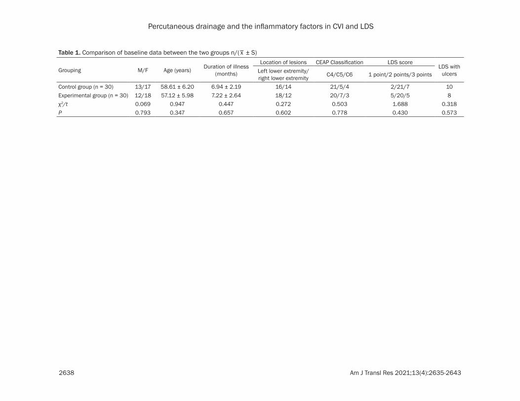
No significant differences were found in terms of sex, age, disease duration, lesion site, CEAP classification, LDS score, or proportion of LDS with ulcers between the two groups, (P > 0.05), indicating that the two groups were comparable (Table 1).
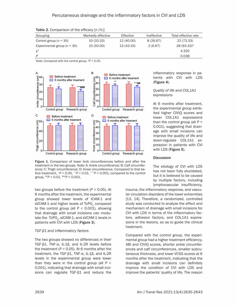
Efficacy of treatment
The total effective rate of the experimental group was higher than of the total effective rate in the control group (P < 0.05), indicating that percutaneous drainage with tiny incisions can enhance the treatment efficacy and promote symptomatic relief in patients with CVI with LDS (Table 2).
Circumference of the lower extremities
There were no significant differences in the thigh and knee circumferences between the two groups (P > 0.05). At 6 months after the treatment, the experimental group exhibited shorter ankle circumferences and calf circum-ferences than the control group (all P < 0.001), indicating that drainage with tiny incisions can reduce the ankle circumferences and calf cir-cumferences in patients with CVI with LDS (Figure 1).
Subcutaneous thicknesses, VCSS scores, and ABI
There were no significant differences in the subcutaneous thicknesses, the VCSS scores, or the ABI between the two groups (P > 0.05). At 6 months after the treatment, the subcutane-ous thicknesses, the VCSS scores, and the ABI in the experimental group were higher than they were in the control group (all P < 0.001), indicating that drainage with small incisions can alleviate the symptoms of CVI with LDS, reduce the subcutaneous thickness, and improve the ABI (Figure 2).
TcPO2, sICAM-1, and sVCAM-1
There were no significant differences in the TcPO2, sICAM-1, or sVCAM-1 levels between the
Percutaneous drainage and the inflammatory factors in CVI and LDS
2638 Am J Transl Res 2021;13(4):2635-2643
Table 1. Comparison of baseline data between the two groups n/(_x ± S)
Grouping M/F Age (years) Duration of illness (months)
Location of lesions CEAP Classification LDS scoreLDS with
ulcersLeft lower extremity/right lower extremity C4/C5/C6 1 point/2 points/3 points
Control group (n = 30) 13/17 58.61 ± 6.20 6.94 ± 2.19 16/14 21/5/4 2/21/7 10Experimental group (n = 30) 12/18 57.12 ± 5.98 7.22 ± 2.64 18/12 20/7/3 5/20/5 8χ2/t 0.069 0.947 0.447 0.272 0.503 1.688 0.318P 0.793 0.347 0.657 0.602 0.778 0.430 0.573
Percutaneous drainage and the inflammatory factors in CVI and LDS
2639 Am J Transl Res 2021;13(4):2635-2643
two groups before the treatment (P > 0.05). At 6 months after the treatment, the experimental group showed lower levels of ICAM-1 and sVCAM-1 and higher levels of TcPO2 compared to the control group (all P < 0.001), showing that drainage with small incisions can modu-late the TcPO2, sICAM-1, and sVCAM-1 levels in patients with CVI with LDS (Figure 3).
TGF-β1 and inflammatory factors
The two groups showed no differences in their TGF-β1, TNF-α, IL-1β, and IL-2R levels before the treatment (P > 0.05). At 6 months after the treatment, the TGF-β1, TNF-α, IL-1β, and IL-2R levels in the experimental group were lower than they were in the control group (all P < 0.001), indicating that drainage with small inci-sions can regulate TGF-β1 and reduce the
trauma, the inflammatory response, and vascu-lar circulation disorders of the lower extremities [13, 14]. Therefore, a randomized, controlled study was conducted to analyze the effect and mechanism of drainage with small incisions for CVI with LDS in terms of the inflammatory fac-tors, adhesion factors, and COL1A1 expres-sions in the lesions, so as to guide the clinical treatment.
Compared with the control group, the experi-mental group had a higher treatment efficiency, ABI and CIVIQ scores, shorter ankle circumfer-ences and calf circumferences, smaller subcu-taneous thickness, and lower VCSS scores at 6 months after the treatment, indicating that the drainage with small incisions can definitely improve the condition of CVI with LDS and improve the patients’ quality of life. The reason
Table 2. Comparison of the efficacy [n (%)]Grouping Markedly effective Effective Ineffective Total effective rateControl group (n = 30) 10 (33.33) 12 (40.00) 8 (26.67) 22 (73.33)Experimental group (n = 30) 15 (50.00) 13 (43.33) 2 (6.67) 28 (93.33)#
χ2 4.320P 0.038Note: Compared with the control group, #P < 0.05.
Figure 1. Comparison of lower limb circumferences before and after the treatment in the two groups. Note: A: Ankle circumference; B: Calf circumfer-ence; C: Thigh circumference; D: Knee circumference. Compared to that be-fore treatment, *P < 0.05, **P < 0.01, ***P < 0.001; compared to the control group, ##P < 0.01, ###P < 0.001.
inflammatory response in pa- tients with CVI with LDS (Figure 4).
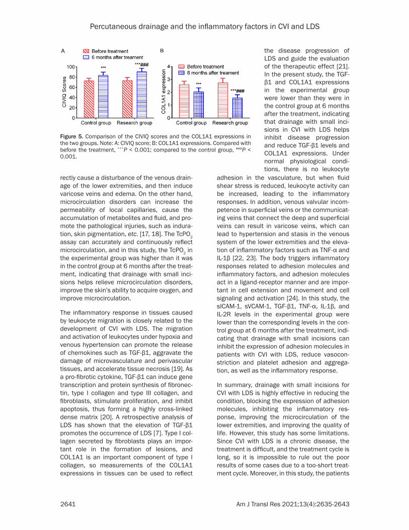
Quality of life and COL1A1 expressions
At 6 months after treatment, the experimental group exhib-ited higher CIVIQ scores and lower COL1A1 expressions than the control group (all P < 0.001), suggesting that drain-age with small incisions can improve the quality of life and down-regulate COL1A1 ex- pression in patients with CVI with LDS (Figure 5).
Discussion
The etiology of CVI with LDS has not been fully elucidated, but it is believed to be caused by multiple factors, including lymphovascular insufficiency,

Percutaneous drainage and the inflammatory factors in CVI and LDS
2640 Am J Transl Res 2021;13(4):2635-2643
Figure 2. Comparison of subcutaneous thicknesses, VCSS scores, and ABI between the two groups. Note: A: Subcu-taneous thickness; B: VCSS score; C: ABI index. Compared with before the treatment, ***P < 0.001; compared with the control group, ###P < 0.001.
Figure 3. Comparison of two groups’ TcPO2, sICAM-1, and sVCAM-1 levels. Note: A: TcPO2; B: sICAM-1; C: sVCAM-1. Compared with before the treatment, ***P < 0.001; compared with the control group, ###P < 0.001.
Figure 4. Comparison of the TGF-β1 and inflammatory factors between the two groups. Note: A: TGF-β1; B: TNF-α; C: IL-1β; D: IL-2R. Compared with be-fore the treatment, **P < 0.01, ***P < 0.001; ###P < 0.001 compared with the control group.
may be that varicose vein stripping alone can eliminate venous hypertension, but the long-
term accumulated necrotic tissue and fluid retention can-not be directly removed, which would be slowly absorbed, making it difficult to achieve satisfactory results. However, when longitudinal, small inci-sions are made at the lesion, followed by blunt dissection and draining, the procedure can completely remove local necrosis and liquefied fatty and necrotic tissues, and the skin induration, skin redness and swelling, and skin tender-ness disappear [15, 16]. Microcirculation disorders in the lower extremities are par-ticularly prominent in the occurrence and development of CVI with LDS. On the one hand, it can cause ischemia and hypoxia of the gastrocne-
mius muscle of the lower extremities, impair the function of the calf muscle pump, and indi-
Percutaneous drainage and the inflammatory factors in CVI and LDS
2641 Am J Transl Res 2021;13(4):2635-2643
rectly cause a disturbance of the venous drain-age of the lower extremities, and then induce varicose veins and edema. On the other hand, microcirculation disorders can increase the permeability of local capillaries, cause the accumulation of metabolites and fluid, and pro-mote the pathological injuries, such as indura-tion, skin pigmentation, etc. [17, 18]. The TcPO2 assay can accurately and continuously reflect microcirculation, and in this study, the TcPO2 in the experimental group was higher than it was in the control group at 6 months after the treat-ment, indicating that drainage with small inci-sions helps relieve microcirculation disorders, improve the skin’s ability to acquire oxygen, and improve microcirculation.
The inflammatory response in tissues caused by leukocyte migration is closely related to the development of CVI with LDS. The migration and activation of leukocytes under hypoxia and venous hypertension can promote the release of chemokines such as TGF-β1, aggravate the damage of microvasculature and perivascular tissues, and accelerate tissue necrosis [19]. As a pro-fibrotic cytokine, TGF-β1 can induce gene transcription and protein synthesis of fibronec-tin, type I collagen and type III collagen, and fibroblasts, stimulate proliferation, and inhibit apoptosis, thus forming a highly cross-linked dense matrix [20]. A retrospective analysis of LDS has shown that the elevation of TGF-β1 promotes the occurrence of LDS [7]. Type I col-lagen secreted by fibroblasts plays an impor-tant role in the formation of lesions, and COL1A1 is an important component of type I collagen, so measurements of the COL1A1 expressions in tissues can be used to reflect
adhesion in the vasculature, but when fluid shear stress is reduced, leukocyte activity can be increased, leading to the inflammatory responses. In addition, venous valvular incom-petence in superficial veins or the communicat-ing veins that connect the deep and superficial veins can result in varicose veins, which can lead to hypertension and stasis in the venous system of the lower extremities and the eleva-tion of inflammatory factors such as TNF-α and IL-1β [22, 23]. The body triggers inflammatory responses related to adhesion molecules and inflammatory factors, and adhesion molecules act in a ligand-receptor manner and are impor-tant in cell extension and movement and cell signaling and activation [24]. In this study, the sICAM-1, sVCAM-1, TGF-β1, TNF-α, IL-1β, and IL-2R levels in the experimental group were lower than the corresponding levels in the con-trol group at 6 months after the treatment, indi-cating that drainage with small incisions can inhibit the expression of adhesion molecules in patients with CVI with LDS, reduce vasocon-striction and platelet adhesion and aggrega-tion, as well as the inflammatory response.
In summary, drainage with small incisions for CVI with LDS is highly effective in reducing the condition, blocking the expression of adhesion molecules, inhibiting the inflammatory res- ponse, improving the microcirculation of the lower extremities, and improving the quality of life. However, this study has some limitations. Since CVI with LDS is a chronic disease, the treatment is difficult, and the treatment cycle is long, so it is impossible to rule out the poor results of some cases due to a too-short treat-ment cycle. Moreover, in this study, the patients
Figure 5. Comparison of the CIVIQ scores and the COL1A1 expressions in the two groups. Note: A: CIVIQ score; B: COL1A1 expressions. Compared with before the treatment, ***P < 0.001; compared to the control group, ###P < 0.001.
the disease progression of LDS and guide the evaluation of the therapeutic effect [21]. In the present study, the TGF-β1 and COL1A1 expressions in the experimental group were lower than they were in the control group at 6 months after the treatment, indicating that drainage with small inci-sions in CVI with LDS helps inhibit disease progression and reduce TGF-β1 levels and COL1A1 expressions. Under normal physiological condi-tions, there is no leukocyte

Percutaneous drainage and the inflammatory factors in CVI and LDS
2642 Am J Transl Res 2021;13(4):2635-2643
were not followed up for a long time, so it was not observed whether prolonging the treatment cycle was more conducive to patient rehabilita-tion. Therefore, it is necessary to appropriately increase the sample size, prolong the treat-ment cycle and follow-up times, so as to further explore the long-term efficacy of the therapy.
Disclosure of conflict of interest
None.
Address correspondence to: Boshun Wan, De- partment of General Surgery, Affiliated Jiading Center Hospital of Shanghai University of Medicine & Health Science, No. 1, Chengbei Road, Jiading District, Shanghai 201800, China. Tel: +86-18930862621; E-mail: [email protected]
References
[1] Eberhardt RT and Raffetto JD. Chronic venous insufficiency. Circulation 2014; 130: 333-346.
[2] Youn YJ and Lee J. Chronic venous insufficien-cy and varicose veins of the lower extremities. Korean J Intern Med 2019; 34: 269-283.
[3] Berti-Hearn L and Elliott B. Chronic venous in-sufficiency: a review for nurses. Nursing 2019; 49: 24-30.
[4] Hyder ON and Soukas PA. Chronic venous in-sufficiency: novel management strategies for an under-diagnosed disease process. R I Med J (2013) 2017; 100: 37-39.
[5] de-Abreu GCG, Camargo O Júnior, de-Abreu MFM and de-Aquino JLB. Ultrasound-guided foam sclerotherapy for severe chronic venous insufficiency. Rev Col Bras Cir 2017; 44: 511-520.
[6] Li Y, Zhou H, Ning Y, Wan B and Zhu Y. Efficacy of point-of-care drainage surgery in chronic ve-nous insufficiency of the lower extremities with lipodystrophy. China Clin Med 2015; 22: 218-221.
[7] Miteva M, Romanelli P and Kirsner RS. Lipo-dermatosclerosis. Dermatol Ther 2010; 23: 375-388.
[8] Péret LA, Vidal HM, Gomes GAC, Oliveira GVB and Aguiar LM. Oxandrolone for treatment of lipodermatosclerosis: case report. J Vasc Bras 2019; 18: e20190031.
[9] Arendsen LP, Vig S, Thakar R and Sultan AH. Impact of copper compression stockings on venous insufficiency and lipodermatosclerosis: a randomised controlled trial. Phlebology 2019; 34: 224-230.
[10] Association VSGotCM. Chinese Expert Consen-sus on Diagnosis and Treatment of Chronic Lower Extremity Venous Disease. in press.
[11] Eklöf B, Rutherford RB, Bergan JJ, Carpentier PH, Gloviczki P, Kistner RL, Meissner MH, Mo-neta GL, Myers K, Padberg FT, Perrin M, Ruck-ley CV, Smith PC and Wakefield TW. Revision of the CEAP classification for chronic venous dis-orders: consensus statement. J Vasc Surg 2004; 40: 1248-1252.
[12] Launois R, Reboul-Marty J and Henry B. Con-struction and validation of a quality of life questionnaire in chronic lower limb venous in-sufficiency (CIVIQ). Qual Life Res 1996; 5: 539-554.
[13] Wittens C, Davies AH, Bækgaard N, Broholm R, Cavezzi A, Chastanet S, de Wolf M, Eggen C, Giannoukas A, Gohel M, Kakkos S, Lawson J, Noppeney T, Onida S, Pittaluga P, Thomis S, Toonder I, Vuylsteke M, Esvs Guidelines C, Kolh P, de Borst GJ, Chakfé N, Debus S, Hinchliffe R, Koncar I, Lindholt J, de Ceniga MV, Vermassen F, Verzini F, Document R, De Maeseneer MG, Blomgren L, Hartung O, Kalodiki E, Korten E, Lugli M, Naylor R, Nicolini P and Rosales A. Editor’s choice - management of chronic ve-nous disease: clinical practice guidelines of the European society for vascular surgery (ESVS). Eur J Vasc Endovasc Surg 2015; 49: 678-737.
[14] Carman TL and Al-Omari A. Evaluation and management of chronic venous disease using the foundation of CEAP. Curr Cardiol Rep 2019; 21: 114.
[15] Menzinger G. [Modern diagnostic methods of chronic venous insufficiency]. Wien Med Wochenschr 2016; 166: 275-277.
[16] Suehiro K, Morikage N, Harada T, Samura M, Nagase T, Mizoguchi T and Hamano K. Com-pression therapy using bandages successfully manages acute or subacute lipodermatoscle-rosis. Ann Vasc Dis 2019; 12: 77-79.
[17] Sun H. Effectiveness of point-of-care drainage surgery in chronic venous insufficiency of the lower extremities with lipid scleroderma. Biped & Health Care 2018; 27: 90,92.
[18] Nyckowski T, McGregor S and Huang WW. A case of gemcitabine-related acute lipoderma-tosclerosis. J Oncol Pharm Pract 2019; 25: 1271-1274.
[19] Balasubramanyam S, Migden MR and Silapunt S. Venous treatment of lipodermatosclerosis to improve ambulatory function. Dermatol Surg 2018; 44: 749-752.
[20] Tidwell WJ, Malone J and Callen JP. Cutaneous T-cell lymphoma misdiagnosed as lipoderma-tosclerosis. JAMA Dermatol 2016; 152: 487-488.
[21] Dahm KT, Myrhaug HT, Strømme H, Fure B and Brurberg KG. Effects of preventive use of com-pression stockings for elderly with chronic ve-nous insufficiency and swollen legs: a system-

Percutaneous drainage and the inflammatory factors in CVI and LDS
2643 Am J Transl Res 2021;13(4):2635-2643
atic review and meta-analysis. BMC Geriatr 2019; 19: 76.
[22] Salmhofer W. [Etiology, nomenclature and pathophysiology of chronic venous insufficien-cy]. Wien Med Wochenschr 2016; 166: 264-269.
[23] Walsh SN and Santa Cruz DJ. Lipodermatoscle-rosis: a clinicopathological study of 25 cases. J Am Acad Dermatol 2010; 62: 1005-1012.
[24] Martis G. Reply letter to the editor, regarding the role of radical surgery in the management of CEAP C5/6 and lipodermatosclerosis. Phle-bology 2016; 31: 770-771.