One and two-dimensional proton NMR, fluorescence and molecular modeling studies on the...
8
J. Med. Chem. 1988, 31,583-590 583 One- and Two-Dimensional ‘H NMR, Fluorescence, and Molecular Modeling Studies on the Tomaymycin-d(ATGCAT)2Adduct. Evidence for Two Covalent Adducts with Opposite Orientations and Stereochemistries at the Covalent Linkage Site Steve Cheatham,’ Alan Kook,? Laurence H. Hurley,*f Mary D. Barkley,t and William Remerss Drug Dynamics Institute, Division of Medicinal Chemistry and Natural Products Chemistry, College of Pharmacy, University of Texas at Austin, Austin, Texas 78712, Department of Chemistry, Louisiana State University, Baton Rouge, Louisiana 70803, and Department of Medicinal Chemistry, College of Pharmacy, University of Arizona, Tucson, Arizona 85721. Received August 10, 1987 Tomaymycin is a member of the pyrrole[ 1,4]benzodiazepine antitumor-antibiotic group that binds covalently to the exocyclic 2-amino group of guanine in DNA. Previous correlation of fluorescence and NMR data suggested that the 11R,llaS and the 11S,llaS diastereomers of tomaymycin could bind to DNA in two orientations relative to the covalently modified guanine (Barkley, M. D.; Cheatham, S.; Thurston, D. E.; Hurley, L. H. Biochemistry 1986,25,3021-3031). We now report on fluorescence, one- and two-dimensional proton NMR, and molecular modeling studies of the tomaymycin-d(ATGCAT)2 adduct, which corroborate these earlier observations. Fluorescence measurements show that there are two species of tomaymycin bound to d(ATGCAT)2, which are tentatively identified as the 11R,llaS and 11S,llaS diastereomers. Two distinct sets of signals for the tomaymycin molecule are present in the proton NMR spectrum of the tomaymycin-d(ATGCAT)2 duplex adduct. Two-dimensional correlation spectroscopy (2D-COSY) studies also show connectivities for four cytosine H5-H6 and eight thymine methyl-H6 protons and thus clearly establish the presence of two distinct species of tomaymycin4(ATGCAT)2 adducts in solution. A single scalar 11-lla lH NMR coupling in the 2D-COSY spectrum is indicative of an adduct species that has an S configuration at the C-11 position. Two-dimensional nuclear Overhauser effect (NOESY) spectra of the to- maymycin-d(ATGCAT)2 duplex adduct show that the adducts are relatively nondistortive. In a NOESY experiment, cross-peaks were identified between both the aromatic H9 proton and the ethylidine methyl protons of tomaymycin and two different adenine H2 protons of d(ATGCAT),. Molecular mechanics calculations with AMBER show that the two species with the thermodynamically most favorable binding energies are the 11R,llaS and 11S,llaS isomers with their aromatic rings to the 5’ and 3’ sides of the covalently bound guanine, respectively. The NOES observed between tomaymycin protons and adenine H2 protons are in accord with molecular modeling studies. Taken together, these results strongly suggest that the two forms of tomaymycin bound to d(ATGCAT)2 are the 11S,llaS and 11R,llaS species, oriented with their aromatic rings to the 3’ and 5’ sides, respectively, of the covalently modified guanines. Tomaymycin (I) (Figure 1) is a member of the potent P(1,4)B1antitumor-antibiotic group, which also includes anthramycin, sibiromycin, and the neothramycins A and B2. The antitumor activity of the P(1,4)Bs is attributed to their ability to form covalent adducts with DNA through N-2 of guanine3 (see Figure 2). Anthramycin has been shown to form an aminal linkage between its carbinolamine carbon at C-11 and N-2 of guanineS4 CPK model studies show that the P(1,4)B nucleus fits completely within the minor groove of B-form DNA, forming a relatively non- distortive adduct.6 Tomaymycin also binds covalently to DNA but not as efficiently as anthramycin or sibiromy- cins2v6 The DNA sequence specificity of tomaymycin and other P(1,4)Bs has been characterized by MPE-Fe(I1) “footprinting”.6 Tomaymycin, anthramycin, and sibiro- mycin display a three base pair sequence selectivity for 5’PuGPu sequences. Guanines within 5’PyGPy trimers are the least preferred sites for covalent binding, with 5’PyGPu and 5’PuGPy sequences being of intermediate preference. The anthramy~in-d(ATGCAT)~ adduct has been par- tially characterized by using one- and two-dimensional(lD, 2D) proton NMR technique^.^^^ Covalent adduct forma- tion results in loss of symmetry of the duple^,^ and two- dimensional lH NMR experiments show that anthramycin binds through an 11s geometry with its aromatic ring on the 3’ side of the covalently modified guanine.s In contrast, two distinct forms of tomaymycin-DNA adducts are observed by time-resolved fluorescence spec- tros~opy.~ lH NMR studies of tomaymycin in various solvents demonstrated that tomaymycin can exist as an equilibrium mixture of 11R,llaS and 11S, l l a S diaste- University of Texas at Austin. t Louisiana State University. *University of Arizona. reomers? The correlation between NMR and fluorescence data led us to postulate that the two species of tomaymycin observed on DNA were the diastereomeric 11R,llaS and 11S,llaS tomaymycin-DNA adducts bound through N-2 of guanine.1° (1) Abbreviations: CPK, Corey, Pauling, and Koltun; COSY, correlation spectroscopy; DMTdTAPSi, dimethoxytrityl-2’- deoxythymidine aminopropyl silica gel; NOESY, two-dimen- sional nuclear Overhauser effect; MPE, methidiumpropyl- ethylenediaminetetraacetic acid; Pu, purine; Py, pyrimidine; THF, tetrahydrofuran; TME, tomaymycin 11-methyl ether; TSP, sodium 3-(trimethylsi1yl)propionate-2,2,3,3-d4; P(1,4)B, pyrrole[ 1,4] benzodiazepine. (2) Hurley, L. H. J. Antibiot. 1977, 30, 349. (3) Hurley, L. H.; Needham-VanDevanter, D. Acc. Chem. Res. 1986, 19, 230. (4) Graves, D. E.; Pattaroni, C.; Krishnan, B. S.; Ostrander, J. M.; Hurley, L. H.; Krugh, T. R. J. Biol. Chem. 1984, 259, 8202. (5) Petrusek, R. L.; Anderson, G. L.; Garner, T. F.; Quinton, F. L.; Fannin, L.; Kaplan, D. J.; Zimmer, S. G.; Hurley, L. H. Bio- chemistry 1984, 23, 1111. (6) Hertzberg, R. P.; Hecht, S. M.; Reynolds, V. L.; Molineux, I. J.; Hurley, L. H. Biochemistry 1986, 25, 1249. (7) Graves, D. E.; Stone, M. P.; Krugh, T. R. Biochemistry 1985, 24, 7573. (8) Boyd, F. L.; Cheatham, S.; Hurley, L. H., submitted for pub- lication in J. Am. Chem. SOC. (9) Barkley, M. D.; Cheatham, S.; Thurston, D. E.; Hurley, L. H. Biochemistry 1986, 25, 3021. (10) In this paper, the stereochemistry at the covalent linkage site between C-11 of tomaymycin and N-2 of guanine shown in Figure 6B is incorrect. The structure for Figure 6B is denoted in the Figure legend as the 11R,llaS diastereomer of the to- maymycin-d(ATGCAT), adduct. However, Figure 6B shows the 11SJlaS diastereomer. Consequently Figures 6A,B only differ such that the drug molecules are oriented in opposite directions in the minor groove of d(ATGCAT),. The correct structure for Figure 6B in ref 8 is the species D shown in Figure 9 of the present manuscript. 0022-2623/88/l831-0583$01.50/0 0 1988 American Chemical Society
-
Upload
independent -
Category
Documents
-
view
0 -
download
0
Transcript of One and two-dimensional proton NMR, fluorescence and molecular modeling studies on the...

J. Med. Chem. 1988, 31,583-590 583
One- and Two-Dimensional ‘H NMR, Fluorescence, and Molecular Modeling Studies on the Tomaymycin-d(ATGCAT)2 Adduct. Evidence for Two Covalent Adducts with Opposite Orientations and Stereochemistries at the Covalent Linkage Site
Steve Cheatham,’ Alan Kook,? Laurence H. Hurley,*f Mary D. Barkley,t and William Remerss Drug Dynamics Institute, Division of Medicinal Chemistry and Natural Products Chemistry, College of Pharmacy, University of Texas a t Austin, Austin, Texas 78712, Department of Chemistry, Louisiana State University, Baton Rouge, Louisiana 70803, and Department of Medicinal Chemistry, College of Pharmacy, University of Arizona, Tucson, Arizona 85721. Received August 10, 1987
Tomaymycin is a member of the pyrrole[ 1,4]benzodiazepine antitumor-antibiotic group that binds covalently to the exocyclic 2-amino group of guanine in DNA. Previous correlation of fluorescence and NMR data suggested that the 11R,llaS and the 11S,llaS diastereomers of tomaymycin could bind to DNA in two orientations relative to the covalently modified guanine (Barkley, M. D.; Cheatham, S.; Thurston, D. E.; Hurley, L. H. Biochemistry 1986,25,3021-3031). We now report on fluorescence, one- and two-dimensional proton NMR, and molecular modeling studies of the tomaymycin-d(ATGCAT)2 adduct, which corroborate these earlier observations. Fluorescence measurements show that there are two species of tomaymycin bound to d(ATGCAT)2, which are tentatively identified as the 11R,llaS and 11S,llaS diastereomers. Two distinct sets of signals for the tomaymycin molecule are present in the proton NMR spectrum of the tomaymycin-d(ATGCAT)2 duplex adduct. Two-dimensional correlation spectroscopy (2D-COSY) studies also show connectivities for four cytosine H5-H6 and eight thymine methyl-H6 protons and thus clearly establish the presence of two distinct species of tomaymycin4(ATGCAT)2 adducts in solution. A single scalar 11-lla lH NMR coupling in the 2D-COSY spectrum is indicative of an adduct species that has an S configuration a t the C-11 position. Two-dimensional nuclear Overhauser effect (NOESY) spectra of the to- maymycin-d(ATGCAT)2 duplex adduct show that the adducts are relatively nondistortive. In a NOESY experiment, cross-peaks were identified between both the aromatic H9 proton and the ethylidine methyl protons of tomaymycin and two different adenine H2 protons of d(ATGCAT),. Molecular mechanics calculations with AMBER show that the two species with the thermodynamically most favorable binding energies are the 11R,llaS and 11S,llaS isomers with their aromatic rings to the 5’ and 3’ sides of the covalently bound guanine, respectively. The NOES observed between tomaymycin protons and adenine H2 protons are in accord with molecular modeling studies. Taken together, these results strongly suggest that the two forms of tomaymycin bound to d(ATGCAT)2 are the 11S,llaS and 11R,llaS species, oriented with their aromatic rings to the 3’ and 5’ sides, respectively, of the covalently modified guanines.
Tomaymycin (I) (Figure 1) is a member of the potent P(1,4)B1 antitumor-antibiotic group, which also includes anthramycin, sibiromycin, and the neothramycins A and B2. The antitumor activity of the P(1,4)Bs is attributed to their ability to form covalent adducts with DNA through N-2 of guanine3 (see Figure 2). Anthramycin has been shown to form an aminal linkage between its carbinolamine carbon at C-11 and N-2 of guanineS4 CPK model studies show that the P(1,4)B nucleus fits completely within the minor groove of B-form DNA, forming a relatively non- distortive adduct.6 Tomaymycin also binds covalently to DNA but not as efficiently as anthramycin or sibiromy- cins2v6 The DNA sequence specificity of tomaymycin and other P(1,4)Bs has been characterized by MPE-Fe(I1) “footprinting”.6 Tomaymycin, anthramycin, and sibiro- mycin display a three base pair sequence selectivity for 5’PuGPu sequences. Guanines within 5’PyGPy trimers are the least preferred sites for covalent binding, with 5’PyGPu and 5’PuGPy sequences being of intermediate preference.
The anthramy~in-d(ATGCAT)~ adduct has been par- tially characterized by using one- and two-dimensional (lD, 2D) proton NMR technique^.^^^ Covalent adduct forma- tion results in loss of symmetry of the duple^,^ and two- dimensional lH NMR experiments show that anthramycin binds through an 11s geometry with its aromatic ring on the 3’ side of the covalently modified guanine.s
In contrast, two distinct forms of tomaymycin-DNA adducts are observed by time-resolved fluorescence spec- t ro s~opy .~ lH NMR studies of tomaymycin in various solvents demonstrated that tomaymycin can exist as an equilibrium mixture of 11R,llaS and 11S, l l a S diaste-
University of Texas a t Austin. t Louisiana State University. *University of Arizona.
reomers? The correlation between NMR and fluorescence data led us to postulate that the two species of tomaymycin observed on DNA were the diastereomeric 11R,llaS and 11S,llaS tomaymycin-DNA adducts bound through N-2 of guanine.1°
(1) Abbreviations: CPK, Corey, Pauling, and Koltun; COSY, correlation spectroscopy; DMTdTAPSi, dimethoxytrityl-2’- deoxythymidine aminopropyl silica gel; NOESY, two-dimen- sional nuclear Overhauser effect; MPE, methidiumpropyl- ethylenediaminetetraacetic acid; Pu, purine; Py, pyrimidine; THF, tetrahydrofuran; TME, tomaymycin 11-methyl ether; TSP, sodium 3-(trimethylsi1yl)propionate-2,2,3,3-d4; P(1,4)B, pyrrole[ 1,4] benzodiazepine.
(2) Hurley, L. H. J. Antibiot. 1977, 30, 349. (3) Hurley, L. H.; Needham-VanDevanter, D. Acc. Chem. Res.
1986, 19, 230. (4) Graves, D. E.; Pattaroni, C.; Krishnan, B. S.; Ostrander, J. M.;
Hurley, L. H.; Krugh, T. R. J. Biol. Chem. 1984, 259, 8202. (5) Petrusek, R. L.; Anderson, G. L.; Garner, T. F.; Quinton, F. L.;
Fannin, L.; Kaplan, D. J.; Zimmer, S. G.; Hurley, L. H. Bio- chemistry 1984, 23, 1111.
(6) Hertzberg, R. P.; Hecht, S. M.; Reynolds, V. L.; Molineux, I. J.; Hurley, L. H. Biochemistry 1986, 25, 1249.
(7) Graves, D. E.; Stone, M. P.; Krugh, T. R. Biochemistry 1985, 24, 7573.
(8) Boyd, F. L.; Cheatham, S.; Hurley, L. H., submitted for pub- lication in J . Am. Chem. SOC.
(9) Barkley, M. D.; Cheatham, S.; Thurston, D. E.; Hurley, L. H. Biochemistry 1986, 25, 3021.
(10) In this paper, the stereochemistry at the covalent linkage site between C-11 of tomaymycin and N-2 of guanine shown in Figure 6B is incorrect. The structure for Figure 6B is denoted in the Figure legend as the 11R,llaS diastereomer of the to- maymycin-d(ATGCAT), adduct. However, Figure 6B shows the 11SJlaS diastereomer. Consequently Figures 6A,B only differ such that the drug molecules are oriented in opposite directions in the minor groove of d(ATGCAT),. The correct structure for Figure 6B in ref 8 is the species D shown in Figure 9 of the present manuscript.
0022-2623/88/l831-0583$01.50/0 0 1988 American Chemical Society
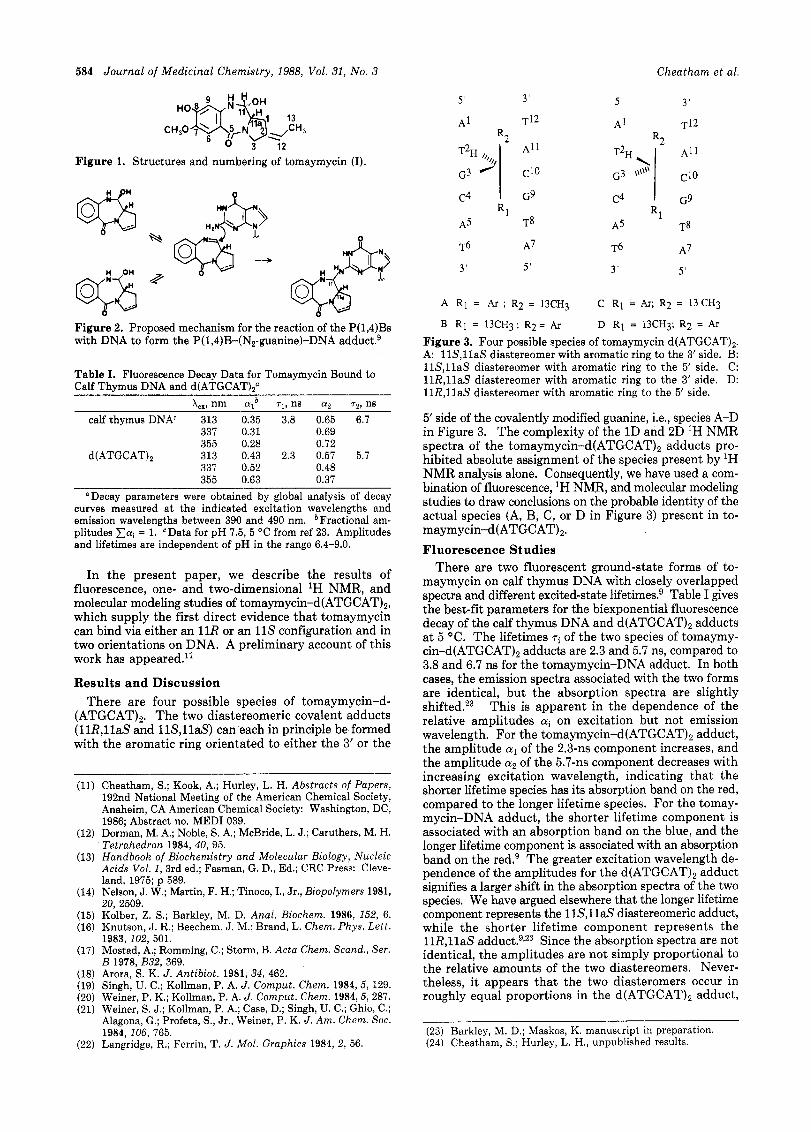
584 Journal of Medicinal Chemistry, 1988, Vol. 31, No. 3 Cheatham et al.
Figure 1. Structures and numbering of tomaymycin (I).
&;) L q - ---
Figure 2. Proposed mechanism for the reaction of the P(1,4)Bs with DNA to form the P(1,4)B-(N2-guanine)-DNA a d d ~ c t . ~
Table I. Fluorescence Decay Data for Tomaymycin Bound to Calf Thymus DNA and d(ATGCATI2"
L, nm arb r, , ns a, 7,. ns calf thymus DNA' 313 0.35 3.8 0.65 6.7
337 0.31 0.69 355 0.28 0.72
337 0.52 0.48 355 0.63 0.37
d(ATGCAT)Z 313 0.43 2.3 0.57 5.7
Decay parameters were obtained by global analysis of decay curves measured at the indicated excitation wavelengths and emission wavelengths between 390 and 490 nm. bFractional am- plitudes xai = 1. cData for pH 7.5, 5 "C from ref 23. Amplitudes and lifetimes are independent of pH in the range 6.4-9.0.
In the present paper, we describe the results of fluorescence, one- and two-dimensional lH NMR, and molecular modeling studies of tomaymycin-d(ATGCAT)2, which supply the first direct evidence that tomaymycin can bind via either an 11R or an 11s configuration and in two orientations on DNA. A preliminary account of this work has appeared.'l
Results and Discussion There are four possible species of tomaymycin-d-
(ATGCAT)2. The two diastereomeric covalent adducts (11RJlaS and 11S,llaS) can each in principle be formed with the aromatic ring orientated to either the 3' or the
(11) Cheatham, S.; Kook, A.; Hurley, L. H. Abstracts of Papers, 192nd National Meeting of the American Chemical Society, Anaheim, CA American Chemical Society: Washington, DC, 1986; Abstract no. MEDI 039.
(12) Dorman, M. A,; Noble, S. A,; McBride, L. J.; Caruthers, M. H. Tetrahedron 1984,40, 95.
(13) Handbook of Biochemistry and Molecular Biology, Nucleic Acids Vol. 1.3rd ed.: Fasman. G. D.. Ed.: CRC Press: Cleve- land, 1975; p 589. '
20, 2509. (14) Nelson, J. W.; Martin, F. H.; Tinoco, I., Jr., Biopolymers 1981,
(15) Kolber, Z. S.; Barkley, M. D. Anal. Biochem. 1986, 152, 6. (16) Knutson, J. R.; Beechem. J. M.; Brand, L. Chern. Phys. Lett.
1983, 102, 501. (17) Mostad, A.; Romming, C.; Storm, B. Acta Chem. Scand., Ser.
B 1978, B32, 369. (18) Arora, S. K. J. Antibiot. 1981, 34, 462. (19) Singh, U. C.; Kollman, P. A. J . Cornput. Chem. 1984,5, 129. (20) Weiner, P. K.; Kollman, P. A. J. Cornput. Chem. 1984,5, 287. (21) Weiner, S. J.; Kollman, P. A.; Case, D.; Singh, U. C.; Ghio, C.;
Alagona, G.; Profeta, S., Jr., Weiner, P. K. J . Am. Chem. Soc. 1984, 106, 765.
(22) Langridge, R.; Ferrin, T. J . Mol. Graphics 1984, 2, 56.
T6 A7 T6 A7
3 ' 5' 3' 5 '
A R1 = Ar ; R2 = 13CH3 R1 = Ar, R2 = 13CH3
B R1 = 13CH3; R2= Ar D Ry = 13CH3; R2 = Ar
C
Figure 3. Four possible species of tomaymycin d(ATGCAT),. A: 11SJlaS diastereomer with aromatic ring to the 3' side. B: 11S,llaS diastereomer with aromatic ring to the 5' side. C: 11R,llaS diastereomer with aromatic ring to the 3' side. D: 11R,llaS diastereomer with aromatic ring to the 5' side.
5' side of the covalently modified guanine, i.e., species A-D in Figure 3. The complexity of the 1D and 2D IH NMR spectra of the t~maymycin-d(ATGCAT)~ adducts pro- hibited absolute assignment of the species present by lH NMR analysis alone. Consequently, we have used a com- bination of fluorescence, lH NMB, and molecular modeling studies to draw conclusions on the probable identity of the actual species (A, B, C, or D in Figure 3) present in to- maymycin-d(ATGCAT),. Fluorescence Studies
There are two fluorescent ground-state forms of to- maymycin on calf thymus DNA with closely overlapped spectra and different excited-state lifetime^.^ Table I gives the best-fit parameters for the biexponential fluorescence decay of the calf thymus DNA and d(ATGCAT)2 adducts at 5 "C. The lifetimes T~ of the two species of tomaymy- cin-d(ATGCAT), adducts are 2.3 and 5.7 ns, compared to 3.8 and 6.7 ns for the tomaymycin-DNA adduct. In both cases, the emission spectra associated with the two forms are identical, but the absorption spectra are slightly shifted.23 This is apparent in the dependence of the relative amplitudes ai on excitation but not emission wavelength. For the tomaymycin-d(ATGCAT), adduct, the amplitude a1 of the 2.3-11s component increases, and the amplitude a2 of the 5.7-11s component decreases with increasing excitation wavelength, indicating that the shorter lifetime species has its absorption band on the red, compared to the longer lifetime species. For the tomay- mycin-DNA adduct, the shorter lifetime component is associated with an absorption band on the blue, and the longer lifetime component is associated with an absorption band on the red.g The greater excitation wavelength de- pendence of the amplitudes for the d(ATGCAT)2 adduct signifies a larger shift in the absorption spectra of the two species. We have argued elsewhere that the longer lifetime component represents the 11S,llaS diastereomeric adduct, while the shorter lifetime component represents the 11R,llaS a d d u ~ t . ~ , ~ ~ Since the absorption spectra are not identical, the amplitudes are not simply proportional to the relative amounts of the two diastereomers. Never- theless, it appears that the two diasteromers occur in roughly equal proportions in the d(ATGCAT), adduct,
(23) Barkley, M. D.; Maskos, K. manuscript in preparation. (24) Cheatham, S.; Hurley, L. H., unpublished results.
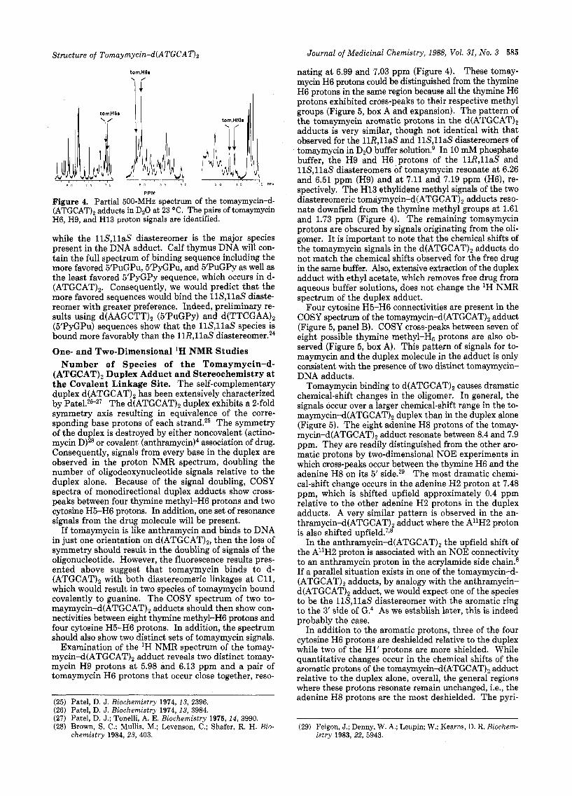
Structure of Tomaymycin-d(ATGCAT)z
torn.H9s
I t 0m.H 6 s I1 ll
PPM
Figure 4. Partial 500-MHz spectrum of the tomaymycin-d- (ATGCAT)2 adducts in D,O at 23 "C. The pairs of tomaymycin H6, H9, and H13 proton signals are identified.
while the 11S,llaS diastereomer is the major species present in the DNA adduct. Calf thymus DNA will con- tain the full spectrum of binding sequence including the more favored ~'PuGPu, 5'PyGPu, and 5'PuGPy as well as the least favored 5'PyGPy sequence, which occurs in d- (ATGCAT)2. Consequently, we would predict that the more favored sequences would bind the 11S,llaS diaste- reomer with greater preference. Indeed, preliminary re- sults using d(AAGCTT), (5'PuGPy) and d(TTCGAA), (5'PyGPu) sequences show that the 11S,llaS species is bound more favorably than the 11R,llaS dia~tereomer.~~
One- and Two-Dimensional 'H NMR Studies Number of Species of the Tomaymycin-d-
(ATGCAT)2 Duplex Adduct and Stereochemistry at the Covalent Linkage Site. The self-complementary duplex d(ATGCAT)2 has been extensively characterized by Pate1.25-27 The d(ATGCAT)2 duplex exhibits a 2-fold symmetry axis resulting in equivalence of the corre- sponding base protons of each strand.25 The symmetry of the duplex is destroyed by either noncovalent (actino- mycin D)28 or covalent (anthramy~in)~ association of drug. Consequently, signals from every base in the duplex are observed in the proton NMR spectrum, doubling the number of oligodeoxynucleotide signals relative to the duplex alone. Because of the signal doubling, COSY spectra of monodirectional duplex adducts show cross- peaks between four thymine methyl-H6 protons and two cytosine H5-H6 protons. In addition, one set of resonance signals from the drug molecule will be present.
If tomaymycin is like anthramycin and binds to DNA in just one orientation on d(ATGCAT)2, then the loss of symmetry should result in the doubling of signals of the oligonucleotide. However, the fluorescence results pres- ented above suggest that tomaymycin binds to d- (ATGCAT), with both diastereomeric linkages at C11, which would result in two species of tomaymycin bound covalently to guanine. The COSY spectrum of two to- maymycin-d(ATGCAT), adducts should then show con- nectivities between eight thymine methyl-H6 protons and four cytosine H5-H6 protons. In addition, the spectrum should also show two distinct sets of tomaymycin signals.
Examination of the 'H NMR spectrum of the tomay- mycin-d(ATGCAT), adduct reveals two distinct tomay- mycin H9 protons at 5.98 and 6.13 ppm and a pair of tomaymycin H6 protons that occur close together, reso-
(25) Patel, D. J. Biochemistry 1974, 13, 2396. (26) Patel, D. J. Biochemistry 1974, 13, 3984. (27) Patel, D. J.; Tonelli, A. E. Biochemistry 1975, 14, 3990. (28) Brown, S. C.; Mullis, M.; Levenson, C.; Shafer, R. H. Bio-
chemistry 1984,23, 403.
Journal of Medicinal Chemistry, 1988, Vol. 31, No. 3 585
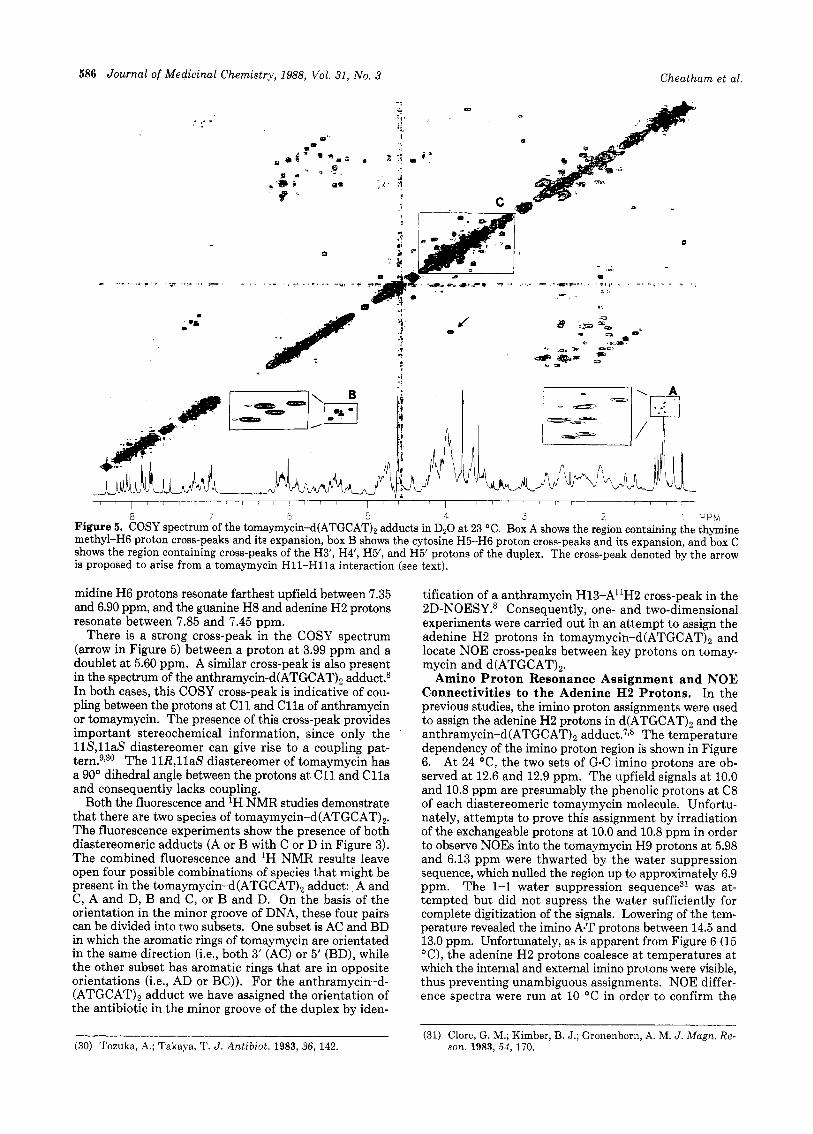
nating at 6.99 and 7.03 ppm (Figure 4). These tomay- mycin H6 protons could be distinguished from the thymine H6 protons in the same region because all the thymine H6 protons exhibited cross-peaks to their respective methyl groups (Figure 5, box A and expansion). The pattern of the tomaymycin aromatic protons in the d(ATGCAT)2 adducts is very similar, though not identical with that observed for the 11R,llaS and 11S,llaS diastereomers of tomaymycin in D20 buffer solution? In 10 mM phosphate buffer, the H9 and H6 protons of the 11R,llaS and 11S,llaS diastereomers of tomaymycin resonate at 6.26 and 6.51 ppm (H9) and at 7.11 and 7.19 ppm (H6), re- spectively. The H13 ethylidene methyl signals of the two diastereomeric t~maymycin-d(ATGCAT)~ adducts reso- nate downfield from the thymine methyl groups at 1.61 and 1.73 ppm (Figure 4). The remaining tomaymycin protons are obscured by signals originating from the oli- gomer. It is important to note that the chemical shifts of the tomaymycin signals in the d(ATGCAT), adducts do not match the chemical shifts observed for the free drug in the same buffer. Also, extensive extraction of the duplex adduct with ethyl acetate, which removes free drug from aqueous buffer solutions, does not change the lH NMR spectrum of the duplex adduct.
Four cytosine H5-H6 connectivities are present in the COSY spectrum of the tomaymycin-d(ATGCAT), adduct (Figure 5, panel B). COSY cross-peaks between seven of eight possible thymine methyl-H, protons are also ob- served (Figure 5, box A). This pattern of signals for to- maymycin and the duplex molecule in the adduct is only consistent with the presence of two distinct tomaymycin- DNA adducts.
Tomaymycin binding to d(ATGCAT), causes dramatic chemical-shift changes in the oligomer. In general, the signals occur over a larger chemical-shift range in the to- maymycin-d(ATGCAT), duplex than in the duplex alone (Figure 5). The eight adenine H8 protons of the tomay- my~in-d(ATGcAT)~ adduct resonate between 8.4 and 7.9 ppm. They are readily distinguished from the other aro- matic protons by two-dimensional NOE experiments in which cross-peaks occur, between the thymine H6 and the adenine H8 on its 5' side.29 The most dramatic chemi- cal-shift change occurs in the adenine H2 proton at 7.48 ppm, which is shifted upfield approximately 0.4 ppm relative to the other adenine H2 protons in the duplex adducts. A very similar pattern is observed in the an- thramycin-d(ATGCAT), adduct where the A'lH2 proton is also shifted ~ p f i e l d . ~ , ~
In the anthramycin-d(ATGCAT), the upfield shift of the A1'H2 proton is associated with an NOE connectivity to an anthramycin proton in the acrylamide side chain.8 If a parallel situation exists in one of the tomaymycin-d- (ATGCAT)2 adducts, by analogy with the anthramycin- d(ATGCAT)2 adduct, we would expect one of the species to be the 11S,llaS diastereomer with the aromatic ring to the 3'side of G.4 As we establish later, this is indeed probably the case.
In addition to the aromatic protons, three of the four cytosine H6 protons are deshielded relative to the duplex while two of the H1' protons are more shielded. While quantitative changes occur in the chemical shifts of the aromatic protons of the tomaymycin-d(ATGCAT), adduct relative to the duplex alone, overall, the general regions where these protons resonate remain unchanged, i.e., the adenine H8 protons are the most deshielded. The pyri-
~~ ~
(29) Feigon, J.; Denny, W. A.; Leupin; W.; Kearns, D. R. Riochem- istry 1983, 22, 5943.
586 Journal of Medicinal Chemistry, 1988, Vol. 31, No. 3 Cheatham et al.
- .
I ..
a 7 E 5 4 3 2 ' PPM Figure 5. COSY spectrum of the tomaymycin-d(ATGCAT), adducts in D20 at 23 "C. Box A shows the region containing the thymine methyl-H6 proton cross-peaks and its expansion, box B shows the cytosine H5-H6 proton cross-peaks and its expansion, and box C shows the region containing cross-peaks of the H3', H4', H5', and H5' protons of the duplex. The cross-peak denoted by the arrow is proposed to arise from a tomaymycin H11-Hlla interaction (see text).
midine H6 protons resonate farthest upfield between 7.35 and 6.90 ppm, and the guanine H8 and adenine H2 protons resonate between 7.85 and 7.45 ppm.
There is a strong cross-peak in the COSY spectrum (arrow in Figure 5) between a proton at 3.99 ppm and a doublet at 5.60 ppm. A similar cross-peak is also present in the spectrum of the anthramycin-d(ATGCAT)2 adduct.8 In both cases, this COSY cross-peak is indicative of cou- pling between the protons at C11 and Cl la of anthramycin or tomaymycin. The presence of this cross-peak provides important stereochemical information, since only the 118,l laS diastereomer can give rise to a coupling pat- tern.9130 The 11R,llaS diastereomer of tomaymycin has a 90" dihedral angle between the protons at C11 and C l l a and consequently lacks coupling.
Both the fluorescence and 'H NMR studies demonstrate that there are two species of tomaymycin-d(ATGCAT),. The fluorescence experiments show the presence of both diastereomeric adducts (A or B with C or D in Figure 3). The combined fluorescence and lH NMR results leave open four possible combinations of species that might be present in the tomaymycin-d(ATGCAT)2 adduct: A and C, A and D, B and C, or B and D. On the basis of the orientation in the minor groove of DNA, these four pairs can be divided into two subsets. One subset is AC and BD in which the aromatic rings of tomaymycin are orientated in the same direction (i.e., both 3' (AC) or 5' (BD), while the other subset has aromatic rings that are in opposite orientations (i.e., AD or BC)). For the anthramycin-d- (ATGCAT), adduct we have assigned the orientation of the antibiotic in the minor groove of the duplex by iden-
tification of a anthramycin H13-Al1H2 cross-peak in the 2D-NOESY.8 Consequently, one- and two-dimensional experiments were carried out in an attempt to assign the adenine H2 protons in tomaymycin-d(ATGCAT), and locate NOE cross-peaks between key protons on tomay- mycin and d(ATGCAT),.
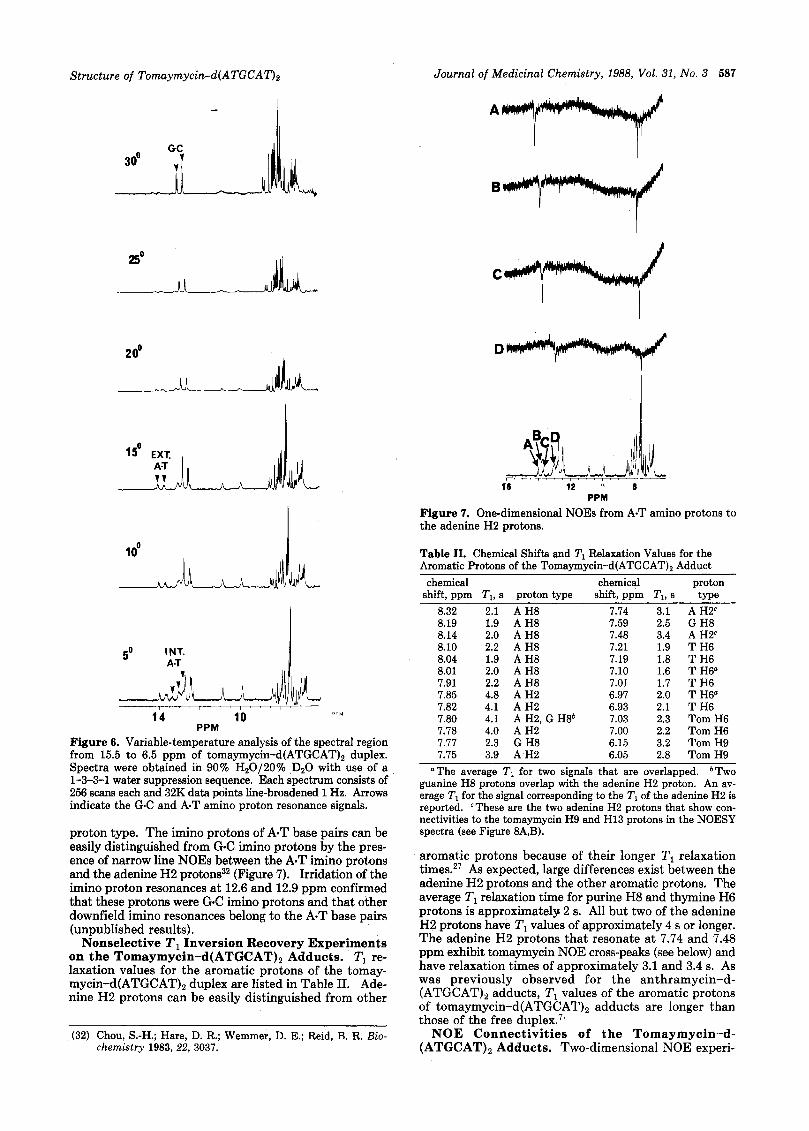
Amino Proton Resonance Assignment and NOE Connectivities to the Adenine H2 Protons. In the previous studies, the imino proton assignments were used to assign the adenine H2 protons in d(ATGCAT)2 and the anthramycin-d(ATGCAT), adduct.'^^ The temperature dependency of the imino proton region is shown in Figure 6. At 24 "C, the two sets of G C imino protons are ob- served at 12.6 and 12.9 ppm. The upfield signals at 10.0 and 10.8 ppm are presumably the phenolic protons at C8 of each diastereomeric tomaymycin molecule. Unfortu- nately, attempts to prove this assignment by irradiation of the exchangeable protons at 10.0 and 10.8 ppm in order to observe NOES into the tomaymycin H9 protons at 5.98 and 6.13 ppm were thwarted by the water suppression sequence, which nulled the region up to approximately 6.9 ppm. The 1-1 water suppression sequence31 was at- tempted but did not supress the water sufficiently for complete digitization of the signals. Lowering of the tem- perature revealed the imino A.T protons between 14.5 and 13.0 ppm. Unfortunately, as is apparent from Figure 6 (15 "C), the adenine H2 protons coalesce at temperatures at which the internal and external imino protons were visible, thus preventing unambiguous assignments. NOE differ- ence spectra were run at 10 "C in order to confirm the
(31) Clore, G. M.; Kimber, B. J.; Gronenborn, A. M. J . Magn. Re- (30) Tozuka, A,; Takaya, T. J . Antibiot. 1983, 36, 142. son. 1983, 54, 170.
Structure of Tomaymycin-d(ATGCAT)2 Journal of Medicinal Chemistry, 1988, Vol. 31, No. 3 587
20°
A n
PPM
Figure 7. One-dimensional NOES from A-T amino protons to the adenine H2 protons.
Table 11. Chemical Shifts and TI Relaxation Values for the Aromatic Protons of the Tomaymycin-d(ATGCAT)2 Adduct
50 INT. A T
PPM 14 10 PPM
Figure 6. Variable-temperature analysis of the spectral region from 15.5 to 6.5 ppm of tomaymycin-d(ATGCAT), duplex. Spectra were obtained in 90% Hz0/20% DzO with use of a 1-3-3-1 water suppression sequence. Each spectrum consists of 256 scans each and 32K data points line-broadened 1 Hz. Arrows indicate the G C and A.T amino proton resonance signals.
proton type. The imino protons of A-T base pairs can be easily distinguished from G-C imino protons by the pres- ence of narrow line NOES between the A-T imino protons and the adenine H2 protons32 (Figure 7). Irridation of the imino proton resonances at 12.6 and 12.9 ppm confirmed that these protons were G C imino protons and that other downfield imino resonances belong to the A-T base pairs (unpublished results).
Nonselective T1 Inversion Recovery Experiments on the Tomaymycin-d(ATGCAT), Adducts. T1 re- laxation values for the aromatic protons of the tomay- my~in-d(ATGcAT)~ duplex are listed in Table 11. Ade- nine H2 protons can be easily distinguished from other
(32) Chou, S.-H.; Hare, D. R.; Wemmer, D. E.; Reid, B. R. Bio- chemistry 1983, 22, 3037.
chemical chemical proton shift, ppm Tl, s proton type shift, ppm Tl, 8 type
8.32 2.1 A H8 8.19 1.9 A H8 8.14 2.0 A H8 8.10 2.2 A H8 8.04 1.9 A H8 8.01 2.0 A H8 7.91 2.2 A H8 7.85 4.8 A H2 7.82 4.1 A H2 7.80 4.1 A H2, G H8b 7.78 4.0 A H2 7.77 2.3 G H8 7.75 3.9 A H2
7.74 3.1 7.59 2.5 7.48 3.4 7.21 1.9 7.19 1.8 7.10 1.6 7.01 1.7 6.97 2.0 6.93 2.1 7.03 2.3 7.00 2.2 6.15 3.2 6.05 2.8
A H2c G H8 A H2c T H6 T H6 T H6" T H6 T H6" T H6 Tom H6 Tom H6 Tom H9 Tom H9
"The average T I for two signals that are overlapped. b T ~ o guanine H8 protons overlap with the adenine H2 proton. An av- erage TI for the signal corresponding to the Tl of the adenine H2 is reported. 'These are the two adenine H2 protons that show con- nectivities to the tomaymycin H9 and H13 protons in the NOESY spectra (see Figure 8A,B).
aromatic protons because of their longer T1 relaxation times.27 As expected, large differences exist between the adenine H2 protons and the other aromatic protons. The average Tl relaxation time for purine H8 and thymine H6 protons is approximately 2 s. All but two of the adenine €32 protons have TI values of approximately 4 s or longer. The adenine H2 protons that resonate a t 7.74 and 7.48 ppm exhibit tomaymycin NOE cross-peaks (see below) and have relaxation times of approximately 3.1 and 3.4 s. As was previously observed for the anthramycin-d- (ATGCAT)2 adducts, T1 values of the aromatic protons of tomaymycin-d(ATGCAT), adducts are longer than those of the free duple^.^‘
NOE Connectivities of the Tomaymycin-d- (ATGCAT)2 Adducts. Two-dimensional NOE experi-
588 Journal of Medicinal Chemistry, 1988, Vol. 31, No. 3 Cheatham et al.
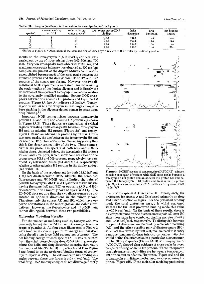
Table 111. Energies (kcal/mol) for Interactions between Species A-D in Figure 3 stereochemistry orientation in total tomaymycin-DNA helix drug net binding
speciesn at C-11 minor grooveb binding distortion distortion energy A S 3’ -37.1 +12.0 +7.2 -17.9 B S 5’ -36.3 +21.0 f 4 . 1 -11.2 C R 3‘ -33.2 +16.8 +8.7 -7.7 D R 5’ -35.5 +13.4 +0.2 -21.9
Refers to Figure 3. Orientation of the aromatic ring of tomaymycin relative to the covalently modified guanine.
ments on the tomaymycin-d(ATGCAT), adducts were carried out by use of three mixing times (300,500, and 700 ms). Very few cross-peaks were observed at 300 ms, and maximum cross-peak intengity was observed at 500 ms, but complete assignment of the duplex adducts could not be accomplished because most of the cross-peaks between the aromatic protons and the deoxyribose H1’ or H2’ and H2” protons of the sugars are absent. However, the two-di- mensional NOE experiments were useful for determining the conformation of the duplex oligomer and indirectly the orientation of two species of tomaymycin molecules relative to the covalently modified guanine. Strong NOE cross- peaks between the adenine H8 protons and thymine H6 protons (Figure 8A, box A) indicate a B helix.29 Tomay- mycin is similar to anthramycin in that large changes in base stacking in the oligomer do not appear to occur upon drug binding.7p8
Important NOE connectivities between tomaymycin protons (H9 and H13) and adenine H2 protons are shown in Figure 8A,B. These figures are expansions of critical regions revealing NOE cross-peaks between tomaymycin H9 and an adenine H2 proton (Figure 8A) and tomay- mycin H13 and an adenine H2 proton (Figure 8B). Of the two cross-peaks, the one between the tomaymycin H9 and its adenine H2 proton is the more intense, suggesting that this is the closer connectivity of the two. Theve connec- tivities are present in spectra a t both 500- and 700-ms mixing times. As noted before, the two adenine H2 protons at 7.48 and 7.74 ppm, which show connectivities to the tomaymycin H13 and H9 protons, respectively, have re- duced TI relaxation times (3.4 and 3.1 s, respectively) relative to other adenine H2 protons in the duplex adduct (see Table 11).
On the basis of the requirement for both 11S,llaS and 11R,llaS diastereomeric DNA adducts, the combined fluorescence and lH NMR results limited the pairs of possible tomaymycin-d(ATGCAT), adducts to two subsets having the same (AC and BD) or opposite (AD and BC) orientations in the minor groove of d(ATGCAT),. The 2D-NOE data require that the two diastereomers be ori- entated in opposite directions in the minor groove. Therefore, only the subset AD and BC, which have op- posite orientations in the minor groove, are viable alter- natives. However, the fluorescence and ‘H NMR data cannot distinguish between these two possibilities.
Molecular Modeling Results For the molecular modeling studies, tomaymycin was
covalently bound via the C-11 atom to the exocyclic amino group of guanine-3. All four ca8es illustrated in Figure 3 were used as the starting point for energy minimization using the all atom force field parameters of AMBER. The net binding energy for each species (A-D) was calculated from the total intermolecular drug-DNA binding energies minus the helix and drug distortion energies that result from induced fits (Table 111). Species A and D in Figure 3 are found to be the preferred binding forms of tomay- mycin-d(ATGCAT),. The difference in net binding en- ergies between these two forms is only 4 kcal/mol. The total drug-DNA binding energies do not differ significantly
0 0
A 0
0
i b 2 1 2 7 2 c 1 8 I 6 I 4 , e r 7 U
Figure 8. NOESY spectra of tomaymycin-d(ATGCAT), adducts showing expansion of regions with NOE cross-peaks between a tomaymycin H9 proton and an adenine H2 proton (A) and be- tween the tomaymycin H13 proton and an adenine H2 proton (B). Spectra were recorded a t 23 “C with a mixing time of 500 ms in DzO.
in any of the species A-D in Table 111. Consequently, the preference for species A and D is based primarily on drug and helix distortion energies. For the preferred binding mode the total distortion energy is +13.6 kcal/mol, whereas for the least preferred binding mode this value is +25.5 kcal/mol. On the basis of these results, there is a clear preference for the diastereomeric pair AD over BC since these pairs have combined binding energies of -49.8 and -18.9 kcal/mol, respectively. To distinguish between the pair of diastereomers favored by molecular modeling (AD) and the other possible pair of diastereomers (BC), which are less favored by 20.9 kcal/mol, we need to identify a unique tomaymycin-base interproton connectivity that would define the orientation in a particular diastereomer.
The NOESY spectra (Figure 8A,B) of tomaymycin-d- (ATGCAT), showed clear evidence of cross-peaks between two pairs of drug-adenine H2 protons. These interproton through-space connectivities are between a tomaymycin H9 proton and an adenine H2 proton (Figure 8A) and the tomaymycin ethylidene methyl and another adenine H2 proton (Figure 8B). If the molecular modeling predictions
Structure of Tomaymycin-d(ATGCA02 Journal of Medicinal Chemistry, 1988, Vol. 31, No. 3 589
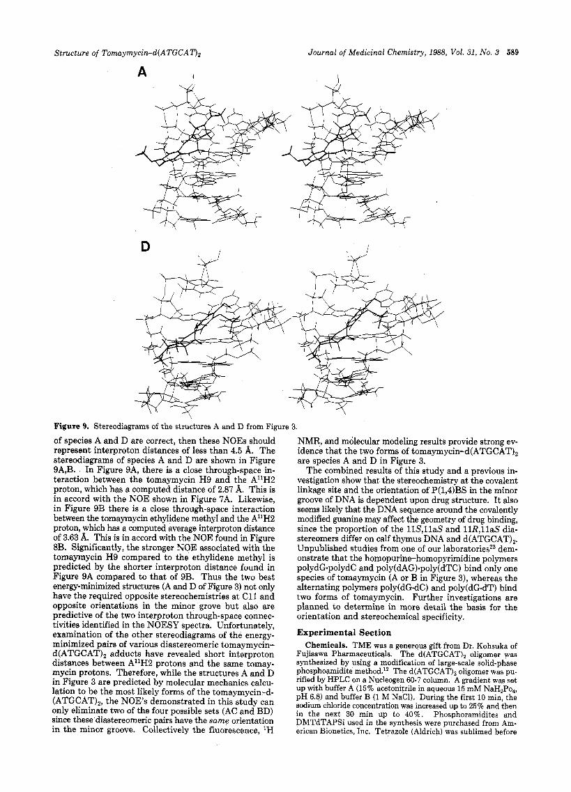
A
J D
Figure 9. Stereodiagrams of the structures A and D from Figure 3.
of species A and D are correct, then these NOES should represent interproton distances of less than 4.5 A. The stereodiagrams of species A and D are shown in Figure 9A,B. In Figure 9A, there is a close through-space in- teraction between the tomaymycin H9 and the A1'H2 proton, which has a computed distance of 2.87 A. This is in accord with the NOE shown in Figure 7A. Likewise, in Figure 9B there is a close through-space interaction between the tomaymycin ethylidene methyl and the A11H2 proton, which has a computed average interproton distance of 3.63 A. This is in accord with the NOE found in Figure 8B. Significantly, the stronger NOE associated with the tomaymycin H9 compared to the ethylidene methyl is predicted by the shorter interproton distance found in Figure 9A compared to that of 9B. Thus the two best energy-minimized structures (A and D of Figure 3) not only have the required opposite stereochemistries at C11 and opposite orientations in the minor grove but also are predictive of the two interproton through-space connec- tivities identified in the NOESY spectra. Unfortunately, examination of the other stereodiagrams of the energy- minimized pairs of various diastereomeric tomaymycin- d(ATGCAT)2 adducts have revealed short interproton distances between AllH2 protons and the Same tomay- mycin protons. Therefore, while the structures A and D in pigure 3 are predicted by molecular mechanics calcu-
(ATGCAT)27 the demonstrated in this study can only eliminate two of the four possible Sets (AC and BD) since these diastereomeric pairs have the same orientation in the minor groove. Collectively the fluorescence, 'H
NMR, and molecular modeling results provide strong ev- idence that the two forms of t~maymycin-d(ATGCAT)~ are species A and D in Figure 3.
The combined results of this study and a previous in- vestigation show that the stereochemistry at the covalent linkage site and the orientation of P(1,4)BS in the minor groove of DNA is dependent upon drug structure. I t also seems likely that the DNA sequence around the covalently modified guanine may affect the geometry of drug binding, since the proportion of the 11S,llaS and 11R,llaS dia- stereomers differ on calf thymus DNA and d(ATGCAT)> Unpublished studies from one of our laboratoriesz3 dem- onstrate that the homopurine-homopyrimidine polymers polydG.polydC and poly(dAG)-poly(dTC) bind only one species of tomaymycin (A or B in Figure 3), whereas the alternating polymers poly(dG.dC) and poly(dG.dT) bind two forms of tomaymycin. Further investigations are planned to determine in more detail the basis for the orientation and stereochemical specificity.
Section Chemicals. TME was a generous gift from Dr. Kohsuka of
Fujisawa Pharmaceuticals. The d(ATGCAT)2 oligomer was synthesized by using a modification of large-scale solid-phase phos~homidi te method.'' The d(ATGCAT12 oligomer was pu- rified by HPLC on a Nucleogen 60-7 column. A gradient was set up with buffer A (15% acetonitrile in aqueous 15 mM NaH2P04,
sodium chloride concentration was increased up to 25% and then in the next 30 min up to 40%. Phosphoramidites and DMTdTAPSi used in the synthesis were purchased from Am- erican Bionetics, Inc. Tetrazole (Aldrich) was sublimed before
lation to be the most likely forms Of the tomaymycin-d- pH 6.8) and buffer B (1 M NaC1). During the first 10 min, the
690 J. Med. Chem.
use at 110 "C (2 mm). Acetic anhydride and trichloroacetic acid (Aldrich) were used without further purification. Acetonitrile was dried by distillation from PzO, followed by distillation from CaHP THF (Fisher Reagent Grade) was used without further purifi- cation. D20 (100 atom %) for Nh4R experiments was purchased from Aldrich.
Preparation of Tomaymycin-d(ATGCAT)z Adduct. To- maymycin-d(ATGCAT)z adducts for NMR experiments were prepared by adding excess solid TME to a 0.5-mL solution of d(ATGCAT)z in buffer (10 mM NaHPO,, pH 7.0,100 mM NaCl, 0.1 mM EDTA) a t 5 "C. After vigorous stirring of the mixture for 4 days, excess tomaymycin was removed by centrifugation followed by extraction with ethyl acetate. Samples were lyo- phylized against DzO three times and dissolved in 0.5 mL of D20. Samples used for NOE difference spectra were lyophilized to remove DzO then dissolved in 0.5 mL of 90% HzO/lO% DzO.
The adduct for fluorescence experiments was prepared by equilibrating a solution of d(ATGCAT), in buffer with Am = 16 and 5 X lo4 M tomaymycin for 12 days at 5 "C. The hexamer concentration was about 3 X M duplex, assuming an ex- tinction coefficient of 4.8 X 10³ M-' cm-l estimated from the base sequencel8 with 20% hyperchromicity as in similar oligomeric d~p1exes.l~ Unbound tomaymycin was removed by extracting four times with ethyl acetate and once with ether. Ether was removed with a stream of N2. The sample was placed in a 4 X 10 mm stoppered cuvette. 'H NMR Studies. A. Two-Dimensional NMR Spectra.
'H NMR spectra were run on GN-500 instrument. All samples were run a t 23 "C unless otherwise indicated and are referenced to HOD a t 23 "C relative to external TSP. COSY spectra of the tomaymycin-d(ATGCAT)z adduct were obtained on a 2 mg/0.5 mL solution of the duplex adduct with a 3-s presaturation pulse before the standard 90°-tl-900 pulse sequence to minimize the residual HOD signal. The COSY spectra contain 512 X 2K data points zero filled to give a 1K X 1K matrix. All other two-di- mensional spectra contain 256 X 1K data points zero filled to give 512 X 512 matrices. Two-dimensional NOE spectra were run on a 10 mg/0.5 mL solution of duplex adduct, with a 90"-tl-900- 0.5tm-90"-l8O0-9O0-0.5t,,,-90~ acquire sequence. The composite 180" pulse in the mixture time removes J coupling contributions from the cross-peaks.
B. NOE Difference Spectra. NOE difference spectra were performed a t 5 "C in 90% H20/10% DzO. A 1-3-3-1 pulse sequence was used to suppress the HzO signal. Signals were irradiated for 1 s with a total recycle delay of 2.4 s. C. Nonselective T1 Inversion Recovery Experiments.
Inversion recovery experiments utilized the standard 180"-t1-90" pulse sequence. Samples were degassed by passing argon through the sample. A total of 32 t values ranging from 0.1 to 10 s were
1988,32, 590-603
used with a total recycle delay of 12.9 s. Time-Resolved Fluorescence Studies. Fluorescence decay
measurements were made by the time-correlated single-photon counting technique as described beforeSg Decay curves were acquired at 5 "C to about 15 X lo3 counts in the peak. The data were fitted by reference deconvolution16 to a s u m of exponential5
with amplitudes ai and lifetimes .r1. The amplitude a, of a com- ponent i depends on a number of factors, including its molar extinction spectrum, fluorescence emission spectrum, radiative lifetime, and concentration. Decay curves measured at various excitation and emission wavelengths were analyzed by a global program,16 assuming that the lifetimes but not the amplitudes are independent of wavelength. Errors in amplitudes and lifetimes were <lo% and <5%, respectively, based on repeated experi- ments.
Molecular Modeling Studies. The crystal structure of to- maymycin methyl ether17J8 was used as the initial structure in this investigation. After removal of the methoxy group, partial atomic charges were obtained from ab initio calculations using GAUSSIAN-80 UCSF and a STO-3G basis set.I9 The resulting 11-demethoxytomaymycin methyl ether structure was minimized by using the program AMBER and all-atom force field parameters presented by Weiner et a1.21 A distance-dependence dielectric constant was used, and the structure was refined until the root mean square gradient was less than 0.1 kcal/mol A. The mini- mized structure was docked in the appropriate location and orientations on the hexanucleotide duplex with the aid of the interactive graphics program MID AS,^^ and then the binding en- ergies were minimized by using AMBER and the parameters de- scribed above. The helix distortion energy was determined by subtracting the energy of the helix in the tomaymycin adduct from that of the separately minlmized isolated helix. Distortion energy induced in the tomaymycin molecule was determined in the same way.
Structural effects of water and counterions on complexing were neglected in the energy calculations. Although these effects influence the absolute values of binding energies, they should be negligible in comparing relative binding energies wherein the same molecule is used a t the same binding site on the duplex.
Acknowledgment. This work was supported by grants from the U.S. Public Health Service (CA35318, GM22873, CA37798) and the Welch Foundation. We thank Dr. Karol Maskos for assistance with fluorescence decay measure- ments and Professor Peter A. Kollman for copies of t he programs AMBER a n d GAUSSIAN-80.
Stereoelectronic Factors Influencing the Biological Activity and DNA Interaction of Synthetic Antitumor Agents Modeled on CC-1065
M. A. Warpehoski,* I. Gebhard, R. C. Kelly, W. C. Krueger, L. H. Li, J. P. McGovren, M. D. Prairie, N. Wicnienski, a n d W. Wierenga
Research Laboratories, The Upjohn Company, Kalamazoo, Michigan 49001. Received August 3, 1987
The synthesis, physicochemical properties, and biological activities of a series of novel spiro cyclopropyl compounds, modeled on the potent antitumor antibiotic CC-1065 (l) , are described. Many of these synthetic analogues are significantly more effective than 1 against murine tumors. In particular, compound 27 exhibits high activity and potency. Structure-activity analysis supports a molecular mechanism for biological action involving hydrophobic interaction of the drug with DNA and acid-catalyzed alkylation of DNA.
T h e structurally novel antibiotic CC-1065 (1) is an ex- tremely potent cytotoxin whose biological action has been
attributed t o i t s sequence selective binding and covalent bonding t o the minor groove of DNA.l-' Because of i ts
0022-2623/88/1831-0590$01.50/0 0 1988 American Chemical Society