Molecular dynamics simulation of the opposite-base ...
19
RESEARCH ARTICLE Open Access Molecular dynamics simulation of the opposite-base preference and interactions in the active site of formamidopyrimidine- DNA glycosylase Alexander V. Popov 1 , Anton V. Endutkin 1,2 , Yuri N. Vorobjev 1,2* and Dmitry O. Zharkov 1,2* Abstract Background: Formamidopyrimidine-DNA glycosylase (Fpg) removes abundant pre-mutagenic 8-oxoguanine (oxoG) bases from DNA through nucleophilic attack of its N-terminal proline at C1′ of the damaged nucleotide. Since oxoG efficiently pairs with both C and A, Fpg must excise oxoG from pairs with C but not with A, otherwise a mutation occurs. The crystal structures of several Fpg–DNA complexes have been solved, yet no structure with A opposite the lesion is available. Results: Here we use molecular dynamic simulation to model interactions in the pre-catalytic complex of Lactococcus lactis Fpg with DNA containing oxoG opposite C or A, the latter in either syn or anti conformation. The catalytic dyad, Pro1–Glu2, was modeled in all four possible protonation states. Only one transition was observed in the experimental reaction rate pH dependence plots, and Glu2 kept the same set of interactions regardless of its protonation state, suggesting that it does not limit the reaction rate. The adenine base opposite oxoG was highly distorting for the adjacent nucleotides: in the more stable syn models it formed non-canonical bonds with out-of- register nucleotides in both the damaged and the complementary strand, whereas in the anti models the adenine either formed non-canonical bonds or was expelled into the major groove. The side chains of Arg109 and Phe111 that Fpg inserts into DNA to maintain its kinked conformation tended to withdraw from their positions if A was opposite to the lesion. The region showing the largest differences in the dynamics between oxoG:C and oxoG:A substrates was unexpectedly remote from the active site, located near the linker joining the two domains of Fpg. This region was also highly conserved among 124 analyzed Fpg sequences. Three sites trapping water molecules through multiple bonds were identified on the protein–DNA interface, apparently helping to maintain enzyme- induced DNA distortion and participating in oxoG recognition. Conclusion: Overall, the discrimination against A opposite to the lesion seems to be due to incorrect DNA distortion around the lesion-containing base pair and, possibly, to gross movement of protein domains connected by the linker. Keywords: DNA glycosylase, 8-oxoguanine, Fpg, Molecular dynamics, Opposite-base specificity, Reaction mechanism * Correspondence: [email protected]; [email protected] 1 SB RAS Institute of Chemical Biology and Fundamental Medicine, 8 Lavrentieva Ave., Novosibirsk 630090, Russia Full list of author information is available at the end of the article © The Author(s). 2017 Open Access This article is distributed under the terms of the Creative Commons Attribution 4.0 International License (http://creativecommons.org/licenses/by/4.0/), which permits unrestricted use, distribution, and reproduction in any medium, provided you give appropriate credit to the original author(s) and the source, provide a link to the Creative Commons license, and indicate if changes were made. The Creative Commons Public Domain Dedication waiver (http://creativecommons.org/publicdomain/zero/1.0/) applies to the data made available in this article, unless otherwise stated. Popov et al. BMC Structural Biology (2017) 17:5 DOI 10.1186/s12900-017-0075-y
-
Upload
khangminh22 -
Category
Documents
-
view
0 -
download
0
Transcript of Molecular dynamics simulation of the opposite-base ...
RESEARCH ARTICLE Open Access
Molecular dynamics simulation of theopposite-base preference and interactionsin the active site of formamidopyrimidine-DNA glycosylaseAlexander V. Popov1, Anton V. Endutkin1,2, Yuri N. Vorobjev1,2* and Dmitry O. Zharkov1,2*
Abstract
Background: Formamidopyrimidine-DNA glycosylase (Fpg) removes abundant pre-mutagenic 8-oxoguanine (oxoG)bases from DNA through nucleophilic attack of its N-terminal proline at C1′ of the damaged nucleotide. Since oxoGefficiently pairs with both C and A, Fpg must excise oxoG from pairs with C but not with A, otherwise a mutationoccurs. The crystal structures of several Fpg–DNA complexes have been solved, yet no structure with A oppositethe lesion is available.
Results: Here we use molecular dynamic simulation to model interactions in the pre-catalytic complex ofLactococcus lactis Fpg with DNA containing oxoG opposite C or A, the latter in either syn or anti conformation. Thecatalytic dyad, Pro1–Glu2, was modeled in all four possible protonation states. Only one transition was observed inthe experimental reaction rate pH dependence plots, and Glu2 kept the same set of interactions regardless of itsprotonation state, suggesting that it does not limit the reaction rate. The adenine base opposite oxoG was highlydistorting for the adjacent nucleotides: in the more stable syn models it formed non-canonical bonds with out-of-register nucleotides in both the damaged and the complementary strand, whereas in the anti models the adenineeither formed non-canonical bonds or was expelled into the major groove. The side chains of Arg109 and Phe111that Fpg inserts into DNA to maintain its kinked conformation tended to withdraw from their positions if A wasopposite to the lesion. The region showing the largest differences in the dynamics between oxoG:C and oxoG:Asubstrates was unexpectedly remote from the active site, located near the linker joining the two domains of Fpg.This region was also highly conserved among 124 analyzed Fpg sequences. Three sites trapping water moleculesthrough multiple bonds were identified on the protein–DNA interface, apparently helping to maintain enzyme-induced DNA distortion and participating in oxoG recognition.
Conclusion: Overall, the discrimination against A opposite to the lesion seems to be due to incorrect DNAdistortion around the lesion-containing base pair and, possibly, to gross movement of protein domains connectedby the linker.
Keywords: DNA glycosylase, 8-oxoguanine, Fpg, Molecular dynamics, Opposite-base specificity, Reactionmechanism
* Correspondence: [email protected]; [email protected] RAS Institute of Chemical Biology and Fundamental Medicine, 8Lavrentieva Ave., Novosibirsk 630090, RussiaFull list of author information is available at the end of the article
© The Author(s). 2017 Open Access This article is distributed under the terms of the Creative Commons Attribution 4.0International License (http://creativecommons.org/licenses/by/4.0/), which permits unrestricted use, distribution, andreproduction in any medium, provided you give appropriate credit to the original author(s) and the source, provide a link tothe Creative Commons license, and indicate if changes were made. The Creative Commons Public Domain Dedication waiver(http://creativecommons.org/publicdomain/zero/1.0/) applies to the data made available in this article, unless otherwise stated.
Popov et al. BMC Structural Biology (2017) 17:5 DOI 10.1186/s12900-017-0075-y
BackgroundFormamidopyrimidine-DNA glycosylase (Fpg or MutM) isa bacterial DNA repair enzyme that removes several abun-dant oxidized bases from DNA. The principal substratebases of Fpg are 8-oxoguanine (oxoG), 2,6-diamino-4-oxo-5-formamidopyrimidine (fapyG) and 2,4-diamino-5-formamidopyrimidine (fapyA) [1, 2] but the enzyme alsocan recognize several dozens of other damaged purinesand pyrimidines [3–10]. By excision of a damaged base,Fpg initiates base excision repair (BER), which engages APendonucleases, a DNA polymerase and a DNA ligase torestore the integrity of DNA [11, 12].The activity of Fpg towards oxoG has attracted much
attention due to abundance and biological importance ofthis lesion, induced in DNA by oxidative metabolismbyproducts, oxidative stress, and ionizing radiation [13].Steric and electrostatic repulsion between the substitu-ent at C8 and the sugar–phosphate atoms effectivelypushes oxoG towards the syn conformation, in whichoxoG forms a Hoogsteen pair with A [14, 15]. Misincor-poration of A by DNA polymerases, in the absence ofrepair, leads to a G→ T transversion after the secondround of replication.Systems for repair of oxoG have been found in all cel-
lular organisms. The tendency of oxoG to form pairswith both C and A presents a challenge to its repair:both oxoG:C and oxoG:A pairs must be converted intoG:C pairs. This requirement is not trivial since a simpleremoval of oxoG from an oxoG:A mispair would gener-ate a G→ T transversion after the repair. This problemis circumvented by repair of oxoG:A pairs in two se-quential rounds of BER [16]. The non-damaged (but in-appropriately incorporated) A is removed first andreplaced with C, and the resulting oxoG:C pair is thenrepaired through the excision of oxoG. In E. coli, themutagenic potential of oxoG is counteracted by threeenzymes, Fpg, MutT, and MutY, collectively known as a“GO system”. Fpg excises oxoG from oxoG:C pairs buthas little activity towards oxoG:A substrates to preventG→ T transversions [1, 17]. Another DNA glycosylase,MutY, specifically excises A from A:oxoG mispairs. If Gin DNA is oxidized to oxoG, it will inevitably be pairedwith C and will be removed by Fpg. If, on the otherhand, A is incorporated during replication opposite anunrepaired oxoG, the resulting oxoG:A mispair will be asubstrate for MutY but not for Fpg. The repair synthesisthen has a chance to incorporate C opposite oxoG [16].The function of the GO system therefore critically de-
pends on the selectivity of Fpg to the base opposite tothe damaged one. X-ray structures are available for freeFpg protein from Thermus thermophilus (Tth-Fpg) andfor various types of complexes of DNA with Fpg fromEscherichia coli (Eco-Fpg), Geobacillus stearothermophi-lus (Bst-Fpg) and Lactococcus lactis (Lla-Fpg) [18–33]
(Fig. 1a). Based on these structures, kinetic data, andcomputational modeling, a reaction mechanism has beensuggested that involves a nucleophilic attack at C1′ ofoxoG by a lone electron pair of the secondary aminogroup of the deprotonated N-terminal Pro1 residue,assisted by protonation of O4′ in the deoxyribose moiety[33–36]. As a result, the N-glycosidic bond is broken,the deoxyribose ring is opened, and a Schiff base cova-lent intermediate between Fpg and DNA is formed(Fig. 1b). This series of events is followed by two sequen-tial steps of elimination of the 3′- and 5′-phosphatesand hydrolysis of the Schiff base. However, many ques-tions about the initial stages of the reaction still remain.For example, the mechanism of oxoG recognition in theactive site of the enzyme is unclear, and the mechanismof proton transfer in the multistep reaction is unknown.Notably, no structural or modeling data is available forFpg in a complex with oxoG:A-containing DNA, limitingour knowledge of the mechanisms of rejection of thisfunctionally relevant mispair. In this work, we use mo-lecular dynamics approach to analyze the structure ofcomplexes of Fpg with oxoG-containing DNA (either Aor C opposite the lesion) to get an insight into the rea-sons behind the opposite-base selectivity of the enzymeand into the dynamic features of the immediate pre-catalytic complex involving oxoG.
MethodsModel preparationThe starting model for the MD analysis of Fpg bound tooxoG-containing DNA was the X-ray structure of Lla-Fpg in a complex with a 14-mer DNA duplex (Fig. 1a,c)containing a non-hydrolysable carbocyclic analog offapyG (PDB ID 1XC8) [23]. The lesion was changed tooxoG using the following protocol. The initial structureof the oxoG base was taken from Bst-Fpg coordinates(PDB ID 1R2Y) [22]. The base was aligned for the bestfit to the fapyG ring and incorporated into the PDB fileinstead of fapyG. The methylene group in the cyclopen-tane ring isosteric to O4′ was manually changed to oxy-gen. Heavy atoms of the side chains lacking in thestructure were built using the Missing Heavy Atom Res-toration module of BioPASED molecular modeling pack-age [37]. Out of 397 water molecules found in thecrystal unit cell, seven that reside in the enzyme’s activesite or in its immediate vicinity were retained as explicitwater, otherwise the modeling was done in implicitwater to broaden the sampled conformational space. Toanalyze the effect of A vs C placed opposite oxoG, threesets of simulations were performed: one with oxoG op-posite C (henceforth termed C models), another withoxoG opposite A(anti) (Aa models), and the third, withoxoG opposite A(syn) (As models). All models contain-ing A opposite to the lesion were constructed by base
Popov et al. BMC Structural Biology (2017) 17:5 Page 2 of 19

replacement. For each group, four simulations weredone, with different protonation states of Pro1 and Glu2:deprotonated Pro1 and Glu2 (PRN-GLU models), pro-tonated Pro1 and deprotonated Glu2 (PRO-GLU),deprotonated Pro1 and protonated Glu2 (PRN-GLH),and protonated Pro1 and Glu2 (PRO-GLH models)(Table 1). The starting structures were checked for er-rors using the PDB Validator tool of BioPASED [37]. Allmodels were energy-minimized in 500 steps of Fletcherenergy optimization algorithm and finally refined bysimulated annealing MD for 500 ps using the BioPASEDpackage [37]. The AMBER force field parameters foroxoG and neutral Pro1 were from [38]. Force field pa-rameters for neutral glutamate were from AMBER ff99[39]. The parameters for the rest of the protein, includ-ing Zn2+, were taken from the classic Amber ff99 forcefield. The protonation states of the other residues were
Fig. 1 a, Structure of Lla-Fpg (1XC8) used as a starting model. The protein is colored according to its secondary structure (cyan, α helices;magenta, β sheets; coral, loops); the DNA is colored by atom type (green, C; blue, N; red, O; orange, P). An orange line is drawn through P atoms inDNA to highlight an axial kink induced by Fpg binding. b, Mechanism of oxoG excision by Fpg proposed from the structural data [35]. The SN2displacement occurs in the C1′→O4′ direction rather than in the C1′→ N9 direction. c, Schematic representation of the modeled DNA duplexand numbering of DNA bases and phosphates (p). N(0) is either C or A. Positions of Arg109 and Phe111 in the complex are indicated. d, Schematicposition of the damaged base relative to the sugar plane in the structures of free oxoG-containing DNA (183D, [56]) or Fpg–DNA complexes containingvarious purine-derived lesions everted into the active site (1XC8, 3C58, 4CIS and 1R2Y; see structure details in the text)
Table 1 Mean r.m.s.d. values of the models and their standarddeviations (Å) over the last 8 ns of the runs
PRN-GLH PRN-GLU PRO-GLH PRO-GLU
Global
oxoG:C 1.50 ± 0.06 1.58 ± 0.07 1.69 ± 0.07 1.51 ± 0.05
oxoG:A(anti) 1.75 ± 0.05 1.43 ± 0.06 1.66 ± 0.06 1.45 ± 0.06
oxoG:A(syn) 1.68 ± 0.06 1.53 ± 0.06 1.41 ± 0.06 1.74 ± 0.06
Active site
oxoG:C 1.37 ± 0.10 1.55 ± 0.08 1.60 ± 0.13 1.58 ± 0.11
oxoG:A(anti) 1.89 ± 0.06 1.57 ± 0.08 2.16 ± 0.06 1.86 ± 0.07
oxoG:A(syn) 1.66 ± 0.12 1.61 ± 0.06 1.44 ± 0.10 1.92 ± 0.13
Popov et al. BMC Structural Biology (2017) 17:5 Page 3 of 19
selected to match physiological pH conditions; e. g., E5was modeled negatively charged. The Zn2+ ion wasdescribed as a single atom with four distance-based har-monic restraints to bind it to the coordinating cysteinsand to maintain the correct geometry. Implicit counter-ion correction was applied by scaling charges of phos-phate groups by a factor of 0.2 [40].
Molecular dynamicsMolecular dynamics simulations (10 ns) were performedusing the BioPASED molecular dynamics modeling soft-ware [37] using the AMBER ff99 force field with Bio-PASED modifications and EEF1 analytical implicitsolvent model [41], with an integration time step of 1 fs.The system was gradually heated from 10 K to 300 Kduring 50 ps and equilibrated at this temperature (theheating time was 150, 200, and 250 ps in the repeat runsof the PRO-GLH models). A classic molecular dynamicstrajectory was generated in the NVT ensemble with har-monic restraints of 0.001 kcal/A2 for the protein andwater atoms, 0.25 kcal/A2 for the atoms of the terminalnucleotides, and 0.0025 kcal/A2 for the rest of the DNAatoms. Coordinates of each atom of the system weresaved each 2 ps, thus producing a trajectory size of 5000snapshots. The trajectories were analyzed using MDTRA[42], a part of the BISON package [43]. Trajectories werecompared using moving MWZ method [44] with bins of50 snapshots. Statistically significant differences in pa-rameters between different models were estimated usingF-test, with false discovery rate method (Benjamini–Hochberg procedure) employed to correct for multiplecomparisons [45]. Hydrogen bonds were searched usingMDTRA [42]. Structures were visualized and renderedusing VMD [46], RasMol [47] and PyMOL (Schrödinger,Portland, OR).
pH dependence of Fpg activityEco-Fpg was purified as described [19]. Oligonucleotideswere synthesized in-house from commercially availablephosphoramidites (Glen Research, Sterling, VA). Anoligonucleotide 5′-CTCTCCCTTCXCTCCTTTCCTCT-3′ (X = oxoG) was 32P-labeled using polynucleotide kin-ase (SibEnzyme, Novosibirsk, Russia) and γ[32P]ATP(PerkinElmer, Waltham, MA) following the manufac-turer’s protocol and annealed to a complementary strandplacing C or A opposite oxoG. The reactions (20 μl) in-cluded 25 mM sodium phosphate buffer (H3PO4/NaH2PO4, NaH2PO4/Na2HPO4, and Na2HPO4/Na3PO4
conjugate pairs spanning the range of pH 4.0–9.0), 100nM duplex oligonucleotide substrate, and either 2 nM(steady-state experiments) or 500 nM Fpg (single-turn-over experiments). The reaction was allowed to proceedfor 1 min either at 37 °C with 2 nM Fpg or at 0 °C with500 nM Fpg, and was stopped by adding 20 μl of
formamide/EDTA gel loading dye and heating at 95° for3 min. The products were separated by electrophoresisin 20% polyacrylamide gel/8 M urea and quantified byphosphorimaging using a Molecular Imager FX system(Bio-Rad Laboratories, Hercules, CA). Three independ-ent experiments have been performed. Calculation ofpKa for Pro1 and Glu2 were done using PROPKA v3.1[48]. Circular dichroism spectra were recorded on aJASCO J-600 CD spectrometer (JASCO Analytical In-struments, Tokyo, Japan) at 25 °C in 30 mM Na phos-phate with a 1-nm step.
Evolutionary analysisA taxonomically balanced sample of 124 bacterial Fpgsequences (limited to two sequences per taxonomic fam-ily) was constructed by protein BLAST search [49] in theNCBI non-redundant protein sequences database usingEco-Fpg as a query, filtered for conservation of the N-terminal Pro–Glu dipeptide and C-terminal zinc finger,and clipped from the absolutely conserved N-terminalPro to the absolutely conserved Gln after the fourth Cysof the zinc finger as described [50, 51]. Alignment ofmultiple sequences and neighbor-joining tree construc-tion was performed using Clustal Omega [52]. Hierarch-ical analysis of conservation of physicochemicalproperties in the alignments was done using AMAS [53]with 5% atypical residues allowed; the results are re-ported as conservation numbers (Cn).
Results and discussionGeneral model considerationsSelection of the starting structureCurrently, the Protein Data Bank holds 56 released X-ray structures of Fpg, belonging to four bacterial speciesand sampling several points along the reaction coordin-ate [18–33]. Our selection of the starting structure forMD was guided by the following considerations. First, itshould contain DNA with the damaged base still inplace, residing in the enzyme’s active site. Second, min-imal deviation from the wild-type enzyme recognizingoxoG should be present. Third, the structure shouldhave good resolution (<2.0 Å), with as few as possibleresidues missing.Based on these considerations, we have chosen
1XC8, the 1.95-Å structure of wild-type Lla-Fpgbound to DNA containing a non-cleavable carbacyclicfapyG analog (carba-fapyG [23]) as a starting model(Fig. 1a). In carba-fapyG, a methylene group substi-tutes for O4′, and the damaged base, fapyG, is differ-ent from oxoG only by the absence of a bondbetween N9 and C8 of the purine heterocycle O4′[54]. The Lla-Fpg is nearly identical to Eco-Fpg in itsselectivity for C vs A as the opposite base [17].
Popov et al. BMC Structural Biology (2017) 17:5 Page 4 of 19
OxoG glycosidic angleBesides 1XC8, the structures of Fpg bound to DNA withan extrahelical damaged base include Lla-Fpg bound toDNA containing carbacyclic N5-benzyl-fapyG (3C58[26]), carbacyclic oxoG (4CIS [33]) or 5-hydroxy-5-methylhydantoin (2XZF, 2XZU [29]) and Bst-Fpg boundto DNA containing oxoG (1R2Y) or 5,6-dihydrouracil(1R2Z) [22]. With the exception of 1R2Y and 4CIS, thedamaged bases in the structures are quite different fromoxoG. In the 1R2Y structure, oxoG is present in DNA,and the cleavage is prevented by changing the absolutelyconserved catalytic Glu2 residue into Gln [22]. In thisstructure, oxoG is often stated to be in the syn conform-ation in the active site, yet its χ angle (101°) is actually inthe anti domain (namely, in its border range, so-called“high syn”) (Fig. 1d). On the contrary, oxoG opposite Cin B-DNA is usually stated to be anti as it forms regularWatson–Crick bonds [55, 56], yet its χ angle in the crys-tal structure (–55°, [56]) is actually in the syn domain.Carba-fapyG in 1XC8 is in the high anti range (χ = –64°), and only in 4CIS, carba-oxoG is unambiguously syn(χ = 27°, Fig. 1d). Moreover, oxoG in 4CIS does not formthe same set of hydrogen bonds with the active site as in1R2Y. The possibility of conformation artifacts inducedby E2Q mutation has been amply discussed in the litera-ture [23, 38, 57–61]. Therefore, we have chosen 1XC8,which straddles the syn/anti border (Fig. 1d) as ourstarting model and allowed the conformation to driftinto the most preferable χ range during MD.
Opposite base glycosidic angleWhile the oxoG:A pair in B-DNA exists as oxoG(sy-n):A(anti), this does not mean that the same conforma-tions will be observed in the complex with Fpg, sincethe hydrogen bonds within the mispair are lost uponoxoG eversion, and the conformation of the nucleotidesis governed largely by their interaction with the proteinresidues. The most relevant example is given by anotheroxoG mispair, oxoG:G, which adopts the conformationoxoG(syn):G(anti) in the B-DNA duplex [62] but the Gopposite to the lesion flips into syn and forms twostrong hydrogen bond with an Arg residue when thisduplex is bound to Bst-Fpg [20]. Therefore, in additionto the oxoG:C pair, we have constructed two series ofmodels with oxoG:A, with A in either anti or syn con-formation to fully explore the range of possible dynamicsof Fpg–oxoG:A complex.
SolventAlthough MD in explicit solvent is common nowadays,recent advances in implicit solvent models revived thepopularity of this alternative [63–65]. The major advan-tages of implicit solvent over the explicit one are speed,better estimates of solvation and folding energy, wider
coverage of conformational space and more accurate ac-count for pH and residue ionization. The latest versionsof Poisson–Boltzmann, generalized Born and hybrid im-plicit/explicit models are comparable with explicitsolvent-based calculations with respect to agreementwith experimental free energy data [63–65]. Althoughthe acceleration of conformation sampling afforded byimplicit solvent strongly depends on the modeled sys-tem, direct comparisons show a 7–10-fold increase for asystem with several conformational transitions [66].Since our primary interest was to sample a wide rangeof conformations available for the Fpg–substrate com-plexes, we have chosen a hybrid model combining animplicit solvent with explicit strongly bound water mole-cules; such approaches retain the advantages of implicitmethods but significantly improve quality of protein–DNA interface models [67].
Protonation state of the catalytic dyadThe ionizable groups of Pro1 and Glu2 directly partici-pate in the enzymatic reaction. Mechanistically, the nu-cleophilic attack by Pro1 at C1′[oxoG] requires N[Pro1]to carry a lone electron pair (Fig. 1b). On the otherhand, opening of the deoxyribose ring involves proton-ation of its O4′, which is near Oε2 of Glu2 (Fig. 1b);quantum mechanics/molecular mechanics (QM/MM)simulations show that O4′ protonation provides a low-barrier path to glycosidic bond cleavage by Fpg and itseukaryotic functional analog, OGG1 [33, 36, 68]. Fromseveral structures Fpg–DNA complexes, it has been sug-gested that the proton is shuttled from N[Pro1] toOε2[Glu2], perhaps through a network of crystallo-graphic water molecules present in the active site [19,26]; this possibility was also favored by QM/MM ana-lysis [69]. However, no attempt to estimate pKa of Pro1and Glu2 has been reported in the literature. It is pos-sible that a mixture of Pro1/Glu2 ionization states existsin the active centers of different Fpg molecules atphysiological pH; although only one of them (PRN-GLH) is permissive for the reaction chemistry, the pathto this state may go through other states. Therefore, wehave performed MD of the full system for fourionization states of the Pro1–Glu2 catalytic dyad: PRN-GLH, PRN-GLU, PRO-GLH, and PRO-GLU (seeMethods).
Overall model characterizationThe root mean square deviation (r.m.s.d.) with respectto the backbone of the starting structure of the complexwas calculated every 2 ps. R.m.s.d. values of all themodels increased rapidly during the first 500 ps of thedynamics and stabilized at approximately 1.6 Å (seeTable 1 and Additional file 1: Fig. S1). Overall r.m.s.d. ofall models was similar; however, in the active center
Popov et al. BMC Structural Biology (2017) 17:5 Page 5 of 19
(defined as all protein residues with at least one atomwithin 4 Å of oxoG nucleotide or the opposite C/A nu-cleotide, plus three nucleotide pairs centered on theoxoG), the r.m.s.d. of the C models was significantlylower than in the A models (p < 10–4). The DNA back-bone displayed higher mobility than the protein back-bone: average r.m.s.d. of the DNA residues was greaterby 0.54–0.98 Å depending on the model. The overallcomplex conformation was stable along the whole trajec-tory with a radius of gyration ~20 Å for each model(20.17 ± 0.04 to 20.30 ± 0.05 Å). The angle of DNA kinkwas also stable (55° ± 2° to 63° ± 2°, depending on themodel).
Simulation reproducibilityTo test the consistence of results between independentruns, we have selected three models (PRO-GLH-C,PRO-GLH-Aa, and PRO-GLH-As) and performed threeadditional simulations with each one, to the total of nineadditional simulations, using different heating times(150, 200, and 250 ps) to provide different conditions forthe start of the production run. Then the resulting fourtrajectories for each model (one original and three new)were compared. The r.m.s.d. values of individual runswere similar (1.0–1.5 Å over the last 8 ns, Additionalfile 1: Fig. S1). The inter-run r.m.s.d. were expectedlyhigher (1.9–2.2 Å, Additional file 1: Fig. S1) but stilldid not show significant divergence of the models.Stable hydrogen bonds, including model-specific ones,were well consistent across the four runs (Additionalfile 1: Fig. S5B); the 90% cut-off of the mean occur-rence identified as stable all Watson–Crick bonds and79% of the main-chain bonds observed in the 1XC8crystal structure.
Pro–Glu catalytic dyadArrangement of the reacting groups in the modelsWe have sampled the population of two key distances ofthe Fpg–DNA complex, N[Pro1]…C1′[oxoG] andOε2[Glu2]…O4′[oxoG] in all our models (Fig. 2, Table 2).In all C models (Fig. 2a, D, G, J), the distribution ofOε2[Glu2]…O4′[oxoG] distances was unimodal andproduced similar central values (Table 2). On the con-trary, the N[Pro1]…C1′[oxoG] distance was less stable:in some models, two peaks in the distribution histogramwere clearly observed, indicative of stable conform-ational basins (Table 2).Another important parameter in the reaction of base
excision is the angle of attack by Pro1 at C1′. Twomechanisms for SN2 displacement initiating Schiff baseformation have been considered for bifunctional DNAglycosylases: with the C1′–O4′ bond or C1′–N9 bondbreaking first [35, 70]. Enzyme-catalyzed SN2 reactionsrequire a 10°–20° alignment of the nucleophile lone pair
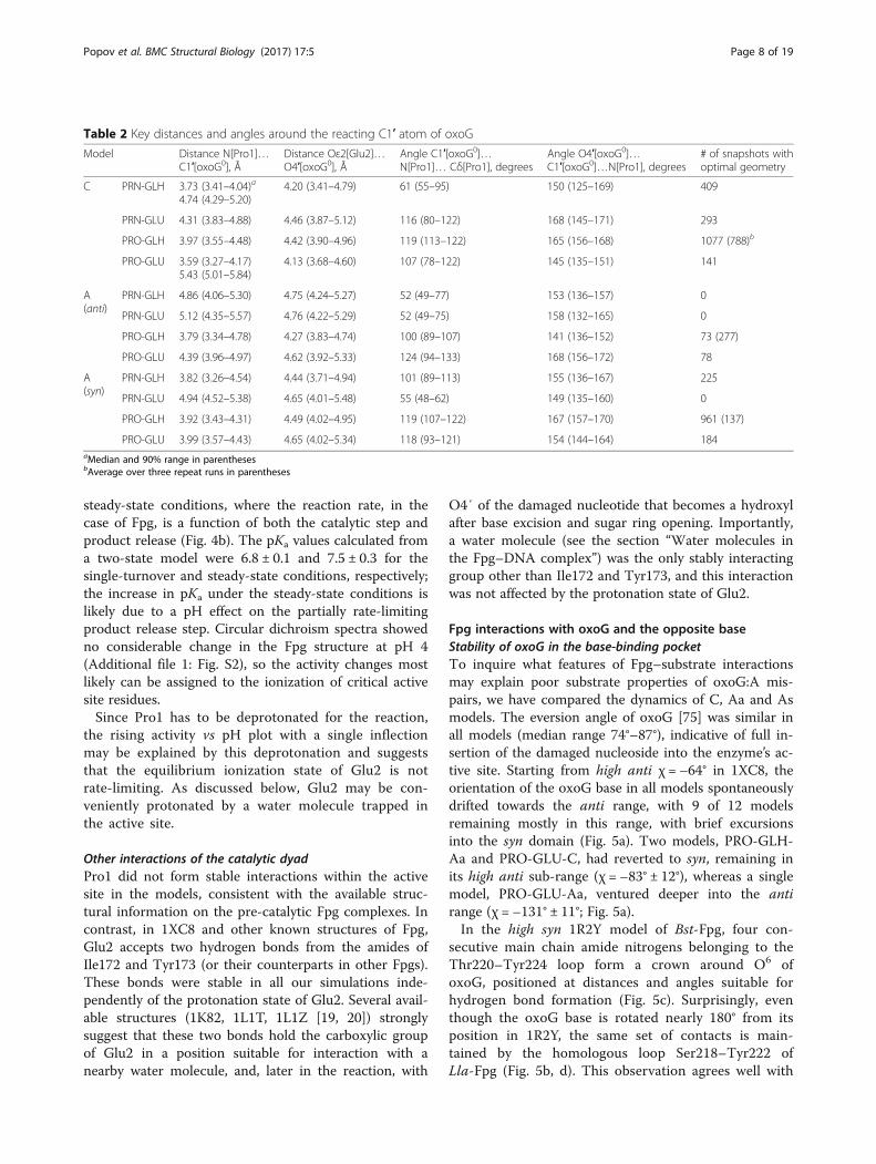
and carbon antibonding orbital [71, 72]. The ideal attackgeometry for Pro1 is thus 107° for the C1′[oxoG]…N[Pro1]…Cδ[Pro1] angle and 180° for the X…C1′[oxoG]…N[Pro1] angle where X is either O4′[oxoG]or N9[oxoG]. As can be seen from Fig. 3 and Table 2,the C1′…N…Cδ angle of all PRO models, as well asPRN-GLH-C, PRN-GLH-As, and PRN-GLU-C, eitherlied in the acceptable domain or made appreciable ex-cursions to it. All models were incompatible with theC1′–N9 direction of nucleophilic attack (Table 2). TheO4′…C1′…N angle for 7 of 12 models lied in the ac-ceptable domain, and was close to this range in othermodels, consistent with the C1′–O4′ attack. The oppos-ite base had no consistent effect on the Pro1 approachangle.Defining the “optimal geometry” as N[Pro1]…
C1′[oxoG] distance < 4 Å, Oε2[Glu2]…O4′[oxoG0] dis-tance < 4.5 Å, and C1′[oxoG0]…N[Pro1]…Cδ[Pro1] andO4′[oxoG]…C1′[oxoG]…N[Pro1] within 20° of the idealvalues, we have sampled the population of the zone withall four parameters optimal (Table 2). All A(anti) modelsshowed the optimal geometry very rarely. For C andA(syn) models, PRO-GLH was most populated, followedby PRN-GLH. Interestingly, PRN models were more se-lective towards C vs A(syn). Other sensible definitions of“optimal” Pro1 and Glu2 distances (e. g., the lowestquartile of the respective distance population), alsoshowed C models spending more time in the optimalconformation than A models. The preference of Cmodels for the optimal geometry was also evident in therepeat runs of the PRO-GLH models (Table 2).
pKa estimate of the catalytic dyad residuesTo get an independent estimate of the protonation stateof Pro1 and Glu2, we have used PROPKA, an empiricalalgorithm based on the spatial proximity of charged resi-dues [48]. In addition to our starting structure, we haveconsidered several other PDB structures of Fpg from dif-ferent species (Additional file 2: Table S1). In all cases,pKa of Pro1 was notably lowered (by 0.35–2.99 units)compared with the reference pKa of N-terminal Pro,while pKa of Glu2 was considerably higher (by 1.54–3.64units) than the reference pKa of the internal Glu sidechain. Similar pKa changes were reported for phage T4endonuclease V, another DNA glycosylase employing theN-terminal amino group and a Glu carboxyl as a cata-lytic dyad [73]. Interestingly, structures of free Fpg andFpg bound to undamaged DNA with the sampled basestill intrahelical displayed more acidic pKa for Glu2, sug-gesting that this group may be specifically activatedupon eversion of the damaged nucleotide. AlthoughPROPKA considers the influence of nucleic acid ligandson amino acid ionization potential only approximately, it
Popov et al. BMC Structural Biology (2017) 17:5 Page 6 of 19

is nevertheless clear that in Fpg, Pro1 is considerablymore acidic, and Glu2, more basic than expected.
pH profile of Fpg activityTo assess the functional importance of the catalytic dyadprotonation states experimentally, we have analyzed thepH profile of activity for Eco-Fpg, assuming that themechanistic features of base excision will be conservedin Eco-Fpg and Lla-Fpg. Usually, when an enzyme’s ac-tive site possesses two functionally important ionizablegroups, one of which has to be protonated while the
other has to be deprotonated for activity, the pH de-pendence is characteristically bell-shaped. For DNA gly-cosylases, such bell-shaped dependence was shown forhuman alkyladenine glycosylase, which is monofunc-tional, structurally different from Fpg, and uses a histi-dine and a glutamate residue as a general acid and ageneral base, respectively [74]. On the contrary, Fpgshowed a single transition in the activity over a pH rangeof 5 units (pH 4 to pH 9) (Fig. 4). This was observedboth under single-turnover conditions, where the rate islimited by the catalytic step of the reaction (Fig. 4a) and
Fig. 2 Distances N[Pro1]…C1′[oxoG0] and Oε2[Glu2]…O4′[oxoG0] during the simulation with different protonation states of N[Pro1] andOε2[Glu2] (a–l, the model nature is indicated in the respective panels)
Popov et al. BMC Structural Biology (2017) 17:5 Page 7 of 19
steady-state conditions, where the reaction rate, in thecase of Fpg, is a function of both the catalytic step andproduct release (Fig. 4b). The pKa values calculated froma two-state model were 6.8 ± 0.1 and 7.5 ± 0.3 for thesingle-turnover and steady-state conditions, respectively;the increase in pKa under the steady-state conditions islikely due to a pH effect on the partially rate-limitingproduct release step. Circular dichroism spectra showedno considerable change in the Fpg structure at pH 4(Additional file 1: Fig. S2), so the activity changes mostlikely can be assigned to the ionization of critical activesite residues.Since Pro1 has to be deprotonated for the reaction,
the rising activity vs pH plot with a single inflectionmay be explained by this deprotonation and suggeststhat the equilibrium ionization state of Glu2 is notrate-limiting. As discussed below, Glu2 may be con-veniently protonated by a water molecule trapped inthe active site.
Other interactions of the catalytic dyadPro1 did not form stable interactions within the activesite in the models, consistent with the available struc-tural information on the pre-catalytic Fpg complexes. Incontrast, in 1XC8 and other known structures of Fpg,Glu2 accepts two hydrogen bonds from the amides ofIle172 and Tyr173 (or their counterparts in other Fpgs).These bonds were stable in all our simulations inde-pendently of the protonation state of Glu2. Several avail-able structures (1K82, 1L1T, 1L1Z [19, 20]) stronglysuggest that these two bonds hold the carboxylic groupof Glu2 in a position suitable for interaction with anearby water molecule, and, later in the reaction, with
O4′ of the damaged nucleotide that becomes a hydroxylafter base excision and sugar ring opening. Importantly,a water molecule (see the section “Water molecules inthe Fpg–DNA complex”) was the only stably interactinggroup other than Ile172 and Tyr173, and this interactionwas not affected by the protonation state of Glu2.
Fpg interactions with oxoG and the opposite baseStability of oxoG in the base-binding pocketTo inquire what features of Fpg–substrate interactionsmay explain poor substrate properties of oxoG:A mis-pairs, we have compared the dynamics of C, Aa and Asmodels. The eversion angle of oxoG [75] was similar inall models (median range 74°–87°), indicative of full in-sertion of the damaged nucleoside into the enzyme’s ac-tive site. Starting from high anti χ = –64° in 1XC8, theorientation of the oxoG base in all models spontaneouslydrifted towards the anti range, with 9 of 12 modelsremaining mostly in this range, with brief excursionsinto the syn domain (Fig. 5a). Two models, PRO-GLH-Aa and PRO-GLU-C, had reverted to syn, remaining inits high anti sub-range (χ = –83° ± 12°), whereas a singlemodel, PRO-GLU-Aa, ventured deeper into the antirange (χ = –131° ± 11°; Fig. 5a).In the high syn 1R2Y model of Bst-Fpg, four con-
secutive main chain amide nitrogens belonging to theThr220–Tyr224 loop form a crown around O6 ofoxoG, positioned at distances and angles suitable forhydrogen bond formation (Fig. 5c). Surprisingly, eventhough the oxoG base is rotated nearly 180° from itsposition in 1R2Y, the same set of contacts is main-tained by the homologous loop Ser218–Tyr222 ofLla-Fpg (Fig. 5b, d). This observation agrees well with
Table 2 Key distances and angles around the reacting C1′ atom of oxoG
Model Distance N[Pro1]…C1′[oxoG0], Å
Distance Oε2[Glu2]…O4′[oxoG0], Å
Angle C1′[oxoG0]…N[Pro1]… Cδ[Pro1], degrees
Angle O4′[oxoG0]…C1′[oxoG0]…N[Pro1], degrees
# of snapshots withoptimal geometry
C PRN-GLH 3.73 (3.41–4.04)a
4.74 (4.29–5.20)4.20 (3.41–4.79) 61 (55–95) 150 (125–169) 409
PRN-GLU 4.31 (3.83–4.88) 4.46 (3.87–5.12) 116 (80–122) 168 (145–171) 293
PRO-GLH 3.97 (3.55–4.48) 4.42 (3.90–4.96) 119 (113–122) 165 (156–168) 1077 (788)b
PRO-GLU 3.59 (3.27–4.17)5.43 (5.01–5.84)
4.13 (3.68–4.60) 107 (78–122) 145 (135–151) 141
A(anti)
PRN-GLH 4.86 (4.06–5.30) 4.75 (4.24–5.27) 52 (49–77) 153 (136–157) 0
PRN-GLU 5.12 (4.35–5.57) 4.76 (4.22–5.29) 52 (49–75) 158 (132–165) 0
PRO-GLH 3.79 (3.34–4.78) 4.27 (3.83–4.74) 100 (89–107) 141 (136–152) 73 (277)
PRO-GLU 4.39 (3.96–4.97) 4.62 (3.92–5.33) 124 (94–133) 168 (156–172) 78
A(syn)
PRN-GLH 3.82 (3.26–4.54) 4.44 (3.71–4.94) 101 (89–113) 155 (136–167) 225
PRN-GLU 4.94 (4.52–5.38) 4.65 (4.01–5.48) 55 (48–62) 149 (135–160) 0
PRO-GLH 3.92 (3.43–4.31) 4.49 (4.02–4.95) 119 (107–122) 167 (157–170) 961 (137)
PRO-GLU 3.99 (3.57–4.43) 4.65 (4.02–5.34) 118 (93–121) 154 (144–164) 184aMedian and 90% range in parenthesesbAverage over three repeat runs in parentheses
Popov et al. BMC Structural Biology (2017) 17:5 Page 8 of 19

Fig. 3 Angles C1′[oxoG0]…N[Pro1]…Cδ[Pro1] (a), O4′[oxoG0]…C1′[oxoG0]…N[Pro1] (b) and N9[oxoG0]…C1′[oxoG0]…N[Pro1] (c) in the models.Moving average of 50 consecutive snapshots is plotted vs time. The traces are color-coded: dark cyan, PRN-GLH-C; light lime, PRN-GLH-Aa; coral,PRN-GLH-As; olive, PRN-GLU-C; dark magenta, PRN-GLU-Aa; light blue, PRN-GLU-As; magenta, PRO-GLH-C; blue, PRO-GLH-Aa; red, PRO-GLH-As; cyan,PRO-GLU-C; yellow, PRO-GLU-Aa; green, PRO-GLU-As
Fig. 4 pH dependence of Fpg activity. a, single-turnover conditions (500 nM Fpg, 100 nM substrate, 0 °C). b, steady-state conditions (2 nM Fpg,100 nM substrate, 37 °C). See Methods for details
Popov et al. BMC Structural Biology (2017) 17:5 Page 9 of 19

the literature data on simulation of Bst-Fpg withoxoG forced into the anti conformation [60] and withthe same pattern of contacts to O6 in the 1XC8structure of Lla-Fpg [23]. Notably, the “distinguish-ing” bond between the main chain carbonyl of Ser220and pyrrolic N7 of oxoG, seen in Bst-Fpg 1R2Y struc-ture but absent from Lla-Fpg 1XC8, was not observedin our simulations.
Interactions and dynamics of the opposite baseIn all reported structures of Fpg bound to DNA with thefully everted damaged nucleotide, specific recognition ofC opposite to the lesion is governed by two hydrogenbonds from Arg109 after its insertion into the DNAvoid: Nε[Arg109]–O2[C(0)] and Nη2[Arg109]–N3[C(0)].If G substitutes for C, Nε and Nη2 of the inserted Argform slightly suboptimal bonds with N7 and O6,
Fig. 5 a, χ angle evolution during the simulation. b, distances between O6[oxoG0] and main chain amide nitrogen atoms of Ile119, Arg220,Thr221, and Tyr222. Moving average of 50 consecutive snapshots is plotted vs time. The colors of the traces are the same as in Fig. 3. c, loopThr220–Tyr224 of Bst-Fpg forms an extensive set of contacts with O6 of oxoG in high syn orientation (χ = 101°, 1R2Y). d, a homologous loopSer218–Tyr222 of Lla-Fpg forms the same set of contacts with O6 when oxoG is flipped around the glycosidic bond (χ = –103°, PRO-GLH-Cmodel, 9 ns)
Popov et al. BMC Structural Biology (2017) 17:5 Page 10 of 19
respectively, of the G base in the syn orientation,whereas T in place of C retains a bond with Nε[Arg109]but experiences a clash between two hydrogen bond do-nors, N3[T] and Nη2[Arg109] [20].In all our C models, O2[C(0)] and N3[C(0)] maintained
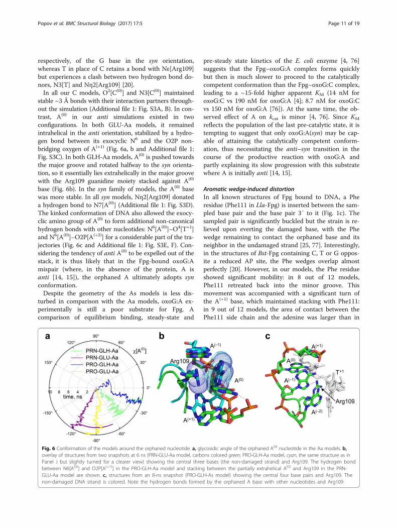
stable ~3 Å bonds with their interaction partners through-out the simulation (Additional file 1: Fig. S3A, B). In con-trast, A(0) in our anti simulations existed in twoconfigurations. In both GLU-Aa models, it remainedintrahelical in the anti orientation, stabilized by a hydro-gen bond between its exocyclic N6 and the O2P non-bridging oxygen of A(+1) (Fig. 6a, b and Additional file 1:Fig. S3C). In both GLH-Aa models, A(0) is pushed towardsthe major groove and rotated halfway to the syn orienta-tion, so it essentially lies extrahelically in the major groovewith the Arg109 guanidine moiety stacked against A(0)
base (Fig. 6b). In the syn family of models, the A(0) basewas more stable. In all syn models, Nη2[Arg109] donateda hydrogen bond to N7[A(0)] (Additional file 1: Fig. S3D).The kinked conformation of DNA also allowed the exocy-clic amino group of A(0) to form additional non-canonicalhydrogen bonds with other nucleotides: N6[A(0)]–O4[T+1]and N6[A(0)]–O2P[A(+2)] for a considerable part of the tra-jectories (Fig. 6c and Additional file 1: Fig. S3E, F). Con-sidering the tendency of anti A(0) to be expelled out of thestack, it is thus likely that in the Fpg-bound oxoG:Amispair (where, in the absence of the protein, A isanti [14, 15]), the orphaned A ultimately adopts synconformation.Despite the geometry of the As models is less dis-
turbed in comparison with the Aa models, oxoG:A ex-perimentally is still a poor substrate for Fpg. Acomparison of equilibrium binding, steady-state and
pre-steady state kinetics of the E. coli enzyme [4, 76]suggests that the Fpg–oxoG:A complex forms quicklybut then is much slower to proceed to the catalyticallycompetent conformation than the Fpg–oxoG:C complex,leading to a ~15-fold higher apparent KM (14 nM foroxoG:C vs 190 nM for oxoG:A [4]; 8.7 nM for oxoG:Cvs 150 nM for oxoG:A [76]). At the same time, the ob-served effect of A on kcat is minor [4, 76]. Since KM
reflects the population of the last pre-catalytic state, it istempting to suggest that only oxoG:A(syn) may be cap-able of attaining the catalytically competent conform-ation, thus necessitating the anti–syn transition in thecourse of the productive reaction with oxoG:A andpartly explaining its slow progression with this substratewhere A is initially anti [14, 15].
Aromatic wedge-induced distortionIn all known structures of Fpg bound to DNA, a Pheresidue (Phe111 in Lla-Fpg) is inserted between the sam-pled base pair and the base pair 3′ to it (Fig. 1c). Thesampled pair is significantly buckled but the strain is re-lieved upon everting the damaged base, with the Phewedge remaining to contact the orphaned base and itsneighbor in the undamaged strand [25, 77]. Interestingly,in the structures of Bst-Fpg containing C, T or G oppos-ite a reduced AP site, the Phe wedges overlap almostperfectly [20]. However, in our models, the Phe residueshowed significant mobility: in 8 out of 12 models,Phe111 retreated back into the minor groove. Thismovement was accompanied with a significant turn ofthe A(+1) base, which maintained stacking with Phe111:in 9 out of 12 models, the area of contact between thePhe111 side chain and the adenine was larger than in
Fig. 6 Conformation of the models around the orphaned nucleotide. a, glycosidic angle of the orphaned A(0) nucleotide in the Aa models. b,overlay of structures from two snapshots at 6 ns (PRN-GLU-Aa model, carbons colored green; PRO-GLH-Aa model, cyan, the same structure as inPanel J but slightly turned for a clearer view) showing the central three bases (the non-damaged strand) and Arg109. The hydrogen bondbetween N6[A(0)] and O2P[A(+1)] in the PRO-GLH-Aa model and stacking between the partially extrahelical A(0) and Arg109 in the PRN-GLU-Aa model are shown. c, structures from an 8-ns snapshot (PRO-GLH-As model) showing the central four base pairs and Arg109. Thenon-damaged DNA strand is colored. Note the hydrogen bonds formed by the orphaned A base with other nucleotides and Arg109
Popov et al. BMC Structural Biology (2017) 17:5 Page 11 of 19

1XC8 for more than half of the simulation (Fig. 7a andAdditional file 1: Fig. S4). As a result, the T–1:A(+1) pairwas grossly distorted, mostly by the propeller twistmovement (Fig. 7a, b). In the remaining four models,one (PRO-GLH-C) displayed brief aborted attempts toretract Phe in the same manner (with full retraction inone of the repeats), in one (PRO-GLH-Aa), the A(+1)
moved by a buckling motion allowing Phe to unstackand adopt an alternative conformation without leavingthe double helix, and only in two models (PRN-GLH-Cand PRN-GLU-Aa) the initial conformation of the wedgeand the adjacent nucleotides was stable.
Specific distant interactions in Fpg–DNA complexesModel-specific hydrogen bondsIn order to select out inter- and intramolecular interac-tions specific for oxoG:C, we have searched for hydrogenbonds that existed (i. e., had an energy > 1.2 kcal/mol) in >1% of the snapshots. Around 600 such hydrogen bondswere found in each model. In all models, less than 50% ofthe found bonds existed for more than 90% of the snap-shots, and less than 25% of the found bonds existed in lessthan 25% of the snapshots (Additional file 1: Fig. S5A, B).The former class may be considered to representstable, functionally important hydrogen bonds,whereas the latter one is most likely due to conform-ational fluctuations. Therefore, all detected hydrogenbonds were first analyzed with respect to their occur-rence in these categories (≤90% vs > 90% and ≤ 25%vs > 25%). Pearson’s mean square contingency coeffi-cients (φ) for pairwise comparison between differentmodels showed no significant contribution of proton-ation state or substrate into the overall distribution ofbonds in the high- and low-stability categories.
We then searched for the bonds that were consist-ently different between C, Aa, and As models, select-ing those deviating > 3σ from the mean distancebetween the models (Fig. 8 and Additional file 1: Fig.S6A–C). Only a few bonds consistently showed differ-ent stability in all C vs A comparisons irrespective ofthe syn or anti conformation of A(0). Unsurprisingly,some of these were formed by the orphaned base it-self (Fig. 8). Notably, the oxoG nucleotide, the O6-binding crown loop, and the Pro1–Glu2 catalytic dyadformed no model-specific bonds. Moreover, a com-parison of bonds specific for protonation states (PROvs PRN, GLU vs GLH) revealed only a few isolatedbonds remote from the active site (Additional file 1:Fig. S6D, E).
Fpg regions with C-specific bonds outside the active siteThe most prominent opposite-base-specific feature inthe protein structure was a cluster at the start of theC-terminal domain immediately next to the interdo-main linker (residues Glu134–Phe140). Most of theamino acid residues there engaged in multiple bonds,forming a network, which existed in two stable con-figurations. In one, which was statistically significantlymore often observed in A models, Thr136 formedtwo bonds with Glu134, one with Asp139, and onewith Phe140 (N[Thr136]–O[Glu134], Oγ[Thr136]–O[Glu134], N[Asp139]–Oγ[Thr136], N[Phe140]–Oγ[Thr136]), and a N[Tyr137]–Oε1/Oε2[Glu138] waspresent. A completely different set of bonds was char-acteristic of C models (Oγ[Thr136]–Oε1/Oε2[Glu138],N[Asp139]–Oε1/Oε2[Glu138], N[Phe140]–O[Thr136]).As a result, the Glu134–Phe140 loop adopted differ-ent conformations in the C and A models (Fig. 8).Importantly, the conservation of Fpg sequence is
Fig. 7 a, overlay of the structures (PRN-GLU-C model) illustrating the retraction of the intercalating side chain of Phe111. The structure withcarbons colored green is the starting structure after minimization (0 ns); the structure with carbons colored cyan is at 8 ns. The protein backbone(residues 109–113) is shown in the cartoon representation, colored in the same way, with Phe111 presented as a stick model. The N, O, and Patoms are colored blue, red, and orange, respectively. In DNA, only the non-damaged strand is colored. Note the protein backbone movement,accompanied with ~90° Phe111 ring turn, and the corresponding turn of A(+1) to keep the phenyl ring stacked with the purine heterocycle. b,propeller twist angle (ω) of the pair T–1:A(+1). In B-DNA (PDB ID 355D) [80], ω = 13° ± 4°
Popov et al. BMC Structural Biology (2017) 17:5 Page 12 of 19

quite high in this region (Additional file 1: Fig. S7),underlying its functional significance despite its pos-ition well away from the active site.The only other region of known functional import-
ance where consistently different bonds existed wasthe β-hairpin zinc finger, a structural motif in Fpg in-volved in major groove tracking and lesion recogni-tion [35] (Fig. 8). Several C/A-specific hydrogenbonds were scattered in the β-sandwich domainaround the C-terminal end of the long α-helix αA,which carries the catalytic Pro–Glu dyad at the otherend (Fig. 8). The functional significance of this re-gion is not clear; most C/A specific residues hereare located in surface loops and are not conserved(Additional file 1: Fig. S7).
Fpg regions with A(syn) and A(anti)-specific bonds outsidethe active siteIn addition, we have searched for bonds specific for Amodels in different (anti or syn) conformations of theorphaned A (Additional file 1: Fig. S6C). Most of the dif-ferences were encountered between protein and DNA,and within DNA, reflecting the conformational changesinflicted by introducing the disfavored A base. The pro-tein residues affected by the conformation of theorphaned nucleotide showed little overlap with the C/A-specific interactions. The most prominent Aa/As-spe-cific contacts were formed by Tyr29/Arg31 and His91/Lys110, two elements that coordinate the phosphatesflanking the orphaned A, and Lys155 that contacts DNAa few nucleotides away from the lesion but is important
Fig. 8 a, surface representation of the PRO-GLH-C model (8 ns) showing parts of the molecule where C/A-specific hydrogen bonds are found.Residues forming C-specific bonds only are colored red, those forming exclusively A-specific bonds are blue, and the residues forming alternativebonds in C and A models are green. b, the same model as in a rotated 180° around the vertical axis. c, interaction difference map showing pairsof hydrogen bond-forming amino acids specific (>3σ difference in bond occurrence calculated over all pairs of models) for C models (red) or Amodels (green). Residues 1–271, protein; 272–285, damaged DNA strand; 286–299, complementary DNA strand; 300, Zn2+; 301–307, structuralwater molecules. The yellow line marks the position of oxoG0, the magenta line, the position of C(0)/A(0)
Popov et al. BMC Structural Biology (2017) 17:5 Page 13 of 19
for Fpg activity [6]. The Glu134–Phe140 C/A-specificlinker-adjacent region showed no significant differencebetween Aa and As models.
Water molecules in the Fpg–DNA complexDynamics of structural water in FpgThe structure of Lla-Fpg–DNA complex, 1XC8, containsthe total of 397 water molecules. However, only 22 ofthose reside at the protein–DNA interface and only sevenare buried at it (i. e., have < 10% solvent exposure). Thestructures of Fpg–DNA complexes from different species,as well as the structures of the homolog of Fpg, Eco-Nei,in a complex with DNA [78], suggest that several watermolecules form a tight network of bonds in the enzyme’sactive site that may serve to shuttle protons during theconcerted cleavage of three bonds catalyzed by Fpg.We have explicitly modeled the seven water molecules
buried at the protein–DNA interface and determinedwhether they form hydrogen bonds with two or threeFpg or DNA donors or acceptors at the same snapshot.Such water bridges, if persistent, may indicate an im-portant role of water in structure maintenance or reac-tion mechanism. There were no significant differencesbetween models or between groups of models in thenumber of water bridges. One particular pair of accep-tors, Oε1/Oε2[Glu76] and O8[oxoG0], was consistentlyfound bridged by two water molecules in 8 of 12 models.In several models, such multiple water-mediated con-nections existed between the non-bridging phosphateoxygens of oxoG0 and T+1 and between O2P[oxoG0]and Nη1[Arg109] but their occurrence was much lesscommon. No donor/acceptor triplets were connected bymultiple bridges.In order to single out the preferred sites of water bind-
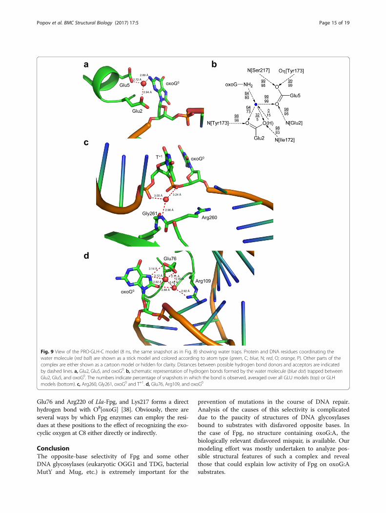
ing in the Fpg–DNA structure, we have looked in moredetail at the water bridges with the occurrence above athreshold of 2000 (for pairs) or 1500 (for triplets). Thesethresholds cut off the lowest quartile of the cumulativedistribution of bridges averaged over all twelve models,i.e., they define the bridges that collectively account for>75% of all occurrences (Additional file 1: Fig. S8). Themost frequent triplet was formed by Oε1/Oε2[Glu2],Oε1[Glu5] and N2[oxoG0] (Fig. 9a); it was found in thehigh range in 11 out of 12 models and was not far belowthe 1500 cut-off (1273) in the remaining one (PRO-GLH-Aa). A cluster of spots habitually occupied by awater molecule was near Nε[Arg260] and N[Gly261] inthe protein and non-bridging oxygens of oxoG0 and T+1
in DNA (Fig. 9c). Usually, a single water molecule wasfound in this region at any one snapshot, alternating be-tween different triplets of donors and acceptors. Finally,Oε1/Oε2[Glu76] formed triplets with Nη1[Arg109] andO2[T+1] or O8[oxoG0] (9 out of 12 models in total) withtwo water molecules involved (Fig. 9d). Other triplets,
even those passing the threshold of 1500, were found in1–3 models and are not expected to be significant.
Possible role of structural water molecules in FpgmechanismThe identified stable triplets are suggestive of an import-ant role of water in the mechanism of action of Fpg. Thewater molecule trapped between Oε1/Oε2[Glu2],Oε1[Glu5] and N2[oxoG0] is located at the position suit-able for proton transfer to Glu2, required for the proton-ation of O4′ of oxoG nucleotide; water-mediated protontransfer to Glu2 was earlier proposed on structural rea-sons [19, 26]. In the GLH models, this water stablydonated a bond only to the unprotonated Oε1[Glu2](73% bond occurrence averaged over all GLH models,compare with 5% for the bond to Oε2) but when Glu2was charged, Oε2 accepted this hydrogen with a higherfrequency (64% and 32% bonds to Oε1 and Oε2, respect-ively) (Fig. 9b). In the GLH-Aa models, the protonatedOε2 showed a tendency to donate a hydrogen bond tothe water molecule rather than accept one (23% in PRN-GLH-Aa, 58% in PRO-GLH-Aa, 0–7% in other GLHmodels), consistent with poor substrate properties ofanti A. It should be mentioned that in QM/MM analysisof fapyG excision by Fpg this water molecule was inhibi-tory to the reaction, preventing the protonation of O4′by neutral Glu2 [36] and should be displaced from itscrystallographic position after donating a proton to Oε2.The water molecule bridging the protein residues with
the phosphates of T+1 and oxoG0 may be important fordistorting the DNA duplex. Notably, the distance be-tween the phosphorus atoms P[T+1] and P[oxoG0] is sig-nificantly shorter than in the regular B-DNA in allmodels. This pinching of the phosphates around T+1, to-gether with wedging of Phe111 and insertion of Met75and Arg109, assists in kinking the DNA axis by ~60°and eversion of the damaged nucleotide.The tightly coordinated two-water bridge to
O8[oxoG0] presents an intriguing conundrum. On onehand, water-mediated recognition of this unique car-bonyl would be an attractive mechanism of direct oxoGsensing in the active site pocket. On the other hand,Glu76, which in our models participates in the water co-ordination, is present only in a small branch of the Fpgfamily tree consisting of two closely phylogenetically re-lated groups, Bacilli and Mollicutes (which include L.lactis and G. stearothermophilus), while in all other Fpgsequences this position is occupied by Ser/Thr with veryrare exceptions (Additional file 1: Fig. S9, Additional file 3).In the structure of Bst-Fpg, the presence of Glu76 stabilizesthe everted oxoG in the high syn conformation throughhydrogen bonding with N2[oxoG], whereas its in silico re-placement with Ser reverts the preferred χ angle to the antidomain [60]. In Eco-Fpg, Ser74 and Lys217 correspond to
Popov et al. BMC Structural Biology (2017) 17:5 Page 14 of 19
Glu76 and Arg220 of Lla-Fpg, and Lys217 forms a directhydrogen bond with O8[oxoG] [38]. Obviously, there areseveral ways by which Fpg enzymes can employ the resi-dues at these positions to the effect of recognizing the exo-cyclic oxygen at C8 either directly or indirectly.
ConclusionThe opposite-base selectivity of Fpg and some otherDNA glycosylases (eukaryotic OGG1 and TDG, bacterialMutY and Mug, etc.) is extremely important for the
prevention of mutations in the course of DNA repair.Analysis of the causes of this selectivity is complicateddue to the paucity of structures of DNA glycosylasesbound to substrates with disfavored opposite bases. Inthe case of Fpg, no structure containing oxoG:A, thebiologically relevant disfavored mispair, is available. Ourmodeling effort was mostly undertaken to analyze pos-sible structural features of such a complex and revealthose that could explain low activity of Fpg on oxoG:Asubstrates.
Fig. 9 View of the PRO-GLH-C model (8 ns, the same snapshot as in Fig. 8) showing water traps. Protein and DNA residues coordinating thewater molecule (red ball) are shown as a stick model and colored according to atom type (green, C; blue, N; red, O; orange, P). Other parts of thecomplex are either shown as a cartoon model or hidden for clarity. Distances between possible hydrogen bond donors and acceptors are indicatedby dashed lines. a, Glu2, Glu5, and oxoG0. b, schematic representation of hydrogen bonds formed by the water molecule (blue dot) trapped betweenGlu2, Glu5, and oxoG0. The numbers indicate percentage of snapshots in which the bond is observed, averaged over all GLU models (top) or GLHmodels (bottom). c, Arg260, Gly261, oxoG0 and T+1. d, Glu76, Arg109, and oxoG0
Popov et al. BMC Structural Biology (2017) 17:5 Page 15 of 19
As our models suggest, introduction of A opposite tooxoG indeed distorts the protein–DNA interface within±2 base pairs around the lesion site, outside of whichDNA exists as a normal duplex. Arg-109 and Phe-111,two residues that Fpg inserts into DNA to sharply kinkit and maintain oxoG everted from the base stack,tended to withdraw if A was opposite to the lesion, indi-cating that the pre-catalytic complex of Fpg with oxoG:Ais inherently unstable. Interestingly, although theoxoG:A mispair adopts oxoG(syn):A(anti) conformationin free DNA, our models showed that upon Fpg bindingand oxoG eversion, the orphaned A is more stable as asyn conformer, engaged both in hydrogen bonding withArg-109 and in base stacking. We speculate that Fpgbinding to oxoG(syn):A(anti) may be energetically disad-vantageous and require rotation of the A base aroundthe glycosidic bond for rare events of base excision; dir-ect test of this hypothesis would require solving thestructure of Fpg–DNA(oxoG:A) complex or stopped-flow kinetics with a series of fluorescent reporter basesincorporated next to A, in which case the anti–syn tran-sition may be expected to be observed in the fluorescenttraces.Analysis of model-specific hydrogen bonds unexpect-
edly revealed a cluster of highly conserved residues nextto the interdomain linker of Fpg, which adopted alterna-tive conformations when C or A was in the oppositestrand. This cluster is remote from what is usually con-sidered the active site of Fpg; however, it is packedagainst a helix–two-turn–helix motif that is present inall Fpg family members and partly forms the DNA-binding groove. Of note, it has been shown that in Nei,a homolog of Fpg specific for oxidized pyrimidines, astructural rearrangement of the linker and the region ad-jacent to it induces productive DNA binding [79]. Thus,our models add weight to a hypothesis of indirect read-out by DNA glycosylases, which states that recognitionof damaged bases is not limited to formation of specificbonds but greatly relies on the differences in energeticsand dynamics of protein and DNA parts that may be faraway from the moiety being recognized.Structural and kinetic data together with QM/MM
modeling of Fpg favor the reaction chemistry that com-bines a nucleophilic attack at C1′ of oxoG by N[Pro1]residue and protonation of O4′ of oxoG by Oε2[Glu2][33, 35, 36]. The latter step is important since it affordsa ~60 kcal/mol lower barrier to glycosidic bond breakagecompared to base protonation as the leaving group acti-vation [33]. Such mechanism requires Pro1 to be in theunprotonated, and Glu2, in the protonated state imme-diately before the reaction, implying that both these resi-dues should change their preferred protonation state.Our measurements of the pH dependence of Fpg activitysuggest that only one group is ionized in a pH-
dependent manner, in which case it is consistent withPro1 N-terminal secondary amine losing a proton at in-creasing pH. Consequently, the ionization state of Glu2in the Fpg–DNA complex shows no evidence of beingpH-dependent, which means that the assembled activesite is capable of protonating Glu2, possibly using awater molecule as a proton shuttle. The arrangement ofthe reacting atoms is only consistent with the reactionstereochemistry with SN2 displacement of O4′ as thefirst step, in agreement with the QM/MM data [33].Since the substitution of Gln for Glu2 inactivates the en-zyme, which rules out simple hydrogen bonding as theprimary function of Glu2, the mechanistic implication ofour results is that Glu2 has to be deprotonated againlater in the reaction, likely by the nascent alkoxide O4′,and contribute its charge to the stabilization of the tran-sition state of the departing oxoG base. In the QM/MMsimulation, several consecutive acts of proton transferbetween Oε2[Glu2], O4′[oxoG], N[Pro1], and O8[oxoG]allow the enzyme to lower the highest barrier in the re-action from 71 kcal/mole (as with direct oxoG proton-ation path) to 13 kcal/mole relative to the lesionrecognition complex [33]; a similar energy gain was cal-culated for fapyG excision [36].Finally, our modeling concerned only the pre-catalytic
complex of Fpg–DNA. It is now clear that the selectivityof DNA glycosylases is not determined exclusively by in-teractions in their pre-catalytic complexes, the structuresof which are relative easy to establish by X-ray crystal-lography, but also relies on several kinetic gates alongthe full recognition pathway, including primary encoun-ter and damaged base eversion. Future modeling of theearly steps of recognition of oxoG-containing pairs willadd clarity to our understanding of the opposite-basediscrimination by Fpg.
Additional files
Additional file 1: Figure S1. A, R.m.s.d. of the models over time. Thetraces are color-coded: dark cyan, PRN-GLH-C; light lime, PRN-GLH-Aa;coral, PRN-GLH-As; olive, PRN-GLU-C; dark magenta, PRN-GLU-Aa; lightblue, PRN-GLU-As; magenta, PRO-GLH-C; blue, PRO-GLH-Aa; red, PRO-GLH-As; cyan, PRO-GLU-C; yellow, PRO-GLU-Aa; green, PRO-GLU-As. B,Reproducibility of the repeat runs. R.m.s.d. of the initial run (red)and three repeat runs (green, blue, and magenta) of the PRO-GLH-Cmodel are shown together with the cross-run r.m.s.d. between twopairs of the repeat runs (black and green). Repeat runs of othermodels produced similar within-run and cross-run r.m.s.d. valuesand are not shown. Figure S2. Circular dichroism spectrum of Fpgat pH 4.0 (black circles) and pH 7.6 (white circles). Figure S3. Conformation ofthe models around the orphaned nucleotide. A, distance Nε[Arg109]…O2[C(0)] in the C models. B, distance Nη2[Arg109]…N3[C(0)] in the C models.C, distance N6[A(0)]…O2P[A(+1)] in the Aa models. D, distanceNη2[Arg109]…N7[A(0)] in the As models. E, distance N6[A(0)]…O4[T+1]in the As models. F, distance N6[A(0)]…O2P[A(+2)] in the As models. Movingaverage of a 50-snapshot window is shown in all panels. Figure S4.Occluded area (inaccessible to a 1.4 Å probe) between Phe111 sidechain and A(+1) base. The colors of the traces are the same as in Fig. S1. The
Popov et al. BMC Structural Biology (2017) 17:5 Page 16 of 19
dashed line indicates the occluded area in the 1XC8 structure. Movingaverage of a 50-snapshot window is shown. Figure S5. A, Cumulative distri-bution of the occurrence of hydrogen bonds in the Lla-Fpg–DNA complex.B, Overall reproducibility of hydrogen bonds in replicate PRO-GLH runs.Dots show the coefficient of variation for the occurrence of a particularhydrogen bond calculated over four replicates plotted against the mean oc-currence of the bond. The histograms show the distribution of the meanoccurrence. The scale in all panels is the same. Numbers above the graphsindicate the percentage of hydrogen bonds with the mean occurrence>90%. Figure S6. Interaction difference maps showing pairs of hydrogen-bond forming amino acids specific (>3σ difference in bond occurrence cal-culated over all pairs of models) for: A, C models (red) or Aa models (blue);B, C models (red) or As models (cyan); C, As models (cyan) or Aa models(blue); D, PRO models (red) or PRN models (blue); E, GLH models (red) or GLUmodels (blue). Larger circles correspond to larger deviations from the meanoccurrence. Residues 1–271, protein; 272–285, damaged DNA strand;286–299, complementary DNA strand; 300, Zn2+; 301–307, structuralwater molecules. The yellow line marks the position of oxoG0, themagenta line, the position of C(0)/A(0). Figure S7. Conservation ofFpg sequence. A, plot of conservation number Cn against the residueposition. B, view of the PRO-GLH-C model (8 ns) colored accordingto Cn. C, the same model as in B rotated 180° around the verticalaxis. The orientation of the molecule in B and C is the same as inFig. 8. Figure S8. Rank plot of water-mediated bridges (top 100occurrences) in the Fpg–DNA structures (A–L, the model nature isindicated in the respective panels). Red, pairs; blue, triplets. Dashedlines indicate cutoffs of 2000 snapshots for pairs and 1500 snapshotsfor triplets. Insets show cumulative distribution frequencies of pairsand triplets. Figure S9. Cladogram of Fpg sequences. The tree wasconstructed as described in Methods and visualized using TreeDyn [81].(PDF 3904 kb)
Additional file 2: Table S1. pKa of Pro1 and Glu2 in selected Fpgstructures. (DOC 42 kb)
Additional file 3: Alignment of 124 sequences from the Fpg family. SeeMethods for sequence selection and alignment details. (TXT 101 kb)
AcknowledgmentsThe modeling was performed on an NKS-30T cluster at the SB RASSupercomputing Center.
FundingThe work was supported by Russian Foundation for Basic Research (grant 17-04-01761-a). The funding body had no role in the design of the studyand collection, analysis, and interpretation of data and in writing themanuscript.
Availability of data and materialsThe trajectories generated in the current study are available from thecorresponding author on reasonable request. All results are presented in themain text and additional supporting files.
Authors’ contributionsDOZ and YNV have designed the study. AVP has carried out the MDsimulations. AVE has performed biochemical experiments. AVP and YNV havecontributed custom software. AVP, YNV and DOZ have participated in theanalysis and interpretation of MD trajectories, and writing of the manuscript.All authors read and approved the final manuscript.
Competing interestsThe authors declare that they have no competing interests.
Consent for publicationNot applicable.
Ethics approval and consent to participateNot applicable.
Publisher’s NoteSpringer Nature remains neutral with regard to jurisdictional claims inpublished maps and institutional affiliations.
Author details1SB RAS Institute of Chemical Biology and Fundamental Medicine, 8Lavrentieva Ave., Novosibirsk 630090, Russia. 2Novosibrsk State University, 2Pirogova St., Novosibirsk 630090, Russia.
Received: 12 November 2016 Accepted: 20 April 2017
References1. Tchou J, Kasai H, Shibutani S, Chung M-H, Laval J, Grollman AP, Nishimura S.
8-oxoguanine (8-hydroxyguanine) DNA glycosylase and its substratespecificity. Proc Natl Acad Sci U S A. 1991;88:4690–4.
2. Karakaya A, Jaruga P, Bohr VA, Grollman AP, Dizdaroglu M. Kinetics ofexcision of purine lesions from DNA by Escherichia coli Fpg protein. NucleicAcids Res. 1997;25:474–9.
3. Boiteux S, O'Connor TR, Laval J. Formamidopyrimidine-DNA glycosylase ofEscherichia coli: cloning and sequencing of the fpg structural gene andoverproduction of the protein. EMBO J. 1987;6:3177–83.
4. Tchou J, Bodepudi V, Shibutani S, Antoshechkin I, Miller J, Grollman AP,Johnson F. Substrate specificity of Fpg protein: recognition and cleavage ofoxidatively damaged DNA. J Biol Chem. 1994;269:15318–24.
5. Hatahet Z, Kow YW, Purmal AA, Cunningham RP, Wallace SS. Newsubstrates for old enzymes: 5-hydroxy-2'-deoxycytidine and 5-hydroxy-2'-deoxyuridine are substrates for Escherichia coli endonuclease III andformamidopyrimidine DNA N-glycosylase, while 5-hydroxy-2'-deoxyuridine isa substrate for uracil DNA N-glycosylase. J Biol Chem. 1994;269:18814–20.
6. Rabow LE, Kow YW. Mechanism of action of base release by Escherichia coliFpg protein: role of lysine 155 in catalysis. Biochemistry. 1997;36:5084–96.
7. Jurado J, Saparbaev M, Matray TJ, Greenberg MM, Laval J. The ringfragmentation product of thymidine C5-hydrate when present in DNA isrepaired by the Escherichia coli Fpg and Nth proteins. Biochemistry. 1998;37:7757–63.
8. Gasparutto D, Ait-Abbas M, Jaquinod M, Boiteux S, Cadet J. Repair andcoding properties of 5-hydroxy-5-methylhydantoin nucleosides inserted intoDNA oligomers. Chem Res Toxicol. 2000;13:575–84.
9. Zhang Q-M, Miyabe I, Matsumoto Y, Kino K, Sugiyama H, Yonei S.Identification of repair enzymes for 5-formyluracil in DNA: Nth, Nei, andMutM proteins of Escherichia coli. J Biol Chem. 2000;275:35471–7.
10. Krishnamurthy N, Muller JG, Burrows CJ, David SS. Unusual structuralfeatures of hydantoin lesions translate into efficient recognition byEscherichia coli Fpg. Biochemistry. 2007;46:9355–65.
11. Friedberg EC, Walker GC, Siede W, Wood RD, Schultz RA, Ellenberger T. DNArepair and mutagenesis. Washington, D.C.: ASM Press; 2006.
12. Zharkov DO. Base excision DNA repair. Cell Mol Life Sci. 2008;65:1544–65.13. Evans MD, Dizdaroglu M, Cooke MS. Oxidative DNA damage and disease:
induction, repair and significance. Mutat Res. 2004;567:1–61.14. Kouchakdjian M, Bodepudi V, Shibutani S, Eisenberg M, Johnson F, Grollman
AP, Patel DJ. NMR structural studies of the ionizing radiation adduct 7-hydro-8-oxodeoxyguanosine (8-oxo-7H-dG) opposite deoxyadenosine in aDNA duplex. 8-Oxo-7H-dG(syn)•dA(anti) alignment at lesion site.Biochemistry. 1991;30:1403–12.
15. McAuley-Hecht KE, Leonard GA, Gibson NJ, Thomson JB, Watson WP,Hunter WN, Brown T. Crystal structure of a DNA duplex containing 8-hydroxydeoxyguanine-adenine base pairs. Biochemistry. 1994;33:10266–70.
16. Grollman AP, Moriya M. Mutagenesis by 8-oxoguanine: an enemy within.Trends Genet. 1993;9:246–9.
17. Duwat P, de Oliveira R, Ehrlich SD, Boiteux S. Repair of oxidative DNAdamage in gram-positive bacteria: the lactococcus lactis Fpg protein.Microbiology. 1995;141:411–7.
18. Sugahara M, Mikawa T, Kumasaka T, Yamamoto M, Kato R, Fukuyama K,Inoue Y, Kuramitsu S. Crystal structure of a repair enzyme of oxidativelydamaged DNA, MutM (Fpg), from an extreme thermophile, Thermusthermophilus HB8. EMBO J. 2000;19:3857–69.
19. Gilboa R, Zharkov DO, Golan G, Fernandes AS, Gerchman SE, Matz E, KyciaJH, Grollman AP, Shoham G. Structure of formamidopyrimidine-DNAglycosylase covalently complexed to DNA. J Biol Chem. 2002;277:19811–6.
Popov et al. BMC Structural Biology (2017) 17:5 Page 17 of 19
20. Fromme JC, Verdine GL. Structural insights into lesion recognition andrepair by the bacterial 8-oxoguanine DNA glycosylase MutM. Nat Struct Biol.2002;9:544–52.
21. Serre L. Pereira de jésus K, boiteux S, zelwer C, castaing B. Crystal structureof the lactococcus lactis formamidopyrimidine-DNA glycosylase bound to anabasic site analogue-containing DNA. EMBO J. 2002;21:2854–65.
22. Fromme JC, Verdine GL. DNA lesion recognition by the bacterial repairenzyme MutM. J Biol Chem. 2003;278:51543–8.
23. Coste F, Ober M, Carell T, Boiteux S, Zelwer C, Castaing B. Structural basis forthe recognition of the FapydG lesion (2,6-diamino-4-hydroxy-5-formamidopyrimidine) by formamidopyrimidine-DNA glycosylase. J BiolChem. 2004;279:44074–83.
24. Pereira de Jésus K, Serre L, Zelwer C, Castaing B. Structural insights intoabasic site for Fpg specific binding and catalysis: comparative high-resolution crystallographic studies of Fpg bound to various models ofabasic site analogues-containing DNA. Nucleic Acids Res. 2005;33:5936–44.
25. Banerjee A, Santos WL, Verdine GL. Structure of a DNA glycosylasesearching for lesions. Science. 2006;311:1153–7.
26. Coste F, Ober M, Le Bihan Y-V, Izquierdo MA, Hervouet N, Mueller H, CarellT, Castaing B. Bacterial base excision repair enzyme Fpg recognizes bulkyN7-substituted-FapydG lesion via unproductive binding mode. Chem Biol.2008;15:706–17.
27. Qi Y, Spong MC, Nam K, Banerjee A, Jiralerspong S, Karplus M, Verdine GL.Encounter and extrusion of an intrahelical lesion by a DNA repair enzyme.Nature. 2009;462:762–6.
28. Qi Y, Spong MC, Nam K, Karplus M, Verdine GL. Entrapment and structure ofan extrahelical guanine attempting to enter the active site of a bacterialDNA glycosylase, MutM. J Biol Chem. 2010;285:1468–78.
29. Le Bihan Y-V, Izquierdo MA, Coste F, Aller P, Culard F, Gehrke TH, Essalhi K,Carell T, Castaing B. 5-Hydroxy-5-methylhydantoin DNA lesion, a moleculartrap for DNA glycosylases. Nucleic Acids Res. 2011;39:6277–90.
30. Qi Y, Nam K, Spong MC, Banerjee A, Sung R-J, Zhang M, Karplus M, VerdineGL. Strandwise translocation of a DNA glycosylase on undamaged DNA.Proc Natl Acad Sci U S A. 2012;109:1086–91.
31. Sung R-J, Zhang M, Qi Y, Verdine GL. Sequence-dependent structuralvariation in DNA undergoing intrahelical inspection by the DNA glycosylaseMutM. J Biol Chem. 2012;287:18044–54.
32. Sung R-J, Zhang M, Qi Y, Verdine GL. Structural and biochemical analysis ofDNA helix invasion by the bacterial 8-oxoguanine DNA glycosylase MutM.J Biol Chem. 2013;288:10012–23.
33. Sadeghian K, Flaig D, Blank ID, Schneider S, Strasser R, Stathis D, WinnackerM, Carell T, Ochsenfeld C. Ribose-protonated DNA base excision repair: acombined theoretical and experimental study. Angew Chem Int Ed. 2014;53:10044–8.
34. Sun B, Latham KA, Dodson ML, Lloyd RS. Studies of the catalytic mechanismof five DNA glycosylases: probing for enzyme-DNA imino intermediates.J Biol Chem. 1995;270:19501–8.
35. Zharkov DO, Shoham G, Grollman AP. Structural characterization of the Fpgfamily of DNA glycosylases. DNA Repair. 2003;2:839–62.
36. Blank ID, Sadeghian K, Ochsenfeld C. A base-independent repair mechanismfor DNA glycosylase—no discrimination within the active site. Sci Rep. 2015;5:10369.
37. Popov AV, Vorob'ev YN. GUI-BioPASED: a program for molecular dynamicssimulations of biopolymers with a graphical user interface. Mol Biol (Mosk).2010;44:648–54.
38. Perlow-Poehnelt RA, Zharkov DO, Grollman AP, Broyde S. Substratediscrimination by formamidopyrimidine-DNA glycosylase: distinguishinginteractions within the active site. Biochemistry. 2004;43:16092–105.
39. Hornak V, Abel R, Okur A, Strockbine B, Roitberg A, Simmerling C.Comparison of multiple Amber force fields and development of improvedprotein backbone parameters. Proteins. 2006;65:712–25.
40. Ravishanker G, Auffinger P, Langley DR, Jayaram B, Young MA, BeveridgeDL. Treatment of counterions in computer simulations of DNA. Rev ComputChem. 1997;11:317–72.
41. Lazaridis T, Karplus M. Effective energy function for proteins in solution.Proteins. 1999;35:133–52.
42. Popov AV, Vorobjev YN, Zharkov DO. MDTRA: a molecular dynamics trajectoryanalyzer with a graphical user interface. J Comput Chem. 2013;34:319–25.
43. Vorobjev YN. Study of the mechanism of interaction of oligonucleotideswith the 3'-terminal region of tRNAPhe by computer modeling. Mol Biol(Mosk). 2005;39:777–84.
44. Mauget S. Time series analysis based on running Mann-Whitney Z Statistics.J Time Ser Anal. 2011;32:47–53.
45. Benjamini Y, Hochberg Y. Controlling the false discovery rate: a practicaland powerful approach to multiple testing. J R Stat Soc Ser B StatMethodol. 1995;57:289–300.
46. Humphrey W, Dalke A, Schulten K. VMD: Visual molecular dynamics. J MolGraph. 1996;14:33–8.
47. Sayle RA, Milner-White EJ. RASMOL: biomolecular graphics for all. TrendsBiochem Sci. 1995;20:374–6.
48. Søndergaard CR, Olsson MHM, Rostkowski M, Jensen JH. Improvedtreatment of ligands and coupling effects in empirical calculation andrationalization of pKa values. J Chem Theory Comput. 2011;7:2284–95.
49. Altschul SF, Madden TL, Schäffer AA, Zhang J, Zhang Z, Miller W, Lipman DJ.Gapped BLAST and PSI-BLAST: a new generation of protein database searchprograms. Nucleic Acids Res. 1997;25:3389–402.
50. Zharkov DO, Grollman AP. Combining structural and bioinformaticsmethods for the analysis of functionally important residues in DNAglycosylases. Free Radic Biol Med. 2002;32:1254–63.
51. Zharkov DO. Predicting functional residues in DNA glycosylases by analysisof structure and conservation. In: Practical Bioinformatics. Edited by BujnickiJN. Berlin–Heidelberg: Springer-Verlag. 2004;15:243-61.
52. Sievers F, Wilm A, Dineen D, Gibson TJ, Karplus K, Li W, Lopez R, McWilliamH, Remmert M, Söding J, et al. Fast, scalable generation of high-qualityprotein multiple sequence alignments using clustal omega. Mol Syst Biol.2011;7:539.
53. Livingstone CD, Barton GJ. Protein sequence alignments: a strategy for thehierarchical analysis of residue conservation. Comput Appl Biosci. 1993;9:745–56.
54. Ober M, Linne U, Gierlich J, Carell T. The two main DNA lesions 8-oxo-7,8-dihydroguanine and 2,6-diamino-5-formamido-4-hydroxypyrimidine exhibitstrongly different pairing properties. Angew Chem Int Ed. 2003;42:4947–51.
55. Oda Y, Uesugi S, Ikehara M, Nishimura S, Kawase Y, Ishikawa H, Inoue H,Ohtsuka E. NMR studies of a DNA containing 8-hydroxydeoxyguanosine.Nucleic Acids Res. 1991;19:1407–12.
56. Lipscomb LA, Peek ME, Morningstar ML, Verghis SM, Miller EM, Rich A,Essigmann JM, Williams LD. X-ray structure of a DNA decamer containing7,8-dihydro-8-oxoguanine. Proc Natl Acad Sci U S A. 1995;92:719–23.
57. Zaika EI, Perlow RA, Matz E, Broyde S, Gilboa R, Grollman AP, Zharkov DO.Substrate discrimination by formamidopyrimidine-DNA glycosylase: amutational analysis. J Biol Chem. 2004;279:4849–61.
58. Amara P, Serre L, Castaing B, Thomas A. Insights into the DNA repairprocess by the formamidopyrimidine-DNA glycosylase investigated bymolecular dynamics. Protein Sci. 2004;13:2009–21.
59. Amara P, Serre L. Functional flexibility of Bacillus stearothermophilusformamidopyrimidine DNA-glycosylase. DNA Repair. 2006;5:947–58.
60. Song K, Hornak V, de los Santos C, Grollman AP, Simmerling C.Computational analysis of the mode of binding of 8-oxoguanine toformamidopyrimidine-DNA glycosylase. Biochemistry. 2006;45:10886–94.
61. Song K, Kelso C, de los Santos C, Grollman AP, Simmerling C. Molecularsimulations reveal a common binding mode for glycosylase binding ofoxidatively damaged DNA lesions. J Am Chem Soc. 2007;129:14536–7.
62. Thiviyanathan V, Somasunderam A, Hazra TK, Mitra S, Gorenstein DG.Solution structure of a DNA duplex containing 8-hydroxy-2'-deoxyguanosine opposite deoxyguanosine. J Mol Biol. 2003;325:433–42.
63. Chen J, Brooks III CL, Khandogin J. Recent advances in implicit solvent-basedmethods for biomolecular simulations. Curr Opin Struct Biol. 2008;18:140–8.
64. Vorobjev YN. Advances in implicit models of water solvent to computeconformational free energy and molecular dynamics of proteins at constantpH. Adv Protein Chem Struct Biol. 2011;85:281–322.
65. Kleinjung J, Fraternali F. Design and application of implicit solvent modelsin biomolecular simulations. Curr Opin Struct Biol. 2014;25:126–34.
66. Anandakrishnan R, Drozdetski A, Walker RC, Onufriev AV. Speed ofconformational change: comparing explicit and implicit solvent moleculardynamics simulations. Biophys J. 2015;108:1153–64.
67. Li S, Bradley P. Probing the role of interfacial waters in protein–DNA recognitionusing a hybrid implicit/explicit solvation model. Proteins. 2013;81:1318–29.
68. Sadeghian K, Ochsenfeld C. Unraveling the base excision repair mechanismof human DNA glycosylase. J Am Chem Soc. 2015;137:9824–31.
69. Sowlati-Hashjin S, Wetmore SD. Computational investigation of glycosylaseand β-lyase activity facilitated by proline: applications to FPG andcomparisons to hOgg1. J Phys Chem B. 2014;118:14566–77.
Popov et al. BMC Structural Biology (2017) 17:5 Page 18 of 19
70. Stivers JT, Jiang YL. A mechanistic perspective on the chemistry of DNArepair glycosylases. Chem Rev. 2003;103:2729–60.
71. Storm DR, Koshland Jr DE. A source for the special catalytic power ofenzymes: orbital steering. Proc Natl Acad Sci U S A. 1970;66:445–52.
72. Bruice TC, Brown A, Harris DO. On the concept of orbital steering incatalytic reactions. Proc Natl Acad Sci U S A. 1971;68:658–61.
73. Fuxreiter M, Warshel A, Osman R. Role of active site residues in theglycosylase step of T4 endonuclease V. Computer simulation studies onionization states. Biochemistry. 1999;38:9577–89.
74. O'Brien PJ, Ellenberger T. Human alkyladenine DNA glycosylase uses acid-base catalysis for selective excision of damaged purines. Biochemistry. 2003;42:12418–29.
75. Song K, Campbell AJ, Bergonzo C, de los Santos C, Grollman AP, SimmerlingC. An improved reaction coordinate for nucleic acid base flipping studies. JChem Theory Comput. 2009;5:3105–13.
76. Kuznetsov NA, Koval VV, Zharkov DO, Vorobjev YN, Nevinsky GA, DouglasKT, Fedorova OS. Pre-steady-state kinetic study of substrate specificity ofEscherichia coli formamidopyrimidine-DNA glycosylase. Biochemistry. 2007;46:424–35.
77. Kuznetsov NA, Bergonzo C, Campbell AJ, Li H, Mechetin GV, de los SantosC, Grollman AP, Fedorova OS, Zharkov DO, Simmerling C. Activedestabilization of base pairs by a DNA glycosylase wedge initiates damagerecognition. Nucleic Acids Res. 2015;43:272–81.
78. Zharkov DO, Golan G, Gilboa R, Fernandes AS, Gerchman SE, Kycia JH,Rieger RA, Grollman AP, Shoham G. Structural analysis of an Escherichia coliendonuclease VIII covalent reaction intermediate. EMBO J. 2002;21:789–800.
79. Golan G, Zharkov DO, Feinberg H, Fernandes AS, Zaika EI, Kycia JH,Grollman AP, Shoham G. Structure of the uncomplexed DNA repair enzymeendonuclease VIII indicates significant interdomain flexibility. Nucleic AcidsRes. 2005;33:5006–16.
80. Shui X, McFail-Isom L, Hu GG, Williams LD. The B-DNA dodecamer at highresolution reveals a spine of water on sodium. Biochemistry. 1998;37:8341–55.
81. Chevenet F, Brun C, Bañuls A-L, Jacq B, Christen R. TreeDyn: towardsdynamic graphics and annotations for analyses of trees. BMC Bioinformatics.2006;7:439.
• We accept pre-submission inquiries
• Our selector tool helps you to find the most relevant journal
• We provide round the clock customer support
• Convenient online submission
• Thorough peer review
• Inclusion in PubMed and all major indexing services
• Maximum visibility for your research
Submit your manuscript atwww.biomedcentral.com/submit
Submit your next manuscript to BioMed Central and we will help you at every step:
Popov et al. BMC Structural Biology (2017) 17:5 Page 19 of 19