On the epigenetic regulation of the human reelin promoter
10
2930–2939 Nucleic Acids Research, 2002, Vol. 30, No. 13 © 2002 Oxford University Press On the epigenetic regulation of the human reelin promoter Ying Chen, Rajiv P. Sharma, Robert H. Costa 1 , Erminio Costa and Dennis R. Grayson* Psychiatric Institute, Department of Psychiatry, 1601 West Taylor Street, M/C 912 and 1 Department of Molecular Genetics, College of Medicine, University of Illinois, Chicago, IL 60612, USA Received February 6, 2002; Revised and Accepted May 7, 2002 ABSTRACT Reln mRNA and protein levels are reduced by ∼50% in various cortical structures of post-mortem brain from patients diagnosed with schizophrenia or bipolar illness with psychosis. To study mechanisms respon- sible for this down-regulation, we have analyzed the promoter of the human reelin gene. We show that the reelin promoter directs expression of a reporter construct in multiple human cell types: neuroblastoma cells (SHSY5Y), neuronal precursor cells (NT2), differ- entiated neurons (hNT) and hepatoma cells (HepG2). Deletion constructs confirmed the presence of multiple elements regulating Reln expression, although the promoter activity is promiscuous, i.e. activity did not correlate with expression of the endogenous gene as reflected in terms of reelin mRNA levels. Co-transfection of the –514 bp human reelin promoter with either Sp1 or Tbr1 demonstrated that these transcription factors acti- vate reporter expression by 6- and 8.5-fold, respect- ively. Within 400 bp of the RNA start site there are 100 potential CpG targets for DNA methylation. Retinoic acid (RA)-induced differentiation of NT2 cells to hNT neurons was accompanied by increased reelin expres- sion and by the appearance of three DNase I hypersen- sitive sites 5′ to the RNA start site. RA-induced differentiation was also associated with demethylation of the reelin promoter. To test if methylation silenced reelin expression, we methylated the promoter in vitro prior to transfection. In addition, we treated NT2 cells with the methylation inhibitor aza-2′ -deoxycytidine and observed a 60-fold increase in reelin mRNA levels. The histone deacetylase inhibitors trichostatin A (TSA) and valproic acid also induced expression of the endogenous reelin promoter, although TSA was considerably more potent. These findings indicate that one determinant responsible for regulating reelin expression is the methylation status of the promoter. Our data also raise the interesting possibility that the down-regulation of reelin expression documented in psychiatric patients might be the consequence of inappropriate promoter hypermethylation. INTRODUCTION To date, in all post-mortem brain areas obtained from patients diagnosed with schizophrenia, reelin and glutamate decarb- oxylase 67 mRNA and protein levels are reduced by ∼50% (1). This includes multiple cortical regions, hippocampi and cere- bella (reviewed in 2). These findings were replicated in the Stanley Foundation cohort of 60 brains, and similar decreases in both parameters were obtained in the prefrontal cortices and cerebella of schizophrenia and bipolar patients with psychosis but not in patients with unipolar depression (3). Since there were no changes in the levels of GAD 65 immunoreactivity, which is expressed in the same neurons that express GAD 67 , and the changes persisted after adjusting for the presence of a specific neuronal marker, these results cannot be explained by a neuronal loss related to the illness. Interestingly, a reduction in the size of the neuropil (4) was detected that could be related to a decrease in the dendritic spines of cortical pyramidal neurons associated with reduced reelin levels. Another report reproduced the decreased reelin levels in patients with schizo- phrenia and also noted a decrease in CA4 areas of subjects with bipolar disorder and a non-significant decrease in this same region in patients with major depression (5). The decrease observed in the hippocampus of unipolar depressed patients is interesting, but the significance remains unclear. Nevertheless, there is considerable interest in understanding mechanisms underlying the regulation of human reelin expression and it is our goal to provide the requisite framework for this under- standing to allow for more detailed studies in the context of psychiatric illness. Reelin is an extracellular matrix protein (6) that is expressed in the developing brain and continues to be expressed during the lifespan of GABAergic neurons of the cortex and hippo- campus, in glutamatergic granule cells of the cerebella, in neostriatal medium spiny neurons of adult rodents (7,8), in non-human primates (9) and in many of these structures in adult humans (1,3,10). Very little is currently known with respect to how the expression of reelin is regulated in mature neurons. The human and mouse cDNAs share 88% nucleotide identity and show a high degree of sequence similarity surrounding the start site of transcription (11). Sequences flanking the human RNA start site are very GC-rich (75%) and form a large CpG island. This suggested to us that the reelin promoter might be epigenetically regulated through changes in DNA cytosine methylation (12–15). In contrast to the vast majority of CpG dinucleotides in the genome which are *To whom correspondence should be addressed. Tel: +1 312 413 4577; Fax: +1 312 413 4569; Email: [email protected]
-
Upload
independent -
Category
Documents
-
view
1 -
download
0
Transcript of On the epigenetic regulation of the human reelin promoter

2930–2939 Nucleic Acids Research, 2002, Vol. 30, No. 13 © 2002 Oxford University Press
On the epigenetic regulation of the human reelinpromoterYing Chen, Rajiv P. Sharma, Robert H. Costa1, Erminio Costa and Dennis R. Grayson*
Psychiatric Institute, Department of Psychiatry, 1601 West Taylor Street, M/C 912 and 1Department of MolecularGenetics, College of Medicine, University of Illinois, Chicago, IL 60612, USA
Received February 6, 2002; Revised and Accepted May 7, 2002
ABSTRACT
Reln mRNA and protein levels are reduced by ∼50% invarious cortical structures of post-mortem brain frompatients diagnosed with schizophrenia or bipolarillness with psychosis. To study mechanisms respon-sible for this down-regulation, we have analyzed thepromoter of the human reelin gene. We show that thereelin promoter directs expression of a reporterconstruct in multiple human cell types: neuroblastomacells (SHSY5Y), neuronal precursor cells (NT2), differ-entiated neurons (hNT) and hepatoma cells (HepG2).Deletion constructs confirmed the presence of multipleelements regulating Reln expression, although thepromoter activity is promiscuous, i.e. activity did notcorrelate with expression of the endogenous gene asreflected in terms of reelin mRNA levels. Co-transfectionof the –514 bp human reelin promoter with either Sp1 orTbr1 demonstrated that these transcription factors acti-vate reporter expression by 6- and 8.5-fold, respect-ively. Within 400 bp of the RNA start site there are 100potential CpG targets for DNA methylation. Retinoicacid (RA)-induced differentiation of NT2 cells to hNTneurons was accompanied by increased reelin expres-sion and by the appearance of three DNase I hypersen-sitive sites 5′ to the RNA start site. RA-induceddifferentiation was also associated with demethylationof the reelin promoter. To test if methylation silencedreelin expression, we methylated the promoter in vitroprior to transfection. In addition, we treated NT2 cellswith the methylation inhibitor aza-2′-deoxycytidine andobserved a 60-fold increase in reelin mRNA levels. Thehistone deacetylase inhibitors trichostatin A (TSA) andvalproic acid also induced expression of the endogenousreelin promoter, although TSA was considerably morepotent. These findings indicate that one determinantresponsible for regulating reelin expression is themethylation status of the promoter. Our data alsoraise the interesting possibility that the down-regulationof reelin expression documented in psychiatricpatients might be the consequence of inappropriatepromoter hypermethylation.
INTRODUCTION
To date, in all post-mortem brain areas obtained from patientsdiagnosed with schizophrenia, reelin and glutamate decarb-oxylase 67 mRNA and protein levels are reduced by ∼50% (1).This includes multiple cortical regions, hippocampi and cere-bella (reviewed in 2). These findings were replicated in theStanley Foundation cohort of 60 brains, and similar decreasesin both parameters were obtained in the prefrontal cortices andcerebella of schizophrenia and bipolar patients with psychosisbut not in patients with unipolar depression (3). Since therewere no changes in the levels of GAD65 immunoreactivity,which is expressed in the same neurons that express GAD67,and the changes persisted after adjusting for the presence of aspecific neuronal marker, these results cannot be explained bya neuronal loss related to the illness. Interestingly, a reductionin the size of the neuropil (4) was detected that could be relatedto a decrease in the dendritic spines of cortical pyramidalneurons associated with reduced reelin levels. Another reportreproduced the decreased reelin levels in patients with schizo-phrenia and also noted a decrease in CA4 areas of subjects withbipolar disorder and a non-significant decrease in this sameregion in patients with major depression (5). The decreaseobserved in the hippocampus of unipolar depressed patients isinteresting, but the significance remains unclear. Nevertheless,there is considerable interest in understanding mechanismsunderlying the regulation of human reelin expression and it isour goal to provide the requisite framework for this under-standing to allow for more detailed studies in the context ofpsychiatric illness.
Reelin is an extracellular matrix protein (6) that is expressedin the developing brain and continues to be expressed duringthe lifespan of GABAergic neurons of the cortex and hippo-campus, in glutamatergic granule cells of the cerebella, inneostriatal medium spiny neurons of adult rodents (7,8), innon-human primates (9) and in many of these structures inadult humans (1,3,10). Very little is currently known withrespect to how the expression of reelin is regulated in matureneurons. The human and mouse cDNAs share 88% nucleotideidentity and show a high degree of sequence similaritysurrounding the start site of transcription (11). Sequencesflanking the human RNA start site are very GC-rich (75%) andform a large CpG island. This suggested to us that the reelinpromoter might be epigenetically regulated through changes inDNA cytosine methylation (12–15). In contrast to the vastmajority of CpG dinucleotides in the genome which are
*To whom correspondence should be addressed. Tel: +1 312 413 4577; Fax: +1 312 413 4569; Email: [email protected]
Nucleic Acids Research, 2002, Vol. 30, No. 13 2931
generally methylated, CpG islands tend to be undermethylatedand this pattern can vary in both a temporal and spatially select-ive manner (14,16). This provides one mechanism by whichpromoters embedded in CpG islands may be differentiallyregulated.
In addition, it has been proposed that there may be an interplaybetween transcription factor binding and DNA methylation ofpromoters regulated epigenetically through alterations in theirpatterns of methylation (17). The interaction of Sp1, forexample, with its recognition site in the adenine phospho-ribosyltransferase promoter has been suggested as a signal forkeeping the CpG island upstream of the adenine phospho-ribosyltransferase gene free of methylation in mouse embryonicstem cells (18). Equally important in this context is the inter-play between methylation, chromatin structure and histonedeacetylaton. Transcription factors and methyl-CpG-bindingproteins interact with various classes of histone deacetylases(HDACs) in complexes that repress transcription and perhapsinduce demethylation (14). This would provide a means bywhich genes could be turned on and off through epigeneticswitches that ultimately regulate local chromatin structure andgene expression. While this type of mechanism has not beenshown to operate in regulating genes expressed in post-mitoticcells, the role of methylation/demethylation as a determinantdynamically regulating gene activity in neurons is largelyunexplored.
In the present study, we examined the human reelin promoterusing transient transfection assays to identify regions thatparticipate in targeting expression to both reelin mRNA-expressing and non-expressing cell lines. We also demonstratethat the non-methylatable cytosine analog 5-aza-deoxycytidine(AzAdc), which prevents methylation, activates expression ofthe endogenous reelin gene, while in vitro methylation of thereelin promoter silences expression. In addition, we presentdata that show that the increased expression of the reelin genefollowing retinoic acid (RA)-induced differentiation of neuralprogenitor cells in vitro is accompanied by a reduced methyla-tion of the reelin promoter. Finally, we examined the effect ofaltering the pattern of histone acetylation of neural progenitor(NT2) cells on reelin expression. The HDAC inhibitor tricho-statin A (TSA) increased reelin mRNA levels. Of particularinterest in the area of psychiatric pharmacology, the mood-stabilizing drug valproic acid (VPA) has been shown to act asan HDAC inhibitor (19,20). VPA moderately increased reelinexpression, although it was considerably less potent than TSA.These studies provide a framework for the pursuit of hypoth-eses which propose that alterations in epigenetic gene regula-tion may be relevant to neuropsychiatric disease.
MATERIALS AND METHODS
Cell culture
SHSY5Y cells were maintained in a 1:1 mixture of Eagle’sMEM/F12K (Life Technologies) supplemented with 10% fetalbovine serum (FBS) and 1% penicillin, streptomycin andglutamine. HepG2 cells were grown in HAM supplementedwith 10% FBS and 1% penicillin/streptomycin. NT2 cells weremaintained in DMEM/F12, 10% FBS and 1% penicillin, strepto-mycin and glutamine. hNT neurons were induced from culturesof NT2 cells by treating with RA for up to 6 weeks. Low
density cultures of NT2 neural progenitors were treated withAzAdC at several concentrations (1, 5 and 10 µM) and TSA(0.2, 1 and 5 µM) for various times and cells were harvestedfor total RNA isolation. In parallel, NT2 cells were treated withvarious concentrations of VPA (0.2, 2, 5 and 10 mM) and theinactive VPA amide valpromide (VPM) (5 mM) for 40 h andcells were harvested for RNA analysis. RNA was isolatedfollowing ultracentrifugation through CsCl as previouslydescribed (21).
Generation of the reelin promoter/reporter constructs
The 5′ portion of the human mRNA and upstream sequencemaps to human BAC clone RG126M09, which contains 163 kbof human genomic DNA. We subcloned a 3′ terminal 4.2 kbEcoRI fragment from this BAC DNA which contained theentire first exon, 255 bp of the first intron and 3.7 kb of 5′flanking DNA (Fig. 1A). To generate the reelin –514 bpminimal promoter construct, we designed primers to PCRamplify this region from the BAC DNA. The sequences of theprimers were: –514 bp, 5′-AAA AAC AGG GCA CAC TGACGG CCA-3′; +76 bp, 5′-CGG AGA GAA GGC GAG AAGAAG GCG-3′. Following PCR, the amplicon was subclonedinto the pGL3-Basic luciferase vector (Promega/Fisher). The–514 bp promoter/reporter was used to introduce additionalupstream sequences using the restriction sites indicated inFigure 1A and the downstream AatII site. The –514 bppromoter/reporter was also used to generate a series of Bal31deletions as previously described (22). Sequences between–303 and –137 were cloned upstream of the SV40 promoterusing the pGL-3-Promoter parent vector (Promega/Fisher) inboth orientations (sense and antisense). We assessed activityby transient transfection of numerous cell lines.
The RNA start site was mapped using a primer extensionassay (as previously described; 22). In brief, the reelin +60primer (reverse complement sequence underlined in Fig. 4B)was end-labeled using T4 polynucleotide kinase. The specificactivity of the primer was 300 000 c.p.m./ng. The primer (5 ng)and 10 µg cellular RNA (SHSY5Y or HepG2) were denaturedin 15 µl of hybridization buffer (10 mM Tris–HCl pH 8.3,150 mM KCl, 1 mM EDTA) at 85°C for 5 min and hybridizedat 50°C for 30 min. Primer extension was carried out by addingan aliquot of the primer extension mix (to 20 mM Tris–HClpH 8.3, 10 mM MgCl2, 10 mM DTT, 1.6 mM dNTP, with 10 UAMV reverse transcriptase). The reaction was incubated at42°C for 1 h. RNase reaction mix (100 µg/ml salmon spermDNA and 20 µg/ml RNase A) was added to the sample andincubated at 37°C for 30 min. Following phenol/chloroformextraction and ethanol precipitation, the reaction products wereanalyzed on a 10% denaturing TBE–urea gel (Invitrogen). Thegel was dried and exposed to a phosphorimager screen. Theimage was scanned using a Storm PhosphorImager (MolecularDynamics) to visualize the reaction products. The 10 bp ladderDNA marker was used for sizing the products (Invitrogen).
DNase I hypersensitive site analysis
Nuclei were isolated from either NT2 neuroprogenitor cells orfrom hNT neurons as previously described (23). Isolated nucleifrom either group were treated with increasing amounts ofDNase I (0, 10, 20, 30, 40, 60, 80 and 100 U DNase I, FPLCpure; Pharmacia) for 10 min at 37°C. Following treatment, thereaction was stopped by the addition of 50 mM Tris–HCl pH 7.5,
2932 Nucleic Acids Research, 2002, Vol. 30, No. 13
100 mM NaCl and 1% SDS. Genomic DNA was extracted,purified and digested with EcoRI and subjected to agarose gelelectrophoresis and Southern blotting. The blot was hybridizedwith random primed, 32P-labeled probe corresponding to the 5′most portion of the subcloned genomic EcoRI fragment(shown in Fig. 1A). The specific activity of the probe used was1 × 109 d.p.m./µg. The washed blots were exposed to phos-phorimager screens for 12–16 h and the image was obtained byscanning the screen with a Storm PhosphorImager (MolecularDynamics).
Transfection assays and reporter expressionmeasurements
Cells were transfected using Lipofectamine 2000 (Life Tech-nologies). We used the dual-luciferase reporter assay system
(Promega/Fisher) and routinely used 2–4 µg each test DNAwith 30 ng pRL-CMV vector (Renilla luciferase) for each wellof a 6-well plate. Cell lysates were prepared 24–36 h aftertransfection and aliquots (∼20 µl) were used for determinationof luciferase activity in a TD20/20 luminometer (TurnerDesign). Data (minimum of three to five transfections/construct) are expressed as a ratio to the signal obtained fromthe SV40 promoter/luciferase vector that was transfected inparallel (pGL3-Control vector; Promega/Fisher).
Competitive RT–PCR assay
We previously developed the use of competitive RT–PCR tomake quantitative measurements of specific mRNAs in totalcellular RNA with specific internal standards (24,25). Theinternal standard corresponding to reelin mRNA has beendescribed (1). Following RT–PCR, amplicons were digestedwith BanI and electrophoresed on agarose gels. Gels werestained with Syber Gold (Molecular Probes) and scanneddirectly using a Storm PhosphorImager (MolecularDynamics). The RNA analysis was performed three timesper sample from a minimum of three different RNA isolations.The data were analyzed for significance using a one-wayANOVA.
In vitro DNA methylation reaction
Escherichia coli DNA methylases were used to methylate the–514 promoter/luciferase construct to test the effects ofsequence-specific methylation on reporter activity. Thefollowing methylases were used, each of which is followed byits selectivity and number of sites present in the template: SssI(*CG), 122 sites in the –514 promoter construct; HpaII(C*CGG), 10 sites; MspI (*CCGG), 11 sites; HhaI (G*CGC),21 sites; HaeIII (GG*CC), 10 sites. The * indicates the methy-lated cytosine. An aliquot of 10 µg –514 promoter/reporter wasused per reaction with 25 U enzyme in 100 mM NaCl, 50 mMTris–HCl pH 7.5, 5 mM 2-mercaptoethanol and 80 µMS-adenosylmethionine for 10 h at 30°C. The extent of methy-lation was examined by digesting aliquots of the reaction withthe corresponding restriction enzyme. The effect of methylationon promoter activity was assessed by transfecting the variousconstructs as indicated above.
Bisulfite modification of genomic DNA
Genomic DNA from various samples (NT2 and hNT neurons)was isolated using the proteinase K/SDS method (26) and wasdigested with EcoRI and denatured by treating with NaOH.Sodium bisulfite (5 M) converts deoxycytosine but not 5-methyl-cytosine residues into uracil, and the reaction was performed asdescribed (27). Following the modification, the strands (desig-nated the A and B strands) are no longer complementary anddifferent sets of primers were designed for each strand. Nestedsets of primers were designed for both strands. For the Bstrand: outer primers, –790 bp, 5′-TTT AAA ATC CTC TACAAA TAA AAC TCT ATC ACT-3′, and +350 bp, 5′-TGTTTG TAA TAT GTA GGG AAA TGA GTA TTT-3′; innerprimers, –527 bp, 5′-ACA TCC TCC CAA AAA AAA CAAAAC ACA CTA A-3′, and +305 bp, 5′-TTT TTT TAG TTTTTT GTG GTG GGT GTA TAG GAA-3′. The reaction prod-ucts of the B strand were amplified and subcloned. Multipleclones from NT2 neural prognitor cells and from hNT neurons
Figure 1. Reelin promoter structure and transient transfections. (A) Schematicrepresentation of the cloned human reelin promoter. The top line shows a linearrepresentation of sequences along the BAC DNA and associated restrictionenzyme cleavage sites. The transcriptional start site is indicated (+1). The positionof the CGG repeat present in the 5′ UTR of the first exon is shown where n var-ies from 4 to 24 repeats. The subcloned minimal promoter/luciferase reportervector contains a region (reelin enhancer) that acts to enhance expression of theSV40 promoter. (B) Primer extension analysis. The products of reverse tran-scription of SHSY5Y RNA using the +60 bp primer is shown. To the right ofthe gel is the standard curve that was generated and the position of migrationof this product and that corresponding to the +110 bp primer. The sequencealignment shows a comparison of the human reelin cDNA (GenBank accessionno. NM_005045) and the genomic sequence (as shown in Fig. 4B). Similarresults were obtained using RNA from HepG2 cells (data not shown). (C) CpGislands plot of the human reelin promoter and first exon. The CpG islands plotwas generated using GeneTool software (BioTools) that is based on an algorithmthat plots the observed number of CpG sites divided by the expected number ofCpG sites along the length of the DNA fragment. The numbering is based onthe RNA start site being equal to +1. y-axis values above the horizontal lineindicate regions likely to be CpG islands (28).
Nucleic Acids Research, 2002, Vol. 30, No. 13 2933
were sequenced by the UIC sequencing facility using fluores-cent dideoxy technology.
RESULTS
The murine reelin mRNA is encoded by a gene that contains 65exons which span ∼450 kb of genomic DNA. The completeintron/exon structure of the rodent gene has been mapped (11).We have mapped the intron/exon structure of human Reln tovarious BAC clones and have found that the intron/exonboundaries are remarkably conserved, as are the sizes of thecorresponding introns (D. R. Grayson, unpublished observations).In the course of making these comparisons we noticed adiscrepancy between the 5′ end of the human mRNA (10) andthe genomic sequence containing the remainder of the firstexon (BAC clone 96012; Research Genetics). The differenceresided in the first 10 bp of the cDNA, which were not presentin the genomic sequence. To verify the location of the RNAstart site within the genomic sequence of the BAC, weperformed primer extension assays using primers that mappedto within 60 and 110 bp of the region that differed in thegenomic DNA and the cDNA. As can be seen from the analysis(Fig. 1B), the assay yielded a single start site using RNA fromSHSY5Y cells. Similar results were obtained with RNAobtained from HepG2 cells (data not shown). The size of theproducts obtained with each primer are indicated on thestandard curve. This site corresponds to that predicted from thegenomic sequence, indicating the possibility that the first 10 bpof the cDNA may be present due to a cloning artifact (Fig. 1B).The downstream primer yielded several minor bands much likethe mouse primer extension, which showed multiple start sites(11). Because these additional products were not seen withboth primers, we presume that the extra sites likely representpausing artifacts due to the high GC content of the template.
Similar to the rodent promoter region (11), sequencesflanking the human RNA start site are very GC-rich (75%)and, together with the first exon, form a large CpG island. Alinear representation of the CpG islands plot corresponding tosequences flanking the human reelin RNA start site is shown inFigure 1C. The abscissa represents the observed number ofCpG sites divided by the expected number of CpG sitesassuming random sorting along the reelin promoter sequence.The reelin promoter and first exon sequences are shown alongthe ordinate, where the transcriptional start site is 1 andsequences 5′ to this are represented as negative numbers whiledownstream sequences are positive. A candidate CpG islandgives a y-value of >0.6 on this CpG islands plot over anextended window size (28). Using these criteria, the reelinpromoter CpG island extends from approximately –1200 to+200 bp relative to the transcription start site.
Transient transfection analysis of the human reelinpromoter
To create promoter expression vectors, we PCR amplified andsubcloned a region that extends from –514 to + 76 bp relative tothe human RNA start site (minimal promoter, Fig. 1A). Theregion subcloned extends into the 5′ UTR but does not includethe previously described CGG repeat region (29,30). Thistriplet repeat was excluded so as to avoid any translationaleffects that might be associated with variations in triplet repeatlength. The reelin promoter constructs, along with the
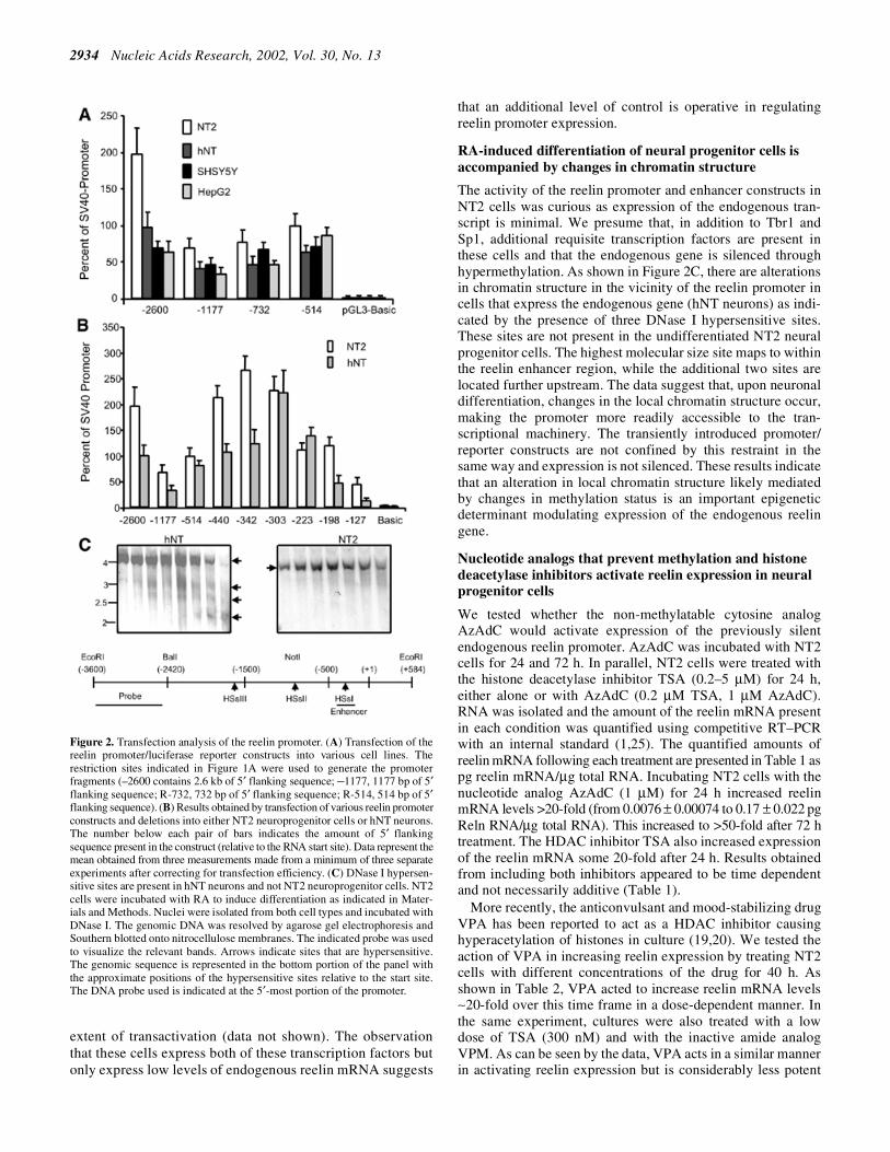
normalization control (pRLCMV; Promega) were transientlytransfected into human neural progenitor cells (NT2, lowreelin levels, 0.0076 ± 0.00074 pg Reln mRNA/µg totalRNA), RA-differentiated NT2 cells (hNT, high reelin mRNAlevels, 0.75 ± 0.046 pg Reln mRNA/µg total RNA), neuro-blastoma cells (SHSY5Y) and hepatoma cells (HepG2).SHSY5Y and HepG2 cells express intermediate reelin mRNAlevels. Following DNA transfection, cell protein lysates wereprepared and the amount of luciferase activity was quantified.The data showed that the reelin promoter is actively expressedin each of the cell types (Fig. 2A) and that the reporter activitywas independent of the endogenous reelin mRNA levels. Forexample, high reelin promoter expression was observed inNT2 cells which express only very low levels of the endo-genous reelin transcript (Table 1). The results suggest thatindividual variations in the levels of expression arising fromeach reelin promoter construct likely reflect differences intranscription factor binding but that biologically accuratereelin expression requires additional information that likelyoccurs at the level of chromatin structure.
We also examined sequences smaller than the reelin minimalpromoter (–514 bp). Multiple deletions were cloned that wereevenly spaced between –514 and –127 bp of the reelinpromoter. Figure 2B shows reporter activity results obtainedfollowing transient transfection of these deletions into neuralprogenitors (NT2) and differentiated neurons (hNT). NT2 cellsexpress only low levels of endogenous reelin mRNA whereashNT neurons express high levels of reelin mRNA. Resultsfrom these transfections showed that in both cell types there was aprogressive decrease in high reporter activity as additionalsequences were removed from the –303 reelin promoterconstruct.
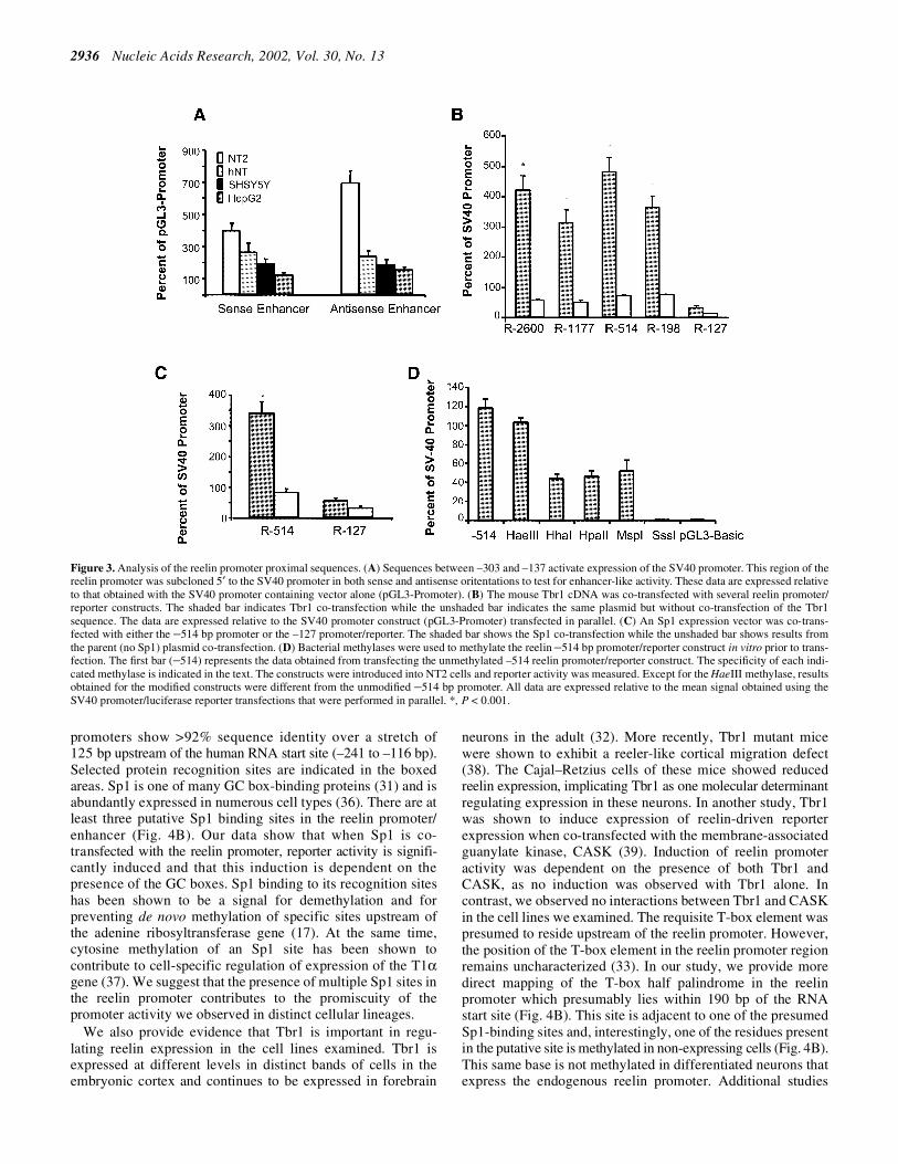
Sequences between –303 and –137 were cloned upstream ofthe SV40 promoter in both orientations to test for enhanceractivity (22). These upstream sequences increased expressionof the SV40 promoter independent of cell type (Fig. 3A).Embedded within this region are three binding sites for anSp1-like transcription factor (31) and a site for the brain-specific T-box factor Tbr1 (32,33). We suspect that the pres-ence of these three Sp1-like sites in the reelin enhancer/promoter may account for the observed promoter promis-cuity. To test whether these factors were able to transactivatethe reelin promoter, we co-transfected several reelinpromoter deletions into NT2 cells along with expressionvectors corresponding to Sp1 and Tbr1. As shown in Figure 3B,Tbr1 activates expression of the reelin promoter from 5- to8-fold. This activation depends on the presence of the putativeTbr1 recognition site, which we have positioned at between–198 and –127 bp relative to the RNA start site (Fig. 4), basedon both the transactivation data and the recognition sites ofother T-box transcription factors. We also see a modest acti-vation of the reelin promoter when we co-transfect a Sp1expression cassette (Fig. 3C). There are three Sp1-like recognitionsites located in the minimal reelin promoter (Fig. 4B) and whenthese sites are deleted, we no longer see Sp1-mediatedpromoter induction. The results imply a role for each of thesetranscription factors in modulating reelin expression (Fig. 3Band C) and provide an approximate location of their bindingsites in the reelin promoter. While the extent of induction byeach transcription factor is not extensive, NT2 cells endo-genously express both Tbr1 and Sp1, which compromises the
2934 Nucleic Acids Research, 2002, Vol. 30, No. 13
extent of transactivation (data not shown). The observationthat these cells express both of these transcription factors butonly express low levels of endogenous reelin mRNA suggests
that an additional level of control is operative in regulatingreelin promoter expression.
RA-induced differentiation of neural progenitor cells isaccompanied by changes in chromatin structure
The activity of the reelin promoter and enhancer constructs inNT2 cells was curious as expression of the endogenous tran-script is minimal. We presume that, in addition to Tbr1 andSp1, additional requisite transcription factors are present inthese cells and that the endogenous gene is silenced throughhypermethylation. As shown in Figure 2C, there are alterationsin chromatin structure in the vicinity of the reelin promoter incells that express the endogenous gene (hNT neurons) as indi-cated by the presence of three DNase I hypersensitive sites.These sites are not present in the undifferentiated NT2 neuralprogenitor cells. The highest molecular size site maps to withinthe reelin enhancer region, while the additional two sites arelocated further upstream. The data suggest that, upon neuronaldifferentiation, changes in the local chromatin structure occur,making the promoter more readily accessible to the tran-scriptional machinery. The transiently introduced promoter/reporter constructs are not confined by this restraint in thesame way and expression is not silenced. These results indicatethat an alteration in local chromatin structure likely mediatedby changes in methylation status is an important epigeneticdeterminant modulating expression of the endogenous reelingene.
Nucleotide analogs that prevent methylation and histonedeacetylase inhibitors activate reelin expression in neuralprogenitor cells
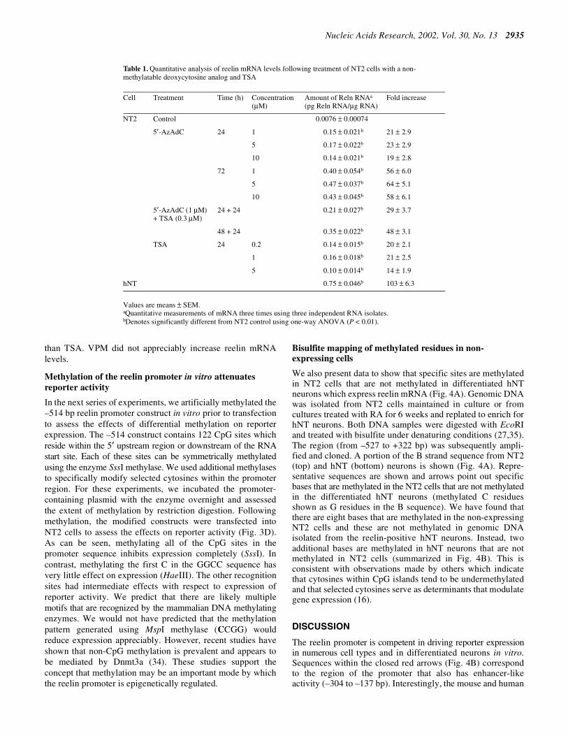
We tested whether the non-methylatable cytosine analogAzAdC would activate expression of the previously silentendogenous reelin promoter. AzAdC was incubated with NT2cells for 24 and 72 h. In parallel, NT2 cells were treated withthe histone deacetylase inhibitor TSA (0.2–5 µM) for 24 h,either alone or with AzAdC (0.2 µM TSA, 1 µM AzAdC).RNA was isolated and the amount of the reelin mRNA presentin each condition was quantified using competitive RT–PCRwith an internal standard (1,25). The quantified amounts ofreelin mRNA following each treatment are presented in Table 1 aspg reelin mRNA/µg total RNA. Incubating NT2 cells with thenucleotide analog AzAdC (1 µM) for 24 h increased reelinmRNA levels >20-fold (from 0.0076 ± 0.00074 to 0.17 ± 0.022 pgReln RNA/µg total RNA). This increased to >50-fold after 72 htreatment. The HDAC inhibitor TSA also increased expressionof the reelin mRNA some 20-fold after 24 h. Results obtainedfrom including both inhibitors appeared to be time dependentand not necessarily additive (Table 1).
More recently, the anticonvulsant and mood-stabilizing drugVPA has been reported to act as a HDAC inhibitor causinghyperacetylation of histones in culture (19,20). We tested theaction of VPA in increasing reelin expression by treating NT2cells with different concentrations of the drug for 40 h. Asshown in Table 2, VPA acted to increase reelin mRNA levels∼20-fold over this time frame in a dose-dependent manner. Inthe same experiment, cultures were also treated with a lowdose of TSA (300 nM) and with the inactive amide analogVPM. As can be seen by the data, VPA acts in a similar mannerin activating reelin expression but is considerably less potent
Figure 2. Transfection analysis of the reelin promoter. (A) Transfection of thereelin promoter/luciferase reporter constructs into various cell lines. Therestriction sites indicated in Figure 1A were used to generate the promoterfragments (–2600 contains 2.6 kb of 5′ flanking sequence; –1177, 1177 bp of 5′flanking sequence; R-732, 732 bp of 5′ flanking sequence; R-514, 514 bp of 5′flanking sequence). (B) Results obtained by transfection of various reelin promoterconstructs and deletions into either NT2 neuroprogenitor cells or hNT neurons.The number below each pair of bars indicates the amount of 5′ flankingsequence present in the construct (relative to the RNA start site). Data represent themean obtained from three measurements made from a minimum of three separateexperiments after correcting for transfection efficiency. (C) DNase I hypersen-sitive sites are present in hNT neurons and not NT2 neuroprogenitor cells. NT2cells were incubated with RA to induce differentiation as indicated in Mater-ials and Methods. Nuclei were isolated from both cell types and incubated withDNase I. The genomic DNA was resolved by agarose gel electrophoresis andSouthern blotted onto nitrocellulose membranes. The indicated probe was usedto visualize the relevant bands. Arrows indicate sites that are hypersensitive.The genomic sequence is represented in the bottom portion of the panel withthe approximate positions of the hypersensitive sites relative to the start site.The DNA probe used is indicated at the 5′-most portion of the promoter.
Nucleic Acids Research, 2002, Vol. 30, No. 13 2935
than TSA. VPM did not appreciably increase reelin mRNAlevels.
Methylation of the reelin promoter in vitro attenuatesreporter activity
In the next series of experiments, we artificially methylated the–514 bp reelin promoter construct in vitro prior to transfectionto assess the effects of differential methylation on reporterexpression. The –514 construct contains 122 CpG sites whichreside within the 5′ upstream region or downstream of the RNAstart site. Each of these sites can be symmetrically methylatedusing the enzyme SssI methylase. We used additional methylasesto specifically modify selected cytosines within the promoterregion. For these experiments, we incubated the promoter-containing plasmid with the enzyme overnight and assessedthe extent of methylation by restriction digestion. Followingmethylation, the modified constructs were transfected intoNT2 cells to assess the effects on reporter activity (Fig. 3D).As can be seen, methylating all of the CpG sites in thepromoter sequence inhibits expression completely (SssI). Incontrast, methylating the first C in the GGCC sequence hasvery little effect on expression (HaeIII). The other recognitionsites had intermediate effects with respect to expression ofreporter activity. We predict that there are likely multiplemotifs that are recognized by the mammalian DNA methylatingenzymes. We would not have predicted that the methylationpattern generated using MspI methylase (CCGG) wouldreduce expression appreciably. However, recent studies haveshown that non-CpG methylation is prevalent and appears tobe mediated by Dnmt3a (34). These studies support theconcept that methylation may be an important mode by whichthe reelin promoter is epigenetically regulated.
Bisulfite mapping of methylated residues in non-expressing cells
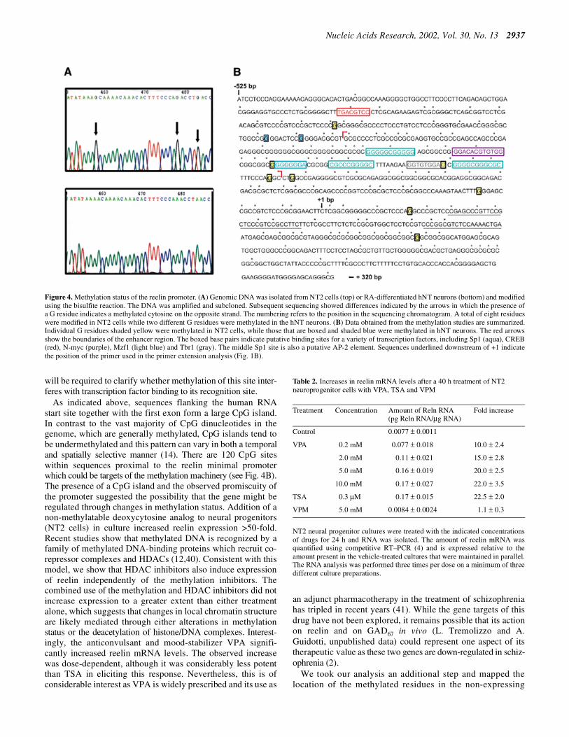
We also present data to show that specific sites are methylatedin NT2 cells that are not methylated in differentiated hNTneurons which express reelin mRNA (Fig. 4A). Genomic DNAwas isolated from NT2 cells maintained in culture or fromcultures treated with RA for 6 weeks and replated to enrich forhNT neurons. Both DNA samples were digested with EcoRIand treated with bisulfite under denaturing conditions (27,35).The region (from –527 to +322 bp) was subsequently ampli-fied and cloned. A portion of the B strand sequence from NT2(top) and hNT (bottom) neurons is shown (Fig. 4A). Repre-sentative sequences are shown and arrows point out specificbases that are methylated in the NT2 cells that are not methylatedin the differentiated hNT neurons (methylated C residuesshown as G residues in the B sequence). We have found thatthere are eight bases that are methylated in the non-expressingNT2 cells and these are not methylated in genomic DNAisolated from the reelin-positive hNT neurons. Instead, twoadditional bases are methylated in hNT neurons that are notmethylated in NT2 cells (summarized in Fig. 4B). This isconsistent with observations made by others which indicatethat cytosines within CpG islands tend to be undermethylatedand that selected cytosines serve as determinants that modulategene expression (16).
DISCUSSION
The reelin promoter is competent in driving reporter expressionin numerous cell types and in differentiated neurons in vitro.Sequences within the closed red arrows (Fig. 4B) correspondto the region of the promoter that also has enhancer-likeactivity (–304 to –137 bp). Interestingly, the mouse and human
Table 1. Quantitative analysis of reelin mRNA levels following treatment of NT2 cells with a non-methylatable deoxycytosine analog and TSA
Values are means ± SEM.aQuantitative measurements of mRNA three times using three independent RNA isolates.bDenotes significantly different from NT2 control using one-way ANOVA (P < 0.01).
Cell Treatment Time (h) Concentration(µM)
Amount of Reln RNAa
(pg Reln RNA/µg RNA)Fold increase
NT2 Control 0.0076 ± 0.00074
5′-AzAdC 24 1 0.15 ± 0.021b 21 ± 2.9
5 0.17 ± 0.022b 23 ± 2.9
10 0.14 ± 0.021b 19 ± 2.8
72 1 0.40 ± 0.054b 56 ± 6.0
5 0.47 ± 0.037b 64 ± 5.1
10 0.43 ± 0.045b 58 ± 6.1
5′-AzAdC (1 µM)+ TSA (0.3 µM)
24 + 24 0.21 ± 0.027b 29 ± 3.7
48 + 24 0.35 ± 0.022b 48 ± 3.1
TSA 24 0.2 0.14 ± 0.015b 20 ± 2.1
1 0.16 ± 0.018b 21 ± 2.5
5 0.10 ± 0.014b 14 ± 1.9
hNT 0.75 ± 0.046b 103 ± 6.3
2936 Nucleic Acids Research, 2002, Vol. 30, No. 13
promoters show >92% sequence identity over a stretch of125 bp upstream of the human RNA start site (–241 to –116 bp).Selected protein recognition sites are indicated in the boxedareas. Sp1 is one of many GC box-binding proteins (31) and isabundantly expressed in numerous cell types (36). There are atleast three putative Sp1 binding sites in the reelin promoter/enhancer (Fig. 4B). Our data show that when Sp1 is co-transfected with the reelin promoter, reporter activity is signifi-cantly induced and that this induction is dependent on thepresence of the GC boxes. Sp1 binding to its recognition siteshas been shown to be a signal for demethylation and forpreventing de novo methylation of specific sites upstream ofthe adenine ribosyltransferase gene (17). At the same time,cytosine methylation of an Sp1 site has been shown tocontribute to cell-specific regulation of expression of the T1αgene (37). We suggest that the presence of multiple Sp1 sites inthe reelin promoter contributes to the promiscuity of thepromoter activity we observed in distinct cellular lineages.
We also provide evidence that Tbr1 is important in regu-lating reelin expression in the cell lines examined. Tbr1 isexpressed at different levels in distinct bands of cells in theembryonic cortex and continues to be expressed in forebrain
neurons in the adult (32). More recently, Tbr1 mutant micewere shown to exhibit a reeler-like cortical migration defect(38). The Cajal–Retzius cells of these mice showed reducedreelin expression, implicating Tbr1 as one molecular determinantregulating expression in these neurons. In another study, Tbr1was shown to induce expression of reelin-driven reporterexpression when co-transfected with the membrane-associatedguanylate kinase, CASK (39). Induction of reelin promoteractivity was dependent on the presence of both Tbr1 andCASK, as no induction was observed with Tbr1 alone. Incontrast, we observed no interactions between Tbr1 and CASKin the cell lines we examined. The requisite T-box element waspresumed to reside upstream of the reelin promoter. However,the position of the T-box element in the reelin promoter regionremains uncharacterized (33). In our study, we provide moredirect mapping of the T-box half palindrome in the reelinpromoter which presumably lies within 190 bp of the RNAstart site (Fig. 4B). This site is adjacent to one of the presumedSp1-binding sites and, interestingly, one of the residues presentin the putative site is methylated in non-expressing cells (Fig. 4B).This same base is not methylated in differentiated neurons thatexpress the endogenous reelin promoter. Additional studies
Figure 3. Analysis of the reelin promoter proximal sequences. (A) Sequences between –303 and –137 activate expression of the SV40 promoter. This region of thereelin promoter was subcloned 5′ to the SV40 promoter in both sense and antisense oritentations to test for enhancer-like activity. These data are expressed relativeto that obtained with the SV40 promoter containing vector alone (pGL3-Promoter). (B) The mouse Tbr1 cDNA was co-transfected with several reelin promoter/reporter constructs. The shaded bar indicates Tbr1 co-transfection while the unshaded bar indicates the same plasmid but without co-transfection of the Tbr1sequence. The data are expressed relative to the SV40 promoter construct (pGL3-Promoter) transfected in parallel. (C) An Sp1 expression vector was co-trans-fected with either the –514 bp promoter or the –127 promoter/reporter. The shaded bar shows the Sp1 co-transfection while the unshaded bar shows results fromthe parent (no Sp1) plasmid co-transfection. (D) Bacterial methylases were used to methylate the reelin –514 bp promoter/reporter construct in vitro prior to trans-fection. The first bar (–514) represents the data obtained from transfecting the unmethylated –514 reelin promoter/reporter construct. The specificity of each indi-cated methylase is indicated in the text. The constructs were introduced into NT2 cells and reporter activity was measured. Except for the HaeIII methylase, resultsobtained for the modified constructs were different from the unmodified –514 bp promoter. All data are expressed relative to the mean signal obtained using theSV40 promoter/luciferase reporter transfections that were performed in parallel. *, P < 0.001.
Nucleic Acids Research, 2002, Vol. 30, No. 13 2937
will be required to clarify whether methylation of this site inter-feres with transcription factor binding to its recognition site.
As indicated above, sequences flanking the human RNAstart site together with the first exon form a large CpG island.In contrast to the vast majority of CpG dinucleotides in thegenome, which are generally methylated, CpG islands tend tobe undermethylated and this pattern can vary in both a temporaland spatially selective manner (14). There are 120 CpG siteswithin sequences proximal to the reelin minimal promoterwhich could be targets of the methylation machinery (see Fig. 4B).The presence of a CpG island and the observed promiscuity ofthe promoter suggested the possibility that the gene might beregulated through changes in methylation status. Addition of anon-methylatable deoxycytosine analog to neural progenitors(NT2 cells) in culture increased reelin expression >50-fold.Recent studies show that methylated DNA is recognized by afamily of methylated DNA-binding proteins which recruit co-repressor complexes and HDACs (12,40). Consistent with thismodel, we show that HDAC inhibitors also induce expressionof reelin independently of the methylation inhibitors. Thecombined use of the methylation and HDAC inhibitors did notincrease expression to a greater extent than either treatmentalone, which suggests that changes in local chromatin structureare likely mediated through either alterations in methylationstatus or the deacetylation of histone/DNA complexes. Interest-ingly, the anticonvulsant and mood-stabilizer VPA signifi-cantly increased reelin mRNA levels. The observed increasewas dose-dependent, although it was considerably less potentthan TSA in eliciting this response. Nevertheless, this is ofconsiderable interest as VPA is widely prescribed and its use as
an adjunct pharmacotherapy in the treatment of schizophreniahas tripled in recent years (41). While the gene targets of thisdrug have not been explored, it remains possible that its actionon reelin and on GAD67 in vivo (L. Tremolizzo and A.Guidotti, unpublished data) could represent one aspect of itstherapeutic value as these two genes are down-regulated in schiz-ophrenia (2).
We took our analysis an additional step and mapped thelocation of the methylated residues in the non-expressing
Figure 4. Methylation status of the reelin promoter. (A) Genomic DNA was isolated from NT2 cells (top) or RA-differentiated hNT neurons (bottom) and modifiedusing the bisulfite reaction. The DNA was amplified and subcloned. Subsequent sequencing showed differences indicated by the arrows in which the presence ofa G residue indicates a methylated cytosine on the opposite strand. The numbering refers to the position in the sequencing chromatogram. A total of eight residueswere modified in NT2 cells while two different G residues were methylated in the hNT neurons. (B) Data obtained from the methylation studies are summarized.Individual G residues shaded yellow were methylated in NT2 cells, while those that are boxed and shaded blue were methylated in hNT neurons. The red arrowsshow the boundaries of the enhancer region. The boxed base pairs indicate putative binding sites for a variety of transcription factors, including Sp1 (aqua), CREB(red), N-myc (purple), Mzf1 (light blue) and Tbr1 (gray). The middle Sp1 site is also a putative AP-2 element. Sequences underlined downstream of +1 indicatethe position of the primer used in the primer extension analysis (Fig. 1B).
Table 2. Increases in reelin mRNA levels after a 40 h treatment of NT2neuroprogenitor cells with VPA, TSA and VPM
NT2 neural progenitor cultures were treated with the indicated concentrationsof drugs for 24 h and RNA was isolated. The amount of reelin mRNA wasquantified using competitive RT–PCR (4) and is expressed relative to theamount present in the vehicle-treated cultures that were maintained in parallel.The RNA analysis was performed three times per dose on a minimum of threedifferent culture preparations.
Treatment Concentration Amount of Reln RNA(pg Reln RNA/µg RNA)
Fold increase
Control 0.0077 ± 0.0011
VPA 0.2 mM 0.077 ± 0.018 10.0 ± 2.4
2.0 mM 0.11 ± 0.021 15.0 ± 2.8
5.0 mM 0.16 ± 0.019 20.0 ± 2.5
10.0 mM 0.17 ± 0.027 22.0 ± 3.5
TSA 0.3 µM 0.17 ± 0.015 22.5 ± 2.0
VPM 5.0 mM 0.0084 ± 0.0024 1.1 ± 0.3

2938 Nucleic Acids Research, 2002, Vol. 30, No. 13
NT2 cells (yellow filled box, Fig. 4B). The sites methylatedin RA-induced hNT neurons are also shown (blue filled box).The importance of those experiments that show that inhibitionof methylation activates reelin expression is underscored bythe change in the methylation profile of the promoter thataccompanies these cells as they differentiate in culture. Astissue-specific and developmental expression patterns areassociated with distinct alterations in chromatin structure andDNA methylation status (42), we suspect that methylation ofcritical residues within these sequences affects the ability oftranscription factors to access these sites. While these datawere obtained using a cell culture system, we suggest thatmethylation of the CpG island represents an epigenetic switchthat is used to silence reelin promoter expression underappropriate conditions. The recent characterization of a DNAdemethylase activity supports a model in which the pattern ofmethylation is maintained by a balance of methylation anddemethylation activities and the local state of histone acetylation(43). Taken together with observations that high levels ofDnmt1 protein are expressed in post-mitotic neurons (44), onemight speculate that methylation is a dynamic process used byneurons to regulate gene expression epigenetically. Recentliterature suggests that the pattern of DNA methylation andlocal histone deacetylation states may be coordinated,establishing a relevant link between the action of thesecomplementary regulatory processes.
The role that methylation plays in regulating gene expressionin the nervous system is still underexplored. Mutations thatoccur in methyl-CpG-binding proteins have drastic conse-quences that occur post-natally. Rett syndrome is a neuro-developmental disorder that results in mental retardation due tomutations in the methyl-CpG-binding protein MECP2 (45). Itwas first noted some 40 years ago that the administration ofL-methionine to schizophrenia patients elicited a profoundexacerbation of symptoms in 60–70% of patients (recentlyreviewed in 46). This exacerbation could not be explained byan increased accumulation of metabolites of catecholaminesand has remained a mystery ever since. We would like tosuggest that the mechanism of the methionine-inducedpsychosis could be related to increased levels of brain S-adenosyl-methionine and subsequent alterations in the methylationstatus of selective promoters, including that corresponding toreelin. In support of this argument, we have preliminary datathat show that the chronic injection of L-methionine to hetero-zygous reeler mice twice daily for 15 days further reduces thelevels of reelin and reelin mRNA (46). These data areconsistent with the hypothesis that the reelin promoter can bemodulated through modifications in methylation and that arelationship may exist between the recrudescence of psychoticsymptoms and the hypermethylation of sites within the reelinCpG island.
While the data presented here are based on experimentsperformed in cells maintained in culture, we anticipate thatadditional studies will provide insight into methylation andreelin promoter regulation in vivo. Ultimately, we are inter-ested in assessing whether the reelin promoter is functionallycompromised in patients diagnosed with psychosis (3). Itseems plausible that alterations in reelin expression may be theconsequence of inappropriate methylation that leads to alteredchromatin structure and silenced expression (46). While we arepresently attempting to substantiate our hypotheses with
direct experiments, we would like to infer that the variablesymptomatology associated with the schizophrenia spectrumof disorders might be the consequence of inappropriate methy-lation patterns resulting from the dysregulation of this funda-mental regulatory mechanism. Interestingly, an analysis ofgenomic DNA from monozygotic twins discordant for schizo-phrenia showed discrepancies in the methylation patterns ofcertain genes, suggesting a possible epigenetic mechanism fordifferences between monozygotic twins (47). Current thinkingalso suggests that schizophrenia is a polygenic disorder.Biologically inappropriate expression patterns of multiplegenes might result from changes that occur in the methylationstatus of specific promoters expressed in the nervous system,providing a potential clue to the polygenic nature of thedisorder.
SUPPLEMENTARY MATERIAL
Supplementary Material is available at NAR Online.
ACKNOWLEDGEMENTS
We would like to thank Mr David Gavin for his excellenttechnical assistance, Dr Genoveva Davidkova for providingreelin internal standard cRNA and Dr Jai Sung Noh for helpwith figure preparation. We would like to acknowledgeDr Robert Tjian (UC, Berkeley, CA) for providing the Sp1expression vector and Dr Morgan Sheng (MassachusettsGeneral Hospital and Harvard University, Cambridge, MA) forthe Tbr1 expression vector. Finally, we would like to acknow-ledge Katwijk Chemie B.V. for the gift of valpromide that wasused in these studies. This work was supported by5R01MH062090-02 to E.C. and in part by 1R01MH062682-01A2to D.R.G.
REFERENCES
1. Impagnatiello,F., Guidotti,A.R., Pesold,C., Dwivedi,Y., Caruncho,H.,Pisu,M.G., Uzunov,D.P., Smalheiser,N.R., Davis,J.M., Pandey,G.N.,Pappas,G.D., Tueting,P., Sharma,R.P. and Costa,E. (1998) A decrease ofreelin expression as a putative vulnerability factor in schizophrenia.Proc. Natl Acad. Sci. USA, 95, 15718–15723.
2. Costa,E., Davis,J., Grayson,D.R., Guidotti,A., Pappas,G.D. and Pesold,C.(2001) Dendritic spine hypoplasia and downregulation of reelin andGABAergic tone in schizophrenia vulnerability. Neurobiol. Dis., 8,723–742.
3. Guidotti,A., Auta,J., Davis,J., Dwivedi,Y., Grayson,D.R.,Impagnatiello,F., Pandey,G., Pesold,C., Sharma,R., Uzunov,D. andCosta,E. (2000) Reelin and GAD67 expression is decreased in postmortembrain of schizophrenia and bipolar disorder patients. Arch. Gen.Psychiatr., 57, 1061–1069.
4. Selemon,L.D. and Goldman-Rakic,P.S. (1999) The reduced neuropilhypothesis: a circuit based model of schizophrenia. Biol. Psychiatr., 45,17–25.
5. Fatemi,S.H., Earle,J.A. and McMenomy,T. (2000) Reduction in reelinimmunoreactivity in hippocampus of subjects with schizophrenia, bipolardisorder and major depression. Mol. Psychiatr., 5, 654–663.
6. Rice,D.S. and Curran,T. (2001) Role of the reelin signaling pathway incentral nervous system development. Annu. Rev. Neurosci., 24,1005–1039.
7. Alcantara,S., Ruiz,M., D’Arcangelo,G., Ezan,F., de Lecea,L., Curran,T.,Sotelo,C. and Soriano,E. (1998) Regional and cellular patterns of reelinmRNA expression in the forebrain of the developing and adult mouse.J. Neurosci., 18, 7779–7799.
8. Pesold,C., Impagnatiello,F., Pisu,M.G., Uzunov,D.P., Costa,E.,Guidotti,A. and Caruncho,H.J. (1998) Reelin is preferentially expressed in
Nucleic Acids Research, 2002, Vol. 30, No. 13 2939
neurons synthesizing γ-aminobutyric acid in cortex and hippocampus ofadult rats. Proc. Natl Acad. Sci. USA, 95, 3221–3226.
9. Rodriguez,M.A., Pesold,C., Liu,W.S., Kriho,V., Guidotti,A., Pappas,G.D.and Costa,E. (2000) Colocalization of integrin receptors and reelin indendritic spine postsynaptic densities of adult nonhuman primate cortex.Proc. Natl Acad. Sci. USA, 97, 3550–3553.
10. DeSilva,U., D’Arcangelo,G., Braden,V.V., Chen,J., Miao,G.G., Curran,T.and Green,E.D. (1997) The human reelin gene: isolation, sequencing, andmapping on chromosome 7. Genome Res., 7, 157–164.
11. Royaux,I., Lambert de Rouvroit,C., D’Arcangelo,G., Demirov,D. andGoffinet,A.M. (1997) Genomic organization of the mouse reelin gene.Genomics, 46, 240–250.
12. Bird,A.P. and Wolffe,A.P. (1999) Methylation-induced repression—belts,braces and chromatin. Cell, 9, 451–454.
13. Lu,R., Au,W.C., Yeow,W.S., Hageman,N. and Pitha,P.M. (2000)Regulation of the promoter activity of interferon regulatory-7 gene.Activation by interferon and silencing by hypermethylation.J. Biol. Chem., 275, 31805–31812.
14. Newell-Price,J., Clark,A.J. and King,P. (2000) DNA methylation andsilencing of gene expression. Trends Endocrinol. Metab., 11, 142–148.
15. Tucker,K. (2001) Methylated cytosine and the brain: a new base forneuroscience. Neuron, 30, 649–652.
16. Costello,J.F. and Plass,C. (2001) Methylation matters. J. Med. Genet., 38,285–303.
17. Han,L., Lin,I.G. and Hsieh,C.L. (2001) Protein binding protects sites onstable episomes and in the chromosome from de novo methylation.Mol. Cell. Biol., 21, 3416–3424.
18. MacLeod,D., Charlton,J., Mullins,J. and Bird,A. (1994) Sp1 sites in themouse aprt gene promoter are required to prevent methylation of the CpGisland. Genes Dev., 8, 2282–2292.
19. Phiel,C.J., Zhang,F., Huang,E.Y., Guenther,M.G., Lazar,M.A. andKlein,P.S. (2001) Histone deacetylase is a direct target of valproic acid, apotent anticonvulsant, mood stabilizer, and teratogen. J. Biol. Chem., 276,36734–36741.
20. Gottlicher,M., Minucci,S., Zhu,P., Kramer,O.H., Schimpf,A., Giavara,S.,Sleeman,J.P., Lo Coco,F., Nervi,C., Pelicci,P.G. and Heinzel,T. (2001)Valproic acid defines a novel class of HDAC inhibitors inducingdifferentiation of transformed cells. EMBO J., 20, 6969–6978.
21. Szekely,A.M., Costa,E. and Grayson,D.R. (1990) Transcriptionalprogram coordination by NMDA-sensitive glutamate receptor stimulationin primary cultures of cerebellar neurons. Mol. Pharmacol., 38, 624–633.
22. Grayson,D.R., Costa,R.H., Xanthopoulos,K.G. and Darnell,J.E.,Jr (1988)A cell-specific enhancer of the mouse α1-antitrypsin gene has multiplefunctional regions and corresponding protein binding sites. Mol. Cell.Biol., 8, 1055–1066.
23. Sinha,S., Degenstein,L., Copenhaver,C. and Fuchs,E. (2000) Defining theregulatory factors required for epidermal gene expression. Mol. Cell.Biol., 20, 2543–2555.
24. Bovolin,P., Santi,M.R., Memo,M., Costa,E. and Grayson,D.R. (1992)Distinct developmental patterns of expression of the rat α1, α5, γ2S andγ2L GABAA receptor subunit mRNAs in vivo and in vitro. J. Neurochem.,59, 62–72.
25. Grayson,D.R. and Ikonomovic,S. (1998) Competitive RT-PCR toquantitate steady state mRNA levels. In Boulton,A.A., Baker,G.B. andBateson,A. (eds), In Vitro Neurochemical Techniques: Neuromethods.Humana Press, Totowa, NJ, Vol. 34, pp. 127–151.
26. Zuccotti,M. and Monk,M. (1995) Methylation of the mouse Xist gene insperm and eggs correlates with imprinted Xist expression and paternalX-inactivation. Nature Genet., 9, 316–320.
27. Clark,S.J., Harrison,J., Paul,C.L. and Frommer,M. (1994) High sensitivitymapping of methylated cytosines. Nucleic Acids Res., 22, 2990–2997.
28. Gardiner-Garden,M. and Frommer,M. (1987) CpG islands in vertebrategenomes. J. Mol. Biol., 196, 261–282.
29. Grayson,D.R., Uzunov,D.P., Chen,Y., Siyanova,E., Raca,G.,Sharma,R.P., Mirkin,S. and Costa,E. (2000) Studies of molecularmechanisms regulating reelin gene expression. Biol. Psychiatr., 47, 41S.
30. Persico,A., D’Agruma,L., Maiorano,N., Totaro,A., Militerni,R.,Bravaccio,C., Wassink,T., Schneider,C., Melmed,R., Trillo,S.,Montecchi,F., Palermo,M., Pascucci,T., Puglisi-Allegra,S., Reichelt,K.-L.,Conciatori,M., Marino,R., Quattrocchi,C., Baldi,A., Zelante,L.,Gasparini,P. and Keller,F. (2001) Reelin gene alleles and haplotypes as afactor predisposing to autistic disorder. Mol. Psychiatr., 6, 150–159.
31. Suske,G. (1999) The Sp-family of transcription factors. Gene, 238,291–300.
32. Bulfone,A., Smiga,S.M., Shimamura,K., Peterson,A., Puelles,L. andRubenstein,J.L.R. (1995) T-brain-1: a homolog of Brachyury whoseexpression defines molecularly distinct domains within the cerebralcortex. Neuron, 15, 63–78.
33. Tada,M. and Smith,J.C. (2001) T-targets: clues to understanding thefunctions of T-box proteins. Dev. Growth Differ., 43, 1–11.
34. Ramsahoye,B.H., Biniszkiewicz,D., Lyko,F., Clark,V., Bird,A.P. andJaenisch,R. (2000) Non-CpG methylation is prevalent in embryonic stemcells and may be mediated by DNA methyltransferase 3a. Proc. NatlAcad. Sci. USA, 97, 5237–5242.
35. Frommer,M., McDonald,L.E., Millar,D.S., Collis,C.M., Watt,F.,Grigg,G.W., Molloy,P.L. and Paul,C.L. (1992) A genomic sequencingprotocol that yields a positive display of 5-methylcytosine residues inindividual DNA strands. Proc. Natl Acad. Sci. USA, 89, 1827–1831.
36. Saffer,J.D., Jackson,S.P. and Annarella,M.B. (1991) Developmentalexpression of Sp1 in the mouse. Mol. Cell. Biol., 11, 2189–2199.
37. Cao,Y.X., Jean,J.C. and Williams,M.C. (2000) Cytosine methylation ofan Sp1 site contributes to organ-specific and cell-specific regulation ofexpression of the lung epithelial gene T1α. Biochem. J., 350, 883–890.
38. Hevner,R.F., Shi,L., Justice,N., Hsueh,Y.P., Sheng,M., Smiga,S.,Bulfone,A., Goffinet,A.M., Campagnoni,A.T. and Rubenstein,J.L.R.(2001) Tbr1 regulated differentiation of the preplate and layer 6.Neuron, 29, 353–366.
39. Hsueh,Y.P., Wang,T.F., Yang,F.C. and Sheng,M. (2000) Nucleartranslocation and transcription regulation by the membrane-associatedguanylate kinase CASK/LIN-2. Nature, 404, 298–302.
40. Szyf,M. (2001) Towards a pharmacology of DNA methylation.Trends Pharmacol. Sci., 22, 350–354.
41. Citrome,L., Levine,J. and Allingham,B. (2000) Changes in use ofvalproate and other mood stabilizers for patients with schizophrenia from1994 to 1998. Psychiatr. Serv., 51, 634–638.
42. Razin,A. (1998) CpG methylation, chromatin structure and genesilencing—a three way connection. EMBO J., 17, 4905–4908.
43. Cervoni,N. and Szyf,M. (2001) Demethylase activity is directed byhistone acetylation. J. Biol. Chem., 276, 40778–40787.
44. Inano,K., Suetake,I., Ueda,T., Miyake,Y., Nakamura,M., Okada,M. andTajima,S. (2000) Maintenance-type DNA methyltransferase is highlyexpressed in post-mitotic neurons and located in the cytoplasmiccompartment. J. Biochem., 128, 315–321.
45. Amir,R.E., Van den Veyver,I.B., Wan,M., Tran,C.Q., Francke,U. andZoghbi,H.Y. (1999) Rett syndrome is caused by mutations in X-linkedMECP2, encoding methyl-CpG-binding protein 2. Nature Genet., 23,185–188.
46. Costa,E., Chen,Y., Davis,J., Dong,E., Noh,J.S., Tremolizzo,L., Veldic,M.,Grayson,D.R. and Guidotti,A. (2002) Reelin and schizophrenia: a diseaseat the interface of the genome and epigenome. Mol. Intervent., 2, 47–57.
47. Tsujita,T., Niikawa,N., Yamashita,H., Imamura,A., Hamada,A.,Nakane,Y. and Okazaki,Y. (1998) Genomic discordance betweenmonozygotic twins discordant for schizophrenia. Am. J. Psychiatry, 155,422–424.