Quality of insertion in cochlear implants - Archives-Ouvertes.fr
Identification of a Single-Nucleotide Insertion in thePromoter Region Affecting the sodC Promoter Activity inBrucella neotomaeDina A. Moustafa1, Neeta Jain2, Nammalwar Sriranganathan2, Ramesh Vemulapalli1*
1 Department of Comparative Pathobiology, Purdue University, West Lafayette, Indiana, United States of America, 2 Department of Biomedical Sciences and Pathobiology,
Virginia Tech, Blacksburg, Virginia, United States of America
Abstract
Brucella neotomae is not known to be associated with clinical disease in any host species. Previous research suggested thatB. neotomae might not express detectable levels of Cu/Zn superoxide dismutase (SOD), a periplasmic enzyme known to beinvolved in protecting Brucella from oxidative bactericidal effects of host phagocytes. This study was undertaken toinvestigate the genetic basis for the disparity in SOD expression in B. neotomae. Our Western blot and SOD enzyme assayanalyses indicated that B. neotomae does express SOD, but at a substantially reduced level. Nucleotide sequence analysis ofregion upstream to the sodC gene identified a single-nucleotide insertion in the potential promoter region. The same single-nucleotide insertion was also detected in the sodC promoter of B. suis strain Thomsen, belonging to biovar 2 in which SODexpression was undetectable previously. Examination of the sodC promoter activities using translational fusion constructswith E. coli b-galactosidase demonstrated that the B. neotomae and B. suis biovar 2 promoters were very weak in drivinggene expression. Site-directed mutation studies indicated that the insertion of A in the B. neotomae sodC promoter reducedthe promoter activity. Increasing the level of SOD expression in B. neotomae through complementation with B. abortus sodCgene did not alter the bacterial survival in J774A.1 macrophage-like cells and in tissues of BALB/c and C57BL/6 mice. Theseresults for the first time demonstrate the occurrence of a single-nucleotide polymorphism affecting promoter function andgene expression in Brucella.
Citation: Moustafa DA, Jain N, Sriranganathan N, Vemulapalli R (2010) Identification of a Single-Nucleotide Insertion in the Promoter Region Affecting the sodCPromoter Activity in Brucella neotomae. PLoS ONE 5(11): e14112. doi:10.1371/journal.pone.0014112
Editor: Paulo Lee Ho, Instituto Butantan, Brazil
Received May 29, 2010; Accepted November 3, 2010; Published November 24, 2010
Copyright: � 2010 Moustafa et al. This is an open-access article distributed under the terms of the Creative Commons Attribution License, which permitsunrestricted use, distribution, and reproduction in any medium, provided the original author and source are credited.
Funding: Dina Moustafa’s PhD program was supported by a fellowship from Egyptian Cultural and Educational Bureau. The funders had no role in study design,data collection and analysis, decision to publish, or preparation of the manuscript.
Competing Interests: The authors have declared that no competing interests exist.
* E-mail: [email protected]
Introduction
The genus Brucella consists of small, non-motile, non-spore
forming, gram-negative, facultatively intracellular bacteria capable
of infecting a variety of mammals. Infection with Brucella in animals
leads to reproductive failure with abortions and infertility in females,
and in some cases, orchitis in males [1,2]. Brucellosis is a zoonotic
disease, and humans usually acquire the infection by consuming
contaminated dairy or meat products or by coming in contact with
the infected animal tissues and secretions [1]. There are six well-
recognized species of Brucella that show a marked host preference –
B. abortus (cattle), B. melitensis (goat and sheep), B. suis (pig), B. canis
(dog), B. ovis (sheep, especially ram), and B. neotomae (wood rat). In
addition, isolates of B. abortus, B. melitensis, and B. suis are subdivided
into biovars (or biotypes) based on the differences in their biological
characteristics. Interestingly, B. suis biovar 2 is known to be
incapable of causing significant clinical disease in pigs as well as in
humans [3]. In the past few years, Brucella has been recovered from
several marine mammals, including cetaceans (dolphin, whale and
porpoise) and pinnipeds (seals and otters); these marine isolates
belong to two potential new species, B. pinnipedialis and B.ceti [4].
Very recently, a new species of Brucella, B. microti, was isolated from
wild common voles suffering from a systemic disease [5,6].
The virulence of Brucella can be attributed to the ability of these
bacteria to escape the host defense mechanisms and survive and
replicate within the host cells. Virulent Brucella organisms are
capable of invading and replicating in professional phagocytes,
such as macrophages, as well as several types of non-phagocytic
cells [7]. The ability of Brucella to maintain long-term residence in
macrophages is the basis for establishing and maintaining chronic
infections, a hallmark of brucellosis. Adaptation to intracellular
survival within macrophages necessitates Brucella to resist the
bactericidal action of reactive oxygen intermediates generated by
the host cells. One of the mechanisms some bacteria employ to
resist the oxidative killing by host macrophages is the production
of periplasmic Cu/Zn-cofactored superoxide dismutase (SOD)
enzyme that catalyzes the dismutation of superoxide (O22) to
hydrogen peroxide [8], which can be subsequently detoxified by
the enzymatic action of catalases and peroxidases. In Brucella spp.,
SOD is a periplasmic protein that is encoded by the sodC gene
[9,10]. Studies with B. abortus indicated that SOD plays a
significant role in setup and maintenance of persistent infections
in murine brucellosis models [11,12].
B. neotomae was first isolated from pooled tissue suspensions of
lung, spleen, liver and kidney of desert wood rats collected in the
Great Salk Lake Desert in Utah [13]. In contrast to other Brucella
PLoS ONE | www.plosone.org 1 November 2010 | Volume 5 | Issue 11 | e14112
spp., there is no documented evidence indicating the pathogenic
potential of B. neotomae in any host species. Though not much
further research has been done on B. neotomae, it is generally
considered to be a less virulent Brucella species. Limited animal
studies conducted immediately after the initial isolation indicated
that B. neotomae can infect mice, pigs, guinea pigs, and wood rats
but does not cause any apparent clinical disease [13–15]. Perhaps
because of its restricted host range and/or low virulence
characteristics, B. neotomae has never been isolated from any other
animal species. No disease in human has been attributed to B.
neotomae.
Bricker et al. (1990) previously demonstrated that, of all the
classical Brucella species and biovars examined, only B. neotomae and
a B. suis biovar 2 isolate could not express detectable levels of
SOD. The goal of this study was to determine the genetic basis for
the lack of detectable SOD expression in B. neotomae. In this paper,
we demonstrate that B. neotomae does express SOD, but at a
substantially reduced level. Our studies discovered the presence of
a single-nucleotide insertion in the promoter region of sodC gene as
the cause for the reduced activity of this promoter in B. neotomae
and B. suis biovar 2. Furthermore, we demonstrate that increasing
the level of SOD expression in B. neotomae through complemen-
tation does not alter its persistence profile in mouse tissues.
Materials and Methods
Ethics statementSome experiments described in this manuscript were performed
in mice. The animal experiments were approved by Purdue
Animal Care and Use Committee (PACUC No. 01-076-07).
Bacterial strainsB. neotomae strain 5K33 was purchased from American Type
Culture Collection. B. abortus strain RB51 was available in our
laboratory. B. abortus virulent strain 2308 was from culture
collection at Virginia Tech. E. coli DH5a was purchased from
Invitrogen. All of the bacteria were grown in tryptic soy broth
(TSB) or tryptic soy agar (TSA) at 37uC. Bacteria containing
plasmids were grown in presence of appropriate antibiotics at 30-
or 100-mg/ml concentrations of chloramphenicol and ampicillin,
respectively. Colony forming units (CFU) of B. neotomae containing
the recombinant plasmids were determined by plating the 10-fold
serial dilutions of the cultures on TSA with and without
appropriate antibiotics, to ascertain the stable maintenance of
the plasmids in the recombinant bacteria.
Complementation of B. neotomae with functional sodCgene
A 1.1 kb fragment containing the B. abortus sodC gene and its
promoter sequence was excised from pBS/SOD [16] with ClaI
restriction enzyme digestion and subcloned into pBBR4MCS to
generate pBB4SOD. B. neotomae was transformed with pBB4SOD
by electroporation following the previously described procedures
[17]. B. neotomae containing the plasmid pBB4SOD (B. neotomae/
pBB4SOD) was selected by plating the transformed bacteria on
TSA plates containing ampicillin. The expression of SOD in the
complemented B. neotomae was determined by SDS-PAGE and
Western blotting.
SDS-PAGE and Western blottingFor determining the expression of SOD in B. neotomae and in the
complemented strain, SDS-PAGE and Western blot analyses were
performed as described previously [18,19]. The antigen extract
prepared from B. abortus strain RB51 was used for the positive
control for SOD expression. The amount of antigen loaded in
each lane of the gel corresponded to total antigens of 107 or 109
CFU of Brucella harvested during late log phase of growth curve.
For Western blotting, goat anti-B. abortus SOD sera was used as the
primary antibody [20]. The membranes were developed with
rabbit anti-goat IgG conjugated with horseradish peroxidase and a
colorimetric substrate (KPL Inc., Gaithersburg, Maryland).
Periplasmic extracts and SOD enzyme assaySelective release of periplasmic contents of B. neotomae, B.
neotomae/pBB4SOD, and B. abortus RB51 was performed as per the
previously described procedure [21]. Briefly, bacteria harvested
during different growth phases were pelleted down by centrifuga-
tion and resuspended in Tris-Hcl buffer, pH 7.5, at 100 ml per
10 mg of pellet wet weight. An equal volume of 0.2M Tris, pH7.5,
containing 1M sucrose and 0.5% zwittergent 316 (Calbiochem)
was added. Lysozyme was added to the suspension to achieve a
final concentration of 100 mg/ml. Bacterial cells were exposed to a
mild osmotic shock by adding an equal volume of distilled water.
The suspensions were shaken for 2 hours at room temperature
and then centrifuged at 8,0006g for 30 min and the supernatants
were used for measuring SOD enzyme activity. The protein
concentration of the supernatant was determined using a Bio-Rad
protein assay kit.
Quantitative determination of SOD activity was achieved by a
colorimetric microtiter plate method using a commercially
available SOD assay kit (Dojindo Molecular Technologies, Inc.).
SOD activity was assayed at 450 nm as the inhibition of the
reduction of the tetrazolium salt 2-(4-iodophenyl) 3-4-(nitrophe-
nyl)-5(2,4-disulfophenyl)-2H tetrazolium (WST-1) by superoxide
produced by xanthine oxidase. Commercially available E. coli
SOD (Sigma-Aldrich) was used to construct standard curves.
Enzyme activity, defined as percent inhibition of WST-1
reduction, was determined as {[(reagent control vo2buffer blank
vo)2(sample vo2sample blank vo)]/(reagent control vo2buffer blank
vo)}6100 for both samples and standards. SOD activity per well
was determined from the standard curve. With each sample, the
SOD assay was performed twice, each time in duplicates, and the
data were reported as specific activity (SOD units/mg of protein).
PCR amplification and nucleotide sequence analysisThe coding sequences and the upstream region of the sodC gene
from the genomic DNA of B. neotomae, B. suis biovars 2 (strain
Thomsen) and 4 (strain 40) were amplified via PCR using specific
primer-pairs - SOD-RBS-F (59-GGGAATGGCCTTACGGTT-
39) and SOD-RBS-R (59-TTATTCGATCACGCCGCA-39) for
coding region, and SOD-upstream-F (59-CGCAGCCACCCG-
TTCATGTT-39) and SOD-upstream-R (59-GTCGGCAGCGC-
CTCATAC-39) for upstream region. The genomic DNA of B. suis
strains was previously obtained from National Animal Disease
Center, Ames, Iowa [22]. The genomic DNA of B. neotomae and B.
abortus RB51 was extracted using DNeasy Tissue kit (Qiagen)
following the manufacturer’s recommended procedure for DNA
isolation from bacteria. Twenty ng of template DNA,10 pmol of
each of the specific primer-pairs and reagents from NovaTaq PCR
Kit PLUS (Novagen) were used to prepare PCR mixtures in a total
volume of 50 ml. The PCR amplifications were performed using a
thermocycler (iCyler, Bio-Rad Laboratories) with the following
conditions: 95uC for 4 min followed by 30 cycles that each
included 30 sec of denaturation at 95uC, 30 sec of annealing at
58uC, and 30 sec of extension at 72uC. The amplified PCR
products were cloned into the pCR2.1 vector, using the TA
cloning system (Invitrogen). The nucleotide sequences of both
strands of the cloned fragments from 3 independent clones were
B. neotomae sodC Promoter
PLoS ONE | www.plosone.org 2 November 2010 | Volume 5 | Issue 11 | e14112
determined by DNA sequencing using M13 forward and reverse
primers at Purdue Genomics Core Facility. The nucleotide
sequences were analyzed using LaserGene sequence analysis
software (DNASTAR, Inc.). The B. neotomae nucleotide sequences
were submitted to databases at GenBank (Accession number
EU056817).
RNA extraction and RT-PCRTotal RNA was extracted from B. neotomae and B. abortus RB51
using RNeasy Mini Kit (Qiagen) according to the manufacturer’s
protocol for DNA-free RNA isolation from bacteria. Bacteria
harvested during late log phase of growth curve were used for the
RNA extraction. The extracted RNA concentration was deter-
mined using Quant-iT RiboGreen RNA reagent and kit
(Molecular Probes). Using SuperScriptTM One-step RT-PCR kit
(Invitrogen), RT-PCR to detect sodC mRNA was performed with
40 ng of the extracted RNA and primers SOD-RBS-F and SOD-
upstream-R. cDNA synthesis and PCR amplification were
performed in the same tube with the following thermocycler
conditions: cDNA synthesis at 45uC for 30 min, inactivation of
reverse-transcriptase enzyme at 95uC for 10 min, amplification of
cDNA sequences with 40 cycles of 95uC for 1 min, 58uC for
1 min, and 72uC for 1 min. As a control to rule out DNA
contamination of the extracted RNA, a set of reaction tubes were
subjected to direct PCR amplification by omitting the reverse
transcription step (45uC for 30 min) of above mentioned
thermocycler conditions. Following the PCR amplification, 12 ml
of the reaction mixtures were separated on a 2% agarose gel
electrophoresis and stained with ethidium bromide to visualize the
amplified products.
59 RACE reactionAmplification of the sodC cDNA 59 end was performed using the
59 RACE System Version 2.0 for rapid amplification of cDNA ends
kit (Invitrogen) as per the manufacturer’s suggested protocol.
Briefly, the first strand cDNA was synthesized from the bacterial
RNA using the gene specific primer 1 (GSP1) SOD-Pext-R (59-
GGGCTTCGGAAATGACCACG-39) and SuperScript II under
the following conditions: denaturation of the RNA at 70uC for
10 min, followed by cDNA synthesis at 42uC for 50 min. The
template RNA was then removed by treating the reaction mixture
with RNase mix. Unincorporated dNTPs and GSP1 were separated
from the cDNA by SNAP column purification. A homopolymeric
tail was added to the 39 end of the cDNA using terminal
deoxynucleotidyl transferase (TdT) and dCTP at 37uC for
10 min. Following inactivation of TdT at 65uC for 10 min, the
tailed cDNA was PCR amplified using gene specific primer 2
(GSP2), SOD-upstream R, and abridged-anchor primer with the
following thermocycler conditions: initial denaturation at 94uC for
1 min, 35 cycles of 94uC for 1 min, 55uC for 1 min, and 72uC for
1 min, and a final extension step at 72uC for 7 min. In order to
generate enough specific product, a nested PCR amplification was
performed using GSP2 and abridged universal anchor primer using
the previous thermocycler conditions. The PCR products were
separated by electrophoresis on a 2% agarose gel, eluted using a
QIAEX II gel extraction kit (Qiagen) and cloned into pGEM-T
vector (Promega). The nucleotide sequences of the cloned fragments
were determined by DNA sequencing using M13 reverse primer.
Construction of plasmids for determining the promoterstrength
The strength of sodC promoters from B. neotomae, B. abortus, and
B. suis biovars 2 and 4 was determined by generating translational
fusion with E. coli b-galactosidase and then measuring the
enzymatic activity of the expressed b-galactosidase in B. neotomae
transformed with the plasmids. For this, a 138 bp fragment
previously determined to contain the sodC promoter region, the
ribosomal binding site and the start codon [16] was amplified from
the genomic DNA of corresponding Brucella via PCR using a
specifically designed primer-pair, SOD-pro-F (59- AAGTCGA-
CATCGATAATTTCGGGGTGGAGA-39) and SOD-pro-R (59-
AACTGCAGATCATCACTTCTCCTGAATATAGTTAGA-39).
A restriction site was added at the 59 end of the each primer (SalI
in SOD-pro-F and PstI in SOD-pro-R) to facilitate directional
cloning into plasmid pNSGroE/lacZ [23]. The amplified DNA
fragments were first cloned into pCR2.1 vector and sequenced to
ascertain the integrity of the nucleotide sequences. The inserts were
then excised from the pCR2.1 vector by digesting with SalI and PstI
restriction enzymes and cloned into plasmid pNSGroE/lacZ
digested with the same enzymes, resulting in the replacement of
the groE promoter with the cloned sodC promoter. In the resulting
recombinant plasmids, the start codon of the sodC gene was in-frame
with the coding sequences of the lacZ gene, which was again
confirmed by nucleotide sequencing using the SOD-pro-F primer.
The resulting plasmids were designated pBnSODpro/lacZ, pBa-
SODpro/lacZ, pBs2SODpro/lacZ, and pBs4SODpro/lacZ. B.
neotomae was transformed with these plasmids and the expression of
b-galactosidase in the recombinant bacteria was measured.
Site-directed mutagenesisTo alter specific nucleotide(s) in the predicted promoter region
of B. neotomae sodC gene, PCR amplification was performed with a
common reverse primer (SOD-pro-R) and one of the three
forward primers, B.n-F-m1 (59- TTTAGCAATTGCTGTAAC-
TATTTGTTTTAATACAGTA-39), B.n-F-m2 (59-TTTAGCAA-
TTGCTGTAACTAATTTTTTTAATACAGTA-39), or B.n-F-
2m1 (59- TTTAGCAATTGCTGTAACTATTTTTTTTAATA-
CAGTA-39) containing the specific mutations. Similarly, two
single-nucleotide mutations were generated in the potential
promoter region of B. suis biovar 4 sodC gene using the forward
primers B.s4-F-m1 (59- TTTAGCAATTGCTGTAACTAATGT-
TTTAATACAGTA-39) and B.s4-F-m2 (59-TTTAGCAATTG-
CTGTAACTATTGGTTTAATACAGTA-39). The 59 ends of
the forward primers with the specific mutations encompass the
sequence containing the site for MfeI restriction enzyme. The
amplified 110 bp DNA product was digested with MfeI and PstI
restriction enzymes and cloned into plasmid pBnSODpro/lacZ
previously digested with the same enzymes, resulting in the
replacement of the wild type sequences with that containing
specific mutated nucleotide(s) which was confirmed by nucleotide
sequencing. The resulting plasmids were designated pBnSODm1/
lacZ, pBnSODm2/lacZ, pBnSOD2m1/lacZ, pBs4SODm1/lacZ
and pBs4SODm2/lacZ. B. neotomae was transformed with these
plasmids and the expression of b-galactosidase in the recombinant
bacteria was measured. B. abortus virulent strain 2308 and vaccine
strain RB51 were also transformed with plasmids containing the B.
neotomae sodC promoter variants and the b-galactosidase expression
in the corresponding recombinant bacteria was measured.
b-Galactosidase enzyme assayb-Galactosidase was assayed in B. neotomae, B. abortus 2308 and
B. abortus RB51 by the methods described previously [24]. The
enzyme activity was calculated in Miller units with the following
formula: (OD42061000/t6v6OD600), where the OD420 is the
optical density at 420 nm, t is the incubation time in minutes, v is
the volume of culture used in milliliters, and OD600 is the optical
density of the bacterial culture used for enzyme assay. The assays
B. neotomae sodC Promoter
PLoS ONE | www.plosone.org 3 November 2010 | Volume 5 | Issue 11 | e14112
were performed in triplicate with bacteria grown from 3 separate
colonies of each strain.
Survival and replication of B. neotomae and B. neotomae/pBB4SOD in J744A.1 cells
The intracellular growth characteristics of B. neotomae and B.
neotomae complemented with the functional sodC gene (B. neotomae
harboring pBB4SOD) was determined in J774A.1 macrophage-like
cells using the method previously described [25]. Briefly, J774A.1
cells were cultured overnight in 6-well plates with antibiotic-free
medium and infected with 108 CFU of B. neotomae or B. neotomae/
pBB4SOD as described above for intracellular b-galactosidase
expression analysis. For each bacterial strain, the assay was
performed in triplicate by infecting cells in 3 wells. At different
time-points, the infected monolayers were washed three times with
antibiotic free media, and the cells were lysed using 0.1%
deoxychloate solution. The CFU of bacteria released from the cells
were determined by plating 10-fold serial dilution of the lysates on
TSA; for B. neotomae/pBB4SOD infected cells, the CFU were also
determined by plating the diluted lysates on TSA with ampicillin.
Mice ExperimentsThe in vivo growth characteristics of B. neotomae and B. neotomae
complemented with pBB4SOD were determined in mice. Four-
week old, female BALB/c and C57BL/6 mice were purchased
from Harlan. Mice were given 1 week of rest before the
experiments were started. Each strain of mice were divided into
two groups, and mice in one group were injected intraperitoneally
each with 36106 CFU of B. neotomae and mice in the other group
were similarly injected with 1.86106 CFU of B. neotomae harboring
pBB4SOD. The actual CFU injected was determined retrospec-
tively by plating the inoculum on TSA plates. At specific time-
points after infection, three mice from each group were
euthanized, their spleens and livers were removed and the
numbers of CFU were determined by plating 10-fold serial
dilutions of tissue homogenates on TSA, and also on TSA with
ampicillin in case of B.neotomae/pBB4SOD inoculated mice.
Statistical analysesOne-way ANOVA with Tukey post test was performed using
GraphPad Prism (GraphPad Software, San Diengo, CA, USA) to
analyze the enzymatic activities of SOD and b-galactosidase.
Student’s t test was used to analyze the intracellular bacterial
counts and the bacterial CFU in the spleens of mice.
Results
Expression of SOD in B. neotomaeLow levels of SOD expression was detected in B. neotomae strain
5K33 by Western blot analysis using specific polyclonal antibodies.
As shown in Fig. 1A and B, no SOD was detected in the total
antigen extract from 107 CFU of B. neotomae, while considerable
amount of SOD was present in the antigen extracts from the same
number of B. neotomae containing plasmid pBB4SOD. When the
Western blot analysis was performed using the total antigen extracts
obtained from 109 CFU of B. neotomae, a thin protein band with size
corresponding to the SOD consistently reacted with the SOD-
specific antibodies (Fig. 1C and D). As expected, SOD was present
in the antigen extracts of both 107 and 109 CFU of B. abortus RB51.
SOD activity in periplasmic extractsAs shown in Fig. 2, SOD activity was detected in B. neotomae, B.
neotomae/pBB4SOD and B. abortus RB51 at all time-points tested
during the 48-hour culture period. The maximum enzyme activity
was detected in B. neotomae during late log phase (24 hours) and
stationary phase (48 hours). However, at all time-points tested, the
SOD specific activity in B. neotomae was lower than that of B. abortus
RB51 and B. neotomae/pBB4SOD.
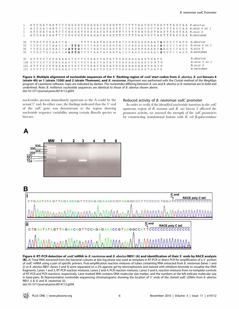
Sequence analysis of B. neotomae sodC geneIn order to verify if the low level of SOD expression in B.
neotomae is because of mutations within the coding or the upstream
regions of sodC gene, we first performed PCR amplification using
B. neotomae genomic DNA as template and a primer-pair designed
based on the known sodC gene sequence from B. abortus. The PCR
amplification resulted in a 575 bp product, the expected size in the
presence of a complete sodC gene. Nucleotide sequence of the
amplified product showed 99.7% identity with the corresponding
region from B. suis and 99.5% identity with that from B. abortus or
B. melitensis. The B. neotomae sequence differed in 2 and 3
nucleotides from that of B. suis and B. abortus or B. melitensis,
respectively. All the nucleotide differences were located within the
open reading frame of the sodC gene and only one of the differed
nucleotides caused a change in the deduced amino acid sequence,
a FRV change at amino acid position 52 of the precursor
polypeptide (data not shown). We then amplified a 350 bp region
flanking the 59 end of the start codon from the B. neotomae DNA.
Nucleotide sequence analysis of this region revealed that B.
neotomae differed in 2 nucleotides from that of the B. suis strain 1330
(biovar 1) sequence (GenBank accession no. NC_009504), one was
a substitution of A with G and the other was an insertion of A
(Fig. 3). Both the changes were within the 138 bp region upstream
to the start codon that was previously shown to contain the sodC
promoter element in B. abortus [16]. We also PCR amplified and
sequenced the upstream region from strains belonging to B. suis
biovars 2 (strain Thomsen) and 4 (strain 40). While the B. suis
biovar 4 sequence was 100% identical with that of B. suis biovar 1,
the sequence from B. suis biovar 2 differed in just 1 nucleotide, an
insertion of A at the same location as that found in the B. neotomae
sequence. Computer analysis of nucleotide sequences reported in
the databases indicated 100% identity within the sodC upstream
region between B. abortus and B. melitensis, and compared to B. suis
biovars 1 and 4, these two bacteria contained 3 fewer nucleotides
at the location where the nucleotide insertion was detected in B.
neotomae and B. suis biovar 2 (Fig. 3). In addition, the nucleotide 48
upstream to the start codon was G in B. abortus and B. melitensis, but
it was A in B. suis.
Determining the 59 end of sodC mRNART-PCR amplification resulted in specific amplification of
150 bp cDNA product from RNA extracted from both B. neotomae
and B. abortus RB51 (Fig. 4A). No amplified products were
detected when reverse-transcription step was omitted, ruling out
the genomic DNA contamination as the source for the specific
amplification obtained with RT-PCR (Fig. 4A). Nested PCR
products of 59 RACE reaction from both B. neotomae and B. abortus
RB51 contained a ,150 bp DNA fragment. The PCR products
were cloned in pGEM-T vector and recombinant plasmids from
6 independent colonies for each Brucella species were used for
sequencing the cloned products. For B. abortus RB51, two of the
recombinant plasmids identified ‘A’ at 61 nucleotides upstream to
the start codon as the 59 end of the cDNA (Fig. 4Bi). In all 6 of
the B. neotomae and the remain 4 of the B. abortus RB51
recombinant plasmids, ‘A’ at 51 nucleotides upstream to the start
codon was found to be the 59 end of the cDNA (Fig. 4Bii);
however, because of the C-tailing of the RACE reaction, we
cannot rule out the possibility of anyone of the five ‘G’
B. neotomae sodC Promoter
PLoS ONE | www.plosone.org 4 November 2010 | Volume 5 | Issue 11 | e14112
Figure 1. Detection of SOD expression in B. neotomae, B. neotomae/pBB4SOD, and B. abortus RB51 by SDS-PAGE (A and C) andWestern blot analysis (B and D). Total antigens of 107 (A and B) or 109 (C and D) CFU of the indicated bacteria were loaded on each lane. The SDS-PAGE gels were stained with Coomassie brilliant blue G (A and C). For Western blot analysis (B and D), goat anti-Brucella SOD serum was used as theprimary antibody for reacting with the antigens. In all panels, lanes marked MW contain molecular weight markers and the numbers at the leftindicate approximate molecular masses in kilodaltons. Arrows in panels A and C indicate the SOD protein.doi:10.1371/journal.pone.0014112.g001
Figure 2. Specific activity of SOD enzyme in the periplasmic extracts of B. neotomae, B. neotomae/pBB4SOD, and B. abortus RB51 atdifferent growth stages. The growth curves of the bacteria are shown in the top panel. The specific activities of SOD enzyme are shown in thebottom panel. For each Brucella, overnight culture was used at 1:200 dilution to inoculate fresh 100 ml media and at different time intervals, theOD600 was measured and an aliquot of the culture was taken for preparing periplasmic extracts. With extracts of each time-point, the SOD assay wasperformed twice, each time in duplicates, and the results are shown as the mean 6 standard deviation of specific activity (units/mg of protein). Ateach time point, the groups with one or two asterisks were significantly different from the B. neotomae group (P,0.001). At 4 and 48 hours time-points, there were significant differences between the groups with different number of asterisks (P,0.001).doi:10.1371/journal.pone.0014112.g002
B. neotomae sodC Promoter
PLoS ONE | www.plosone.org 5 November 2010 | Volume 5 | Issue 11 | e14112
nucleotides present immediately upstream to the A could be the
actual 59 end. In either case, the findings indicated that the 59 end
of the sodC gene was downstream to the region showing
nucleotide sequence variability among certain Brucella species or
biovars.
Reduced activity of B. neotomae sodC promoterIn order to verify if the identified nucleotide insertion in the sodC
upstream region of B. neotomae and B. suis biovar 2 affected the
promoter activity, we assessed the strength of the sodC promoters
by constructing translational fusions with E. coli b-galactosidase
Figure 3. Multiple alignment of nucleotide sequences of the 59 flanking region of sodC start codon from B. abortus, B. suis biovars 4(strain 40) or 1 (strain 1330) and 2 (strain Thomsen), and B. neotomae. Alignment was performed with the Clustal method of the MegAlignprogram of LaserGene software. Gaps are indicated by dashes. The nucleotides differing between B. suis and B. abortus or B. neotomae are in bold andunderlined. Note: B. melitensis nucleotide sequences are identical to those of B. abortus shown above.doi:10.1371/journal.pone.0014112.g003
Figure 4. RT-PCR detection of sodC mRNA in B. neotomae and B. abortus RB51 (A) and identification of their 59 ends by RACE analysis(B). A) Total RNA extracted from the bacterial cultures at late log phase was used as template in RT-PCR or direct PCR for amplification of a 59 portionof sodC mRNA using a pair of specific primers. Post-amplification reaction mixtures of tubes containing RNA extracted from B. neotomae (lanes 1 and2) or B. abortus RB51 (lanes 3 and 4) were separated on a 2% agarose gel by electrophoresis and stained with ethidium bromide to visualize the DNAfragments. Lanes 1 and 3, RT-PCR reaction mixtures. Lanes 2 and 4, PCR reaction mixtures. Lanes 5 and 6, reaction mixtures from no-template-controlsof RT-PCR and PCR reactions, respectively. Lane marked MW contains DNA molecular size marker, and the numbers at the left indicate molecular sizein base-pairs. B) Representative nucleotide sequencing chromatograms showing the location of 59 ends of the cloned sodC cDNAs from B. abortusRB51 (i & ii) and B. neotomae (ii).doi:10.1371/journal.pone.0014112.g004
B. neotomae sodC Promoter
PLoS ONE | www.plosone.org 6 November 2010 | Volume 5 | Issue 11 | e14112
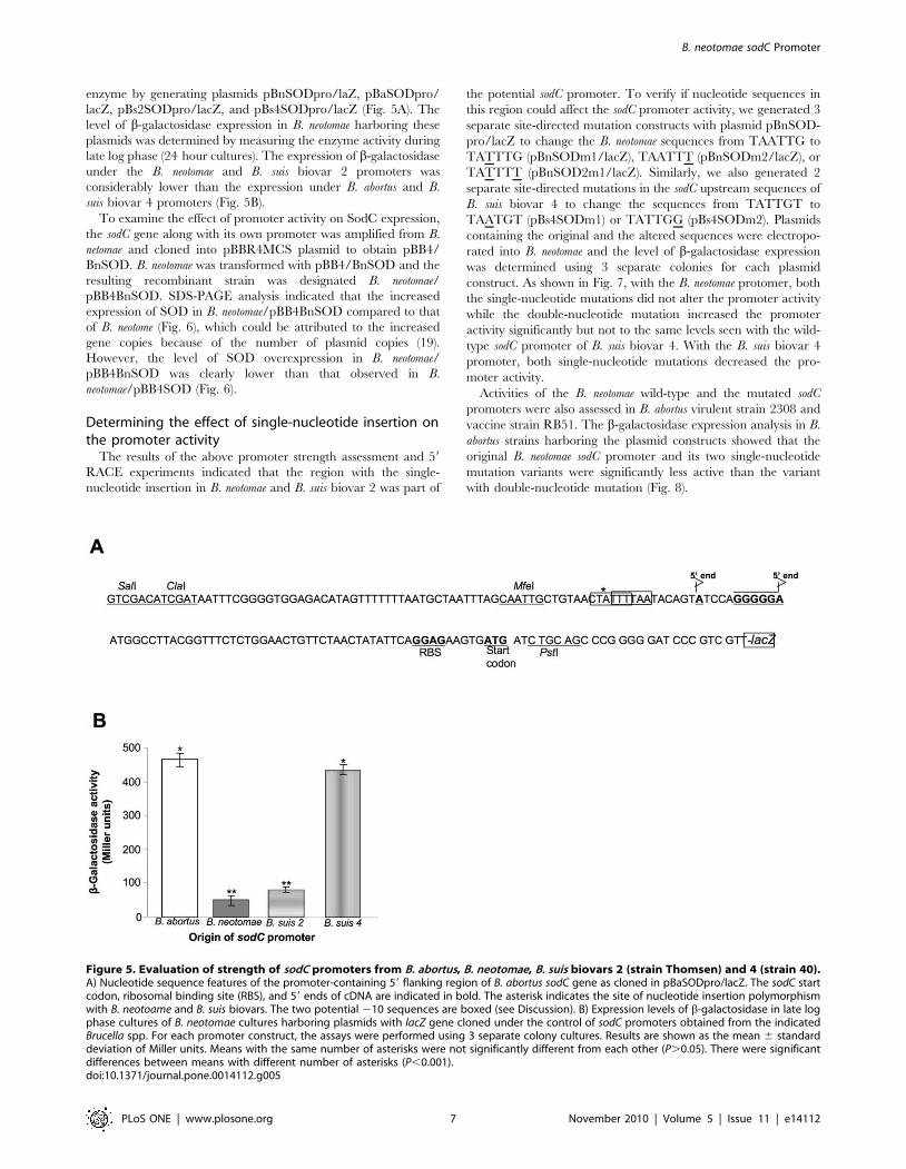
enzyme by generating plasmids pBnSODpro/laZ, pBaSODpro/
lacZ, pBs2SODpro/lacZ, and pBs4SODpro/lacZ (Fig. 5A). The
level of b-galactosidase expression in B. neotomae harboring these
plasmids was determined by measuring the enzyme activity during
late log phase (24 hour cultures). The expression of b-galactosidase
under the B. neotomae and B. suis biovar 2 promoters was
considerably lower than the expression under B. abortus and B.
suis biovar 4 promoters (Fig. 5B).
To examine the effect of promoter activity on SodC expression,
the sodC gene along with its own promoter was amplified from B.
netomae and cloned into pBBR4MCS plasmid to obtain pBB4/
BnSOD. B. neotomae was transformed with pBB4/BnSOD and the
resulting recombinant strain was designated B. neotomae/
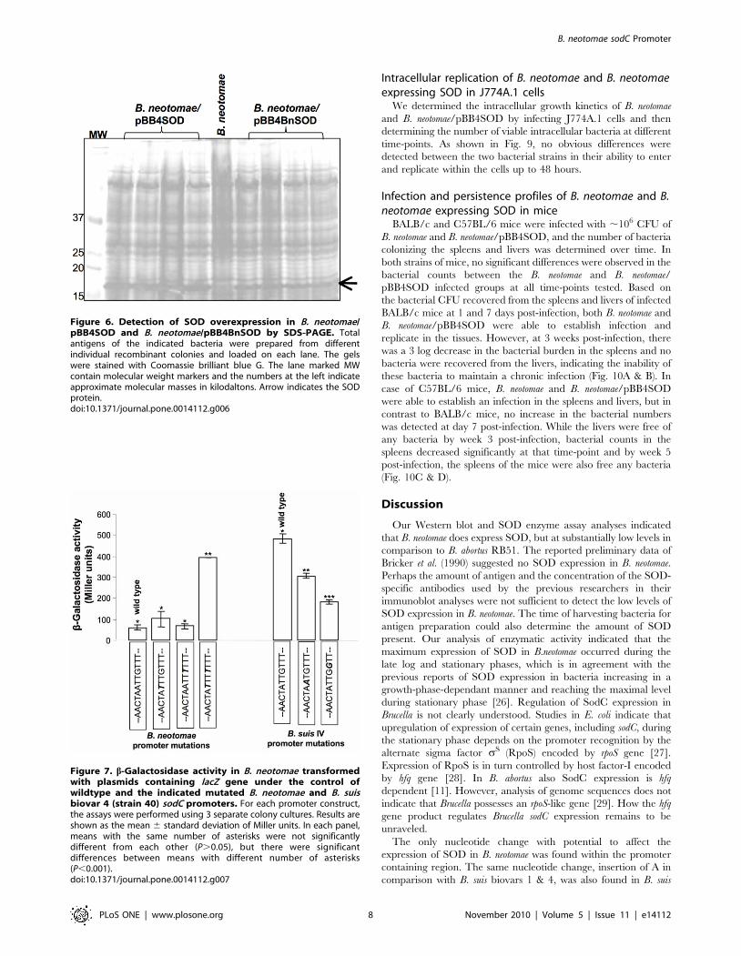
pBB4BnSOD. SDS-PAGE analysis indicated that the increased
expression of SOD in B. neotomae/pBB4BnSOD compared to that
of B. neotome (Fig. 6), which could be attributed to the increased
gene copies because of the number of plasmid copies (19).
However, the level of SOD overexpression in B. neotomae/
pBB4BnSOD was clearly lower than that observed in B.
neotomae/pBB4SOD (Fig. 6).
Determining the effect of single-nucleotide insertion onthe promoter activity
The results of the above promoter strength assessment and 59
RACE experiments indicated that the region with the single-
nucleotide insertion in B. neotomae and B. suis biovar 2 was part of
the potential sodC promoter. To verify if nucleotide sequences in
this region could affect the sodC promoter activity, we generated 3
separate site-directed mutation constructs with plasmid pBnSOD-
pro/lacZ to change the B. neotomae sequences from TAATTG to
TATTTG (pBnSODm1/lacZ), TAATTT (pBnSODm2/lacZ), or
TATTTT (pBnSOD2m1/lacZ). Similarly, we also generated 2
separate site-directed mutations in the sodC upstream sequences of
B. suis biovar 4 to change the sequences from TATTGT to
TAATGT (pBs4SODm1) or TATTGG (pBs4SODm2). Plasmids
containing the original and the altered sequences were electropo-
rated into B. neotomae and the level of b-galactosidase expression
was determined using 3 separate colonies for each plasmid
construct. As shown in Fig. 7, with the B. neotomae protomer, both
the single-nucleotide mutations did not alter the promoter activity
while the double-nucleotide mutation increased the promoter
activity significantly but not to the same levels seen with the wild-
type sodC promoter of B. suis biovar 4. With the B. suis biovar 4
promoter, both single-nucleotide mutations decreased the pro-
moter activity.
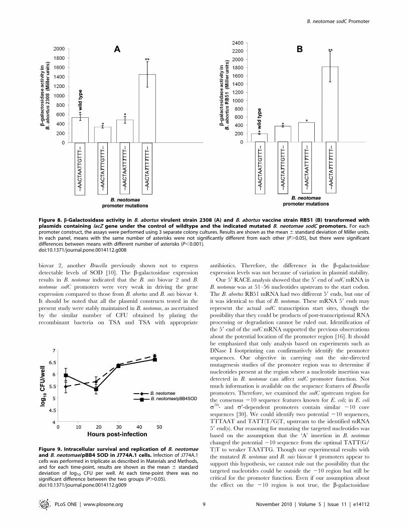
Activities of the B. neotomae wild-type and the mutated sodC
promoters were also assessed in B. abortus virulent strain 2308 and
vaccine strain RB51. The b-galactosidase expression analysis in B.
abortus strains harboring the plasmid constructs showed that the
original B. neotomae sodC promoter and its two single-nucleotide
mutation variants were significantly less active than the variant
with double-nucleotide mutation (Fig. 8).
Figure 5. Evaluation of strength of sodC promoters from B. abortus, B. neotomae, B. suis biovars 2 (strain Thomsen) and 4 (strain 40).A) Nucleotide sequence features of the promoter-containing 59 flanking region of B. abortus sodC gene as cloned in pBaSODpro/lacZ. The sodC startcodon, ribosomal binding site (RBS), and 59 ends of cDNA are indicated in bold. The asterisk indicates the site of nucleotide insertion polymorphismwith B. neotoame and B. suis biovars. The two potential 210 sequences are boxed (see Discussion). B) Expression levels of b-galactosidase in late logphase cultures of B. neotomae cultures harboring plasmids with lacZ gene cloned under the control of sodC promoters obtained from the indicatedBrucella spp. For each promoter construct, the assays were performed using 3 separate colony cultures. Results are shown as the mean 6 standarddeviation of Miller units. Means with the same number of asterisks were not significantly different from each other (P.0.05). There were significantdifferences between means with different number of asterisks (P,0.001).doi:10.1371/journal.pone.0014112.g005
B. neotomae sodC Promoter
PLoS ONE | www.plosone.org 7 November 2010 | Volume 5 | Issue 11 | e14112
Intracellular replication of B. neotomae and B. neotomaeexpressing SOD in J774A.1 cells
We determined the intracellular growth kinetics of B. neotomae
and B. neotomae/pBB4SOD by infecting J774A.1 cells and then
determining the number of viable intracellular bacteria at different
time-points. As shown in Fig. 9, no obvious differences were
detected between the two bacterial strains in their ability to enter
and replicate within the cells up to 48 hours.
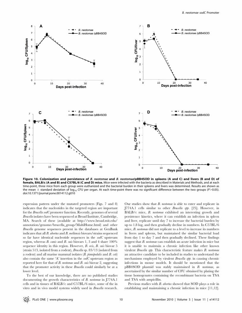
Infection and persistence profiles of B. neotomae and B.neotomae expressing SOD in mice
BALB/c and C57BL/6 mice were infected with ,106 CFU of
B. neotomae and B. neotomae/pBB4SOD, and the number of bacteria
colonizing the spleens and livers was determined over time. In
both strains of mice, no significant differences were observed in the
bacterial counts between the B. neotomae and B. neotomae/
pBB4SOD infected groups at all time-points tested. Based on
the bacterial CFU recovered from the spleens and livers of infected
BALB/c mice at 1 and 7 days post-infection, both B. neotomae and
B. neotomae/pBB4SOD were able to establish infection and
replicate in the tissues. However, at 3 weeks post-infection, there
was a 3 log decrease in the bacterial burden in the spleens and no
bacteria were recovered from the livers, indicating the inability of
these bacteria to maintain a chronic infection (Fig. 10A & B). In
case of C57BL/6 mice, B. neotomae and B. neotomae/pBB4SOD
were able to establish an infection in the spleens and livers, but in
contrast to BALB/c mice, no increase in the bacterial numbers
was detected at day 7 post-infection. While the livers were free of
any bacteria by week 3 post-infection, bacterial counts in the
spleens decreased significantly at that time-point and by week 5
post-infection, the spleens of the mice were also free any bacteria
(Fig. 10C & D).
Discussion
Our Western blot and SOD enzyme assay analyses indicated
that B. neotomae does express SOD, but at substantially low levels in
comparison to B. abortus RB51. The reported preliminary data of
Bricker et al. (1990) suggested no SOD expression in B. neotomae.
Perhaps the amount of antigen and the concentration of the SOD-
specific antibodies used by the previous researchers in their
immunoblot analyses were not sufficient to detect the low levels of
SOD expression in B. neotomae. The time of harvesting bacteria for
antigen preparation could also determine the amount of SOD
present. Our analysis of enzymatic activity indicated that the
maximum expression of SOD in B.neotomae occurred during the
late log and stationary phases, which is in agreement with the
previous reports of SOD expression in bacteria increasing in a
growth-phase-dependant manner and reaching the maximal level
during stationary phase [26]. Regulation of SodC expression in
Brucella is not clearly understood. Studies in E. coli indicate that
upregulation of expression of certain genes, including sodC, during
the stationary phase depends on the promoter recognition by the
alternate sigma factor sS (RpoS) encoded by rpoS gene [27].
Expression of RpoS is in turn controlled by host factor-I encoded
by hfq gene [28]. In B. abortus also SodC expression is hfq
dependent [11]. However, analysis of genome sequences does not
indicate that Brucella possesses an rpoS-like gene [29]. How the hfq
gene product regulates Brucella sodC expression remains to be
unraveled.
The only nucleotide change with potential to affect the
expression of SOD in B. neotomae was found within the promoter
containing region. The same nucleotide change, insertion of A in
comparison with B. suis biovars 1 & 4, was also found in B. suis
Figure 6. Detection of SOD overexpression in B. neotomae/pBB4SOD and B. neotomae/pBB4BnSOD by SDS-PAGE. Totalantigens of the indicated bacteria were prepared from differentindividual recombinant colonies and loaded on each lane. The gelswere stained with Coomassie brilliant blue G. The lane marked MWcontain molecular weight markers and the numbers at the left indicateapproximate molecular masses in kilodaltons. Arrow indicates the SODprotein.doi:10.1371/journal.pone.0014112.g006
Figure 7. b-Galactosidase activity in B. neotomae transformedwith plasmids containing lacZ gene under the control ofwildtype and the indicated mutated B. neotomae and B. suisbiovar 4 (strain 40) sodC promoters. For each promoter construct,the assays were performed using 3 separate colony cultures. Results areshown as the mean 6 standard deviation of Miller units. In each panel,means with the same number of asterisks were not significantlydifferent from each other (P.0.05), but there were significantdifferences between means with different number of asterisks(P,0.001).doi:10.1371/journal.pone.0014112.g007
B. neotomae sodC Promoter
PLoS ONE | www.plosone.org 8 November 2010 | Volume 5 | Issue 11 | e14112
biovar 2, another Brucella previously shown not to express
detectable levels of SOD [10]. The b-galactosidase expression
results in B. neotomae indicated that the B. suis biovar 2 and B.
neotomae sodC promoters were very weak in driving the gene
expression compared to those from B. abortus and B. suis biovar 4.
It should be noted that all the plasmid constructs tested in the
present study were stably maintained in B. neotomae, as ascertained
by the similar number of CFU obtained by plating the
recombinant bacteria on TSA and TSA with appropriate
antibiotics. Therefore, the difference in the b-galactosidase
expression levels was not because of variation in plasmid stability.
Our 59 RACE analysis showed that the 59 end of sodC mRNA in
B. neotomae was at 51–56 nucleotides upstream to the start codon.
The B. abortus RB51 mRNA had two different 59 ends, but one of
it was identical to that of B. neotomae. These mRNA 59 ends may
represent the actual sodC transcription start sites, though the
possibility that they could be products of post-transcriptional RNA
processing or degradation cannot be ruled out. Identification of
the 59 end of the sodC mRNA supported the previous observations
about the potential location of the promoter region [16]. It should
be emphasized that only analysis based on experiments such as
DNase I footprinting can confirmatively identify the promoter
sequences. Our objective in carrying out the site-directed
mutagenesis studies of the promoter region was to determine if
nucleotides present at the region where a nucleotide insertion was
detected in B. neotomae can affect sodC promoter function. Not
much information is available on the sequence features of Brucella
promoters. Therefore, we examined the sodC upstream region for
the consensus 210 sequence features known for E. coli; in E. coli
s70- and ss-dependent promoters contain similar 210 core
sequences [30]. We could identify two potential 210 sequences,
TTTAAT and TATT(T/G)T, upstream to the identified mRNA
59 end(s). Our reasoning for mutating the targeted nucleotides was
based on the assumption that the ‘A’ insertion in B. neotomae
changed the potential 210 sequence from the optimal TATT(G/
T)T to weaker TAATTG. Though our experimental results with
the mutated B. neotomae and B. suis biovar 4 promoters appear to
support this hypothesis, we cannot rule out the possibility that the
targeted nucleotides could be outside the 210 region but still be
critical for the promoter function. Even if our assumption about
the effect on the 210 region is not true, the b-galactosidase
Figure 8. b-Galactosidase activity in B. abortus virulent strain 2308 (A) and B. abortus vaccine strain RB51 (B) transformed withplasmids containing lacZ gene under the control of wildtype and the indicated mutated B. neotomae sodC promoters. For eachpromoter construct, the assays were performed using 3 separate colony cultures. Results are shown as the mean 6 standard deviation of Miller units.In each panel, means with the same number of asterisks were not significantly different from each other (P.0.05), but there were significantdifferences between means with different number of asterisks (P,0.001).doi:10.1371/journal.pone.0014112.g008
Figure 9. Intracellular survival and replication of B. neotomaeand B. neotomae/pBB4 SOD in J774A.1 cells. Infection of J774A.1cells was performed in triplicate as described in Materials and Methods,and for each time-point, results are shown as the mean 6 standarddeviation of log10 CFU per well. At each time-point there was nosignificant difference between the two groups (P.0.05).doi:10.1371/journal.pone.0014112.g009
B. neotomae sodC Promoter
PLoS ONE | www.plosone.org 9 November 2010 | Volume 5 | Issue 11 | e14112
expression pattern under the mutated promoters (Figs. 7 and 8)
indicates that the nucleotides in the targeted region are important
for the Brucella sodC promoter function. Recently, genomes of several
Brucella isolates have been sequenced at Broad Institute, Cambridge,
MA. Search of these (available at http://www.broad.mit.edu/
annotation/genome/brucella_group/MultiHome.html) and other
Brucella genome sequences present in the databases at GenBank
indicates that all B. abortus and B. melitensis biovars/strains sequenced
so far have identical nucleotide sequences in the sodC upstream
region, whereas B. canis and B. suis biovars 1, 3 and 4 share 100%
sequence identity in this region. However, B. ovis, B. suis biovar 5
(strain 513, isolated from a rodent), Brucella sp. 83/13 (isolated from
a rodent) and all marine mammal isolates (B. pinnipedalis and B. ceti)
also contain the same ‘A’ insertion in the sodC upstream region as
reported here for that of B. neotomae and B. suis biovar 2, suggesting
that the promoter activity in these Brucella could similarly be at a
lower level.
To the best of our knowledge, there are no published studies
documenting the growth characteristics of B. neotomae in J774A.1
cells and in tissues of BALB/c and C57BL/6 mice, some of the in
vitro and in vivo model systems widely used in Brucella research.
Our studies show that B. neotomae is able to enter and replicate in
J774A.1 cells similar to other Brucella spp. [25]. However, in
BALB/c mice, B. neotomae exhibited an interesting growth and
persistence kinetics, where it can establish an infection in spleen
and liver, replicate until day 7 to increase the bacterial burden by
up to 1.8 log, and then gradually decline in numbers. In C57BL/6
mice, B. neotomae did not replicate to a level to increase its numbers
in livers and spleens, but maintained the similar bacterial load
from day 1 to day 7 and then gradually declined. These findings
suggest that B. neotomae can establish an acute infection in mice but
it is unable to maintain a chronic infection like other known
virulent Brucella spp. This characteristic feature makes B. neotomae
an attractive candidate to be included in studies to understand the
mechanisms employed by virulent Brucella spp. in causing chronic
infections in mouse models. It should be mentioned that the
pBB4SOD plasmid was stably maintained in B. neotomae, as
ascertained by the similar number of CFU obtained by plating the
tissue homogenates containing the recombinant bacteria on TSA
and TSA with ampicillin.
Previous studies with B. abortus showed that SOD plays a role in
establishing and maintaining a chronic infection in mice [11,12].
Figure 10. Colonization and persistence of B. neotomae and B. neotomae/pBB4SOD in spleens (A and C) and livers (B and D) offemale, BALB/c (A and B) and C57BL/6 (C and D) mice. Mice were infected with the bacteria as described in Materials and Methods, and at eachtime-point, three mice from each group were euthanized and the bacterial burden in their spleens and livers was determined. Results are shown asthe mean 6 standard deviation of log10 CFU per organ. At each time-point there was no significant difference between the two groups (P.0.05).doi:10.1371/journal.pone.0014112.g010
B. neotomae sodC Promoter
PLoS ONE | www.plosone.org 10 November 2010 | Volume 5 | Issue 11 | e14112
However, if reduction in the level of SOD expression by Brucella
spp. affects their ability to cause chronic infection is not known.
Increasing the level of SOD expression in B. neotomae did not alter
the bacterial survival in BALB/c and C57BL/6 mice (Fig. 10). It is
possible that the low level of SOD expression in B. neotomae is
sufficient to inactivate the host-derived superoxide radicals and
any additional increase in the amount of SOD has no functional
effect. It is also possible that increasing the level of SOD expression
per se is insufficient to compensate for other critical genetic
deficiencies of B. neotomae affecting its ability to maintain a chronic
infection.
In conclusion, we have identified the presence of a single-
nucleotide insertion in the promoter region as the cause for the
reduced activity of sodC promoter of B. neotomae. Though promoter
nucleotide polymorphism has been shown to modulate gene
expression in other bacteria [31,32], this is the first report
demonstrating the occurrence of a single-nucleotide difference
affecting promoter function and gene expression in Brucella spp.
This finding highlights the possibility of single nucleotide
polymorphisms in promoter regions contributing to the differences
in expression of certain genes among Brucella species/biovars/
strains.
Author Contributions
Conceived and designed the experiments: DM RV. Performed the
experiments: DM NJ NS. Analyzed the data: DM NJ NS RV. Contributed
reagents/materials/analysis tools: DM NS RV. Wrote the paper: DM RV.
References
1. Corbel MJ (1997) Brucellosis: An overview. Emerg Infect Dis 3: 213–221.
2. Cutler SJ, Whatmore AM, Commander NJ (2005) Brucellosis - new aspects of an
old disease. J Appl Microbiol 98: 1270–1281.
3. Godfroid J, Cloeckaert A, Liautard JP, Kohler S, Fretin D, et al. (2005) From the
discovery of the Malta fever’s agent to the discovery of a marine mammal
reservoir, brucellosis has continuously been a re-emerging zoonosis. Vet Res 36:
313–326.
4. Foster G, Osterman BS, Godfroid J, Jacques I, Cloeckaert A (2007) Brucella ceti sp
nov and Brucella pinnipedialis sp nov for Brucella strains with cetaceans and seals as
their preferred hosts. Int J Syst Evol Microbiol 57: 2688–2693.
5. Hubalek Z, Scholz HC, Sedlacek I, Melzer F, Sanogo YO, et al. (2007)
Brucellosis of the common vole (Microtus arvalis). Vector-Borne and Zoonotic Dis
7: 679–687.
6. Scholz HC, Hubalek Z, Sedlacek I, Vergnaud G, Tomaso H, et al. (2008)
Brucella microti sp nov., isolated from the common vole Microtus arvalis. Int J Syst
Evol Microbiol 58: 375–382.
7. Celli J (2006) Surviving inside a macrophage: The many ways of Brucella. Res
Microbiol 157: 93–98.
8. Korshunov SS, Imlay JA (2002) A potential role for periplasmic superoxide
dismutase in blocking the penetration of external superoxide into the cytosol of
Gram-negative bacteria. Mol Microbiol 43: 95–106.
9. Beck BL, Tabatabai LB, Mayfield JE (1990) A Protein Isolated from Brucella
abortus Is a Cu-Zn Superoxide-Dismutase. Biochemistry 29: 372–376.
10. Bricker BJ, Tabatabai LB, Judge BA, Deyoe BL, Mayfield JE (1990) Cloning,
Expression, and Occurrence of the Brucella Cu-Zn Superoxide-Dismutase. Infect
Immun 58: 2935–2939.
11. Gee JM, Valderas MW, Kovach ME, Grippe VK, Robertson GT, et al. (2005)
The Brucella abortus Cu,Zn superoxide dismutase is required for optimal
resistance to oxidative killing by murine macrophages an wild-type virulence
in experimentally infected mice. Infect Immun 73: 2873–2880.
12. Tatum FM, Detilleux PG, Sacks JM, Halling SM (1992) Construction of Cu-Zn
superoxide dismutase deletion mutants of Brucella abortus: analysis of survival in
vitro in epithelial and phagocytic cells and in vivo in mice. Infect Immun 60:
2863–2869.
13. Stoenner HG, Lackman DB (1957) A new species of Brucella isolated from the
desert wood rat, Neotoma lepida Thomas. Am J Vet Res 18: 947–951.
14. Beal GA, Lewis RE, McCullough NB, Claflin RM (1959) Experimental
Infection of Swine with Brucella neotomae. Am J Vet Res 20: 872–875.
15. Stoenner HG (1963) The behavior of Brucella neotomae and Brucella suis in
reciprocal superinfection experiments in mice and guinea pigs. Am J Vet Res 24:
376–380.
16. Vemulapalli R, He Y, Boyle SM, Sriranganathan N, Schurig GG (2000) Brucella
abortus strain RB51 as a vector for heterologous protein expression and induction
of specific Th1 type immune responses. Infect Immun 68: 3290–3296.
17. McQuiston JR, Schurig GG, Sriranganathan N, Boyle SM (1995) Transforma-
tion of Brucella species with suicide and broad host-range plasmids. Methods Mol
Biol 47: 143–148.
18. Vemulapalli R, Duncan AJ, Boyle SM, Sriranganathan M, Toth ME, et al.
(1998) Cloning and sequencing of yajC and secD homologs of Brucella abortus anddemonstration of immune responses to YajC in mice vaccinated with B. abortus
RB51. Infect Immun 66: 5684–5691.
19. Vemulapalli R, He Y, Cravero S, Sriranganathan N, Boyle SM, et al. (2000)Overexpression of protective antigen as a novel approach to enhance vaccine
efficacy of Brucella abortus strain RB51. Infect Immun 68: 3286–3289.20. Onate AA, Vemulapalli R, Andrews E, Schurig GG, Boyle S, et al. (1999)
Vaccination with live Escherichia coli expressing Brucella abortus Cu/Zn
superoxide dismutase protects mice against virulent B. abortus. Infect Immun67: 986–988.
21. Stabel TJ, Sha ZG, Mayfield JE (1994) Periplasmic Location of Brucella abortus
Cu/Zn Superoxide-Dismutase. Vet Microbiol 38: 307–314.
22. Vemulapalli R, McQuiston JR, Schurig GG, Sriranganathan N, Halling SM,et al. (1999) Identification of an IS711 element interrupting the wboA gene of
Brucella abortus vaccine strain RB51 and a PCR assay to distinguish strain RB51
from other Brucella species and strains. Clin Diagn Lab Immun 6: 760–764.23. Seleem MN, Vemulapalli R, Boyle SM, Schurig GG, Sriranganathan N (2004)
Improved expression vector for Brucella species. Biotechniques 37: 740–744.24. Miller JH (1992) A short course in bacterial genetics: a laboratory manual and
handbook for Escherichia coli and related bacteria, Cold Spring Harbor
Laboratory, New York.25. Wise DJ, Sriranganathan N, Boyle SM, Schurig GG (1998) Evaluation of the
intracellular growth of various Brucella species in J774A.1 and PU5-1.8macrophage-like cell lines as an in vitro model of assessing attenuation in vivo.
In JFFrank, ed. Networking in brucellosis research II., Proceedings of the UNU/BIOLAC Brucellosis Workshop, United Nations University Press, Tokyo, Japan.
93 p.
26. Benov LT, Fridovich I (1994) Escherichia coli Expresses a Copper-Containing andZinc-Containing Superoxide-Dismutase. J Biol Chem 269: 25310–25314.
27. Gort AS, Ferber DM, Imlay JA (1999) The regulation and role of theperiplasmic copper, zinc superoxide dismutase of Escherichia coli. Mol Microbiol
32: 179–191.
28. Muffler A, Traulsen DD, Fischer D, Lange R, HenggeAronis R (1997) TheRNA-binding protein HF-I plays a global regulatory role which is largely, but
not exclusively, due to its role in expression of the sigma(s) subunit of RNApolymerase in Escherichia coli. J Bacteriol 179: 297–300.
29. Roop RM, Gee JM, Robertson GT, Richardson JM, Ng WL, et al. (2003)
Brucella stationary-phase gene expression and virulence. Ann Rev Microbiol 57:57–76.
30. Hengge-Aronis R (2004) Molecular mechanisms of heat resistance and stressresponses in bacteria. Bulletin of the International Dairy Federation No 392/
2004. pp 31–33.31. Stermann M, Sedlacek L, Maass S, Bange FC (2004) A Promoter Mutation
Causes Differential Nitrate Reductase Activity of Mycobacterium tuberculosis and
Mycobacterium bovis. J Bacteriol 186: 2856–2861.32. Gronewold TMA, Kaiser D (2007) Mutations of the act promoter in Myxococcus
xanthus. J Bacteriol 189: 1836–1844.
B. neotomae sodC Promoter
PLoS ONE | www.plosone.org 11 November 2010 | Volume 5 | Issue 11 | e14112
Copyright © 2022 FDOKUMEN