A role of MAP1B in Reelin-dependent Neuronal Migration
12
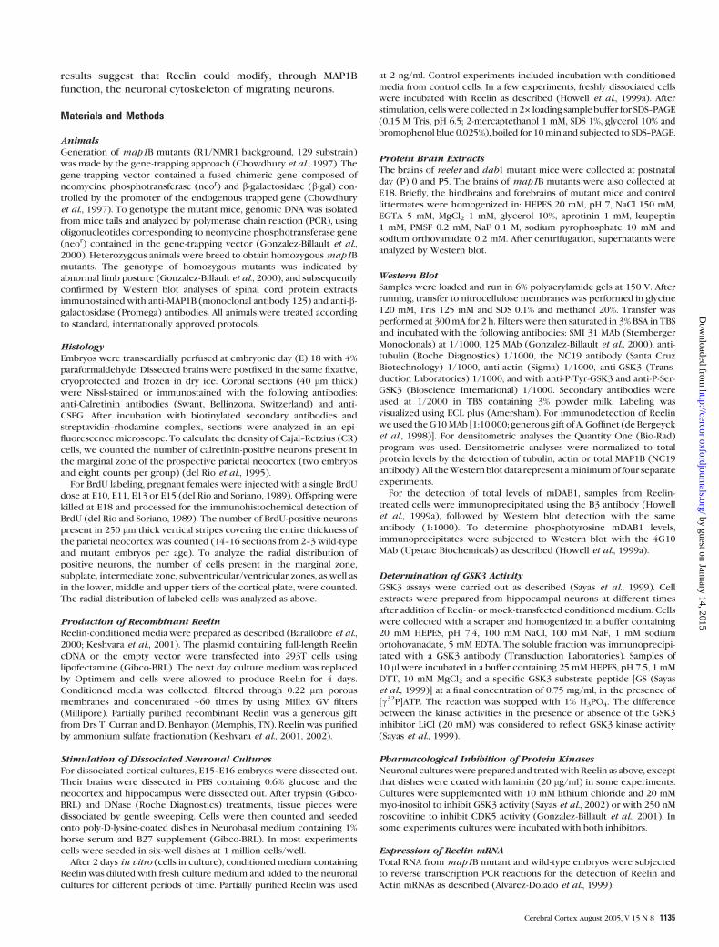
A role of MAP1B in Reelin-dependent Neuronal Migration Christian Gonza´lez-Billault 1,3 , Jose´ A. Del Rı´o 2 , Jesu´s M. Uren˜a 2 , Eva M. Jime´nez-Mateos 1 , Marı´a J. Barallobre 2 , Marta Pascual 2 , Lluı´s Pujadas 2 , Sergi Simo´ 2 , Anna La Torre 2 , Rosalina Gavin 2 , Francisco Wandosell 1 , Eduardo Soriano 2 and Jesu´s A ´ vila 1 1 Centro de Biologı´a Molecular, Universidad Autonoma de Madrid-CSIC, Madrid 28049, Spain, 2 IRBB-Barcelona Science Park and Department of Cell Biology, University of Barcelona, Barcelona 08028, Spain 3 Present address: Department of Biology and Millennium Institute for Advanced Studies in Cell Biology and Biotechnology (CBB), Faculty of Sciences, Universidad de Chile, Las Palmeras 3425, N ˜ un˜oa, Santiago, Chile The signaling cascades governing neuronal migration are believed to link extracellular signals to cytoskeletal components. MAP1B is a neuron-specific microtubule-associated protein implicated in the control of the dynamic stability of microtubules and in the cross-talk between microtubules and actin filaments. Here we show that Reelin can induce mode I MAP1B phosphorylation, both in vivo and in vitro, through gsk3 and cdk5 activation. Additionally, mDab1 participates in the signaling cascade responsible for mode I MAP1B phosphoryl- ation. Conversely, MAP1B-deficient mice display an abnormal struc- turing of the nervous system, especially in brain laminated areas, indicating a failure in neuronal migration. Therefore, we propose that Reelin can induce post-translational modifications on MAP1B that could correlate with its function in neuronal migration. Keywords: cyclin-dependent kinase 5, glycogen synthase kinase 3, microtubule-associated protein, neuronal migration, Reelin Introduction Ordered neural migration is an essential step in the organization of brain nuclei and laminated brain regions. Another essential step in neural development is axonal guidance and the for- mation of neural connections with specific targets. These two processes are controlled by specific guidance cues, including extracellular and membrane-anchored proteins (Tessier- Lavigne and Goodman, 1996; Rice and Curran, 2001). Both in ex- perimental animals and in humans, disruption of ordered neural migration leads to structural and functional defects that are associated with neurological abnormalities including sensory- motor disorders and mental retardation (Feng and Walsh, 2001; Rice and Curran, 2001). Reelin is an extracellular matrix protein which is essential for the correct migration and positioning of neurons in laminated brain regions such as the cerebral cortex, hippocampus and cerebellum (D’Arcangelo et al., 1995; Hirotsune et al., 1995; Ogawa et al., 1995). Reelin receptors include the very low density lipoprotein receptor (VLDLR) and the ApoE receptor 2 (ApoER2) (D’Arcangelo et al., 1999; Hiesberger et al., 1999; Trommsdorff et al., 1999). Binding of Reelin to these receptors leads to phosphorylation of the adaptor protein Disabled 1 (mDab1), which may control the activity of the serine/threo- nine kinase cyclin-dependent kinase 5 (CDK5) through inter- action with the regulatory subunits p35 and p39 (Ohshima et al., 1996; Chae et al., 1997; Howell et al., 1999a,b; Walsh and Goffinet, 2000; Keshvara et al., 2001; Ko et al., 2001). It has recently been shown that Reelin activates the tyrosin kinases Fyn and Src to phosphorylate mDab1 (Arnaud et al., 2003; Bock et al., 2003), and that Reelin stimulates serine phosphorylation of glycogen synthase kinase 3 (GSK3), which is believed to decrease GSK3 activity (Beffert et al., 2002). Reelin has also been reported to bind to a3 and b1 integrins, and to cadherin neuronal related (CNR) protocadherins (Senzaki et al., 1999; Dulabon et al., 2000). Since dab1(–/–) and cdk5(–/–) mice show migration abnormalities reminiscent of those in reeler, these genes are believed to be essential for Reelin signaling (Ohshima et al., 1996; Howell et al., 1997; Sheldon et al., 1997; Gilmore et al., 1998; Kwon and Tsai, 1998). Because substrates of CDK5 include cytoskeletal proteins (Ishiguro et al., 1994; Pigino et al., 1997), the Reelin signaling might transduce a signal that regulates the cytoskeleton and cell motility during neuronal migration, in a manner as yet unknown. Indeed, mutations in the microtubule-associated proteins (MAPs) LIS1 and Doublecortin cause severe migration disorders (Hirotsune et al., 1998; Francis et al., 1999; Gleeson et al., 1999; Cahana et al., 2001). MAP1B is a neuron-specific MAP that is expressed in virtually all developing neurons, both in vivo and in vitro (Avila et al., 1994a). Its function is regulated at the post-translational level by phosphorylation (Avila et al., 1994b). Mode I phosphorylation is mediated by CDK5 and GSK3, being dynamically regulated by functional stimuli such as lysophosphatidic acid (LPA) (Sayas et al., 1999), and axonal regeneration (Gonzalez-Billault et al., 2004). Mode II phosphorylation is catalyzed by casein kinase II and appears to be activated constitutively (Diaz-Nido et al., 1988; Gonzalez-Billault et al., 2004). MAP1B has been implicated in neurite extension, the dynamic stability of microtubules and the cross-talk between microtubules and actin microfilaments, in a mode I phosphorylation-dependent manner (Lucas et al., 1998; Goold et al., 1999; Mack et al., 2000; Gonzalez-Billault et al., 2001). Map1B-deficient mice have variable degrees of abnormalities in axonal tracts, which are believed to be a consequence of decreased capacity for axonal elongation in these mice (Takei et al., 1997, 2000; Gonzalez-Billault et al., 2000; Meixner et al., 2000; Teng et al., 2001). A role in axonal growth and synapto- genesis has also been proposed for the Drosophila MAP1B ortholog Futsch (Hummel et al., 2000; Roos et al., 2000). In contrast, the involvement of MAP1B in neuronal migration is more controversial (Gonzalez-Billault et al., 2000; Takei et al., 2000; Teng et al., 2001). Here we examine whether MAP1B function can be modified by a cannonical signaling pathway controling neuronal migration, such as Reelin. We show that map1B mutant mice display migration deficits in several brain structures, and also that Reelin may control mode I phosphoryl- ation of MAP1B in a GSK3- and CDK5-dependent manner. These Ó Oxford University Press 2004; all rights reserved Cerebral Cortex August 2005;15:1134--1145 doi:10.1093/cercor/bhh213 Advance Access publication December 8, 2004 by guest on January 14, 2015 http://cercor.oxfordjournals.org/ Downloaded from
-
Upload
independent -
Category
Documents
-
view
1 -
download
0
Transcript of A role of MAP1B in Reelin-dependent Neuronal Migration
A role of MAP1B in Reelin-dependentNeuronal Migration
Christian Gonzalez-Billault1,3, Jose A. Del Rıo2, Jesus M. Urena2,
Eva M. Jimenez-Mateos1, Marıa J. Barallobre2, Marta Pascual2,
Lluıs Pujadas2, Sergi Simo2, Anna La Torre2, Rosalina Gavin2,
Francisco Wandosell1, Eduardo Soriano2 and Jesus Avila1
1Centro de Biologıa Molecular, Universidad Autonoma de
Madrid-CSIC, Madrid 28049, Spain, 2IRBB-Barcelona Science
Park and Department of Cell Biology, University of Barcelona,
Barcelona 08028, Spain3Present address: Department of Biology and Millennium
Institute for Advanced Studies in Cell Biology and
Biotechnology (CBB), Faculty of Sciences, Universidad de
Chile, Las Palmeras 3425, Nunoa, Santiago, Chile
The signaling cascades governing neuronal migration are believed tolink extracellular signals to cytoskeletal components. MAP1B isa neuron-specific microtubule-associated protein implicated in thecontrol of the dynamic stability of microtubules and in the cross-talkbetweenmicrotubules and actin filaments. Herewe show that Reelincan induce mode I MAP1B phosphorylation, both in vivo and in vitro,through gsk3 and cdk5 activation. Additionally, mDab1 participatesin the signaling cascade responsible for mode I MAP1B phosphoryl-ation. Conversely, MAP1B-deficient mice display an abnormal struc-turing of the nervous system, especially in brain laminated areas,indicating a failure in neuronal migration. Therefore, we propose thatReelin can induce post-translational modifications on MAP1B thatcould correlate with its function in neuronal migration.
Keywords: cyclin-dependent kinase 5, glycogen synthase kinase 3,microtubule-associated protein, neuronal migration, Reelin
Introduction
Ordered neural migration is an essential step in the organization
of brain nuclei and laminated brain regions. Another essential
step in neural development is axonal guidance and the for-
mation of neural connections with specific targets. These two
processes are controlled by specific guidance cues, including
extracellular and membrane-anchored proteins (Tessier-
Lavigne and Goodman, 1996; Rice and Curran, 2001). Both in ex-
perimental animals and in humans, disruption of ordered neural
migration leads to structural and functional defects that are
associated with neurological abnormalities including sensory-
motor disorders and mental retardation (Feng and Walsh, 2001;
Rice and Curran, 2001).
Reelin is an extracellular matrix protein which is essential for
the correct migration and positioning of neurons in laminated
brain regions such as the cerebral cortex, hippocampus and
cerebellum (D’Arcangelo et al., 1995; Hirotsune et al., 1995;
Ogawa et al., 1995). Reelin receptors include the very low
density lipoprotein receptor (VLDLR) and the ApoE receptor 2
(ApoER2) (D’Arcangelo et al., 1999; Hiesberger et al., 1999;
Trommsdorff et al., 1999). Binding of Reelin to these receptors
leads to phosphorylation of the adaptor protein Disabled 1
(mDab1), which may control the activity of the serine/threo-
nine kinase cyclin-dependent kinase 5 (CDK5) through inter-
action with the regulatory subunits p35 and p39 (Ohshima
et al., 1996; Chae et al., 1997; Howell et al., 1999a,b; Walsh and
Goffinet, 2000; Keshvara et al., 2001; Ko et al., 2001). It has
recently been shown that Reelin activates the tyrosin kinases
Fyn and Src to phosphorylate mDab1 (Arnaud et al., 2003; Bock
et al., 2003), and that Reelin stimulates serine phosphorylation
of glycogen synthase kinase 3 (GSK3), which is believed to
decrease GSK3 activity (Beffert et al., 2002). Reelin has also
been reported to bind to a3 and b1 integrins, and to cadherin
neuronal related (CNR) protocadherins (Senzaki et al., 1999;
Dulabon et al., 2000). Since dab1(–/–) and cdk5(–/–) mice show
migration abnormalities reminiscent of those in reeler, these
genes are believed to be essential for Reelin signaling (Ohshima
et al., 1996; Howell et al., 1997; Sheldon et al., 1997; Gilmore
et al., 1998; Kwon and Tsai, 1998). Because substrates of CDK5
include cytoskeletal proteins (Ishiguro et al., 1994; Pigino et al.,
1997), the Reelin signaling might transduce a signal that
regulates the cytoskeleton and cell motility during neuronal
migration, in a manner as yet unknown. Indeed, mutations in the
microtubule-associated proteins (MAPs) LIS1 and Doublecortin
cause severe migration disorders (Hirotsune et al., 1998; Francis
et al., 1999; Gleeson et al., 1999; Cahana et al., 2001).
MAP1B is a neuron-specific MAP that is expressed in virtually
all developing neurons, both in vivo and in vitro (Avila et al.,
1994a). Its function is regulated at the post-translational level by
phosphorylation (Avila et al., 1994b). Mode I phosphorylation is
mediated by CDK5 and GSK3, being dynamically regulated by
functional stimuli such as lysophosphatidic acid (LPA) (Sayas
et al., 1999), and axonal regeneration (Gonzalez-Billault et al.,
2004). Mode II phosphorylation is catalyzed by casein kinase II
and appears to be activated constitutively (Diaz-Nido et al.,
1988; Gonzalez-Billault et al., 2004). MAP1B has been implicated
in neurite extension, the dynamic stability of microtubules and
the cross-talk between microtubules and actin microfilaments,
in a mode I phosphorylation-dependent manner (Lucas et al.,
1998; Goold et al., 1999; Mack et al., 2000; Gonzalez-Billault
et al., 2001).
Map1B-deficient mice have variable degrees of abnormalities
in axonal tracts, which are believed to be a consequence of
decreased capacity for axonal elongation in these mice (Takei
et al., 1997, 2000; Gonzalez-Billault et al., 2000; Meixner et al.,
2000; Teng et al., 2001). A role in axonal growth and synapto-
genesis has also been proposed for the Drosophila MAP1B
ortholog Futsch (Hummel et al., 2000; Roos et al., 2000). In
contrast, the involvement of MAP1B in neuronal migration is
more controversial (Gonzalez-Billault et al., 2000; Takei et al.,
2000; Teng et al., 2001). Here we examine whether MAP1B
function can be modified by a cannonical signaling pathway
controling neuronal migration, such as Reelin. We show that
map1B mutant mice display migration deficits in several brain
structures, and also that Reelin may control mode I phosphoryl-
ation of MAP1B in a GSK3- and CDK5-dependent manner. These
� Oxford University Press 2004; all rights reserved
Cerebral Cortex August 2005;15:1134--1145
doi:10.1093/cercor/bhh213
Advance Access publication December 8, 2004
by guest on January 14, 2015http://cercor.oxfordjournals.org/
Dow
nloaded from
results suggest that Reelin could modify, through MAP1B
function, the neuronal cytoskeleton of migrating neurons.
Materials and Methods
AnimalsGeneration of map1B mutants (R1/NMR1 background, 129 substrain)
was made by the gene-trapping approach (Chowdhury et al., 1997). The
gene-trapping vector contained a fused chimeric gene composed of
neomycine phosphotransferase (neor) and b-galactosidase (b-gal) con-trolled by the promoter of the endogenous trapped gene (Chowdhury
et al., 1997). To genotype the mutant mice, genomic DNA was isolated
from mice tails and analyzed by polymerase chain reaction (PCR), using
oligonucleotides corresponding to neomycine phosphotransferase gene
(neor) contained in the gene-trapping vector (Gonzalez-Billault et al.,
2000). Heterozygous animals were breed to obtain homozygousmap1B
mutants. The genotype of homozygous mutants was indicated by
abnormal limb posture (Gonzalez-Billault et al., 2000), and subsequently
confirmed by Western blot analyses of spinal cord protein extracts
immunostained with anti-MAP1B (monoclonal antibody 125) and anti-b-galactosidase (Promega) antibodies. All animals were treated according
to standard, internationally approved protocols.
HistologyEmbryos were transcardially perfused at embryonic day (E) 18 with 4%
paraformaldehyde. Dissected brains were postfixed in the same fixative,
cryoprotected and frozen in dry ice. Coronal sections (40 lm thick)
were Nissl-stained or immunostained with the following antibodies:
anti-Calretinin antibodies (Swant, Bellinzona, Switzerland) and anti-
CSPG. After incubation with biotinylated secondary antibodies and
streptavidin--rhodamine complex, sections were analyzed in an epi-
fluorescence microscope. To calculate the density of Cajal--Retzius (CR)
cells, we counted the number of calretinin-positive neurons present in
the marginal zone of the prospective parietal neocortex (two embryos
and eight counts per group) (del Rio et al., 1995).
For BrdU labeling, pregnant females were injected with a single BrdU
dose at E10, E11, E13 or E15 (del Rio and Soriano, 1989). Offspring were
killed at E18 and processed for the immunohistochemical detection of
BrdU (del Rio and Soriano, 1989). The number of BrdU-positive neurons
present in 250 lm thick vertical stripes covering the entire thickness of
the parietal neocortex was counted (14--16 sections from 2--3 wild-type
and mutant embryos per age). To analyze the radial distribution of
positive neurons, the number of cells present in the marginal zone,
subplate, intermediate zone, subventricular/ventricular zones, as well as
in the lower, middle and upper tiers of the cortical plate, were counted.
The radial distribution of labeled cells was analyzed as above.
Production of Recombinant ReelinReelin-conditioned media were prepared as described (Barallobre et al.,
2000; Keshvara et al., 2001). The plasmid containing full-length Reelin
cDNA or the empty vector were transfected into 293T cells using
lipofectamine (Gibco-BRL). The next day culture medium was replaced
by Optimem and cells were allowed to produce Reelin for 4 days.
Conditioned media was collected, filtered through 0.22 lm porous
membranes and concentrated ~60 times by using Millex GV filters
(Millipore). Partially purified recombinant Reelin was a generous gift
from Drs T. Curran and D. Benhayon (Memphis, TN). Reelin was purified
by ammonium sulfate fractionation (Keshvara et al., 2001, 2002).
Stimulation of Dissociated Neuronal CulturesFor dissociated cortical cultures, E15--E16 embryos were dissected out.
Their brains were dissected in PBS containing 0.6% glucose and the
neocortex and hippocampus were dissected out. After trypsin (Gibco-
BRL) and DNase (Roche Diagnostics) treatments, tissue pieces were
dissociated by gentle sweeping. Cells were then counted and seeded
onto poly-D-lysine-coated dishes in Neurobasal medium containing 1%
horse serum and B27 supplement (Gibco-BRL). In most experiments
cells were seeded in six-well dishes at 1 million cells/well.
After 2 days in vitro (cells in culture), conditioned medium containing
Reelin was diluted with fresh culture medium and added to the neuronal
cultures for different periods of time. Partially purified Reelin was used
at 2 ng/ml. Control experiments included incubation with conditioned
media from control cells. In a few experiments, freshly dissociated cells
were incubated with Reelin as described (Howell et al., 1999a). After
stimulation, cellswere collected in 23 loading sample buffer for SDS--PAGE
(0.15 M Tris, pH 6.5; 2-mercaptethanol 1 mM, SDS 1%, glycerol 10% and
bromophenol blue 0.025%), boiled for 10min and subjected to SDS--PAGE.
Protein Brain ExtractsThe brains of reeler and dab1 mutant mice were collected at postnatal
day (P) 0 and P5. The brains of map1B mutants were also collected at
E18. Briefly, the hindbrains and forebrains of mutant mice and control
littermates were homogenized in: HEPES 20 mM, pH 7, NaCl 150 mM,
EGTA 5 mM, MgCl2 1 mM, glycerol 10%, aprotinin 1 mM, leupeptin
1 mM, PMSF 0.2 mM, NaF 0.1 M, sodium pyrophosphate 10 mM and
sodium orthovanadate 0.2 mM. After centrifugation, supernatants were
analyzed by Western blot.
Western BlotSamples were loaded and run in 6% polyacrylamide gels at 150 V. After
running, transfer to nitrocellulose membranes was performed in glycine
120 mM, Tris 125 mM and SDS 0.1% and methanol 20%. Transfer was
performed at 300mA for 2 h. Filters were then saturated in 3% BSA in TBS
and incubated with the following antibodies: SMI 31 MAb (Sternberger
Monoclonals) at 1/1000, 125 MAb (Gonzalez-Billault et al., 2000), anti-
tubulin (Roche Diagnostics) 1/1000, the NC19 antibody (Santa Cruz
Biotechnology) 1/1000, anti-actin (Sigma) 1/1000, anti-GSK3 (Trans-
duction Laboratories) 1/1000, and with anti-P-Tyr-GSK3 and anti-P-Ser-
GSK3 (Bioscience International) 1/1000. Secondary antibodies were
used at 1/2000 in TBS containing 3% powder milk. Labeling was
visualized using ECL plus (Amersham). For immunodetection of Reelin
weused theG10MAb [1:10 000; generous gift of A. Goffinet (de Bergeyck
et al., 1998)]. For densitometric analyses the Quantity One (Bio-Rad)
program was used. Densitometric analyses were normalized to total
protein levels by the detection of tubulin, actin or total MAP1B (NC19
antibody). All theWestern blot data represent aminimumof four separate
experiments.
For the detection of total levels of mDAB1, samples from Reelin-
treated cells were immunoprecipitated using the B3 antibody (Howell
et al., 1999a), followed by Western blot detection with the same
antibody (1:1000). To determine phosphotyrosine mDAB1 levels,
immunoprecipitates were subjected to Western blot with the 4G10
MAb (Upstate Biochemicals) as described (Howell et al., 1999a).
Determination of GSK3 ActivityGSK3 assays were carried out as described (Sayas et al., 1999). Cell
extracts were prepared from hippocampal neurons at different times
after addition of Reelin- or mock-transfected conditioned medium. Cells
were collected with a scraper and homogenized in a buffer containing
20 mM HEPES, pH 7.4, 100 mM NaCl, 100 mM NaF, 1 mM sodium
ortohovanadate, 5 mM EDTA. The soluble fraction was immunoprecipi-
tated with a GSK3 antibody (Transduction Laboratories). Samples of
10 ll were incubated in a buffer containing 25 mM HEPES, pH 7.5, 1 mM
DTT, 10 mM MgCl2 and a specific GSK3 substrate peptide [GS (Sayas
et al., 1999)] at a final concentration of 0.75 mg/ml, in the presence of
[c32P]ATP. The reaction was stopped with 1% H3PO4. The difference
between the kinase activities in the presence or absence of the GSK3
inhibitor LiCl (20 mM) was considered to reflect GSK3 kinase activity
(Sayas et al., 1999).
Pharmacological Inhibition of Protein KinasesNeuronal cultureswere prepared and tratedwithReelin as above, except
that dishes were coated with laminin (20 lg/ml) in some experiments.
Cultures were supplemented with 10 mM lithium chloride and 20 mM
myo-inositol to inhibit GSK3 activity (Sayas et al., 2002) or with 250 nM
roscovitine to inhibit CDK5 activity (Gonzalez-Billault et al., 2001). In
some experiments cultures were incubated with both inhibitors.
Expression of Reelin mRNATotal RNA from map1B mutant and wild-type embryos were subjected
to reverse transcription PCR reactions for the detection of Reelin and
Actin mRNAs as described (Alvarez-Dolado et al., 1999).
Cerebral Cortex August 2005, V 15 N 8 1135
by guest on January 14, 2015http://cercor.oxfordjournals.org/
Dow
nloaded from
Results
Reelin Stimulates Mode I MAP1B PhosphorylationIn Vitro and In Vivo
The response of developing neurons to Reelin is mediated
through a complex signaling pathway comprising receptors,
adaptor proteins and protein kinases (Walsh and Goffinet, 2000;
Rice and Curran, 2001; Arnaud et al., 2003; Bock et al., 2003),
which is believed to transduce the Reelin signal into cytoskeletal
changes. MAP1B binds microtubules and actin microfilaments,
contributing to their stabilization via a process that is believed to
depend on type I MAP1B phosphorylation (Pedrotti and Islam
1996; Pigino et al., 1997; Goold et al., 1999). To examine
whether Reelin can induce MAP1B mode I phosphorylation,
we treated embryonic cortical neurons (E15--E16)with recombi-
nant Reelin, a paradigm that has been shown to activate the
Reelin signaling cascade (Howell et al., 1999a; Keshvara et al.,
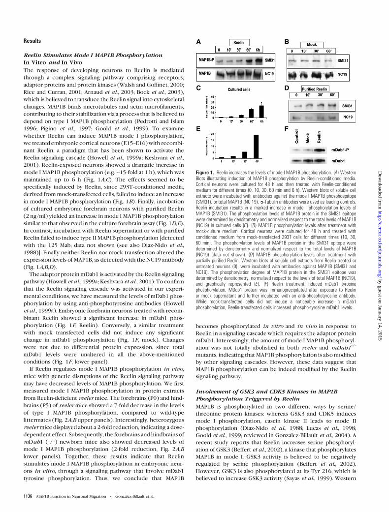
2001). Reelin-exposed neurons showed a dramatic increase in
mode I MAP1B phosphorylation (e.g. ~15-fold at 1 h), which was
maintained up to 6 h (Fig. 1A,C). The effects seemed to be
specifically induced by Reelin, since 293T-conditioned media,
derived frommock-transfected cells, failed to induce an increase
in mode I MAP1B phosphorylation (Fig. 1B). Finally, incubation
of cultured embryonic forebrain neurons with purified Reelin
(2 ng/ml) yielded an increase in mode I MAP1B phosphorylation
similar to that observed in the culture forebrain assay (Fig. 1D,E).
In contrast, incubation with Reelin supernatant or with purified
Reelin failed to induce type II MAP1B phosphorylation [detected
with the 125 Mab; data not shown (see also Diaz-Nido et al.,
1988)]. Finally neither Reelin nor mock transfection altered the
expression levels of MAP1B, as detected with the NC19 antibody
(Fig. 1A,B,D).
The adaptor proteinmDab1 is activated by the Reelin signaling
pathway (Howell et al., 1999a; Keshvara et al., 2001). To confirm
that the Reelin signaling cascade was activated in our experi-
mental conditions, we have measured the levels of mDab1 phos-
phorylation by using anti-phosphotyrosine antibodies (Howell
et al., 1999a). Embryonic forebrain neurons treated with recom-
binant Reelin showed a significant increase in mDab1 phos-
phorylation (Fig. 1F, Reelin). Conversely, a similar treatment
with mock transfected cells did not induce any significant
change in mDab1 phosphorylation (Fig. 1F, mock). Changes
were not due to differential protein expression, since total
mDab1 levels were unaltered in all the above-mentioned
conditions (Fig. 1F, lower panel).
If Reelin regulates mode I MAP1B phosphorylation in vivo,
mice with genetic disruptions of the Reelin signaling pathway
may have decreased levels of MAP1B phosphorylation. We first
measured mode I MAP1B phosphorylation in protein extracts
from Reelin-deficient reelermice. The forebrains (P0) and hind-
brains (P5) of reelermice showed a 7-fold decrease in the levels
of type I MAP1B phosphorylation, compared to wild-type
littermates (Fig. 2A,B upper panels). Interestingly, heterozygous
reelermice displayed about a 2-fold reduction, indicating a dose-
dependent effect. Subsequently, the forebrains and hindbrains of
mDab1 (–/–) newborn mice also showed decreased levels of
mode I MAP1B phosphorylation (2-fold reduction, Fig. 2A,B
lower panels). Together, these results indicate that Reelin
stimulates mode I MAP1B phosphorylation in embryonic neur-
ons in vitro, through a signaling pathway that involve mDab1
tyrosine phosphorylation. Thus, we conclude that MAP1B
becomes phosphorylated in vitro and in vivo in response to
Reelin in a signaling cascade which requires the adaptor protein
mDab1. Interestingly, the amount of mode I MAP1B phosphoryl-
ation was not totally abolished in both reeler and mDab1–/–
mutants, indicating that MAP1B phosphorylation is alsomodified
by other signaling cascades. However, these data suggest that
MAP1B phosphorylation can be indeed modified by the Reelin
signaling pathway.
Involvement of GSK3 and CDK5 Kinases in MAP1BPhosphorylation Triggered by Reelin
MAP1B is phosphorylated in two different ways by serine/
threonine protein kinases: whereas GSK3 and CDK5 induces
mode I phosphorylation, casein kinase II leads to mode II
phosphorylation (Diaz-Nido et al., 1988; Lucas et al., 1998;
Goold et al., 1999; reviewed in Gonzalez-Billault et al., 2004). A
recent study reports that Reelin increases serine phosphoryl-
ation of GSK3 (Beffert et al., 2002), a kinase that phosphorylates
MAP1B in mode I. GSK3 activity is believed to be negatively
regulated by serine phosphorylation (Beffert et al., 2002).
However, GSK3 is also phosphorylated at its Tyr 216, which is
believed to increase GSK3 activity (Sayas et al., 1999). Western
Figure 1. Reelin increases the levels of mode I MAP1B phosphorylation. (A) WesternBlots illustrating induction of MAP1B phosphorylation by Reelin-conditioned media.Cortical neurons were cultured for 48 h and then treated with Reelin-conditionedmedium for different times (0, 10, 30, 60 min and 6 h). Western blots of soluble cellextracts were incubated with antibodies against the mode I MAP1B phosphoepitope(SMI31), or total MAP1B (NC 19). a-Tubulin antibodies were used as loading controls.Reelin incubation results in a marked increase in mode I phosphorylation levels ofMAP1B (SMI31). The phosphorylation levels of MAP1B protein in the SMI31 epitopewere determined by densitometry and normalized respect to the total levels of MAP1B(NC19) in cultured cells (C). (B) MAP1B phosphorylation levels after treatment withmock-culture medium. Cortical neurons were cultured for 48 h and treated withconditioned medium from mock-transfected 293T cells for different times (10, 30,60 min). The phosphorylation levels of MAP1B protein in the SMI31 epitope weredetermined by densitometry and normalized respect to the total levels of MAP1B(NC19) (data not shown). (D) MAP1B phosphorylation levels after treatment withpartially purified Reelin. Western blots of soluble cell extracts from Reelin-treated oruntreated neurons (0), were incubated with antibodies against MAP1B (SMI31 andNC19). The phosphorylation degree of MAP1B protein in the SMI31 epitope wasdetermined by densitometry, normalized respect to the levels of total MAP1B (NC19),and graphically represented (E). (F) Reelin treatment induced mDab1 tyrosinephosphorylation. MDab1 protein was immunoprecipitated after exposure to Reelinor mock supernatant and further incubated with an anti-phosphotyrosine antibody.While mock-transfected cells did not induce a noticeable increase in mDab1phosphorylation, Reelin-transfected cells increased phospho-tyrosine mDab1 levels.
1136 MAP1B Function in Neuronal Migration d Gonzalez-Billault et al.
by guest on January 14, 2015http://cercor.oxfordjournals.org/
Dow
nloaded from
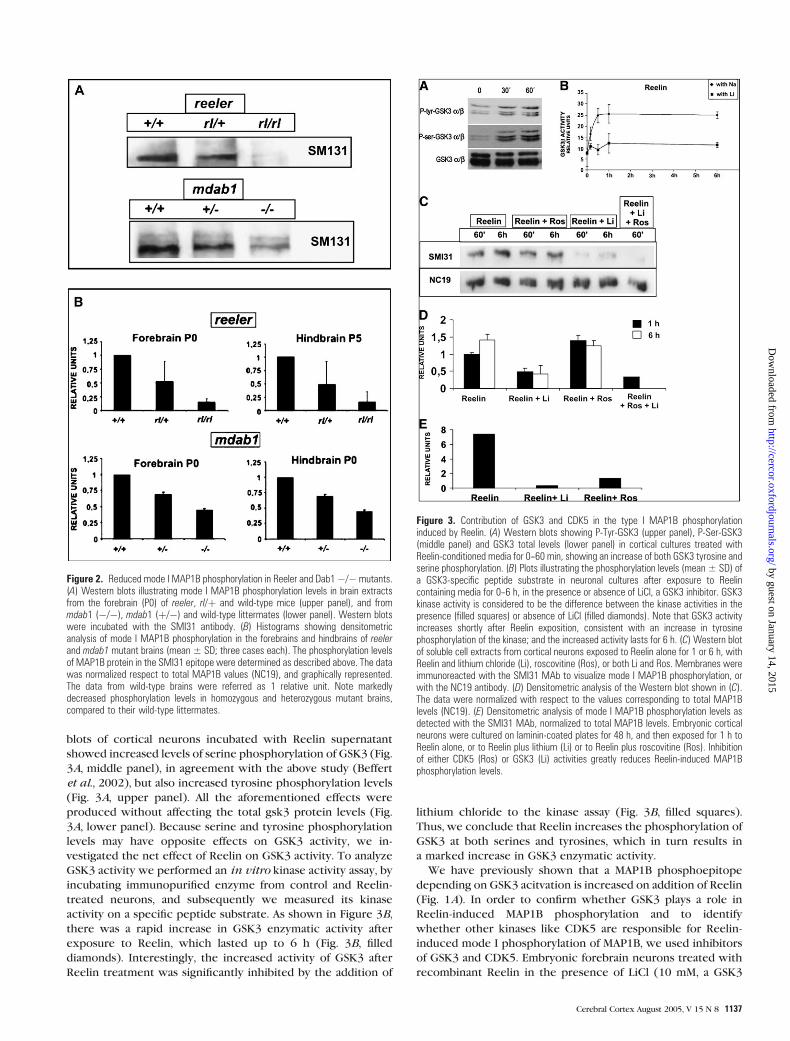
blots of cortical neurons incubated with Reelin supernatant
showed increased levels of serine phosphorylation of GSK3 (Fig.
3A, middle panel), in agreement with the above study (Beffert
et al., 2002), but also increased tyrosine phosphorylation levels
(Fig. 3A, upper panel). All the aforementioned effects were
produced without affecting the total gsk3 protein levels (Fig.
3A, lower panel). Because serine and tyrosine phosphorylation
levels may have opposite effects on GSK3 activity, we in-
vestigated the net effect of Reelin on GSK3 activity. To analyze
GSK3 activity we performed an in vitro kinase activity assay, by
incubating immunopurified enzyme from control and Reelin-
treated neurons, and subsequently we measured its kinase
activity on a specific peptide substrate. As shown in Figure 3B,
there was a rapid increase in GSK3 enzymatic activity after
exposure to Reelin, which lasted up to 6 h (Fig. 3B, filled
diamonds). Interestingly, the increased activity of GSK3 after
Reelin treatment was significantly inhibited by the addition of
lithium chloride to the kinase assay (Fig. 3B, filled squares).
Thus, we conclude that Reelin increases the phosphorylation of
GSK3 at both serines and tyrosines, which in turn results in
a marked increase in GSK3 enzymatic activity.
We have previously shown that a MAP1B phosphoepitope
depending on GSK3 acitvation is increased on addition of Reelin
(Fig. 1A). In order to confirm whether GSK3 plays a role in
Reelin-induced MAP1B phosphorylation and to identify
whether other kinases like CDK5 are responsible for Reelin-
induced mode I phosphorylation of MAP1B, we used inhibitors
of GSK3 and CDK5. Embryonic forebrain neurons treated with
recombinant Reelin in the presence of LiCl (10 mM, a GSK3
Figure 2. Reduced mode I MAP1B phosphorylation in Reeler and Dab1�/� mutants.(A) Western blots illustrating mode I MAP1B phosphorylation levels in brain extractsfrom the forebrain (P0) of reeler, rl/þ and wild-type mice (upper panel), and frommdab1 (�/�), mdab1 (þ/�) and wild-type littermates (lower panel). Western blotswere incubated with the SMI31 antibody. (B) Histograms showing densitometricanalysis of mode I MAP1B phosphorylation in the forebrains and hindbrains of reelerand mdab1 mutant brains (mean ± SD; three cases each). The phosphorylation levelsof MAP1B protein in the SMI31 epitope were determined as described above. The datawas normalized respect to total MAP1B values (NC19), and graphically represented.The data from wild-type brains were referred as 1 relative unit. Note markedlydecreased phosphorylation levels in homozygous and heterozygous mutant brains,compared to their wild-type littermates.
Figure 3. Contribution of GSK3 and CDK5 in the type I MAP1B phosphorylationinduced by Reelin. (A) Western blots showing P-Tyr-GSK3 (upper panel), P-Ser-GSK3(middle panel) and GSK3 total levels (lower panel) in cortical cultures treated withReelin-conditioned media for 0--60 min, showing an increase of both GSK3 tyrosine andserine phosphorylation. (B) Plots illustrating the phosphorylation levels (mean ± SD) ofa GSK3-specific peptide substrate in neuronal cultures after exposure to Reelincontaining media for 0--6 h, in the presence or absence of LiCl, a GSK3 inhibitor. GSK3kinase activity is considered to be the difference between the kinase activities in thepresence (filled squares) or absence of LiCl (filled diamonds). Note that GSK3 activityincreases shortly after Reelin exposition, consistent with an increase in tyrosinephosphorylation of the kinase; and the increased activity lasts for 6 h. (C) Western blotof soluble cell extracts from cortical neurons exposed to Reelin alone for 1 or 6 h, withReelin and lithium chloride (Li), roscovitine (Ros), or both Li and Ros. Membranes wereimmunoreacted with the SMI31 MAb to visualize mode I MAP1B phosphorylation, orwith the NC19 antibody. (D) Densitometric analysis of the Western blot shown in (C).The data were normalized with respect to the values corresponding to total MAP1Blevels (NC19). (E) Densitometric analysis of mode I MAP1B phosphorylation levels asdetected with the SMI31 MAb, normalized to total MAP1B levels. Embryonic corticalneurons were cultured on laminin-coated plates for 48 h, and then exposed for 1 h toReelin alone, or to Reelin plus lithium (Li) or to Reelin plus roscovitine (Ros). Inhibitionof either CDK5 (Ros) or GSK3 (Li) activities greatly reduces Reelin-induced MAP1Bphosphorylation levels.
Cerebral Cortex August 2005, V 15 N 8 1137
by guest on January 14, 2015http://cercor.oxfordjournals.org/
Dow
nloaded from
inhibitor; Lucas et al. 1998) showed a marked decrease in the
levels of mode I phosphorylation at 1 and 6 h (Fig. 3C,D). In
contrast, incubation with the CDK5 inhibitor roscovitine
(250 nM, Calbiochem) does not modify mode I MAP1B phos-
phorylation (Fig. 3C,D).
Since Reelin actions are believed to be mediated by CDK5, we
investigated the contribution of CDK5. It is known that neurons
cultured on laminin upregulate p35 expression, thus promoting
CDK5 activity (Paglini et al., 1998). We therefore performed
similar experiments but with cultured forebrain neurons on
laminin. Under these conditions, incubation with roscovitine
inhibited mode I MAP1B phosphorylation induced by Reelin
(Fig. 3E). Furthermore, inhibition of both GSK3 and CDK5
almost completely abolished mode I phosphorylation of MAP1B
(Fig. 3C,D).
Altogether these results strongly suggest that MAP1B phos-
phorylation can be in part controlled by the Reelin signaling
pathway, in a cascade that includes mDab1 tyrosine phosphoryl-
ation as well as GSK3 and CDK5 activation.
Map1B-deficient Mice Show Migratory Deficits
Because mode I of MAP1B phosphorylation could be induced
after treatment with Reelin, we decided to investigate whether
MAP1B function could be modified by Reelin. For this reason, in
the first set of experiments we investigated if the map1B
mutation affected the expression of Reelin. Sections from wild-
type and mutant embryos were immunostained with calretinin
antibodies to map the distribution of CR cells, the major source
of Reelin at embryonic stages (D’Arcangelo et al., 1995; Ogawa
et al., 1995; Alcantara et al., 1998). No differences were ob-
served in the distribution or the density of CR cells (18.7 ± 1.2
cells and 19.3 ± 0.9 cells per 250 lm in wild-type and mutant
mice, respectively; two embryos and eight counts per group),
indicating that MAP1B is not required either for the migration of
CR cells or for their settlement in the marginal zone. To confirm
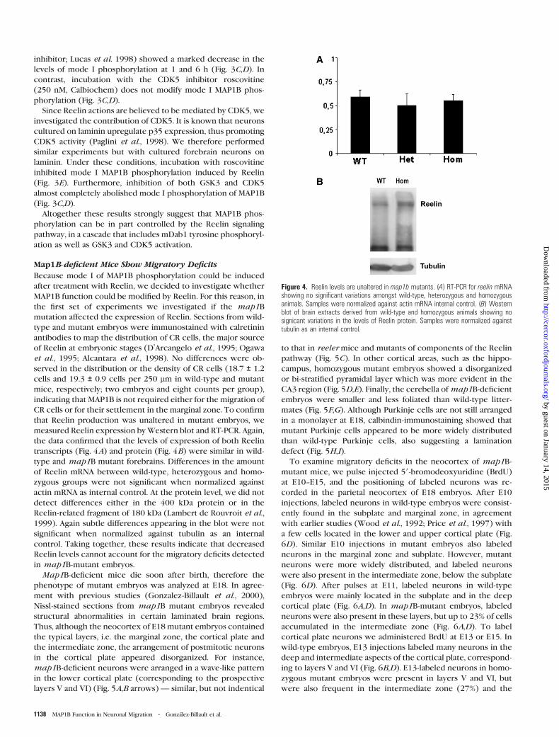
that Reelin production was unaltered in mutant embryos, we
measured Reelin expression byWestern blot and RT-PCR. Again,
the data confirmed that the levels of expression of both Reelin
transcripts (Fig. 4A) and protein (Fig. 4B) were similar in wild-
type and map1B mutant forebrains. Differences in the amount
of Reelin mRNA between wild-type, heterozygous and homo-
zygous groups were not significant when normalized against
actin mRNA as internal control. At the protein level, we did not
detect differences either in the 400 kDa protein or in the
Reelin-related fragment of 180 kDa (Lambert de Rouvroit et al.,
1999). Again subtle differences appearing in the blot were not
significant when normalized against tubulin as an internal
control. Taking together, these results indicate that decreased
Reelin levels cannot account for the migratory deficits detected
in map1B-mutant embryos.
Map1B-deficient mice die soon after birth, therefore the
phenotype of mutant embryos was analyzed at E18. In agree-
ment with previous studies (Gonzalez-Billault et al., 2000),
Nissl-stained sections from map1B mutant embryos revealed
structural abnormalities in certain laminated brain regions.
Thus, although the neocortex of E18 mutant embryos contained
the typical layers, i.e. the marginal zone, the cortical plate and
the intermediate zone, the arrangement of postmitotic neurons
in the cortical plate appeared disorganized. For instance,
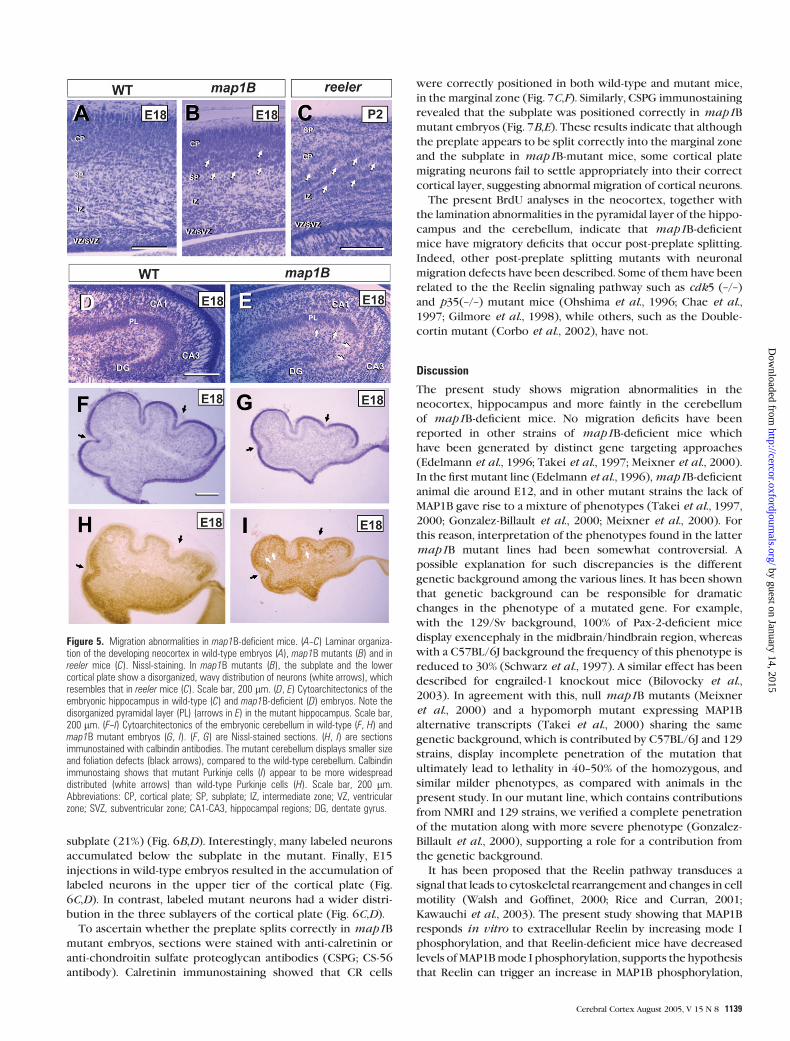
map1B-deficient neurons were arranged in a wave-like pattern
in the lower cortical plate (corresponding to the prospective
layers V and VI) (Fig. 5A,B arrows) — similar, but not indentical
to that in reeler mice and mutants of components of the Reelin
pathway (Fig. 5C). In other cortical areas, such as the hippo-
campus, homozygous mutant embryos showed a disorganized
or bi-stratified pyramidal layer which was more evident in the
CA3 region (Fig. 5D,E). Finally, the cerebella ofmap1B-deficient
embryos were smaller and less foliated than wild-type litter-
mates (Fig. 5F,G). Although Purkinje cells are not still arranged
in a monolayer at E18, calbindin-immunostaining showed that
mutant Purkinje cells appeared to be more widely distributed
than wild-type Purkinje cells, also suggesting a lamination
defect (Fig. 5H,I).
To examine migratory deficits in the neocortex of map1B-
mutant mice, we pulse injected 59-bromodeoxyuridine (BrdU)
at E10--E15, and the positioning of labeled neurons was re-
corded in the parietal neocortex of E18 embryos. After E10
injections, labeled neurons in wild-type embryos were consist-
ently found in the subplate and marginal zone, in agreement
with earlier studies (Wood et al., 1992; Price et al., 1997) with
a few cells located in the lower and upper cortical plate (Fig.
6D). Similar E10 injections in mutant embryos also labeled
neurons in the marginal zone and subplate. However, mutant
neurons were more widely distributed, and labeled neurons
were also present in the intermediate zone, below the subplate
(Fig. 6D). After pulses at E11, labeled neurons in wild-type
embryos were mainly located in the subplate and in the deep
cortical plate (Fig. 6A,D). In map1B-mutant embryos, labeled
neurons were also present in these layers, but up to 23% of cells
accumulated in the intermediate zone (Fig. 6A,D). To label
cortical plate neurons we administered BrdU at E13 or E15. In
wild-type embryos, E13 injections labeled many neurons in the
deep and intermediate aspects of the cortical plate, correspond-
ing to layers V and VI (Fig. 6B,D). E13-labeled neurons in homo-
zygous mutant embryos were present in layers V and VI, but
were also frequent in the intermediate zone (27%) and the
Figure 4. Reelin levels are unaltered in map1b mutants. (A) RT-PCR for reelin mRNAshowing no significant variations amongst wild-type, heterozygous and homozygousanimals. Samples were normalized against actin mRNA internal control. (B) Westernblot of brain extracts derived from wild-type and homozygous animals showing nosignicant variations in the levels of Reelin protein. Samples were normalized againsttubulin as an internal control.
1138 MAP1B Function in Neuronal Migration d Gonzalez-Billault et al.
by guest on January 14, 2015http://cercor.oxfordjournals.org/
Dow
nloaded from
subplate (21%) (Fig. 6B,D). Interestingly, many labeled neurons
accumulated below the subplate in the mutant. Finally, E15
injections in wild-type embryos resulted in the accumulation of
labeled neurons in the upper tier of the cortical plate (Fig.
6C,D). In contrast, labeled mutant neurons had a wider distri-
bution in the three sublayers of the cortical plate (Fig. 6C,D).
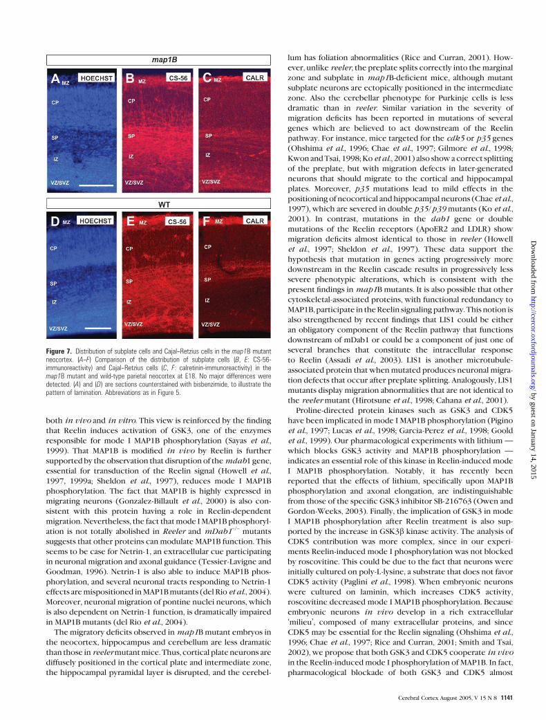
To ascertain whether the preplate splits correctly in map1B
mutant embryos, sections were stained with anti-calretinin or
anti-chondroitin sulfate proteoglycan antibodies (CSPG; CS-56
antibody). Calretinin immunostaining showed that CR cells
were correctly positioned in both wild-type and mutant mice,
in the marginal zone (Fig. 7C,F). Similarly, CSPG immunostaining
revealed that the subplate was positioned correctly in map1B
mutant embryos (Fig. 7B,E). These results indicate that although
the preplate appears to be split correctly into the marginal zone
and the subplate in map1B-mutant mice, some cortical plate
migrating neurons fail to settle appropriately into their correct
cortical layer, suggesting abnormal migration of cortical neurons.
The present BrdU analyses in the neocortex, together with
the lamination abnormalities in the pyramidal layer of the hippo-
campus and the cerebellum, indicate that map1B-deficient
mice have migratory deficits that occur post-preplate splitting.
Indeed, other post-preplate splitting mutants with neuronal
migration defects have been described. Some of them have been
related to the the Reelin signaling pathway such as cdk5 (–/–)
and p35(–/–) mutant mice (Ohshima et al., 1996; Chae et al.,
1997; Gilmore et al., 1998), while others, such as the Double-
cortin mutant (Corbo et al., 2002), have not.
Discussion
The present study shows migration abnormalities in the
neocortex, hippocampus and more faintly in the cerebellum
of map1B-deficient mice. No migration deficits have been
reported in other strains of map1B-deficient mice which
have been generated by distinct gene targeting approaches
(Edelmann et al., 1996; Takei et al., 1997; Meixner et al., 2000).
In the first mutant line (Edelmann et al., 1996),map1B-deficient
animal die around E12, and in other mutant strains the lack of
MAP1B gave rise to a mixture of phenotypes (Takei et al., 1997,
2000; Gonzalez-Billault et al., 2000; Meixner et al., 2000). For
this reason, interpretation of the phenotypes found in the latter
map1B mutant lines had been somewhat controversial. A
possible explanation for such discrepancies is the different
genetic background among the various lines. It has been shown
that genetic background can be responsible for dramatic
changes in the phenotype of a mutated gene. For example,
with the 129/Sv background, 100% of Pax-2-deficient mice
display exencephaly in the midbrain/hindbrain region, whereas
with a C57BL/6J background the frequency of this phenotype is
reduced to 30% (Schwarz et al., 1997). A similar effect has been
described for engrailed-1 knockout mice (Bilovocky et al.,
2003). In agreement with this, null map1B mutants (Meixner
et al., 2000) and a hypomorph mutant expressing MAP1B
alternative transcripts (Takei et al., 2000) sharing the same
genetic background, which is contributed by C57BL/6J and 129
strains, display incomplete penetration of the mutation that
ultimately lead to lethality in 40--50% of the homozygous, and
similar milder phenotypes, as compared with animals in the
present study. In our mutant line, which contains contributions
from NMRI and 129 strains, we verified a complete penetration
of the mutation along with more severe phenotype (Gonzalez-
Billault et al., 2000), supporting a role for a contribution from
the genetic background.
It has been proposed that the Reelin pathway transduces a
signal that leads to cytoskeletal rearrangement and changes in cell
motility (Walsh and Goffinet, 2000; Rice and Curran, 2001;
Kawauchi et al., 2003). The present study showing that MAP1B
responds in vitro to extracellular Reelin by increasing mode I
phosphorylation, and that Reelin-deficient mice have decreased
levels ofMAP1Bmode I phosphorylation, supports the hypothesis
that Reelin can trigger an increase in MAP1B phosphorylation,
Figure 5. Migration abnormalities in map1B-deficient mice. (A--C) Laminar organiza-tion of the developing neocortex in wild-type embryos (A), map1B mutants (B) and inreeler mice (C). Nissl-staining. In map1B mutants (B), the subplate and the lowercortical plate show a disorganized, wavy distribution of neurons (white arrows), whichresembles that in reeler mice (C). Scale bar, 200 lm. (D, E) Cytoarchitectonics of theembryonic hippocampus in wild-type (C) and map1B-deficient (D) embryos. Note thedisorganized pyramidal layer (PL) (arrows in E) in the mutant hippocampus. Scale bar,200 lm. (F--I) Cytoarchitectonics of the embryonic cerebellum in wild-type (F, H) andmap1B mutant embryos (G, I). (F, G) are Nissl-stained sections. (H, I) are sectionsimmunostained with calbindin antibodies. The mutant cerebellum displays smaller sizeand foliation defects (black arrows), compared to the wild-type cerebellum. Calbindinimmunostaing shows that mutant Purkinje cells (I) appear to be more widespreaddistributed (white arrows) than wild-type Purkinje cells (H). Scale bar, 200 lm.Abbreviations: CP, cortical plate; SP, subplate; IZ, intermediate zone; VZ, ventricularzone; SVZ, subventricular zone; CA1-CA3, hippocampal regions; DG, dentate gyrus.
Cerebral Cortex August 2005, V 15 N 8 1139
by guest on January 14, 2015http://cercor.oxfordjournals.org/
Dow
nloaded from
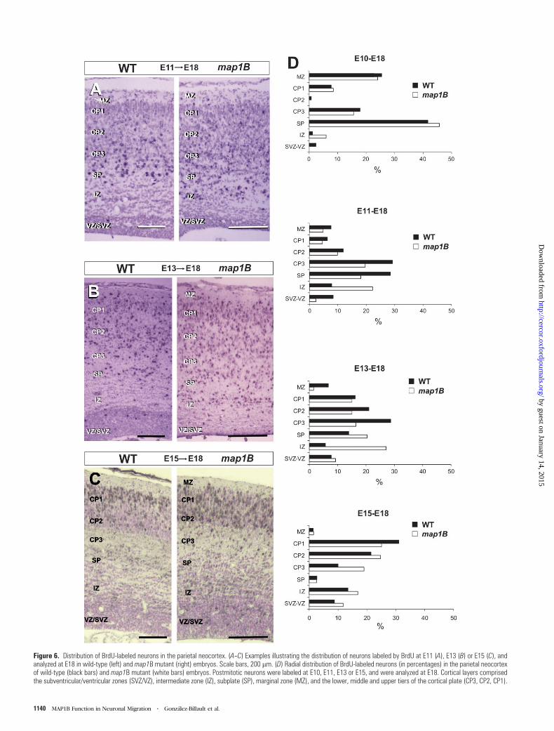
Figure 6. Distribution of BrdU-labeled neurons in the parietal neocortex. (A--C) Examples illustrating the distribution of neurons labeled by BrdU at E11 (A), E13 (B) or E15 (C), andanalyzed at E18 in wild-type (left) and map1B mutant (right) embryos. Scale bars, 200 lm. (D) Radial distribution of BrdU-labeled neurons (in percentages) in the parietal neocortexof wild-type (black bars) and map1B mutant (white bars) embryos. Postmitotic neurons were labeled at E10, E11, E13 or E15, and were analyzed at E18. Cortical layers comprisedthe subventricular/ventricular zones (SVZ/VZ), intermediate zone (IZ), subplate (SP), marginal zone (MZ), and the lower, middle and upper tiers of the cortical plate (CP3, CP2, CP1).
1140 MAP1B Function in Neuronal Migration d Gonzalez-Billault et al.
by guest on January 14, 2015http://cercor.oxfordjournals.org/
Dow
nloaded from
both in vivo and in vitro. This view is reinforced by the finding
that Reelin induces activation of GSK3, one of the enzymes
responsible for mode I MAP1B phosphorylation (Sayas et al.,
1999). That MAP1B is modified in vivo by Reelin is further
supported by the observation that disruption of themdab1 gene,
essential for transduction of the Reelin signal (Howell et al.,
1997, 1999a; Sheldon et al., 1997), reduces mode I MAP1B
phosphorylation. The fact that MAP1B is highly expressed in
migrating neurons (Gonzalez-Billault et al., 2000) is also con-
sistent with this protein having a role in Reelin-dependent
migration. Nevertheless, the fact thatmode IMAP1B phosphoryl-
ation is not totally abolished in Reeler and mDab1–/– mutants
suggests that other proteins can modulate MAP1B function. This
seems to be case for Netrin-1, an extracellular cue participating
in neuronal migration and axonal guidance (Tessier-Lavigne and
Goodman, 1996). Netrin-1 is also able to induce MAP1B phos-
phorylation, and several neuronal tracts responding to Netrin-1
effects aremispositioned inMAP1Bmutants (del Rio et al., 2004).
Moreover, neuronal migration of pontine nuclei neurons, which
is also dependent on Netrin-1 function, is dramatically impaired
in MAP1B mutants (del Rio et al., 2004).
The migratory deficits observed inmap1Bmutant embryos in
the neocortex, hippocampus and cerebellum are less dramatic
than those in reelermutantmice. Thus, cortical plate neurons are
diffusely positioned in the cortical plate and intermediate zone,
the hippocampal pyramidal layer is disrupted, and the cerebel-
lum has foliation abnormalities (Rice and Curran, 2001). How-
ever, unlike reeler, the preplate splits correctly into themarginal
zone and subplate in map1B-deficient mice, although mutant
subplate neurons are ectopically positioned in the intermediate
zone. Also the cerebellar phenotype for Purkinje cells is less
dramatic than in reeler. Similar variation in the severity of
migration deficits has been reported in mutations of several
genes which are believed to act downstream of the Reelin
pathway. For instance, mice targeted for the cdk5 or p35 genes
(Ohshima et al., 1996; Chae et al., 1997; Gilmore et al., 1998;
KwonandTsai, 1998;Ko et al., 2001) also showacorrect splitting
of the preplate, but with migration defects in later-generated
neurons that should migrate to the cortical and hippocampal
plates. Moreover, p35 mutations lead to mild effects in the
positioning of neocortical and hippocampal neurons (Chae et al.,
1997), which are severed in double p35/p39mutants (Ko et al.,
2001). In contrast, mutations in the dab1 gene or double
mutations of the Reelin receptors (ApoER2 and LDLR) show
migration deficits almost identical to those in reeler (Howell
et al., 1997; Sheldon et al., 1997). These data support the
hypothesis that mutation in genes acting progressively more
downstream in the Reelin cascade results in progressively less
severe phenotypic alterations, which is consistent with the
present findings inmap1B mutants. It is also possible that other
cytoskeletal-associated proteins, with functional redundancy to
MAP1B, participate in theReelin signalingpathway. This notion is
also strengthened by recent findings that LIS1 could be either
an obligatory component of the Reelin pathway that functions
downstream of mDab1 or could be a component of just one of
several branches that constitute the intracellular response
to Reelin (Assadi et al., 2003). LIS1 is another microtubule-
associated protein that whenmutated produces neuronal migra-
tion defects that occur after preplate splitting. Analogously, LIS1
mutants display migration abnormalities that are not identical to
the reeler mutant (Hirotsune et al., 1998; Cahana et al., 2001).
Proline-directed protein kinases such as GSK3 and CDK5
have been implicated in mode I MAP1B phosphorylation (Pigino
et al., 1997; Lucas et al., 1998; Garcia-Perez et al., 1998; Goold
et al., 1999). Our pharmacological experiments with lithium —
which blocks GSK3 activity and MAP1B phosphorylation —
indicates an essential role of this kinase in Reelin-induced mode
I MAP1B phosphorylation. Notably, it has recently been
reported that the effects of lithium, specifically upon MAP1B
phosphorylation and axonal elongation, are indistinguishable
from those of the specific GSK3 inhibitor SB-216763 (Owen and
Gordon-Weeks, 2003). Finally, the implication of GSK3 in mode
I MAP1B phosphorylation after Reelin treatment is also sup-
ported by the increase in GSK3b kinase activity. The analysis of
CDK5 contribution was more complex, since in our experi-
ments Reelin-induced mode I phosphorylation was not blocked
by roscovitine. This could be due to the fact that neurons were
initially cultured on poly-L-lysine, a substrate that does not favor
CDK5 activity (Paglini et al., 1998). When embryonic neurons
were cultured on laminin, which increases CDK5 activity,
roscovitine decreased mode I MAP1B phosphorylation. Because
embryonic neurons in vivo develop in a rich extracellular
‘milieu’, composed of many extracellular proteins, and since
CDK5 may be essential for the Reelin signaling (Ohshima et al.,
1996; Chae et al., 1997; Rice and Curran, 2001; Smith and Tsai,
2002), we propose that both GSK3 and CDK5 cooperate in vivo
in the Reelin-inducedmode I phosphorylation of MAP1B. In fact,
pharmacological blockade of both GSK3 and CDK5 almost
Figure 7. Distribution of subplate cells and Cajal--Retzius cells in the map1B mutantneocortex. (A--F) Comparison of the distribution of subplate cells (B, E: CS-56-immunoreactivity) and Cajal--Retzius cells (C, F: calretinin-immunoreactivity) in themap1B mutant and wild-type parietal neocortex at E18. No major differences weredetected. (A) and (D) are sections counterstained with bisbenzimide, to illustrate thepattern of lamination. Abbreviations as in Figure 5.
Cerebral Cortex August 2005, V 15 N 8 1141
by guest on January 14, 2015http://cercor.oxfordjournals.org/
Dow
nloaded from
completely abolished type I MAP1B phosphorylation. This
is consistent with a synergistic effect for those kinases. In
fact, several reports have indicated that many GSK3 substrates
must be previously phosphorylated in the –4 position by other
protein kinases, including CDK5, to be then modified by GSK3
(Cohen and Frame, 2001). Indeed, the MAP1B phospho-epitope
recognized by the SMI31 MAb contains different serines at a +4position that could be modified by cdk5. Thus, we propose that
MAP1B can be phosphorylated by CDK5 to act as a primer to
favor GSK3 modification.
It is believed that CDK5 is a downstream effector in the
Reelin pathway, although there is no evidence of activation of
CDK5 by Reelin (Zukerberg et al., 2000; Smith and Tsai, 2002).
In addition, mDab1 is phosphorylated by CDK5 in a Reelin-
independent manner (Keshvara et al., 2001). A recent study
reports that Reelin regulates serine phosphorylation of GSK3,
suggesting an inhibition of enzymatic activity (Beffert et al.,
2002). However, GSK3 has two isoforms, a and b, and its activity
is downregulated by phosphorylation of serines and upregu-
lated by phosphorylation on tyrosines (Cohen and Frame, 2001;
Grimes and Jope, 2001). In contrast, our study shows that Reelin
increases phosphorylation of both serines and tyrosines of
GSK3, resulting in a marked increase in GSK3 enzymatic
activity. This observation is also consistent with that indicating
that phosphorylation of MAP1B, a GSK3 substrate, increases
upon exposure to Reelin (Fig. 8). Several pathways have been
proposed to inhibit GSK3 activity, including the insulin/IGF-I
pathway through activation of the PI3K-Akt/PKB pathway
(Cohen and Frame, 2001; Grimes and Jope, 2001) and the
Wnt/Wingless signaling cascade (Welsh and Proud, 1993; Cook
et al., 1996). In contrast, only a few studies report activation of
GSK3 by FGF2 or LPA (Sayas et al., 1999, 2002; Hashimoto et al.,
2002) by mechanisms that remain largely unknown. We have
shown here that the Reelin signaling pathway also activates
GSK3 (Fig. 8).
The present analyses in vivo and in vitro indicate that MAP1B
can be modified by the Reelin signaling pathway. It is thought
that both microtubules and actin filaments, which are concen-
trated at the leading process of migrating neurons, are essential
for neuronal migration (Rivas and Hatten, 1995). MAP1B has
been shown to control the dynamic properties of microtubules
(Pedrotti and Islam, 1996; Togel et al., 1998). For instance, it has
been suggested that culturedmap1B-deficient neurons contain
fewer microtubules than control cells (Gonzalez-Billault et al.,
2001). Moreover, in the absence of MAP1B, the number of
dynamic microtubules in the distal part of the axons is much
lower, whereas the number and distribution of stable micro-
tubules increase (Gonzalez-Billault et al., 2001).
We also show that mode I MAP1B phosphorylation is acti-
vated by Reelin. Mode I phosphorylated MAP1B is upregulated
during development and is present in a gradient-dependent
manner in axons and neurites, being enriched in the distal part
(Riederer et al., 1990; Ulloa et al, 1993, 1994; Mack et al., 2000).
At the cellular level, inactivation of GSK3 or CDK5 kinases
induces a neural phenotype in vitro that resembles that of
map1B-deficient neurons (Pigino et al., 1997; Lucas et al., 1998;
Paglini et al, 1998; Goold et al., 1999; Gonzalez-Billault et al.,
2002). Moreover, ablation of mode I MAP1B phosphorylation by
micro-CALI experiments affects growth cone tuning in cultured
neurons (Mack et al., 2000). Finally, it has been reported that
overexpression of Wnt7b, a signal that inactivates GSK3 in-
ducing a decrease of MAP1B phosphorylation in cortical ex-
plants, leads to abnormal possitioning of cortical neurons (Viti
et al., 2003). All these data have led to the suggestion that most
functions of MAP1B are regulated by mode-I phosphorylation,
which is likely to increase microtubule and actin instability and
cross-talk (Goold et al., 1999; Mack et al., 2000).
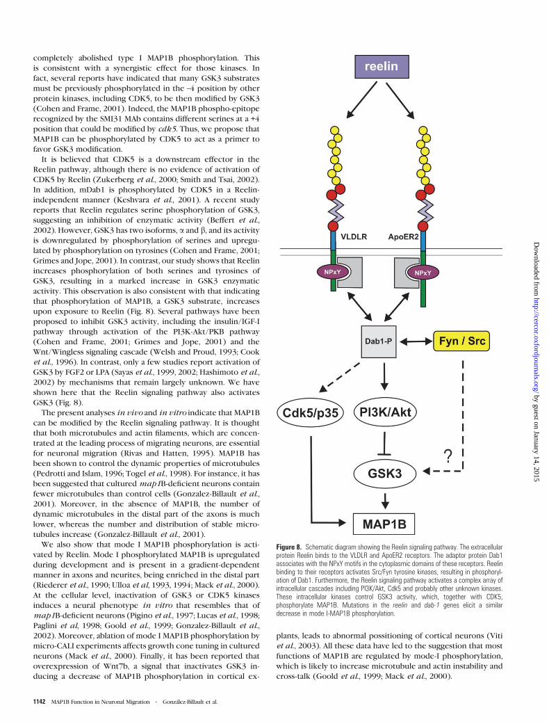
Figure 8. Schematic diagram showing the Reelin signaling pathway. The extracellularprotein Reelin binds to the VLDLR and ApoER2 receptors. The adaptor protein Dab1associates with the NPxY motifs in the cytoplasmic domains of these receptors. Reelinbinding to their receptors activates Src/Fyn tyrosine kinases, resulting in phosphoryl-ation of Dab1. Furthermore, the Reelin signaling pathway activates a complex array ofintracellular cascades including PI3K/Akt, Cdk5 and probably other unknown kinases.These intracellular kinases control GSK3 activity, which, together with CDK5,phosphorylate MAP1B. Mutations in the reelin and dab-1 genes elicit a similardecrease in mode I-MAP1B phosphorylation.
1142 MAP1B Function in Neuronal Migration d Gonzalez-Billault et al.
by guest on January 14, 2015http://cercor.oxfordjournals.org/
Dow
nloaded from
In conclusion, we propose that MAP1B function can be
modified by the Reelin signaling pathway. Absence of MAP1B
can to some extent mimic alterations in the Reelin pathway,
producing abnormal migration of neurons. Nevertheless, MAP1B
function could also be altered by other extracellular cues, acting
either independently or in paralel with Reelin. Finally, the
increase in MAP1B phosphorylation is dependent on GSK3 and
CDK5 protein kinases.
Notes
Christian Gonzalez-Billault and Jose A. Del Rıo contributed equally to
this study. We thank to Drs T. Curran and D. Benhayon (Memphis, TN)
for helpful discussions and for generously providing the Reelin
expression vector and purified Reelin. We thank Drs J. Cooper (Seattle)
and A. Goffinet (Namur) for generously providing us with the B3and
G10 antibodies. We also thank S. Maqueda for technical assistance and
R. Rycroft for editorial assistance. This study was supported by grants
from MCYT (SAF01-3098), The Caixa Foundation, The Pfizer Founda-
tion and The Marato de TV3 Foundation to E.S., from MCYT (EET2002-
05149 and BFI2003-03459) and Thr Caixa Foundation to J.A.D.R., and by
grants from MCYT and The Lilly Foundation to J.A. M.J.B., L.P. and E.M.J.
were supported by fellowships from the Spanish Ministry of Education.
S.S. was supported by a fellowship from MCYT. J.M.U. is a recipient of
a Ramon y Cajal contract from MCYT.
Address correspondence to either Jesus Avila, Centro de Biologia
Molecular Severo Ochoa, CSIC-UAM, Cantonblanco 28049, Madrid,
Spain. Email: [email protected]. Or Eduardo Soriano, IRBB-Parc Cien-
tıfic de Barcelona, Cell and Developmental Biology Programme, Univer-
sity of Barcelona, Josep Samitier 1--5, Barcelona 08028, Spain. Email:
References
Alcantara S, Ruiz M, D’Arcangelo G, Ezan F, de Lecea L, Curran T, Sotelo
C, Soriano E (1998) Regional and cellular patterns of reelin mRNA
expression in the forebrain of the developing and adult mouse. J
Neurosci 18:7779--7799.
Alvarez-Dolado M, Ruiz M, Del Rio JA, Alcantara S, Burgaya F, Sheldon M,
Nakajima K, Bernal J, Howell BW, Curran T, Soriano E, Munoz A
(1999) Thyroid hormone regulates reelin and dab1 expression
during brain development. J Neurosci 19:6979--6993.
Arnaud L, Ballif BA, Forster E, Cooper JA (2003) Fyn tyrosine kinase is
a critical regulator of disabled-1 during brain development. Curr Biol
13:9--17.
Assadi AH, Zhang G, Beffert U, McNeil RS, Renfro AL, Niu S, Quattrocchi
CC, Antalffy BA, Sheldon M, Armstrong DD, Wynshaw-Boris A, Herz J,
D’Arcangelo G, Clark GD (2003) Interaction of reelin signaling and
Lis1 in brain development. Nat Genet 35:270--276.
Avila J, Dominguez J, Diaz-Nido J (1994a) Regulation of microtu-
bule dynamics by microtubule-associated protein expression and
phosphorylation during neuronal development. Int J Dev Biol 38:
13--25.
Avila J, Ulloa L, Diez-Guerra J, Diaz-Nido J (1994b) Role of phosphoryl-
ated MAPlB in neuritogenesis. Cell Biol Int 18:309--314.
Barallobre MJ, Del Rio JA, Alcantara S, Borrell V, Aguado F, Ruiz M,
Carmona MA, Martin M, Fabre M, Yuste R, Tessier-Lavigne M, Soriano
E (2000) Aberrant development of hippocampal circuits and altered
neural activity in netrin 1-deficient mice. Development 127:
4797--4810.
Beffert U, Morfini G, Bock HH, Reyna H, Brady ST, Herz J (2002) Reelin-
mediated signaling locally regulates protein kinase B/Akt and
glycogen synthase kinase 3beta. J Biol Chem 277:49958--49964.
Bilovocky NA, Romito-DiGiacomo RR, Murcia CL, Maricich SM, Herrup K
(2003) Factors in the genetic background suppress the engrailed-1
cerebellar phenotype. J Neurosci 23:5105--5112.
Bock HH, Jossin Y, Liu P, Forster E, May P, Goffinet AM, Herz J (2003)
Phosphatidylinositol 3-kinase interacts with the adaptor protein
Dab1 in response to Reelin signaling and is required for normal
cortical lamination. J Biol Chem 278:38772--38779.
Cahana A, Escamez T, Nowakowski RS, Hayes NL, Giacobini M, von Holst
A, Shmueli O, Sapir T, McConnell SK, Wurst W, Martinez S, Reiner O
(2001) Targeted mutagenesis of Lis1 disrupts cortical development
and LIS1 homodimerization. Proc Natl Acad Sci USA 98:6429--6434.
Chae T, Kwon YT, Bronson R, Dikkes P, Li E, Tsai LH (1997) Mice lacking
p35, a neuronal specific activator of Cdk5, display cortical lamination
defects, seizures, and adult lethality. Neuron 18:29--42.
Chowdhury K, Bonaldo P, Torres M, Stoykova A, Gruss P (1997)
Evidence for the stochastic integration of gene trap vectors into
the mouse germline. Nucleic Acids Res 25:1531--1536.
Cohen P, Frame S (2001) The renaissance of GSK3. Nat Rev Mol Cell Biol
2:769--776.
Cook D, Fry MJ, Hughes K, Sumathipala R, Woodgett JR, Dale TC (1996)
Wingless inactivates glycogen synthase kinase-3 via an intracellular
signalling pathway which involves a protein kinase C. Embo J
15:4526--4536.
Corbo JC, Deuel TA, Long JM, LaPorte P, Tsai E, Wynshaw-Boris A, Walsh
CA (2002) Doublecortin is required in mice for lamination of the
hippocampus but not the neocortex. J Neurosci 22:7548--7557.
D’Arcangelo G, Homayouni R, Keshvara L, Rice DS, Sheldon M, Curran T
(1999) Reelin is a ligand for lipoprotein receptors. Neuron
24:471--479.
D’Arcangelo G, Miao GG, Chen SC, Soares HD, Morgan JI, Curran T
(1995) A protein related to extracellular matrix proteins deleted in
the mouse mutant reeler. Nature 374:719--723.
de Bergeyck V, Naerhuyzen B, Goffinet AM, Lambert de Rouvroit C
(1998) A panel of monoclonal antibodies against reelin, the
extracellular matrix protein defective in reeler mutant mice. J
Neurosci Methods 82:17--24.
del Rio JA, Soriano E (1989) Immunocytochemical detection of 59-
bromodeoxyuridine incorporation in the central nervous system of
the mouse. Brain Res Dev Brain Res 49:311--317.
del Rio JA, Martinez A, Fonseca M, Auladell C, Soriano E (1995)
Glutamate-like immunoreactivity and fate of Cajal--Retzius cells in
the murine cortex as identified with calretinin antibody. Cereb
Cortex 5:13--21.
del Rio JA, Gonzalez-Billault C, Urena JM, Jimenez EM, Barallobre MJ,
Pascual M, Pujadas Ll, Simo S, La Torre A, Wandosell F, Avila J, Soriano
E (2004) MAP1B is required for netrin signaling in neuronal migra-
tion and axonal guidance. Curr Biol 14:840--850.
Diaz-Nido J, Serrano L, Mendez E, Avila J (1988) A casein kinase II-related
activity is involved in phosphorylation of microtubule-associated
protein MAP-1B during neuroblastoma cell differentiation. J Cell Biol
106:2057--2065.
Dulabon L, Olson EC, Taglienti MG, Eisenhuth S, McGrath B, Walsh CA,
Kreidberg JA, Anton ES (2000) Reelin binds alpha3beta1 integrin and
inhibits neuronal migration. Neuron 27:33--44.
Edelmann W, Zervas M, Costello P, Roback L, Fischer I, Hammarback JA,
Cowan N, Davies P, Wainer B, Kucherlapati R (1996) Neuronal
abnormalities in microtubule-associated protein 1B mutant mice.
Proc Natl Acad Sci USA 93:1270--1275.
Feng Y, Walsh CA (2001) Protein--protein interactions, cytoskeletal
regulation and neuronal migration. Nat Rev Neurosci 2:408--416.
Francis F, Koulakoff A, Boucher D, Chafey P, Schaar B, Vinet MC,
Friocourt G, McDonnell N, Reiner O, Kahn A, McConnell SK,
Berwald-Netter Y, Denoulet P, Chelly J (1999) Doublecortin is
a developmentally regulated, microtubule-associated protein ex-
pressed in migrating and differentiating neurons. Neuron
23:247--256.
Garcia-Perez J, Avila J, Diaz-Nido J (1998) Implication of cyclin-
dependent kinases and glycogen synthase kinase 3 in the phosphoryl-
ation of microtubule-associated protein 1B in developing neuronal
cells. J Neurosci Res 52:445--452.
Gilmore EC, Ohshima T, Goffinet AM, Kulkarni AB, Herrup K (1998)
Cyclin-dependent kinase 5-deficient mice demonstrate novel de-
velopmental arrest in cerebral cortex. J Neurosci 18:6370--6377.
Gleeson JG, Lin PT, Flanagan LA, Walsh CA (1999) Doublecortin is
a microtubule-associated protein and is expressed widely by mi-
grating neurons. Neuron 23:257--271.
Gonzalez-Billault C, Demandt E, Wandosell F, Torres M, Bonaldo P,
Stoykova A, Chowdhury K, Gruss P, Avila J, Sanchez MP (2000)
Cerebral Cortex August 2005, V 15 N 8 1143
by guest on January 14, 2015http://cercor.oxfordjournals.org/
Dow
nloaded from
Perinatal lethality of microtubule-associated protein 1B-deficient
mice expressing alternative isoforms of the protein at low levels.
Mol Cell Neurosci 16:408--421.
Gonzalez-Billault C, Avila J, Caceres A (2001) Evidence for the role of
MAP1B in axon formation. Mol Biol Cell 12:2087--2098.
Gonzalez-Billault C, Owen R, Gordon-Weeks PR, Avila J (2002)
Microtubule-associated protein 1B is involved in the initial stages
of axonogenesis in peripheral nervous system cultured neurons.
Brain Res 943:56--67.
Gonzalez-Billault C, Jimenez-Mateos EM, Caceres A, Diaz-Nido J,
Wandosell F, Avila J (2004) Microtubule-associated protein 1B
function during normal development, regeneration and pathological
conditions in the nervous system. J Neurobiol 58:48--59.
Goold RG, Owen R, Gordon-Weeks PR (1999) Glycogen synthase kinase
3beta phosphorylation of microtubule-associated protein 1B regu-
lates the stability of microtubules in growth cones. J Cell Sci
112:3373--3384.
Grimes CA, Jope RS (2001) CREB DNA binding activity is inhibited by
glycogen synthase kinase-3 beta and facilitated by lithium. J Neuro-
chem 78:1219--1232.
Hashimoto T, Nakano Y, Yamashita M, Fang YI, Ohata H, Momose K
(2002) Role of Rho-associated protein kinase and histamine in
lysophosphatidic acid-induced airway hyperresponsiveness in
guinea pigs. Jpn J Pharmacol 88:256--261.
Hiesberger T, Trommsdorff M, Howell BW, Goffinet A, Mumby MC,
Cooper JA, Herz J (1999) Direct binding of Reelin to VLDL receptor
and ApoE receptor 2 induces tyrosine phosphorylation of disabled-1
and modulates tau phosphorylation. Neuron 24:481--489.
Hirotsune S, Takahara T, Sasaki N, Hirose K, Yoshiki A, Ohashi T,
Kusakabe M, Murakami Y, Muramatsu M, Watanabe S, et al. (1995)
The reeler gene encodes a protein with an EGF-like motif expressed
by pioneer neurons. Nat Genet 10:77--83.
Hirotsune S, Fleck MW, Gambello MJ, Bix GJ, Chen A, Clark GD,
Ledbetter DH, McBain CJ, Wynshaw-Boris A (1998) Graded re-
duction of Pafah1b1 (Lis1) activity results in neuronal migration
defects and early embryonic lethality. Nat Genet 19:333--339.
Howell BW, Hawkes R, Soriano P, Cooper JA (1997) Neuronal position in
the developing brain is regulated by mouse disabled-1. Nature
389:733--737.
Howell BW, Herrick TM, Cooper JA (1999a) Reelin-induced tryosine
phosphorylation of disabled 1 during neuronal positioning. Genes
Dev 13:643--648.
Howell BW, Lanier LM, Frank R, Gertler FB, Cooper JA (1999b) The
disabled 1 phosphotyrosine-binding domain binds to the internal-
ization signals of transmembrane glycoproteins and to phospholipids.
Mol Cell Biol 19:5179--5188.
Hummel T, Krukkert K, Roos J, Davis G, Klambt C (2000) Drosophila
Futsch/22C10 is a MAP1B-like protein required for dendritic and
axonal development. Neuron 26:357--370.
Ishiguro K, Kobayashi S, Omori A, Takamatsu M, Yonekura S, Anzai K,
Imahori K, Uchida T (1994) Identification of the 23 kDa subunit of
tau protein kinase II as a putative activator of cdk5 in bovine brain.
FEBS Lett 342:203--208.
Kawauchi T, Chihama K, Nabeshima Y, Hoshino M (2003) The in vivo
roles of STEF/Tiam1, Rac1 and JNK in cortical neuronal migration.
Embo J 22:4190--4201.
Keshvara L, Benhayon D, Magdaleno S, Curran T (2001) Identification of
reelin-induced sites of tyrosyl phosphorylation on disabled 1. J Biol
Chem 276:16008--16014.
Keshvara L, Magdaleno S, Benhayon D, Curran T (2002) Cyclin-
dependent kinase 5 phosphorylates disabled 1 independently of
Reelin signaling. J Neurosci 22:4869--4877.
Ko J, Humbert S, Bronson RT, Takahashi S, Kulkarni AB, Li E, Tsai LH
(2001) p35 and p39 are essential for cyclin-dependent kinase 5
function during neurodevelopment. J Neurosci 21:6758--6771.
Kwon YT, Tsai LH (1998) A novel disruption of cortical development in
p35(–/–) mice distinct from reeler. J Comp Neurol 395:510--522.
Lambert de Rouvroit C, de Bergeyck V, Cortvrindt C, Bar I, Eeckhout Y,
Goffinet AM (1999) Reelin, the extracellular matrix protein deficient
in reeler mutant mice, is processed by a metalloproteinase. Exp
Neurol 156:214--217.
Lucas FR, Goold RG, Gordon-Weeks PR, Salinas PC (1998) Inhibition of
GSK-3beta leading to the loss of phosphorylated MAP-1B is an early
event in axonal remodelling induced by WNT-7a or lithium. J Cell Sci
111:1351--1361.
Mack TG, Koester MP, Pollerberg GE (2000) Themicrotubule-associated
protein MAP1B is involved in local stabilization of turning growth
cones. Mol Cell Neurosci 15:51--65.
Meixner A, Haverkamp S, Wassle H, Fuhrer S, Thalhammer J, Kropf N,
Bittner RE, Lassmann H, Wiche G, Propst F (2000) MAP1B is required
for axon guidance and is involved in the development of the central
and peripheral nervous system. J Cell Biol 151:1169--1178.
Ogawa M, Miyata T, Nakajima K, Yagyu K, Seike M, Ikenaka K, Yamamoto
H, Mikoshiba K (1995) The reeler gene-associated antigen on Cajal--
Retzius neurons is a crucial molecule for laminar organization of
cortical neurons. Neuron 14:899--912.
Ohshima T, Ward JM, Huh CG, Longenecker G, Veeranna, Pant HC,
Brady RO, Martin LJ, Kulkarni AB (1996) Targeted disruption of the
cyclin-dependent kinase 5 gene results in abnormal corticogenesis,
neuronal pathology and perinatal death. Proc Natl Acad Sci USA 93:
11173--11178.
Owen R, Gordon-Weeks PR (2003) Inhibition of glycogen synthase
kinase 3beta in sensory neurons in culture alters filopodia dynamics
and microtubule distribution in growth cones. Mol Cell Neurosci
23:626--637.
Paglini G, Pigino G, Kunda P, Morfini G, Maccioni R, Quiroga S, Ferreira
A, Caceres A (1998) Evidence for the participation of the neuron-
specific CDK5 activator P35 during laminin-enhanced axonal
growth. J Neurosci 18:9858--9869.
Pedrotti B, Islam K (1996) Dephosphorylated but not phosphorylated
microtubule associated protein MAP1B binds to microfilaments.
FEBS Lett 388:131--133.
Pedrotti B, Ulloa L, Avila J, Islam K (1996) Characterization of
microtubule-associated protein MAP1B: phosphorylation state,
light chains, and binding to microtubules. Biochemistry 35:
3016--3023.
Pigino G, Paglini G, Ulloa L, Avila J, Caceres A (1997) Analysis of the
expression, distribution and function of cyclin dependent kinase
5 (cdk5) in developing cerebellar macroneurons. J Cell Sci
110:257--270.
Price DJ, Aslam S, Tasker L, Gillies K (1997) Fates of the earliest gen-
erated cells in the developing murine neocortex. J Comp Neurol
377:414--422.
Rice DS, Curran T (2001) Role of the reelin signaling pathway in central
nervous system development. Annu Rev Neurosci 24:1005--1039.
Riederer BM, Guadano-Ferraz A, Innocenti GM (1990) Difference
in distribution of microtubule-associated proteins 5a and 5b during
the development of cerebral cortex and corpus callosum in cats:
dependence on phosphorylation. Brain Res Dev Brain Res 56:
235--243.
Rivas RJ, Hatten ME (1995) Motility and cytoskeletal organization of
migrating cerebellar granule neurons. J Neurosci 15:981--989.
Roos J, Hummel T, Ng N, Klambt C, Davis GW (2000) Drosophila Futsch
regulates synaptic microtubule organization and is necessary for
synaptic growth. Neuron 26:371--382.
Sayas CL, Moreno-Flores MT, Avila J, Wandosell F (1999) The neurite
retraction induced by lysophosphatidic acid increases Alzheimer’s
disease-like Tau phosphorylation. J Biol Chem 274:37046--37052.
Sayas CL, Avila J, Wandosell F (2002) Glycogen synthase kinase-3 is
activated in neuronal cells by Galpha12 and Galpha13 by Rho-
independent and Rho-dependent mechanisms. J Neurosci 22:
6863--6875.
Schwarz M, Alvarez-Bolado G, Urbanek P, Busslinger M, Gruss P (1997)
Conserved biological function between Pax-2 and Pax-5 in midbrain
and cerebellum development: evidence from targeted mutations.
Proc Natl Acad Sci USA 94:14518--14523.
Senzaki K, Ogawa M, Yagi T (1999) Proteins of the CNR family are
multiple receptors for Reelin. Cell 99:635--647.
Sheldon M, Rice DS, D’Arcangelo G, Yoneshima H, Nakajima K,
Mikoshiba K, Howell BW, Cooper JA, Goldowitz D, Curran T
(1997) Scrambler and yotari disrupt the disabled gene and produce
a reeler-like phenotype in mice. Nature 389:730--733.
1144 MAP1B Function in Neuronal Migration d Gonzalez-Billault et al.
by guest on January 14, 2015http://cercor.oxfordjournals.org/
Dow
nloaded from
Smith DS, Tsai LH (2002) Cdk5 behind the wheel: a role in trafficking
and transport? Trends Cell Biol 12:28--36.
Takei Y, Kondo S, Harada A, Inomata S, Noda T, Hirokawa N (1997)
Delayed development of nervous system in mice homozygous for
disrupted microtubule-associated protein 1B (MAP1B) gene. J Cell
Biol 137:1615--1626.
Takei Y, Teng J, Harada A, Hirokawa N (2000) Defects in axonal
elongation and neuronal migration in mice with disrupted tau and
map1b genes. J Cell Biol 150:989--1000.
Teng J, Takei Y, Harada A, Nakata T, Chen J, Hirokawa N (2001)
Synergistic effects of MAP2 and MAP1B knockout in neuronal
migration, dendritic outgrowth, and microtubule organization. J
Cell Biol 155:65--76.
Tessier-Lavigne M, Goodman CS (1996) The molecular biology of axon
guidance. Science 274:1123--1133.
Togel M, Wiche G, Propst F (1998) Novel features of the light chain of
microtubule-associated protein MAP1B: microtubule stabilization,
self interaction, actin filament binding, and regulation by the heavy
chain. J Cell Biol 143:695--707.
Trommsdorff M, Gotthardt M, Hiesberger T, Shelton J, Stockinger W,
Nimpf J, Hammer RE, Richardson JA, Herz J (1999) Reeler/Disabled-
like disruption of neuronal migration in knockout mice lacking the
VLDL receptor and ApoE receptor 2. Cell 97:689--701.
Ulloa L, Diaz-Nido J, Avila J (1993) Depletion of casein kinase II by
antisense oligonucleotide prevents neuritogenesis in neuroblastoma
cells. Embo J 12:1633--1640.
Ulloa L, Diez-Guerra FJ, Avila J, Diaz-Nido J (1994) Localization of
differentially phosphorylated isoforms of microtubule- associated
protein 1B in cultured rat hippocampal neurons. Neuroscience
61:211--223.
Viti J, Gulacsi A, Lillien L (2003) Wnt regulation of progenitor
maturation in the cortex depends on Shh or fibroblast growth factor
2. J Neurosci 23:5919--5927.
Walsh CA, Goffinet AM (2000) Potential mechanisms of mutations that
affect neuronal migration in man and mouse. Curr Opin Genet Dev
10:270--274.
Welsh GI, Proud CG (1993) Glycogen synthase kinase-3 is rapidly
inactivated in response to insulin and phosphorylates eukaryotic
initiation factor eIF-2B. Biochem J 294:625--629.
Wood JG, Martin S, Price DJ (1992) Evidence that the earliest generated
cells of the murine cerebral cortex form a transient population in the
subplate and marginal zone. Brain Res Dev Brain Res 66:137--140.
Zukerberg LR, Patrick GN, Nikolic M, Humbert S, Wu CL, Lanier LM,
Gertler FB, Vidal M, Van Etten RA, Tsai LH (2000) Cables links Cdk5
and c-Abl and facilitates Cdk5 tyrosine phosphorylation, kinase
upregulation, and neurite outgrowth. Neuron 26:633--646.
Cerebral Cortex August 2005, V 15 N 8 1145
by guest on January 14, 2015http://cercor.oxfordjournals.org/
Dow
nloaded from