eIF2B activator prevents neurological defects caused by a ...
Upload
independentCategory
view
1download
0
10.1128/MCB.25.17.7812-7827.2005.
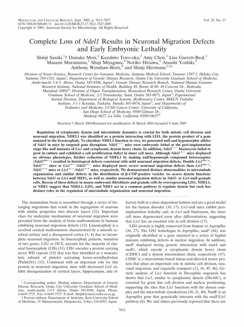
2005, 25(17):7812. DOI:Mol. Cell. Biol. and Shinji HirotsuneNoriko Hiraiwa, Atsushi Yoshiki, Anthony Wynshaw-BorisLisa Garrett-Beal, Masami Muramatsu, Shuji Miyagawa, Shinji Sasaki, Daisuke Mori, Kazuhito Toyo-oka, Amy Chen, Embryonic LethalityNeuronal Migration Defects and Early
Results inNdel1Complete Loss of
http://mcb.asm.org/content/25/17/7812Updated information and services can be found at:
These include:
REFERENCEShttp://mcb.asm.org/content/25/17/7812#ref-list-1at:
This article cites 47 articles, 15 of which can be accessed free
CONTENT ALERTS more»articles cite this article),
Receive: RSS Feeds, eTOCs, free email alerts (when new
http://journals.asm.org/site/misc/reprints.xhtmlInformation about commercial reprint orders: http://journals.asm.org/site/subscriptions/To subscribe to to another ASM Journal go to:
on Novem
ber 7, 2013 by guesthttp://m
cb.asm.org/
Dow
nloaded from
on Novem
ber 7, 2013 by guesthttp://m
cb.asm.org/
Dow
nloaded from
on Novem
ber 7, 2013 by guesthttp://m
cb.asm.org/
Dow
nloaded from
on Novem
ber 7, 2013 by guesthttp://m
cb.asm.org/
Dow
nloaded from
on Novem
ber 7, 2013 by guesthttp://m
cb.asm.org/
Dow
nloaded from
on Novem
ber 7, 2013 by guesthttp://m
cb.asm.org/
Dow
nloaded from
on Novem
ber 7, 2013 by guesthttp://m
cb.asm.org/
Dow
nloaded from
on Novem
ber 7, 2013 by guesthttp://m
cb.asm.org/
Dow
nloaded from
on Novem
ber 7, 2013 by guesthttp://m
cb.asm.org/
Dow
nloaded from
on Novem
ber 7, 2013 by guesthttp://m
cb.asm.org/
Dow
nloaded from
on Novem
ber 7, 2013 by guesthttp://m
cb.asm.org/
Dow
nloaded from
on Novem
ber 7, 2013 by guesthttp://m
cb.asm.org/
Dow
nloaded from
on Novem
ber 7, 2013 by guesthttp://m
cb.asm.org/
Dow
nloaded from
on Novem
ber 7, 2013 by guesthttp://m
cb.asm.org/
Dow
nloaded from
on Novem
ber 7, 2013 by guesthttp://m
cb.asm.org/
Dow
nloaded from
on Novem
ber 7, 2013 by guesthttp://m
cb.asm.org/
Dow
nloaded from
on Novem
ber 7, 2013 by guesthttp://m
cb.asm.org/
Dow
nloaded from
MOLECULAR AND CELLULAR BIOLOGY, Sept. 2005, p. 7812–7827 Vol. 25, No. 170270-7306/05/$08.00�0 doi:10.1128/MCB.25.17.7812–7827.2005Copyright © 2005, American Society for Microbiology. All Rights Reserved.
Complete Loss of Ndel1 Results in Neuronal Migration Defectsand Early Embryonic Lethality
Shinji Sasaki,1† Daisuke Mori,2 Kazuhito Toyo-oka,2 Amy Chen,3 Lisa Garrett-Beal,3Masami Muramatsu,1 Shuji Miyagawa,4 Noriko Hiraiwa,5 Atsushi Yoshiki,5
Anthony Wynshaw-Boris,6 and Shinji Hirotsune1,2*Division of Neuro-Science, Research Center for Genomic Medicine, Saitama Medical School, Yamane 1397-1, Hidaka City,Saitama 350-1241, Japan1; Department of Genetic Disease Research, Osaka City University Graduate School of Medicine,
Asahi-machi 1-4-3, Abeno, Osaka 545-8586, Japan2; Genetic Disease Research Branch, National Human GenomeResearch Institute, National Institutes of Health, Building 49, Room 4C80, 49 Convent Dr., Bethesda,Maryland 208923; Division of Organ Transplantation, Biomedical Research Center, Osaka University
Graduate School of Medicine, 2-2 Yamadaoka, Suita, Osaka 565-0871, Japan4; ExperimentalAnimal Division, Department of Biological Systems, BioResource Center, RIKEN Tsukuba
Institute, 3-1-1 Koyadai, Tsukuba, Ibaraki 305-0074, Japan5; and Departments ofPediatrics and Medicine, UCSD Cancer Center, University of California,
San Diego School of Medicine, 9500 Gilman Dr.,Mailstop 0627, La Jolla, California 92093-06276
Received 3 March 2004/Returned for modification 28 March 2005/Accepted 8 June 2005
Regulation of cytoplasmic dynein and microtubule dynamics is crucial for both mitotic cell division andneuronal migration. NDEL1 was identified as a protein interacting with LIS1, the protein product of a genemutated in the lissencephaly. To elucidate NDEL1 function in vivo, we generated null and hypomorphic allelesof Ndel1 in mice by targeted gene disruption. Ndel1�/� mice were embryonic lethal at the peri-implantationstage like null mutants of Lis1 and cytoplasmic dynein heavy chain. In addition, Ndel1�/� blastocysts failed togrow in culture and exhibited a cell proliferation defect in inner cell mass. Although Ndel1�/� mice displayedno obvious phenotypes, further reduction of NDEL1 by making null/hypomorph compound heterozygotes(Ndel1cko/�) resulted in histological defects consistent with mild neuronal migration defects. Double Lis1cko/�-Ndel1�/� mice or Lis1�/�-Ndel1�/� mice displayed more severe neuronal migration defects than Lis1cko/�-Ndel1�/� mice or Lis1�/�-Ndel1�/� mice, respectively. We demonstrated distinct abnormalities in microtubuleorganization and similar defects in the distribution of �-COP-positive vesicles (to assess dynein function)between Ndel1 or Lis1-null MEFs, as well as similar neuronal migration defects in Ndel1- or Lis1-null granulecells. Rescue of these defects in mouse embryonic fibroblasts and granule cells by overexpressing LIS1, NDEL1,or NDE1 suggest that NDEL1, LIS1, and NDE1 act in a common pathway to regulate dynein but each hasdistinct roles in the regulation of microtubule organization and neuronal migration.
The mammalian brain is assembled through a series of far-ranging migrations that result in the segregation of neuronswith similar properties into discrete layers (32). Importantclues for molecular mechanisms of neuronal migration wereprovided from the analysis of brain malformations in humansexhibiting neuronal migration defects (13). Lissencephaly is acerebral cortical malformation characterized by a smooth ce-rebral surface and a disorganized cortex (3, 4) due to incom-plete neuronal migration. In lissencephaly patients, mutationof two genes, LIS1 or DCX, account for the majority of clas-sical lissencephaly (LIS) (31). LIS1 encodes a protein carryingseven WD repeats (33) that was first identified as a noncata-lytic subunit of platelet activating factor-acetylhydrolase(Pafah1b1) (15). Consistent with an important role for thisprotein in neuronal migration, mice with decreased Lis1 ex-hibit disorganization of cortical layers, hippocampus, and ol-
factory bulb in a dose-dependent fashion and are a good modelfor the human disorder (10, 17). Lis1-null mice exhibit peri-implantation lethality and, in Lis1-null blastocysts, the innercell mass degenerated soon after differentiation, suggestingthat Lis1 has an essential role in cell division (17).
LIS1 protein is highly conserved from human to Aspergillus(26, 27). The LIS1 homologue in Aspergillus, nudF (44), wasoriginally identified as a gene mutated in a series of hyphalmutants exhibiting defects in nuclear migration. In addition,nudF displayed strong genetic interaction with nudA andnudG, which encode a cytoplasmic dynein heavy chain(CDHC) and a dynein intermediate chain, respectively (47).CDHC is a microtubule-based minus-end-directed motor pro-tein that plays an important role in mitotic cell division, neu-ronal migration, and organelle transport (12, 16, 45, 46). Ge-netic analysis of Lis1 function in Drosophila oogenesis hasshown that Lis1, similar to cytoplasmic dynein (Dhc64C), isessential for germ line cell division and nuclear positioning,supporting the idea that Lis1 functions with the dynein com-plex and the microtubule cytoskeleton (20, 21, 40). NudE is anAspergillus gene that genetically interacts with the nudF/Lis1pathway (6). We and others previously reported that there are
* Corresponding author. Mailing address: Department of GeneticDisease Research, Osaka City University Graduate School of Medi-cine, Asahi-machi 1-4-3 Abeno, Osaka 545-8586, Japan. Phone:6-6645-3725. Fax: 6-6645-3727. E-mail: [email protected].
† Present address: Department of Anatomy, Keio University Schoolof Medicine, 35 Shinanomachi, Shinjuku-ku, Tokyo 160-8582, Japan.
7812

FIG. 1. Generation of a gene-disrupted mouse of Ndel1 [Ndel1cko(III), Ndel1ko(III)]. (A) Summary of targeting strategy for the Ndel1 locus. Adiagrammatic representation of the targeting vector and genomic loci of Ndel1 gene is shown. Exons are represented by gray boxes. In this genetargeting, Ndel1 function was expected to be intact (Ndel1cko(III)). Exon 3 was removed by CRE-mediated recombination to inactivate Ndel1function [Ndel1ko(III)]. (B) Southern blot analysis of tail DNA from wild-type (�/�, lane 1) and heterozygous mutant (�/�, lanes 2 and 3) animals.(C) Southern blot analysis of tail DNA from deletion mutant (�/del, lane 1), wild-type (�/�, lane 2), and heterozygous mutant (�/loxP, lane 3)mice. CRE-mediated deletion of exon 3 was evaluated by Southern blotting. Note the difference in band sizes after deletion. We used Ndel1ko(III)
as a Ndel1�/� in the all experiment.
VOL. 25, 2005 RESULTS OF COMPLETE LOSS OF Ndel1 7813
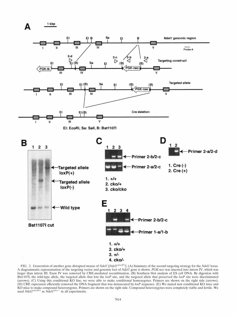
FIG. 2. Generation of another gene disrupted mouse of Ndel1 [Ndel1cko(IV)]. (A) Summary of the second targeting strategy for the Ndel1 locus.A diagrammatic representation of the targeting vector and genomic loci of Ndel1 gene is shown. PGK-neo was inserted into intron IV, which waslarger than intron III. Exon IV was removed by CRE-mediated recombination. (B) Southern blot analysis of ES cell DNA. By digestion withBst1107I, the wild-type allele, the targeted allele that lost the loxP site, and the targeted allele that preserved the loxP site were discriminated(arrows). (C) Using this conditional KO line, we were able to make conditional homozygotes. Primers are shown on the right side (arrows).(D) CRE expression efficiently removed the DNA fragment that was demarcated by loxP sequence. (E) We mated new conditional KO mice andKO mice to make compound heterozygotes. Primers are shown on the right side. Compound heterozygotes were completely viable and fertile. Weused Ndel1cko(IV) as Ndel1cko/� in all experiments.
7814
two mammalian homologues of Aspergillus NudE, NDE1 (8)and NDEL1 (29, 35, 36); the latter, a homologue of NudE fromAspergillus nidulans, is a LIS1 binding protein that participateswith LIS1 in the regulation of CDHC function via phosphor-ylation by CDK5/p35 (8, 29, 35, 36), a complex known to beessential for neuronal migration (1, 7, 11, 13, 18, 30). We havealso shown that phosphorylation of NDEL1 is protected by theserine-threonine binding protein 14-3-3ε and that 14-3-3ε isrequired for normal neuronal migration (42), providing furthersupport that the phosphorylation of NDEL1 is required forproper neuronal migration. NDE1 has a similar function incells but is expressed later in development than NDEL1. Re-cently, Nde1-null mice have been produced (9). These mice areviable and display microcephaly. They display defects in neu-rogenesis that result from reduced progenitor cell division andprogenitor cell fate, as well as modest defects in neuronalmigration. To date, targeted mutants of Ndel1 have not beengenerated, and the in vitro phenotype of null mutants of Ndel1is unknown.
To understand the role of NDEL1 in vivo and to determinewhether it plays a role in corticogenesis and cell division, wegenerated Ndel1-disrupted mice. We found that, similar toLis1, Ndel1 mutant mice display a dosage-dependent neuronalmigration phenotype, and complete loss of Ndel1 resulted inperinatal lethality. Examination of in vivo and in vitro pheno-types suggests that NDEL1, LIS1, and NDE1 act in a commonpathway to regulate dynein, but each has distinct roles in theregulation of microtubule organization and neuronal migration.
MATERIALS AND METHODS
Generation and analysis of Ndel1 KO mice. To understand the function ofNdel1 in vivo, we generated a first conditional knockout (KO) mouse to inacti-vate Ndel1 by Cre-mediated recombination. We assembled a targeting constructin which a PGK-neo gene flanked by a loxP site and an additional loxP site wereinserted into intron III and intron II, respectively. The linearized targetingconstruct was introduced into TC1 embryonic stem (ES) cells (2) from a 129S6background by electroporation. The targeted ES clones were screened by South-ern blot and injected into blastocysts to create chimeric mice. Highly agouti
chimeric males were mated to wild-type females to give rise to heterozygotes forthe conditional allele [Ndel1cko(III)/�], which was identified by Southern blotanalysis and PCR. These heterozygous mice were mated with EIIa-Cre germ linedeleter transgenic mice (19). Offspring from the matings between anNdel1cko(III)/� line and an EIIa-Cre transgenic line were genotyped by Southernblot analysis and PCR. Southern blot analysis and PCR examination indicatedefficient deletion of the fragment carrying exon III by CRE mediated recombi-nation in vivo. Ndel1cko(III)/cko(III) mice exhibited embryonic lethality similar withNdel1ko(III)/ko(III) mice, suggesting that the presence of the neo gene severelyaffects expression of Ndel1. It was not possible to remove neo gene by itself. Forthe second conditional KO strategy, we inserted a PGK-neo gene flanked by aloxP site and an additional loxP site into intron IV and intron III, respectively.Highly agouti chimeric males were mated to wild-type females to give rise toheterozygotes for the second conditional allele [Ndel1cko(IV)/�]. In contrast to thefirst line of conditional KO, homozygotes of the second conditional KO[Ndel1cko(IV)/cko(IV)] line was completely viable and fertile. CRE-mediated dele-tion of the second conditional KO mouse line was also confirmed by PCR withprimers located flanking the site of the deleted region. We used Ndel1ko(III)/� asNdel1�/�, whereas Ndel1cko(IV)/� was used as Ndel1cko/� in this experiment.Expression of NDEL1 was examined by Western blotting with the C-6 NDEL1specific antibody (36) and Northern blotting with a full-length Ndel1 cDNAfragment. All experiments with mouse models were performed based on theanimal experiment guidelines of our universities.
BrdU birth dating study. For bromodeoxyuridine (BrdU) experiments, preg-nant dams (embryonic day 15.5 [E15.5]) were injected with BrdU (50 �g/g,intraperitoneally). Subsequently, the distribution of BrdU-positive cells was de-termined at P0. The incorporation of BrdU in cells was detected with a mouseanti-BrdU monoclonal primary antibody (Roche), followed by an alkaline phos-phatase-conjugated secondary antibody (Boehringer Mannheim). We analyzedthree independent mice for each genotype.
Histological examination and immunohistochemistry. After perfusion withBouin’s or 4% paraformaldehyde fixative, tissues from wild-type and variousmutant mice were subsequently embedded in paraffin and sectioned at 5-�mthickness. After deparaffination, endogenous peroxidase activity was blocked byincubating the sections in 1.5% peroxide in methanol for 20 min. The sectionswere then boiled in 0.01 mol of citrate buffer (pH 6.0)/liter for 20 min and cooledslowly. Before staining, the sections were blocked with rodent block (LabVision)for 60 min. The sections were washed in phosphate-buffered saline and incubatedwith each antibody. Primary cultures of cerebellar granule cells were generatedfrom 5-day-old C57BL/6 pups as described previously (36). Cells were plated ata density of 2.5 � 105 cells/cm2 and maintained in basal Eagle medium contain-ing 10% fetal calf serum, 25 mM KCl, 2 mM glutamine, and 50 mg of gentamicin/ml. After 18 to 20 h, cytosine arabinoside (10 mM) was added to the culturemedia to halt non-neuronal cell proliferation. Blastocysts were collected byflushing the oviducts of female mice at 3.5 days postcoitum and cultured indi-vidually for 3 days on gelatinized coverslips in ES cell medium. Cultured blas-tocysts were fixed in 2% paraformaldehyde–phosphate-buffered saline for 10 minat room temperature. Some samples were fixed in 2% paraformaldehyde and0.2% glutaraldehyde for 10 min at room temperature and blocked in 1 mg ofNaBH4/ml for 30 min at 4°C. After primary culture from each tissue, cells werefixed in cold methanol (�20°C) and postfixed by 3% formaldehyde. Incubationwith 0.15% Triton X-100 was used for cell permeabilization after fixation. Im-munohistochemistry was performed based on the standard procedure. Apoptoticcells were detected by TUNEL (terminal deoxynucleotidyltransferase-mediateddUTP-biotin nick end labeling) assay (Chemicon). Incorporated biotin was de-tected with an ABC kit and visualized with diaminobenzidene.
Primary cell culture, transfection, and immunofluorescence. Establishment ofmouse embryonic fibroblasts (MEFs) was performed as previously described (17,36). A red fluorescent protein (RFP)-tagged Cre expression vector was intro-duced into these primary culture cells by using Lipofectamine 2000 reagents(Invitrogen). After CRE-mediated inactivation of Lis1 or Ndel1 gene, these cellswere subjected to immunohistochemistry by using anti-�-tubulin antibody (CloneTUB 2.1; Sigma) or anti-�-COP antibody (Sigma). For rescue experiments,green fluorescent protein (GFP)-conjugated to LIS1, NDEL1, or NDE1 wassimultaneously transfected with the CRE expression vector.
Reaggregate neuronal migration assay. Cerebellar granule neurons were dis-sociated from postnatal 5-day-old mice (17, 41) and transfected with variousvectors by using LipoTrust SR transfection reagents (Hokkaido System ScienceCo., Ltd.) according to the manufacturer’s instructions with a small modification.Briefly, 2 � 106 cells were transfected with 3 �g of DNA vector in 4 �l ofLipoTrust SR reagents in 100 �l of Dulbecco modified Eagle medium mediawithout fetal bovine serum for 20 h, resulting in 30 to 40% transfection efficiency.Reaggregates were transferred to poly-L-lysine (Sigma-Aldrich)- and laminin
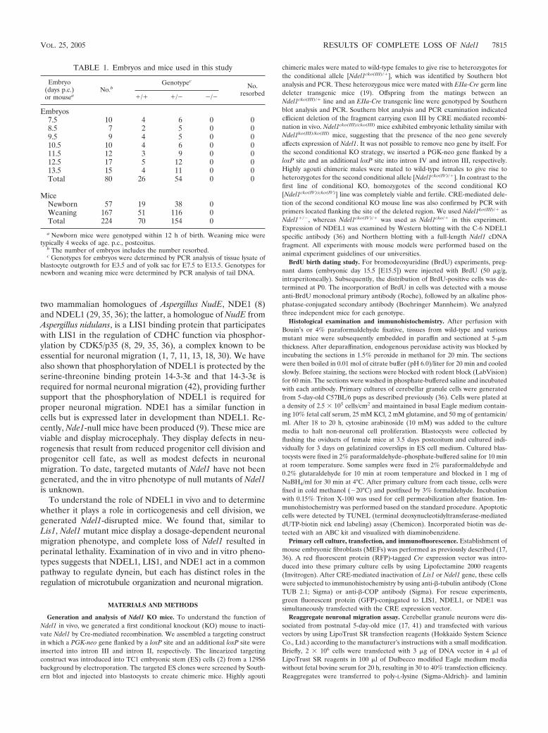
TABLE 1. Embryos and mice used in this study
Embryo(days p.c.)or mousea
No.bGenotypec
No.resorbed�/� �/� �/�
Embryos7.5 10 4 6 0 08.5 7 2 5 0 09.5 9 4 5 0 010.5 10 4 6 0 011.5 12 3 9 0 012.5 17 5 12 0 013.5 15 4 11 0 0Total 80 26 54 0 0
MiceNewborn 57 19 38 0Weaning 167 51 116 0Total 224 70 154 0
a Newborn mice were genotyped within 12 h of birth. Weaning mice weretypically 4 weeks of age. p.c., postcoitus.
b The number of embryos includes the number resorbed.c Genotypes for embryos were determined by PCR analysis of tissue lysate of
blastocyte outgrowth for E3.5 and of yolk sac for E7.5 to E13.5. Genotypes fornewborn and weaning mice were determined by PCR analysis of tail DNA.
VOL. 25, 2005 RESULTS OF COMPLETE LOSS OF Ndel1 7815

FIG. 3. Embryonic lethality of Ndel1-null mutants. (A) Homozygous mutants of Ndel1 were not observed, suggesting that null mutants areembryonic lethal. Normal (left) and degenerated deciduas (right) resulting from the Ndel1�/� cross were sectioned at E5.5 and stained withhematoxylin and eosin. ec, ectoplacental cone; ee, extra-embryonic ectoderm; e, epiblast. (B) Wild-type (upper) and homozygous (lower)blastocysts were isolated from a heterozygous cross and cultured. Both blastocysts hatched and differentiated into trophoblast (tr) and inner cellmass (ICM). The inner cell mass of homozygotes exhibited poor growth and degenerated. (C) Quantitation of the amount of Ndel1 expression inthe various genotypes. Northern blot (left side) and Western blot (right side) are shown. Expression was calculated by densitometry compared tothe wild type. We repeated three independent experiments, and the results were highly reproducible. (D) Expression of LIS1 or NDEL1 in Ndel1KO mice or Lis1 KO mice was examined. Total protein was extracted from a brain of E15.5 embryo and subjected to Western blotting. There wasno obvious difference in LIS1 or NDEL1 expression in Ndel1 or Lis1 KO mice, respectively.
7816

(Sigma-Aldrich)-treated slides. After 15 h, the distance between cell bodies andthe edge of reaggregates was measured. The details of the experimental proce-dure have been described (17, 41). An RFP-tagged Cre expression vector with orwithout GFP-tagged LIS1, NDEL1, or NDE1 vector were transfected as de-scribed above.
RESULTS
Targeted gene disruption of Ndel1. To explore the in vivorole of NDEL1 and genetic interaction between Ndel1 and Lis1during mitotic cell division and neuronal development, we gen-erated two independent Ndel1 conditional KO lines (Fig. 1 andFig. 2; see also Materials and Methods). We could not deletethe PGK-neo gene used for selection in ES cells in the exon IIIfloxed allele, and the undeleted allele with the neo insertionhad a homozygous lethal phenotype identical to that of the nullallele produced after deletion of the PGK-neo gene and exonIII (see Materials and Methods). Therefore, we produced aconditional allele that floxed exon IV. We used mice in whichexon III of Ndel1 was deleted as Ndel1�/�, whereas we usedmice in which exon IV of Ndel1 was conditionally deleted asNdel1cko/�.
Mice heterozygous for the Ndel1 gene (Ndel1�/�) were out-
wardly normal, fertile born in appropriate Mendelian ratios.Ndel1�/� mice were bred to produce homozygous mutants(Ndel1�/�). An analysis of F2s from the mating of Ndel1�/�
mice showed that the genotype ratios of �/� to �/� to �/�animals were 70:154:0, suggesting that the complete loss ofNdel1 resulted in embryonic lethality (Table 1). Therefore, weexamined embryos from heterozygous crosses at several devel-opmental stages. Cumulative genotyping between E7.5 andE13.5 demonstrated that the ratios of �/� to �/� to �/�mice were 26:54:0 (Table 1). We dissected embryos at betweenE5.5 and E13.5 and found degenerated embryos and emptydeciduas only at the earliest postimplantation stages (Fig. 3A).We found 35 normal embryos (80%) and 10 empty decidua(20%) at E5.5 among 45 animals, and there were 5 �/� (21%),15 �/� (66%), and 3 �/� (13%) blastocyst cultures at E3.5among 23 animals. These findings suggested that Ndel1�/�
mice are lethal at very early stages of embryogenesis. This is incontrast to Nde1-null mice, which are viable and display mi-crocephaly from defects in neurogenesis (9).
To define the role of NDEL1 in the early embryo, we ex-amined the behavior of blastocyst explants isolated from
FIG. 4. Synergistic effects of Lis1 and Ndel1 mutations on brain morphogenesis. Genotypes are shown at the top of the panels. (A) Midsagittalsections of cerebral cortex (upper) and hippocampus (lower). Lis1cko/� or Ndel1�/� samples were grossly normal. In contrast, mild cell dispersionof CA3 region was observed in Ndel1cko/� compound heterozygotes or Lis1cko/� and Ndel1�/� double heterozygotes (arrowheads). (B) Loss of cellcompaction (arrowheads) in CA3 region was clearly observed by NeuN staining. (C) Abnormal corticogenesis was characterized by fragmentationof the subplate layer (arrowheads) which was visualized by MAP2 staining (upper panels) and chondroitin sulfate proteoglycan staining (lowerpanels) at E15.5. MZ, marginal zone; CP, cortical plate; SP, subplate; IZ, intermediate zone.
VOL. 25, 2005 RESULTS OF COMPLETE LOSS OF Ndel1 7817

FIG. 5. Examination of migration defects of Lis1 and Ndel1 mutants. (A) BrdU birth dating analysis reveals neuronal migration defects in thecross of Ndel1�/� and Ndel1cko/�, in the cross of Lis1cko/� and Ndel1�/�, or in the cross of Lis1�/� and Ndel1�/�. Genotypes are described at thetop of the panels. Mice were injected with BrdU at E15.5 and sacrificed at P0. Quantitative analysis was performed by measuring the distributionof BrdU-labeled cells in each bin which equally divided the cortex from the molecular layer (ML) to the subplate (SP). The staining patterns arerepresentative of three different experiments. Note the shift downward toward the ventricular side as Lis1 or Ndel1 dosage was reduced. CPs,cortical plate surface; CPi, cortical plate inner. (B) Summary of mean migration distance of each genotype.
7818 SASAKI ET AL. MOL. CELL. BIOL.
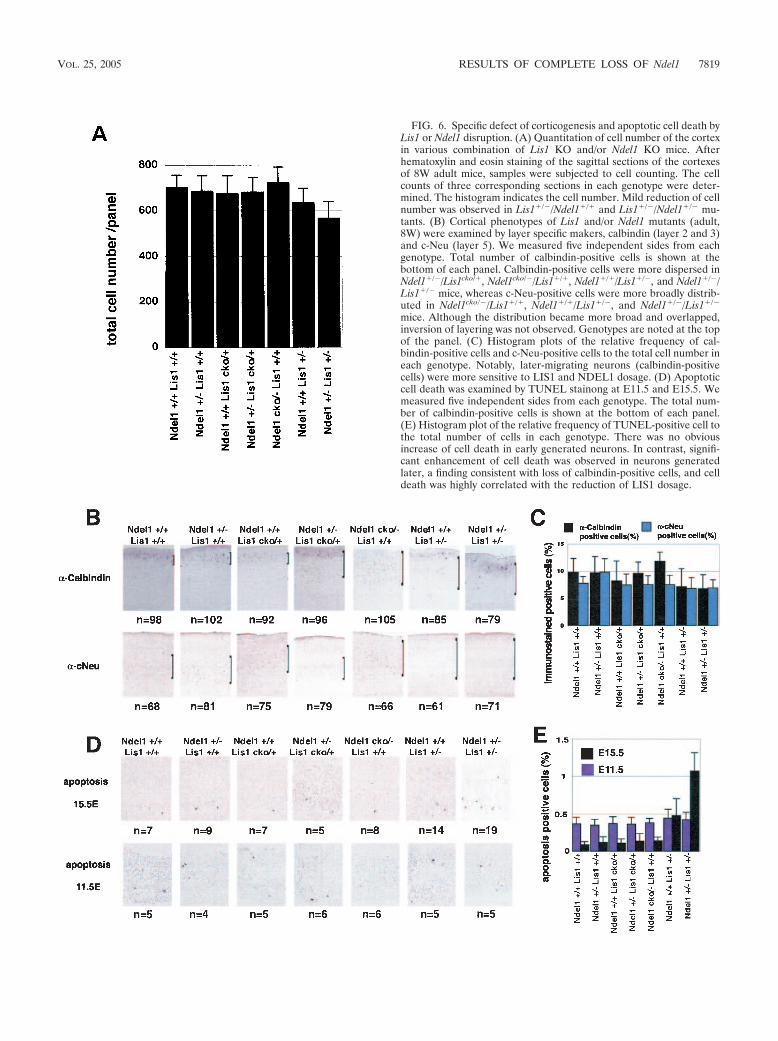
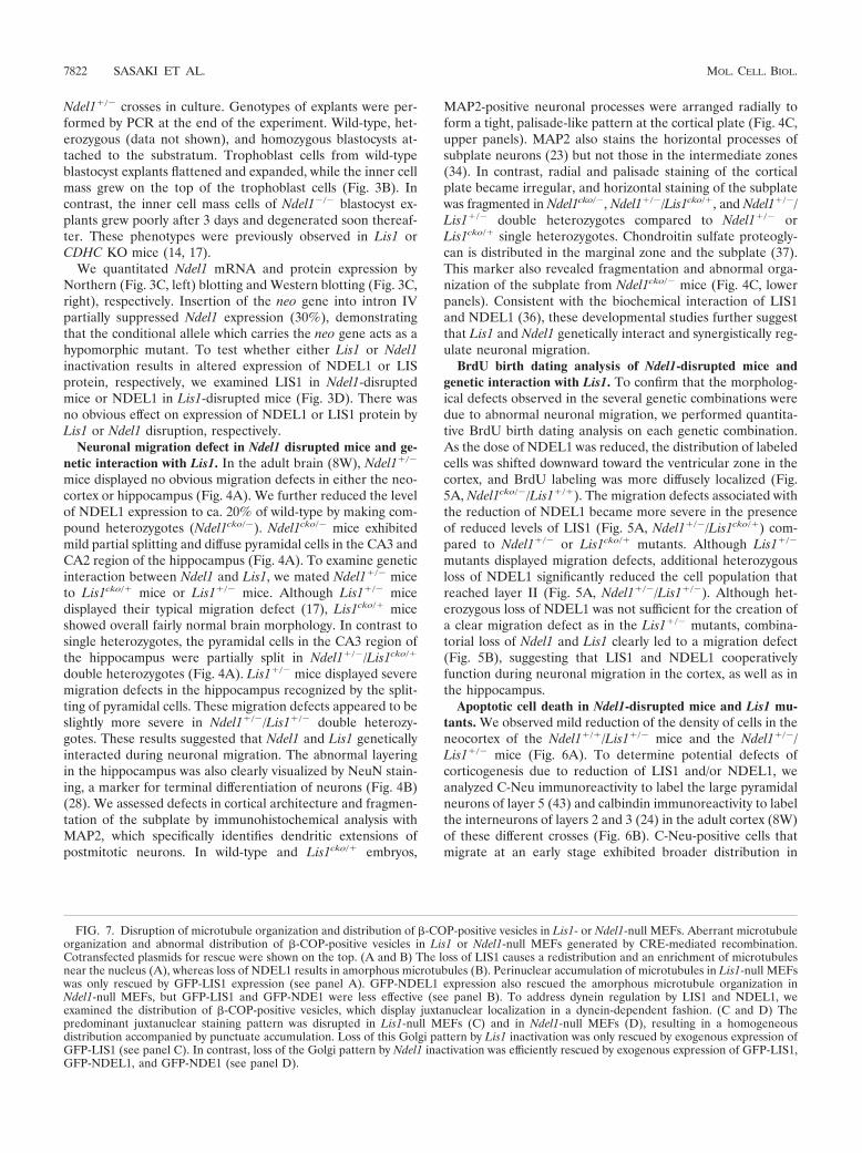
FIG. 6. Specific defect of corticogenesis and apoptotic cell death byLis1 or Ndel1 disruption. (A) Quantitation of cell number of the cortexin various combination of Lis1 KO and/or Ndel1 KO mice. Afterhematoxylin and eosin staining of the sagittal sections of the cortexesof 8W adult mice, samples were subjected to cell counting. The cellcounts of three corresponding sections in each genotype were deter-mined. The histogram indicates the cell number. Mild reduction of cellnumber was observed in Lis1�/�/Ndel1�/� and Lis1�/�/Ndel1�/� mu-tants. (B) Cortical phenotypes of Lis1 and/or Ndel1 mutants (adult,8W) were examined by layer specific makers, calbindin (layer 2 and 3)and c-Neu (layer 5). We measured five independent sides from eachgenotype. Total number of calbindin-positive cells is shown at thebottom of each panel. Calbindin-positive cells were more dispersed inNdel1�/�/Lis1cko/�, Ndel1cko/�/Lis1�/�, Ndel1�/�/Lis1�/�, and Ndel1�/�/Lis1�/� mice, whereas c-Neu-positive cells were more broadly distrib-uted in Ndel1cko/�/Lis1�/�, Ndel1�/�/Lis1�/�, and Ndel1�/�/Lis1�/�
mice. Although the distribution became more broad and overlapped,inversion of layering was not observed. Genotypes are noted at the topof the panel. (C) Histogram plots of the relative frequency of cal-bindin-positive cells and c-Neu-positive cells to the total cell number ineach genotype. Notably, later-migrating neurons (calbindin-positivecells) were more sensitive to LIS1 and NDEL1 dosage. (D) Apoptoticcell death was examined by TUNEL stainong at E11.5 and E15.5. Wemeasured five independent sides from each genotype. The total num-ber of calbindin-positive cells is shown at the bottom of each panel.(E) Histogram plot of the relative frequency of TUNEL-positive cell tothe total number of cells in each genotype. There was no obviousincrease of cell death in early generated neurons. In contrast, signifi-cant enhancement of cell death was observed in neurons generatedlater, a finding consistent with loss of calbindin-positive cells, and celldeath was highly correlated with the reduction of LIS1 dosage.
VOL. 25, 2005 RESULTS OF COMPLETE LOSS OF Ndel1 7819

7820 SASAKI ET AL. MOL. CELL. BIOL.
VOL. 25, 2005 RESULTS OF COMPLETE LOSS OF Ndel1 7821
Ndel1�/� crosses in culture. Genotypes of explants were per-formed by PCR at the end of the experiment. Wild-type, het-erozygous (data not shown), and homozygous blastocysts at-tached to the substratum. Trophoblast cells from wild-typeblastocyst explants flattened and expanded, while the inner cellmass grew on the top of the trophoblast cells (Fig. 3B). Incontrast, the inner cell mass cells of Ndel1�/� blastocyst ex-plants grew poorly after 3 days and degenerated soon thereaf-ter. These phenotypes were previously observed in Lis1 orCDHC KO mice (14, 17).
We quantitated Ndel1 mRNA and protein expression byNorthern (Fig. 3C, left) blotting and Western blotting (Fig. 3C,right), respectively. Insertion of the neo gene into intron IVpartially suppressed Ndel1 expression (30%), demonstratingthat the conditional allele which carries the neo gene acts as ahypomorphic mutant. To test whether either Lis1 or Ndel1inactivation results in altered expression of NDEL1 or LISprotein, respectively, we examined LIS1 in Ndel1-disruptedmice or NDEL1 in Lis1-disrupted mice (Fig. 3D). There wasno obvious effect on expression of NDEL1 or LIS1 protein byLis1 or Ndel1 disruption, respectively.
Neuronal migration defect in Ndel1 disrupted mice and ge-netic interaction with Lis1. In the adult brain (8W), Ndel1�/�
mice displayed no obvious migration defects in either the neo-cortex or hippocampus (Fig. 4A). We further reduced the levelof NDEL1 expression to ca. 20% of wild-type by making com-pound heterozygotes (Ndel1cko/�). Ndel1cko/� mice exhibitedmild partial splitting and diffuse pyramidal cells in the CA3 andCA2 region of the hippocampus (Fig. 4A). To examine geneticinteraction between Ndel1 and Lis1, we mated Ndel1�/� miceto Lis1cko/� mice or Lis1�/� mice. Although Lis1�/� micedisplayed their typical migration defect (17), Lis1cko/� miceshowed overall fairly normal brain morphology. In contrast tosingle heterozygotes, the pyramidal cells in the CA3 region ofthe hippocampus were partially split in Ndel1�/�/Lis1cko/�
double heterozygotes (Fig. 4A). Lis1�/� mice displayed severemigration defects in the hippocampus recognized by the split-ting of pyramidal cells. These migration defects appeared to beslightly more severe in Ndel1�/�/Lis1�/� double heterozy-gotes. These results suggested that Ndel1 and Lis1 geneticallyinteracted during neuronal migration. The abnormal layeringin the hippocampus was also clearly visualized by NeuN stain-ing, a marker for terminal differentiation of neurons (Fig. 4B)(28). We assessed defects in cortical architecture and fragmen-tation of the subplate by immunohistochemical analysis withMAP2, which specifically identifies dendritic extensions ofpostmitotic neurons. In wild-type and Lis1cko/� embryos,
MAP2-positive neuronal processes were arranged radially toform a tight, palisade-like pattern at the cortical plate (Fig. 4C,upper panels). MAP2 also stains the horizontal processes ofsubplate neurons (23) but not those in the intermediate zones(34). In contrast, radial and palisade staining of the corticalplate became irregular, and horizontal staining of the subplatewas fragmented in Ndel1cko/�, Ndel1�/�/Lis1cko/�, and Ndel1�/�/Lis1�/� double heterozygotes compared to Ndel1�/� orLis1cko/� single heterozygotes. Chondroitin sulfate proteogly-can is distributed in the marginal zone and the subplate (37).This marker also revealed fragmentation and abnormal orga-nization of the subplate from Ndel1cko/� mice (Fig. 4C, lowerpanels). Consistent with the biochemical interaction of LIS1and NDEL1 (36), these developmental studies further suggestthat Lis1 and Ndel1 genetically interact and synergistically reg-ulate neuronal migration.
BrdU birth dating analysis of Ndel1-disrupted mice andgenetic interaction with Lis1. To confirm that the morpholog-ical defects observed in the several genetic combinations weredue to abnormal neuronal migration, we performed quantita-tive BrdU birth dating analysis on each genetic combination.As the dose of NDEL1 was reduced, the distribution of labeledcells was shifted downward toward the ventricular zone in thecortex, and BrdU labeling was more diffusely localized (Fig.5A, Ndel1cko/�/Lis1�/�). The migration defects associated withthe reduction of NDEL1 became more severe in the presenceof reduced levels of LIS1 (Fig. 5A, Ndel1�/�/Lis1cko/�) com-pared to Ndel1�/� or Lis1cko/� mutants. Although Lis1�/�
mutants displayed migration defects, additional heterozygousloss of NDEL1 significantly reduced the cell population thatreached layer II (Fig. 5A, Ndel1�/�/Lis1�/�). Although het-erozygous loss of NDEL1 was not sufficient for the creation ofa clear migration defect as in the Lis1�/� mutants, combina-torial loss of Ndel1 and Lis1 clearly led to a migration defect(Fig. 5B), suggesting that LIS1 and NDEL1 cooperativelyfunction during neuronal migration in the cortex, as well as inthe hippocampus.
Apoptotic cell death in Ndel1-disrupted mice and Lis1 mu-tants. We observed mild reduction of the density of cells in theneocortex of the Ndel1�/�/Lis1�/� mice and the Ndel1�/�/Lis1�/� mice (Fig. 6A). To determine potential defects ofcorticogenesis due to reduction of LIS1 and/or NDEL1, weanalyzed C-Neu immunoreactivity to label the large pyramidalneurons of layer 5 (43) and calbindin immunoreactivity to labelthe interneurons of layers 2 and 3 (24) in the adult cortex (8W)of these different crosses (Fig. 6B). C-Neu-positive cells thatmigrate at an early stage exhibited broader distribution in
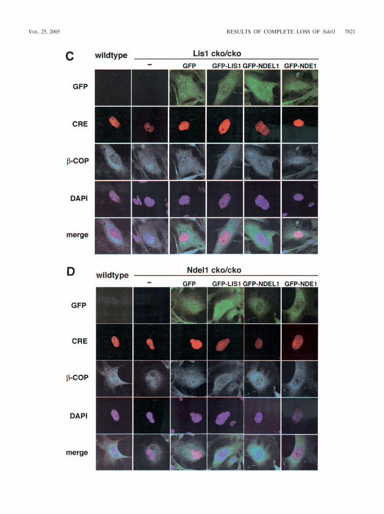
FIG. 7. Disruption of microtubule organization and distribution of �-COP-positive vesicles in Lis1- or Ndel1-null MEFs. Aberrant microtubuleorganization and abnormal distribution of �-COP-positive vesicles in Lis1 or Ndel1-null MEFs generated by CRE-mediated recombination.Cotransfected plasmids for rescue were shown on the top. (A and B) The loss of LIS1 causes a redistribution and an enrichment of microtubulesnear the nucleus (A), whereas loss of NDEL1 results in amorphous microtubules (B). Perinuclear accumulation of microtubules in Lis1-null MEFswas only rescued by GFP-LIS1 expression (see panel A). GFP-NDEL1 expression also rescued the amorphous microtubule organization inNdel1-null MEFs, but GFP-LIS1 and GFP-NDE1 were less effective (see panel B). To address dynein regulation by LIS1 and NDEL1, weexamined the distribution of �-COP-positive vesicles, which display juxtanuclear localization in a dynein-dependent fashion. (C and D) Thepredominant juxtanuclear staining pattern was disrupted in Lis1-null MEFs (C) and in Ndel1-null MEFs (D), resulting in a homogeneousdistribution accompanied by punctuate accumulation. Loss of this Golgi pattern by Lis1 inactivation was only rescued by exogenous expression ofGFP-LIS1 (see panel C). In contrast, loss of the Golgi pattern by Ndel1 inactivation was efficiently rescued by exogenous expression of GFP-LIS1,GFP-NDEL1, and GFP-NDE1 (see panel D).
7822 SASAKI ET AL. MOL. CELL. BIOL.
Ndel1cko/�/Lis1�/�, Ndel1�/�/Lis1�/�, and Ndel1�/�/Lis1�/�
genotypes, whereas calbindin-positive cells which migrate at alater stage exhibited broader distribution in Ndel1�/�/Lis1cko/�, Ndel1cko/�/Lis1�/�, Ndel1�/�/Lis1�/�, and Ndel1�/�/Lis1�/� genotypes, suggesting that later migrating neurons aremore sensitive to reduction of LIS1 and/or NDEL1. Quantita-tion of densities indicated mild reduction of calbindin-positivecells in the cortexes of the Ndel1�/�/Lis1�/� mice and theNdel1�/�/Lis1�/� mice, whereas C-Neu positive cells did notdisplay obvious genotype-dependent differences (Fig. 6C).These results prompted us to investigate apoptotic cell deathduring corticogenesis in various crosses by TUNEL (terminaldeoxynucleotidyltransferase-mediated dUTP-biotin nick endlabeling) staining (Fig. 6D). At E11.5 when early migratingneurons are generated, significant differences of apoptotic celldeath were not observed. In contrast, at E15.5 when latermigrating neurons are generated, significant acceleration ofapoptotic cell death in the ventricular zone were observed. Inparticular, cell death was correlated with the reduction of LIS1amount, a finding in good agreement with the reduction ofcalbindin-positive cells in the cortexes of the Ndel1�/� Lis1�/�
mice and Ndel1�/� Lis1�/� mice (Fig. 6E).Abnormal organization of microtubules and distribution of
�-COP-positive vesicles in Lis1- or Ndel1-null MEFs. We nextaddressed LIS1 and NDEL1 functions on microtubule organi-zation and dynein regulation. In wild-type MEFs, microtubulearrays were displayed throughout the cell from a clear focusthat likely represented the centrosome (Fig, 7A). Loss of eitherLis1 or Ndel1 caused profound changes in the organization ofmicrotubules of MEFs. Microtubules exhibited a perinuclearconcentration in Lis1-disrupted MEFs (39) (Fig. 7A). Thisaberrant distribution of microtubules in Lis1-null MEFs wasonly rescued by exogenous GFP-LIS1 expression (Fig. 7A) butnot by GFP-NDEL1 or GFP-NDE1. In contrast, microtubulesin Ndel1-disrupted MEFs displayed an amorphous and fragilepattern (Fig. 7B). Similarly, the microtubule organization de-fect in Ndel1-null MEFs was efficiently rescued by GFP-NDEL1 but not by either LIS1 or NDE1, which only slightlyrescued the disorganization of microtubules (Fig. 7B). Basedon the different phenotypes in Lis1- and Ndel1-null MEFs,these data suggest that NDEL1 plays a distinct role in theregulation of microtubule organization.
We also examined the distribution of �-COP-positive vesi-cles. �-COP proteins are part of a coatomer complex thatrecruits some specific areas of the endomembrane system tothe Golgi complex in a cytoplasmic dynein-dependent fashion(5). Immunofluorescence demonstrated that �-COP-positivevesicles displayed a predominantly juxtanuclear staining pat-tern in wild-type MEFs (Fig. 7C). Upon inactivation of Lis1 orNdel1 by CRE recombinase, this clustering in the juxtanuclearregion was disrupted, and �-COP was redistributed homoge-neously within the cell accompanied by a punctuate accumu-lation (Fig. 7C and D). Aberrant distribution of �-COP posi-tive vesicles in Lis1-null MEFs was only rescued by exogenousintroduction of GFP-LIS1 (Fig. 7C). In contrast, disruptedorganization of of �-COP-positive vesicles in Ndel1-null MEFswas efficiently rescued by GFP-NDE1, as well as GFP-NDEL1,and it was partially rescued by GFP-LIS1 (Fig. 7D). These datasuggest that LIS1, NDEL1, and NDE1 share some overlapping
and some distinct functions in the regulation of cytoplasmicdynein function.
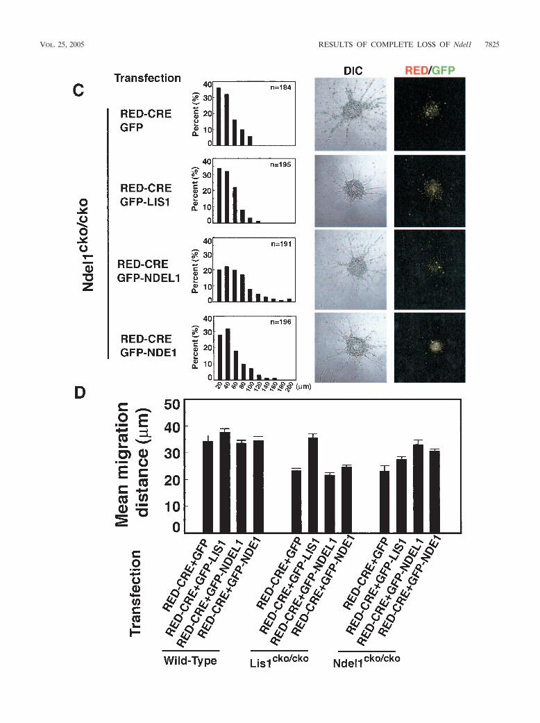
Neuronal migration defects in Lis1 and Ndel1 mutant gran-ule cells. To define the mechanistic roles of NDEL1 and LIS1in mammalian neuronal migration, we used mouse cerebellargranule neurons in an in vitro migration assay with wild-type ormutant neurons (17, 41). Migration distances in neurons trans-fected with RFP-CRE alone were positioned indistinguishablyfrom untransfected neurons, suggesting that RFP-CRE trans-fection had no effect on migration (Fig. 8A and D).12 Overex-pression of LIS1-GFP led to a mild increase in neuronal mi-gration (Fig. 8A and D), as previously reported (41), whereasGFP-NDEL1 or GFP-NDE1 overexpression had no such effect(Fig. 8A,D). Lis1-null granule neurons displayed severe migra-tion defects compared to wild-type neurons, characterized by aleftward shift of the bin distribution of migration distance, andthe mean distance decreased by �60% from the WT level (Fig.8B and D). This migration defect due to Lis1 inactivation wasefficiently rescued by exogenous expression of GFP-LIS1 (Fig.8B and D) but not by GFP-NDEL1 or GFP-NDE1.
Similar migration defects were observed in Ndel1-null neu-rons (Fig. 8C and D). This migration defect due to loss ofNdel1 was rescued by exogenous expression of GFP-NDEL1(Fig. 8C and D). GFP-LIS1 expression only moderately res-cued the migration defect. More interestingly, GFP-NDE1 ex-pression partially but incompletely rescued the migration de-fect derived from loss of Ndel1. The reduced magnitude ofrescue suggests that NDEL1 and NDE1 have similar functions,but NDEL1 might have a more crucial and distinct function.These data are consistent with the early embryonic lethalitydisplayed by Ndel1-null mice compared to the viable pheno-type of Nde1-null mice, as well as the less-efficient recovery ofdisorganized microtubule network in Ndel1-null MEFs byGFP-NDE1 expression.
DISCUSSION
Ndel1 is one of the mouse homologues of Nude from As-pergillus, which was identified as a multicopy suppressors of amutation in the nudF gene, the Aspergillus homologue of LIS1(6). NudE is also a homologue of the nuclear distributionprotein RO11 (24) of Neurospora crassa and mitotic phospho-protein 43 of Xenopus laevis (6). LIS1 and NDEL act in theCDHC pathway, which is the large molecular motor that trans-locates to the minus (�) ends of microtubules. Dynein, whosemotor domain comprises six AAA modules and two potentialmechanical levers, generates movement by a mechanism that isfundamentally different than that which underlies the motionof myosin and kinesin. CDHC is involved in numerous func-tions, including microtubule organization, vesicle transport(especially from the endoplasmic reticulum to the Golgi appa-ratus), chromosome separation, and nuclear distribution (12,16, 45, 46).
To understand the in vivo role of NDEL1, we generatedNdel1-disrupted mice. Complete loss of NDEL1 caused em-bryonic lethality at the peri-implantation stage and a deficiencyof cell proliferation in the inner cell mass. These data suggestthat NDEL1-dependent regulation of CDHC is essential forproliferation and may play a role in important aspects of mi-tosis. Compared to Lis1�/� mice, which displayed clear neu-
VOL. 25, 2005 RESULTS OF COMPLETE LOSS OF Ndel1 7823

7824 SASAKI ET AL. MOL. CELL. BIOL.
VOL. 25, 2005 RESULTS OF COMPLETE LOSS OF Ndel1 7825
ronal migration defects (10, 17), Ndel1�/� mice did not exhibitobvious phenotypes. Further reduction of Ndel1 using a hypo-morphic conditional allele resulted in mild neuronal migrationdefects. Double Lis1/Ndel1 mutants displayed more severe mi-gration defect than single mutants, suggesting that Lis1 andNdel1 genetically interact. Elevated apoptotic cell death duringneurogenesis was observed in Lis1 mutants, whereas mutationof Ndel1 did not result in apoptosis. We also examined LIS1and NDEL1 functions on dynein regulation and microtubuleorganization. Lis1- and Ndel1-null MEFs displayed similar dis-ruption of the compact juxtanuclear Golgi complex of �-COP-positive vesicles. The aberrant distribution of �-COP-positivevesicles in Lis1-null MEFs was only rescued by exogenousintroduction of GFP-LIS1, while the disrupted organization of�-COP-positive vesicles in Ndel1-null MEFs was efficiently res-cued by GFP-NDE1, as well as GFP-NDEL1, and it was par-tially rescued by GFP-LIS1. Lis1 disruption resulted in per-nuclear accumulation of microtubules, whereas Ndel1inactivation displayed amorphous distribution of microtubules.We also demonstrated that the disorganized microtubule net-work in Ndel1-null cells was completely rescued by GFP-NDEL1 but only partially rescued by GFP-LIS1 and GFP-NDE1. These results were similar to those found for neuronalmigration using cerebellar granule cell reaggregation assays.Neuronal migration defects displayed by Ndel1-null neuronswere completely rescued by GFP-NDEL1 but only partiallyrescued by GFP-LIS1 or GFP-NDE1 exogenous expression.Our data suggest that NDEL1, LIS1, and NDE1 act in a com-mon pathway to regulate dynein but that each has distinct rolesin the regulation of microtubule organization and neuronalmigration.
Neuronal migration is the critical cellular process whichinitiates corticogenesis of cerebral cortex. Upon becomingpostmitotic after their final mitosis, neurons migrate from theventricular zone toward the cortical plate and then establishneuronal lamina via an “inside-out” gradient of maturation.Mutations of genes involved in neuronal migration result indefects of corticogenesis. Lis1�/� mutants display recognizableneuronal migration defects. In contrast, Ndel1�/� mutants ex-hibited no obvious phenotypes, Ndel1cko/� mutants exhibitedclear aberration of neuronal migration, although the pheno-type was milder than the phenotype of Liscko/� mutants. Wealso demonstrated synergistic regulation of neuronal migrationby LIS1 and NDEL1 using various crosses of Lis1 and Ndel1mutants, suggesting that LIS1 and NDEL1 at least partiallyshare the same pathway during neuronal migration. A recentreport indicated that loss of function of Ndel1, Lis1, or dyneinby RNA interference in the developing neocortex impairs neu-ronal positioning and causes the uncoupling of the centrosome
and nucleus. These observations support our conclusion thatLIS and NDEL1 share a common neuronal migration pathway(38).
In the developing central nervous system apoptosis plays animportant role in the normal organization of the neuronalcircuit. Roughly half of all of the neurons produced duringneurogenesis die apoptotically before the nervous system ma-tures (22). We have demonstrated that apoptotic cell death issignificantly elevated in Lis1 mutants. Apoptotic cell death wasrestricted to the ventricular zone, suggesting that LIS1 is es-sential for cell division of neuronal stem cells. Interestingly,neurons generated later were more sensitive to the reductionof LIS1. Long-lasting reduction of LIS1 might cause deregu-lation of proliferation and/or accumulation of disorganizedorganellar distribution that might trigger apoptosis or increasesensitivity to apoptotic inducers. Although migration defectswere observed in brains from Ndel1cko/� mice, apoptotic celldeath was not observed.
Another recent report demonstrated that the targeted dis-ruption of the second homologue of NudE, Nde1, resulted inviable mice with a small brain (microcephaly) phenotype, withthe most dramatic reduction affecting the cerebral cortex (9).In Nde1 mutants (Nde1�/�), cortical lamination was mostlypreserved, but the mutant cortex had fewer neurons and verythin superficial cortical layers (II to IV). The small cerebralcortex is thought to reflect both reduced progenitor cell divi-sion and altered neuronal cell fates. In contrast to Nde1, com-plete loss of Ndel1 resulted in perimplanation lethality, a phe-notype similar to complete loss-of-function of Lis1 (17) andCdhc (14). Ndel1 mutants more clearly displayed neuronalmigration defect than Nde1 mutants. These phenotypic differ-ences between two homologues could be attributed to differ-ences in the pattern and levels of expression during develop-ment (8, 29, 35, 36). NDE1 is expressed mainly in neuronalstem cells and is downregulated in the postmitotic neurons. Incontrast, NDEL1 is first expressed in first in the early embryo.Later, NDEL1 is expressed in neuronal stem cells, and itsexpression continues in postmitotic neurons, suggesting thatNDEL1 plays a broader role during development than NDE1.Another nonexclusive possibility is that NDEL1 has distinctmolecular targets from NDE1. This possibility is supported byour observations that the disorganized microtubule networkseen in the NDEL1-null MEFs was not completely rescued byNDE1 expression. Partial rescue of neuronal migration defectin NDEL1-null neurons by NDE1 introduction also supportsthis possibility. Further experiments with mutant mice for eachof these components, which are now all available, as well asfurther biochemical studies, will provide more detailed expla-nations to address the similarities and differences in the roles
FIG. 8. Migration distance in Lis1- or Ndel1-null neurons determined by reaggregation assay. Granular neurons were isolated from P3 neonatalpups and subjected to the neuronal migration reaggregation assay. Lis1 or Ndel1 was inactivated by CRE-mediated recombination. Cotransfectedplasmids for rescue are shown on the left side. The migration distance of each neurons after 12 h was binned. (A) Wild-type neurons expressingRFP-CRE display normal migration distance compared to untransfected neurons. Cotransfection of GFP-LIS1 mildly enhanced migration,whereas GFP-NDEL1 or GFP-NDE1 did not have obvious effect. (B and C) Lis1 (B)- or Ndel1 (C)-null neurons display a shift in the distributionof bins toward the right. Migration defects in Lis1-null neurons were only rescued by exogenous expression of GFP-LIS1. Migration defects inNdel1-null neurons were rescued by exogenous expression of GFP-LIS1, GFP-NDEL1, and GFP-NDE1, but GFP-LIS1 and GFP-NDE1 were lesseffective (see panel C). Mean migration distances are summarized at the bottom. n, Number of neurons measured for each examination.(D) Summary of mean migration distance of each genotype.
7826 SASAKI ET AL. MOL. CELL. BIOL.
of LIS1, NDEL1, and NDE1 in the regulation of dynein func-tion and microtubule organization, as well as their roles inproliferation, neurogenesis, and neuronal migration.
ACKNOWLEDGMENTS
We thank Yoshihiko Funae, Hiroshi Iwao, Toshio Yamauch, Yo-shitaka Nagai, and L. Robert Nussbaum for generous support andencouragement. We are grateful to Yuzuru Yamauchi, Shino Oku-mura, Michiyo Ishida, Katsumi Hagiwara, Nobuko Tominaga, MasamiSuzuki, and Masashi Harada for mouse breeding and technical sup-port. We thank Shin-ichi Hisanaga for providing a recombinant pro-tein of cdk5/p35 and Francis J. MacNally for providing mutated kine-sin.
This study was supported by NIH grant NS41030 to A.W.-B. andGrants-in-Aid for Scientific Research from the Ministry of Education,Science, Sports, and Culture of Japan to A.Y., K.T., and S.H. Thisstudy was also supported by Nissan Science Foundation grants to K.T.and a Sankyo Foundation of Life Science and Japan Brain Foundationgrant to S.H.
REFERENCES
1. Chae, T., Y. T. Kwon, R. Bronson, P. Dikkes, E. Li, and L. H. Tsai. 1997.Mice lacking p35, a neuronal specific activator of Cdk5, display corticallamination defects, seizures, and adult lethality. Neuron 18:29–42.
2. Deng, C., A. Wynshaw-Boris, F. Zhou, A. Kuo, and P. Leder. 1996. Fibroblastgrowth factor receptor 3 is a negative regulator of bone growth. Cell 84:911–921.
3. Dobyns, W. B. 1987. Developmental aspects of lissencephaly and the lissen-cephaly syndromes. Birth Defects 23:225–241.
4. Dobyns, W. B., O. Reiner, R. Carrozzo, and D. H. Ledbetter. 1993. Lissen-cephaly: a human brain malformation associated with deletion of the LIS1gene located at chromosome 17p13. JAMA 23:2838–2842.
5. Donaldson, J. G., J. Lippincott-Schwartz, G. S. Bloom, T. E. Kreis, and R. D.Klausner. 1990. Dissociation of a 110-kD peripheral membrane protein fromthe Golgi apparatus is an early event in brefeldin A action. J. Cell Biol.111:2295–2306.
6. Efimov, V. P., and N. R. Morris. 2000. The LIS1-related NUDF protein ofAspergillus nidulans interacts with the coiled-coil domain of the NUDE/RO11 protein. J. Cell Biol. 150:681–688.
7. Faulkner, N. E., D. L. Dujardin, C. Y. Tai, K. T. Vaughan, C. B. O’Connell,Y. Wang, and R. B. Vallee. 2000. A role for the lissencephaly gene LIS1 inmitosis and cytoplasmic dynein function. Nat. Cell Biol. 2:784–791.
8. Feng, Y., E. C. Olson, P. T. Stukenberg, L. A. Flanagan, M. W. Kirschner,and C. A. Walsh. 2000. Interactions between LIS1 and mNudE, a centralcomponent of the centrosome, are required for CNS lamination. Neuron28:665–679.
9. Feng, Y., and C. A. Walsh. 2004. Mitotic spindle regulation by nde1 controlscerebral cortical size. Neuron 44:279–293.
10. Gambello, M. J., D. L. Darling, J. Yingling, T. Tanaka, J. G. Gleeson, and A.Wynshaw-Boris. 2003. Multiple dose-dependent effects of Lis1 on cerebralcortical development. J. Neurosci. 23:1719–1729.
11. Gilmore, E. C., T. Ohshima, A. M. Goffinet, A. B. Kulkarni, and K. Herrup.1998. Cyclin-dependent kinase 5-deficient mice demonstrate novel develop-mental arrest in cerebral cortex. J. Neurosci. 18:6370–6377.
12. Gee, M. A., J. E. Heuser, and R. B. Vallee. 1997. An extended microtubule-binding structure within the dynein motor domain. Nature 390:636–639.
13. Gupta, A., L. H. Tsai, and A. Wynshaw-Boris. 2002. Life is a journey: agenetic look at neocortical development. Nat. Rev. Genet. 3:342–355.
14. Harada, A., Y. Takei, Y. Kanai, Y. Tanaka, S. Nonaka, and N. Hirokawa.1998. Golgi vesiculation and lysosome dispersion in cells lacking cytoplasmicdynein. J. Cell Biol. 141:51–59.
15. Hattori, M., H. Adachi, M. Tsujimoto, H. Arai, and K. Inoue. 1994. Miller-Dieker lissencephaly gene encodes a subunit of brain platelet-activatingfactor acetylhydrolase. Nature 370:216–218.
16. Helfand, B. T., A. Mikami, R. B. Vallee, and R. D. Goldman. 2002. Arequirement for cytoplasmic dynein and dynactin in intermediate filamentnetwork assembly and organization. J. Cell Biol. 157:795–806.
17. Hirotsune, S., M. W. Fleck, M. J. Gambello, G. J. Bix, A. Chen, G. D., Clark,D. H. Ledbetter, C. J. McBain, and A. Wynshaw-Boris. 1998. Graded reduc-tion of Pafah1b1 (Lis1) activity results in neuronal migration defects andearly embryonic lethality. Nat. Genet. 19:333–339.
18. Kwon, Y. T., and L. H. Tsai. 2000. The role of the p35/cdk5 kinase in corticaldevelopment. Results Probl. Cell Differ. 30:241–253.
19. Lakso, M., J. G. Pichel, J. R. Gorman, B. Sauer, Y. Okamoto, E. Lee, F. W.Alt, and H. Westphal. 1996. Efficient in vivo manipulation of mouse genomicsequences at the zygote stage. Proc. Natl. Acad. Sci. USA 93:5860–5865.
20. Liu, Z., T. Xie, and R. Steward. 1999. Lis1, the Drosophila homologue of ahuman lissencephaly disease gene, is required for germline cell division andoocyte differentiation. Development 126:4477–4488.
21. Liu, Z., R. Steward, and L. Luo. 2000. Drosophila Lis1 is required forneuroblast proliferation, dendritic elaboration and axonal transport. Nat.Cell Biol. 2:776–783.
22. Liu, D. X., and L. A. Greene. 2001. Neuronal apoptosis at the G1/S cell cyclecheckpoint. Cell Tissue Res. 305:217–228.
23. Luskin, M. B., and C. J. Shatz. 1985. Studies of the earliest generated cellsof the cat’s visual cortex: cogeneration of subplate and marginal zones.J. Neurosci. 99:7881–7888.
24. Miller, M. W., and F. A. Pitts. 2000. Neurotrophin receptors in the somato-sensory cortex of the mature rat: colocalization of p75, trk, isoforms andc-Neu. Brain Res. 852:355–366.
25. Minke, P. F., I. H. Lee, J. H. Tinsley, K. S. Bruno, and M. Plamann. 1999.Neurospora crassa ro-10 and ro-11 genes encode novel proteins required fornuclear distribution. Mol. Microbiol. 32:1065–1076.
26. Morris, R. N., V. P. Efimov, and X. Xiang. 1998. Nuclear migration, nucleo-kinesis, and lissencephaly. Trends Cell Biol. 8:467–470.
27. Morris, R. N. 2000. Nuclear migration: from fungi to the mammalian brain.J. Cell Biol. 148:1097–1101.
28. Mullen, R. J., C. R. Buck, and A. M. Smith. 1992. NeuN, a neuronal specificnuclear protein in vertebrates. Development 116:201–211.
29. Niethammer, M., D. S. Smith, R. Ayala, J. Peng, J. Ko, M. Lee, M. Morabito,and L. Tsai. 2000. The Lis1 interacting protein Nude1 is a Cdk5 substratethat interacts with cytoplasmic dynein. Neuron 28:697–711.
30. Oshima, T., J. M. Ward, C. G. Huh, G. Longenecker, C. Veeranna, H. Pant,R. O. Brady, L. J. Martin, and A. B. Kulkarni. 1996. Targeted disruption ofthe cyclin-dependent kinase 5 gene results in abnormal corticogenesis, neuronalpathology and perinatal death. Proc. Natl. Acad. Sci. USA 93:11173–11178.
31. Pliz, D. T., N. Matsumoto, S. Minnerath, P. Mills, and J. G. Glesson. 1999.Lis1 and XLIS/doublecortin mutations cause most human classical lissen-cephaly, but different patterns if malformation. Hum. Mol. Genet. 7:2029–2037.
32. Rakic, P. 1972. Model of cell migration to the superficial layers of fetalmonkey neocortex. J. Comp. Neurol. 145:61–83.
33. Reiner, O., R. Carrozzo, Y. Shen, M. Wehnert, F. Faustinella, W. B. Dobyns,T. Caskey, and D. H. Ledbetter. 1993. Isolation of a Miller-Dieker lissen-cephaly gene containing G protein b-subunit-like repeat. Nature 364:717–721.
34. Ringstedt, T., S. Linnarsson, J. Wagnoer, U. Lendahl, Z. Kokaia, E. Arenas,P. Ernfors, and C. F. Ibanez. 1998. BDNF regulates reelin expression andCajal-Retzius cell development in the cerebral cortex. Neuron 21:305–315.
35. Saito, T., R. Onuki, Y. Fujita, G. Kusakawa, K. Ishiguro, J. A. Bibb, T.Kishimoto, and S. Hisanaga. 2003. Developmental regulation of the prote-olysis of the p35 cyclin-dependent kinase 5 activator by phosphorylation.J. Neurosci. 23:1189–1197.
36. Sasaki, S., A. Shionoya, M. Ishida, M. J. Gambello, J. Yingling, A. Wynshaw-Boris, and S. Hirotsune. 2000. A LIS1/NUDEL/cytoplasmic dynein heavychain complex in the developing and adult nervous system. Neuron 28:681–696.
37. Sheppard, A. M., and A. L. Pearlman. 1997. Abnormal organization ofpreplate neurons and their associated extracellular matrix: an early manifes-tation of altered neocortical development in the reeler mutant mouse.J. Comp. Neurol. 378:173–179.
38. Shu, T., R. Ayala, M. D. Nguyen, Z. Xie, J. G. Gleeson, and L. H. Tsai. 2004.Ndel1 operates in a common pathway with LIS1 and cytoplasmic dynein toregulate cortical neuronal positioning. Neuron 44:263–277.
39. Smith, D. S., M. Niethammer, R. Ayala, Y. Zhou, M. J. Gambello, A. Wyn-shaw-Boris, and L.-H. Tsai. 2000. Regulation of cytoplasmic dynein behaviourand microtubule organization by mammalian Lis1. Nat. Cell Biol. 2:767–775.
40. Swan, A., T. Nguyen, and B. Suter. 1999. Drosophila lissencephaly-1 func-tions with Bic-D and dynein in oocyte determination and nuclear positioning.Nat. Cell Biol. 1:444–449.
41. Tanaka, T., Serneo, C. F. F. Higgins, M. J. Gambello, A. Wynshaw-Boris,and J. G. Gleeson. 2004. Lis1 and doublecortin function with dynein tomediate coupling of the nucleus to the centrosome in neuronal migration.J. Cell Biol. 165:709–721.
42. Toyo-oka, K., A. Shionoya, M. J. Gambello, C. Cardoso, R. Leventer, H. L.Ward, R. Ayala, L. H. Tsai, W. Dobyns, D. Ledbetter, S. Hirotsune, and A.Wynshaw-Boris. 2003. 14-3-3epsilon is important for neuronal migration bybinding to NUDEL: a molecular explanation for Miller-Dieker syndrome.Nat. Genet. 34:274–285.
43. Trommsdorff, M., M. Gotthardt, T. Hiesberger, J. Shelton, W. Stockinger, J.Nimpf, R. E. Hammer, J. A. Richardson, and J. Herz. 1999. Reeler/Disabled-like disruption of neuronal migration in knockout mice lacking the VLDLreceptor and ApoE receptor 2. Cell 97:689–701.
44. Xiang, X., A. H. Osmani, S. A. Osmani, M. Xin, and N. R. Morris. 1995.NudF, a nuclear migration gene in Aspergillus nidulans, is similar to thehuman LIS-1 gene required for neuronal migration. Mol. Biol. Cell 6:297–310.
45. Vallee, R. B., J. S. Wall, B. M. Paschal, and H. S. Shpetner. 1988. Microtu-bule-associated protein 1C from brain is a two-headed cytosolic dynein.Nature 332:561–563.
46. Vallee, R. B., and M. P. Sheetz. 1996. Targeting of motor proteins. Science271:1539–1544.
47. Willins, D. A., B. Liu, X. Xiang, and N. R. Morris. 1997. Mutations in theheavy chain of cytoplasmic dynein suppress the nudF nuclear migrationmutation of Aspergillus nidulans. Mol. Gen. Genet. 255:194–200.
VOL. 25, 2005 RESULTS OF COMPLETE LOSS OF Ndel1 7827
Copyright © 2022 FDOKUMEN