nvri seminar series 2015
159
i | Page NVRI SEMINAR SERIES 2015 This seminar series is a collation of seminar papers presented by staff and visiting scientists to the National Veterinary Research Institute, Vom during 2015 Compiled and edited by NVRI Seminar/Publication Committee 2015 Dr P. A. Okewole . . . Chairman Dr J. Kamani . . . Member Dr I. S. Tekki . . . Member Dr A. T. Oladokun . . Member Dr W. Bertu . . . Member Dr (Mrs.) V. Ifende. . . Member Dr O. O. Oladipo . . . Member Dr I. J. Barde . . . Member Mr. T. Barko . . . Member Mr. J. U. Ogbu . . . Member Mr. Jerry Musa . . . Member Dr (Mrs.) C. I. Odita . . Secretary Mr. Francis Dama. . . Assistant Secretary National Veterinary Research Institute, Vom PMB 01, Vom Email [email protected] or [email protected] Website: www.nvri.gov.ng © 2009 NVRI ISSN1597-1651
-
Upload
khangminh22 -
Category
Documents
-
view
1 -
download
0
Transcript of nvri seminar series 2015

i | P a g e
NVRI SEMINAR SERIES 2015
This seminar series is a collation of seminar papers presented by staff and
visiting scientists to the National Veterinary Research Institute, Vom during
2015
Compiled and edited by
NVRI Seminar/Publication Committee 2015
Dr P. A. Okewole . . . Chairman
Dr J. Kamani . . . Member
Dr I. S. Tekki . . . Member
Dr A. T. Oladokun . . Member
Dr W. Bertu . . . Member
Dr (Mrs.) V. Ifende. . . Member
Dr O. O. Oladipo . . . Member
Dr I. J. Barde . . . Member
Mr. T. Barko . . . Member
Mr. J. U. Ogbu . . . Member
Mr. Jerry Musa . . . Member
Dr (Mrs.) C. I. Odita . . Secretary
Mr. Francis Dama. . . Assistant Secretary
National Veterinary Research Institute, Vom
PMB 01, Vom
Email [email protected] or [email protected]
Website: www.nvri.gov.ng
© 2009 NVRI
ISSN1597-1651

ii | P a g e
TABLE OF CONTENTS
Modelling the Risk of Haemonchosis in Sheep . . . 1
Evaluation of Tephrosia vogelii Leaf Extracts for Molluscidal
property on Lymnae . . . . . . . 10
Sources of Salmonella Infections in Some Selected
Poultry Farms in Jos, Northern Nigeria . . . . 23
Prevalence and Antimicrobial Susceptibility Profile of
Salmonella Serovars from Cattle in Jos, Nigeria . . . 30
Reverse Genetics: A Useful Tool in Functional Genomics
(Role of Znt1 Gene in Vertebrate Development) . . . 37
Molecular Detection of Bovine Trypanosomiasis in Some
Parts of Central Senatorial Zone of Plateau State-Nigeria . 44
Methods for the Identification of Myiatic Flies in Dogs in
Vom, Jos-South LGA, Plateau State . . . . . 51
Endogenous Retrovirus Contaminants of Feline Cell
Lines: Implications for Veterinary Vaccine Manufacture. . 59
Seroprevalence of Influenza A Antibodies in Pigs in Jos-South
Local Government Area of Plateau State, Nigeria . . . 69
Pathogenicity of Pasteurella multocida Strains
(Serotypes A: 1, 3 and 4) in Commercial Chickens and
Japanese Quails in Jos, Nigeria . . . . . 76
Isolation of Brucella abortus Biotype 3 from Cattle in
Kachia Grazing Reserve and Jos Plateau and their
Identification by Bruce-Ladder Multiplex PCR . . . 90
iii | P a g e
Antibody Engineering: Single domain antibody (Nanobody)
as a research tool . . . . . . . . 102
A Study on helminthiasis of cattle herds in Kachia Grazing
Reserve (KGR) of Kaduna State, Nigeria . . . . 108
Therapeutic and Safety Evaluation of Combined Aqueous
Extracts of Azadirachta indica and Khaya senegalensis
in Chickens Experimentally Infected with Eimeria Oocysts . 117
Public Service Regulations: A Key to Achieving Productivity . 128
Relationship between Emotional Intelligence and Job
Satisfaction among Employees of National Veterinary
Research Institute, Vom . . . . . . . 141
Highly pathogenic Avian Influenza in Nigeria: Overview
of 2015 Outbreaks . . . . . . . 146
Guidelines for presentation and manuscript preparation . 152

iv | P a g e
INTRODUCTION
Seminar presentation in the Institute is accorded priority in recognition of the
fact that any information that is not shared is knowledge lost. Current trends
and cutting edge research methodologies were presented by staff of the
Institute and Visiting Scientist in order to open up new vistas and broaden the
research horizon of other researchers and staff. A blend of topical research and
diagnostic procedures, current concepts in administration and psychological
assessment of job performance were aptly presented to savor the appetite of all
cadres of staff.
In this compilation, “NVRI Seminar Series 2015”, we invite you to read through
the collation of seminars presented in the year. Papers presented cover diverse
areas of research in Veterinary Medicine, Molecular Biology, Microbiology,
Pathology and Genetic Engineering, etc. The socio economic effect of parasitic
infection to man and animals and the need to curb their menace were
presented. This fact is being buttressed by the high prevalence of helminths
infection in cattle in Kachia Grazing Reserve and molecular detection and
characterization of Trypanosoma congolense and T. brucei in cattle in Plateau
state. Overall, a multi faceted approach to the control of parasitic diseases was
considered. They include the use of modeling to map the risk and predict
intervention strategy for haemonchosis, the use of bio active substances to
control lymnae snails as well as exploiting the synergistic/potentiating effects
of Azadirachta indica and Khaya senegalensis in the control of avian
coccidiosis.
The role of Salmonellae as a major pathogen of birds and livestock in Nigeria
was elucidated in several of the papers presented. Several serovars were
isolated and characterized from hatcheries, poultry farms and cattle indicating
widespread circulation of this bacterium in the environment and between
different animal species. Similarly, an experimental pathogenicity test of
Pasteurella multocida in chicken, quails and mice revealed that the organism is
capable of inducing high mortality in susceptible stock. The attention of
researchers was drawn to the use of Bruce-ladder Multiplex PCR for the
characterization of Brucella isolates, the organism which was reported to be
endemic in the areas studied.
Serological evidence of the circulation of Swine Influenza A virus in pigs in Jos
South LGA was presented with the possible role of pigs as a mixing vessel
highlighted. Could this be linked to the emergence of a new clade of Avian

v | P a g e
Influenza virus as the etiology of the current outbreaks in poultry in 20 states
and FCT. Read through these papers and adjudicate for yourself.
Vaccine producers were cautioned on the contaminative role of endogenous
retrovirus in feline cell lines routinely used for vaccine production and the
possible long term effect on vaccinated animals. New research areas such as
Antibody engineering as a research tool and the use of reverse genetics in
functional genomics are featured in this series.
It is my pleasure to usher you to read this document for the dissemination and
advancement of knowledge for improved agricultural research and livestock
and human health.
Dr. P. A. Okewole
Chairman,
Seminar and Publications Committee
1 | P a g e
MODELLING THE RISK OF HAEMONCHOSIS IN SHEEP
Bolajoko Muhammad-Bashir1*, Eric Morgan2
1 Epidemiology Unit, NVRI, Vom, Plateau, Nigeria
2 University of Bristol Veterinary School, c/o Bristol Life Sciences Building, 24
Tyndall Avenue, Bristol BS8 1TQ
INTRODUCTION
Of all the diseases caused by the gastrointestinal nematodes of small
ruminants, haemonchosis is perhaps one of the most common and serious
diseases in the tropics and summer dominant rainfall regions of the world
(O’Connor et al., 2006).
Eggs and larval development of H. contortus is optimal in warm and humid
conditions; as such temperature and moisture are the dominant influences on
the free-living larval stages, with other factors often controlling these two at the
microclimate level (O’Connor et al., 2006). It is therefore agreed that
temperature and moisture alone determine the success rate and speed of
development, both of which predict the epidemiology and ecology of H.
contortus (Tembely et al., 1997). Studies conducted by various researchers
around the globe confirm that knowledge of local climate and how it drives
seasonal and shorter-term patterns in L3 availability is therefore crucial to
ability to predict and manage infection and disease threat (Rose, 1963).
Grazing animals pick up infective larvae on herbage that is relatively short or
from herbage tops (Silva, et al., 2008). Outbreaks are worst with sudden
increased pasture larval density when warm summer rains follow a period of
drought break up the faecal pellet and create a moist environment for the
migration of hatched L3 (Silva, et al., 2008). When moisture is not a limiting
factor, temperature asserts a greater influence over migration (Stromberg,
1997).
Mathematical modelling has received increasingly wide application in ecological
and epidemiological studies of infectious diseases. This is the result of
increased understanding of what models can offer in terms of prediction and
understanding of a disease process (Smith and Grenfell, 1994). In epidemiology
and ecology, models permit the prediction of disease

2 | P a g e
Seminar presented 14th May, 2015 at NVRI Auditorium
dynamics at the level of the entire population from the understanding of
epidemiological factors at the individual level (Keeling and Rohani, 2008).
Modelling also enables the design and experimental evaluation of the impact of
specific management practices or control measures based on the predicted
parasite dynamics (Roberts and Heesterbeck, 1995). Further work needs to be
done on how modelling can be adapted as a tool for predicting risk in practice
(Kahn, 2010), and to guide targeted selective treatment (TST) and targeted
treatment (TT) as part of holistic and sustainable control against H. contortus.
The objectives of this study are to develop and validate a predictive
mechanistic model of haemonchosis in sheep by verifying and comparing model
predictions with either sourced or on-farm data on haemonchosis, as well as to
map the geographic distribution of haemonchosis risk.
MATERIALS AND METHODS
Threshold quantities
Threshold quantity is defined as the average number of adult parasites
produced by one female parasite in the absence of density dependent
constraints during the entire reproductive lifespan when introduced into a
previously unexposed host population (Heesterbeek and Roberts, 1995).
Basic reproduction rate (Q0)
As a threshold quantity, Q0 should be able to identify and define conditions or
factors that affect the parasite and host population (Scott and Smith, 1994).
This is very critical to sustainable control of H. contortus. A large value of Q0
indicates that a nematode is relatively well adapted to a given climate and
region, which implies stronger tendency towards population growth in the
absence of immunity resulting in higher infection pressure (Kao et al., 2000).
Assumptions on which the present Q0 model was built
Q0 estimates the mean output of adult progeny of an adult worm in a non
immune host without any density dependent constraint.
infective larvae are evenly spread over pasture (Kao et al., 2000);

3 | P a g e
development, mortality and transmission rates of all free-living stages are
dependent on the prevailing microclimate at any given point in time (Kao
et al., 2000);
The Q0 model
The Q0 model is a combination of four components.
Term 1 (fecundity rate): This measures the output during the entire
reproductive lifetime of the adult female within the host.
Mathematical representation of term 1 = λ /2μp............................. (1)
λ is a measure of fecundity of female in terms of egg production rate and μp is
the mortality rate of the parasite where the 2 represent the contribution of both
the male and female to the whole process.
Term 2 (probability of development to infective larvae as well as extra-pellet
migration): This component estimates the chance that eggs being deposited by
sheep on the pasture will successfully develop and yield L3, followed by
successful migration of L3 out of faeces unto pasture.
The second term is measured using dedh/ (μe+de)(μL3+dh)............... (2)
Where de is development rate from egg to L3, dh is the migration rate of L3 from
the ground onto herbage, μe is the mortality rate of egg and μL3 is the mortality
rate of L3 on ground. The combination of the success of both de and dh is a
measure of infective potential of Haemonchus contortus.
Term 3 (probability of ingestion of L3 on herbage): This component is built to
estimate the probability of actual transmission of H. contortus to the host via
grazing.
The third term is measured using the formula = cH/ (bAμLh+cH).......(3)
The mortality rate of Lh (μLh), host stocking density (H/A) , standing biomass
(b) , grazing area (A) and daily larval ingestion rate per sheep per grazing area
(c) are all factors that have contributory effects on the overall chance of
successful transmission of Lh to the host via ingestion.
Term 4 (probability of establishment): The fourth component measures the
probability of establishment of L3 following ingestion by the host.
The fourth term is represented by the symbol = pe...................... (4)
4 | P a g e
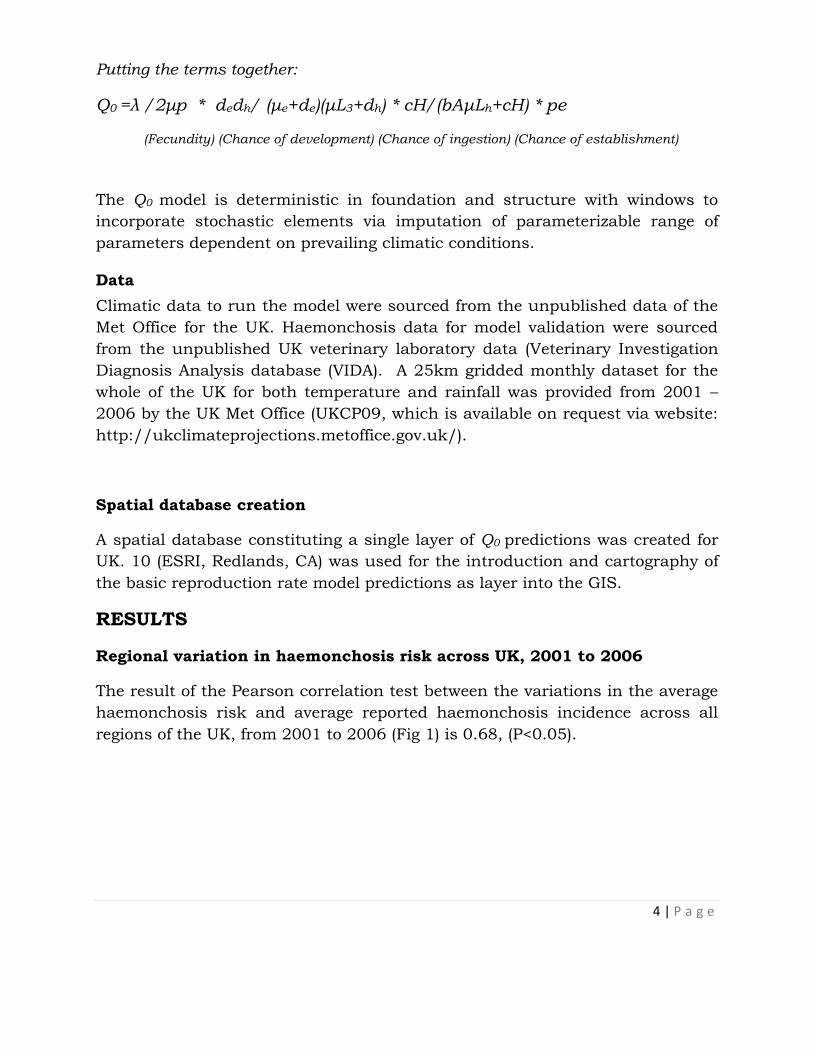
Putting the terms together:
Q0 =λ /2μp * dedh/ (μe+de)(μL3+dh) * cH/(bAμLh+cH) * pe
(Fecundity) (Chance of development) (Chance of ingestion) (Chance of establishment)
The Q0 model is deterministic in foundation and structure with windows to
incorporate stochastic elements via imputation of parameterizable range of
parameters dependent on prevailing climatic conditions.
Data
Climatic data to run the model were sourced from the unpublished data of the
Met Office for the UK. Haemonchosis data for model validation were sourced
from the unpublished UK veterinary laboratory data (Veterinary Investigation
Diagnosis Analysis database (VIDA). A 25km gridded monthly dataset for the
whole of the UK for both temperature and rainfall was provided from 2001 –
2006 by the UK Met Office (UKCP09, which is available on request via website:
http://ukclimateprojections.metoffice.gov.uk/).
Spatial database creation
A spatial database constituting a single layer of Q0 predictions was created for
UK. 10 (ESRI, Redlands, CA) was used for the introduction and cartography of
the basic reproduction rate model predictions as layer into the GIS.
RESULTS
Regional variation in haemonchosis risk across UK, 2001 to 2006
The result of the Pearson correlation test between the variations in the average
haemonchosis risk and average reported haemonchosis incidence across all
regions of the UK, from 2001 to 2006 (Fig 1) is 0.68, (P<0.05).

5 | P a g e
Figure 1: Comparison between the variations in the averaged haemonchosis
incidence reported with that of the averaged predicted
haemonchosis risk across all regions of the UK, from 2001 to
2006.
Seasonal variation in haemonchosis risk across the UK, 2001 – 2006
The results of the Pearson correlation between the average monthly predictions
of haemonchosis risk and the total monthly haemonchosis incidence reported
across the UK (Fig. 2) = 0.93, (P<0.01).
Figure 2: A comparison between the seasonal variations in the average Q0
predicted haemonchosis risk and the total haemonchosis
incidence across the UK, 2001 to 2006.

6 | P a g e
January
February
March
April
May
June
July
August
September
October
November
December
Figure 3: Average monthly Q0-based haemonchosis risk map showing the
geographic distribution and relative risk of H. contortus infection
pressure across the UK, 2001-2006
7 | P a g e
DISCUSSION AND CONCLUSION
This study shows that the Q0 model is able to predict the timing of risks due to
increased population of H. contortus or periodic changes in the infection
pressures from the free-living stages of the parasite. By indicating the arrival
of conditions suitable for the development and survival of infective larvae, high
Q0 values could trigger management measures such as treating sheep with
anthelmintic to reduce egg output, or moving them to lower risk pastures.
However, the model did not do so well in either location, when predicting
variations in risk within the haemonchosis season. This deficiency is evident in
the time-lag between predicted risk and the appearance of disease early in the
season. Hypothetically, this is because the model cannot account for the
following key biological processes associated with haemonchosis pathogenesis:
hypobiosis, the time lag for larval development from eggs, the pre-patent
period, accumulation of infection through time, physiological compensation for
blood loss, and time lag in the decline of parasite numbers in the sheep
population following the end of high risk periods for transmission (Kao et al.,
2000).
The cases in winter and/or early spring may in fact be accidental findings
resulting from resumption of arrested fourth stage larvae or even burdens from
the previous year. Q0 is a model based on instantaneous parameters; however,
in reality, egg development to L3 and the pre-patent period are not
instantaneous, but go through successive stages over a certain period. This
could explain the early predictions of infection pressure before the actual
incidence of disease. Furthermore, during unfavourable environmental
conditions, H. contortus inhibits the maturation of the fourth stage larva, which
is resumed once the climatic situations gradually become favourable (even
though climatic conditions are not yet suitable for transmission outside the
host). Therefore, the resumed maturation of hypobiotic larvae may be
responsible for the cases when Q0 predicted nil infection pressure from L3 in
winter and/or early spring (Fig.3). At that point in time; Q0 indicates that, H.
contortus infection pressure from the environment is absent because the
prevailing climate is unsuitable for the availability of L3 for infection. This is
especially likely to be the case in late winter and early spring in the UK.
However, during these periods, even though the environmental conditions are
not conducive for availability of new free-living stages for transmission (as
identified by Q0), there can be very good survival of already developed L3 at the
temperature range of 7 – 9 ºC. As such, some of the sheep population might
still be suffering from parasite burden or are chronically infected, or indeed

8 | P a g e
newly infected by L3 that persisted on pasture after the end of favourable
conditions for development. In effect, these situations could be responsible for
cases when infection pressure is predicted to be absent by Q0.
Conclusively, it appears that the Q0 model, based on its assumptions, is
capable of transforming the prevailing climatic situations into the prediction of
the periodic risk of haemonchosis transmission and occurrence. The model
effectively predicts the periods of haemonchosis season, more importantly the
peak point of each seasonal haemonchosis was predicted. Moreover, this study
has been able to extend and establish the capability of the basic reproduction
rate model as a monitor and predictor of the homogeneity or heterogeneity in
the level of haemonchosis risk across different geo-climatic zones.
REFERENCES
Heesterbeek, J.A.P., Roberts, M.G., (1995). Mathematical models for micro
parasites of wildlife. In GRENFELL, B.T., Dobson, A.P., eds, Ecology of
infectious disease in natural populations. Cambridge: Cambridge
University Press; pp. 90-122.
Kahn, L., (2010). A moisture index to predict development success of barber’s
pole worm and allow better management of pastures. Turning the Worm
26, 3-5.
Kao, R.R., Leathwick, D.M., Roberts, M.G., Sutherland, I.A., (2000).
Nematode parasites of sheep: a survey of epidemiological parameters
and their application in simple model. Parasitol. 121, 85-103.
Keeling, M.J., Rohani, P., (2008). Modelling infectious diseases in humans and
animals. Princeton, N.J. and Oxford: Princeton University Press.
O’Connor, L.J., Walkden-Brown, S.W., Kahn, L.P., (2006). Ecology of the free-
living stages of major trichostrongylid parasites of sheep. Vet. Parasitol.
142, 1-15.
Roberts, M.G., Heesterbeck, J., (1995). Bluff your way into epidemiological
models. Trends Microbiol. In press
Roberts, M.G., 1995. A pocket guide to host-parasite models. Parasitology
Today. 11, 5, 172-177.
Rose, J.H., (1963). Observations on the free-living stages of the stomach worm
Haemonchus contortus. Parasitolgy, 53: 469-481.
Scott, M.E., Smith, G., (1994). Parasitic and Infectious Diseases: Epidemiology
and Ecology, Academic Press.

9 | P a g e
Silva, B.F., Amarante, M.R.V., Kadri, S.M., Carrijio-Mauad, J.R., Amarante,
A.F.T., (2008). Vertical migration of Haemonchus contortus third stage
larvae on Brachiaria decumbens grass. Vet Parasitol. 158, 85-92.
Smith, G., Grenfell, B.T., (1994). Modelling of parasites populations:
gastrointestinal nematode models. Vet Parasitol. 54, 127-143.
Stromberg, B.E., (1997). Environmental factors influencing transmission. Vet
Parasitol. 72, 247-264.
Tembely, S., Lahlou-kassi, A., Rege, J.E.O., Sovani, S., Diedhiou, M.L., Baker,
R.L., (1997). The epidemiology of nematode infections in sheep in a cool
tropical environment. Vet Parasitol. 70, 129-141.

10 | P a g e
EVALUATION OF TEPHROSIA VOGELII LEAF EXTRACTS FOR
MOLLUSCIDAL PROPERTY ON LYMNAE
Tanko James Tenshak
Parasitology Division
INTRODUCTION
Fasciola gigantica and Fasciola hepatica are parasitic trematodes that cause
fasciolosis or liver rot disease of ruminants which is widely distributed in
tropical, subtropical and temperate countries (Schillhorn Van veen, 1980).
Lymnaeid snails are the main intermediate hosts (Soliman et al., 2000). The
incidence of snail-borne diseases is on the increase worldwide because of
climate change and the development of land and water resources for
agriculture. Livestock grazing in regions or areas that are frequently flooded or
have a high water table are at high risk of becoming infected with liver fluke
(Kanyari et al., 2010). Snails act as amplifiers of the parasite by asexual
reproduction, liberating large numbers of free-swimming cercariae that encyst
on vegetation to infect animals and man (Alison, 2011). Control of the snail
disrupts the life cycle of the parasite stopping the transmission of infection
(Soulsby 1982). Several strategies have been employed to control snail
population such as the synthetic organic molluscicides, but they are
considered toxic to non target animals and may have long-term detrimental
effects on the aquatic environment (Gehad et al., 2009). In recent years, there
has been increased attention on molluscicides that will be highly effective,
rapidly biodegradable, and less expensive. Therefore, plant molluscicide could
be appropriate for snail control measure against fasciolosis and
schistosomiasis in endemic areas (Fayez, 2009).
In Nigeria, evaluation of many plants as potential molluscicides against
different snail vectors have been carried out (Adewunmi and Sofowora, 1980;
Kela, 1992; Azare et al., 2002; 2007; Agboola et al., 2012; Benson and
Olajumoke 2012; Labe et al., 2012 and Olofintoye, 2012). The aim of this study
is to evaluate the molluscicidal properties of T. vogelli on lymnae snails.
Seminar presented 14th May, 2015 at NVRI Auditorium

11 | P a g e
MATERIALS AND METHODS
Snails/Plant materials
Fresh leaves of Tephrosia vogelii were collected in Vom, Jos South Local
Government Area of Plateau State (Plate 1), identified and authenticated at the
Herbarium, Department of Biological Sciences; Ahmadu Bello University Zaria,
Nigeria. The leaves were air dried for two weeks at room temperature and
pounded to a powdered form in a wooden mortar with pestle. Lymnae snails
(Plate 2) were collected from the dam outlet of Ahmadu Bello University Zaria
dam and Bassawa stream, in Sabo Gari Local Government Area of Kaduna
state Nigeria.
A total of nine hundred and fifty two snails were collected into a sterile glass jar
and transported to the Department of Parasitology and Entomology laboratory
Ahmadu Bello University Zaria for identification and experimental studies. The
snails were sorted and lymnaea snails identified and selected using the shape
and size of the shells as described by Moema et al., (2008).
The dried powdered leaf material weighing 250g were exhaustively and serially
extracted with 1000ml of methanol and water respectively for 48 hours at room
temperature of 27°C as described by Eman (2011). The extraction using each
solvent was carried out three times and the extracts pooled together and kept
in the desiccator. Each preparation was filtered separately through sterilized
Whatmann No.1 filter paper. It was then concentrated in a rotary evaporator
under vacuum to yield a semi-solid residue which was stored at 4° C until use
(Agboola et al., 2012).
Phytochemical Screening
Phytochemical analysis of the plant extract was conducted to determine the
presence of the following; saponins, glycosides, alkaloids, flavonoids, tannins,
steroid/terpenes and anthraquinones using standard procedures as described
by Trease and Evans (1989) and Sofowora (1993).
Bioassay
The experimental design consists of positive control group exposed to standard
molluscicide Niclosamide, negative control group exposed to 2% tween 80 in
distilled water and positive test groups exposed to Tephrosia vogelii leaf
extracted with water and methanol respectively. The bioassay was done
according to WHO (1983) guide lines with slight modifications (Adenusi and

12 | P a g e
Odaibo, 2007; Farheen and Singh, 2012). The snails were exposed to the
extracts at different concentrations of 50, 75, 100, 125, 150, 175 and 200 mg/l
while a standard molluscicide, Niclosamide was used at concentration of 2.5,
5.0, 7.5, 10.0, 12.5, 15.0, 17.5, 20.0 and 22.5mg/l. For each concentration, 10
medium size snails in three replicates were exposed and average mortality
calculated after 6, 12, 18, 24 and 48 hours respectively. This was repeated for
each of the different solvent extracts and the snails were observed at an
interval of one hour for behavioural changes or mortalities. Mortality was
checked using crushing technique as described by Adenusi and Odaibo (2008).
At the end of the exposure time, test solution for each concentration was
removed; snails were observed for sign of toxicity for a period of 24 hours and
then washed five times using dam or stream water, kept in a new bowl for 24h
post exposure and observed for resumption of activities as described by
(Salawu and Odaibo 2011).
Probit value was plotted over the log concentration of the various extracts in
mg/l and a correspondent line on the vertical and horizontal axis for the log of
the concentration intercepted was taken as the LC50 and LC90 respectively
(Kovendan et al. 2011).
Plate I: Tephrosia vogelii plant.

13 | P a g e
Plate 2: Lymnaea species used for the experiment
RESULT
Table 1: Results of phytochemical screening of T. vogelii leaf extracts
S/No Test Observation Inference Reference(s)
1. Glycosides Brown ring + Evans
(1989) &
Sofowora
(1993)
2. Alkaloid Brown ppt + ”
3. Flavonoids Yellow coloration + ”
4. Saponins Persistent froth + ”
5. Anthraquinones No colour change − ”
6. Steroids/terpenes Reddishbrowncoloration + ”
7. Tannins Blue-black +
14 | P a g e
Key: (+) indicates the presence of the compound tested for.
(−) indicates the absence of the compound tested for.
From the phytochemical screening the following compounds were present in
the leaf extracts of Tephrosia vogelii; glycosides, alkaloid, flavonoids, saponins,
steroids/ terpenes and tannins.
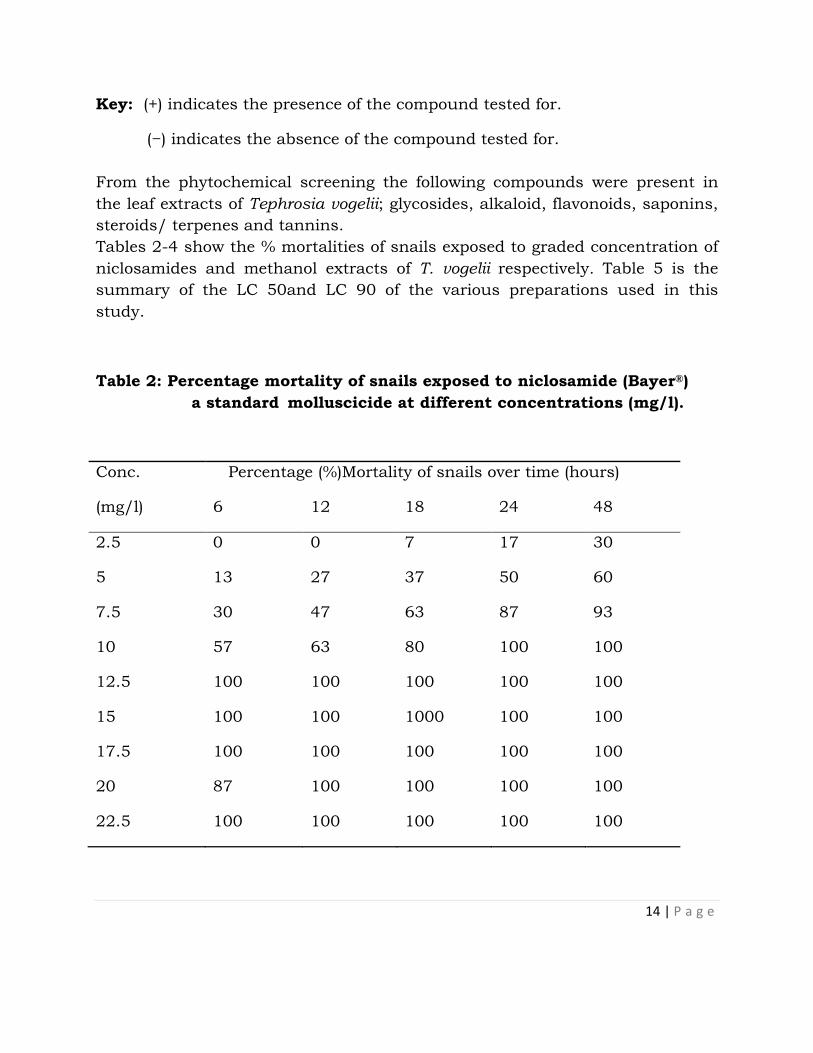
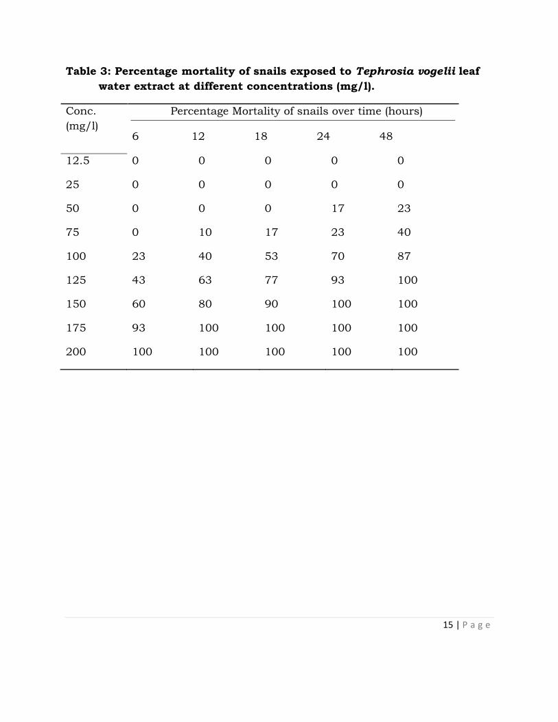
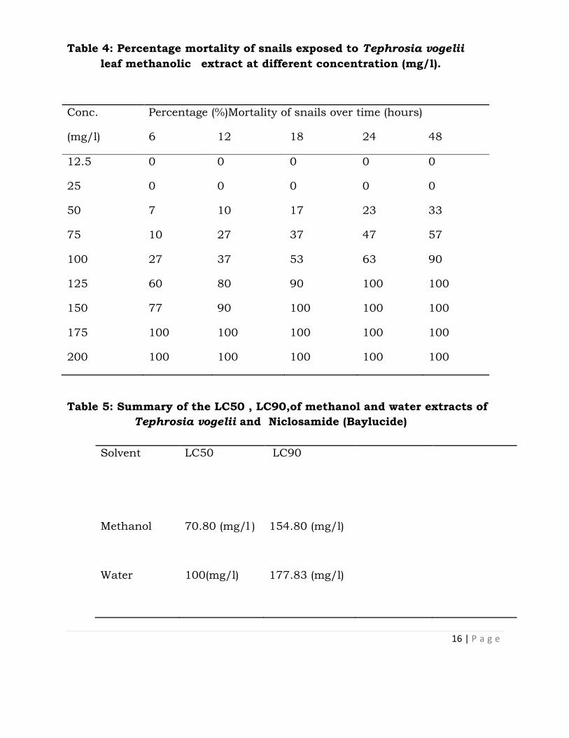
Tables 2-4 show the % mortalities of snails exposed to graded concentration of
niclosamides and methanol extracts of T. vogelii respectively. Table 5 is the
summary of the LC 50and LC 90 of the various preparations used in this
study.
Table 2: Percentage mortality of snails exposed to niclosamide (Bayer®)
a standard molluscicide at different concentrations (mg/l).
Conc.
(mg/l)
Percentage (%)Mortality of snails over time (hours)
6 12 18 24 48
2.5 0 0 7 17 30
5 13 27 37 50 60
7.5 30 47 63 87 93
10 57 63 80 100 100
12.5 100 100 100 100 100
15 100 100 1000 100 100
17.5 100 100 100 100 100
20 87 100 100 100 100
22.5 100 100 100 100 100
15 | P a g e
Table 3: Percentage mortality of snails exposed to Tephrosia vogelii leaf
water extract at different concentrations (mg/l).
Conc.
(mg/l)
Percentage Mortality of snails over time (hours)
6 12 18 24 48
12.5 0 0 0 0 0
25 0 0 0 0 0
50 0 0 0 17 23
75 0 10 17 23 40
100 23 40 53 70 87
125 43 63 77 93 100
150 60 80 90 100 100
175 93 100 100 100 100
200 100 100 100 100 100
16 | P a g e
Table 4: Percentage mortality of snails exposed to Tephrosia vogelii
leaf methanolic extract at different concentration (mg/l).
Conc.
(mg/l)
Percentage (%)Mortality of snails over time (hours)
6 12 18 24 48
12.5 0 0 0 0 0
25 0 0 0 0 0
50 7 10 17 23 33
75 10 27 37 47 57
100 27 37 53 63 90
125 60 80 90 100 100
150 77 90 100 100 100
175 100 100 100 100 100
200 100 100 100 100 100
Table 5: Summary of the LC50 , LC90,of methanol and water extracts of
Tephrosia vogelii and Niclosamide (Baylucide)
Solvent LC50 LC90
Methanol 70.80 (mg/l ) 154.80 (mg/l)
Water 100(mg/l) 177.83 (mg/l)

17 | P a g e
Niclosamide 5.01(mg/l) 7.94 (mg/l)
DISCUSSION
The molluscicidal properties observed were both time and concentration
dependent using log-dose and time-log probit analysis for the determination of
lethal concentration and exposure time (Teixeira et al, 2011). There was a
significant difference (P<0.05) between concentration and mortality at 6, 12,
18, 24 and 48 hours of exposure to the extract, but at concentrations above
125mg/l, there was no significant difference (P>0.05) in mortality at the same
exposure time between leaf extracted with methanol and water. Also as the
concentration increased the exposure time required to cause mortality of the
snail decreases. This agreed with Kloos et al., (1987) that studied the
molluscicidal activity of water extract of different parts of T.vogelii (leaves, stem
and seeds). The behavioural changes of the snails observed in this study is
characteristic for all molluscicides both plant and synthetic (Singh et al., 1996;
Salawu and Odaibo, 2011). Mortality observed might have occurred as a result
of loss of water equilibrium between the snail and its habitat due to
interference of the plant extracts. This may create anaerobic conditions that
induce snail inactivity and its extrusion from the shell (Abdel-Aziz et al., 1990;
Osuala and Okwuosa,1993; Clark and Appleton, 1996; Brackenbury, 1999;
Adetunji and Salawu, 2010). Other possible causes of mortality include,
damage to the cutaneous respiration which therefore result in change in
oxygen consumption.
CONCLUSION
Results from this study showed that the leaf extracts of Tephrosia vogelii has
efficient molluscicidal activity against Lymnaea snails in a dose and time
dependent gradient. Tephrosia vogelii may be a promising plant for the
development of a cheap, safe and effective molluscicides in Nigeria.

18 | P a g e
REFERENCES
Abdel-Aziz, A., Brain, K. and Bashir, A.K. (1990). Screening of Sudanese plants
for molluscicidal activity and identification of Tacca leontopetaloides (L)
(Tuccaceae) as a potential new exploitable resource. Journal of
Phytotherapeutic Research, 4: 62-65.
Adenusi, A.A. and Odaibo, A.B. (2007). Preliminary laboratory assessment of
the crude aqueous and ethanolic extracts of Dalbergia sissoo plant
parts for molluscicidal, ovicidal and cercaricidal activities.. Travels
Medicine Infectious Disease. 6: 219-227.
Adenusi, A.A and Odaibo A.B. (2008). Preliminary laboratory assessment of the
crude aqueous and ethanolic extracts of Dalbergia sissoo plant parts
for molluscicidal, ovicidal and cercaricidal activities. Travel Medicine
Infectious Disease. 6: 219-227.
Adetunji, V.O. and Salawu, O.T. (2010). Efficacy of ethanolic leaf extracts of
Carica papaya and Terminalia catappa as molluscicides against the
snail intermediate hosts of schistosomiasis. Journal of Medicinal Plant
Research, 4(22): 2348-2352.
Adewumi, C.O. and Sofowora, E.A. (1980). Preliminary Screening of some Plant
extracts for molluscicidal activity. Planta Medica, Journal of Medicinal
Plants Research 39: 57-64
Agboola, O., Isaiah, A., Glory, O., Adesegun, S. and Adesanya S. A. (2012).
Investigating the molluscicidal potential of some members of Nigeria
sapindaceae family. Archives of Applied Science Research, 4 (3): 1240-
1243.
Alison H. (2011). Snail-borne diseases in the bovids at high and low altitut in
Eastern Uganda.Integrated Parasitological and Malaclogical mapping.
An M.Sc Dissertation. Department of Biology, Control of Parasites and
Disease Vectors.Liverpool School of Tropical Medicine.
Azare, B.A., Ebele, S.O. and Enwerem N. (2002). Relative molluscicidal potency
of aqueous leaf extracts of Alternanthera sesselis on Bulinus (Phy)
globosus. Zuma Journal of Pure and Applied Science, 1: 110-112.

19 | P a g e
Azare, B.A., Okwute, S.K and Kela, S.L (2007). Molluscicidal activity of crude
water leaf extracts of Alternanthera sesselis on Bulinus (phy) globosus.
African Journal of Biotechnology, 6(4): 441-444.
Benson, O. and Olajumoke A.M. (2012). Molluscicidal effect of aqueous and
ethanolic extracts of Lemongrass (Cymbopogon citratus) leaf against
the different developmental stages of Biomphalaria Pfeifferi. New York
Journal of Science, 5(8): 70-76.
Brackenbury, T.D. (1999). Gross histopathological effects of an extract of Agave
attenuate on the epithelium of the digestive tract of Bulinus africanus.
Annals of Tropical Medical Parasitology, 93(5): 519-526.
Clark, T.E and Appleton, C.C. (1996). A semi quantitative approach to the
selection of appropriate candidate plant molluscicides: A South African
application. Journal of Ethnopharmacology, 56: 1-13.
Eman, E. Taher, Narmeen F. Mahmoud and Maha A. Mahmoud. (2011).
LaboratorEvaluation of Some Egyptian Native Plants versus Some
Parasitic Vectors. Research Journal of Medicine and Medical Sciences,
6(2): 85-90.
Farheen H. and Singh D. K. (2012). Molluscicidal activity of Morus nigra
against the freshwater snail Lymnaea acuminata Journal of Biology and
Earth Sciences 2: 14-23
Fayez, A. B. (2009). Use of some plant extracts to control Biomphalaria
alexandrina snails with emphasis on some Biological effects. World
Applied Sciences Journal 6(10): 1335-1345.
Gehad, T.E., Rawia, A.Z. and Eman, T.E. (2009).Molluscicidal activity of some
Solanum species extract against the snail, Biomphalaria alexandrina.
Journal of Parasiitology Research, 1-5 Doi. 10. 1155-47360.
Kanyari, P. W. N., Kagira, J. M. and Mhoma, J. R. L. (2010) 'Prevalence of
endoparasites in cattle within urban and peri-urban areas of Lake

20 | P a g e
Victoria Basin, Kenya with special reference to zoonotic potential',
Scientia Parasitologica, 11(4), 171-178.
Kela, S.L. (1992). Molluscicidal properties of Polygonum limbatum (L.). Paper
presented at the 16th Annual Conference of the Nigerian Society for
Parasitology,Ahamadu Bello University, Zaria,23-26th September,1992.
Kloos,H.,Thiongo,F.W.,Ouma,J.H. and Butterworth,A.E. (1987). Preliminary
evaluation of some wild and cultivated plants for snail control in
Machakos district, Kenya. Journal of Tropical Medicine and Hygiene 90:
197-204.
Kovendan, K., K. Murugan, C. Panneerselvam, P.M. Kumar, D. Amerasan, J.
Subramaniam, S. and. Barnard, D.R.( 2011). Laboratory and field
evaluation of medicinal plant extracts against filarial vector, Culex
quinquefasciatus Say (Diptera: Culicidae). Parasitology Research. 453-
464.
Labe, Y., Inabo, H. I. and Yakubu, S.E. (2012). Comparative molluscicidal
activity of aqueous and methanolic extract of Zingiber offinale against
Bulinus globusus. Advance in Enviromental Biology, 6 (2): 831-835.
Moema, E.B.E., King, P.H., and Baker, C. (2008). Cercariae development in
Lymnae natalensis. Krauss1848 collected in the vicinity of Pretoria,
Gauteng Province South Africa. Ondersteport Journal of Research, 75:
215-223.
Olofintoye, L.K. (2012). Comparative evaluation of molluscicidal effects of
Seecuridaca longepedunculata (Fres.) and Tephrosia brateolate
(Guilland Perr) on Bulinus globosus. New York Journal of Science .5(8).
Accessed August 28,2012h t t pi/www.science pub. Net/newyork.
Osuala, F.O.U. and Okwuosa, V.N. (1993). Toxicity of Azadirichta indica to
freshwater snails and fish with reference to Physicochemical. In: Kela
21 | P a g e
SL, Bowen ID (1995). Control of snail and snail – borne diseases.
Pesticide outlook . 22 – 27.
Salawu, O. T. and Odaibo, A. B. (2011). The molluscicidal effects of Hyptis
suaveolens on
different stages of Bulinus globosus in the laboratory. African Journal
of Biotechnology, 10 (50): 10241-10247.
Schillhorn, van veen, T. W. (1980). Dynamics of Lymnae natalensis population
in the Zaria area (Nigeria) and the relation to Fasciola gigantic infections.
Acta Tropica, 37: 183-194.
Singh, A., Singh, D.K., Mishara, T.N. and Agarwal, R.A. (1996). Molluscicides of
plant origin. Biology Agriculture and Horticulture Journal, 15: 205-252.
Sofowora, A. (1993). Recent trends in research into African Medicinal Plants.
Journal of Ethnopharmacology, 38: 209-214.
Soliman, K.M, El-Ansary,A.K and Mohamed, A.M. (2000). Effect of carnosine
administration on certain metabolic parametetres in bilharzial infected
hamsters. Journal of Egyptian Society of Parasitology. 30: 455-468.
Soulsby, E. J. L. (1982). Helminth, Arthropod and Protozoa of Domestic Animals.
7th edition Baillere Tindall, London, UK.
Teixeira, T; Jose, S; Nuno, R; Baptista, J; Armindo, R (2011). Assessment of
molluscicidal activity of essential oils from five Azorean plants against
Radix peregra (Muller 1777), Chemosphere 87 (1-6), Elservier Ltd
Trease, G.E. and Evans, W.C. (1989). Text book of Pharmacognosy.Balliere
Tindall: London (12): 176-180.
World Health Organisation (1983). Guidelines for evaluation of plant
molluscicides. WHO. Report of scientific working group on plant
molluscicide and guidelines for evaluation of plant molluscicide.
Geneva , (TDR/SCH- SW (4)/ 83.3).
22 | P a g e
SOURCES OF SALMONELLA INFECTIONS IN SOME SELECTED POULTRY
FARMS IN JOS, NORTHERN NIGERIA
Sati, N. M1٭., Okolocha, E. C2., Kazeem, H. M3., Kabir, J2, Fagbamila, I.
O4., Muhammad, M4 and Lettini, A. A5.
1 Poultry Division, National Veterinary Research Institute Vom, Nigeria. 2 Department of Veterinary Public Health and Preventive Medicine, Ahmadu
Bello University Zaria, Nigeria. 3 Department of Veterinary Microbiology, Ahmadu Bello University Zaria,
Nigeria. 4 Bacterial Research Division, National Veterinary Research Institute Vom,
Nigeria. 5 OIE Italian Reference Laboratory for Salmonella, Istituto Zooprofilatico
Sperimentale delle Venezie, Padova, Italy.
Correspondence: [email protected]. Tel: +2348026928771٭
INTRODUCTION
Poultry farming is an activity that is popular, both in rural and urban settings,
in Nigeria. Poultry occupies a prominent position in the provision of animal
protein and it accounts for about 25% of local meat production in Nigeria
(Agbaje et al., 2010).
Salmonella species are classified into serovars (serotypes) based on the
lipopolysaccharide (O), flagellar protein (H), and sometimes the capsular (Vi)
antigens. There are more than 2,500 known serovars (Popoff et al., 2001; Chao
et al., 2007).
Small- scale poultry farmers in Nigeria lose up to 18% of chicks in the first two
weeks of rearing. The mortality is often associated with salmonellosis and this
exerts negative socio economic and food security impacts on farmers
(Muhammad, 2008). The aim of this study is to identify sources of Salmonella
infection on poultry farms in Jos, Plateau State.
Seminar presented 27th May, 2015 at NVRI Auditorium

23 | P a g e
MATERIALS AND METHODS
Sampling methods
Three (3) hatcheries and three (3) poultry farms in Jos, northern Nigeria were
selected using a multistage random sampling method.
Evaluation of day old chicks (DOC) for Salmonella at the hatcheries
Eighteen samples per hatchery were collected from each of the three
hatcheries. A total of 54 samples were collected at the hatcheries before the
chicks were taken to their respective farms.
Evaluation of the chicks for Salmonella at the farms
Six samples each of faecal, feed, water and litter were collected from the nine
farms every two weeks for 8weeks. Proper disinfection as well as change of
laboratory wears was observed from farm to farm so as to check cross
contamination.
Laboratory procedures
Faecal, feed, litter, water and tissue samples
The samples collected were processed using standard methods (Waltman et al.,
1998). One gram or litre (1g or 1l) of the sample was pre-enriched in buffered
peptone water (BPW) in the ratio of 1:10 and incubated at 37oC for 24 hours.
Samples were then enriched on Rappaport Vassiliadis (RV) broth (0.1ml of
sample from BPW into 10ml of RV broth) and incubated at 42oC for 24 hours.
Tissues from dead birds consisting of lungs, liver, spleen, caeca and heart were
processed according to standard methods of isolation by Waltman et al. (1998).
After enrichment, broth cultures were inoculated onto two selective media,
Xylose Lysine Tergitol 4 (XLT4) and Brilliant Green Novobiocin Agar (BGN).
Suspect colonies were then inoculated into Triple Sugar Iron (TSI) agar for
24hours at 37oC. These isolates were sent to the Salmonella reference
laboratory in Padova, Italy for serotyping following specific pattern of
agglutination reactions using the Kauffmann-White classification scheme
(Popoff et al., 2001).
RESULTS
From the three hatcheries selected (hatchery A, B and C), two hatcheries
(66.7%) had Salmonella isolated either from the tissues or faeces of birds before
they were introduced to the different farms (Table1).

24 | P a g e
By the 14th day when the first farm visit was made, all the three farms from
hatchery A maintained their Salmonella-free status. There was no trace of
Salmonella in farm B3 where there was previous infection. Only farm C1 had
Salmonella and it was found in the feed, faeces and drinking water of the
chicks. All the nine farms yielded no Salmonella on the three more farm visits
except farm A3 where Salmonella was isolated during the third farm visit. Table
2 shows the distribution of Salmonella from the hatcheries to the farms during
the course of this study
A total of 864 samples were taken after the chicks had been introduced to the
nine farms and the total number of isolates obtained was 6 (0.7%). Salmonella
Oakland, Salmonella Bonariensis and Salmonella Kentucky were obtained
(Table 3). Eighteen tissue samples were analysed and four (22.2%) were
positive for Salmonella (S. Oakland, S. enterica subsp Enterica). A total of 252
faecal samples were collected and 3 (1.2%) were positive for Salmonella (S.
Oakland), while 2 (0.9%) out of the feed samples were positive (S. Oakland and
S. Bonariensis). One water sample out of 216 (0.5%) (S. Bonariensis) and one
(1) litter sample out of 216 (0.5%) were positive for Salmonella (S. Kentucky)
(Tables 4 and 5).
Table 1: Table showing number of hatcheries tested for Salmonella in Jos,
northern Nigeria
Hatchery No of
dead
birds
No of
dead
birds
positive
No of
faecal
samples
No of
faecal
samples
positive
Total No
of
Samples
Salmonella
Isolation
(%)
A 6 Nil 12 Nil 18 Nil (0%)
B 6 1 12 Nil 18 1 (5.6%)
C 6 3 12 1 18 4 (22.2%)
Total 18 4 36 1 54 5 (27.8%)
Table 2: Distribution of Salmonella from the hatcheries to the farms
during the course of this study
Farm visits A1 A2 A3 B1 B2 B3 C1 C2 C3
Hatchery status - - - - - + + + +

25 | P a g e
2weeks - - - - - - + - -
4weeks - - - - - - - - -
6weeks - - + - - - - - -
8weeks - - - - - - - - -
Key:
+ Positive for Salmonella
- Negative for Salmonella
Table 3: Serotypes of Salmonella isolated from the different farms
sampled in Jos, northern Nigeria
Farm Total no. of samples No. Salmonella positive Serotype isolated
A1 96 None None
A2 96 None None
A3 96 1 S. Kentucky
B1 96 None None
B2 96 None None
B3 96 None None
C1 96 5 S. Oakland,
S.Bonariensis
C2 96 None None
C3 96 None None
Total 864 6
Table 4: Isolation of Salmonella from the different samples collected.
Type of sample Number of samples Number positive for Salmonella (%)
Tissues 18 4 (22.2)
Faeces 252 3 (1.2)
Feed 216 2 (0.9)
Water 216 1 (0.5)
Litter 216 1 (0.5)
Total 918 11 (1.2)

26 | P a g e
Table 5: Salmonella serotypes isolated from the different samples and
their percentages
Isolate Sample type (Number) Number isolated (%)
S. Kentucky Litter 1 (9.1)
S. enterica
subspEnterica
Tissue 1 (9.1)
S. Oakland Faeces(3),Feed
(1)Tissues(3)
7 (63.6)
S. Bonariensis Feed (1), Water (1) 2 (18.2)
DISCUSSION
Epidemiological studies have demonstrated a variety of routes through which
Salmonella can be disseminated within a poultry enterprise (Nayak et al.,
2004). Salmonella may infect young chicks directly through ovarian
transmission or penetrate the egg shell after the egg has been laid (Cox et al.,
2000). Newly hatched chicks are at their peak of susceptibility to Salmonella
(Gast, 2007). Poultry can become infected by horizontal transmission through
infected litter, faeces, feed, water, dust, fluff insects, equipment, fomites,
contaminated with Salmonella (Poppe, 2000), and some of these have been seen
in the course of this study. These findings are in agreement with the report of
Muhammad et al. (2010), who isolated Salmonella from day old chicks from
hatcheries in Jos, Nigeria.
Environmental sources are some of the ways Salmonella gets into poultry
farms. Numerous environmental factors can influence the likelihood and
outcome of infections of poultry with Salmonella. Lengthy environmental
persistence of pathogens can generate extended opportunities for horizontal
transmission within and between flocks (Gast, 2007). In this study, Salmonella
from environmental sources account for 63.6% (7 out of 11) of the isolates
obtained. Isolating the organism from the environment is difficult because of
the few salmonellae in these sources (Waltman et al., 1998) and the fragility of
the organism in these samples.
It has been reported that bacteriological sampling does not always provide an
accurate indication of infection within a flock because of low incidence of
infection and the intermittent excretion of Salmonella organisms (Hassan et al.,

27 | P a g e
1990). This might also explain why the rate of isolation of the organism was low
in this study.
CONCLUSION AND RECOMMENDATIONS
Salmonella infections are not just acquired on farms, but sometimes the
hatcheries that supply chicks are responsible for disseminating the organisms
as was seen in this study, resulting in high chick mortality, poor feed
conversion and unnecessary exposure of farmers/consumers to infections.
Government should enforce the existing laws that prohibit establishment of
poultry farms and hatcheries without adequate training for farmers.
Government should provide veterinary and animal health extension services in
all the local government areas to ensure adequate records, proper monitoring,
and effective management of Salmonella infections.
Surveillance is needed to help prevent food-borne disease outbreaks and raise
awareness among health authorities, food producers, food regulators, and
consumers.
REFERENCES
Agbaje, M., Davies, R., Oyekunle, M.A., Ojo, O.E., Fasina, F.O. and Akinduti,
P.A. (2010).Observation on the occurrence and transmission of
Salmonella gallinarum in commercial poultry farms in Ogun State,
South Western Nigeria. African Journal of Microbiology Research 4(9),
796-800.
Chao, M.R., Hsien, C.H., Yeh, C.M., Chou, S.J., Chu, C., Su, C.Y. and Yu, C.Y.
(2007). Assessing the Prevalence of Salmonella enterica in Poultry
Hatcheries by Using Hatched Eggshell Membranes. Poultry Science
86:1651-1655.
Cox, N.A., Berrang, M.E., Cason, J.E. (2000). Salmonella Penetration of egg
shells and proliferation in broiler hatching eggs: a review. Poultry
Science 79:1571–1574.
Gast, R.K. (2007).Serotype-specific and serotype-independent strategies for
preharvest control of food borne Salmonella in Poultry. Avian Diseases
51:817-828.

28 | P a g e
Hassan JO, Barrow PA, Mockett APA, McLeod S (1990). Antibody response to
experimental Salmonella typhimurium infection in chickens measured by
ELISA. Veterinary Records. 126: 519-522.
Muhammad, M (2008). The role of Salmonella sp in early chick mortality and
the application of hazard analysis critical control point (HACCP) for
control. Ph.D Thesis University of Maiduguri
Muhammad, M., Muhammad, L.U., Ambali, A., Mani, A.U., Azard, S. and
Barco, L. (2010) Prevalence of Salmonella associated with chick mortality
at hatching and their susceptibility to antimicrobial agents, Veterinary
Microbiology 140: 131-135.
Nayak, R., Stewart, T., Wand, R.F., Lin, J., Cerniglia, C.E., Kenney, P.B (2004)
Genetic diversity and virulence gene determinants of antibiotic resistant
Salmonella isolated from preharvest turkey production sources.
International Journal of Food Microbiology 91: 51–62.
Popoff, M.Y., Bockemühl, J., Brenner, F.W.and Gheesling, L.L. (2001).
Supplement 2000 (no. 44) to the Kauffmann-White scheme, Research in
Microbiology, 152:907–909.
Poppe, C. (2000). Salmonella infections in the domestic fowl, in Wray, C. &
Wray, A. (Eds) Salmonella in domestic animals, (Wallingford CABI
Publishing). Pp 107-132.
Waltman, W.D., Gast R.K. and Mallinson E.T. (1998). Salmonellosis; A
laboratory manual for the isolation and identification of avian
pathogens In: Swayne D.E., Glisson J.R., Jackwood M.W., Pearson J.E
and Reed W.M (Eds) American Association of avian pathologists 4th
edition. University of Pennsylvania, USA, 4-13.

29 | P a g e
PREVALENCE AND ANTIMICROBIAL SUSCEPTIBILITY PROFILE OF
SALMONELLA SEROVARS FROM CATTLE IN JOS, NIGERIA
Mercy N. Benson
Quality Control Division
National Veterinary Research Institute, Vom
E mail: [email protected]
INTRODUCTION
Salmonellae are zoonotic bacteria with a rising prevalence in the cattle industry
(Randall, 2001). Salmonellosis remains a global problem with a significant
economic impact on the cattle industry causing livestock mortality, abortion
and reduced milk production. They are among the most common bacterial
food-borne pathogens worldwide (Todd, 1997). Under-reporting of salmonellosis
and the presence of other diseases considered to be of higher priority may have
over-shadowed the problem of the disease in some developing countries. In
Nigeria, efforts at prevention and control have largely been by improved
conditions on grazing reserves, improved sanitation and hygiene (Adene and
Oguntade, 2008), improvement in public water supply, safe disposal of waste,
use of newer antimicrobials and general public health measures (Adeniran et
al., 2005).
This study was designed to determine the serovars and antimicrobial
susceptibility profile of Salmonella species from cattle in Jos, Nigeria.
MATERIALS AND METHODS
Sample collection
A total of 712 faecal samples were collected from cattle from 3 sites including
cattle farms (572), cattle control-post (60) and an abattoir (80). The 712 cattle
comprised of 164 males and 548 females, 120 calves and 592 adults, 609
White Fulani cattle, 3 Cross breeds and 100 Holstein Friesians. One hundred
were raised under intensive system, 224 under semi intensive, 248 under
extensive while 140 were unknown (control post and abattoir). Faecal samples
were obtained per rectum and packed in a cold box and then transported
immediately from point of collection to the laboratory for examination.
Isolation and serotyping
Salmonellae were isolated and identified according to the techniques
recommended by the International Organization for Standardization (ISO) 6579

30 | P a g e
Seminar presented 27th May, 2015 at NVRI Auditorium
(2002) from pre-enrichment to serotyping (which was done in The Salmonella
Reference Laboratory Padova, Italy).
Antimicrobial susceptibility profile
The antimicrobial susceptibility pattern of the isolates was conducted using a
modified Kirby-Bauer disk diffusion method (Bauer et al., 1966). A panel of 16
antibiotics namely: colistin (10 µg), sulphamethoxazole + trimethoprim (23.75
µg +1.75 µg), kanamycin (30 µg), gentamicin (10 µg), cefotaxime (30 µg),
amoxicillin + clavulanic acid (20 µg + 10 µg), ceftazidine (30 µg), nalidixic acid
(30 µg), tetracycline (30 µg), ampicillin (10 µg), streptomycin (10 µg), triple sulfa
(0.25 µg), chloramphenicol (30 µg), cephalothin (30 µg), enrofloxacin (5 µg),
ciprofloxacin (5µg) were tested. Each isolate was diluted in sterile saline
solution to a 0.5 McFarland standard. The diluted bacterial suspension was
transferred onto Mueller Hinton agar plates using sterile swabs. The plates
were seeded uniformly by rubbing the swabs against the entire agar surface.
Each antimicrobial impregnated disk was applied onto the surface of the
inoculated plate by using a sterile disc dispenser. The plates were incubated at
370C for 18 hours. Interpretation of the growth inhibition zones and
classification of isolates as susceptible, intermediate and resistant was done
following guidelines of the National Clinical Laboratory Standards (NCCLS,
2004).
Statistical analysis
The occurrence of Salmonella was calculated using percentages and the results
were analysed to determine if any relationship existed between the presence of
Salmonella and age, sex, breed and management system using Chi square test
by Statistical Package for Social Sciences (SPSS) Version 15 (Inc.,Chicago,USA).
RESULTS
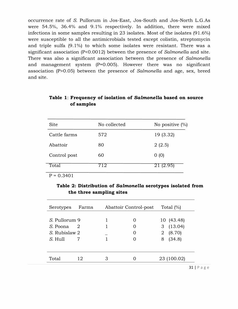
An isolation rate of 2.95% (21 of 712) was recorded in this study. Higher
isolation rates were recorded in adults (3.21%), females (4.19%),White Fulani
breeds (3.28%) and extensively managed cattle (6.45%) compared to that in
calves (1.67%), males (1.22%), Holstein Freisian (1%) and Cross-breeds (0%),
intensively (1%) and semi-intensively bred cattle (1.34%) respectively. Isolation
rates of 6.29%, 3.06% and 1.59% were recorded in Jos-East, Jos-North and
Jos-South respectively. Four serotypes were isolated; S. Pullorum (43.48%), S.
Hull (34.8%), S. Poona (13.04%) and S. Rubislaw (8.7%) (Table 2). The
31 | P a g e
occurrence rate of S. Pullorum in Jos-East, Jos-South and Jos-North L.G.As
were 54.5%, 36.4% and 9.1% respectively. In addition, there were mixed
infections in some samples resulting in 23 isolates. Most of the isolates (91.6%)
were susceptible to all the antimicrobials tested except colistin, streptomycin
and triple sulfa (9.1%) to which some isolates were resistant. There was a
significant association (P<0.0012) between the presence of Salmonella and site.
There was also a significant association between the presence of Salmonella
and management system (P=0.005). However there was no significant
association (P>0.05) between the presence of Salmonella and age, sex, breed
and site.
Table 1: Frequency of isolation of Salmonella based on source
of samples
Site No collected No positive (%)
Cattle farms 572 19 (3.32)
Abattoir 80 2 (2.5)
Control post 60 0 (0)
Total 712 21 (2.95)
P = 0.3401
Table 2: Distribution of Salmonella serotypes isolated from
the three sampling sites
Serotypes Farms Abattoir Control-post Total (%)
S. Pullorum 9 1 0 10 (43.48)
S. Poona 2 1 0 3 (13.04)
S. Rubislaw 2 _ 0 2 (8.70)
S. Hull 7 1 0 8 (34.8)
Total 12 3 0 23 (100.02)
32 | P a g e
DISCUSSION
Salmonellae were isolated from cattle on farms and those slaughtered for
meat at the abattoir. Overall, 2.95% (21 of 712) of cattle sampled were
positive for Salmonella species. The low isolation rate of Salmonella obtained
in this study might be due to single sampling per animal, season, regional
variation, management practices or a low carrier rate in cattle in the study
area at the period of sampling.
In this study, Salmonella was isolated in all the three localities sampled
though Jos-East had a higher isolation rate with a statistically significant
value (P<0.05) compared to Jos-North and Jos-South. The higher isolation
rate in Jos-East could be attributed to the movement of high population of
cattle (especially nomadic) through the area.
The higher frequency of isolation on farms, though not statistically
significant (P>0.05), compared to the abattoir or control-post was likely due
to the lowered gastric acidity in the animals from starvation during
movement from the farms to the abattoir.
The extensively managed cattle produced the highest isolation rate with a
statistically significant value (P < 0.05) compared to those on semi-intensive
or intensive farms. This was attributed to their mode of feeding which is
majorly random grazing where they may be exposed to various pathogens.
Interestingly, from the 21 positive samples, host adapted Salmonella (S.
Pullorum) had the highest occurrence rate compared to the other serotypes.
This observation may be associated with random grazing habits of the cattle
in different regions including areas where poultry farms are situated or
where poultry have roamed, thereby contaminating the pasture with poultry
faeces/wastes. In addition, potential sources of infection arise when poultry
waste is released into waterways and is thereafter used to irrigate livestock
forage crops or inadvertently contaminates pasture on which cattle graze or
is ingested via drinking.
Other serotypes: S. Hull, S. Poona and S. Rubislaw were isolated in a
decreasing order respectively. These serotypes or other environmental
Salmonella may be the cause of food-borne outbreaks, in particular from
fruits, vegetables and spices contaminated by Salmonella from feral reptiles
or other animals. This may explain recent outbreaks of S. Poona in the USA
(Molbak et al., 2012). Reports of S. Poona and S. Rubislaw have been made
in Nigeria by Olayemi (1978) in sewage waste from the abattoir in Zaria and
Collard and Sen (1956) in Ibadan respectively. Stevens et al. (2006) equally
isolated S. Poona from beef in Senegal. If more colonies per sample had been

33 | P a g e
picked, we might have found more serotypes, masked by dominant ones. In
this work however, isolated colonies were randomly selected among
characteristic ones for confirmation. The bias is therefore weak, but a
different competitiveness for each serotype cannot be excluded. The
serotypes obtained in this study may reflect the serotypes prevalent in this
environment.
Majority of the isolates were susceptible to the panel of antimicrobials.
However, 2 isolates were resistant to sulfonamide and 1 isolate each were
resistant to colistin and streptomycin. Among the 23 serotypes, 8.69%
displayed resistance to at least one antimicrobial. In contrast 91.3% were
sensitive to all the antimicrobials. Development of resistance to
sulfonamides may be as a result of indiscriminate use of the antimicrobial
in animal husbandry in the area. In Ethiopia, Alemayehu et al. (2003)
reported that 52% of the Salmonella isolated at the slaughterhouse from
beef were resistant to at least three antimicrobials. In the United States,
84% of the Salmonella isolates from retail meats were resistant to at least
one antimicrobial, and 53% to at least three antimicrobials (Duffy et al.,
1999). By comparison therefore, the strains isolated from Jos showed a low
level of resistance to commonly used antimicrobials. Among the serotypes,
only S. Poona manifested multiple drug resistance. This resistance was
exhibited to sulphonamide, which may be due to inappropriate use of
antimicrobials in the area due to quackery.
CONCLUSIONS
A 1ow overall Salmonella occurrence rate of 2.95% in cattle in Jos-Nigeria
was recorded in this study. Samples from the control-post in Jos failed to
yield Salmonella isolates. The data documented rare or unusual serovars of
Salmonella in cattle (S. Poona, S. Rubislaw, S. Hull and S. Pullorum). To the
best of our knowledge, this is the first report of such Salmonella serotypes in
cattle in the study area. This result is significant because S. Pullorum is
known to be a poultry associated serotype and coincidentally recorded the
highest occurrence among the serotypes found, implying that the serotype
may have a wider host range.

34 | P a g e
RECOMMENDATIONS
We recommend that there is need to improve surveillance especially at
control-posts to avoid spread and introduction of new serotypes into clean
areas. Information on Salmonella isolates, their antimicrobial susceptibility
patterns and virulence characteristics need to be passed on to medical
practitioners. Public education on the need for hygienic slaughter, handling,
processing or cooking of meat should be enhanced.
REFERENCES
Adene, D.F. and Oguntade, A. E. (2008). Poultry sector country review:
Nigeria, pp 15
Adeniran, J.O, Taiwo, J.O. and Abdu-Rahaman, L.O (2005). Salmonella
intestinal perforation; 27 perforations in one patient, 14
perforations in another. Are the goal post changing. Journal of
Indian Association of Pediatric Surgery, 10(4): 248-252.
Alemayehu, D., Molla, B., Muckle, A. (2003). Prevalence and antimicrobial
resistance pattern of Salmonella isolates from apparently healthy
slaughtered cattle in Ethiopia. Tropical Animal Health Production, 35:
309–319.
Bauer, A. W., Kirby, W. M. M., Sherris, J. C. and Turck, M. (1966).
Antibiotic susceptibility testing by a standardized single disk method.
American Journal of Clinical Pathology, 45:493-496.
Collard, P and Sen, R. (1956). Isolation of Salmonellae from cattle in
Ibadan. West African Journal of Medicine, 5: 118-120.
Duffy, G., Cloak, O.M., O'Sullivan, M.G., Guillet, A., Sheridan, J.J., Blair,
I.S., McDowell, D.A. (1999). The incidence and antibiotic resistance
profiles of Salmonella spp. on Irish retail meat products. Food
Microbiology, 16: 623–631.
ISO. (2002) International Organization for Standardization 6579.
Microbiology of food and animal feeding stuff-Horizontal method for
the detection of Salmonella, Geneva. PP. 1-18.

35 | P a g e
Mølbak, K., Olsen, J. E. and Wegener, H.C (2012) Salmonella infections,
retrieved on July 12, 2012.12.35 am from
www.123foodscience.com/food_microbiology/Salmonella_ infections.
NCCLS (2004) National Clinical Laboratory Standards, Performance
standards for antimicrobial disk susceptibility testing. NCCLS document
M100-514.Wayne, Pa.
Olayemi, A. B. (1978). The incidence of Salmonella in cattle slaughtered at
the Zaria abattoir, M. Sc. Thesis, Ahmadu Bello University, Zaria,
Nigeria.
Randall, J.B. (2001). Salmonellosis in cattle 1:8 Retrieved on 17 April,
2010, from www.the cattle site.com/Salmonella-dublin infection.
Stevens, A., Kaboré, Y., Perrier-Gros-Claude J., Millemann,Y.,
Brisabois, A., Catteau, M., Cavin, J. and Dufour, B. (2006).
Prevalence and antibiotic-resistance of Salmonella isolated from beef
sampled from the slaughterhouse and from retailers in Dakar
(Senegal). International Journal of Food Microbiology, 110: 178–186.
Todd, E.C.D (1997). Epidemiology of food-borne diseases: A worldwide
review. World Health Statistics Quarterly, 50: 30–50.

36 | P a g e
REVERSE GENETICS: A USEFUL TOOL IN FUNCTIONAL GENOMICS
(ROLE OF ZNT1 GENE IN VERTEBRATE DEVELOPMENT)
Muraina Issa1,2, Graham Anthony3 and Hogstrand Christer2
1 National Veterinary Research Institute, PMB 01, Vom, Nigeria 2King’s College London, Faculty of Life Sciences and Medicine, Diabetes and
Nutritional Sciences Division, Metal Metabolism Group, Waterloo Campus, UK 3King’s College London, MRC Centre for Neurodevelopmental Biology, Guy’s
Campus, UK
Correspondence: [email protected]
INTRODUCTION
Reverse genetics methodologies generally refer to the generation or targeted
discovery of a mutation in a gene that is known by its sequence. It is a useful
approach to discover gene’s function by analysing the phenotypic effect of
specific gene sequence. This is a reverse of the so-called forward or classical
genetics in which the investigative process proceeds in opposite direction. That
is, while forward genetics attempts to find the genetic basis of a phenotype or
trait, reverse genetics aims to find what phenotype arises as a result of
particular genetic sequences. Different effective reverse genetics approaches are
in existence but they tend to be organism specific. Examples include gene
knockout via homologous recombination in mice (and recently in Drosophila),
gene knock-in via insertion (transgene) in mouse and gene knockdown or
silencing via antisense morpholino oligonucleotides in Xenopus and zebrafish
oocytes or via RNA interference (RNAi) in Ceanorhabditis elegans, Drosophila
and mouse. Other methods include TE (Transposable Elements) in Drosophila
and Ceanorhabditis elegans, and TILLING (Targeting Induced Local Lesions in
Genomes) which is applicable to most organisms identifies mutations in
specific genes of interest in chemically mutagenized populations (Stemple,
2004).
Among the mammalian zinc transporter genes, ZnT1 (SLC30A1) was the first to
be discovered with its expression throughout the body notably in the
basolateral membranes of epithelia involved in zinc acquisition or recycling like
intestine, kidney and placenta (Lichten and Cousins, 2009) and functions to
transport excess cytoplasmic zinc out of the cells (Palmiter and Findley, 1995).

37 | P a g e
Seminar presented 11th June, 2015 at NVRI Auditorium
Previous works have shown that homozygous deletion or knockout of the
whole ZnT1 gene in mice was embryonic lethal (Andrews et al., 2004) whereas
in Caenorhabditis elegans as well as in Drosophila, a loss-of-function mutation
of their respective ZnT1 orthologs result in animal with impair growth and
development (Bruinsma et al., 2002; Wang et al., 2009).
The zebrafish (Danio rerio) is an important vertebrate model system; well suited
for studies in genetics, embryology, development, and cell biology because of
the unique characteristics it possesses (Westerfield, 1993). In addition, there is
strong conservation between zebrafish and humans for most genes, which
makes zebrafish an exellent model organism for studying complex biological
processes (Chen and Fishman, 1996). With the advent of TILLING technology,
it is now possible to generate zebrafish knock-outs or mutants for specific
genes, making reverse genetics available for this model.
METHODOLOGY
Animal model Zebrafish embryos (strains sa0017) recovered from the out-crossing of a wild-type female fish and a male fish that is heterozygous for slc30a1 (znt1) gene
mutation (using TILLING technology) were collected from Sanger Centre Zebrafish Mutation Project (http://www.sanger.ac.uk/cgi-
bin/Projects/D_rerio/zmp/search.pl?q=slc30a1). This mutation caused a transition of nucleotide ‘‘A’’ to ‘‘T’’ at 355 nucleotide sequence leading to premature termination of the Znt1 protein sequences which resulted in a
truncated protein, short of the last forty (40) amino acids. These embryos were reared to adult in a stand-alone zebrafish facility of King’s College London under standard fish husbandry practice and thereafter different genotypes
were classified. Fish were bred and embryos from different genotypes were collected for study.
In a related experiment, wild-type embryos were micro-injected at 1-4 cell stage with 2-4ng of translational blocking anti-sense morpholino-modified oligonucleotides (MO) for znt1 gene knockdown
(5'GCGGAGCACAGACAGAAACAAAAGCT3') (GENETOOLS, Philomath, USA) as previously described (Nasevicius and Ekker, 2000). A scramble or mismatch
MO (Random-control-MASO) was injected in a similar way to serve as injected control along with un-injected wild-type control. Because of the potential problem of off-target effects produced by most MOs as a result of p53 gene
activation causing a non-specific neuronal cell death (Pickart et al., 2005), a p53 translational blocking MO (5'GCGCCATTGCTTTGCAAGAATTG3') was co-
38 | P a g e
injected with znt1 MOs in the ratio of 1.0 : 1.5 to suppress the p53-mediated apoptosis, and the result of the embryonic development was compared to that
resulting from injecting znt1 morpholino alone. All embryos were incubated at 28.5oC and monitored through developmental stages.
For exposure analysis, embryos were incubated in embryo water either supplemented with zinc by 100µM of ZnSO4 or depleted of zinc by 5µM TPEN
(N,N,N,N,-Tetrakis(2-pyridymethyl ethylene-diamine).
Mutation detection and genotype classification A simple and rapid Locked Nucleic Acid (LNA) method was adapted for detection of SNP (single nucleotide polymorphism) mutation which was used to
classify the genotype (Johnson et al., 2004). This is based on TaqMan assay which utilizes dual-labelled fluorescence probes to discriminate between allele
A and T in the target region of the genomic DNA of fish using a Real-Time PCR (qPCR) technique. The primers and probe sequence sets for the assay were designed and synthesized by Integrated DNA Technology (IDT). This simple
and cost-effective method of mutation detection was re-confirmed in a number of samples using conventional nested PCR followed by sequence analysis of the target region. The primers for conventional PCR were designed by Sanger
Centre’s Zebrafish Mutation Project and synthesized by Sigma. In this technique, both internal forward and reverse primers were further re-designed
with M13 forward and reverse tail primers for bi-directional sequencing of the PCR products.
Gene expression analysis DIG-labelled anti-sense RNA for znt1 gene (probe) was produced according to
standard gene cloning procedures followed by in vitro transcription. The forward and reverse primer sequences used for amplification of the gene fragment gave a product size of 538bp
(f: 5'AGACCCAGTCCACCAACAAG3'; r: 5'AGGACATGCAGGAAAACACC3'). This probe was used for gene expression analysis on 24hpf embryos using the
method of whole mount In Situ Hybridization (ISH) as described by Thiese et al., 2004.
Total and free zinc analysis in embryo
Chorionated embryos at 24hpf of znt1 homozygotes, znt1 morphants and wild-
types backgrounds were acid digested respectively and total zinc
concentrations were measured using ICP-MS (Perkin Elmer, model ELAN
6100DRC) as previously described (Zheng et al., 2008). In a related experiment,
free zinc (Zn2+) ions were assayed in the embryos by both fluorescent
spectrophotometer and fluorescent microscope at 360/530nm
excitation/emission wavelength using a synthetic ratiometric zinc-specific

39 | P a g e
fluorophore termed “ZTRS” probe which was a kind gift from Dr. Zhaochao Xu
of the University of Cambridge.
Phosphorylated- extracellular regulated kinase (ERK) 1/2 activity in
embryos Embryos at early to mid gastrulation stages (5-8hpf) were used for phospho-ERK immuno-staining technique for Znt1 homozygote mutants, Znt1 morphants, control wild-type and control morphant. A primary antibody to phospho-ERK1/2 (Cell Signalling®; rabbit polyclonal, #4370S) was used at
1:100 dilutions and a secondary antibody (Abcam®; goat anti-rabbit conjugated to Alexa Fluor 488, ab15007) was used at 1:200 dilutions as previously described (Krens et al., 2008). Expression of p-ERK1/2 proteins in stained
embryos were observed under epi-fluorescence microscope. In a parallel experiment, about 10µg of protein was extracted from 24 hpf embryos of wild-
types and homozygote mutants respectively and analysed for phospho-ERK by Western blotting as previously described (Krens et al., 2008), using rabbit polyclonal p-ERK1/2 as the primary antibody at 1:2,000 and goat anti-rabbit
conjugated to horse radish peroxidase (GE Healthcare Life Sciences, RPN2108)
as the secondary antibodies at 1:5,000. Antibodies to total ERK 1/2 (Cell
Signalling®; #9102S) were used as normaliser or reference protein at 1:1000 along with the secondary antibody at 1:2,000. Films (blots) were processed and developed by automatic machine (Konica Minolta SRX-101A).
RESULT AND DISCUSSION
Bioinformatics The result of bioinformatics analysis using ClustalW Multiple Sequence
Alignment of orthologue Znt1 proteins between teleosts and mammals revealed a high degree of homology between zebrafish and mammals including human (i.e. 96% similarity and 56% identity with human), suggesting that this protein
perform similar function in the species (data not shown). Analysis by Eukaryotic Linear Motif (ELM) search of truncated (40 amino acids) region in the C-termini of Znt1 revealed some evolutionarily conserved residues between
human ZnT1 and zebrafish Znt1 especially the PDZ domain containing the “ESSL” motif, suggesting that the truncation of the C-terminus in zebrafish
Znt1 might affect the function of the protein (Fig 1). Znt1 deficiency or disruption caused delay in stages of embryo development
The result of growth and development in zebrafish (Fig 2A & 2B) showed that embryo with Znt1 homozygote mutation was viable unlike mouse embryo with
ZnT1 null mutation which was embryonic lethal at 9th day of gestation (Andrew et al., 2004). This may be as a result of partial loss-of-function or altered function in the zebrafish mutation compared to the complete loss-of-function in
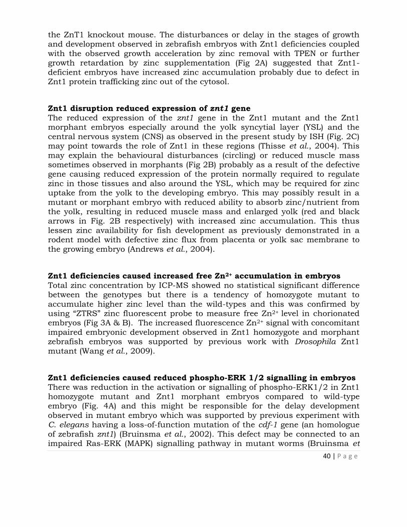
40 | P a g e
the ZnT1 knockout mouse. The disturbances or delay in the stages of growth and development observed in zebrafish embryos with Znt1 deficiencies coupled
with the observed growth acceleration by zinc removal with TPEN or further growth retardation by zinc supplementation (Fig 2A) suggested that Znt1-
deficient embryos have increased zinc accumulation probably due to defect in Znt1 protein trafficking zinc out of the cytosol.
Znt1 disruption reduced expression of znt1 gene
The reduced expression of the znt1 gene in the Znt1 mutant and the Znt1 morphant embryos especially around the yolk syncytial layer (YSL) and the
central nervous system (CNS) as observed in the present study by ISH (Fig. 2C) may point towards the role of Znt1 in these regions (Thisse et al., 2004). This
may explain the behavioural disturbances (circling) or reduced muscle mass sometimes observed in morphants (Fig 2B) probably as a result of the defective gene causing reduced expression of the protein normally required to regulate
zinc in those tissues and also around the YSL, which may be required for zinc uptake from the yolk to the developing embryo. This may possibly result in a
mutant or morphant embryo with reduced ability to absorb zinc/nutrient from the yolk, resulting in reduced muscle mass and enlarged yolk (red and black arrows in Fig. 2B respectively) with increased zinc accumulation. This thus
lessen zinc availability for fish development as previously demonstrated in a rodent model with defective zinc flux from placenta or yolk sac membrane to
the growing embryo (Andrews et al., 2004).
Znt1 deficiencies caused increased free Zn2+ accumulation in embryos
Total zinc concentration by ICP-MS showed no statistical significant difference between the genotypes but there is a tendency of homozygote mutant to
accumulate higher zinc level than the wild-types and this was confirmed by using “ZTRS” zinc fluorescent probe to measure free Zn2+ level in chorionated embryos (Fig 3A & B). The increased fluorescence Zn2+ signal with concomitant
impaired embryonic development observed in Znt1 homozygote and morphant zebrafish embryos was supported by previous work with Drosophila Znt1
mutant (Wang et al., 2009).
Znt1 deficiencies caused reduced phospho-ERK 1/2 signalling in embryos
There was reduction in the activation or signalling of phospho-ERK1/2 in Znt1 homozygote mutant and Znt1 morphant embryos compared to wild-type embryo (Fig. 4A) and this might be responsible for the delay development
observed in mutant embryo which was supported by previous experiment with C. elegans having a loss-of-function mutation of the cdf-1 gene (an homologue
of zebrafish znt1) (Bruinsma et al., 2002). This defect may be connected to an impaired Ras-ERK (MAPK) signalling pathway in mutant worms (Bruinsma et

41 | P a g e
al., 2002) or in Xenopus oocytes, which also showed a delayed meiotic maturation when the gene was disrupted and expressed in frog oocytes
(Jirakulaporn and Muslin, 2004). These observations lend further credence to support the interaction between ZnT1 and MAPK pathway activation being
necessary for diverse cellular process including cell growth, proliferation, differentiation, survival and vertebrate development (Ballif and Blenis, 2001).
REFERENCES Andrews GK, Wang H, Dey SK, Palmiter RD (2004). Mouse zinc transporter 1 gene provides an essential function during early embryonic development.
Genesis 40: 74-81.
Ballif BA, and Blenis J (2001). Molecular mechanisms mediating mammalian mitogenactivated protein kinase (MAPK) kinase (MEK)-MAPK cell survival signals. Cell Growth Diff., 12:397-408.
Bruinsma JJ, Jirakulaporn T, Muslin AJ, Kornfeld K (2002). Zinc ions and
cation diffusion facilitator proteins regulate Ras-mediated signaling. Dev. Cell. 2: 567-578.
Chen JN, Fishman MC (1996). Zebrafish tinman homolog demarcates the heart field and initiates myocardial differentiation. Development 123:293-302.
Jirakulaporn T, and Muslin AJ (2004). Cation diffusion facilitator proteins
modulate Raf-1 activity. J Biol Chem 279: 27807-27815.
Johnson MP, LM Haupt, and LR Griffiths 2004 Locked nucleic acids (LNA)
single nucleotide polymorphism (SNP) genotype analysis and validation using real-time PCR. NAR 32: e55.
Krens SFG, He S, Lamers GEM, Meijer AH, Bakkers J Schmidt T Spaink HP, Snaar-Jagalska BE (2008). Distinct functions for ERK1 and ERK2 in cell
migration processes during zebrafish gastrulation. Developmental Biology 319: 370-383.
Lichten, L.A., Cousins, R.J. (2009). Mammalian zinc transporters: Nutritional and physiologic regulation. Annu. Rev. Nutr, 29:153-176.
Nasevicius A and Ekker SC (2000). Effective targeted gene “knockdown” in zebrafish. Nat Genet. 26:216-220
Palmiter RD and Findley SD (1995). Cloning and functional characterization of
a mammalian zinc transporter that confers resistance to zinc. Embo J 14(4), 639-649.

42 | P a g e
Pickart MA, Klee EW, Nielson AL, Sivasubbu S, Mendenhall EM, et al. (2006). Genome-wide reverse genetics framework to identify novel functions of the
vertebrate secretome. PLoS One 1: e104. doi:10.1371/journal.pone.0000104.
Stemple DL (2004). TILLING- a high-thoroughput harvest for functional genomics. Nature reviews (Genetics), 4:1-5.
Thiese B and Thisse C (2004) Fast Release Clones: A High Throughput Expression Analysis. ZFIN Direct Data Submission.
Wang X, Wu Y, Zhou B (2009). Dietary zinc absorption is mediated by ZnT1 in Drosophila melanogaster. FASEB Journal 23: 2650-2661.
FIGURES
Fig 1: ClustalW multiple
sequence alignment between
zebrafish and human of the last
40 amino acids of the C-terminal
of ZnT1 showing potential
phosphorylation sites as
predicted by ELM search
Fig 2: (A) Phenotype of Znt1 mutant embryo (znt1Δ40/Δ40) and (B) Znt1 morphant embryo (Znt1MO) showing
showing morphogenetic difference in the stage of development (i.e. delayed development) as compared to wild-
type (znt1+/+). (C) Znt1 gene expression showing low expression in homozygote and morphant embryos
compared to wild-type (arrows).
+/+
Znt140
40/
Znt1
MO
Znt1
0
5000
10000
15000
20000
Total Zinc (picogram)
Free zinc (arbitrary unit)
***
***
Zin
c c
on
ce
ntr
ati
on
/em
bry
o
Fig 3: (A & B): Effects of znt1 gene mutation or knockdown on total and free zinc contents in Znt1 homozygote mutant
(znt1Δ40/Δ40) and Znt1 morphant embryos (Znt1MO) compared to wild-type (znt1+/+).
43 | P a g e
MOLECULAR DETECTION OF BOVINE TRYPANOSOMIASIS IN SOME
PARTS OF CENTRAL SENATORIAL ZONE OF PLATEAU STATE-NIGERIA
1Joana G. Peter,2 E.I. Ikeh, 1Dinchi J. Tyem,1Joannis T.M, and 1Bitrus Y.
1. National Veterinary Research Institute, Vom
2. University of Jos, Department of Medical Laboratory Science
Correspondence: [email protected] +2340833892259
INTRODUCTION
Trypanosomes are flagellates protozoa that belong to the genus Trypanosoma.
They cause diseases that are generally referred to as trypanosomiasis. These
protozoa can infect all classes of vertebrates (Connor and Van den Bossche,
2004). African animal trypanosomiasis (AAT) is a tsetse transmitted disease
affecting livestock in Sub-Saharan Africa, (Cheneret et al., 2006). In Nigeria,
trypanosomiasis is probably one of the most important diseases of cattle, and
one of the predominant obstacles to profitable animal production. The control
of African animal trypanosomiasis is based primarily on chemoprophylaxis and
chemotherapy. Therefore, in order to design national control strategies,
knowledge on the distribution of tsetse flies and detection of trypanosome
species/subspecies across the country is important. The latter requires
methods for sensitive and specific detection of the trypanosomes. Jos Plateau
offers a very suitable climate for livestock production especially with the
presence of Integrated Diary Farm (IDF) and the National Veterinary Research
Institute which adopted Friesian breeds to improve local breeds of cattle.
Gathering of accurate data regarding the diagnosis of trypanosomiasis is
crucial for the development of good treatment and control strategies. The
diagnosis of trypanosomiasis including carrier status has been improved by the
development and application of DNA based techniques such as PCR, which is
very sensitive and effective method for the detection of chronic stage or pre-
patent period of disease (Davila, 2005). Killed tyrpanosomal DNA does not
remain in the blood for more than 24-48 hours, thus PCR based assay helps in
the detection of only active infections after the drug therapy (OIE, 2003).
Seminar presented 25th June, 2015 at NVRI Auditorium

44 | P a g e
The aim of the study is to determine the prevalence of the disease in the study
areas using the polymerase chain Reaction, and to characterize the
Trypanosoma species.
MATERIALS AND METHODS
Study Area
The study was conducted in Bokkos Local Government Area of Plateau State. It
is made up of 8 districts; namely, Bokkos, Daffo, Sha, Mushere, Manguna,
Richa, Toff and Kamwai. It is located between 90 18’00’’N and 90 00’00’’E. It has
a total area of 1682km2 and a population of 178,454 as at the 2006 census.
Ethical Approval Statement
The study was carried out with the full approval of cattle keepers, the Plateau
state ministry of Agriculture, the National Veterinary Research Institute, (NVRI)
and the Nigerian Institute for Trypanosomiasis Research (NITR) Vom.
Sample collection
Blood samples were collected from cattle in the rural areas of the local
government area which have high cattle population. Five (5) militres of blood
were collected from the jugular vein using 10mls syringe and was transferred
into bijou bottles containing EDTA salts and packaged in flasks containing
icepack and were transported to Parasitology laboratory of National Veterinary
Research Institute Vom.
Molecular Screening for Bovine Trypanosome
DNA Extraction
Total DNA was extracted from the buffy coat using the QIAmp®DNA mini kit
according to manufacturer’s specification. DNA was extracted from Buffy-coat
layer of all the samples.
PCR Amplification and DNA probe analysis.
The PCR amplification of the samples was performed in a 50µl reaction in
micro centrifuge tubes.
The primer sequences used were as follows:

45 | P a g e
100bp
800bp
500bp
Kin1 (reverse) 5’- GCG TTC AAA GAT TGG GCA AT-3’
Kin2 (forward) 5’-CGC CCG AAA GTT CAC C-3’
Gel Electrophoresis
Eight micro litres (8µl) of PCR amplicons were electrophorosed in a 1.5%
agarose gel stained with ethidium bromide in the presence of 2µl of gel loading
buffer (Fermentas®). Positive and negative controls were included. The
electrophoresis was carried out at 100volts for 60minutes.
The electrophosed protein bands were read with the aid of a transilluminator.
RESULT
A total of 110 samples were collected from Bokkos and Daffo districts of
Bokkos local government area of Plateau state. Out of the total cattle examined
30(27.27%) were males with 1(3.33%) positive; and 80(72.72%) females with
4(36.36%) positive. The Overall prevalence rate obtained was 5(4.54%) out of
which 3(2.72%) were Trypanosoma congolense and 2(1.81%) were Trypanosoma
brucei. Figures 1-3 shows the gel with amplification of samples and controls.
M 1 2 3 4 5 6 7 8 9 10 11 12 13 14 15
Figure 1: 1.5% Agarose gel electrophoresis of PCR products using kin 1 & kin
2 specific primers. Lane M: 100bp DNA molecular marker

46 | P a g e
(Fermentas®).Lane 1-T.congolense forest (780bp) Lanes 3&9 - T. brucei
(540bp) Lanes - 10 & 15 are the positive and negative controls
respectively.
Figure 2: 1.5% Agarose gel electrophoresis of PCR products using kin 1 & kin
2 specific primers. Lane M: 50bp DNA molecular marker (Fermentas®).
Lanes 1&2 – T. congolense Kenya (680bp). Lanes 4 & 17: Positive and
Negative controls respectively.
M 1 2 3 4 5 6 7 8 9 10 11 12 13
500bp
250bp

47 | P a g e
Figure 3: Absence of bands using specific primer for Trypanosoma vivax. Lane
5 is control.
DISCUSSION
Molecular methods for the species-specific identification of trypanosomes have
been available for nearly 10 years (Masake et al., 1994, Majiwa et al., 1993,
1994). Despite this, their application in disease control and research
programmes in the field has been limited to a handful of studies on cattle like
that of Clausen et al. (1998). The PCR characterised Trypanosoma brucei, the
savannah and the riverine forest of Trypanosoma congolense respectively.
These strains are the types circulating in Daffo districts of Bokkos local
government area. The application of DNA techniques provides much improved
levels of sensitivity, such as the possibility of detection of individual organisms
in samples of whole blood. The prevalence of trypanosoma species was
generally higher in males 3(2.72%) than females 2(1.82%) possibly due to the
fact that males are kept much longer for crossing and also the bulls are always
in the fore front serving as a security to the flock as they go for pasture. This
could possibly make the vectors come in contact with the bulls first. The
absence of the parasites in young animals as compared to adults can be
attributed to restrict grazing of young animals which tends to reduce their
chances of contact with the vectors of the disease.
Trypanosoma congolense accounts for most of the infection, followed by
Trypanosoma brucei. This is in contrast to the work done by Fajinmi, et al.,
(2011). He observed that Trypanosoma vivax accounts for most of the infection
in cattle at Sokoto Abattoir, Nigeria. The result obtained in this study is in
agreement with the work done by Majekundun et al., (2013) who detected a
prevalence rate of 27.7% infection due to T. congolense in the Jos–Plateau. The
specie most detected in this study T. congolense is in agreement with the result
obtained by Michael et al., (2013); He reported a prevalence rate of 48.7% due
to T. congolense infection covering the northern and southern parts of the
country. The present findings are also in consonance with the reports of
Ogunsanmi et al. (2000) who reported a higher incidence of T. congolense in a
survey carried out in South-western Nigeria.
48 | P a g e
CONCLUSION This study has shown that PCR can be used effectively to study and validate
carrier status of animal trypanosome infection. This study also suggests that T.
congolense is the most prevalent species in the study area.
REFERENCES
Cherenet, T., Sani, R.A; Speybroeck, N., Pamandam, Nadzr, J.M., Vanden S.,
and Bossche, P.A (2006) Comparative longitudinal study of bovine
Trypanosomiasis in tsetse-free and tsetse-infested zone of the
Amhara Region, North-West Ethopia, Veterinary Parasitology, 140:251-
258.
Clausen, P.H.,Wiemann A., Patzelt R., Kakaire D., Poetzsch C., Pergrine A.,
Mehlitz D.(1998). Use of a PCR assay for the specific and sensitive
detection of Trypanosoma Spp. in naturally infected dairy cattle in
peri-urban Kampala, Uganda. Annals of NewYork Academy of science,
849:21-31.
Connor, R.J., and Van den Bossche, P., (2004). African animal trypanosomiasis
In: Coetzer, J.A.W., Justin, R.C. (Eds), Infectious diseases of
livestock 2nd edition. Oxford University Press 1, 251-296.
Davila, A.M.R. (2005). The use of ITSI rDNA PCR in detecting pathogenic
African trypanosomes, Parasitology Research 95:186-192.
Fajinmi, A.O., Faleke O.O., Magaji, A.A., Daneji A.I and Gweba (2011). Presence
of Trypanosome species and Determination of Anaemia in Trade cattle at
Sokoto Abbatoir, Nigeria. Research journal parasitology, 6: 31-42.
Majiwa, P.A.O., Maina, M., Waitumbi, J.N., Mihok, S., and weygarth, E. (1993).
Trypanosoma (Nannomonas) congolense: Molecular characterisation of a
new genotype from Tsavo, Kenya, Parasitology 106:151-162
Majiwa, P. A., Thatthi R., Moloo S.K., Nyeko J.H., Otieno L. H., Maloo S(1994).
Detection of trypanosome infections in the saliva of tsetse flies and buffy-

49 | P a g e
coat samples from antigenaemic but aparasitaemic cattle, Parasitology
108: 313-322.
Masake,R.A., Nantulya V.M., Pelle R., Makau, J.M, Gathuo, H., and Ole-
iYoi,O.K(1994). A species-specific antigen of Trypanosoma (Dutonella)
vivax detectable in the course of infection is encoded by a differentially
expressed tandemly reiterated gene. Molecular Biochemistry and
Parasitology 64: 207-218.
Majekodunmi, A.O., Fajinmi A, Dongkum C, Picozzi K, MacLeod, E, Thrusfield,
M.V, Alexandra, P.M, Welburn S.C. (2013) Social factors affecting
seasonal variation in bovine trypanosomiasis on the Jos Plateau,
Nigeria. Parasites & Vectors 6: 293
Michael, I. Takeet., Benjamin O. Fagbemi., Marcos De Donato., Abdulmojeed
Yakubu., Hectorina E., Rodulfo.,Sunday O. Peters., Matthew Wheto.,
and Ikhide G. Imumorin. (2013) Molecular survey of pathogenic
trypanosomes in naturally infected Nigerian cattle. Research in
Veterinary Science 94: Pg 555-561
Ogunsanmi, A.O., Ikede, B.O., Akpavie, S.O., (2000). Effect of management,
season, vegetation zones and breed on the prevalence of bovine
trypanosomosis in Southwestern Nigeria. Israel Journal of Veterinary
Medicine 55(2)
OIE: Terrestrial Animal Health code. Institute for International Cooperation in
AnimalBiologics,(2003).Trypanosomiasis,
www.who.int/tdr/publications/pdf/tsetsetraps.pdf
50 | P a g e
METHODS FOR THE IDENTIFICATION OF MYIATIC FLIES IN DOGS
IN VOM, JOS-SOUTH LGA, PLATEAU STATE
Onovoh EO1, Nwobu GO2, Ogo IN1, Peter JG1, Onovoh EM3, Bot CJ4,
Salihu AA4
1. Parasitology Division, National Veterinary Research Institute, Vom,
Plateau State, Nigeria.
2. College of Health Sciences, Igbinedion University Okada, Edo State,
Nigeria
3. Federal College of Veterinary and Medical Laboratory Technology, Vom,
Plateau State, Nigeria
4. Veterinary Clinic, Federal College of Animal Health and Production
Technology, Vom, Plateau State, Nigeria.
Correspondences: [email protected] . +2348065541363
INTRODUCTION
Myiasis is a zoonotic disease caused by dipterous fly larvae feeding on the
host’s necrotic or living tissue. It is the infestation of the living human and
vertebrate animals with dipterous larvae, which at least for a period feed on the
host’s dead or living tissue, liquid body substances, or ingested food (Zumpt,
1965).
Myiasis was first described by Hope in 1840 (Hope, 1840). It is a term applied
to the infestation of live humans and other vertebrate animals with the larvae
(maggots) of dipteral (two winged) flies. When open wounds are involved, the
myiasis is known as traumatic and when boil-like, the lesion is termed
furuncular (Veraldi, et al., 1993).
Cutaneous myiasis is a temporary parasitic infestation of the skin of human
and other vertebrates by larvae, the immature stage (maggots) of flies (Imam et
al., 2005). Myiasis can be accidental, as when fly larvae occasionally find their
way into the human body, or facultative, when fly larvae enter living tissue
opportunistically after feeding on decaying tissue in neglected, malodorous
wounds. Myiasis can also be obligate, in which the fly larvae must spend part
of their developmental stages in living tissue. Obligate myiasis is true
parasitism and is the most serious form of the condition. Dogs and small
rodents are a particularly important reservoir for the parasite (Ugwu and

51 | P a g e
Seminar presented 14th May, 2015 at NVRI Auditorium
Nwadiaro, 1999). The flies that cause furuncular myiasis include Cordylobia
anthropophaga (tumbu fly, in sub-Saharan Africa) Cordylobia rhodaini (Lund
fly, found in the rainforest areas of tropical Africa) and Dermatobia hominis
(human botfly, which is endemic in Central and South America). As the
modern, rapid international travel increases these myiatic infestations are now
encountered outside these endemic regions (Imam et al., 2005). Improper
disposal of carcasses of both animals and man, surface effluents, faeces and
other decomposing materials which man and domestic animals are exposed to
have been identified as breeding sites for myiasis causing flies (Ogo et al.,
2005). Myiasis is a serious problem for the livestock industry, causing severe
economic losses worldwide. Although infestation by fly larvae is much more
prevalent in animals, it is a relatively frequent occurrence in humans in rural,
tropical and subtropical regions (Adisa and Mbanaso, 2004). In humans,
subcutaneous myiasis is caused by the larvae (maggots) of Cordylobia
anthropophaga in sub-Saharan Africa, and Dermatobia hominis in Central and
South America. Female D. hominis flies attach their eggs to mosquitoes or
muscoid flies, which deposit them on warm-blooded hosts (e.g. cattle,
humans); body warmth then triggers the larvae to ‘hatch’ and penetrate the
host's skin. The larvae develop subcutaneously over 6-12 weeks through three
stages, producing furunculoid lesions with central pores through which they
breathe via spiracles and excrete serous/sero-purulent gut secretions. Left
alone the third-stage larva will eventually leave the host and pupate in the soil
to emerge as an adult fly in 1-3 months.
The eggs of Cordylobia species are however, deposited on the soil or wet and
soiled clothes hung outside for drying. The hatched larvae invade unexposed
skin of the body (buttocks, trunk, breasts, the limbs and penis) in contact with
the wet clothes. Mature larvae then emerge from the host and pupate in the
soil (Imam et al., 2005). A red papule forms at the site of penetration and
gradually enlarges; the lesions develop into a furuncle. The furuncle's aperture
opens, permitting fluids containing blood and waste products of the maggot to
drain (Geary et al., 1999)
Cutaneous myiasis caused by the tumbu fly should be suspected when a
patient who has just spent time in endemic area presents with ulcers or boil-
like sores. A definitive diagnosis can only be made when the larvae are found.

52 | P a g e
They should be removed and allowed to develop into adult flies for
identification purposes (John and Petri, 2006). This paper describes a cultural
method in addition to standard method for the diagnosis and identification of
myiasis causing flies.
MATERIALS AND METHODS
The study was conducted in Vom which is located within latitude 090 44’ N,
longitude 080 47’ E and at an altitude of 1293.2 meters above sea level in the
Jos South Local Government Area of Plateau State, in the Savannah region of
Nigeria. Relative humidity range from 22% in January to 78% in July/August
Extraction of Larvae
Samples were collected from dogs attending the Veterinary Clinic, Federal
College of Animal Health & Production Technology, Vom. Dogs showing
symptoms of myiasis only were selected for this study. The larvae were
harvested by the method described by Olumide, (1994). These were carefully
expressed from the dogs by gentle pressure on the sides of the boil-like lesion
and with the use of forceps; the larvae were pulled out and placed in Petri
dishes. The expressed larvae were immediately taken to the Entomology
laboratory, National Veterinary Research Institute Vom, where the third instars
larvae were carefully identified and separated for in-vitro culture and posterior
spiracular plating. Larvae for posterior spiracular plating were placed in
universal bottles containing 10% formol saline for dissection.
Demonsration of Posterior Spiracular Plate
Posterior spiracular plating was carried out to identify the species of fly. Each
of the larvae was heated in 10% potassium hydroxide solution for 3 minutes at
56oC. The posterior part was cut with the aid of a sharp scalpel blade at about
1mm from the end. The tissues around the spiracular plate were teased out
leaving the chitinous posterior spiracular plate on the slide. This was
dehydrated with different changes of ascending grade (70%, 90%, and 100%) of
ethanol. It was then cleared with xylene, mounted with DPX and examined
under the microscope using 4x and 10x objectives (Service, 1996).
Culture of the Third Stage Larvae
The third instars (L3) larvae collected from dogs were placed on fine sterile
sand collected in wide mouthed medium sized plastic containers. The larvae

53 | P a g e
were allowed to burrow into the sand. These containers were carefully and
securely covered with net. The set up was then placed in the insect proof
experimental room and allowed to pupate and for the adult fly to emerge. Daily
temperature and humidity (ranging from 22% in January – 78% in August)
were monitored.
RESULTS
A total of 325 larvae were extracted from 58 dogs, and were all indentified to be
Cordylobia anthropophaga with the demonstration of the characteristic
posterior spiracles. Out of this number, 138(42.5%) larvae were from 23(39.7%)
male dogs, while 187(57.5%) were from 35(60.3%) female dogs (Table1). By age
group, the infestation was more in puppies (0 – 6 months) having 164 (50 .5%)
larvae extracted from 34(58.6 %) dogs. Those between 7 – 12 months had 60
(18.5%) larvae extracted from 15 (25. 9%) dogs and the last group >12 months
had 101(31.0%) larvae extracted from 9(15.5%) dogs
The result, by the cultural method also showed the flies responsible for the dog
cutaneous myiasis were of Cordylobia anthropophaga species. 13 out of the 15
carefully selected 3rd instar larvae which were subjected to in-vitro culture,
hatched into the adult flies with the characteristic morphological diagnostic
features as shown in the Appendix.
Results showing age and sex of dogs in relation to number of larvae extracted
Age of dogs (Months)/Number of larvae extracted
0-6 months 7-12months >12months Total Total No of
No of dogs
(%)
Larvae
Extracted
(%)
No of dogs
(%)
Larvae
Extracted
(%)
No of
dogs (%)
Larvae
Extracted
(%)
No of dogs
(%)
Larvae (%)
Sex
Male 13(38.2%) 79(48.2%) 6(40.0%) 23(38.3%) 4(44.4%) 36(35.6%) 23(39.7%) 138(42.5%)`
Female
21(61.8%)
85(51.8%)
9(60.0%)
37(61.7%)
5(55.6%)
65(64.4%)
35(60.3%)
187(57.5%)
34(58.6%) 164(50.5%) 15(25.9%) 60(18.5%) 9(15.5%) 101(31.0%) 58(100%) 325(100%)

54 | P a g e
DISCUSSION
The PCR or molecular approach to the identification of myiatic flies remains the
most sensitive method, but the cost and the fact that it is not done routinely
makes the cultural method most acceptable in a lean economy.
This results show that all the larvae 325(100%) extracted from infested dogs
sampled were identified by both posterior spiracular and cultural methods to
be Cordylobia anthropophaga. This is in line with the findings of Ogo et al.
(2009) and Ojemudia et al. (2010) but contrasts with that of Uva and Onyeka
(1998) who separated Oestrus ovis and Gastrophilus nasalis in other domestic
animals different from dogs within the same area. Cordylobia anthropophaga
has also been incriminated in humans as is evident in the works of Ojemudia
et al (2010) which showed that the infestation is found in humans as well.
Myiasis of domestic and wild animals have been considered issues of economic
and public health, along side most arthropod/arthropod transmitted diseases,
since ancient times, due to the significant damage they cause to the hides and
skin of livestock, and also the fact that some of these larvae parasitize humans
(Baily and Moody, 1985; Otranto and Stevens, 2002).
CONCLUSION
Dogs are close companions of humans and serve as reservoir host for the
larvae of C. anthropophaga. It is therefore necessary to inform local residents
and especially dog owners of the need to remove rubbish around homes
regularly, dispose of faeces, carcases and decomposing materials all of which
serve as breeding sites for this fly.
More attention should be paid to the presence of this fly now that it can be
easily recognized from the result of the culture.
ACKNOWLEDGEMENTS
We are grateful to the EDVR and the management of NVRI Vom, for sponsoring
this research work, as well as Mr. Pam Dambiong and other staff of
Parasitology Division and the Vet clinic FCAH&PT Vom.

55 | P a g e
REFERENCES:
Adisa, Charles and Mbanaso, Augustus (2004). Furuncular myiasis of the
breast caused by the larva of the tumbu fly (Cordylobia anthropophaga).
BMC Surgery 4:5.
Baily, G.G., A.H. Moody (1985). Cutaneous myiasis caused by C.
anthropophaga acquired in Europe. Bri. Med. J. 290, 1473-1474.
Geary, Merilyn J; Bernard J. Hudson; Richard C. Russell and Andrew Hardy
(1999). “Exotic myiasis with Lund’s fly (Cordylobia rodhaini)” Medical
Journal of Australia 171 (11-12): 654–5.
Hope F.W.; (1840). On insects and their larvae occasionally found in human
body. Trans R Soc Entomol, 2:256-271.
Imam, A.M., Musa, H.A. and Nugud, O.O. (2005).Cutaneous Myiasis: Report of
two cases. Sudanese Journal of Dermatology; 3(1): 48-50.
John and Petri (2006). Medical parasitology: 9th Edition. Missouri: Saunders
Elsevier, p. 328-334.
Ogo I.N., Mwansat G.S. Jambalang A. Ogo, M.F., Onovoh E., Ogunsan, E.A.,
Dogo, G.I., Bayingyi, S., Odoya, E.M., Chukwu, O.O.C., Yako, A.B., and
Iyama, P.U. (2005). Retrospective study on the prevalence of canine
myiasis in Jos-South Local Government Area of Plateau State, Nigeria, J.
est. Dis. Vect. Mgt. 6, 385-390.
Ogo, N.I., E. Onovoh, D.R. Ayodele, O.O. Ajayi, C.O. Chukwu, M. Sugun, I.O.
Okeke (2009): Cutaneous canine myiasis in the Jos metropolis of Plateau
State, Nigeria, associated with Cordylobia anthropophaga. Vet. Arhiv 79,
293-299.
Ojemudia (2010): Emergence of Zoonotic myiasis in Vom and Bukuru
metropolis, Jos South LGA, Plateau State, Nigeria. Global Health Science
Journal, Vol. 3 No.1.

56 | P a g e
Otranto, D., J.R. Stevens (2002): Molecular approaches to the study of myiasis
causing larvae. Int. J. Parasitol. 32 (11), 1345-1360.
Olumide Y.(1994) Cutaneous miasis: a simple and effective technique for
extraction of Dermatobia hominis larvae. Int J Dermatol.; 33:148-149.
Service MW (1996) Medical Entomology for students. London and New York:
Chapman and Hall.
Uva, C.U.T., Onyeka J.O.A. (1998): Preliminary studies on myiasis in
domestic animals slaughtered in Jos abattoir, Plateau State, Nigeria. Afri.
J. Nat. Sc. 2, 111-112.
Ugwu, B.T and Nwadiaro, P.O. (1999) Cordylobia anthropophaga Mastitis
mimicking Breast Cancer: Case Report. East African Medical Journal
76(2): 115–116.
Veraldi S.; Brusasco A; Suss; (1993) Cutaneous myiasis caused by Cordylobia
anthropophaga (Blanchard). Int J Dermat, 32:184-182.
Zumpt, F (1965). Myiasis in Man and Animals in the Old World, Butterworths,
London, UK, 267 pp.

57 | P a g e
Appendix
Collected larvae Preserved Larvae
Larvae incubating in sand Hatched Cordylobia anthropophaga fly
Characteristic Wing Vein
58 | P a g e
ENDOGENOUS RETROVIRUS CONTAMINANTS OF FELINE CELL LINES:
IMPLICATIONS FOR VETERINARY VACCINE MANUFACTURE
Arowolo Oluwadamilola A
Viral Vaccines Production Division, National Veterinary Research Institute,
Vom
Email: [email protected]
INTRODUCTION
During evolution, the genomes of all animal species have been colonized by
endogenous retroviruses (ERVs). These ERVs have integrated into the host cell
germ line as a “provirus” that can be vertically transmitted between generations
like any other Mendelian gene (Boeke and Stoye, 1997). All domestic cats have
a replication-competent ERV, the RD-114 virus, incorporated into their genome
(Baumann et al., 1998; Okada et al., 2011; van der Kuyl et al., 1999).
RD-114 is a member of the Gammaretrovirus genus, whose members include
feline and murine leukemia virus (Miyazawa et al., 2010). The virus was first
isolated in 1971 from human rhabdomyosarcoma cells that were transplanted
into foetal cats (McAllister et al. 1972). However, RD-114 is considered to have
originated in baboons due to its genetic similarity to baboon endogenous
retrovirus. The env gene of RD-114 is almost identical to that of baboon
endogenous retrovirus, and similar to those of betaretroviruses. The gag-pol
genes however, are similar to gammaretroviruses (van der Kuyl et al., 1999).
The baboon endogenous retrovirus is thought to have infected an ancestral
Felis species, with a de novo recombinant colonising the cat germ line before
the Felis genus branched into separate species (van der Kuyl et al., 1999).
Some feline cell lines such as Crandell-Rees feline kidney (CrFK) cells
commonly used to grow feline and canine viruses express variable amounts of
RD-114 (Baumann et al., 1998), and a number of live-attenuated feline and
canine vaccines produced using feline cell lines (Miyazawa, 2010; Yoshikawa et
al., 2011) and even non-feline cell lines (Yoshikawa et al., 2012) have been
found to be contaminated with the RD-114 virus. These vaccines are commonly
used on millions of animals worldwide.
Seminar presented 9th July, 2015 at NVRI Auditorium

59 | P a g e
Completely knocking out the RD-114 provirus from feline cells is difficult since
ERVs are usually integrated (in multiples) into the host cell genome. However,
it may be possible to reduce the contamination of RD-114 virus by regulating
the release of the virus from cells (Fukuma et al., 2011).
Since the stability of the RD-114 virus in the presence of other viruses is
uncertain, a major part of this study was to investigate if other feline virus
infections interfere with RD-114 production and release from cells. The study
aimed to detect the production and presence of RD-114 virus in culture fluids
of three established feline cell lines using an optimized Product Enhanced
Reverse Transcriptase (PERT) assay.
Examine the effect of virus infection of feline cell lines on the production and
release of RD-114 virus, by infecting RD-114-producing and non-producing
feline cell lines with three different feline viruses
METHODS
Cell culture
Three different feline cell lines were used in this study: the ID10 cell line, a
clone of Crandell Rees Feline Kidney (CrFK) cells (Willett et al., 1997), the FEA
cell line, and the AH927 cell line. ID10 cells naturally release RD-114 virus into
culture fluids (Baumann et al., 1998) while the FEA and AH927 cell lines do
not, even though they both have the RD-114 provirus incorporated into their
cellular genomes (Okada et al., 2011).
All cell lines were maintained in Dulbecco’s Modified Eagle’s medium (Life
Technologies Ltd.,UK) supplemented with 10 % (v/v) heat-inactivated Foetal
Bovine Serum (Hyclone, UK), 100 IU/ml Penicillin and 100 µg/ml Streptomycin
(Life Technologies, Ltd., UK), subcultured into 25cm3 culture flasks (Corning
Inc.,USA) at a concentration of 105 cells/ml (or 1.4 x 10⁶ cells/ml for ID10 cells
infected with feline calicivirus (FCV) and incubated at 37 ˚C under 5 % CO2 in a
humidified chamber.
Virus Infection
Three feline viruses were used in this study: feline calicivirus (FCV), 4.1 x 10⁶
CCID50/ml; feline herpes virus (FHV), 107.5 CCID50/ml; and feline pox virus
(FPV), 104.5 CCID50/ml. Seven 5-fold serial dilutions of the stock viruses were
made, starting at 125 CCID50 for FCV, 103 CCID50 for FHV, and the original
stock of FPV. 50 µl from each 5-fold virus dilution was infected into each cell

60 | P a g e
line. 100 µl aliquots of culture fluid were taken from each cell line before virus
infection (day 0) and every 24 hours for 7 days after virus infection. Aliquots
were stored at -20 ˚C until used for PERT assay. Culture fluid from uninfected
cells were used as test control (NI-C), culture fluid from established ID10 cells
used as positive control (PC) and distilled water used as negative control (NC).
Molecular Biology
PERT assay
To detect RD-114 virus, PERT assay (adapted from Pizzato et al., 2009) was
performed on culture fluid samples and controls in wells of a MicroAmp® fast
optical 96-well reaction plate (Life Technologies Ltd., UK), allowed to run in a
7500 fast real-time PCR machine and analysed using the ABI 7500 software
(Applied Biosystems).
DNA, RNA extraction and sequencing
Genomic DNA (gDNA) was extracted from ID10, FEA and AH927 cell pellets
using the QIAamp DNA Blood Mini kit (Qiagen, UK). RD-114 virus was
extracted and purified from culture supernatants of ID10 and AH927 cells
using the QIAamp UltraSens Virus Kit (Qiagen, UK), and then reverse
transcribed to complementary DNA (cDNA) using a Transcriptor High Fidelity
cDNA Synthesis Kit (Roche). A partial RD-114 DNA segment was amplified from
gDNA and cDNA using primers corresponding to a 500 bp portion of the pol
gene, forward primer (5’ TCA TAA TTG ACC TAA AGC CCAC 3’
) and reverse primer (5’ CTG AGT TGT GGG TCT TCA TTG 3’) and the KOD Hot
Start DNA Polymerase Kit (Merck Chemicals Ltd., UK) in a GeneAMP PCR
system 9700 Thermocycler (Applied Biosystems). The gDNA and cDNA pol PCR
products were sequenced by GATC Biotech Ltd., London, and using the ABI
3700 automated capillary array sequencer respectively. Raw chromatograph
data were analysed using Bioedit software.
61 | P a g e
RESULTS AND DISCUSSION
Cell culture
Virus-infected cells in culture showed cytopathic effects (CPE), usually from the
2nd day post-infection. The onset and degree of CPE observed depended on the
type and concentration of virus infected. CPE ranged from cell rounding to
empty spaces in the monolayer and eventually to floating dead cells. Uninfected
cells did not show CPE and remained healthy until about the 6th day of culture
when they started to age normally (figure 1).
PERT assay
PERT assay results were interpreted using the threshold cycle (Ct) value which
is inversely proportional to reverse transcriptase (RT) enzyme activity (which in
this case represents the quantity of RD-114 virus particles in the culture fluid
samples). Results from samples obtained from ID10 cells and AH927 cells were
similar. RD-114 RT activity was detected, which gradually increased to a peak
level and then later decreased (figure 2). An increase in RT activity was noticed
as cell population increased and a decrease in RT activity coincided with onset
of CPE (in infected cells) or cell aging (in uninfected cells). There was no RD-
114 RT activity detected in samples from FEA cells.
The feline viruses (FCV/FHV/FPV) introduced appeared to have no effect on
RD-114 production/ replication from the feline cell lines under study. Instead,
RD-114 production corresponded to the population of viable cells in culture
(similar trend of Ct values in infected and uninfected cells). FEA cells, as
expected, did not produce the RD-114 virus. However, RD-114 virus was
detected in supernatants from AH927 cells which were previously assumed to
be non-producers of the RD-114 virus.
Sequence analysis
RD-114 pol sequences from AH927 and FEA genomes showed only one
nucleotide difference when compared with each other, but had 13 and 14
nucleotide differences respectively when compared with the published RD-114
molecular clone. The pol sequence from ID10 cells, however, was more similar
to the RD-114 molecular clone having only 8 nucleotide differences (figure 3).
This result also confirms the presence of the RD-114 provirus in the genome of
the three cell lines, with the release of RD-114 virus particles into the culture
fluid from ID10 and AH927 cells.

62 | P a g e
CONCLUSION
The production and release of RD-114 virus was found to increase with
increase in cell number and to decrease with cell death resulting either from
the normal aging process or viral cytopathic activity. This finding is expected;
as RD-114 is an endogenous retrovirus with the feline cell genome responsible
for its viral propagation. More copies of the virus will be produced when there
is a higher volume of cellular genomic material, which in turn has increased as
a result of increased cellular proliferation.
The results also imply that exogenous virus infections of feline cell lines have
no apparent direct effect on the production and release of RD-114 into culture
fluid. These viruses themselves utilize cellular materials for their own
replication and do not seem to interfere with the propagation of RD-114.
Although we observe no apparent interaction between exogenous virus
infection and endogenous RD-114 virus production and release from feline cell
lines, the risk of using these cell lines in vaccine production still exists. RD-
114 virus may not be pathogenic to feline hosts but cross-species transmission
is unpredictable and chronic effects may occur in future. Further studies
should be conducted to establish if RD-114 causes a chronic negative impact
on cats and dogs. This is because RD-114 is a xenotrophic virus capable of
productively infecting cell lines from other species (such as dogs and humans).
A sodium-dependent neutral amino acid transporter, termed ASCT2, has been
identified as the receptor for RD-114 virus on such cell lines (Okabe et al.
1973; Baumann et al. 1998; Miyazawa et al. 2010; Yoshikawa et al. 2012).
No problems have yet been reported following current vaccination procedures,
as it is likely that low-level exposure to RD-114 does not induce a significant
risk. Despite a low possibility of disease, it would be better to remove RD-114
from future vaccinations, and the most feasible method for now is to avoid
using feline cell lines like CrFK cells that produce RD-114 virus in the
manufacture of vaccines.

63 | P a g e
Figure 1: FEA cells (x 10) showing typical CPE from FCV infection at day 2 (B),
FHV infection at day 4 (C) and FPV infection at day 6 (D) and confluent
monolayer of uninfected cells (A). CPE was seen as cell rounding, vacant
spaces in the cell monolayer, and floating cells (see arrows).

64 | P a g e
Figure 2: PERT assay of culture supernatants showing a similar trend in Ct
values for ID10 and AH927 feline cell lines (infected and uninfected).
Decreasing Ct values correspond to increasing RD-114 RT activity. Onset
of CPE was marked by a sharp increase in Ct values, which implies
lowered production of RD-114 virus/RT activity.

65 | P a g e
Figure 3: ID10, FEA and AH927 partial RD-114 pol sequence alignment with a
known RD-114 virus molecular clone. The similarity confirms the presence of
the RD-114 provirus in the genomes of these feline cell lines.
ACKNOWLEDGEMENTS
Drs Margaret J. Hosie, Ayman A. Samman, and Brian J. Willett of the College
of Medical, Veterinary and Life Sciences, University of Glasgow who all
supervised this work.
REFERENCES
Baumann, J.G., Gonzburg, W.H. and Salmon, B. (1998). CrFK Feline Kidney
Cells Produce an RD114-Like Endogenous Virus That Can Package
Murine Leukemia Virus-Based Vectors, Journal of Virology, 72(9),
pp.7685-87.
Boeke, J. D; Stoye, J.P (1997). Retrotransposons, endogenous retroviruses and
the evolution of retroelements, p. 343–436: In J. M. Coffin, S. H. Hughes,

66 | P a g e
and H. E. Varmus (ed.), Retroviruses. Cold Spring Harbor Laboratory
Press, Plainview, NY
Fukuma, A., Abe, M., Urata, S., Yoshikawa, R., Morikowa, Y., Miyazawa, T. and
Yasuda, J. (2011). Viral and cellular requirements for the budding of
feline endogenous retrovirus RD-114, Virology Journal, 8(1), p.540
McAllister, R.M. and Nicolson, M. (1972). C-type virus released from cultured
human rhabdomyosarcoma cells, New Nature Biology, 235(53), pp.3-6
Miyazawa, T., Yoshikawa, R., Golder, M., Okada, M., Stewart, H. and
Palmarini, M (2010). Isolation of an infectious endogenous retrovirus in a
proportion of live attenuated vaccines for pets, Journal of Virology, 84(7),
pp.3690–4.
Miyazawa, T., (2010). Endogenous retroviruses as potential hazards for
vaccines. Biologicals : Journal of the International Association of Biological
Standardization, 38(3), pp.371–6.
Okabe, H., Gilden, R. V and Hatanaka, M., (1973). RD 114 virus-specific
sequences in feline cellular RNA: detection and characterization. Journal
of Virology, 12(5), pp.984–94
Okada, M., Yoshikawa, R., Shojima, T., Baba, K. and Miyazawa, T (2011).
Susceptibility and production of a feline endogenous retrovirus (RD-114
virus) in various feline cell lines, Virus Research, 155(1), pp.268–73
Pizzato, M., Erlwein, O., Bonsall, D., Kaye, S., Muir, D. and McClure, M.O
(2009). A one-step SYBR Green I-based product-enhanced reverse
transcriptase assay for the quantitation of retroviruses in cell culture
supernatants, Journal of Virological Methods, 156 (1-2), pp.1–7.
van der Kuyl, A.C., Dekker, J.T. and Goudsmit, J. (1999). Discovery of a new
endogenous type C retrovirus (FcEV) in cats: evidence for RD114 being
an FcEVGagePol/baboon endogenous virus BaEVEnv recombinant.
Journal of Virology, 73(10), pp. 7994-8002.
Willett, B., Hosie, M., Shaw, A and Neil, J., (1997). Inhibition of feline
immunodeficiency virus infection by CD9 antibody operates after virus
entry and is independent of virus tropism. The Journal of General
Virology, 78 ( Pt 3), pp.611–8.

67 | P a g e
Yoshikawa, R., Sato, E and Miyazawa, T (2011). Contamination of infectious
RD-114 virus in vaccines produced using non-feline cell lines.
Biologicals : Journal of the International Association of Biological
Standardization, 39(1), pp.33–7.
Yoshikawa, R., Sato, E. and Miyazawa, T., (2012). Presence of Infectious RD-
114 Virus in a Proportion of Canine Parvovirus Isolates. Journal of
Veterinary Medical Science, 74(3), pp.347–350.

68 | P a g e
SEROPREVALENCE OF INFLUENZA A ANTIBODIES IN PIGS IN JOS-SOUTH LOCAL
GOVERNMENT AREA OF PLATEAU STATE, NIGERIA.
Ijomanta, O.J*1, Okolocha, E.C3, Bello, M3, Kudi, C.A4, Meseko, C2,
Nwosuh, C.I2, Micheal, C.A5
1Viral Vaccines Production Division, NVRI, Vom 2Viral Research Department, NVRI, Vom 3Department of Veterinary Public Health and Preventive Medicine, ABU, Zaria 4Department of Veterinary Medicine, ABU, Zaria 5Federal Department of Veterinary and Pest control Services, Abuja.
Correspondence: [email protected]
INTRODUCTION
Swine influenza virus (SIV), a type A influenza, causes highly contagious viral
respiratory disease that can have significant economic impact on affected pig
herd (Oslen et al.,2008).
Swine influenza is a major concern to pig farmers as it can cause up to 100%
morbidity and abortion in an infected herd (OIE, 2010).
Pigs are mixing vessels for re-assortment of avian, swine and human influenza
viruses (Eileen and Bruce, 2008).
Occupational exposures among farmers and abattoir workers are contemporary
health risks that can no longer be ignored (Meseko et al.,2010).
The chances of interspecies transmission can be enhanced by mixing of
different species of animals as commonly practiced in semi intensive and rural
farming in Nigeria where pigs, birds and other species inter-mingle (Awosanya
et al., 2014).
The aim of this study was to determine the sero-prevalence of influenza A
antibodies and the subtypes in pigs in Jos South, Plateau State, Nigeria.
69 | P a g e
Seminar presented 9th July, 2015 at NVRI Auditorium
MATERIALS AND METHODS
The sample size used in this study was determined as described by Mugo
(2008) using the formula below and a prevalence of 67% by Awosanya et al.
(2014)
N = Z2pq/d2
A sample size of 340 was arrived at but it was increased to 500 to increase
precision. 350 pigs were sampled from slaughter slabs while 150 from piggeries
in the study area using a systematic and purposive sampling methods.
Blood was collected from each pig using a 10 ml syringe with a 19 G needle
from the anterior vena cava, into a sterile sample bottle. The blood was allowed
to clot and the sera obtained from the blood stored at 4oC until use.
The sera was used to determine the seroprevalence of swine influenza A
antibodies using competitive ELISA influenza A kit (IDEXX, France).
While the subtypes were determined from the seropositive sera following ELISA,
using the Haemagglutination inhibition test.
Statistical Analysis
Data obtained were summarized into tables using descriptive statistics. Chi-
square and odds ratio were used to test for association between the variables.
Values of p < 0.05 were considered significant.

70 | P a g e
RESULTS
Table 1: distribution of swine influenza antibodies among pigs in jos south LGA
plateau state.
Variables Number tested
Number (%) positive
X2 P. OR CI
Sampling site
Slaughter slab
Pig farms
350
150
37(10.6)
15 (10)
0.04 0.85 1.1 0.56-2.0
Sex
Male
Female
242
158
24(9.9)
28(10.9)
0.1 0.73 0.9 0.5-1.61
Age
>1 year
<1 year
420
80
41(9.8)
11(13.8)
1.1 0.28 0.68 0.33-1.60
Location of farm
Kuru
Vwang
Gyel
30
50
70
4(13.3)
3(6.0)
8(11.4)
1.4 0.5
Table 1 shows the distribution of swine influenza antibodies among pig
population in Jos South LGA, Plateau State. A total of 52 out of 500 (10.4%)
samples were positive to swine influenza antibodies by the ELISA Test. More
female than male pigs (10.9% vs 9.9%), and pigs less than 1 year than those
above 1 year (13.8% vs 9.8%) were sero positive although the differences were

71 | P a g e
not statistically significant (p>0.05). similarly, the differences in sero
prevalence was not significant (p>0.05) between the various sampling sites and
farm locations (table1)
Table 2: Distribution of swine influenza subtypes among pigs in Jos South LGA
Variables Number of ELISA
positive
H1 H3
Sex:
Male
Female
24
28
9
13
1
2
Source of sample:
Slaughter slab
Pig farms
37
15
18
4
2
0
Two swine influenza subtypes; H1 and H3 were detected in the study (table 2).
H1 subtype occurs more frequently than H3 subtype.
DISCUSSION
The results of the present study illustrate the existence of swine influenza A
virus antibodies in piggeries and pigs slaughtered in slaughter slabs in Jos
South Plateau State, Nigeria. The overall seroprevalence of 10.40% for swine
influenza A in pigs in Jos South L.G.A recorded in this study is lower than the
29.4% and 67% recorded by Meseko (2013) and Awosanya et al. (2014),
respectively, in the South Western part of Nigeria. The seroprevalence is
however higher than the 4.6% recorded in Uganda by Kirunda et al. (2014),
and also lower than the 39.1% recorded by Jeong et al. (2004) in South Korea.
The study also showed that the seroprevalence of swine influenza A antibodies
in pig slaughtered in slaughter slabs and piggeries in Jos South were similar.
This could indicate exposure to influenza A strain viruses which could be from

72 | P a g e
pigs brought from other parts of the State or neighbouring States for slaughter
in the slaughter slabs or from human or other animals especially avian species.
The non-significant association (p > 0.05) obtained from the study between
swine influenza A antibody detection and the sex of pigs sampled indicates that
sex does not influence swine influenza infection in pigs. This agrees with
similar reports by Markowska-Daniel and Stankericius (2005) in Poland among
sow and boars and Kirunda et al. (2014) in Uganda.
The non-significant association (p > 0.05) between detection of swine influenza
A antibodies in pigs and the ages of the pig in Jos South L.G.A also indicates
that age does not influence swine influenza infection. This contradicts the
report of Jeong et al. (2004) who reported seroprevalence of antibodies to swine
influenza according to ages as 47.2%, 19.6%, 36% and 66.7% for suckling,
weaned, growing and finishing pigs and sows, respectively, as well as the
findings of Takemae et al. (2011) and Kirunda. et al. (2014) who isolated
influenza virus in young pigs and detected influenza antibodies in adult pigs
only. This study showed that location does not significantly influence the
detection of swine influenza A antibodies in piggeries sampled in Jos South
L.G.A. The variation observed in their seroprevalences may be due to
management system or herd size of the farms, as herd size have been reported
by Kirunda et al. (2014) to influence swine influenza detection and in this
study Kuru had the highest pig population followed by Gyel and Vwang, with
similar patterns for their seroprevalence recorded there.
The study revealed that H1 and H3 subtypes of swine influenza A virus co-
circulate in Jos South L.G.A. and this provides further evidence that pigs serve
as intermediate host or mixing vessels, and emphasizes the importance of
reinforcing swine influenza virus surveillance in every part of the country as
recommended by Oladipo et al. (2013).
CONCLUSION
The study showed an overall seroprevalence of swine influenza antibodies in
Jos south L.G.A of 10.40%.
There was no significant association between detection of swine influenza A
antibodies and sex, age and location.
73 | P a g e
The study also showed that the subtypes of swine influenza found in the study
area were H1 and H3.
RECOMMENDATIONS
Surveillance and monitoring of circulating swine influenza antibodies
in other parts of the State and the country at large is important and
should be carried out so as to establish the true prevalence of the
disease in the country.
Public enlightenment and education should target populations that
are not aware of the disease.
Broad and extensive studies of swine and other animals like domestic
birds should be carried out in order to clearly understand the
epidemiology of swine influenza viruses in Jos and Nigeria as a whole.
This will help to establish the human-animal interface in the country.
REFERENCES
Awosanya, E.J.,Ogundipe, G., Babalobi, O., Omilabu, S. and Nguku, P. (2014). Prevalence and correlate of influenza 4 in piggery workers and pigs in
two communites in Lagos, Nigerian. Pan African Medical Journal, Doi:10.11604/Pamj. 2013.16.102.1450.
Charbonneau, G., Henning, B. M., Low, D. E., Burton, L., and
Broukhanski, G. (2006). Triple reassortant H3N2 influenza A viruses, Canada, 2005. Emerging Infectious Diseases, 12 (7): 1132–1135.
Eileen, T. and Bruce, J. (2008). Swine Influenza Virus, zoonotic potential and
vaccination strategies for the control of avian and swine influenzas.
Journal of Infectious Diseases, 197: 19 – 24
http://www.socialresearchmethods.net/tutorial/Mugo/tutorial.htm.
Jeong, K., Park, Y.I., Jin w., Han, J.H., Jeong, H.K .,Kim, H.J and Rha, J. (2004). Sero-prevalence of swine influenza in Korea. Proceeding 18th International pig Veterinary Society Congress, Hamburg, Germany. P123.
Kirunda, H., Erima, B., Tumushabe, A., Kiconco, J., Tugume T., Mulei, S.,
Mimbe O., Mworozi, E. Bwogi, J., Luswa, L., Kibuuka, H., Millard, M.

74 | P a g e
Byaruhanga, A., Ducatez, M.f., Krauss, S., Webby, R.J. Webster, R,G., Wurapa, K., Byarugaba, D.K. and Wabwiremnagen, F. (2014). Prevalence
of influenza A viruses in livestock and free-living water fowl in Uganda. BMC Veterinary Research, 10:50. http://www.biomedrentral.com /174-
6148/10/50. Markowska-Daniel, I and Stankevicius, A. (2005). Seroprevalence of antibodies
against swine influenza virus in pigs of different age. Bulletin Veterinary Institute of Pulawy, 49:3-7.
Meseko, C., Olaleye, D., Capua, I. and Cattoli, G. (2013). Swine Influenza in
Sub-Saharan Africa – Current Knowledge and Emerging Insights. Zoonoses and Public Health, doi:10.1111/zph.12068.
Meseko, C.A., Odaibo, G.N. and Olaleye, D.O. (2010): Biosecurity practices and swine influenza exposure risks a case study of a commercial pig farm.
Proceedings of the 1st Nigerian International pig summit. Institute of Agricultural Research and Training, Moor Plantation, Ibadan, pp. 162-167.
Mugo, F.W. (2008). Sampling in Research. Accessed from Office International
des Epizooties (2010) (World Organization for Animal Health): Chapter
2.8.8. Swine influenza. OIE Terrestrial Manual, 2010.pp1-11.
Office International des Epizooties (2010) (World Organization for Animal Health): Chapter 2.8.8. Swine influenza.OIE Terrestrial Manual, 2010.pp1-11.
Oladipo, E.K Adeniji, J.A. and Fagbami, A.H. (2013). Isolation of influenza A
and B viruses from pigs at Bodija Abattoir, Ibadan, Nigeria. Global Veterinaria, 11(2): 252-257.
Olsen, C. W., Karasin, A. I., Carman, S., Li, Y., Bastien, N., Ojkic, D., Alves, D.,
Charbonneau, G., Henning, B. M., Low, D. E., Burton, L., and Broukhanski, G. (2008). Triple reassortant H3N2 influenza A viruses, Canada, 2005. Emerging Infectious Diseases, 12 (7): 1132–1135.
Takemae, N. Parchariyanon, S. Ruttanapumma, R. Hromoto, Y., Hayashi, T.
and Uchida, Y.S.T. (2011). Swine influenza virus in Thailand. Virology Journal, 8: 537-542.

75 | P a g e
PATHOGENICITY OF PASTEURELLA MULTOCIDA STRAINS (SEROTYPES A:
1, 3 AND 4) IN COMMERCIAL CHICKENS AND JAPANESE QUAILS IN JOS,
NIGERIA
Dashe, Yakubu Gunya
National Veterinary Research Institute, Akure Zonal Office, Ondo State.
*Correspondence: [email protected]; phone: +234 8035778490
INTRODUCTION
Poultry such as chickens, quails, ostriches offer the quickest supply of animal
protein to man, and they provide comparatively faster returns on investment
than cattle, small ruminants or pigs (Etukudo and Adegboye, 1983). Japanese
quails are highly prolific, they have short generation time and the meat has low
cholesterol (Haruna et al., 1997). Quails have been reported to be generally
resistant to common diseases that may adversely affect other birds; although
they can still be affected by poultry diseases (National Research Council, 1991).
Despite the unquantifiable economic gains from this industry, it often suffers
disease induced losses due to sustained challenges from environmental factors,
viral, bacterial, protozoan, parasitic and fungal infections among others. The
epizootic of fowl cholera in the world poses a serious threat to poultry. Fowl
cholera has often occurred as a sporadic disease in many parts of the world
including Nigeria with grave economic consequences on livestock and humans
(Ambali et al., 2003). Fowl cholera is endemic in regions of Asia and Africa
where it causes immense economic losses to the poultry industry due to cost of
treatment, weight loss and mortality (Townsend and Papadimetrious, 1997).
Despite the established role of P. multocida as an etiologic agent of fowl cholera
in chickens and quails in Jos, Nigeria, there is inadequate information on the
clinical, gross pathologic and histopathologic features of the disease caused by
P. multocida serotypes. This study therefore seeks to document the
clinicopathologic features observed in chickens and quails inoculated with P.
multocida serotypes A: 1, 3 and 4.
Seminar presented 23rd July, 2015 at NVRI Auditorium

76 | P a g e
MATERIALS AND METHODS
Management and housing of experimental chickens and quails
Thirty 6-week-old (male and female) Japanese quails (Coturnix coturnix
japonica) and thirty commercial chickens of eighteen weeks old were provided
by the National Veterinary Research institute, Vom, for this study. The thirty
chickens were divided into groups A, B, C, D and E of 6 chickens each. Groups
A, B, C and D were further sub-divided into three sub-groups, with each group
consisting of two chickens. One week to the onset of the study, all birds were
screened for the presence of P. multocida by taking oro-pharyngeal swabs. The
chickens were fed with commercial pelleted layer feed and water ad libitum
before and during the experiment. An experimental pen with adequate floor
space (4x5M2) was cleaned, disinfected and used for the study. Chickens were
allowed to acclimatize for two weeks prior to the commencement of the
pathogenicity study.
Similarly, the thirty Japanese quails were divided into groups as earlier
described for the chickens. Birds in each group were housed separately in wire
mesh cages with floor space of 35 cm by 35 cm in size in an enclosed house
which was cleaned and disinfected weekly. The birds were fed on layer mash
and water given ad libitum.
Source of Pasteurella multocida
The isolates of P. multocida serotypes A: 1, 3 and 4 were recovered by the
authors from chickens with clinical cases of fowl cholera in Jos, Nigeria. The
isolates were confirmed by biochemical test, Microbact, Polymerase Chain
Reaction and Multiplex PCR. They were sorotyped at USDA in USA.
Inoculation of Quails and Chickens
Five groups (six chickens each and six quails each) were used for the
pathogenicity test. The first group of chickens (group A) were inoculated with a
concentration of 108, 107 and 106 CFU of P. multocida serotype A: 1 (contained
in 0.1ml of Typtose Soya broth) intramuscularly at the breast muscle. The
same concentration of 108, 107 and 106 CFU of A:3 were administered to the
second group (group B), while serotype A:4 was given to the third group (C) at
the same concentrations, while the 4th group (D) were given vaccine strain at
the same concentrations stated above. Appropriate control, i.e., the 5th group
(E) was inoculated with 0.1 ml normal saline. The procedure above was carried
out for the five groups of Japanese quails. Death occurring within the period of
77 | P a g e
twenty one days of the experiment was recorded. Findings from clinical,
postmortem examination of carcasses and histopathological features were
recorded.
Mice Inoculated with Pasteurella multocida
Five groups of six mice each were used for the pathogenicity test. In the first
three groups consisting of eighteen apparently healthy 4 week old Swiss-
Webster mice were challenged by inoculating intra-peritoneally with 108,
107and 106 CFU of P. multocida serotypes A: 1, 3 and 4 in 0.1 ml Tryptic Soya
broth and returned to their cages (floor space of 30 cm2). Mice were fed with
pelleted feed and water ad libitum before and during the experiment. The mice
were observed for death over 24 h. The fourth group consisting of six mice was
inoculated with P. multocida vaccine strain obtained from National Veterinary
Research Institute, Vom, at the same concentrations above. The fifth group
which was the control also consisted of six apparently healthy mice and
received 0.1 ml normal saline. All experimental mice were kept under the same
environment.
Clinical and pathological examinations
Chickens and quails in all the groups were observed for clinical signs and
mortality post inoculation. Postmortem examinations were conducted on
chickens and quails that died; and tissue section of the spleen, liver, heart and
lungs were prepared for histology. The section of grossly affected organs were
fixed in 10% buffered formalin; paraffin embedded tissues were sectioned at 5
microns, stained with haematoxylin and eosin (H &E ) and mounted on glass
slides and examined under light microscope at x200 and x400 respectively.
Statistical analysis
The entry and sorting of primary data was performed with Microsoft excel,
2010. Descriptive statistical analysis was conducted using statistical package
for social sciences SPSS (version 12.01) (2004) and the results were
summarized as percentages in tables.

78 | P a g e
RESULTS
Chickens
Ninety two percent (92%) of the experimental chickens in groups A and B
which were inoculated with P. multocida serotypes A: 1 and A: 3 manifested
clinical signs such as depression, inappetence, ruffled feathers, dyspnoa,
sitting on hock within 24 h post inoculation. No clinical sign was noticed in
groups (C, D and E). On day five, four carcasses were picked in the group
inoculated with P. multocida serotypes A: 1 at concentration of 108 ,one carcass
at 107 CFU and three in groups inoculated with P. multocida serotype A: 3 at
concentrations of 108 and 107 CFU. No death was recorded in vaccine strain, A:
4 and control groups (Table 1). Lesions observed during postmortem
examinations in chickens inoculated with P. multocida serotypes A1 and A3
were: prominent keel with congested heart, liver, kidneys and lungs (Table 2).
Frothy exudates from the lungs, petechial and ecchymotic haemorrhages were
seen on the heart, subserosal haemorrhages (Plate I). lymphocytic, heterophilic
and macrophagic cellular infiltration in lungs and heart; multifocal
haemorrhages in lungs were observed (Plate II).
Japanese Quails
All Japanese quails in groups A, B, and C became sick within 24 h. Weakness,
inapettence; somnolence and sudden death were also observed in the three
groups. No clinical signs and mortality were recorded in groups D (vaccine
strain) and E (normal saline). Congested heart, liver, lungs, petechial and
ecchymotic hemorrhages were noticed on the heart and breast muscles (Table
3). Mortality rates were 100% for concentrations of 108 and 107 CFU in group A
and B, while 0% mortality recorded for 106 CFU groups B and C (Table 4).
Histopathological findings in this study indicated a moderate to severe
lymphocytic, heterophilic and macrophages cellular infiltration in the cardiac
muscles of experimental infected Japanese quails (Pate III).
Mice
All the 18 Swiss-Webster mice inoculated with P. multocida serotypes: A: 1, A: 3
and A: 4 at concentrations of 108, 107 and 106 CFU were found dead within 24
h post inoculation.

79 | P a g e
Table 1: Mortality in chickens inoculated intramuscularly with 0.1ml of
Pasteuralla multocida serotypes A: 1 A: 3 and A: 4
Groups Pasteurella
multocida
serotype
Conc.
of
Inoc.
Num.
Inoc.
Mortality per day
1 2 3 4 5 9
A A: 1 108 2 2/2(100%)
107 2 2/2(100%)
106 2 1/2(50%)
B A: 3 108 2 2/2(100%)
107 2 1/2(50%)
106 2 (0%)
C A: 4 108 2 (0%)
107 2 (0%)
106 2 (0%)
D Vaccine
Strain
108 2 (0%)
107 2 (0%)
106 2 (0%)
E Control
(Normal
Saline)
- 6 (0%)

80 | P a g e
Table 2: Gross lesions in chickens inoculated with Pasteurella multocida
serotypes A: 1, A: 3 and A: 4
Pasteurella multocida serotypes
Gross Lesions A1 A3 A4
Congested liver 6/6(100%) 6/6(100%) 0/6(0%)
Congested heart 6/6 (100%) 6/6 (100%) 0/6 (0%)
Congested kidneys 6/6 (100%) 6/6(100%) 0/6(0%)
Congested lungs 6/6 (100%) 6/6 (100%) 0/6(0%)
Frothy exudates from lung 3/6 (50%) 0/6 (0%) 0/6 (0%)
Haemorrhages on the heart 6/6 (100%) 6/6 (100%) 0/6(0%)
Fatty degeneration 1/6 (16.7%) 0/6(0%) 0/6 (0%)
Egg yolk peritonitis 3/6 (50%) 3/6 (50%) 0/6(0%)
Prominent keel 6/6 (100%) 6/6 (100%) 0/6 (0%)

81 | P a g e
Plate I: A - Haemorrhages (arrowhead) on the pectoral
muscle of chicken inoculated with Pasteurella
multocida Serotypes A: 1.
A

82 | P a g e
a. b.
Plate II a: Photomicrograph of a lung from a chicken infected with Pasteurella
multocida A: 3=108. Note the congested blood vessels (small arrow), the
areas with obliterated alveoli and infiltrated with inflammatory cells (big
arrow) H and E Stain. X 200. b: normal lung of a chicken at X 400.
Table 3: Gross lesions in Japanese quail Inoculated with Pasteurella multocida
Serotypes A: 1, A: 3 and A: 4

83 | P a g e
Gross Lesions A1 A3 A4
Congested heart 6/6 (100%) 6/6 (100%) 3/6 (50%)
Congested liver 6/6 (100%) 6/6 (100%) 6/6 (100%)
Congested lungs 3/6 (50%) 3/6 (50%) 3/6 (50%)
Haemorrhages on the heart 6/6 (100%) 6/6 (100%) 0/6 (0%)
Hemorr of pectoral muscles 3/6 (50%) 3/6 (50%) 0/6(0%)
Table 4: Mortality in Japanese quails inoculated intramuscularly

84 | P a g e
with 0.1ml of Pasteurella multocida serotypes A: 1 A: 3 and A:4
Group Pasteurella
multocida
serotype
Conc.
of
inocu
Numb.
Inocul.
Day mortality
Day 7
A A: 1 108 2 2/2(100%)
107 2 2/2(100%)
106 2 1/2(50%)
B A: 3 108 2 2/2(100%)
107 2 2/2(100%)
106 2 (0%)
C A: 4 108 2 2/2(100%)
107 2 (0%)
106 2 (0%)
D Vaccine
Strain
108 2 (0%)
107 2 (0%)
106 2 (0%)
E Control
Normal
Saline
- 6 (0%)
Key
Inocul – Inoculation
Inocu – Inoculum
Conc = Concerntration
Numb = Number

85 | P a g e
Plate III: Photomicrograph of cardiac muscle of a Japanese quail inoculated
with P. multocida A: 4= 108. Note the infiltrated inflammatory cells in an
area of myocardial necrosis (arrow). H and E Stain X 400.
DISCUSSION
Pasteurella multocida is one of the bacterial agents responsible for significant
economic losses in poultry industry worldwide. The report of this study
indicates that only serotypes A: 1 and 3 caused clinical disease in chickens
with high morbidity and mortality; while Japanese quails were susceptible to P.
multocida serotypes A: 1, 3, 4. Interestingly, these serotypes also demonstrated
various degree of gross and histopathological lesions in the experimental
chickens and Japanese quails. In a similar study conducted in India by Kumar
et al. (2004), they opined that majority of P. multocida belonging to serotypes A:

86 | P a g e
1, 3 and 4 were associated with fowl cholera in chickens. The findings in this
study differ slightly from that of Kumar and others in that serotype A: 4 in this
study did not cause mortality in chickens. Another report by Elfak et al. (2002)
reported the involvement of P. multocida serotypes 1, 3, 4 and 5 in an outbreak
of fowl cholera in ostriches. The grave implication of these reports is that the
production of universal fowl cholera vaccine would be difficult considering the
fact that P. multocida serotypes vary from one country to the other and
immunity is known to be serotype specific.
The gross lesions observed at postmortem in the experimentally challenged
chickens support the findings of Visut et al. (2010). The result of this
pathogenicity study indicates that all the experimental chickens inoculated
with serotypes A: 1 and A: 3 showed clinical signs of fowl cholera similar to
those reported by Rimler and Glisson (1997). This buttressed the fact that
adult chickens above 16 weeks of age are susceptible to fowl cholera. The
widespread pathological lesions that were observed in all the organs of quails
were indications of bacterial septicemia; this could possibly explain the
profound debilitation observed in birds suffering from acute form of fowl
cholera. Similar findings were previously reported by Odugbo et al. (2004).
Akpavi et al. (2011) reported 92% mortality in Japanese quails inoculated with
P. multocida serotype A: 4; their figure was rather high compared to the 33.3%
(group C which was inoculated P. multocida serotype A: 4) in this study. The
difference could be attributed to depression in immunity which led to high
susceptibility in the present study. Myint and Carter (1988) also reported 60%
mortality in a natural outbreak of fowl cholera in quails in Burma. In this
study, mortality in the Japanese quails was first noticed only on day seven post
inoculation. This report is at variance with the findings Akpavi et al. (2011)
who reported that mortality was first noticed one day post inoculation and
lasted five days with highest mortality rate on day one post inoculation. High
mortality was recorded in groups of quails inoculated with P. multocida
serotypes A: 1, 3 and 4 at concentration 108, this finding showed that there is a
correlation between the severity of fowl cholera and high concentration of P.
multocida inoculum. This report differs from the findings Akpavi et al. (2011)
who reported that there was no correlation between the dose of inoculum and
mortality rate.
All field isolates of P. multocida recovered in this study were found to be
pathogenic to Swiss-Webster mice. They exhibited high pathogenicity by
causing 100% mortality in mice within 24 h post inoculation.
The findings of this study have revealed that chickens and Japanese quails are
highly susceptible to infections with P. multocida serotypes A: 1, and 3 and in

87 | P a g e
addition, Japanese quails were also susceptible to serotype A: 4. It is therefore
recommended that Japanese quails should be vaccinated against fowl cholera
and the current fowl cholera vaccine by National Veterinary Research Institute
should be revalidated by producing a polyvalent vaccine consisting of P.
multocida serotypes A: I, 3, and 4 in order to protect birds against P. multocida
infections.
REFERENCES
Akpavi, V., Abdu, P. A., Mamman, P. H. and Sa’idu, L. (2011).
Clinicopathological features in Japanese quails (Coturnix coturnix
japonica) infected with Pasteurella multocida multocida. Sahel Journal
of Veterinary Science, 19: 15-20.
Ambali, A. G., Abubakar, N. B. and James, T. E. (2003). An assessment of
poultry health problems in Maiduguri, Borno State, Nigeria. Tropical
Veterinarian, 21: 138-145.
Elfak, M. G., Abbas, B., Mahmoud, M. O., Haroun, E. M. and Abdel-Magied, E.
M. (2002). Septicaemic pasteurellosis in ostriches (Stricthio camelus) in
Central Saudi Arabia. The Veterinary Journal, 163: 218-221.
Etukudo, B. O. and Adegboye, D. S. (1983). Studies of chronic respiratory
diseases of poultry in Nigeria. Bacteriological examination of clinically
diagnosed cases. Nigerian Veterinary Journal, 12: 66-69.
Haruna, E. S., Musa, U., Okewole, P. A., Shamaki, D., Lombin, L. H., Molokwu,
J., Edache, J.A. and Karsin, P. D. (1997). Protein requirement of quail
chicks in Plateau State, Nigeria.Nig. Vet. Journal, 18:108-113.
Kumar, A. A., Shivachandra, S. B., Biswas, A., Singh, V. P., Vijendra, P. and
Srivastava, S. K. (2004). Prevalent serotypes of Pasteurella multocida
isolated from different animals and avian species in India. Veterinary
Research Communication, 28: 657- 667.
Myint, A. and Carter, G. R. (1988). Fowl cholera in quails in Burma. Tropical
Asian Health Produc. 20:35-36.
National Research Council (NRC). (1991). The quail bird. Board for Science and
Technology for International Development, Academic Press, Washington,
D. C. Pp. 147-155

88 | P a g e
Odugbo. M. O., Muhammad, M., Musa, U., Suleiman, A. B., Ekundayo, S. O.
and Ogunjumo, S. O. (2004). Pasteurellosis in Japanese quail (Coturnix
coturnix japonica) caused by Pasteurella multocida multocida A: 4.
Veterinary Record, 155: 90-91.
Rimler, R. B. and Glisson, J. R. (1997). Fowl cholera. In: Calnek, B. W., Barnes,
H. J., Beard, E. W., McDougald, L. R. and Saif, Y. M. (Eds), Diseases of
Poultry, 10th edition, Iowa State University Press, Ames, Iowa,
U.S.A, Pp. 143-159.
Statistical Package For Social Science (SPSS), (2004), Version 12.01. Chicago
Incorporated, United States of America.
Townsend, K. M. and Papadimetrious, J. M. (1997). REP-PCR analysis of
Pasteurella multocida isolates that cause haemorrhagic septicaemia.
Research in Veterinary Science, 63: 151- 155.
Visut, R., Piyarat, C. and Niwat, C. (2010). Comparison of efficacy of
enrofloxacin against Escherichia coli or Pasteurella multocida infection in
chickens. Thai Journal of Veterinary Medicine, 40: 297-301.

89 | P a g e
ISOLATION OF BRUCELLA ABORTUS BIOTYPE 3 FROM CATTLE IN
KACHIA GRAZING RESERVE AND JOS PLATEAU AND THEIR
IDENTIFICATION BY BRUCE-LADDER MULTIPLEX PCR
*Bertu1, W. J; Kwaga2, J. K. P; Kabir2, J; Ocholi3, R. A and I. Moriyon4
1. Brucellosis Research Laboratory, Bacterial Research Division, National
Veterinary Research Institute, Vom, Nigeria.
2. Department of Veterinary Public Health and Preventive Medicine, Faculty
of Veterinary Medicine, Ahmadu Bello University, Zaria, Nigeria
3. Quality Control Division, National Veterinary Research Institute, Vom,
Nigeria.
4. Department of Microbiology and Parasitology, Medical School and
Institute for Tropical Health, Universidad de Navarra, Pamplona, Spain.
Correspondence: [email protected]
INTRODUCTION
Brucellosis is caused by slow-growing, small, gram negative, cocobacilli,
facultative intracellular bacteria belonging to the genus Brucella. Six species
were earlier recognized which include Brucella abortus (cattle), Brucella
melitensis (goats and sheep), Brucella suis (pigs), Brucella ovis (sheep), Brucella
canis (dog), Brucella neotomae (desert wood rat) (Osterman and Moriyon, 2006).
Much later, two species were reported from marine mammals namely, Brucella
ceti ( porpoises and dolphins) and Brucella pennipedialis (seals) (Foster et al.,
2007). Brucella microti was later isolated from the common vole (Microtus
arvalis) and Brucella inopinata from human breast implant (Scholz et al., 2010;
Nymo et al., 2011).
Members of the genus Brucella represent some of the world’s major zoonotic
pathogens responsible for enormous economic losses and considerable human
morbidity (Pappas et al., 2006). Brucellosis remains a major bacterial zoonotic
disease of global importance (Cutler et al., 2005). While Brucella is sometimes

90 | P a g e
Seminar presented 23rd July, 2015 at NVRI Auditorium
cited as a re-emerging pathogen, often reflecting socioeconomic changes in
particular locations, the organism has been recognised as a major scourge of
mankind since its first isolation from humans (Bruce, 1887) cited by Alton et
al. (1988). It is also regarded as a major problem among ruminants in sub-
Saharan Africa and its epidemiology and impact has been reported in several
countries (Mcdermott and Arimi, 2002; Bronsvoort et al., 2009).
Key to understanding of brucellosis epidemiology is identification of circulating
Brucella species (and biovars). This is because antibodies are not species
specific. There are various reports on cultural isolation of Brucella organisms
from Nigerian livestock over the years.
Although, isolation of the organism remains the most reliable and confirmatory
diagnosis of brucellosis (OIE, 2009; FAO, 2003), it is usually laborious, time
consuming and sometimes very hazardous (Lopez-Goni et al., 2008; OIE,
2009).
The advent of molecular methods, especially polymerase chain reaction (PCR)
technique has enhanced disease diagnostic capacities to a point that
identification of causative agents is less obscure (Ron-Roman et al., 2012). One
of these methods is a multiplex conventional PCR called Bruce-Ladder (Garcia-
Yoldi et al., 2006; Lopez-Goni et al., 2008). The Bruce-ladder multiplex PCR is
robust, performs very well and can differentiate all known Brucella species and
the vaccine strains and is as sensitive as classical culture-based techniques
(Lopez-Goni et al., 2008).
The Kachia Grazing reserve and the Jos Plateau are home to semi-sedentary
and nomadic Fulani herdsmen that practice extensive animal husbandry with
free movement of the animals within and outside their locations. This life style
could encourage the spread of brucellosis among cattle and herdsmen in the
areas. The aim of this study was to isolate Brucella organisms from cattle in the
two study areas and carry out classical biotyping and molecular
characterization of the isolates using Bruce-ladder multiplex PCR.
MATERIALS AND METHODS
Study Areas
Kachia Grazing reserve (KGR) in Kaduna state is located between Latitude
10.10112-10.294770N and Longitude 8.00708-8.151540E. It is a wide span of

91 | P a g e
flat lowland area covering about 33, 411 hectares covering three LGAs in the
present Kaduna state, namely Kachia, Kajuru and Zangon Kataf. The livestock
population in the Grazing Reserve was made up of 41,234 cattle, 10,161 sheep,
4, 828 goats. The Jos Plateau study area comprises villages in Bokkos, Mangu
and Pankshin LGAs of Plateau State. It comprises 10 selected villages with
several households owning a total of 10,264 cattle.
Sampling Design
A purposive sampling technique was used. Samples were collected only from
cattle with history of abortion, repeat breeding and those with hygroma.
Sample Collection and Handling
Fifty five (55) vaginal swabs, 70 milk samples, 2 hygroma fluids and 1
placenta were collected from cattle in selected herds in KGR while 63 vaginal
swabs, 36 milk samples and 2 hygroma fluids were collected from cattle on the
Jos Plateau. Milk samples and hygroma fluids were collected into sterile 10 ml
sample tubes and 10 ml syringes respectively, all containing tryptone broth
(transport medium) and properly labelled. Vaginal swabs were collected using
cotton swabs sticks in which 2ml of tryptone broth was added by the
manufacturers. The samples were transported on ice in cold boxes to the
Brucellosis Research Laboratory at the National Veterinary Research Institute,
Vom for culture.
Culturing of samples and biochemical tests
All samples for isolation were cultured immediately on arrival to the laboratory
on serum dextrose agar (SDA) or trypticase soy agar (TSA) (Brucella selective
media) for isolation of Brucella organisms as described by Alton et al. (1988).
The inoculated plates were incubated at 370C for 3-7 days in the presence of 5-
10% CO2 atmosphere.
Isolates that appeared typical of Brucella organisms under the microscope were
further subjected to classical biotyping tests as described by Alton et al. (1988).
Isolates were then lyophilized and stored at -200C for further work.
Classical Biotyping
All the sixteen Brucella isolates were identified using Staphylococcus aureus
Cowan 1test. They were then subjected to classical biotyping tests as described
by Alton et al. (1988).

92 | P a g e
Bruce-Ladder Multiplex PCR
Extraction of genomic DNA
Genomic DNA was extracted from pure Brucella cultures using a standard
commercial microbial DNA extraction kit (QIAamp DNA Mini Kit, Homburg,
Germany) according to the manufacturer’s instructions. DNA was then
extracted by a standard protocol with phenol-chloroform-isoamyl alcohol,
precipitated with isopropanol, washed with ethanol. The DNA pellet was re-
dissolved in 50-100µl of sterile distilled water for PCR.
Preparation of primer cocktail
0.5µl of the 16 primers (8 pairs) was aliquoted into a pre-labelled 1.5ml thin
walled microfuge tube. A total of 8 µl of primer cocktail was required for each
isolate. This was prepared for 25 isolates, giving a total volume of 200 µl of
primer cocktail which was briefly vortexed to ensure homogeneity.
Preparation of Master Mix
Reagents Final conc. 1rxn x 25rxn
PCR buffer 10 X 1x 2.5µl 62.5
dNTPs Mix (2mM) 400mM each one 5.0µl 125.0
MgCl2 (50mM) 3.0mM 1.5µl 37.5
Bruce-ladder eight primer
cocktail (12.5) 6.25pmol each one7.6µl 190.0
H2O (PCR grade) - 7.1µl 177.5
DNA Polymerase 1.5U 0.3µl 7.5
Total Volume 24.0µl 600.0µl
24 µl was pipetted into 25 pre-laballed 0.2 ml thin walled microfuge tubes. 1.0
µl of template DNA was added to the 24 µl PCR reaction mixture, giving a total
volume of 25 µl for each sample.

93 | P a g e
PCR Cycling Conditions
PCR reaction mixture was placed in a thermocycler and set for amplification.
The initial denaturation temperature was 95 0C for 7 minutes, template
denaturation was 95 0C for 35 seconds and primer annealing at 64 0C for 45
seconds. This was followed by primer extension at 72 0C for 3 minutes, final
extension at 72 0C for 6 minutes for a total of 25 cycles and cooled at 40C.
Gel Electrophoresis and Analysis
After PCR amplification, 2 µl of PCR product and 8 µl of bromophenol blue
(loading buffer) were loaded into wells in 1.5% agarose gel in TBE in a cuvette
flooded with TBE slightly covering the gel.
100-bp DNA ladder or 1kb plus DNA ladder (Invitrogen LTD)
(www.invitrogen.com) was used as molecular marker.
The gel was stained with 7% SYBR Safe DNA Gel Stain
(www.lifetechnologies.com). The electrophoresis equipment was set to run at
130V for 50 minutes. The gel was then visualised under UV light in the
computerized gel documentation equipment.
RESULTS
Three Brucella abortus isolates were obtained from Kachia Grazing Reserve
while four (4) Brucella abortus were isolated from Jos Plateau. The isolates grew
after 3 days incubation at 370C in the presence of 10% CO2 and without CO2
respectively.
The results of Bruce-ladder multiplex PCR for all the Brucella isolates obtained
in this study are presented (Figures 1).
The amplicons of the isolates were compared with those of the reference
Brucella strains. The Bruce-ladder multiplex PCR from DNA from the sixteen
Brucella abortus amplified five fragment sizes of 1,682bp, 794bp, 587bp, 450bp
and 152bp which were consistent with amplicons amplified by DNA of Brucella
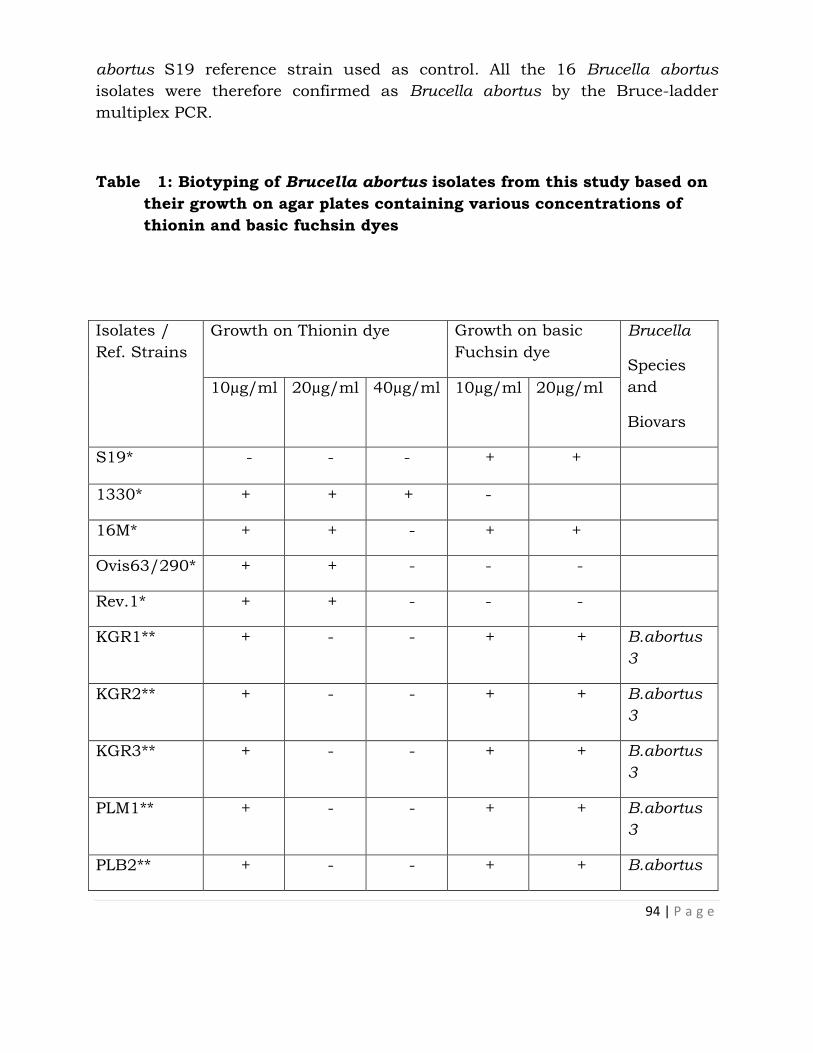
94 | P a g e
abortus S19 reference strain used as control. All the 16 Brucella abortus
isolates were therefore confirmed as Brucella abortus by the Bruce-ladder
multiplex PCR.
Table 1: Biotyping of Brucella abortus isolates from this study based on
their growth on agar plates containing various concentrations of
thionin and basic fuchsin dyes
Isolates /
Ref. Strains
Growth on Thionin dye Growth on basic
Fuchsin dye
Brucella
Species
and
Biovars
10µg/ml 20µg/ml 40µg/ml 10µg/ml 20µg/ml
S19* - - - + +
1330* + + + -
16M* + + - + +
Ovis63/290* + + - - -
Rev.1* + + - - -
KGR1** + - - + + B.abortus
3
KGR2** + - - + + B.abortus
3
KGR3** + - - + + B.abortus
3
PLM1** + - - + + B.abortus
3
PLB2** + - - + + B.abortus
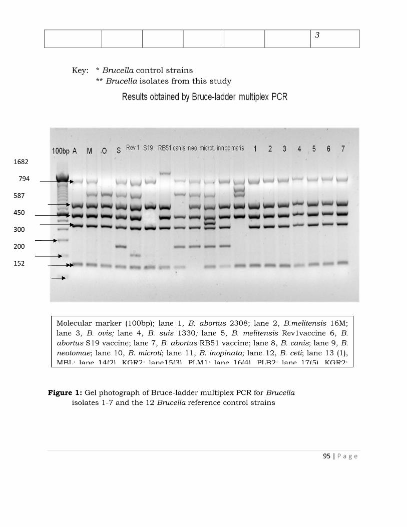
95 | P a g e
3
Key: * Brucella control strains
** Brucella isolates from this study
Figure 1: Gel photograph of Bruce-ladder multiplex PCR for Brucella
isolates 1-7 and the 12 Brucella reference control strains
1682
794
587
450
300
200
152
100
Molecular marker (100bp); lane 1, B. abortus 2308; lane 2, B.melitensis 16M;
lane 3, B. ovis; lane 4, B. suis 1330; lane 5, B. melitensis Rev1vaccine 6, B.
abortus S19 vaccine; lane 7, B. abortus RB51 vaccine; lane 8, B. canis; lane 9, B.
neotomae; lane 10, B. microti; lane 11, B. inopinata; lane 12, B. ceti; lane 13 (1),
MBL; lane 14(2), KGR2; lane15(3), PLM1; lane 16(4), PLB2; lane 17(5), KGR2;
lane 18(6),KGR1, lane19(7), HZ16.

96 | P a g e
DISCUSSION
Since brucellosis was first reported in Nigeria in 1927 (Banerjee and Bhatty,
1970), only five bacteriological studies have been undertaken in cattle in
various locations and Brucella abortus isolated (Ducrotoy et al., 2014). There is
concern that brucellosis could emerge as a result of the settling of previously
migratory herds or from increased contacts between infected nomadic herds
and susceptible intensive commercial or settled semi-intensive herds (Ducrotoy
et al., 2014). This concern has been confirmed by the successful isolation of
Brucella abortus in both KGR and on the Jos Plateau, indicating that
brucellosis is endemic in the study areas. This is very significant and has far
reaching implications as the infected cows may spread the infection to other
cattle within the herds as well as sheep and goats which are usually reared
along with cattle as the organisms are shed on pasture. Infection could also be
spread from infected herds to non-infected herds through contact and through
loaning of heifers and bulls commonly practiced by herdsmen.
The isolation of Brucella abortus from milk in both KGR and Jos Plateau is of
great public health significance because Brucella organisms are usually shed
through the milk of infected cows which is consumed raw or as fermented milk.
The most common means of transmission of brucellosis from animals to
humans is through the consumption of unpasteurized or raw milk and milk
products (Kang’ethe et al., 2000, Capasso, 2002).
The isolation of Brucella abortus from the vaginal swab is also of great public
health significance. This is because herdsmen are in the habit of using their
bare hands to assist cows to ensure successful parturition in cases of dystocia
or to pull out placenta in cases of retained placenta. These habits and
practices expose them to high risk of contracting brucellosis through direct
contact with the organisms.
There was higher rate of isolation of Brucella organisms from hygroma fluids in
this study compared to other samples because hygroma is unlikely to get
contaminated compared to other samples. This finding is consistent with
reports of previous investigators in Nigeria where a majority of isolates were
from hygroma fluid and only a few from other samples such as milk, vaginal
swabs, aborted foetuses and placentae (Eze, 1978; Bale and Kumi-Diaka, 1981;
Ocholi et al., 2004b). The finding also agrees with reports of other authors that
found that although Brucella could be isolated from other samples and tissues,
hygroma is the most valuable in terms of success rate of isolation in nomadic

97 | P a g e
and semi-nomadic cattle herds in Africa (Corbel, 2006; Godfroid et al., 2010;
OIE, 2009).
Based on classical biotyping, the isolates were all identified as Brucella abortus
biotype 3. This finding differs from those of previous investigators (Eze, 1978;
Ocholi et al., 2004a, b; Ocholi et al., 2005) in which Brucella abortus biotype 1
was reported as the prevailing biotype in Nigeria. Differences between biotypes
1 and 3 are not very apparent and can only be distinguished based on their
sensitivity to thionin dye (Banai and Corbel, 2010). While Brucella abortus S19
Biotype 1 did not grow in presence of thionin at all three concentrations of
10µg/ml, 20µg/ml and 40µg/ml, all the isolates tested in this study grew in the
presence of thionin only at 10µg/ml which is a typical behaviour of Brucella
abortus biotype 3. This confirmed all the isolates as Brucella abortus biotype 3.
The Bruce-ladder multiplex PCR successfully identified all phenotypically
characterized Brucella abortus isolates to species level as Brucella abortus. To
our knowledge, this is the first molecular technique used to identify Brucella to
species level in Nigeria.
The present study in which Nigerian isolates of Brucella abortus were identified
by Bruce-ladder multiplex PCR has included Nigeria among countries from
where Brucella strains have been successfully subjected to molecular
identification by PCR. These findings have established the endemicity of
Brucella infection due to Brucella abortus biotype 3 in KGR and on the Jos
Plateau. The study provides useful data for the establishment of appropriate
brucellosis intervention and control measures in the study areas. Phylogenetic
studies of the isolates are recommended.
CONCLUSION
The successful isolation of Brucella in KGR and Jos Plateau shows that
brucellosis is prevalent in the areas and Brucella abortus is the prevailing
Brucella species. All the species from the study areas were Brucella abortus
biotype 3. Molecular characterization by Bruce-ladder Multiplex PCR of all the
isolates from the study areas showed that they were Brucella abortus.
ACKNOWLEDGEMENT
This work was supported by a grant from European Union Framework 7,
Integrated Control of Neglected Zoonoses (ICONZ) project. We thank the
Executive Director, NVRI, Vom for sponsoring this study. We also thank the
staff of Brucella Research Laboratory and field work team, NVRI Vom, staff of

98 | P a g e
Department of Microbiology and Parasitology, Medical School and Institute for
Tropical Health, Universidad de Navarra, Pamplona, Spain and staff of Centro
Investigacion Technologia Agroalimentaria, (CITA) Laboratory, Zaragoza, Spain,
for technical assistance.
REFERENCES
Alton G. G., Jones .L. M., Angus R. D., and Verger .J. M. (1988). Techniques for
the Brucellosis Laboratory, Institute National de la Vacherche
Agronomique, (INRA) Paris, 1st Edition, pp. 63-129.
Bale, J. O. O., and Kumi-Diaka, J. (1981). Serological and bacteriological study
of bovine brucellae from livestock investigation and breeding centers in
Nigeria. British Veterinary Journal, 137: 256-261.
Banai, M and Corbel, M. (2010). Taxonomy of Brucella, Open Access Veterinary
Science Journal, 4, 85-101.
Banerjee AK, Bhatty MA (1970). A survey of bovine brucellosis in northern
Nigeria (a preliminary communication). Bulletin of Epizootic Diseases in
Africa, 18, 333–338.
Bronsvoort, B. M., Koterwas, B., Land, F., Handel, I. G., Tucker, J., Morgan, K.
l., Tanya, V. N., Abdoel, T. H., and Smits, H. L. (2009). Comparison of a
flow assay for brucellosis antibodies with the reference cELISA test in
West African Bos indicus, PLoS one, 4, 5221
Bruce, D. (1887). Note on the discovery of a microorganism in Malta fever.
Practitioner, 39, 161.
Capasso, L. (2002). Bacteria in two-millennia-old cheese, and related
epizoonoses in Roman populations, Journal of Infections 45, 122–127
Corbel, M. J. (2006) Brucellosis in humans and animals, WHO Manual, pp. 1-
21
Cutler, S.J., Whatmore, A.M., and Commander, N.J. (2005) Brucellosis –New
aspect of an old disease, Journal of applied Microbiology, 98: 1270-1281.

99 | P a g e
Ducrotoy, M. J., Bertu, W. J., Ocholi, R. A., Gusi, A. M., Ward Bryssinckx, W.,
Welburn, S., Moriyon., I. (2014). Brucellosis as an emerging threat in
developing economies: Lessons from Nigeria. PLOS Neglected Tropical
Diseases, 8: 1-18.
Eze, E.N. (1978). Isolation of Brucellae from the Nigeria livestock and the typing
of such isolates. Bulletin of Animal Health Production Africa, 26: 29-36.
FAO (2003) Guidelines for coordinated human and animal brucellosis
surveillance, FAO animal production and Health, paper No.156, Rome
p.56.
Foster, G., Osterman, B. S., Godfroid, J., Jacgues, I., and Cloeckaert, A.
(2007) Brucella ceti sp.nov and Brucella pinnipedialis sp.nov.for
Brucella strains with cetaceans and seals as their preferential hosts.
International Journal of Systemic and Evolutionary Microbiology, 57; 2688-
2693
Garcia-Yoldi, D., Marin, C. M., de Miguel, M. J., Munoz, P. M., Vizmanos, J. L.,
Lopez-Goni,I. (2006). Multiplex PCR for the identification and
differentiation of all Brucella species and the vaccine strains Brucella
abortus S19 and RB51 and Brucella melitensis Rev.1. Cinical Chemistry,
52, 779-781.
Godfroid, J., Nielsen, K., Saegerman, C. (2010). Diagnosis of brucellosis in
livestock and wildlife, Croatian Medical Journal, 51, 296-305
Kang’ethe, E. K., Arimi, S. M., Omore, A. O., Mcdermit, J. J., Nduhiu, J. G.,
Macharia, J. K., Githua, A. (2000). Prevalence of Brucella antibodies in
small holder dairy farms in Kenya, A paper prepared for oral presentation
at the 3rd All African Conference on Animal Agriculture,proceedings, pp.6-
9.
Lo´ pez-Gon˜ i, I., Garcı´a-Yoldi, D., Marı´n, C. M., de Miguel, M. J., Mun˜oz, P.
M., Blasco, J. M., Jacques, I., Grayon, M., Cloeckaert, A., Ferreira, A. C.,
Cardoso, R., Correˆa deSa´, M. I., Walravens, K., Albert, D., Garin-
Bastuji, B. (2008). Evaluation of a multiplex PCR assay (Bruce-ladder) for
molecular typing of all Brucella species, including the vaccine strains,
Journal of Clinical Microbiology, 46: 3484–3487
McDermott, J. J, Arimi, S. M (2002). Brucellosis in sub-Saharan Africa:
100 | P a g e
epidemiology, control and impact. Veterinary Microbiology, 90, 111–134.
Nymo, I. H; Tryland, M; Godfroid, J. (2011). A review of Brucella infection in
marine mammals, with special emphasis on Brucella pinnipedialis in
hooded seal (Cystophora cristata), Veterinary Research, 42, 93
Ocholi, R.A; Kwaga, J. K. P; Ajogi, I; Bale, J.O. (2005). Abortion due to Brucella
abortus in sheep in Nigeria, Revieu Scientific Technical Office
Internationale Epizootics, 24 (3), 973-979
Ocholi, R. A; Kwaga, J. K. P; Ajogi, I; Bertu, W. J., and Okpara, J. (2004a).
Carpal bursitis associated with Brucella abortus in a horse in Nigeria,
Veterinary Record, 155:566-567.
Ocholi, R.A; Kwaga J. K. P; Ajogi, I., and Bale, J. O. O. (2004b). Phenotypic
characterization of Brucella strains isolated from Livestock in Nigeria.
Veterinary Microbiology 103, 47-53.
OIE, (2009). World Organization for Animal Health, Chapter 2.4.3.Bovine
Brucellosis, OIE Manual of Diagnostic tests and vaccins for Terrestrial
Animals.1-35
Osterman, B; Moriyon, I. (2006). International committee on systematic of
prokaryotes, Subcommittee on the taxonomy of Brucella.Report of the
September, 2003, Pamplona, Spain, International Journal of Systemic and
Evolutionary Microbiology, 56, 1173-1175.
Pappas, G., Papadimitriou, P., Akritidis, N., Christou, L., Tsianos, E.V. (2006).
The new global map of human brucellosis, Lancet Infectious Disease, 6,
91–99
Ron-Roman, J; Saegerman, C; Minda-Aluisa, E; Benitz-Ortiz, J; Brandt, J;
Douce, R (2012)First report of orchitis in man caused by Brucella
abortus biovar 1 in Ecuador. American Journal of Tropical Medicine and
Hygiene, 87:524-528
Scholz, H. C; Nockler, K; Gollner, C; Bahn, P; Vergnaud, G; Tomaso, H; Al
Dahouk, S; Kampfer, P; Cloeckaert, A; Maquart, M; Zygmunt, M.S;
Whatmore, A; Pfeffer, M; Huber, B; Busse, H. J; De, B.K. (2010). Brucella

101 | P a g e
inopinata sp.nov.isolated from a breast implant infection. International
Journal of Systemic and Evolutionary Microbiology, 60, 801-808.
Zinsstag, J., Schelling, E., Solera., Blasco, J. M. and I. Moriyón, I. (2011).
Brucelosis, pp. 54–62. In S. R. Palmer, L. Soulsby, P. R. Torgeson, and D.
G. Brown (eds.), Handbook of Zoonoses, 2nd ed. Oxford University Press,
Oxford & New York.

102 | P a g e
ANTIBODY ENGINEERING: SINGLE DOMAIN ANTIBODY (NANOBODY) AS A
RESEARCH TOOL
Obishakin E. T.
Applied Biotechnology Division, National Veterinary Research Institute, Vom,
Jos, Plateau State
INTRODUCTION
The structure of conventional immunoglobulin-G (IgG) is composed of highly
conserved two identical heavy (H) chains and two identical light (L) chains that
make up the antigen-binding site. The heavy chain consists of four domains:
the variable N-terminal (VH), conserved CH1, CH2 and CH3 domains. The light
chain consists of two domains; the variable light (VL) and the constant light
(CL) domain. The variable light and the variable heavy chains make up the
antigen recognizing fragment (Figure 1 A) (Muyldermans, 2013). Camelids
(llamas, camels, and alpacas) and sharks produce heavy-chain antibodies
(HCAbs) which are devoid of light chains, lacks CH1 domain and are capable of
antigen recognition. The antigen-binding fragments comprise of a single
domain, and the product of the recombinant expression of this single domain is
referred to as nanobody (Figure 1B). In camels, 50-80% of immunoglobulins
are devoid of light chains. The antigen-binding part of the nanobody can be
produced by recombinant expression in bacteria cells (Figure 1 C). Nanobodies
are used in a wide variety of applications because of their small size (15
kilodaltons), high solubility, thermal stability and good tissue penetration. They
can be stored for months at 4°C for longer periods at -20°C. Some nanobodies
have been shown to resist temperatures above 90°C (Van der Linden et al.,
1999).
There have been several attempts to incorporate nanobodies in the place of
conventional antibodies in research, diagnosis, therapy and industry.
Nanobodies have been generated for the purpose of experimental therapeutic
applications against different viruses: HIV, hepatitis B virus, influenza virus,
respiratory syncytial virus, rabies virus, FMDV, poliovirus, rotavirus, and
porcine endogenous retroviruses. Because of their molecular weight of 15,000
which is smaller than the renal clearance cut off of 50,000, they are
eliminated from the blood rapidly making them suitable for in vivo-imaging
(Vaneycken et al., 2011).
Recently, the investigation of nanobodies for use in therapeutic, diagnostic,
trypanolytic, imaging capacities in trypanosome research have been shown.

103 | P a g e
Seminar presented 6th August, 2015 at NVRI Auditorium
Ability of VHH to bind into clefts or pockets is greatly enhanced based on the
compact nature and its convex paratope. This feature has been greatly
exploited as nanobodies are able to bind to the cryptic epitopes of the VSG of
trypanosomes that are not exposed to large molecules of convectional
antibodies.
MATERIALS AND METHODS
Overview of generation of recombinant antigen specific nanobody
To generate recombinant antigen specific nanobodies, llamas, dromedaries,
camels and alpacas are first immunized for 5–7 weeks with 0.5-1mg of
proteinaceous antigens with 0.1-0.2 mg of antigen per week. This will allow for
the affinity maturation of antibodies and raise the titer of B cell producing
antigen specific HCAbs. Freund's adjuvant or Gerbu adjuvants can be used for
immunization. Four days after the last immunization, 50-100mls of blood is
collected from the camelid. Using lymphocyte isolation methods, lymphocytes
are isolated from the blood sample and mRNA is further isolated from the
lymphocytes (Deckers et al., 2009). From the total mRNA, a first strand cDNA
is produced. The gene fragments encoding the variable domain until the CH2
domain are amplified from the cDNA with specific primers. This yields two
separate bands 900 kb (VH– CH1–CH2 exons) and 600kb (VHH–CH2) without
CH1 exon. The 600 kb fragment is excised from 1% agarose gel and purified.
Furthermore, VHH fragment is amplified from the VHH-CH2 fragment with
nested primers encompassing framework-1 and framework-4 of VHH. The VHH
fragment is first restricted alongside an expression vector. It is subsequently
ligated into expression vectors which could be with or without a detection or
purification tag e.g pMECS or pHEN 4 respectively. (If the expression vector is
without a tag, the VHH fragment is further re-cloned into an expression vector
with a tag (e.g. pHEN6) for easy purification afterwards). Bacteria cells are then
transformed with the nanobody gene repertoire in the expression vectors to
obtain a nanobody library (Hmila et al., 2010). The nanobody library is super-
infected with helper phages. Biopanning method is used to enrich for specific
nanobodies which are attached on virions. Phage enrichment is achieved by
several rounds of in vitro selection on the antigen of choice coated on microtiter
plates or immunotubes. Phages are eluted with alkaline agent. The eluted
phages are further used to infect E. coli cells, the bacteria cells are plated out to
select each colony of the library containing an enriched nanobody gene. Viable
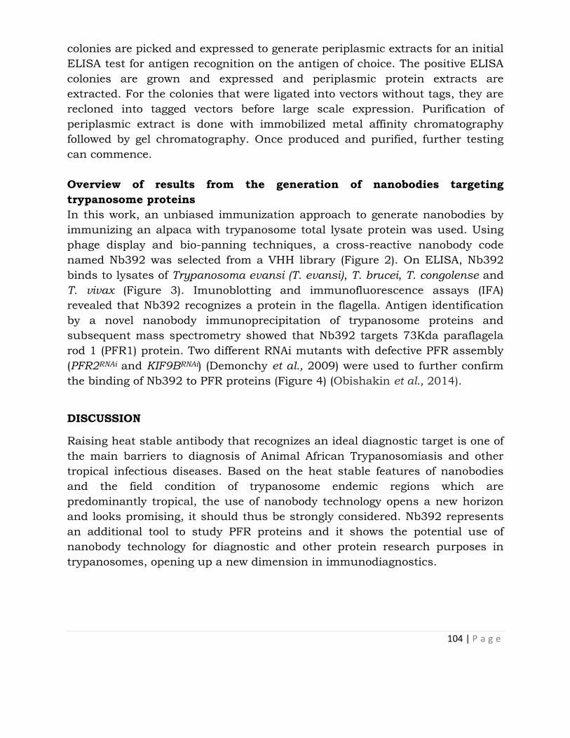
104 | P a g e
colonies are picked and expressed to generate periplasmic extracts for an initial
ELISA test for antigen recognition on the antigen of choice. The positive ELISA
colonies are grown and expressed and periplasmic protein extracts are
extracted. For the colonies that were ligated into vectors without tags, they are
recloned into tagged vectors before large scale expression. Purification of
periplasmic extract is done with immobilized metal affinity chromatography
followed by gel chromatography. Once produced and purified, further testing
can commence.
Overview of results from the generation of nanobodies targeting
trypanosome proteins
In this work, an unbiased immunization approach to generate nanobodies by
immunizing an alpaca with trypanosome total lysate protein was used. Using
phage display and bio-panning techniques, a cross-reactive nanobody code
named Nb392 was selected from a VHH library (Figure 2). On ELISA, Nb392
binds to lysates of Trypanosoma evansi (T. evansi), T. brucei, T. congolense and
T. vivax (Figure 3). Imunoblotting and immunofluorescence assays (IFA)
revealed that Nb392 recognizes a protein in the flagella. Antigen identification
by a novel nanobody immunoprecipitation of trypanosome proteins and
subsequent mass spectrometry showed that Nb392 targets 73Kda paraflagela
rod 1 (PFR1) protein. Two different RNAi mutants with defective PFR assembly
(PFR2RNAi and KIF9BRNAi) (Demonchy et al., 2009) were used to further confirm
the binding of Nb392 to PFR proteins (Figure 4) (Obishakin et al., 2014).
DISCUSSION
Raising heat stable antibody that recognizes an ideal diagnostic target is one of
the main barriers to diagnosis of Animal African Trypanosomiasis and other
tropical infectious diseases. Based on the heat stable features of nanobodies
and the field condition of trypanosome endemic regions which are
predominantly tropical, the use of nanobody technology opens a new horizon
and looks promising, it should thus be strongly considered. Nb392 represents
an additional tool to study PFR proteins and it shows the potential use of
nanobody technology for diagnostic and other protein research purposes in
trypanosomes, opening up a new dimension in immunodiagnostics.

105 | P a g e
Figures
A. B. C.
Figure 1. Conventional and VHH antibodies. Classical antibody (A), heavy-
chain antibody (B), single-domain antigen-binding fragment (nanobody) derived
from a heavy-chain antibody (C) (Muyldermans, 2009).
Figure 2. Sequence alignment of deduced amino acid sequences of the different cross-
reactive nanobodies highlighting the framework (FR) and complementarity
determining region CDR sequences. CDR sequences are highlighted in blue, red and
green (corresponding to different families), while FR differences are marked in yellow.
(Obishakin et al., 2014).
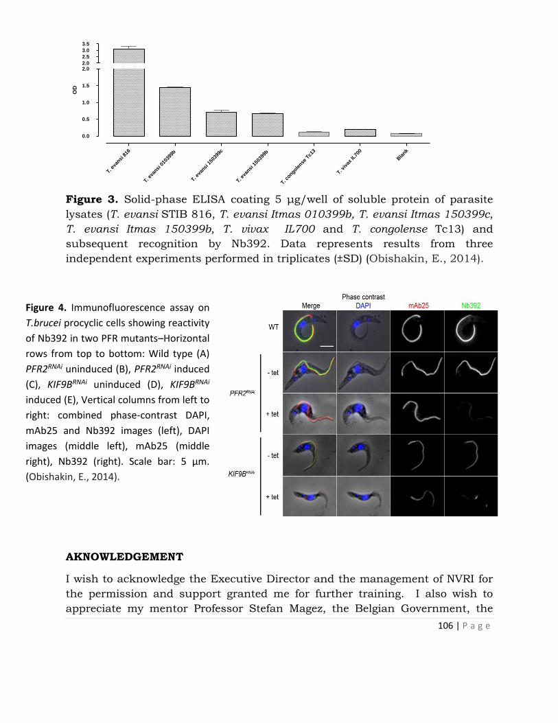
106 | P a g e
T. eva
nsi 8
16
T. eva
nsi 0
1039
9b
T. eva
nsi 1
5039
9c
T. eva
nsi 1
5039
9b
T. congole
nse T
c13
T. viv
ax IL
700
Bla
nk
0.0
0.5
1.0
1.5
2.02.0
2.5
3.0
3.5
OD
Figure 3. Solid-phase ELISA coating 5 μg/well of soluble protein of parasite
lysates (T. evansi STIB 816, T. evansi Itmas 010399b, T. evansi Itmas 150399c,
T. evansi Itmas 150399b, T. vivax IL700 and T. congolense Tc13) and
subsequent recognition by Nb392. Data represents results from three
independent experiments performed in triplicates (±SD) (Obishakin, E., 2014).
AKNOWLEDGEMENT
I wish to acknowledge the Executive Director and the management of NVRI for
the permission and support granted me for further training. I also wish to
appreciate my mentor Professor Stefan Magez, the Belgian Government, the
Figure 4. Immunofluorescence assay on
T.brucei procyclic cells showing reactivity
of Nb392 in two PFR mutants–Horizontal
rows from top to bottom: Wild type (A)
PFR2RNAi uninduced (B), PFR2RNAi induced
(C), KIF9BRNAi uninduced (D), KIF9BRNAi
induced (E), Vertical columns from left to
right: combined phase-contrast DAPI,
mAb25 and Nb392 images (left), DAPI
images (middle left), mAb25 (middle
right), Nb392 (right). Scale bar: 5 µm.
(Obishakin, E., 2014).

107 | P a g e
Flemish Institute for Biotechnology (VIB) and the management of Vrije
University Brussel for their funding supports.
REFERENCES
Deckers, N; Saerens, D; Kanobana, K; Conrath, K; Victor, B; Wernery, U;
Vercruysse, J; Muyldermans, S; Dorny, P (2009). Nanobodies, a
promising tool for species-specific diagnosis of taenia solium
cysticercosis. International Journal for Parasitology 39: 625-633.
Demonchy, R; Blisnick, T; Deprez, C; Toutirais, G; Loussert, C; Marande, W;
Grellier, P; Bastin, P; Kohl, L (2009). Kinesin 9 family members perform
separate functions in the trypanosome flagellum. Journal of Cell Biology
187: 615-622.
Hmila, I; Saerens, D; Abderrazek, R.B; Vincke, C; Abidi, N; Benlasfar, Z;
Govaert, J; El-Ayeb, M; Bouhaouala-Zahar, B; Muyldermans, S (2010). A
bispecific nanobody to provide full protection against lethal scorpion
envenoming. The FASEB Journal 24: 3479-3489.
Muyldermans, S (2013). Nanobodies: Natural single-domain antibodies.
Annual Review of Biochemistry.
Muyldermans, S; Baral, T.N; Retamozzo, V.C; De Baetselier, P; De Genst, E;
Kinne, J; Leonhardt, H; Magez, S; Nguyen, V.K; Revets, H; Rothbauer,
U; Stijlemans, B; Tillib, S; Wernery, U; Wyns, L; Hassanzadeh-
Ghassabeh, G; Saerens, D (2009). Camelid immunoglobulins and
nanobody technology. Veterinary Immunology and Immunopathology
128: 178-183.
Obishakin, E., Stijlemans, B., Santi-Rocca, J., Vandenberghe, I., Devreese, B.,
Muldermans, S., Bastin P., Magez, S. (2014). Generation of a nanobody
targeting the paraflagellar rod protein of trypanosomes, PloS one, 9(12),
e115893?
Van der Linden, R; Frenken, L; De Geus, B; Harmsen, M; Ruuls, R; Stok, W;
De Ron, L; Wilson, S; Davis, P; Verrips, C (1999). Comparison of
physical chemical properties of llama VHH antibody fragments and
mouse monoclonal antibodies. Biochimica et Biophysica Acta
(BBA)-Protein Structure and Molecular Enzymology 1431: 37-46.
Vaneycken, I; D’huyvetter, M; Hernot, S; De Vos, J; Xavier, C; Devoogdt, N;
Caveliers, V; Lahoutte, T (2011). Immuno-imaging using nanobodies.
Current Opinion Biotechnology 22: 877-881.

108 | P a g e
A STUDY ON HELMINTHIASIS OF CATTLE HERDS IN KACHIA GRAZING
RESERVE (KGR) OF KADUNA STATE, NIGERIA
Henry .E. Nnabuife
Parasitology Division, National Veterinary Research Institute, Vom.
Email: [email protected]
INTRODUCTION
Among all the livestock that make up the farm animals in Nigeria, ruminants,
comprising sheep, goats and cattle, constitute the farm animals largely reared
by farm families in the country’s agricultural system (Lawal-Adebowale, 2012).
Nigeria has population of 34.5million goats, 22.1million sheep and 13.9million
cattle. Cattle represents a valuable asset in both traditional and modern
agriculture, they provide meat, milk, skin and draught power. Additionally they
may, in a traditional society be an essential part of the social system,
representing a family wealth or they can be regarded as a survival kit by
nomadic people (Fabiyi, 1984).
Despite its usefulness to man, cattle may be infected with pathogens that are
dangerous to man and other domestic animals. Such are parasites which
include helminthes of Gastrointestinal Tract (GIT). Helminth parasites come in
three major classifications namely cestodes, nematodes and trematodes (Tibbo
et al, 2011).
Helminth infections are important cause of production losses in livestock
worldwide often necessitating antihelminthic treatment (Vercruysse and
Clarebout, 2001). The significance of helminthiasis has been recognized by
livestock farmers, right from the earliest of times and various methods have
been employed by them to control helminthes in their animals including the
use of medicinal plants and herbs and different grazing techniques (Bukhari
and Sanyal 2011). The major control method employed against helminth
parasites in developing countries as elsewhere is the use of chemotherapy
(Aragaw et al., 2010; Sargison, 2011).
The strategies for preventive control must involve stopping a build-up of large
numbers of larvae on pasture and anticipation of periods for likely
109 | P a g e
Seminar presented 6th August, 2015 at NVRI Auditorium
multiplication of larvae on pasture and to move animals from such pastures
during those periods. In order to achieve this goal there has to be a
multifaceted approach involving grazing management, use of anthelmintics and
dependence on acquisition of immunity.
Despite this importance, helminthes are the most neglected area of Veterinary
care in the developing countries (including Nigeria) due to its chronic and
insidious nature, with endemic pathogenic, vectors and diseases particularly
where extensive grazing is practiced (Magona et al., 2008; Bizimenyera et al.,
2008).
Grazing Reserves were established in Nigeria in major cattle producing areas to
stabilize the pastoral production system, one of which is Kachia Grazing
Reserve (KGR) in Kaduna State Nigeria. Monitoring cattle population and
habitat conditions as well as ascertaining parasites and disease status are
important component of herd health programme. Therefore, this study aims to
determine the prevalence of gastrointestinal helminthes in cattle in KGR and to
identify risk factors associated with helminthiasis in the study area.
MATERIALS AND METHODS
Study Area
The study was conducted in Kachia, a sedentary Fulani community of KGR in
Kaduna state, Nigeria. The KGR comprises six (6) contiguous ‘Blocks’
containing 581 farmers and 5,252 people. Households per block ranges from
80 to 188 and a total of 23,327 cattle are owned by 569 households.
Sample size
Eighty-eight (88) households were selected randomly from 569 cattle-owning
households. A total of 3,651 cattle were sampled and the sampling was spread
over three (3) periods; Pre-intervention (1,525), Intervention (1,609) and Post-
intervention (517).
Collection and examination of faecal samples
Fresh faecal samples were collected per-rectum of cattle, labeled and kept in
cool boxes before transporting them to the laboratory for further analysis.
Faecal examination was by a quantitative floatation (McMaster technique) and

110 | P a g e
sedimentation method for detection of nematode, cestode and trematode eggs
as described by [Hansen and Perry, 1990, Soulsby, 1982] respectively.
Data analysis
Data obtained was analyzed using Chi-Square (x2) and Kruskal Wallis test
[Sokal and Rohlf, 2001].
Egg of Paramphistomum cervi Egg of Oesophagostomum radiatum
RESULTS AND DISCUSSION
Distribution and types of parasite eggs in the Pre-intervention,
Intervention and Post-intervention periods
In March (Pre-intervention) 820 (53.77 %) were positive, intervention in June,
946 (58.79%) were positive and 205 (39.65%) were positive for post-
intervention (Table 1).

111 | P a g e
Table 1: Intervention period-related prevalence of helminth parasites in cattle
in KGR, Kachia
Period Number examined Number positive
(%, infection rate)
Pre-intervention
(March-April)
Intervention
(June-July)
Post-intervention
(Sept.-Oct.)
1,525
1,609
517
820(53.77)
946(58.79)
205(39.65)

112 | P a g e
Table 2: Distribution of parasites of cattle during the survey
Parasites Pre-intervention Intervention Post-
intervention
Number infected
(% infection rate)
Number infected
(% infection rate)
Number
infected
(%infection
rate)
Oesophagostomum
radiatum
232 (17.90) 237(16.39) 57(23.08)
Bunostomum
phlebotumum
112 (8.64) 106(7.33) 12(4.86)
Trichuris globulosa
189 (14.58) 213(14.73) 43(17.41)
Cooperia pectinita
93(7.18) 45(3.11) 3(1.21)
Monieza benedeni
9(0.69) 1(0.07) 0(0.00)
Toxocara
vitulorum
7(0.54) 8(0.55) 0(0.00)
Strongyloides
papillosus
76(5.86) 14(0.97) 1(0.40)
Strongylus
langamus
1(0.08) 0(0.00) 0(0.00)
Schistosoma bovis
0(0.00) 2(0.14) 3(1.21)
Paramphistomum
cervi
540(41.67) 736(50.90) 127(51.43)
Fasciola gigantic 37(2.86) 84(5.81) 1(0.40)

113 | P a g e
Table 3: Single and mixed helminth infections among cattle during the periods
in KGR
Number of
Parasites
Pre-intervention Intervention Post-intervention
Number infected
(%)
Number infected
(%)
Number infected
(%)
1
2
3
4
5
6
498 (32.66)
192 (12.59)
74 (4.85)
33 (2.16)
5 (0.33)
1 (0.07)
585 (36.36)
251 (15.60)
69 (4.29)
38 (2.36)
16 (0.99)
7 (0.44)
171 (33.08)
33 ( 6.38)
4 ( 0.77)
0 ( 0.00)
0 (0.00)
0 (0.00)
Table 4: Prevalence of helminth parasites in different age groups of cattle in
KGR
Age
group
Pre-intervention Intervention Post-intervention
Num.
examined
Num. infec.
(%)
Num.
examined
Num. infec.
(%)
Num.
examined
Num. infec.
(%)
<8month 101 39(2.56) 93 45(2.80) 27 3(0.58)
8mth-
2yr 252 109(7.15) 256 117(7.27) 56 14(2.71)
>2yrs 1,172 672(44.07) 1,260 784(48.73) 434 188(36.36)
Total 1,525 820 (53.77) 1,609 946 (58.79) 517 205 (39.65)

114 | P a g e
The helminth eggs of Nematodes-Oesophagostomum radiatum, Bonustonum
phlebotomum, Tricuris globulosa, Toxocara vitulorum, Strongyloides papillous,
Strongylus langamus; Cestode-Moneizia benedeni, and Trematodes-
Schistosoma bovis, Paramphistomum cervi and Fasciola gigantica were identified
(Table-2). The range of parasites found in this present study was almost similar
to that found in Benin City, Southern Nigeria (Edosomwan and Shoyemi,
2012).
Age and season played significant role in determining helminthes community
species concentration in cattle in the KGR which is in agreement with the
report of Fuentes et al. (2004).The result shows that age was a factor in the
abundance of the parasites in the animals. Parasites were demonstrated more
in adult animals than the young stock, and this could be due to the fact that
older livestock may have been exposed more frequently to infective stages of the
parasites. This is in agreement with the report by (Ibrahim et al., 2008).The
present study demonstrated that P.cervi was the most prevalent in all the
periods followed by O. radiatum (Table 2).The high prevalence of P.cervi may be
attributed to the favorable environmental factors of temperature and
precipitation found in the KGR (Gupta et al., 2004; Sukhdeo and Sukhdeo,
2004). Another possible factor is the preferential attention received by young
stock compared to the adults by herdsmen who believed that the adult cattle
have outgrown parasitic attacks
The significant effect of season on helminthes concentration in this study is
evident in the parasite prevalence observed in the different periods. The higher
prevalence observed during the intervention period could be ascribed to the
fact that sample collection was done during the wet season. During this period,
high moisture content increased development of larvae and abundant pasture,
thus resulting in increased contact between the host and parasites. These
findings are similar to Fakae (1990) and Bhattacharya and Ahmed (2005) who
recorded higher incidence of parasitic infection during rainy season in Nigeria
and India respectively. The study also revealed that the cattle in the study area
were infected with single and mixed (multiple) infections. In addition to
seasonal influence, the low post-intervention prevalence could be ascribed to
the effectiveness of the deworming intervention of the animals using a broad-
spectrum antihelmintic bolus containing a combination of 60% oxyclozanide
and 40% levamisole hydrochloride (Kepxan Bolus, Kepro®, Holland).

115 | P a g e
CONCLUSION AND RECOMMENDATION
The results of this study indicate that helminthes of cattle are common in KGR.
However, it was also observed that herd-herd treatment takes care of not only
clinical cases, but subclinical ones too. It is recommended that farmers in KGR
be enlightened on the need to treat the herds instead of treating individual
cases. Creation of Grazing Reserve such as KGR for all the nomadic Fulanis
will help to trace and effectively tackle the problems such as helminth
infections.
ACKNWOLEDGEMENT
I am very grateful to the Executive Director and the Management of NVRI, Vom
for the permission granted me to undergo the study.
REFERENCES
Aragaw, K., Teferi, M., Haile, A., &Tibbo, M. (2010). Effects of strategic
helminthosis control on age at first lambing and lambing interval of
communally grazed Menz ewes in Ethiopia. Livestock Science, 135(1):
38-43.
Bhattacharya, D.K. and Ahmed, K. (2005). Prevalence of helminthic infection in
cattle and buffaloes. India Veterinary Journal, 82: 900 -901
Bizimenyera, E.S., Swan, G.E., Eloff T.N., Meyer, S. and Naidoo, V. (2008).
Efficacy of Peltophorum affricanum sond (fabaceace) extracts on
Haemonthus contortus and Trichostrongylus colubriformis in sheep.
Journal of Animal and Veterinary advances, 7(4): 364-371.
Bukhari, S., and Sanyal, P. K. (2011). Epidemiological Intelligence for Grazing
Management in Strategic Control of Parasitic Gastroenteritis in Small
Ruminants in India – A Review, Vet.World, 4(2): 92-96.
Edosomwan, E. U. And Shoyemi, O.O. (2012). Prevalence of gastrointestinal
helminth parasites of cattle and goats slaughtered at abattoirs in Benin
City, Nigeria, African Scientist. Vol. 13(2): 109-114.
116 | P a g e
Fabiyi, J.P. (1984). Coping with parasitic disease problem in Nigeria. Livestock
Farmers Series 4:13
Fakae, B. B (1990). The epidemiology of helminthosis in small ruminants under
the traditional husbandry system in eastern Nigeria. Veterinary Research
Communication, 14 (5): 381-391.
Fuentes ,M.V., Saez, S., Trelis, M., Galan-Puchades ,M.T., Esteban, J.G,(2004)
The helminth community of the wood mouse, Apodemussylvaticus , in
the Sierra Espuna, Murda, Spain , Journal of Helminthology, 78(3):219-
223.
Gupta, B .V.Parshad, S.Giraya (2004). Maturation of Paramphistomum cervi in
sheep in India. Veterinary Parasitology, 15:239-245.
Hansen, J. and Perry, B. (1990) The Epidemiology, Diagnosis and Control of
Gastro-intestinal Parasites of Ruminants in Africa, A Handbook, (ILRAD)
Nairobi, Kenya, pp 25-50.
Ibrahim, M.M., Al Ghamdi, M.A., Al Ghamdi and M.S. (2008). Helminthes
community of Veterinary Importance of Livestock in Relation to Some
Ecological and Biological
Lawal-Adebowale, O. A., (2012). Intech- Dynamics of Ruminant livestock
management in the context of the Nigerian Agricultural System 4: 61-80.
Magona J.W., Walubengo J. and Odimin J.T (2008). Acute haemorrhagic
syndronae of bovine trypanosomoses in Uganda. Acta Tropica, 107(2):
186-191.
Sargison, N. D. (2011). Pharmaceutical Control of EndoparasiticHelminth
Infections in Sheep. Veterinary Clinics of North America: Food Animal
Practice, 27(1): 139-156.
Sokal R.R. and Rohlf F.J (2001). Biometry Freeman Publishers, 887pp
Soulsby E.J.W (1982). Helminths, Arthropods and Protozoa of Domesticated
Animals, 7th Edn Lea and Febiger, Philadelphia: Bailliere Tindall, London,
pp. 212-258.

117 | P a g e
Sukhdeo, M., and Sukhdeo, S (2004). Trematode behavior and perceptual
worlds of parasites, Canadian Journal of Zoology, 82: 292-315
Tibbo, M., Aragaw, K., Teferi, M., & Haile, A (2011). Effect of strategic
helminthosis control on mortality of communally grazed Menz lambs of
small holders in the cool central Ethiopian highlands, Small Ruminant
Research 90(1-3), 58-63.
Vercruysse, J. and Clarebout, E. (2001). Treatment vs. non-treatment of
helminth infections in cattle: Defining the thresholds. Veterinary
Parasitology, 98: 195-214.
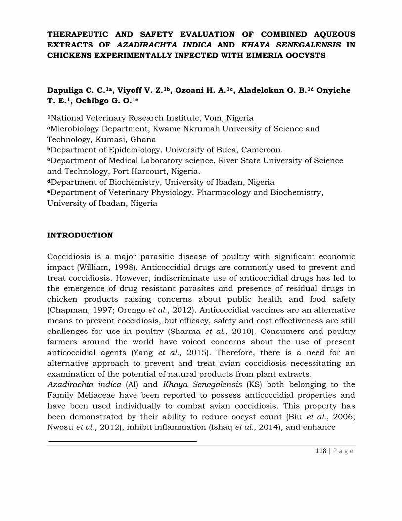
118 | P a g e
THERAPEUTIC AND SAFETY EVALUATION OF COMBINED AQUEOUS
EXTRACTS OF AZADIRACHTA INDICA AND KHAYA SENEGALENSIS IN
CHICKENS EXPERIMENTALLY INFECTED WITH EIMERIA OOCYSTS
Dapuliga C. C.1a, Viyoff V. Z.1b, Ozoani H. A.1c, Aladelokun O. B.1d Onyiche
T. E.1, Ochibgo G. O.1e
1National Veterinary Research Institute, Vom, Nigeria aMicrobiology Department, Kwame Nkrumah University of Science and
Technology, Kumasi, Ghana bDepartment of Epidemiology, University of Buea, Cameroon.
cDepartment of Medical Laboratory science, River State University of Science
and Technology, Port Harcourt, Nigeria. dDepartment of Biochemistry, University of Ibadan, Nigeria eDepartment of Veterinary Physiology, Pharmacology and Biochemistry,
University of Ibadan, Nigeria
INTRODUCTION
Coccidiosis is a major parasitic disease of poultry with significant economic
impact (William, 1998). Anticoccidial drugs are commonly used to prevent and
treat coccidiosis. However, indiscriminate use of anticoccidial drugs has led to
the emergence of drug resistant parasites and presence of residual drugs in
chicken products raising concerns about public health and food safety
(Chapman, 1997; Orengo et al., 2012). Anticoccidial vaccines are an alternative
means to prevent coccidiosis, but efficacy, safety and cost effectiveness are still
challenges for use in poultry (Sharma et al., 2010). Consumers and poultry
farmers around the world have voiced concerns about the use of present
anticoccidial agents (Yang et al., 2015). Therefore, there is a need for an
alternative approach to prevent and treat avian coccidiosis necessitating an
examination of the potential of natural products from plant extracts.
Azadirachta indica (AI) and Khaya Senegalensis (KS) both belonging to the
Family Meliaceae have been reported to possess anticoccidial properties and
have been used individually to combat avian coccidiosis. This property has
been demonstrated by their ability to reduce oocyst count (Biu et al., 2006;
Nwosu et al., 2012), inhibit inflammation (Ishaq et al., 2014), and enhance

119 | P a g e
Seminar presented 25th August, 2015 at NVRI Auditorium
erythropoiesis (Iyare and Obaji, 2014; Sanni et al., 2005).
Tipu et al., (2006) showed that combinations of herbs used against coccidiosis
are effective and an economical alternative for prophylactic anticoccidial
medication. Therefore, this study aimed to evaluate the therapeutic efficacy of
the combined aqueous extracts of leaves of Azadirachta indica and stem bark of
Khaya senegalensis in chickens experimentally infected with Eimeria oocysts.
METHODOLOGY
The stem bark of Khaya senegalensis and leaves of Azadirachta indica were
collected from the environs of the National Veterinary Research Institute
(NVRI), Vom-Nigeria. The plants were identified and authenticated in the
herbarium at Federal College of Forestry, Jos, Nigeria and assigned voucher
numbers FHJ 198 and FHJ199 for Azadirachta indica and Khaya Senegalensis
respectively. Dried plant samples were ground into powder and macerated with
distilled water for 72 hours. At the end of the extraction, the mixture was
sieved and filtered. The filtrate was concentrated by drying in the oven at 40oC.
The dried extracts were stored at 4˚C until needed.
The extracts were screened individually to detect the presence of some
phytochemicals according to the methods described by Trease and Evans,
(1989).
Mixed Eimeria oocyst suspension (Eimeria tenella, E. necatrix and E. brunetti)
was obtained from the Parasitology Division of the National Veterinary
Research Institute (NVRI). Apparently healthy day old broiler chicks were
obtained from a hatchery in Jos, Nigeria and reared for three weeks before
commencement of the study.
In the efficacy studies, infected birds were monitored by daily collection and
screening of faeces for oocysts presence and count was carried out. After
establishment of the infection (7 days post inoculation), treatment commenced
by oral gavage of the extract. The combined aqueous extracts of Azadirachta
indica and Khaya senegalensis was administered at a dose ratio of 1:1 at
100mg/kg, 200mg/kg, and 400mg/kg. The experiment included two control
groups; negative and positive treated with distilled water and amprolium
(Amprolium 250 WSP, Kepro (R) B.V Holland) respectively. All treatments lasted
for five (5) days. Hematological and serum analysis as well as histopathological
and oxidative stress examinations were carried out.
In the safety studies, the combined aqueous extracts of Azadirachta indica and
Khaya senegalensis was administered at the dose ratio of 1:1 at 100mg/kg,

120 | P a g e
200mg/kg, and 400mg/kg for 5 days while the control group received distilled
water. Serum biochemical analysis, liver oxidative stress assays and
histopathological examination was conducted.
All experiments were conducted in accordance with the principles and guide for
the care and use of laboratory animals (NRC, 1996) and approved by the
animal ethics committee of NVRI, Vom.
Data obtained from the study were summarized as Means ± Standard Error of
Mean and differences between the means determined at 5% level of significance
using the one-way analysis of variance.
RESULTS
The yield of the plants was 4.90% for AI and 7.02% for KS. The phytochemical
screening revealed the presence of tannins, saponins, cardiac glycosides and
steroids in both extracts. In addition, alkaloids and flavonoids were detected in
Azadirachta indica while resins, terpenes and anthraquinones were not
detected in both extracts.
Figure 1: Effect of AI extract on Oocyst count
Figure 2: Effect of KS extract on Oocyst count

121 | P a g e
Figure 3: Effect of the Combined extracts on Oocyst count
Fig. 4: Effect of Combined extracts of Azadirachta indica and Khaya
senegalensis on Catalase Activity in the intestine of chickens infected with
Eimeria oocysts
Fig. 5: Effect of Combined extracts of Azadirachta indica and Khaya
senegalensis on Glutathione (GSH) levels of the intestine of chickens infected
with Eimeria oocysts
122 | P a g e
Fig. 6: Effect of Combined extracts of Azadirachta indica and Khaya
senegalensis on lipid peroxidation (Malondialdehyde, MDA) of chickens infected
with Eimeria oocysts
Fig. 7: Effect of Combined extracts of Azadirachta indica and Khaya
senegalensis on Superoxide dismutase (SOD) activity of the intestine of
chickens infected with Eimeria oocysts.
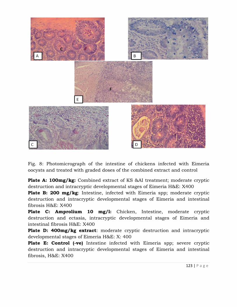
123 | P a g e
Fig. 8: Photomicrograph of the intestine of chickens infected with Eimeria
oocysts and treated with graded doses of the combined extract and control
Plate A: 100mg/kg: Combined extract of KS &AI treatment; moderate cryptic
destruction and intracryptic developmental stages of Eimeria H&E: X400
Plate B: 200 mg/kg: Intestine, infected with Eimeria spp; moderate cryptic
destruction and intracryptic developmental stages of Eimeria and intestinal
fibrosis H&E: X400
Plate C: Amprolium 10 mg/l: Chicken, Intestine, moderate cryptic
destruction and ectasia, intracryptic developmental stages of Eimeria and
intestinal fibrosis H&E: X400
Plate D: 400mg/kg extract: moderate cryptic destruction and intracryptic
developmental stages of Eimeria H&E: X: 400
Plate E: Control (-ve) Intestine infected with Eimeria spp; severe cryptic
destruction and intracryptic developmental stages of Eimeria and intestinal
fibrosis, H&E: X400
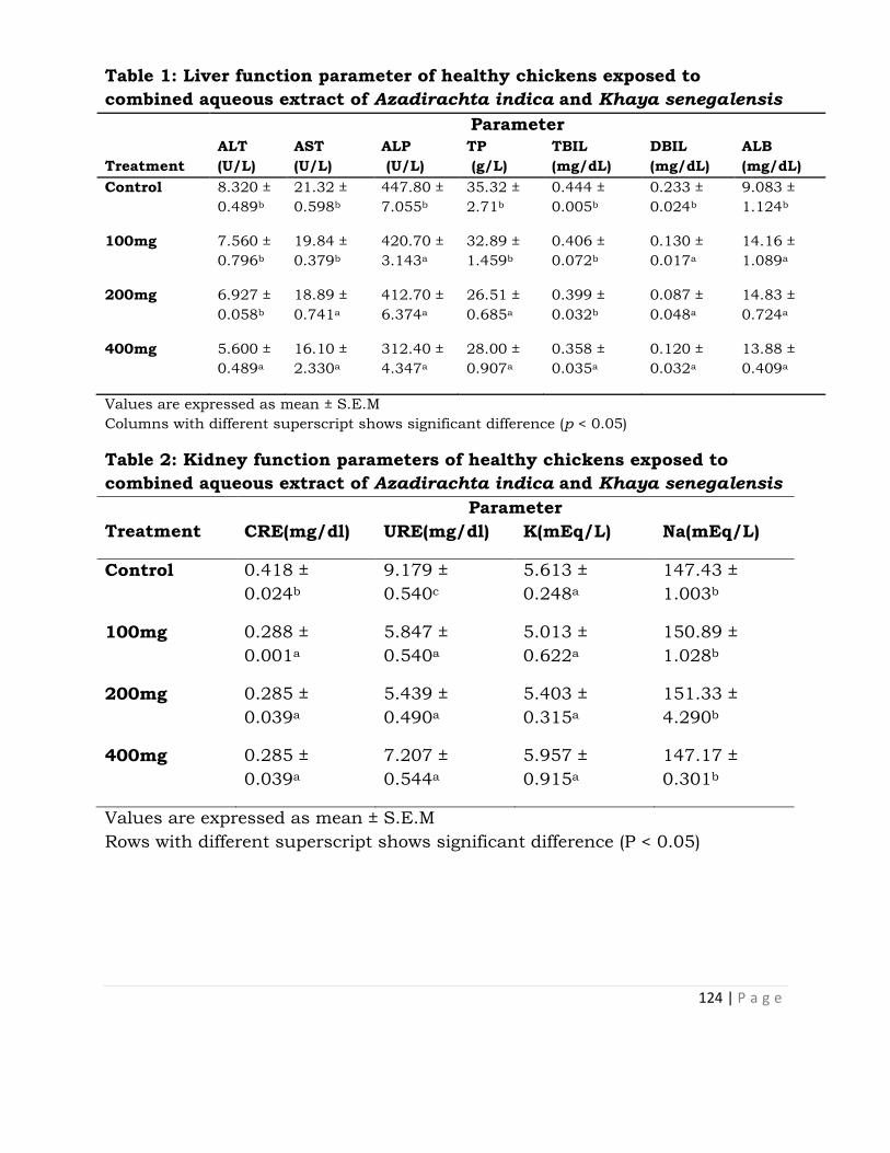
124 | P a g e
Table 1: Liver function parameter of healthy chickens exposed to
combined aqueous extract of Azadirachta indica and Khaya senegalensis
Parameter
Treatment
ALT
(U/L)
AST
(U/L)
ALP
(U/L)
TP
(g/L)
TBIL
(mg/dL)
DBIL
(mg/dL)
ALB
(mg/dL)
Control 8.320 ±
0.489b
21.32 ±
0.598b
447.80 ±
7.055b
35.32 ±
2.71b
0.444 ±
0.005b
0.233 ±
0.024b
9.083 ±
1.124b
100mg 7.560 ±
0.796b
19.84 ±
0.379b
420.70 ±
3.143a
32.89 ±
1.459b
0.406 ±
0.072b
0.130 ±
0.017a
14.16 ±
1.089a
200mg 6.927 ±
0.058b
18.89 ±
0.741a
412.70 ±
6.374a
26.51 ±
0.685a
0.399 ±
0.032b
0.087 ±
0.048a
14.83 ±
0.724a
400mg 5.600 ±
0.489a
16.10 ±
2.330a
312.40 ±
4.347a
28.00 ±
0.907a
0.358 ±
0.035a
0.120 ±
0.032a
13.88 ±
0.409a
Values are expressed as mean ± S.E.M
Columns with different superscript shows significant difference (p < 0.05)
Table 2: Kidney function parameters of healthy chickens exposed to
combined aqueous extract of Azadirachta indica and Khaya senegalensis
Treatment
Parameter
CRE(mg/dl) URE(mg/dl) K(mEq/L) Na(mEq/L)
Control 0.418 ±
0.024b
9.179 ±
0.540c
5.613 ±
0.248a
147.43 ±
1.003b
100mg 0.288 ±
0.001a
5.847 ±
0.540a
5.013 ±
0.622a
150.89 ±
1.028b
200mg 0.285 ±
0.039a
5.439 ±
0.490a
5.403 ±
0.315a
151.33 ±
4.290b
400mg 0.285 ±
0.039a
7.207 ±
0.544a
5.957 ±
0.915a
147.17 ±
0.301b
Values are expressed as mean ± S.E.M
Rows with different superscript shows significant difference (P < 0.05)

125 | P a g e
Reduced Glutathione
a
Fig 8: Effect of combined aqueous extracts of Azadirachta indica and
Khaya senegalensis on reduced glutathione (GSH) levels of the liver of
healthy chickens
Values are expressed as mean ± SEM where n=5 a Significant as compared with control ; (p < 0.05)
a a
a
Superoxide Dismutase
Fig 9: Effect of combined aqueous extracts of Azadirachta indica and
Khaya Senegalensis on superoxide dismutase activity (SOD) of the liver of
healthy chickens
Values are expressed as mean ± SEM where n=5 a significant as compared with control ; (p < 0.05)

126 | P a g e
a
a
a
Catalase
Fig 10: Effect of combined aqueous extracts of Azadirachta indica and
Khaya Senegalensis on Catalase activity of the liver of healthy chickens
Values are expressed as mean ± SEM where n=5 a Significant as compared with control ; (p < 0.05)
CONCLUSION
This study shows novel findings with respect to the possible synergistic efficacy
of the combination of aqueous extracts of K. senegalensis stem bark and A.
indica leaves lending further credence to the folkloric use of these plants in the
treatment of coccidiosis. The combination was safe at the doses and duration of
administration and appeared to be protective to the liver and kidney.
RECOMMENDATION
The active phytocompounds in the extracts should be identified, isolated and
characterized. Studies should be conducted using Molecular assays to explain
the effect of the extracts on target regulatory genes involved in the disease
pathogenesis. Other extraction solvents should be tried for possible enhanced
activity.
ACKNOWLEDGEMENT
We wish to acknowledge the African Education Initiative (NEF), USA in
partnership with the National Veterinary Research Institute, Vom Nigeria for
providing grant for the research. We also thank all our mentors and Divisions
that hosted the 2015 NEF Toxicology Internship Programme.

127 | P a g e
REFERENCES
Biu, A. A., Yusuf S. D., and Rabo J. S. (2006). Use of neem (Azadirachta
indica) aqueous extract as a treatment for poultry coccidiosis in Borno
State, Nigeria, African Scientist;7(3): 47 – 153.
Chapman, H.D. (1997). Biochemical, genetic and applied aspects of drug
resistance in Eimeria parasites of the fowl, Avian Pathology, 26: 221–244.
Gararawa, J.U., Usman N.G., Ayi V.K., Alim H.M. (2011). Anticoccidial
Resistance in Poultry: A Review, New York Science Journal, 4(8): 102-109.
Ishaq, F. N., Zezi A. U., and Olurishe T. O. (2014). Khaya senegalensis inhibits
piroxicam mediated gastro-toxicity in wistar rats, Avicenna Journal of
Phytomedicine; 4(6): 377 – 384.
Iyare, E.E. and Obaji N. N. (2014). “Effects of aqueous leaf extract of
Azadirachta indica on some haematological parameters and blood
glucose level in female rats,” Nigerian Journal of Experimental and Clinical
Biosciences; 2: 54 – 58.
National Research Council (1996). Guide for the care and use of laboratory
animals, National Academy Press, Washington, D.C
Nwosu, C. U., Hassan S. W., Abubakar M. G. et al., (2012). Anti-diarrhoeal
and Toxicological studies of leaf extract of Khaya Senegalensis, Journal
of Pharmacology and Toxicology; 7: 1–10.
Orengo, J., Buendia A.J., Ruiz-Ibanez M.R. et al., (2012). Evaluating the
efficacy of cinnamaldehyde and Echinacea purpurea plant extract in
broilers against Eimeria acervulina, Veterinary Parasitology, 185: 158–
163.
Sanni, F. S., Ibrahim S., Esievo K. A. N., et al., (2005). Effect of oral
administration of aqueous extract of Khaya senegalensis stem bark on
phenylhydrazine-induced anaemia in rats, Pakistan Journal of Biological
Science, 8: 255 – 258.
Sharma, P.A., Smith N.C., Wallach M.G et al (2010). Chasing the golden egg:
vaccination against poultry coccidiosis, Parasite Immunology, 32: 590–
598

128 | P a g e
Tipu, A. M., Akhtar M. S., Anjum M. I., et al., (2006). New dimension of
medicinal plants as animal feed, Pakistan Veterinary Journal, 26: 144–
148
Trease, G. E. and Evans, M. D (1989). A Textbook of Pharmacognosy, Builler
Tindall and Caussel London, 13th edn, Pp. 176-180
Williams, R.B (1998). Epidemiological aspects of the use of live anticoccidial
vaccines for chickens, International Journal for Parasitology, 28: 1089–
1098
Yang, W.C., Tien, Y.J., Chung, C.Y (2015). Effect of Bidens pilosa on
infection and drug resistance of Eimeria in chickens, Research in
Veterinary Science, 98: 74–81.

129 | P a g e
PUBLIC SERVICE REGULATIONS: A KEY TO ACHIEVING PRODUCTIVITY
Samme U. Andrew
Administration Division
INTRODUCTION
There are varied definition of the word administration by different Scholars and
Writers. Augustus Adebayo in his book “Principles and Practice of Public
Administration in Nigeria” define administration as the organization and
direction of persons in order to accomplish a specified task; and in his words
“when two men cooperate to roll a stone that neither could move alone, the
rudiments of administration have appeared”.
Based on the above, it is apt to state that an organization cannot exist without
administration. In the National Veterinary Research Institute, the Executive
Director/CEO has under him a hierarchy of subordinates, each with functions
and responsibilities assigned for the purpose of achieving set objectives of the
organization. The process requires planning, organizing, staffing, directing,
coordinating, reporting and budgeting. These are the primary functions of an
administrator commonly referred by the acronym POSDCORB.
The aim of this paper is to highlight some salient points, akin to public service
regulation in Nigeria for enhanced productivity.
Protocols
The words Civil Service and Public Service are used interchangeably by
different people. “The charter for the Public Service in Africa” adopted by the 3rd
Biennial Pan-African Conference of Ministers of Civil Service, Winhoek,
Namibia, 5th February, 2001 defines Public Service as “any Public entity that
implements government policies and take decisions that affect the right of
others or entities, be they individuals or not, and/or has a Public Service
responsibility.

130 | P a g e
Seminar presented 10th September, 2015 at NVRI Auditorium
It has also been defined as “a body of professionals, full-time officials employed
in the civil affairs or a state in non-political capacity” (Encyclopaedia
Britannica, vol. 4 in F. E. Iyoha 1987:3).
The definition of the Civil Service or Public Service that is more encompassing
is that which engulfs all adjuncts of the state that views Civil Service as “… a
body of generalists and professionals, full-time workers employed by the
National, State and Local Governments to help in the formulation and
implementation of Public Policies or Laws” (Iyoha 1987:4).
“Public Service Employee” on the other hand is a person in Public employment
given the responsibility for taking, implementation or enforcing decision within
the public service regulation.
Public Service regulations determine the order in which the Public Service
system can function and the principles and procedures governing its activity.
Public service regulations provide the protection of government property in a
wider sense by preventing abuse of office or power of authorities. Organizations
are expected to operate in strict compliance with rules and where they do not,
it brings about unhealthy tendencies in the organizations and in the society.
Thus, the Public Service Rules may be regarded as the most important
instrument of the Public Service system. All Public Servants must therefore
submit themselves to the provisions of the rules and regulations as no one has
the right to violate the rules of the Public Rules procedures.
As a key to achieving productivity, the Public Service Rules (PSR) lay down the
measures of combating encroachments on the Public Service system, the
existing rules of human and public relations together with the form and
procedure on whish this is done. In other words, they determine the behaviour
of Public Servants and regulate their most important human and social
relations thereby exercising a substantial influence on the course of social
development by way of formulating and implementing of government policies
and programmes.
The Public Service Rules lie in their influence on Staff behaviour and on social
relations. In any society law or rule performs a definite function of furthering
the development of certain relations and obstructing the development of
unhealthy tendencies.

131 | P a g e
The Public Service is a body or organ which enjoys continuity of existence. Its
members unlike members of the National Assembly or a House of Assembly are
not limited to a short term of office at the end of which they may or may not
return to office. Elected members come and go but Public Servants remain in
office.
Collectively, Public Servants command a pool of experience and know-how for
formulating and implementing government policies and programmes. The
Public Service is the instrument of the government of the day, but neither the
service nor its members are the partisan of any particular political party.
A Public Servant is required to assist in formulating and implementing the
policies approved by government whatever his personal or private opinions or
attitudes may be. This does not mean that a Public Servant should undertake
illegal action; if so directed, he should invite attention immediately to the legal
position or requirement and advice on the proper action to take. He has the
duty to also to advice on the implications of a policy or action.
The Public Service is indispensable irrespective of the type or regime, whether
military or civilian. Under any political weather, it has to continue its
traditional role of ensuring the orderly administration of Nigeria on continuous
basis in the interest of political and social stability.
The administration of the country on continuous basis requires discipline and
sanction. Rules and regulations must be adhered to and the interest of the
service must be paramount. The Public Servant must, at all times, resist
temptation from some quarters and uphold the principles of accountability,
probity and transparency, national consciousness and the good image of the
service.
Appointment
To be eligible for permanent and pensionable appointment into the Federal
Public Service, the appointee must be a Nigerian as defined in the Constitution
of the Federal Republic of Nigeria. In addition, the applicant must:
i. Possess a Certificate signed by the Chairman or Secretary of his Local
Government indicating his State of Origin and Local Government.
ii. Not less than 18 years or more than 50 years of age.

132 | P a g e
iii. Possess minimum qualifications as specified in the approved Scheme of
Service.
iv. Be certified by a Government Medical Officer as medically fit.
v. Possess a testimonial of good conduct from his last employer or if not
previously employed, from the last School or Institution attended.
vi. State whether or not he has been convicted of a criminal offence.
vii. State all employment he was engaged in and, if he had left any
employment, why he did so or if he is still in any employment whether
or not he is under any obligation to remain in it.
viii. State whether or not he is free, from financial embarrassment, and
ix. Possess the NYSC discharge or exemption certificate where applicable.
Promotion Guidelines in Research Institutions and Colleges
The following criteria shall form the basis for advancement in any of the cadres
within the Institution:
i. Length of service and pertinent experience.
ii. Performance
iii. Qualification
iv. Publications (where applicable) and
v. Vacancies in the establishment
(a) Movement from one grade level to a higher level will not be effected in
less than two (2) years except in exceptional cases.
(b) All promotions and advancements shall be subject to positive
performance rating from supervising officers and satisfactory service
records. Performance will be given the greatest weighting in determining
advancement.

133 | P a g e
(c) The acquisition of additional qualifications may not form the basis for
consideration for promotions, except that the performance rating of the
office is found consistently good and given that there is an appropriate
vacancy at the next higher level.
(d) All officers who fall within the field of selection for any promotion exercise
shall be considered except those who are under disciplinary action. The
minimum number of years that an officer must spend in a post being
considered eligible for promotion shall be as follows:
CONMESS Number of Years in Post
2 – 4 Minimum of 3 years
5 – 7 Minimum of 4 years
CONHESS & CONRAISS Number of Years in Post
01 – 05 Minimum of 2 years
06 – 12 Minimum of 3 years
13 – 15 Minimum of 4 years
In the case of Research Officers, moving from Senior Research Officer
(CONHESS 02 to CONHESS 9) to Principal Research Officer (CONHESS 03 –
CONHESS 11), the candidate or Staff must fulfil the following conditions:
By promotion of a qualified Senior Research Officer with a
minimum of Masters Degree plus the required relevant
publications after spending at least three years on the previous
grade (i.e. Senior Research Officer (CONHESS 02 or CONHESS
09).
Discipline
According to PSR, it is the duty of every Officer to acquaint himself with the
disciplinary rules and any other regulations in force. Responsibility for
breaches of the provisions PSR lies on every Staff.
As a general rule, disciplinary proceedings are initiated because of an Officer’s
misconduct or general inefficiency, under the provisions of PSR.

134 | P a g e
Misconduct
Is defined as a specific act of wrong-doing or improper behaviour which is
inimical to the image of the service and which can be investigated and proven.
It can also lead to termination and retirement. It includes
(a) Scandalous conduct such as:
i. Immoral behaviour
ii. Unruly behaviour
iii. Drunkenness
iv. Assault
v. Battery
vi. Refusal to proceed on transfer or to accept posting
vii. Habitual lateness to work
viii. Deliberate delay in treating of official documents
ix. Failure to keep records
x. Unauthorized removal of public records
xi. Dishonesty
xii. Negligence
xiii. Membership of cults
xiv. Sleeping on duty
xv. Improper dressing while on duty
xvi. Hawking merchandise within office premises
xvii. Refusal to take/carry out lawful instruction from superior officers
xviii. Malingering
xix. Insubordination
xx. Discourteous behaviour to the Public
General Inefficiency
General inefficiency consists of a series of omissions or incompetence the
cumulative effect of which shows that the Officer is not capable of discharging
efficiently the duties of the Office he holds.

135 | P a g e
Serious Misconduct
Serious misconduct is a specific act of very serious wrong-doing and improper
behaviour which is inimical to the image of the service and which can be
investigated and proven, may lead to dismissal. These include:
(a) Falsification of records
(b) Suppression of records
(c) Withholding of files
(d) Conviction on a criminal charge (other than a minor traffic or sanitary
offence or the like)
(e) False claims against Government Officials
(f) Bankruptcy/Serious financial embarrassment
(g) Unauthorized disclosure of official information
(h) Bribery
(i) Corruption
(j) Embezzlement
(k) Misappropriation
(l) Violation of Oath of Secrecy
(m) Action prejudicial to the security of the State
(n) Advance Fee Fraud (Criminal Code 419)
(o) Holding more than one full-time paid job
(p) Nepotism or any other form of preferential treatment
(q) Divided loyalty
(r) Sabotage
(s) Willful damage to Public property
(t) Sexual harassment, and
(u) Any other act unbecoming of a Public Officer

136 | P a g e
Penalties for Breaches of Disciplinary Rules
The following are disciplinary measures that may be taken or applied under the
Public Service Rules:
1. Verbal warning
2. Written warning
3. Reprimand
4. Stick reprimand
5. Reduction in rank or demotion for specified period
6. Reduction in Salary
7. Withholding or deferment of increment for a specified period
8. Loss of pay/non-payment for the corresponding days an Officer is not at
duty post/station without authority
9. Stoppage of salary
10. Surcharge
11. Interdiction
12. Suspension
13. Termination of appointment
14. Dismissal from the Service
Disciplinary sanctions and penalties are many and are varied. Liability for
breaches of service discipline is necessary to force the subordination of the
personnel in the service for the proper and effective administration.
Interdiction
When a serious case that may lead to dismissal has been instituted against an
Officer, such Officer may be placed on interdiction on not more than half pay
pending the final determination of the case.
Suspension
137 | P a g e
Suspension is applied where a prima facie case, the nature of which is serious,
has been established against an Officer and it is considered necessary in the
public interest that he should forthwith be prohibited from carrying on his
duties; pending the final determination of the case, the suspended Officer will
cease to exercise the powers and functions of his Office and the enjoyment of
his salary.
Dismissal
It must be noted that the ultimate penalty for serious misconduct is dismissal.
An Officer who is dismissed from the service forfeits all claims to retiring
benefits, leave or transport grant, etc.
Dismissal for breach of disciplinary rules is only permitted in cases stipulated
in the Public Service Rules on serious misconduct. Any Officer who absents
himself from duty or from Nigeria without leave renders himself liable to be
dismissed from the service and, of course, the onus rests on him, to show that
the circumstances do not justify the imposition of the full penalty. Again, an
Officer involved in the conduct prejudicial to the security of the State may be
dismissed from the service without following the normal procedure.
It is important to distinguish between dismissal and termination of
appointment. Dismissal and termination are considered as punishment for
misconduct. An officer who is dismissed or whose appointment is terminated
solely for misconduct forfeits all rights and privileges such as leave, retiring
benefits, transport, etc. An Office whose appointment is terminated for
inefficiency does not forfeit them, but his retiring benefits may be reduced or
entirely withheld if he also misconducted himself. That is say, if an Officer’s
services are terminated solely for misconduct, this is the equivalent of
dismissal.
It should also be noted that if an Officer is convicted on a criminal charge, the
officer may thereupon be dismissed without any of the procedures prescribed
in the PSR being followed.
Removal of Temporary Staff
The employment of temporary Staff may be terminated provided such Staff has
been informed on the grounds on which it is proposed to terminate his
appointment and has been given and opportunity to submit representation,
why he should not be terminated. Similarly, prior to the termination of his

138 | P a g e
service for inefficiency, such Staff should be warned of his failings and be given
an opportunity to improve or to offer a satisfactory explanation of his failure to
perform his duties efficiently.
Termination of Appointment during Probationary Period
If within his probationary period, it is established to the satisfaction of the
authority empowered to appoint an Officer that he is not qualified for efficient
service, his appointment may be terminated by that authority at any time
without any further compensation than free transport to the place from which
he was engaged; and such free transport will be granted only if his conduct has
been good. If the termination is not due to misconduct, a month’s notice will be
given and leave may be granted if the Officer is eligible.
The appointment of an Officer on probation, who fails to secure confirmation in
the pensionable establishment at the expiration of his probationary period,
including such extension thereof as prescribed under PSR, may be terminated
in the manner specific in PSR.
Retirement in Public Interest
Any Officer may be required to compulsorily retire from the Service on grounds
which cannot be suitably dealt with by the disciplinary procedures and it is
desirable in the public interest to do so. In every such case the question of
pension and gratuity will be dealt with under the Pension Act for the time being
in force.
Retiring Benefits
An Officer or employee who is dismissed or whose appointment is terminated
for misconduct is not eligible to receive any retiring benefits. However,
termination of appointment for inefficiency or in the public interest does not
necessarily disqualify an Officer or employee from the award of retiring
benefits. He is “retired” and, if otherwise eligible, may receive retiring benefits.
Other Penalties
Reproof, reprimand and strict reprimand mean censure of the Staff’s breach of
the provisions of PSR by the Management and do not entail any other negative
consequences for the Staff.

139 | P a g e
Demotion is a heavier penalty and combines censure of the fault with a
temporary reduction of earnings. The demotion or reduction in rank must be a
temporary measure and should be applied for not more than one year or even a
shorter period of six months may also be possible. The Staff should be returned
to his previous post when the period expires. Furthermore, the demotion must
consist in actually giving the offender a lower paid job, that is the job which
must be less responsible and less skill.
As a general rule, only one penalty may be imposed for each breach of PSR.
The right of choice is the Management’s and the degree of the penalty must
correspond to the seriousness of the breach committed.
Applying and lifting disciplinary sanctions are laid-down in the PSR
procedures. Disciplinary sanctions are an essential feature of the PSR and of
course, rules cannot be effective unless it is enforceable and no rule can be
enforceable unless there is a sanction for its breach.
Fair Dismissal
Dismissal of an employee can only be fair if the employer can show the reason
for it as one of those listed in the rules and regulations, and furthermore can
show that the Management acted reasonably in the circumstances in treating
that reasons as sufficient to justify dismissing the employee. Such reason may
include:
1. A reason related to the employee’s capability or qualifications for the job.
2. A reason related to the employee’s conduct.
3. Redundancy.
4. A legal duty or restriction on either the employer of the employee which
prevents the employment being continued.
5. Some other substantial reasons which could justify the dismissal.
Of course, unfair or wrongful dismissal occurs when sufficient notice is not
given. When practicable, three queries and warnings should be served on
erring Officer before considering the option of dismissal. Subject of course, to

140 | P a g e
the gravity of misconduct and circumstances of each case. In judging cases,
the relevant sections of PSR should be applied.
Remedies for Unfair Dismissal
There are three possible remedies for unfair dismissal:
a. Reinstatement, that is to say, the employee is to be treated in all respects
as though the dismissal had not occurred.
b. Re-engagement, that it to say, the employee is to be re-employed but not
necessarily in the same job or on the same terms and conditions of
employment.
c. Compensation, that is to say, payment of just, fair and reasonable
compensation for loss of career expectations.
Model Disciplinary Procedure
i. A disciplinary procedure should be fair, full and quick.
ii. Each Staff should know that what the procedure consists of, its rules
and the offences which can lead to dismissal.
iii. The procedure should state who has power to dismiss.
iv. Before dismissal takes place, a previous warning should have been
issued, because as forewarned is to be forearmed.
v. The degree of misconduct in any particular case should determine
whether it is sufficient to justify dismissal without notice.
vi. The records of past performance should be critically examined in order to
establish the case of deadwoodism, incompetence and ineffectiveness.
vii. It is necessary to ensure that no one is dismissed unfairly and that there
is a defence if a dismissed employee complains to a higher authority.
Right of Appeal and Petitions
Every Officer has a right of appeal against the decisions of the appropriate
authority through the appropriate channels. However, a former Officer who is
dissatisfied or aggrieved as a result of any decision of the appropriate authority
has the right of appeal to the Federal Service Commission.

141 | P a g e
Again, an Officer or a person who was formerly an Officer has right of appeal to
the President against the decision of the Federal Public Service Commission. A
petition should be submitted within the stipulated period of not more than six
months.
Record of Outstanding Work
Disciplinary sanctions are not the sole possible way of influencing offenders
against labour discipline. Labour discipline is ensured by a conscientious
attitude to work.
CONCLUSION
The Administrators’ Position within an establishment or organization is
strategic and pivotal. It is true to say that the pace of administration has come
to be clearly recognized in every sector of human endeavour, as being the
keystone to the success and indeed to the very existence of an organization.
Being concerned with the planning, supervision and control of the
establishment with which it is involved, it is no overstatement to declare that
“whatever may be the future, the science of administration will be an essential
instrument of human welfare”.
REFERENCES
Federal Republic of Nigeria Official Gazette; Public Service Rules (2008)
Edition
Augustus Adebayo “Principles and Practice of Public Administration in Nigeria”.
“3rd Biennial Pan African Conference of Ministers of Civil Service”
Winhoek Namibia 5th February, 2001.
Encyclopaedia Britannica Vol. 4 in F. E. Iyoha 1987:3
142 | P a g e
RELATIONSHIP BETWEEN EMOTIONAL INTELLIGENCE AND JOB
SATISFACTION AMONG EMPLOYEES OF NATIONAL VETERINARY
RESEARCH INSTITUTE, VOM
Mshellia Priscilla Swatdunio (B.Sc, MBA, CNA, AMNIM)
Bursary Department, Federal College of Veterinary and Medical Laboratory
Technology, National Veterinary Research Institute, P.M.B 02, Vom
Correspondence: [email protected], 2348035983950
INTRODUCTION
We may know someone who is incredibly bright but cannot seem to pull
his/her life together. Some may be brilliant students who drop out of the
University or incredibly intelligent workers who can’t seem to get ahead in their
organization. We often know from our interactions with them that they have a
good or superior intelligence even though it doesn’t seem to ensure success.
This scenario has led scientists over time to realize that Intelligence Quotient
(IQ) may not be enough requirements for success in life. This has led to the
realization that there is another type of intelligence which is equally very
relevant for success which has been termed ‘Emotional Intelligence’.
Emotional intelligence (EI) as a concept has gained popularity since Goleman
wrote the book entitled “Emotional Intelligence” in 1995. EI can be acclaimed
to be of psychological background even though it has gradually formed close
ties with key management areas. Goleman (1998) defined EI as “the capacity
for recognizing our own feelings and those of others, for motivating ourselves
and for managing emotions well in ourselves and in our relationships”. EI has
as one of its premises that emotional responses may be logically consistent or
inconsistent with particular beliefs about emotion. Relatively pure emotional
reactions such as instances of happiness or fear displayed early in infancy may
involve relatively little cognition; these probably are best evaluated as adaptive
or maladaptive. But as the person develops increasingly complex
representation of situations, his or her emotional reactions may merge with
more complex thoughts to develop such cognitively saturated emotions as guilt
or regret. Moreover, the person may develop sophisticated internal modules

143 | P a g e
Seminar presented 8th October, 2015 at NVRI Auditorium
that include standards of emotional functioning. These emotional reactions and
models can be assessed according to their logical consistency, and hence their
‘intelligence’ (Mayer & Salovey, 1995). Social emotional competencies have been
adjudged to be important aspects of both performance effectiveness and
measures of professional success. Research has shown that rational
intelligence quotient is no longer considered a good predictor of how effective
an employee will be at work and in life; rather it has been suggested that EI is
a better predictor in determining employee's effectiveness.
According to Spector (1997), job satisfaction refers to the evaluation of the job
in all its ramifications and its antecedents. Factors such as financial rewards,
resources to get the job completed, interest, challenge, use of valued skills,
variety, occupational prestige, autonomy, relation to co-workers and
supervisors, involvement in decision making and comfort factors such as
hours, physical environment and travel time. Emotional intelligence has been
said to be linked with job satisfaction through creation of self motivation which
results from understanding the influence of our emotions on the factors
responsible for creation of job satisfaction (Wong & Law, 2002; Ayub & Rafif,
2011; Seyal & Afzaal, 2013).
METHODOLOGY
Population and Sampling
The population of the study consisted of staff of the Federal College of
Veterinary and Medical Laboratory Technology (FCVMLT), National Veterinary
Research Institute, Vom. These staff comprised mainly of scientists and
support staff. At first step, the population was ascertained from the nominal
roll of the organization having a total of 215 staff and an additional 30
volunteers from the institute. A sample size of 132 was arrived at using the
Cochran (1977) sample size determination table assuming p = 0.50 and t =
1.96. The Schutte (1998) questionnaire was used for data collection on
Emotional Intelligence. Job satisfaction was measured using a one item overall
job satisfaction questionnaire. Copies of questionnaires were administered and
retrieved from respondents within a time span of two weeks. Pearson’s
correlation analysis was used as a tool for the analysis. The SPSS software was
used for processing data collected.

144 | P a g e
RESULTS AND DISCUSSION
This study has the following hypothesis stated in null form
Ho: Emotional Intelligence has no significant relationship with employee job
satisfaction
The hypothesis was tested using Pearson’s correlation analysis and the results
are shown in the table below.
TABLE 1: Correlation results
JS EI
JS Pearson Correlation 1 0.047
Sig. (2-tailed) 0.597
N 131 131
EI Pearson Correlation 0.047 1
Sig. (2-tailed) 0.597
N 131 131
Key:
JS = Job satisfaction
EI = Emotional intelligence
Source: SPSS Software
From the correlation results, the Pearson’s correlation value (0.047) suggests a
significant relationship between EI and Job satisfaction (p<0.05). This means
that the null hypothesis is rejected. It shows that EI creates a feeling of
satisfaction not necessarily because the job may be so enjoyable but because
the employees find ways of coping with their jobs despite any challenges that
may be faced on the job. The findings are consistent with those of Adeyemo
(2007), Abraham (2000) & Goleman (1995).
Employees with high EI are more likely to have higher levels of job satisfaction
because they are more adept at appraising and regulating their own emotions
than employees with low EI. For example, employees with high EI may be
better at identifying feelings of frustration and stress, then subsequently
regulating those emotions to reduce stress. Employees with high EI are more

145 | P a g e
resilient because they are able to understand the causes of stress and develop
strategies and perseverance to deal with the negative consequences of stress
(Copper & Sawaf, 1997). Conversely, employees with low EI are likely to be less
aware of their emotions and possess fewer abilities to cope with their emotions
when faced with difficult situations, thereby increasing their levels of stress
and decreasing their level of job satisfaction.
CONCLUSION
Based on the research findings the study concludes that Emotional intelligence
has significant relationship with employee job satisfaction. The pearsons
correlation tests revealed that EI has a significant relationship with job
satisfaction. Employees that are emotionally intelligent are likely to be more
satisfied with their jobs.
It is therefore recommended that National Veterinary Research Institute, Vom
and other allied organizations should evaluate EI levels of their prospective
employees during recruitment. This can be done by administration of EI tests
and consideration of test scores as significant factors in making recruitment
decisions. Also, since EI is much less fixed than IQ it can be developed over
time. This is what Goleman refers to as ‘maturity’. This can be achieved by
training, coaching and feedback so that steady progress can be made in the
organization. Employees should be encouraged to work on imbibing emotional
competencies so that they can attain emotional maturation since this is helpful
in self motivation towards satisfaction. Job satisfaction resulting from EI
maturation is likely to lead to increased productivity and create a healthy work
environment.
Finally the researcher recommends that more studies be carried out in this
area. More organizations should be studied with larger sample sizes especially
with regards to gender, physical ability and job specialization.
ACKNOWLEDGEMENT
I acknowledge my supervisor, Professor Bello Sabo of Department of Business
Administration, Ahmadu Bello University Zaria, and staff of Federal College of
Veterinary and Medical Laboratory Technology, Vom for participating in the
study.
REFERENCES

146 | P a g e
Abraham, R. (2000). The role of job control as a moderator of emotional
Dissonance and emotional intelligence: Outcome relationship. Journal of
Psychology, 134(2):35-42
Adeyemo, D.A. (2007). Emotional intelligence and relationship with job
satisfaction and organizational commitment of employees in public
parastatals of Oyo state, Ibadan Medwell Journals 2(1): 6-11
Ayub, N. & Rafif, S. (2011): The relationship between Motivation and Job
satisfaction. Pakistan Business Review: 28
Cochran, W.G. (1977). Sampling Techniques (3rded) New York: John Wiley &
Sons.
Copper, R & Sawaf, A (1997). Emotional intelligence in leadership and
organization (1st Ed), Perigee trade
Goleman, D. (1995). Emotional Intelligence: Why it can matter more than IQ.
New York, Bantam.
Goleman, D. (1998). Working with Emotional Intelligence, New York, Bantam
Mayer, J.D. & Salovey, P. (1995).Emotional Intelligence and the Construction
and Regulation of Feelings, Applied and Preventive Psychology 4, 197-208
Seyal, H.A. & Afzaal, T (2013).An Investigation of relationship among Emotional
Intelligence, Organizational Commitment and Job Satisfaction, Evidence
from Academics in Brunei Darusalam. International Business Research,
6(3), 217-228 http://dx.doi.org/10.5539/ibr.v6n3p217
Spector, P.E (1977). Job satisfaction application, assessment, causes and
consequences, Thousand Oaks, CA: Sage
Wong, C.S. & Law, K.S (2002).The effects of leader and follower emotional
intelligence on performance and attitude: An explanatory study,The
leadership quarterly, 13(3)

147 | P a g e
HIGHLY PATHOGENIC AVIAN INFLUENZA IN NIGERIA: OVERVIEW OF
2015 OUTBREAKS
Ifende, V.I. 1, Meseko, C.A. 2, Odita, C.I. 3, Akanbi, B.O. 3, Moses, G.D.3,
James, A.S.3, Barde, I.J3, Rimfa, A.G. 3, Ugbe, D3., Shittu, I.A. 2, Gado D 2,
Olawuyi, A 2., Joannis, T.M. 2, Kumbish, P.R3., Muhammad, L.U. 1,
Okewole, P.A1,3, Ahmed M.S. 2, Shamaki D.2
1Veterinary Extension Research and Liaison Services, NVRI, Vom 2Regional Center for Animal Influenza and TADs, NVRI, Vom 3Central Diagnostic Division, NVRI, Vom
Corresponding author: [email protected]
INTRODUCTION
Avian influenza (AI) is an important disease of zoonotic origin that has caused
morbidity and mortality in domestic animals, wildlife and humans (Muzaffar et
al., 2006; Olsen et al., 2006; Gauthier-Clerc et al., 2007). It is known to be
caused by type A viruses of the family Orthomyxoviridae, and they are
classified by their hemagglutinin (HA) and neuraminidase (NA) surface
glycoproteins. Highly Pathogenic Avian Influenza (HPAI) has caused respiratory
disease and deaths in poultry and poultry handlers that were inappropriately
exposed to aerosols generated from handling chickens (Oladokun et al., 2012)
The first case of HPAI in Nigeria was in February 2006 in a commercial poultry
farm in Northern Nigeria and the causal virus was the H5N1. The source of the
initial outbreak in Nigeria is still not clear; international trade in birds or wild
migratory birds could be responsible (Ducatez et al., 2006).
The Federal Government already had an emergency preparedness plan in place
which to a great extent paved way for the initial control strategy. The
implementation curbed the first introduction of the disease as outbreaks were
last recorded in 2008. However, a resurgence of the disease occurred in
January 2015.
This study presents a synopsis of the 2015 outbreaks in Nigeria, from records
available in NVRI as a major participant in the diagnosis and control of HPAI in
Nigeria.
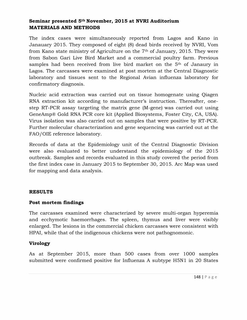
148 | P a g e
Seminar presented 5th November, 2015 at NVRI Auditorium
MATERIALS AND METHODS
The index cases were simultaneously reported from Lagos and Kano in
Janauary 2015. They composed of eight (8) dead birds received by NVRI, Vom
from Kano state ministry of Agriculture on the 7th of January, 2015. They were
from Sabon Gari Live Bird Market and a commercial poultry farm. Previous
samples had been received from live bird market on the 5th of Janaury in
Lagos. The carcasses were examined at post mortem at the Central Diagnostic
laboratory and tissues sent to the Regional Avian influenza laboratory for
confirmatory diagnosis.
Nucleic acid extraction was carried out on tissue homogenate using Qiagen
RNA extraction kit according to manufacturer’s instruction. Thereafter, one-
step RT-PCR assay targeting the matrix gene (M-gene) was carried out using
GeneAmp® Gold RNA PCR core kit (Applied Biosystems, Foster City, CA, USA).
Virus isolation was also carried out on samples that were positive by RT-PCR.
Further molecular characterization and gene sequencing was carried out at the
FAO/OIE reference laboratory.
Records of data at the Epidemiology unit of the Central Diagnostic Division
were also evaluated to better understand the epidemiology of the 2015
outbreak. Samples and records evaluated in this study covered the period from
the first index case in January 2015 to September 30, 2015. Arc Map was used
for mapping and data analysis.
RESULTS
Post mortem findings
The carcasses examined were characterized by severe multi-organ hyperemia
and ecchymotic haemorrhages. The spleen, thymus and liver were visibly
enlarged. The lesions in the commercial chicken carcasses were consistent with
HPAI, while that of the indigenous chickens were not pathognomonic.
Virology
As at September 2015, more than 500 cases from over 1000 samples
submitted were confirmed positive for Influenza A subtype H5N1 in 20 States

149 | P a g e
and the FCT. The virus was widely disseminated in Kano, Lagos, Bauchi,
Plateau and still circulating in Rivers State as at the time of this analysis.
Samples that are positive by RT-PCR were also successfully isolated in chicken
embryonated eggs within 48 hours.
Molecular sequencing
The complete gene sequence of the current HPAI H5N1 in Nigeria was
determined. The gene sequence has been submitted to GISAID under accession
number EPI 556504 and EPI 567299- EPI 567305.
The topology of the phylogenetic tree of the HA gene demonstrated that the
H5N1 virus from Nigeria A/chicken/Nigeria/15VIR 339-2/2015 falls within
genetic clade 2.3.2.1c. In particular, the HA gene clustered with H5 viruses
from China in 2013 and with H5N1 virus isolated from a Canadian returnee
from China. The similarities observed were 99.3 to 99.5% respectively (Monne
et al., 2015).
Epidemiology
In the current outbreak, Avian Influenza outbreak was first confirmed in Lagos
State in a live bird market followed by commercial poultry farm in Kano. By the
end of January 2015, 15 States were already confirmed positive for the disease.
At the time of this report, 20 States and the FCT are positive for AI in the on-
going outbreak.
The epidemic curve of the disease shows that the outbreaks reached its peak in
February (Fig. 1) and has declined since then with a sustained disease
condition presently, with Kano and Plateau States having the highest number
of disease outbreaks (Fig. 2)

150 | P a g e
Figure 1: Graph showing HPAI positive cases per month
0
50
100
150
200
250
AB
IA
AN
AM
BR
A
BA
UC
HI
DE
LTA
ED
O
EN
UG
U
FC
T
GO
MB
E
IMO
JIG
AW
A
KA
DU
NA
KA
NO
KA
TS
INA
LA
GO
S
NA
SA
RA
WA
OG
UN
OYO
PLA
TE
AU
RIV
ER
S
SO
KO
TO
ZA
MF
AR
A
P
o
s
i
t
i
v
e
A
I
c
a
s
e
s
Affected States
Positive AI cases by States

151 | P a g e
Figure 2: Graph showing HPAI positive cases per state
DISCUSSION
The resurgence of HPAI in 2015 since the last case in 2008 is a cause for
concern. Molecular analysis revealed a new clade of H5N1 virus present in
Nigeria in 2015, possibly introduced by migratory birds or trade in poultry.
The epidemic curve spiked in the month of February 2015, similar to 2006 and
2007 outbreaks respectively (Joannis et al., 2008). This may suggest a seasonal
pattern for the virus that should be further investigated.
The results obtained provide evidence that a new clade of H5N1 virus has been
introduced to Nigeria in 2015. A more prognostic approach and pragmatic plan
is required for prevention and control of HPAI in Nigeria with this resurgence,
in order to elude a likely endemicity of the disease.
ACKNOWLEDGEMENT
We like to also acknowledge the efforts of the following towards the diagnosis of HPAI;
Drs. Felix G.P., Makanju O., Chinyere C., Anefu E., Nwauda L., Mrs. Abiayi,
E., Mrs. Chukwu, I., Mrs. Agama, Mrs. Liman F., Mr. Agada, O., Messers.
Nyam D., Daspan A., T. Amos, J. Jonathan, R Akintola, B. Goma, L., Buba,
M., Waje Atam S., Zal A., Dung G., Davou T.
Staff of VERLS and Poultry Divisions, FAO/OIE, and Federal Vetereinary
Service
REFERENCES
Oladokun, AT; Meseko, CA; Ighodalo, E; Benshak, J; Ekong, PS (2012) Effect
of intervention on the control of Highly Pathogenic Avian Influenza in
Nigeria, Pan African Medical Journal.
Ducatez MF, Olinger CM, Owoade AA, De Landtsheer S, Ammerlaan W,
Niesters HG, Osterhaus AD, Fouchier RA, Muller CP (2006). Avian flu:
multiple introductions of H5N1 in Nigeria. Nature 442, 37

152 | P a g e
Gauthier-Clerc M, Lebarbenchon C and Thomas F (2007) Recent expansion of
highly pathogenic avian influenza H5N1: a critical review. Ibis 149: 202-
214.
Monne, I; Meseko, C; Joannis, T; Shittu, I; Ahmed, M; Tassoni, L; Fusaro, A;
Cattoli, G (2015). Highly Pathogenic Avian Influenza A(H5N1) Virus in
Poultry, Nigeria, 2015. Emerg Infect Dis 21.doi:10.3201/eid2107.150421.
Muzaffar, SB; Ydenberg, RC and Jones IL (2006) Avian Influenza: an ecological
and evolutionary perspective for waterbird scientists, Water birds 29:
243-257
Olsen, B; Munster, VJ; Wallenstrom, J; Osterhaus, ADME; Fouchier, RAM;
(2006). Global pattern of Influenza A virus in wildbirds, Science 312
(5772): 384–388.
Joannis, T M; Meseko, C A; Oladokun, A T; Ularamu, H G; Egbuji, A N;
Solomon, P; Nyam, D C; Gado, D A; Luka, P; Ogedengbe, M E; Yakubu,
M B; Tyem, AD; Akinyede, O; Shittu, A I; Sulaiman, L K; Owolodun, O A;
Olawuyi, A K; Obishakin, E T; Fasina, F O (2008). Serologic and virologic
surveillance of Avian Influenza in Nigeria, 2006-7, Eurosurveillance Vol
13, Issue 42

153 | P a g e
NVRI SEMINAR SERIES
GUIDELINES FOR PRESENTATION AND MANUSCRIPT PREPARATION
The NVRI Seminar Series is an edited publication of seminar presentations
made at NVRI during the course of one calendar year. Seminar
presentations provide a platform for research staff of the Institute and
visiting scientists to present their research findings. The series is published
annually.
Types of presentations
Research proposals, progress reports of on-going research, reports of
completed studies and thesis are accepted for presentation. Reviews in any
area of the Institute’s mandate are made by invitation only.
Preparation for of manuscript for presentation
A Microsoft PowerPoint® presentation and an extended abstract are to be
submitted to the secretary of the Committee one week before the
presentation is due.
Preparation of manuscript
The manuscript should be written as an extended abstract with the
following: Title of Paper, Author(s), Address including e-mail, Introduction,
Methodology, Results and Discussion, Acknowledgements and References.
Only one figure and one table will be accepted. The title of presentation
should not be more than 15 words with the names of all authors and their
affiliations. Only the email of the corresponding author and telephone
numbers are required.
Duration of Presentation
The presentation usually lasts 20 minutes followed by another 20 minutes
of discussion.

154 | P a g e
Check list for slide presentation
1. Visual Aid
Please check that slides are:
a. Not too many for a twenty-minute presentation
b. Contents are coordinated with speech
c. Slides are neat and spaced to fill the screen
d. Simple and free from excessive data
e. Free from garish colour and other embellishments that may
distract from your message
f. Have a balance of data, lists, and information with photographs
interspersed throughout
2. Speaker
Please ensure that
a. You are familiar with speech and slides
b. You are dressed properly to make your audience comfortable
c. Reduce excessive gestures
d. Avoid reading from the slides and notes all the time
e. Shall keep to the time allocated for presentation