Active gels as a description of the actin‐myosin cytoskeleton
doi:10.1016/j.jmb.2010.11.053 J. Mol. Biol. (2011) 407, 79–91
Contents lists available at www.sciencedirect.com
Journal of Molecular Biologyj ourna l homepage: ht tp : / /ees .e lsev ie r.com. jmb
Nucleotide Pocket Thermodynamics Measured byEPR Reveal How Energy Partitioning RelatesMyosin Speed to Efficiency
Thomas J. Purcell1,2⁎, Nariman Naber1, Kathy Franks-Skiba1,2,Alexander R. Dunn3, Catherine C. Eldred4, Christopher L. Berger5,András Málnási-Csizmadia6, James A. Spudich7, Douglas M. Swank4,Edward Pate8 and Roger Cooke1,2
1Department of Biochemistry and Biophysics, UCSF MC 2240, Genentech Hall Room S416C, 600 16th Street,San Francisco, CA 94158-2517, USA2Cardiovascular Research Institute, University of California, San Francisco, CA 94158, USA3Department of Chemical Engineering, Stanford University, Stanford, CA 94305, USA4Department of Biology, Rensselaer Polytechnic Institute, Troy, NY 12180, USA5Department of Molecular Physiology and Biophysics, College of Medicine, University of Vermont,Burlington, VT 05405, USA6Department of Biochemistry, Eötvös Lorand University, Budapest 1117, Hungary7Department of Biochemistry, Stanford University School of Medicine, Stanford, CA 94305, USA8Department of Mathematics, Washington State University, Pullman, WA 99164, USA
Received 1 July 2010;received in revised form24 November 2010;accepted 26 November 2010Available online23 December 2010
Edited by M. Moody
Keywords:myosin;actin;muscle;efficiency;EPR spectroscopy
*Corresponding author. DepartmenBiophysics, UCSF MC 2240, Genente600 16th Street, San Francisco, CA 9E-mail address: [email protected] used: EPR, electron
resonance; IFM, insect flight muscle;ADP.
0022-2836/$ - see front matter © 2011 E
We have used spin-labeled ADP to investigate the dynamics of thenucleotide-binding pocket in a series of myosins, which have a range ofvelocities. Electron paramagnetic resonance spectroscopy reveals that thepocket is in equilibrium between open and closed conformations. In theabsence of actin, the closed conformation is favored. When myosin bindsactin, the open conformation becomes more favored, facilitating nucleotiderelease. We found that faster myosins favor a more closed pocket in theactomyosin•ADP state, with smaller values of ΔH0 and ΔS0, even thoughthese myosins release ADP at a faster rate. A model involving a partitioningof free energy between work-generating steps prior to rate-limiting ADPrelease explains both the unexpected correlation between velocity andopening of the pocket and the observation that fast myosins are less efficientthan slow myosins.
© 2011 Elsevier Ltd. All rights reserved.
t of Biochemistry andch Hall Room S416C,4158-2517, USA.edu.paramagneticSLADP, spin-labeled
lsevier Ltd. All rights reserve
Introduction
Different members of the myosin superfamilymove along actin filaments with widely varyingvelocities. This difference was first characterized inthe case of skeletal muscle myosin. Skeletal musclescan be broadly classified into two types: slow-twitch(type I) fibers and fast-twitch (type II) fibers. Thetype I fibers have three to five times slower velocities
d.
NH2
N
NN
N O O O
O
O
P O
O
O
P
O
O
P
CH3
CH3
H3C
H3C
O
O
N
C
O H
2' 3'
5' O
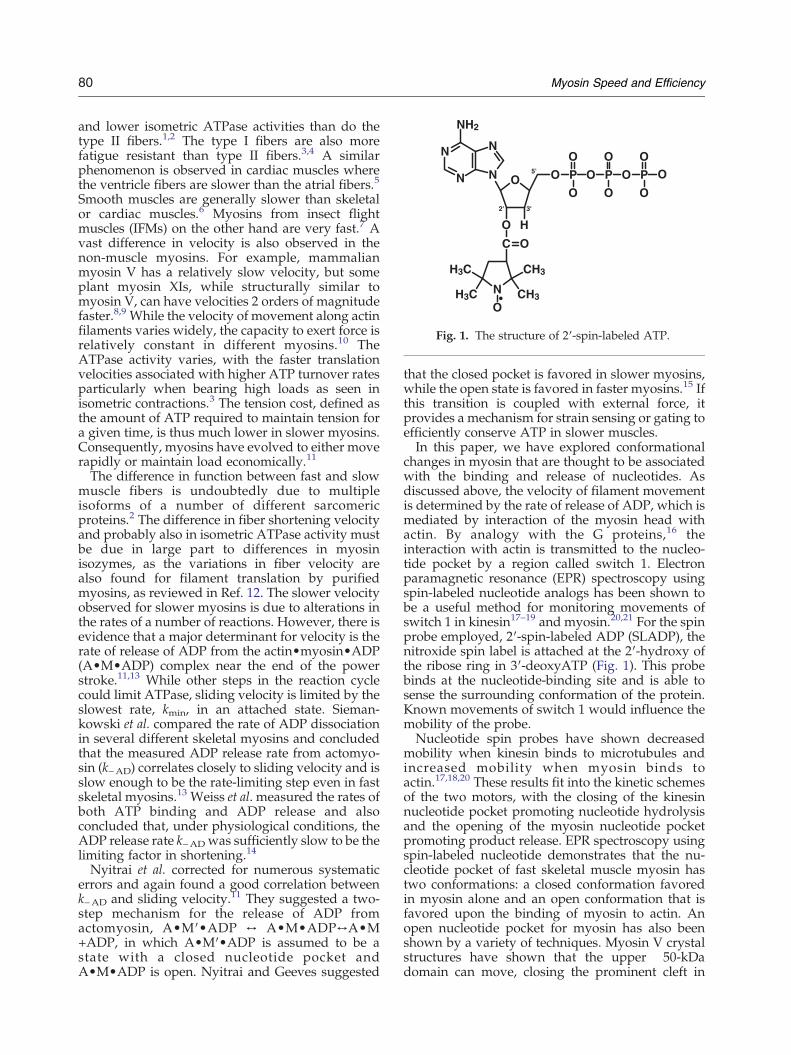
Fig. 1. The structure of 2′-spin-labeled ATP.
80 Myosin Speed and Efficiency
and lower isometric ATPase activities than do thetype II fibers.1,2 The type I fibers are also morefatigue resistant than type II fibers.3,4 A similarphenomenon is observed in cardiac muscles wherethe ventricle fibers are slower than the atrial fibers.5
Smooth muscles are generally slower than skeletalor cardiac muscles.6 Myosins from insect flightmuscles (IFMs) on the other hand are very fast.7 Avast difference in velocity is also observed in thenon-muscle myosins. For example, mammalianmyosin V has a relatively slow velocity, but someplant myosin XIs, while structurally similar tomyosin V, can have velocities 2 orders of magnitudefaster.8,9 While the velocity of movement along actinfilaments varies widely, the capacity to exert force isrelatively constant in different myosins.10 TheATPase activity varies, with the faster translationvelocities associated with higher ATP turnover ratesparticularly when bearing high loads as seen inisometric contractions.3 The tension cost, defined asthe amount of ATP required to maintain tension fora given time, is thus much lower in slower myosins.Consequently, myosins have evolved to either moverapidly or maintain load economically.11
The difference in function between fast and slowmuscle fibers is undoubtedly due to multipleisoforms of a number of different sarcomericproteins.2 The difference in fiber shortening velocityand probably also in isometric ATPase activity mustbe due in large part to differences in myosinisozymes, as the variations in fiber velocity arealso found for filament translation by purifiedmyosins, as reviewed in Ref. 12. The slower velocityobserved for slower myosins is due to alterations inthe rates of a number of reactions. However, there isevidence that a major determinant for velocity is therate of release of ADP from the actin•myosin•ADP(A•M•ADP) complex near the end of the powerstroke.11,13 While other steps in the reaction cyclecould limit ATPase, sliding velocity is limited by theslowest rate, kmin, in an attached state. Sieman-kowski et al. compared the rate of ADP dissociationin several different skeletal myosins and concludedthat the measured ADP release rate from actomyo-sin (k−AD) correlates closely to sliding velocity and isslow enough to be the rate-limiting step even in fastskeletal myosins.13 Weiss et al. measured the rates ofboth ATP binding and ADP release and alsoconcluded that, under physiological conditions, theADP release rate k−ADwas sufficiently slow to be thelimiting factor in shortening.14
Nyitrai et al. corrected for numerous systematicerrors and again found a good correlation betweenk−AD and sliding velocity.11 They suggested a two-step mechanism for the release of ADP fromactomyosin, A•M′•ADP ↔ A•M•ADP↔A•M+ADP, in which A•M′•ADP is assumed to be astate with a closed nucleotide pocket andA•M•ADP is open. Nyitrai and Geeves suggested
that the closed pocket is favored in slower myosins,while the open state is favored in faster myosins.15 Ifthis transition is coupled with external force, itprovides a mechanism for strain sensing or gating toefficiently conserve ATP in slower muscles.In this paper, we have explored conformational
changes in myosin that are thought to be associatedwith the binding and release of nucleotides. Asdiscussed above, the velocity of filament movementis determined by the rate of release of ADP, which ismediated by interaction of the myosin head withactin. By analogy with the G proteins,16 theinteraction with actin is transmitted to the nucleo-tide pocket by a region called switch 1. Electronparamagnetic resonance (EPR) spectroscopy usingspin-labeled nucleotide analogs has been shown tobe a useful method for monitoring movements ofswitch 1 in kinesin17–19 and myosin.20,21 For the spinprobe employed, 2′-spin-labeled ADP (SLADP), thenitroxide spin label is attached at the 2′-hydroxy ofthe ribose ring in 3′-deoxyATP (Fig. 1). This probebinds at the nucleotide-binding site and is able tosense the surrounding conformation of the protein.Known movements of switch 1 would influence themobility of the probe.Nucleotide spin probes have shown decreased
mobility when kinesin binds to microtubules andincreased mobility when myosin binds toactin.17,18,20 These results fit into the kinetic schemesof the two motors, with the closing of the kinesinnucleotide pocket promoting nucleotide hydrolysisand the opening of the myosin nucleotide pocketpromoting product release. EPR spectroscopy usingspin-labeled nucleotide demonstrates that the nu-cleotide pocket of fast skeletal muscle myosin hastwo conformations: a closed conformation favoredin myosin alone and an open conformation that isfavored upon the binding of myosin to actin. Anopen nucleotide pocket for myosin has also beenshown by a variety of techniques. Myosin V crystalstructures have shown that the upper 50-kDadomain can move, closing the prominent cleft in
81Myosin Speed and Efficiency
the 50-kDa domain and moving switch 1 to producea more open conformation of the nucleotidepocket.22 Reconstructions of electron micrographsof the actin•myosin complex have shown that thisconformational change occurs when myosin bindsto actin.23
An opening of the myosin nucleotide pocket uponbinding to actin is also reported in myosin V,24
smooth muscle myosin,25 and Dictyostelium myosinII26 and by a variety of other techniques reviewed inRef. 27. Opening of the pocket has also been shownby a functional assay that showed that ATP analogswith large moieties attached to the gamma phos-phate could still bind to myosin.28 Together, theseresults strongly suggest that upon binding ofmyosin to actin, switch 1 opens, allowing morerapid binding and release of nucleotides. Further-more, the spin-labeled nucleotide analogs haveshown that the pocket is in equilibrium betweenopen and closed forms in the actomyosin•ADPcomplex, with the open conformation favored athigher temperatures.20
Here, we have extended our previous EPR studieson the conformation of the nucleotide pocket ofmyosin to include myosins from both fast and slowmuscle types aswell as two non-musclemyosins.Wehave developed more sensitive techniques forquantitating the population of states, allowing directobservation of the thermodynamics of the closed-to-open transition. We show that, upon binding toactin, the equilibrium between the open and theclosed conformations of the myosin nucleotidepocket correlates with the velocity of the myosin.To our surprise, the open conformation of thenucleotide site is favoredmore strongly in the slowermyosin. The observation of a more open pocket inslowermyosins is unexpected, as the slowermyosinsare known to bind ADP more strongly. In addition,we find that the values of ΔH0 and TΔS0 for thetransition are greater for slower myosins. Here, weexplain the EPR results in terms of a model of theactin–myosin–ADP states that reconciles the EPRobservations with the kinetic data and connects bothto the observed correlation between efficiency ofdoing work and myosin velocity.
Results
EPR results
Figure 2a shows representative EPR spectra ofSLADP in the presence of slow skeletal myosin.Each spectral component contributes three lines. Forthe unbound probes in solution, the fast rotationalmotion produces spin narrowing, leading to threevery sharp lines labeled P2, P3, and P4. When theprobe binds to the protein, its rotational mobility is
restricted by the protein surface and the spectrallines are broadened, leading to peaks at the low andhigh fields labeled P1 and P5, respectively. Allprobes contribute to the central peak P3. For slowskeletal myosin, shown in Fig. 2a, the main boundcomponent has a P1–P5 splitting of 6.2 mT. Thissplitting was similar in the absence of actin for allmyosins studied. The spectra can be modeled byrapid rotational diffusion within a cone of vertexangles defined by the protein surface.29,30 Theobserved splitting for myosin is consistent with acone angle of 35° (all values are given as half anglesof the cone vertex).In the presence of actin, the bound component has
a more narrow P1–P5 splitting of 5.3 mT, indicatingthat the nucleotide pocket has opened, consistentwith rotational diffusion within a more open conewith a vertex angle of 51° (Fig. 2a). Additionally, thespectra show that although the main low-fieldcomponent is changing, the low-field componentalso retains a shoulder, indicating a mix of more andless mobile probes. This indicates equilibriumbetween open and closed conformations of thenucleotide site. Similar data were obtained using apanel of different myosins in the actomyosincomplex. The low-field components of the differentEPR spectra are shown in Fig. 2b.
Deconvolution
Deconvolution allows quantitative measurementof the occupancy of the closed and open componentsof an experimentally observed spectrum. Basisspectra representing open and closed myosinnucleotide pockets were obtained by differentialsubtraction. The number of available spectra forquantitative analysis varied with myosin isoform.Depending on the isoform, 16 to 81 spectra that hadprimarily open nucleotide pockets as reported byprobe mobility were averaged together and thensubtracted from 25 to 42 averaged spectra with aprimarily closed bound nucleotide pocket. Slowmyosins and fast myosins were partitioned into twogroups to determine the open and closed basisspectra. This significantly reduced the noise in thefits relative to open and closed basis spectradetermined using only a single isoform. For Dictyos-telium myosin II, there were sufficient spectra thatthis isoform could be analyzed by itself. Thenormalized difference results in a spectrum thathas no spectral intensity corresponding to the moremobile bound probes as estimated for peaks P1 andP5, which represent the open nucleotide pocketcomponent. Instead, the difference spectrum hasintensity from more immobile probes representingthe closed nucleotide pocket component of thespectrum and also from the three sharp P2–P4components from unbound probe free in solution.The inverse subtraction results in a spectrum
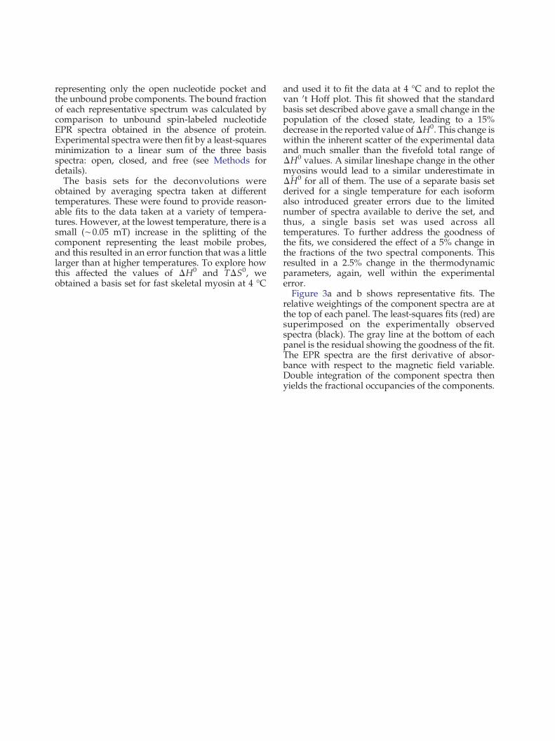

(a)(b)
i
g
.
3
.
D
e
c
o
n
v
o
l
u
t
i
o
n
o
f
f
a
s
t
s
k
e
l
e
t
a
l
a
c
t
o
m
y
o
s
i
n
.
R e p r e s e n t a t i v e s p e c t r a
f o r
c l o s e d
( g r e e n ) ,
o p e n
( b l u e ) , a n d
u n b o u n d
( m a g e n t a )
a r e
s u m m e d
t o c r e a t e
a
l e a s t - s q u a r e s
b e s t - f i t
s p e c
t r u m
( r e d ,
m i d d l e p a n e l )
t o t h e
s p e c t r u m
( b l a c k ,
m i d d l e
p a n e l )
w i t h t h e
s h o w n
i n
g r a y
b e l o w .
T h e
o p e n
a n d
c l o s e d r e p r e s e n t a t i v e
s p e c t r a
a r e
a
c o m b i n a t i o n
o f
b o u n d
a n d f r e e
s p e c t r a ; t h u s ,
t h e
p u r e
f r e e
s p e c t r u m i s
a c t u a l l y
c a s e st o
c o m p e n s a t ef o r
t h ef r a c t i o n
o f
i n
t h e
e x p e r i m e n t a l
s p e c t r u m .
( a )
R o o m t e m p e r a t u r e a n d ( b ) 4° C .
y o s i nS p e e d
a n d
E f f i c i e n c y
Once the population of each spectral componenthas been determined as described above, theequilibrium constant Keq=[open fractional compo-nent of the spectrum]/[closed fractional component]can be determined from a single spectrum. Values ofKeq were used to calculate the free energy of theclosed-to-open transition, ΔG0 =−RT ln Keq. Deter-mination of ΔG0 as a function of temperature wasused to determine ΔH0 and ΔS0. Values for ΔG0,below, are given at 25 °C.Under most conditions, myosin with SLADP
bound shows a mixture of open and closed states.In rabbit fast skeletal muscle myosin in the absenceof actin, the closed form is favored, with a ΔG0 forthe closed-to-open transition of 2.0±0.1 kJ mol−1. In
the presence of rabbit skeletal actin, the closed-to-open transition becomes favorable, with a ΔG0 of−1.1 kJ mol−1.
ΔG0 correlation to speed
An advantage of using spin-labeled nucleotides isthat they can be used in any myosin that can beobtained in sufficient quantities for EPR spectros-copy. Rabbit slow skeletal myosin also showed aclosed nucleotide pocket in the absence of actin(ΔG0 =1.9 kJ mol−1). For slow actomyosin, the opennucleotide pocket state becomes greatly favored,ΔG0 =−3.9 kJ mol−1. Surprisingly, slow musclewith an unloaded sliding velocity of 1.6 μm s−1 hasa greater propensity for an open conformation ofthe nucleotide pocket compared with fast skeletalmyosin (4.7 μm s−131) (both values of velocity weremeasured using skinned rabbit skeletal musclefibers). Because faster myosin has a faster ADPrelease rate, we anticipated that the open nucleo-tide pocket conformation would be more energet-ically favored rather than less occupied as seen inour data.In order to see if an increased velocity was the key
correlative factor for this observation, we extendedour analysis to other myosins with a range ofspeeds. We examined pig cardiac muscle, includingatrial (fast, 2.7 μm s−15) and ventricle (slow, 1.3 μms−15). These tissues recapitulated the trend seen infast and slow skeletal muscles, with the ΔG0 for fastatrial at −1.9 kJ mol−1 and slow ventricle at −3.9 kJmol−1. Smooth muscle from chicken gizzard isslower (0.67 μm s−1)32 than slow skeletal andcardiac. Purified chicken gizzard myosin bound toactin shows a very open nucleotide pocket with aΔG0 of −3.8 kJ mol−1. Similar results were obtainedin expressed recombinant motor domain withessential light chain alone bound to actin. Thecorrelation extends to faster muscle myosins aswell. Even in the actomyosin state, in Drosophilamelanogaster indirect flight muscle (in vitro motilityof 6.4 μm s−133), the ΔG0 for the closed-to-opentransition is +2.9 kJ mol−1.In addition, we looked at non-muscle myosins.
For velocity, we used the in vitro motility rate as ananalogue of unloaded sliding velocity. Baculovirusexpressed chicken myosin V is a slow myosin withan in vitro motility rate of 0.3 μm s−1,8 and it has anopen nucleotide pocket when bound to actin, ΔG0 =−3.9 kJ mol−1. Expressed Dictyostelium myosin II isintermediate between fast and slow skeletal myosins[2.4 μm s−1 (average value from Refs. 34 and 35)]and has an intermediate ΔG0 of −2.4 kJ mol−1
favoring the open nucleotide pocket.Thus, the degree that the nucleotide pocket stays
closed in the actomyosin state correlates to slidingvelocity (Fig. 4). There are several factors thatcontribute to the sliding velocity for a given myosin,
Fe x p e r i m e n t a l
r e s i d u a l
s u b t r a c t e di n
s o m e
f r e e
n u c l e o t i d e
M

Table 1
Myosin
Slidingvelocity(μm/s)
ΔG0 25 °C(kJ mol−1)a
ΔH0
(kJ mol−1)
ΔS0
(J mol−1
K−1)
IFM 6.4 +2.9±0.1 10.4±3.6 25±12Fast skeletal 4.7 −1.1±0.2 17.7±2.8 63±10Atrial 2.7 −1.9±0.1 25.5±5.3 92±2Dictyostelium 2.4 −2.7±0.2 44.1±1.7 157±5Slow
skeletal1.6 −3.9±0.2 50.4±2.2 182±8
Ventricle 1.3 −3.9±0.2 55.5±9.0 199±31Smooth 0.7 −3.8±0.1 43.9±0.1 160±1Myosin V 0.3 −3.9±0.1 50.1±2.0 181±7
a Extrapolated from van 't Hoff plot.
-4
-2
0
2
4
clos
ed -
> o
pen
Δ G0 2
5°C
(kJ
mol
-1)
76543210sliding velocity (μm s-1)
IFM
Fas
t Fib
ers
Slo
w F
iber
s
Sm
ooth
Mus
cle
Dic
ty
Myo
sin
V
vent
ricle
atriu
m
Fig. 4. ΔG0 versus in vitro motility. The equilibriumbetween closed and open nucleotide pockets calculatedfrom EPR data for a range of myosins from slow myosin Vto fast IFM. Velocities are at room temperature (22–25 °C),taken from fiber shortening for fast and slow muscles,31
cardiac muscle,5 and smooth muscle;32 where fibershortening velocities are not available, in vitro slidingactin filament assay values were used for myosin V,8
Dictyostelium myosin,34,35 and IFM.33
-15
-10
-5
0
5
10
15
0.00360.00340.0032
IFMFast Skeletal FibersDictySmooth Muscle
1/T (K-1)
-R ln
K (
J K
-1 m
ol-1
)
Fig. 5. Representative van 't Hoff plots. Data for theclosed-to-open transition equilibrium (K) are fit by −R lnK=ΔH0 (1/T)−ΔS0. The fits show that both ΔH0 and ΔS0
increase going from faster IFM and fast skeletal fibers toslower Dictyostelium and smooth muscle myosins. Errorbars give the standard error of the mean for each group ofdata points with approximately 6 data points perobservation.
84 Myosin Speed and Efficiency
including lever arm length and other rate-limitingsteps, which could account for the deviation from alinear relationship. However, as we show below, thedata suggest a model in which the nucleotide pocketreflects the energy of the actomyosin•ADP(A•M•D) complex and should correlate to thethermodynamics of this complex formation.
Temperature variation gives ΔH0 and TΔS0
The closed-to-open transition is temperaturesensitive. For all of the myosins studied, theequilibrium of the actomyosin complex shifts backto favor the closed conformation (Fig. 2b) as thetemperature is lowered. We measured the equilib-rium constant as a function of temperature for eachof the myosins studied. Plotting the log of theequilibrium constant as a function of reciprocaltemperature yields the entropic and enthalpiccomponents by a fit to the linear equation −R lnK=ΔH0 T−1 −ΔS0 (Fig. 5). The magnitudes of ΔH0
and ΔS0 indicate a significant conformationalchange (Table 1). For example, the values are ofthe same magnitude as observed for kinesin necklinker undocking.36 Naber et al. showed a muchlarger ΔH0 (79 kJ mol−1) and ΔS0 (280 J K−1 mol−1)for fast skeletal acto-S1.20 However, those valueswere from S1 and an earlier version of thetemperature control that had calibration issues.The exact nature of the conformational change isdifficult to determine from these experiments,whether it's dynamics in the myosin–actin interfaceor dynamics within the myosin itself.
Discussion
Our previous work with spin-labeled nucleotideshas shown that the mobility of bound probesincreases when myosin binds to actin, implyingthat the nucleotide pocket of myosin adopts a moreopen conformation.20 We have interpreted ourspectral changes in terms of opening and closingof the switch 1 region because this is the region withknown structural changes that would be in aposition to influence the mobility of our ribose-bound probe.28 Known movements of switch 2would not alter probe mobility. As discussed inIntroduction, a variety of techniques agree that thenucleotide pocket of myosin opens upon binding toactin.20–22,24,26,27,37
85Myosin Speed and Efficiency
Our previous results showing an opening of thenucleotide pocket upon binding to actin support ourfindings in light of the structural interpretationabove. The bond with actin promotes release ofphosphate first and, subsequently, ADP. Release ofADP would be expected to be enhanced by an openconformation of the nucleotide pocket, weakeningthe association of ADP. This has been postulated tobe the state from which ADP is released in previouskinetic models.38 Furthermore, a greater populationof the open state could be expected to promote morerapid release of ADP, as has also been postulated inprevious kinetic models.38 Thus, the present results,which show that the more open form is favored inslower myosins, is counterintuitive. One wouldexpect slower myosins to favor the closed form toslow the release of ADP. However, we note that therate of release of ADP is not always correlated withthe switch 1 conformation. X-ray structures ofkinesin and nonclaret disjunctional protein showan extremely open switch 1 coupled with very tightbinding and a very slow release rate of ADP. Releaseof nucleotides from G-proteins is associated withalterations at the nucleotide site in the P-loop andswitch regions by guanine exchange factors.39
Our observation that actomyosin•ADP has mul-tiple conformational states is consistent with otherkinetic and spectroscopic studies for several myosinsstudied here, including myosin V,40,41 smoothmuscle myosin II,42 and Dictyostelium myosin.26
The kinetics of myosin V actomyosin•ADP areconsistent with high- and low-affinity states asmeasured in Rosenfeld et al.40 This could correspondto the open and closed states with a ΔG0 at 20 °C of−3.0 kJ mol−1. Hannemann et al. also measured asimilar apparent equilibrium between high- andlow-affinity states, −3.3 kJ mol−1 at 25 °C.41 Both ofthese values are close to the equilibrium observed inEPR of −3.0 kJmol−1 and −3.9 kJ mol−1 for 20 °C and25 °C, respectively. In the absence of actin, theHannemann equilibrium Kisom,Mg of +1.3 kJ mol−1 isclose to what we observe with EPR in the absence ofactin (+0.9 kJ mol−1). This value for ΔG0 is differentfrom that measured by Rosenfeld (−1.9 kJ mol−1),although both papers agree on the general observa-tion that binding to actin results in a relative increasein the fraction of the open nucleotide pocket.Spectroscopic experiments with smooth musclemyosin II are also consistent with two fluorescentstates in the actomyosin•mant-ADP state. At 20 °C,acto-S1•ADP has a ΔG0 of −0.7 kJ mol−1 comparedto our EPR-derived value of−3.0 kJmol−1. Rosenfeldet al. obtained a value strongly favoring the closedform, with a ΔG0 of +7.6 kJ mol−1 in the absence ofactin, which is greater than that found here.42 Theyalso found the entropy and enthalpy values by van 'tHoff analysis in the absence of actin. The ΔH0 andΔS0 values were 44 kJ mol−1 and 123 J K−1 mol−1,respectively, similar to our values of 44 kJ mol−1 and
160 J K−1 mol−1 that were calculated for actomyosinand 32 kJ mol−1 and 109 J K−1 mol−1 for myosin.Fluorescence spectroscopy experiments with Dic-
tyostelium myosin also showed evidence for twoA•M•ADP states.26 Using a construct with a singletryptophan mutant, F239W, in a tryptophan-freebackground, they found a ΔG0 of +0.8 kJ mol−1 forADP binding in the absence of actin at 12 °C, close toour own value of +1.0 kJ mol−1 measured by EPRspectroscopy using the same mutant. In summary,there are a variety of techniques that agree that themyosin nucleotide pocket has two conformations,with reasonable agreement in the magnitude of theenergetic differences between them, given theuncertainties in the methods.
A model to explain the correlation between ΔG0
and velocity
Below, we discuss a model that can explain theunexpected relationship between the open and theclosed conformations of the nucleotide pocket andADP release. As with previous models, ADP releaseoccurs only from the open state. In previous models,the rate-limiting state in ADP release was thetransition from the closed to the open state. Oncein the open state, ADP release was fast. If instead,the rate-limiting step is placed between the openstate and the rigor state and the free energy of theopen state is varied while keeping the free energiesof the closed and ADP release transition states fixed,the correlation between velocity and ΔG0 can beexplained. The lower the free energy of the openstate relative to the closed state, the more the openconformation of the nucleotide pocket is favored. Asseen in Fig. 6, slow myosin (red) has a lower energystate than the fast myosin (blue) in the A•Mopen•Dstate. The greater ΔG0 between the open state andthe transition state results in a slower release ofADP, assuming that the ADP-releasing transitionstate A•M‡–D is similar in energy for faster andslower myosins. We further show below that thismodel explains the correlation between ΔG0 andvelocity and makes a connection between both ofthese observations and the efficiency of performingmechanical work.As discussed in Introduction, there is a reason-
able inverse correlation between the velocity andthe affinity of ADP for the actomyosin complexKAD.
11 Our own measurements, however, relatenot directly to the affinity of ADP but to therelative energies of the A•M•ADP states,A•M•ADP states, A•Mclosed•D and A•Mopen•D,in Fig. 6. The four affinities defining the binding ofactin, myosin, and ADP to form the aggregateA•M•ADP state are related by virtue of beinginvolved in a closed thermodynamic box (toppanel, Fig. 7a). The thermodynamic box definesthe association constants KD, KA, KAD, and KDA.

A•Mopen•D
A•Mopen•D
wor
ksp
eed
wor
ksp
eed
A•M•D•P
A•M
A•M‡ DA•Mclosed•D
Fre
e en
erg
y
Fig. 6. Energy diagram. Slowmyosin (red) has a lower energy state than the fast myosin (blue) in the A•Mopen•D state.The greater ΔG0 causes a slower release of ADP, assuming that the ADP-releasing transition state A•M‡–D is similar inenergy for faster and slower myosins. The relationship between speed and efficiency is illustrated by the partitioningbetween two phases. The steps up to the A•Mopen•D state contribute to the work output of the myosin; the large free-energy drop in the slower myosin allows a greater portion of the free energy to be used for work, resulting in a moreefficient myosin. The faster myosin budgets less energy for the work phases of the stroke but has a greater fraction ofenergy left over to drive nucleotide release, resulting in a faster but less efficient myosin.
86 Myosin Speed and Efficiency
The values most directly associated with theA•M•ADP state, KAD and KDA, have a strongercorrelation with the velocity of the myosin sincethey modulate the fraction of myosin in the boundactomyosin–diphosphate state. The coupling coef-ficient is a measure of the reduction of KAD andKDA compared to KD and KA. Slow myosins have alower coupling coefficient, indicating that ADPaffinity is high, with or without actin, and actinaffinity is high, with or without ADP. On the otherhand, the coupling coefficient is high in fastmyosins, indicating that ADP binding is destabi-lized in actomyosin relative to myosin. If oneassumes, arbitrarily, that the free energy of theA•Mclosed•D state (Fig. 6) is reasonably constantwith velocity, and with the A•M‡•D transition-state intermediate only sparsely populated, thenchanges in the free energy of the A•Mopen•D statewill determine the overall free energy of theaggregate A•M•D state and the overall affinityof actomyosin to ADP. If the other states (M, A•M,and M•D) were relatively constant in energy, thiswould indicate that the A•M•ADP state isspecifically destabilized in faster myosins. This isrepresented diagrammatically in Fig. 7b by thetilting of the thermodynamic boxes with respect tothe vertical free-energy axis. Comparing the redand blue boxes, the red box vertex representingthe A•M•Dslow myosin isoform is of lower freeenergy than the corresponding vertex in the bluebox representing the A•M•Dfast isoform. In otherwords, the decrease in free energy associated with
the formation of the A•M•ADP complex (i.e., theA•Mclosed•D-to-A•Mopen•D transition) becomesgreater as the velocity decreases for the slowerisoform.As shown in Figs. 6 and 7, the free energy
experimentally measured here, ΔG0, reports onthe change in energy between the two loweststates of the A•M•ADP complex: A•Mclosed•D toA•Mopen•D. As noted above, if the free energy ofA–Mclosed–D is reasonably constant with velocity,then changes in the free energy of the A•Mopen•Dstate will determine the overall free energy of theA•M•D state and overall affinity of actomyosin toADP. Thus, the relationship between the change infree energy and the equilibrium constant betweenthe A•Mclosed•D and the A•Mopen•D states and thefact that the transition out of the A•Mopen•D state israte limiting for sliding velocity imply that the log ofthe velocity would correlate inversely to the stabili-zation of the open state. Thus, this model explainsthree things: (1) the pocket is more open in slowermyosins, (2) the affinity of ADP for the A•Mcomplex is greater, and (3) a greater amount ofenergy is released in the formation of the A•M•ADPcomplex. This last quantity can further be related tomuscle efficiency, as discussed below.We also note that the slower myosins are
associated with greater values of ΔH0 and TΔS0.The magnitude of the change is great, amounting toover a fivefold difference between the fastestmyosin, ΔH0=10 kJ mol−1, and the slowest myosin,ΔH0=50 kJ mol−1. As noted in the Methods section,

20
18
16
14
12
10
8
1.00.0-1.0ln velocity (μm s-1)
ln K
ln KD ln KA ln KDA ln KAD
E
MD
AM
M
M
KD
KA
KAD
KDA
M MD
AMDAM
AM
MD
AMDFast
AMDSlow
KADKA
KD KDA
(a) (b)
Fig. 7. (a) Equilibrium constant K versus velocity. Association equilibrium constants estimated from published valuesfor the closed thermodynamic box (after Ref. 15). The box represents two paths from myosin to actomyosin•ADP, and inboth cases, the second step has a stronger influence on myosin sliding speed. K values are taken from the literature. Fromslowest to fastest, the myosins are human non-muscle myosin IIB,43 chicken myosin V,44 chicken gizzard smooth musclemyosin II,45 Dictyostelium myosin II,46 bovine ventricle cardiac myosin II, rabbit slow skeletal muscle (soleus) myosin II,47
rabbit fast skeletal (psoas) myosin II,11 and Drosophila flight muscle.33,48 Affinities at 100 mM KCl were estimated frompublished values using the equation log K α 5×√ionic strength49 and corrected to 25 °C using the published temperaturedependence.50 (b) Energy landscape for the closed thermodynamic box for slow versus fast myosins. Data from the graphshowing the energy difference between fast (blue) and slow (red) myosins are from the estimated K values correspondingto 0.3 μm s−1 for slow myosins and 7 μm s−1 for fast myosins. Vertical axis scales the relative energy differenceproportional to log K. Setting the energy for the myosin state as the reference, there is only a modest difference betweenslow and fast myosins in the A•M andM•D states. However, there is a large difference in the A•M•D state resulting in akinked box for fast myosins (blue). The energy landscape for a fast myosin has a kink in the middle, indicating that thecoupling constant relates specifically to destabilization of the A•M•D state.
87Myosin Speed and Efficiency
the changes in lineshape that occur with tempera-ture lead to a 15% underestimate in the values ofΔH0 for all myosins. If changes in ΔH0 and TΔS0
reflect alteration in the size of the protein interfaceformed in the actomyosin complex, the observedcorrelations would suggest that a greater amount ofprotein–protein interface is formed in the open stateof the pocket in slower myosins. It is possible thatthis interface contributes to the lower free energyof the A•M•ADP complex. The collection ofmyosins at the lowest end of the speed rangehas approximately the same ΔG0 for the closed-to-open equilibrium. There might no longer be a wayto stabilize the A•M•ADP complex withoutequally affecting both A•Mclosed•ADP and theA•Mopen•ADP states.
Efficiency
There is evidence that the efficiency, defined as thepercentage of the chemical energy available from
ATP that is converted to mechanical work, is greaterin slower fibers. This was first determined from acomparison of two vertebrate muscles: frog andtortoise fibers.51 In particular, this can be seen uponcomparison of different muscle types from a givenorganism or upon comparison of different isoformsfrom the same family. For example, mouse slow-twitch soleus was reported to have an efficiency of43% compared to fast-twitch extensor digitorumlongus with an efficiency of 33%.52 A similarcorrelation is seen with fast and slow muscles ofthe chicken.53 The efficiency ofmyosin V is estimatedfrom in vitro measurements to approach 100%.54
Myosin XI in tobacco yellow bright-2 cells is anotherprocessive myosin that is functionally and structur-ally similar to myosin V in that it takes 36-nm stepscorresponding to the actin pseudo-repeat but has avelocity that is 20-fold faster than myosin V. For thismyosin isoform, the efficiency drops down to 20%.9
This correlation further extends across species fromvery slow muscles such as tortoise rectus femoris,

1.0
0.8
0.6
0.4
0.2
0.0
86420
sliding velocity (μm s-1)
frac
tiona
l effi
cien
cy
Myosin V
Myosin XI
Tortiose
ChickenALD
Frog
ChickenPLD
MouseSoleus Mouse
EDL
DrosophilaIFM
Fig. 8. Efficiency versus velocity. Fractional efficienciesfor muscle (squares) and processive non-muscle (circles)myosins. The best least-squares fit is given by eff=−1.6 ln v+0.59, assuming a linear relationship between fractionalefficiency (eff) and the logarithm of velocity (v). Muscles:tortoise rectus femoris,51 chicken anterior latissimus dorsi,53 frogsartorius,51 chicken posterior latissimus dorsi,53 mouse soleus,52
Drosophila adult indirect flight muscle,55 and mouse extensordigitorum muscle.52 Non-muscles: MV chicken myosin V54
and MXI tobacco yellow bright-2 cells myosin XI.9
88 Myosin Speed and Efficiency
efficiency of 77%,51 and myosin V, 80%,8 to thefastest muscles considered here, Drosophila indirectflight muscle. Drosophila flight muscle (IFM) has anestimated efficiency of around 30%.55 Significantly,extrapolating the correlation shown in Fig. 8 out tozero efficiency gives a theoretical maximum velocityfor actomyosin of around 50 μm s−1, which is on theorder of the fastest observed sliding velocities fromplant myosin XI.56
There is evidence that the stronger the A•M•ADPcomplex, the more energy is available to do work. Avery large amount of free energy is released uponformation of this complex, and the high efficienciesof some myosins would require that a large fractionof this free energy be used to do work. A number ofmodels have assumed that the energy released information of this complex is used to drive theproduction of mechanical work (Ref. 57 and refer-ences therein). Variation of the strength of theA•M•ADP complex has additionally shown thatthe isometric force is directly correlated to the freeenergy released by formation of the A•M•ADPcomplex.58 Thus, the model above, which linksslower velocities to a more open myosin nucleotidepocket and to a strongerA•M•ADP complex,wouldexplain the correlation between efficiency andvelocity. In this model, a partitioning of some subsetof the total energy available between work (W) andADP release would result in a negative correlationbetween the efficiency and the logarithm of slidingvelocity. Figure 8 shows that, indeed, a very good fitis obtained, making the simplest possible assump-tion that the relationship between efficiency andlogarithm of sliding velocity is linear.
Discussion summary
EPR spectroscopy using a spin-labeled nucleotidehas resulted in structural information about theactomyosin•ADP state, a state that has not beenstudied by X-ray crystallography. Furthermore,quantitative analysis results in thermodynamicinformation about this state, confirming equilibriumbetween open and closed conformations of thenucleotide pocket. Because the spectroscopic probewas attached to a nucleotide that bound effectivelyto myosin, a panel of myosins with a greater than 20-fold range of sliding velocities could be studied,resulting in the counterintuitive result that the open-nucleotide-pocket state is favored in myosins thatslowly release ADP, even though ADP release isthought to occur from the open conformation. Ouranalysis suggests that destabilization of the openactomyosin ADP state in faster myosins lowers thefree-energy barrier for ADP release through atransition-state intermediate, resulting in fasterADP release. This destabilization comes at the costof work-producing transitions leading up to theopening of the nucleotide-binding pocket. This
model results in a simplified thermodynamicmodel for the relationship between speed andefficiency in myosin-based motility.
Methods
Protein preparations
F-actin was purified from rabbit skeletal muscle.59 TheDictyostelium discoideum tryptophan-free M761 motordomain cDNA fragment was expressed in Dd AX2-ORF+cells and isolated as described previously.60 Cardiac atrialand ventricular myosins were isolated from porcine heartsusing the protocols of Tonomura et al.61 Chicken gizzardsmooth muscle myosin was isolated following theprotocols of Ikebe and Hartshorne.62 Myosin V motordomain was expressed, harvested, and purified as inSweeney et al.63 Myosin subfragment-1 (S1) was preparedby chymotryptic digestion of myosin as described inWeeds and Taylor.64
Myosin fiber preparations
Fast (psoas) and slow (soleus) rabbit skeletal fibers wereharvested and chemically skinned as described in Naberet al.20 IFM fibers were dissected from a 2- to 3-day-old

89Myosin Speed and Efficiency
female Drosophila and chemically skinned and stored aspreviously described, except that fibers were not split inhalf.65 The demembranated fibers were used within5 days.
Solutions
The basic rigor experimental buffer consisted of 5 mMMg(OAc)2, 1 mM ethylene glycol bis(β-aminoethyl ether)N,N′-tetraacetic acid, and 40 mM 4-morpholinepropane-sulfonic acid, pH 7.0. For experiments involving skinnedfibers, 50 mM KOAc was added to the rigor buffer.
EPR spectroscopy
For experiments with S1 or expressed motor domain,with or without actin present, protocols for spin labelingand mounting the protein in a capillary for EPR spectralaccumulation were as in Naber et al.20 IFM fibers werewashed in a 500-fold excess (v/v) of rigor buffer andisolated via centrifugation to eliminate the DTT present inthe glycerination and storage buffers. The process wasrepeated four times. Rabbit psoas and soleus fibers wereminced with a razor blade for EPR spectroscopy experi-ments. The fiber preparation was then spin labeled andmounted on a flat cell for spectral accumulation asdescribed in Naber et al.20 EPR measurements wereperformed with a Bruker EMX EPR spectrometer (Bill-erica, MA). First-derivative X-band spectra were recordedin a high-sensitivity microwave cavity using 50-s, 10-mT-wide magnetic field sweeps. The instrument settings wereas follows: microwave power, 25 mW; time constant,164 ms; frequency, 9.83 GHz; and modulation, 0.1 mT at afrequency of 100 kHz. Each spectrum used in data analysiswas an average of 5–50 sweeps from an individualexperimental preparation. Temperature was controlledby blowing dry air (warm or cool) into the cavity andmonitored using a thermistor placed close to the exper-imental sample. Effective cone angles of mobility weredetermined following Griffith and Jost and Alessi et al.29,30
Deconvolution of spectral components
The goal is to represent the experimentally observedspectrum as a linear combination of three basis spectra.The basis spectra are the spectral components from theopen conformation of the nucleotide pocket, the closedconformation of the nucleotide pocket, and unboundprobe free in solution. Each spectrum is discretized into1024 individual data pairs with a cubic spline interpola-tion used to align the data points. Let Oi, Ci, Fi, and Ei,i=1.1024, be the discretized open, closed, unbound probefree in solution, and experimentally observed spectra.Then the relative weighting of the three individual open,closed, and free spectra will be given by the constants, A,B, and C that minimize
X1024
i=1
AOi + BCi + CFi−Eið Þ2
(least-squares minimization). The minimization was doneusing the Matlab V7.8 routine fminsearch (Nelder–Mead
simplex method) with a convergence criteria of 0.0001 forTolFun and TolX. Then AOi+BCi+CFi, i=1.1024, is theleast-squares fit to the experimentally determined spectrum.
Acknowledgements
This work was supported by National Institutesof Health grants HL032145 (T.J.P. and K.F.-S.),AR042895 (R.C. and N.N.), GM077067 (E.P. andN.N.), AR055611 (D.M.S. and C.C.E.), HL063798(C.L.B.), and GM33289 (J.S.), Burroughs WellcomeCareer Award at the Scientific Interface, AmericanHeart Association Postdoctoral Fellowship, JaneCoffin Childs Postdoctoral Fellowship (A.R.D.), andthe European Research Council IDEAS forcemap(208319) (A.M.-C.).
References
1. Bottinelli, R., Betto, R., Schiaffino, S. & Reggiani, C.(1994). Unloaded shortening velocity and myosinheavy chain and alkali light chain isoform compo-sition in rat skeletal muscle fibres. J. Physiol. 478,341–349.
2. Weiss, A. & Leinwand, L. A. (1996). The mammalianmyosin heavy chain gene family. Annu. Rev. Cell Dev.Biol. 12, 417–439.
3. Bottinelli, R., Canepari,M., Reggiani, C. & Stienen, G. J.(1994). Myofibrillar ATPase activity during isometriccontraction and isomyosin composition in rat singleskinned muscle fibres. J. Physiol. 481, 663–675.
4. Fitts, R. H. (1994). Cellular mechanisms of musclefatigue. Physiol. Rev. 74, 49–94.
5. Malmqvist, U. P., Aronshtam, A. & Lowey, S. (2004).Cardiac myosin isoforms from different species haveunique enzymatic and mechanical properties. Bio-chemistry, 43, 15058–15065.
6. Arner, A., Lofgren, M. & Morano, I. (2003). Smooth,slow and smart muscle motors. J. Muscle Res. CellMotil. 24, 165–173.
7. Swank, D. M., Vishnudas, V. K. & Maughan, D. W.(2006). An exceptionally fast actomyosin reactionpowers insect flight muscle. Proc. Natl Acad. Sci.USA, 103, 17543–17547.
8. Mehta, A. D., Rock, R. S., Rief, M., Spudich, J. A.,Mooseker, M. S. & Cheney, R. E. (1999). Myosin-Vis a processive actin-based motor. Nature, 400,590–593.
9. Tominaga, M., Kojima, H., Yokota, E., Orii, H.,Nakamori, R., Katayama, E. et al. (2003). Higherplant myosin XI moves processively on actin with35 nm steps at high velocity. EMBO J. 22, 1263–1272.
10. Stienen, G. J., Kiers, J. L., Bottinelli, R. & Reggiani, C.(1996). Myofibrillar ATPase activity in skinned humanskeletal muscle fibres: fibre type and temperaturedependence. J. Physiol. 493, 299–307.
11. Nyitrai, M., Rossi, R., Adamek, N., Pellegrino, M. A.,Bottinelli, R. & Geeves, M. A. (2006). What limits thevelocity of fast-skeletal muscle contraction in mam-mals? J. Mol. Biol. 355, 432–442.

90 Myosin Speed and Efficiency
12. Bottinelli, R. (2001). Functional heterogeneity ofmammalian single muscle fibres: do myosin isoformstell the whole story? Pfluegers Arch. 443, 6–17.
13. Siemankowski, R. F., Wiseman, M. O. & White, H. D.(1985). ADP dissociation from actomyosin subfrag-ment 1 is sufficiently slow to limit the unloadedshortening velocity in vertebrate muscle. Proc. NatlAcad. Sci. USA, 82, 658–662.
14. Weiss, S., Rossi, R., Pellegrino, M. A., Bottinelli, R. &Geeves, M. A. (2001). Differing ADP release rates frommyosin heavy chain isoforms define the shorteningvelocity of skeletal muscle fibers. J. Biol. Chem. 276,45902–45908.
15. Nyitrai, M. & Geeves, M. A. (2004). Adenosinediphosphate and strain sensitivity in myosin motors.Philos. Trans. R. Soc. London, Ser. B, 359, 1867–1877.
16. Vetter, I. R. & Wittinghofer, A. (2001). The guaninenucleotide-binding switch in three dimensions. Science,294, 1299–1304.
17. Naber, N., Minehardt, T. J., Rice, S., Chen, X.,Grammer, J., Matuska, M. et al. (2003). Closing of thenucleotide pocket of kinesin-family motors uponbinding to microtubules. Science, 300, 798–801.
18. Wong, Y. L., Dietrich, K. A., Naber, N., Cooke, R. &Rice, S. E. (2009). The Kinesin-1 tail conformationallyrestricts the nucleotide pocket.Biophys. J. 96, 2799–2807.
19. Larson, A. G., Naber, N., Cooke, R., Pate, E. & Rice,S. E. (2010). The conserved L5 loop establishes thepre-powerstroke conformation of the Kinesin-5motor, eg5. Biophys. J. 98, 2619–2627.
20. Naber, N., Purcell, T. J., Pate, E. & Cooke, R. (2007).Dynamics of the nucleotide pocket of myosin mea-sured by spin-labeled nucleotides. Biophys. J. 92,172–184.
21. Naber, N., Malnasi-Csizmadia, A., Purcell, T. J.,Cooke, R. & Pate, E. (2010). Combining EPR withfluorescence spectroscopy to monitor conformationalchanges at the myosin nucleotide pocket. J. Mol. Biol.396, 937–948.
22. Coureux, P. D., Sweeney, H. L. & Houdusse, A. (2004).Three myosin V structures delineate essential featuresof chemo-mechanical transduction. EMBO J. 23,4527–4537.
23. Holmes, K. C., Angert, I., Kull, F. J., Jahn, W. &Schroder, R. R. (2003). Electron cryo-microscopyshows how strong binding of myosin to actin releasesnucleotide. Nature, 425, 423–427.
24. Sun, M., Oakes, J. L., Ananthanarayanan, S. K.,Hawley, K. H., Tsien, R. Y., Adams, S. R. & Yengo,C. M. (2006). Dynamics of the upper 50-kDa domainof myosin V examined with fluorescence resonanceenergy transfer. J. Biol. Chem. 281, 5711–5717.
25. Robertson, C. I., Gaffney, D. P., II, Chrin, L. R. &Berger, C. L. (2005). Structural rearrangements in theactive site of smooth-muscle myosin. Biophys. J. 89,1882–1892.
26. Kintses, B., Gyimesi, M., Pearson, D. S., Geeves, M. A.,Zeng, W., Bagshaw, C. R. & Malnasi-Csizmadia, A.(2007). Reversiblemovement of switch 1 loopofmyosindetermines actin interaction. EMBO J. 26, 265–274.
27. Malnasi-Csizmadia, A., Dickens, J. L., Zeng, W. &Bagshaw, C. R. (2005). Switch movements and themyosin crossbridge stroke. J. Muscle Res. Cell Motil. 26,31–37.
28. Pate, E., Naber, N., Matuska, M., Franks-Skiba, K. &Cooke, R. (1997). Opening of the myosin nucleotidetriphosphate binding domain during the ATPasecycle. Biochemistry, 36, 12155–12166.
29. Griffith, O. H. & Jost, P. C. (1976). Lipid spin labels inbiological membranes. In (Berlinger, L. J., ed.),Academic Press, New York, NY.
30. Alessi, D. R., Corrie, J. E., Fajer, P. G., Ferenczi, M. A.,Thomas, D. D., Trayer, I. P. & Trentham, D. R. (1992).Synthesis and properties of a conformationally re-stricted spin-labeled analog of ATP and its interactionwith myosin and skeletal muscle. Biochemistry, 31,8043–8054.
31. Cuda, G., Pate, E., Cooke, R. & Sellers, J. R. (1997). Invitro actin filament sliding velocities produced bymixtures of different types of myosin. Biophys. J. 72,1767–1779.
32. Warshaw, D. M., Desrosiers, J. M., Work, S. S. &Trybus, K. M. (1990). Smooth muscle myosin cross-bridge interactions modulate actin filament slidingvelocity in vitro. J. Cell Biol. 111, 453–463.
33. Swank, D. M., Knowles, A. F., Kronert, W. A., Suggs,J. A., Morrill, G. E., Nikkhoy, M. et al. (2003). VariableN-terminal regions of muscle myosin heavy chainmodulate ATPase rate and actin sliding velocity.J. Biol. Chem. 278, 17475–17482.
34. Kron, S. J. & Spudich, J. A. (1986). Fluorescent actinfilaments move on myosin fixed to a glass surface.Proc. Natl Acad. Sci. USA, 83, 6272–6276.
35. Uyeda, T. Q., Abramson, P. D. & Spudich, J. A. (1996).The neck region of the myosin motor domain acts as alever arm to generate movement. Proc. Natl Acad. Sci.USA, 93, 4459–4464.
36. Rice, S., Cui, Y., Sindelar, C., Naber, N., Matuska, M.,Vale, R. & Cooke, R. (2003). Thermodynamic proper-ties of the kinesin neck-region docking to the catalyticcore. Biophys. J. 84, 1844–1854.
37. Schröder, R. R., Manstein, D. J., Jahn, W., Holden, H.,Rayment, I., Holmes, K. C. et al. (1993). Three-dimensional atomic model of F-acton decorated withDictyostelium myosin S1. Nature, 364, 171–174.
38. Geeves, M. A. & Holmes, K. C. (1999). Structuralmechanism of muscle contraction. Annu. Rev. Biochem.68, 687–728.
39. Bos, J. L., Rehmann, H. & Wittinghofer, A. (2007).GEFs and GAPs: critical elements in the control ofsmall G proteins. Cell, 129, 865–877.
40. Rosenfeld, S. S., Houdusse, A. & Lee Sweeney, H.(2005). Magnesium regulates ADP dissociation frommyosin V. J. Biol. Chem. 280, 6072–6079.
41. Hannemann, D. E., Cao, W., Olivares, A. O., Robblee,J. P. & De La Cruz, E. M. (2005). Magnesium, ADP,and actin binding linkage of myosin V: evidence formultiple myosin V–ADP and actomyosin V–ADPstates. Biochemistry, 44, 8826–8840.
42. Rosenfeld, S. S., Xing, J., Whitaker, M., Cheung, H. C.,Brown, F., Wells, A. et al. (2000). Kinetic and spectro-scopic evidence for three actomyosin:ADP states insmooth muscle. J. Biol. Chem. 275, 25418–25426.
43. Wang, F., Kovacs, M., Hu, A., Limouze, J., Harvey,E. V. & Sellers, J. R. (2003). Kinetic mechanism ofnon-muscle myosin IIB: functional adaptations fortension generation and maintenance. J. Biol. Chem.278, 27439–27448.

Copyright © 2022 FDOKUMEN